Isolation and partial characterization of secretory protein I from bovine parathyroid glands

Thymosin b4 and b10 Levels in Pre-Term Newborn OralCavity and Foetal Salivary Glands Evidence a Switch ofSecretion during Foetal DevelopmentSonia Nemolato1, Irene Messana2, Tiziana Cabras2, Barbara Manconi2, Rosanna Inzitari3, Chiara Fanali3,
Giovanni Vento4, Chiara Tirone4, Costantino Romagnoli4, Alessandro Riva1, Daniela Fanni1, Eliana Di
Felice1, Gavino Faa1, Massimo Castagnola3*
1 Dipartimento di Citomorfologia, Universita di Cagliari, Cagliari, Italy, 2 Dipartimento di Scienze Applicate ai Biosistemi, Universita di Cagliari, Cagliari, Italy, 3 Istituto di
Biochimica e di Biochimica Clinica, Universita Cattolica and/or Istituto per la Chimica del Riconoscimento Molecolare, CNR, Istituto Scientifico Internazionale (ISI) Paolo VI,
Roma, Italy, 4 Istituto di Clinica Pediatrica, Divisione di Neonatologia, Universita Cattolica, Roma, Italy
Abstract
Background: Thymosin b4, its sulfoxide, and thymosin b10 were detected in whole saliva of human pre-term newborns byreversed-phase high performance chromatography coupled to electrospray ion-trap mass spectrometry.
Methodology/Principal Findings: Despite high inter-individual variability, concentration of b-thymosins increases with aninversely proportional trend to postmenstrual age (PMA: gestational age plus chronological age after birth) reaching a valuemore than twenty times higher than in adult whole saliva at 190 days (27 weeks) of PMA (thymosin b4 concentration: morethan 2.0 mmol/L versus 0.1 mmol/L). On the other hand, the ratio between thymosin b4 and thymosin b10 exhibits a constantvalue of about 4 along all the range of PMA (190–550 days of PMA) examined. In order to investigate thymosin b4 origin andto better establish the trend of its production as a function of gestational age (GA), immunohistochemical analysis of majorand minor salivary glands of different pre-term fetuses were carried out, starting from 84 days (12 weeks) of gestational age.Reactive granules were seen in all glands with a maximum of expression around 140–150 days of GA, even though withhigh inter- and intra-individual variability. In infants and adults reactive granules in acinar cells were not observed, but just adiffuse cytoplasmatic staining in ductal cells.
Significance: This study outlines for the first time that salivary glands during foetal life express and secrete peptides such asb-thymosins probably involved in the development of the oral cavity and its annexes. The secretion increases from about 12weeks till to about 21 weeks of GA, subsequently it decreases, almost disappearing in the period of expected date ofdelivery, when the gland switches towards the secretion of adult specific salivary peptides. The switch observed may be anexample of further secretion switches involving other exocrine and endocrine glands during foetal development.
Citation: Nemolato S, Messana I, Cabras T, Manconi B, Inzitari R, et al. (2009) Thymosin b4 and b10 Levels in Pre-Term Newborn Oral Cavity and Foetal SalivaryGlands Evidence a Switch of Secretion during Foetal Development. PLoS ONE 4(4): e5109. doi:10.1371/journal.pone.0005109
Editor: Rory Edward Morty, University of Giessen Lung Center, Germany
Received January 7, 2009; Accepted March 5, 2009; Published April 1, 2009
Copyright: � 2009 Nemolato et al. This is an open-access article distributed under the terms of the Creative Commons Attribution License, which permitsunrestricted use, distribution, and reproduction in any medium, provided the original author and source are credited.
Funding: We acknowledge the financial support of Universita’ di Cagliari, Universita’ Cattolica in Rome, MIUR, Italian National Research Council (CNR), RegioneSardegna and Fondazione Banco di Sardegna, International Scientific Institute Paolo VI (ISI), and Telethon Grant N. GGP06170 thanks to their programs of scientificresearch promotion and diffusion. The funders had no role in study design, data collection and analysis, decision to publish, or preparation of the manuscript.
Competing Interests: The authors have declared that no competing interests exist.
* E-mail: [email protected]
Introduction
Beta thymosins are ubiquitous peptides having interesting intra-
and extra-cellular functions, whose name derives from their first
characterization from thymus extracts [1,2]. Thymosin b4 (Tb4) is
usually the most abundant b-thymosin in the cytoplasm of most
cell types and it plays pivotal roles in the cell cytoskeletal system as
G-actin sequestring peptide [3]. Tb4 frequently co-exists with a
second member of the b-thymosin family, i.e. thymosin b10 (Tb10)
in man, cat, rat, mouse and rabbit, thymosin b9 in sheep, calf and
pig [4]. Although the secretion pathway is not fully understood,
recent studies highlighted various extra-cellular roles for these
peptides [1]. For instance, Tb4 modifies the rate of attachment and
spreading of endothelial cells on matrix components inducing
matrix metalloproteinases [5], it stimulates the migration of
human umbilical vein endothelial cells [6], it is involved in the
repair of damaged cornea [7], it induces hair growth by activating
hair follicle stem cells [8], and it might be involved in the
development and repair of heart [9] and brain damages [10].
Whereas Tb4 is a potent enhancer of angiogenesis, Tb10 inhibits it
and changes of the two peptides ratio can exert either positive or
negative control [11]. Tb4 has been detected in human whole
saliva and tears by immunological techniques [12] and recent
studies of our group evidenced that in the oral cavity a main
contribution derives from crevicular fluid [13], where, as
demonstrated by Reti and co-workers [14], Tb4 plays an
important role in suppressing the production of interleukin-8
following stimulation by tumour-necrosis factor a and it acts on
the whole as antimicrobial, anti-inflammatory and anti-apoptotic
peptide on gingival fibroblasts. This study describes the detection
PLoS ONE | www.plosone.org 1 April 2009 | Volume 4 | Issue 4 | e5109

of high concentrations of thymosins b4 and b10 in the whole saliva
of pre-term newborns, during a survey carried out by proteomic
approaches. Because a crevicular origin for these peptides in
newborns has to be obviously excluded, further immunohisto-
chemical analyses of the major and minor salivary glands of
human fetuses and newborns of different postmenstrual age (PMA:
gestational age (GA) plus chronological age after birth) were
performed, in order to establish the source of the high amounts of
Tb4 detected in whole saliva and to add further information on
potential roles of this peptide during foetal development.
Materials and Methods
Ethics statementsThe study protocol and written consent forms were approved by
the Pediatric Department Ethics Committee of the Faculty of
Medicine of the Catholic University of Rome (according to the
instructions of the Declaration of Helsinki). Full written consent
forms were obtained from the parents of the newborns and all
rules were respected.
ReagentsAll common chemicals and reagents were of analytical grade
and were purchased from Farmitalia-Carlo Erba, (Milan, Italy),
Merck (Damstadt, Germany) and Sigma Aldrich (St. Louis, MI,
USA). Standards of thymosin b4 and thymosin b10 were purchased
from Bachem (Bubendorf, Switzerland).
ApparatusThe HPLC-ESI-IT-MS apparatus was a Surveyor HPLC
system (ThermoFinnigan, San Jose, CA, USA) connected by a T
splitter to a PDA diode-array detector and to an LCQ Deca XP
Plus mass spectrometer. The mass spectrometer was equipped
with an electrospray ion (ESI) source. The chromatographic
column was a Vydac (Hesperia, CA, USA) C8 column, with 5 mm
particle diameter (column dimensions 15062.1 mm).
Subjects enrolledTwelve newborns with a birth weight ranging between 500 g
and 1250 g and gestational age between 27–31 weeks (193–217
days), admitted to the Neonatal Intensive Care Unit (NICU) were
enrolled for this study. Infants with major congenital malforma-
tions or prenatal infections were excluded. Relevant clinical
information and major clinical outcomes with regard to the study
group are provided in our previous studies [15].
Moreover, 4 at-term infants (2 females, 2 males) born after
uncomplicated pregnancies and vaginal delivery and admitted to
Policlinico ‘‘A. Gemelli’’ nursery, were studied. Their mean6SD
GA and birth weight were 27267 days (3861 weeks) and
32806150 grams, respectively. All these 4 newborns had no
clinical problems and therefore they were discharged after three
days and breast-fed.
Sample collection and treatmentWhole saliva samples were collected from pre-term and at-term
newborns with a very soft plastic aspirator during the usual
newborn management. Pre-term newborn sample collection was
performed during several weeks after birth at established time
intervals (one or two weeks). When possible, it was also performed
after discharge from the neonatal unit, during the periodical check
visits with about one year follow-up. Saliva was not collected from
newborns when sample collection seemed to cause even a minimal
stress to the newborn and/or to his/her parents. For these reasons,
sample collection was more frequent during the hospital stay and
sporadic after this period. After collection samples were immedi-
ately mixed with an equal volume of 0.2% 2,2,2 trifluoacetic acid
(TFA) (v/v) in ice bath. After stirring, the acidic solution was
centrifuged at 9000 g for three min to remove the precipitate
(mainly mucins) and the acidic solution was immediately analyzed
by HPLC-ESI-MS (100 mL, corresponding to 50 mL of whole
saliva) or stored at 280uC until the analysis.
RP-HPLC-ESI-MS analysisThe following solutions were utilized for the chromatographic
separation: (eluent A) 0.056% aqueous TFA and (eluent B)
0.050% TFA in acetonitrile-water 80/20 (v/v). The gradient
applied was linear from 0 to 55% in 40 min, at a flow rate of
0.30 mL/min. The T splitter addressed a flow-rate of about
0.20 mL/min towards the diode array detector and 0.10 mL/min
towards the ESI source. During the first 5 min of separation the
eluate was not addressed to the mass spectrometer to avoid
instrument damage due to the high salt concentration. The diode
array detector was set at a wavelength of 214 and 276 nm. Mass
spectra were collected every 3 millisecond in the positive ion
mode. MS spray voltage was 4.50 kV and the capillary
temperature was 220uC.
Detection of b-thymosins in whole saliva anddetermination of their concentration
Deconvolution of averaged ESI mass spectra was automatically
performed either by the software provided with the Deca-XP
instrument (Bioworks Browser) or by MagTran 1.0 [16] software.
Experimental Mass values were compared with average theoretical
values available at the Swiss-Prot data bank (http://us.expasy.org/
tools), where Tb4 and Tb10 have the P62328 and P63313 codes,
respectively. Identification of thymosin b4 and thymosin b10 was
based on the coincidence of elution times, ESI-MS spectra and ESI-
MS/MS spectra with peptide standards, as described in previous
studies [13]. Thymosin b4 sulfoxide was identified by the
coincidence of elution times, ESI-MS and ESI-MS/MS spectra
with a standard obtained by treating thymosin b4 with H2O2.
Under the chromatographic conditions used the elution times
measured at the peak maximum were: Tb4, 20.0 (60.4) min; Tb4
sulfoxide, 18.5 (60.4) min; and Tb10, 21.3 (60.4) min. The average
masses obtained after deconvolution of the ESI spectra were: Tb4
4963.560.4, Tb4 sulfoxide 4979.560.4, Tb10 4936.560.4 Da.
The concentration of b-thymosins was determined by considering
the extracted ion current (XIC) peak area, which is linearly related to
the peptide absolute quantity, as established performing LC-MS
analyses on b-thymosin standards. The linear correlation between
absolute amount of the standards and XIC peak area (r = 0.999)
provided the following relationship: 1 picomol = 2.16106 (XIC peak
area) for Tb4, Tb10 and Tb4 sulfoxide. The concentration was
therefore computed by considering the absolute quantity and injected
sample volume (usually 100 mL of acidic solution corresponding to
50 mL of WS). The XIC peaks were revealed by selecting the
following charged ions: Tb4, [M+5H]5+ = 993.8 m/z; [M+4H]4+ =
1241.9 m/z; [M+3H]3+ = 1655.5 m/z; Tb4 sulfoxide [M+5H]5+ =
996.9 m/z; [M+4H]4+ = 1245.9 m/z; [M+3H]3+ = 1660.8 m/z;
Tb10, [M+5H]5+ = 988.3 m/z; [M+4H]4+ = 1235.1 m/z; [M+3H]3+ =
1646.5 m/z. The window for all these values was 60.5 m/z. The
percentage error of the measurements was less than 10%.
Immunohistochemical analysesSamples of the parotid, submandibular and minor salivary
glands (the posterior superficial lingual glands or Weber) at the
base of the tongue were obtained at autopsy from 5 subjects: two
bThymosins in Oral Development
PLoS ONE | www.plosone.org 2 April 2009 | Volume 4 | Issue 4 | e5109

foetuses of 12 and 13 weeks of gestational age, respectively, and 3
pre-term infants, with a gestational age of 20, 21 and 31 weeks. In
addition, two specimens of the major sublingual gland, the only
two that we were able to obtain with certainty from this gland,
belonging to the pre-term infants with 21 and 31 weeks of
gestational age, were studied. Moreover, specimens of only
submandibular and sublingual glands were obtained at the
autopsy from a sixth subject, an 18 months old boy who
underwent a sudden death. As a control group we utilized
surrounding tissue of surgical specimens from the peritumoral
parotid gland of 5 adult patients who underwent surgery for
resection of pleomorphic adenoma. Tissue samples were formalin-
fixed, paraffin embedded and routinely processed. Deparaffinized
5 m-thick sections were immunostained with the antibody directed
against Tb4 (rabbit) at a dilution of 1:500. The polyclonal antibody
from Bachem-Peninsula Lab (San Carlos, CA, USA) has not cross
reactivity against Tb10 and Ta1. The staining procedure was
ensured by using a biotin-labeled affinity isolated goat anti-rabbit
IgG followed by streptavidin conjugated to horseradish peroxidase
(Dako, Cytomation LS-AB2 System – HRP, Denmark). Sections
of reactive lymph nodes rich in macrophages (positive controls)
and lymphocytes (negative controls) were also used.
Results
Determination of b-thymosin concentrations in differentsamples under analysis
In general, the study of Tb4 and Tb10 concentrations in the
saliva during development of human newborns evidenced a
marked inter-individual variability and an overall age-dependent
trend, showing decrease of thymosin ‘s concentration as a function
of PMA.
Figure 1 shows the concentration of Tb4 and Tb10 detected in
whole saliva of one pre-term infant plotted as a function of
postmenstrual age, for example. Both thymosins b4 and b10 show
high concentration values immediately after birth at 26–27 weeks
(193 days of GA, in this subject Tb4 concentration in the range
1.0–1.5 mmol/L) which constantly decrease as a function of PMA,
reaching values comparable to that ones of adult (Tb4 concentra-
tion about 0.1 mmol/L [13]) after 38 weeks (270 days) of PMA.
Similar age-dependent trends were observed for all the investigat-
ed pre-term newborns despite the high inter-individual variability,
as evident from Fig 2 (12 pre-term newborns, 78 measurements at
different days of PMA). In the majority of subjects the
concentration of Tb4 measured in the range 27–31 weeks (190–
210 days) of PMA was 1–2 mmol/L, but one subject showed a
spike to about 6 mmol/L. The concentration of Tb10 in the same
range of PMA was between 0.1–0.4 mmol/L, with one subject
with a spike of 0.6 mmol/L (but not the same showing theTb4
spike). In Fig. 2 are also reported (squares) the mean values
observed in four at-term newborns (270 days of gestational age, 38
weeks) which values for Tb4 were 0.2160.12 mmol/L and
0.04960.022 mmol/L for Tb10. Fig. 2 also shows the ratio
between the two b-thymosins concentrations in the different
samples under analysis, which has a constant value of about 4 in all
the range of PMA studied (at-term newborns comprised), except
sporadic increases in some pre-term newborns not connected to
any evident clinical outcome. Obviously, the constant ratio implies
that the concentration of the two peptides correlates with high
statistical significance (N = 78, r = 0.679, p,0.001).
Similarly to the spikes of Tb4/Tb10 concentration ratio, no
clinical outcome can be associated until now to the presence of
Tb4 sulfoxide sporadically detected in whole saliva of some pre-
term newborns as shown in Fig. 3 (27 out of 78 measurements).
Figure 1. Decrease of the concentration of thymosin b4 (whitesymbols) and thymosin b10 (black symbols) in whole saliva as afunction of postmenstrual age (days) of a pre-term newborn.doi:10.1371/journal.pone.0005109.g001
Figure 2. Concentration of thymosin b4 (white symbols, uppergraph) and thymosin b10 (black symbols, middle graph) inwhole saliva of pre-term newborns (12 subjects, 78 measure-ments) as a function of postmenstrual age (days). In the lowergraph the Thymosin b4/Thymosin b10 ratio is reported. The squaresymbols in the three graphs correspond to the mean values determinedin at-term newborns (4 subjects).doi:10.1371/journal.pone.0005109.g002
bThymosins in Oral Development
PLoS ONE | www.plosone.org 3 April 2009 | Volume 4 | Issue 4 | e5109

The oxidation of Tb4 methionine provides a sensible decrease of
the interaction with G-actin [17]. Some subjects did not show the
presence of Tb4 sulfoxide in any sample of whole saliva collected
at different days of PMA, others showed its presence immediately
after birth in the period 190–230 days of PMA, others showed its
presence two or three weeks after birth corresponding to 230–260
days of PMA, while Tb4 sulfoxide was almost completely absent in
whole saliva of newborns after 270 days of PMA and it was
detected only in two out of four at-term newborns, with a mean
value of 0.042 mmol/L (square of Fig. 3). In any case, when
present, its mean percentage value was about 10% and it never
overcame 25% of total Tb4.
Because the main origin of Tb4 and Tb10 in adult oral fluid is
the crevicular fluid [13] and this origin is not possible in newborn
saliva, we were induced to establish by immunohistochemistry,
thanks to the availability of specific antibodies, the source
responsible for the high levels of Tb4 detected by HPLC-MS
analysis in the oral cavity of newborns.
Histology and immunohistochemistryImmunoreactivity for Tb4 was detected in all cases tested in this
study, both in major and minor salivary glands obtained from
foetuses, newborns and adult patients. We observed marked inter-
individual differences both in the degree of reactivity and in the
immunohistochemical pattern. Moreover, a striking variability in
the expression and in the localization of the peptide was observed
among the different salivary glands tested, even in the same
subject. As for morphological observations, the salivary gland
structure before birth showed different degrees of immaturity,
typical of differentiation from tubular structures towards acinar
and ductal cells.
12 weeks of gestation (84 days of GA). At this gestational age,
major salivary glands showed marked immaturity, typical of the
tubular phase [18]. In the parotid gland a particulate immuno-
reactivity for Tb4 was found in the cytoplasm of tubular cells and
particularly at the internal site of the luminar membrane and
inside the lumen. Particulate deposits of the peptide were also seen
in the mesenchyma surrounding epithelial cells (Fig 4a). A minor
expression of the peptide was observed in the other major salivary
gland tested (Fig 4b). Minor salivary glands at the base of the
tongue showed an immature architecture, characterized by
branching solid cell nests and tubuli dispersed in abundant loose
mesenchyma. Fine particulate deposits of Tb4 are detectable at
this magnification in the tubular epithelium and in their lumen
(Fig 4c).
13 weeks of gestation (91 days of GA). The expression of Tb4
was similar to that observed in the previous stages, but for the
marked differences in the degree of positivity among the major
and minor salivary glands. The highest reactivity for Tb4 was
Figure 3. Concentration of thymosin b4 sulfoxide in wholesaliva of pre-term newborns as a function of postmenstrualage (days). This derivative of thymosin b4 was detected only in 27whole saliva samples (out of the 78 samples of Fig. 2). The squaresymbol corresponds to the mean value determined in at-termnewborns (2 out of 4 subjects).doi:10.1371/journal.pone.0005109.g003
Figure 4. Immunohistochemical detection of thymosin b4 insalivary glands at 12 weeks (84 days) of gestational age. a)(4006) Parotid gland showing a granular immunoreactivity for Tb4 inthe cytoplasm of tubular cells and particularly at the luminar membraneand inside the lumen. Particulate deposits of the peptide are alsodetected in the mesenchyma surrounding the epithelial structures. b)(4006) Submandibular glands: a minor expression of the peptide isdetected in the cytoplasm of tubular cells and inside the lumen.Granular deposits are also present in the mesenchyma. c) (4006) Minorsalivary glands: it is possible to detect fine particulate deposits of Tb4 inthe tubular epithelium and in the surrounding mesenchyma.doi:10.1371/journal.pone.0005109.g004
bThymosins in Oral Development
PLoS ONE | www.plosone.org 4 April 2009 | Volume 4 | Issue 4 | e5109

observed in parotid glands (Fig 5a) and particularly in the tubular
lumen; that seen in submandibular glands was somewhat lesser
(Fig 5b); an even lower degree of reactivity was seen in minor
salivary glands (Fig 5c).
20 weeks of gestation (140 days of GA). The parotid glands
appeared immature, with branching structures surrounded by
abundant loose connective tissue. The epithelium was strongly
positive for Tb4, which was stored in punctate granules dispersed
inside the cytoplasm of immature cells and, in higher amounts, in
the apices and within the lumen (Fig. 6a). Some immunoreactivity
was also present in the mesenchymal space, in between the
glandular structures. Occasionally, large isolated immunoreactive
cells were present in the connective tissue. A similar pattern of Tb4
expression was observed in the submandibular glands (Fig. 6b).
Minor salivary glands at the base of the tongue were markedly
immature, with branching tubuli containing immunoreactive
granular deposits of the peptide (Fig. 6c), in the absence of a
significant positivity in the loose mesenchyma.
Figure 5. Immunohistochemical detection of thymosin b4 insalivary glands at 13 weeks (91 days) of gestational age. a)(4006) abundant coarse granular deposits of Tb4 are observed insidethe lumen of tubular structures and in the apices of tubular cells ofparotid gland. b) (4006) submandibular glands showing a granularreactivity for Tb4 in the apices of tubular cells and, occasionally, withinthe lumen. Abundant reactive granules are also observed dispersed inthe mesenchyma. c) (4006) A very mild immunoreactivity for thepeptide is present in immature minor salivary gland.doi:10.1371/journal.pone.0005109.g005
Figure 6. Immunohistochemical detection of thymosin b4 insalivary glands at 20 weeks (140 days) of gestational age. a)(2506) Parotid gland: Tb4, is stored in punctate granules dispersedinside the cytoplasm of immature duct cells, particularly in the apices.Some immunoreactivity for the peptide is also present in the loosemesenchyma. b) (2506) Submandibular glands: immature tubularstructures are strongly positive for Tb4 , which is stored in punctategranules dispersed inside the cytoplasm of immature cells. c) (2506)Immature minor salivary glands containing few reactive granulardeposits in the apices of epithelial cells, in the absence of a significantpositivity in the loose mesenchyma.doi:10.1371/journal.pone.0005109.g006
bThymosins in Oral Development
PLoS ONE | www.plosone.org 5 April 2009 | Volume 4 | Issue 4 | e5109

21 weeks (147 days of GA). The parotid showed reactivity in the
glandular cells, appearing as granular deposits in the apical
cytoplasm of cells as well as in the lumen (Fig. 7a). Submandibular
glands exhibited, in this subject, a degree of maturation much
higher when compared with the same glands of 20 weeks subject,
in spite of a single week of difference in the gestational age. Acini
appeared well differentiated with evident serous and mucous cells.
Fine particles reactive for Tb4 were mainly detected in the
cytoplasm of both serous and mucous cells (Fig. 7b), whereas
reactivity for the peptide in the connective tissue was very mild. In
sublingual gland reactivity was seen in the apical cytoplasm of
epithelial cells and in the lumen, whereas reactive granules were
diffusely present in the connective tissue (Fig. 7c). Minor salivary
glands were well developed for the gestational age, often showing a
predominance of mucous cells in the acini. Tb4 were present in the
cytoplasm of acinar cells, as fine and coarse amounts (Fig. 7d). Tb4
positive granules were also observed in the mesenchyma
surrounding the epithelial structures. Muscle cells of the tongue
showed a weak homogeneous reactivity for the peptide (Fig 7d).
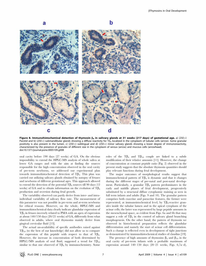
31 weeks of gestation (217 days of GA). The pattern of reactivity
for Tb4 observed in the major salivary glands was significantly
different from that observed in the previous stages. Parotid (Fig 8a)
and submandibular (Fig 8b) glands showed a similar immunohis-
tochemical pattern characterized by a diffuse reactivity for Tb4
localized in the cytoplasm of tubular cells, associated with a
granular positivity in the lumen. Sublingual (Fig. 8c) and minor
(Fig. 8d) glands showed a lower degree of immunoreactivity,
characterized by the presence of particles of different size in the
cytoplasm of serous and mucous cells.
1.5 year. In this subject only specimens from submandibular
and sublingual glands were available (Figs. 9a,b). The pattern of
reactivity for Tb4 changed completely: in submandibular salivary
glands, ducts showed the highest degree of immunoreactvity,
which appeared as an homogeneous staining of the entire
cytoplasm (Fig. 9a). Tb4 was also detected in the cytoplasm of
acinar cells, mainly as coarse granular deposits irregular in shape.
A similar pattern of reactivity was observed in the sublingual gland
acini and tubules (Fig. 9b).
Adults. In major and minor salivary glands of the five adult subjects
tested, utilized in this study as a control group, at variance with
prenatal developmental stages, the pattern of reactivity for Tb4 was
characterized by the absence of granular deposits. Tb4 detection was
restricted to the cytoplasm of intra- and interlobular striated duct
cells, as homogeneous diffuse staining of the entire cytoplasm. An
example of parotid immunoreactivity is reported in Fig. 10.
Discussion
Trends in the concentrations of Tb4 and Tb10 observed as a
function of PMA (Figs. 1 and 2) lead us to suppose higher values in
Figure 7. Immunohistochemical detection of thymosin b4 in salivary glands at 21 weeks (147 days) of gestational age. a) (4006)Parotid glands show a weak granular reactivity in the glandular cells, as well as in the loose mesenchyma. b) (2506) Submandibular glands show thepresence of fine particles reactive for Tb4 mainly stored in the cytoplasm of both serous (arrow) and mucous cells (arrowhead). c) (2506) Sublingualglands show large amounts of Tb4 as fine and coarse granules in the cytoplasm of acinar (arrowhead) and ductal cells (arrow) and in the periglandularmesenchyma. d) (2506) Minor salivary glands showing large amounts of Tb4 in the cytoplasm of acinar serous and mucous cells (arrow), as fine andcoarse granules. Tb4-positive granules are also observed in the mesenchyma and in muscle cells (arrowhead).doi:10.1371/journal.pone.0005109.g007
bThymosins in Oral Development
PLoS ONE | www.plosone.org 6 April 2009 | Volume 4 | Issue 4 | e5109
oral cavity before 190 days (27 weeks) of GA. On the obvious
impossibility to extend the HPLC-MS analysis of whole saliva at
lower GA ranges and with the aim at finding the sources
responsible for the high concentration observed in the oral cavity
of pre-term newborns, we addressed our experimental plan
towards immunohistochemical detection of Tb4. This plan was
carried out utilizing salivary glands obtained by autopsy of fetuses
and newborns of different gestational ages. This approach allowed
to extend the detection of the potential Tb4 sources till 90 days (12
weeks) of GA and to obtain information on the evolution of Tb4
production and secretion during fetal growth.
The variability observed can partly derive from inter- and intra-
individual variability of salivary flow rate. The measurement of
this parameter was not possible in pre-term and at-term newborns
for ethical reasons. However, on the whole, HPLC-MS and
immunohistochemical data clearly indicate glandular expression of
Tb4 in fetuses inversely related to PMA with an apex of expression
at about 140/150 days (20/21 weeks) of GA, differently from what
observed in adults, where oral thymosins mainly derive from
gingival crevicular fluid [13].
The actual un-availability of specific antibodies raised against
Tb10 (to the best of our knowledge) did not allow us to compare
the expression of this peptide with respect to that of Tb4.
However, the increase in concentration of both determined by
HPLC/MS analysis of oral fluid, suggested a trend for Tb10
similar to that one observed of Tb4 by immunochemistry. Some
roles of the Tb4 and Tb10 couple are linked to a subtle
modification of their relative amounts [11]. However, the change
of concentration at constant peptide ratio (Fig. 2) observed in the
present study suggests that the absolute thymosin quantities should
play relevant functions during fetal development.
The major outcomes of morphological results suggest that
immunochemical pattern of Tb4 is dynamic and that it changes
during the different stages of pre-natal and post-natal develop-
ment. Particularly, a granular Tb4 pattern predominates in the
early and middle phases of fetal development, progressively
substituted by a structural diffuse cytoplasmic staining as seen in
full term infants and adults (Figs. 9 and 10). The granular pattern
comprises both exocrine and paracrine features, the former were
represented, at immunoistochemical level, by Tb4-reactive gran-
ules inside the tubular lumen and in the apical cytoplasm of the
acinar cells; the latter was represented by large peptide amounts in
the mesenchymal space, as evident from Figs. 6a and 6b that may
suggest a role of Tb4 in the control of salivary gland branching
morphogenesis. On the other hand, the pattern of thymosin we
observed in histochemical preparation reflects the glandular
differentiation and namely the start of acinar cell differentiation.
Such a change is reflected even in development of tight junctions
as demonstrated by immunohistochemical studies of claudins [19].
The presence of high amounts of thymosins b4 and b10 in the
oral cavity of pre-term infants with a probable maximum of
expression around 140–150 days (20–21 weeks, Figs. 6,7a–d),
Figure 8. Immunohistochemical detection of thymosin b4 in salivary glands at 31 weeks (217 days) of gestational age. a) (2506)Parotid and b) (2506) submandibular glands showing a diffuse reactivity for Tb4 localized in the cytoplasm of tubular cells (arrow). Some granularpositivity is also present in the lumen. c) (2506) sublingual and d) (2506) minor salivary glands showing a lower degree of immunoreactivity,characterized by the presence of granules of different size in the cytoplasm of serous (arrow) and mucous cells (arrowhead).doi:10.1371/journal.pone.0005109.g008
bThymosins in Oral Development
PLoS ONE | www.plosone.org 7 April 2009 | Volume 4 | Issue 4 | e5109

starting at least from about 85 days (12 weeks) of GA (Figs. 4a–c),
put forward a role for these peptides in the development of oral
cavity and its annexes. Until now the salivary glands were believed
organs of slow development, reaching a complete secretory
maturation after birth. Surprisingly, our results suggest that
salivary glands do possess specific secretory capability even during
prenatal maturation stages. In fact, at least since 85 days (12 weeks)
of GA they start secreting peptides, such as thymosins b4 and b10,
which should have specific roles during development. After an
apex reached at around 150 days (21 weeks) of GA, production of
these peptides decreases rapidly and the secretion of salivary
glands slowly switches towards the expression and secretion of
salivary peptides typical of adult age (i.e. acidic proline-rich
proteins [15], statherin, histatins, basic proline-rich proteins and
salivary cystatins). The switch of secretion disclosed by HPLC-MS
analysis of whole saliva, was confirmed by immunoistochemical
analysis of developing salivary glands, which evidenced a
progressive decrease in the secretion of Tb4 starting from the
middle phases of foetal development. The switch could involve
other exocrine and endocrine glands during foetal development.
This study offers only a partial description of this switching
phenomenon and obviously it does not pretend to establish any
role for this high Tb4 and Tb10 salivary gland production during a
particular period of foetal life, even though the immunohisto-
chemical results are suggestive for a contemporaneous paracrine
and exocrine functions. The exocrine pathway arises puzzling
questions about the environment where the peptides are
potentially released. In fact, salivary glands discharge during
foetal life directly in amniotic fluid, where thymosins may be
dispersed. The secretion of Tb4 and Tb10 from salivary gland
might be connected to a demand of the whole amniotic sac. On
the other hand, fluid secreted by salivary glands may be ingested
and addressed towards the foetal gastrointestinal and respiratory
tracts putatively influencing their pre-natal development. Alterna-
tively, potential interactions with specific mucins could maintain
these peptides adherent to the epithelium of the oral cavity.
Author Contributions
Conceived and designed the experiments: SN IM TC RI GV CR AR GF
MC. Performed the experiments: SN TC BM RI CF CT DF EDF.
Analyzed the data: SN IM TC BM RI CF GV CT AR DF EDF GF MC.
Contributed reagents/materials/analysis tools: IM GV CR GF MC. Wrote
the paper: SN IM TC GV CR AR GF MC.
References
1. Hannappel E (2007) b-Thymosins. Ann N Y Acad Sci 1112: 21–37.
2. Low TL, Hu SK, Goldstein AL (1981) Complete amino acid sequence of bovine
thymosin b4: a thymic hormone that induces terminal deoxynucleotidyl
transferase activity in thymocyte populations. Proc Natl Acad Sci U S A 78:
1162–1166.
3. Weller FE, Mutchnick MG (1987) Enzyme immunoassay measurement of
thymosin b4. J Immunoassay 8: 203–217.
4. Huff T, Muller C, Otto A, Netzker R, Hannappel E (2001) b-Thymosins, small
acidic peptides with multiple functions. Int J Biochem Cell Biol 33: 205–220.
5. Grant DS, Kinsella JL, Kibbey MC, LaFlamme S, Burbelo PD, et al. (1995)
Matrigel induces thymosin b4 gene in differentiating endothelial cells. J Cell Sci
108: 3685–3694.
6. Malinda KM, Goldstein AL, Kleinman HK (1997) Thymosin b4 stimulates
directional migration of human umbilical vein endothelial cells. FASEB J 11:
474–481.
7. Sosne G, Chan CC, Thai K, Kennedy M, Szliter EA, et al. (2001) Thymosin b4
promotes corneal wound healing and modulates inflammatory mediators in vivo.
Exp Eye Res 72: 605–608.
Figure 9. Immunohistochemical detection of thymosin b4 insalivary glands in an infant 18 month old. a) (4006) submandib-ular and b) (2506) sublingual glands showing the highest degree ofimmunoreactivity in duct cells. Coarse granular deposits of Tb4 are alsopresent in tubuloacinar cells (arrow).doi:10.1371/journal.pone.0005109.g009
Figure 10. Immuno-histochemical detection of thymosin b4 inthe parotid gland of an adult subject (1006). Tb4 immunoreac-tivity is restricted to the cytoplasm of intra- and interlobular striatedduct cells, appearing as homogeneous diffuse staining of the entirecytoplasm.doi:10.1371/journal.pone.0005109.g010
bThymosins in Oral Development
PLoS ONE | www.plosone.org 8 April 2009 | Volume 4 | Issue 4 | e5109

8. Philp D, Nguyen M, Scheremeta B, St-Surin S, Villa AM, et al. (2004)
Thymosin b4 increases hair growth by activation of hair follicle stem cells.FASEB J 18: 385–387.
9. Bock-Marquette I, Saxena A, White MD, Dimaio GM, Srivastava D (2004)
Thymosin b4 activates integrin-linked kinase and promotes cardiac cellmigration, survival and cardiac repair. Nature 432: 466–472.
10. Smart N, Risebro CA, Melville AA, Moses K, Schwartz RJ, et al. (2007)Thymosin b4 induces adult epicardial progenitor mobilization and neurovascu-
larization. Nature 445: 177–182.
11. Smart N, Rossdeutsch A, Riley PR (2007) Thymosin b4 and angiogenesis: modesof action and therapeutic potential. Angiogenesis 10: 229–241.
12. Badamchian M, Damavandy AA, Damavandy H, Wadhwa SD, Katz B, et al.(2007) Identification and quantification of Thymosin b4 in human saliva and
tears. Ann N Y Acad Sci 1112: 458–465.13. Inzitari R, Cabras T, Pisano E, Fanali C, Manconi B, et al. (2009) HPLC-ESI-
MS analysis of oral human fluid reveals that gingival crevicular fluid is the main
source of oral thymosin b4 and b10. J Sep Sci 32: 57–63.14. Reti R, Kwon E, Qiu P, Wheater M, Sosne G (2008) Thymosin beta 4 is
cytoprotective in human gingival fibroblasts. Eur J Oral Sci 116: 424–430.
15. Inzitari R, Vento G, Capoluongo E, Boccacci S, Fanali C, et al. (2007)
Proteomic analysis of salivary acidic proline-rich proteins in human pre-term
and at-term newborns. J Proteome Res 6: 1371–1377.
16. Zhang Z, Marshall AG (1998) A universal algorithm for fast and automated
charge state deconvolution of electrospray mass-to-charge ratio spectra. J Am
Soc Mass Spectrom 9: 225–233.
17. Huff T, Hannappel E (1997) Oxidation and reduction of thymosin b4 and its
influence on the interaction with G-actin studied by reversed-phase HPLC and
postcolumn derivatization with fluorescamine. Anal Chim Acta 352: 249–255.
18. Lourenco SV, Uyekita SH, Correia Lima DM, Soares FA (2008) Developing
human minor salivary glands: morphological parallel relation between the
expression of TGF-beta isoforms and cytoskeletal markers of glandular
maturation. Virchows Arch 452: 427–434.
19. Lourenco SV, Coutinho-Camillo CM, Buim ME, Uyekita SH, Soares FA (2007)
Human salivary gland branching morphogenesis: morphological localization of
claudins and its parallel relation with developmental stages revealed by
expression of cytoskeleton and secretion markers. Histochem Cell Biol 128:
361–369.
bThymosins in Oral Development
PLoS ONE | www.plosone.org 9 April 2009 | Volume 4 | Issue 4 | e5109
Copyright © 2022 FDOKUMEN