Identification and subcellular localization of paracellin-1 (claudin-16) in human salivary glands
14
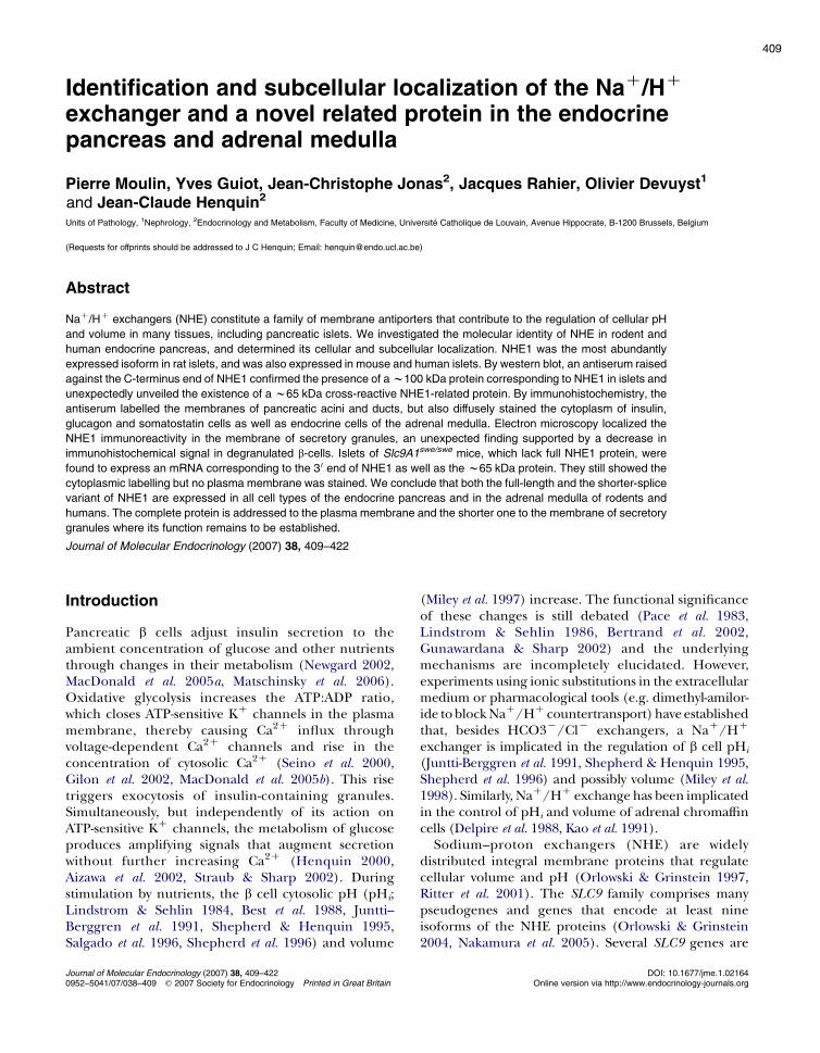
Identification and subcellular localization of the Na C /H C exchanger and a novel related protein in the endocrine pancreas and adrenal medulla Pierre Moulin, Yves Guiot, Jean-Christophe Jonas 2 , Jacques Rahier, Olivier Devuyst 1 and Jean-Claude Henquin 2 Units of Pathology, 1 Nephrology, 2 Endocrinology and Metabolism, Faculty of Medicine, Universite ´ Catholique de Louvain, Avenue Hippocrate, B-1200 Brussels, Belgium (Requests for offprints should be addressed to J C Henquin; Email: [email protected]) Abstract Na C /H C exchangers (NHE) constitute a family of membrane antiporters that contribute to the regulation of cellular pH and volume in many tissues, including pancreatic islets. We investigated the molecular identity of NHE in rodent and human endocrine pancreas, and determined its cellular and subcellular localization. NHE1 was the most abundantly expressed isoform in rat islets, and was also expressed in mouse and human islets. By western blot, an antiserum raised against the C-terminus end of NHE1 confirmed the presence of a w100 kDa protein corresponding to NHE1 in islets and unexpectedly unveiled the existence of a w65 kDa cross-reactive NHE1-related protein. By immunohistochemistry, the antiserum labelled the membranes of pancreatic acini and ducts, but also diffusely stained the cytoplasm of insulin, glucagon and somatostatin cells as well as endocrine cells of the adrenal medulla. Electron microscopy localized the NHE1 immunoreactivity in the membrane of secretory granules, an unexpected finding supported by a decrease in immunohistochemical signal in degranulated b-cells. Islets of Slc9A1 swe/swe mice, which lack full NHE1 protein, were found to express an mRNA corresponding to the 3 0 end of NHE1 as well as the w65 kDa protein. They still showed the cytoplasmic labelling but no plasma membrane was stained. We conclude that both the full-length and the shorter-splice variant of NHE1 are expressed in all cell types of the endocrine pancreas and in the adrenal medulla of rodents and humans. The complete protein is addressed to the plasma membrane and the shorter one to the membrane of secretory granules where its function remains to be established. Journal of Molecular Endocrinology (2007) 38, 409–422 Introduction Pancreatic b cells adjust insulin secretion to the ambient concentration of glucose and other nutrients through changes in their metabolism (Newgard 2002, MacDonald et al. 2005a, Matschinsky et al. 2006). Oxidative glycolysis increases the ATP:ADP ratio, which closes ATP-sensitive K C channels in the plasma membrane, thereby causing Ca 2C influx through voltage-dependent Ca 2C channels and rise in the concentration of cytosolic Ca 2C (Seino et al. 2000, Gilon et al. 2002, MacDonald et al. 2005b). This rise triggers exocytosis of insulin-containing granules. Simultaneously, but independently of its action on ATP-sensitive K C channels, the metabolism of glucose produces amplifying signals that augment secretion without further increasing Ca 2C (Henquin 2000, Aizawa et al. 2002, Straub & Sharp 2002). During stimulation by nutrients, the b cell cytosolic pH (pH i ; Lindstrom & Sehlin 1984, Best et al. 1988, Juntti-- Berggren et al. 1991, Shepherd & Henquin 1995, Salgado et al. 1996, Shepherd et al. 1996) and volume (Miley et al. 1997) increase. The functional significance of these changes is still debated (Pace et al. 1983, Lindstrom & Sehlin 1986, Bertrand et al. 2002, Gunawardana & Sharp 2002) and the underlying mechanisms are incompletely elucidated. However, experiments using ionic substitutions in the extracellular medium or pharmacological tools (e.g. dimethyl-amilor- ide to block Na C /H C countertransport) have established that, besides HCO3 K /Cl K exchangers, a Na C /H C exchanger is implicated in the regulation of b cell pH i (Juntti-Berggren et al. 1991, Shepherd & Henquin 1995, Shepherd et al. 1996) and possibly volume (Miley et al. 1998). Similarly, Na C /H C exchange has been implicated in the control of pH i and volume of adrenal chromaffin cells (Delpire et al. 1988, Kao et al. 1991). Sodium–proton exchangers (NHE) are widely distributed integral membrane proteins that regulate cellular volume and pH (Orlowski & Grinstein 1997, Ritter et al. 2001). The SLC9 family comprises many pseudogenes and genes that encode at least nine isoforms of the NHE proteins (Orlowski & Grinstein 2004, Nakamura et al. 2005). Several SLC9 genes are 409 Journal of Molecular Endocrinology (2007) 38, 409–422 DOI: 10.1677/jme.1.02164 0952–5041/07/038–409 q 2007 Society for Endocrinology Printed in Great Britain Online version via http://www.endocrinology-journals.org
Transcript of Identification and subcellular localization of paracellin-1 (claudin-16) in human salivary glands
409
Identification and subcellular l
ocalization of the NaC/HCexchanger and a novel related protein in the endocrinepancreas and adrenal medulla
Pierre Moulin, Yves Guiot, Jean-Christophe Jonas2, Jacques Rahier, Olivier Devuyst1
and Jean-Claude Henquin2
Units of Pathology, 1Nephrology, 2Endocrinology and Metabolism, Faculty of Medicine, Universite Catholique de Louvain, Avenue Hippocrate, B-1200 Brussels, Belgium
(Requests for offprints should be addressed to J C Henquin; Email: [email protected])
Abstract
NaC/HC exchangers (NHE) constitute a family of membrane antiporters that contribute to the regulation of cellular pH
and volume in many tissues, including pancreatic islets. We investigated the molecular identity of NHE in rodent and
human endocrine pancreas, and determined its cellular and subcellular localization. NHE1 was the most abundantly
expressed isoform in rat islets, and was also expressed in mouse and human islets. By western blot, an antiserum raised
against the C-terminus end of NHE1 confirmed the presence of a w100 kDa protein corresponding to NHE1 in islets and
unexpectedly unveiled the existence of a w65 kDa cross-reactive NHE1-related protein. By immunohistochemistry, the
antiserum labelled the membranes of pancreatic acini and ducts, but also diffusely stained the cytoplasm of insulin,
glucagon and somatostatin cells as well as endocrine cells of the adrenal medulla. Electron microscopy localized the
NHE1 immunoreactivity in the membrane of secretory granules, an unexpected finding supported by a decrease in
immunohistochemical signal in degranulated b-cells. Islets of Slc9A1swe/swe mice, which lack full NHE1 protein, were
found to express an mRNA corresponding to the 3 0 end of NHE1 as well as the w65 kDa protein. They still showed the
cytoplasmic labelling but no plasma membrane was stained. We conclude that both the full-length and the shorter-splice
variant of NHE1 are expressed in all cell types of the endocrine pancreas and in the adrenal medulla of rodents and
humans. The complete protein is addressed to the plasma membrane and the shorter one to the membrane of secretory
granules where its function remains to be established.
Journal of Molecular Endocrinology (2007) 38, 409–422
Introduction
Pancreatic b cells adjust insulin secretion to theambient concentration of glucose and other nutrientsthrough changes in their metabolism (Newgard 2002,MacDonald et al. 2005a, Matschinsky et al. 2006).Oxidative glycolysis increases the ATP:ADP ratio,which closes ATP-sensitive KC channels in the plasmamembrane, thereby causing Ca2C influx throughvoltage-dependent Ca2C channels and rise in theconcentration of cytosolic Ca2C (Seino et al. 2000,Gilon et al. 2002, MacDonald et al. 2005b). This risetriggers exocytosis of insulin-containing granules.Simultaneously, but independently of its action onATP-sensitive KC channels, the metabolism of glucoseproduces amplifying signals that augment secretionwithout further increasing Ca2C (Henquin 2000,Aizawa et al. 2002, Straub & Sharp 2002). Duringstimulation by nutrients, the b cell cytosolic pH (pHi;Lindstrom & Sehlin 1984, Best et al. 1988, Juntti--Berggren et al. 1991, Shepherd & Henquin 1995,Salgado et al. 1996, Shepherd et al. 1996) and volume
Journal of Molecular Endocrinology (2007) 38, 409–4220952–5041/07/038–409 q 2007 Society for Endocrinology Printed in Great Britain
(Miley et al. 1997) increase. The functional significanceof these changes is still debated (Pace et al. 1983,Lindstrom & Sehlin 1986, Bertrand et al. 2002,Gunawardana & Sharp 2002) and the underlyingmechanisms are incompletely elucidated. However,experiments using ionic substitutions in the extracellularmedium or pharmacological tools (e.g. dimethyl-amilor-ide toblockNaC/HC countertransport)haveestablishedthat, besides HCO3K/ClK exchangers, a NaC/HC
exchanger is implicated in the regulation of b cell pHi
(Juntti-Berggren et al. 1991, Shepherd & Henquin 1995,Shepherd et al. 1996) and possibly volume (Miley et al.1998). Similarly, NaC/HC exchange has been implicatedin the control of pHi and volume of adrenal chromaffincells (Delpire et al. 1988, Kao et al. 1991).
Sodium–proton exchangers (NHE) are widelydistributed integral membrane proteins that regulatecellular volume and pH (Orlowski & Grinstein 1997,Ritter et al. 2001). The SLC9 family comprises manypseudogenes and genes that encode at least nineisoforms of the NHE proteins (Orlowski & Grinstein2004, Nakamura et al. 2005). Several SLC9 genes are
DOI: 10.1677/jme.1.02164Online version via http://www.endocrinology-journals.org

P MOULIN and others . NaC/HC exchanger in the endocrine pancreas410
also known to give rise to multiple transcripts or partialmRNA (Orlowski & Grinstein 2004). The first fiveisoforms (NHE1 to NHE5) are well characterized anddisplay distinct physiological and pharmacologicalproperties. NHE1 is ubiquitous. In polarized cells, it isusually inserted in the basolateral domain of the plasmamembrane where it fulfils housekeeping regulation ofcell volume and pH (Orlowski & Grinstein 2004).NHE2 and NHE3 are mainly found at the apical pole ofepithelial cells in kidney (Chambrey et al. 1998),intestine (Chu et al. 2002) and duct cells of salivaryglands and pancreas (Lee et al. 1998, 2000), where theyplay a role in NaC and fluid absorption, and secretionof protons (Orlowski & Grinstein 2004). NHE4 hasbeen identified in the macula densa of the kidney(Peti-Peterdi et al. 2000) and in the stomach (Rossmannet al. 2001). NHE5 has mainly been found in the brainwhere it seems to behave like NHE3 (Baird et al. 1999).Although NHE1 to NHE5 are usually localized in theplasma membrane, NHE3 and NHE5 have also beenobserved in recycling vesicles (Kurashima et al. 1998,Szaszi et al. 2002). In contrast, the ubiquitouslydistributed NHE6 to NHE9 have not been localized tothe plasma membrane but to intracellular organelles(endosomes, trans-Golgi network or mitochondria;Nakamura et al. 2005).
The primary aims of the present study were todetermine the molecular identity of NHE in humanand rodent islets and to establish its subcellularlocalization. The initial results unexpectedly led to thediscovery of a novel protein related to NHE1 andheavily concentrated in the membrane of secretorygranules in all cell types of the endocrine pancreas, andalso in the adrenal medulla.
Materials and methods
Samples
All studies were approved by and conducted inaccordance with the regulations of our institutionalethics committees. Human pancreases were obtainedeither at autopsy of normoglycaemic patients (nZ4)performed within 6 h after death, or from organ donors(nZ6). None of these patients had a disease affectingthe pancreas. Autopsy samples from human kidney andheart were used as control tissue for RT-PCR. Humanislets isolated (Dufrane et al. 2005) from the pancreas ofone organ donor were provided by Dr D Dufrane(Experimental Surgery, Universite Catholique deLouvain, Brussels, Belgium).
Normal male Wistar rats and National marineresearch institute (NMRI) mice were from a localcolony. To evaluate NHE1 distribution after massiveinsulin secretion, Wistar rats received two i.p. injections
Journal of Molecular Endocrinology (2007) 38, 409–422
of glibenclamide (5.0 mg/kg body weight) or saline at12 h interval. Animals were fed ad libitum anddecapitated 4 h after the second injection.C57BL/6.SJL, C/swe (slow wave epilepsy) mice (Coxet al. 1997) were purchased from the Jackson Labo-ratory (Bar Harbor, MI, USA). These heterozygous micewere mated and the resulting homozygous NHE1 nullmutant (Slc9A1swe/swe) and wild-type (Slc9A1C/C) werestudied at 5–7 weeks of age. Homozygous mutant miceexhibited a neurological phenotype including ataxia inthe hind limbs, and seizures. Genotyping wasperformed as described (Cox et al. 1997) to confirmthe phenotype. Rat islets were obtained by collagenasedigestion of the pancreas (Jonas et al. 1998), and wereused freshly or after 7 days of culture in RPMI 1640medium containing 10 mM glucose and 0.5 g/100 mlBSA. A similar procedure was used to obtain mouseislets except that they were cultured for 18–42 h inRPMI 1640 medium containing 10% heat-inactivatedFCS, 100 IU/ml penicillin, 100 mg/ml streptomycinand 10 mm glucose.
Antibodies
Most experiments were performed with a rabbit serumdirected against the C-terminus end of NHE1 (Gosset al. 1994), which was raised, characterized and kindlygiven by Dr M Donowitz (Johns Hopkins UniversitySchool of Medicine). A mouse monoclonal antibodyalso directed against the C-terminus end of NHE (clone4E9, MAB3140, Chemicon, Temecula, CA, USA) wasused in a few, clearly identified, control experiments.Mousemonoclonal antibodies against insulin, glucagonand somatostatin were obtained from NovoBiolabs(HUI-018, GLU-001 and SOM-018 respectively;Bagsvaerd, Denmark).
Other reagents and supplies
Biotinylated secondary antibodies against mouse andrabbit Fab’ Ig fragments were obtained from VectorLaboratories (Burlingame, CA, USA) and Chemiconrespectively. Streptavidin-peroxidase (SP) and Strepta-vidin-Texas-Red (STR) complexes were obtained fromRoche-Diagnostics and Zymed (San Francisco, CA,USA). En-Vision detection system (EV) and Fluoresceinisothiocyanate (FITC)-conjugated secondary antibodieswere purchased from Dako (Glostrup, Denmark).Immunogold reagents including nanogold-labelledanti-rabbit Ig (NG-Ig) and silver enhancement solutionswere obtained from Nanoprobes (Stony Brook, NY,USA). Electrophoresis reagents were from Bio-Radand Amersham. Other reagents were obtained fromAmresco (Solon, OH, USA), Sigma and J T Baker(Philipsburg, NJ, USA).
www.endocrinology-journals.org
NaC/HC exchanger in the endocrine pancreas . P MOULIN and others 411
Tissue processing
Human or rodent samples were frozen in liquid nitrogenor fixed in 4% paraformaldehyde for 6 or 24 h.
Tissue proteins were extracted from snap-frozensamples (w500 mg) homogenized in (w10 ml/gtissue) ice-cold buffer (20 mM Imidazole, pH 7.2,1 mM EDTA, 250 mM sucrose) containing proteaseinhibitors (Complete, Roche) by ten passes of P6Ultra-Turrax (Labortechnik, Staufen, Germany) andsonicated (Branson Sonifier B12, Danbury, CT, USA).The homogenate was centrifuged at 1000 g for 15 minat 4 8C. The postnuclear supernatant was centrifugedat 80 000 g for 1 h at 4 8C to separate membrane andcytosolic fractions (Combet et al. 1999). The mem-brane pellet was suspended in the homogenizationbuffer and protein concentrations were determinedusing the Bradford method (Bio-Rad) with BSA asstandard.
Total RNA was extracted from snap-frozen samples inTrizol according to manufacturer’s instructions (LifeBiotechnologies, Gibco-BRL). RNA was reverse-tran-scribed in cDNA using random hexamers and theSuperscript RNA reverse transcriptase (Gibco-BRL).
Immunoblot analysis
SDS-PAGE and immunoblotting were performed asdescribed (Combet et al. 1999). The extracts weresolubilized by heating at 95 8C for 3 min in samplebuffer. Proteins (10–40 mg/lane) were separated byelectrophoresis through 7.5% acrylamide slabs andtransferred to nitrocellulose. Membranes were blockedfor 30 min at room temperature in blotting buffer,followed by incubation with the primary antibody (anti-NHE1 at 1/2000). The membranes were then washedand incubated for 1 h at room temperature with anti-rabbit Fab’ peroxidase-labelled antibody (Dako). Afterwashing, immunoblots were visualized with ECL-Plusreagent (Amersham).
Table 1 Immunohistochemical staining
Antibody; dilution
Antigen retrievalYes Polyclonal rabbit anti-NHE1; 1/2000Yes Polyclonal rabbit anti-NHE1; 1/1000No Monoclonal mouse anti-insulin; 1/1000No Polyclonal rabbit anti-NHE1; 1/50No Monoclonal mouse anti-insulin; 1/80No Monoclonal mouse anti-glucagon; 1/80No Monoclonal mouse anti-somatostatin; 1/8
EV, En-Vision; 2B, biotinylated secondary antibody (1/500); 2F, secondary antiperoxydase complex (1/500); STR, streptavidin-Texas Red conjugate (1/50).
www.endocrinology-journals.org
Immunohistochemistry
Paraffin-embedded specimens were cut into 3 mmthick sections and processed as described elsewhere(Sempoux et al. 1998) including, when necessary, anantigen retrieval treatment. Primary antibodies werediluted in Tris (pH 7.4) supplemented with 1% BSAand applied overnight at 4 8C. All subsequent incu-bations lasted 1 h at room temperature. For doubleimmunofluorescence experiments, anti-hormone andNHE1 antisera incubations were carried out sequen-tially. When necessary, a tyramine amplification step wasadded (Sempoux et al. 2003). The peroxidase activitywas revealed by 3,3 0-diaminobenzidine hypochloride(DAB: 50 mg/100 ml, pH 7.4; Fluka Chemie, Buchs,Switzerland) for 10 min. Antibodies and detailedconditions are described in Table 1. Specific opticaldensity of the immunohistochemical signal wasmeasured as described previously (Rahier et al. 1989).
Electron microscopy and immunogold labelling
After 24-h fixation in paraformaldehyde, small pancreasblocks were cryoprotected in PBS containing 15%sucrose for 48 h, before being frozen into liquidnitrogen. Forty micrometre thick cryosections were cutand incubated with NHE1 antiserum (1/50) for 45 minat 4 8C. After rinsing in PBS, NG-Ig (1/40) was appliedfor 1 h. After short fixation in 2.5% glutaraldehyde, thesignal was amplified by a silver enhancer solutionaccording to the manufacturer’s instructions. Thesections were rinsed and embedded into Epon 812 andprocessed for electron microscopy (Rahier et al. 1989).
Radioactive RT-PCR analysis of rat NHE isoformsmRNA
The primers are indicated in Table 2. Radioactive PCRwas performed as described previously (Jonas et al.1999), with a thermal cycle profile consisting of a
Amplification system Detection system
EV DABEV DAB2B-SP DAB2B-SP-BT-STR Fluorescence2F Fluorescence2F Fluorescence
0 2F Fluorescence
body FITC conjugate (1/20); BT, biotinylated tyramine; SP, streptavidin-
Journal of Molecular Endocrinology (2007) 38, 409–422
Table
2S
equences
of
olig
onucle
otide
prim
ers
used
for
PC
Ranaly
sis
of
NH
Eis
ofo
rms
Size
(bp)
50 -Senseprimer-30
50 -Antisenseprimer-30
ReferenceorGenBankAccessionno.
Gene
Rat
NH
E1
298
GA
AC
AT
CC
AC
CC
CA
AG
TC
TG
CA
GT
GG
GT
CT
GA
GC
CT
AT
GC
NM
_012652
Rat
NH
E2
333
AA
AC
CA
AC
CC
AA
GT
CT
AG
CA
TT
GT
CG
GT
TT
AA
GC
TG
TT
GT
CC
TT
CC
TA
NM
_012653
Rat
NH
E3
282
AG
AG
CT
TC
AC
AT
CC
GT
CT
TA
TG
GG
AA
AG
TC
GC
TT
GA
TT
CC
CT
GT
AT
CM
85300
Rat
NH
E4
396
CA
GC
GT
GT
TT
AC
CC
TC
TT
CT
AT
GT
TT
CC
GC
AA
AT
AT
CT
GT
GG
TC
AA
AC
M85301
Rat
NH
E5
235
GC
GG
TC
AG
CC
TA
TC
GT
AT
CC
GC
CA
CT
GC
GT
AC
TG
TG
TC
AA
TC
NM
138858
Hum
an
NH
E1
492
AC
CA
CC
AC
TG
GA
AG
GA
CA
AG
AT
AG
GC
CA
GT
GG
GT
CT
GA
GC
M81768
Mouse
NH
E1:
50 e
x2–P
41834
AC
GT
CT
TC
TT
CC
TC
TT
CC
TG
CT
GG
GC
TC
CT
TG
CT
CC
GA
AT
CA
TG
Belletal.
1999;
NM
_012652
Mouse
NH
E1:
P1–P
4706
AG
GA
CA
TC
TG
TG
GT
CA
TT
AT
GG
CG
GC
TC
CT
TG
CT
CC
GA
AT
CA
TG
NM
_012652
Slc9A1C
/C:
MR
0975–M
R0977
200
CC
TG
AC
CT
GG
TT
CA
TC
AA
CA
TC
AT
GC
CC
TG
CA
CA
AA
GA
CG
Coxetal.
1997
Slc9A1swe/swe:
MR
0976–M
R0977
200
CC
TG
AC
CT
GG
TT
CA
TC
AA
CT
TC
AT
GC
CC
TG
CA
CA
AA
GA
CG
Coxetal.
1997
P MOULIN and others . NaC/HC exchanger in the endocrine pancreas412
Journal of Molecular Endocrinology (2007) 38, 409–422
10 min denaturing step at 95 8C followed by 30 cycles ofamplification (1 min steps at 94, 60 and 72 8C each) anda final extension step of 10 min at 72 8C. TATAbox-binding protein (TBP) was used as control gene andamplified by a 24 cycles PCR (Jonas et al. 1999). Theamplimers were then separated on a 6% polyacrylamidegel in Tris borate EDTA buffer, in parallel with a 100-bpDNA ladder. The gel was dried, and the amount of[a-32P]dCTP incorporated in each amplicon wasquantified with a Cyclone Storage Phosphor System(Packard, Meriden, CT, USA). The ratio of specificproduct/control gene was then calculated for eachsample.
Amplification of NHE1 species-specific cDNA andhuman NHE1 probe production for in situhybridization
The primers used for human and rodent NHE isoformsare described in Table 2. PCRs were performed in athermocycler 2400 (Applied Biosystems, Foster City, CA,USA) with a total volume of 25 ml mixture containingGeneAmpPCRbuffer, 1.5 mMMgCl2, 200 mMdeoxyNTP,0.5 mM primers and 1 U Taq Gold polymerase (AppliedBiosystems). The thermal cycle profile was 10 mindenaturation at 94 8C followed by 35 cycles (30 s at94 8C, 45 s at 62 8C and 1 min at 72 8C) and a finalextension of 10 min at 72 8C. Abelson protooncogene orglyceraldehyde-3-phosphate dehydrogenase (GAPDH)were taken as controls.
The amplified DNA samples were electrophoresedon ethidium bromide agarose gel and quantified byGelDoc 2000 scanning device (Bio-Rad). The identity ofNHE1 PCR product was confirmed by DNA sequenceanalysis using the dye terminator sequencing system ona Genetic analyser 3600 (Applied Biosystems). The PCRprocedure was repeated with a no-reverse transcriptioncontrol to exclude genomic DNA contamination andcarry-over.
The probe production procedure for in situ hybrid-ization was similar to that described previously forinsulin-like growth factor-II including negative andsequence controls (Sempoux et al. 2003) except that ahuman NHE1 hybridization probe was produced fromhuman heart using specific primers (Table 2).
In silico analyses
Electronic database searches for mouse Slc9A1 gene ormRNA matching sequences, structure and theoreticalalternative splicing or start sites were available at http://www.ensembl.org/Mus_musculus/geneview? geneZENSMUSG00000028854&dbZcore (Stalker et al. 2004).RT-PCR primers were tested using blastn against all
www.endocrinology-journals.org
NaC/HC exchanger in the endocrine pancreas . P MOULIN and others 413
nucleotide databases available from the National Centerfor Biotechnology Information (NCBI) at http://www.ncbi.nlm.nih.gov/BLAST/ (Altschul et al. 1990). Alter-nate splicing sites were searchedwithonline tool availableat http://www.fruitfly.org/cgi-bin/seq_tools/splice.pl.
Results
Identification of NHE isoforms expressed in theendocrine pancreas
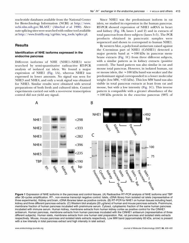
Different isoforms of NHE (NHE1–NHE5) weresearched by semi-quantitative radioactive RT-PCRanalysis of isolated rat islets. We found a majorexpression of NHE1 (Fig. 1A), whereas NHE2 wasexpressed in lower amounts. No signal was seen forNHE3 and NHE4, and only a weak signal was obtainedfor NHE5. Similar results were obtained with severalpreparations of both fresh and cultured islets. Controlexperiments carried out with a no-reverse transcriptioncontrol did not yield any signal.
Figure 1 Expression of NHE isoforms in the pancreas and control tissafter 30-cycles amplification. RT-, non-reverse transcript negative conthree experiments). Kidney and brain, cDNA libraries taken as positivekidney and three different pancreas extracts. (C) Western blot analysismembrane fraction of human pancreas incubated with preimmune serincubated with immune serum. Human kidney, membrane extracts froantiserum. Human pancreas, membrane extracts from human pancredifferent subjects). Human islets, membrane extracts from one humanrespectively. Mouse, mouse pancreas and isolated islets extracts respwith a low intensity in total pancreas extract and high intensity in islet
www.endocrinology-journals.org
Since NHE1 was the predominant isoform in ratislets, we studied its expression in the human pancreas.RT-PCR showed expression of NHE1 mRNA in heartand kidney (Fig. 1B, lanes 1 and 2) and in extracts oftotal pancreas from three subjects (lanes 3–5). The PCRproducts obtained in pancreatic samples weresequenced and shown to correspond to human NHE1.
By western blot, a polyclonal antiserum raised againstthe C-terminus part of NHE1 (CtNHE1) detected amajor protein band at w100 kDa in pancreas mem-brane extracts (Fig. 1C) from three different subjects,with a similar pattern as in kidney extracts (positivecontrol). The band pattern was also similar in rat andmouse total pancreas. However, in isolated human, rator mouse islets, thew100 kDa band was weaker and thepredominant signal corresponded to a lower molecularweight (low MW,w65 kDa). This low MW band was alsovisible in total pancreas extracts at least from rat andmouse, but with a low intensity (Fig. 1C). This inversepattern is compatible with a greater abundance of thew100 kDa protein in the exocrine pancreas (98% of
ues. (A) Radioactive RT-PCR analysis of NHE isoforms and TBPtrol; Islets, cDNA library from isolated rat islets (representative ofcontrols. (B) RT-PCR for NHE1 on human tissues including heart,(20 mg/lane) of human and mouse pancreas extracts. Preimmune,um. Cytosol, cytoplasmic fraction of the same human pancreasm human kidney cortex as positive control for the CtNHE1as incubated with the CtNHE1 antiserum (representative of threeislet preparation. Rat, rat pancreas and isolated islets extracts
ectively. Low MW band (approximately 65 kDa, arrow) is presentextract.
Journal of Molecular Endocrinology (2007) 38, 409–422
P MOULIN and others . NaC/HC exchanger in the endocrine pancreas414
the tissue) and of thew65 kDa protein in the islets (2%of the tissue). No similar signal was found whencytosolic extracts of pancreas were incubated with theantiserum. Several minor bands were variably observeddepending on the preparations, and a weak signal wasdetected above 75 kDa in human kidney and in humanand rat pancreas and islets. They were considerednon-specific either because of the lack of repro-ducibility or because a similar band was observedwhen membrane extracts were incubated withpreimmune serum (Fig. 1C).
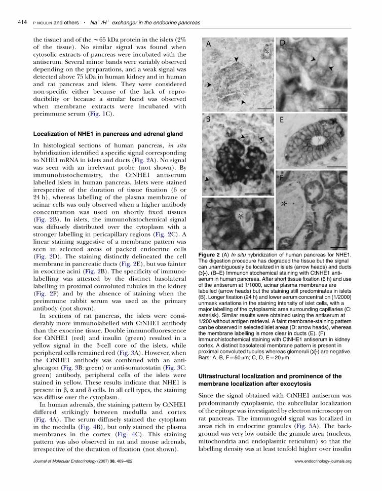
Figure 2 (A) In situ hybridization of human pancreas for NHE1.The digestion procedure has degraded the tissue but the signalcan unambiguously be localized in islets (arrow heads) and ducts( ). (B–E) Immunohistochemical staining with CtNHE1 anti-serum in human pancreas. After short tissue fixation (6 h) and useof the antiserum at 1/1000, acinar plasma membranes arelabelled (arrow heads) but the staining still predominates in islets(B). Longer fixation (24 h) and lower serum concentration (1/2000)unmask variations in the staining intensity of islet cells, with amajor labelling of the cytoplasmic area surrounding capillaries (C:asterisk). Similar results were obtained using the antiserum at1/200 without antigen retrieval. A faint membrane-staining patterncan be observed in selected islet areas (D: arrow heads), whereasthe membrane labelling is more clear in ducts (E). (F)Immunohistochemical staining with CtNHE1 antiserum in kidneycortex. A distinct basolateral membrane pattern is present inproximal convoluted tubules whereas glomeruli ( ) are negative.Bars: A, B, FZ50mm; C, D, EZ20mm.
Localization of NHE1 in pancreas and adrenal gland
In histological sections of human pancreas, in situhybridization identified a specific signal correspondingto NHE1 mRNA in islets and ducts (Fig. 2A). No signalwas seen with an irrelevant probe (not shown). Byimmunohistochemistry, the CtNHE1 antiserumlabelled islets in human pancreas. Islets were stainedirrespective of the duration of tissue fixation (6 or24 h), whereas labelling of the plasma membrane ofacinar cells was only observed when a higher antibodyconcentration was used on shortly fixed tissues(Fig. 2B). In islets, the immunohistochemical signalwas diffusely distributed over the cytoplasm with astronger labelling in pericapillary regions (Fig. 2C). Alinear staining suggestive of a membrane pattern wasseen in selected areas of packed endocrine cells(Fig. 2D). The staining distinctly delineated the cellmembrane in pancreatic ducts (Fig. 2E), but was fainterin exocrine acini (Fig. 2B). The specificity of immuno-labelling was attested by the distinct basolaterallabelling in proximal convoluted tubules in the kidney(Fig. 2F) and by the absence of staining when thepreimmune rabbit serum was used as the primaryantibody (not shown).
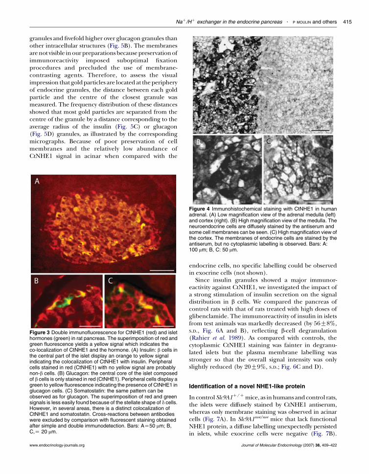
In sections of rat pancreas, the islets were consi-derably more immunolabelled with CtNHE1 antibodythan the exocrine tissue. Double immunofluorescencefor CtNHE1 (red) and insulin (green) resulted in ayellow signal in the b-cell core of the islets, whileperipheral cells remained red (Fig. 3A). However, whenthe CtNHE1 antibody was combined with an anti-glucagon (Fig. 3B: green) or anti-somatostatin (Fig. 3C:green) antibody, peripheral cells of the islets werestained in yellow. These results indicate that NHE1 ispresent in b, a and d cells. In all cell types, the stainingwas diffuse over the cytoplasm.
In human adrenals, the staining pattern by CtNHE1differed strikingly between medulla and cortex(Fig. 4A). The serum diffusely stained the cytoplasmin the medulla (Fig. 4B), but only stained the plasmamembranes in the cortex (Fig. 4C). This stainingpattern was also observed in rat and mouse adrenals,irrespective of the duration of fixation (not shown).
Journal of Molecular Endocrinology (2007) 38, 409–422
Ultrastructural localization and prominence of the
membrane localization after exocytosis
Since the signal obtained with CtNHE1 antiserum waspredominantly cytoplasmic, the subcellular localizationof the epitope was investigated by electronmicroscopy onrat pancreas. The immunogold signal was localized inareas rich in endocrine granules (Fig. 5A). The back-ground was very low outside the granule area (nucleus,mitochondria and endoplasmic reticulum) so that thelabelling density was at least tenfold higher over insulin
www.endocrinology-journals.org
NaC/HC exchanger in the endocrine pancreas . P MOULIN and others 415
granules andfivefold higher over glucagon granules thanother intracellular structures (Fig. 5B). The membranesarenot visible inourpreparationsbecausepreservationofimmunoreactivity imposed suboptimal fixationprocedures and precluded the use of membrane-contrasting agents. Therefore, to assess the visualimpression that goldparticles are located at theperipheryof endocrine granules, the distance between each goldparticle and the centre of the closest granule wasmeasured. The frequency distribution of these distancesshowed that most gold particles are separated from thecentre of the granule by a distance corresponding to theaverage radius of the insulin (Fig. 5C) or glucagon(Fig. 5D) granules, as illustrated by the correspondingmicrographs. Because of poor preservation of cellmembranes and the relatively low abundance ofCtNHE1 signal in acinar when compared with the
Figure 4 Immunohistochemical staining with CtNHE1 in humanadrenal. (A) Low magnification view of the adrenal medulla (left)and cortex (right). (B) High magnification view of the medulla. Theneuroendocrine cells are diffusely stained by the antiserum andsome cell membranes can be seen. (C) High magnification view ofthe cortex. The membranes of endocrine cells are stained by theantiserum, but no cytoplasmic labelling is observed. Bars: A:100 mm; B, C: 50 mm.
Figure 3 Double immunofluorescence for CtNHE1 (red) and islethormones (green) in rat pancreas. The superimposition of red andgreen fluorescence yields a yellow signal which indicates theco-localization of CtNHE1 and the hormone. (A) Insulin: b cells inthe central part of the islet display an orange to yellow signalindicating the colocalization of CtNHE1 with insulin. Peripheralcells stained in red (CtNHE1) with no yellow signal are probablynon-b cells. (B) Glucagon: the central core of the islet composedof b cells is only stained in red (CtNHE1). Peripheral cells display agreen to yellow fluorescence indicating the presence of CtNHE1 inglucagon cells. (C) Somatostatin: the same pattern can beobserved as for glucagon. The superimposition of red and greensignals is less easily found because of the stellate shape of d cells.However, in several areas, there is a distinct colocalization ofCtNHE1 and somatostatin. Cross-reactions between antibodieswere excluded by comparison with fluorescent staining obtainedafter simple and double immunodetection. Bars: AZ50 mm; B,C,Z 20 mm.
www.endocrinology-journals.org
endocrine cells, no specific labelling could be observedin exocrine cells (not shown).
Since insulin granules showed a major immunor-eactivity against CtNHE1, we investigated the impact ofa strong stimulation of insulin secretion on the signaldistribution in b cells. We compared the pancreas ofcontrol rats with that of rats treated with high doses ofglibenclamide. The immunoreactivity of insulin in isletsfrom test animals was markedly decreased (by 56G8%,S.D., Fig. 6A and B), reflecting b-cell degranulation(Rahier et al. 1989). As compared with controls, thecytoplasmic CtNHE1 staining was fainter in degranu-lated islets but the plasma membrane labelling wasstronger so that the overall signal intensity was onlyslightly reduced (by 20G9%, S.D.; Fig. 6C and D).
Identification of a novel NHE1-like protein
In control Slc9A1C/Cmice, as inhumans and control rats,the islets were diffusely stained by CtNHE1 antiserum,whereas only membrane staining was observed in acinarcells (Fig. 7A). In Slc9A1swe/swe mice that lack functionalNHE1 protein, a diffuse labelling unexpectedly persistedin islets, while exocrine cells were negative (Fig. 7B).
Journal of Molecular Endocrinology (2007) 38, 409–422
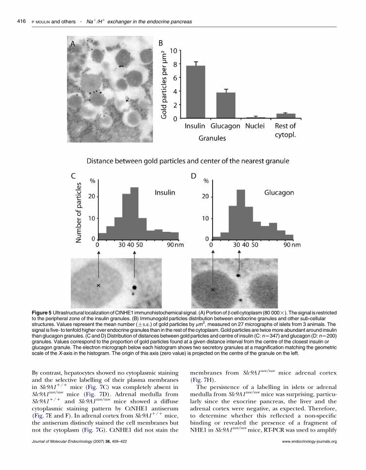
Figure 5 Ultrastructural localizationofCtNHE1 immunohistochemical signal. (A) Portion ofbcell cytoplasm (80 000!).The signal is restrictedto the peripheral zone of the insulin granules. (B) Immunogold particles distribution between endocrine granules and other sub-cellularstructures. Values represent the mean number (GS.E.) of gold particles by mm2, measured on 27 micrographs of islets from 3 animals. Thesignal is five- to tenfold higher over endocrine granules than in the rest of the cytoplasm. Gold particles are twice more abundant around insulinthan glucagon granules. (C and D) Distribution of distances between gold particles and centre of insulin (C: nZ347) and glucagon (D: nZ200)granules. Values correspond to the proportion of gold particles found at a given distance interval from the centre of the closest insulin orglucagon granule. The electron micrograph below each histogram shows two secretory granules at a magnification matching the geometricscale of the X-axis in the histogram. The origin of this axis (zero value) is projected on the centre of the granule on the left.
P MOULIN and others . NaC/HC exchanger in the endocrine pancreas416
By contrast, hepatocytes showed no cytoplasmic stainingand the selective labelling of their plasma membranesin Slc9A1C/C mice (Fig. 7C) was completely absent inSlc9A1swe/swe mice (Fig. 7D). Adrenal medulla fromSlc9A1C/C and Slc9A1swe/swe mice showed a diffusecytoplasmic staining pattern by CtNHE1 antiserum(Fig. 7E and F). In adrenal cortex from Slc9A1C/C mice,the antiserum distinctly stained the cell membranes butnot the cytoplasm (Fig. 7G). CtNHE1 did not stain the
Journal of Molecular Endocrinology (2007) 38, 409–422
membranes from Slc9A1swe/swe mice adrenal cortex(Fig. 7H).
The persistence of a labelling in islets or adrenalmedulla from Slc9A1swe/swe mice was surprising, particu-larly since the exocrine pancreas, the liver and theadrenal cortex were negative, as expected. Therefore,to determine whether this reflected a non-specificbinding or revealed the presence of a fragment ofNHE1 in Slc9A1swe/swe mice, RT-PCR was used to amplify
www.endocrinology-journals.org
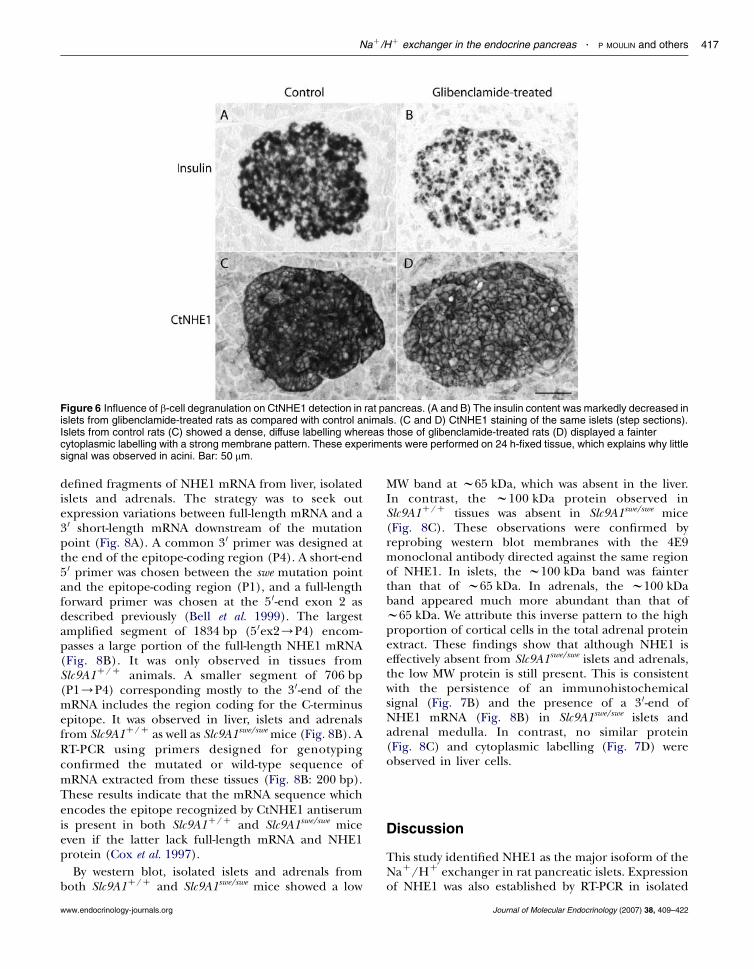
Figure 6 Influence of b-cell degranulation on CtNHE1 detection in rat pancreas. (A and B) The insulin content was markedly decreased inislets from glibenclamide-treated rats as compared with control animals. (C and D) CtNHE1 staining of the same islets (step sections).Islets from control rats (C) showed a dense, diffuse labelling whereas those of glibenclamide-treated rats (D) displayed a faintercytoplasmic labelling with a strong membrane pattern. These experiments were performed on 24 h-fixed tissue, which explains why littlesignal was observed in acini. Bar: 50 mm.
NaC/HC exchanger in the endocrine pancreas . P MOULIN and others 417
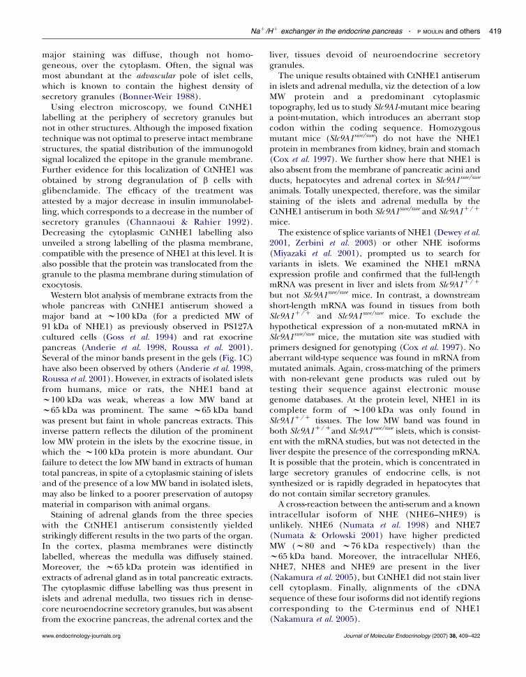
defined fragments of NHE1 mRNA from liver, isolatedislets and adrenals. The strategy was to seek outexpression variations between full-length mRNA and a3 0 short-length mRNA downstream of the mutationpoint (Fig. 8A). A common 3 0 primer was designed atthe end of the epitope-coding region (P4). A short-end5 0 primer was chosen between the swe mutation pointand the epitope-coding region (P1), and a full-lengthforward primer was chosen at the 5 0-end exon 2 asdescribed previously (Bell et al. 1999). The largestamplified segment of 1834 bp (5 0ex2/P4) encom-passes a large portion of the full-length NHE1 mRNA(Fig. 8B). It was only observed in tissues fromSlc9A1C/C animals. A smaller segment of 706 bp(P1/P4) corresponding mostly to the 3 0-end of themRNA includes the region coding for the C-terminusepitope. It was observed in liver, islets and adrenalsfrom Slc9A1C/C as well as Slc9A1swe/swe mice (Fig. 8B). ART-PCR using primers designed for genotypingconfirmed the mutated or wild-type sequence ofmRNA extracted from these tissues (Fig. 8B: 200 bp).These results indicate that the mRNA sequence whichencodes the epitope recognized by CtNHE1 antiserumis present in both Slc9A1C/C and Slc9A1swe/swe miceeven if the latter lack full-length mRNA and NHE1protein (Cox et al. 1997).
By western blot, isolated islets and adrenals fromboth Slc9A1C/C and Slc9A1swe/swe mice showed a low
www.endocrinology-journals.org
MW band at w65 kDa, which was absent in the liver.In contrast, the w100 kDa protein observed inSlc9A1C/C tissues was absent in Slc9A1swe/swe mice(Fig. 8C). These observations were confirmed byreprobing western blot membranes with the 4E9monoclonal antibody directed against the same regionof NHE1. In islets, the w100 kDa band was fainterthan that of w65 kDa. In adrenals, the w100 kDaband appeared much more abundant than that ofw65 kDa. We attribute this inverse pattern to the highproportion of cortical cells in the total adrenal proteinextract. These findings show that although NHE1 iseffectively absent from Slc9A1swe/swe islets and adrenals,the low MW protein is still present. This is consistentwith the persistence of an immunohistochemicalsignal (Fig. 7B) and the presence of a 3 0-end ofNHE1 mRNA (Fig. 8B) in Slc9A1swe/swe islets andadrenal medulla. In contrast, no similar protein(Fig. 8C) and cytoplasmic labelling (Fig. 7D) wereobserved in liver cells.
Discussion
This study identified NHE1 as the major isoform of theNaC/HC exchanger in rat pancreatic islets. Expressionof NHE1 was also established by RT-PCR in isolated
Journal of Molecular Endocrinology (2007) 38, 409–422
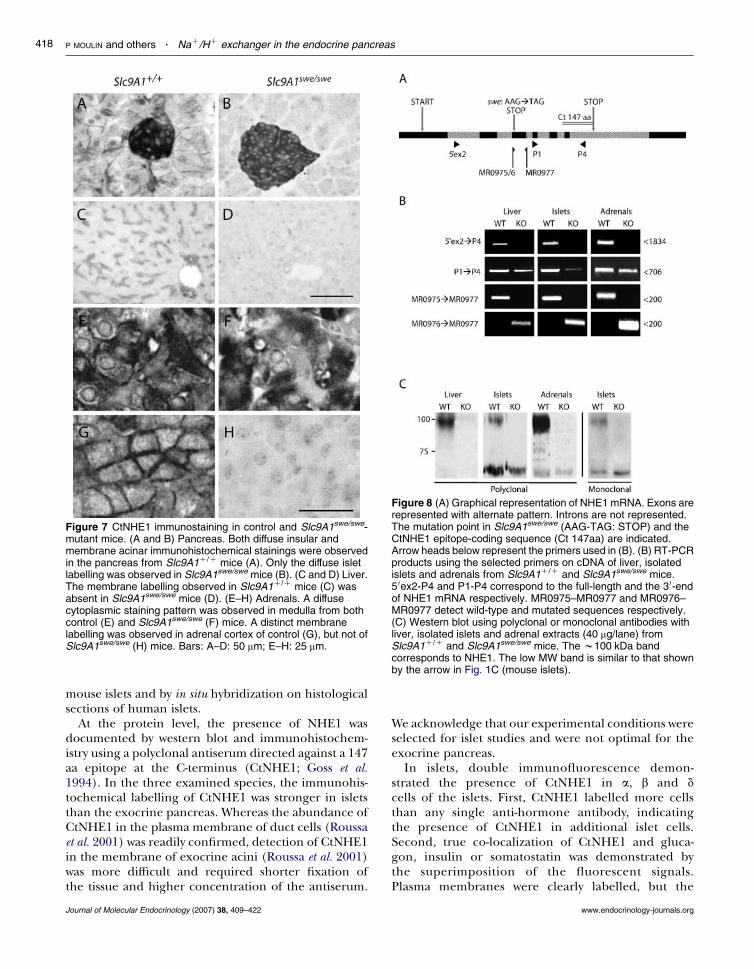
Figure 7 CtNHE1 immunostaining in control and Slc9A1swe/swe-mutant mice. (A and B) Pancreas. Both diffuse insular andmembrane acinar immunohistochemical stainings were observedin the pancreas from Slc9A1C/C mice (A). Only the diffuse isletlabelling was observed in Slc9A1swe/swe mice (B). (C and D) Liver.The membrane labelling observed in Slc9A1C/C mice (C) wasabsent in Slc9A1swe/swe mice (D). (E–H) Adrenals. A diffusecytoplasmic staining pattern was observed in medulla from bothcontrol (E) and Slc9A1swe/swe (F) mice. A distinct membranelabelling was observed in adrenal cortex of control (G), but not ofSlc9A1swe/swe (H) mice. Bars: A–D: 50 mm; E–H: 25 mm.
Figure 8 (A) Graphical representation of NHE1 mRNA. Exons arerepresented with alternate pattern. Introns are not represented.The mutation point in Slc9A1swe/swe (AAG-TAG: STOP) and theCtNHE1 epitope-coding sequence (Ct 147aa) are indicated.Arrow heads below represent the primers used in (B). (B) RT-PCRproducts using the selected primers on cDNA of liver, isolatedislets and adrenals from Slc9A1C/C and Slc9A1swe/swe mice.5 0ex2-P4 and P1-P4 correspond to the full-length and the 3 0-endof NHE1 mRNA respectively. MR0975–MR0977 and MR0976–MR0977 detect wild-type and mutated sequences respectively.(C) Western blot using polyclonal or monoclonal antibodies withliver, isolated islets and adrenal extracts (40 mg/lane) fromSlc9A1C/C and Slc9A1swe/swe mice. The w100 kDa bandcorresponds to NHE1. The low MW band is similar to that shownby the arrow in Fig. 1C (mouse islets).
P MOULIN and others . NaC/HC exchanger in the endocrine pancreas418
mouse islets and by in situ hybridization on histologicalsections of human islets.
At the protein level, the presence of NHE1 wasdocumented by western blot and immunohistochem-istry using a polyclonal antiserum directed against a 147aa epitope at the C-terminus (CtNHE1; Goss et al.1994). In the three examined species, the immunohis-tochemical labelling of CtNHE1 was stronger in isletsthan the exocrine pancreas. Whereas the abundance ofCtNHE1 in the plasma membrane of duct cells (Roussaet al. 2001) was readily confirmed, detection of CtNHE1in the membrane of exocrine acini (Roussa et al. 2001)was more difficult and required shorter fixation ofthe tissue and higher concentration of the antiserum.
Journal of Molecular Endocrinology (2007) 38, 409–422
We acknowledge that our experimental conditions wereselected for islet studies and were not optimal for theexocrine pancreas.
In islets, double immunofluorescence demon-strated the presence of CtNHE1 in a, b and dcells of the islets. First, CtNHE1 labelled more cellsthan any single anti-hormone antibody, indicatingthe presence of CtNHE1 in additional islet cells.Second, true co-localization of CtNHE1 and gluca-gon, insulin or somatostatin was demonstrated bythe superimposition of the fluorescent signals.Plasma membranes were clearly labelled, but the
www.endocrinology-journals.org
NaC/HC exchanger in the endocrine pancreas . P MOULIN and others 419
major staining was diffuse, though not homo-geneous, over the cytoplasm. Often, the signal wasmost abundant at the advascular pole of islet cells,which is known to contain the highest density ofsecretory granules (Bonner-Weir 1988).
Using electron microscopy, we found CtNHE1labelling at the periphery of secretory granules butnot in other structures. Although the imposed fixationtechnique was not optimal to preserve intact membranestructures, the spatial distribution of the immunogoldsignal localized the epitope in the granule membrane.Further evidence for this localization of CtNHE1 wasobtained by strong degranulation of b cells withglibenclamide. The efficacy of the treatment wasattested by a major decrease in insulin immunolabel-ling, which corresponds to a decrease in the number ofsecretory granules (Channaoui & Rahier 1992).Decreasing the cytoplasmic CtNHE1 labelling alsounveiled a strong labelling of the plasma membrane,compatible with the presence of NHE1 at this level. It isalso possible that the protein was translocated from thegranule to the plasma membrane during stimulation ofexocytosis.
Western blot analysis of membrane extracts from thewhole pancreas with CtNHE1 antiserum showed amajor band at w100 kDa (for a predicted MW of91 kDa of NHE1) as previously observed in PS127Acultured cells (Goss et al. 1994) and rat exocrinepancreas (Anderie et al. 1998, Roussa et al. 2001).Several of the minor bands present in the gels (Fig. 1C)have also been observed by others (Anderie et al. 1998,Roussa et al. 2001). However, in extracts of isolated isletsfrom humans, mice or rats, the NHE1 band atw100 kDa was weak, whereas a low MW band atw65 kDa was prominent. The same w65 kDa bandwas present but faint in whole pancreas extracts. Thisinverse pattern reflects the dilution of the prominentlow MW protein in the islets by the exocrine tissue, inwhich the w100 kDa protein is more abundant. Ourfailure to detect the low MW band in extracts of humantotal pancreas, in spite of a cytoplasmic staining of isletsand of the presence of a low MW band in isolated islets,may also be linked to a poorer preservation of autopsymaterial in comparison with animal organs.
Staining of adrenal glands from the three specieswith the CtNHE1 antiserum consistently yieldedstrikingly different results in the two parts of the organ.In the cortex, plasma membranes were distinctlylabelled, whereas the medulla was diffusely stained.Moreover, the w65 kDa protein was identified inextracts of adrenal gland as in total pancreatic extracts.The cytoplasmic diffuse labelling was thus present inislets and adrenal medulla, two tissues rich in dense-core neuroendocrine secretory granules, but was absentfrom the exocrine pancreas, the adrenal cortex and the
www.endocrinology-journals.org
liver, tissues devoid of neuroendocrine secretorygranules.
The unique results obtained with CtNHE1 antiserumin islets and adrenal medulla, viz the detection of a lowMW protein and a predominant cytoplasmictopography, led us to study Slc9A1-mutant mice bearinga point-mutation, which introduces an aberrant stopcodon within the coding sequence. Homozygousmutant mice (Slc9A1swe/swe) do not have the NHE1protein in membranes from kidney, brain and stomach(Cox et al. 1997). We further show here that NHE1 isalso absent from the membrane of pancreatic acini andducts, hepatocytes and adrenal cortex in Slc9A1swe/swe
animals. Totally unexpected, therefore, was the similarstaining of the islets and adrenal medulla by theCtNHE1 antiserum in both Slc9A1swe/swe and Slc9A1C/C
mice.The existence of splice variants of NHE1 (Dewey et al.
2001, Zerbini et al. 2003) or other NHE isoforms(Miyazaki et al. 2001), prompted us to search forvariants in islets. We examined the NHE1 mRNAexpression profile and confirmed that the full-lengthmRNA was present in liver and islets from Slc9A1C/C
but not Slc9A1swe/swe mice. In contrast, a downstreamshort-length mRNA was found in tissues from bothSlc9A1C/C and Slc9A1swe/swe mice. To exclude thehypothetical expression of a non-mutated mRNA inSlc9A1swe/swe mice, the mutation site was studied withprimers designed for genotyping (Cox et al. 1997). Noaberrant wild-type sequence was found in mRNA frommutated animals. Again, cross-matching of the primerswith non-relevant gene products was ruled out bytesting their sequence against electronic mousegenome databases. At the protein level, NHE1 in itscomplete form of w100 kDa was only found inSlc9A1C/C tissues. The low MW band was found inboth Slc9A1C/Cand Slc9A1swe/swe islets, which is consist-ent with the mRNA studies, but was not detected in theliver despite the presence of the corresponding mRNA.It is possible that the protein, which is concentrated inlarge secretory granules of endocrine cells, is notsynthesized or is rapidly degraded in hepatocytes thatdo not contain similar secretory granules.
A cross-reaction between the anti-serum and a knownintracellular isoform of NHE (NHE6–NHE9) isunlikely. NHE6 (Numata et al. 1998) and NHE7(Numata & Orlowski 2001) have higher predictedMW (w80 and w76 kDa respectively) than thew65 kDa band. Moreover, the intracellular NHE6,NHE7, NHE8 and NHE9 are present in the liver(Nakamura et al. 2005), but CtNHE1 did not stain livercell cytoplasm. Finally, alignments of the cDNAsequence of these four isoforms did not identify regionscorresponding to the C-terminus end of NHE1(Nakamura et al. 2005).
Journal of Molecular Endocrinology (2007) 38, 409–422
P MOULIN and others . NaC/HC exchanger in the endocrine pancreas420
The presence of a limited Slc9A1 mRNA sequencecorresponding to the CtNHE1 epitope in bothSlc9A1swe/swe and Slc9A1C/C mice suggests that atruncated gene product comprising the C-terminusend of the original protein is constitutively producedin neuroendocrine cells. One possible explanationcould be the presence of alternate transcripts ofSlc9A1. Indeed, analysis of the whole Slc9A1 sequenceidentifies 119 donor and 195 acceptor sites forpossible splicing using a probability threshold of0.70. In addition to these in silico analyses, supportfor alternate splicing can be found in previous work.First, early after the cloning of NHE1, short-lengthtranscripts have been described in rabbit myocardium(Dyck et al. 1992). Second, amongst two alternatetranscripts detected in brain and lung fromengineered Slc9A1 knockout mice, one was shown tohave an in-phase downstream sequence that canproduce an intact C-terminus peptide (Bell et al.1999). However, this transcript should produce aprotein of predicted MW at w80 kDa, which does notcorrespond to the present w65 kDa protein. A splicevariant of NHE1 with an intact C-terminus sequenceexists in erythrocytes and kidney, not in liver(pancreas was not examined), and could mediateNa:Li countertransport (Zerbini et al. 2003). Again,the predicted MW of the product (w80 kDa) differsfrom the present w65 kDa protein. Finally, there arereports of splice variants of a unique gene that areeither differentially expressed in tissues (Zhang et al.2004) or addressed to different subcellular locations(Ozaita et al. 2002). All these observations are inkeeping with our proposal that a splice variant ofSLC9A1 is specifically expressed in the neuro-endocrine cells of the pancreas and the adrenal,where it produces a w65 kDa protein that isaddressed to the membrane of the secretory granules.
In conclusion, the present study shows that both thefull-length and a shorter-splice variant of NHE1 areexpressed in pancreatic islets and adrenal medulla ofrodents and humans. As in other cell types (Cavet et al.1999), NHE1 is addressed to the plasma membranewhere it serves its functions of cytosolic pH(Juntti-Berggren et al. 1991, Shepherd & Henquin1995, Shepherd et al. 1996) and volume (Miley et al.1998) regulation. The shorter protein is associated withneuroendocrine secretory vesicles. In b cells, insulingranule acidification by a proton-pump is important forproinsulin conversion by the pH-sensitive prohormoneconvertases (Orci et al. 1994). Recent studies alsoindicate that an acidic granular pH is important forCa2C-induced insulin secretion (Barg et al. 2001,Stiernet et al. 2006). Whether the NHE1-like protein isinvolved in the regulation of granular pH is aninteresting possibility to be investigated in future work.
Journal of Molecular Endocrinology (2007) 38, 409–422
Acknowledgements
P M was supported by grant FIRST 415795 from theWalloon Region of Belgium. J C J is Senior ResearchAssociate from the Fonds National de la RechercheScientifique, Brussels, Belgium. This work was sup-ported by Grants (to J R, J C J and O D) from the Fondsde la Recherche Scientifique Medicale, Brussels, andgrant ARC 05/10-328 from the Direction de laRecherche Scientifique de la Communaute Francaisede Belgique. We are grateful to Dr M Donowitzfor kindly providing the CtNHE1 antiserum and to DrD Dufrane for providing human islets. We are alsoindebted to E Riveira Munoz for her expertise ingenetic analysis and to Ph Camby, Y Cnops, H Debaix, MNenquin and L Wenderickx for their skilful help. Theauthors declare that there is no conflict of interest thatwould prejudice the impartiality of this scientific work.
References
Aizawa T, Sato Y & Komatsu M 2002 Importance of nonionic signals forglucose-induced biphasic insulin secretion. Diabetes 51 (Suppl 1)S96–S98.
Altschul SF, Gish W, Miller W, Myers EW & Lipman DJ 1990 Basic localalignment search tool. Journal of Molecular Biology 215 403–410.
Anderie I, Blum R, Haase W, Grinstein S & Thevenod F 1998Expression of NHE1 and NHE4 in rat pancreatic zymogen granulemembranes. Biochemical and Biophysical Research Communications 246330–336.
Baird NR, Orlowski J, Szabo EZ, Zaun HC, Schultheis PJ, Menon AG &Shull GE 1999 Molecular cloning, genomic organization, andfunctional expression of NaC/HC exchanger isoform 5 (NHE5)from human brain. Journal of Biological Chemistry 274 4377–4382.
Barg S, Huang P, Eliasson L, Nelson DJ, Obermuller S, Rorsman P,Thevenod F & Renstrom E 2001 Priming of insulin granules forexocytosis by granular ClK uptake and acidification. Journal of CellScience 114 2145–2154.
Bell SM, Schreiner CM, Schultheis PJ, Miller ML, Evans RL, VorheesCV, Shull GE & Scott WJ 1999 Targeted disruption of the murineNhe1 locus induces ataxia, growth retardation, and seizures.American Journal of Physiology 276 C788–C795.
Bertrand G, Ishiyama N, Nenquin M, Ravier MA & Henquin JC 2002The elevation of glutamate content and the amplification of insulinsecretion in glucose-stimulated pancreatic islets are not causallyrelated. Journal of Biological Chemistry 277 32883–32891.
Best L, Yates AP, Gordon C & Tomlinson S 1988 Modulation bycytosolic pH of calcium and rubidium fluxes in rat pancreatic islets.Biochemical Pharmacology 37 4611–4615.
Bonner-Weir S 1988 Morphological evidence for pancreatic polarity ofbeta-cell within islets of Langerhans. Diabetes 37 616–621.
Cavet ME, Akhter S, de Medina FS, Donowitz M & Tse CM 1999NaC/HC exchangers (NHE1-3) have similar turnover numbers butdifferent percentages on the cell surface. American Journal ofPhysiology 277 C1111–C1121.
Chambrey R, Warnock DG, Podevin RA, Bruneval P, Mandet C, BelairMF, Bariety J & Paillard M 1998 Immunolocalization of theNaC/HC exchanger isoform NHE2 in rat kidney. AmericanJournal of Physiology 275 F379–F386.
Channaoui K & Rahier J 1992 Influence of the decrease ofintracellular antigenic content on morphometric analysis: effect ofthe type and dilution of the first antibody.Histochemistry 97 389–395.
www.endocrinology-journals.org
NaC/HC exchanger in the endocrine pancreas . P MOULIN and others 421
Chu J, Chu S & Montrose MH 2002 Apical NaC/HC exchange nearthe base of mouse colonic crypts. American Journal of Physiology. CellPhysiology 283 C358–C372.
Combet S, Van Landschoot M, Moulin P, Piech A, Verbavatz JM,Goffin E, Balligand JL, Lameire N & Devuyst O 1999 Regulationof aquaporin-1 and nitric oxide synthase isoforms in a rat modelof acute peritonitis. Journal of the American Society of Nephrology 102185–2196.
Cox GA, Lutz CM, Yang CL, Biemesderfer D, Bronson RT, Fu A,Aronson PS, Noebels JL & Frankel WN 1997 Sodium/hydrogenexchanger gene defect in slow-wave epilepsy mutant mice. Cell 91139–148.
Delpire E, Duchene C, Cornet M & Gilles R 1988 Amiloride: aninhibitor of regulatory volume decrease in rat pheochromocytomacultured cells. Pflugers Archiv 411 223–225.
Dewey MJ, Ennis TM & Bowman LH 2001 cDNA cloning andexpression of the mouse Na/H antiporter (NHE-1) and a potentialsplice variant. Molecular Biology Reports 28 111–117.
Dufrane D, Goebbels RM, Guiot Y, Squifflet JP, Henquin JC & GianelloP 2005 A simple method using a polymethylpenten chamber forisolation of human pancreatic islets. Pancreas 30 e51–e59.
Dyck JR, Lopaschuk GD & Fliegel L 1992 Identification of a smallNaC/HC exchanger-like message in the rabbit myocardium. FEBSLetters 310 255–259.
Gilon P, Ravier MA, Jonas JC & Henquin JC 2002 Control mechanismsof the oscillations of insulin secretion in vitro and in vivo. Diabetes 51(Suppl 1) S144–S151.
Goss GG, Woodside M, Wakabayashi S, Pouyssegur J, Waddell T,Downey GP & Grinstein S 1994 ATP dependence of NHE-1, theubiquitous isoform of the NaC/HC antiporter. Analysis ofphosphorylation and subcellular localization. Journal of BiologicalChemistry 269 8741–8748.
Gunawardana SC & Sharp GWG 2002 Intracellular pH plays a criticalrole in glucose-induced time-dependent potentiation of insulinrelease in rat islets. Diabetes 51 105.
Henquin JC 2000 Triggering and amplifying pathways of regulation ofinsulin secretion by glucose. Diabetes 49 1751–1760.
Jonas JC, Gilon P & Henquin JC 1998 Temporal and quantitativecorrelations between insulin secretion and stably elevated oroscillatory cytoplasmic Ca2C in mouse pancreatic beta-cells. Diabetes47 1266–1273.
Jonas JC, Sharma A, Hasenkamp W, Ilkova H, Patane G, Laybutt R,Bonner-Weir S & Weir GC 1999 Chronic hyperglycemia triggers lossof pancreatic beta cell differentiation in an animal model ofdiabetes. Journal of Biological Chemistry 274 14112.
Juntti-Berggren L, Arkhammar P, Nilsson T, Rorsman P & BerggrenPO 1991 Glucose-induced increase in cytoplasmic pH in pancreaticbeta-cells is mediated by NaC/HC exchange, an effect notdependent on protein kinase C. Journal of Biological Chemistry 26623537–23541.
Kao LS, Ho MY & Cragoe EJ Jr. 1991 Intracellular pH andcatecholamine secretion from bovine adrenal chromaffin cells.Journal of Neurochemistry 57 1656–1660.
Kurashima K, Szabo EZ, Lukacs G, Orlowski J & Grinstein S 1998Endosomal recycling of the NaC/HC exchanger NHE3 isoform isregulated by the phosphatidylinositol 3-kinase pathway. Journal ofBiological Chemistry 273 20828–20836.
Lee MG, Schultheis PJ, Yan M, Shull GE, Bookstein C, Chang E, Tse M,Donowitz M, Park K & Muallem S 1998 Membrane-limitedexpression and regulation of NaC/HC exchanger isoforms by P2receptors in the rat submandibular gland duct. Journal of Physiology513 341–357.
Lee MG, Ahn W, Choi JY, Luo X, Seo JT, Schultheis PJ, Shull GE,Kim KH & Muallem S 2000 NaC-dependent transportersmediate HCO3
K salvage across the luminal membrane of themain pancreatic duct. Journal of Clinical Investigation 1051651–1658.
www.endocrinology-journals.org
Lindstrom P & Sehlin J 1984 Effect of glucose on the intracellular pHof pancreatic islet cells. Biochemical Journal 218 887–892.
Lindstrom P & Sehlin J 1986 Effect of intracellular alkalinization onpancreatic islet calcium uptake and insulin secretion. BiochemicalJournal 239 199–204.
MacDonald MJ, Fahien LA, Brown LJ, Hasan NM, Buss JD & KendrickMA 2005a Perspective: emerging evidence for signaling roles ofmitochondrial anaplerotic products in insulin secretion. AmericanJournal of Physiology Endocrinology and Metabolism 288 E1–E15.
MacDonald PE, Joseph JW & Rorsman P 2005b Glucose-sensingmechanisms in pancreatic beta-cells. Philosophical Transactions of theRoyal Society of London. Series B: Biological Sciences 360 2211–2225.
Matschinsky FM, Magnuson MA, Zelent D, Jetton TL, Doliba N, Han Y,Taub R & Grimsby J 2006 The network of glucokinase-expressingcells in glucose homeostasis and the potential of glucokinaseactivators for diabetes therapy. Diabetes 55 1–12.
Miley HE, Sheader EA, Brown PD & Best L 1997 Glucose-inducedswelling in rat pancreatic beta-cells. Journal of Physiology 504191–198.
Miley HE, Holden D, Grint R, Best L & Brown PD 1998Regulatory volume increase in rat pancreatic beta-cells. PflugersArchiv 435 227–230.
Miyazaki E, Sakaguchi M, Wakabayashi S, Shigekawa M & Mihara K2001 NHE6 protein possesses a signal peptide destined forendoplasmic reticulum membrane and localizes in secretoryorganelles of the cell. Journal of Biological Chemistry 27649221–49227.
Nakamura N, Tanaka S, Teko Y, Mitsui K & Kanazawa H 2005 FourNaC/HC exchanger isoforms are distributed to Golgi and post-Golgi compartments and are involved in organelle pH regulation.Journal of Biological Chemistry 280 1561–1572.
Newgard CB 2002 While tinkering with the beta-cell.metabolicregulatory mechanisms and new therapeutic strategies. Diabetes 513141–3150.
Numata M & Orlowski J 2001 Molecular cloning and characterizationof a novel (NaC,KC)/HC exchanger localized to the trans-Golginetwork. Journal of Biological Chemistry 276 17387–17394.
Numata M, Petrecca K, Lake N & Orlowski J 1998 Identification of amitochondrial NaC/HC exchanger. Journal of Biological Chemistry273 6951–6959.
Orci L, Halban P, Perrelet A, Amherdt M, Ravazzola M & AndersonRGW 1994 pH-independent and -dependent cleavage of proinsulinin the same secretory vesicle. Journal of Cell Biology 126 1149–1156.
Orlowski J & Grinstein S 1997 NaC/HC exchangers of mammaliancells. Journal of Biological Chemistry 272 22373–22376.
Orlowski J & Grinstein S 2004 Diversity of the mammaliansodium/proton exchanger SLC9 gene family. Pflugers Archiv 447549–565.
Ozaita A, Martone ME, Ellisman MH & Rudy B 2002 Differentialsubcellular localization of the two alternatively spliced isoforms ofthe Kv3.1 potassium channel subunit in brain. Journal of Neurophy-siology 88 394–408.
Pace CS, Tarvin JT & Smith JS 1983 Stimulus-secretion coupling inbeta-cells: modulation by pH. American Journal of Physiology 244E3–E18.
Peti-Peterdi J, Chambrey R, Bebok Z, Biemesderfer D, St John PL,Abrahamson DR, Warnock DG & Bell PD 2000 Macula densaNaC/HC exchange activities mediated by apical NHE2 andbasolateral NHE4 isoforms. American Journal of Physiology. RenalPhysiology 278 F452–F463.
Rahier J, Stevens M, de Menten Y & Henquin JC 1989 Determinationof antigen concentration in tissue sections by immunodensitome-try. Laboratory Investigations 61 357–363.
Ritter M, Fuerst J, Woll E, Chwatal S, Gschwentner M, Lang F, DeetjenP & Paulmichl M 2001 NaC/HC exchangers: linking osmoticdysequilibrium to modified cell function. Cellular Physiology andBiochemistry 11 1–18.
Journal of Molecular Endocrinology (2007) 38, 409–422
P MOULIN and others . NaC/HC exchanger in the endocrine pancreas422
Rossmann H, Sonnentag T, Heinzmann A, Seidler B, Bachmann O,Vieillard-Baron D, Gregor M & Seidler U 2001 Differentialexpression and regulation of NaC/HC exchanger isoforms inrabbit parietal and mucous cells. American Journal of Physiology.Gastrointestinal and Liver Physiology 281 G447–G458.
Roussa E, Alper SL & Thevenod F 2001 Immunolocalization of anionexchanger AE2, NaC/HC exchangers NHE1 and NHE4, andvacuolar type HC-ATPase in rat pancreas. Journal of Histochemistryand Cytochemistry 49 463–474.
Salgado A, Silva AM, Santos RM & Rosario LM 1996 Multiphasic actionof glucose and alpha-ketoisocaproic acid on the cytosolic pH ofpancreatic beta-cells. Evidence for an acidification pathway linkedto the stimulation of Ca2C influx. Journal of Biological Chemistry 2718738–8746.
Seino S, Iwanaga T, Nagashima K & Miki T 2000 Diverse roles ofK(ATP) channels learned from Kir6.2 genetically engineered mice.Diabetes 49 311–318.
Sempoux C, Guiot Y, Dubois D, Nollevaux MC, Saudubray JM, Nihoul-Fekete C & Rahier J 1998 Pancreatic B-cell proliferation inpersistent hyperinsulinemic hypoglycemia of infancy: an immuno-histochemical study of 18 cases. Modern Pathology 11 444–449.
Sempoux C, Guiot Y, Dahan K, Moulin P, Stevens M, Lambot V,de Lonlay P, Fournet JC, Junien C, Jaubert F et al. 2003 The focalform of persistent hyperinsulinemic hypoglycemia of infancy:morphological and molecular studies show structural andfunctional differences with insulinoma. Diabetes 52 784–794.
Shepherd RM & Henquin JC 1995 The role of metabolism,cytoplasmic Ca2C, and pH-regulating exchangers in glucose-induced rise of cytoplasmic pH in normal mouse pancreatic islets.Journal of Biological Chemistry 270 7915–7921.
Journal of Molecular Endocrinology (2007) 38, 409–422
Shepherd RM, Gilon P & Henquin JC 1996 Ketoisocaproic acid andleucine increase cytoplasmic pH in mouse pancreatic B cells: role ofcytoplasmic Ca2C and pH-regulating exchangers. Endocrinology 137677–685.
Stalker J, Gibbins B, Meidl P, Smith J, Spooner W, Hotz HR & Cox AV2004 The Ensembl Web site: mechanics of a genome browser.Genome Research 14 951–955.
Stiernet P, Guiot Y, Gilon P & Henquin JC 2006 Glucose acutelydecreases pH of secretory granules in mouse pancreatic islets.Mechanisms and influence on insulin secretion. Journal of BiologicalChemistry 281 22142–22151.
Straub SG & Sharp GW 2002 Glucose-stimulated signaling pathways inbiphasic insulin secretion. Diabetes/Metabolism Research and Reviews18 451–463.
Szaszi K, Paulsen A, Szabo EZ, Numata M, Grinstein S & Orlowski J2002 Clathrin-mediated endocytosis and recycling of the neuron-specific NaC/HC exchanger NHE5 isoform. Regulation byphosphatidylinositol 3 0-kinase and the actin cytoskeleton. Journal ofBiological Chemistry 277 42623–42632.
Zerbini G, Maestroni A, Breviario D, Mangili R & Casari G 2003Alternative splicing of NHE-1 mediates Na–Li countertransport andassociates with activity rate. Diabetes 52 1511–1518.
Zhang T, Haws P & Wu Q 2004 Multiple variable first exons: amechanism for cell- and tissue-specific gene regulation. GenomeResearch 14 79–89.
Received 3 November 2006Accepted 16 November 2006
www.endocrinology-journals.org