Nanoparticle penetration and transport in living pumpkin plants: in situ subcellular identification
11
BioMed Central Page 1 of 11 (page number not for citation purposes) BMC Plant Biology Open Access Research article Nanoparticle penetration and transport in living pumpkin plants: in situ subcellular identification Eduardo Corredor 1,5 , Pilar S Testillano 1 , María-José Coronado 1 , Pablo González-Melendi 1,6 , Rodrigo Fernández-Pacheco 2 , Clara Marquina 3 , M Ricardo Ibarra 2,3 , Jesús M de la Fuente 2 , Diego Rubiales 4 , Alejandro Pérez- de-Luque 4 and María-Carmen Risueño* 1 Address: 1 Centro de Investigaciones Biológicas, (CIB) CSIC, Ramiro de Maeztu 9, E-28040, Madrid, Spain, 2 Instituto de Nanociencia de Aragón, Universidad de Zaragoza, Edificio Interfacultativo II, Pedro Cerbuna 12, 50009, Zaragoza, Spain, 3 Instituto de Ciencia de Materiales de Aragón (ICMA)Departamento de Física de la Materia Condensada, CSIC-Universidad de Zaragoza Pedro Cerbuna 12, 50009, Zaragoza, Spain, 4 Instituto de Agricultura Sostenible, CSIC, Alameda del Obispo s/n, Apdo, 4084, E-14080, Córdoba, Spain, 5 School of Biosciences, University of Birmingham, B15 2TT Birmingham, UK and 6 Centro de Biotecnología y Genómica de Plantas, Universidad Politécnica de Madrid, ETS Ingenieros Agrónomos, Ciudad Universitaria s/n, 28040, Madrid, Spain Email: Eduardo Corredor - [email protected]; Pilar S Testillano - [email protected]; María-José Coronado - [email protected]; Pablo González-Melendi - [email protected]; Rodrigo Fernández-Pacheco - [email protected]; Clara Marquina - [email protected]; M Ricardo Ibarra - [email protected]; Jesús M de la Fuente - [email protected]; Diego Rubiales - [email protected]; Alejandro Pérez-de- Luque - [email protected]; María-Carmen Risueño* - [email protected] * Corresponding author Abstract Background: In recent years, the application of nanotechnology in several fields of bioscience and biomedicine has been studied. The use of nanoparticles for the targeted delivery of substances has been given special attention and is of particular interest in the treatment of plant diseases. In this work both the penetration and the movement of iron-carbon nanoparticles in plant cells have been analyzed in living plants of Cucurbita pepo. Results: The nanoparticles were applied in planta using two different application methods, injection and spraying, and magnets were used to retain the particles in movement in specific areas of the plant. The main experimental approach, using correlative light and electron microscopy provided evidence of intracellular localization of nanoparticles and their displacement from the application point. Long range movement of the particles through the plant body was also detected, particles having been found near the magnets used to immobilize and concentrate them. Furthermore, cell response to the nanoparticle presence was detected. Conclusion: Nanoparticles were capable of penetrating living plant tissues and migrating to different regions of the plant, although movements over short distances seemed to be favoured. These findings show that the use of carbon coated magnetic particles for directed delivery of substances into plant cells is a feasible application. Published: 23 April 2009 BMC Plant Biology 2009, 9:45 doi:10.1186/1471-2229-9-45 Received: 6 October 2008 Accepted: 23 April 2009 This article is available from: http://www.biomedcentral.com/1471-2229/9/45 © 2009 Corredor et al; licensee BioMed Central Ltd. This is an Open Access article distributed under the terms of the Creative Commons Attribution License (http://creativecommons.org/licenses/by/2.0 ), which permits unrestricted use, distribution, and reproduction in any medium, provided the original work is properly cited.
-
Upload
independent -
Category
Documents
-
view
0 -
download
0
Transcript of Nanoparticle penetration and transport in living pumpkin plants: in situ subcellular identification

BioMed CentralBMC Plant Biology
ss
Open AcceResearch articleNanoparticle penetration and transport in living pumpkin plants: in situ subcellular identificationEduardo Corredor1,5, Pilar S Testillano1, María-José Coronado1, Pablo González-Melendi1,6, Rodrigo Fernández-Pacheco2, Clara Marquina3, M Ricardo Ibarra2,3, Jesús M de la Fuente2, Diego Rubiales4, Alejandro Pérez-de-Luque4 and María-Carmen Risueño*1Address: 1Centro de Investigaciones Biológicas, (CIB) CSIC, Ramiro de Maeztu 9, E-28040, Madrid, Spain, 2Instituto de Nanociencia de Aragón, Universidad de Zaragoza, Edificio Interfacultativo II, Pedro Cerbuna 12, 50009, Zaragoza, Spain, 3Instituto de Ciencia de Materiales de Aragón (ICMA)Departamento de Física de la Materia Condensada, CSIC-Universidad de Zaragoza Pedro Cerbuna 12, 50009, Zaragoza, Spain, 4Instituto de Agricultura Sostenible, CSIC, Alameda del Obispo s/n, Apdo, 4084, E-14080, Córdoba, Spain, 5School of Biosciences, University of Birmingham, B15 2TT Birmingham, UK and 6Centro de Biotecnología y Genómica de Plantas, Universidad Politécnica de Madrid, ETS Ingenieros Agrónomos, Ciudad Universitaria s/n, 28040, Madrid, Spain
Email: Eduardo Corredor - [email protected]; Pilar S Testillano - [email protected]; María-José Coronado - [email protected]; Pablo González-Melendi - [email protected]; Rodrigo Fernández-Pacheco - [email protected]; Clara Marquina - [email protected]; M Ricardo Ibarra - [email protected]; Jesús M de la Fuente - [email protected]; Diego Rubiales - [email protected]; Alejandro Pérez-de-Luque - [email protected]; María-Carmen Risueño* - [email protected]
* Corresponding author
AbstractBackground: In recent years, the application of nanotechnology in several fields of bioscience andbiomedicine has been studied. The use of nanoparticles for the targeted delivery of substances hasbeen given special attention and is of particular interest in the treatment of plant diseases. In thiswork both the penetration and the movement of iron-carbon nanoparticles in plant cells have beenanalyzed in living plants of Cucurbita pepo.
Results: The nanoparticles were applied in planta using two different application methods,injection and spraying, and magnets were used to retain the particles in movement in specific areasof the plant. The main experimental approach, using correlative light and electron microscopyprovided evidence of intracellular localization of nanoparticles and their displacement from theapplication point. Long range movement of the particles through the plant body was also detected,particles having been found near the magnets used to immobilize and concentrate them.Furthermore, cell response to the nanoparticle presence was detected.
Conclusion: Nanoparticles were capable of penetrating living plant tissues and migrating todifferent regions of the plant, although movements over short distances seemed to be favoured.These findings show that the use of carbon coated magnetic particles for directed delivery ofsubstances into plant cells is a feasible application.
Published: 23 April 2009
BMC Plant Biology 2009, 9:45 doi:10.1186/1471-2229-9-45
Received: 6 October 2008Accepted: 23 April 2009
This article is available from: http://www.biomedcentral.com/1471-2229/9/45
© 2009 Corredor et al; licensee BioMed Central Ltd. This is an Open Access article distributed under the terms of the Creative Commons Attribution License (http://creativecommons.org/licenses/by/2.0), which permits unrestricted use, distribution, and reproduction in any medium, provided the original work is properly cited.
Page 1 of 11(page number not for citation purposes)
BMC Plant Biology 2009, 9:45 http://www.biomedcentral.com/1471-2229/9/45
BackgroundNanobiotechnology was born as a hybrid discipline, acombination of biotechnology and nanoscience. In recentyears, nanoparticles with sizes typically below 100 nm,have been applied in several fields of bioscience and bio-medicine, with an increasing number of commercialapplications [1]. Advances have been made in the field ofbiomedicine, including the development of tools forpathogen bio-detection, tissue engineering and MRI con-trast enhancement [2]. Special interest have been focusedon those applications developed for targeted delivery ofsubstances and drugs, implying direct movement of nan-oparticles to specific organs [3-5].
The possibility of targeting the movement of nanoparti-cles to specific sites of an organism paves the way for theuse of nanobiotechnology in the treatment of plant dis-eases that affect specific parts of a plant. Different proce-dures have made use of nanoparticles in plants, such asthe controlled release of bioactive substances in solidwood [6-8] and plant transformation through bombard-ment with gold or tungsten particles coated with plas-midic DNA [9]. In recent years, a breakthrough has beenmade as a result of Torney et al. [10] who were able to con-trol the intracellular release of substances into protoplastsusing mesoporous silica nanoparticles. Despite theseadvances, the delivery of nanoparticles into plant tissueshas been limited to methods involving bombardment[9,10], a methodology that does not allow massive appli-cation of particles in large numbers of plants, thus beingof little use for agronomic purposes.
Recently, our group has applied carbon-coated iron nano-particles to pumpkin plants in order to develop tools forthe directed release of chemicals into plant organs suscep-tible to infection by pathogens that specifically attackthem [11]. Using different microscopy methodologies tomonitor their presence in plant tissues, we have shownthat these nanoparticles penetrate living plant tissues. Butas the foregoing observations are only a first step in thedirected distribution of nanoparticles in living plants, towhat extent these particles are capable of migrating prop-erly to reach their target has yet to be established
The aim of this work was to analyse the penetration andmovement of nanoparticles in plant cells, and the capacityof a magnetic field to retain them in specific parts of theplant. Two different application methods were used:injection, and spraying, the latter being a more practicaloption from an agronomic standpoint. Correlative micro-scopy, on both light and electron microscopy levels, wasused as a convenient experimental methodology, provid-ing evidence of intracellular localization of nanoparticlesand their displacements from the application points. Longrange movement of particles through the plant body were
also detected, these particles being found near one of themagnets used to immobilize and concentrate them.
MethodsPlant material and growth conditionsPumpkin (Cucurbita pepo L.) plants were selected for pre-liminary experiments due to their large vessel size, whichwould facilitate the transport of nanoparticles through thevascular system. Plants were grown using a polyethylenebag system as previously described [11,12]. A strip (11 ×28 cm) of glass fibre paper (Whatmann GF/A) wasinserted into a polyethylene bag (25 × 35 cm) as physicalsupport for the development of the root and as a way forthe nutrient solution to rise by capillary action and reachthe whole root. Pumpkin seeds were germinated in Petridishes with moistened filter paper. Fifteen day old pump-kin seedlings having root lengths of about 4–5 cm weretransferred to the bag, placing them on the upper side ofthe system. Twenty ml. of Hoagland nutrient solution[13] was added to each bag, and refilled later when neces-sary. The bags were suspended vertically in boxes and theplants were grown in a controlled environment chamberwith a day/night temperature of 20 ± 0.5 8C, a 14 h pho-toperiod and a photosynthetic photon flux density of 200μmol m-2 s-1.
Some nanoparticle-treated plants and some control plantswere placed in pots to evaluate whether the treatmentwould have any effect on the subsequent growth of theplants.
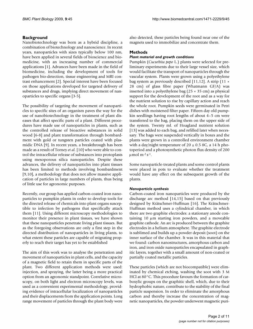
Nanoparticle synthesisCarbon-coated iron nanoparticles were produced by thedischarge arc method [14,15] based on that previouslydesigned by Krätschmer-Huffman [16]. The Krätschmer-Huffman method uses a cylindrical chamber, in whichthere are two graphite electrodes: a stationary anode con-taining 10 μm starting iron powders, and a moveablegraphite cathode. An arc is produced between the graphiteelectrodes in a helium atmosphere. The graphite electrodeis sublimed and builds up a powder deposit (soot) on theinner surface of the chamber. It was in this material thatwe found: carbon nanostructures, amorphous carbon andiron, and iron oxide nanoparticles encapsulated in graph-itic layers, together with a small amount of non-coated orpartially coated metallic particles.
These particles (which are not biocompatible) were elim-inated by chemical etching, washing the soot with 3 MHCl at 80°C. This procedure favours the formation of car-boxylic groups on the graphitic shell, which, due to theirhydrophobic nature, contribute to the stability of the finalparticle suspension. In order to eliminate the amorphouscarbon and thereby increase the concentration of mag-netic nanoparticles, the powder underwent magnetic puri-
Page 2 of 11(page number not for citation purposes)
BMC Plant Biology 2009, 9:45 http://www.biomedcentral.com/1471-2229/9/45
fication. To do this, stable suspensions of the particleswere prepared in a surfactant solution: 2.5 g of SDS in 500ml of distilled water. A field gradient produced by a per-manent magnet (maximum magnetic field 3 KOe) wasused for magnetic separation of this suspension. Employ-ing this method, only the carbon-coated magnetic nano-particles are attracted by the magnet whereas theamorphous carbon remains in the supernatant, which issubsequently eliminated.
Nanoparticle applicationNanoparticles were suspended in gelafundine, a commer-cial succinated gel (30 g/l gelatine, 0.45% w/v sodiumchloride, 0.21 g/l calcium, Braun, Melsungen-Germany),and the solution was kept in water in an ultrasonic bathfor several minutes. This solution was selected because ithad been used as a convenient suspension medium fornanoparticles in experiments with animals. A biocompat-ible magnetic fluid was obtained in this way, which wasused for the inoculation of plants.
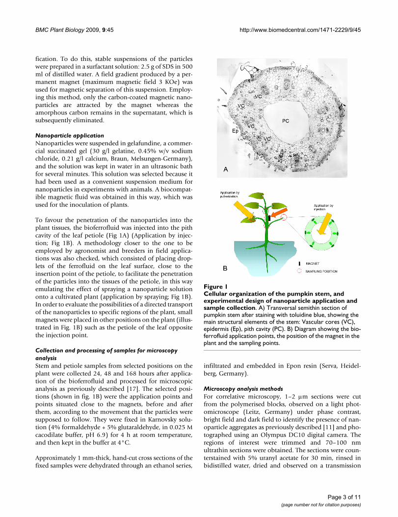
To favour the penetration of the nanoparticles into theplant tissues, the bioferrofluid was injected into the pithcavity of the leaf petiole (Fig 1A) (Application by injec-tion; Fig 1B). A methodology closer to the one to beemployed by agronomist and breeders in field applica-tions was also checked, which consisted of placing drop-lets of the ferrofluid on the leaf surface, close to theinsertion point of the petiole, to facilitate the penetrationof the particles into the tissues of the petiole, in this wayemulating the effect of spraying a nanoparticle solutiononto a cultivated plant (application by spraying; Fig 1B).In order to evaluate the possibilities of a directed transportof the nanoparticles to specific regions of the plant, smallmagnets were placed in other positions on the plant (illus-trated in Fig. 1B) such as the petiole of the leaf oppositethe injection point.
Collection and processing of samples for microscopy analysisStem and petiole samples from selected positions on theplant were collected 24, 48 and 168 hours after applica-tion of the bioferrofluid and processed for microscopicanalysis as previously described [17]. The selected posi-tions (shown in fig. 1B) were the application points andpoints situated close to the magnets, before and afterthem, according to the movement that the particles weresupposed to follow. They were fixed in Karnovsky solu-tion (4% formaldehyde + 5% glutaraldehyde, in 0.025 Mcacodilate buffer, pH 6.9) for 4 h at room temperature,and then kept in the buffer at 4°C.
Approximately 1 mm-thick, hand-cut cross sections of thefixed samples were dehydrated through an ethanol series,
infiltrated and embedded in Epon resin (Serva, Heidel-berg, Germany).
Microscopy analysis methodsFor correlative microscopy, 1–2 μm sections were cutfrom the polymerised blocks, observed on a light phot-omicroscope (Leitz, Germany) under phase contrast,bright field and dark field to identify the presence of nan-oparticle aggregates as previously described [11] and pho-tographed using an Olympus DC10 digital camera. Theregions of interest were trimmed and 70–100 nmultrathin sections were obtained. The sections were coun-terstained with 5% uranyl acetate for 30 min, rinsed inbidistilled water, dried and observed on a transmission
Cellular organization of the pumpkin stem, and experimental design of nanoparticle application and sample collectionFigure 1Cellular organization of the pumpkin stem, and experimental design of nanoparticle application and sample collection. A) Transversal semithin section of pumpkin stem after staining with toluidine blue, showing the main structural elements of the stem: Vascular cores (VC), epidermis (Ep), pith cavity (PC). B) Diagram showing the bio-ferrofluid application points, the position of the magnet in the plant and the sampling points.
Page 3 of 11(page number not for citation purposes)
BMC Plant Biology 2009, 9:45 http://www.biomedcentral.com/1471-2229/9/45
electron microscope Jeol 1010 at 80 kV. Micrographs weretaken with a Megaview digital camera (Gatan).
ResultsCorrelative microscopy was used to score the presence ofnanoparticles in samples collected from different posi-tions, close to the application points and magnet loca-tions. Following the procedures previously established[11], a first level of analysis was performed by examining1–2 μm Epon sections on the light microscope in searchof nanoparticles. Aggregates of nanoparticles appear asdark, optically dense signals under bright field, that corre-late with a refringent signal under dark field and phasecontrast [11]. To make a precise identification of the pres-ence of nanoparticles by light microscopy, their presencewas only considered as proven when the three microscop-ies employed (phase contrast, bright field and dark field)provided correlated images (see additional files 1, 2, 3:correlative images 1, 2, 3 for the micrographs used toselect the nanoparticle-containing cells to be analyzed atTEM). For correlative microscopy, those areas thatappeared to contain nanoparticles were selected for anal-ysis under TEM.
Two different application methods were employed,namely, injection and spraying (see MM). Plant sampleswere collected 24 h, 48 h and 168 h after application,these time periods having been previously determined tobe appropriate for detectingt internalization of nanoparti-cles into the tissues [11].
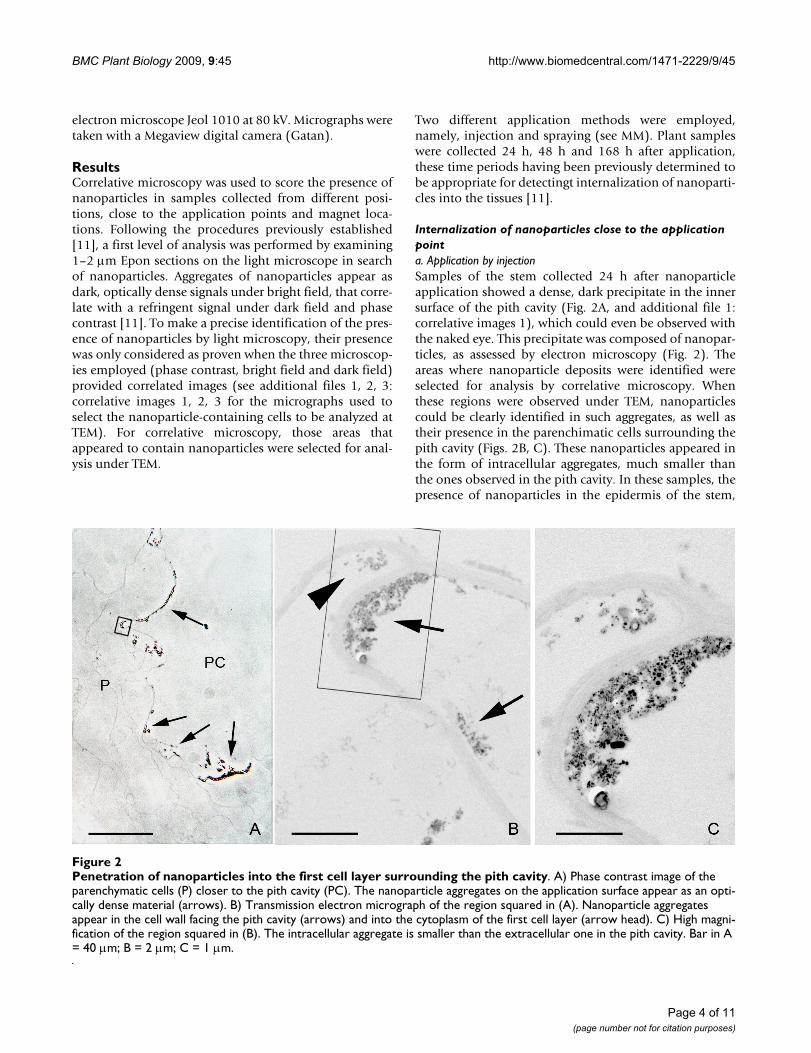
Internalization of nanoparticles close to the application pointa. Application by injectionSamples of the stem collected 24 h after nanoparticleapplication showed a dense, dark precipitate in the innersurface of the pith cavity (Fig. 2A, and additional file 1:correlative images 1), which could even be observed withthe naked eye. This precipitate was composed of nanopar-ticles, as assessed by electron microscopy (Fig. 2). Theareas where nanoparticle deposits were identified wereselected for analysis by correlative microscopy. Whenthese regions were observed under TEM, nanoparticlescould be clearly identified in such aggregates, as well astheir presence in the parenchimatic cells surrounding thepith cavity (Figs. 2B, C). These nanoparticles appeared inthe form of intracellular aggregates, much smaller thanthe ones observed in the pith cavity. In these samples, thepresence of nanoparticles in the epidermis of the stem,
Penetration of nanoparticles into the first cell layer surrounding the pith cavityFigure 2Penetration of nanoparticles into the first cell layer surrounding the pith cavity. A) Phase contrast image of the parenchymatic cells (P) closer to the pith cavity (PC). The nanoparticle aggregates on the application surface appear as an opti-cally dense material (arrows). B) Transmission electron micrograph of the region squared in (A). Nanoparticle aggregates appear in the cell wall facing the pith cavity (arrows) and into the cytoplasm of the first cell layer (arrow head). C) High magni-fication of the region squared in (B). The intracellular aggregate is smaller than the extracellular one in the pith cavity. Bar in A = 40 μm; B = 2 μm; C = 1 μm.
Page 4 of 11(page number not for citation purposes)
BMC Plant Biology 2009, 9:45 http://www.biomedcentral.com/1471-2229/9/45
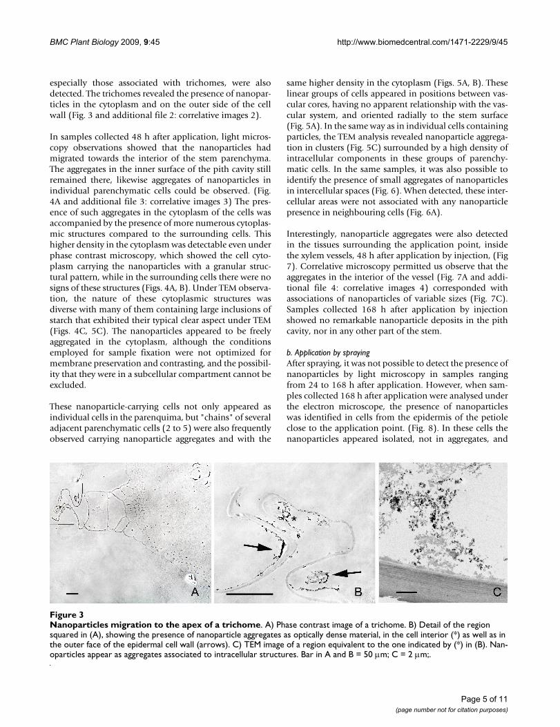
especially those associated with trichomes, were alsodetected. The trichomes revealed the presence of nanopar-ticles in the cytoplasm and on the outer side of the cellwall (Fig. 3 and additional file 2: correlative images 2).
In samples collected 48 h after application, light micros-copy observations showed that the nanoparticles hadmigrated towards the interior of the stem parenchyma.The aggregates in the inner surface of the pith cavity stillremained there, likewise aggregates of nanoparticles inindividual parenchymatic cells could be observed. (Fig.4A and additional file 3: correlative images 3) The pres-ence of such aggregates in the cytoplasm of the cells wasaccompanied by the presence of more numerous cytoplas-mic structures compared to the surrounding cells. Thishigher density in the cytoplasm was detectable even underphase contrast microscopy, which showed the cell cyto-plasm carrying the nanoparticles with a granular struc-tural pattern, while in the surrounding cells there were nosigns of these structures (Figs. 4A, B). Under TEM observa-tion, the nature of these cytoplasmic structures wasdiverse with many of them containing large inclusions ofstarch that exhibited their typical clear aspect under TEM(Figs. 4C, 5C). The nanoparticles appeared to be freelyaggregated in the cytoplasm, although the conditionsemployed for sample fixation were not optimized formembrane preservation and contrasting, and the possibil-ity that they were in a subcellular compartment cannot beexcluded.
These nanoparticle-carrying cells not only appeared asindividual cells in the parenquima, but "chains" of severaladjacent parenchymatic cells (2 to 5) were also frequentlyobserved carrying nanoparticle aggregates and with the
same higher density in the cytoplasm (Figs. 5A, B). Theselinear groups of cells appeared in positions between vas-cular cores, having no apparent relationship with the vas-cular system, and oriented radially to the stem surface(Fig. 5A). In the same way as in individual cells containingparticles, the TEM analysis revealed nanoparticle aggrega-tion in clusters (Fig. 5C) surrounded by a high density ofintracellular components in these groups of parenchy-matic cells. In the same samples, it was also possible toidentify the presence of small aggregates of nanoparticlesin intercellular spaces (Fig. 6). When detected, these inter-cellular areas were not associated with any nanoparticlepresence in neighbouring cells (Fig. 6A).
Interestingly, nanoparticle aggregates were also detectedin the tissues surrounding the application point, insidethe xylem vessels, 48 h after application by injection, (Fig7). Correlative microscopy permitted us observe that theaggregates in the interior of the vessel (Fig. 7A and addi-tional file 4: correlative images 4) corresponded withassociations of nanoparticles of variable sizes (Fig. 7C).Samples collected 168 h after application by injectionshowed no remarkable nanoparticle deposits in the pithcavity, nor in any other part of the stem.
b. Application by sprayingAfter spraying, it was not possible to detect the presence ofnanoparticles by light microscopy in samples rangingfrom 24 to 168 h after application. However, when sam-ples collected 168 h after application were analysed underthe electron microscope, the presence of nanoparticleswas identified in cells from the epidermis of the petioleclose to the application point. (Fig. 8). In these cells thenanoparticles appeared isolated, not in aggregates, and
Nanoparticles migration to the apex of a trichomeFigure 3Nanoparticles migration to the apex of a trichome. A) Phase contrast image of a trichome. B) Detail of the region squared in (A), showing the presence of nanoparticle aggregates as optically dense material, in the cell interior (*) as well as in the outer face of the epidermal cell wall (arrows). C) TEM image of a region equivalent to the one indicated by (*) in (B). Nan-oparticles appear as aggregates associated to intracellular structures. Bar in A and B = 50 μm; C = 2 μm;.
Page 5 of 11(page number not for citation purposes)
BMC Plant Biology 2009, 9:45 http://www.biomedcentral.com/1471-2229/9/45
the densities of intracellular structures observed in thehost cell were similar to those in neighbouring cells. It wasalso noted that no nanoparticles were clearly detected incells beyond the first epidermal layer. Neither were nano-particles detected in other stem samples collected 24 or 48h after application.
Internalization of nanoparticles in locations far from the application pointLight microscopy observations did not detect nanoparti-cles in samples at positions far from the application point,or near magnet locations, either before or after the mag-netic device. Direct analysis of these samples was carriedout by TEM, selecting for the analysis the vascular coresand the surrounding cells, given that these were expectedto form part of the route for long range transport in plants.In these areas, the presence of particles was detected onlyoccasionally in individual cells close to the xylem (Figs.9A–B) and in samples near the magnet collected 48 h aftertheir application by injection. The particles appeared iso-lated in the cytoplasm, without any clear associationamong them in most cases (Figs. 9C–E). The diameters ofthe particles present in a single section of the cytoplasm ofone cell were measured and showed a mean diameter of46.7 ± 1.7 nm (n = 18). There were no apparent differ-ences in the cytoplasm content of the host cell withrespect to that of the neighbouring cells. No particles weredetected after exhaustive screening of ultrathin sectionsfrom samples collected from positions far from the appli-
cation point and distant from the magnet, irrespective ofwhether the application was by injection or spraying.
To evaluate whether the application of nanoparticles hadany effect on the subsequent growth of the plants, sometreated plants were planted in pots and kept under con-trolled conditions until the flowering period. No apparentdifferences were observed between plants subjected to thetreatment and the control plants, the former even beingable to produce fruit which indicated that treatment withnanoparticles did not provoke a toxicological effect onplant growth. At tissue level, the structural organization ofthe stem samples of treated and non-treated plants wassimilar, with no signs of damage at light and electronmicroscopy levels. Only the cells containing the nanopar-ticle agglomerates exhibited more dense cytoplasms, aspreviously stated. This fact suggested that the penetrationof nanoparticles through the tissues did not damagethem.
DiscussionIn the present work, intracellular penetration of carbon-coated iron nanoparticles applied in planta was trackedusing correlative microscopy, including a first screening ofthe samples with optical microscopy (bright field, darkfield and phase contrast) followed by analysis throughtransmission electron microscopy. This strategy hasallowed us to confirm both the progressive penetration ofparticles through the plant tissues and their presence inthe form of intracellular aggregates, when injected into tis-
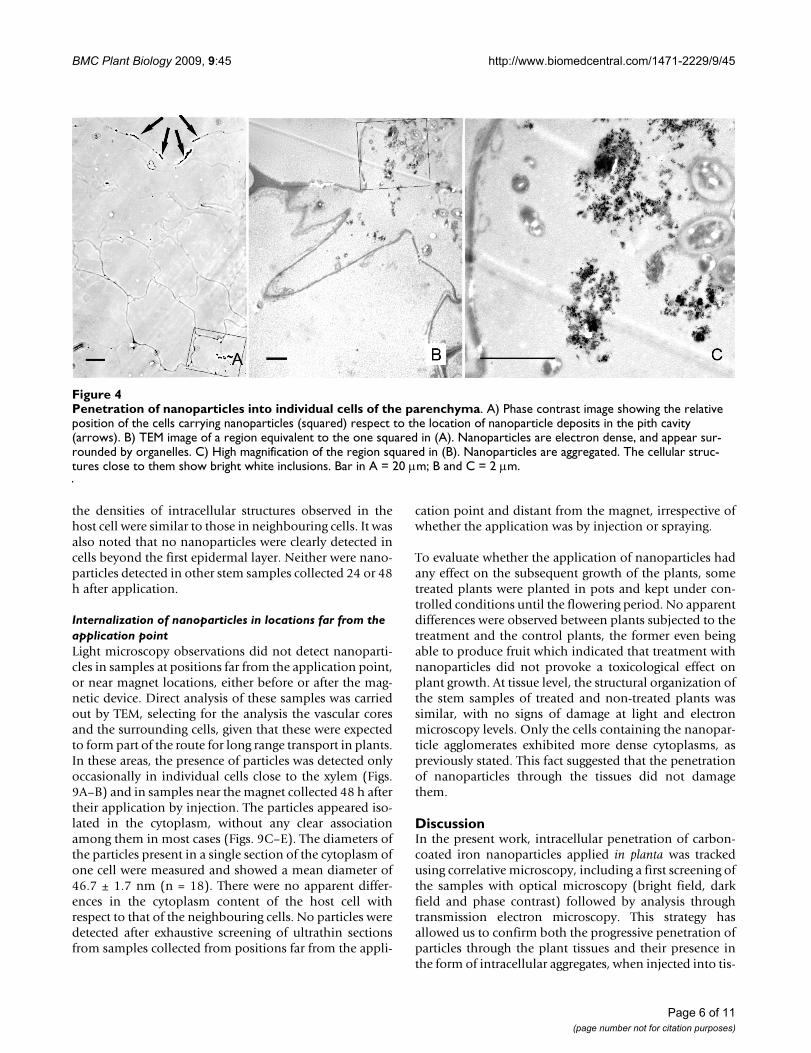
Penetration of nanoparticles into individual cells of the parenchymaFigure 4Penetration of nanoparticles into individual cells of the parenchyma. A) Phase contrast image showing the relative position of the cells carrying nanoparticles (squared) respect to the location of nanoparticle deposits in the pith cavity (arrows). B) TEM image of a region equivalent to the one squared in (A). Nanoparticles are electron dense, and appear sur-rounded by organelles. C) High magnification of the region squared in (B). Nanoparticles are aggregated. The cellular struc-tures close to them show bright white inclusions. Bar in A = 20 μm; B and C = 2 μm.
Page 6 of 11(page number not for citation purposes)

BMC Plant Biology 2009, 9:45 http://www.biomedcentral.com/1471-2229/9/45
sues near the application site, in as short a time as 48 h.Furthermore, 48 h after injection, isolated nanoparticlesin the cytoplasm of individual cells close to a vascular corewere observed in the proximity of a magnetic devicelocated far from the application point. When applied byspraying, isolated nanoparticles were also found in thecytoplasm of epidermic cells, in regions near the applica-tion point.
Cell and tissue response to the presence of nanoparticlesNanoparticles move away from the application point, onthe interior of the stem, where they were injected, to theouter epidermis through the tissues. The fact that a densecytoplasm with starch-containing organelles was observedconcomitantly with nanoparticle aggregates in thecytosol, suggests that plant cells could respond to the pres-ence of a high density of nanoparticles by changing theirsubcellular organization. Results also showed that thenanoparticles had already appeared in the outer surface ofthe plant, both inside and outside of the trichomes, 24 hafter application, indicating that at least part of the biofer-rofluid can be expelled in a short time. Several examplesof cytotoxicity of carbon nanoparticles have beendescribed in various animal systems [18-23] as well as dif-ferent effects derived from the application of magnetic fer-rofluid [24,25]. Cytotoxicity has also been associated withthe dose of nanoparticles [26]. This correlation between
Presence of nanoparticles in adjacent parenchymatic cellsFigure 5Presence of nanoparticles in adjacent parenchymatic cells. A) Bright field image, toluidine blue staining of a sec-tion of the stem showing cells carrying nanoparticles (squared area) between two vascular cores (VC). The nano-particle deposits in the cell wall facing the pith cavity (PC) appear labelled with arrowheads. B) Phase contrast image of a section consecutive to the one showed in (A), with a detail of the region squared in (A). Dark material appears in the cell cytoplasm surrounded by dense structures. The surrounding cells do not show such a dense cytoplasm. C) TEM image showing a detail of a nanoparticle aggregate inside one of the cells showed in (B) which is labelled by (*). Bar in A and B = 100 μm; C = 1 μm.
Ultrastructural imaging of nanoparticles in the extracellular spaceFigure 6Ultrastructural imaging of nanoparticles in the extra-cellular space. A) image showing three confluent parenchy-matic cells with a triangle shaped extracellular region between them. The parenchymatic cells do not show traces of presence of nanoparticles. B) High magnification of the extracellular channel shown in (A) displaying nanoparticle aggregates. Bar in A = 10 μm; B = 200 nm
Page 7 of 11(page number not for citation purposes)
BMC Plant Biology 2009, 9:45 http://www.biomedcentral.com/1471-2229/9/45
the number of nanoparticles and cytotoxicity agrees withour results, which showed that no subcellular rearrange-ments were detected in those plant cells in which only afew isolated nanoparticles were detected (Figs. 8 and 9),compared with the response detected in cells carryingaggregates of nanoparticles. But, to date, there has been nodescription of changes in cell architecture and organiza-tion after nanoparticle internalization in vivo, so it is notpossible to assess if the observed reaction is specific to ourmaterial or if it is a general trait of cell reaction to highconcentrations of nanoparticles within the cell. Moreover,the solution used for nanoparticle suspension, namelygelafundine, contains calcium, an important second mes-senger in plants. The possibility that some calciumremains attached to the carbon coat of the nanoparticleand could have an effect on cell response, does not seemto be very plausible due to the chemical properties of thenanoparticle surface (hydrophobic, not negativelycharged...), even so, it cannot be completely excluded.
Internalization of nanoparticles near the application pointThe observation of particle aggregates in adjacent paren-chymatic cells suggests a movement from cell to cell.Another question to be addressed is the presence of nan-oparticles as intracellular aggregates, as it seems quiteunlikely that nanoparticles have the capability of enteringcells other than as individual particles. The mechanisminvolved in this aggregation is unclear. As indicated inMM, the carbon-iron nanoparticles employed show a ten-dency to aggregate in aqueous solutions because of thechemical characteristics of the carbon coat. Aggregation ofnanoparticles is a common phenomenon [27], so it is
possible that this reclustering takes place spontaneouslyafter individual internalization into the cytoplasm,although it could not be discarded that the nanoparticleswere redirected by the cell to a specific subcompartmentor region in the cytoplasm.
The presence of nanoparticles in epidermal cells afterapplication by spraying is of special interest. As statedbefore, one of the main drawbacks of other methods isthat they cannot be employed for agronomic purposes.The method used in this work resembles the procedureswhich would be used by breeders and coordinators ofphytosanitary control, employing both large scale andhand-on spraying to leaf surfaces. The fact that nanoparti-cles passed through the epidermal cell wall opens up thepossible application of these nanotechnology tools foragronomical purposes. Given the special characteristics ofthe epidermic outer cell wall, specifically its considerablethickness, and the presence of protective waxes, a possibleparticle penetration point could be through the stomataand the subestomatic chambers. In fact, this aperture is aroute used by pathogens of different species, such as thewhite pine blinter rust [28]. Interestingly, water-sus-pended 43 nm hydrophilic particles have been describedas occasionally penetrating Vicia faba leaves through sto-matal pores [29]. Recently, the uptake of magnetite nano-particles through the root system of Cucurbita maximaplants was reported, though a significant uptake of parti-cles was only found in plants growing in liquid media[30]. So, in order to make the system suitable for agro-nomical purposes, methodological improvements wouldneed to be made.
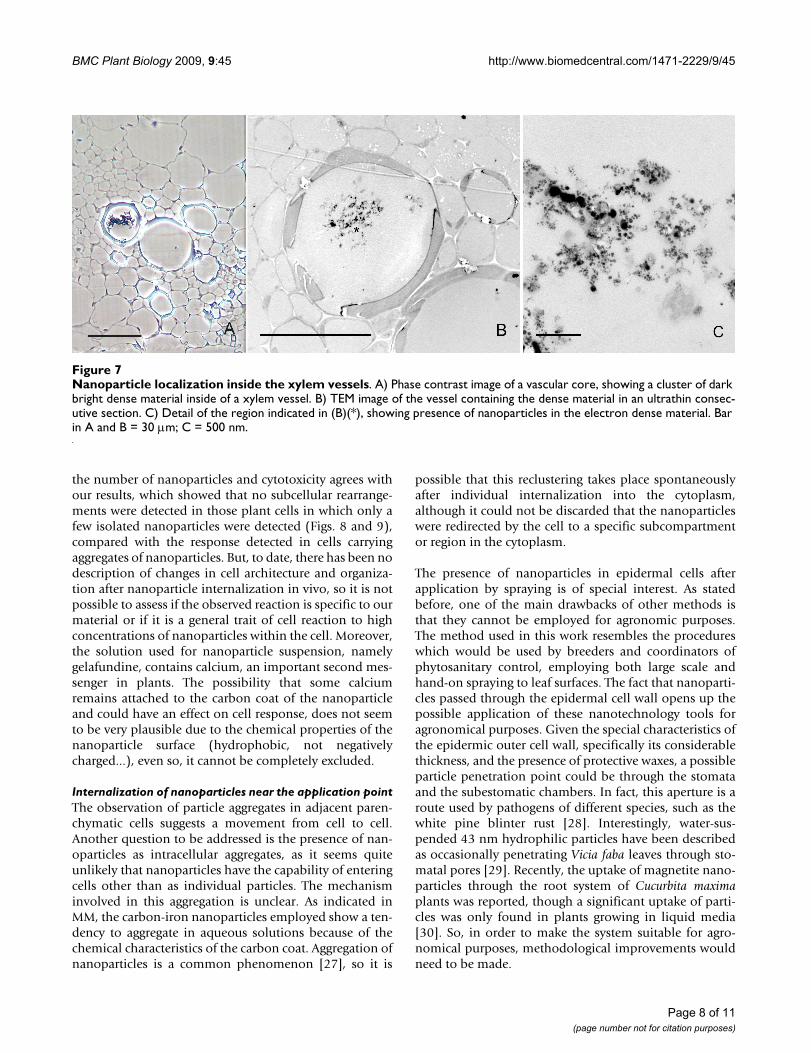
Nanoparticle localization inside the xylem vesselsFigure 7Nanoparticle localization inside the xylem vessels. A) Phase contrast image of a vascular core, showing a cluster of dark bright dense material inside of a xylem vessel. B) TEM image of the vessel containing the dense material in an ultrathin consec-utive section. C) Detail of the region indicated in (B)(*), showing presence of nanoparticles in the electron dense material. Bar in A and B = 30 μm; C = 500 nm.
Page 8 of 11(page number not for citation purposes)
BMC Plant Biology 2009, 9:45 http://www.biomedcentral.com/1471-2229/9/45
Long range transport and putative movement through the vascular systemThe main reason for developing the carbon-iron nanopar-ticles and their application in this work was to accumulatethem at a specific site in the plant by means of magneticfields applied specifically to a certain area. As shown inFig. 9, isolated nanoparticles were detected in the cyto-plasm of cells close to the vascular core, far from the appli-cation point and near the magnet. The position of thesecells suggests that the route taken by the particles involvedthe use of the plant vascular system. Direct observation offreshly cut material revealed the presence of bioferrofluid,specifically in the interior of the xylem vessels [11], alongwith nanoparticle aggregates, 48 h after application (Fig.
7) suggesting that the particles can use this system for longrange transport. It has to be noted that those particlesfound far from their application point (Fig. 9) were quitehomogeneous in size, around 46 nm in diameter on aver-age, when compared with the variable sizes found in theoriginal mixture detected in the aggregates in the pith cav-ity and cells close to the application point. (Figs. 2, 3, 4, 5,6, 7). This may suggest that a certain critical size isrequired for the appropriate movement of particlesthrough the plant by long range transport mechanisms.
ConclusionTaken together, the results reported here demonstrate thatthe carbon-iron nanoparticles are able to get into the cellsof living plants, and move to remote positions, possiblythrough the vascular system. These facts constitute animportant step forward in elucidating the mechanisms ofinteraction between plant cells and nanoparticles andthus, in designing strategies for using nanoparticles fortargeted delivery of substances. In this sense, methodolog-ical improvements are required to make the system suita-ble for agronomical purposes, one of them being thefunctionalization of the nanoparticles with organicgroups that can help their penetration into the phloem ormake their internalization into cells more efficient. Theevidence reported here, that nanoparticles were able tomove to locations where the magnets were located, farfrom the application point, would suggest the potentialuse of carbon-coated nanoparticles for directed delivery ofsubstances for phytosanitary purposes, using magneticfields to retain the particles in areas of interest.
Authors' contributionsEC carried out the electron microscopy study, the correla-tive light and electron microscopy analysis and wrote thefirst manuscript draft. PST participated in the experimen-tal design, contributed to the draft and writing of the man-uscript and its revision, and participated in thecoordination of the work. MJC and PGM carried out theprocessing of plant samples, the correlative light micros-copy analysis under different visualization modes andsome electron microscopy essays. RFP carried out the syn-thesis of nanoparticles and the bioferrofluid suspension.CM and MRI participated in the design of the nanoparticlesynthesis and preparation of the suspension, and in thedesign of the study. JMF contributed to the experimentaldesign of nanoparticle synthesis and to the writing ofparts of the manuscript. DR participated in the design ofthe study and helped in experiments of nanoparticle treat-ments to the plants. APL designed and carried out the nan-oparticle treatments to the plants, and helped in thewriting of some parts of the manuscript. MCR conceivedthe study, participated in the design and coordination ofthe work and helped to draft the manuscript. All authorsread and approved the final manuscript.
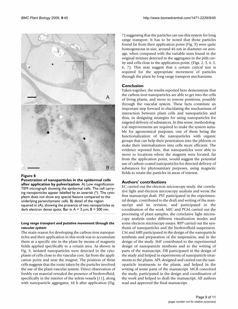
Penetration of nanoparticles in the epidermal cells after application by pulverizationFigure 8Penetration of nanoparticles in the epidermal cells after application by pulverization. A) Low magnification TEM micrograph showing the epidermal cells. The cell carry-ing nanoparticles appear labelled by an asterisk (*). The cyto-plasm does not show any special feature compared to the underlying parenchymatic cells. B) detail of the region squared in (A), showing the presence of two nanoparticles as dark electron dense spots. Bar in A = 3 μm; B = 500 nm.
Page 9 of 11(page number not for citation purposes)

BMC Plant Biology 2009, 9:45 http://www.biomedcentral.com/1471-2229/9/45
Migration/transport of nanoparticles to individual cells close to the magnet locationFigure 9Migration/transport of nanoparticles to individual cells close to the magnet location. A) Low magnification TEM micrograph, showing the position of the cell carrying nanoparticles (CCN) respect to a xylem vessel (Xy). The CCN does not show any special differential features in the cytoplasm in comparison with the neighbouring cells. B) High magnification image of the CCN showing the location of several nanoparticles (arrows). C, D, E) Details of some isolated nanoparticles of those arrowed in (B). Bar in A and B = 5 μm; C = 100 nm.
Additional material
Additional file 1Correlative images 1. Correlative images 1. Bright and dark field visualization of the nanopar-ticles shown in Figure 2A: A) Bright field image, of the same field in (Fig. 2A). The nanoparticle aggregates appear as a dark material. B) Dark field image of the same field in (Fig. 2A). The nanoparticles appear as bright refringent. Bar = 40 μmClick here for file[http://www.biomedcentral.com/content/supplementary/1471-2229-9-45-S1.jpeg]
Additional file 2Correlative images 2.Correlative images 2. Bright and dark field visualization, and electron microscopy of the nanoparticles shown in Figure 3B. A) Dark field image, B) Bright field image, C) Electron micrograph of the nanoparticle aggre-gates of (A) and (B). Bar in A and B = 50 μm; C = 2 μm.Click here for file[http://www.biomedcentral.com/content/supplementary/1471-2229-9-45-S2.jpeg]
Additional file 3Correlative images 3.Correlative images 3. Detail of the cell squared in fig 4A. A) Phase con-trast image, showing nanoparticle aggregates that are visible as an intense dark material. The cytoplasm appears dense and displaying numerous structures and organelles in comparison with the surrounding cells. B) Same field that in (A), bright field image. Bar = 20 μm.Click here for file[http://www.biomedcentral.com/content/supplementary/1471-2229-9-45-S3.jpeg]
Page 10 of 11(page number not for citation purposes)

BMC Plant Biology 2009, 9:45 http://www.biomedcentral.com/1471-2229/9/45
Publish with BioMed Central and every scientist can read your work free of charge
"BioMed Central will be the most significant development for disseminating the results of biomedical research in our lifetime."
Sir Paul Nurse, Cancer Research UK
Your research papers will be:
available free of charge to the entire biomedical community
peer reviewed and published immediately upon acceptance
cited in PubMed and archived on PubMed Central
yours — you keep the copyright
Submit your manuscript here:http://www.biomedcentral.com/info/publishing_adv.asp
BioMedcentral
AcknowledgementsThis research was supported by the project NanoAgro-200540F0041 funded by CSIC (Proyectos Intramurales de Frontera) and by the project NAN2004-09270-C03-03 funded by the Spanish Strategic Action on Nano-science and Nanotechnology of the Spanish Ministry of Education and Sci-ence. The work was also partially supported by projects granted by the Spanish Ministry of Science and Innovation, MICINN, BFU2008-00203, AGL2008-04255 and AGL2008-01467. JMF thanks ARAID Foundation for financial support.
References1. Salata OV: Applications of nanoparticles in biology and medi-
cine. J Nanobiotech 2004, 2:3.2. Ernest H, Shetty R: Impact of nanotechnology on biomedical
sciences: review of current concepts on convergence of nan-otechnology with biology. AZojono 2005, 1:1-14.
3. Rawat M, Singh D, Saraf S, Saraf S: Nanocarriers: promising vehi-cle for bioactive drugs. Biol Pharm Bull 2006, 29:1790-1798.
4. Xu ZP, Zeng OH, Lu GO, Yu AB: Inorganic nanoparticles as car-riers for efficient cellular delivery. Chem Eng Sci 2006,61:1027-1040.
5. Yih TC, Al-Fandhi M: Engineered nanoparticles as precise drugdelivery systems. J Cell Biochem 2006, 97:1184-1190.
6. Liu Y, Laks P, Heiden P: Controlled release of biocides in solidwood. I. Efficacy against brown rot wood decay fungus (Gloe-ophyllum trabeum). J Appl Polym Sci 2002, 86:596-607.
7. Liu Y, Laks P, Heiden P: Controlled release of biocides in solidwood. II. Efficacy against Trametes versicolor and Gloeophyl-lum trabeum wood decay fungi. J Appl Polym Sci 2002, 86:608-614.
8. Liu Y, Laks P, Heiden P: Controlled release of biocides in solidwood. III. Preparation and characterization of surfactant-free nanoparticles. J Appl Polym Sci 2002, 86:615-621.
9. Taylor NJ, Fauquet CM: Microparticle bombardment as a toolin plant science and agricultural biotechnology. DNA Cell Biol2002, 21:963-977.
10. Torney F, Trewyn BG, Lin VS-Y, Wang K: Mesoporous silica nan-oparticles deliver DNA and chemicals into plants. Nature Nan-otech 2007, 2:295-300.
11. González-Melendi P, Fernández-Pacheco R, Coronado MJ, CorredorE, Testillano PS, Risueño MC, Marquina C, Ibarra MR, Rubiales D,Pérez-de-Luque A: Nanoparticles as smart treatment deliverysystems in plants: assessment of different techniques ofmicroscopy for their visualisation in plant tissues. Ann Bot-Lon-don 2008, 101:187-195.
12. Pérez-de-Luque A, Lozano MD, Cubero JI, González-Melendi P, Ris-ueño MC, Rubiales D: Mucilage production during the incom-patible interaction between Orobanche crenata and Viciasativa. J Exp Bot 2006, 57:931-942.
13. Hoagland DR, Arnon DI: The water-culture method for grow-ing plants without soil. California Agricultural Experiment Station Cir-cular 1950, 347:1-32.
14. De Teresa JM, Marquina C, Serrate D, Fernández-Pacheco R, Morel-lon L, Algarabel PA, Ibarra MR: From magnetoelectronic to bio-medical applications based on the nanoscale properties ofadvanced magnetic materials. Int J Nanotech 2005, 2:3-22.
15. Fernández-Pacheco R, Ibarra MR, Valdivia JG, Marquina C, Serrate D,Romero MS, Gutiérrez M, Arbiol J: Carbon coated nanoparticlesfor local drug delivery using magnetic implants. In TechnicalProceedings of the 2005 NSTI Nanotechnology Conference and Trade
Show Volume 1. Edited by: Laudon M, Romanowicz B. Anaheim, U.S.A.:Nano Science and Technology Institute :144-147.
16. Kratschmer W, Lamb LD, Fostiropoulos K, Huffman DR: Solid C60:a new form of carbon. Nature 1990, 347:354-358.
17. Testillano PS, Ramírez C, Domenech J, Matthys-Rochon E, RisueñoMC: Young microspore-derived maize embryos show twodomains with defined features also present in zigotic embry-ogenesis. Int J Dev Biol 2002, 46:1035-1047.
18. Oberdörster E: Manufactured nanomaterials (fullerenes, C60)induce oxidative stress in the brain of juvenile large mouthbass. Environ Health Perspect 2004, 112:1058-1062.
19. Sayes CM, Gobin AM, Ausman KD, Mendez J, West JL, Colvin VL:Nano-C60 cytotoxicity is due to lipid peroxidation. Biomaterials2005, 26:7587-7595.
20. Warheit DB, Laurence BR, Reed KL, Roach DH, Reynolds GA, WebbTR: Comparative pulmonary toxicity assessment of single-wall carbon nanotubes in rats. Toxicol Sci 2004, 77:117-125.
21. Lam CW, James JT, McCluskey R, Hunter RL: Pulmonary toxicityof single-wall carbon nanotubes in mice 7 and 90 days afterintratracheal instillation. Toxicol Sci 2004, 77:126-134.
22. Shvedova AA, Kisin ER, Mercer R, Murray AR, Johnson VJ, PotapovichAI, Tyurina YY, Gorelik O, Arepalli S, Schwegler-Berry D, Hubbs AF,Antonini J, Evans DE, Ku BK, Ramsey D, Maynard A, Kagan VE, Cas-tranova V, Baron P: Unusual inflammatory and fibrogenic pul-monary responses to single-walled carbon nanotubes inmice. Am J Physiol-Lung C 2005, 289:698-708.
23. Panessa-Warren BJ, Warren JB, Wong SS, Misewich JA: Biologicalcellular response to carbon nanoparticle toxicity. J Phys-Con-dens Mat 2006, 18:S2185-S2201.
24. Cotae V, Creanga I: LHC II system sensitivity to magnetic flu-ids. J Magn Magn Mat 2005, 289:459-462.
25. Pavel A, Creanga DE: Chromosomal aberrations in plantsunder magnetic fluid influence. J Magn Magn Mat 2005,289:469-472.
26. Bottini M, Bruckner S, Nika K, Bottini N, Bellucci S, Magrini A, Berga-maschi A, Mustelin T: Multi-walled carbon nanotubes induce Tlymphocyte apoptosis. Toxicol Lett 2006, 160:121-126.
27. Borm PJA, Robbins D, Haubold S, Kuhlbusch T, Fissan H, DonaldsonK, Schins R, Stone V, Kreyling W, Lademann J, Krutmann J, WarheitD, Oberdorster E: The potential risks of nanomaterials: areview carried out for ECETOC. Particle and Fibre Toxicology2006, 3:11.
28. Ekramoddoullah AKM, Hunt RS: Challenges and opportunities instudies of host-pathogen interactions in forest tree species.Can J Plant Pathol 2002, 24:408-415.
29. Eichert T, Kurtz A, Steiner U, Goldbach HE: Size exclusion limitsand lateral heterogeneity of the stomatal foliar uptake path-way for aqueous solutes and water-suspended nanoparticles.Physiol Plant 2008, 134:151-160.
30. Zhu H, Han J, Xiao JQ, Jin Y: Uptake, translocation, and accumu-lation of manufactured iron oxide nanoparticles by pumpkinplants. J Environ Monit 2008, 10:713-717.
Additional file 4Correlative images 4.Correlative images 4. Bright and dark field visualization of the nanopar-ticles shown in Figure 7A. A) bright field image. B) dark field image. Bar = 30 μm.Click here for file[http://www.biomedcentral.com/content/supplementary/1471-2229-9-45-S4.jpeg]
Page 11 of 11(page number not for citation purposes)