Subcellular proteomics of Trypanosoma cruzi reservosomes
18
Subcellular proteomics of Trypanosoma cruzi reservosomes Celso Sant’Anna 1,3,# , Ernesto S. Nakayasu 2,# , Miria G. Pereira 1 , Daniela Lourenço 1,3 , Wanderley de Souza 1,3 , Igor C. Almeida 2,* , and Narcisa L. Cunha-e-Silva 1,* 1 Laboratório de Ultraestrutura Celular Hertha Meyer, Instituto de Biofísica Carlos Chagas Filho, Universidade Federal do Rio de Janeiro, Brazil 2 Department of Biological Sciences, The Border Biomedical Research Center, University of Texas at El Paso, El Paso, TX, USA 3 Diretoria de Programas, Instituto Nacional de Metrologia, Normalização e Qualidade Industrial- INMETRO, Rio de Janeiro-Brazil Abstract Reservosomes are the endpoint of the endocytic pathway in Trypanosoma cruzi epimastigotes. These organelles have the particular ability to concentrate proteins and lipids obtained from medium together with the main proteolytic enzymes originated from the secretory pathway, being at the same time a storage organelle and the main site of protein degradation. Subcellular proteomics have been extensively used for profiling organelles in different cell types. Here, we combine cell fractionation and liquid chromatography-tandem mass spectrometry (LC-MS/MS) analysis to identify reservosome-resident proteins. Starting from a purified reservosome fraction, we established a protocol to isolate reservosome membranes. Transmission electron microscopy was applied to confirm the purity of the fractions. To achieve a better coverage of identified proteins we analyzed the fractions separately and combined the results. LC-MS/MS analysis identified in total 709 T. cruzi-specific proteins; of these, 456 had predicted function and 253 were classified as hypothetical proteins. We could confirm the presence of most of the proteins validated by previous work and identify new proteins from different classes such as enzymes, proton pumps, transport proteins and others. The definition of the reservosome protein profile is a good tool to assess their molecular signature, identify molecular markers, and understand their relationship with different organelles. Keywords mass spectrometry; proteomic analysis; reservosomes; Trypanosoma cruzi INTRODUCTION The protozoan parasite Trypanosoma cruzi, the etiologic agent of Chagas’ disease, has a complex life cycle, which includes three different developmental stages [1]. Like other eukaryotic cells, T. cruzi needs to ingest macromolecules by endocytosis, which constitutes one of the main challenges during its life cycle. In T. cruzi, significant nutrient uptake takes place only in epimastigotes and is low or absent in both trypomastigotes and amastigotes [2]. Epimastigotes can ingest exogenous macromolecules by the flagellar pocket and mainly by the *Corresponding authors: Narcisa L. Cunha-e-Silva: Av. Carlos Chagas Filho 373, bloco G subsolo, Cidade Universitária, Ilha do Fundão, Rio de Janeiro, Brasil, 21941-902. Tel: 55-21-2562-6593 Fax: 55-21-2260-2364; [email protected] and Igor C. Almeida: 500 W. University Ave, Biosciences building, El Paso, TX 79968, USA; Tel: (915)747-6086, Fax: (915)747-5808; [email protected]. # These authors contributed equally to the work. The authors declare that this work does not represent any conflicts of interest of commercial or financial nature. NIH Public Access Author Manuscript Proteomics. Author manuscript; available in PMC 2009 October 19. Published in final edited form as: Proteomics. 2009 April ; 9(7): 1782–1794. doi:10.1002/pmic.200800730. NIH-PA Author Manuscript NIH-PA Author Manuscript NIH-PA Author Manuscript
-
Upload
independent -
Category
Documents
-
view
4 -
download
0
Transcript of Subcellular proteomics of Trypanosoma cruzi reservosomes
Subcellular proteomics of Trypanosoma cruzi reservosomes
Celso Sant’Anna1,3,#, Ernesto S. Nakayasu2,#, Miria G. Pereira1, Daniela Lourenço1,3,Wanderley de Souza1,3, Igor C. Almeida2,*, and Narcisa L. Cunha-e-Silva1,*1 Laboratório de Ultraestrutura Celular Hertha Meyer, Instituto de Biofísica Carlos Chagas Filho,Universidade Federal do Rio de Janeiro, Brazil2 Department of Biological Sciences, The Border Biomedical Research Center, University of Texasat El Paso, El Paso, TX, USA3 Diretoria de Programas, Instituto Nacional de Metrologia, Normalização e Qualidade Industrial-INMETRO, Rio de Janeiro-Brazil
AbstractReservosomes are the endpoint of the endocytic pathway in Trypanosoma cruzi epimastigotes. Theseorganelles have the particular ability to concentrate proteins and lipids obtained from mediumtogether with the main proteolytic enzymes originated from the secretory pathway, being at the sametime a storage organelle and the main site of protein degradation. Subcellular proteomics have beenextensively used for profiling organelles in different cell types. Here, we combine cell fractionationand liquid chromatography-tandem mass spectrometry (LC-MS/MS) analysis to identifyreservosome-resident proteins. Starting from a purified reservosome fraction, we established aprotocol to isolate reservosome membranes. Transmission electron microscopy was applied toconfirm the purity of the fractions. To achieve a better coverage of identified proteins we analyzedthe fractions separately and combined the results. LC-MS/MS analysis identified in total 709 T.cruzi-specific proteins; of these, 456 had predicted function and 253 were classified as hypotheticalproteins. We could confirm the presence of most of the proteins validated by previous work andidentify new proteins from different classes such as enzymes, proton pumps, transport proteins andothers. The definition of the reservosome protein profile is a good tool to assess their molecularsignature, identify molecular markers, and understand their relationship with different organelles.
Keywordsmass spectrometry; proteomic analysis; reservosomes; Trypanosoma cruzi
INTRODUCTIONThe protozoan parasite Trypanosoma cruzi, the etiologic agent of Chagas’ disease, has acomplex life cycle, which includes three different developmental stages [1]. Like othereukaryotic cells, T. cruzi needs to ingest macromolecules by endocytosis, which constitutesone of the main challenges during its life cycle. In T. cruzi, significant nutrient uptake takesplace only in epimastigotes and is low or absent in both trypomastigotes and amastigotes [2].Epimastigotes can ingest exogenous macromolecules by the flagellar pocket and mainly by the
*Corresponding authors: Narcisa L. Cunha-e-Silva: Av. Carlos Chagas Filho 373, bloco G subsolo, Cidade Universitária, Ilha do Fundão,Rio de Janeiro, Brasil, 21941-902. Tel: 55-21-2562-6593 Fax: 55-21-2260-2364; [email protected] and Igor C. Almeida: 500 W.University Ave, Biosciences building, El Paso, TX 79968, USA; Tel: (915)747-6086, Fax: (915)747-5808; [email protected].#These authors contributed equally to the work.The authors declare that this work does not represent any conflicts of interest of commercial or financial nature.
NIH Public AccessAuthor ManuscriptProteomics. Author manuscript; available in PMC 2009 October 19.
Published in final edited form as:Proteomics. 2009 April ; 9(7): 1782–1794. doi:10.1002/pmic.200800730.
NIH
-PA Author Manuscript
NIH
-PA Author Manuscript
NIH
-PA Author Manuscript

cytostome [3,4]. From the entry sites, endocytic vesicles bud off and fuse with branchedtubular-vesicular early endosomes. Subsequently, the macromolecules are delivered to T.cruzi storage organelles, the reservosomes [4].
Reservosomes are the main site for the storage of ingested proteins and lipids, as well as ofsecretory proteins synthesized by the protozoan [2,5]. Based on characteristics found inmammalian lysosome-related organelles (LROs) [6], we recently proposed that T. cruzireservosomes are members of the LRO group [7]. They have been considered as pre-lysosomalorganelles due to the absence of bona fide lysosomal molecular markers and to pH evaluationat 6.0 [8]. Reservosomes are round organelles (average diameter of 400 – 600 nm) surroundedby a membrane and mainly localized at the posterior region of epimastigotes. The core of theorganelles is composed of an electrondense protein matrix and electronlucent lipid inclusions[2]. Recently, we have demonstrated the presence of inner membranes and described unusualrod-shaped bodies, which are presumably lipids [9]. We have also determined the presenceand distribution of transmembrane proteins in the organelle membranes by freeze-fracture.Furthermore, we have described organelles that share typical reservosome properties intrypomastigotes and amastigotes, also characterized as LROs [7]. This finding has inferred thatthe reservosomes may be a potential chemotherapy target against Chagas’ disease.
Few proteins have been identified and characterized in reservosomes. Among them, there aretwo lysosomal proteases, cruzipain [8,10,11] and serine carboxipeptidase [7,12]. Due to theconcentration of these proteases, reservosomes have been hypothesized to be the main site ofprotein degradation. Chagasin, a tight-binding natural inhibitor of cruzipain, was also localizedin the reservosomes, suggesting modulation of proteolytic activity inside the organelle [13].In addition, two P-type H+-ATPase isoforms (TcHA1 and TcHA2), usually found in the plasmamembrane of plant and yeast cells [14], were shown to be responsible for generating the acidiccharacter of reservosomes [15]. Unexpectedly, TcRab11, a homologue of mammalian rab11,generally found in recycling endosomes [16] was also suggested to be localized in thereservosomes [17].
Even though some reservosome proteins have been identified, a molecular marker for thisorganelle has not yet been characterized. Seeking a better understanding of the function of thisorganelle and the determination of possible molecular markers, we performed a comprehensivesubcellular proteomic analysis of the purified epimastigote reservosome fraction by liquidchromatography-tandem mass spectrometry (LC-MS/MS).
MATERIALS AND METHODSParasites
T. cruzi epimastigotes from the Dm28c clone were cultivated for 4 days at 28°C in liver infusiontryptose (LIT) medium [18] supplemented with 10% fetal calf serum.
Reservosome fractionationThe reservosome fraction was obtained according to [11]. Briefly, epimastigotes in TMS buffer(20 mM Tris-HCl, pH 7.2, 2 mM MgCl2, 250 mM sucrose) were disrupted by sonication onice, in an ultrasonic apparatus (Sigma, GEX 600 Model) using a standard probe. Aftercentrifugation at 2,450g for 10 min, the supernatant was mixed with an equal volume of 2.3M sucrose in TMS buffer, deposited into a Beckman SW28 centrifuge tube, overlaid with 10mL of 1.2 M, 10 mL of 1.0 M and 5 mL of 0.8 M sucrose (in TMS buffer) and centrifuged at97,000 g for 150 min. The interface 0.8 M/1.0 M was collected, diluted in TMS buffer andcentrifuged at 120,000g for 30 min. The pellet, named B1 reservosome fraction, was
Sant’Anna et al. Page 2
Proteomics. Author manuscript; available in PMC 2009 October 19.
NIH
-PA Author Manuscript
NIH
-PA Author Manuscript
NIH
-PA Author Manuscript
resuspended in TMS, characterized by transmission electron microscopy (TEM) and submittedto LC-MS/MS.
Isolation of reservosome membranesIsolated reservosome fraction (B1) was disrupted by 5 cycles of freezing in liquid nitrogen andthawing in a water bath at 37°C. Subsequently, the sample was extracted with 200 mM sodiumcarbonate, pH 11.5, at 4°C for 30 minutes, under mild agitation. The membrane fraction (B1M)was obtained by centrifugation at 120,000g for 2 h at 4°C, characterized by TEM and submittedto LC-MS/MS.
Transmission electron microscopyIsolated fractions were processed according to [11] and embedded in Epon. Ultrathin sectionswere stained with uranyl acetate and lead citrate and examined in a Jeol 1200EX electronmicroscope.
Protein digestion and peptide fractionationTotal proteins from B1 and B1M fractions were digested with trypsin (TU strategy) or withtrypsin and endoproteinase Glu-C (TG strategy). For the TU strategy, the proteins weredigested as described [19]. For the TG strategy, samples were digested with the same procedureas for the TU strategy but, after the trypsin digestion, two micrograms sequencing-gradeendoproteinase Glu-C were added, and the reaction was allowed to proceed for 24 h at 37°C.The reaction was terminated by adding 0.05% trifluoroacetic acid (TFA), and the digestedproteins were desalted using reverse-phase ZipTip columns (POROS R2 50, AppliedBiosystems, Foster City, CA) as described [20]. The resulting peptides were fractionated in astrong cation-exchange (SCX) SCX ZipTip (POROS HS 50, Applied Biosystems) by elutingwith increasing concentrations of NaCl (25, 50, 100, 200, and 500 mM) [21]. The peptidefractions were dried to remove the acetonitrile (ACN) and desalted in reverse-phase ZipTips.
Liquid chromatography-tandem mass spectrometry (LC-MS/MS) analysisThe LC-MS/MS analysis was performed in a electrospray ionization-linear ion-trap massspectrometer (ESI-LIT-MS) equipped with a nanospray source (LTQXL, Thermo FisherScientific, San Jose, CA). Each SCX fraction was resuspended in 20 μl 0.05% TFA, and 8-μlaliquot from each sample was loaded onto a C18 trap column (0.25 μL C18, OPTI-PAK.Oregon City, OR). Tryptic peptide separation was performed on a capillary reverse-phasecolumn (Acclaim, 3-μm C18, 75 μm × 25 cm, LC Packings/Waters, Amsterdam, TheNetherlands) connected to a nanoHPLC system (nanoLC 1D plus, Eksigent, Dublin, CA). Thepeptides were eluted using a linear gradient from 0 to 40% ACN in 0.1% FA for 200 min anddirectly analyzed in the ESI-LIT-MS. MS spectra were collected in centroid mode in the 400to 1700 m/z range, and the five most abundant ions of each spectrum were subjected twice tocollision-induced dissociation (CID) using 35% normalized collision energy, before dynamicexclusion for 120 sec.
Database search and peptide identification/validationMS/MS spectra from peptides with 600–3500 Da and at least 100 counts and 15 fragmentswere converted into DTA files using Bioworks v.3.3.1 (Thermo Scientific). The DTA fileswere submitted to a database search using TurboSequest [22] (available in Bioworks) againsta database composed of T. cruzi, bovine, human keratin and porcine trypsin sequences(downloaded March 17th, 2008 from GenBank), in the forward and reverse orientations,forming a dataset of 191,762 sequences. Database search parameters included: i) trypsin orendoproteinase Glu-C cleavage in both peptide termini with one allowed missed cleavage site;ii) carbamidomethylation of cysteine residues as a fixed modification; iii) oxidation of
Sant’Anna et al. Page 3
Proteomics. Author manuscript; available in PMC 2009 October 19.
NIH
-PA Author Manuscript
NIH
-PA Author Manuscript
NIH
-PA Author Manuscript
methionine residues as a variable modification; and iv) 2.0 Da and 1.0 Da for peptide andfragment mass tolerances, respectively. The following filters were applied in Bioworks: DCn≥ 0.085; protein probability ≤1E-3; consensus score; and Xcorr ≥1.5, 2.2, and 2.7 for singly-,doubly-, and triply-charged peptides, respectively. The false-positive rate (FPR) was calculatedas previously described [21].
Bioinformatic analysis of identified protein sequencesAll valid T. cruzi-specific protein sequences were compared to sequences deposited in theGenBank using the Blast tool (http://www.ncbi.nlm.nih.gov/BLAST/). Gene ontology (GO)annotation was assigned by similarity searches against the Swiss-Prot and TrEMBL databases(invertebrate taxonomy, which includes all eukaryotic entries, except those from vertebrates,fungi, and plants) using GOblet tool [23], which is available online athttp://goblet.molgen.mpg.de. Only GOs from proteins with evalues ≤1e-10 for database searchwere accepted. This analysis was performed on October 3rd, 2008. The sequences were alsoanalyzed to determine potential transmembrane domains using the TMHMM server v2.0software (http://www.cbs.dtu.dk/services/TMHMM-2.0/).
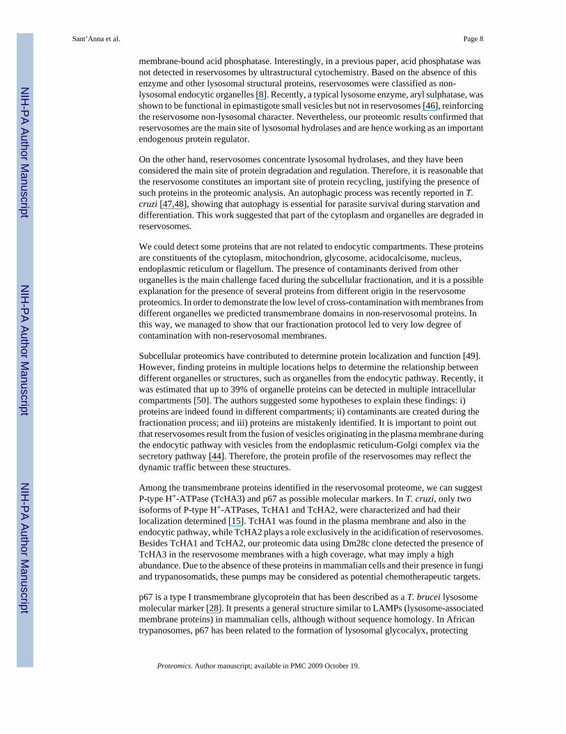
RESULTSUltrastructure of isolated reservosomes and their isolated membranes
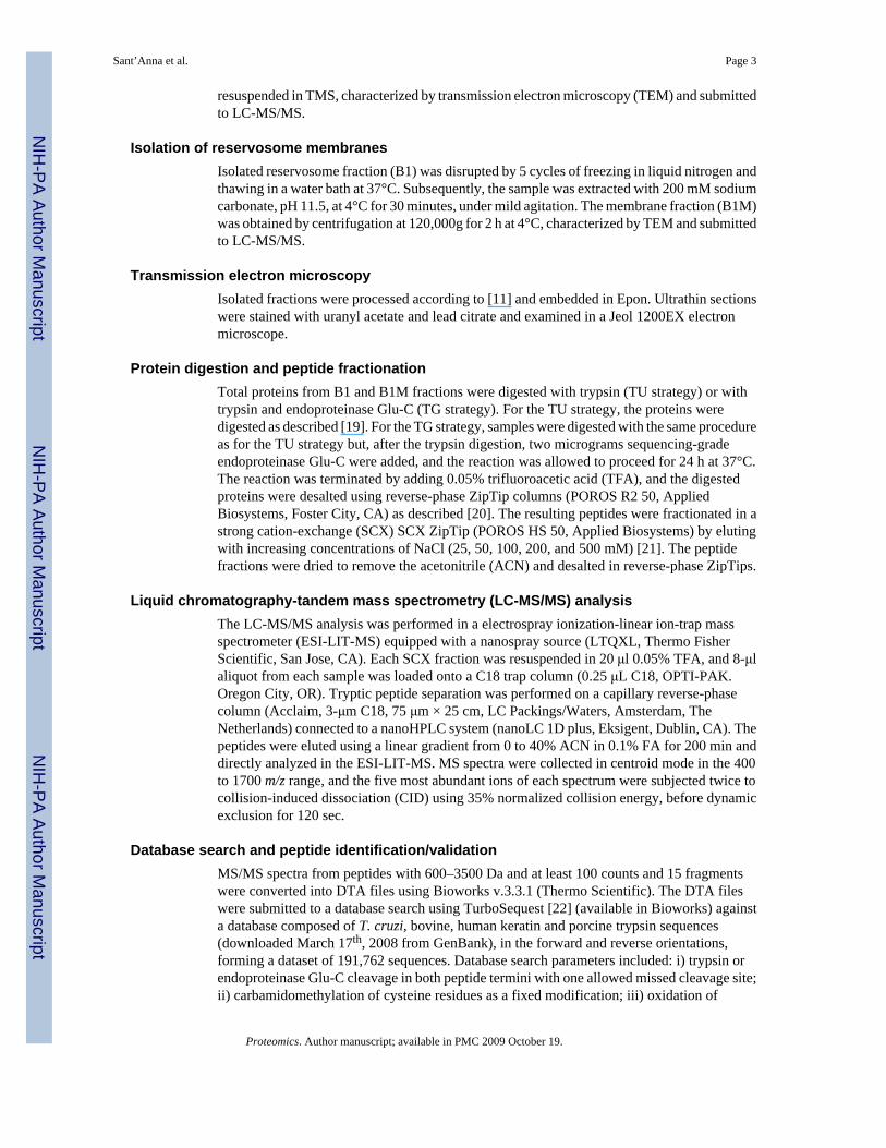
In order to identify reservosome-resident proteins, we initially purified reservosomes to obtainthe intact organelles (B1). Subsequently, we established a new protocol to isolate reservosomemembranes. Isolated organelles were disrupted by freezing and thawing, the associated proteinswere removed with sodium carbonate buffer, and the membrane (B1M) fraction was recoveredby centrifugation. To assess the purity and preservation of isolated fractions, B1 and B1Mfractions were analyzed by TEM (Fig. 1b,c). The B1 fraction displayed intact reservosomeswith a characteristic morphology observed for in situ reservosomes (Fig. 1a). Some disruptedorganelles were also visualized, as previously reported by Cunha-e-Silva and co-workers[11], probably due to the fractionation procedures. While examining several randomlycollected ultrathin sections, the presence of other structures such as mitochondria, kinetoplast,endoplasmic reticulum cisternae, glycosomes, acidocalcisomes, nuclei or flagella were notdetected, thus indicating that the preparation was virtually free of these contaminatingorganelles (data not shown). The B1M fraction showed highly purified total reservosomemembranes (Fig. 1c) because no intact organelle was detected. The B1 and B1M fractions werethen separately subjected to LC-MS/MS analysis.
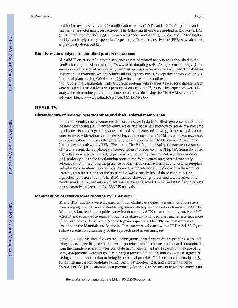
Identification of reservosome proteins by LC-MS/MSB1 and B1M fractions were digested with two distinct strategies: i) trypsin, with urea as adenaturing agent (TU), and ii) double digestion with trypsin and endoproteinase Glu-C (TG).After digestion, resulting peptides were fractionated by SCX chromatography, analyzed LC-MS/MS, and submitted to search through a database containing forward and reverse sequencesof T. cruzi, bovine, keratin and porcine trypsin sequences. The FPR was determined asdescribed in the Materials and Methods. Our data were validated with a FRP = 2.41%. Figure2 shows a schematic summary of the approach used in our analyses.
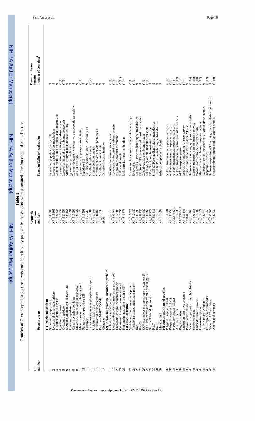
In total, LC-MS/MS data allowed the unambiguous identification of 869 proteins, with 709being T. cruzi-specific proteins and 160 as proteins from the culture medium and contaminantsfrom the sample preparation (see complete list in Supplementary Table 1). In the case of T.cruzi, 456 proteins were assigned as having a predicted function, and 253 were assigned ashaving an unknown function or being hypothetical proteins. Of these proteins, cruzipain [8,10, 11], serine carboxipeptidase [7, 12], ABC transporters [24], and a protein tyrosinephosphatase [25] have already been previously described to be present in reservosomes. Our
Sant’Anna et al. Page 4
Proteomics. Author manuscript; available in PMC 2009 October 19.
NIH
-PA Author Manuscript
NIH
-PA Author Manuscript
NIH
-PA Author Manuscript
proteomic analysis of reservosomes allowed the identification of a novel isoform of P-typeH+-ATPase, TcHA3. We also confirmed the presence of TcHA1 and TcHA2, the other protonpump described in reservosomes [15]. We could identify several additional hydrolases, suchas cysteine peptidases, α-mannosidases, acid phosphatase, acid phosphatase 2, calpain cysteinepeptidases, lipase, and serine carboxypeptidase S28. In addition, a calcium translocation pumpand calcium-binding proteins were found. A chloride channel protein, also identified inmammalian endosomes [26], was detected. Members of the ABC family, which act in lipidmetabolism [27], also appeared, as well as a P-glycoprotein. Surprisingly, a multidrugresistance (MDR) protein was identified. An interesting finding was the presence of p67, aglycoprotein molecular marker of T. brucei lysosome [28]. In addition, we detected aglycoprotein, which thus far has an undefined function, which is also present in the Golgicomplex and lysosomes.
In our analysis, the reservosomes exhibited endosomal integral membrane proteins and proteinsinvolved in vesicular traffic, such as COP-coated vesicle membrane proteins erv25 and gp25L,mu-adaptin 1, huntingtin-interacting protein (HIP), vesicle-associated membrane proteins(VAMPs), and the GTP-binding proteins Rab1, Rab2a, Rab7, and Rab18.
Also, our reservosome proteomic data showed proteins related to lipid metabolism, includingsterol 24-c-methyltransferase, fatty acyl CoA synthetase, phospholipid-translocating ATPase,C-8 sterol isomerase, fatty acyl CoA syntetase 1, and phosphatidylcholine:ceramidecholinephosphotransferase, which suggests that these organelles play a role in lipid synthesisin addition to storing lipids ingested by the endocytic pathway [11]. We also found enzymesthat act as regulators of signal transduction pathways in most eukaryotic cell types, e. g., proteintyrosine phosphatase, regulatory subunit of protein kinase A, casein kinase, MCAK-likekinesin, activated protein kinase C receptor, protein kinase, and serine/threonine protein kinase.
Transmembrane proteins from the plasma membrane, such as dispersed gene family protein 1(DGF-1), ferric reductase, hexose transporter, folate/pteridine transporter and GPI-anchoredp63, were identified in the reservosome proteome, confirming a membrane-traffickingrelationship between the plasma membrane and reservosomes. We also identified cytoskeletoncomponents essential for vesicular traffic, among other functions, such as alpha and betatubulin, cytoskeleton-associated protein CAP5.5, actin, and cofilin/actin depolymerizingfactor.
Other proteins that were detected can be characterized as non-reservosomal, includingmitochondrial, glycosomal or flagellar proteins. This suggests a functional relationshipbetween the organelles, a recycling process of these proteins in the reservosomes, or a possiblecontamination, commonly observed in subcellular fractionation protocols. However, only veryfew of these presumed contaminants are transmembrane proteins, thus suggesting a low levelof contamination with other organelle membranes. Predictably, we could also identify bovineserum proteins, which are supposed to be derived from the culture medium and likely reachedthe reservosomes by an endocytic process.
Gene ontology analysisIn order to understand their function and to identify processes related to reservosomes, weperformed gene ontology (GO) annotation of all proteins identified in the proteome analysis(Fig. 3 and Supplemental material 1). Catalytic activity was prevalent in the reservosomemolecular function. Reservosomes are also rich in proteins involved in binding and hydrolaseactivity. Proteins found in the reservosome proteome are involved in transporter, carrier andion-transporter activities, each representing about 12% of the proteins identified. Our analysisfound significant relationships to nucleotide-binding, transferase activity, ion-binding, andoxidoreductase activity. A large number of reservosomal proteins were related to metabolism
Sant’Anna et al. Page 5
Proteomics. Author manuscript; available in PMC 2009 October 19.
NIH
-PA Author Manuscript
NIH
-PA Author Manuscript
NIH
-PA Author Manuscript
as well as to physiological and cellular processes. Membrane localization of the proteinsidentified by LC-MS/MS analysis was remarkable. Reservosome proteins were also predictedto be localized in the proton-transporting complex.
DISCUSSIONSubcellular proteomics have been extensively used to identify the molecular composition ofseveral cytoplasmic organelles and has been considered an effective approach to understandcellular processes and integrated cell function. It takes advantage of the subcellularfractionation strategies, which are based on sequential and/or density-gradient centrifugation,allowing the separation of different population of organelles based on their size, density andcharge, in combination with mass spectrometry analysis [29]. This strategy greatly reducessample complexity in comparison with whole cell proteomic analysis.
Sub-cellular fractionation methodologies have been widely employed in protozoan parasites[30] in order to carry out parasite-specific organelles characterization and understand their rolein parasite cell biology. These organelles have particular interest since their absence in highereukaryotes may comprise a potential chemotherapeutic target against human and animalparasitism. By centrifugation in a sucrose gradient, our group reported an accurate andreproducible protocol to obtain a highly purified T. cruzi epimastigote reservosome fraction[11]. Afterwards, the same protocol was successfully used to measure proton transport inisolated reservosomes [15]. The purity of the fraction was assessed using three strategies: (i)enzymatic assays by means of enzymes recognized as markers of cell organelles in mammalcells and protozoa such as acid phosphatase (lysosomes), hexokinase (glycosomes), vacuolarH+-pyrophosphatase (acidocalcisomes) and succinate-cytochrome c reductase (mitochondria);(ii) Western blot, using antibodies against cruzipain (reservosomes) andlipopeptidephosphoglycan (plasma membrane) and (iii) ultrastructure analysis by transmissionelectron microcopy, a powerful mean for judging the efficiency of the method. In conclusion,we could essentially assure the high degree of purity and reproducibility of the reservosomefraction obtained.
Taking advantage of efficiency of the cell fractionation of reservosome [11], we isolated forthe first time a highly purified reservosome fraction from Dm28c epimastigotes (Fig. 1b). Inaddition, to enrich the membrane protein identification and avoid the contamination of theproteomic analysis with a great number of medium-derived peptides, we developed amethodology to purify total reservosome membranes (Fig. 1c). Our morphological analysisdid not allow discrimination between reservosome-surrounding membranes and membranesderived from inside the organelle. Reservosomes had been described as an organelle wherefew or no inner membranes were seen [2]. However, we have recently demonstrated, usingdifferent electron microscopy approaches, the presence of vesicles and planar membranes inthe lumen of reservosomes [9]. Nevertheless, the composition and function of these structuresneed to be clarified and are objects of our studies. Intending to achieve better proteome profilingof reservosomes, we used both B1 and B1M fractions in our proteomic analysis.
Proteomic analysis of B1 and B1M fractions led to the identification of 709 T. cruzi-specificproteins. Several identified proteins are expected components for endocytic organelles, whichinteract and fuse with vesicles coming from the secretory pathway and/or the plasma membraneas part of the endocytic process.
We also found cytoskeleton proteins, as did other proteomic analyses of T. cruzi subcellularfractions [31]. Since the pool of unpolymerized tubulin is very small in epimastigote forms[32], this finding may represent an association of reservosomes with microtubules, which areresponsible for traffic and intracellular organelle positioning.
Sant’Anna et al. Page 6
Proteomics. Author manuscript; available in PMC 2009 October 19.
NIH
-PA Author Manuscript
NIH
-PA Author Manuscript
NIH
-PA Author Manuscript

Rab 1, Rab 2a, Rab 7, and Rab 18, small GTPases of the Rab family, which are crucial regulatorsof vesicle traffic in endocytic and secretory pathways [33], were another important class ofproteins found in our proteomic analysis. Rab 1 and Rab 2 have been implicated in the transportfrom the ER to the Golgi complex [34]. Orthologues of mammalian Rab 1 and Rab 2 werecharacterized in T. brucei [35], and it was shown that TbRab1 and TbRab2 play a role in theearly events of the secretory pathway, suggesting a conserved function. Rab 7 has been regardedas a mediator of early to late endosome transport [36], and evidence has accumulated suggestingthat Rab 7 may work in the transport from late endosomes to lysosomes [37]. In Leishmania[38] and T. brucei [39], the orthologue of mammalian Rab 7 was localized in the endosomal/lysosomal system. Surprisingly, T. cruzi Rab 7 was found in high concentration in the Golgicomplex by ultrastructural localization [40]. Our analysis detected TcRab 7 in the reservosomeproteome, which suggests a possible role of this small GTPase in the membrane traffic betweenGolgi complex/reservosomes. Unexpectedly, TcRab 11, previously suggested to be localizedin the reservosomes [17], was not detected in our proteomic analysis. The suggestion ofTcRab11 localization in reservosomes was based exclusively on immunofluorescence images.No further confirmation by immunoelectronmicroscopy or western blot using isolatedreservosome fraction was published since then. We cannot exclude that the absence of thisprotein in reservosome proteomic profile be a consequence of sample preparation, but thispossibility does not seem to be probable, as sample processing did not affect other Rab proteins.
While mammalian Rab 18 has been involved in endocytic transport and in association withlipid droplets [41], T. brucei Rab 18 seems to function in Golgi transport [42]. The presenceof Rab18 in the reservosome proteomic profile may point to its role in the mobilization of theabundant lipid storage [9,11], similar to Rab18 function in adipocytes [41]. Undoubtedly, thepresence of several Rab proteins associated with reservosomes argues for the dynamic natureof this organelle and is compatible with the nature of a LRO [7] with a secretory character,sending molecules to other cytoplasmic compartments or the extracellular medium, whilesimultaneously receiving molecules from their cell “progenitors” [43], the Golgi complex, andendocytic pathway [44]. Furthermore, proteins that belong to the fusion machinery, such assyntaxyn, NSF, SNAP and others whose genes were found in the T. cruzi genome, were notidentified during our analysis. This virtual absence may be justified by the low level of proteinexpression, differences in the sequences from different strains (the T. cruzi genome projectwas carried out with the CL Brener strain, while we used the Dm28c clone), or proteinmodifications that cannot or are difficult to be identified by LC-MS/MS. We also cannotexclude the possibility of losing this sort of protein during the subcellular fractionationprocedure.
We also found isoforms of ABC transporters that, in mammalian cells, work in lipidmetabolism, and P-glycoprotein (Table 1). Recently, our group reported the intracellulartrafficking of heme [45]. Using a specific inhibitor, cyclosporin A, it was hypothesized that aP-glycoprotein transporter is likely to be the responsible for heme transport through the plasmamembrane. In addition, heme is stored in reservosomes, probably by the action of a secondtransporter. The identification of P-glycoprotein in the proteomic profile of reservosomesreinforces this hypothesis. Moreover, reservosomes store high levels of neutral lipids, such ascholesteryl esters [11], as a result of the endocytic process. The high concentration of lipids inthe reservosome lumen seems to be involved in the formation of rectangular lipid bodies [9].Like in mammals, ABCA1 could serve to control the rate of exogenous cholesterol internalizedinto early and late endosomes, back to plasma membrane or, yet, out of the cell [27].
LC-MS/MS analysis confirmed the reservosomal localization of cruzipain and its inhibitorchagasin and serine carboxypeptidase, shown by previous works to be present [7,10,13] andfunctional [11,12] in reservosomes. Novel hydrolases were found, such as lysosomal α-mannosidase, cysteine proteases, calpain cysteine peptidase, cysteine protease C, and
Sant’Anna et al. Page 7
Proteomics. Author manuscript; available in PMC 2009 October 19.
NIH
-PA Author Manuscript
NIH
-PA Author Manuscript
NIH
-PA Author Manuscript
membrane-bound acid phosphatase. Interestingly, in a previous paper, acid phosphatase wasnot detected in reservosomes by ultrastructural cytochemistry. Based on the absence of thisenzyme and other lysosomal structural proteins, reservosomes were classified as non-lysosomal endocytic organelles [8]. Recently, a typical lysosome enzyme, aryl sulphatase, wasshown to be functional in epimastigote small vesicles but not in reservosomes [46], reinforcingthe reservosome non-lysosomal character. Nevertheless, our proteomic results confirmed thatreservosomes are the main site of lysosomal hydrolases and are hence working as an importantendogenous protein regulator.
On the other hand, reservosomes concentrate lysosomal hydrolases, and they have beenconsidered the main site of protein degradation and regulation. Therefore, it is reasonable thatthe reservosome constitutes an important site of protein recycling, justifying the presence ofsuch proteins in the proteomic analysis. An autophagic process was recently reported in T.cruzi [47,48], showing that autophagy is essential for parasite survival during starvation anddifferentiation. This work suggested that part of the cytoplasm and organelles are degraded inreservosomes.
We could detect some proteins that are not related to endocytic compartments. These proteinsare constituents of the cytoplasm, mitochondrion, glycosome, acidocalcisome, nucleus,endoplasmic reticulum or flagellum. The presence of contaminants derived from otherorganelles is the main challenge faced during the subcellular fractionation, and it is a possibleexplanation for the presence of several proteins from different origin in the reservosomeproteomics. In order to demonstrate the low level of cross-contamination with membranes fromdifferent organelles we predicted transmembrane domains in non-reservosomal proteins. Inthis way, we managed to show that our fractionation protocol led to very low degree ofcontamination with non-reservosomal membranes.
Subcellular proteomics have contributed to determine protein localization and function [49].However, finding proteins in multiple locations helps to determine the relationship betweendifferent organelles or structures, such as organelles from the endocytic pathway. Recently, itwas estimated that up to 39% of organelle proteins can be detected in multiple intracellularcompartments [50]. The authors suggested some hypotheses to explain these findings: i)proteins are indeed found in different compartments; ii) contaminants are created during thefractionation process; and iii) proteins are mistakenly identified. It is important to point outthat reservosomes result from the fusion of vesicles originating in the plasma membrane duringthe endocytic pathway with vesicles from the endoplasmic reticulum-Golgi complex via thesecretory pathway [44]. Therefore, the protein profile of the reservosomes may reflect thedynamic traffic between these structures.
Among the transmembrane proteins identified in the reservosomal proteome, we can suggestP-type H+-ATPase (TcHA3) and p67 as possible molecular markers. In T. cruzi, only twoisoforms of P-type H+-ATPases, TcHA1 and TcHA2, were characterized and had theirlocalization determined [15]. TcHA1 was found in the plasma membrane and also in theendocytic pathway, while TcHA2 plays a role exclusively in the acidification of reservosomes.Besides TcHA1 and TcHA2, our proteomic data using Dm28c clone detected the presence ofTcHA3 in the reservosome membranes with a high coverage, what may imply a highabundance. Due to the absence of these proteins in mammalian cells and their presence in fungiand trypanosomatids, these pumps may be considered as potential chemotherapeutic targets.
p67 is a type I transmembrane glycoprotein that has been described as a T. brucei lysosomemolecular marker [28]. It presents a general structure similar to LAMPs (lysosome-associatedmembrane proteins) in mammalian cells, although without sequence homology. In Africantrypanosomes, p67 has been related to the formation of lysosomal glycocalyx, protecting
Sant’Anna et al. Page 8
Proteomics. Author manuscript; available in PMC 2009 October 19.
NIH
-PA Author Manuscript
NIH
-PA Author Manuscript
NIH
-PA Author Manuscript
against hydrolases. The p67 gene was found in T. cruzi, and its reservosomal localization wasdemonstrated in our proteomic analysis. Because orthologues of p67 are not found in mammals,it might be a candidate for chemotherapy as well.
In protozoan parasites, subcellular proteomic analyses were successfully applied to thecharacterization of Entamoeba histolytica [51] and Dictyostelium discoideum [52] phagosome.In trypanosomatids, we can highlight the proteome of flagella [53], glycosomes [54], and thecomparative proteomic analysis of glycosomes and mitochondria [55] of T. brucei. Recently,Ferella and co-workers [31] prepared a subcellular fraction from T. cruzi CL Brenerepimastigotes that was enriched in acidocalcisomes and glycosomes but also containedendoplasmic reticulum and Golgi components. Using 1D and 2D electrophoresis followed byMS analysis, they identified proteins corresponding to 396 genes, 258 of which were annotatedas previously described and 138 were hypothetical proteins. Among the identified proteins,only three are presumably reservosomal: glutathione S-transferase [56], cysteine peptidase[10], and serine carboxypeptidase [7,12]. These studies yielded valuable information aboutprotein localization and reported the identification of novel proteins in these organelles. In thecurrent paper, we performed the proteomic analysis of T. cruzi epimastigote reservosomes,which is the first protein profiling of an endocytic organelle in a trypanosomatid familymember. This analysis offers a new perspective in the identification of reservosomal molecularmarkers and contributes to the understanding of the biogenesis and dynamic interactions ofthese organelles with other T. cruzi structures.
Supplementary MaterialRefer to Web version on PubMed Central for supplementary material.
AcknowledgmentsThe authors would like to thank Mr. Antonio Bosco Carlos for technical support. This work was supported by ConselhoNacional de Desenvolvimento Científico e Tecnológico (CNPq), Fundação Carlos Chagas Filho de Amparo à Pesquisado Estado do Rio de Janeiro (FAPERJ) e Coordenação de Aperfeiçoamento de Pessoal de Nível Superior (CAPES),and National Institutes of Health (NIH). ICA was supported by the grants 1R01AI070655, 2S06GM008012-37, and5G12RR008124 from the NIH. E.S.N. was supported by the George A. Krutilek memorial graduate scholarship fromGraduate School, UTEP. We thank the Biomolecule Analysis Core Analysis at the Border Biomedical ResearchCenter/UTEP (NIH grant 5G12RR008124), for the access to the LC-MS instrumentation.
References1. De Souza W. From the cell biology to the development of new chemotherapeutic approaches against
trypanosomatids: dreams and reality. Kinetoplastid Biol Dis 2002;1:3. [PubMed: 12234386]2. Soares MJ, De Souza W. Cytoplasmic organelles of trypanosomatids: a cytochemical and stereological
study. J Submicrosc Cytol Pathol 1988;20:349–361. [PubMed: 3135113]3. Soares MJ, de Souza W. Endocytosis of gold-labeled proteins and LDL by Trypanosoma cruzi.
Parasitol Res 1991;77:461–468. [PubMed: 1656428]4. Porto-Carreiro I, Attias M, Miranda K, De Souza W, Cunha-e-Silva N. Trypanosoma cruzi epimastigote
endocytic pathway: cargo enters the cytostome and passes through an early endosomal network beforestorage in reservosomes. Eur J Cell Biol 2000;79:858–869. [PubMed: 11139150]
5. Cunha-e-Silva N, Sant’Anna C, Pereira MG, Porto-Carreiro I, et al. Reservosomes: multipurposeorganelles? Parasitol Res 2006;99:325–327. [PubMed: 16794853]
6. Raposo G, Marks MS, Cutler DF. Lysosome-related organelles: driving post-Golgi compartments intospecialisation. Curr Opin Cell Biol 2007;19:394–401. [PubMed: 17628466]
7. Sant’Anna C, Parussini F, Lourenço D, de Souza W, et al. All Trypanosoma cruzi developmental formspresent lysosome-related organelles. Histochem Cell Biol. 200810.1007/s00418-008-0486-8
8. Soares MJ, Souto-Padron T, De Souza W. Identification of a large pre-lysosomal compartment in thepathogenic protozoon Trypanosoma cruzi. J Cell Sci 1992;102:157–167. [PubMed: 1500438]
Sant’Anna et al. Page 9
Proteomics. Author manuscript; available in PMC 2009 October 19.
NIH
-PA Author Manuscript
NIH
-PA Author Manuscript
NIH
-PA Author Manuscript

9. Sant’Anna C, Pereira MG, Lemgruber L, de Souza W, Cunha e Silva NL. New insights into themorphology of Trypanosoma cruzi reservosome. Microsc Res Tech 2008;71:599–605. [PubMed:18452191]
10. Souto-Padron T, Campetella OE, Cazzulo JJ, de Souza W. Cysteine proteinase in Trypanosomacruzi: immunocytochemical localization and involvement in parasite-host cell interaction. J Cell Sci1990;96:485–490. [PubMed: 2229199]
11. Cunha-e-Silva NL, Atella GC, Porto-Carreiro IA, Morgado-Diaz JA, et al. Isolation andcharacterization of a reservosome fraction from Trypanosoma cruzi. FEMS Microbiol Lett2002;214:7–12. [PubMed: 12204365]
12. Parussini F, Garcia M, Mucci J, Aguero F, et al. Characterization of a lysosomal serinecarboxypeptidase from Trypanosoma cruzi. Mol Biochem Parasitol 2003;131:11–23. [PubMed:12967708]
13. Santos CC, Sant’Anna C, Terres A, Cunha-e-Silva NL, et al. Chagasin, the endogenous cysteine-protease inhibitor of Trypanosoma cruzi, modulates parasite differentiation and invasion ofmammalian cells. J Cell Sci 2005;118:901–915. [PubMed: 15713748]
14. Serrano R, Villalba JM, Palmgren MG, Portillo F, et al. Studies of the plasma membrane H(+)-ATPaseof yeast and plants. Biochem Soc Trans 1992;20:562–566. [PubMed: 1426592]
15. Vieira M, Rohloff P, Luo S, Cunha-e-Silva NL, et al. Role for a P-type H+-ATPase in the acidificationof the endocytic pathway of Trypanosoma cruzi. Biochem J 2005;392:467–474. [PubMed:16149915]
16. Ullrich O, Reinsch S, Urbe S, Zerial M, Parton RG. Rab11 regulates recycling through thepericentriolar recycling endosome. J Cell Biol 1996;135:913–924. [PubMed: 8922376]
17. Mendonca SM, Nepomuceno da Silva JL, Cunha e-Silva N, de Souza W, Gazos Lopes U.Characterization of a Rab11 homologue in Trypanosoma cruzi. Gene 2000;243:179–185. [PubMed:10675626]
18. Camargo EP. Growth and Differentiation in Trypanosoma cruzi. I. Origin of MetacyclicTrypanosomes in Liquid Media. Rev Inst Med Trop Sao Paulo 1964;12:93–100. [PubMed:14177814]
19. Stone, KL.; Williams, KR. Enzymatic digestion of proteins in solution and in SDS polyacrylamidegel. In: Walker, JM., editor. The protein protocol handbook. Humana Press; New Jersey: 1996. p.415-425.
20. Jurado JD, Rael ED, Lieb CS, Nakayasu E, et al. Complement inactivating proteins and intraspeciesvenom variation in Crotalus oreganus helleri. Toxicon 2007;49:339–350. [PubMed: 17134729]
21. Rodrigues ML, Nakayasu ES, Oliveira DL, Nimrichter L, et al. Extracellular vesicles produced byCryptococcus neoformans contain protein components associated with virulence. Eukaryot Cell2008;7:58–67. [PubMed: 18039940]
22. Eng JK, McCormack AL, Yates JR III. An approach to correlate tandem mass spectral data of peptideswith amino acid sequences in a protein database. J Am Soc Mass Spectrom 1994;5:976–989.
23. Groth D, Lehrach H, Hennig S. GOblet: a platform for Gene Ontology annotation of anonymoussequence data. Nucleic Acids Res 2004;32:W313–317. [PubMed: 15215401]
24. Torres C, Perez-Victoria FJ, Parodi-Talice A, Castanys S, Gamarro F. Characterization of an ABCA-like transporter involved in vesicular trafficking in the protozoan parasite Trypanosoma cruzi. MolMicrobiol 2004;54:632–646. [PubMed: 15491356]
25. Cuevas IC, Rohloff P, Sanchez DO, Docampo R. Characterization of farnesylated protein tyrosinephosphatase TcPRL-1 from Trypanosoma cruzi. Eukaryot Cell 2005;4:1550–1561. [PubMed:16151248]
26. Jentsch TJ. CLC chloride channels and transporters: from genes to protein structure, pathology andphysiology. Crit Rev Biochem Mol Biol 2008;43:3–36. [PubMed: 18307107]
27. Attie AD. ABCA1: at the nexus of cholesterol, HDL and atherosclerosis. Trends Biochem Sci2007;32:172–179. [PubMed: 17324574]
28. Alexander DL, Schwartz KJ, Balber AE, Bangs JD. Developmentally regulated trafficking of thelysosomal membrane protein p67 in Trypanosoma brucei. J Cell Sci 2002;115:3253–3263. [PubMed:12140257]
29. Dreger M. Subcellular proteomics. Mass Spectrom Rev 2003;22:27–56. [PubMed: 12768603]
Sant’Anna et al. Page 10
Proteomics. Author manuscript; available in PMC 2009 October 19.
NIH
-PA Author Manuscript
NIH
-PA Author Manuscript
NIH
-PA Author Manuscript

30. De Souza W, Cunha-e-Silva N. Cell fractionation of parasitic protozoa – a review. Mem Inst OswaldoCruz 2003;98:151–170.
31. Ferella M, Nilsson D, Darban H, Rodrigues C, et al. Proteomics in Trypanosoma cruzi -localizationof novel proteins to various organelles. Proteomics 2008;8:2735–2749. [PubMed: 18546153]
32. da Silva RA, Bartholomeu DC, Teixeira SM. Control mechanisms of tubulin gene expression inTrypanosoma cruzi. Int J Parasitol 2006;36:87–96. [PubMed: 16233898]
33. Zerial M, McBride H. Rab proteins as membrane organizers. Nat Rev Mol Cell Biol 2001;2:107–117. [PubMed: 11252952]
34. Tisdale EJ, Bourne JR, Khosravi-Far R, Der CJ, Balch WE. GTP-binding mutants of rab1 and rab2are potent inhibitors of vesicular transport from the endoplasmic reticulum to the Golgi complex. JCell Biol 1992;119:749–761. [PubMed: 1429835]
35. Dhir V, Goulding D, Field MC. TbRAB1 and TbRAB2 mediate trafficking through the early secretorypathway of Trypanosoma brucei. Mol Biochem Parasitol 2004;137:253–265. [PubMed: 15383296]
36. Feng Y, Press B, Wandinger-Ness A. Rab 7: an important regulator of late endocytic membranetraffic. J Cell Biol 1995;131:1435–1452. [PubMed: 8522602]
37. Bucci C, Thomsen P, Nicoziani P, McCarthy J, van Deurs B. Rab7: a key to lysosome biogenesis.Mol Biol Cell 2000;11:467–480. [PubMed: 10679007]
38. Denny PW, Lewis S, Tempero JE, Goulding D, et al. Leishmania RAB7: characterisation of terminalendocytic stages in an intracellular parasite. Mol Biochem Parasitol 2002;123:105–113. [PubMed:12270626]
39. Morgan GW, Hall BS, Denny PW, Field MC, Carrington M. The endocytic apparatus of thekinetoplastida. Part II: machinery and components of the system. Trends Parasitol 2002;18:540–546.[PubMed: 12482539]
40. Araripe JR, Cunha e Silva NL, Leal ST, de Souza W, Rondinelli E. Trypanosoma cruzi: TcRAB7protein is localized at the Golgi apparatus in epimastigotes. Biochem Biophys Res Commun2004;321:397–402. [PubMed: 15358190]
41. Martin S, Parton RG. Characterization of Rab18, a lipid droplet-associated small GTPase. MethodsEnzymol 2008;438:109–129. [PubMed: 18413244]
42. Jeffries TR, Morgan GW, Field MC. TbRAB18, a developmentally regulated Golgi GTPase fromTrypanosoma brucei. Mol Biochem Parasitol 2002;121:63–74. [PubMed: 11985863]
43. Hu ZZ, Valencia JC, Huang H, Chi A, et al. Comparative Bioinformatics Analyses and Profiling ofLysosome-Related Organelle Proteomes. Int J Mass Spectrom 2007;259:147–160. [PubMed:17375895]
44. Sant’Anna C, de Souza W, Cunha-e-Silva N. Biogenesis of the reservosomes of Trypanosomacruzi. Microsc Microanal 2004;10:637–646. [PubMed: 15525436]
45. Lara FA, Sant’anna C, Lemos D, Laranja GA, et al. Heme requirement and intracellular traffickingin Trypanosoma cruzi epimastigotes. Biochem Biophys Res Commun 2007;355:16–22. [PubMed:17292866]
46. Adade CM, de Castro SL, Soares MJ. Ultrastructural localization of Trypanosoma cruzi lysosomesby aryl sulphatase cytochemistry. Micron 2007;38:252–256. [PubMed: 16860560]
47. Alvarez VE, Kosec G, Sant Anna C, Turk V, et al. Blocking autophagy to prevent parasitedifferentiation: a possible new strategy for fighting parasitic infections? Autophagy 2008;4:361–363.[PubMed: 18212533]
48. Alvarez VE, Kosec G, Sant’Anna C, Turk V, et al. Autophagy is involved in nutritional stress responseand differentiation in Trypanosoma cruzi. J Biol Chem 2008;283:3454–3464. [PubMed: 18039653]
49. Andersen JS, Mann M. Organellar proteomics: turning inventories into insights. EMBO Rep2006;7:874–879. [PubMed: 16953200]
50. Foster LJ, de Hoog CL, Zhang Y, Zhang Y, et al. A mammalian organelle map by protein correlationprofiling. Cell 2006;125:187–199. [PubMed: 16615899]
51. Okada M, Huston CD, Mann BJ, Petri WA Jr, et al. Proteomic analysis of phagocytosis in the entericprotozoan parasite Entamoeba histolytica. Eukaryot Cell 2005;4:827–831. [PubMed: 15821141]
Sant’Anna et al. Page 11
Proteomics. Author manuscript; available in PMC 2009 October 19.
NIH
-PA Author Manuscript
NIH
-PA Author Manuscript
NIH
-PA Author Manuscript
52. Gotthardt D, Warnatz HJ, Henschel O, Brückert F, et al. High-resolution dissection of phagosomematuration reveals distinct membrane trafficking phases. Mol Biol Cell 2002;13:3508–3520.[PubMed: 12388753]
53. Broadhead R, Dawe HR, Farr H, Griffiths S, et al. Flagellar motility is required for the viability ofthe bloodstream trypanosome. Nature 2006;440:224–227. [PubMed: 16525475]
54. Colasante C, Ellis M, Ruppert T, Voncken F. Comparative proteomics of glycosomes frombloodstream form and procyclic culture form Trypanosoma brucei brucei. Proteomics 2006;6:3275–3293. [PubMed: 16622829]
55. Vertommen D, Van Roy J, Szikora JP, Rider MH, et al. Differential expression of glycosomal andmitochondrial proteins in the two major life-cycle stages of Trypanosoma brucei. Mol BiochemParasitol 2008;158:189–201. [PubMed: 18242729]
56. Ouaissi MA, Dubremetz JF, Schöneck R, Fernandez-Gomez R, et al. Trypanosoma cruzi: a 52-kDaprotein sharing sequence homology with glutathione S-transferase is localized in parasite organellesmorphologically resembling reservosomes. Exp Parasitol 1995;81:453–461. [PubMed: 8542986]
Sant’Anna et al. Page 12
Proteomics. Author manuscript; available in PMC 2009 October 19.
NIH
-PA Author Manuscript
NIH
-PA Author Manuscript
NIH
-PA Author Manuscript
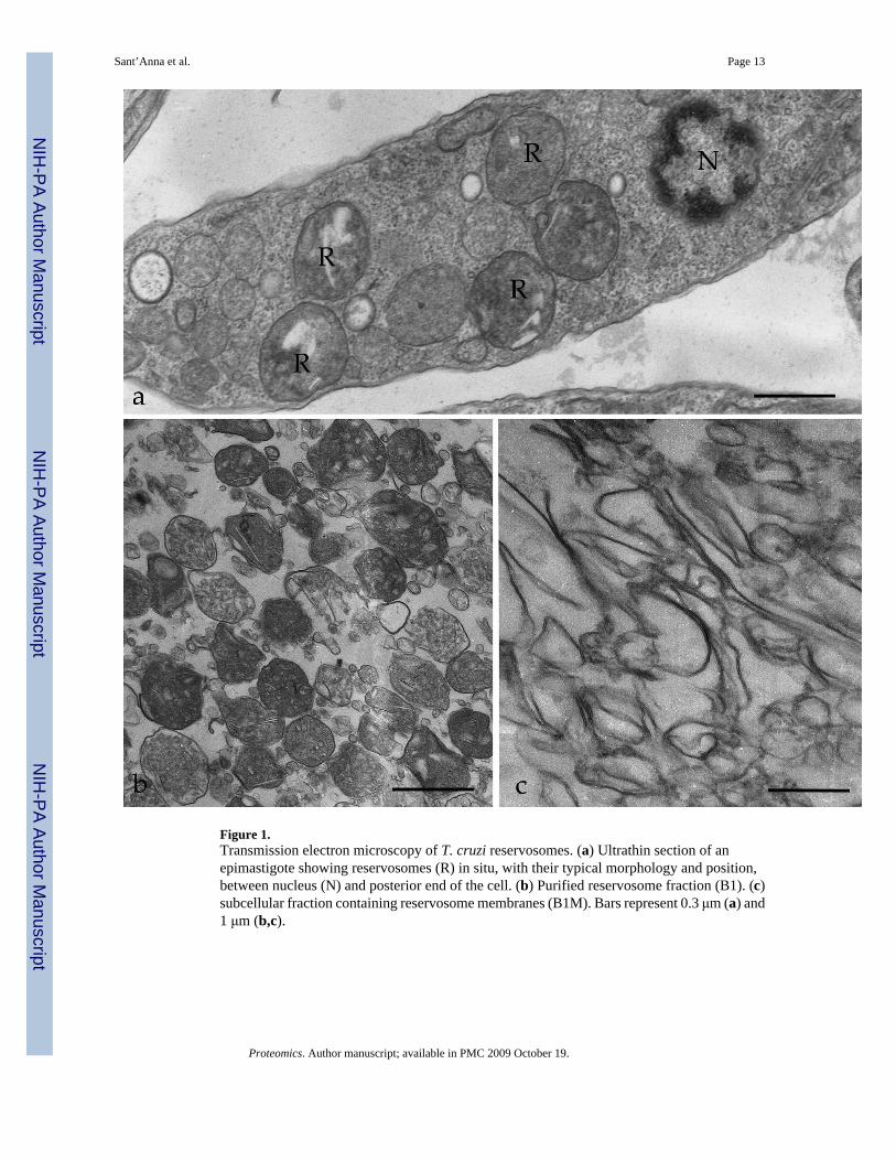
Figure 1.Transmission electron microscopy of T. cruzi reservosomes. (a) Ultrathin section of anepimastigote showing reservosomes (R) in situ, with their typical morphology and position,between nucleus (N) and posterior end of the cell. (b) Purified reservosome fraction (B1). (c)subcellular fraction containing reservosome membranes (B1M). Bars represent 0.3 μm (a) and1 μm (b,c).
Sant’Anna et al. Page 13
Proteomics. Author manuscript; available in PMC 2009 October 19.
NIH
-PA Author Manuscript
NIH
-PA Author Manuscript
NIH
-PA Author Manuscript
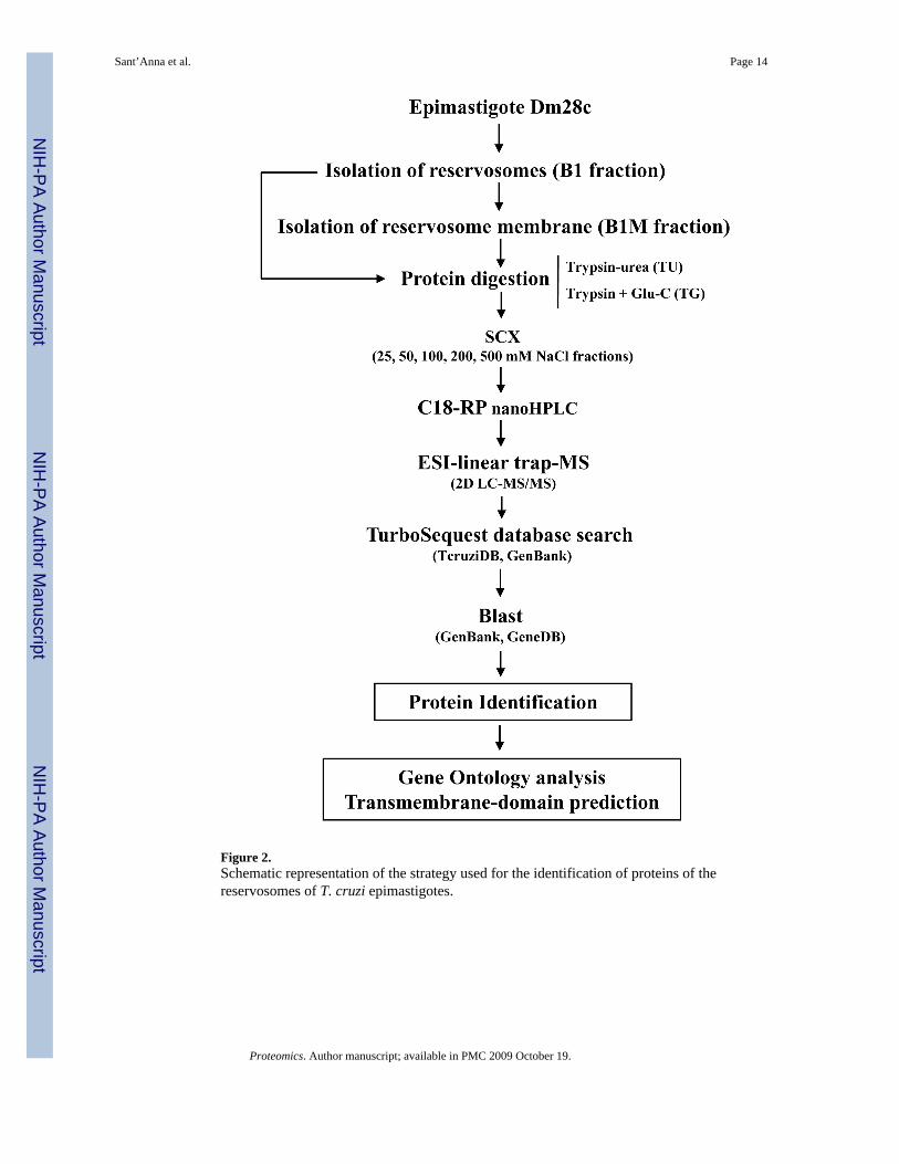
Figure 2.Schematic representation of the strategy used for the identification of proteins of thereservosomes of T. cruzi epimastigotes.
Sant’Anna et al. Page 14
Proteomics. Author manuscript; available in PMC 2009 October 19.
NIH
-PA Author Manuscript
NIH
-PA Author Manuscript
NIH
-PA Author Manuscript

Figure 3.Functional classification of T. cruzi reservosome proteins by gene ontology. The gene ontologyprediction was performed using GOblet and the most representative categories (≥3% from thetotal sequences) were plotted in the graph. For the complete list see Supplemental Table 1.
Sant’Anna et al. Page 15
Proteomics. Author manuscript; available in PMC 2009 October 19.
NIH
-PA Author Manuscript
NIH
-PA Author Manuscript
NIH
-PA Author Manuscript
NIH
-PA Author Manuscript
NIH
-PA Author Manuscript
NIH
-PA Author Manuscript
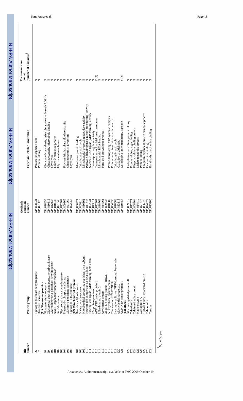
Sant’Anna et al. Page 16Ta
ble
1Pr
otei
ns o
f T. c
ruzi
epi
mas
tigot
e re
serv
osom
es id
entif
ied
by p
rote
omic
ana
lysi
s and
with
ann
otat
ed fu
nctio
n or
cel
lula
r loc
aliz
atio
n
Hit
num
ber
Prot
ein
grou
p
Gen
Ban
kac
cess
ion
num
ber
Func
tion/
Cel
lula
r lo
caliz
atio
n
Tra
nsm
embr
ane
dom
ain
(num
ber
of d
omai
ns)1
(1) P
rote
in m
etab
olis
m1
Serin
e ca
rbox
ypep
tidas
eX
P_80
3003
Lyso
som
al, p
eptid
ase
fam
ily S
10N
2Ly
soso
mal
alp
ha-m
anno
sida
seX
P_81
9105
Lyso
som
al, c
arbo
hydr
ate
met
abol
ism
N3
His
tidin
e am
mon
ia-ly
ase
XP_
8203
36C
onve
rt hi
stid
ine
into
am
mon
ia a
nd u
roca
nic
acid
N4
Cys
tein
e pr
otei
nase
XP_
8119
57Ly
soso
mal
, sim
ilar t
o en
dope
ptid
ase
papa
inY
(1)
5C
yste
ine
pept
idas
eX
P_82
1954
Lyso
som
al in
tegr
al to
mem
bran
e, p
rote
olys
isY
(1)
6S-
Ade
nosy
lhom
ocys
tein
e hy
drol
ase
XP_
8091
53A
deno
sylh
omoc
yste
ine
hydr
olas
e ac
tivity
N7
Cys
tein
e pe
ptid
ase
CX
P_81
2694
Lyso
som
al, p
rote
olys
isN
8C
alpa
in c
yste
ine
pept
idas
eX
P_81
6696
Cal
cium
-dep
ende
nt c
yste
ine-
type
end
opep
tidas
e ac
tivity
N9
Mem
bran
e-bo
und
acid
pho
spha
tase
XP_
8083
29C
atal
ytic
act
ivity
N10
Mem
bran
e-bo
und
acid
pho
spha
tase
2X
P_81
5570
Lyso
som
al, a
cid
phos
phat
ase
activ
ityY
(1)
11Se
rine
carb
oxyp
eptid
ase
S28
XP_
8185
92Pe
ptid
ase
S28
N12
Cru
zipa
inA
AF7
5547
Cys
tein
e pe
ptid
ase,
cla
n C
A, f
amily
C1
N13
Tartr
ate-
resi
stan
t aci
d ph
osph
atas
e ty
pe 5
XP_
8110
82M
etal
loph
osph
oest
eras
eY
(2)
14U
biqu
itin
hydr
olas
eX
P_82
1344
Prot
ein
deub
iqui
tinat
ion
N15
Glu
tam
amyl
car
boxy
pept
idas
eX
P_80
4169
Met
allo
pept
idas
e ac
tivity
, pro
teol
ysis
N16
Pept
idas
e M
20/M
25/M
40X
P_81
3635
Met
allo
pept
idas
e ac
tivity
N17
Cha
gasi
n2F
O8
Cys
tein
e Pr
otea
se In
hibi
tor
N(2
) End
osom
al/ly
soso
mal
mem
bran
e pr
otei
ns18
Gol
gi/ly
soso
me
glyc
opro
tein
XP_
8179
16G
olgi
/lyso
som
e m
embr
ane
prot
ein
Y (1
)19
Lyso
som
al/e
ndos
omal
mem
bran
e pr
otei
n p6
7X
P_80
3464
Lyso
som
al/e
ndos
omal
mem
bran
e pr
otei
nY
(1)
20En
doso
mal
inte
gral
mem
bran
e pr
otei
nX
P_81
7908
Inte
gral
to e
ndos
omal
mem
bran
eY
(9)
21En
doso
mal
inte
gral
mem
bran
e pr
otei
nX
P_81
3892
Endo
som
al p
rote
inY
(11)
22H
untin
gtin
inte
ract
ing
prot
ein
(HIP
)X
P_81
6878
Endo
cyto
sis,
met
al io
n bi
ndin
gY
(4)
(3) V
esic
ular
traf
fic23
Ves
icle
-ass
ocia
ted
mem
bran
e pr
otei
nX
P_81
6323
Inte
gral
to p
lasm
a m
embr
ane,
ves
icle
targ
etin
gY
(1)
24V
esic
le-a
ssoc
iate
d m
embr
ane
prot
ein
XP_
8124
89Ex
ocyt
osis
Y (1
)25
Rab
1X
P_80
5990
ER, s
mal
l GTP
ase
med
iate
d si
gnal
tran
sduc
tion
N26
Rab
-2a
XP_
8097
35ER
, Sm
all G
TPas
e m
edia
ted
sign
al tr
ansd
uctio
nN
27C
OP-
coat
ed v
esic
le m
embr
ane
prot
ein
erv2
5X
P_82
1489
Coa
ted
vesi
cle
mem
bran
e pr
otei
nY
(1)
28C
OP-
coat
ed v
esic
le m
embr
ane
prot
ein
gp25
lX
P_80
5665
ER to
Gol
gi v
esic
le-m
edia
ted
trans
port
Y (1
)29
Smal
l GTP
-bin
ding
pro
tein
XP_
8087
72ER
to G
olgi
ves
icle
-med
iate
d tra
nspo
rtN
30R
ab7
XP_
8216
19Sm
all G
TPas
e m
edia
ted
sign
al tr
ansd
uctio
nN
31R
ab18
XP_
8180
13Sm
all G
TPas
e m
edia
ted
sign
al tr
ansd
uctio
nN
32M
u-ad
aptin
1X
P_81
8899
Ada
ptor
com
plex
es 1
subu
nit
N(4
) pum
ps a
nd c
hann
el p
rote
ins
33P-
type
H+-
atpa
se (t
cha3
)X
P_81
2631
ATP
ase,
tran
smem
bran
e pr
oton
tran
spor
tY
(6)
34P-
type
H+-
atpa
se (t
cha1
)X
P_80
4256
ATP
ase,
tran
smem
bran
e pr
oton
tran
spor
tY
(3)
35P-
type
H+-
atpa
se (t
cha2
)A
AL8
7542
.1A
TPas
e, tr
ansm
embr
ane
prot
on tr
ansp
ort
Y (8
)36
AB
C tr
ansp
orte
rX
P_81
8638
ATP
ase,
tran
smem
bran
e tra
nspo
rt of
subs
tanc
esY
(12)
37P-
glyc
opro
tein
AA
C09
044.
1A
BC
tran
spor
ter p
rote
inY
(8)
38M
ultid
rug
resi
stan
ce p
rote
in E
XP_
8151
45X
enob
iotic
-tran
spor
ting
ATP
ase
activ
ityY
(4)
39Pr
oton
mot
ive
atpa
seA
AB
7015
2C
atio
n tra
nspo
rt A
TPas
e, P
-type
ATP
ase
N40
Vac
uola
r-ty
pe p
roto
n py
roph
osph
atas
eX
P_81
3460
Hyd
roge
n-tra
nslo
catin
g py
roph
osph
atas
e ac
tivity
Y (1
5)41
Ferr
ic re
duct
ase
XP_
8082
51El
ectro
n tra
nspo
rt, o
xido
redu
ctas
e ac
tivity
Y (1
2)42
Chl
orid
e ch
anne
l pro
tein
XP_
8162
07V
olta
ge-g
ated
chl
orid
e ch
anne
l act
ivity
Y (1
0)43
Hex
ose
trans
porte
rX
P_81
4821
Glu
cose
tran
smem
bran
e tra
nspo
rter a
ctiv
ityY
(12)
44V
-type
atp
ase,
A su
buni
tX
P_80
7670
Lyso
som
al p
roto
n-tra
nspo
rting
V-ty
pe A
TPas
e co
mpl
exN
45Fo
late
/pte
ridin
e tra
nspo
rter
XP_
8107
59B
iopt
erin
tran
spor
tY
(12)
46V
acuo
lar A
TP sy
ntha
seX
P_80
8900
Hyd
roge
n-ex
porti
ng A
TP a
ctiv
ity, p
hosp
hory
lativ
e m
echa
nism
N47
Am
ino
acid
per
mea
seX
P_80
2539
Tran
smem
bran
e am
ino
acid
tran
spor
ter p
rote
inY
(10)
Proteomics. Author manuscript; available in PMC 2009 October 19.

NIH
-PA Author Manuscript
NIH
-PA Author Manuscript
NIH
-PA Author Manuscript
Sant’Anna et al. Page 17
Hit
num
ber
Prot
ein
grou
p
Gen
Ban
kac
cess
ion
num
ber
Func
tion/
Cel
lula
r lo
caliz
atio
n
Tra
nsm
embr
ane
dom
ain
(num
ber
of d
omai
ns)1
48M
FS tr
ansp
orte
rX
P_80
4323
Tran
spor
t sm
all s
olut
es a
cros
s mem
bran
esY
(7)
49A
min
o ac
id p
erm
ease
/tran
spor
ter
XP_
8198
01A
min
o ac
id tr
ansp
ort
Y (1
0)50
Cal
cium
-tran
sloc
atin
g P-
type
atp
ase
XP_
8142
28C
alci
um-tr
ansp
ortin
g A
TPas
e ac
tivity
Y (3
)(5
) Cel
l sur
face
pro
tein
s51
Surf
ace
prot
ease
GP6
3X
P_82
1289
Neu
tral z
inc
met
allo
pept
idas
esY
(1)
52D
ispe
rsed
gen
e fa
mily
pro
tein
1 (D
GF-
1)X
P_82
0404
Cel
l sur
face
pro
tein
Y (9
)53
Proc
yclic
form
surf
ace
glyc
opro
tein
XP_
8038
86Y
(3)
54K
inet
opla
stid
mem
bran
e pr
otei
n K
MP-
11X
P_80
8865
kine
topl
astid
mem
bran
e pr
otei
nN
(6) L
ipid
met
abol
ism
55St
erol
24-
c-m
ethy
ltran
sfer
ase
XP_
8028
64B
iosy
nthe
sis o
f erg
oste
rol
N56
Fatty
aci
d el
onga
seX
P_81
3971
Long
-cha
in fa
tty a
cid
bios
ynth
etic
pro
cess
Y (7
)57
3-ox
o-5-
alph
a-st
eroi
d 4-
dehy
drog
enas
eX
P_80
2815
Ver
y-lo
ng-c
hain
fatty
aci
d m
etab
olic
pro
cess
Y (4
)58
Fatty
acy
l coa
synt
heta
seX
P_81
7096
Long
-cha
in-f
atty
-aci
d-C
oA li
gase
act
ivity
N59
Serin
e-pa
lmito
yl-c
oa tr
ansf
eras
eX
P_81
2739
Sphi
ngol
ipid
met
abol
ic p
roce
ssN
60St
erol
C-2
4 re
duct
ase
XP_
8168
88Er
gost
erol
bio
synt
hesi
sY
(9)
61Ph
osph
olip
id-tr
ansl
ocat
ing
atpa
seX
P_81
7305
Phos
phol
ipid
-tran
sloc
atin
g P-
type
ATP
ase,
flip
pase
Y (1
2)62
Fatty
acy
l coa
synt
heta
se 2
XP_
8027
08Lo
ng-c
hain
fatty
aci
d ac
tivat
ion
N63
C-8
ster
ol is
omer
ase
XP_
8211
86Er
gost
erol
bio
synt
hetic
pro
cess
Y (1
)64
Fatty
acy
l coa
synt
etas
e 1
XP_
8123
51Fa
tty a
cid
met
abol
ic p
roce
ssN
65Ph
osph
atid
ylch
olin
e:ce
ram
ide
chol
inep
hosp
hotra
nsfe
rase
XP_
8215
06Sp
hing
omye
lin b
iosy
nthe
tic p
roce
ssY
(5)
66Ph
osph
atid
ic a
cid
phos
phat
ase
prot
ein
XP_
8147
14Ph
osph
olip
id m
etab
olic
pro
cess
Y (5
)67
Lipa
se d
omai
n pr
otei
nX
P_81
4907
Lipi
d m
etab
olic
pro
cess
Y (7
)(7
) Car
bohy
drat
e m
etab
olis
m68
UD
P-G
al o
r UD
P-gl
cnac
-dep
ende
nt g
lyco
syltr
ansf
eras
eX
P_81
4599
Tran
sfer
gal
acto
se to
Glc
NA
c te
rmin
al c
hain
sN
69En
olas
eX
P_81
9700
Gly
coly
sis,
phos
phop
yruv
ate
hydr
atas
e ac
tivity
N70
Hex
okin
ase
XP_
8089
92C
arbo
hydr
ate
met
abol
ic p
roce
ssN
71M
anno
syl-o
ligos
acch
arid
e 1,
2-al
pha-
man
nosi
dase
IBX
P_81
1964
Prot
ein
amin
o ac
id N
-link
ed g
lyco
syla
tion
N72
Bet
a ga
lact
ofur
anos
yl g
lyco
syltr
ansf
eras
eX
P_81
0392
N73
Lect
inX
P_80
7732
Man
nose
bin
ding
N74
Glu
coki
nase
1X
P_82
1474
Glu
coki
nase
act
ivity
N75
Glu
cosa
min
e-6-
phos
phat
e is
omer
ase
XP_
8078
57N
-ace
tylg
luco
sam
ine
cata
bolic
pro
cess
N(8
) Cel
l sig
nalin
g76
Prot
ein
kina
seX
P_80
7538
Prot
ein
amin
o ac
id p
hosp
horil
atio
nN
77Pr
otei
n ty
rosi
ne p
hosp
hata
seX
P_81
6679
Prot
ein
tyro
sine
pho
spha
tase
act
ivat
or fa
ctor
N78
Serin
e/th
reon
ine
prot
ein
kina
seX
P_82
2060
Prot
ein
amin
o ac
id p
hosp
horil
atio
nN
79Se
rine/
thre
onin
e pr
otei
n ph
osph
atas
eX
P_80
9469
Cal
cium
-dep
ende
nt p
rote
in se
rine/
thre
onin
e ph
osph
atas
eY
(2)
80R
ac se
rine-
thre
onin
e ki
nase
XP_
8151
40N
81R
egul
ator
y su
buni
t of p
rote
in k
inas
e a
XP_
8100
45Pr
otei
n ki
nase
act
ivity
N82
Cas
ein
kina
se d
elta
isof
orm
XP_
8031
79C
asei
n ki
nase
act
ivity
N83
Act
ivat
ed p
rote
in k
inas
e C
rece
ptor
XP_
8177
33Pr
otei
n ki
nase
C b
indi
ngN
84C
asei
n ki
nase
XP_
8031
79Pr
otei
n am
ino
acid
pho
spho
ryla
tion
N(9
) Cyt
oske
leto
n-as
soci
ated
pro
tein
s85
Bet
a tu
bulin
XP_
8166
90M
icro
tubu
le c
ytos
kele
ton
orga
niza
tion
and
biog
enes
isN
86A
lpha
tubu
linX
P_80
2499
Mic
rotu
bule
cyt
oske
leto
n or
gani
zatio
n an
d bi
ogen
esis
N87
Act
inX
P_80
7262
Mic
rofil
amen
t org
aniz
atio
n an
d bi
ogen
esis
N88
MC
AK
-like
kin
esin
XP_
8132
21M
icro
tubu
le a
ssoc
iate
d co
mpl
exN
89C
ytos
kele
ton-
asso
ciat
ed p
rote
in C
AP5
.5X
P_80
9814
Cyt
oske
leto
n-as
soci
ated
pro
tein
N90
Cof
ilin/
actin
dep
olym
eriz
ing
fact
orX
P_80
6100
Act
in fi
lam
ent o
rgan
izat
ion
91I/6
aut
oant
igen
XP_
8131
69C
alci
um io
n bi
ndin
g, st
ruct
ural
con
stitu
ent o
f cyt
oske
leto
nN
(10)
Cyt
osol
ic p
rote
ins
92El
onga
tion
fact
or a
lpha
G5
AA
U47
272
Cal
cium
med
iate
d si
gnal
ing,
mic
rotu
bule
-bas
ed p
roce
ssN
93Tr
ypar
edox
in p
erox
idas
eX
P_80
2393
Tryp
anot
hion
e-di
sulfi
de re
duct
ase
activ
ityN
94H
ypox
anth
ine-
guan
ine
phos
phor
ibos
yltra
nsfe
rase
XP_
8133
97N
ucle
otid
e tra
nspo
rt an
d m
etab
olis
mN
95M
ethy
lthio
aden
osin
e ph
osph
oryl
ase
XP_
8199
27A
min
o ac
id sa
lvag
eN
Proteomics. Author manuscript; available in PMC 2009 October 19.
NIH
-PA Author Manuscript
NIH
-PA Author Manuscript
NIH
-PA Author Manuscript
Sant’Anna et al. Page 18
Hit
num
ber
Prot
ein
grou
p
Gen
Ban
kac
cess
ion
num
ber
Func
tion/
Cel
lula
r lo
caliz
atio
n
Tra
nsm
embr
ane
dom
ain
(num
ber
of d
omai
ns)1
966-
phos
phog
luco
nate
deh
ydro
gena
seX
P_80
8031
Pent
ose-
phos
phat
e sh
unt
N97
Dis
ulfid
e is
omer
ase
XP_
8211
73Pr
otei
n fo
ldin
gN
(11)
Gly
coso
mal
pro
tein
s98
Glu
tam
ate
dehy
drog
enas
eX
P_81
8855
Glu
tam
ate
bios
ynth
esis
, usi
ng g
luta
mat
e sy
ntha
se (N
AD
PH)
N99
Gly
coso
mal
pho
spho
enol
pyru
vate
car
boxy
kina
seX
P_81
0815
Glu
cone
osen
esis
, mic
rotu
bule
bin
ding
N10
0G
lyce
rald
ehyd
e 3-
phos
phat
e de
hydr
ogen
ase
XP_
8121
37G
lyco
lysi
sN
101
NA
DH
-dep
ende
nt fu
mar
ate
redu
ctas
eX
P_81
0232
Glu
cose
met
abol
ic p
roce
ssN
102
Gim
5A p
rote
inX
P_82
1649
Gly
coso
mal
mem
bran
eN
103
Gly
coso
mal
mal
ate
dehy
drog
enas
eX
P_81
2467
N10
4Fr
ucto
se-b
isph
osph
ate
aldo
lase
XP_
8093
69Fr
ucto
se-b
isph
osph
ate
aldo
lase
act
ivity
N10
5G
luco
se-6
-pho
spha
te is
omer
ase
XP_
8219
69G
luco
neog
enes
is, g
lyco
lysi
sN
106
6-ph
osph
o-1-
fruc
toki
nase
XP_
8220
53G
lyco
lysi
sN
(12)
Mito
chon
dria
l pro
tein
s10
7H
eat s
hock
70
kda
prot
ein
XP_
8062
21M
embr
ane
prot
ein
fold
ing
N10
8M
alat
e de
hydr
ogen
ase
XP_
8092
10Tr
icar
boxi
lyc
acid
cyc
leN
109
Mito
chon
dria
l pro
cess
ing
pept
idas
e, b
eta
subu
nit
XP_
8163
61M
itoch
ondr
ial p
roce
ssin
g pe
ptid
ase
activ
ityN
110
Pyru
vate
deh
ydro
gena
se E
1 be
ta su
buni
tX
P_81
1646
Pyru
vate
deh
ydro
gena
se (a
cety
l-tra
nsfe
rrin
g) a
ctiv
ityN
111
Succ
inyl
-coa
liga
se [G
DP-
form
ing]
bet
a-ch
ain
XP_
8026
41Su
ccin
ate-
CoA
liga
se (G
DP-
form
ing)
act
ivity
N11
2P2
2 pr
otei
n pr
ecur
sor
XP_
8133
11Tr
ansc
riptio
n re
gula
tor a
ctiv
ityN
113
AD
P, A
TP c
arrie
r pro
tein
1X
P_81
2264
Tran
spor
t, m
itoch
ondr
ial i
nner
mem
bran
eY
(3)
114
RN
A-b
indi
ng p
rote
in 2
XP_
8187
66M
itoch
ondr
ial R
NA
bin
ding
N11
5A
cyl c
arrie
r pro
tein
XP_
8149
61Fa
tty a
cid
bios
ynth
etic
pro
cess
N11
6O
ligo_
U b
indi
ng p
rote
in T
BR
GG
1X
P_80
9536
N11
7A
TP sy
ntha
se, e
psilo
n ch
ain
XP_
8189
87Pr
oton
-tran
spor
ting
ATP
synt
hase
com
plex
N11
8C
hape
roni
n H
SP60
/CN
P60
XP_
8040
30Pr
otei
n im
port
into
mito
chon
dria
l mat
rixN
119
Succ
inyl
-coa
liga
se [G
DP-
form
ing]
bet
a-ch
ain
XP_
8185
83Tr
icar
boxy
lic a
cid
cycl
eN
120
Ald
ehyd
e de
hydr
ogen
ase
XP_
8131
15O
xida
tion
of a
ldeh
ydes
N12
1A
DP,
ATP
car
rier p
rote
in 1
XP_
8194
58M
itoch
odria
inne
r mem
bran
e, tr
ansp
ort
Y (3
)(1
3) o
ther
s12
2G
luco
se-r
egul
ated
pro
tein
78
XP_
8098
17En
dopl
asm
ic re
ticul
um, p
rote
in fo
ldin
gN
123
Cal
retic
ulin
XP_
8125
71Pr
otei
n fo
ldin
g, su
gar b
indi
ngN
124
Cal
cium
-bin
ding
pro
tein
XP_
8083
04Fl
agel
lar c
alci
um b
indi
ng p
rote
inN
125
Mal
ic e
nzym
eX
P_81
4410
Mal
ate
met
abol
ic p
roce
ssN
126
Cyc
loph
ilin
AX
P_82
1578
Prot
ein
fold
ing
N12
7S-
phas
e ki
nase
-ass
ocia
ted
prot
ein
XP_
8069
72U
biqu
itin-
depe
nden
t pro
tein
cat
abol
ic p
roce
ssN
128
Cal
mod
ulin
XP_
8743
77C
alci
um io
n bi
ndin
gN
129
Cen
trin
XP_
8131
65B
asal
bod
y, c
alci
um io
n bi
ndin
gN
1 N, n
o; Y
, yes
Proteomics. Author manuscript; available in PMC 2009 October 19.