Comparative Proteomics Analysis of Gastric Cancer Stem Cells
16
Comparative Proteomics Analysis of Gastric Cancer Stem Cells Tamami Morisaki 1 , Masakazu Yashiro 1,2 *, Anna Kakehashi 3 , Azusa Inagaki 3 , Haruhito Kinoshita 1 , Tatsunari Fukuoka 1 , Hiroaki Kasashima 1 , Go Masuda 1 , Katsunobu Sakurai 1 , Naoshi Kubo 1 , Kazuya Muguruma 1 , Masaichi Ohira 1 , Hideki Wanibuchi 3 , Kosei Hirakawa 1 1 Department of Surgical Oncology, Osaka City University Graduate School of Medicine, Osaka, Japan, 2 Oncology Institute of Geriatrics and Medical Science, Osaka City University Graduate School of Medicine, Osaka, Japan, 3 Department of Pathology, Osaka City University Graduate School of Medicine, Osaka, Japan Abstract Cancer stem cells (CSCs) are responsible for cancer progression, metastasis, and recurrence. To date, the specific markers of CSCs remain undiscovered. The aim of this study was to identify novel biomarkers of gastric CSCs for clinical diagnosis using proteomics technology. CSC-like SP cells, OCUM-12/SP cells, OCUM-2MD3/SP cells, and their parent OCUM-12 cells and OCUM-2MD3 cells were used in this study. Protein lysates from each cell line were analyzed using QSTAR Elite Liquid Chromatography with Tandem Mass Spectrometry, coupled with isobaric tags for relative and absolute quantitation technology. Candidate proteins detected by proteomics technology were validated by immunohistochemical analysis of 300 gastric cancers. Based on the results of LC-MS/MS, eight proteins, including RBBP6, GLG1, VPS13A, DCTPP1, HSPA9, HSPA4, ALDOA, and KRT18, were up-regulated in both OCUM-12/SP cells and OCUM-2MD3/SP cells when compared to their corresponding parent cells. RT-PCR analysis indicated that the expression level of RBBP6, HSPA4, DCTPP1, HSPA9, VPS13A, ALDOA, GLG1, and CK18 was high in OCUM-12/SP and OCUM-2MD3/SP, in compared with the control of parent OCUM-12 and OCUM-2MD3. These proteins were significantly associated with advanced invasion depth, lymph node metastasis, distant metastasis, or advanced clinical stage. RBBP6, DCTPP1, HSPA4, and ALDOA expression in particular were significantly associated with a poor prognosis in the 300 gastric cancer patients. RBBP6 was determined to be an independent prognostic factor. The motility-stimulating ability of OCUM-12/SP cells and OCUM-2MD3/SP cells was inhibited by RBBP6 siRNA. These findings might suggest that the eight proteins, RBBP6, GLG1, VPS13A, DCTPP1, HSPA9, HSPA4, ALDOA, and KRT18, utilizing comparative proteomics analysis, were perceived to be potential CSC markers of gastric cancer. Of the eight candidate proteins, RBBP6 was suggested to be a promising prognostic biomarker and a therapeutic target for gastric cancer. Citation: Morisaki T, Yashiro M, Kakehashi A, Inagaki A, Kinoshita H, et al. (2014) Comparative Proteomics Analysis of Gastric Cancer Stem Cells. PLoS ONE 9(11): e110736. doi:10.1371/journal.pone.0110736 Editor: Anita B. Hjelmeland, University of Alabama at Birmingham, United States of America Received May 3, 2014; Accepted September 16, 2014; Published November 7, 2014 Copyright: ß 2014 Morisaki et al. This is an open-access article distributed under the terms of the Creative Commons Attribution License, which permits unrestricted use, distribution, and reproduction in any medium, provided the original author and source are credited. Data Availability: The authors confirm that all data underlying the findings are fully available without restriction. All relevant data are within the paper and its Supporting Information files. Accession numbers for protein data are included as UniProt/Swiss-Prot in Table 2. Funding: This study is partially funded by KAKENHI (Grant-in-Aid for Scientific Research, Nos. 22390262, 23390329, and 26293307), by the National Cancer Center Research and Development Fund (23-A-9), and by Priority Research Fund of Osaka City University. The funders had no role in study design, data collection and analysis, decision to publish, or preparation of the manuscript. Competing Interests: The authors have declared that no competing interests exist. * Email: [email protected] Introduction Cancer stem cells (CSCs) are defined as a unique subpopulation in tumors that possess the ability to initiate tumor growth and sustain self-renewal [1]. It has been proposed that they can cause the heterogeneous lineage of cancer cells that constitute the tumor as well as play an important role in the malignant progression of carcinoma, such as distant metastasis, recurrence, and chemore- sistance [2–4]. CSCs were initially identified in acute myeloid leukemia [5], but have recently been reported to exist in a wide variety of cancers, including gastric cancer [6]. The identification of CSC markers may open a new therapeutic perspective on the basis of selectively targeting this small population of cells [7,8]. Recently, it has been reported that CSCs possibly do express their own unique markers, such as aldehyde dehydrogenase 1 (ALDH1) [9], CD44 [10,11], and CD133 [12]. However, many of the published markers are not unique to CSCs. Quantitative protein expression profiling allows efficient identification of accurate and reproducible differential expression values for proteins [13]. Isobaric tags for relative and absolute quantitation (iTRAQ) combined with multidimensional liquid chromatography (LC) and tandem mass spectrometry (LC-MS/MS) analysis is emerging as a powerful methodology in the search for tumor biomarkers [14]. We previously reported that the side population (SP) cells are able to self-renew and produce non-SP cells, and that cancer cells in SP fractions possess high potential for tumorigenicity, distant metas- tasis[3], and chemoresistance[2]. This suggests that SP cells of gastric cancer possess cancer stem cell-like properties. Therefore, the aim of this study was to detect a novel CSC marker(s) of gastric cancer by comparing the proteomes among parent cells and stem cell-like SP cells that have been known to possess a rich CSC population[15]. PLOS ONE | www.plosone.org 1 November 2014 | Volume 9 | Issue 11 | e110736
Transcript of Comparative Proteomics Analysis of Gastric Cancer Stem Cells

Comparative Proteomics Analysis of Gastric Cancer StemCellsTamami Morisaki1, Masakazu Yashiro1,2*, Anna Kakehashi3, Azusa Inagaki3, Haruhito Kinoshita1,
Tatsunari Fukuoka1, Hiroaki Kasashima1, Go Masuda1, Katsunobu Sakurai1, Naoshi Kubo1,
Kazuya Muguruma1, Masaichi Ohira1, Hideki Wanibuchi3, Kosei Hirakawa1
1 Department of Surgical Oncology, Osaka City University Graduate School of Medicine, Osaka, Japan, 2 Oncology Institute of Geriatrics and Medical Science, Osaka City
University Graduate School of Medicine, Osaka, Japan, 3 Department of Pathology, Osaka City University Graduate School of Medicine, Osaka, Japan
Abstract
Cancer stem cells (CSCs) are responsible for cancer progression, metastasis, and recurrence. To date, the specific markers ofCSCs remain undiscovered. The aim of this study was to identify novel biomarkers of gastric CSCs for clinical diagnosis usingproteomics technology. CSC-like SP cells, OCUM-12/SP cells, OCUM-2MD3/SP cells, and their parent OCUM-12 cells andOCUM-2MD3 cells were used in this study. Protein lysates from each cell line were analyzed using QSTAR Elite LiquidChromatography with Tandem Mass Spectrometry, coupled with isobaric tags for relative and absolute quantitationtechnology. Candidate proteins detected by proteomics technology were validated by immunohistochemical analysis of300 gastric cancers. Based on the results of LC-MS/MS, eight proteins, including RBBP6, GLG1, VPS13A, DCTPP1, HSPA9,HSPA4, ALDOA, and KRT18, were up-regulated in both OCUM-12/SP cells and OCUM-2MD3/SP cells when compared to theircorresponding parent cells. RT-PCR analysis indicated that the expression level of RBBP6, HSPA4, DCTPP1, HSPA9, VPS13A,ALDOA, GLG1, and CK18 was high in OCUM-12/SP and OCUM-2MD3/SP, in compared with the control of parent OCUM-12and OCUM-2MD3. These proteins were significantly associated with advanced invasion depth, lymph node metastasis,distant metastasis, or advanced clinical stage. RBBP6, DCTPP1, HSPA4, and ALDOA expression in particular were significantlyassociated with a poor prognosis in the 300 gastric cancer patients. RBBP6 was determined to be an independentprognostic factor. The motility-stimulating ability of OCUM-12/SP cells and OCUM-2MD3/SP cells was inhibited by RBBP6siRNA. These findings might suggest that the eight proteins, RBBP6, GLG1, VPS13A, DCTPP1, HSPA9, HSPA4, ALDOA, andKRT18, utilizing comparative proteomics analysis, were perceived to be potential CSC markers of gastric cancer. Of the eightcandidate proteins, RBBP6 was suggested to be a promising prognostic biomarker and a therapeutic target for gastriccancer.
Citation: Morisaki T, Yashiro M, Kakehashi A, Inagaki A, Kinoshita H, et al. (2014) Comparative Proteomics Analysis of Gastric Cancer Stem Cells. PLoS ONE 9(11):e110736. doi:10.1371/journal.pone.0110736
Editor: Anita B. Hjelmeland, University of Alabama at Birmingham, United States of America
Received May 3, 2014; Accepted September 16, 2014; Published November 7, 2014
Copyright: � 2014 Morisaki et al. This is an open-access article distributed under the terms of the Creative Commons Attribution License, which permitsunrestricted use, distribution, and reproduction in any medium, provided the original author and source are credited.
Data Availability: The authors confirm that all data underlying the findings are fully available without restriction. All relevant data are within the paper and itsSupporting Information files. Accession numbers for protein data are included as UniProt/Swiss-Prot in Table 2.
Funding: This study is partially funded by KAKENHI (Grant-in-Aid for Scientific Research, Nos. 22390262, 23390329, and 26293307), by the National Cancer CenterResearch and Development Fund (23-A-9), and by Priority Research Fund of Osaka City University. The funders had no role in study design, data collection andanalysis, decision to publish, or preparation of the manuscript.
Competing Interests: The authors have declared that no competing interests exist.
* Email: [email protected]
Introduction
Cancer stem cells (CSCs) are defined as a unique subpopulation
in tumors that possess the ability to initiate tumor growth and
sustain self-renewal [1]. It has been proposed that they can cause
the heterogeneous lineage of cancer cells that constitute the tumor
as well as play an important role in the malignant progression of
carcinoma, such as distant metastasis, recurrence, and chemore-
sistance [2–4]. CSCs were initially identified in acute myeloid
leukemia [5], but have recently been reported to exist in a wide
variety of cancers, including gastric cancer [6]. The identification
of CSC markers may open a new therapeutic perspective on the
basis of selectively targeting this small population of cells [7,8].
Recently, it has been reported that CSCs possibly do express their
own unique markers, such as aldehyde dehydrogenase 1 (ALDH1)
[9], CD44 [10,11], and CD133 [12]. However, many of the
published markers are not unique to CSCs. Quantitative protein
expression profiling allows efficient identification of accurate and
reproducible differential expression values for proteins [13].
Isobaric tags for relative and absolute quantitation (iTRAQ)
combined with multidimensional liquid chromatography (LC) and
tandem mass spectrometry (LC-MS/MS) analysis is emerging as a
powerful methodology in the search for tumor biomarkers [14].
We previously reported that the side population (SP) cells are able
to self-renew and produce non-SP cells, and that cancer cells in SP
fractions possess high potential for tumorigenicity, distant metas-
tasis[3], and chemoresistance[2]. This suggests that SP cells of
gastric cancer possess cancer stem cell-like properties. Therefore,
the aim of this study was to detect a novel CSC marker(s) of gastric
cancer by comparing the proteomes among parent cells and stem
cell-like SP cells that have been known to possess a rich CSC
population[15].
PLOS ONE | www.plosone.org 1 November 2014 | Volume 9 | Issue 11 | e110736

Materials and Methods
Cell CulturesTwo gastric cancer cell lines, OCUM-2MD3 [16] and OCUM-
12 [17], were used in this study. These cell lines were derived from
diffuse-type gastric cancer. The culture condition was cultivated in
Dulbecco’s modified Eagle medium (DMEM; Nikken, Kyoto,
Japan) with 10% heat-inactivated fetal calf serum (FCS; Life
Technologies, Grand Island, NY), penicillin and streptomycin,
and 0.5 mM sodium pyruvate, and incubated at 37uC. OCUM-
12/SP and OCUM-2MD3/SP cell lines were SP cells that were
evaluated by a flow cytometric analysis using Hoechst 33342 from
their parent cell lines, OCUM-2MD3 and OCUM-12, respec-
tively. Sorting was performed three times to establish a stable
population of SP-enriched cells. After a three month incubation
period post-sorting, OCUM-12/SP cells (6.5%) and OCUM-
2MD3/SP cells (12.2%) still represented a high percentage of the
SP fraction, compared to parent OCUM-12 (3.2%) and OCUM-
2MD3 (6.3%) cells (Figure S1). Subsequently, these SP-enriched
cells with a stable population were the cell lines used for the
analysis, as previously reported [18].
Human Tissue Specimens and Patient InformationTissue specimens were obtained from 300 patients diagnosed
with gastric cancer permitted operations at Osaka City University.
Table 1 shows the clinicopathologic characteristics of the 300
gastric cancer patients. There were 208 male and 92 female
patients, with the median age of 64 years (range, 28–85 years) at
the time of operation. The diagnoses were confirmed by at least
two people. Staging was determined in accordance with the
Japanese classification of gastric carcinoma (14th edition) [19].
This study was approved by the Osaka City University Ethics
Committee (Osaka, Japan). Written informed consent from the
donor was obtained for use of this sample in research.
Protein Identification and Quantification by QSTAR EliteLC-MS/MS
The cancer cells (60 mg each) were homogenized and then lysed
using either 100 mL of T-PER lysis buffer (Thermo Scientific) or
500 mL of 9 M Urea, and 2% CHAPS lysis buffer with a protease
inhibitor. Subsequently, the cell lysate was then treated by
ultrasonication. After acetone precipitation, protein concentrations
were measured by BCA Protein Assay (Pierce, IL, USA).
Reduction, alkylation, digestion, and subsequent peptide labeling
of 50 mg of protein for each sample were performed using the AB
Sciex iTRAQ Reagent Multi-Plex Kit (AB Sciex, Concord, ON,
Canada) [20]. The iTRAQ-labeled samples were loaded onto an
ICAT cation exchange cartridge (AB Sciex). The peptides were
eluted as six fractions (1 mL KCL solution of 10, 50, 70, 100, 200,
and 350 mM), and the supernatant of each was evaporated within
a vacuum centrifuge. Samples were then desalted and concentrat-
ed using Sep-Pak Light C18 cartridges (Waters Corporation,
Milford, MA), evaporated within a vacuum centrifuge, resus-
pended in 20 mL of 0.1% (v/v) formic acid, and subsequently
applied onto QSTAR Elite LC-MS/MS. Each sample was run for
150 minutes. MS/MS data was searched against the Swiss Protein
database (HUMAN) using ProteinPilot software (version 2.0, AB
Sciex) with trypsin set as the digestion enzyme and methyl
methanethiosulfinate as the cysteine modification. In order to
remove redundant hits and comparative quantitation, the search
results obtained were further processed by ProteinPilot software
using the Paragon Algorithm. This resulted in the minimal set of
justifiable identified proteins. All reported data was used with a
95% confidence cut-off limit. Relative quantitation of peptides was
calculated as a ratio by dividing the iTRAQ reporter intensity.
The ratios of peptides that support the existence of one protein
were averaged for the relative protein quantitation. Thereafter, the
ProteinPilot analysis and Ingenuity pathway analysis (IPA)
(Ingenuity System, Mountain View, CA) were performed. After
performing a Simple t-test on one of the calculated averaged
protein ratios against 1 to assess the validity of the protein
expression changes, a p-value was reported. Protein ratios with a
p-value of less than 0.05 were considered reliable. It should be
known that in 90% of the iTRAQ experimental runs done
previously, the standard deviations of the protein ratios, which
stem from technical variations, were reported to be less than 0.3.
Therefore, expression changes greater than 1.2-fold or less than
0.8-fold of normalized expression levels were considered to be
outside the range of technical variability. We also performed a
non-labeled analysis, and detected the presence of proteins only
within OCUM-12/SP cells and OCUM-2MD3/SP cells, but not
within parent cells [21]. Each sample was run twice. The applied
LC-MS/MS examination coupled with iTRAQ technology have
been reported as a reliable quantitative method for protein
expression, being even more sensitive than the western blot which
depends on the type of applied antibodies [22].
IPA and Selection of Candidate ProteinsThe IPA database is primarily used in the field of proprietary
ontology, containing up to 300,000 biological articles including
genes, proteins, molecular and cellular processes. Therefore, IPA
was employed for the analysis of protein molecular functions,
localization. In addition, detailed information regarding the
functions and cellular locations of the identified proteins was
obtained. Based on the results of LC MS/MS and IPA analyses,
proteins that were observed to be over-expressed in SP cell-lines,
when compared to their corresponding frequency of expression in
parent cell-lines, were selected as candidate biomarkers for SP cells
of gastric cancer. The identification of networks of interacting
proteins, as well as functional groups and pathways was generated
by IPA, and the analysis depends on the previously characterized
associations.
Quantitative Real-time Reverse Transcription-polymeraseChain Reaction (RT-PCR)
Gastric cancer cells were cultured. And the total cellular RNA
was extracted using RNeasy Mini Kit (QIAGEN, Carlsbad, CA).
cDNA was prepared from 2 mg RNA using random primers
(Invitrogen). To determine fold changes in each gene, RT-PCR
was performed on the ABI Prism 7000 (Applied Biosystems, Foster
City, CA), with commercially available gene expression assays
(Applied Biosystems) for RBBP6 (retinoblastoma binding protein 6;
Hs00544663), HSPA4 (heat shock 70kDa protein 4; Hs00382884),
HSPA9 (heat shock 70 kDa protein 9; Hs00269818), GLG1 (Golgi
glycoprotein 1; Hs00939452), DCTPP1 (dCTP pyrophosphatase 1;
Hs00225433), VPS13A (vacuolar protein sorting 13 homolog A;
Hs00362891), CK18 (keratin 18; Hs028277483), ALDOA (aldol-ase A; Hs00605108), CD44 (Hs01075862), CD133 (Hs01009250)
and NANOG (Hs02387400). GAPDH (SIGMA) was used as an
internal standard to normalize mRNA levels. The threshold cycle
(Ct) values were used to calculate the relative expression ratios
between control and treated cells.
Western blot analysisExpression level of RBBP6 and ALDOA in cancer cells was
examined as follows. Cell lysates were collected after different
treatments. After the protein concentration of each sample was
Gastric Cancer Stem Cell Markers by Proteomics Analysis
PLOS ONE | www.plosone.org 2 November 2014 | Volume 9 | Issue 11 | e110736

adjusted, electrophoresis was carried out using 10% Tris/Gly gels
(Life Technologies, Carlsbad, CA). The protein bands obtained
were transferred to an Immobilon-P Transfer membrane (Amer-
sham, Aylesbury, UK). Then, the membrane was placed in PBS-T
solution containing anti-RBBP6 (WH0005930M1, Sigma-aldrich,
MO, USA), anti-ALDOA (HPA004177, ATLAS), and anti-b-
actin (1:300 dilution; Sigma-aldrich), and allowed to react at room
temperature for 2 hours. The levels of specific proteins in each
lysate were detected by enhanced chemiluminescence using ECL
plus (Amersham) followed by autoradiography.
Small Interfering RNA DesignThe sequences for RBBP6 small interfering RNA (siRNA) are
designed as follows: siRBBP6 sense, 59-GAAAGAAGAAUAUA-
CUGAUtt-39; antisense, 59- AUCAGUAUAUUCUUCUUUCgt-
39, and nontargeting siRNA (negative-siRNA) was purchased from
Ambion (Life Technologies, Carlsbad, CA).OCUM-12/SP and
OCUM-2MD3/SP cells were prepared at 60% confluence in six-
well dishes. The transfection mixture was prepared by adding
150 mL of Opti-MEM including 9 mL of Lipofectamine RNA
iMAX Regant (Life Technologies) to 150 mL of Opti-MEM
including 90 pmol of siRNA and incubating for 5 min at room
temperature. Finally, the above transfection mixture was added to
prepared six-well dish. Twenty-four hours after transfection, RT-
PCR was performed.
Wound-healing AssayCancer cells were cultured in 96-well plates (Essen Instruments,
Birmingham, UK). After the cells reached semi-confluence, a
wound was created in the cell monolayer with the 96-well by
WoundMaker (Essen Bioscience, MI, USA). Scratched fields were
taken pictured every 3 hours and was monitored with Incucyte
Live-Cell Imaging System and software (Essen Instruments). The
degree of cell migrations was analyzed 24 hours after wound
treatment as a percentage of wound confluence. The mean of 4
fields was calculated as the sample value.
Invasion AssayWe used the chemotaxicell chambers (Kubota, Osaka, Japan)
with a 12-mm pore membrane filter coated with 50-mg Matrigel
(Collaborative Research Co., Bedford, MA). The chamber (upper
component) was placed in a 24-well culture plate (lower
component). Gastric cancer cells were re-suspended to a final
concentration of 56103 cells/mL. Next, 500-mL lower compo-
nents. After incubation for 48 h, cancer cells on the upper surface
of the membrane were removed by wiping and stained with
hematoxylin. Cancer cells that invaded through a filter coated with
Matrigel into the lower membrane were manually counted under
a microscope at 6200 magnification. Six randomly chosen fields
were counted for each assay. The mean of four fields was
calculated as the sample value. For each group, the culture was
done in triplicate.
Validation of Protein Expression byImmunohistochemistry
Immunohistochemistry was performed on formalin-fixed, par-
affin-embedded tissue samples that were deparaffinized in xylene
and dehydrated through graded ethanol. The sections were heated
for 10 minutes at 105uC by autoclave in Target Retrieval Solution
(DAKO). The samples were subsequently incubated with 3%
Table 1. Clinicopathological characteristics of 300 gastric cancer patients.
Clinicopathological features Number
Sex Female 92
male 208
Age .60 97
#60 203
Macroscopic type Type-4 33
Other types 267
Tumor differentiation Intestinal type 154
Diffuse type 146
Depth of tumor invasion T1 137
T2 32
T3 24
T4 107
Lymph node metastasis Negative 165
Positive 135
Stage I 146
II 49
III 44
IV 61
Total number of resected lymph node #29 155
$30 145
Surgery type D1 141
D2 159
doi:10.1371/journal.pone.0110736.t001
Gastric Cancer Stem Cell Markers by Proteomics Analysis
PLOS ONE | www.plosone.org 3 November 2014 | Volume 9 | Issue 11 | e110736

hydrogen peroxide to block endogenous peroxidase activity. The
following antibodies were used in the immunohistochemical
process: anti-RBBP6 (retinoblastoma binding protein 6;
WH0005930M1, 6:1000; Sigma-Aldrich), anti-GLG1 (Golgi
glycoprotein 1; HPA010815, 1:80; ATLAS), anti-VPS13A (vacu-
olar protein sorting 13 homolog A; NBP1-85642, 1:500; Novus
Biologicals), anti-ALDOA (aldolase A, fructose-bisphosphate;
HPA004177, 1:400; ATLAS), anti-DCTPP1 (dCTP pyrophos-
phatase 1; HPA002832, 1:200; ATLAS), anti-HSPA9 (heat shock
70 kDa protein 9; HPA000898, 1:200; ATLAS), anti-HSPA4
(heat shock 70kDa protein 4; HPA010023, 1:200; ATLAS), and
anti-KRT18 (keratin 18; ab668, 1:500; Abcam). The samples were
incubated with each antibody overnight at approximately 4uC.
Thereafter, samples were incubated in appropriated immunoglob-
ulin G for 10 minutes, followed by three washes with PBS. All
samples were then treated with streptavidin-peroxidase reagent,
and incubated in PBS diaminobenzidine and 1% hydrogen
peroxide (vol/vol), followed by counterstaining with Mayer’s
hematoxylin.
Immunohistochemical EvaluationRBBP6, GLG1, VPS13A, DCTPP1, HSPA9, HSPA4, AL-
DOA, and KRT18 expression levels were evaluated by both
intensity of staining and proportion of stained tumor cells. The
staining intensity was scored on a scale of 0-3 (0 = no, 1 = mild,
2 = moderate, 3 = intense). Staining proportions were scored on a
scale of 0–4 (the percentage was different with each antibody)
based on the percentage of positively stained cells. Therefore, the
final staining score, which was calculated as a multiple of the
staining intensity score and the staining proportion score, would be
on a scale of 0–12. Expression levels of DCTPP1 were considered
positive when it received a score of 3. Expression levels of HSPA4
were considered positive when it received a score of 6. Expression
levels of ALDOA, KRT18, VPS13A, and GLG1 were considered
positive when each received a score of 8. RBBP6, the evaluation of
which only the staining proportion score was used for calculation,
was considered positive when it received a score of 3. HSPA9, the
evaluation of which only the staining intensity score was used for
calculation, was considered positive when it received a score of 3.
All evaluations were made by two observers who were unaware of
clinical data and outcome. When a discrepant evaluation between
the two independent observers was found, the slides were
rechecked and reevaluated after discussion.
Statistical AnalysisThe SPSS software program (SPSS Japan, Tokyo, Japan) was
used for data analysis. Statistical significance of the associations
between the expression of proteins and the various clinicopath-
ological variables, including age, sex, macroscopic type, tumor
differentiation, total number of resected lymph node, and type of
surgery (D1 or D2 gastrectomy) was evaluated using Fisher’s and
Chi-squared tests. Survival curves were calculated from the day of
surgery to the time of death or to the last follow-up observation
using the Kaplan-Meier Method. Additionally, any differences
between survival curves were assessed using the Log-rank Test.
Multivariate analyses were performed according to the Cox
Regression Model to determine the associations between clinico-
pathological variables and mortality. P-values of ,0.05 was
considered statistically significant.
Results
The stemness of gastric cancer cell linesThe percentages of SP cells were higher in the OCUM-12/SP
and OCUM-2MD3/SP cells than in their parent OCUM-12 and
OCUM-2MD3 cells (Figure S1). Cancer stem cell markers of SP
cells, OCUM-12/SP and OCUM-2MD3/SP, such as CD44,
CD133, and NANOG, were analyzed by RT-PCR. The
expression level of these markers was significantly increased in
both SP cell lines, in comparison with their parent cell lines
(Figure S2). The number of spheroid colony was significantly
higher in both OCUM-12/SP and OCUM-2MD3/SP cells than
their parent OCUM-12 and OCUM-2MD3 cells (data not
shown).
Detection of Candidate ProteinsWe investigated whether proteins were differentially or inde-
pendently expressed in SP cells, and compared our findings to
those of their parent cells using QSTAR Elite LC-MS/MS. In
analyzing biological processes, and with a 95% confidence cut-off
limit and p,0.05, we identified that proteins were indeed
differentially expressed. The results of these findings are presented
in Figure 1. Most of the proteins were over-expressed in the
cytoplasm of tumor cells (Figure 1A). The P value included in the
ingenuity analysis is stated in Table S1. These proteins were
determined to be related to cellular processes, such as cell death,
metabolism, cellular organization, DNA metabolism, protein
degradation, and processing of RNA (Figure 1B). The top
canonical pathways associated with these targets and identified by
IPA are shown in Table S2.
When compared to their corresponding parent cells, 40 proteins
were up-regulated in OCUM-12/SP, and 35 proteins were up-
regulated in OCUM-2MD3/SP. Among these proteins, eight were
up-regulated in both OCUM-12/SP cells and OCUM-2MD3/SP
cells, whereas no such association was observed between their
corresponding parent cells (Table 2 and Figure 1C). Of these
eight proteins, the three proteins, RBBP6, GLG1, and VPS13A,
were independently detected in both SP cell lines, but not in their
corresponding parent cells. The five proteins, DCTPP1, HSPA9,
HSPA4, ALDOA, and KRT18, were significantly over-expressed
by at least 1.2-fold in both SP cells when compared to their
corresponding parent cells.
The mRNA expression level of these 8 candidate molecules,
RBBP6, HSPA4, DCTPP1, HSPA9, VPS13A, ALDOA, GLG1,
and CK18 was increased in OCUM-12/SP (9.15 fold, 9.36 fold,
4.14 fold, 7.80 fold, 2.08 fold, 1.46 fold, 3.44 fold, and 1.99 fold,
respectively) and OCUM-2MD3/SP (6.15 fold, 1.71 fold, 2.33
fold, 2.30 fold, 2.03 fold, 1.32 fold, 1.35 fold, and 1.31 fold,
respectively) cells, in comparison with those of the control of
parent OCUM-12 and OCUM-2MD3 cells (Figure 1D). West-
ern blot analysis indicated that the expression level of RBBP6 and
ALDOA was high in CUM-12/SP and OCUM-2MD3/SP cells,
in comparison with that OCUM-12 and OCUM-2MD3 cells
(Figure S3).
The network presented in Figure 1E was generated by IPA,
and the analysis depends on the previously characterized and
reported protein interactions. Thus, RBBP6, which was observed
to be over-expressed in CSC-like SP cells, OCUM-12/SP cells and
OCUM-2MD3/SP cells, was directly related to HSPA4 and Rb,
and indirectly associated with Hsp90 and TAGLN2. HSPA4,
Hsp90 and TAGLN2 were also found to be up-regulated in CSC-
like SP cells.
Gastric Cancer Stem Cell Markers by Proteomics Analysis
PLOS ONE | www.plosone.org 4 November 2014 | Volume 9 | Issue 11 | e110736

Effect of siRBBP6 transfection on the migration andinvasive abilities of gastric cancer cells
Figure 2A shows that siRBBP6 transfection significantly
decreased mRNA expression level of both SP cell lines (OCUM-
12/SP was 12%, p,0.01, OCUM-2MD3/SP was 2.5%. p,0.01),
in compared with that of negative-siRNA transfection. RBBP6siRNA knockdown significantly decreased the invasion (Fig-ure 2B) and migration activity (Figure 2C) of both SP cells.
Immunohistochemical Assessment of Candidate Proteinsand their Association with Clinicopathological Features
RBBP6, DCTPP1, and HSPA9 were observed to be primarily
expressed in the cytoplasm and nuclei of gastric cancer cells.
GLG1, VPS13A, HSPA9, HSPA4, ALDOA, and KRT18 were
observed to be primarily expressed in the cytoplasm (Figure 3A).
In normal epithelial cells, RBBP6, GLG1, VPS13A, DCTPP1,
and HSPA9 expression were found some cells in the epithelial
gland. KRT18 were expressed in most epithelial cells. HSPA4 and
ALDOA expression was not found in normal cells. BBP6,
DCTPP1, and HSPA9 were expressed in the cytoplasm and
nuclei of normal epithelial cells. GLG1, VPS13A, HSPA9, and
KRT18 were observed in the cytoplasm of epithelial cells
(Figure 3B).
We explored the association between the expression level of the
eight candidate proteins and the clinicopathological features.
Number of cases to each score for the eight targets was shown in
Table S3. These eight proteins were determined to be associated
with potentially malignant processes, such as distant metastasis,
lymph node (LN) metastasis, invasion depth, or stage advancement
(Table 3). The calculated p-values were as follows: RBBP6 was
significantly associated with invasion depth (p,0.001), LN
metastasis (p,0.001), distant metastasis (p = 0.013), and clinical
stage (p,0.001); GLG1 was significantly associated with only
distant metastasis (p = 0.045). VPS13A was significantly associated
with invasion depth (p = 0.005), LN metastasis (p,0.001), and
stage advancement (p = 0.005); DCTPP1 was significantly associ-
ated with invasion depth (p,0.001), LN metastasis (p,0.001),
Figure 1. Ingenuity Pathway Analysis (IPA) of Proteins Expressed in SP Cells. (A) Localization; (B) Biological processes of identified proteins.We categorized the proteins based on their functional assignments using Ingenuity Pathway Analysis. The molecular functions reported include celldeath, metabolism, cellular organization, metabolism of DNA, protein degradation, processing of RNA, production of reactive oxygen species, nitricoxide, molecular transport, cell cycle, folding protein, and cellular movement. % = 100 X number of identified proteins/all 932 proteins analyzed. (C), AVenn diagram confer to Table 2. Forty proteins were significantly increased in OCUM-12/SP cells compared to their parent OCUM-12 cells. Thirty-fiveproteins were significantly increased in OCUM-2MD3/SP cells compared to their parent OCUM-2MD3cells. Eight candidate proteins, RBBP6, HSPA4,HSPA9, GLG1, DCTPP1, VPS13A, CK18 and ALDOA, overlap in both OCUM-12/SP and OCUM-2MD3/SP cells. (D), mRNA expression. RT-PCR analysisindicated that the expression level of RBBP6, HSPA4, DCTPP1, HSPA9, VPS13A, ALDOA, GLG1, and CK18 was high in OCUM-12/SP (9.15 fold, 9.36 fold,4.14 fold, 7.80 fold, 2.08 fold, 1.46 fold, 3.44 fold, and 1.99 fold, respectively) and OCUM-2MD3/SP (6.15 fold, 1.71 fold, 2.33 fold, 2.30 fold, 2.03 fold,1.32 fold, 1.35 fold, and 1.31 fold, respectively), in compared with the control of parent OCUM-12 and OCUM-2MD3. (E), Correlation of SignalingPathways between RBBP6 and Differentially-Expressed Proteins in CSC-like SP cells. RBBP6 is over-expressed (red) in CSC-like SP cells (OCUM-12/SPcells and OCUM-2MD3/SP cells). Hsp90, HSPA4, and TAGLN2 (pink) up-regulated in CSC-like SP cells were associated with the RBBP6 signalingpathway.doi:10.1371/journal.pone.0110736.g001
Gastric Cancer Stem Cell Markers by Proteomics Analysis
PLOS ONE | www.plosone.org 5 November 2014 | Volume 9 | Issue 11 | e110736

Ta
ble
2.
Pro
tein
sin
cre
ase
din
bo
thO
CU
M-1
2/S
Pan
dO
CU
M-2
MD
3/S
Pce
llsco
mp
are
dto
the
irp
are
nt
cells
de
tect
ed
by
QST
AR
Elit
eLC
/MS/
MS.
Sy
mb
ol
Pro
tein
Na
me
GI
Nu
mb
er
Un
iPro
t/S
wis
s-P
rot
Ra
tio
ap
va
lue
Lo
ca-t
ion
cT
yp
ed
OC
UM
-12
/SP
OC
UM
-12
/SP
RB
BP
6re
tin
ob
last
om
ab
ind
ing
pro
tein
67
47
62
44
0Q
7Z
6E9
qb
NA
NEn
GLG
1g
olg
ig
lyco
pro
tein
12
18
51
20
60
Q9
28
96
qN
AC
O
VP
S13
Ava
cuo
lar
pro
tein
sort
ing
13
ho
mo
log
A(S
.ce
revi
siae
)7
11
52
97
5Q
96
RL7
qN
AC
Tp
DC
TP
P1
dC
TP
pyr
op
ho
sph
atas
e1
74
73
36
24
Q9
H7
73
2.2
58
0.0
20
6C
En
HSP
A9
he
atsh
ock
70
kDa
pro
tein
9(m
ort
alin
)2
12
64
42
8P
38
64
61
.51
10
.00
06
CO
HSP
A4
he
atsh
ock
70
kDa
pro
tein
42
06
72
99
34
P3
49
32
1.1
87
0.0
09
CO
ALD
OA
ald
ola
seA
,fr
uct
ose
-bis
ph
osp
hat
e1
13
60
6P
04
07
51
.58
8,
0.0
00
1C
En
KR
T1
8ke
rati
n1
81
25
08
3P
05
78
31
.32
90
.00
03
CO
NA
MP
Tn
ico
tin
amid
ep
ho
sph
ori
bo
sylt
ran
sfe
rase
11
72
02
7Q
4LA
Y1
1.8
05
0ES
cyto
kin
e
AC
O2
(in
clu
de
sEG
:11
42
9)
aco
nit
ase
2,
mit
och
on
dri
al6
68
62
75
Q9
97
98
1.5
49
0.0
31
2C
En
AK
R1
B1
ald
o-k
eto
red
uct
ase
fam
ily1
,m
em
be
rB
1(a
ldo
sere
du
ctas
e)
11
35
96
P1
51
21
1.9
06
0.0
02
1C
En
AK
R1
B1
0al
do
-ke
tore
du
ctas
efa
mily
1,
me
mb
er
B1
0(a
ldo
sere
du
ctas
e)
20
53
19
83
O8
39
05
1.9
46
0C
En
CB
R1
carb
on
ylre
du
ctas
e1
11
85
19
P1
61
52
1.4
72
0.0
45
6C
En
GA
PD
Hg
lyce
rald
eh
yde
-3-p
ho
sph
ate
de
hyd
rog
en
ase
12
06
49
P0
44
06
1.0
95
0C
En
GA
RT
ph
osp
ho
rib
osy
lgly
cin
amid
efo
rmyl
tran
sfe
rase
,p
ho
sph
ori
bo
sylg
lyci
nam
ide
syn
the
tase
,p
ho
sph
ori
bo
syla
min
oim
idaz
ole
syn
the
tase
13
16
16
P2
21
02
1.2
54
0.0
36
CEn
GST
M3
glu
tath
ion
eS-
tran
sfe
rase
mu
3(b
rain
)2
12
64
42
3P
21
26
61
.30
40
.00
18
CEn
HSP
90
AA
1h
eat
sho
ckp
rote
in9
0kD
aal
ph
a(c
yto
solic
),cl
ass
Am
em
be
r1
92
09
06
06
P0
79
00
1.2
50
.00
22
CEn
HSP
90
AB
1h
eat
sho
ckp
rote
in9
0kD
aal
ph
a(c
yto
solic
),cl
ass
Bm
em
be
r1
17
86
57
18
P0
82
38
1.4
87
0.0
06
8C
En
HSP
D1
he
atsh
ock
60
kDa
pro
tein
1(c
hap
ero
nin
)1
29
37
9P
10
80
91
.56
90
CEn
MT
HFD
1m
eth
yle
ne
tetr
ahyd
rofo
late
de
hyd
rog
en
ase
(NA
DP
+d
ep
en
de
nt)
1,
me
the
nyl
tetr
ahyd
rofo
late
cycl
oh
ydro
lase
,fo
rmyl
tetr
ahyd
rofo
late
syn
the
tase
11
52
06
P1
15
86
1.2
12
0.0
19
6C
En
PR
DX
6p
ero
xire
do
xin
61
71
80
24
P3
00
41
1.3
45
0.0
15
CEn
TA
LDO
1tr
ansa
ldo
lase
16
64
80
92
P3
78
37
1.2
02
0.0
04
4C
En
UG
DH
UD
P-g
luco
se6
-de
hyd
rog
en
ase
61
75
08
6B
1Y
LK1
1.1
98
0.0
31
8N
En
PFK
Pp
ho
sph
ofr
uct
oki
na
se,
pla
tele
t1
34
63
55
Q0
18
13
1.3
38
0.0
01
9C
K
PK
M2
pyr
uva
teki
nas
e,
mu
scle
20
17
82
96
P1
46
18
1.2
75
0C
K
AC
TN
4ac
tin
in,
alp
ha
41
31
23
94
3I4
AQ
Q0
1.2
08
0.0
01
1C
O
CA
NX
caln
exi
n5
43
92
0P
27
82
41
.61
20
.00
05
CO
HSP
A2
he
atsh
ock
70
kDa
pro
tein
21
70
83
07
P5
46
52
1.3
96
0.0
35
8C
O
RP
L6ri
bo
som
alp
rote
inL6
13
50
76
2Q
02
87
81
.36
80
.00
27
CO
TA
GLN
2tr
ansg
elin
25
86
00
0P
37
80
21
.23
90
.01
34
CO
WD
R1
WD
rep
eat
do
mai
n1
12
64
36
36
G2
TFZ
51
.35
80
.03
81
ESO
PT
MA
pro
thym
osi
n,
alp
ha
13
58
34
P0
64
54
1.1
56
0.0
40
1N
O
Gastric Cancer Stem Cell Markers by Proteomics Analysis
PLOS ONE | www.plosone.org 6 November 2014 | Volume 9 | Issue 11 | e110736

Ta
ble
2.
Co
nt.
Sy
mb
ol
Pro
tein
Na
me
GI
Nu
mb
er
Un
iPro
t/S
wis
s-P
rot
Ra
tio
ap
va
lue
Lo
ca-t
ion
cT
yp
ed
PP
P2
R1
Ap
rote
inp
ho
sph
atas
e2
,re
gu
lato
rysu
bu
nit
A,
alp
ha
14
38
11
35
5P
30
15
31
.43
90
.00
03
Cp
ho
sph
atas
e
SQST
M1
seq
ue
sto
som
e1
74
73
56
28
Q1
35
01
1.8
77
0.0
03
8C
TR
EIF3
Be
uka
ryo
tic
tran
slat
ion
init
iati
on
fact
or
3,
sub
un
itB
21
85
12
09
4P
55
88
41
.23
40
.02
29
CT
R
RP
SAri
bo
som
alp
rote
inSA
12
59
69
P0
88
65
1.3
10
.00
17
CT
R
AT
P2
A1
AT
Pas
e,
Ca2
+tr
ansp
ort
ing
,ca
rdia
cm
usc
le,
fast
twit
ch1
12
64
35
44
G2
TF5
21
.05
80
.03
05
CT
p
AT
P5
BA
TP
syn
thas
e,
H+
tran
spo
rtin
g,
mit
och
on
dri
alF1
com
ple
x,b
eta
po
lyp
ep
tid
e1
14
54
9P
06
57
61
.59
40
.01
11
CT
p
ETFA
ele
ctro
n-t
ran
sfe
r-fl
avo
pro
tein
,al
ph
ap
oly
pe
pti
de
11
96
36
P1
38
04
1.4
73
0.0
00
3C
Tp
KP
NB
1ka
ryo
ph
eri
n(i
mp
ort
in)
be
ta1
20
98
17
01
Q1
49
74
1.1
88
0.0
39
7N
Tp
OC
UM
-2M
D3
/SP
OC
UM
-2M
D3
/SP
rati
o
RB
BP
6re
tin
ob
last
om
ab
ind
ing
pro
tein
67
47
62
44
0Q
7Z
6E9
qN
AN
En
GLG
1g
olg
ig
lyco
pro
tein
12
18
51
20
60
Q9
28
96
qN
AC
O
VP
S13
Ava
cuo
lar
pro
tein
sort
ing
13
ho
mo
log
A(S
.ce
revi
siae
)7
11
52
97
5Q
96
RL7
qN
AC
Tp
DC
TP
P1
dC
TP
pyr
op
ho
sph
atas
e1
74
73
36
24
Q9
H7
73
1.4
67
0C
En
HSP
A9
he
atsh
ock
70
kDa
pro
tein
9(m
ort
alin
)2
12
64
42
8P
38
64
61
.77
80
.00
05
CO
HSP
A4
he
atsh
ock
70
kDa
pro
tein
42
06
72
99
34
P3
49
32
1.2
70
.01
CO
ALD
OA
ald
ola
seA
,fr
uct
ose
-bis
ph
osp
hat
e1
13
60
6P
04
07
51
.53
30
.03
5C
En
KR
T1
8ke
rati
n1
81
25
08
3P
05
78
31
.61
80
.00
66
CO
MD
H2
(in
clu
de
sEG
:17
44
8)
mal
ate
de
hyd
rog
en
ase
2,
NA
D(m
ito
cho
nd
rial
)2
15
27
41
14
P4
09
26
1.6
04
0.0
24
4C
En
PR
DX
1p
ero
xire
do
xin
15
48
45
3Q
06
83
01
.33
40
.00
03
CEn
DLS
Td
ihyd
rolip
oam
ide
S-su
ccin
yltr
ansf
era
se(E
2co
mp
on
en
to
f2
-oxo
-glu
tara
teco
mp
lex)
20
67
29
90
9P
36
95
71
.27
70
.04
83
CEn
PD
IA4
pro
tein
dis
ulf
ide
iso
me
rase
fam
ilyA
,m
em
be
r4
11
95
30
P1
36
67
1.4
94
0.0
35
3C
En
PY
GB
ph
osp
ho
ryla
se,
gly
cog
en
;b
rain
20
17
83
17
P1
12
16
1.8
70
.00
71
CEn
NA
RS
asp
arag
inyl
-tR
NA
syn
the
tase
39
15
05
9O
53
85
72
.23
90
.00
89
CEn
UB
A1
ub
iqu
itin
-lik
em
od
ifie
rac
tiva
tin
ge
nzy
me
12
44
18
86
5P
22
31
41
.35
30
.04
67
CEn
P4
HB
pro
lyl
4-h
ydro
xyla
se,
be
tap
oly
pe
pti
de
25
07
46
0P
07
23
71
.85
20
.04
3C
En
LDH
Ala
ctat
ed
eh
ydro
ge
nas
eA
12
60
47
P0
03
38
1.3
73
0.0
01
4C
En
PP
IBp
ep
tid
ylp
roly
lis
om
era
seB
(cyc
lop
hili
nB
)2
15
27
38
69
P2
32
84
1.7
83
0.0
11
2C
En
GP
Ig
luco
se-6
-ph
osp
hat
eis
om
era
se1
73
80
38
5P
06
74
42
.02
30
.00
27
ESEn
SSB
Sjo
gre
nsy
nd
rom
ean
tig
en
B(a
uto
anti
ge
nLa
)1
25
98
5P
05
45
51
.50
10
.02
22
NEn
HD
GF
he
pat
om
a-d
eri
ved
gro
wth
fact
or
17
08
15
7P
51
85
82
.53
70
.04
68
ESg
row
thfa
cto
r
HY
OU
1h
ypo
xia
up
-re
gu
late
d1
10
72
01
85
Q9
Y4
L11
.64
30
.00
05
CO
HSP
90
B1
he
atsh
ock
pro
tein
90
kDa
be
ta(G
rp9
4),
me
mb
er
11
19
36
0P
14
62
53
.23
30
.00
03
CO
AG
R2
ante
rio
rg
rad
ien
t2
ho
mo
log
(Xe
no
pu
sla
evi
s)6
74
62
10
5O
95
99
42
.17
20
.01
07
ESO
Gastric Cancer Stem Cell Markers by Proteomics Analysis
PLOS ONE | www.plosone.org 7 November 2014 | Volume 9 | Issue 11 | e110736

Ta
ble
2.
Co
nt.
Sy
mb
ol
Pro
tein
Na
me
GI
Nu
mb
er
Un
iPro
t/S
wis
s-P
rot
Ra
tio
ap
va
lue
Lo
ca-t
ion
cT
yp
ed
HIS
T1
H1
Eh
isto
ne
clu
ste
r1
,H
1e
12
19
19
P1
04
12
1.5
62
0.0
35
3N
O
HN
RN
PA
2B
1h
ete
rog
en
eo
us
nu
cle
arri
bo
nu
cle
op
rote
inA
2/B
11
33
25
7P
22
62
61
.63
40
.00
59
NO
HN
RN
PF
he
tero
ge
ne
ou
sn
ucl
ear
rib
on
ucl
eo
pro
tein
F1
71
06
28
P5
25
97
1.4
45
0.0
39
3N
O
MA
RC
KS
myr
isto
ylat
ed
alan
ine
-ric
hp
rote
inki
nas
eC
sub
stra
te7
68
03
79
8P
29
96
62
.01
80
PM
O
CA
LRca
lre
ticu
lin1
17
50
1P
27
79
74
.18
10
CT
R
YB
X1
Yb
ox
bin
din
gp
rote
in1
54
04
00
30
P6
78
09
1.4
01
0.0
29
7N
TR
NP
M1
nu
cle
op
ho
smin
(nu
cle
ola
rp
ho
sph
op
rote
inB
23
,n
um
atri
n)
11
47
62
P0
67
48
1.3
20
.02
01
NT
R
HN
RN
PD
he
tero
ge
ne
ou
sn
ucl
ear
rib
on
ucl
eo
pro
tein
D(A
U-r
ich
ele
me
nt
RN
Ab
ind
ing
pro
tein
1,
37
kDa)
13
12
44
89
Q1
41
03
1.4
83
0.0
06
8N
TR
TU
FMT
utr
ansl
atio
ne
lon
gat
ion
fact
or,
mit
och
on
dri
al1
70
66
11
P4
94
11
2.2
05
0.0
14
3C
TR
EIF2
S1e
uka
ryo
tic
tran
slat
ion
init
iati
on
fact
or
2,
sub
un
it1
alp
ha,
35
kDa
12
42
00
P0
51
98
1.4
74
0C
TR
HN
RN
PU
he
tero
ge
ne
ou
sn
ucl
ear
rib
on
ucl
eo
pro
tein
U(s
caff
old
atta
chm
en
tfa
cto
rA
)2
54
76
34
63
Q0
08
39
1.6
37
0.0
25
2N
Tp
ara
tio
,O
CU
M-1
2/S
Pra
tio
:O
CU
M-1
2/S
Pce
llco
mp
are
dw
ith
par
en
tO
CU
M-1
2ce
ll.O
CU
M-2
MD
3/S
Pra
tio
:O
CU
M-2
MD
3/S
Pce
llco
mp
are
dw
ith
par
en
tO
CU
M-2
MD
3ce
ll.bq
,p
rote
ins
exp
ress
ed
on
lyin
SPce
llsb
ut
no
tp
are
nt
cells
.cLo
cati
on
:C
,cy
top
lasm
;P
M,
pla
sma
me
mb
ran
e;
ES,
ext
race
llula
rsp
ace
;N
,n
ucl
eu
s.dT
ype
:En
,e
nzy
me
;K
,ki
nas
e;
Tp
,tr
ansp
ort
er;
O,
oth
er;
TR
,tr
ansc
rip
tio
nal
reg
ula
tor.
NA
,n
ot
avai
lab
le.
do
i:10
.13
71
/jo
urn
al.p
on
e.0
11
07
36
.t0
02
Gastric Cancer Stem Cell Markers by Proteomics Analysis
PLOS ONE | www.plosone.org 8 November 2014 | Volume 9 | Issue 11 | e110736

Figure 2. siRBBP6 transfection into gastric cancer cells. (A), OCUM-12/SP and OCUM-2MD3/SP showed higher level of RBBP6 mRNA expressionthan their parent cells, OCUM-12 and OCUM-2MD3. RBBP6 expression in OCUM-12/SP and OCUM-2MD3/SP cells was effectively downregulated bysiRBBP6 transfection. (B), Representative images of invading OCUM-12/SP cells showed the number of cancer cells invading the pore membrane filterwas decreased by RBBP6 siRNA treatment. siRBBP6 transfection for OCUM12/SP cells significantly inhibited the invasion abilities. Data are presented asthe mean and SD (error bars) of four experiments. * p,0.05, ** p,0.01. (C), siRBBP6 treatment for OCUM12/SP and OCUM-2MD3/SP cells significantlyinhibited the migration abilities, in comparison with that of the control of negative-siRNA treatment. Data are presented as the mean and SD (errorbars) of four experiments. * p,0.05, ** p,0.01.doi:10.1371/journal.pone.0110736.g002
Figure 3. Immunohistochemical Determination. (a) Expression in cancer cells. RBBP6, DCTPP1, and HSPA9 expression were observed primarilyin the cytoplasm and nucleus. GLG1, VPS13A, HSPA4, ALDOA, and KRT18 were expressed in the cytoplasm. (b), Expression in epithelial cells. RBBP6,GLG1, VPS13A, DCTPP1, and HSPA9 expression were found some cells in the epithelial gland. KRT18 were expressed in most epithelial cells. HSPA4and ALDOA expression was not found in normal cells.doi:10.1371/journal.pone.0110736.g003
Gastric Cancer Stem Cell Markers by Proteomics Analysis
PLOS ONE | www.plosone.org 9 November 2014 | Volume 9 | Issue 11 | e110736

Ta
ble
3.
Co
rre
lati
on
be
twe
en
pro
tein
se
xpre
ssio
nan
dcl
inic
op
ath
olo
gic
alva
riab
les.
Fa
cto
rsR
BB
P6
GL
G1
VP
S1
3A
DC
TP
P1
po
siti
ve
ne
ga
tiv
ep
-va
lue
po
siti
ve
ne
ga
tiv
ep
-va
lue
po
siti
ve
ne
ga
tiv
ep
-va
lue
po
siti
ve
ne
ga
tiv
ep
-va
lue
(n=
15
1)
(n=
14
3)
(n=
70
)(n
=2
21
)(n
=1
49
)(n
=1
47
)(n
=1
40
)(n
=1
59
)
Tca
teg
ory
T2
-T4
10
25
8,
0.0
01
46
11
50
.04
59
36
80
.00
51
05
57
,0
.00
1
(64
%)
(36
%)
(29
%)
(71
%)
(58
%)
(42
%)
(65
%)
(35
%)
T1
49
85
24
10
65
67
93
51
02
(37
%)
(63
%)
(19
%)
(81
%)
(42
%)
(58
%)
(65
%)
(35
%)
Lym
ph
no
de
me
tast
asis
po
siti
ve8
64
4,
0.0
01
34
97
0.4
93
81
50
,0
.00
18
84
4,
0.0
01
(66
%)
(34
%)
(26
%)
(74
%)
(62
%)
(38
%)
(67
%)
(33
%)
ne
gat
ive
65
99
36
12
46
89
75
21
15
(40
%)
(60
%)
(23
%)
(77
%)
(41
%)
(59
%)
(31
%)
(69
%)
Dis
tan
tm
eta
stas
is
po
siti
ve4
02
10
.01
31
54
60
.91
23
52
60
.21
74
12
0,
0.0
01
(66
%)
(34
%)
(25
%)
(75
%)
(57
%)
(43
%)
(67
%)
(33
%)
ne
gat
ive
11
11
22
55
17
51
14
12
19
91
39
(48
%)
(52
%)
(24
%)
(76
%)
(48
%)
(52
%)
(42
%)
(58
%)
Clin
ical
Stag
e
I5
58
9,
0.0
01
26
11
40
.11
65
88
70
.00
54
41
03
,0
.00
1
(38
%)
(62
%)
(19
%)
(81
%)
(40
%)
(60
%)
(30
%)
(70
%)
II2
32
41
43
33
11
62
62
2
(49
%)
(51
%)
(30
%)
(70
%)
(66
%)
(34
%)
(54
%)
(46
%)
III3
39
15
28
25
18
29
14
(79
%)
(21
%)
(35
%)
(65
%)
(58
%)
(42
%)
(67
%)
(33
%)
IV4
02
11
54
63
52
64
12
0
(66
%)
(34
%)
(25
%)
(75
%)
(57
%)
(43
%)
(67
%)
(33
%)
Fa
cto
rsH
SP
A9
HS
PA
4A
LD
OA
KR
T1
8
po
siti
ve
ne
ga
tiv
ep
-va
lue
po
siti
ve
ne
ga
tiv
ep
-va
lue
po
siti
ve
ne
ga
tiv
ep
-va
lue
po
siti
ve
ne
ga
tiv
ep
-va
lue
(n=
14
3)
(n=
15
1)
(n=
16
9)
(n=
12
8)
(n=
77
)(n
=2
18
)(n
=1
27
)(n
=1
63
)
Tca
teg
ory
T2
-T4
96
65
,0
.00
11
09
52
,0
.00
15
01
11
0.0
34
84
76
0.0
01
(60
%)
(40
%)
(68
%)
(32
%)
(31
%)
(69
%)
(53
%)
(47
%)
T1
47
86
60
76
27
10
74
38
7
(35
%)
(65
%)
(44
%)
(56
%)
(20
%)
(80
%)
(33
%)
(67
%)
Gastric Cancer Stem Cell Markers by Proteomics Analysis
PLOS ONE | www.plosone.org 10 November 2014 | Volume 9 | Issue 11 | e110736

Ta
ble
3.
Co
nt.
Fa
cto
rsH
SP
A9
HS
PA
4A
LD
OA
KR
T1
8
po
siti
ve
ne
ga
tiv
ep
-va
lue
po
siti
ve
ne
ga
tiv
ep
-va
lue
po
siti
ve
ne
ga
tiv
ep
-va
lue
po
siti
ve
ne
ga
tiv
ep
-va
lue
(n=
14
3)
(n=
15
1)
(n=
16
9)
(n=
12
8)
(n=
77
)(n
=2
18
)(n
=1
27
)(n
=1
63
)
Lym
ph
no
de
me
tast
asis
po
siti
ve7
95
2,
0.0
01
97
34
,0
.00
14
58
60
.00
47
25
90
.00
1
(60
%)
(40
%)
(74
%)
(26
%)
(34
%)
(66
%)
(55
%)
(45
%)
ne
gat
ive
64
99
72
94
32
13
25
51
04
(39
%)
(61
%)
(43
%)
(57
%)
(20
%)
(80
%)
(35
%)
(65
%)
Dis
tan
tm
eta
stas
is
po
siti
ve3
62
50
.06
94
41
70
.00
72
14
00
.09
63
42
70
.03
4
(59
%)
(41
%)
(72
%)
(28
%)
(34
%)
(66
%)
(56
%)
(44
%)
ne
gat
ive
10
71
26
12
51
11
56
17
89
31
36
(46
%)
(54
%)
(47
%)
(53
%)
(24
%)
(76
%)
(44
%)
(56
%)
Clin
ical
Stag
e
I4
99
4,
0.0
01
60
86
,0
.00
12
71
17
0.0
29
48
91
0.0
04
(34
%)
(66
%)
(41
%)
(59
%)
(19
%)
(81
%)
(35
%)
(65
%)
II3
11
63
21
51
33
41
92
8
(66
%)
(34
%)
(68
%)
(32
%)
(28
%)
(72
%)
(40
%)
(60
%)
III2
71
63
31
01
62
72
61
7
(63
%)
(37
%)
(77
%)
(23
%)
(37
%)
(63
%)
(60
%)
(40
%)
IV3
62
54
41
72
14
03
42
7
(59
%)
(41
%)
(72
%)
(28
%)
(34
%)
(66
%)
(56
%)
(44
%)
do
i:10
.13
71
/jo
urn
al.p
on
e.0
11
07
36
.t0
03
Gastric Cancer Stem Cell Markers by Proteomics Analysis
PLOS ONE | www.plosone.org 11 November 2014 | Volume 9 | Issue 11 | e110736
distant metastasis (p,0.001), and stage advancement (p,0.001);
HSPA9 was significantly associated with invasion depth (p,
0.001), LN metastasis (p,0.001), and stage advancement (p,
0.001); HSPA4 was significantly associated with invasion depth
(p,0.001), LN metastasis (p,0.001), distant metastasis (p = 0.007),
and stage advancement (p,0.001); ALDOA was significantly
associated with invasion depth (p = 0.034), LN metastasis
(p = 0.004), and stage advancement (p = 0.029); and KRT18 was
significantly associated with invasion depth (p = 0.001), LN
metastasis (p = 0.001), distant metastasis (p = 0.034), and stage
advancement (p = 0.004).
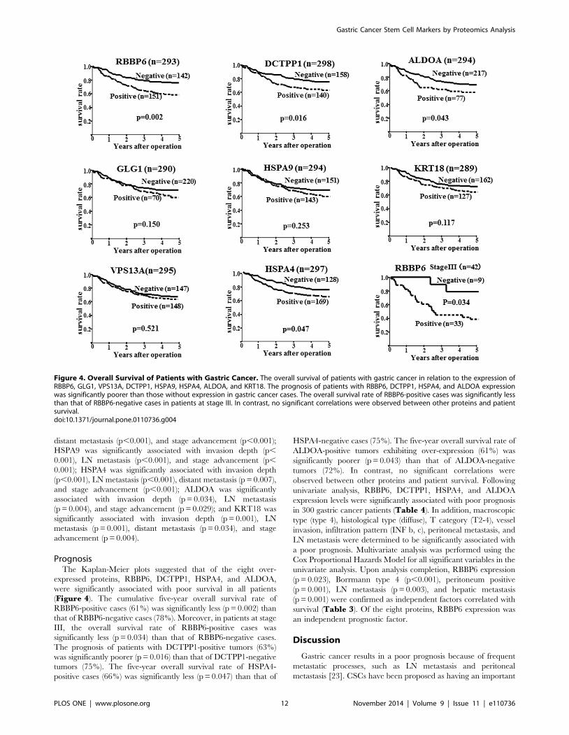
PrognosisThe Kaplan-Meier plots suggested that of the eight over-
expressed proteins, RBBP6, DCTPP1, HSPA4, and ALDOA,
were significantly associated with poor survival in all patients
(Figure 4). The cumulative five-year overall survival rate of
RBBP6-positive cases (61%) was significantly less (p = 0.002) than
that of RBBP6-negative cases (78%). Moreover, in patients at stage
III, the overall survival rate of RBBP6-positive cases was
significantly less (p = 0.034) than that of RBBP6-negative cases.
The prognosis of patients with DCTPP1-positive tumors (63%)
was significantly poorer (p = 0.016) than that of DCTPP1-negative
tumors (75%). The five-year overall survival rate of HSPA4-
positive cases (66%) was significantly less (p = 0.047) than that of
HSPA4-negative cases (75%). The five-year overall survival rate of
ALDOA-positive tumors exhibiting over-expression (61%) was
significantly poorer (p = 0.043) than that of ALDOA-negative
tumors (72%). In contrast, no significant correlations were
observed between other proteins and patient survival. Following
univariate analysis, RBBP6, DCTPP1, HSPA4, and ALDOA
expression levels were significantly associated with poor prognosis
in 300 gastric cancer patients (Table 4). In addition, macroscopic
type (type 4), histological type (diffuse), T category (T2-4), vessel
invasion, infiltration pattern (INF b, c), peritoneal metastasis, and
LN metastasis were determined to be significantly associated with
a poor prognosis. Multivariate analysis was performed using the
Cox Proportional Hazards Model for all significant variables in the
univariate analysis. Upon analysis completion, RBBP6 expression
(p = 0.023), Borrmann type 4 (p,0.001), peritoneum positive
(p = 0.001), LN metastasis (p = 0.003), and hepatic metastasis
(p = 0.001) were confirmed as independent factors correlated with
survival (Table 3). Of the eight proteins, RBBP6 expression was
an independent prognostic factor.
Discussion
Gastric cancer results in a poor prognosis because of frequent
metastatic processes, such as LN metastasis and peritoneal
metastasis [23]. CSCs have been proposed as having an important
Figure 4. Overall Survival of Patients with Gastric Cancer. The overall survival of patients with gastric cancer in relation to the expression ofRBBP6, GLG1, VPS13A, DCTPP1, HSPA9, HSPA4, ALDOA, and KRT18. The prognosis of patients with RBBP6, DCTPP1, HSPA4, and ALDOA expressionwas significantly poorer than those without expression in gastric cancer cases. The overall survival rate of RBBP6-positive cases was significantly lessthan that of RBBP6-negative cases in patients at stage III. In contrast, no significant correlations were observed between other proteins and patientsurvival.doi:10.1371/journal.pone.0110736.g004
Gastric Cancer Stem Cell Markers by Proteomics Analysis
PLOS ONE | www.plosone.org 12 November 2014 | Volume 9 | Issue 11 | e110736

role in the malignant potential of cancer cells, including distant
metastasis and chemoresistance [4]. We have since discovered that
SP cells obtained from gastric cancer subjects possess these CSC
properties [3]. We confirmed that SP cells utilized in this study
express candidate gastric cancer stem cell markers including
CD44[24], CD133 [25], and NANOG [26] (Figure S2). The
spheroid colony formation activity of these SP cells was higher
than that of the parent cells [18]. Also, these CSC-like SP cells
display chemoresistance to anticancer drugs [2]. These findings
have confirmed that OCUM-12/SP cells and OCUM-2MD3/SP
Table 4. Univariate and multivariate analyses with respect to survival.
Clinicopathological features Univariate analysis Multivariate analysis
Risk ratio 95% CI p-value Risk ratio 95% CI p-value
RBBP6
positive vs negative 1.964 1.275–3.025 0.002 1.770 1.080–2.901 0.023
GLG1
positive vs negative 1.391 0.885–2.186 0.152
VPS13A
positive vs negative 1.145 0.885–2.186 0.521
DCTPP1
positive vs negative 1.661 1.096–2.516 0.017 0.902 0.558–1.458 0.674
HSPA9
positive vs negative 0.983 0.747–1.295 0.905
HSPA4
positive vs negative 1.544 1.002–2.378 0.049 0.929 0.549–1.571 0.784
ALDOA
positive vs negative 1.563 1.010–2.421 0.045 1.454 0.866–2.442 0.156
KRT18
positive vs negative 1.390 0.919–2.101 0.119
Age
.60 vs ,60 1.424 0.894–2.268 0.137
Sex
Male vs female 1.065 0.680–1.667 0.785
Macroscopic type
Type4 vs Other types 9.084 5.701–14.476 ,0.001 4.894 2.687–8.914 ,0.001
Tumor differentiation
diffuse vs intestinal 1.647 1.088–2.500 0.018 1.155 0.679–1.965 0.595
T category
T2-4 vs T1 4.479 2.674–7.504 ,0.001 1.187 0.568–2.479 0.648
Vessel invasion
positive vs negative 3.070 1.967–4.793 ,0.001 0.961 0.559–1.653 0.886
INFa
c vs a & b 1.782 1.160–2.737 0.008 0.923 0.512–1.666 0.791
Hepatic metastasis
positive vs negative 7.776 3.369–17.950 ,0.001 4.927 1.953–12.429 0.001
Peritoneal metastasis
positive vs negative 8.209 4.734–14.236 ,0.001 3.043 1.600–5.789 0.001
Lymph node metastasis
positive vs negative 6.315 3.841–10.384 ,0.001 2.848 1.419–5.717 0.003
Total number of resected lymph node
,29 vs.30 0.903 0.600–1.359 0.626
Surgery type
D2 vs D1 1.886 0.589–1.333 0.886
aNF; Infiltration pattern of tumor. The predominant pattern of infiltrating growth into the surrounding tissue is classified as follows; INF a: The tumor shows expandinggrowth and a distinct border with the surrounding tissue. INF b: This category is between INF a and INF b. INF c: The tumor shows infiltrating growth and an indistinctborder with the surrounding tissue.doi:10.1371/journal.pone.0110736.t004
Gastric Cancer Stem Cell Markers by Proteomics Analysis
PLOS ONE | www.plosone.org 13 November 2014 | Volume 9 | Issue 11 | e110736

cells may represent cancer stem cell-like properties. Since specific
markers for gastric CSCs have not been published as of yet,
elucidation of the specific signaling pathways and mechanisms
underlying the actions of CSCs might improve the prognosis of
gastric cancer. In this study, eight candidate CSCs markers were
identified by proteomic techniques using LC-MS/MS coupled
with iTRAQ technology. Three proteins, RBBP6, GLG1 and
VPS13A, were detected only in SP cell lines but not in their
respective parent cell lines. In addition, the five proteins, HSPA9,
ALDOA, DCTPP1, HSPA4, and KRT18, were over-expressed in
both SP cell lines relative to their respective parent cell lines. RT-
PCR analysis also indicated that the expression level of RBBP6,HSPA4, DCTPP1, HSPA9, VPS13A, ALDOA, GLG1, and
CK18 was high in OCUM-12/SP and OCUM-2MD3/SP, in
compared with the control of parent OCUM-12 and OCUM-
2MD3. These proteins were suspected to be novel biomarkers for
gastric CSCs. This hypothesis was tested by immunohistochemical
analysis of 300 gastric cancer cases.
RBBP6 is a nuclear protein, which is a known to be associated
and potentially related to p53, and retinoblastoma binding Q
protein 1 (RBQ-1) [27]. RBBP6 interacts with the tumor
suppressor proteins p53 and Rb, and plays a role in the induction
of apoptosis as well as regulating the cell cycle [28,29]. RBBP6
binds to wild-type p53 proteins but not to p53 mutants [30]. It has
also been shown to promote the binding of MDM2 [31], an E3
ubiquitin ligase that targets the p53[32], and to interfere with its
ability to transactivate the target genes [33]. Up-regulation of
RBBP6 has been strongly correlated with tumor progression in
esophageal cancer and cervical cancer [32]. In this study, RBBP6
expression was associated with ‘T’ category cancer with regards to
invasion depth, distant metastasis, lymph node metastasis, and
clinical stage. Moreover, RBBP6 expression was significantly
associated with poor survival in patients at all stages, particularly at
stage III, resulting in the conclusion that RBBP6 was an
independent factor for survival. We performed the RBBP6 siRNA
knockdown of RBBP6 gene using OCUM-12/SP cells and
OCUM-2MD3/SP cells in this study. RBBP6 siRNA knockdown
significantly decreased the invasion and migration activity of both
SP cells, while the sphere forming activity was not different
between the negative-siRNA and RBBP6 siRNA in SP cells (data
not shown). IHC analysis also indicated that RBBP6 expression
was associated with invasion depth, LN metastasis, distant
metastasis, and a poor prognosis in gastric cancer patients. RBBP6
might be a novel biomarker of gastric CSCs for clinical diagnosis.
These findings suggested that RBBP6 might be closely associated
with malignant potential of cancer stem cells, rather than stem cell
phenotype.
GLG1 is known as a cysteine-rich fibroblast growth factor
receptor (FGFR) [34]. GLG1 was concluded to be associated with
the tumorigenesis of some carcinomas [35] and malignancy of
brain tumors [36]. FGFR signaling possesses broad mitogenic and
cell survival mechanisms, and is involved in a variety of biological
processes, including embryonic development, cell growth, and
tumor invasion [34]. As observed in this study, GLG1 was
concluded to be significantly associated with T category cancer
with regards to invasion depth. It should be noted that GLG1 may
be associated with invasion potential of CSCs via FGFR signaling.
Vacuolar protein sorting (VPS) plays a crucial role in the
trafficking of molecules between cellular organelles, such as
through the trans-Golgi network. The mutation in this gene
causes the autosomal disorder characterized by progressive
neurodegeneration. A past study found that frameshift mutations
of VPS genes, along with loss of expression of VPS13A proteins,
are common in gastric cancers with high microsatellite instability
and suggests that these alterations may contribute to the
development of cancer [37]. VPS is involved in cancer-related
cellular mechanisms, such as proliferation [38,39]. As observed in
this study, VPS13A was expressed in SP cells and was concluded
to be associated with a T category cancer and lymph node
metastasis of gastric cancer. It should be noted that VPS13A may
be associated with the invasion potential of CSCs.
DCTPP1 is expressed in the nucleus of various cancer cells.
Zhang et al. suggested that the accumulation of DCTPP1 in the
nucleus of tumor cells might be sufficient for maintaining proper
DNA replication needed in order to fulfill the requirement for
survival and proliferation of the cells [40]. In conclusion, DCTPP1
was determined to be significantly associated with metastatic
activity. It should be noted that DCTPP1 may be associated with
the DNA replication of CSCs.
HSPA9 and HSPA4 were concluded to be associated with the
invasion and metastatic activity of gastric cancer. These two
proteins are members of the heat shock protein (HSP)-70 family of
chaperones, and HSPA9 is the major protein in the mitochondria.
Some studies have suggested that HSPA9 and HSPA4 are relevant
to cellular apoptosis and promoting proliferation[41,42]; HSPA9
is over-expressed in colon and hepatocellular carcinomas [43,44],
while HSPA4 is over-expressed in breast, colon, ovarian, and
pancreatic cancers [45]. The HSPA4/HSPA14 axis induces the
migration, invasion, and transformation of cancer cells [46].
HSP27 regulates EMT processes and NF-kB activity to contribute
to the maintenance of breast CSCs [47]. Inhibition of the HSP70
protein reduced adhesion and induced apoptosis of both acquired
and de novo drug resistant cancer cells [48]. The HSPA9 protein is
one of the markers of a colon cancer stem cell population [49]. In
conclusion, these findings suggest that HSPA9 and HSPA4 are
associated with CSCs properties via chaperones for EMT-
associated molecules.
ALDOA, aldolase isozymes, is a key glycolytic enzyme that
catalyzes the reversible conversion of fructose-1,6-bisphosphate
into glyceraldehyde 3-phosphate and dihydroxyacetone phosphate
[50]. Glycolysis is one of the key factors for CSC properties.
ALDOA has been expressed in a variety of cancers, such as lung
cancer, renal cell and hepatocellular carcinoma [51,52]. As
observed in this study, ALDOA was concluded to be significantly
associated with the malignant potential of gastric cancer with
regards to invasion depth, LN metastasis, and clinical stage. In
addition, these findings suggest that ALDOA may be associated
with the glycolysis of CSCs.
KRT18 is a type I intermediate filament and its filament
partner is keratin 8 (KRT8). KRT18 is involved in intracellular
signaling pathways that regulate cell growth [53]. Fortier et al.
currently reported that KRT18/KRT8 is associated with the
epithelial-mesenchymal transition (EMT) of cancer cells [53].
EMT is ultimately thought to promote tumor progression through
the generation of CSC properties [54,55]. These findings
suggested that KRT18 may be associated with the EMT of CSCs.
We previously reported on the proteomic differential display
analysis of normal gastric mucosal tissues and human gastric
carcinoma cell lines, including OCUM-12 and OCUM-2MD3
[56]. Nineteen protein spots were observed to be up-regulated in
SGC cell lines when compared to normal gastric mucosa tissues by
using 2-DE and LC-MS/MS. Among the identified increased
spots, two proteins, including UDP-glucose 6-dehydrogenase and
the electron transfer flavoprotein subunit alpha, were also up-
Gastric Cancer Stem Cell Markers by Proteomics Analysis
PLOS ONE | www.plosone.org 14 November 2014 | Volume 9 | Issue 11 | e110736

regulated in OCUM-12/SP cells, as opposed to OCUM-12 cells.
Three proteins, including nucleophosmin, peroxiredoxin-1, and
elongation factor were up-regulated in OCUM-2MD3/SP cells, as
opposed to OCUM-2M cells. Conversely, three proteins, 14-3-3
protein sigma, glucosidase 2 subunit beta, and protein DJ-1, were
down-regulated in OCUM-12/SP cells as opposed to OCUM-12
cells in this study. Overall, the eight proteins, UDP-glucose 6-
dehydrogenase, electron transfer flavoprotein, nucleophosmin,
peroxiredoxin-1, elongation factor, 14-3-3 protein sigma, glucosi-
dase 2 subunit beta, and protein DJ-1 may be associated with
CSCs as well. To the best of our knowledge, this is the first
proteomic analysis that provides evidence that HSPA9, ALDOA,
DCTPP1, HSPA4, KRT18, RBBP6, GLG1, and VPS13A may be
candidate CSC markers for gastric cancer. In particular, RBBP6
may be a promising predictive marker for the prognosis of patients
with gastric cancer.
Supporting Information
Figure S1 Representative picture of side populationfraction. Cancer cells, which disappear in the presence of
verapamil (lower panel), are outlined and defined as the SP cells.
OCUM-12/SP and OCUM-2MD3/SP cells were sorted as SP
cells from each of the parent OCUM-12 and OCUM-2MD3 cells,
respectively. The percentages of SP cells were higher in the
OCUM-12/SP and OCUM-2MD3/SP cells than in their parent
OCUM-12 and OCUM-2MD3 cells.
(TIF)
Figure S2 Stem cell markers expression. The expression
level of CSC markers, CD44, CD133 and NANOG was
significantly higher in OCUM-12/SP (1.6-, 3.6- and 33.2-fold)
and OCUM-2MD3/SP cells (2.1-, 5.9- and 3.6-fold) than that in
parent OCUM-12 and OCUM-2MD3 cells.
(TIF)
Figure S3 Expression of RBBP6 and ALDOA. The
expression level of RBBP6 and ALDOA was high in CUM-12/
SP and OCUM-2MD3/SP cells, in comparison with that OCUM-
12 and OCUM-2MD3 cells.
(TIF)
Table S1 (DOCX)
Table S2 Proteins increased in both OCUM-12/SP andOCUM-2MD3/SP cells.
(DOCX)
Table S3 (DOCX)
Acknowledgments
We thank Mayumi Oda (Osaka City University Graduate School of
Medicine) for technical advice.
Author Contributions
Conceived and designed the experiments: TM MY AK. Performed the
experiments: TM MY AI H. Kinoshita TF. Analyzed the data: TM MY
AK AI. Contributed reagents/materials/analysis tools: TM MY AK AI H.
Kasashima TF H. Kinoshita GM KS NK KM MO HW KH. Wrote the
paper: TM MY AK HW KH.
References
1. Clarke MF, Dick JE, Dirks PB, Eaves CJ, Jamieson CH, et al. (2006) Cancerstem cells—perspectives on current status and future directions: AACR
Workshop on cancer stem cells. Cancer Res 66: 9339–9344.
2. Yashiro M, Nishii T, Hasegawa T, Matsuzaki T, Morisaki T, et al. (2013) A c-
Met inhibitor increases the chemosensitivity of cancer stem cells to the irinotecan
in gastric carcinoma. Br J Cancer 109: 2619–2628.
3. Nishii T, Yashiro M, Shinto O, Sawada T, Ohira M, et al. (2009) Cancer stem
cell-like SP cells have a high adhesion ability to the peritoneum in gastriccarcinoma. Cancer Sci 100: 1397–1402.
4. Glinsky GV, Berezovska O, Glinskii AB (2005) Microarray analysis identifies adeath-from-cancer signature predicting therapy failure in patients with multiple
types of cancer. J Clin Invest 115: 1503–1521.
5. Bonnet D, Dick JE (1997) Human acute myeloid leukemia is organized as a
hierarchy that originates from a primitive hematopoietic cell. Nat Med 3: 730–
737.
6. Takaishi S, Okumura T, Wang TC (2008) Gastric cancer stem cells. J Clin
Oncol 26: 2876–2882.
7. Stojnev S, Krstic M, Ristic-Petrovic A, Stefanovic V, Hattori T (2014) Gastric
cancer stem cells: therapeutic targets. Gastric Cancer 17: 13–25.
8. Hartgrink HH, Jansen EP, van Grieken NC, van de Velde CJ (2009) Gastric
cancer. Lancet 374: 477–490.
9. Charafe-Jauffret E, Ginestier C, Iovino F, Tarpin C, Diebel M, et al. (2010)Aldehyde dehydrogenase 1-positive cancer stem cells mediate metastasis and
poor clinical outcome in inflammatory breast cancer. Clin Cancer Res 16: 45–55.
10. Al-Hajj M, Wicha MS, Benito-Hernandez A, Morrison SJ, Clarke MF (2003)Prospective identification of tumorigenic breast cancer cells. Proc Natl Acad
Sci U S A 100: 3983–3988.
11. Li C, Heidt DG, Dalerba P, Burant CF, Zhang L, et al. (2007) Identification ofpancreatic cancer stem cells. Cancer Res 67: 1030–1037.
12. Ricci-Vitiani L, Lombardi DG, Pilozzi E, Biffoni M, Todaro M, et al. (2007)Identification and expansion of human colon-cancer-initiating cells. Nature 445:
111–115.
13. Chen S, Zhao H, Deng J, Liao P, Xu Z, et al. (2013) Comparative proteomics of
glioma stem cells and differentiated tumor cells identifies S100A9 as a potential
therapeutic target. J Cell Biochem 114: 2795–2808.
14. Kakehashi A, Ishii N, Shibata T, Wei M, Okazaki E, et al. (2011) Mitochondrial
prohibitins and septin 9 are implicated in the onset of rat hepatocarcinogenesis.Toxicol Sci 119: 61–72.
15. Goodell MA, Rosenzweig M, Kim H, Marks DF, DeMaria M, et al. (1997) Dyeefflux studies suggest that hematopoietic stem cells expressing low or
undetectable levels of CD34 antigen exist in multiple species. Nat Med 3:1337–1345.
16. Yashiro M, Chung YS, Nishimura S, Inoue T, Sowa M (1996) Peritoneal
metastatic model for human scirrhous gastric carcinoma in nude mice. Clin ExpMetastasis 14: 43–54.
17. Kato Y, Yashiro M, Noda S, Tendo M, Kashiwagi S, et al. (2010) Establishment
and characterization of a new hypoxia-resistant cancer cell line, OCUM-12/
Hypo, derived from a scirrhous gastric carcinoma. Br J Cancer 102: 898–907.
18. Hasegawa T, Yashiro M, Nishii T, Matsuoka J, Fuyuhiro Y, et al. (2013)
Cancer-associated fibroblasts might sustain the stemness of scirrhous gastric
cancer cells via transforming growth factor-beta signaling. Int J Cancer.
19. (2011) Japanese classification of gastric carcinoma: 3rd English edition. GastricCancer 14: 101–112.
20. Chung K, Nishiyama N, Wanibuchi H, Yamano S, Hanada S, et al. (2012)
AGR2 as a potential biomarker of human lung adenocarcinoma. Osaka CityMed J 58: 13–24.
21. Kakehashi A, Hagiwara A, Imai N, Nagano K, Nishimaki F, et al. (2013) Mode
of action of ethyl tertiary-butyl ether hepatotumorigenicity in the rat: evidencefor a role of oxidative stress via activation of CAR, PXR and PPAR signaling
pathways. Toxicol Appl Pharmacol 273: 390–400.
22. Ross PL, Huang YN, Marchese JN, Williamson B, Parker K, et al. (2004)Multiplexed protein quantitation in Saccharomyces cerevisiae using amine-
reactive isobaric tagging reagents. Mol Cell Proteomics 3: 1154–1169.
23. Hippo Y, Yashiro M, Ishii M, Taniguchi H, Tsutsumi S, et al. (2001) Differentialgene expression profiles of scirrhous gastric cancer cells with high metastatic
potential to peritoneum or lymph nodes. Cancer Res 61: 889–895.
24. Takaishi S, Okumura T, Tu S, Wang SS, Shibata W, et al. (2009) Identification
of gastric cancer stem cells using the cell surface marker CD44. Stem Cells 27:1006–1020.
25. Wen L, Chen XZ, Yang K, Chen ZX, Zhang B, et al. (2013) Prognostic value of
cancer stem cell marker CD133 expression in gastric cancer: a systematic review.PLoS One 8: e59154.
26. Liu J, Ma L, Xu J, Liu C, Zhang J, et al. (2013) Spheroid body-forming cells in
the human gastric cancer cell line MKN-45 possess cancer stem cell properties.Int J Oncol 42: 453–459.
27. Chibi M, Meyer M, Skepu A, DJ GR, Moolman-Smook JC, et al. (2008) RBBP6
interacts with multifunctional protein YB-1 through its RING finger domain,leading to ubiquitination and proteosomal degradation of YB-1. J Mol Biol 384:
908–916.
28. Scott RE, White-Grindley E, Ruley HE, Chesler EJ, Williams RW (2005) P2P-Rexpression is genetically coregulated with components of the translation
Gastric Cancer Stem Cell Markers by Proteomics Analysis
PLOS ONE | www.plosone.org 15 November 2014 | Volume 9 | Issue 11 | e110736

machinery and with PUM2, a translational repressor that associates with the
P2P-R mRNA. J Cell Physiol 204: 99–105.29. Pugh DJ, Ab E, Faro A, Lutya PT, Hoffmann E, et al. (2006) DWNN, a novel
ubiquitin-like domain, implicates RBBP6 in mRNA processing and ubiquitin-
like pathways. BMC Struct Biol 6: 1.30. Simons A, Melamed-Bessudo C, Wolkowicz R, Sperling J, Sperling R, et al.
(1997) PACT: cloning and characterization of a cellular p53 binding protein thatinteracts with Rb. Oncogene 14: 145–155.
31. Midgley CA, Lane DP (1997) p53 protein stability in tumour cells is not
determined by mutation but is dependent on Mdm2 binding. Oncogene 15:1179–1189.
32. Yoshitake Y, Nakatsura T, Monji M, Senju S, Matsuyoshi H, et al. (2004)Proliferation potential-related protein, an ideal esophageal cancer antigen for
immunotherapy, identified using complementary DNA microarray analysis. ClinCancer Res 10: 6437–6448.
33. Li L, Deng B, Xing G, Teng Y, Tian C, et al. (2007) PACT is a negative
regulator of p53 and essential for cell growth and embryonic development. ProcNatl Acad Sci U S A 104: 7951–7956.
34. Kibriya MG, Jasmine F, Argos M, Andrulis IL, John EM, et al. (2009) A pilotgenome-wide association study of early-onset breast cancer. Breast Cancer Res
Treat 114: 463–477.
35. Crnogorac-Jurcevic T, Efthimiou E, Capelli P, Blaveri E, Baron A, et al. (2001)Gene expression profiles of pancreatic cancer and stromal desmoplasia.
Oncogene 20: 7437–7446.36. Yamaguchi F, Morrison RS, Gonatas NK, Takahashi H, Sugisaki Y, et al.
(2003) Identification of MG-160, a FGF binding medial Golgi sialoglycoprotein,in brain tumors: an index of malignancy in astrocytomas. Int J Oncol 22: 1045–
1049.
37. An CH, Kim YR, Kim HS, Kim SS, Yoo NJ, et al. (2012) Frameshift mutationsof vacuolar protein sorting genes in gastric and colorectal cancers with
microsatellite instability. Hum Pathol 43: 40–47.38. Thompson BJ, Mathieu J, Sung HH, Loeser E, Rorth P, et al. (2005) Tumor
suppressor properties of the ESCRT-II complex component Vps25 in
Drosophila. Dev Cell 9: 711–720.39. Vaccari T, Bilder D (2005) The Drosophila tumor suppressor vps25 prevents
nonautonomous overproliferation by regulating notch trafficking. Dev Cell 9:687–698.
40. Zhang Y, Ye WY, Wang JQ, Wang SJ, Ji P, et al. (2013) dCTPpyrophosphohydrase exhibits nucleic accumulation in multiple carcinomas.
Eur J Histochem 57: e29.
41. Yenari MA, Liu J, Zheng Z, Vexler ZS, Lee JE, et al. (2005) Antiapoptotic andanti-inflammatory mechanisms of heat-shock protein protection. Ann N Y Acad
Sci 1053: 74–83.42. Garrido C, Brunet M, Didelot C, Zermati Y, Schmitt E, et al. (2006) Heat shock
proteins 27 and 70: anti-apoptotic proteins with tumorigenic properties. Cell
Cycle 5: 2592–2601.
43. Peng C, Yang P, Cui Y, He M, Liang L, et al. (2013) HSPA9 overexpression
inhibits apoptin-induced apoptosis in the HepG2 cell line. Oncol Rep 29: 2431–
2437.
44. Gestl EE, Anne Bottger S (2012) Cytoplasmic sequestration of the tumor
suppressor p53 by a heat shock protein 70 family member, mortalin, in human
colorectal adenocarcinoma cell lines. Biochem Biophys Res Commun 423: 411–
416.
45. Zhong MA, Zhang H, Qi XY, Lu AG, You TG, et al. (2011) ShRNA-mediated
gene silencing of heat shock protein 70 inhibits human colon cancer growth. Mol
Med Rep 4: 805–810.
46. Wu CY, Lin CT, Wu MZ, Wu KJ (2011) Induction of HSPA4 and HSPA14 by
NBS1 overexpression contributes to NBS1-induced in vitro metastatic and
transformation activity. J Biomed Sci 18: 1.
47. Wei L, Liu TT, Wang HH, Hong HM, Yu AL, et al. (2011) Hsp27 participates
in the maintenance of breast cancer stem cells through regulation of epithelial-
mesenchymal transition and nuclear factor-kappaB. Breast Cancer Res 13:
R101.
48. Nimmanapalli R, Gerbino E, Dalton WS, Gandhi V, Alsina M (2008) HSP70
inhibition reverses cell adhesion mediated and acquired drug resistance in
multiple myeloma. Br J Haematol 142: 551–561.
49. Giampieri R, Scartozzi M, Loretelli C, Piva F, Mandolesi A, et al. (2013) Cancer
stem cell gene profile as predictor of relapse in high risk stage II and stage III,
radically resected colon cancer patients. PLoS One 8: e72843.
50. Kajita E, Moriwaki J, Yatsuki H, Hori K, Miura K, et al. (2001) Quantitative
expression studies of aldolase A, B and C genes in developing embryos and adult
tissues of Xenopus laevis. Mech Dev 102: 283–287.
51. Shimizu T, Inoue KI, Hachiya H, Shibuya N, Shimoda M, et al. (2013)
Frequent alteration of the protein synthesis of enzymes for glucose metabolism in
hepatocellular carcinomas. J Gastroenterol.
52. Du S, Guan Z, Hao L, Song Y, Wang L, et al. (2014) Fructose-bisphosphate
aldolase a is a potential metastasis-associated marker of lung squamous cell
carcinoma and promotes lung cell tumorigenesis and migration. PLoS One 9:
e85804.
53. Fortier AM, Asselin E, Cadrin M (2013) Keratin 8 and 18 loss in epithelial
cancer cells increases collective cell migration and cisplatin sensitivity through
claudin1 up-regulation. J Biol Chem 288: 11555–11571.
54. Polyak K, Weinberg RA (2009) Transitions between epithelial and mesenchymal
states: acquisition of malignant and stem cell traits. Nat Rev Cancer 9: 265–273.
55. Devarajan E, Song YH, Krishnappa S, Alt E (2012) Epithelial-mesenchymal
transition in breast cancer lines is mediated through PDGF-D released by tissue-
resident stem cells. Int J Cancer 131: 1023–1031.
56. Kuramitsu Y, Baron B, Yoshino S, Zhang X, Tanaka T, et al. (2010) Proteomic
differential display analysis shows up-regulation of 14-3-3 sigma protein in
human scirrhous-type gastric carcinoma cells. Anticancer Res 30: 4459–4465.
Gastric Cancer Stem Cell Markers by Proteomics Analysis
PLOS ONE | www.plosone.org 16 November 2014 | Volume 9 | Issue 11 | e110736