Targeting proline in (phospho)proteomics - Utrecht University ...
19
Targeting proline in (phospho)proteomics Saar A. M. van der Laarse 1,2 , Charlotte A. G. H. van Gelder 1,2 , Marshall Bern 3 , Michiel Akeroyd 4 , Maurien M. A. Olsthoorn 4 and Albert J. R. Heck 1,2 1 Biomolecular Mass Spectrometry and Proteomics, Bijvoet Center for Biomolecular Research and Utrecht Institute for Pharmaceutical Sciences, Utrecht University, The Netherlands 2 Netherlands Proteomics Center, Utrecht, The Netherlands 3 ProteinMetrics, Cupertino, CA, USA 4 DSM Biotechnology Center, Delft, The Netherlands Keywords (phospho)proteomics; EndoPro; mass spectrometry; proline effect; protease Correspondence A. J. R. Heck, Biomolecular Mass Spectrometry and Proteomics, Bijvoet Center for Biomolecular Research and Utrecht Institute for Pharmaceutical Sciences, Utrecht University, Padualaan 8, 3584 CH Utrecht, The Netherlands Tel: +31 30 253 6797 E-mail: [email protected] Saar A. M. van der Laarse and Charlotte A. G. H. van Gelder contributed equally to this article. (Received 23 July 2019, revised 25 November 2019, accepted 19 December 2019) doi:10.1111/febs.15190 Mass spectrometry-based proteomics experiments typically start with the digestion of proteins using trypsin, chosen because of its high specificity, availability, and ease of use. It has become apparent that the sole use of trypsin may impose certain limits on our ability to grasp the full proteome, missing out particular sites of post-translational modifications, protein seg- ments, or even subsets of proteins. To tackle this problem, alternative pro- teases have been introduced and shown to lead to an increase in the detectable (phospho)proteome. Here, we argue that there may be further room for improvement and explore the protease EndoPro. For optimal peptide identification rates, we explored multiple peptide fragmentation techniques (HCD, ETD, and EThcD) and employed Byonic as search algo- rithm. We obtain peptide IDs for about 40% of the MS2 spectra (66% for trypsin). EndoPro cleaves with high specificity at the C-terminal site of Pro and Ala residues and displays activity in a broad pH range, where we focused on its performance at pH = 2 and 5.5. The proteome coverage of EndoPro at these two pH values is rather distinct, and also complementary to the coverage obtained with trypsin. As about 40% of mammalian pro- tein phosphorylations are proline-directed, we also explored the perfor- mance of EndoPro in phosphoproteomics. EndoPro extends the coverable phosphoproteome substantially, whereby both the, at pH = 2 and 5.5, acquired phosphoproteomes are complementary to each other and to the phosphoproteome obtained using trypsin. Hence, EndoPro is a powerful tool to exploit in (phospho)proteomics applications. Introduction Proteins are involved in nearly all biological processes. Their functionality can be regulated extensively, through the formation of complexes, changes in expression levels, and widespread post-translational modifications, such as acetylation and phosphoryla- tion. Proteins must be tightly regulated as undesired changes at the protein level can cause disease and other unintended biological effects [1,2]. Owing to their crucial role, identifying and quantifying proteins present in certain biological states is of great impor- tance and can improve our understanding of the mech- anisms underlying health and disease. To this end, the field of proteomics aims to measure all proteins expressed by a certain organism or cell type [3,4]. Abbreviations ETD, electron transfer dissociation; EThcD, electron transfer higher-energy collision dissociation; HCD, higher-energy collision dissociation; LC, liquid chromatography; MS, mass spectrometry; ON, overnight; pI, isoelectric point; PSM, peptide spectrum match; PTM, post- translational modification. 2979 The FEBS Journal 287 (2020) 2979–2997 ª 2019 The Authors. The FEBS Journal published by John Wiley & Sons Ltd on behalf of Federation of European Biochemical Societies This is an open access article under the terms of the Creative Commons Attribution-NonCommercial License, which permits use, distribution and reproduction in any medium, provided the original work is properly cited and is not used for commercial purposes.
-
Upload
khangminh22 -
Category
Documents
-
view
3 -
download
0
Transcript of Targeting proline in (phospho)proteomics - Utrecht University ...

Targeting proline in (phospho)proteomicsSaar A. M. van der Laarse1,2 , Charlotte A. G. H. van Gelder1,2 , Marshall Bern3 ,Michiel Akeroyd4, Maurien M. A. Olsthoorn4 and Albert J. R. Heck1,2
1 Biomolecular Mass Spectrometry and Proteomics, Bijvoet Center for Biomolecular Research and Utrecht Institute for Pharmaceutical
Sciences, Utrecht University, The Netherlands
2 Netherlands Proteomics Center, Utrecht, The Netherlands
3 ProteinMetrics, Cupertino, CA, USA
4 DSM Biotechnology Center, Delft, The Netherlands
Keywords
(phospho)proteomics; EndoPro; mass
spectrometry; proline effect; protease
Correspondence
A. J. R. Heck, Biomolecular Mass
Spectrometry and Proteomics, Bijvoet
Center for Biomolecular Research and
Utrecht Institute for Pharmaceutical
Sciences, Utrecht University, Padualaan 8,
3584 CH Utrecht, The Netherlands
Tel: +31 30 253 6797
E-mail: [email protected]
Saar A. M. van der Laarse and Charlotte A.
G. H. van Gelder contributed equally to this
article.
(Received 23 July 2019, revised 25
November 2019, accepted 19 December
2019)
doi:10.1111/febs.15190
Mass spectrometry-based proteomics experiments typically start with the
digestion of proteins using trypsin, chosen because of its high specificity,
availability, and ease of use. It has become apparent that the sole use of
trypsin may impose certain limits on our ability to grasp the full proteome,
missing out particular sites of post-translational modifications, protein seg-
ments, or even subsets of proteins. To tackle this problem, alternative pro-
teases have been introduced and shown to lead to an increase in the
detectable (phospho)proteome. Here, we argue that there may be further
room for improvement and explore the protease EndoPro. For optimal
peptide identification rates, we explored multiple peptide fragmentation
techniques (HCD, ETD, and EThcD) and employed Byonic as search algo-
rithm. We obtain peptide IDs for about 40% of the MS2 spectra (66% for
trypsin). EndoPro cleaves with high specificity at the C-terminal site of Pro
and Ala residues and displays activity in a broad pH range, where we
focused on its performance at pH = 2 and 5.5. The proteome coverage of
EndoPro at these two pH values is rather distinct, and also complementary
to the coverage obtained with trypsin. As about 40% of mammalian pro-
tein phosphorylations are proline-directed, we also explored the perfor-
mance of EndoPro in phosphoproteomics. EndoPro extends the coverable
phosphoproteome substantially, whereby both the, at pH = 2 and 5.5,
acquired phosphoproteomes are complementary to each other and to the
phosphoproteome obtained using trypsin. Hence, EndoPro is a powerful
tool to exploit in (phospho)proteomics applications.
Introduction
Proteins are involved in nearly all biological processes.
Their functionality can be regulated extensively,
through the formation of complexes, changes in
expression levels, and widespread post-translational
modifications, such as acetylation and phosphoryla-
tion. Proteins must be tightly regulated as undesired
changes at the protein level can cause disease and
other unintended biological effects [1,2]. Owing to
their crucial role, identifying and quantifying proteins
present in certain biological states is of great impor-
tance and can improve our understanding of the mech-
anisms underlying health and disease. To this end, the
field of proteomics aims to measure all proteins
expressed by a certain organism or cell type [3,4].
Abbreviations
ETD, electron transfer dissociation; EThcD, electron transfer higher-energy collision dissociation; HCD, higher-energy collision dissociation;
LC, liquid chromatography; MS, mass spectrometry; ON, overnight; pI, isoelectric point; PSM, peptide spectrum match; PTM, post-
translational modification.
2979The FEBS Journal 287 (2020) 2979–2997 ª 2019 The Authors. The FEBS Journal published by John Wiley & Sons Ltd on behalf of
Federation of European Biochemical Societies
This is an open access article under the terms of the Creative Commons Attribution-NonCommercial License, which permits use,
distribution and reproduction in any medium, provided the original work is properly cited and is not used for commercial purposes.

Proteomics comes in a range of different workflows
[3]. In the more standard bottom-up workflow, pro-
teins are extracted from the material of interest and
subjected to proteolysis, which results in a complex
mixture of peptides that originated from the proteins
present in the targeted cells. Through LC-MS/MS
analysis, these peptides are separated, fragmented, and
analyzed. Then, the collected empirical spectra are cor-
related to peptide and thus protein sequences through
the use of theoretical in silico fragmentation spectra
[5,6]. However, due to the indirect nature of this
assignment, how peptides are generated from the intact
protein is of critical importance.
Most proteomic studies use trypsin for the protein
digestion as it cleaves with very high specificity pro-
teins C terminally to only arginine (Arg) and lysine
(Lys) residues. As both amino acids are basic, the
resulting peptides have basic C termini. This, com-
bined with the free amine at the peptide N terminus,
ensures that tryptic peptides carry a positive charge at
either end of the peptide, making them very suitable
for fragmentation-based sequencing [6]. In addition,
trypsin’s high specificity reduces the complexity of the
subsequent database searches as they can be restricted
to peptides ending with Arg or Lys, which reduces
computational requirements of the search. However,
the use of trypsin also has limitations and is not the
optimal enzyme for all analyses.
Owing to the high specificity of trypsin, the spacing
of Arg and Lys amino acids across the proteome dic-
tates the length of peptides, and thus the number of
unique peptides. For standard intracellular proteins,
Arg and Lys occur at a high frequency (5.6% and
5.7%, respectively), which leads to the fact that
roughly 50% of the peptides produced by trypsin are
too short (<6 amino acids) to be nicely fragmented
and uniquely assigned to a protein [7]. Conversely,
some proteins, notably membrane proteins, exhibit few
tryptic cleavage sites and extreme hydrophobicity,
resulting in poor coverage of this class of proteins in
trypsin-based proteomics [8]. These combined effects
all contribute to undetected, less visible areas of the
proteome. To illustrate this, we have performed an
in silico digestion of the human proteome using the
specificity listed in Table 1 and asked what the upper
limit of detection was for each protease using the
search and mass spectrometry settings employed in this
study (Table 1). For trypsin, a maximum of 87% of
the proteome would in theory be detectable using this
proteomics setup, assuming every peptide of suitable
characteristics is actually fragmented and identified.
To improve on this boundary, efforts have been made
to utilize different proteases within bottom-up
workflows. Several groups have shown that by using
proteases that cleave at different amino acid motifs,
the number of unique peptides identified, and thus the
proteome coverage, can be substantially improved
[7,9–12].
Numerous alternative proteases have been used for
the digestion of proteins from a lysate, whereby each
has its own cleavage specificity and optimal condi-
tions (Table 1). By combining the proteases either in
parallel or sequentially, one is able to improve the
proteome coverage through combining the results of
individual proteases together. For instance, work by
Swaney et al. [7] nicely illustrated that expanding
beyond a single protease can yield a roughly 20%
increase in protein identifications and achieved double
the proteome sequence coverage. Similarly, our group
has shown that the use of multiple proteases in paral-
lel for phosphoproteomics gives rise to highly comple-
mentary sets of phosphosites, where only 27% of all
identified sites were found in more than one protease
dataset [17].
While the combination of proteases has already been
shown to aid in expanding the proteome sequence cov-
erage, the presence of (multiple) proline residues pre-
sents a particular challenge for many proteases.
Proline is a unique amino acid in peptides/proteins as
it is the only cyclic amino acid, giving rise to a tertiary
amide, limiting hydrogen donating properties and
imposing rigid structural constraints on peptide bonds
[18,19]. Because of its unique properties, proline often
leads to missed proteolytic events during digestion [9],
increasing the resultant peptide length and database
search complexity. Moreover, proline also effects the
fragmentation step during mass analysis, known as the
‘proline effect’ [20], where fragmentation shows
enhanced production of y-ions spanning from the pro-
line to the peptide C terminus due to the enhanced
Table 1. Cleavage specificities reported for some of the most
commonly used proteases in bottom-up proteomics [9,10,13–16].
Xnp indicates any amino acid except proline.
Protease
Cleavage
site (↓) Optimal pH
Max proteome
coverage
AspN ↓D 8 78%
LysargiNase ↓K/R 7.5 87%
LysC K↓Xnp 8 79%
LysN ↓KXnp 8 78%
ArgC R↓Xnp 8 82%
GluC E↓ 8 86%
Chymotrypsin F/W/Y↓ 8 87%
Trypsin K/R↓Xnp 8–9 87%
Sap-9 K/R↓Xnp 6–7 87%
2980 The FEBS Journal 287 (2020) 2979–2997 ª 2019 The Authors. The FEBS Journal published by John Wiley & Sons Ltd on behalf of
Federation of European Biochemical Societies
Targeting proline in (phospho)proteomics S. A. M. van der Laarse et al.

basicity of the proline nitrogen, restricting the peptide
sequence coverage [20–22].
To overcome these limitations, research efforts have
been directed toward finding a proline-directed pro-
tease as such a protease would decrease database
search complexity by well defining the proline position,
as well as substantially improve proteome sequence
coverage due to its high complementarity to Arg- and
Lys-directed proteases. In 2009, �Sebela et al. [23] eval-
uated an acidic prolyl endoprotease from Aspergillus
niger, called An-PEP, for its use in proteomics and
found that the enzyme has potential for in-solution
digestion studies. Moreover, our laboratory showed
that An-PEP, also termed EndoPro, exhibited maxi-
mum activity at pH = 2 and is active at moderately
high urea concentrations and low temperatures, mak-
ing it very suitable for use in mass spectrometry-based
hydrogen–deuterium exchange experiments [24]. In
addition, work published on another prolyl endopepti-
dase originally from Nepenthes ventrata, termed nepro-
sin, showed that almost half of the sequence coverage
achieved by the proline-directed protease on proteins
detected in both tryptic and neprosin digests were not
observed when digestion was performed with trypsin
[25]. Collectively, these works suggest huge potential
of proline-directed proteases to shed light on previ-
ously undetectable areas of the proteome. In phospho-
proteomics, however, proline-induced complications
are even more prevalent as in eukaryotic systems
around 40% of the phosphorylation events detected
are proline-directed, dominated by so-called SP or TP
motifs [26]. Hence, in most eukaryotic phosphopro-
teomics experiments, prolines are highly enriched and
even more prevalent than in a standard proteomics
analysis.
Here, we extend substantially on previous work
using proline-directed proteases. We first benchmark
EndoPro versus trypsin, thereby generating large pro-
teomics datasets on HeLa lysates digested by EndoPro
at pH = 2, EndoPro at pH = 5.5, and trypsin at
pH = 8.5. We optimize the peptide ID rates using mul-
tiple peptide fragmentation techniques, and the search
engine Byonic, allowing us to increase the ID rate sub-
stantially to about 40% and 66% of all PSMs for
EndoPro and trypsin, respectively. When using Endo-
Pro at these two different pH values, we find the speci-
ficities and activities to be similar. However, our
datasets reveal a substantial difference between the
peptides generated with EndoPro at pH = 2, EndoPro
at pH = 5.5, and trypsin, indicating the cleavage of
different proteins and/or sites at different pH values.
Overall, EndoPro enabled us to detect over 2200
unique proteins not observed in our tryptic digests and
contributed 49% of the total unique phosphosites
detected, making it a protease almost equally powerful
as, and complementary, to trypsin.
Results
To assess the benefit of EndoPro in shotgun pro-
teomics [25], we decided to evaluate and optimize the
performance of this proline-directed protease on a
complex HeLa cell lysate, first focusing on nonmodi-
fied peptides. To characterize the specificity of Endo-
Pro in a full proteome, we performed a quadruplicate
digestion of a HeLa lysate with EndoPro for 1 h at
pH = 5.5. For comparison, we also performed a stan-
dard trypsin overnight digestion at pH = 8.5. These
data were subjected to a nonspecific search in Byonic,
and subsequently, the environment of all cleaved sites
was analyzed using an in-house R script. The distribu-
tion of amino acids following the residue cleaved by
either EndoPro (purple) or trypsin (orange) is shown
in Fig. 1A. EndoPro showed a strong specificity for
cleavage C-terminal to proline (49.1%) and alanine
(35.5%), resulting in an overall cleavage specificity
close to that observed for trypsin (84.6% Ala/Pro ver-
sus 89.6% Arg/Lys in our datasets). Inspection of the
cleavage site environment of EndoPro (Fig. 1B)
revealed a disfavor for cleaving when the cleavage site
is preceding a proline. In these cases, only the last pro-
line is cleaved. In addition, positively charged residues
appear disfavored in the P + 2 position (Fig. 1B).
Since EndoPro reached almost 85% specificity, we
subsequently used less computationally heavy semis-
pecific database searches (allowing one side of the pep-
tide to result from nonspecific cleavages), which saves
data analysis time and is inherently less error-prone.
Doing these two searches on the same dataset, we
observed that we still captured nearly all the peptides
formed (97.6%).
Performance evaluation of EndoPro at pH = 2
and pH = 5.5
Next, we set out to compare the performance of Endo-
Pro and trypsin. Thereby, we took into account that
EndoPro exhibits several maxima in its activity profile,
with maxima at pH = 2 and 5.5, as also reported ear-
lier [24]. Therefore, HeLa cell lysates were digested
with either EndoPro at pH = 2 and pH = 5.5 and
digested for 1 h or overnight (ON), and additionally,
for benchmarking, the same HeLa cell lysate sample
was digested with trypsin using conventional condi-
tions (i.e., pH = 8.5, ON). The resulting peptides were
analyzed by LC-MS/MS on a Fusion hybrid mass
2981The FEBS Journal 287 (2020) 2979–2997 ª 2019 The Authors. The FEBS Journal published by John Wiley & Sons Ltd on behalf of
Federation of European Biochemical Societies
S. A. M. van der Laarse et al. Targeting proline in (phospho)proteomics

spectrometer using in parallel ETD, EThcD, and HCD
as peptide fragmentation methods. Spectra were
searched with Byonic. A global overview of the search
outcomes is shown in Table S1. The different fragmen-
tation methods resulted in a highly similar number of
protein identifications (Table S1). It was therefore
decided to pool all the data acquired with different
fragmentation techniques to assess the performance of
EndoPro across the different digestion conditions,
independent of the used fragmentation method. An
overview of the pooled datasets is shown in Table 2.
In terms of unique proteins detected, an overlap of
35% was observed between the two proteases
(Fig. 1C). The four different EndoPro digestion condi-
tions resulted in comparable identification rates, with
slightly more PSMs and unique peptides in the experi-
ments performed at pH = 5.5 when compared to
pH = 2. Under all four tested conditions roughly, the
same number of peptides (� 15 000) and proteins
(� 2600) could be identified (Table 2). As expected,
the peptide identification rate achieved with trypsin
(67%) could not be reached with EndoPro (� 40%).
Still, the EndoPro ID rates of � 40% are better than
what has been reported for many other alternative
enzymes (e.g., LysN, AspN, chymotrypsin typically
reach 20–30%) [10,24,25].
A D F G K L P R S W Y0
20
40
60
Amino Acid
% c
leav
ed
Trypsin
EP pH5.5 1 h
10 2 3 4 5 6 7 8 9 10 11 12
2204 2511 252530% 35% 35%
EndoPro (4715)
Trypsin (5036)
A
B
C
Fig. 1. Characterization of EndoPro
cleavage specificity. (A) Overview of amino
acids after which was cleaved by EndoPro
(n = 4, purple) and trypsin (n = 4, orange),
based on a nonspecific search, revealing a
high specificity of 84.6% A/P and 89.6% R/
K for EndoPro and trypsin, respectively.
Only amino acids with a cleavage frequency
of 1% or higher were included. Data are
represented as mean percentage of total
cleavages per protease � SEM. (B) An
iceLogo showing the differences between
the EndoPro cleavage site environment
(17 032 unique environments from
nonspecific search) and the human
proteome, illustrating a disfavor for R/K on
the +2 position and a reluctance to cleave
between proline residues. (C) Overlap of
unique proteins identified by EndoPro or
trypsin using a semispecific search.
Although the sizes of the identified
proteomes are roughly equal, the overlap
between the two is only 35%.
2982 The FEBS Journal 287 (2020) 2979–2997 ª 2019 The Authors. The FEBS Journal published by John Wiley & Sons Ltd on behalf of
Federation of European Biochemical Societies
Targeting proline in (phospho)proteomics S. A. M. van der Laarse et al.

Characteristics of EndoPro peptides generated at
pH = 2 and 5.5
For a more in-depth exploration of the type of pep-
tides produced by EndoPro, we compared general pep-
tide characteristics such as peptide length, mass, amino
acid content, and cleavage specificity as observed in
the unique peptides identified from the EndoPro and,
for comparison, tryptic digests, as depicted in Fig. 2.
In terms of peptide length and charge, the four
explored different EndoPro cleavage conditions pro-
duced similar peptides. We observed a substantially
broader peptide length distribution for EndoPro pep-
tides than for trypsin, revealing more peptides with a
length of more than 20 amino acids and a tail toward
peptides with a length of 50 or more amino acids
(Fig. 2A). This already indicates that EndoPro gener-
ates peptides with more missed cleavages than trypsin.
In terms of peptide charge, an average of about 33%
of the unique EndoPro peptides carried four or more
charges, compared to only 5% for the tryptic peptides
(Fig. 2B). This difference in charge distribution could
not be explained by the increase in peptide length, as
the average number of amino acids to charge ratio of
the different EndoPro conditions was lower than we
found for trypsin (5.75 and 6.22 amino acids per posi-
tive charge, respectively).
The identified unique peptide length and number of
charges found after EndoPro digestions did not vary
much with digestion time. In contrast, the amino acid
content of the peptides as well as the cleavage speci-
ficity of EndoPro appeared to be sensitive to the diges-
tion conditions. In total, four amino acids (alanine,
aspartic acid, glutamic acid, and proline) showed a
substantial change in abundance when comparing
EndoPro digests prepared at pH = 2 and 5.5
(Fig. 2C). With the increase in pH, the contribution of
alanine and proline to the total amino acid content of
the peptides decreased, whereas the contribution of the
negatively charged aspartic acid and glutamic acid
increased. Although clearly visible after 1 h of
digestion, this effect is even more pronounced after
ON digestion with EndoPro, where the proline content
of the peptides at pH = 5.5 decreased to less than half
of the value observed at pH = 2. The cleavage speci-
ficity of EndoPro also slightly decreased with increas-
ing pH (Fig. 2D). Interestingly, the location of Asp on
the peptides also changed with pH (Fig. 2E,F). This
indicates that a different set of peptides is generated,
depending on the digestion condition used.
The complete overview of amino acid content of the
peptides generated by EndoPro under the four evalu-
ated conditions and trypsin is shown in Fig. S1. For
reference, the natural occurrence of each amino acid
within the human proteome is indicated with a dashed
line. Due to the Arg/Lys-specific cleavage by trypsin,
these tryptic peptides clearly underrepresent the abun-
dance of Arg/Lys in the human proteome. Peptides
generated by EndoPro do not impose limits on the
number of Arg/Lys residues and hence are richer in
these positively charged residues, which is in agreement
with the on average higher charges we observe for
EndoPro peptides. In addition, at low pH these basic
amino acids carry a positive charge, which may help
to prevent aggregation and therefore aid protein solu-
bility. Similarly, the observed increase in Asp/Glu con-
tent with pH may also be related to their charge, as
the presence of negatively charged amino acids has
been correlated to an increase in solubility [32]. At
pH = 2, virtually none of the carbonic acid side chains
will be negatively charged due to the excess in protons.
At pH = 5.5, however, these amino acids would be
predominantly negatively charged and essentially all
would be charged at pH = 8.5. Therefore, this Asp/
Glu rich subset of the proteome may have a better sol-
ubility over other proteins at increasing pH, which
could explain why they are more abundantly repre-
sented on the peptide level.
As indicated above, we found the peptide dataset
generated by EndoPro to be sensitive to both the
cleavage time and pH (Fig. 2D). This could be partly
Table 2. Characteristics of measured and analyzed EndoPro and trypsin datasets.
Protease pH
Digestion
time Fragmentation
Byonic semispecific search
# MS2 scans #PSMs 0.1 FDR
# unique
peptides
# unique
proteins % identification
Trypsin 8.5 ON ETD/EThcD/HCD 163 823 109 682 35 330 5036 67%
EndoPro 2 1 h ETD/EThcD/HCD 152 064 54 251 13 631 2633 36%
EndoPro 2 ON ETD/EThcD/HCD 151 115 57 722 15 264 2439 38%
EndoPro 5.5 1 h ETD/EThcD/HCD 155 229 68 902 18 268 2810 44%
EndoPro 5.5 ON ETD/EThcD/HCD 155 565 60 318 17 378 2621 39%
EndoPro cumulative 613 973 241 193 38 004 4715 39%
2983The FEBS Journal 287 (2020) 2979–2997 ª 2019 The Authors. The FEBS Journal published by John Wiley & Sons Ltd on behalf of
Federation of European Biochemical Societies
S. A. M. van der Laarse et al. Targeting proline in (phospho)proteomics

attributed to more subtle changes in specificity. After
1-h digestion at pH = 2, 26% of the peptides were
cleaved after alanine and 68% of the cleavages were
proline-specific. This decreased slightly to 25% alanine
and 62% proline-specific cleavages following ON
digestion. At pH = 5.5, however, the percentage of
Charge
0
25
50
75
100Cleavage specificity
Other
EP pH2 1
hEP pH
2 ON
EP pH5.5
1 h
EP pH5.5
ON
Tryp
sin
0
25
50
75
100
AA content
2
3
4
>4
Unq
iue
pept
ides
(%)
EP pH2 1
hEP pH
2 ON
EP pH5.5
1 h
EP pH5.5
ON
A
D
E
P
0
10
20
30
% o
f tot
al
EP pH2 1
hEP pH
2 ON
EP pH5.5
1 h
EP pH5.5
ON
Tryp
sin
% o
f tot
al
A
R
K
P
0
2
4
6
8
10
100
Unique peptides
Peptide length
Uni
que
pept
ides
(%)
0 10 20 30 40 50
EP pH 21 h
EP pH2 ON
EP pH5 1 h
EP pH5 ON
Trypsin
1 2 3 4 5 6 7 8 9 10 11 12 1413 15 16 1817 19 20 2221 23 24 25 26
Amino acid position
7
8
9
10
11
12
14
13
15
16
18
17
19
20
22
21
23
24
25
26
Pept
ide
Leng
th
Ratio observed/expected0.2 5< 0.2 0.2 5
27 A/P
27
A/P
A/P
A/P
A/P
A/P
A/P
A/P
A/P
A/P
A/P
A/P
A/P
A/P
A/P
A/P
A/P
A/P
A/P
A/P
A/P
1 2 3 4 5 6 7 8 9 10 11 12 1413 15 16 1817 19 20 2221 23 24 25 26
Amino acid position
7
8
9
10
11
12
14
13
15
16
18
17
19
20
22
21
23
24
25
26
Pept
ide
Leng
th
27 A/P
27
A/P
A/P
A/P
A/P
A/P
A/P
A/P
A/P
A/P
A/P
A/P
A/P
A/P
A/P
A/P
A/P
A/P
A/P
A/P
A/P
A
B
E F
C D
Fig. 2. Comparison of peptide characteristics in EndoPro and tryptic digests. (A) Peptide length distribution of identified unique peptides
following digestion with trypsin or EndoPro. All four EndoPro conditions probed here reveal a similar distribution, exhibiting a long tail
toward peptides with more than 50 amino acids, which was not observed for tryptic peptides. (B) Charge distribution of all unique peptides
identified following the different digestion conditions, where digestion with EndoPro results in more highly charged peptides (z ≥ 4). (C)
Amino acid content of the peptides identified in the EndoPro digests under various digestion conditions. With increase in pH and digestion
duration, negatively charged amino acids are more frequently observed and the A/P content of the peptides is reduced. (D) Cleavage
specificity of the identified peptides. Digestion with EndoPro yields highly specific proline and alanine C-terminal peptides, especially at
pH = 2, with a Pro/Ala specificity close to that of trypsin for Arg/Lys. (E, F) Location of Asp on peptides digested ON with EndoPro at (E)
pH = 2 and (F) pH = 5.5. At pH = 5.5, the negatively charged amino acid is disfavored at the C terminus of the generated peptides. This
was not observed for peptides produced at pH = 2, indicating that two distinct sets of peptides are formed at these pH values.
2984 The FEBS Journal 287 (2020) 2979–2997 ª 2019 The Authors. The FEBS Journal published by John Wiley & Sons Ltd on behalf of
Federation of European Biochemical Societies
Targeting proline in (phospho)proteomics S. A. M. van der Laarse et al.
cleavages C-terminal to alanine increased to 36% (1 h)
and 37% (ON), with only 49% (1 h) and 39% (ON)
proline-specific cleavages. Furthermore, the percentage
of nonspecific cleavages (i.e., not C-terminal of Ala/
Pro) observed after EndoPro digestion increased with
an increase in pH from pH = 2–5.5. Therefore, the
specificity of EndoPro can be to some extent con-
trolled via the pH in the digestion step. Notably,
EndoPro reaches up to 87% specificity for Pro/Ala at
pH = 2 and 1-h digestion, thereby achieving a very
high specificity, on par with trypsin that reaches 91%
specificity for Arg/Lys in our data. The ability of
EndoPro to perform proteome-wide digestion with
such high specificity could be advantageous in down-
stream data analysis, as specific searches are far less
computationally demanding. Hence, we conclude that
49%
61%
35%
overlap
Trypsin
EP pH5.5 O
N
EP pH5.5 1 h
EP pH2 O
N
EP pH2 1 h
44%
30%
70%
EndoPro A (1885)
EndoPro B (1753)
63.4%1412473 341 1384
64.4%395 369
61.8%1298
481 323
Trypsin A (3726)
Trypsin B (3742)
284061.4%886 902 3024
65.7% 858718 316769.4% 679715
Trypsin B (3742)
Trypsin C (3882)
Trypsin C (3882)
Trypsin D (3846)
EndoPro B (1753)
EndoPro C (1779)
EndoPro C (1779)
EndoPro D (1621)
Overlap unique protein in at least 3 replicates
Trypsin (3540)
EndoPro (1459)
10042536 455
30% of the proteins reproducibly identified in EndoPro are not identified in Trypsin
A
B
C
D
Fig. 3. Highly complementary protein identifications observed by using EndoPro or trypsin. (A) Overview of the overlap in proteins identified
by using the different proteases and varying digestion conditions as listed in Table 2, illustrating how complementarity increases when
cleaving with EndoPro at different conditions. The smallest overlap, 35%, is observed between EndoPro and trypsin. (B, C) Reproducibility
of (B) Trypsin and (C) EndoPro technical replicate analyses, revealing a robust overlap of around 65%. (D) When comparing all unique
protein groups identified in at least three out of four technical replicates, 30% of the proteins that are reproducibly identified using EndoPro
are not identified in tryptic lysates.
2985The FEBS Journal 287 (2020) 2979–2997 ª 2019 The Authors. The FEBS Journal published by John Wiley & Sons Ltd on behalf of
Federation of European Biochemical Societies
S. A. M. van der Laarse et al. Targeting proline in (phospho)proteomics

Mol
ecul
ar w
eigh
t (kD
a)Trypsin
EP pH5.5 ON
EP pH5.5 1 h
EP pH2 ON
EP pH2 1 h
Sequence coverage
0%
0
20
40
60
80
90100
EP pH2 1
hEP pH
2 ON
EP pH5.5
1 h
EP pH5.5
ON
Tryp
sin
Proteo
me
1%
100%
5
10
15✱✱✱ ✱✱✱ ✱✱✱ ✱✱✱
RK
con
tent
(%)
EP pH2 1
hEP pH
2 ON
EP pH5.5
1 h
EP pH5.5
ON
Tryp
sin
3
4
5
6
P co
nten
t (%
)
EP pH2 1
hEP pH
2 ON
EP pH5.5
1 h
EP pH5.5
ON
Tryp
sin✱✱
✱✱
4
6
8
10
12
Pred
icte
d pI
EP pH2 1
hEP pH
2 ON
EP pH5.5
1 h
EP pH5.5
ON
Tryp
sin
Proteo
me
400
A
B
E
C D
2986 The FEBS Journal 287 (2020) 2979–2997 ª 2019 The Authors. The FEBS Journal published by John Wiley & Sons Ltd on behalf of
Federation of European Biochemical Societies
Targeting proline in (phospho)proteomics S. A. M. van der Laarse et al.

EndoPro may be used as a high-performance protease
for proteomics, as in many aspects its performance is
comparable to that of trypsin.
Performance of EndoPro versus trypsin
Comparing the search input and output characteristics
for all EndoPro and tryptic digests, we found that all
digestion conditions generated a similar number of
MS2 scans (Table S1), indicating that a similar num-
ber of peptides with suitable charge states were pro-
duced by EndoPro and trypsin. However, we observed
a lower conversion of MS/MS events to peptide identi-
fications for EndoPro (around 40%) than for trypsin
(67%). Still, the 40% ID rate, which we obtained
using Byonic, is well above what has been typically
reported for other proteases (i.e., ~ 15–30%) than
trypsin [9,10,25]. To objectively compare the character-
istics and performance of EndoPro and trypsin, the
peptide and protein identification datasets should ide-
ally be of similar size. Therefore, we decided to accu-
mulate all nonredundant peptide and protein IDs
obtained by EndoPro under the four tested digestion
conditions, which resulted in a dataset in numbers
comparable with that acquired following tryptic diges-
tion (see Table 2).
Using these equally large datasets (around 5000 pro-
teins and 35 000 peptides each, see Table 2), we com-
pared the overlap of unique proteins identified
following digestion by either EndoPro or trypsin
(Fig. 3A). Of the 7240 unique proteins identified in
total, only 35% were identified by both proteases,
whereas 30% and 35% were uniquely identified in
tryptic and EndoPro digests, respectively (Fig. 3B,C).
Typically, in our laboratory (and in line with many
other laboratories), the overlap between proteome
analyses on digests acquired under exactly identical
digestion conditions is around 65% (Fig. 3B,C), lar-
gely due to the undersampling problem which cannot
be avoided in shotgun proteomics [33]. Hence, we con-
sider this to be the maximum achievable protein over-
lap. The overlap in protein ID between the datasets
obtained following digestion at pH = 2, comparing 1-h
and ON digestions, was 61%, slightly superior to the
overlap between the datasets obtained following diges-
tion at pH = 5.5, for either 1 h or ON (49%). The
overlap between the datasets acquired either at pH = 2
or at pH = 5.5 was found to be only 44%. Even more
strikingly, the overlap between peptides generated with
EndoPro and trypsin was even much lower, namely
only 35% (Fig. 3A). We conclude that this low overlap
is not simply due to the stochastic nature of shotgun
mass spectrometry, as the increase in protein identifi-
cations when adding a replicate of the same protease is
significantly smaller than when using another protease
and 30% of the proteins reproducibly identified in
EndoPro were not identified using trypsin (Fig. 3D).
Next, we set out to assess what kind of characteris-
tics form the basis for the complementarity in pro-
teome coverage we observed between EndoPro and
trypsin. To this end, we compared proteins for which
one protease clearly outperformed the other. As a met-
ric, we focused on proteins whose obtained sequence
coverage with EndoPro was at least 50% higher than
with trypsin, or vice versa (Fig. 4A). These data
proved to be very consistent in all four biological repli-
cates, as demonstrated in Fig. S2. Although we identi-
fied many proteins with a sufficient sequence coverage
in both EndoPro and tryptic digests, our data also
revealed large clusters of proteins that remain seem-
ingly undetectable by using trypsin. These data nicely
illustrate the increase in proteome depth that can be
achieved when digesting with a protease other than
trypsin.
Since the digestions with EndoPro and trypsin are
performed at distinct pH values, the source of the low
overlap could be due to differences in protein solubil-
ity and thus accessibility to the protease (i.e., different
proteins precipitate at pH = 2, 5.5, and 8.5, removing
them from the possible substrate pool), or on the pro-
teases’ substrate preferences. We considered various
protein characteristics that might cause the comple-
mentarity between the two proteases (Fig. 4B–E). Fol-lowing expectations, EndoPro resulted in better
sequence coverage for proteins that have a high argi-
nine and/or lysine content (see Fig. 4B), as these
Fig. 4. Proteome Characteristics. (A) Comparison of the sequence coverage achieved by using trypsin and EndoPro (the latter under 4
different digestion conditions) for in total 380 selected proteins. Only these 380 proteins showing at least 50% more sequence coverage in
one of the datasets were considered in B–E. For clarity, proteins for which the two proteases performed comparably were not included.
Black indicates no coverage of a protein in a certain condition. (B) Comparison of the arginine and/or lysine content, which is significantly
higher in EndoPro peptides. (C–E) Comparison of the proline content (C), isoelectric point (D), and molecular weight (E) of proteins identified
using EndoPro (at 4 different conditions) or trypsin. Notably, as shown in (E) EndoPro favors smaller proteins; trypsin shows a bias for larger
proteins. Significance was determined using one-way ANOVA, with a = 0.05. **P < 0.01, and ***P < 0.001; error bars represent SEM.
2987The FEBS Journal 287 (2020) 2979–2997 ª 2019 The Authors. The FEBS Journal published by John Wiley & Sons Ltd on behalf of
Federation of European Biochemical Societies
S. A. M. van der Laarse et al. Targeting proline in (phospho)proteomics

proteins likely give rise to very small and potentially
ambiguous peptides when digested with trypsin. With
regard to the proline content, however, this trend is
not observed (Fig. 4C). No significant difference in
proline content was found between trypsin and Endo-
Pro at pH = 5.5 and at pH = 2; EndoPro even outper-
formed trypsin on proteins with a high proline
content. This distinction might be caused by frequent
occurrence of proline-rich regions. These Pro-Pro
bonds are not cleaved by EndoPro; hence, the protease
likely produced less short, ambiguous peptides. In
most cases, we only observed cleavage C-terminal to
the last proline in a proline repeat. Following GO term
analysis, no clear differences in protein function or
localization were found between the proteins identified
with EndoPro or trypsin.
Subsequently, we evaluated whether the observed
complementarity stems from the use of different pro-
teases or is influenced significantly by the different
digestion conditions, such as pH. Although the solubil-
ity of a protein is influenced by many factors, a key
feature is its isoelectric point (pI), the pH where the
protein carries no net charge. A comparison of the pI
values of the identified proteins is shown in Fig. 4D.
For reference, we also included the distribution of pIs
found in the total human proteome [34]. Despite the
large pH difference between the five different condi-
tions (i.e., four distinct EndoPro digestions and a tryp-
sin digestion), the pI distributions all have a median
well below the median for the complete human pro-
teome. Although some differences may be observed
between the five conditions, it seems they differ more
from the complete proteome than from each other.
Hence, we conclude that solubility is not likely the
cause of the increase in proteome depth that can be
achieved by utilizing EndoPro.
Finally, we evaluated whether there was a size bias
within the subset of proteins for which one of the pro-
teases outperformed the other (Fig. 4E). When com-
pared to the whole human proteome, trypsin preferred
slightly larger proteins, whereas EndoPro favored
smaller substrates. Evaluation of protein function or
localization yielded no clear preferences for either of
the two proteases. Taken together, these data reveal
that at the protein level, EndoPro and trypsin perform
comparable and give highly complementary results.
The source of complementarity could be solubility
based due to the large pH range spanned in these
experiments, but this hypothesis is not supported by
the distribution of pIs. Therefore, it is likely that
enzyme specificity drives the observed complementar-
ity. Interestingly, EndoPro digests also show clear dif-
ferences based on the cleavage conditions used to
generate them, making EndoPro a remarkably flexible
proline-specific protease with great potential in bot-
tom-up proteomic studies.
Phosphoproteomics with EndoPro
In addition to changes in its abundance, a proteins’
function and/or activity can also be regulated by post-
translational modifications (PTMs), such as phospho-
rylation. These phosphorylation events can be chal-
lenging to study due to their low stoichiometry
compared to their nonmodified counterparts and insta-
bility of the modification itself. The field of phospho-
proteomics specializes in the analysis of this
modification, usually employing enrichment of phos-
phorylated peptides prior to their analysis by LC-MS/
MS. A common problem, however, is that many con-
ventional proteases (e.g., trypsin) have difficulties
cleaving near a phosphorylated amino acid, leading to
increased missed cleavages around phosphosites
[17,35,36]. Using first several synthetic (phospho)pep-
tides, however, we observed that EndoPro does not
exhibit a significant decrease in cleavage rate when
cleaving phosphorylated peptides when compared to
their nonphosphorylated counterparts (data not
shown). We hypothesized that this feature, combined
with the high proline content present near
Table 3. Search input and outcome characteristics for EndoPro and tryptic phospho-enriched digests.
Protease pH
Digestion
time Fragmentation
# MS2
scans
Byonic semispecific search
#PSMs
FDR < 0.1
#PSMs
dmod > 20
Phospho
PSMs
%
identification
%
phos
Total phos
sites
Unique
phos sites
Trypsin 8.5 ON ETD/EThcD/HCD 96 641 51 502 44 933 35 319 46% 79% 39 905 8898
EndoPro 2 1 h ETD/EThcD/HCD 87 736 25 285 20 532 14 918 23% 73% 15 422 3275
EndoPro 2 ON ETD/EThcD/HCD 87 415 25 254 19 895 16 423 23% 83% 17 489 3794
EndoPro 5.5 1 h ETD/EThcD/HCD 90 021 27 805 23 213 19 406 26% 84% 20 471 4326
EndoPro 5.5 ON ETD/EThcD/HCD 93 374 26 370 22 658 17 667 24% 78% 19 070 4316
EndoPro cumulative 104 714 86 298 68 414 24% 79% 72 452 8486
2988 The FEBS Journal 287 (2020) 2979–2997 ª 2019 The Authors. The FEBS Journal published by John Wiley & Sons Ltd on behalf of
Federation of European Biochemical Societies
Targeting proline in (phospho)proteomics S. A. M. van der Laarse et al.

phosphorylation sites, could make EndoPro an enzyme
very well suitable for phosphoproteomics.
To assess how EndoPro performs in phosphopro-
teomics, we enriched peptides generated by digestion
with EndoPro at pH = 2 and 5 for 1 h or ON using Fe
(III)-NTA cartridges in an automated fashion using the
AssayMAP Bravo Platform [27]. To benchmark the
performance of EndoPro, phosphorylated tryptic pep-
tides were enriched in parallel. For comparison, a gen-
eral overview of the resulting datasets is shown in
Table 3 and an extended overview of the contribution of
each fragmentation technique is available in Table S2.
Since the main goal of looking beyond trypsin as a
protease in (phospho)proteomics is to increase our
EndoPro (2937)
Trypsin (3124)
1285 1652 1472
EndoPro (5874)
Trypsin (6184)
3095 2779 340529% 37% 34% 33% 30% 37%
Trypsin
EndoPro
Spectral count scoreLow High
0 25 50 75 100
Trypsin
EP pH2 1 h
EP pH2 ON
EP pH5 1 h
EP pH5 ON
Kinase motifs
%
Proline directed
Acidophilic
Basophilic
Other
EP pooled
A
C
B
D
Fig. 5. EndoPro is highly complementary to trypsin in the identification of site-specific phosphorylation events. (A) Comparison of identified
unique phosphoproteins between EndoPro and trypsin, revealing a 37% overlap. (B) Overlap in identified unique phosphosites on 1652
phosphoproteins identified by both proteases, indicating that on these shared phosphoproteins, only 30% of the phosphosites could be
identified by both proteases. (C) Heatmap displaying phosphosite spectral count scores of 13 762 phosphosites from low (1) to high (> 10),
revealing that EndoPro is highly complementary to trypsin in identification of phosphosites. Black indicated not identified. (D) Global kinase
classification analysis of all identified phosphopeptides, dividing them into 4 categories: proline-directed, acidophilic, basophilic, or other.
Although in all analyses the SP/TP motif encompasses over 50% of the detected sites, short digestion with EndoPro results in a further
increase of this proline-directed motif to about 70%.
2989The FEBS Journal 287 (2020) 2979–2997 ª 2019 The Authors. The FEBS Journal published by John Wiley & Sons Ltd on behalf of
Federation of European Biochemical Societies
S. A. M. van der Laarse et al. Targeting proline in (phospho)proteomics

coverage of the phosphorylation sites present in the
human proteome, we first set out to assess whether
EndoPro is complementary to trypsin in terms of
phosphoprotein and unique phosphosite coverage.
Using EndoPro, we identified 2937 unique phospho-
proteins, which is comparable to the 3124 unique
phosphoproteins identified using trypsin, see Fig. 5A.
Interestingly, just 37% of the 4409 unique proteins
identified in total were identified by both proteases. If
we delve deeper into these shared phosphoproteins, it
becomes evident that the two proteases mostly reveal
different phosphosites on these shared proteins, see
Fig. 5B. On the 1652 proteins identified by both Endo-
Pro and trypsin, 9279 phosphosites were identified of
which only 30% were found by both proteases. The
remaining 6500 sites were identified by only one of the
two enzymes; 3095 sites were uniquely identified by
EndoPro and 3405 sites by trypsin; therefore, the pro-
teases appear extremely orthogonal and employing
EndoPro in this setting yields a large increase in
attainable information. To evaluate the coverage of
phosphosites more thoroughly, we plotted the number
of spectral counts we observed for each phosphosite,
see Fig. 5C (or Fig. S3 for more extended heatmaps).
This figure revealed that many phosphosites consis-
tently identified with EndoPro (in at least 2 out of 3
biological replicates) were not found at all when
digesting with trypsin and vice versa, highlighting fur-
ther the complementarity of the enzymes and the
importance of extending phosphoproteomics analysis
beyond the use of just a single protease[9].
Localization of phosphorylation and motif
analysis
Since we expected EndoPro to cleave after prolines
and these are extremely frequently occurring in mam-
malian phosphorylation motifs, we evaluated both the
phosphorylated motifs present in our datasets and the
location of the phosphorylation sites on the identified
phosphopeptides. To assess the different types of
kinase motifs present in the dataset, we isolated the
environment of each phosphosite identified (seven
amino acids up- and downstream of the phosphory-
lated amino acid) and assessed the relative
contribution of known motifs to the EndoPro and
tryptic datasets. For clarity, the motifs were classified
in only four categories: proline-directed, acidophilic,
basophilic, or other (Fig. 5D). Markedly, the contribu-
tion of proline-directed motifs is even larger for Endo-
Pro digestions than we observe for trypsin, most
notably under the short digestion conditions (1 h).
This observation is in line with the decrease in relative
proline content observed at longer digestion times as
depicted in Fig. 2C. As expected, we see an increase in
motifs containing arginine and lysine after EndoPro
digestion. Overall, our findings are in agreement with
previous work from this laboratory, in which a thor-
ough examination of multiple proteases for phospho-
proteomics revealed that each protease exhibits a bias
toward different classes of phosphorylation sites [17].
As EndoPro precisely cleaves after prolines, which are
found in the most frequently occurring Ser-Pro/Thr-Pro
phosphorylation sites, and since it is well known that a
phosphorylation close to an Arg/Lys hampers the cleav-
age activity of trypsin, we queried whether the phospho-
rylation on these motifs would prevent cleavage of the
following proline residue. To assess this, we evaluated
the location of the phosphorylation on unique phospho-
peptides. We computed the frequency of phosphoryla-
tions for each position of the phosphopeptide, with the
exception of the last amino acid, as we expect this to be
Ala/Pro and Arg/Lys for EndoPro and trypsin, respec-
tively. The frequency of the phosphorylation site was
compared to the frequency expected if phosphorylations
would have been randomly distributed across the amino
acids of the phosphopeptide (Fig. 6). The under- or
overrepresentation of a phosphorylation location on the
peptides is shown by a color gradient, and extreme
underrepresentation (at least fivefold lower than
expected) was indicated in purple. These ‘dot-plots’ dis-
play several very interesting features.
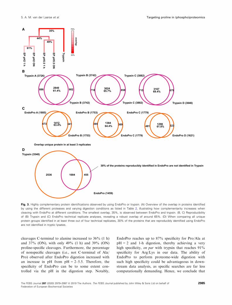
For the EndoPro phosphopeptides, the highly pre-
ferred phosphorylation on the penultimate C-terminal
amino acid is very evident, as is the disproportion for
phosphorylation on the penultimate amino acid at the
N terminus (Fig. 6A). Interestingly, EndoPro also seems
to disfavor positively charged amino acids on this posi-
tion (Fig. 1B), which implies no charge is tolerated at
this position in the substrate-binding pocket. Repulsion
Fig. 6. Amino acid length and localization of phosphorylation sites on the identified phosphopeptides. (A) Localization of the phosphorylation
on unique phosphopeptides from EndoPro, showing the highly preferred phosphorylation on the second to last amino acid on the peptide
(i.e., Ser-Pro or Thr-Pro), and the disfavor for phosphorylation on the penultimate N-terminal amino acid on the EndoPro peptides. (B)
Localization of phosphorylation on unique phosphopeptides following trypsin digestion at pH = 8.5, revealing a strong disfavor for
phosphorylation on the ultimate and penultimate N-terminal amino acids on the peptides, and preferential phosphorylation on the third
amino acid of the identified phosphopeptides.
2990 The FEBS Journal 287 (2020) 2979–2997 ª 2019 The Authors. The FEBS Journal published by John Wiley & Sons Ltd on behalf of
Federation of European Biochemical Societies
Targeting proline in (phospho)proteomics S. A. M. van der Laarse et al.
R/K
R/K
R/K
R/K
R/K
R/K
R/K
R/K
R/K
R/K
R/K
R/K
R/K
R/K
R/K
R/K
R/K
R/K
R/K
1 2 3 4 5 6 7 8 9 10 11 12 1413 15 16 1817 19 20 2221 23 24 25 26Amino acid position
Localization of phosphorylation on phosphopeptidesTrypsin
R/K
R/K
200400800 6001000Number of unique peptides
A/P
A/P
A/P
A/P
A/P
A/P
A/P
A/P
A/P
A/P
A/P
A/P
A/P
A/P
A/P
A/P
A/P
A/P
A/P
1 2 3 4 5 6 7 8 9 10 11 12 1413 15 16 1817 19 20 2221 23 24 25 26Amino acid position
EndoPro
A/P
A/P
200400800 6001000Number of unique peptides
Ratio observed/expected0.2 5< 0.2
R/K
R/K
R/K
R/K
R/K
R/K
R/K
R/K
R/K
R/K
R/K
R/K
R/K
R/K
R/K
R/K
R/K
R/K
R/K
1 2 3 4 5 6 7 8 9 10 11 12 1413 15 16 1817 19 20 2221 23 24 25 26
Localization of phosphorylation on phosphopeptidesTrypsin
R/K
R/K
200400800 6001000
A/P
A/P
A/P
A/P
A/P
A/P
A/P
A/P
A/P
A/P
A/P
A/P
A/P
A/P
A/P
A/P
A/P
A/P
A/P
1 2 3 4 5 6 7 8 9 10 11 12 1413 15 16 1817 19 20 2221 23 24 25 26
Localization of phosphorylation on phosphopeptides
A/P
A/P
200400800 6001000
0.2 5
0.2 5< 0.2
Ratio observed/expected0.2 5
A
B
2991The FEBS Journal 287 (2020) 2979–2997 ª 2019 The Authors. The FEBS Journal published by John Wiley & Sons Ltd on behalf of
Federation of European Biochemical Societies
S. A. M. van der Laarse et al. Targeting proline in (phospho)proteomics

of both charges would suggest steric hindrance to be the
source of this lack of activity. For trypsin-like proteases,
the N + 2 position is reported to be situated in a
hydrophobic pocket prior to cleavage [37]. Based on our
data, this might also be the case for EndoPro. For the
tryptic phosphopeptides, the dot-plot reveals a strong
disfavor for phosphorylation on the ultimate and penul-
timate N-terminal amino acids and the penultimate C-
terminal amino acid (Fig. 6B), confirming that phospho-
rylation near the Arg/Lys hinders cleavage by trypsin.
Trypsin displays a preferential phosphorylation on the
third N-terminal amino acid (likely representing the
well-known RXXS/T basophilic kinase motif). These
findings largely explain the increase in missed cleavages
on phosphopeptides observed [36].
In contrast to what is observed with trypsin, the
activity of EndoPro seemed unaffected by a phospho-
rylation directly preceding the cleavage site, resulting
in an overrepresentation of phosphorylation events on
the second to last amino acid of the phosphopeptides,
see Fig. 5A. In total, of all detected singly phosphory-
lated EndoPro phosphopeptides, 19% had their phos-
phorylation on the C-terminal SP/TP. See Fig. S4 for
the phosphosite localization of all specific EndoPro
digestion conditions employed.
Discussion
Although still not frequently used, the use of proline-
directed proteases in a mass spectrometry-based pro-
teomics setting has been explored previously [23–25].
Schr€ader et al. [25] recognized the potential of proline-
directed proteases in proteomics characterizing nepro-
sin, a protease originally from Nepenthes ventrata. In
their work, the ON digestion of a HeLa cell lysate at
pH = 2.5 yielded 61% proline-specific cleavages for
neprosin, which is comparable to our findings for
EndoPro (62% after ON digestion at pH = 2). Addi-
tionally, they nicely illustrated the potential of proline-
specific proteases for the mapping of PTMs on a his-
tone sample. Due to the high activity of EndoPro at
low pH, the protease has also found applications in
the food industry, where EndoPro was assessed for its
ability to degrade gluten and the debittering of protein
hydrolysates, as well as in structural studies based on
hydrogen–deuterium exchange mass spectrometry,
where a low pH is essential to reduce the rate of deu-
terium back exchange [24,38–40]. Thus, proline-di-
rected proteases are versatile proteases that can be
used orthogonally to the more conventional proteases
in various mass spectrometry-based studies.
Here, we have evaluated EndoPro for its use in bot-
tom-up (phospho)proteomics, with the aim to boost its
performance by optimizing different digestion condi-
tions, peptide fragmentation methods, and scoring algo-
rithms. We showed that the protease has a capacity to
generate peptides from proteins comparable to trypsin,
evidenced by similar numbers of MS/MS events. When
the proper digestion conditions are chosen, the cleavage
specificity for alanine and proline is very high. Interest-
ingly, EndoPro cleavage patterns appear influenced by
the pH during the digestion, with a lower overall speci-
ficity observed using EndoPro at pH = 5.5 than at
pH = 2. The mechanism underlying this pH-dependency
was not studied thoroughly here; however, we did find
that the overall Pro content of the proteins identified at
pH = 5.5 was significantly lower than at pH = 2. Hence,
it might be possible that fewer proline residues were
available for cleavage, possibly due to the occurrence of
a different pool of soluble proteins at pH = 5.5 when
compared to pH = 2. This hypothesis is supported by
the limited overlap (44%) in identified proteins between
the two EndoPro conditions. We did not observe an
effect on the length of the peptides identified, which also
implies proline residues were not missed during diges-
tion but likely not present as frequently.
Through this work, we show that the performance
of EndoPro as protease for proteomics applications is
already very good, but its full potential is still not
reached. As observed also with other proteases, Endo-
Pro also suffers from the tryptic bias that has been
created in the conventional proteomics pipelines, both
in the peptide separation, fragmentation, and scoring
segments of the proteomics experiment. Despite that,
our data show that EndoPro is a protease very suitable
for producing peptides for proteomics analysis. One
should keep in mind, however, that the obtainable
proteome coverage is rather distinct when the digestion
is performed at pH = 2 or at pH = 5.5. In addition,
the proteome coverage generated with EndoPro is
highly complementary to the coverage that can be
reached using trypsin. Finally, EndoPro provides one
of the most complementary proteases for phosphopro-
teomics, delivering a large subset of phosphosites not
easily covered by trypsin. Furthermore, in contrast to
trypsin, cleavage by EndoPro is not hampered by the
presence of a neighboring phosphorylation.
Identifying nontryptic MS/MS spectra
One of the main concerns when using less conven-
tional proteases in proteomics-type experiments is that
the resulting datasets almost always give a lower pep-
tide identification rate than the tryptic datasets. For
ArgC, AspN, chymotrypsin, GluC, LysC, and LysN,
average identification rates of 22%, 11%, 17%, 13%,
2992 The FEBS Journal 287 (2020) 2979–2997 ª 2019 The Authors. The FEBS Journal published by John Wiley & Sons Ltd on behalf of
Federation of European Biochemical Societies
Targeting proline in (phospho)proteomics S. A. M. van der Laarse et al.

23%, and 11% have been reported, compared to a
37% identification rate for trypsin [10]. Similarly,
Schr€ader et al. [25] previously reported identification
rates of 20%, 46%, and 52% for neprosin, LysC, and
trypsin, respectively. These findings are in agreement
with our finding, where EndoPro identification rates
are also about half of the trypsin identification rate.
The lower rates associated with nontryptic digestions
are probably not caused by a lack of good peptides, as
the number of MS2 scans for these runs is similar.
Hence, the number of peptides with suitable mass-to-
charge ratio is expected to be similar for all digests.
This leaves several other sources likely responsible
for the reduced identification rates. Firstly, the pep-
tides produced by each of these proteases may have
characteristics that make them less suitable for current
fragmentation-based sequencing methods by mass
spectrometry. They may, for instance, carry less posi-
tive charges, reducing the likelihood of observing
charged fragment ions that can be used for database
matching. For our dataset, however, this is not the
case as the peptides generated by EndoPro have even
more positive charges than the tryptic peptides. Sec-
ondly, the peptides’ chemical composition may lead to
fragmentations patterns or cleavages at positions that
are unexpected for trypsin. For instance, EndoPro
peptides do not carry an arginine or lysine residue at
their C terminus, which likely leads to a less extended
sequence informative y-ion series. Thirdly, database
search and peptide scoring algorithms have mainly
been optimized for tryptic peptides. Any fragmentation
behavior not observed in tryptic peptides, therefore, is
likely penalized by the conventional scoring algo-
rithms, resulting in lower scores. Notably, using stan-
dard conditions with other search engines such as
Mascot, Andromeda, and Sequest gave us even lower
identification rates than those reported here (by a fac-
tor 2, data not shown), evidently depending also on
the fragmentation method employed.
Due to EndoPro’s high preference to cleave C-termi-
nal to proline, many of the peptides generated with
this protease are expected to have a Pro residue at
their C terminus, making them very dissimilar to the
typical tryptic peptides that carry a Lys or Arg at their
C terminus. Indeed, we observed a clear C-terminal
proline effect in their fragmentation spectra. During
HCD fragmentation, more than 95% of the EndoPro
MS2 spectra contained a very prominent y1 ion at
116.07 m/z, corresponding to the preferential gas-phase
cleavage of the bond preceding the proline. Assuming
that the presence of a 116.07 m/z ion is diagnostic for
a peptide ending in C-terminal proline, we noticed in
our LC-MS runs many more MS2 spectra likely
originating from EndoPro peptides in which sequence
could not be assigned. This could possibly be
improved by optimizing the MS parameters such as
the collision energy, to maintain the diagnostic y1 ion
while also allowing sufficient fragmentation in other
parts of the peptide. In EThcD spectra, we observed
significantly less proline y1 ion formation, only about
50% of the recorded MS2 spectra, which allows for
the detection and assignment of other fragment ions
and hence a better scoring of the PSMs. This is also
reflected in the higher ID rate observed with the
EThcD/HCD DT method.
Taken together, many factors contribute to a lower
score for the EndoPro, illustrating a deeply rooted
tryptic bias in proteomic workflows, resulting in lower
PSMs for nontryptic peptides. This argues especially
for a better optimization of MS methods and search
algorithms toward nontryptic peptides.
Fragmenting with a C-terminal phosphorylation
and phosphosite localization
Given the large proportion of phosphopeptides that
carry their phosphorylation on the penultimate amino
acid of the EndoPro peptide, we hypothesize that these
phosphopeptides may have a negatively charged C ter-
minus. Again, this is in sharp contrast to tryptic phos-
phopeptides, which have a positively charged C
terminus and for the most part carry their phosphory-
lation somewhere in the middle of the peptide. Phos-
phorylation of the amino acid before the C-terminal
proline seemed to reduce the proline effect observed
for the EndoPro peptides, resulting in a better frag-
mentation ion coverage than observed for nonphos-
phorylated peptides. In addition, since the EndoPro
phosphopeptides predominantly carry their phosphory-
lation at the C terminus, this affects the probability of
having multiple potential phosphorylation sites directly
adjacent to each other. When a phosphorylation site is
directly preceding the Ala/Pro on the C terminus, there
cannot be a second potential phosphorylation site on
that end of the peptide; hence, the odds of having
many potential sites side by side on a phosphopeptide
is lower than when phosphorylations are located more
toward the middle of a peptide. This could potentially
boost phosphosite localization certainty, especially in
peptides that harbor multiple putative phosphate
acceptors, such as Ser, Thr, and Tyr. Since one of the
major remaining issues in phosphoproteomics is the
confident assignment of the exact site of phosphoryla-
tion, much computational effort has been invested in
improving fragmentation methods and algorithms to
boost confident site assignments. Knowledge about the
2993The FEBS Journal 287 (2020) 2979–2997 ª 2019 The Authors. The FEBS Journal published by John Wiley & Sons Ltd on behalf of
Federation of European Biochemical Societies
S. A. M. van der Laarse et al. Targeting proline in (phospho)proteomics

natural occurrence of phosphorylation sites for each
used protease, as depicted graphically in Fig. 6, can be
used to further improve scoring algorithms and boost
the confidence in site localization.
Conclusion
Here, we evaluated EndoPro and show it is a versatile
protease with a very high proline- and alanine-directed
specificity. Its activity can be influenced by adjusting the
pH of the digestion buffer, whereby it largely retains its
specificity but seemingly samples a different part of the
proteome. By benchmarking its performance against
trypsin, we observed that over 30% of all unique HeLa
proteins were solely identified by EndoPro, as well as
5705 phosphosites that were not observed in the tryptic
digests, illustrating EndoPro’s high complementarity to
trypsin. This complementarity allows EndoPro to
expand our coverage of the various proteomes and
sheds light on previously dark, invisible stretches of
(phospho)proteins. Since EndoPro clearly outperforms
trypsin on arginine- and lysine-rich proteins, we see
potential for EndoPro in studying proteins involved in
nucleotide and chromatin binding, which are often
enriched in these positively charged amino acids [41]. In
addition, the longer peptides generated by EndoPro and
its ability to cleave close to modifications make the
enzyme an interesting candidate for middle-down
approaches allowing for more combinatorial PTM
information [42,43]. Compared to other alternative pro-
teases, such as LysC, chymotrypsin, ArgC, EndoPro
performs better and is in our view one of the most com-
plementary alternatives to trypsin, due to its completely
different activity profile and specificity. It is rather
unique in effectively targeting proline residues in (phos-
pho)proteomics that are often causing complications for
the other proteases.
Materials and methods
In silico proteome coverage
Human proteins deposited in the Swissprot database
(20 417 reviewed proteins, downloaded July 25, 2019) were
digested in silico using the specificity requirements listed in
Table 1. Zero, one, or two missed cleavages were allowed
for each peptide, resulting in a database with all possible
peptides formed by each of the nine listed proteases. Subse-
quently, these peptides were filtered on precursor m/z
(375 ≤ m/z ≤ 1500); mass (m ≤ 10 000 Da) and only fully
specific peptides were taken into account. All peptides pass-
ing these filters were mapped to the proteome to find the
theoretical upper limit of proteome coverage possible.
Cell culture
HeLa cells were cultured in Dulbecco’s modified Eagle’s med-
ium (DMEM) supplemented with 10% fetal bovine serum
and 10 mM glutamine (Lonza, Basel, Switzerland) at 37°C/5%CO2. One hour prior to harvesting, the medium was refreshed
to stimulate phosphorylation. Cells were washed with ice-cold
PBS, and cell pellets were collected by mild centrifugation
(150 xg) for 3 min, and stored at�80°C until lysis.
Sample preparation
Cell pellets were lysed in a boiling lysis buffer containing 6 M
guanidinium HCl (GuHCl), 5 mM Tris (2-carboxyethyl)phos-
phine (TCEP), 10 mM chloroacetamide, 100 mM Tris/HCl pH
8.5, supplemented with protease inhibitor (cOmplete mini
EDTA-free, Roche, Woerden, The Netherlands). Pellets were
boiled for 10 min at 99 °C, sonicated for 30 rounds of 5 s
(Bioruptor Plus, Diagenode, Seraing, Belgium), and spun
down at 20 000 g for 15 min. Protein concentration was
determined using PierceTM BCA protein assay kit. Equal
amounts of protein per condition were diluted to a final con-
centration of 2 M GuHCl, and pH was adjusted to pH = 2
and 5.5 for EndoPro, or pH = 8.5 for trypsin, using formic
acid (FA) (Merck, Zwijndrecht, The Netherlands). Finally,
proteins were digested with EndoPro (1:100, DSM, Delft, The
Netherlands) or trypsin (1:100, Sigma-Aldrich, Zwijndrecht,
The Netherlands) for 1 h or overnight at 37 °C. The resultingpeptides were acidified to a final concentration of 1% FA,
cleaned up using Sep-Pak cartridges (Waters, Etten-Leur, The
Netherlands), and dried in vacuo.
Phosphopeptide enrichment
Phosphorylated peptides were enriched using Fe(III)-NTA
cartridges (Agilent Technologies) in an automated fashion
using the AssayMAP Bravo Platform (Agilent Technolo-
gies) [27]. The cartridges were primed with 0.1% TFA in
ACN and equilibrated with loading buffer (80% ACN/
0.1% TFA). Samples were suspended in loading buffer and
loaded onto the cartridge. The peptides bound to the car-
tridges were washed with loading buffer, and the phospho-
rylated peptides were eluted with 1% ammonia directly
into 10% formic acid. Samples were dried in vacuo and
stored at �80 °C until LC-MS/MS analysis.
LC-MS/MS analysis
Peptide samples were resuspended in 20 mM citric acid with 2%
FA and analyzed with an UHPLC 1290 system (Agilent Tech-
nologies) coupled to an Orbitrap Fusion mass spectrometer
(Thermo Fisher Scientific, Waltham, MA, USA). Peptides were
trapped (DrMaisch Reprosil C18, 3 µm, 2 cm 9 100 µm) and
then separated on an analytical column (Agilent Poroshell
EC-C18, 2.7 µm, 50 cm 9 75 µm). All columns were made
2994 The FEBS Journal 287 (2020) 2979–2997 ª 2019 The Authors. The FEBS Journal published by John Wiley & Sons Ltd on behalf of
Federation of European Biochemical Societies
Targeting proline in (phospho)proteomics S. A. M. van der Laarse et al.

in-house. Trapping was performed for 5 min in solvent A (0.1%
FA), followed by a gradient of the following: 0–8% solvent B
(0.1% FA in 80% ACN) in 10 s, 8–32% in 100 min, 32–100%in 5 min, hold for 5 min, 100–0% in 1 min, and hold for 4 min.
Flowwas passively split to 300 nL�min�1.
The mass spectrometer was operated in data-dependent
mode. Full scan MS spectra from m/z 375 to 1500 were
acquired at a resolution of 60 000 after accumulation to a
target value or 4e5 or a maximum injection time of 50 ms.
The most intense precursor ions were selected for fragmen-
tation for a duration of 3 s with a 24-s dynamic exclusion
duration. Target peaks were isolated in a 1.6 Da isolation
window and subjected to either higher-energy collision-in-
duced dissociation (HCD), electron transfer dissociation
(ETD), or electron transfer higher-energy collision-induced
dissociation (EThcD) [28]. MS/MS spectra were acquired
with a resolution of 30 000 using an AGC target of 1e5
ions with a maximum injection time of 125 ms. Charge
state screening was enabled, and precursors with an
unknown charge state or a charge state of 1+ were
excluded. For the decision tree strategy, HCD and EThcD
fragmentation were performed with normalized collision
energies of 35% and 40%, respectively. Fragmentation was
done based on charge state. HCD was selected for peptide
ions with charge states of 2+ and 3+; and for EThcD,
charge states 4+ to 20+ were selected.
Data analysis
The resulting mass spectra were searched using Byonic
(Protein Metrics Inc., Cupertino, CA, USA, v.3.3.11) in a
fully nonspecific or semispecific search (C-terminal cleavage
on Arg/Lys or Ala/Pro for trypsin or EndoPro, respec-
tively). The number of missed cleavages was not restricted.
Mass tolerance was set at 10 and 20 ppm for precursor and
fragment ions, respectively. Carbamidomethylation was set
as a fixed cysteine modification, oxidation of methionine,
deamidation of asparagine, and sodium adducts of aspar-
tate, glutamate, serine, and threonine were set as common
modifications. The formation of pyro-glutamine from N-
terminal glutamine or glutamate, loss of ammonia, and
acetylation was set as rare modifications. Overall, one com-
mon and one rare modification were allowed in the stan-
dard bottom-up workflow. For the phospho-enriched
peptides, phosphorylation on serine or threonine was
included as a common modification, and in total, 3 com-
mon and 1 rare modification were allowed.
Using Byonic Viewer (Protein Metrics Inc., v.3.3-421),
the PSMs were filtered by a PEP 2D < 0.001 resulting in a
0.1% PSM level FDR. In the phospho-enriched dataset, we
also asked that the delta mod. score (dmod) was larger
than 20, to only include the more confident phosphosite
localizations for each PSM. The resulting PSMs from dif-
ferent fragmentation methods were combined prior to fur-
ther data analysis.
Peptide and/or protein characteristics such as peptide
length, charge, amino acid content, and location of phos-
phosites on the peptide were determined using in-house R
scripts (available upon request), Venn diagrams were made
using both VENNY (BioinfoGP v.2.1.0) [29] and BIOVENN
[30], and the bar graphs, boxplots, and heatmaps were visu-
alized using GRAPHPAD PRISM 8.0.1. IceLogo was generated
as described in Colaert et al.[31].
In Fig. 5D, the phosphorylation motifs were assigned to
one of four categories: proline-directed phosphosites p(S/T)
P, acidophillic (D/E after the phosphosite), basophillic (R/
K before phosphosite), or other. Assignment was hierarchi-
cal, meaning that a phosphosite exhibiting both proline-di-
rected and basophilic characteristic was only included in
the proline-directed group.
Acknowledgements
We acknowledge support from the Netherlands Orga-
nization for Scientific Research (NWO) funding the
large-scale proteomics facility Proteins@Work (project
184.032.201) and X-omics (project 184.034.019)
embedded in the Netherlands Proteomics Centre.
A.J.R.H. acknowledges further support by the NWO
TOP-Punt Grant 718.015.003 and the EU Horizon
2020 program INFRAIA project Epic-XS (Project
823839). We thank Simone Lemeer and Liana Tsiat-
siani for the critical evaluation of the manuscript.
Conflict of interest
The authors declare the following competing financial
interest(s): M.A. and M.O. are DSM employees. DSM
sells An-PEP for food applications. MB is the founder
and employee of ProteinMetrics. ProteinMetrics devel-
ops and commercializes the Byonic software.
Author contributions
SAML, CAGHG, and AJRH designed experiments
and wrote the manuscript, SAML and CAGHG per-
formed the (phospho)proteomics experiments. SAML
wrote the scripts for data analysis; SAML and
CAGHG analyzed and interpreted the data supported
by MB; and MA and MO provided EndoPro as well
as critical comments to the manuscript. All authors
read and approved the manuscript.
References
1 Cohen P (2001) The role of protein phosphorylation in
human health and disease. Eur J Biochem 268, 5001–5010.
2995The FEBS Journal 287 (2020) 2979–2997 ª 2019 The Authors. The FEBS Journal published by John Wiley & Sons Ltd on behalf of
Federation of European Biochemical Societies
S. A. M. van der Laarse et al. Targeting proline in (phospho)proteomics

2 Hanash S (2003) Disease proteomics. Nature 422, 226–232.
3 Zhang Y, Fonslow BR, Shan B, Baek M-C & Yates JR
(2013) Protein analysis by shotgun/bottom-up
proteomics. Chem Rev 113, 2343–2394.4 Mann M, Kulak NA, Nagaraj N & Cox J (2013) The
coming age of complete, accurate, and ubiquitous
proteomes. Mol Cell 49, 583–590.5 Steen H & Mann M (2004) The abc’s (and xyz’s) of
peptide sequencing. Nat Rev Mol Cell Biol 5, 699–711.6 Vandermarliere E, Mueller M & Martens L (2013)
Getting intimate with trypsin, the leading protease in
proteomics. Mass Spectrom Rev 32, 453–465.7 Swaney DL, Wenger CD & Coon JJ (2010) Value of
using multiple proteases for large-scale mass
spectrometry-based proteomics. J Proteome Res 9,
1323–1329.8 Eichacker LA, Granvogl B, Mirus O, M€uller BC, Miess
C & Schleiff E (2004) Hiding behind hydrophobicity. J
Biol Chem 279, 50915–50922.9 Tsiatsiani L & Heck AJR (2015) Proteomics beyond
trypsin. FEBS J 282, 2612–2626.10 Giansanti P, Tsiatsiani L, Low TY & Heck AJR (2016)
Six alternative proteases for mass spectrometry–basedproteomics beyond trypsin. Nat Protoc 11, 993–1006.
11 Biringer RG (2006) Enhanced sequence coverage of
proteins in human cerebrospinal fluid using multiple
enzymatic digestion and linear ion trap LC-MS/MS.
Brief Funct Genomic Proteomic 5, 144–153.12 Gauci S, Helbig AO, Slijper M, Krijgsveld J, Heck AJR
& Mohammed S (2009) Lys-N and trypsin cover
complementary parts of the phosphoproteome in a
refined SCX-based approach. Anal Chem 81, 4493–4501.
13 Huesgen PF, Lange PF, Rogers LD, Solis N, Eckhard
U, Kleifeld O, Goulas T, Gomis-R€uth FX & Overall
CM (2015) LysargiNase mirrors trypsin for protein C-
terminal and methylation-site identification. Nat
Methods 12, 55–58.14 Wu Z, Huang J, Huang J, Li Q & Zhang X (2018) Lys-
C/Arg-C, a more specific and efficient digestion
approach for proteomics studies. Anal Chem 90, 9700–9707.
15 Raijmakers R, Neerincx P, Mohammed S & Heck AJR
(2010) Cleavage specificities of the brother and sister
proteases Lys-C and Lys-N. Chem Commun 46, 8827.
16 Burkhart JM, Schumbrutzki C, Wortelkamp S,
Sickmann A & Zahedi RP (2012) Systematic and
quantitative comparison of digest efficiency and
specificity reveals the impact of trypsin quality on MS-
based proteomics. J Proteomics 75, 1454–1462.17 Giansanti P, Aye TT, van den Toorn H, Peng M, van
Breukelen B & Heck AJR (2015) An augmented
multiple-protease-based human phosphopeptide Atlas.
Cell Rep 11, 1834–1843.
18 Schimmel PR & Flory PJ (1968) Conformational
energies and configurational statistics of copolypeptides
containing L-proline. J Mol Biol 34, 105–120.19 MacArthur MW & Thornton JM (1991) Influence of
proline residues on protein conformation. J Mol Biol
218, 397–412.20 Vaisar T & Urban J (1996) Probing the proline effect in
CID of protonated peptides. J Mass Spectrom 31,
1185–1187.21 Raulfs MDM, Breci L, Bernier M, Hamdy OM, Janiga
A, Wysocki V & Poutsma JC (2014) Investigations of
the mechanism of the “proline effect” in tandem mass
spectrometry experiments: the “pipecolic acid effect”. J
Am Soc Mass Spectrom 25, 1705–1715.22 Huo D, Qin T & Zu L (2019) Energetic switch of the
proline effect in collision-induced dissociation of singly
and doubly protonated peptide Ala-Ala-Arg-Pro-Ala-
Ala. J Mass Spectrom 54, 55–65.23 �Sebela M, �Rehulka P, K�abrt J, �Rehulkov�a H, O�zdian
T, Raus M, Franc V & Chmel�ık J (2009) Identification
of N-glycosylation in prolyl endoprotease from
Aspergillus niger and evaluation of the enzyme for its
possible application in proteomics. J Mass Spectrom 44,
1587–1595.24 Tsiatsiani L, Akeroyd M, Olsthoorn M & Heck
AJR (2017) Aspergillus niger prolyl endoprotease for
hydrogen-deuterium exchange mass spectrometry
and protein structural studies. Anal Chem 89, 7966–7973.
25 Schr€ader CU, Lee L, Rey M, Sarpe V, Man P, Sharma
S, Zabrouskov V, Larsen B & Schriemer DC (2017)
Neprosin, a selective prolyl endoprotease for bottom-up
proteomics and histone mapping. Mol Cell Proteomics
16, 1162–1171.26 Chen X, Wu D, Zhao Y, Wong BHC & Guo L (2011)
Increasing phosphoproteome coverage and
identification of phosphorylation motifs through
combination of different HPLC fractionation methods.
J Chromatogr B 879, 25–34.27 Post H, Penning R, Fitzpatrick MA, Garrigues LB, Wu
W, MacGillavry HD, Hoogenraad CC, Heck AJR &
Altelaar AFM (2017) Robust, sensitive, and automated
phosphopeptide enrichment optimized for low sample
amounts applied to primary hippocampal neurons. J
Proteome Res 16, 728–737.28 Frese CK, Zhou H, Taus T, Altelaar AFM, Mechtler
K, Heck AJR & Mohammed S (2013) Unambiguous
phosphosite localization using electron-transfer/higher-
energy collision dissociation (EThcD). J Proteome Res
12, 1520–1525.29 Oliveros JC (2007) VENNY. An interactive tool for
comparing lists with Venn Diagrams. https://bioinfogp.
cnb.csic.es/tools/venny/index.html.
30 Hulsen T, de Vlieg J & Alkema W (2008) BioVenn – a
web application for the comparison and visualization of
2996 The FEBS Journal 287 (2020) 2979–2997 ª 2019 The Authors. The FEBS Journal published by John Wiley & Sons Ltd on behalf of
Federation of European Biochemical Societies
Targeting proline in (phospho)proteomics S. A. M. van der Laarse et al.
biological lists using area-proportional Venn diagrams.
BMC Genom 9, 488.
31 Colaert N, Helsens K, Martens L, Vandekerckhove J &
Gevaert K (2009) Improved visualization of protein
consensus sequences by iceLogo. Nat Methods 6, 786–787.
32 Trevino SR, Scholtz JM & Pace CN (2007) Amino acid
contribution to protein solubility: Asp, Glu, and Ser
contribute more favorably than the other hydrophilic
amino acids in RNase Sa. J Mol Biol 366, 449–460.33 Tabb DL, Vega-Montoto L, Rudnick PA, Variyath
AM, Ham A-JL, Bunk DM, Kilpatrick LE, Billheimer
DD, Blackman RK, Cardasis HL et al. (2010)
Repeatability and reproducibility in proteomic
identifications by liquid chromatography-tandem mass
spectrometry. J Proteome Res 9, 761–776.34 Kozlowski LP (2017) Proteome- pI: proteome
isoelectric point database. Nucleic Acids Res 45, D1112–D1116.
35 Schlosser A, Pipkorn R, Bossemeyer D & Lehmann
WD (2001) Analysis of protein phosphorylation by a
combination of elastase digestion and neutral loss
tandem mass spectrometry. Anal Chem 73, 170–176.36 Molina H, Horn DM, Tang N, Mathivanan S &
Pandey A (2007) Global proteomic profiling of
phosphopeptides using electron transfer dissociation
tandem mass spectrometry. Proc Natl Acad Sci USA
104, 2199–2204.37 Huber R & Bode W (1978) Structural basis of the
activation and action of trypsin. Acc Chem Res 11,
114–122.38 Mitea C, Havenaar R, Drijfhout JW, Edens L, Dekking
L & Koning F (2008) Efficient degradation of gluten by
a prolyl endoprotease in a gastrointestinal model:
implications for coeliac disease. Gut 57, 25–32.39 K€onig J, Holster S, Bruins MJ & Brummer RJ (2017)
Randomized clinical trial: effective gluten degradation
by Aspergillus niger-derived enzyme in a complex meal
setting. Sci Rep 7, 13100.
40 Edens L, Dekker P, van der Hoeven R, Deen F, de
Roos A & Floris R (2005) Extracellular prolyl
endoprotease from Aspergillus niger and its use in the
debittering of protein hydrolysates. J Agric Food Chem
53, 7950–7957.41 Chandana T & Venkatesh YP (2016) Occurrence,
functions and biological significance of arginine-rich
proteins. Curr Protein Pept Sci 17, 507–516.42 Pandeswari PB & Sabareesh V (2019) Middle-down
approach: a choice to sequence and characterize
proteins/proteomes by mass spectrometry. RSC Adv 9,
313–344.43 Cristobal A, Marino F, Post H, van den Toorn HWP,
Mohammed S & Heck AJR (2017) Toward an
optimized workflow for middle-down proteomics. Anal
Chem 89, 3318–3325.
Supporting information
Additional supporting information may be found
online in the Supporting Information section at the end
of the article.Fig. S1. Amino acid content of PSMs following differ-
ent digestion conditions.
Fig. S2. Extended heatmap of proteome dataset.
Fig. S3. Extended heatmap of the phosphoproteomics
data and assessment of reproducibility.
Fig. S4. Phosphosite localization as extracted from the
unique phosphopeptides for the four different Endo-
Pro digestion conditions.
Table S1. Search input and outcome characteristics for
EndoPro and tryptic digests.
Table S2. Search input and outcome characteristics for
EndoPro and tryptic phosphopeptides.
2997The FEBS Journal 287 (2020) 2979–2997 ª 2019 The Authors. The FEBS Journal published by John Wiley & Sons Ltd on behalf of
Federation of European Biochemical Societies
S. A. M. van der Laarse et al. Targeting proline in (phospho)proteomics