Proteomics of Trypanosoma evansi Infection in Rodents
10
Proteomics of Trypanosoma evansi Infection in Rodents Nainita Roy 1. , Rishi Kumar Nageshan 1. , Rani Pallavi 1 , Harshini Chakravarthy 1 , Syama Chandran 1 , Rajender Kumar 2 , Ashok Kumar Gupta 2 , Raj Kumar Singh 2 , Suresh Chandra Yadav 2 , Utpal Tatu 1 * 1 Department of Biochemistry, Indian Institute of Science, Bangalore, India, 2 National Research Centre on Equines, Hisar, India Abstract Background: Trypanosoma evansi infections, commonly called ‘surra’, cause significant economic losses to livestock industry. While this infection is mainly restricted to large animals such as camels, donkeys and equines, recent reports indicate their ability to infect humans. There are no World Animal Health Organization (WAHO) prescribed diagnostic tests or vaccines available against this disease and the available drugs show significant toxicity. There is an urgent need to develop improved methods of diagnosis and control measures for this disease. Unlike its related human parasites T. brucei and T. cruzi whose genomes have been fully sequenced T. evansi genome sequence remains unavailable and very little efforts are being made to develop improved methods of prevention, diagnosis and treatment. With a view to identify potential diagnostic markers and drug targets we have studied the clinical proteome of T. evansi infection using mass spectrometry (MS). Methodology/Principal Findings: Using shot-gun proteomic approach involving nano-lc Quadrupole Time Of Flight (QTOF) mass spectrometry we have identified over 160 proteins expressed by T. evansi in mice infected with camel isolate. Homology driven searches for protein identification from MS/MS data led to most of the matches arising from related Trypanosoma species. Proteins identified belonged to various functional categories including metabolic enzymes; DNA metabolism; transcription; translation as well as cell-cell communication and signal transduction. TCA cycle enzymes were strikingly missing, possibly suggesting their low abundances. The clinical proteome revealed the presence of known and potential drug targets such as oligopeptidases, kinases, cysteine proteases and more. Conclusions/Significance: Previous proteomic studies on Trypanosomal infections, including human parasites T. brucei and T. cruzi, have been carried out from lab grown cultures. For T. evansi infection this is indeed the first ever proteomic study reported thus far. In addition to providing a glimpse into the biology of this neglected disease, our study is the first step towards identification of diagnostic biomarkers, novel drug targets as well as potential vaccine candidates to fight against T. evansi infections. Citation: Roy N, Nageshan RK, Pallavi R, Chakravarthy H, Chandran S, et al. (2010) Proteomics of Trypanosoma evansi Infection in Rodents. PLoS ONE 5(3): e9796. doi:10.1371/journal.pone.0009796 Editor: Hilal Lashuel, Swiss Federal Institute of Technology Lausanne, Switzerland Received August 21, 2009; Accepted February 18, 2010; Published March 22, 2010 Copyright: ß 2010 Roy et al. This is an open-access article distributed under the terms of the Creative Commons Attribution License, which permits unrestricted use, distribution, and reproduction in any medium, provided the original author and source are credited. Funding: UT would like to thank Department of Biotechnology, New Delhi, India for financial support. UT would also like to thank the funding support from Roche made available from University of Bern. NR, RK and RP would like to thank United Grants Commission/Council of Scientific and Industrial Research for their scholarships. The funders had no role in study design, data collection and analysis, decision to publish, or preparation of the manuscript. Competing Interests: The authors have declared that no competing interests exist. * E-mail: [email protected] . These authors contributed equally to this work. Introduction T. evansi is a widely-distributed, monomorphic protozoan which infects large animals such as equines, camels, dogs, cattle and deer. Epidemics of the disease called ‘surra’ which is caused by T. evansi infections results in the death of thousands of animals amongst which horses are the most susceptible [1]. T. evansi is geographically distributed in Asia, Africa and South America. With the number and severity of surra outbreaks escalating radically over the last few years, this protozoan parasite has a significant impact on the increasing mortality rates of livestock across the world. T. evansi is mechanically transmitted by blood-sucking insect species such as Tabanus, Stomoxys, Lypersoia and Haematopota. Keeping in view the large economic losses caused by this disease, an ad hoc group of Organisation Internationale des Epizootie (O.I.E.) on Non Tsetse Transmitted Animal Trypanosomes (NTTAT) was set up under the sponsorship of Food and Agriculture Organization (F.A.O.), Division of Production and Animal Health and International Livestock Research Institute (I.L.R.I.) in relationship with World Health Organization (W.H.O.) during the O.I.E. general session by representatives of above organizations and O.I.E. delegates of Asia, Africa and Europe. Compared to its closely related species T. brucei and T. cruzi, which infect humans, less attention is being given to T. evansi infections. While T. evansi infections are mainly restricted to animals, a recent report suggested its ability to jump species barrier and infect humans, in a minority of population having defect in ApoL1 [2]. In contrast to the closely related species T. brucei [3,4], T. evansi lacks the maxicircle kinetoplastid DNA. The loss of maxicircle kDNA in T. evansi has been shown to lock the trypanosome in the bloodstream stages in vertebrates where the parasite multiplies by longitudinal binary fission. The absence of intermittent development in any insect vector has enabled T. evansi to spread beyond the tse-tse fly belt of Africa to other areas such as Asia, central and South America [5]. PLoS ONE | www.plosone.org 1 March 2010 | Volume 5 | Issue 3 | e9796
Transcript of Proteomics of Trypanosoma evansi Infection in Rodents

Proteomics of Trypanosoma evansi Infection in RodentsNainita Roy1., Rishi Kumar Nageshan1., Rani Pallavi1, Harshini Chakravarthy1, Syama Chandran1,
Rajender Kumar2, Ashok Kumar Gupta2, Raj Kumar Singh2, Suresh Chandra Yadav2, Utpal Tatu1*
1 Department of Biochemistry, Indian Institute of Science, Bangalore, India, 2 National Research Centre on Equines, Hisar, India
Abstract
Background: Trypanosoma evansi infections, commonly called ‘surra’, cause significant economic losses to livestockindustry. While this infection is mainly restricted to large animals such as camels, donkeys and equines, recent reportsindicate their ability to infect humans. There are no World Animal Health Organization (WAHO) prescribed diagnostic testsor vaccines available against this disease and the available drugs show significant toxicity. There is an urgent need todevelop improved methods of diagnosis and control measures for this disease. Unlike its related human parasites T. bruceiand T. cruzi whose genomes have been fully sequenced T. evansi genome sequence remains unavailable and very littleefforts are being made to develop improved methods of prevention, diagnosis and treatment. With a view to identifypotential diagnostic markers and drug targets we have studied the clinical proteome of T. evansi infection using massspectrometry (MS).
Methodology/Principal Findings: Using shot-gun proteomic approach involving nano-lc Quadrupole Time Of Flight (QTOF)mass spectrometry we have identified over 160 proteins expressed by T. evansi in mice infected with camel isolate.Homology driven searches for protein identification from MS/MS data led to most of the matches arising from relatedTrypanosoma species. Proteins identified belonged to various functional categories including metabolic enzymes; DNAmetabolism; transcription; translation as well as cell-cell communication and signal transduction. TCA cycle enzymes werestrikingly missing, possibly suggesting their low abundances. The clinical proteome revealed the presence of known andpotential drug targets such as oligopeptidases, kinases, cysteine proteases and more.
Conclusions/Significance: Previous proteomic studies on Trypanosomal infections, including human parasites T. brucei andT. cruzi, have been carried out from lab grown cultures. For T. evansi infection this is indeed the first ever proteomic studyreported thus far. In addition to providing a glimpse into the biology of this neglected disease, our study is the first steptowards identification of diagnostic biomarkers, novel drug targets as well as potential vaccine candidates to fight against T.evansi infections.
Citation: Roy N, Nageshan RK, Pallavi R, Chakravarthy H, Chandran S, et al. (2010) Proteomics of Trypanosoma evansi Infection in Rodents. PLoS ONE 5(3): e9796.doi:10.1371/journal.pone.0009796
Editor: Hilal Lashuel, Swiss Federal Institute of Technology Lausanne, Switzerland
Received August 21, 2009; Accepted February 18, 2010; Published March 22, 2010
Copyright: � 2010 Roy et al. This is an open-access article distributed under the terms of the Creative Commons Attribution License, which permits unrestricteduse, distribution, and reproduction in any medium, provided the original author and source are credited.
Funding: UT would like to thank Department of Biotechnology, New Delhi, India for financial support. UT would also like to thank the funding support fromRoche made available from University of Bern. NR, RK and RP would like to thank United Grants Commission/Council of Scientific and Industrial Research for theirscholarships. The funders had no role in study design, data collection and analysis, decision to publish, or preparation of the manuscript.
Competing Interests: The authors have declared that no competing interests exist.
* E-mail: [email protected]
. These authors contributed equally to this work.
Introduction
T. evansi is a widely-distributed, monomorphic protozoan which
infects large animals such as equines, camels, dogs, cattle and deer.
Epidemics of the disease called ‘surra’ which is caused by T. evansi
infections results in the death of thousands of animals amongst
which horses are the most susceptible [1]. T. evansi is geographically
distributed in Asia, Africa and South America. With the number
and severity of surra outbreaks escalating radically over the last few
years, this protozoan parasite has a significant impact on the
increasing mortality rates of livestock across the world. T. evansi is
mechanically transmitted by blood-sucking insect species such as
Tabanus, Stomoxys, Lypersoia and Haematopota. Keeping in view the
large economic losses caused by this disease, an ad hoc group of
Organisation Internationale des Epizootie (O.I.E.) on Non Tsetse
Transmitted Animal Trypanosomes (NTTAT) was set up under the
sponsorship of Food and Agriculture Organization (F.A.O.),
Division of Production and Animal Health and International
Livestock Research Institute (I.L.R.I.) in relationship with World
Health Organization (W.H.O.) during the O.I.E. general session by
representatives of above organizations and O.I.E. delegates of Asia,
Africa and Europe. Compared to its closely related species T. brucei
and T. cruzi, which infect humans, less attention is being given to T.
evansi infections. While T. evansi infections are mainly restricted to
animals, a recent report suggested its ability to jump species barrier
and infect humans, in a minority of population having defect in
ApoL1 [2].
In contrast to the closely related species T. brucei [3,4], T. evansi
lacks the maxicircle kinetoplastid DNA. The loss of maxicircle kDNA
in T. evansi has been shown to lock the trypanosome in the bloodstream
stages in vertebrates where the parasite multiplies by longitudinal
binary fission. The absence of intermittent development in any insect
vector has enabled T. evansi to spread beyond the tse-tse fly belt of
Africa to other areas such as Asia, central and South America [5].
PLoS ONE | www.plosone.org 1 March 2010 | Volume 5 | Issue 3 | e9796

There is no vaccine for prevention of surra. The lists of drugs
that are commonly used for treatment include suramin, dimin-
azene aceturate, quinapyramine and cymelarsan [6,7]. While
effective, toxic side effects are exhibited by many of these drugs
and exposure to sub curative doses of trypanocides in field is
known to give rise to drug resistance. Development of drug
resistance in T. evansi was reported from India [8] and Sudan [9].
Therefore there is a need to find more effective and relatively
harmless treatment strategies. Despite the growing incidence of
this disease, there are relatively few prescribed diagnostic test for
timely detection of this infection. Practiced diagnostic techniques
include wet blood film examination, thick/thin blood smear
examination, Card Agglutination Test (CATT), ELISA, PCR
detection. The available serological tests do not clearly differen-
tiate between T. evansi, T. equiperdum and T. brucei infection due to
lot of structural and functional homology. Since these three species
are closely related, it is very important to find specific markers for
each species to be able to distinguish between them and provide
reliable diagnostic tests for timely treatment.
Here we report the first proteomic analysis of T. evansi obtained
from naturally infected camel which was passaged and purified
from mouse blood. Despite unavailability of T. evansi genome data
our MS based proteomic analysis allowed us to identify more than
100 gene products expressed by the parasite during infection in the
rodent model. In addition to providing a glimpse into the
biochemical pathways operational during infection, our study
has also revealed potential diagnostic markers and possible drug or
vaccine candidates against this neglected disease.
Methods
Culturing of T. evansi and purification of parasites fromhost
This study was conducted adhering to the institution’s
guidelines for animal husbandry at National Research Centre on
Equines at Hissar. The study was also approved by the
institutional review board and ethics committee at National
Research Centre on Equines. T. evansi camel isolate was
maintained in vivo using Swiss albino mice through serial passage
in the laboratory. At high parasitaemia, blood was collected from
tail and subsequently mixed with 0.5–0.8 ml Alsever’s solution and
inoculated intraperitoneally into healthy rodents. The inoculated
rodents were examined 24 hour onwards daily for the presence of
parasites and sub inoculated further in fresh rodents; accordingly
the strain was maintained.
Purification of host cell free T. evansi parasites from rodents/
equines are essentially needed for proteomic studies and antigen
characterization, which were achieved by the method [10] with
minor modifications. For bulk harvest of parasites, 5–10 gm of pre
swollen DEAE cellulose (Whatman) was suspended in 200 ml PBS
pH 8.0 and allowed to equilibrate for 1 hour at room tempera-
ture. The slurry was there after allowed to settle for 30 minutes
and supernatant containing the fine granules discarded till the
floating granules disappeared from supernatant. The equilibrated
suspensions of DEAE cellulose were stored at 4uC until further
use.
Cellulose beads were packed into the column of 22 cm length
and 3 cm diameter and equilibrated with Phosphate Saline
Glucose (PSG) buffer pH 8.0. Infected blood was collected in an
anticoagulant (heparin 10 IU/ml) and diluted 1:1 with PSG buffer
and charged through the sides of the column. Column elutes were
examined under microscope to spot the purified trypanosomes.
Chromatography was performed at room temperature and the
parasites were then pelleted after washing thrice with PBS, pH 7.2
and pellet was kept at 280uC for further use.
Protein extraction and in-gel trypsin digestion for LiquidChromatography (LC)/MS/MS analysis
Enriched fraction containing 108 to 109 Trypanosomal cells
were used for proteomic analysis. The proteins were extracted
from enriched parasites by boiling in SDS sample buffer and
resolved on an SDS PAGE gel. The proteins were visualized by
Coomassie Brilliant Blue (CBB) staining following which the entire
lane was excised into 26 pieces. Peptides were extracted by in-gel
tryptic digestion from each gel piece. Briefly, proteins from each
gel piece were first reduced using DTT and alkylated by
iodoacetamide. The modified proteins were then digested with
trypsin. The digested peptides were then extracted from the gel
using 5% formic acid, 60% Acetonitrile. These peptide mixtures
were dissolved in solvent (2% acetonitrile and 98% water
containing 0.5% formic acid) and further fractionated by Reverse
Phase chromatography on C-18 material on a nano- HPLC
system connected online to a nanospray ESI hybrid Q-TOF mass
spectrometer from Applied Biosystems. LC-MS-MS experiments
were carried out for each sample containing the complex mixture
of peptides. A 100 mm ID, 5 mm particle size, 100 A porosity
Michrom column was used for chromatographic separation. The
RP chromatography runtime was set for 144 minutes with a flow
rate of 300 nL/minute. The mobile phase was adjusted to an
increasing concentration of acetonitrile from 5% to 90%, to elute
peptides from the column on the basis of their hydrophobicity.
Data Acquisition using Analyst QS softwareAnalyst QS software was used to systematically acquire the
TOF MS and MS/MS data for each precursor ion entering the
instrument from the nanoLC. Eluted peptides were analyzed by
one full MS scan and four consecutive product ion scans of the
four most intense peaks, using rolling Collision Energy and
Dynamic Background Subtract function. An Information Depen-
dant Acquisition (IDA) experiment was used to specify the criteria
for selecting each parent ion for fragmentation which included
selection of ions in m/z range: .400 and ,1600, of charge state
of +2 to +5, exclusion of former target ions for 30 seconds,
accumulation time of 1 second for a full scan and 2 seconds for
MS/MS, ion source voltage of 2200 V and temperature 120uC.
MS data for each sample was collected using such user defined
criteria for 144 minutes to accommodate the Nano-LC gradient
program. The data generated by the Analyst software was stored
in a .wiff format.
Data interpretation using Protein Pilot 2.0 and GlobalProteome Machine (GPM)
The original data files were analyzed using the Protein Pilot 2.0
software from a combined database (Swiss Prot 2005, TrEMBL,
NCBI and PDB). Peptide scores above 50 and a protein score of
minimum 1.3 corresponding to a confidence level greater than
95% were used. GPM analysis tool with mammalian database
combined with protozoa was used for peak lists of MASCOT
Generic Format generated from the original data file. The error
tolerance was set at 100 p.p.m (ESI-QTOF). Spectra of each
peptide used for identification of the proteins were verified
manually. Presence of at least 5 consecutive y ions or b ions or a
combination of both was considered to be significant. The result
presented is a combination of 3 independent experiments from
mice. Data from each independent experiment was combined and
discussed.
Trypanosoma evansi Proteome
PLoS ONE | www.plosone.org 2 March 2010 | Volume 5 | Issue 3 | e9796

Results
Identification of T. evansi proteins using LC MS/MSTo examine the proteome of T. evansi, we isolated parasites from
an infected camel with high parasitaemia and passaged in mice.
The mice were then sacrificed and parasites were purified from the
blood using ion exchange chromatography as stated in methods.
Further, the purified parasites were lysed and protein extraction
was carried out as described under methods. One dimensional
SDS PAGE was used to resolve the parasite proteins based on
their molecular weights and the protein bands were visualized by
CBB staining. As can be seen from Figure 1 the most intense bands
reproducibly appeared in the molecular weight region between
66 kDa and 45 kDa. We carried out shot-gun proteomics to
identify expressed proteins. The entire lane from the SDS-PAGE
was excised into 26 contiguous gel slices (Figure 1) and each gel
slice was individually processed for in-gel trypsin digestion as
described under methods. The peptides obtained from each band
were eluted and subjected to nano-LC based RPLC, interfaced
with Hybrid Mass Spectrometry. The peak list which included
TOF-MS and MS/MS was analyzed by ProteinPilot2.0 software
for protein identification. This led to the identification of 166
proteins based on their homologs in Trypanosoma as well as other
related species whose genome sequences are available.
Functional categorization reveals an abundance ofglycolytic enzymes
Proteins identified as above were classified according to their
functions as shown in Table 1. Details of the protein identifications
including their score and number of peptides obtained on data
analysis have been listed in Table S1. Metabolic enzymes
constituted about 23% of total proteins identified as represented
in Figure 2. Among these, enzymes of the glycolytic pathway were
most conspicuous, suggesting their abundance and possibly
dependence of the parasite on this pathway as the main source
of energy. Proteins involved in nucleic acid metabolism, amino
acid metabolism, fatty acid metabolism and lipid biosynthesis were
also identified from the Trypanosomatids purified from the blood.
Figure 1. Summary of the protocol followed for MS analyses of samples. A. Purified parasites observed at 406magnification. B. Parasiteswere lysed as described in methods and proteins were fractionated on 10% SDS PAGE. C. Individual gel slices were processed as stated in methodsand total ion count, MS and MS/MS spectra for Fructose bisphosphate aldolase is shown as an example (from top to bottom respectively).doi:10.1371/journal.pone.0009796.g001
Trypanosoma evansi Proteome
PLoS ONE | www.plosone.org 3 March 2010 | Volume 5 | Issue 3 | e9796
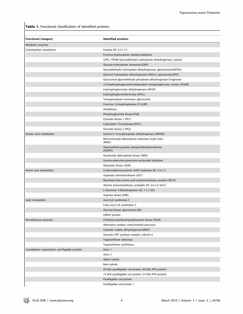
Table 1. Functional classification of identified proteins.
Functional Category Identified proteins
Metabolic enzymes
Carbohydrate metabolism Enolase (EC 4.2.1.11)
Fructose-bisphosphate aldolase.(aldolase)
G3PC_TRYBB Glyceraldehyde-3-phosphate dehydrogenase, cytosol.
Glucose-6-phosphate isomerase.(G6PI)
Glyceraldehyde-3-phosphate dehydrogenase, glycosomal.(GAPDH)
Glycerol-3-phosphate dehydrogenase [NAD+], glycosomal.(GPD)
Glycosomal glyceraldehyde phosphate dehydrogenase (Fragment)
2,3-bisphosphoglycerate-independent phosphoglycerate mutase (PGAM)
6-phosphogluconate dehydrogenase (6PGD)
6-phosphogluconolactonase (6PGL)
Triosephosphate isomerase, glycosomal.
Fructose-1,6-bisphosphatase (F1,6,BP)
Hexokinase.
Phosphoglycerate kinase.(PGK)
Pyruvate kinase 1 (PK1)
6-phospho-1-fructokinase.(PFK1)
Pyruvate kinase 2 (PK2)
Nucleic acid metabolism Inosine-59-monophosphate dehydrogenase (IMPDH)
Ribonucleoside-diphosphate reductase small chain(RNR2)
Hypoxanthine-guanine phosphoribosyltransferase(HGPRT)
Nucleoside diphosphate kinase (NDK)
Inosine-adenosine-guanosine-nucleoside hydrolase
Adenylate kinase (ADK)
Amino acid metabolism S-adenosylhomocysteine (SAH) hydrolase (EC 3.3.1.1)
Aspartate aminotransferase (AST)
Branched-chain amino acid aminotransferase, putative (BCAT)
Alanine aminotransferase, probable (EC 2.6.1.2) (ALT)
L-threonine 3-dehydrogenase (EC 1.1.1.103)
Arginine kinase (ARK)
Lipid metabolism Acyl-CoA synthetase 5
Fatty acyl CoA synthetase 3.
Glycerol kinase, glycosomal (GK)
GIM5A protein
Miscellaneous enzymes Pyridoxine/pyridoxal/pyridoxamine kinase (PdxK)
Alternative oxidase, mitochondrial precursor.
Cytosolic malate dehydrogenase.(MDH)
Vacuolar ATP synthase catalytic subunit A.
Trypanothione reductase.
Trypanothione synthetase.
Cytoskeleton organization and flagellar proteins Actin 1
Actin 2
Alpha tubulin
Beta tubulin
69 kDa paraflagellar rod protein (69 kDa PFR protein)
73 kDa paraflagellar rod protein (73 kDa PFR protein)
Paraflagellar rod protein
Paraflagellar rod protein 1
Trypanosoma evansi Proteome
PLoS ONE | www.plosone.org 4 March 2010 | Volume 5 | Issue 3 | e9796

Functional Category Identified proteins
Flagellar calcium-binding protein TB-1.7G (Fragment)
Par3
Valosin-containing protein homolog (VCP)
I/6 autoantigen
Axoneme central apparatus protein, possible
Microtubule-associated protein p320.(MAP320)
Cytoskeleton-associated protein CAP5.5
Calflagin Tb-44A.
Open reading frame A, partial cds. (Fragment)
Antigen GM6 (Fragment)
Protein synthesis 40S ribosomal protein S14
40S ribosomal protein S4
40S ribosomal protein S8
60S ribosomal protein L10a
60S ribosomal protein L23a (L25)
60S ribosomal protein L4 (L1)
ADP-ribosylation factor 1
Elongation factor 2 (EF2)
Ribosomal P0 subunit protein (RPP0)
Ribosomal protein L24
Ribosomal protein S12
EIF-4A
QM-like protein
Acidic ribosomal protein P0
Eukaryotic peptide chain release factor subunit 1(eRFI)
Ribosomal protein L3
Elongation factor 1-alpha (EF1-alpha)
Elongation factor 1 gamma (EF1-gamma)
Cellular communication/signal transduction 14-3-3 protein I
14-3-3 protein II
Cyclic nucleotide phosphodiesterase (PDE)
Cyclic nucleotide-specific phosphodiesterase PDE2A
Guanine nucleotide-binding protein beta subunit-like protein (GNB2L1)
Glycosylphosphatidylinositol-specific phospholipase C(GPI-PLC)
Phospholipase A1, possible (PLA1)
GTP binding protein, putative (GTR1)
Adenylyl cyclase (AC)
Lysophospholipase
Regulatory subunit of protein kinase A (PKAr)
CAMP specific phosphodiesterase
Nucleic acid associated proteins Histone D = CORE histone H4 homolog (Fragments)
Histone H2B (O96761)
Histone H3, probable
Histone H4, putative
Mitochondrial RNA-binding protein RBP38
Tcc2i18.9.
RNA binding protein La-like protein.
TbRRM1
RNA binding protein (Rpn)
Argonaute-like protein 1
Table 1. Cont.
Trypanosoma evansi Proteome
PLoS ONE | www.plosone.org 5 March 2010 | Volume 5 | Issue 3 | e9796

Functional Category Identified proteins
Poly(A) binding protein I (PABP1)
Cell rescue Defence (Virulence) 75 kDa invariant surface glycoprotein precursor
Variable surface glycoprotein (VSG) (trm|Q26840)
Variant surface glycoprotein (trm|Q968M4)
Variable surface glycoprotein (Q968M5)
Variable surface glycoprotein (Q6QA67)
Variant surface glycoprotein precursor (trm|Q26841)
Non-variant surface glycoprotein (Fragment)
Tryparedoxin peroxidase
H25N7.12 protein
IgE-dependent histamine-releasing factor, putative (HRF)
Chaperones and cochaperones Chaperonin HSP60, mitochondrial precursor
Cyclophilin A (CypA)
DnaJ protein, putative
Heat shock 70 kDa protein 4 (HSP70) (spt|P11145)
Heat shock protein 83
Mitochondrial HSP70
BiP/GRP78 precursor
P69 antigen
TcSTI1
Protein fate Proteasome subunit alpha type 1
Lysosomal/endosomal membrane protein p67.
Proteasome regulatory non-ATP-ase subunit 5
Ubiquitin
Ubiquitin-conjugating enzyme E2
Proteasome subunit alpha type 5(PSMA5)
Proteasome subunit beta type 3 (PSMB3)
20S proteasome alpha 7 subunit(PSMA7)
Proteins involved in trafficking Dynamin-related protein (DRP1)
Gpi8 transamidase precursor
ESAG5
Putative coatomer beta subunit (COBP)
Transferrin-binding protein (TBP)
Soluble N-ethylmaleimide sensitive factor (NSF) attachment protein possible
Adaptor gamma-1 chain (AP1G1)
Clathrin heavy chain (CHC)
Rab1
Hypothetical proteins Hypothetical protein (trm|Q7YVL1)
Hypothetical protein (trm|Q7YSV0)
Hypothetical protein (Q7YVJ5)
Hypothetical protein Tb10.70.1130 (XP_822825.1)
Hypothetical protein Tb09.211.3955(XP_827537.1)
Hypothetical protein, conserved (XP_844790.1)
Proteases/Peptidases Cysteine protease
Cysteine proteinase precursor
Oligopeptidase A
Oligopeptidase B
Calpain-like protein, probable (CAP)
Metacaspase
Transport Proteins Rhodesiense ADP/ATP carrier
Table 1. Cont.
Trypanosoma evansi Proteome
PLoS ONE | www.plosone.org 6 March 2010 | Volume 5 | Issue 3 | e9796

Interestingly, enzymes involved in TCA cycle could not be
detected. This could be due to their lower abundance in the
proteome. Cytoskeletal proteins which are generally known to be
abundantly present were readily detected. In addition flagellar
proteins were also abundantly expressed and identified. Proteins
involved in translation as well as main components of Hsp90
chaperone machinery, including Hsp70, DnaJ and cyclophilins
were identified. Various isoforms of histone proteins along with
other nucleic acid binding proteins were also detected. Proteins
involved in cell defence including variable surface glycoproteins
and peroxidases were identified. Proteasomal proteins and
ubiquitin proteins involved in deciding protein fates were detected.
Relatively few organellar proteins or secretory proteins were
detected suggesting that the parasite may not be very active in
protein traffic related functions during growth in the bloodstream.
Proteases, peptidases protein, kinases and phosphatases previously
implicated as potential drug targets were also detected. The list of
proteins identified also included 6 hypothetical proteins which
were homologous to proteins from T. brucei.
Furthermore, based on homology of the identified proteins to
other Trypanosomal species and their known localizations in those
species, we have categorized the proteins according to their
presence in different cellular compartments as shown in Figure 3.
We have also attempted to classify the identified proteins on the
basis of their solubility. Table S2 provides a list of probable
membrane bound and soluble proteins from a total of 166
identified proteins. The membrane proteins possibly constitute
those which are associated with organellar membranes or the
plasma membrane. Additionally, we have also detected post
translational modifications in some proteins amongst which
acetylations and phosphorylations were the most common
modifications observed. Table S3 provides a list of the identified
proteins having PTMs along with their MS/MS spectra which is
shown in Figure S1. It is interesting to note that the identified
proteins constitute those which are involved in host pathogen
interactions including potential surface antigens and virulence
factors from parasites. Indeed many of the proteins identified were
known drug targets or vaccine candidates in related trypanosomal
species.
Discussion
Over the past few years there is an increasing emphasis on
examination of parasite pathways prevalent during disease
manifestation within the host. There is growing awareness that
some of these pathways could be significantly present only during
host pathogen interactions often missing in lab grown cultures.
Proteins unique to clinical samples could thus prove to be keystone
drug targets.
In recent times a neglected animal disease called surra caused
by T. evansi has lead to significant economic losses to livestock
industry. While traditionally T. evansi infections have been
observed in domestic and wild animals, recent reports suggest
their ability to infect humans [2]. Currently available treatments
have many toxic side effects and there is an urgent need to identify
better treatment strategies. T. evansi is the least studied parasite
among all the Trypanosomatids. Proteome studies of T. cruzi, T.
brucei and Leishmania major have been carried out in the recent years
revealing several interesting features of the parasite lifecycle
[11–17]. In T. cruzi a total of 396 proteins were identified by LC-
MS/MS from epimastigotes [11]. All these studies had been
carried out using parasites cultured in-vitro. In this study, we report
the proteomic analysis of T. evansi from camel which was passaged
in mice.
Using mass spectrometry based proteomics approaches we have
developed a snapshot of the proteome of trypanosomatid stage in
the blood stream during infection in camel. Based on homology
driven searches, we have identified 166 proteins of T. evansi. The
Functional Category Identified proteins
P-type H+-ATPase
Aquaglyceroporin 3 (AQP3)
Calcium motive P-type ATPase TBCA1 (Fragment)
Probable biopterin transporter (Esag10)
Pentamidine resistance protein (PRP)
Kinases and phosphatases Acidic phosphatase (AP)
Casein kinase 1 homolog 1(CK1)
C-terminal kinesin KIFC1
Protein kinase CK2 alpha
Protein phosphatase 2A catalytic subunit (PP2A)
Unknown functions Bloodstream-specific protein 2 precursor
Bloodstream-specific protein 1.3-4.
Glycosomal membrane protein
Laminin receptor-like protein/p40 ribosome associated-like protein
RHS6a
Transmembrane glycoprotein
Retrotransposon hot spot protein, RHS4l
RHS4a (Retrotransposon hot spot protein RHS4-a
Retrotransposon hot spot protein RHS4-b
doi:10.1371/journal.pone.0009796.t001
Table 1. Cont.
Trypanosoma evansi Proteome
PLoS ONE | www.plosone.org 7 March 2010 | Volume 5 | Issue 3 | e9796

most abundant proteins identified were the metabolic enzymes in
which enzymes involved in the glycolytic pathway constituted a
major class. Out of the 40 metabolic enzymes identified, 17
enzymes were of the glycolytic pathway. Glycolysis has been
shown to be the main source of ATP generation in bloodstream
stages in T. brucei as well [18]. Infact glycolytic enzymes of
Trypanosoma have been proposed as attractive drug targets. Indeed
characteristic flagellar motion of T. evansi is known to depend on
glucose concentration in the medium. Enzymes of the TCA cycle
were conspicuously absent in our study. Trypanosomes have been
shown to rely on mitochondrial metabolism only in the insect
vector since the absence of kDNA has been shown to hamper
mitochondrial development. T. evansi which lacks maxi-circle
kDNA relies on glycolytic enzymes as their main energy source.
Among the other proteins identified, there were many potential
diagnostic markers, drug targets as well as possible vaccine
candidates. These included cytoskeletal proteins such as two actin
proteins [19], two paraflagellar rod proteins [20] and calflagin
[21], Metabolic enzymes including trypanothione reductase [22],
tryparedoxin peroxidase [23], trypanothione synthetase [24],
kinases such as casein kinase 1 [25], chaperone proteins such as
heat shock like 85 kDa protein [26], signal transduction proteins
like glycosylphosphatidylinositol-specific phospholipase C [27],
proteases and peptidases such as cysteine protease [28], oligopep-
tidase B [29], trafficking proteins such as GPI 8 transamidase [30],
transport proteins such as aquaglyceroporin 3 [31] and calcium
motive P-type ATPase [32]. Having obtained partial sequences of
these proteins future studies towards cloning their genes and
molecular characterisations may be possible.
While actin is a ubiquitously expressed conserved cytoskeletal
protein, a recent study showed that recombinant T.evansi actin
expressed in Esherichia coli could be a potential vaccine candidate.
Immunization of animals with actin provided immunity against 3
species of Trypanosomal infections, namely T.evansi, T.equiperdum
and T.brucei. This cross reactivity is due to the extensive homology
shared by the actin of these three species [19]. Recently around 90
different GPI (glycosylphosphatidylinositol)-species were identified
from lab grown cultures of T. cruzi by an LC-MS based approach
[33]. We have identified four different VSG’s. Furthermore,
proteins involved in the shedding of the GPI anchored VSG such
Figure 2. Functional classifications of identified proteins. A. Pie chart showing different functional classes of proteins which includesmetabolic enzymes, cytoskeletal proteins, proteins involved in synthesis, signal transduction proteins, nucleic acid associated proteins, proteininvolved in virulence, chaperones and co-chaperones, proteins involved in deciding protein fate, proteins involved in trafficking, hypoptheticalproteins, proteases and peptidases, transport proteins, kinases and phosphatases and proteins with unknown functions. B. The numbers of proteinspresent in each functional category have been indicated.doi:10.1371/journal.pone.0009796.g002
Trypanosoma evansi Proteome
PLoS ONE | www.plosone.org 8 March 2010 | Volume 5 | Issue 3 | e9796

as ESAG5 and GPI specific phosholipase C were also identified.
There are reports which suggest sequence similarity of GPI specific
phospholipase C and bacterial phospholipase C. It would be
interesting to also look at the GPI-moieties of these VSG’s since
they are known to be potent immunogens [34]. T. evansi are known
to exhibit a typical lashing action of the powerful locomotory
flagella which results in mechanical injury to erythrocytes and
other cells in the blood leading to anaemia. We have identified
Calflagin a known calcium binding protein localized in the flagella
of the parasite. Calflagin has been shown to act as a calcium sensor
and modulate the flagellar mobility [35]. Also the C terminal
fragment of the calflagin was recommended as a probable
candidate for serological detection of T. cruzi in humans [21].
Furthermore paraflagellar rod proteins which were detected in our
study are currently being investigated as potential vaccine
candidates in T. cruzi [36,37]. It has been reported that these
proteins were purified from T. cruzi epimastigotes and used to
immunize mice against trypanosomal infections [20].
Among the potential drug targets, tryparedoxin peroxidase is a
glutathione peroxiredoxin, unique to trypanosomes. This enzyme
detoxifies peroxynitrite radicals produced by macrophages and
hence plays an important role in successful evasion of host defense
system. Our study also identified protein kinases expressed by T.
evansi during infection. The kinome of T. cruzi and T. brucei have
been studied and novel chemotherapeutic agents against these
kinases have been suggested [25]. Additionally, we have identified
oligopeptidase B in our study. This protein is known to play a
major role during parasite invasion into host cell in T. cruzi [38]. In
T. evansi infections Oligopeptidase B has a major role to play in
manifestation of disease. It is shown to proteolyticaly cleave many
of the host derived peptides and proteins like kinogen, atrial
natriuretic factor etc. Inability to inhibit this cysteine peptidase by
host derived protease inhibitors makes Oligopeptidase B important
during pathogenesis, thus making it an attractive drug target [39].
Overall our study highlights the use of contemporary proteomic
approaches to study clinical proteome of T. evansi. Observations
made in this study are potentially extrapolatable to T. cruzi, T.
brucei infections in humans for which clinical proteomes have not
been examined thus far. In addition to providing a glimpse into
the cell biology, pathogenesis and metabolic state of the parasite,
Figure 3. Representation of proteins according to their cellular localization in T. evansi. All the proteins identified have been categorizedbased on their homology to related Trypanosomal species and their known localizations in those species.doi:10.1371/journal.pone.0009796.g003
Trypanosoma evansi Proteome
PLoS ONE | www.plosone.org 9 March 2010 | Volume 5 | Issue 3 | e9796

this study will aid in the development of diagnostic markers, drug
targets and vaccine candidates.
Supporting Information
Table S1 Details of the protein identifications including their
score and number of peptides obtained on data analysis.
Found at: doi:10.1371/journal.pone.0009796.s001 (0.16 MB
XLS)
Table S2 List of proteins classified on the basis of their solubility.
Found at: doi:10.1371/journal.pone.0009796.s002 (0.11 MB
DOC)
Table S3 List of proteins and their respective peptides along
with their post translational modifications.
Found at: doi:10.1371/journal.pone.0009796.s003 (0.04 MB
DOC)
Figure S1 MS/MS spectra of identified proteins having post
translational modifications.
Found at: doi:10.1371/journal.pone.0009796.s004 (1.58 MB
DOC)
Acknowledgments
Authors are thankful to Ms. Pragyan Acharya for critically reading the
manuscript.
Author Contributions
Conceived and designed the experiments: UT. Performed the experiments:
NR RKN RP HC SC RK AKG RKS. Analyzed the data: NR RKN RP
HC SC UT. Contributed reagents/materials/analysis tools: SCY UT.
Wrote the paper: UT.
References
1. Silva RA, Arosemena NA, Herrera HM, Sahib CA, Ferreira MS (1995)
Outbreak of trypanosomosis due to Trypanosoma evansi in horses of PantanalMato-grossense, Brazil. Vet Parasitol 60(1–2): 167–171.
2. Joshi PP, Shegokar VR, Powar RM, Herder S, Katti R, et al. (2005) Humantrypanosomiasis caused by Trypanosoma evansi in India: the first case report.
Am J Trop Med Hyg 73(3): 491–495.
3. Hoare CA (1972) The Trypanosomes of Mammals. A Zoological Monograph,
Blackwell Scientific Publications, Oxford. pp 288–323.
4. Lai DH, Hashimi H, Lun ZR, Ayala FJ, Lukes J (2008) Adaptations of
Trypanosoma brucei to gradual loss of kinetoplast DNA: Trypanosomaequiperdum and Trypanosoma evansi are petite mutants of T. brucei. Proc
Natl Acad Sci U S A 105: 1999–2004.
5. Luckins AG (1988) Trypanosoma evansi in Asia. Parasitol Today 4(5): 137–142.
6. Tuntasuvan D, Jarabrum W, Viseshakul N, Mohkaew K, Borisutsuwan S, et al.(2003) Chemotherapy of surra in horses and mules with diminazene aceturate.
Vet Parasitol 110(3–4): 227–233.
7. Kaminsky R, Brun R (1998) In vitro and in vivo activities of trybizine
hydrochloride against various pathogenic trypanosome species. Antimicrob
Agents Chemother 42(11): 2858–2862.
8. Gill BS (1971) Drug-resistance in Trypanosoma evansi. Trop Anim Health Prod
3: 195–198.
9. Lumsden WHR, Herbert WH, Mc Nielage GJC (1973) Techniques with
trypanosomes. ;Churchill Livingston Publishers, Edinburg and London, 57–94.
10. Lanham SM, Godfrey DG (1970) Isolation of salivarian trypanosomes from man
and other mammals using DEAE-cellulose. Exp Parasitol 28(3): 521–534.
11. Ferella M, Nilsson D, Darban H, Rodrigues C, Bontempi EJ, et al. (2008)
Proteomics in Trypanosoma cruzi–localization of novel proteins to variousorganelles. Proteomics 8(13): 2735–2749.
12. Drummelsmith J, Brochu V, Girard I, Messier N, Ouellette M (2003) Proteomemapping of the protozoan parasite Leishmania and application to the study of
drug targets and resistance mechanisms. Mol Cell Proteomics 2(3): 146–155.
13. Atwood JA, 3rd, Weatherly DB, Minning TA, Bundy B, Cavola C, et al. (2005)
The Trypanosoma cruzi proteome. Science 309(5733): 473–476.
14. Colosante C, Ellis M, Ruppert T, Voncken F (2006) Comparative proteomics of
glycosomes from bloodstream form and procyclic culture form Trypanosoma brucei
brucei. Proteomics 6: 3275–3293.
15. Holzmuller P, Grebaut P, Peltier JB, Brizard JP, Perrone T, et al. (2008)Secretome of animal trypanosomes. Ann N Y Acad Sci 1149: 337–342.
16. Panigrahi AK, Ogata Y, Zikova A, Anupama A, Dalley RA, et al. (2009) A
comprehensive analysis of Trypanosoma brucei mitochondrial proteome.Proteomics 9: 434–450.
17. Ayub MJ, Atwood J, Nuccio A, Tarleton R, Levin MJ (2009) Proteomic analysisof the Trypanosoma cruzi ribosomal proteins. Biochem Biophys Res Commun
382: 30–34.
18. Clayton CE, Michels P (1996) Metabolic compartmentation in African
trypanosomes. Parasitol Today 12: 465–471.
19. Li SQ, Yang WB, Ma LJ, Xi SM, Chen QL, et al. (2009) Immunization with
recombinant actin from Trypanosoma evansi induces protective immunityagainst T. evansi, T. equiperdum and T. b. brucei infection. Parasitol Res
104(2): 429–435.
20. Wrightsman RA, Miller MJ, Saborio JL, Manning JE (1995) Pure paraflagellar
rod protein protects mice against Trypanosoma cruzi infection. Infect Immun
63(1): 122–125.
21. Marcipar IS, Roodveldt C, Corradi G, Cabeza ML, Brito ME, et al. (2005) Use
of full-length recombinant calflagin and its c fragment for improvement ofdiagnosis of Trypanosoma cruzi infection. J Clin Microbiol 43(11): 5498–5503.
22. Krauth-Siegel RL, Inhoff O (2003) Parasite-specific trypanothione reductase as adrug target molecule. Parasitol Res 90 Suppl 2: S77–85.
23. Nogueira FB, Ruiz JC, Robello C, Romanha AJ, Murta SM (2009) Molecularcharacterization of cytosolic and mitochondrial tryparedoxin peroxidase in
Trypanosoma cruzi populations susceptible and resistant to benznidazole.Parasitol Res 104(4): 835–844.
24. Ariyanayagam MR, Oza SL, Guther ML, Fairlamb AH (2005) Phenotypic
analysis of trypanothione synthetase knockdown in the African trypanosome.Biochem J 391(Pt 2): 425–432.
25. Naula C, Parsons M, Mottram JC (2005) Protein kinases as drug targets intrypanosomes and Leishmania. Biochim Biophys Acta 1754(1–2): 151–159.
26. Graefe SE, Wiesgigl M, Gaworski I, Macdonald A, Clos J (2002) Inhibition of
HSP90 in Trypanosoma cruzi induces a stress response but no stagedifferentiation. Eukaryot Cell 1(6): 936–943.
27. Gruszynski AE, DeMaster A, Hooper NM, Bangs JD (2003) Surface coatremodeling during differentiation of Trypanosoma brucei. J Biol Chem 278(27):
24665–24672.28. Scory S, Stierhof YD, Caffrey CR, Steverding D (2007) The cysteine proteinase
inhibitor Z-Phe-Ala-CHN2 alters cell morphology and cell division activity of
Trypanosoma brucei bloodstream forms in vivo. Kinetoplastid Biol Dis 6: 2.29. Tsuji A, Yoshimoto T, Yuasa K, Matsuda Y (2006) Protamine: a unique and
potent inhibitor of oligopeptidase B. J Pept Sci 12: 65–71.30. Hong Y, Nagamune K, Ohishi K, Morita YS, Ashida H, et al. (2006) TbGPI16
is an essential component of GPI transamidase in Trypanosoma brucei. FEBS
Lett 580(2): 603–606.31. Uzcategui NL, Carmona-Gutierrez D, Denninger V, Schoenfeld C, Lang F, et
al. (2007) Antiproliferative effect of dihydroxyacetone on Trypanosoma bruceibloodstream forms: cell cycle progression, subcellular alterations, and cell death.
Antimicrob Agents Chemother 51(11): 3960–3968.32. Luo S, Rohloff P, Cox J, Uyemura SA, Docampo R (2004) Trypanosoma brucei
plasma membrane-type Ca(2+)-ATPase 1 (TbPMC1) and 2 (TbPMC2) genes
encode functional Ca(2+)-ATPases localized to the acidocalcisomes and plasmamembrane, and essential for Ca(2+) homeostasis and growth. J Biol Chem
279(14): 14427–14439.33. Nakayasu ES, Yashunsky DV, Nohara LL, Torrecilhas AC, Nikolaev AV, et al.
(2009) GPIomics: global analysis of glycosylphosphatidylinositol-anchored
molecules of Trypanosoma cruzi. Mol Syst Biol 5: 261.34. Subramanya S, Hardin CF, Steverding D, Mensa-Wilmot K (2009) Glycosyl-
phosphatidylinositol-specific phospholipase C regulates transferrin endocytosis inthe African trypanosome. Biochem J 417(3): 685–694.
35. Pinto AP, Campana PT, Beltramini LM, Silber AM, Araujo AP (2003)
Structural characterization of a recombinant flagellar calcium-binding proteinfrom Trypanosoma cruzi. Biochim Biophys Acta 1652(2): 107–114.
36. Wrightsman RA, Manning JE (2000) Paraflagellar rod proteins administeredwith alum and IL-12 or recombinant adenovirus expressing IL-12 generates
antigen-specific responses and protective immunity in mice against Trypanoso-ma cruzi. Vaccine 18: 1419–1427.
37. Michailowsky V, Luhrs K, Rocha MO, Fouts D, Gazzinelli RT, et al. (2003)
Humoral and cellular immune responses to Trypanosoma cruzi-derivedparaflagellar rod proteins in patients with Chagas’ disease. Infect Immun 71:
3165–3171.38. Coetzer TH, Goldring JP, Huson LE (2008) Oligopeptidase B: a processing
peptidase involved in pathogenesis. Biochimie 90: 336–344.
39. Morty RE, Pelle R, Vadasz I, Uzcanga GL, Seeger W, et al. (2005)Oligopeptidase B from Trypanosoma evansi. A parasite peptidase that
inactivates atrial natriuretic factor in the bloodstream of infected hosts. J BiolChem 280: 10925–10937.
Trypanosoma evansi Proteome
PLoS ONE | www.plosone.org 10 March 2010 | Volume 5 | Issue 3 | e9796