Thymosin beta 4 mRNA and peptide expression in phagocytic cells of different mouse tissues

Peptides xxx (2009) xxx–xxx
G Model
PEP-67782; No of Pages 11
Thymosin beta 4 mRNA and peptide expression in phagocytic cells of differentmouse tissues
Melissa Paulussen a, Bart Landuyt b, Liliane Schoofs b, Walter Luyten c, Lut Arckens a,*a Laboratory of Neuroplasticity and Neuroproteomics, Katholieke Universiteit Leuven, Naamsestraat 59, B-3000 Leuven, Belgiumb Laboratory of Functional Genomics and Proteomics, Katholieke Universiteit Leuven, Naamsestraat 59, 3000 Leuven, Belgiumc Basic Research in Gynaecology, Department of Woman and Child, Biomedical Sciences Group, Katholieke Universiteit Leuven, Herestraat 49, 3000 Leuven, Belgium
A R T I C L E I N F O
Article history:
Received 7 April 2009
Received in revised form 15 July 2009
Accepted 15 July 2009
Available online xxx
Keywords:
Thymosin beta 4
Macrophage
Microglia
In situ hybridization
Immunocytochemistry
A B S T R A C T
Thymosin beta 4 (Tb4) is a peptide of 43 amino acids, mainly recognized as a regulator of actin
polymerization by sequestering G-actin. Meanwhile, the peptide has been implicated in lymphocyte
maturation, carcinogenesis, apoptosis, angiogenesis, blood coagulation and wound healing. The peptide
is also involved in lesion-induced neuroplasticity through microglia upregulation and it participates in
the growth of neuronal processes. However, its precise cellular localization throughout the entire body of
the mouse has not been documented. We therefore initiated a detailed investigation of the tissue
distribution and cellular expression of the Tb4 peptide and its precursor mRNA by immunocytochem-
istry and in situ hybridization, respectively. In the brain, Tb4 was clearly present in neurons of the
olfactory bulb, neocortex, hippocampus, striatum, amygdala, piriform cortex and cerebellum, and in
microglia across the entire brain. We further localized Tb4 in cells, typically with many processes, inside
thymus, spleen, lung, kidney, liver, adrenal gland, stomach and intestine. Remarkably, Tb4 was thus
associated with microglia and macrophages, the differentiated phagocytic cells residing in every tissue.
Motility and phagocytosis, two important activities of macrophages, depend on actin, which can explain
the presence of Tb4 in these cells.
� 2009 Elsevier Inc. All rights reserved.
Contents lists available at ScienceDirect
Peptides
journa l homepage: www.e lsev ier .com/ locate /pept ides
1. Introduction
Thymosin beta 4 (Tb4), a peptide of 43 amino acids, wasdiscovered by extraction from calf thymus and sequenced in theearly eighties. It is part of thymosin fraction 5, a partially purifiedthymic preparation that participates in thymus-dependent lym-phocyte regulation [42,43]. The b-thymosin family is distin-guished from the a- and g-thymosin family by its isoelectric point,which is located between pH 5.0 and 7.0 [20]. Tb4 is the mostprominent member of the b-thymosin family [1,28,43]. It is wellconserved during evolution and expressed widely throughout thevertebrate phylum, including fish and amphibia [18].
The first function ascribed to Tb4 was an immunological one:control of thymus-dependent lymphocyte maturation [43]. Later itwas discovered that it mainly functions as an actin-sequesteringpeptide [53]. Tb4 forms a 1:1 complex with actin monomers and
* Corresponding author. Tel.: +32 16 323951; fax: +32 16 324263.
E-mail addresses: [email protected] (M. Paulussen),
[email protected] (B. Landuyt), [email protected] (L.
Schoofs), [email protected] (W. Luyten),
[email protected] (L. Arckens).
Please cite this article in press as: Paulussen M, et al. Thymosin betamouse tissues. Peptides (2009), doi:10.1016/j.peptides.2009.07.010
0196-9781/$ – see front matter � 2009 Elsevier Inc. All rights reserved.
doi:10.1016/j.peptides.2009.07.010
thus inhibits polymerization. The polymerization and depolymer-ization of actin is essential for a variety of cellular processes, likecell migration, chemotaxis, maintenance of cell shape and celldivision [4,14,53]. Associated with its role in actin polymerization,Tb4 has a variety of other functions [56]. Tb4 promotesangiogenesis [26,27,56] and in this way enhances wound healing[44,56]. It is involved in tumor metastasis; in its presence tumorsare larger, with more cell migration and an increase in blood vesselformation [10]. Furthermore, Tb4 has anti-apoptotic activity,which is independent of its actin-sequestering activity [11], and itplays a role during brain development [1,8]. After brain lesionssuch as deafferentiation of the entorhinal cortex, ischemia orkainate treatment, Tb4 is upregulated in microglia [7,17,39,62].Likewise, Yoshida et al. found an upregulation in the kidney in anischemia-reperfusion model [67]. In the brain, Tb4 has aneuroprotective effect [48] and it participates in remodeling ofneuronal processes and in synaptogenesis [1,7,22].
The presence of Tb4 in the brain is now well established[7,23,28,62]. Reverse-phase high performance liquid chromatogra-phy and Northern blot revealed significant quantities of Tb4 and itsmRNA in rat brain [23,28]. Carpintero et al. found Tb4 mRNA in therat hippocampus, dentate gyrus, neocortex, piriform cortex,entorhinal cortex, amygdala, supraoptic nucleus, premammilary
4 mRNA and peptide expression in phagocytic cells of different

M. Paulussen et al. / Peptides xxx (2009) xxx–xxx2
G Model
PEP-67782; No of Pages 11
dorsal nucleus, substantia nigra and infundibulum [7]. In addition,Vartiainen et al. localized Tb4 mRNA in the hippocampus, dentategyrus, neocortex, piriform cortex, supraoptic nucleus, cingulatecortex, striatum and thalamus in rat brain [62].
Tb4 is also known to be present in a variety of other organs, likespleen, thymus, lung, ovary, kidney, intestine, heart, liver and testis[23,28]. Based on reverse-phase HPLC, Xu et al. concluded that Tb4is synthesized by peritoneal macrophages and spleen cells [65].Other authors also pointed out the relation between Tb4 andmacrophages [24,28,65] or microglia [1,7,17,54,62]. However, sofar research on the visualization of Tb4 at the peptide level innormal mouse tissues is limited. We therefore conducted adetailed distribution analysis of Tb4 both at the transcript andpeptide level throughout the entire mouse body and compared it tothe macrophage markers CD11b and CD68.
2. Materials and methods
2.1. Animals and tissue preparation
Adult male mice (age 3 months and up) from the inbred strainC57Bl/6J were obtained from Janvier Elevage (Le Genest-St-Isle,France) and housed under standard laboratory conditions. Theywere raised in an 11/13 h dark/light cycle with food and water adlibitum. All experiments have been approved by the InstitutionalLaboratory Animal Use and Care Committee (Animal Facilities, K.U.Leuven, Belgium) and were conducted in strict accordance to theEuropean Communities Council Directive of November 24th 1986(86/609/EEC).
For in situ hybridization, animals (n = 3) were sacrificed bycervical dislocation. The brains and parts of the different organswere rapidly removed and immediately frozen in 2-methylbutane(Merck, Overijse, Belgium) at a temperature of�40 8C and stored at�70 8C until sectioning. Twenty-five-micron thick sections wereprepared on a cryostat (Microm HM 500 OM, Walldorf, Germany),mounted on 0.1% poly-L-lysine (Sigma–Aldrich, St. Louis, MO)-coated slides and stored at �20 8C until further processing.
For immunocytochemistry of brain, liver, kidney and spleen,animals (n = 4) were sacrificed with an overdose of sodiumpentobarbital (60 mg/ml, i.p.; CEVA Sante Animale, Brussels,Belgium) and immediately perfused transcardially with 1%paraformaldehyde (Sigma–Aldrich, St. Louis, MO), followed by4% paraformaldehyde in 0.15 M sodium phosphate-buffered saline(PBS, pH 7.42). Brains and parts of the different organs were thenpostfixed in the same fixative for 24 h, rinsed three times with tapwater and stored in PBS at 4 8C. Fifty-micron thick sections werecut with a Vibratome (Leica, Leitz instruments, Heidelberg,Germany) and kept as free-floating sections in PBS at 4 8C.
Immunocytochemistry of stomach, intestine, thymus, adrenalgland and lung was done on 25 micron thick cryostat sections(n = 3). These sections were prepared in the same way as for in situhybridization.
2.2. In situ hybridization
Tb4 in situ hybridization was performed with a syntheticoligonucleotide probe with sequence 50-agccatatcgggtttgtcaga-catggttgctggaaggagccgagcgag-30 (Eurogentec, Seraing, Belgium).As described earlier [2,12], the probe was 30-end labeled with 33P-dATP using terminal deoxynucleotidyl transferase (Invitrogen,Merelbeke, Belgium). Unincorporated nucleotides were separatedfrom the labeled probe with miniQuick SpinTM Oligo Columns(Roche Diagnostics, Brussels, Belgium). Series of cryostat sectionswere fixated, dehydrated and delipidated. The radioactivelylabeled probe was added to a hybridization cocktail (50%formamide, 4� standard saline sodium citrate buffer, 1�
Please cite this article in press as: Paulussen M, et al. Thymosin betamouse tissues. Peptides (2009), doi:10.1016/j.peptides.2009.07.010
Denhardt’s solution, 1% sarcosyl N-lauroyl sarcosine sodium salt,20 mM NaHPO4 pH 7.4, 10% dextran sulphate, 100 mg/ml salmonsperm DNA, 250 mg/ml tRNA) and applied to the cryostat sections(106 counts per section) for an overnight incubation at 37 8C in ahumid chamber. The following day, sections were rinsed in 1�standard saline sodium citrate buffer at a temperature of 42 8C,dried and exposed to an autoradiographic film (Kodak, Zaventem,Belgium) for 10 days. Afterwards the films were developed inKodak D19 developing solution and fixed in Rapid fixer (IlfordHypam, Kodak). For image production from the autoradiograms,bright field images were captured with a digital Olympus SP-550UZ camera (Olympus Belgium N.V., Belgium).
As a control, the complementary sense probe was used underidentical conditions. No signal was detected for this sense probe(data not shown).
2.3. Immunocytochemistry
Immunocytochemistry was performed as described earlier[59,60] with a primary polyclonal rabbit antiserum against Tb4(9520, aa 1–14, immundiagnostik AG, Bensheim). Series ofvibratome sections were stained free-floating, while series ofcryostat sections were stained on glass slides in humid chambersupon fixation with 4% paraformaldehyde (300). All dilutions andrinses were performed with Tris Buffered Saline (TBS, 0.01 M Tris,0.9% NaCl, 0.3% Triton-X 100, pH 7.6). Sections were pretreatedwith 0.3% H2O2 in TBS, rinsed and preincubated with normal goatserum for 45 min (1:5, Chemicon, Temecula, CA), followed byovernight incubation at room temperature with primary anti-serum (1:8000). The next day, sections were rinsed and incubatedfor 30 min with biotinylated goat anti-rabbit antibody IgGs (1:500,Dako, Glostrup, Denmark). Further detection was performedwith streptavidin–horseradish peroxidase (1:500, 30 min, Dako,Glostrup, Denmark).
For comparison to a first macrophage marker, the primarymonoclonal rat antibody against CD11b, clone 5C6 (MCA711G,AbD Serotec, Oxford, UK) was used, which is directed against anepitope on the alphaM/beta2 integrin complement receptor 3(CR3). Microglia and macrophages express CR3, which is the sameas the macrophage differentiation antigen Mac-1, also namedCD11b [6,38,51]. As a second macrophage marker, the primarymonoclonal rat antibody against CD68 (MCA1957FA, AbD Serotec,Oxford, UK) was used. CD68, macrosialin in the mouse, is known asa classical macrophage marker [13,30–32,49]. For CD11b and CD68immunocytochemistry, the only modification in the protocolwas the detection of the primary antibody (CD11b: 1/8000, CD68:1/2000) using biotinylated goat anti-rat antibody IgGs (1:500,30 min, Abcam, Cambridge, UK), followed by an intensification ofthe signal with an avidin–biotin–horseradish peroxidase solution(2 h, Vectastain Elite ABC, Vector Laboratories, Burlingame, CA).
For the three antibodies immunostaining was done using theglucose oxidase–diaminobenzidine–nickel method which resultsin a black staining pattern [55,59,60]. Vibratome sections weremounted on gelatin-coated glass slides. Finally, vibratome andcryostat sections were dehydrated, cleared, cover-slipped andseries of sections from each tissue were analyzed with a lightmicroscope (Imager Z1, Zeiss) and photographs were made with anAxiocam MRc5 camera and Axiovision software.
Omission of the primary antibodies abolished the immunocy-tochemical staining completely, indicating method specificity.
2.4. Histology
Histology was performed to aid interpretation of the distribu-tion patterns obtained by in situ hybridization and immunocy-tochemistry. For brain, adjacent sections were Nissl stained (Cresyl
4 mRNA and peptide expression in phagocytic cells of different

Fig. 1. (A, C, F, K and M) mRNA and (B, D, E, G–J, L and N) peptide distribution of Tb4 in mouse brain. (A and B) Tb4 expression in olfactory bulb in the granular layer (GrO),
mitral layer (Mi) and anterior olfactory nucleus (AO). (C–E) mRNA and peptide expression in septal nucleus (SN) and striatum (Caudate Putamen, CPu). (F and K) mRNA
expression of Tb4 in all hippocampal subfields (CA1, CA2, CA3 and dentate gyrus, DG), habenula (Hb), amygdala (Am), piriform cortex (Pir), zona incerta (ZI), compact
dorsomedial hypothalamic nucleus (DMC) and supramammilar nucleus (SuM). In the central thalamic region and in the medial geniculate nucleus (MGN) the signal was very
low. (G) The CA1 subfield of the hippocampus was filled with immunoreactive pyramidal cells with long neurites, while in the dentate gyrus (DG) immunoreactive granule
cells were present. (H) All cortical layers, except layer IV, contained immunoreactive neurons. (I) Tb4 immunoreactivity was present in neurons in the piriform cortex
M. Paulussen et al. / Peptides xxx (2009) xxx–xxx 3
G Model
PEP-67782; No of Pages 11
Please cite this article in press as: Paulussen M, et al. Thymosin beta 4 mRNA and peptide expression in phagocytic cells of differentmouse tissues. Peptides (2009), doi:10.1016/j.peptides.2009.07.010
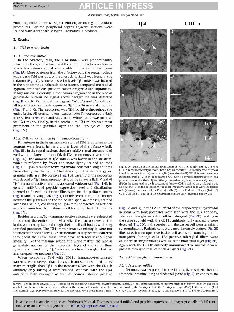
Fig. 2. Comparison of the cellular localization of (A, C and E) Tb4 and (B, D and F)
CD11b immunoreactivity in mouse brain. (A) In neocortex Tb4 immunoreactivity was
found in neurons (arrows) and microglia (arrowheads) (B) CD11b in neocortex only
stained microglia. (C) In the hippocampal CA1 subfield pyramidal neurons with long
processes stained with the Tb4 antibody: stained microglia are sporadically present.
(D) On the same level in the hippocampus (arrow) CD11b stained only microglia, but
no neurons. (E) In the cerebellum, the most intensely stained cells were the basket
cells (arrows) that surround the Purkinje cells (P) in the Purkinje cell layer (PuC). (F)
CD11b on the same level in the cerebellum stained only microglia. Bar 50 mm.
M. Paulussen et al. / Peptides xxx (2009) xxx–xxx4
G Model
PEP-67782; No of Pages 11
violet 1%, Fluka Chemika, Sigma–Aldrich) according to standardprocedures. For the peripheral organs adjacent sections werestained with a standard Mayer’s Haematoxilin protocol.
3. Results
3.1. Tb4 in mouse brain
3.1.1. Precursor mRNA
In the olfactory bulb, the Tb4 mRNA was predominantlysituated in the granular layer and the anterior olfactory nucleus; amuch less intense signal was visible in the mitral cell layer(Fig. 1A). More posterior from the olfactory bulb the septal nucleuswas clearly Tb4-positive, while a less dark signal was found in thestriatum (Fig. 1C). At more posterior levels Tb4 mRNA was locatedin the hippocampus, habenula, zona incerta, compact dorsomedialhypothalamic nucleus, piriform cortex, amygdala and supramam-milary nucleus. Centrally in the thalamic region and in the medialgeniculate nucleus no signal above background was detected(Fig. 1F and K). With the dentate gyrus, CA1, CA2 and CA3 subfield,all hippocampal subfields expressed Tb4 mRNA in equal amounts(Fig. 1F and K). The neocortex was Tb4-positive throughout theentire brain. All cortical layers, except layer IV, expressed a darkmRNA signal (Fig. 1C, F and K). Also, the white matter was positivefor Tb4 mRNA. Finally, in the cerebellum Tb4 mRNA was mostprominent in the granular layer and the Purkinje cell layer(Fig. 1M).
3.1.2. Cellular localization by immunocytochemistry
Far anterior in the brain intensely stained Tb4-immunoreactiveneurons were found in the granular layer of the olfactory bulb(Fig. 1B). In the septal nucleus, the dark mRNA signal correspondedwell with the large number of dark Tb4-immunoreactive neurons(Fig. 1D). The amount of Tb4 mRNA was lower in the striatum,which is reflected by fewer and more lightly stained neurons(Fig. 1E). Tb4-immunoreactive pyramidal cells with long neuriteswere clearly visible in the CA-subfields; in the dentate gyrus,granular cells are Tb4-positive (Fig. 1G). Layer IV of the neocortexwas devoid of Tb4 immunoreactivity, while in layers II/III, V and VITb4-immunoreactive neurons appeared widespread (Fig. 1H). Ingeneral, mRNA and peptide expression level and distributionseemed to fit well, as further illustrated for the piriform cortex(Fig. 1I) and the amygdala (Fig. 1J). In the cerebellum, at the borderbetween the granular and the molecular layer, an intensely stainedlayer was visible, consisting of Tb4-immunoreactive basket cellaxons surrounding the unstained cell bodies of the Purkinje cells(Fig. 1N).
Besides neurons, Tb4-immunoreactive microglia were detectedthroughout the entire brain. Microglia, the macrophages of thebrain, were recognizable through their small cell bodies with longramified processes. The Tb4-immunoreactive microglia were notrestricted to specific areas like the neurons, but appeared scatteredthroughout the entire brain. Brain areas with low mRNA signalintensity, like the thalamic region, the white matter, the medialgeniculate nucleus or the molecular layer of the cerebellumtypically showed only Tb4-immunoreactive microglia, but noimmunopositive neurons (Fig. 1L).
When comparing Tb4 with CD11b immunocytochemistrypatterns, we observed that the CD11b antiserum stained manymore microglia than Tb4 in the neocortex. Yet with the CD11bantibody only microglia were stained, whereas with the Tb4antiserum both microglia as well as neurons stained positive
(arrows) and (J) in the amygdala. (L) Regions where the mRNA signal was low, like thalam
cerebellum, the most intensely stained cells were the basket cell axon terminals (arrows)
and granular layer (GrC) only immunoreactive microglia were present. Bar 1 mm in (A,
Please cite this article in press as: Paulussen M, et al. Thymosin betamouse tissues. Peptides (2009), doi:10.1016/j.peptides.2009.07.010
(Fig. 2A and B). In the CA1 subfield of the hippocampus pyramidalneurons with long processes were seen with the Tb4 antibody,whereas microglia were difficult to distinguish (Fig. 2C). Looking inthe same subfield with the CD11b antibody, only microglia weredetected (Fig. 2D). In the cerebellum, the basket cell axon terminalssurrounding the Purkinje cells were most intensely stained. Fig. 2Eillustrates immunopositive basket cell axons surrounding immu-nonegative Purkinje cells. Tb4-positive microglial fibers wereabundant in the granular as well as in the molecular layer (Fig. 2E).Again with the CD11b antibody immunoreactive microglia werepresent throughout all cerebellar layers (Fig. 2F).
3.2. Tb4 in peripheral mouse organs
3.2.1. Precursor mRNA
Tb4 mRNA was expressed in the kidney, liver, spleen, thymus,stomach, intestine, lung and adrenal gland (Fig. 3). In contrast, no
us and MGN, still contained immunoreactive microglia (arrowheads). (M and N) In
surrounding the Purkinje cells in the Purkinje cell layer (PuC). In the molecular (Mo)
C, F, K and M), 100 mm in (B, D, E, J, L and N), 400 mm in (G and H), 200 mm in (I).
4 mRNA and peptide expression in phagocytic cells of different

Fig. 3. (A, D, G, J, M, P, S and V) mRNA and (B, C, E, F, H, I, K, L, N, O, Q, R, T, U, W and X) peptide distribution of Tb4 in mouse organs. (A) In the kidney, Tb4 mRNA was
predominantly present in the cortex, and less in the medulla. (B and C) Large immunoreactive ramified cells appeared in contact with Bowman’s capsules (arrows), and
surrounding the collecting tubules. (D–F) Tb4 mRNA and peptide expression in the liver. (G–I) In the spleen the Tb4 mRNA signal was the strongest in the white pulp (WP).
The difference in Tb4-immunoreactivity between red pulp (RP) and white pulp was not that pronounced. The largest number of Tb4-immunoreactive cells were found in the
marginal zone (MZ) of the white pulp. (J–L) Tb4 in the thymus. (M–O) In the stomach, Tb4 immunoreactivity could be found in the lamina propria (LP), the cell layer just
M. Paulussen et al. / Peptides xxx (2009) xxx–xxx 5
G Model
PEP-67782; No of Pages 11
Please cite this article in press as: Paulussen M, et al. Thymosin beta 4 mRNA and peptide expression in phagocytic cells of differentmouse tissues. Peptides (2009), doi:10.1016/j.peptides.2009.07.010

Fig. 3. (Continued ).
M. Paulussen et al. / Peptides xxx (2009) xxx–xxx6
G Model
PEP-67782; No of Pages 11
specific signal was detected in the pancreas or the pituitary. Heart,ovary and testis were not included in this study.
In the kidney, Tb4 mRNA was present in discrete, punctatestructures both in the cortex and the medulla, but moreextensively in the cortex (Fig. 3A). In the liver, spots of highintensity were spread along the entire organ (Fig. 3D). Tb4 mRNAin spleen was most prominent in the white pulp region, withmoderate expression in red pulp regions (Fig. 3G). The mRNAsignal in the thymus was difficult to distinguish from background(Fig. 3J), while in the stomach and intestine, mRNA expression wasvery intense (Fig. 3M and P). The Tb4-positive signal in thestomach was situated in the lamina propria, the cell layer justbeneath the epithelial layer. In lung tissue, a punctate signalgrouped in patches was distributed across the organ (Fig. 3S).Finally, in the adrenal gland, Tb4 mRNA was very extensivelyexpressed in the cortex, but much less in the medulla (Fig. 3V).
3.2.2. Cellular localization by immunocytochemistry
The morphology of the immunoreactive cells was similar inkidney and liver: characteristic for the Tb4-immunoreactive cellsare the small cell bodies with long branched processes (Fig. 3B, C, Eand F). In the kidney, these cells were clearly in direct contact withBowman’s capsule and lining the collecting tubules (Fig. 3C). In theliver, Tb4-immunoreactive cells were distributed across the entireorgan, although more concentrated towards the portal veins(Fig. 3E). Tb4-immunoreactive cells were present throughout theentire spleen, although the marginal zone of the white pulp wasstained more intensely. In red as in white pulp, all cells again had
beneath the epithelial layer (Ep) which lines the lumen (Lu). (P–R) Tb4-immunoreactive
cells were spread in patches across the entire lung (arrows). (V–X) In the adrenal gland
1 mm in (A, D, G, J, M, P, S an dV), 400 mm in (B, E, H, K, N Q, T and W), 100 mm in (C
Please cite this article in press as: Paulussen M, et al. Thymosin betamouse tissues. Peptides (2009), doi:10.1016/j.peptides.2009.07.010
ramified processes, but these are typically smaller than in kidneyand liver (Fig. 3H and I). No clear Tb4 mRNA signal was detected inthe thymus, but with immunocytochemistry, a few Tb4-positivecells could be seen (Fig. 3K and L). These cells were present in thecortex and medulla, but more abundantly in the medulla. In thethymus, no large cells with ramifications were found. Theimmunoreactive elements here were relatively small and roundor had a fiber-like structure. Also in the stomach, the Tb4-immunoreactive cells did not have large processes, but were largeand round (Fig. 3O). These cells were all located in the laminapropria (Fig. 3N). In contrast, in the intestine the immunopositivesignal mainly lined the intestinal villi, although also someimmunoreactive cells were found in the lamina propria (Fig. 3Qand R). In the lung, intensely immunostained cells were visible(Fig. 3U). These round cells were present throughout the entirelung, although areas of higher abundance were visible (Fig. 3T),comparable with detection at the mRNA level (Fig. 3S). In theadrenal medulla, a very small number of Tb4-immunoreactivecells were present, while the adrenal cortex was intensely stained(Fig. 3W). The outer zona glomerulosa contained the most positivecells, followed by the zona fasciculata below it. These cells againhad large ramified branches (Fig. 3X).
To reveal whether the Tb4 immunoreactivity was located inmacrophages, a comparison was made with an antibody to CD11b,a conventional macrophage marker. Since a few tissues did notshow the same immunoreactivity patterns, an additional, moreclassical, macrophage marker was used, allowing comparison withthe two markers. In the kidney (Fig. 4A–C) and the liver (Fig. 4D–F),
cells predominantly lined the intestinal villi. (S–U) In the lung, Tb4-immunoreactive
, Tb4 was mostly expressed in the cortex (cx), much less in the medulla (med). Bar
, F, I, L, O, R, U and X).
4 mRNA and peptide expression in phagocytic cells of different

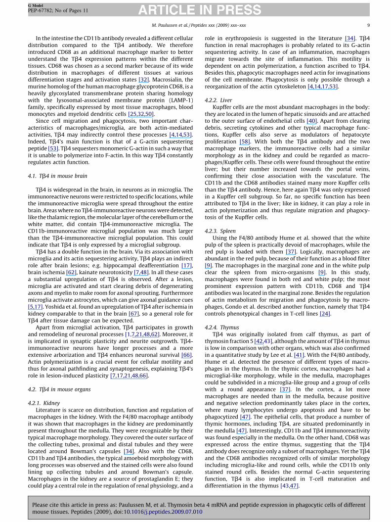
Fig. 4. Comparison of the cellular localization of (A, D, G, J, M, P, S and V) Tb4, (B, E, H, K, N, Q, T and W) CD11b and (C, F, I, L, O, R, U and X) CD68-immunoreactivity in mouse
organs. (A–C) Tb4, CD11b and CD68-immunoreactivity in kidney, (D–F) liver, (G–I) spleen, (J–L) thymus. (L) CD68-immunoreactivity was not only found in round cells
(arrows), but also in microglia-like cells (arrowheads). (M–O) In the stomach, Tb4, CD11b and CD68 were all present in the lamina propria (LP). (P–R) Tb4, CD11b and CD68-
immunoreactivity in lung and (S–U) adrenal gland. (V) Tb4-immunoreactivity in the intestine could be found in cells lining the villi, although some immunopositive cells
were found in the lamina propria. (W and X) CD11b and CD68-immunoreactive cells resided only inside the lamina propria. The bar represents 25 mm for panels A–F, S–U;
50 mm for panels G–L, V–X, and 200 mm for panels M–R.
M. Paulussen et al. / Peptides xxx (2009) xxx–xxx 7
G Model
PEP-67782; No of Pages 11
Please cite this article in press as: Paulussen M, et al. Thymosin beta 4 mRNA and peptide expression in phagocytic cells of differentmouse tissues. Peptides (2009), doi:10.1016/j.peptides.2009.07.010
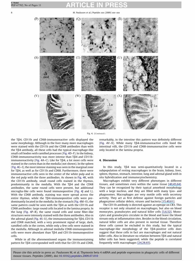
Fig. 4. (Continued ).
M. Paulussen et al. / Peptides xxx (2009) xxx–xxx8
G Model
PEP-67782; No of Pages 11
the Tb4, CD11b and CD68-immunoreactive cells displayed thesame morphology. Although in the liver many more macrophageswere stained with the CD11b and the CD68 antibodies than withthe Tb4 antibody, all these cells had the typical macrophage-likesmall cell bodies with ramified processes (Fig. 4D–F). In the kidney,CD68-immunoreactivity was more intense than Tb4 and CD11b-immunoreactivity (Fig. 4A–C). Like for Tb4, a lot more cells werestained in the cortex than in the medulla (not shown). In the spleen(Fig. 4G–I), the most intense staining was seen in the marginal zonefor Tb4, as well as for CD11b and CD68. Furthermore, there wereimmunoreactive cells seen in the center of the white pulp and inthe red pulp with the three antibodies. As shown in Fig. 4K, withthe CD11b antibody, small round cells stained in the thymus,predominantly in the medulla. With the Tb4 and the CD68antibodies, the same round cells were present, but additionalmicroglia-like cells were found immunopositive (Fig. 4J and L).With the CD68 antibody, staining was more spread across theentire thymus, while the Tb4-immunopositive cells were pre-dominantly located in the medulla. In the stomach (Fig. 4M–O), thesame pattern could be seen with the Tb4 as with the CD11b andCD68 antibodies: large round cells located in the lamina propria. Inthe lung (Fig. 4P–R), the same round cells and some fiber-likestructures were intensely stained with the three antibodies. Also inthe adrenal gland (Fig. 4S–U), the immunostaining for Tb4, CD11band CD68 matched, with a very prominent signal of large, dark,ramified cells in the cortex, while only a few cells were stained inthe medulla. Although in adrenal medulla CD68-immunopositivecells were more abundant than Tb4 and CD11b-immunopositivecells.
While in all the aforementioned organs the immunostainingpattern for Tb4 corresponded well with that for CD11b and CD68,
Please cite this article in press as: Paulussen M, et al. Thymosin betamouse tissues. Peptides (2009), doi:10.1016/j.peptides.2009.07.010
remarkably, in the intestine this pattern was definitely different(Fig. 4V–X). While many Tb4-immunoreactive cells lined theintestinal villi, the CD11b and CD68-immunoreactive cells wereonly located in the lamina propria.
4. Discussion
In this study, Tb4 was semi-quantitatively located in asubpopulation of resting macrophages in the brain, kidney, liver,spleen, thymus, stomach, intestine, lung and adrenal gland with insitu hybridization and immunocytochemistry.
Macrophages exhibit very different phenotypes in differenttissues, and sometimes even within the same tissue [40,45,64].They can be recognized by their typical amoeboid morphologywith a large nucleus, and they are filled with many lyso- andphagosomes. Macrophages are very motile cells with secretoryactivity. They act as first defense against foreign particles andphagocytose cellular debris, viruses and bacteria [15,40,61].
The CD11b antibody is directed against an epitope on CR3. Thisreceptor is not only situated on macrophages, but also on bloodmonocytes, granulocytes and natural killer cells [3,6,52]. Mono-cytes and granulocytes circulate in the blood and leave the bloodstream only at inflammation sites. Besides in the blood circulation,natural killer cells are also present in normal tissue. Thereforethese cells cannot be excluded in this study, but the typicalmacrophage-like morphology of the Tb4-positive cells doessuggest that these cells in fact are macrophages and not naturalkiller cells. Also in literature no relation between Tb4 and naturalkiller cells has been suggested, while the peptide is correlatedfrequently with macrophages [24,28,65].
4 mRNA and peptide expression in phagocytic cells of different
M. Paulussen et al. / Peptides xxx (2009) xxx–xxx 9
G Model
PEP-67782; No of Pages 11
In the intestine the CD11b antibody revealed a different cellulardistribution compared to the Tb4 antibody. We thereforeintroduced CD68 as an additional macrophage marker to betterunderstand the Tb4 expression patterns within the differenttissues. CD68 was chosen as a second marker because of its widedistribution in macrophages of different tissues at variousdifferentiation stages and activation states [32]. Macrosialin, themurine homolog of the human macrophage glycoprotein CD68, is aheavily glycosylated transmembrane protein sharing homologywith the lysosomal-associated membrane protein (LAMP-1)family, specifically expressed by most tissue macrophages, bloodmonocytes and myeloid dendritic cells [25,32,50].
Since cell migration and phagocytosis, two important char-acteristics of macrophages/microglia, are both actin-mediatedactivities, Tb4 may indirectly control these processes [4,14,53].Indeed, Tb4’s main function is that of a G-actin sequesteringpeptide [53]. Tb4 sequesters monomeric G-actin in such a way thatit is unable to polymerize into F-actin. In this way Tb4 constantlyregulates actin function.
4.1. Tb4 in mouse brain
Tb4 is widespread in the brain, in neurons as in microglia. Theimmunoreactive neurons were restricted to specific locations, whilethe immunoreactive microglia were spread throughout the entirebrain. Areas where no Tb4-immunoreactive neurons were detected,like the thalamic region, the molecular layer of the cerebellum or thewhite matter, did contain Tb4-immunoreactive microglia. TheCD11b-immunoreactive microglial population was much largerthan the Tb4-immunoreactive microglial population. This couldindicate that Tb4 is only expressed by a microglial subgroup.
Tb4 has a double function in the brain. Via its association withmicroglia and its actin sequestering activity, Tb4 plays an indirectrole after brain lesions; e.g. hippocampal deafferentiation [17],brain ischemia [62], kainate neurotoxicity [7,48]. In all these casesa substantial upregulation of Tb4 is observed. After a lesion,microglia are activated and start clearing debris of degeneratingaxons and myelin to make room for axonal sprouting. Furthermoremicroglia activate astrocytes, which can give axonal guidance cues[5,17]. Yoshida et al. found an upregulation of Tb4 after ischemia inkidney comparable to that in the brain [67], so a general role forTb4 after tissue damage can be expected.
Apart from microglial activation, Tb4 participates in growthand remodeling of neuronal processes [1,7,21,48,62]. Moreover, itis implicated in synaptic plasticity and neurite outgrowth. Tb4-immunoreactive neurons have longer processes and a moreextensive arborization and Tb4 enhances neuronal survival [66].Actin polymerization is a crucial event for cellular motility andthus for axonal pathfinding and synaptogenesis, explaining Tb4’srole in lesion-induced plasticity [7,17,21,48,66].
4.2. Tb4 in mouse organs
4.2.1. Kidney
Literature is scarce on distribution, function and regulation ofmacrophages in the kidney. With the F4/80 macrophage antibodyit was shown that macrophages in the kidney are predominantlypresent throughout the medulla. They were recognizable by theirtypical macrophage morphology. They covered the outer surface ofthe collecting tubes, proximal and distal tubules and they werelocated around Bowman’s capsules [34]. Also with the CD68,CD11b and Tb4 antibodies, the typical amoeboid morphology withlong processes was observed and the stained cells were also foundlining up collecting tubules and around Bowman’s capsule.Macrophages in the kidney are a source of prostaglandin E; theycould play a central role in the regulation of renal physiology, and a
Please cite this article in press as: Paulussen M, et al. Thymosin betamouse tissues. Peptides (2009), doi:10.1016/j.peptides.2009.07.010
role in erythropoiesis is suggested in the literature [34]. Tb4function in renal macrophages is probably related to its G-actinsequestering activity. In case of an inflammation, macrophagesmigrate towards the site of inflammation. This motility isdependent on actin polymerization, a function ascribed to Tb4.Besides this, phagocytic macrophages need actin for invaginationsof the cell membrane. Phagocytosis is only possible through areorganization of the actin cytoskeleton [4,14,17,53].
4.2.2. Liver
Kupffer cells are the most abundant macrophages in the body:they are located in the lumen of hepatic sinusoids and are attachedto the outer surface of endothelial cells [40]. Apart from clearingdebris, secreting cytokines and other typical macrophage func-tions, Kupffer cells also serve as modulators of hepatocyteproliferation [58]. With both the Tb4 antibody and the twomacrophage markers, the immunoreactive cells had a similarmorphology as in the kidney and could be regarded as macro-phages/Kupffer cells. These cells were found throughout the entireliver; but their number increased towards the portal veins,confirming their close association with the vasculature. TheCD11b and the CD68 antibodies stained many more Kupffer cellsthan the Tb4 antibody. Hence, here again Tb4 was only expressedin a Kupffer cell subgroup. So far, no specific function has beenattributed to Tb4 in the liver; like in kidney, it can play a role inactin polymerization and thus regulate migration and phagocy-tosis of the Kupffer cells.
4.2.3. Spleen
Using the F4/80 antibody Hume et al. showed that the whitepulp of the spleen is practically devoid of macrophages, while thered pulp is loaded with them [37]. Logically, macrophages areabundant in the red pulp, because of their function as a blood filter[9]. The macrophages in the marginal zone and in the white pulpclear the spleen from micro-organisms [9]. In this study,macrophages were found in both red and white pulp; the mostprominent expression pattern with CD11b, CD68 and Tb4antibodies was located in the marginal zone. Besides the regulationof actin metabolism for migration and phagocytosis by macro-phages, Gondo et al. described another function, namely that Tb4controls phenotypical changes in T-cell lines [24].
4.2.4. Thymus
Tb4 was originally isolated from calf thymus, as part ofthymosin fraction 5 [42,43], although the amount of Tb4 in thymusis low in comparison with other organs, which was also confirmedin a quantitative study by Lee et al. [41]. With the F4/80 antibody,Hume et al. detected the presence of different types of macro-phages in the thymus. In the thymic cortex, macrophages had amicroglial-like morphology, while in the medulla, macrophagescould be subdivided in a microglia-like group and a group of cellswith a round appearance [37]. In the cortex, a lot moremacrophages are needed than in the medulla, because positiveand negative selection predominantly takes place in the cortex,where many lymphocytes undergo apoptosis and have to bephagocytized [47]. The epithelial cells, that produce a number ofthymic hormones, including Tb4, are situated predominantly inthe medulla [47]. Interestingly, CD11b and Tb4 immunoreactivitywas found especially in the medulla. On the other hand, CD68 wasexpressed across the entire thymus, suggesting that the Tb4antibody does recognize only a subset of macrophages. Yet the Tb4and the CD68 antibodies recognized cells of similar morphologyincluding microglia-like and round cells, while the CD11b onlystained round cells. Besides the normal G-actin sequesteringfunction, Tb4 is also implicated in T-cell maturation anddifferentiation in the thymus [43,47].
4 mRNA and peptide expression in phagocytic cells of different

M. Paulussen et al. / Peptides xxx (2009) xxx–xxx10
G Model
PEP-67782; No of Pages 11
4.2.5. Stomach and intestine
In the stomach and intestine, the lamina propria is densely filledwith macrophages [15,33,36,37,41]. Denning et al. showed CD11b-immunoreactive macrophages scattered throughout the laminapropria of the intestinal villi along the entire intestine. Phenotyp-ing revealed that the CD11b-immunoreactive cells were indeedmacrophages and not natural killer cells [15]. Macrophages stainedwith the CD11b and the CD68 antibodies were indeed typicallyseen in the lamina propria of the stomach and intestine. In thestomach the same pattern was seen with Tb4. In the intestinehowever the Tb4-immunoreactive cells clearly also lined the villi.These Tb4-immunopositive cells were located in the epithelium ofthe intestines. Because of their close lining of the villi and theirhigh cylindrical shape, these Tb4-positive cells could be identifiedas enterocytes [57]. Intestinal macrophages clear debris, respondto luminal contents by transporting intestinal particles, likebacteria, into extraintestinal sites, and they produce a range ofcytokines. In the stomach, macrophages are a source of prosta-glandin E, which controls acid secretion [15,33,63]. Here, Tb4 mostlikely functions as a G-actin sequestering peptide to arrangechanges in macrophage morphology and migration. No otherfunctions in the gastrointestinal tract are known.
4.2.6. Lung
There are two types of macrophages in the lung, whichmorphologically are not easy to distinguish: the alveolar macro-phages in the alveolus and the interstitial macrophages in the lungparenchyma. The alveolar macrophages are involved in theprimary defense against inhaled particles [40]. Lung macrophagesare round cells present throughout the entire lung [16,19], apattern observed with CD68, CD11b and Tb4 antibodies. Nospecific lung functions have yet been attributed to Tb4.
4.2.7. Adrenal gland
In the adrenal gland F4/80-immunoreactive macrophages werepredominantly present in the cortex, with the highest density inthe outer zona glomerulosa, and lower density in the layer below,the zona fasciculata. In the adrenal medulla, macrophages weresparse [35]. CD11b, CD68 and Tb4-immunoreactive cells had thesame distribution pattern, they were mainly found in the zonaglomerulosa of the adrenal cortex; while in the adrenal medullaonly a few immunoreactive cells were found. With the CD68antibody more immunoreactive cells were observed in the medullathan with the Tb4 and CD11b antibodies, which again indicatesthat the latter were only expressed by a macrophage subgroup inadrenal medulla. Next to their phagocytotic activity, macrophagesin the adrenal gland control extracellular processing of peptidehormones, secrete prostaglandins E and produce cytokines, whichcan influence hormone production, the main function of adrenalglands [35,46]. These immune-endocrine interactions are impor-tant in modulating adrenal responses to inflammatory andimmune challenges [46]. Tb4 probably contributes to thisresponse by controlling actin polymerization to induce cellmigration. Although in primates intravenous thymosin fraction5 (which contains Tb4), elevates corticotrophin and cortisolconcentrations, these glucocorticoids are produced by the zonafasciculata of the adrenal gland cortex [29]. Before ascribing thismore specific function to Tb4, further research is needed, becausefraction 5 contains many peptides that could trigger glucocorticoidsecretion.
5. Conclusion
Many distinct functions have already been ascribed to Tb4,though a detailed analysis of its distribution in the whole body ofthe mouse was lacking, especially at the cellular level. In the
Please cite this article in press as: Paulussen M, et al. Thymosin betamouse tissues. Peptides (2009), doi:10.1016/j.peptides.2009.07.010
present study, we have used two techniques: in situ hybridizationto allow an overall screening of the Tb4 mRNA distribution, andimmunocytochemistry for a more precise localization of thepeptide at the cellular level. We localized Tb4 in the brain, thymus,spleen, lung, kidney, liver, adrenal gland, stomach and intestine.Through a comparison with the macrophage differentiationantigen CR3, it was shown that Tb4 is expressed by almost everytype of tissue macrophage, including microglia. In the tissues, asecond macrophage marker, CD68, confirmed our findings.Comparison with the two markers made clear that in some tissuesTb4 is only expressed by a subgroup of macrophages.
Acknowledgements
We thank Inge Van der Auwera for her excellent technicalassistance. This work was supported by grants from the Institutefor the promotion of Innovation by Science and Technology inFlanders (IWT 50164) and by the Fund for Scientific Research,Flanders, Belgium (FWO-Flanders).
References
[1] Anadon R, Rodriguez MI, Carpintero P, Evangelatos G, Livianou E, Leondiadis L,et al. Differential expression of thymosins beta(4) and beta(10) during ratcerebellum postnatal development. Brain Res 2001;894:255–65.
[2] Arckens L, Zhang F, Vanduffel W, Mailleux P, Vanderhaeghen JJ, Orban GA, et al.Localization of the two protein kinase C beta-mRNA subtypes in cat visualsystem. J Chem Neuroanat 1995;8:117–24.
[3] Avni O, Pur Z, Yefenof E, Baniyash M. Complement receptor 3 of macrophagesis associated with galectin-1-like protein. J Immunol 1998;160:6151–8.
[4] Ballweber E, Hannappel E, Huff T, Stephan H, Haener M, Taschner N, et al.Polymerisation of chemically cross-linked actin:thymosin beta(4) complex tofilamentous actin: alteration in helical parameters and visualisation of thy-mosin beta(4) binding on F-actin. J Mol Biol 2002;315:613–25.
[5] Bechmann I, Nitsch R. Involvement of non-neuronal cells in entorhinal-hip-pocampal reorganization following lesions. Ann NY Acad Sci 2000;911:192–206.
[6] Beller DI, Springer TA, Schreiber RD. Anti-Mac-1 selectively inhibits the mouseand human type three complement receptor. J Exp Med 1982;156:1000–9.
[7] Carpintero P, Anadon R, az-Regueira S, Gomez-Marquez J. Expression ofthymosin beta4 messenger RNA in normal and kainate-treated rat forebrain.Neuroscience 1999;90:1433–44.
[8] Carpintero P, Franco del AF, Anadon R, Gomez-Marquez J. Thymosin beta10mRNA expression during early postimplantation mouse development. FEBSLett 1996;394:103–6.
[9] Cesta MF. Normal structure, function, and histology of the spleen. ToxicolPathol 2006;34:455–65.
[10] Cha HJ, Jeong MJ, Kleinman HK. Role of thymosin beta4 in tumor metastasisand angiogenesis. J Natl Cancer Inst 2003;95:1674–80.
[11] Choi SY, Noh MR, Kim DK, Sun W, Kim H. Neuroprotective function ofthymosin-beta and its derivative peptides on the programmed cell death ofchick and rat neurons. Biochem Biophys Res Commun 2007;362:587–93.
[12] Cnops L, Hu TT, Eysel UT, Arckens L. Effect of binocular retinal lesions onCRMP2 and CRMP4 but not Dyn I and Syt I expression in adult cat area 17. Eur JNeurosci 2007;25:1395–401.
[13] da Silva RP, Gordon S. Phagocytosis stimulates alternative glycosylation ofmacrosialin (mouse CD68), a macrophage-specific endosomal protein. Bio-chem J 1999;338(Pt 3):687–94.
[14] Dedova IV, Nikolaeva OP, Safer D, De La Cruz EM, dos Remedios CG. Thymosinbeta4 induces a conformational change in actin monomers. Biophys J2006;90:985–92.
[15] Denning TL, Wang YC, Patel SR, Williams IR, Pulendran B. Lamina propriamacrophages and dendritic cells differentially induce regulatory and inter-leukin 17-producing T cell responses. Nat Immunol 2007;8:1086–94.
[16] Dhami R, He X, Gordon RE, Schuchman EH. Analysis of the lung pathology andalveolar macrophage function in the acid sphingomyelinase-deficient mousemodel of Niemann–Pick disease. Lab Invest 2001;81:987–99.
[17] Dong JH, Ying GX, Liu X, Wang WY, Wang Y, Ni ZM, et al. Expression ofthymosin beta4 mRNA by activated microglia in the denervated hippocampus.Neuroreport 2005;16:1629–33.
[18] Erickson-Viitanen S, Ruggieri S, Natalini P, Horecker BL. Distribution ofthymosin beta 4 in vertebrate classes. Arch Biochem Biophys 1983;221:570–6.
[19] Fehrenbach H, Zissel G, Goldmann T, Tschernig T, Vollmer E, Pabst R, et al.Alveolar macrophages are the main source for tumour necrosis factor-alpha inpatients with sarcoidosis. Eur Respir J 2003;21:421–8.
[20] Goldstein AL, Low TL, McAdoo M, McClure J, Thurman GB, Rossio J, et al.Thymosin alpha1: isolation and sequence analysis of an immunologicallyactive thymic polypeptide. Proc Natl Acad Sci USA 1977;74:725–9.
4 mRNA and peptide expression in phagocytic cells of different

M. Paulussen et al. / Peptides xxx (2009) xxx–xxx 11
G Model
PEP-67782; No of Pages 11
[21] Gomez-Marquez J. Function of prothymosin alpha in chromatin decondensa-tion and expression of thymosin beta-4 linked to angiogenesis and synapticplasticity. Ann NY Acad Sci 2007;1112:201–9.
[22] Gomez-Marquez J, Anadon R. The beta-thymosins, small actin-binding pep-tides widely expressed in the developing and adult cerebellum. Cerebellum2002;1:95–102.
[23] Gomez-Marquez J, Dosil M, Segade F, Bustelo XR, Pichel JG, Dominguez F, et al.Thymosin-beta 4 gene. Preliminary characterization and expression in tissues,thymic cells, and lymphocytes. J Immunol 1989;143:2740–4.
[24] Gondo H, Kudo J, White JW, Barr C, Selvanayagam P, Saunders GF. Differentialexpression of the human thymosin-beta 4 gene in lymphocytes, macrophages,and granulocytes. J Immunol 1987;139:3840–8.
[25] Gordon S. Macrophage-restricted molecules: role in differentiation and acti-vation. Immunol Lett 1999;65:5–8.
[26] Grant DS, Kinsella JL, Kibbey MC, LaFlamme S, Burbelo PD, Goldstein AL, et al.Matrigel induces thymosin beta 4 gene in differentiating endothelial cells. JCell Sci 1995;108(Pt 12):3685–94.
[27] Grant DS, Rose W, Yaen C, Goldstein A, Martinez J, Kleinman H. Thymosinbeta4 enhances endothelial cell differentiation and angiogenesis. Angiogen-esis 1999;3:125–35.
[28] Hannappel E, Xu GJ, Morgan J, Hempstead J, Horecker BL. Thymosin beta 4: aubiquitous peptide in rat and mouse tissues. Proc Natl Acad Sci USA1982;79:2172–5.
[29] Healy DL, Hodgen GD, Schulte HM, Chrousos GP, Loriaux DL, Hall NR, et al. Thethymus-adrenal connection: thymosin has corticotropin-releasing activity inprimates. Science 1983;222:1353–5.
[30] Holness CL, da Silva RP, Fawcett J, Gordon S, Simmons DL. Macrosialin, a mousemacrophage-restricted glycoprotein, is a member of the lamp/lgp family. J BiolChem 1993;268:9661–6.
[31] Holness CL, Simmons DL. Molecular cloning of CD68, a human macrophagemarker related to lysosomal glycoproteins. Blood 1993;81:1607–13.
[32] Horikawa T, Komohara Y, Kiyota E, Terasaki Y, Takagi K, Takeya M. Detection ofguinea pig macrophages by a new CD68 monoclonal antibody, PM-1K. J MolHistol 2006;37:15–25.
[33] Hume DA, Allan W, Hogan PG, Doe WF. Immunohistochemical characterisa-tion of macrophages in human liver and gastrointestinal tract: expression ofCD4, HLA-DR, OKM1, and the mature macrophage marker 25F9 in normal anddiseased tissue. J Leukoc Biol 1987;42:474–84.
[34] Hume DA, Gordon S. Mononuclear phagocyte system of the mouse defined byimmunohistochemical localization of antigen F4/80. Identification of residentmacrophages in renal medullary and cortical interstitium and the juxtaglo-merular complex. J Exp Med 1983;157:1704–9.
[35] Hume DA, Halpin D, Charlton H, Gordon S. The mononuclear phagocyte systemof the mouse defined by immunohistochemical localization of antigen F4/80:macrophages of endocrine organs. Proc Natl Acad Sci USA 1984;81:4174–7.
[36] Hume DA, Perry VH, Gordon S. The mononuclear phagocyte system of themouse defined by immunohistochemical localisation of antigen F4/80: macro-phages associated with epithelia. Anat Rec 1984;210:503–12.
[37] Hume DA, Robinson AP, MacPherson GG, Gordon S. The mononuclear phago-cyte system of the mouse defined by immunohistochemical localization ofantigen F4/80. Relationship between macrophages, Langerhans cells, reticularcells, and dendritic cells in lymphoid and hematopoietic organs. J Exp Med1983;158:1522–36.
[38] Hutchings P, Rosen H, O’Reilly L, Simpson E, Gordon S, Cooke A. Transfer ofdiabetes in mice prevented by blockade of adhesion-promoting receptor onmacrophages. Nature 1990;348:639–42.
[39] Kim Y, Kim EH, Hong S, Rhyu IJ, Choe J, Sun W, et al. Expression of thymosinbeta in the rat brain following transient global ischemia. Brain Res2006;1085:177–82.
[40] Laskin DL, Weinberger B, Laskin JD. Functional heterogeneity in liver and lungmacrophages. J Leukoc Biol 2001;70:163–70.
[41] Lee SH, Starkey PM, Gordon S. Quantitative analysis of total macrophagecontent in adult mouse tissues. Immunochemical studies with monoclonalantibody F4/80. J Exp Med 1985;161:475–89.
[42] Low TL, Goldstein AL. Chemical characterization of thymosin beta 4. J BiolChem 1982;257:1000–6.
Please cite this article in press as: Paulussen M, et al. Thymosin betamouse tissues. Peptides (2009), doi:10.1016/j.peptides.2009.07.010
[43] Low TL, Hu SK, Goldstein AL. Complete amino acid sequence of bovinethymosin beta 4: a thymic hormone that induces terminal deoxynucleotidyltransferase activity in thymocyte populations. Proc Natl Acad Sci USA1981;78:1162–6.
[44] Malinda KM, Sidhu GS, Mani H, Banaudha K, Maheshwari RK, Goldstein AL,et al. Thymosin beta4 accelerates wound healing. J Invest Dermatol1999;113:364–8.
[45] Nibbering PH, Leijh PC, van Furth R. Quantitative immunocytochemical char-acterization of mononuclear phagocytes. II. Monocytes and tissue macro-phages. Immunology 1987;62:171–6.
[46] Nussdorfer GG, Mazzocchi G. Immune-endocrine interactions in the mamma-lian adrenal gland: facts and hypotheses. Int Rev Cytol 1998;183:143–84.
[47] Pearse G. Normal structure, function and histology of the thymus. ToxicolPathol 2006;34:504–14.
[48] Popoli P, Pepponi R, Martire A, Armida M, Pezzola A, Galluzzo M, et al.Neuroprotective effects of thymosin beta4 in experimental models of excito-toxicity. Ann NY Acad Sci 2007;1112:219–24.
[49] Pulford KA, Sipos A, Cordell JL, Stross WP, Mason DY. Distribution of the CD68macrophage/myeloid associated antigen. Int Immunol 1990;2:973–80.
[50] Ramprasad MP, Fischer W, Witztum JL, Sambrano GR, Quehenberger O,Steinberg D. The 94- to 97-kDa mouse macrophage membrane protein thatrecognizes oxidized low density lipoprotein and phosphatidylserine-richliposomes is identical to macrosialin, the mouse homologue of humanCD68. Proc Natl Acad Sci USA 1995;92:9580–4.
[51] Reichert F, Rotshenker S. Complement-receptor-3 and scavenger-receptor-AI/II mediated myelin phagocytosis in microglia and macrophages. Neurobiol Dis2003;12:65–72.
[52] Rosen H, Gordon S. Monoclonal antibody to the murine type 3 complementreceptor inhibits adhesion of myelomonocytic cells in vitro and inflammatorycell recruitment in vivo. J Exp Med 1987;166:1685–701.
[53] Safer D, Elzinga M, Nachmias VT. Thymosin beta 4 and Fx, an actin-sequester-ing peptide, are indistinguishable. J Biol Chem 1991;266:4029–32.
[54] Sapp E, Kegel KB, Aronin N, Hashikawa T, Uchiyama Y, Tohyama K, et al. Earlyand progressive accumulation of reactive microglia in the Huntington diseasebrain. J Neuropathol Exp Neurol 2001;60:161–72.
[55] Shu SY, Ju G, Fan LZ. The glucose oxidase-DAB-nickel method in peroxidasehistochemistry of the nervous system. Neurosci Lett 1988;85:169–71.
[56] Smart N, Rossdeutsch A, Riley PR. Thymosin beta4 and angiogenesis: modes ofaction and therapeutic potential. Angiogenesis 2007;10:229–41.
[57] Stevens A, Low J. Human histology. London: Mosby; 1997.[58] Takeishi T, Hirano K, Kobayashi T, Hasegawa G, Hatakeyama K, Naito M. The
role of Kupffer cells in liver regeneration. Arch Histol Cytol 1999;62:413–22.[59] Van Der Gucht E, Hof PR, Van Brussel L, Burnat K, Arckens L. Neurofilament
protein and neuronal activity markers define regional architectonic parcella-tion in the mouse visual cortex. Cereb Cortex 2007;17:2805–19.
[60] Van Der Gucht E, Vandenbussche E, Orban GA, Vandesande F, Arckens L. A newcat Fos antibody to localize the immediate early gene c-fos in mammalian visualcortex after sensory stimulation. J Histochem Cytochem 2000;48:671–84.
[61] van Furth R, Cohn ZA. The origin and kinetics of mononuclear phagocytes. J ExpMed 1968;128:415–35.
[62] Vartiainen N, Pyykonen I, Hokfelt T, Koistinaho J. Induction of thymosinbeta(4) mRNA following focal brain ischemia. Neuroreport 1996;7:1613–6.
[63] Wells CL, Maddaus MA, Simmons RL. Role of the macrophage in the transloca-tion of intestinal bacteria. Arch Surg 1987;122:48–53.
[64] Witmer-Pack MD, Hughes DA, Schuler G, Lawson L, McWilliam A, Inaba K, et al.Identification of macrophages and dendritic cells in the osteopetrotic (op/op)mouse. J Cell Sci 1993;104(Pt 4):1021–9.
[65] Xu GJ, Hannappel E, Morgan J, Hempstead J, Horecker BL. Synthesis ofthymosin beta 4 by peritoneal macrophages and adherent spleen cells. ProcNatl Acad Sci USA 1982;79:4006–9.
[66] Yang H, Cheng X, Yao Q, Li J, Ju G. The promotive effects of thymosin beta4 onneuronal survival and neurite outgrowth by upregulating L1 expression.Neurochem Res 2008;33:2269–80.
[67] Yoshida T, Kurella M, Beato F, Min H, Ingelfinger JR, Stears RL, et al. Monitoringchanges in gene expression in renal ischemia-reperfusion in the rat. Kidney Int2002;61:1646–54.
4 mRNA and peptide expression in phagocytic cells of different
Copyright © 2022 FDOKUMEN