Differentiation and Glucocorticoid Regulated Apopto-Phagocytic Gene Expression Patterns in Human...
10
Differentiation and Glucocorticoid Regulated Apopto- Phagocytic Gene Expression Patterns in Human Macrophages. Role of Mertk in Enhanced Phagocytosis Ga ´ bor Zahuczky 1 , Endre Kristo ´f 1 , Gyo ¨ ngyike Majai 1,2 , La ´ szlo ´ Fe ´su ¨s 1 * 1 Apoptosis and Genomics Research Group, Department of Biochemistry and Molecular Biology, Hungarian Academy of Sciences, University of Debrecen, Debrecen, Hungary, 2 Third Department of Internal Medicine, University of Debrecen, Debrecen, Hungary Abstract The daily clearance of physiologically dying cells is performed safely mainly by cells in the mononuclear phagocyte system. They can recognize and engulf dying cells utilizing several cooperative mechanisms. In our study we show that the expression of a broad range of apopto-phagocytic genes is strongly up-regulated during differentiation of human monocytes to macrophages with different donor variability. The glucocorticoid dexamethasone has a profound effect on this process by selectively up-regulating six genes and down-regulating several others. The key role of the up-regulated mer tyrosine kinase (Mertk) in dexamethasone induced enhancement of phagocytosis could be demonstrated in human monocyte derived macrophages by gene silencing as well as blocking antibodies, and also in a monocyte-macrophage like cell line. However, the additional role of other glucocorticoid induced elements must be also considered since the presence of autologous serum during phagocytosis could almost completely compensate for the blocked function of Mertk. Citation: Zahuczky G, Kristo ´f E, Majai G, Fe ´su ¨ s L (2011) Differentiation and Glucocorticoid Regulated Apopto-Phagocytic Gene Expression Patterns in Human Macrophages. Role of Mertk in Enhanced Phagocytosis. PLoS ONE 6(6): e21349. doi:10.1371/journal.pone.0021349 Editor: Colin Combs, University of North Dakota, United States of America Received January 24, 2011; Accepted May 27, 2011; Published June 24, 2011 Copyright: ß 2011 Zahuczky et al. This is an open-access article distributed under the terms of the Creative Commons Attribution License, which permits unrestricted use, distribution, and reproduction in any medium, provided the original author and source are credited. Funding: The study has been funded by grants from the Hungarian Scientific Research Fund (OTKA NI 67877), the Hungarian Ministry of Health and EU (MRTN- CT-2006-036032, MRTN-CT 2006-035624, LSHB-CT-2007-037730) as well as the TA ´ MOP 4.2.1./B-09/1/KONV-2010-0007 project implemented through the New Hungary Development Plan, co-financed by the European Social Fund. The funders had no role in study design, data collection and analysis, decision to publish, or preparation of the manuscript. Competing Interests: The authors have declared that no competing interests exist. * E-mail: [email protected] Introduction The efficient elimination of apoptotic cells or those dying through necrosis is performed mainly by the cells of the mononuclear phagocyte system [1–2]. Circulating monocytes, resident macrophages and those that infiltrate tissues or divide locally in circumstances of injury or inflammation are the major elements of this system [3]. The process of apoptotic cell corpse removal by professional phagocytes is remarkably complex and only partly defined [4–6]. It consists of two major steps: (1) recognition and (2) subsequent engulfment of apoptotic cells [1]. Ligands appearing on the apoptotic cells, receptors on the phagocyte and bridging molecules in the environment may act to drive either or both of these steps [7,33]. While elements of the recognition and receptor elements of the apopto-phagocytic machinery seem to be highly redundant [8], the signaling pathways for the engulfing machinery converge to switch on rac- 1 dependent cytoskeletal processes [7]. Glucocorticoids (GC) have an extensive range of effects in target tissues throughout the organism eliciting both rapid and delayed changes in physiological functions and pathologic tissues environ- ment. Their therapeutic effects are mediated by the classical cytosolic glucocorticoid receptors (cGCRs) which move to the nucleus to regulate gene expression following ligand binding or by membrane-bound GCR and direct interactions with the cell membrane [9–10]. The potentiating effect of glucocorticoids on the phagocytosis of apoptotic neutrophils, which can be inhibited by GCR antagonists, has been described [11–12]. As an explanation of the enhanced phagocytic uptake of apoptotic cells, an increased capacity for engulfment oriented reorganization of cytoskeletal elements, loss of phosphorylation of adhesion mediators (paxillin and pyk2) and increased amount of Rac GTPase were considered [13–14]. By analyzing the GC-induced expression patterns in human monocytes by microarray technol- ogy the following pathways and gene-clusters were proposed as possible functional markers of the developing anti-inflammatory subtype: up-regulated antioxidative, migration/chemotaxis, phagocytosis, anti-inflammatory genes and down-regulated T-cell chemotaxis, adhesion, apoptosis, oxidative functions and IFNc regulated genes. [15]. The importance of Mer tyrosine kinase (Mertk), as a member of of the Tyro3/Axl/Mer family of receptor tyrosine kinases in the engulfment and efficient clearance of apoptotic cells has been clearly demonstrated [16] and it was recently found that the glucocorticoid dexamethasone (DXM) treated human monocyte derived macrophages (HMDMs) exhibit augmented capacity of phagocytosis only in the presence of a serum factor that was identified as protein S, a ligand for Mertk. [17]. Here, we investigated the effects of differentiation and treatment by DXM on the gene-expression pattern of HMDMs using a custom designed apopto-phagocyte panel. Our data show that during differentiation of monocytes to macrophages most of the apopto-phagocytic genes are highly up-regulated. Dexamethasone led to further up-regulation of 6 genes while some others were PLoS ONE | www.plosone.org 1 June 2011 | Volume 6 | Issue 6 | e21349
-
Upload
independent -
Category
Documents
-
view
2 -
download
0
Transcript of Differentiation and Glucocorticoid Regulated Apopto-Phagocytic Gene Expression Patterns in Human...

Differentiation and Glucocorticoid Regulated Apopto-Phagocytic Gene Expression Patterns in HumanMacrophages. Role of Mertk in Enhanced PhagocytosisGabor Zahuczky1, Endre Kristof1, Gyongyike Majai1,2, Laszlo Fesus1*
1 Apoptosis and Genomics Research Group, Department of Biochemistry and Molecular Biology, Hungarian Academy of Sciences, University of Debrecen, Debrecen,
Hungary, 2 Third Department of Internal Medicine, University of Debrecen, Debrecen, Hungary
Abstract
The daily clearance of physiologically dying cells is performed safely mainly by cells in the mononuclear phagocyte system.They can recognize and engulf dying cells utilizing several cooperative mechanisms. In our study we show that theexpression of a broad range of apopto-phagocytic genes is strongly up-regulated during differentiation of humanmonocytes to macrophages with different donor variability. The glucocorticoid dexamethasone has a profound effect onthis process by selectively up-regulating six genes and down-regulating several others. The key role of the up-regulated mertyrosine kinase (Mertk) in dexamethasone induced enhancement of phagocytosis could be demonstrated in humanmonocyte derived macrophages by gene silencing as well as blocking antibodies, and also in a monocyte-macrophage likecell line. However, the additional role of other glucocorticoid induced elements must be also considered since the presenceof autologous serum during phagocytosis could almost completely compensate for the blocked function of Mertk.
Citation: Zahuczky G, Kristof E, Majai G, Fesus L (2011) Differentiation and Glucocorticoid Regulated Apopto-Phagocytic Gene Expression Patterns in HumanMacrophages. Role of Mertk in Enhanced Phagocytosis. PLoS ONE 6(6): e21349. doi:10.1371/journal.pone.0021349
Editor: Colin Combs, University of North Dakota, United States of America
Received January 24, 2011; Accepted May 27, 2011; Published June 24, 2011
Copyright: � 2011 Zahuczky et al. This is an open-access article distributed under the terms of the Creative Commons Attribution License, which permitsunrestricted use, distribution, and reproduction in any medium, provided the original author and source are credited.
Funding: The study has been funded by grants from the Hungarian Scientific Research Fund (OTKA NI 67877), the Hungarian Ministry of Health and EU (MRTN-CT-2006-036032, MRTN-CT 2006-035624, LSHB-CT-2007-037730) as well as the TAMOP 4.2.1./B-09/1/KONV-2010-0007 project implemented through the NewHungary Development Plan, co-financed by the European Social Fund. The funders had no role in study design, data collection and analysis, decision to publish,or preparation of the manuscript.
Competing Interests: The authors have declared that no competing interests exist.
* E-mail: [email protected]
Introduction
The efficient elimination of apoptotic cells or those dying
through necrosis is performed mainly by the cells of the
mononuclear phagocyte system [1–2]. Circulating monocytes,
resident macrophages and those that infiltrate tissues or divide
locally in circumstances of injury or inflammation are the major
elements of this system [3]. The process of apoptotic cell corpse
removal by professional phagocytes is remarkably complex and
only partly defined [4–6]. It consists of two major steps: (1)
recognition and (2) subsequent engulfment of apoptotic cells [1].
Ligands appearing on the apoptotic cells, receptors on the
phagocyte and bridging molecules in the environment may act
to drive either or both of these steps [7,33]. While elements of the
recognition and receptor elements of the apopto-phagocytic
machinery seem to be highly redundant [8], the signaling
pathways for the engulfing machinery converge to switch on rac-
1 dependent cytoskeletal processes [7].
Glucocorticoids (GC) have an extensive range of effects in target
tissues throughout the organism eliciting both rapid and delayed
changes in physiological functions and pathologic tissues environ-
ment. Their therapeutic effects are mediated by the classical
cytosolic glucocorticoid receptors (cGCRs) which move to the
nucleus to regulate gene expression following ligand binding or by
membrane-bound GCR and direct interactions with the cell
membrane [9–10]. The potentiating effect of glucocorticoids on
the phagocytosis of apoptotic neutrophils, which can be inhibited
by GCR antagonists, has been described [11–12]. As an
explanation of the enhanced phagocytic uptake of apoptotic cells,
an increased capacity for engulfment oriented reorganization of
cytoskeletal elements, loss of phosphorylation of adhesion
mediators (paxillin and pyk2) and increased amount of Rac
GTPase were considered [13–14]. By analyzing the GC-induced
expression patterns in human monocytes by microarray technol-
ogy the following pathways and gene-clusters were proposed as
possible functional markers of the developing anti-inflammatory
subtype: up-regulated antioxidative, migration/chemotaxis,
phagocytosis, anti-inflammatory genes and down-regulated T-cell
chemotaxis, adhesion, apoptosis, oxidative functions and IFNcregulated genes. [15].
The importance of Mer tyrosine kinase (Mertk), as a member of
of the Tyro3/Axl/Mer family of receptor tyrosine kinases in the
engulfment and efficient clearance of apoptotic cells has been
clearly demonstrated [16] and it was recently found that the
glucocorticoid dexamethasone (DXM) treated human monocyte
derived macrophages (HMDMs) exhibit augmented capacity of
phagocytosis only in the presence of a serum factor that was
identified as protein S, a ligand for Mertk. [17].
Here, we investigated the effects of differentiation and treatment
by DXM on the gene-expression pattern of HMDMs using a
custom designed apopto-phagocyte panel. Our data show that
during differentiation of monocytes to macrophages most of the
apopto-phagocytic genes are highly up-regulated. Dexamethasone
led to further up-regulation of 6 genes while some others were
PLoS ONE | www.plosone.org 1 June 2011 | Volume 6 | Issue 6 | e21349

significantly down-regulated. Of the up-regulated ones only
silencing of Mertk could prevent DXM-mediated increase in
phagocytosis of apoptotic cells in a serum-independent manner;
this observation was confirmed by applying blocking antibodies
against Mertk and showing that in monocytic cell lines low level
and lack of Mertk inducibility by DXM is accompanied by their
inability to engulf apoptotic cells.
Materials and Methods
Ethics StatementHuman monocytes were isolated from ‘buffy coats’ of healthy
blood donors. Buffy coats were provided anonymously by the
Hungarian National Blood Service where blood were taken from
healthy volunteers and written informed consent from all
participants were obtained. For these studies approval was
obtained from the ethics committee of the Medical and Health
Science Center, University of Debrecen (DEOEC RKEB/IKEB
Prot. No. 2745 -2008). The ethics committee approved this
consent procedure.
Preparation of cells, apoptosis and phagocytosisquantification assays
Human monocytes from ‘buffy coats’ of healthy blood donors
were isolated by Ficoll–Paque Plus (Amersham Biosciences,
Uppsala, Sweden) gradient and magnetic separation using CD14
human microbeads (Miltenyi Biotec, Auburn, CA, USA). Human
macrophages were obtained through a 5-day differentiation using
5 ng/ml macrophage colony-stimulating factor (M-CSF) (Pepro-
tech EC, London, United Kingdom) at 37uC at a cell density of
16106 cells/ml in Iscove’s Modified Dulbecco’s Medium (IMDM)
(Gibco) containing 10% human AB serum in a 5% CO2
atmosphere. Cytokines were added on day 0 and day 3. The
glucocorticoid treated cells were differentiated in the presence of
1 mM DXM (Sigma–Aldrich St. Louis, USA). Neutrophil
granulocytes were isolated by Histopaque (Sigma–Aldrich) frac-
tionation of EDTA-treated venous blood following erythrocyte
sedimentation using 3% filtered dextrane solution. Default
apoptosis in neutrophils was obtained by culture of freshly isolated
cells in IMDM containing 10% human AB serum for 20 h at 37uCin a 5% CO2 atmosphere. Cell death was assessed by the Annexin-
V-fluorescein isothio-cyanate Apoptosis Detection Kit (MBL,
Woburn, MA, USA) according to manufacturer’s recommenda-
tions; proportion of stained Annexin-V+ and Annexin-V+
propidium iodide+ (PI+) cells was determined by fluorescence
activated cell sorter (FACS) analysis on BD Bioscience flow
cytometer. Mono Mac 6 and THP-1 cells were maintained in
RPMI 1640 medium containing 10% Fetal Bovine Serum (FBS), l-
glutamine (300 mg/l), penicillin and streptomycin (Sigma–Al-
drich). The cells were washed with phosphate-buffered saline
(PBS) three times, followed by incubation in 10% FCS-RPMI,
100 nM PMA for 72 h at 37uC at a cell density of 56105 cells/ml
in a 5% CO2 atmosphere. For vitamin D3 and dexamethasone
treated THP-1 cells, 0.1 mM DXM concentration was applied.
Dying neutrophils were labeled with carboxyfluoresceindiace-
tate-succinimidyl ester (CFDA-SE, Invitrogen, 15 mM, overnight),
washed free of conditioned media and resuspended in PBS before
their addition to a prewashed Cell Tracker Orange 5-(and-6)-(((4-
chloromethyl) benzoyl)amino)tetramethylrhodamine labeled (CM-
TMR, Invitrogen, 3,75 mM, overnight) macrophage monolayer.
Before adding dying neutrophils, macrophages were preincubated
with anti-Mertk blocking antibodies for 10 min at a concentration
of 10 mg/ml. Macrophages and apoptotic neutrophils were mixed
at a ratio of 1:5 and incubated for 30 min at 37uC in 5% CO2
atmosphere. The assay was performed in the presence or absence
of human 10% AB serum. The whole-cell mixture was collected by
trypsin digestion, centrifuging, washing twice in PBS and fixing in
1% PBS-buffered paraformaldehyde (pH 7.4). The net phagocy-
tosis rate was determined by FACS analysis (BD Bioscience) as
percent phagocytic cells that have engulfed (positive for both
CMTMR and CFDA) as previously described [34].
RNA preparation and TaqMan real-time RT-PCRTotal cellular RNA was isolated from untreated and dexameth-
asone treated human monocyte-derived macrophages using
TRIzol Reagent (Invitrogen Life Technologies). Pre-designed,
factory-loaded 384- well TaqMan low-density array (Applied
Biosystems, Foster City, CA, USA) was used to determine the level
of expression of genes listed in Table S1. Two replicates per target
gene and two parallels per biological sample were carried out.
Expression levels of target genes were normalized to 18S rRNA as
endogenous control. Gene expression values were calculated based
on the DDCt method, where one sample was designated as
calibrator, through which all other samples were analyzed. DCt
represents the threshold cycle (Ct) of the target minus that of 18S
rRNA and DDCt represents the DCt of each target minus that of
the calibrator. Relative quantities (RQ or fold changes) were
determined using the equation where relative quantity equals
22DDCt (average of three repeated experiments).
siRNAs and electroporation of human monocyte derivedmacrophages
In order to knock-down each investigated genes, siRNA
constructs were obtained from Ambion, targeting adenosine A3
receptor [39-CGUCUAUGCCUAUAAAAUAtt (sense) and 59-
UAUUUUAUAGGCAUAGACGat (antisense)], AXL receptor
tyrosine kinase [39-GGAACUGCAUGCUGAAUGAtt (sense) and
59-UCAUUCAGCAUGCAGUUCCtg (antisense)], complement
C1q subcomponent subunit A [39-CCAACCAGGAAGAACC-
GUAtt (sense) and 59- UACGGUUCUUCCUGGUUGGtt (anti-
sense)], c-mer proto-oncogene tyrosine kinase [I: 39- CAGUAG-
CCGUGUUAACGAAtt (sense) and 59-UUCGUUAACACGG-
CUACUGtt (antisense); II: 39-GAACUUACCUUACAUAGCUtt
(sense) and 59-AGCUAUGUAAGGUAAGUUCaa (antisense)]
and thrombospondin-1 [39-GGACUGCGUUGGUGAUGUAtt
(sense) and 59- UACAUCACCAACGCAGUCCtt (antisense)].
Non-targeting siRNA negative control (scrambled) was obtained
from Sigma-Genosys (3781976-F/112). Three days after isolation
HMDMs were harvested from the 24 well plates and washed once
with IMDM and once with PBS (all at room temperature). The
cells were resuspended in OptiMEM without phenol red
(Invitrogen Life Technologies) at a concentration of 46107/ml.
siRNA was transferred to a 4-mm cuvette (3 mM final concentra-
tion or as indicated). A volume of 100 ml of cell suspension was
added and incubated for 3 min before being pulsed in a
Genepulser Xcell (Bio-Rad). Pulse conditions were square-wave
pulse, 500 V, 0.5 ms. Immediately after electroporation, the cells
were transferred to human AB serum supplemented IMDM with
the previously indicated concentrations of M-CSF and DXM.
Quantitative PCR analysis of the knock-down effect inHMDMs and THP-1 cells
Total cellular RNA was isolated from electroporated macro-
phages and THP-1 cells as previously described. Total RNA
concentrations were quantified by spectrometry after DNase
treatment (Sigma-Aldrich). TaqMan reverse transcription reagent
kit (Applied Biosystems) was used for generating cDNA according
Role of Mertk in the Phagocytosis of Macrophages
PLoS ONE | www.plosone.org 2 June 2011 | Volume 6 | Issue 6 | e21349

to manufacturers instructions using 200 ng of total RNA in a 20 ml
reaction volume. An ABI Prism 7700 sequence detection system
(Applied Biosystems) was used to determine relative gene
expression. Gene primers and probes were designed and supplied
by Applied Biosystems. Human cyclophyllin was used as
endogenous control to normalize the amount of the sample cDNA
added to the reaction. The human cyclophyllin primers were
labelled with TAMRA and sample primer with FAM. All samples
were run in triplicate. Relative mRNA expression was quantified
by comparing the cycle threshold (Ct) between control and knock-
down cell samples.
Antibodies and immunoblottingMouse monoclonal antibodies against Mertk were purchased
from R&D systems (Minneapolis, USA) (MAB8911) and ABCAM
(ab52591) (Cambridge, MA, USA).
Human macrophages were collected and washed with PBS
followed by lysing in 50 mMTris–HCl; 0.1% Triton X-100; 1 mM
EDTA; 15 mM 2-MEA and proteinase inhibitors. Insoluble
cellular material was removed by centrifugation and the lysates
were mixed with 56 Laemmli loading buffer (LB), boiled for
10 min and loaded onto a 10% SDS polyacrylamide gel. Proteins
were transferred onto PVDF membranes followed by blocking
with 5% skimmed milk. Membranes were probed by monoclonal
anti-Mertk antibody (ab52591) and b-actin (Sigma-Aldrich)
overnight at 4uC, followed by incubation with horseradish-
peroxidase (HRP)-conjugated anti-mouse antibody (Sigma-Al-
drich) for 1 h at room temperature. Immunoblots were developed
with Immobilon Western chemiluminescent substrate (Millipore).
Statistical analysisStatistical analysis of phagocytosis data was performed by using the
paired Student’s t-test (two tailed). Statistical evaluation of the
expression changes was performed in ‘‘R’’ using BioConductor
implementing a moderate t-test based on Bayesian statistic which
allows for a comparably reliable estimation of the SD even in case of
few biological replicates. Correlation between gene expression levels
and phagocytosis was evaluated using linear regression by Partial Least
Squares (PLS) module of Statistica software that is a comprehensive
implementation of partial least squares regression analysis.
Results
Induction of apopto-phagocytic genes duringdifferentiation of monocytes to macrophages
During differentiation of monocytes to macrophages, phago-
cytic ability of mononuclear phagocyte cells dramatically increases
[18]. For example, we observed an increase from 8.3% to 32.4%
in phagocytosis of apoptotic neutrophils by differentiated macro-
phages compared to monocytes [19]. Using a self-designed
apopto-phagocyte gene-array (Table S1) we examined expression
changes during differentiation to explain this phenomenon.
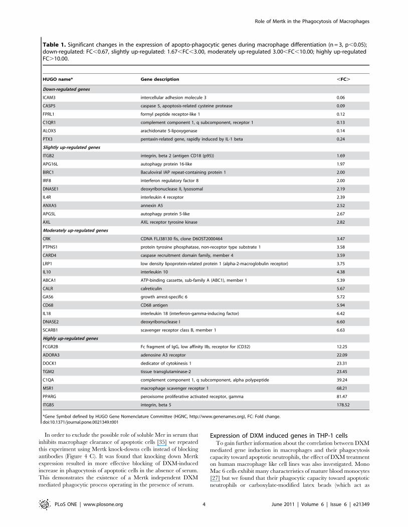
Table 1 lists genes that were significantly up- or down-regulated
during differentiation. The expression level of 29 genes of the
analyzed 95 genes increased significantly and only 6 genes were
down-regulated (e. g. C1QR1, ICAM3, PTX3, FPRL1) in
accordance with our assumption that phagocytosis increase during
macrophage differentiation is resulted from the induction of
apopto - phagocytic genes. The expression level of MERTK in 2
of 3 donors was strongly up-regulated, though this did not prove to
be significant. There was also a heterogenous group of genes with
high level of expression in monocytes that did not change during
differentiation: TGFB1, PECAM1, CD14, THBS1, CAPN 1 and
2, RHOG, ANXA1, PYCARD, ITGAX.
Comparison of gene expression patterns of macrophagesfrom different donors
The relative expressions of apopto-phagocytic genes in
macrophages differentiated from monocytes of different donors
were also compared. We observed that there were non-variable
genes, whose relative expression level varied only 20–50%
amongst donors, and there were highly variable ones, whose
expression level varied in 2–3 orders of magnitude (Figure 1). The
expression level of the non-variable genes was typically higher and
some of them were up-regulated during differentiation (e.g.
PPARc, DOCK1, CD68, ITGAX etc.). Based on these data it
can be assumed that these genes encode proteins that are essential
to macrophage functions and most of them are induced during
differentiation. Phagocytic capacity of each of the eight different
donors was measured but no correlation was found between gene
expression levels and phagocytic capacity (Table S2).
Effect of dexamethasone on the gene expression patternof differentiated macrophages
Glucocorticoids can highly increase the phagocytic capacity of
macrophages toward apoptotic neutrophils [12,13]; according to
our data obtained with macrophages from a large number of
donors and carrying out differentiation for 5 days in the presence
of 1 mM DXM there is usually a 2–2.5 fold enhancement of
phagocytosis (n = 15, data not shown). As a result of DXM
treatment 6 apopto-phagocytic genes were significantly up-
regulated and 2 down-regulated as compared to macrophages
differentiated in the absence of DXM (Figure 2.). The DXM
induced genes encode either receptors (MERTK, AXL,
ADORA3) or bridge-building proteins (C1QA, MFGE8, THBS1)
between phagocytes and apoptotic cell. It should be noted that 4 of
them (AXL, ADORA3, MFGE8 and THBS1) were from the
group of genes whose expression was quite variable among donors,
and the other 4 of them (MERTK, C1QA, ITGB2, TGM2) were
in the intermediate group regarding their variance.
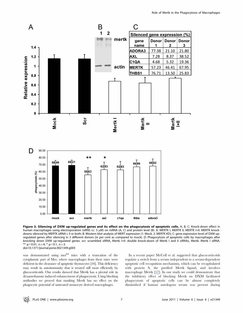
Functional effect of knocking-down of DXM up-regulatedgenes on phagocytosis
To investigate whether DXM induced up-regulation has a
direct effect on the enhanced phagocytic capacity of macrophages
the up-regulated genes were silenced using siRNA. The silencing
efficiency varied in the different genes (Figure 3C). Although the
weakest silencing effect was achieved for MERTK (about 50%
knock-down effect, Figure 3A, C) the phagocytosis of only the
MERTK knock down, DXM treated macrophages decreased
significantly (Figure 3D). The combination of MERTK silencing
with siRNAs for AXL or any of the other DXM up-regulated
genes as well as bridging molecules with each other did not show a
synergistic effect (data not shown).
Impact of blocking Mertk receptor on the phagocytosisof apoptotic neutrophils
We also investigated whether blocking of Mertk with an
antibody verifies its key role in DXM induced increase of
phagocytosis. It was found that while Mertk blocking antibodies
did not inhibit phagocytosis by macrophages differentiated in the
absence of DXM, the DXM-induced increase of phagocytic
capacity of macrophages could be prevented by treatment with
this antibody (Figure 4 A, B). This effect was much weaker when
the assay was performed in the presence of serum, when the
enhancing effect of DXM treatment on macrophages was also
apparent, suggesting that the consequence of blocking Mertk can
be compensated by other engulfing mechanisms in macrophages.
Role of Mertk in the Phagocytosis of Macrophages
PLoS ONE | www.plosone.org 3 June 2011 | Volume 6 | Issue 6 | e21349
In order to exclude the possible role of soluble Mer in serum that
inhibits macrophage clearance of apoptotic cells [35] we repeated
this experiment using Mertk knock-downs cells instead of blocking
antibodies (Figure 4 C). It was found that knocking down Mertk
expression resulted in more effective blocking of DXM-induced
increase in phagocytosis of apoptotic cells in the absence of serum.
This demonstrates the existence of a Mertk independent DXM
mediated phagocytic process operating in the presence of serum.
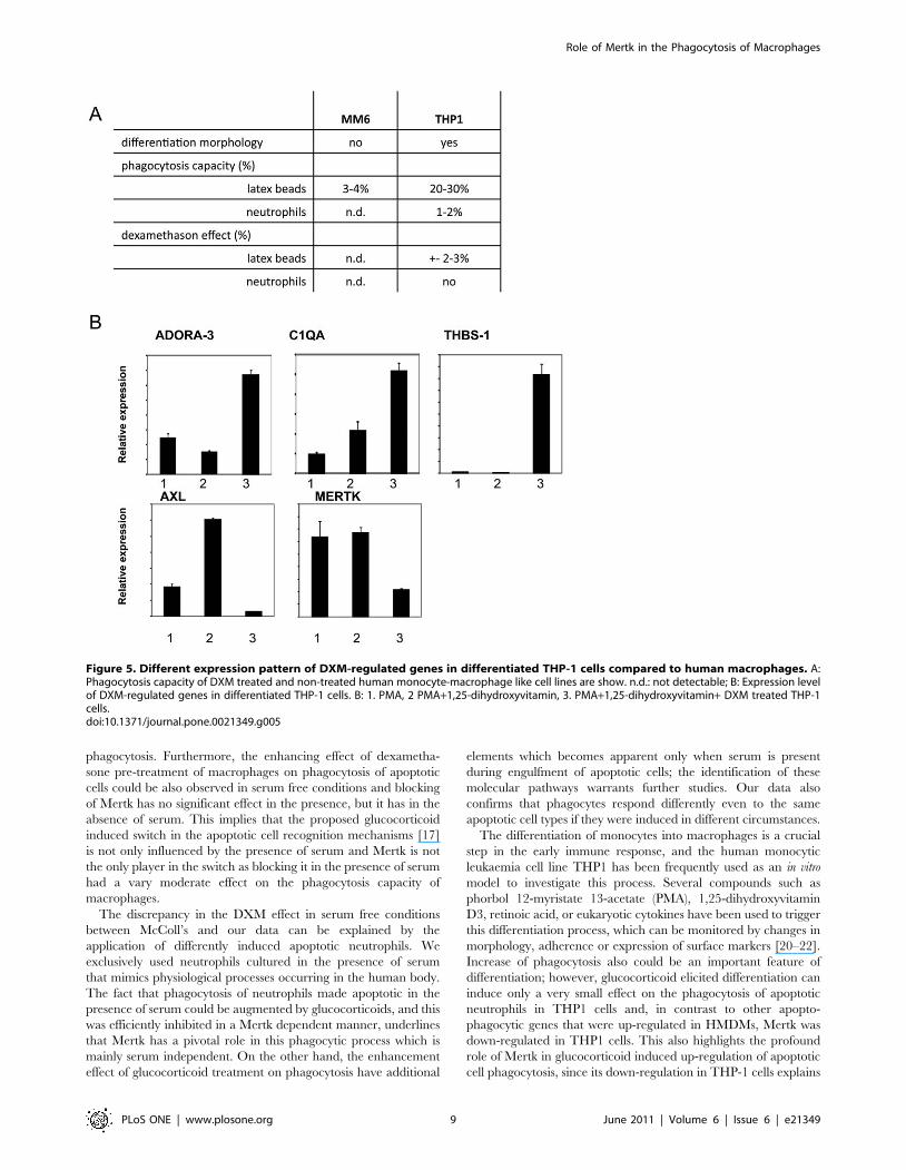
Expression of DXM induced genes in THP-1 cellsTo gain further information about the correlation between DXM
mediated gene induction in macrophages and their phagocytosis
capacity toward apoptotic neutrophils, the effect of DXM treatment
on human macrophage like cell lines was also investigated. Mono
Mac 6 cells exhibit many characteristics of mature blood monocytes
[27] but we found that their phagocytic capacity toward apoptotic
neutrophils or carboxylate-modified latex beads (which act as
Table 1. Significant changes in the expression of apopto-phagocytic genes during macrophage differentiation (n = 3, p,0.05);down-regulated: FC,0.67, slightly up-regulated: 1.67,FC,3.00, moderately up-regulated 3.00,FC,10.00; highly up-regulatedFC.10.00.
HUGO name* Gene description ,FC.
Down-regulated genes
ICAM3 intercellular adhesion molecule 3 0.06
CASP5 caspase 5, apoptosis-related cysteine protease 0.09
FPRL1 formyl peptide receptor-like 1 0.12
C1QR1 complement component 1, q subcomponent, receptor 1 0.13
ALOX5 arachidonate 5-lipoxygenase 0.14
PTX3 pentaxin-related gene, rapidly induced by IL-1 beta 0.24
Slightly up-regulated genes
ITGB2 integrin, beta 2 (antigen CD18 (p95)) 1.69
APG16L autophagy protein 16-like 1.97
BIRC1 Baculoviral IAP repeat-containing protein 1 2.00
IRF8 interferon regulatory factor 8 2.00
DNASE1 deoxyribonuclease II, lysosomal 2.19
IL4R interleukin 4 receptor 2.39
ANXA5 annexin A5 2.52
APG5L autophagy protein 5-like 2.67
AXL AXL receptor tyrosine kinase 2.82
Moderately up-regulated genes
CRK CDNA FLJ38130 fis, clone D6OST2000464 3.47
PTPNS1 protein tyrosine phosphatase, non-receptor type substrate 1 3.58
CARD4 caspase recruitment domain family, member 4 3.59
LRP1 low density lipoprotein-related protein 1 (alpha-2-macroglobulin receptor) 3.75
IL10 interleukin 10 4.38
ABCA1 ATP-binding cassette, sub-family A (ABC1), member 1 5.39
CALR calreticulin 5.67
GAS6 growth arrest-specific 6 5.72
CD68 CD68 antigen 5.94
IL18 interleukin 18 (interferon-gamma-inducing factor) 6.42
DNASE2 deoxyribonuclease I 6.60
SCARB1 scavenger receptor class B, member 1 6.63
Highly up-regulated genes
FCGR2B Fc fragment of IgG, low affinity IIb, receptor for (CD32) 12.25
ADORA3 adenosine A3 receptor 22.09
DOCK1 dedicator of cytokinesis 1 23.31
TGM2 tissue transglutaminase-2 23.45
C1QA complement component 1, q subcomponent, alpha polypeptide 39.24
MSR1 macrophage scavenger receptor 1 68.21
PPARG peroxisome proliferative activated receptor, gamma 81.47
ITGB5 integrin, beta 5 178.52
*Gene Symbol defined by HUGO Gene Nomenclature Committee (HGNC, http://www.genenames.org), FC: Fold change.doi:10.1371/journal.pone.0021349.t001
Role of Mertk in the Phagocytosis of Macrophages
PLoS ONE | www.plosone.org 4 June 2011 | Volume 6 | Issue 6 | e21349

surrogate apoptotic cells) was very low (Figure 5A). Human THP- 1
leukemia cells are known to differentiate along the monocytic
lineage following exposure to phorbol-12-myristate-13-acetate
(PMA) or 1,25-dihydroxyvitamin D3 (VD3) [20]. Although THP-
1 cells are capable of taking up carboxylated polystyrene latex
beads, they can engulf apoptotic neutrophils very poorly and DXM
treatment could not enhance the phagocytosis of either latex beads
or apoptotic cells. Checking the expression of those genes which
were up-regulated in human macrophages by DXM treatment we
found that although ADORA3, C1QA and THBS1 were induced,
the expression level of both MERTK and AXL (the two tyrosine
kinase genes) dropped in contrast to HMDMs (Figure 5B).
Discussion
Differentiation of monocytes to macrophages is associated with
an enhancement of phagocytosis of apoptotic cells and glucocor-
ticoids are capable to further augment this process [19,22]. In this
study we have examined the gene expression changes that underlie
these enhancements in phagocytic capacity. We used a TaqMan
Low Density Array configuration containing TaqMan assays for
the apopto-phagocytic genes of the following categories: receptors
(integrins, scavenger receptors, adenosine receptors, tyrosine
kinases, etc.), bridging molecules, signal generators, effector,
cytokines, nuclear receptors, engulfment genes, autophagy genes,
interferon regulatory family genes (Table S1.). We found that the
expression level of the majority of these genes were altered and
most of them up-regulated during differentiation of monocytes to
macrophages. Among the highly and moderately up-regulated
genes there are several genes whose importance in the phagocy-
tosis of macrophages have already been described either as
receptors (scavenger receptors as CD36, CD68 and CD 204,
FccRIIB, integrin b5), as bridging molecules (C1QA, GAS6)
[23,24], as a molecule with functions on both apoptotic and
Figure 1. Relative levels of gene expression in macrophages differentiated from monocytes of different donors. A: non-variable genes(genes with SD below 20 percentile) represented by average values and error, B: variable genes (genes with SD over 80 percentile) represented byindividual dots, n = 8.doi:10.1371/journal.pone.0021349.g001
Role of Mertk in the Phagocytosis of Macrophages
PLoS ONE | www.plosone.org 5 June 2011 | Volume 6 | Issue 6 | e21349

engulfing cells (calreticulin), or as effector molecules participating
in different signaling pathways e.g. CRK, PTPNS1, CARD4,
ADORA3, DOCK1 and TGM2 [31,32]). In a recent paper we
demonstrated that during differentiation of macrophages natural
ligands of PPARg (also highly up-regulated during differentiation)
are formed, regulating the expression of genes responsible for
effective clearance of apoptotic cells and macrophage-mediated
inflammatory responses [19]. The reason of the down-regulation
of genes with previously demonstrated functions in different
phagocytosis pathways (C1QR1, ICAM3, PTX3, FPRL1) needs
to be clarified in future studies.
The variability of expression levels of apopto-phagocytic genes
among differentiated macrophages of human donors was also
investigated and striking differences were found. Although there
were several genes with very low variability of SD between 20–50%,
the expression levels of another set of genes varied in 2–3 orders of
magnitude. The average relative expression level of non-variable
genes was typically higher than that of the variable ones. During
macrophage differentiation up-regulated genes partly overlapped
with non-variable genes in some cases such as calreticulin, PPARcand CD68 while some with variable ones like FccRIIB, GAS6,
ADORA3. The non-variable gene set might represent permanently
switched on tools for the phagocytosis machinery while the variable
set may provide alternative ways for phagocytosis under various
local conditions to which macrophages may be exposed.
Glucocorticoids are still the most potent immunosuppressive
agents with complex and cell type specific actions on immune cells
[28]. Macrophages are relevant targets for anti-inflammatory
therapy by glucocorticoids not only because of their large repertoire
of inflammatory regulators, but also their alternative anti-inflam-
matory phenotype that was described recently [15]. It was found
that GCs modulate the expression of 130 genes, including anti-
inflammatory ones and those involved in chemotaxis, phagocytosis
and antioxidative stress, while suppresses pro-inflammatory genes
related to apoptosis, adhesion and T-cell chemotaxis. However, the
authors generated the expression profiles of monocytes following
only a 18 h stimulation with fluticasone, a new generation GC with
a higher binding affinity to the GR than dexamethasone; this is a
short time course and GCs may modulate gene transcription
differently over a longer time period [29]. According to our results
most of the up-regulated apopto-phagocytic genes after 5 days of
DXM exposure (C1QA, MFGE8, THBS1, ADORA3, MFGE8)
are the same as those observed after 18 hours treatment. However,
several genes that were found to be induced after 18 h stimulation
were not responding after 5 days treatment; for example, the
receptor for anti-inflammatory mediators FPR1 was not up-
regulated or the adhesion molecules ITGAL, CD36 or OLR1 were
not down-regulated after the long term DXM treatment. This
means that in macrophages only part of the apopto-phagocytic
genes is regulated on the long term by glucocorticoids as compared
to the early or transient gene expression pattern.
Mertk is a member of the TAM (TYRO3, AXL, MER) receptor
protein tyrosine kinase family which have substantial roles in innate
immunity. Their known two ligands, growth-arrest-specific 6 (GAS6)
and protein S bind to and activate all the three TAM receptors with
different affinities and due to their Gla domains they also can bind
phosphatidylserine on apoptotic cells simultaneously [30]. However,
only Mertk seems to be critical for the clearance of apoptotic cells that
Figure 2. Changes in normalized expression levels of apopto-phagocytic genes in macrophages as a result of DXM tretament.Macrophages were differentiated in the presence or absence of dexamethasone. Relative gene expression data of DXM treated macrophages werenormalized to non-treated ones and are shown in heat map. Red: up-regulation, blue: down-regulation. Six genes were up- and 2 down-regulated asdemonstrated by the heat map; the ones up-regulated in each donor (n = 3, p,0.05) are highlighted. Table shows average relative expression levelsof control and DXM treated macrophages, their standard deviation (SD) and fold change (FC) as a ratio of expression levels of DXM treated andcontrol macrophages.doi:10.1371/journal.pone.0021349.g002
Role of Mertk in the Phagocytosis of Macrophages
PLoS ONE | www.plosone.org 6 June 2011 | Volume 6 | Issue 6 | e21349
was demonstrated using merkd mice with a truncation of the
cytoplasmic part of Mer, where macrophages from these mice were
deficient in the clearance of apoptotic thymocytes [16]. This deficiency
may result in autoimmunity that is treated still most efficiently by
glucocorticoids. Our results showed that Mertk has a pivotal role in
dexamethasone induced enhancement of phagocytosis. Using blocking
antibodies we proved that masking Mertk has no effect on the
phagocytic potential of untreated monocyte derived macrophages.
In a recent paper McColl et al. suggested that glucocorticoids
regulate a switch from a serum independent to a serum-dependent
apoptotic cell recognition mechanism, which can be recapitulated
with protein S, the purified Mertk ligand, and involves
macrophage Mertk [17]. In our study we could demonstrate that
the inhibitory effect of blocking Mertk on DXM facilitated
phagocytosis of apoptotic cells can be almost completely
diminished if human autologous serum was present during
Figure 3. Silencing of DXM up-regulated genes and its effect on the phagocytosis of apoptotic cells. A, B, C: Knock-down effect inhuman macrophages using electroporation (siRNS cc. 3 mM) on mRNA (A, C) and protein level (B). A: MERTK I, MERTK II, MERTK I+II: MERTK knock-downs silenced by MERTK siRNA I, II or both; B: Western blot analysis of MERT expression (1: Mock, 2: MERTK KD); C: gene expression level of DXM up-regulated genes after silencing in 3 different donors (in per cent as compared to mock). D: Phagocytosis of apoptotic cells by macrophages afterknocking down DXM up-regulated genes. scr: scrambled siRNA, Mertk I+II: double knock-down of Mertk I and II siRNAs, Mertk: Mertk I siRNA.** p,0.01, n = 4; * p,0.1, n = 3.doi:10.1371/journal.pone.0021349.g003
Role of Mertk in the Phagocytosis of Macrophages
PLoS ONE | www.plosone.org 7 June 2011 | Volume 6 | Issue 6 | e21349

Figure 4. Effect of blocking cell surface Mertk by antibodies on the phagocytosis of apoptotic neutrophils. Mertk on the surface ofDXM treated and nontreated macrophages was blocked by different antibodies in the presence (A) and in the absence (B) of serum; spotted bar: DXMtreated, empty bar: DXM non-treated C: Serum effect on the phagocytosis of apoptotic neutrophils by Mertk knock-down cells. RD and ABC indicateantibodies of R&D Systems and Abcam, respectively. KD: Mertk knock down silenced by Mertk II siRNA; filled bar: including serum, empty bar:excluding serum. * p,0.05, n = 3.doi:10.1371/journal.pone.0021349.g004
Role of Mertk in the Phagocytosis of Macrophages
PLoS ONE | www.plosone.org 8 June 2011 | Volume 6 | Issue 6 | e21349
phagocytosis. Furthermore, the enhancing effect of dexametha-
sone pre-treatment of macrophages on phagocytosis of apoptotic
cells could be also observed in serum free conditions and blocking
of Mertk has no significant effect in the presence, but it has in the
absence of serum. This implies that the proposed glucocorticoid
induced switch in the apoptotic cell recognition mechanisms [17]
is not only influenced by the presence of serum and Mertk is not
the only player in the switch as blocking it in the presence of serum
had a vary moderate effect on the phagocytosis capacity of
macrophages.
The discrepancy in the DXM effect in serum free conditions
between McColl’s and our data can be explained by the
application of differently induced apoptotic neutrophils. We
exclusively used neutrophils cultured in the presence of serum
that mimics physiological processes occurring in the human body.
The fact that phagocytosis of neutrophils made apoptotic in the
presence of serum could be augmented by glucocorticoids, and this
was efficiently inhibited in a Mertk dependent manner, underlines
that Mertk has a pivotal role in this phagocytic process which is
mainly serum independent. On the other hand, the enhancement
effect of glucocorticoid treatment on phagocytosis have additional
elements which becomes apparent only when serum is present
during engulfment of apoptotic cells; the identification of these
molecular pathways warrants further studies. Our data also
confirms that phagocytes respond differently even to the same
apoptotic cell types if they were induced in different circumstances.
The differentiation of monocytes into macrophages is a crucial
step in the early immune response, and the human monocytic
leukaemia cell line THP1 has been frequently used as an in vitro
model to investigate this process. Several compounds such as
phorbol 12-myristate 13-acetate (PMA), 1,25-dihydroxyvitamin
D3, retinoic acid, or eukaryotic cytokines have been used to trigger
this differentiation process, which can be monitored by changes in
morphology, adherence or expression of surface markers [20–22].
Increase of phagocytosis also could be an important feature of
differentiation; however, glucocorticoid elicited differentiation can
induce only a very small effect on the phagocytosis of apoptotic
neutrophils in THP1 cells and, in contrast to other apopto-
phagocytic genes that were up-regulated in HMDMs, Mertk was
down-regulated in THP1 cells. This also highlights the profound
role of Mertk in glucocorticoid induced up-regulation of apoptotic
cell phagocytosis, since its down-regulation in THP-1 cells explains
Figure 5. Different expression pattern of DXM-regulated genes in differentiated THP-1 cells compared to human macrophages. A:Phagocytosis capacity of DXM treated and non-treated human monocyte-macrophage like cell lines are show. n.d.: not detectable; B: Expression levelof DXM-regulated genes in differentiated THP-1 cells. B: 1. PMA, 2 PMA+1,25-dihydroxyvitamin, 3. PMA+1,25-dihydroxyvitamin+ DXM treated THP-1cells.doi:10.1371/journal.pone.0021349.g005
Role of Mertk in the Phagocytosis of Macrophages
PLoS ONE | www.plosone.org 9 June 2011 | Volume 6 | Issue 6 | e21349

why glucocorticoids can not augment phagocytosis of apoptotic
neutrophils by these cells.
Efficient phagocytic clearance of apoptotic cells is crucial in
many biological processes. If this machinery is impaired the
apoptotic cell debris can contribute to the development of sterile
inflammation and autoimmunity [25]. During the resolution phase
of inflammation apoptotic neutrophils are cleared away by a rapid
and efficient process that does not stimulate proinflammatory
macrophage responses [26]. Glucocorticoids represent one of the
most powerful clinical treatments for a range of inflammatory
conditions including severe acute inflammation and autoimmune
diseases. Understanding the mechanism of action used by
glucocorticoids to enhance clearance of dying cells, to which this
study has provided further details, may lead to the development of
novel therapeutic compounds with higher specificity and fewer or
less side effects.
Supporting Information
Table S1 Gene list of the apopto-phagocyte panel. Gene symbol
(HUGO), Alias, Gene description, HGNC code, and Location is
given for each gene as defined by HUGO Gene Nomenclature
Committee (HGNC, http://www.genenames.org).
(DOC)
Table S2 Correlation data between gene expression levels and
phagocytic capacity. It contains HUGO gene ID and R2
calculated with linear regression by Partial Least Squares (PLS) for
correlation between gene expression and phagocytosis data.
(DOC)
Acknowledgments
The authors would like to thank Jennifer Nagy for the technical assistance
in laboratory work and Dr. Goran Petrovski for his criticism in writing this
manuscript.
Author Contributions
Conceived and designed the experiments: GZ LF. Performed the
experiments: GZ EK GM. Analyzed the data: GZ EK. Contributed
reagents/materials/analysis tools: LF. Wrote the paper: GZ EK GM LF.
References
1. Lauber K, Blumenthal SG, Waibel M, Wesselborg S (2004) Clearance ofApoptotic Cells: getting rid of the corpses. Mol Cell 14(3): 277–87.
2. Henson PM, Hume DA (2006) Apoptotic cell removal in development and tissue
homeostasis. Trends Immunol 27(5): 244–50. Rewiew.3. Hume DA (2006) The mononuclear phagocyte system. Curr Opin Immunol
18(1): 49–53. Review.4. Stuart LM, Ezekowitz AB (2005) Phagocytosis: Elegant complexity. Immunity
22(5): 539–50. Rewiew.
5. Savill J, Dransfield I, Gregory C, Haslett C (2002) A blast from the past:clearance of apoptotic cells regulates immune responses. Nat Rev Immunol
2(12): 965–75. Rewiew.6. De Almeida CJ, Linden R (2005) Phagocytosis of apoptotic cells: a matter of
balance. Cell Mol Life Sci 62(14): 1532–46.
7. Gardai SJ, Bratton DL, Ogden CA, Henson PM (2006) Recognition ligands onapoptotic cells: a perspective. J Leukoc Biol 79(5): 896–903.
8. Erwig LP, Henson PM (2008) Clearance of apoptotic cells by phagocytes. CellDeath Differ 15(2): 243–50. Rewiew.
9. Tasker JG, Di S, Malcher-Lopes R (2006) Minireview: rapid glucocorticoidsignaling via membrane-associated receptors. Endocrinology 147(12): 5549–56.
10. Stahn C, Lowenberg M, Hommes DW, Buttgereit F (2007) Molecular
mechanisms of glucocorticoid action and selective glucocorticoid receptoragonists. Mol Cell Endocrinol 275: 71–78.
11. Lim H, Muller N, Herold MJ, Brandt JB, Reichardt HM (2007) Glucocorticoidsexert opposing effects on macrophage function dependent on their concentra-
tion. Immunology 122: 47–53.
12. Liu Y, Cousin JM, Hughes J, Van Damme J, Seckl JR, et al. (1999)Glucocorticoids promote nonphlogistic phagocytosis of apoptotic leukocytes.
J Immunol 162(6): 3639–46.13. Mosser DM (2003) The many faces of macrophage activation. J Leukoc Biol
73(2): 209–12.14. Giles KM, Ross K, Rossi AG, Hotchin NA, Haslett C, et al. (2001)
Glucocorticoid augmentation of macrophage capacity for phagocytosis of
apoptotic cells is associated with reduced p130Cas expression, loss of paxillin/pyk2 phosphorylation, and high levels of active Rac. J Immunol 167(2): 976–86.
15. Ehrchen J, Steinmuller L, Barczyk K, Tenbrock K, Nacken W, et al. (2007)Glucocorticoids induce differentiation of a specifically activated, anti-inflamma-
tory subtype of human monocytes. Blood 109(3): 1265–74.
16. Scott RS, McMahon EJ, Pop SM, Reap EA, Caricchio R, et al. (2001)Phagocytosis and clearance of apoptotic cells is mediated by MER. Nature
411(6834): 207–11.17. McColl A, Bournazous S, Franz S, Perretti M, Morgan PB, et al. (2009)
Glucocorticoids induce protein S-dependent phagocytosis of apoptotic neutro-phils by human macrophages. J Immunol 183: 2167–2175.
18. van Furth R, Raeburn JA, van Zwet TL (1979) Characteristics of human
mononuclear phagocytes. Blood 54(2): 485–500.
19. Majai G, Sarang Z, Csomos K, Zahuczky G, Fesus L (2007) PPARgamma-dependent regulation of human macrophages in phagocytosis of apoptotic cells.
Eur J Immunol 37(5): 1343–54.
20. Schwende H, Fitzke E, Ambs P, Dieter P (1996) Differences in the state ofdifferentiation of THP-1 cells induced by phorbol ester and 1,25-dihydrox-
yvitamin D3. J Leukoc Biol 59(4): 555–61.21. Kohro T, Tanaka T, Murakami T, Wada Y, Aburatani H, et al. (2004) A
comparison of differences in the gene expression profiles of phorbol 12-myristate
13-acetate differentiated THP-1 cells and human monocyte-derived macro-phage, J Atheroscler Thromb 11: 88–97.
22. Daigneault M, Preston JA, Marriott HM, Whyte MK, Dockrell DH (2010) Theidentification of markers of macrophage differentiation in PMA-stimulated
THP-1 cells and monocyte-derived macrophages. PLoS One 5(1): e8668.
23. Mevorach D, Mascarenhas JO, Gershov D, Elkon KB (1998) Complement-dependent clearance of apoptotic cells by human macrophages. J Exp Med
188(12): 2313–20.24. Hart SP, Smith JR, Dransfield I (2004) Phagocytosis of opsonized apoptotic cells:
roles for ‘old-fashioned’ receptors for antibody and complement. Clin ExpImmunol 135(2): 181–5. Review.
25. Erwig LP, Henson PM (2007) Immunological consequences of apoptotic cell
phagocytosis. Am J Pathol 171(1): 2–8.26. Meagher LC, Savill JS, Baker A, Fuller RW, Haslett C (1992) Phagocytosis of
apoptotic neutrophils does not induce macrophage release of thromboxane B2.J Leukoc Biol 52(3): 269–73.
27. Ziegler-Heitbrock HW, Schraut W, Wendelgass P, Strobel M, Sternsdorf T,
et al. (1994) Distinct patterns of differentiation induced in the monocytic cell lineMono Mac 6. J Leukoc Biol 55(1): 73–80.
28. Baschant U, Tuckermann J (2010) The role of the glucocorticoid receptor ininflammation and immunity. J Steroid Biochem Mol Biol 120(2–3): 69–75.
29. Yona S, Gordon S (2007) Inflammation: Glucocorticoids turn the monocyteswitch. Immunol Cell Biol 85(2): 81–2.
30. Lemke G, Rothlin CV (2008) Immunobiology of the TAM receptors. Nat Rev
Immunol 8(5): 327–336.31. Hasko G, Pacher P, Deitch EA, Vizi ES (2007) Shaping of monocyte and
macrophage function by adenosine receptors. Pharmacol Ther 113(2): 264–75.32. Elliott M, Ravichandran KS (2010) Clearance of apoptotic cells: implications in
health and disease. J Cell Biol 189(7): 1059–70. Review.
33. Majai G, Petrovski G, Fesus L (2006) Inflammation and the apopto-phagocyticsystem. Immunol Lett 104(1–2): 94–101. Short review.
34. Petrovski G, Zahuczky G, Katona G, Vereb G, Martinet W, et al. (2007)Clearance of dying autophagic cells of different origin by professional and non-
professional phagocytes. Cell Death and Differentiation 14: 1117–1128.35. Sather S, Kenyon KD, Lefkowitz JB, Liang X, Varnum BC, et al. (2007) A
soluble form of the Mer receptor tyrosine kinase inhibits macrophage clearance
of apoptotic cells and platelet aggregation. Blood 109(3): 1026–33.
Role of Mertk in the Phagocytosis of Macrophages
PLoS ONE | www.plosone.org 10 June 2011 | Volume 6 | Issue 6 | e21349