Correlation between Platelet Count and Lung Dysfunction in ...
Upload
independentCategory
view
5download
0
1
PROLONGED PLATELET PRESERVATION BY TRANSIENT
METABOLIC SUPPRESSION
Bahram Alamdary Badlou
2
Cover: “Girl in Hibernation” adapted from Google
A drawing of resting of platelets (Platelet Hibernation) and after activation by
prolonged storage without recovery made by the Scanning Electron Microscopy
(SEM). The middel photo is made by regular Fluorescence Microscopy of the
interaction between mepacrine-labeled platelets with THP-1 macrophages.
ISBN nr: 90-393-417-61
3
PROLONGED PLATELET PRESERVATION BY TRANSIENT METABOLIC SUPPRESSION
HET VERLENGEN VAN DE BEWAARTIJD VAN BLOEDPLAATJES MET BEHULP VAN METABOLE
SUPPRESSIE
ACADEMISCH PROEFSCRIFT
Ter verkrijgen van de graad van doctor
aan de Universiteit Utrecht
op gezag van de Rector Manificus, Prof. Dr. W.H. Gispen,
in gevolge het besluit van het College voor promoties
in het openbaar te verdedigen
op maandag 13 Maart 2006 des middag te 2.30 uur
door
Bahram Alamdary Badlou
geboren op 11 Juni 1964, te Teheran-Iran
4
Promoter: Prof. Dr. J.W.N. Akkerman
Thrombosis and Haemostasis lab, Hematology Dept.
University Medical Center Utrecht
Medicine Faculty, Utrecht University
The Netherlands
Co-promoter: Dr. W.M. Smid
Sanquin Blood bank NW region
Amsterdam – The Netherlands
The thesis was prepared at the Thrombosis and Haemostasis laboratory, Department of Haematology,
University Medical Centre Utrecht, and The institute for Biomembranes, Utrecht University, Utrecht,
The Netherlands; and Sanquin Blood supply region North West, Amsterdam The Netherlands.
Supported by the Sanquin Blood Supply Foundation (grant nr PPO 01.019).
Financial support by the Netherlands Heart Foundation, for the publication of this thesis is greatfully
acknowledged.
Additional financial support for the publication of this thesis by Sanquin Bloed bank Stichting, AMGEN
B.V., Baxter Homecare, Tebu-Bio, Dr. Ir. Van de Laar Stichting, especially Fresenius Homecare for
their extra product support is gratefully acknowledged.

5
It is not important who you are……… It is important what you want to achieve…. Best future perspective can be yours if you do your best for it… This is our call … to change the course in appropriate way. Yes that is what I am trying to do!
Bahram A. Badlou
For my family and my dear father and mother
you all, that I will always love you
wherever you are….
For you dear Majid Mashadi
As I promised you …

6
7
Content
Page
Abbreviations 8
Chapter 1: General Introduction 9
Chapter 2: Prolonged platelet preservation by metabolic suppression 33
(Transfusion 2005 Feb;45(2):214-22)
Chapter 3: Regulation of Platelet Phagocytosis by Macrophages 49
(Transfusion 2006, in press)
Chapter 4: Role of glycoprotein Ibα in phagocytosis of platelets by macrophages 69
(Submitted)
Chapter 5: Role of Surface Markers for Binding and Phagocytosis of Platelets 87
by Macrophages (Submitted)
Chapter 6: Metabolic Energy Reduction by Glucose Deprivation and Low O2 103
Exchange Preserves Platelet Function After 48 hour Storage at 4°C
(Submitted)
Chapter 7: General discussion 117
Chapter 8: Nederlandse samenvatting 129
Dankwoorden 133
Curriculum Vitea and Publications 137
8
Abbreviations AA = amino acids
ACD = (2.5 g tri-sodium citrate, 1.5 g citric acid and 2.0 g D-glucose in 100 mL distilled water)
AEC = adenylate energy charge
AN51 = anti human GPIb, CD42b
Antimycin A = an inhibitor of mitochondrial respiration, inhibits transfer of electrons from cytochrome B to C
BAPTA-AM = a calcium chelator
BC’s = buffy coats
[Ca2+]i = intercellular calcium concentration in platelets
cAMP = cyclic adenosine monophosphate
CD14 = anti human-monocytic and macrophages antigen; GP53-55
CD42b = anti human –GPIb; AN51 clone R7014 unlabeled from Dako cytomation
CD62p = anti human - P-selectin a component of the α-granule membrane in platelets
CD63 = anti human -GPIIbIIIa
C0 = control group stored on ice
Cyto B = cytochalasin B an inhibitor of actin polymerisation
ER = Energy-reduced platelets induced by glucose deprivation
GPIb = glycoprotein Ibα, a member of GPIbαβ-V-IX complex receptor at surface of platelets
GlcNAc = N-linked glucosamine, a monosaccharide at GPIb
Glycocalicin = a proteolysis sensitive region of GPIbα receptor, contains vWF binding site
HBSS buffer = (0.3 mM KH2PO4, 13.7 mM NaCl, 417 mM NaHCO3, 31 mM Na2HPO4 and 0.5 mM KCl
in aqua dest buffer)
Hepes-Tyrode buffer = (137 mM NaCl, 2.68 mM KCl, 0.42 mM NaH2PO4, 1.7 mM MgCl2, and 11.9
mM NaHCO3, pH 7.2)
Mac-1 = monocytes and macrophages CR3bi receptor; GP170; CD11b
MSP4 = metabolic suppressed platelets prior to storage at 4°C (refrigerator)
PC’s = platelet concentrates
PFA = paraformaldehyde 2%
PGI2 = prostaglandin I2 used to prevent platelet activation during differential centrifugations
PMA = phorbol 12-myristate 13-acetate
PO2 = oxygen pressure and tension in the medium
PS = phosphotyl serine
PLT = platelets
PSL = platelet storage lesion
TAPI = N (R)-[2-(hydroxyaminocarbonyl)methyl]-4-methylpentanoyl-L-alanine amine TNF-alpha
protease inhibitor
THP-1 = monocytic cell lines THP-1
TPO = thrombopoietin hormone
TRAP = Thrombin receptor-activating peptide SFLLRN
VWF = von Willebrand Factor
General introduction
9
Chapter 1
General introduction
- Platelet formation, survival 10
- Platelet functions 11
- Platelet cytoskeleton and organelles 12
- Platelets contain three different types of secretion granules 13
- Platelet mitochondria 13
- Platelet receptors involved in platelet function and survival 16
- Prolonged preservation of platelet viability and function 17
- Platelet isolation, storage and transfusion procedures in the blood bank 19
- Current additive solutions for platelet storage and quality control of PC’s 19
- Attempts to improve platelet survival posttransfusion 20
- Optimal storage conditions for platelet transfusion 22
- Scope of this thesis 23
Chapter 1
10
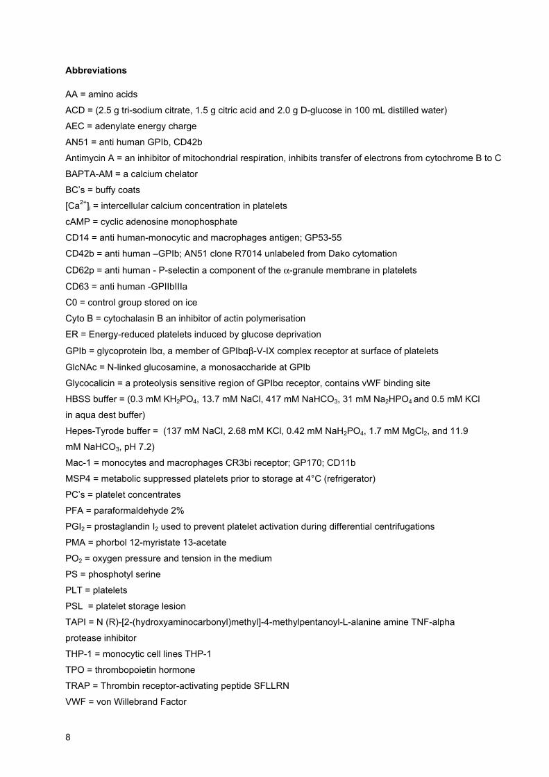
1.1. Platelet formation, survival Platelets are anucleated cells that arise in the bone marrow from megakaryocytes under control of
thrombopoietin (TPO).1 After the megakaryocytes mature, proplatelets are formed and the cytoplasm
becomes demarcated into platelet fields (Figure 1). Platelets are released into the circulation through a
process of megakaryocytic fragmentation.2 The minimal platelet age is about 9 days, and the
maximum about 19 days.3 Normally, two-third of the platelets released from the bone marrow stay in
the peripheral circulation; the remainder is sequestered in the spleen and is freely exchangeable with
the circulating platelets.1;3 Smith4 postulated that in patients with splenomegaly, a larger percentage of
the platelets is sequestered in the spleen, and that peripheral thrombocytopenia may develop. There
is a direct relationship between the megakaryocyte mass in the bone marrow and the rate at which
platelets are released into the circulation.2 When bone marrow is maximally stimulated, it can increase
platelet production six- fold.5 When platelets are rapidly destroyed, increased delivery of platelets to
the peripheral blood shows a lag time of approximately 5 days. Even then, thrombocytopenia may
continue if the rate of platelet production cannot keep up with the rate of platelet destruction.4
Figure 1. Platelet generation from megakaryocytes
General introduction
11
1.2. Platelet functions In haemostasis, platelets play two major roles:
1) Platelets arrest bleeding from severed blood vessels.6 When platelets encounter a disturbance
in the endothelial surface, platelets adhere, aggregate and prevent excessive blood loss.
2) They provide phospholipids that act as the catalytic surface for the coagulation cascade7;8
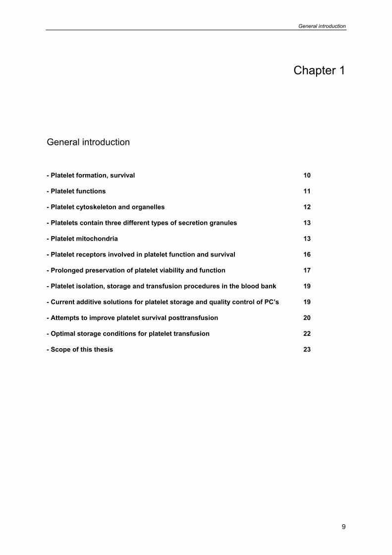
Figure 2. Platelets in a resting state
Platelets remain in a resting condition through release of NO and PGI2 from the endothelium (Figure
2). This keeps platelets in a resting state by inducing formation of cyclic GMP (cGMP) and AMP
(cAMP), which are major inhibitory second messengers.9 Other activators of a cAMP increase are
prostaglandin E1 (PGE1), PGE2, PGD2, PGI2 and adenosine.9-11
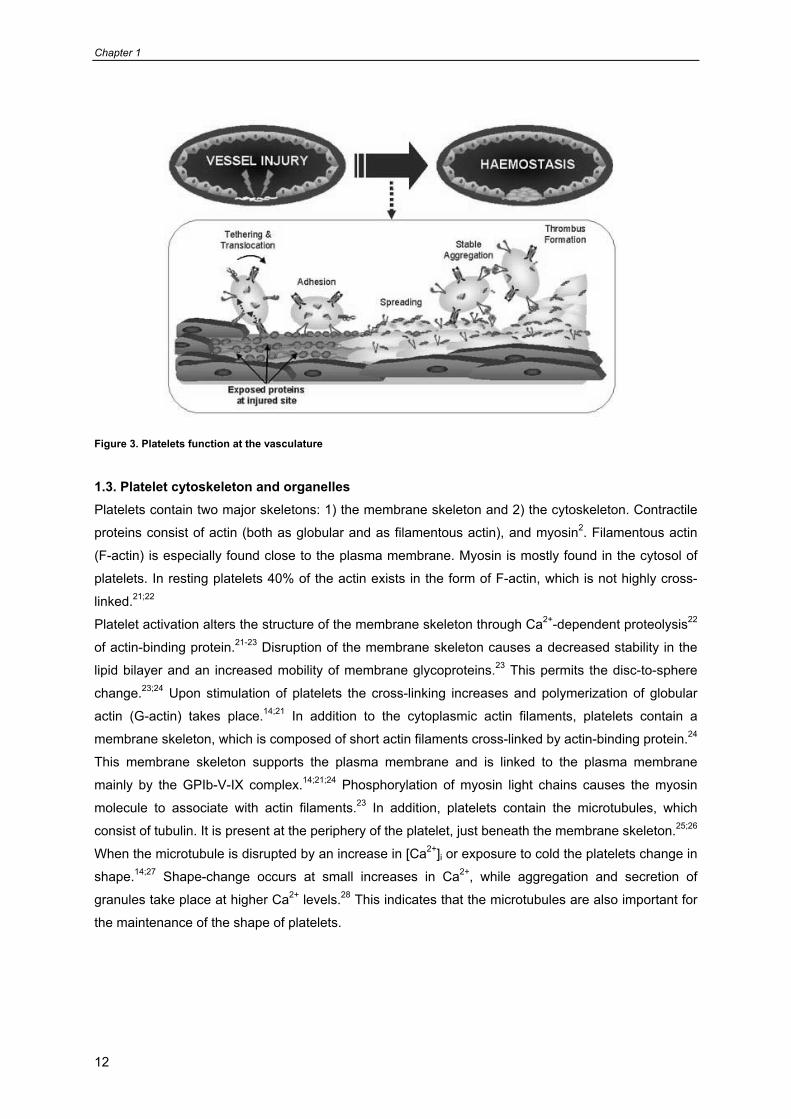
Upon activation by stimulating agents, platelets undergo a rapid transition in shape from a discoid
morphology to spheres with pseudopods.12 Intima injury associated with endothelial denudation and
plaque rupture expose subendothelial collagen and von Willebrand factor (vWF), which support
prompt platelet adhesion and activation.13 The platelet is extremely sensitive to changes in the
environment. A variety of stimuli such as physiological activators, artificial surfaces, mechanical stress,
low temperature, and drugs induce a disc-to-sphere transformation.12;14;15 Platelet responses start with
binding of the glycoprotein (GP) Ib-IX-V complex to vWF in the injured vessel wall. Rolling platelets
slow down by adhesion to exposed vWF and undergo shape change (Figure 3).16
Platelet-specific adhesion receptors mediate these interactions. Other membrane glycoproteins, such
as the collagen receptor GP VI, trigger platelet activation. Engagement of GPIb-IX-V or GP VI
ultimately leads to platelet aggregation mediated by the integrin, αIIbβ3 (GPIIb-IIIa).16-18 After
spreading, platelets start releasing ADP and TXA2.17 Subsequently, the generation of thrombin starts,
which is very strong thrombogenic factor. Platelets form stable aggregates and seal the damaged
vessel wall.19 Following clot retraction, fibrinolysis starts that dissolves the fibrin network and makes
room for wound healing processes.19;20
Chapter 1
12
Figure 3. Platelets function at the vasculature
1.3. Platelet cytoskeleton and organelles Platelets contain two major skeletons: 1) the membrane skeleton and 2) the cytoskeleton. Contractile
proteins consist of actin (both as globular and as filamentous actin), and myosin2. Filamentous actin
(F-actin) is especially found close to the plasma membrane. Myosin is mostly found in the cytosol of
platelets. In resting platelets 40% of the actin exists in the form of F-actin, which is not highly cross-
linked.21;22
Platelet activation alters the structure of the membrane skeleton through Ca2+-dependent proteolysis22
of actin-binding protein.21-23 Disruption of the membrane skeleton causes a decreased stability in the
lipid bilayer and an increased mobility of membrane glycoproteins.23 This permits the disc-to-sphere
change.23;24 Upon stimulation of platelets the cross-linking increases and polymerization of globular
actin (G-actin) takes place.14;21 In addition to the cytoplasmic actin filaments, platelets contain a
membrane skeleton, which is composed of short actin filaments cross-linked by actin-binding protein.24
This membrane skeleton supports the plasma membrane and is linked to the plasma membrane
mainly by the GPIb-V-IX complex.14;21;24 Phosphorylation of myosin light chains causes the myosin
molecule to associate with actin filaments.23 In addition, platelets contain the microtubules, which
consist of tubulin. It is present at the periphery of the platelet, just beneath the membrane skeleton.25;26
When the microtubule is disrupted by an increase in [Ca2+]i or exposure to cold the platelets change in
shape.14;27 Shape-change occurs at small increases in Ca2+, while aggregation and secretion of
granules take place at higher Ca2+ levels.28 This indicates that the microtubules are also important for
the maintenance of the shape of platelets.
General introduction
13
1.4. Platelets contain three different types of secretion granules
Platelets contain three major secretion granules: 1) α- granules 2) dense granules and 3) lysosomal
granules. The contents of the α- granule can be divided into platelet-specific and non-platelet specific
proteins. These proteins are involved in the promotion of haemostasis and tissue repair.29 The internal
side of the α- granules membrane contains the GPIIb/IIIa complex and P-selectin. Both proteins are
exposed on the platelet membrane upon secretion.30 31 P-selectin is a good marker for platelet
activation because in the resting state it is not surface-expressed. Its surface expression correlates
with the release of α- granule content. P-selectin can not be re-internalized following secretion.32;33
The second important granule is the dense- granule, which contains mainly Ca2+, inorganic
pyrophosphate, ATP, ADP, serotonin and catecholamine.34;35 ADP and serotonin play an important
role in amplifying the platelet response. The significance of the dense granules is illustrated in
hereditary and acquired storage pool deficiency.36;37 Both diseases are characterized by a depletion of
the content of the dense granules and this deficiency leads to defect in the secondary phase of
aggregation and to a bleeding tendency.38 The lysosomal granules are common cell organelles, which
play a role in the cellular degradation system as well as in autolytic processes.
1.5. Platelet mitochondria Platelet mitochondria are an important subcellular compartment and are responsible for oxidative
phosphorylation. Platelet mitochondria consist of an outer membrane, an intermembrane space, an
innermembrane, and the matrix. The energy production is a result of electron transport from the matrix
to the innermembrane space leading to phosphorylation of ADP to ATP. In this process oxygen serves
as the electron acceptor. Oxidative ATP synthesis is coupled to transmembrane proton fluxes.
The flow of electrons from NADH or FADH2 to O2 through protein complexes located in the inner
membrane leads to the pumping of protons out of the mitochondrial matrix. Metabolic energy is
generated by the conversion of fatty acids to CO2. Although the rate of ATP resynthesis varies among
different donors the actual availability of energy is present in the steady-state levels of metabolic ATP
(ATPm) and metabolic ADP (ADPm).A better reflection of the energy status in the cell is the adenylate
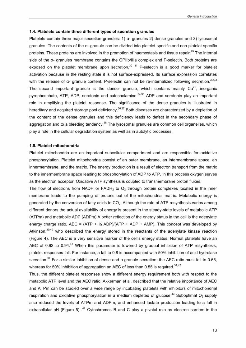
energy charge ratio, AEC = (ATP + ½ ADP)/(ATP + ADP + AMP). This concept was developed by
Atkinson.39;40 who described the energy stored in the reactants of the adenylate kinase reaction
(Figure 4). The AEC is a very sensitive marker of the cell’s energy status. Normal platelets have an
AEC of 0.92 to 0.94.41 When this parameter is lowered by gradual inhibition of ATP resynthesis,
platelet responses fall. For instance, a fall to 0.8 is accompanied with 50% inhibition of acid hydrolase
secretion.37 For a similar inhibition of dense and α-granule secretion, the AEC ratio must fall to 0.65,
whereas for 50% inhibition of aggregation an AEC of less than 0.55 is required.37;42
Thus, the different platelet responses show a different energy requirement both with respect to the
metabolic ATP level and the AEC ratio. Akkerman et al. described that the relative importance of AEC
and ATPm can be studied over a wide range by incubating platelets with inhibitors of mitochondrial
respiration and oxidative phosphorylation in a medium depleted of glucose.43 Suboptimal O2 supply
also reduced the levels of ATPm and ADPm, and enhanced lactate production leading to a fall in
extracellular pH (Figure 5) .44 Cytochromes B and C play a pivotal role as electron carriers in the
Chapter 1
14
respiration chain of platelets during rest and activation. Antimycin A blocks electron transport from
cytochrome B to C (Figure 4).
Figure 4. Metabolic suppression by different inhibitors, adjusted from Akkerman et al. 1985. Adenylate energy charge (AEC)
Compared with other cell types, unstimulated platelets have a high rate of energy production and
consumption. This is exceptional for a cell that lacks a number of energy consuming processes that
are common in other cells, e.g. protein synthesis, biosynthesis of complex carbohydrates, etc. This
has led to hypothesis that resting platelets build up a store of energy that is liberated during
aggregation and secretion.45;46;37 In this respect the role of actin is of special importance since it
undergoes a rapid polymerization-depolymerization cycle.46;47
Platelets resynthesize metabolic ATP in glycogenolysis, glycolysis, and oxidative phosphorylation and
consume the energy stored in metabolic ATP in the various energy utilizing processes present in
unstimulated platelets (basal energy consumption). During shape change, aggregation, and secretion,
the energy consumption increases (incremental energy consumption). The balance between energy
production and consumption is depicted by the AEC. The energy in ATP and ADP is expressed as
ATP equivalents (ATPeq) in which 1 ATPeq represents the energy liberated in the conversion of 1
mole of ATP to 1 mole of ADP.
Rapid abolishment of ATP resynthesis is achieved with metabolic inhibitors, each specific for a certain
pathway (gluconolactone, deoxyglucose, antimycin A). This causes a rapid fall in metabolic ATP and,
later, in metabolic ADP. An important feature of platelets with reduced energy production and
consumption is that the cells lose their responsiveness to activating agents. Apparently, under
conditions of limited energy availability priorities are set to preserve cell integrity at the expense of cell
viability
platelet functions
[ATP + ½ ADP +
[ATP + ½ ADP] AEC =
ATPm
glycogenolysis
glycolysis
oxidative phosphorylation
ADPm
antimycin A or cyanide
deoxyglucose Energy consumption
gluconolactone
General introduction
15
functions. Thus, it is possible to prevent platelet activation by transient metabolic suppression and to
restore functionality prior to infusion by restoring energy supply.
Platelets preserve their rapidly accessible energy in the form of metabolic ATP and ADP (ATPm and
ADPm) as opposed to storage ATP and ADP in the dense granules, which is not accessible to energy
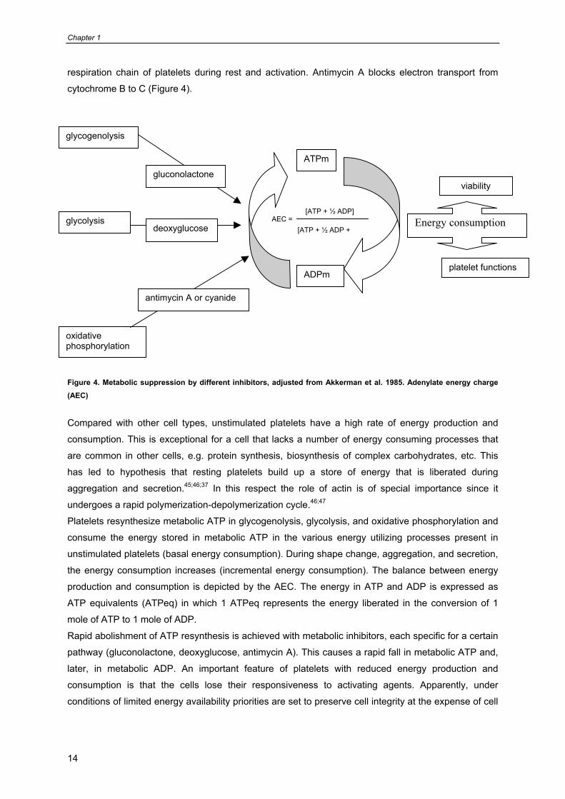
demanding functions.45 With sufficient glucose and O2 in plasma the combined glycolytic and
mitochondrial activity suffices to maintain stable levels of ATPm and ADPm and an optimal adenylate
energy charge AEC.41;48
Figure 5. Platelet metabolic energy production. Rapidly accessible metabolic energy is metabolic ATP (ATPm) and ADP (ATPm).
The absence of glucose compromises optimal energy parameters and impairs platelet functions.49
Under prolonged hypoxic conditions, platelet mitochondria and capases are directly involved in platelet
apoptosis.48-51
In O2-rich conditions glycolysis is suppressed (Pasteur effect) conversely, in O2-poor conditions
glycolysis is not suppressed and lactate production increases (Crabtree effect) (Figure 5). Genova et
al. described that mitochondria are strong producers of reactive oxygen species (ROS), and at the
same time, vulnerable to the oxidative damage produced by their action on lipids and proteins.52;53 In
particular, damage to mitochondrial DNA induces alterations to the polypeptides encoded by this DNA
in the respiratory complexes, resulting in a decrease of electron transfer and a further production of
ROS.52;53 This deficiency in mitochondrial energetic capacity is considered a cause of cell ageing.
Complex I would be the enzyme most affected by ROS, since it contains seven of the 13 subunits
encoded by mitochondrial DNA.52
CO2 O2
mitochondrial respiration
glycolysis / glycogenolysis
ATPm
ADPm
ATPm
ADPm
Lactate formation
pH
aerobic conditions
Pasture effect Crabtree effect
glycolysis glycolysis
anaerobic conditions
Chapter 1
16
Leytin et al proposed the view that platelet mitochondria may be an important apoptotic target during
isolation and prolonged storage.54 In addition, it has been suggested that depletion of growth factors or
treatment with apoptotic stimuli artificially induces anucleated cells to undergo apoptotic features that
are indistinguishable from those of their parent nucleated cell during apoptosis.54;55 Therefore, the
collective evidence so far suggests that in platelets apoptosis may be induced by mitochondria.56
After chronic exposure to various harmful stimuli platelet mitochondrial function reduces53, for instance
after: prolonged exposure to cold (< 15°C); 57-59 exposure to high concentrations of mitochondrial
inhibitors e.g. cyanide and antimycin A;51 or by an increase in lactate production during a prolonged
anaerobic condition.60,52;53
1.6. Platelet receptors involved in platelet function and survival
Platelets contain different receptors and phospholipids involved in platelet function. The major
membrane receptors are: GPIb-V-IX complex (vWF receptor), GPIIbIIIa (fibrinogen receptor), GPVI
(collagen receptor). The major phospholipid involved in procoagulant activity of platelets is
phosphotidyl serine (PS), which in the resting state is kept in the inner leaflet of the plasma membrane
by energy and Ca2+-dependent processes. The major adhesive molecule expressed from α-granule to
the surface of platelets after activation is P-selectin.
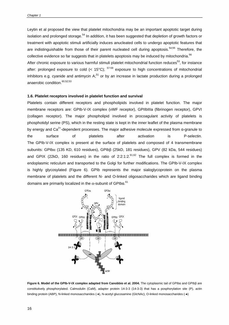
The GPIb-V-IX complex is present at the surface of platelets and composed of 4 transmembrane
subunits: GPIbα (135 KD, 610 residues), GPIbβ (25kD, 181 residues), GPV (82 kDa, 544 residues)
and GPIX (22kD, 160 residues) in the ratio of 2:2:1:2.61,62 The full complex is formed in the
endoplasmic reticulum and transported to the Golgi for further modifications. The GPIb-V-IX complex
is highly glycosylated (Figure 6). GPIb represents the major sialoglycoprotein on the plasma
membrane of platelets and the different N- and O-linked oligosaccharides which are ligand binding
domains are primarily localized in the α-subunit of GPIbα.61
Figure 6. Model of the GPIb-V-IX complex adapted from Canobbio et al. 2004. The cytoplasmic tail of GPIbα and GPIbβ are
constitutively phosphorylated. Calmodulin (CaM), adapter protein 14-3-3 (14-3-3) that has a posphorylation site (P), actin
binding protein (ABP), N-linked monosaccharides (-♦), N-acetyl glucosamine (GlcNAc), O-linked monosaccharides (-●)
General introduction
17
The C-terminal serine of GPIbα is constitutively phosphorylated and is under control of cAMP-
dependent protein kinase A. Canobbio et al61 described that the cytoplasmic domains of the single
subunits of the GPIbα interact with the cytoskeletal actin-binding protein and 14-3-3ξ adapter proteins.
The β-subunits of GPIb contain N-linked oligosaccharides which are covalently linked to GPIbα and
non-covalently to GPIX. The cytoplasmic domain of GPIbβ interacts with calmodulin and adapter
protein 14-3-3ξ.17 GPV is composed of an extracellular domain with 15 Leucine-rich-repeats (LRRs), a
single transmembrane domain, and a cytoplasmic tail of 16 amino acids.63;64The cytoplasmic tail of
GPV associates with calmodulin and 14-3-3ξ protein as well.17 GPV binds via ligand binding domains
and the different N- and O-linked oligosaccharides to GPIbα. GPIb on platelets may also mediate
platelet-endothelium or platelet-leukocyte adhesion by recognition of P-selectin or Mac-1,
respectively.17
P-selectin (also known as CD62p, GMP-140, and PADGEM) is a component of the α granule
membrane that becomes surface-expressed after activating agents induce exocytosis of secretion
granules.65;66 It mediates the tethering of platelets to leukocytes in vitro and in vivo.66-68 Another
important feature of platelet activation is the membrane appearance of negatively charged
phospholipids such as PS.69;70 PS is also exposed on cells going into apoptosis.50;54;71-73 PS exposure
may form recognition sites for destruction of senescent platelets and a similar mechanism might
remove stored platelets from the circulation.71;72;74;75
1.7. Prolonged preservation of platelet viability and function
The storage of platelet concentrates at room temperature facilitates bacterial multiplication76 and
introduces changes in platelets indicative for activation and initiation of apoptosis.58;77 Improvements
have been sought in lowering the storage temperature. Platelet metabolic energy production shows a
decrease approximately by factor 2 per 10°C, lowering in storage temperature.78
Attempts to preserve platelet membrane integrity have been based on addition of glycine, trehalose,
and DMSO during chilling and freezing of isolated platelet concentrates.59;79 Eventually, platelets
become activated and form aggregates that make them vulnerable for rapid clearance by phagocytes
in vivo.15;57-59;80 Despite all these problems, the anticipated advantages of cold storage are so powerful
that several laboratories continue to investigate possible alternatives to oppose the disadvantages of
the chilling process.
Under current investigation is the addition of N-linked monosaccharide to preserve GPIb-V-IX complex
against clustering during chilling and rewarming of PC’s.80 Other investigations focus on addition of
second messenger regulators to increase cAMP, cGMP levels to prevent platelet activation during
storage.76 Other laboratories investigate which fraction of platelet poor plasma (PPP) (> 10 to 30%) in
combination with a platelet additive solution optimally preserves platelet function.81-83
Chapter 1
18
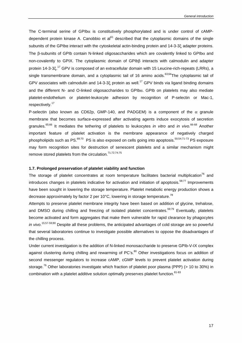
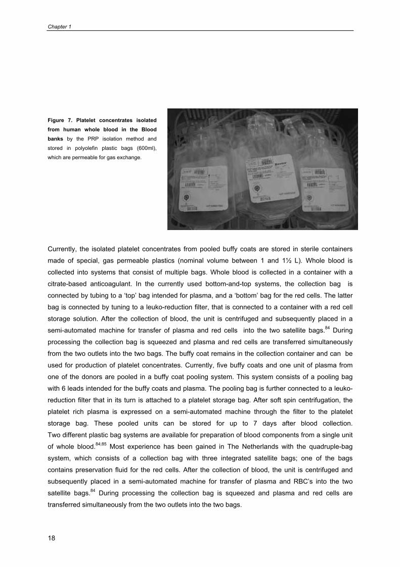
Figure 7. Platelet concentrates isolated from human whole blood in the Blood banks by the PRP isolation method and
stored in polyolefin plastic bags (600ml),
which are permeable for gas exchange.
Currently, the isolated platelet concentrates from pooled buffy coats are stored in sterile containers
made of special, gas permeable plastics (nominal volume between 1 and 1½ L). Whole blood is
collected into systems that consist of multiple bags. Whole blood is collected in a container with a
citrate-based anticoagulant. In the currently used bottom-and-top systems, the collection bag is
connected by tubing to a ‘top’ bag intended for plasma, and a ‘bottom’ bag for the red cells. The latter
bag is connected by tuning to a leuko-reduction filter, that is connected to a container with a red cell
storage solution. After the collection of blood, the unit is centrifuged and subsequently placed in a
semi-automated machine for transfer of plasma and red cells into the two satellite bags.84 During
processing the collection bag is squeezed and plasma and red cells are transferred simultaneously
from the two outlets into the two bags. The buffy coat remains in the collection container and can be
used for production of platelet concentrates. Currently, five buffy coats and one unit of plasma from
one of the donors are pooled in a buffy coat pooling system. This system consists of a pooling bag
with 6 leads intended for the buffy coats and plasma. The pooling bag is further connected to a leuko-
reduction filter that in its turn is attached to a platelet storage bag. After soft spin centrifugation, the
platelet rich plasma is expressed on a semi-automated machine through the filter to the platelet
storage bag. These pooled units can be stored for up to 7 days after blood collection.
Two different plastic bag systems are available for preparation of blood components from a single unit
of whole blood.84;85 Most experience has been gained in The Netherlands with the quadruple-bag
system, which consists of a collection bag with three integrated satellite bags; one of the bags
contains preservation fluid for the red cells. After the collection of blood, the unit is centrifuged and
subsequently placed in a semi-automated machine for transfer of plasma and RBC’s into the two
satellite bags.84 During processing the collection bag is squeezed and plasma and red cells are
transferred simultaneously from the two outlets into the two bags.
General introduction
19
1.8. Platelet isolation, storage and transfusion procedures in the blood bank In the past thirty years the use of blood components for treatment of thrombocytopenic patients has
increased significantly. Specific devices (Figure 7) have been developed to prepare separate fractions
from a unit of whole blood for transfusion: plasma, buffy coat and red blood cells (RBC).
For PC’s there are three methods to isolate platelets from whole blood in use in blood banks: 1) PRP
method, 2)apheresis, and 3) buffy coat method. The PRP method carried out by direct isolation of
PRP and RBC from whole blood, this method is mainly used in USA (Figure 7). On demand five or six
single donor units of platelets are pooled for preparation of the PC’s containing a fraction of plasma
about 10 to 30%.83;86-88 The buffy coat method is used in European blood banks, where from whole
blood direct platelet poor plasma, buffy coat and RBC are isolated and prepared for transfusion
(Figure 8).89 PC’s produced by pooling of buffy coats (4-6) and a unit of PAS or PPP. It has been
demonstrated that white cell can be significantly reduced by special filters either in the blood bank or
at the bedside.90-92 At present PC’s, independent of their production are stored at room temperature,
continuously are laying on flat bed shakers in gas permeable bags to maintain O2 tension and to lose
CO2.
1.9. Current additive solutions for platelet storage and quality control of PC’s Replacing plasma with synthetic media for storage has the advantage of reducing the allergic
reactions posttransfusion, and reduces the need for plasma.93,94 There is an extensive body of
literature about the use of synthetic media for storage of platelets.82;83;95;96 The semiautomated devices
prepare buffy coats on a large scale, and 4 to 20 buffy coats can be pooled immediately after
preparation and resuspended in specific synthetic media with 10 to 30% plasma.83;96 Platelets
resuspended in additive solutions show a gradual increase in P-selectin expression and lactate
production during prolonged storage. Hence, a minimum of 30% plasma remains is necessary to
preserve premature activation.81;97 Major progress has been made by the introduction of acetate (PAS-
II).86,98 Eggen et al. showed that addition of potassium, gluconate and magnesium to additive solution,
better preserves use of PC’s.83;87;99 Similar results were seen with platelets prepared by buffy coats
and apheresis methods, despite differences in equipment, the preparation technique and the final
platelet counts. Gullikson et al. described that storage of platelets in PAS-IIIM (containing potassium,
magnesium and acetate) improves maintenance of platelet function and allows a plasma reduction to
20%.96;97
Methods to evaluate the effect of additive solutions on PC’s are based on P-selectin expression,100;101
and PS exposure (PS flip-flop).50;54;77 Morphological score, hypotonic shock response, and shape
change are also used by a number of Blood banks (Table 1).59;101;102 Today it would be valuable to
develop a so-called golden standard for in vitro methods for evaluation of platelet function, which can
be used to predict platelet haemostatic effectiveness and survival in vivo.
Currently, different quantitative measurements of P-selectin expression and annexin V binding to
exposed PS56;68;79;103-107 carried out by FACS flowcytometry in combination with aggregation studies
that offer valuable information about platelet function, while for survival there is still no good tool yet.
Chapter 1
20
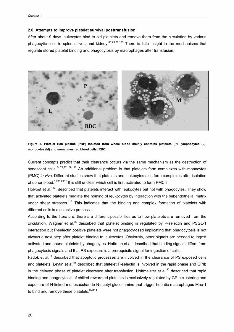
2.0. Attempts to improve platelet survival posttransfusion After about 9 days leukocytes bind to old platelets and remove them from the circulation by various
phagocytic cells in spleen, liver, and kidney.65;73;80;108 There is little insight in the mechanisms that
regulate stored platelet binding and phagocytosis by macrophages after transfusion.
Figure 8. Platelet rich plasma (PRP) isolated from whole blood mainly contains platelets (P), lymphocytes (L), monocytes (M) and sometimes red blood cells (RBC).
Current concepts predict that their clearance occurs via the same mechanism as the destruction of
senescent cells.54;73;77;109;110 An additional problem is that platelets form complexes with monocytes
(PMC) in vivo. Different studies show that platelets and leukocytes also form complexes after isolation
of donor blood.13;111;112 It is still unclear which cell is first activated to form PMC’s.
Holvoet et al.113, described that platelets interact with leukocytes but not with phagocytes. They show
that activated platelets mediate the homing of leukocytes by interaction with the subendothelial matrix
under shear stresses.113 This indicates that the binding and complex formation of platelets with
different cells is a selective process.
According to the literature, there are different possibilities as to how platelets are removed from the
circulation. Wagner et al.66 described that platelet binding is regulated by P-selectin and PSGL-1
interaction but P-selectin positive platelets were not phagocytosed implicating that phagocytosis is not
always a next step after platelet binding to leukocytes. Obviously, other signals are needed to ingest
activated and bound platelets by phagocytes. Hoffman et al. described that binding signals differs from
phagocytosis signals and that PS exposure is a prerequisite signal for ingestion of cells.
Fadok et al.75 described that apoptotic processes are involved in the clearance of PS exposed cells
and platelets. Leytin et al.55 described that platelet P-selectin is involved in the rapid phase and GPIb
in the delayed phase of platelet clearance after transfusion. Hoffmeister et al.80 described that rapid
binding and phagocytosis of chilled-rewarmed platelets is exclusively regulated by GPIb clustering and
exposure of N-linked monosaccharide N-acetyl glucosamine that trigger hepatic macrophages Mac-1
to bind and remove these platelets.80;114
L
RBC
P
M
General introduction
21
Furthermore, Josefsson EC et al.114 have specified that the macrophage αMβ2 integrin αM lectin
domain mediates the phagocytosis of chilled-rewarmed platelets. Unfortunately, they did not show the
exact mechanism that underlies the GPIb clustering of cold and room temperature stored platelets.
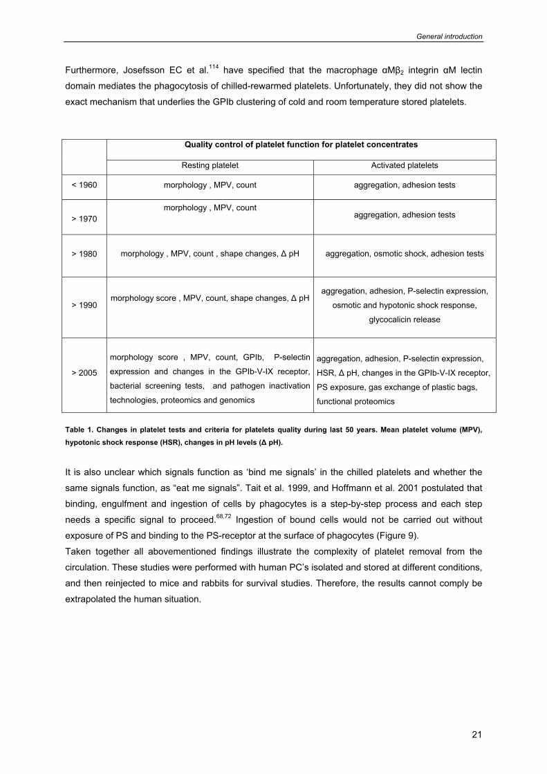
Table 1. Changes in platelet tests and criteria for platelets quality during last 50 years. Mean platelet volume (MPV), hypotonic shock response (HSR), changes in pH levels (Δ pH).
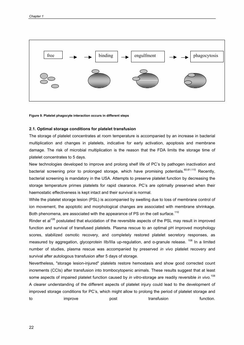
It is also unclear which signals function as ‘bind me signals’ in the chilled platelets and whether the
same signals function, as “eat me signals”. Tait et al. 1999, and Hoffmann et al. 2001 postulated that
binding, engulfment and ingestion of cells by phagocytes is a step-by-step process and each step
needs a specific signal to proceed.68,72 Ingestion of bound cells would not be carried out without
exposure of PS and binding to the PS-receptor at the surface of phagocytes (Figure 9).
Taken together all abovementioned findings illustrate the complexity of platelet removal from the
circulation. These studies were performed with human PC’s isolated and stored at different conditions,
and then reinjected to mice and rabbits for survival studies. Therefore, the results cannot comply be
extrapolated the human situation.
Quality control of platelet function for platelet concentrates
Resting platelet Activated platelets
< 1960 morphology , MPV, count aggregation, adhesion tests
> 1970 morphology , MPV, count
aggregation, adhesion tests
> 1980
morphology , MPV, count , shape changes, Δ pH aggregation, osmotic shock, adhesion tests
> 1990
morphology score , MPV, count, shape changes, Δ pH
aggregation, adhesion, P-selectin expression,
osmotic and hypotonic shock response,
glycocalicin release
> 2005
morphology score , MPV, count, GPIb, P-selectin
expression and changes in the GPIb-V-IX receptor,
bacterial screening tests, and pathogen inactivation
technologies, proteomics and genomics
aggregation, adhesion, P-selectin expression,
HSR, Δ pH, changes in the GPIb-V-IX receptor,
PS exposure, gas exchange of plastic bags,
functional proteomics
Chapter 1
22
Figure 9. Platelet phagocyte interaction occurs in different steps 2.1. Optimal storage conditions for platelet transfusion The storage of platelet concentrates at room temperature is accompanied by an increase in bacterial
multiplication and changes in platelets, indicative for early activation, apoptosis and membrane
damage. The risk of microbial multiplication is the reason that the FDA limits the storage time of
platelet concentrates to 5 days. New technologies developed to improve and prolong shelf life of PC’s by pathogen inactivation and
bacterial screening prior to prolonged storage, which have promising potentials.60;81;115 Recently,
bacterial screening is mandatory in the USA. Attempts to preserve platelet function by decreasing the
storage temperature primes platelets for rapid clearance. PC’s are optimally preserved when their
haemostatic effectiveness is kept intact and their survival is normal.
While the platelet storage lesion (PSL) is accompanied by swelling due to loss of membrane control of
ion movement, the apoptotic and morphological changes are associated with membrane shrinkage.
Both phenomena, are associated with the appearance of PS on the cell surface.110
Rinder et al108 postulated that elucidation of the reversible aspects of the PSL may result in improved
function and survival of transfused platelets. Plasma rescue to an optimal pH improved morphology
scores, stabilized osmotic recovery, and completely restored platelet secretory responses, as
measured by aggregation, glycoprotein IIb/IIIa up-regulation, and α-granule release. 108 In a limited
number of studies, plasma rescue was accompanied by preserved in vivo platelet recovery and
survival after autologous transfusion after 5 days of storage.
Nevertheless, "storage lesion-injured" platelets restore hemostasis and show good corrected count
increments (CCIs) after transfusion into trombocytopenic animals. These results suggest that at least
some aspects of impaired platelet function caused by in vitro-storage are readily reversible in vivo.108
A clearer understanding of the different aspects of platelet injury could lead to the development of
improved storage conditions for PC’s, which might allow to prolong the period of platelet storage and
to improve post transfusion function.
bindingfree engulfment phagocytosis
General introduction
23
2.2. Scope of this thesis Platelet transfusion therapy is associated with several problems, including refractoriness and
transmission of infectious agents and transfusion reactions.116-119 In addition, there is a significant
decrease in platelet responsiveness to activating agents79 and appearance of phagocytosis markers
during prolonged storage resulting in decreased haemostatic effectiveness and rapid clearance from
the circulation after transfusion.105 Improvements have been sought in lowering the storage
temperature from 22 to 0 – 4 °C and replacing plasma by synthetic media to prolong preservation time
and suppress allergic reactions. However, platelets become activated by a sharp decrease in
temperature and this so-called cold-induced platelet lesion has long been a major argument against
platelet storage at low temperature.
Previous work has shown that suppression of energy generation reduces the capacity of platelets to
respond to activating agents. Platelets could even sustain a short period of blocked energy generation
without losing their functional properties.
The present study was undertaken to investigate whether metabolic arrest would protect platelets
against cold-induced platelet disturbances and would make it possible to store platelets in the cold for
a prolonged period of time. Specific questions that were addressed included:
1. Does transient metabolic arrest followed by storage at 4°C better preserve platelet functions
than conventional storage (Chapter 2)?
2. Does transient metabolic arrest followed by storage at 4°C suppress the surface expression of
phagocytotic signals that would lead to enhanced platelet destruction after transfusion
(Chapter 3)?
3. If phagocytosis of cold-stored platelets is mediated via clusters of the Von Willebrand Factor
receptor GPIbα, is it possible to design a method for quantitative analysis of GPIbα clustering
(Chapter 4)?
4. What are the quantitative relations between the surface markers on platelets for platelet-
macrophage interaction and binding/ phagocytosis by macrophages (Chapter 5)?
5. Originally, transient metabolic arrest was induced by incubating platelets in glucose-free,
antimycin A-containing medium, conditions that are incompatible with platelet transfusion. Can
metabolic arrest be induced without antimycin A under conditions that are suitable for platelet
transfusion (Chapter 6).
6. Finally, the results of these studies were compared with current literature in order to design
strategies for better storage conditions in the near future (Chapter 7).
Chapter 1
24
References
1. Harker LA. Platelet survival time: its measurement and use. Prog.Hemost.Thromb.
1978;4:321-47.:321-347.
2. Hartwig J, Italiano J, Jr. The birth of the platelet. J.Thromb.Haemost. 2003;1:1580-1586.
3. Harker LA, Roskos LK, Marzec UM et al. Effects of megakaryocyte growth and development
factor on platelet production, platelet life span, and platelet function in healthy human
volunteers. Blood 2000;95:2514-2522.
4. D.M.Smith., S. H. Summers. 1. Blood platelets-Transfusion 2. Platelet. D.M.Smith., S. H.
Summers. 1. 1988. Arlington, Virginia 22209.
Ref Type: Serial (Book,Monograph)
5. Kunicki TJ, Tuccelli M, Becker GA, Aster RH. A study of variables affecting the quality of
platelets stored at "room temperature". Transfusion 1975;15:414-421.
6. Nieswandt B, Aktas B, Moers A, Sachs UJ. Platelets in atherothrombosis: lessons from mouse
models. J.Thromb.Haemost. 2005;3:1725-1736.
7. Mendelsohn EE, Solum NO, Brosstad F. Effects of platelets and platelet-derived material on
the activated partial thromboplastin time (Cephotest) coagulation test. Scand.J.Clin.Lab Invest
2005;65:321-332.
8. Solum NO. Procoagulant expression in platelets and defects leading to clinical disorders.
Arterioscler.Thromb.Vasc.Biol. 1999;19:2841-2846.
9. Dutta-Roy AK, Sinha AK. Purification and properties of prostaglandin E1/prostacyclin receptor
of human blood platelets. J.Biol.Chem. 1987;262:12685-12691.
10. Hourani SM, Cusack NJ. Pharmacological receptors on blood platelets. Pharmacol.Rev.
1991;43:243-298.
11. Akkerman JW, van WG. Platelet activation via trimeric GTP-binding proteins. Haemostasis
1996;26 Suppl 4:199-209.:199-209.
12. Winokur R, Hartwig JH. Mechanism of shape change in chilled human platelets. Blood
1995;85:1796-1804.
13. Freedman JE, Loscalzo J. Platelet-monocyte aggregates: bridging thrombosis and
inflammation. Circulation 2002;105:2130-2132.
14. Hartwig JH. Mechanisms of actin rearrangements mediating platelet activation. J.Cell Biol.
1992;118:1421-1442.
15. Hoffmeister KM, Falet H, Toker A et al. Mechanisms of cold-induced platelet actin assembly.
J.Biol.Chem. 2001;276:24751-24759.
16. Andrews RK, Berndt MC. Platelet physiology and thrombosis. Thromb.Res. 2004;114:447-
453.
17. Andrews RK, Shen Y, Gardiner EE, Berndt MC. Platelet adhesion receptors and
(patho)physiological thrombus formation. Histol.Histopathol. 2001;16:969-980.
General introduction
25
18. Andrews RK, Lopez JA, Berndt MC. Molecular mechanisms of platelet adhesion and
activation. Int.J.Biochem.Cell Biol. 1997;29:91-105.
19. Farndale RW, Sixma JJ, Barnes MJ, de Groot PG. The role of collagen in thrombosis and
hemostasis. J.Thromb.Haemost. 2004;2:561-573.
20. Kuijper PH, Gallardo Torres HI, Lammers JW et al. Platelet and fibrin deposition at the
damaged vessel wall: cooperative substrates for neutrophil adhesion under flow conditions.
Blood 1997;89:166-175.
21. Fox JE, Boyles JK, Reynolds CC, Phillips DR. Actin filament content and organization in
unstimulated platelets. J.Cell Biol. 1984;98:1985-1991.
22. Fox JE, Reynolds CC, Phillips DR. Calcium-dependent proteolysis occurs during platelet
aggregation. J.Biol.Chem. 1983;258:9973-9981.
23. Fox JE, Phillips DR. Polymerization and organization of actin filaments within platelets.
Semin.Hematol. 1983;20:243-260.
24. Fox JE. Identification of actin-binding protein as the protein linking the membrane skeleton to
glycoproteins on platelet plasma membranes. J.Biol.Chem. 1985;260:11970-11977.
25. Nachmias VT. Platelet and megakaryocyte shape change: triggered alterations in the
cytoskeleton. Semin.Hematol. 1983;20:261-281.
26. Leven RM, Gonnella PA, Reeber MJ, Nachmias VT. Platelet shape change and cytoskeletal
assembly: effects of pH and monovalent cation ionophores. Thromb.Haemost. 1983;49:230-
234.
27. White JG. Arrangements of actin filaments in the cytoskeleton of human platelets.
Am.J.Pathol. 1984;117:207-217.
28. Blockmans D, Deckmyn H, Vermylen J. Platelet activation. Blood Rev. 1995;9:143-156.
29. Cramer EM, Savidge GF, Vainchenker W et al. Alpha-granule pool of glycoprotein IIb-IIIa in
normal and pathologic platelets and megakaryocytes. Blood 1990;75:1220-1227.
30. McEver RP, Martin MN. A monoclonal antibody to a membrane glycoprotein binds only to
activated platelets. J.Biol.Chem. 1984;259:9799-9804.
31. Stenberg PE, McEver RP, Shuman MA, Jacques YV, Bainton DF. A platelet alpha-granule
membrane protein (GMP-140) is expressed on the plasma membrane after activation. J.Cell
Biol. 1985;101:880-886.
32. George JN, Pickett EB, Saucerman S et al. Platelet surface glycoproteins. Studies on resting
and activated platelets and platelet membrane microparticles in normal subjects, and
observations in patients during adult respiratory distress syndrome and cardiac surgery.
J.Clin.Invest 1986;78:340-348.
33. Heijnen HF, Schiel AE, Fijnheer R, Geuze HJ, Sixma JJ. Activated platelets release two types
of membrane vesicles: microvesicles by surface shedding and exosomes derived from
exocytosis of multivesicular bodies and alpha-granules. Blood 1999;94:3791-3799.
34. de Korte D, Gouwerok CW, Fijnheer R, Pietersz RN, Roos D. Depletion of dense granule
nucleotides during storage of human platelets. Thromb.Haemost. 1990;63:275-278.
Chapter 1
26
35. Jedlitschky G, Tirschmann K, Lubenow LE et al. The nucleotide transporter MRP4 (ABCC4) is
highly expressed in human platelets and present in dense granules, indicating a role in
mediator storage. Blood 2004;104:3603-3610.
36. Hardisty RM. Disorders of platelet secretion. Baillieres Clin.Haematol. 1989;2:673-694.
37. Holmsen H, Robkin L, Day HJ. Effects of antimycin A and 2-deoxyglucose on secretion in
human platelets. Differential inhibition of the secretion of acid hydrolases and adenine
nucleotides. Biochem.J. 1979;182:413-419.
38. Weiss HJ, Lages B. Evidence for tissue factor-dependent activation of the classic extrinsic
coagulation mechanism in blood obtained from bleeding time wounds. Blood 1988;71:629-
635.
39. Chapman AG, Atkinson DE. Adenine nucleotide concentrations and turnover rates. Their
correlation with biological activity in bacteria and yeast. Adv.Microb.Physiol 1977;15:253-
306.:253-306.
40. Walker-Simmons M, Atkinson DE. Functional capacities and the adenylate energy charge in
Escherichia coli under conditions of nutritional stress. J.Bacteriol. 1977;130:676-683.
41. Akkerman JW, Gorter G. Relation between energy production and adenine nucleotide
metabolism in human blood platelets. Biochim.Biophys.Acta 1980;590:107-116.
42. Holmsen H, Kaplan KL, Dangelmaier CA. Differential energy requirements for platelet
responses. A simultaneous study of aggregation, three secretory processes, arachidonate
liberation, phosphatidylinositol breakdown and phosphatidate production. Biochem.J.
1982;208:9-18.
43. Akkerman JW, Holmsen H. Interrelationships among platelet responses: studies on the burst
in proton liberation, lactate production, and oxygen uptake during platelet aggregation and
Ca2+ secretion. Blood 1981;57:956-966.
44. Gardner FH, Murphy S. Granulocyte and platelet functions in paroxysmal nocturnal
hemoglobinuria. Ser.Haematol. 1972;5:78-87.
45. Akkerman JW, Holmsen H, Loughnane M. Simultaneous measurement of aggregation,
secretion, oxygen uptake, proton production, and intracellular metabolites in the same platelet
suspension. Anal.Biochem. 1979;97:387-393.
46. Akkerman JW, Holmsen H, Driver HA. Platelet aggregation and Ca2+ secretion are
independent of simultaneous ATP production. FEBS Lett. 1979;100:286-290.
47. Akkerman JW, Niewiarowski S, Holmsen H. Identification of granular heterogeneity in blood
platelets by controlled digitonin-induced cell lysis. Thromb.Res. 1980;17:249-254.
48. Verhoeven AJ, Mommersteeg ME, Akkerman JW. Balanced contribution of glycolytic and
adenylate pool in supply of metabolic energy in platelets. J.Biol.Chem. 1985;260:2621-2624.
49. Verhoeven AJ, Gorter G, Mommersteeg ME, Akkerman JW. The energetics of early platelet
responses. Energy consumption during shape change and aggregation with special reference
to protein phosphorylation and the polyphosphoinositide cycle. Biochem.J. 1985;228:451-462.
General introduction
27
50. Brown SB, Clarke MC, Magowan L, Sanderson H, Savill J. Constitutive death of platelets
leading to scavenger receptor-mediated phagocytosis. A caspase-independent cell clearance
program. J.Biol.Chem. 2000;275:5987-5996.
51. Verhoeven AJ, Mommersteeg ME, Akkerman JW. Kinetics of energy consumption in human
platelets with blocked ATP regeneration. Int.J.Biochem. 1986;18:985-990.
52. Genova ML, Pich MM, Bernacchia A et al. The mitochondrial production of reactive oxygen
species in relation to aging and pathology. Ann.N.Y.Acad.Sci. 2004;1011:86-100.:86-100.
53. Lenaz G, D'Aurelio M, Merlo PM et al. Mitochondrial bioenergetics in aging.
Biochim.Biophys.Acta 2000;1459:397-404.
54. Leytin V, Freedman J. Platelet apoptosis in stored platelet concentrates and other models.
Transfus.Apheresis.Sci. 2003;28:285-295.
55. Leytin V, Allen DJ, Gwozdz A, Garvey B, Freedman J. Role of platelet surface glycoprotein
Ibalpha and P-selectin in the clearance of transfused platelet concentrates. Transfusion
2004;44:1487-1495.
56. Li J, Lockerbie O, de KD et al. Evaluation of platelet mitochondria integrity after treatment with
Mirasol pathogen reduction technology. Transfusion 2005;45:920-926.
57. Gao DY, Neff K, Xiao HY et al. Development of optimal techniques for cryopreservation of
human platelets. I. Platelet activation during cold storage (at 22 and 8 degrees C) and
cryopreservation. Cryobiology 1999;38:225-235.
58. Mondoro TH, Vostal JG. Cold temperatures reduce the sensitivity of stored platelets to
disaggregating agents. Platelets. 2002;13:11-20.
59. Tablin F, Wolkers WF, Walker NJ et al. Membrane reorganization during chilling: implications
for long-term stabilization of platelets. Cryobiology 2001;43:114-123.
60. Verhoeven AJ, Verhaar R, Gouwerok EG, de Korte D. The mitochondrial membrane potential
in human platelets: a sensitive parameter for platelet quality. Transfusion 2005;45:82-89.
61. Canobbio I, Balduini C, Torti M. Signalling through the platelet glycoprotein Ib-V-IX complex.
Cell Signal. 2004;16:1329-1344.
62. Ware J. Molecular analyses of the platelet glycoprotein Ib-IX-V receptor. Thromb.Haemost.
1998;79:466-478.
63. Hickey MJ, Hagen FS, Yagi M, Roth GJ. Human platelet glycoprotein V: characterization of
the polypeptide and the related Ib-V-IX receptor system of adhesive, leucine-rich
glycoproteins. Proc.Natl.Acad.Sci.U.S.A 1993;90:8327-8331.
64. Lanza F, Morales M, de La SC et al. Cloning and characterization of the gene encoding the
human platelet glycoprotein V. A member of the leucine-rich glycoprotein family cleaved
during thrombin-induced platelet activation. J.Biol.Chem. 1993;268:20801-20807.
65. Holme S, Sawyer S, Heaton A, Sweeney JD. Studies on platelets exposed to or stored at
temperatures below 20 degrees C or above 24 degrees C. Transfusion 1997;37:5-11.
66. Berger G, Hartwell DW, Wagner DD. P-Selectin and platelet clearance. Blood 1998;92:4446-
4452.
Chapter 1
28
67. Sikora L, Rao SP, Sriramarao P. Selectin-dependent rolling and adhesion of leukocytes in
nicotine-exposed microvessels of lung allografts. Am.J.Physiol Lung Cell Mol.Physiol
2003;285:L654-L663.
68. Tait JF, Smith C, Wood BL. Measurement of phosphatidylserine exposure in leukocytes and
platelets by whole-blood flow cytometry with annexin V. Blood Cells Mol.Dis. 1999;25:271-
278.
69. Stuart MC, Bevers EM, Comfurius P et al. Ultrastructural detection of surface exposed
phosphatidylserine on activated blood platelets. Thromb.Haemost. 1995;74:1145-1151.
70. Watala C, Waczulikova I, Wieclawska B et al. Merocyanine 540 as a fluorescent probe of
altered membrane phospholipid asymmetry in activated whole blood platelets. Cytometry
2002;49:119-133.
71. Fadok VA, Henson PM. Apoptosis: giving phosphatidylserine recognition an assist--with a
twist. Curr.Biol. 2003;13:R655-R657.
72. Hoffmann PR, deCathelineau AM, Ogden CA et al. Phosphatidylserine (PS) induces PS
receptor-mediated macropinocytosis and promotes clearance of apoptotic cells. J.Cell Biol.
2001;155:649-659.
73. Li J, Xia Y, Bertino AM, Coburn JP, Kuter DJ. The mechanism of apoptosis in human platelets
during storage. Transfusion 2000;40:1320-1329.
74. Pereira J, Soto M, Palomo I et al. Platelet aging in vivo is associated with activation of
apoptotic pathways: studies in a model of suppressed thrombopoiesis in dogs.
Thromb.Haemost. 2002;87:905-909.
75. Fadok VA, Savill JS, Haslett C et al. Different populations of macrophages use either the
vitronectin receptor or the phosphatidylserine receptor to recognize and remove apoptotic
cells. J.Immunol. 1992;149:4029-4035.
76. Connor J, Currie LM, Allan H, Livesey SA. Recovery of in vitro functional activity of platelet
concentrates stored at 4 degrees C and treated with second-messenger effectors. Transfusion
1996;36:691-698.
77. Seghatchian J, Krailadsiri P. Platelet storage lesion and apoptosis: are they related?
Transfus.Apheresis.Sci. 2001;24:103-105.
78. Sturk A, Burt LM, Hakvoort T, ten Cate JW, Crawford N. The effect of storage on platelet
morphology. Transfusion 1982;22:115-120.
79. Cetin M, Eser B, Er O et al. Effects of DMSO on platelet functions and P-selectin expression
during storage. Transfus.Apheresis Sci. 2001;24:261-267.
80. Hoffmeister KM, Felbinger TW, Falet H et al. The clearance mechanism of chilled blood
platelets. Cell 2003;112:87-97.
81. Van Der Meer PF, Pietersz RN, Reesink HW. Storage of platelets in additive solution for up to
12 days with maintenance of good in-vitro quality. Transfusion 2004;44:1204-1211.
82. Wildt-Eggen J, Gulliksson H. In vivo and in vitro comparison of platelets stored in either
synthetic media or plasma. Vox Sang. 2003;84:256-264.
General introduction
29
83. Wildt-Eggen J, Schrijver JG, Bins M, Gulliksson H. Storage of platelets in additive solutions:
effects of magnesium and/or potassium. Transfusion 2002;42:76-80.
84. Pietersz RN, Dekker WJ, Reesink HW. Comparison of a conventional quadruple-bag system
with a 'top-and-bottom' system for blood processing. Vox Sang. 1990;59:205-208.
85. Fijnheer R, Pietersz RN, de KD et al. Platelet activation during preparation of platelet
concentrates: a comparison of the platelet-rich plasma and the buffy coat methods.
Transfusion 1990;30:634-638.
86. Bertolini F, Murphy S, Rebulla P, Sirchia G. Role of acetate during platelet storage in a
synthetic medium. Transfusion 1992;32:152-156.
87. Gulliksson H, AuBuchon JP, Cardigan R et al. Storage of platelets in additive solutions: a
multicentre study of the in vitro effects of potassium and magnesium. Vox Sang. 2003;85:199-
205.
88. Pietersz RN, Reesink HW, Dekker WJ. Preparation of leukocyte-poor platelet concentrates
from buffy coats. II. Lack of effect on storage of different plastics. Vox Sang. 1987;53:208-213.
89. Hogman CF, Eriksson L, Hedlund K, Wallvik J. The bottom and top system: a new technique
for blood component preparation and storage. Vox Sang. 1988;55:211-217.
90. Hyllner M, Tylman M, Bengtson JP, Rydberg L, Bengtsson A. Complement activation in
prestorage leucocyte-filtered plasma. Transfus.Med. 2004;14:45-52.
91. Van Der Meer PF, Vrielink H, Pietersz RN. Preparation and storage of white blood cell-
reduced split apheresis platelet concentrates for pediatric use. Transfusion 2005;45:223-227.
92. Van Der Meer PF, Gratama JW, van Delden CJ et al. Comparison of five platforms for
enumeration of residual leucocytes in leucoreduced blood components. Br.J.Haematol.
2001;115:953-962.
93. Chambers LA, Kruskall MS, Drago SS, Ellis AM. Directed-donor programs may adversely
affect autologous donor participation. Transfusion 1990;30:246-248.
94. Goodnough LT, Riddell J, Lazarus H et al. Prevalence of platelet transfusion reactions before
and after implementation of leukocyte-depleted platelet concentrates by filtration. Vox Sang.
1993;65:103-107.
95. Heddle NM, Klama L, Singer J et al. The role of the plasma from platelet concentrates in
transfusion reactions. N.Engl.J.Med. 1994;331:625-628.
96. Gulliksson H. Defining the optimal storage conditions for the long-term storage of platelets.
Transfus.Med.Rev. 2003;17:209-215.
97. Murphy S. Platelets from pooled buffy coats: an update. Transfusion 2005;45:634-639.
98. Gulliksson H, Sallander S, Pedajas I, Christenson M, Wiechel B. Storage of platelets in
additive solutions: a new method for storage using sodium chloride solution. Transfusion
1992;32:435-440.
99. Van Der Meer PF, Pietersz RN, Reesink HW. Comparison of two platelet additive solutions.
Transfus.Med. 2001;11:193-197.
100. Fijnheer R, Modderman PW, Veldman H et al. Detection of platelet activation with monoclonal
antibodies and flow cytometry. Changes during platelet storage. Transfusion 1990;30:20-25.
Chapter 1
30
101. Dijkstra-Tiekstra MJ, de Korte D, Pietersz RN et al. Comparison of various
dimethylsulphoxide-containing solutions for cryopreservation of leucoreduced platelet
concentrates. Vox Sang. 2003;85:276-282.
102. Van Der Meer PF, Gulliksson H, AuBuchon JP et al. Interruption of agitation of platelet
concentrates: effects on in vitro parameters. Vox Sang. 2005;88:227-234.
103. Beck KH. Quality control of platelets during storage by the PFA-100: a comparison to platelet
aggregation. Transfus.Apheresis.Sci. 2002;27:247-253.
104. Ghosh K, Nair S, Kulkarni B et al. Platelet function tests using platelet aggregometry: need for
repetition of the test for diagnosis of defective platelet function. Platelets. 2003;14:351-354.
105. Rinder HM, Snyder EL. Activation of platelet concentrate during preparation and storage.
Blood Cells 1992;18:445-456.
106. Valeri CR, Macgregor H, Barnard MR et al. In vitro testing of fresh and lyophilized
reconstituted human and baboon platelets. Transfusion 2004;44:1505-1512.
107. Wang C, Mody M, Herst R, Sher G, Freedman J. Flow cytometric analysis of platelet function
in stored platelet concentrates. Transfus.Sci. 1999;20:129-139.
108. Rinder HM, Snyder EL, Tracey JB et al. Reversibility of severe metabolic stress in stored
platelets after in vitro plasma rescue or in vivo transfusion: restoration of secretory function
and maintenance of platelet survival. Transfusion 2003;43:1230-1237.
109. Perrotta PL, Perrotta CL, Snyder EL. Apoptotic activity in stored human platelets. Transfusion
2003;43:526-535.
110. Wadhawan V, Karim ZA, Mukhopadhyay S et al. Platelet storage under in vitro condition is
associated with calcium-dependent apoptosis-like lesions and novel reorganization in platelet
cytoskeleton. Arch.Biochem.Biophys. 2004;422:183-190.
111. Fernandes LS, Conde ID, Wayne SC et al. Platelet-monocyte complex formation: effect of
blocking PSGL-1 alone, and in combination with alphaIIbbeta3 and alphaMbeta2, in coronary
stenting. Thromb.Res. 2003;111:171-177.
112. Harding SA, Sommerfield AJ, Sarma J et al. Increased CD40 ligand and platelet-monocyte
aggregates in patients with type 1 diabetes mellitus. Atherosclerosis 2004;176:321-325.
113. Holvoet P, Collen D. Thrombosis and atherosclerosis. Curr.Opin.Lipidol. 1997;8:320-328.
114. Josefsson EC, Gebhard HH, Stossel TP, Hartwig JH, Hoffmeister KM. The macrophage
alphaMbeta2 integrin alphaM lectin domain mediates the phagocytosis of chilled platelets.
J.Biol.Chem. 2005;280:18025-18032.
115. Munksgaard L, Albjerg L, Lillevang ST, Gahrn-Hansen B, Georgsen J. Detection of bacterial
contamination of platelet components: six years' experience with the BacT/ALERT system.
Transfusion 2004;44:1166-1173.
116. Mohammadi T, Pietersz RN, Scholtalbers LA et al. Optimal sampling time after preparation of
platelet concentrates for detection of bacterial contamination by quantitative real-time
polymerase chain reaction. Vox Sang. 2005;89:208-214.
General introduction
31
117. Mohammadi T, Pietersz RN, Vandenbroucke-Grauls CM, Savelkoul PH, Reesink HW.
Detection of bacteria in platelet concentrates: comparison of broad-range real-time 16S rDNA
polymerase chain reaction and automated culturing. Transfusion 2005;45:731-736.
118. Houissa B, Abdelkefi S, Bouslama M et al. [Fever-shivers reaction and standard platelet
concentrates transfusion: a prospective study]
Reaction frissons-hyperthermie et transfusion de concentres plaquettaires standard: etude
prospective. Transfus.Clin.Biol. 2003;10:271-274.
119. jkstra-Tiekstra MJ, Pietersz RN, Huijgens PC. Correlation between the extent of platelet
activation in platelet concentrates and in vitro and in vivo parameters. Vox Sang. 2004;87:257-
263.

Chapter 1
32
Prolonged platelet preservation by metabolic suppression
33
Chapter 2
Prolonged platelet preservation by transient metabolic suppression
B.A. Badlou, M.J.W. IJseldijk, W. M. Smid and J. W. N. Akkerman
Transfusion 2005 Feb;45(2):214-22
Chapter 2
34
Abstract Background: In this study whether metabolic suppression can be used to preserve platelet (PLT)
function during prolonged storage was investigated.
Study design and Methods: Washed human platelets were incubated without glucose and with
antimycine A to block energy generation. Metabolic suppressed platelets (MSP) were stored for 72 hrs
at different temperatures to find the optimal storage temperature. Controls were incubated with 5 mM
glucose and stored at 22°C and 4°C.
Results: Following metabolic recovery with glucose, MSP stored at 37, 22, and 4°C showed (i) an
increase in basal P-selectin expression (PSE) reaching > 40% after about 2, 20 and 48 hrs, (ii) a
decrease in TRAP-induced PSE inversely related to the increase in basal PSE, (iii) a decrease in
TRAP induced aggregation reaching < 30% after about 4, 24 and >72 hrs. When compared with
control suspensions, MSP stored at 4°C better preserved a low basal PSE and in addition showed a
better adhesion to surface coated-von Willebrand Factor and fibrinogen in a flow chamber.
Conclusions: Metabolic suppression prior to storage at 4°C contributes to better preservation of
platelet function.
Keywords: platelet preparation and storage, platelet function, energy, P-selectin, aggregation,
adhesion.
Prolonged platelet preservation by metabolic suppression
35
Introduction Modern blood banking procedures aim to provide platelet concentrates (PC's) with optimal
haemostatic effectiveness1;2, and minimal bacterial contamination.2;3 Unfortunately, current procedures
for storage of PC’s are accompanied by a gradual activation of the platelets as illustrated by surface-
expression of the α-granule marker P-selectin and release of granule contents.4;5 In addition there is
an exponential increase in bacterial growth.3;6 These factors limit the storage time to 5 - 7 days and
longer storage leads to decrease of platelet viability and increases the chance for febrile reactions.7,8
The changes in platelets inflicted during storage are an inevitable consequence of the idea that
platelets are best preserved under optimal metabolic conditions at 22°C in bags made permeable to
O2 and CO2.1;9;10 Consequently, current PC’s contain platelets that after transfusion fail to survive for
more than about 4 days.11-14
The cause for the removal from the circulation of stored platelets is not entirely clear. P-selectin (also
known as CD62p, GMP-140, and PADGEM) is a component of the α granule membrane that becomes
surface-expressed after activating agents induce exocytosis of secretion granules.15 It mediates the
tethering of platelets to leukocytes in vitro and in vivo15-18 and might mediate the clearance of platelets
from the circulation,15 although this has been denied. Attempts to improve the quality of PC’s include
the application of lower storage temperatures by chilling and freezing of PC’s and the use of different
cryoprotective agents e.g. trehalose and DMSO.13;19;20 An important drawback of storage at low
temperature is the risk of spontaneous platelet activation, so called cold-induced activation21;22 and the
chance that upon transfusion platelets are rapidly phagocytosed by liver macrophages.
Earlier studies have shown that platelets sustain a short period of metabolic suppression without
losing their capacity to aggregate and secrete their granule contents.13;23;24 Metabolic suppression was
induced by a glucose-free medium thereby preventing anaerobic energy generation and the presence
of antimycin A, an inhibitor of mitochondrial ATP resynthesis.25 The result was a rapid fall in the
adenylate energy charge (AEC), a sensitive reflection of the rapidly accessible metabolic energy in the
cell, from a normal level of 9.2 to values as low as 0.2-0.3.25;26 At this stage platelets were
unresponsive to aggregation- and secretion-inducing stimuli but when energy production was restored
by addition of glucose platelet functions recovered.
In this study, we investigated whether metabolic suppression can be used to keep platelets in an
unresponsive state during platelet storage while preventing irreversible damage of the capacity to
expose P-selectin, aggregate and to bind to adhesive surfaces under flow.
The results show that the successive induction of metabolic suppression, storage at 4°C and recovery
with glucose at 37°C better preserve platelet functions than conditions that sustain energy metabolism
during storage or are based on cold preservation without prior induction of a low energy turnover.
Chapter 2
36
Materials and methods
We obtained: antimycin A from Sigma Chemicals (Mannheim, FRG), monoclonal antibodies CD42b-
FITC, CD42b-PE (R7014) and CD62p-PE (R7200) from Dako A/S (Glusdorp, Denmark), Bovine
Serum Albumin (BSA) and Tween-20 from Organon Technika, (Eppelheim, FRG), paraformaldehyde
from Sigma-Aldrisch, (Manheim, FGR), human fibrinogen (VWF-free) from Enzyme Research Lab
(SouthBend, IN, USA), and ristocetin from DiaMed AG, (Cressier s/Morat, Switzerland). Recombinant
human von Willebrand Factor (VWF) was purified as described27;28. Thrombin receptor-activating
peptide SFLLRN (TRAP) was synthesized with a semi-automatic peptide synthesizer (Labortec AG
SP650, Switzerland) according to van Scharrenburg et al.29 FITC-labeled IgG (Dako A/S) was used as
a negative control in the FACS experiments.
Platelet preparation and incubations Freshly drawn venous blood (40 ml) from healthy volunteers was collected with informed consent into
1:10 v/v 130 mmol/L trisodium citrate. The donors claimed not to have taken any medication during
two weeks prior to blood collection. Platelet-rich plasma (PRP) was prepared by centrifugation (200 g,
15 minutes, 22°C). ACD (0.1 volume of 2.5 g tri-sodium citrate, 1.5 g citric acid and 2.0 g D-glucose in
100 ml distilled water) was added to lower the pH to 6.5 and prevent platelet activation during further
isolation. The suspension was centrifuged (330 g, 15 minutes, 22oC) and resuspended in Hepes-
Tyrode (137 mM NaCl, 2.68 mM KCl, 0.42 mM NaH2PO4, 1.7 mM MgCl2, and 11.9 mM NaHCO3, pH
7.2). Platelet count was measured on a Cellcounter AL871 (Molab, Hilden, Germany). The platelet
number was adjusted to 450 000 cells/µl for perfusion experiments and to 200 000 cells/µl for the other
experiments. Metabolic suppressed platelets (MSP) were prepared by incubating the cells for 40
minutes at 37°C in glucose-free Hepes-Tyrode (pH 7.2) in the presence of 20 M antimycin A. To
find the optimal storage temperature for MSP, suspensions (2 . 108 platelets in 1 ml buffer) were kept
at the indicated temperatures in closed Ependorf tubes (impermeable to gas exchange) without
agitation for up to 72 hrs. At the times indicated, 100 µl samples were collected, incubated with 20
mM glucose in Hepes tyrode buffer for 1 hr at 37°C to restore energy generation and used for the
functional assays described in “Results”. 30;31 Controls were platelet suspensions in Hepes-Tyrode pH
7.2 containing 5 mM glucose stored at 22°C (indicated as Controls 22°C) or immediately cooled to 4°C
(indicated as Controls 4°C) under similar conditions as the MSP. Controls 22°C and Controls 4°C were
incubated with 20 mM glucose (diluted in Hepes-Tyrode) (1 hr, 37°C) prior to the functional
measurements to account for a possible glucose shortage. Platelet poor plasma (PPP) was prepared
from PRP by centrifugation (650 g, 15 minutes, 22°C).
P-selectin- and glycoprotein Ib expression and aggregation P-selectin expression (PSE) and expression of glycoprotein Ib (GPIb, CD42b) were measured on the
FACScalibur (Becton Dickinson S.A., Aalst, Belgium) in double-labeling experiments according to
Tibbles et al.32 PSE was analyzed before and after stimulation with the activating peptide for the PAR-
1 receptor, TRAP (15 µM, 2 minutes, 22°C). Samples were fixed with 2% paraformaldehyde (30
µ
Prolonged platelet preservation by metabolic suppression
37
minutes, 22°C) and washed with 500 µl PBS (500 g, 5 minutes, 22°C). To the pellets were added 50 µl
PBS containing 1% BSA and 0.01%-Tween-20. Cells were incubated with monoclonal CD42b-FITC
and CD62-p-PE (1 μg/ml) for 1 hr at 22°C in the dark. Then, samples were washed in 500 µl PBS (500
g, 5 minutes, 22°C). To the pellets 300 µl PBS was added and analyzed in the FACScalibur by
counting 10 000 particles. PSE was expressed as percentage P-selectin positive GPIb expressing
particles.
Aggregation was analyzed in stirred suspensions (1000 rev/minute; 37°C) in a multi-channel
aggregometer from Chronolog corporation (Havertown, USA) after stimulation with 15µM TRAP or a
combination of 1 mg/ml ristocetin and 50 µl autologous PPP as a source for VWF. Data were
expressed as maximal aggregation after 10-15 minutes stimulation.
Perfusion studies under flow Perfusion assays were carried out in a single passage perfusion chamber as described.33
Suspensions analyzed were MSP stored at 4°C (MSP 4°C) and controls stored at 22°C and 4°C.
Washed platelets were reconstituted with red blood cells and autologous plasma to obtain
reconstituted blood with 100 000 platelets/µl and a haematocrit of 40%. Red blood cells were obtained
by centrifugation (200 g, 10 minutes, 22°C) and twice washed in saline containing 5 mM glucose (2000
g, 10 minutes, 22°C). Adhesion was measured under flow on fibrinogen-coated thermonox-coverslips
(10 µg fibrinogen/ml PBS) and VWF-coated glass strips (10 µg VWF/ml PBS) at a shear rate of 800 s-1
at 37°C. Coverslips were fixed in 0.5 % glutaraldehyde in PBS, dehydrated in methanol (5 minutes,
22°C) and stained with May-Grunwald-Giemsa.34 Platelet adhesion was evaluated by Camera linked
to computer-assisted analysis with OPTIMAS 6.0 software (DVS, Breda, The Netherlands) and data
were acquired from 20 a-selected random areas and expressed as % surface coverage.34
Statistical analysis Data are expressed as means ± SD with number of observations, n. Statistical analysis was based on
a paired t-test or an one-way ANOVA (with post t-test) for comparison between 2 and >2 groups
respectively. Differences were considered significant at P<0.05.
Chapter 2
38
Results
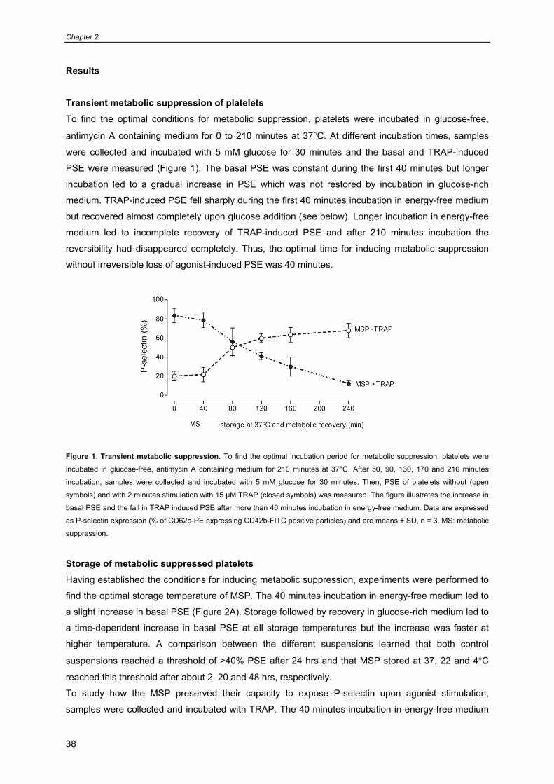
Transient metabolic suppression of platelets To find the optimal conditions for metabolic suppression, platelets were incubated in glucose-free,
antimycin A containing medium for 0 to 210 minutes at 37°C. At different incubation times, samples
were collected and incubated with 5 mM glucose for 30 minutes and the basal and TRAP-induced
PSE were measured (Figure 1). The basal PSE was constant during the first 40 minutes but longer
incubation led to a gradual increase in PSE which was not restored by incubation in glucose-rich
medium. TRAP-induced PSE fell sharply during the first 40 minutes incubation in energy-free medium
but recovered almost completely upon glucose addition (see below). Longer incubation in energy-free
medium led to incomplete recovery of TRAP-induced PSE and after 210 minutes incubation the
reversibility had disappeared completely. Thus, the optimal time for inducing metabolic suppression
without irreversible loss of agonist-induced PSE was 40 minutes.
Figure 1. Transient metabolic suppression. To find the optimal incubation period for metabolic suppression, platelets were
incubated in glucose-free, antimycin A containing medium for 210 minutes at 37°C. After 50, 90, 130, 170 and 210 minutes
incubation, samples were collected and incubated with 5 mM glucose for 30 minutes. Then, PSE of platelets without (open
symbols) and with 2 minutes stimulation with 15 µM TRAP (closed symbols) was measured. The figure illustrates the increase in
basal PSE and the fall in TRAP induced PSE after more than 40 minutes incubation in energy-free medium. Data are expressed
as P-selectin expression (% of CD62p-PE expressing CD42b-FITC positive particles) and are means ± SD, n = 3. MS: metabolic
suppression.
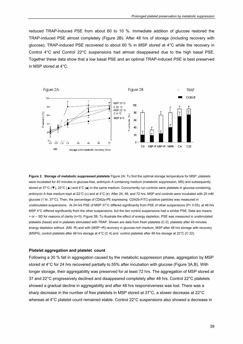
Storage of metabolic suppressed platelets Having established the conditions for inducing metabolic suppression, experiments were performed to
find the optimal storage temperature of MSP. The 40 minutes incubation in energy-free medium led to
a slight increase in basal PSE (Figure 2A). Storage followed by recovery in glucose-rich medium led to
a time-dependent increase in basal PSE at all storage temperatures but the increase was faster at
higher temperature. A comparison between the different suspensions learned that both control
suspensions reached a threshold of >40% PSE after 24 hrs and that MSP stored at 37, 22 and 4°C
reached this threshold after about 2, 20 and 48 hrs, respectively.
To study how the MSP preserved their capacity to expose P-selectin upon agonist stimulation,
samples were collected and incubated with TRAP. The 40 minutes incubation in energy-free medium
Prolonged platelet preservation by metabolic suppression
39
reduced TRAP-induced PSE from about 60 to 10 %. Immediate addition of glucose restored the
TRAP-induced PSE almost completely (Figure 2B). After 48 hrs of storage (including recovery with
glucose), TRAP-induced PSE recovered to about 60 % in MSP stored at 4°C while the recovery in
Control 4°C and Control 22°C suspensions had almost disappeared due to the high basal PSE.
Together these data show that a low basal PSE and an optimal TRAP-induced PSE is best preserved
in MSP stored at 4°C.
Figure 2. Storage of metabolic suppressed platelets Figure 2A: To find the optimal storage temperature for MSP, platelets
were incubated for 40 minutes in glucose-free, antimycin A containing medium (metabolic suppression, MS) and subsequently
stored at 37°C (▼), 22°C (▲) and 4°C (■) in the same medium. Concurrently run controls were platelets in glucose-containing,
antimycin A free medium kept at 22°C (ο) and at 4°C (•). After 24, 48, and 72 hrs, MSP and controls were incubated with 20 mM
glucose (1 hr, 37°C). Then, the percentage of CD62p-PE expressing CD42b-FITC-positive particles was measured in
unstimulated suspensions. At 24 hrs PSE of MSP 37°C differed significantly from PSE of other suspensions (P< 0.05); at 48 hrs
MSP 4°C differed significantly from the other suspensions, but the two control suspensions had a similar PSE. Data are means
+ or – SD for reasons of clarity (n=3). Figure 2B: To illustrate the effect of energy depletion, PSE was measured in unstimulated
platelets (basal) and in platelets stimulated with TRAP. Shown are data from fresh platelets (C 0), platelets after 40 minutes
energy depletion without (MS -R) and with (MSP +R) recovery in glucose-rich medium, MSP after 48 hrs storage with recovery
(MSP4), control platelets after 48 hrs storage at 4°C (C 4) and control platelets after 48 hrs storage at 22°C (C 22).
Platelet aggregation and platelet count Following a 30 % fall in aggregation caused by the metabolic suppression phase, aggregation by MSP
stored at 4°C for 24 hrs recovered partially to 55% after incubation with glucose (Figure 3A,B). With
longer storage, their aggregability was preserved for at least 72 hrs. The aggregation of MSP stored at
37 and 22°C progressively declined and disappeared completely after 48 hrs. Control 22°C platelets
showed a gradual decline in aggregability and after 48 hrs responsiveness was lost. There was a
sharp decrease in the number of free platelets in MSP stored at 37°C, a slower decrease at 22°C
whereas at 4°C platelet count remained stable. Control 22°C suspensions also showed a decrease in
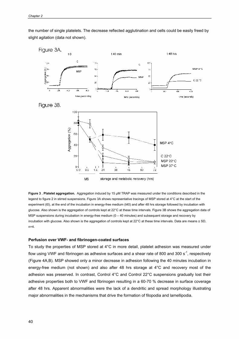
Chapter 2
40
the number of single platelets. The decrease reflected agglutination and cells could be easily freed by
slight agitation (data not shown).
Figure 3 . Platelet aggregation. Aggregation induced by 15 µM TRAP was measured under the conditions described in the
legend to figure 2 in stirred suspensions. Figure 3A shows representative tracings of MSP stored at 4°C at the start of the
experiment (t0), at the end of the incubation in energy-free medium (t40) and after 48 hrs storage followed by incubation with
glucose. Also shown is the aggregation of controls kept at 22°C at these time intervals. Figure 3B shows the aggregation data of
MSP suspensions during incubation in energy-free medium (0 – 40 minutes) and subsequent storage and recovery by
incubation with glucose. Also shown is the aggregation of controls kept at 22°C at these time intervals. Data are means ± SD,
n=4.
Perfusion over VWF- and fibrinogen-coated surfaces To study the properties of MSP stored at 4°C in more detail, platelet adhesion was measured under
flow using VWF and fibrinogen as adhesive surfaces and a shear rate of 800 and 300 s-1, respectively
(Figure 4A,B). MSP showed only a minor decrease in adhesion following the 40 minutes incubation in
energy-free medium (not shown) and also after 48 hrs storage at 4°C and recovery most of the
adhesion was preserved. In contrast, Control 4°C and Control 22°C suspensions gradually lost their
adhesive properties both to VWF and fibrinogen resulting in a 60-70 % decrease in surface coverage
after 48 hrs. Apparent abnormalities were the lack of a dendritic and spread morphology illustrating
major abnormalities in the mechanisms that drive the formation of filopodia and lamellipodia.
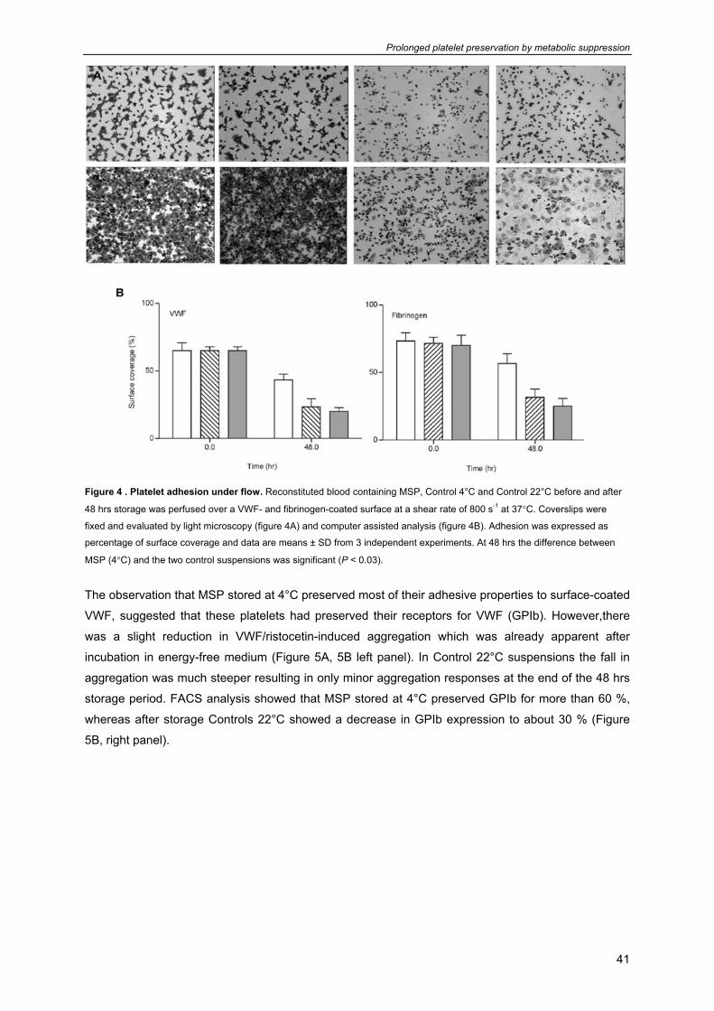
Prolonged platelet preservation by metabolic suppression
41
Figure 4 . Platelet adhesion under flow. Reconstituted blood containing MSP, Control 4°C and Control 22°C before and after
48 hrs storage was perfused over a VWF- and fibrinogen-coated surface at a shear rate of 800 s-1 at 37°C. Coverslips were
fixed and evaluated by light microscopy (figure 4A) and computer assisted analysis (figure 4B). Adhesion was expressed as
percentage of surface coverage and data are means ± SD from 3 independent experiments. At 48 hrs the difference between
MSP (4°C) and the two control suspensions was significant (P < 0.03).
The observation that MSP stored at 4°C preserved most of their adhesive properties to surface-coated
VWF, suggested that these platelets had preserved their receptors for VWF (GPIb). However,there
was a slight reduction in VWF/ristocetin-induced aggregation which was already apparent after
incubation in energy-free medium (Figure 5A, 5B left panel). In Control 22°C suspensions the fall in
aggregation was much steeper resulting in only minor aggregation responses at the end of the 48 hrs
storage period. FACS analysis showed that MSP stored at 4°C preserved GPIb for more than 60 %,
whereas after storage Controls 22°C showed a decrease in GPIb expression to about 30 % (Figure
5B, right panel).
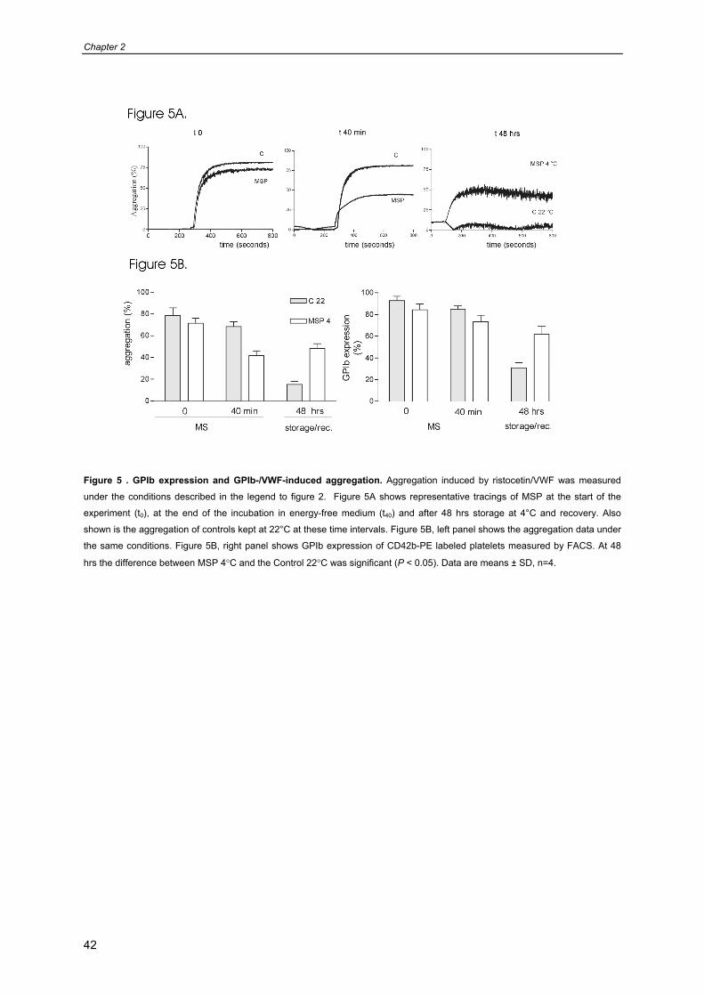
Chapter 2
42
Figure 5 . GPIb expression and GPIb-/VWF-induced aggregation. Aggregation induced by ristocetin/VWF was measured
under the conditions described in the legend to figure 2. Figure 5A shows representative tracings of MSP at the start of the
experiment (t0), at the end of the incubation in energy-free medium (t40) and after 48 hrs storage at 4°C and recovery. Also
shown is the aggregation of controls kept at 22°C at these time intervals. Figure 5B, left panel shows the aggregation data under
the same conditions. Figure 5B, right panel shows GPIb expression of CD42b-PE labeled platelets measured by FACS. At 48
hrs the difference between MSP 4°C and the Control 22°C was significant (P < 0.05). Data are means ± SD, n=4.
Prolonged platelet preservation by metabolic suppression
43
Discussion The main finding of the present study is that metabolic suppression followed by storage at 4°C better
preserves platelet functions than conditions that support energy metabolism during storage at 22°C or
apply low temperature without prior induction of metabolic arrest. Compared with both control
suspensions, these MSP better maintain a low basal PSE, show better TRAP-induced PSE and
aggregation and show an almost normal adhesion to VWF and fibrinogen under flow. However, there
is a slight decrease in GPIb expression and VWF/ristocetin induced aggregation in MSP stored at 4°C.
The difference with control suspensions is much smaller when MSP are stored at 22°C and storage of
MSP at 37°C triggers a dramatic loss of platelet functions within the first hours of the storage period.
Basis of the concept that metabolic suppression could protect platelets against activating stimuli
inflicted during storage are earlier findings showing that platelets rapidly lose their capacity to respond
to aggregation and secretion-inducing agents when energy supply and demand are out of balance.
Incubation in a glucose-free medium (to block glycolytic energy generation) in the presence of
antimycin A (to block oxidative energy generation) leaves only glycogen catabolism as a source for
metabolic energy.23;24 Previous studies have shown that glycogenolysis alone is unable to meet the
energy demands of the resting platelets despite the huge amounts of glycogen stored in the cell.26
The result is a rapid fall in the AEC, AEC= ([ATPm] + ½ [ADPm] +[AMP]) / ([ATPm] + [ADPm] +
[AMP]) (“m” refers to the metabolic compartment) to values as low as 0.2 – 0.3 until a new equilibrium
is reached.23;25;26 Surprisingly, platelets survive a period with a low AEC even though their functionality
is transiently lost. Apparently, they set priorities in energy consuming mechanisms and preserve the
mechanisms that are essential for survival at the expense of their capacity to aggregate and secrete.
Subsequent addition of glucose restores the normal AEC and platelet regain their aggregating and
secreting properties.
Despite the better preservation of MSP compared with controls illustrated in the present report, it is
clear that the preservation of platelet properties is far from complete. A clear change is the increase in
basal PSE in these cells resulting in >40% positive platelets after 48 hrs storage at 4°C. P-selectin is a
protein embedded in the alpha-granule membrane and its surface expression reflects a gradual
secretion of alpha-granule contents during the storage period. In earlier work the metabolic energy
required for complete secretion was estimated at 5.2 ATP equivalents /1011 platelets.35 It is unlikely
that under the suppressed metabolic conditions applied to MSP this energy is readily available.
Instead, the expression of P-selectin might reflect cell damage especially since it is accompanied by
an increase in phosphatidylserine exposure and Annexin-V binding (data not shown). After transfusion
of the PC’s, platelets bind strongly to monocytes and neutrophils, probably as a result of P-selectin
expression36. P-selectin exposure has been taken as a marker for platelet viability in vitro, 19,15 and in
vivo.37 Holme et al.1997 found a poor correlation between P-selectin expression and platelet recovery
post transfusion and a somewhat better correlation with platelet survival.18
Another disturbance is the fall in the capacity to aggregate upon stimulation by TRAP and
ristocetin/VWF. Apparently the protection by metabolic suppression is incomplete and some damage
to the mechanisms that drive platelet aggregation cannot be prevented. In contrast, there is little
change in the adhesion to VWF and fibrinogen by MSP stored at 4°C. Normal adhesion to VWF
Chapter 2
44
accords with the almost normal GPIb expression in these cells stored at 4°C for 48 hrs. At high shear
GPIb mediates the rolling and adhesion of platelets on a VWF-coated damaged vessel wall and
initiates signal transduction leading to platelet activation.38,39 Exposure of platelets to high shear stress
causes aggregation via VWF and GPIb.40 This interaction leads to platelet activation and binding of
fibrinogen to αIIbβ3, which forms a firm bridge between aggregating platelets.38 Adelman et al.39
showed that platelet storage at 22°C is accompanied by a decrease in GPIb due to proteolysis. We
found a similar decrease in our Control 22°C suspensions and could reduce the fall in GPIb
expression by 30 % by adding epsilon-aminocapronic acid (1.0 mM), confirming the proteolytic nature
of this decrease (not shown). GPIb experession in MSP 4°C was unchanged by this treatment
illustrating minimal proteolysis during storage at low temperature. The almost normal adhesion to
fibrinogen indicates that the receptor for fibrinogen, integrin αIIbβ3, is well preserved.
Platelet storage at low temperature is accompanied by a rise in cytosolic Ca2+, actin filament
fragmentation,41 activation of Ca2+-dependent proteases41;42 and clustering of phosphoinositides in the
plasma membrane. These changes are thought to cause cold-induced actin filament rearrangements
and platelet rounding.12;41 Depletion of intracellular Ca2+stores appears to increase the permeability of
the plasma membrane for Ca2+ ions.42 These changes are accompanied by tyrosine phosphorylation of
the 130-kDa protein vinculin.42 Whether these changes are prevented by prior induction of metabolic
arrest remains to be investigated.
Storage at low temperature has long been considered incompatible with platelet transfusion since
cold-activation led to alterations in platelet shape41 and P-selectin expression 21;43 thought to trigger
rapid removal from the circulation. The use of pharmacological agents that preserve the disc shape at
low temperature2 and studies in P-selectin-deficient mice6 have made clear that neither changes in
platelet morphology nor surface expressed P-selectin are triggers for platelet removal post transfusion.
Recent studies in animals show that the cold-induced clustering of GPIb might be an important trigger
for the removal of chilled/rewarmed platelets from the circulation through binding to αMβ2 receptors
expressing macrophages.12;13 The possibility that metabolic suppression interferes with clustering of
GPIb molecules and might thereby suppress the recognition by macrophages is an interesting subject
for further studies.
The fact that MSP kept at 4°C better sustain a 48 hrs storage period than control platelets kept at 22°C
suggests that metabolic deprivation might be a suitable means to improve the quality of PC’s stored
under blood banking conditions. It is obvious that the incubation conditions applied in the present
study are worse than those of modern blood banks, where gas-permeable bags in combination with
platelet additive solutions and autologous plasma lead to much better platelet preservation than in the
present Control 22°C suspensions. However, these suboptimal conditions were applied to all
suspensions and used to fasten the appearance of storage-induced platelet defects. It is clear that
further studies are required to adapt metabolic suppression to conditions compatible with transfusion
in humans making use of current procedures for optimal storage of PC’s.

Prolonged platelet preservation by metabolic suppression
45
Acknowledgements This project was supported by Sanquin Blood Supply Foundation (project id. PPO 01-019). JWNA is
supported by the Netherlands Thrombosis Foundation.
Chapter 2
46
Reference List
1. Gulliksson H. Defining the optimal storage conditions for the long-term storage of platelets.
Transfus.Med.Rev. 2003;17:209-215.
2. Hoffmeister KM, Josefsson EC, Isaac NA et al. Glycosylation restores survival of chilled blood platelets. Science 2003;301:1531-1534.
3. Brecher ME, Hay SN, Rothenberg SJ. Monitoring of apheresis platelet bacterial contamination with an automated liquid culture system: a university experience. Transfusion 2003;43:974-978.
4. Fijnheer R, Modderman PW, Veldman H et al. Detection of platelet activation with monoclonal antibodies and flow cytometry. Changes during platelet storage. Transfusion 1990;30:20-25.
5. de Korte D, Gouwerok CW, Fijnheer R, Pietersz RN, Roos D. Depletion of dense granule nucleotides during storage of human platelets. Thromb.Haemost. 1990;63:275-278.
6. Wagner S. Transfusion-related bacterial sepsis. Curr.Opin.Hematol. 1997;4:464-469.
7. Yuasa T, Ohto H, Suzuki A, Shishido F. New plasma-reduced synthetic media, Fukushima cocktails, for the storage of platelets for transfusion. Transfus.Sci. 2000;23:37-46.
8. Dormann D, Clemetson KJ, Kehrel BE. The GPIb thrombin-binding site is essential for thrombin-induced platelet procoagulant activity. Blood 2000;96:2469-2478.
9. Wildt-Eggen J, Gulliksson H. In vivo and in vitro comparison of platelets stored in either synthetic media or plasma. Vox Sang. 2003;84:256-264.
10. Gulliksson H, AuBuchon JP, Cardigan R et al. Storage of platelets in additive solutions: a multicentre study of the in vitro effects of potassium and magnesium. Vox Sang. 2003;85:199-205.
11. Norol F, Bierling P, Roudot-Thoraval F et al. Platelet transfusion: a dose-response study. Blood 1998;92:1448-1453.
12. Hoffmeister KM, Falet H, Toker A et al. Mechanisms of cold-induced platelet actin assembly. J.Biol.Chem. 2001;276:24751-24759.
13. Hoffmeister KM, Felbinger TW, Falet H et al. The clearance mechanism of chilled blood platelets. Cell 2003;112:87-97.
14. Murphy S. Radiolabeling of PLTs to assess viability: a proposal for a standard. Transfusion 2004;44:131-133.
15. Berger G, Hartwell DW, Wagner DD. P-Selectin and platelet clearance. Blood 1998;92:4446-4452.
16. Sikora L, Rao SP, Sriramarao P. Selectin-dependent rolling and adhesion of leukocytes in nicotine-exposed microvessels of lung allografts. Am.J.Physiol Lung Cell Mol.Physiol 2003;285:L654-L663.
17. Tait JF, Smith C, Wood BL. Measurement of phosphatidylserine exposure in leukocytes and platelets by whole-blood flow cytometry with annexin V. Blood Cells Mol.Dis. 1999;25:271-278.
18. Holme S, Sweeney JD, Sawyer S, Elfath MD. The expression of p-selectin during collection, processing, and storage of platelet concentrates: relationship to loss of in vivo viability. Transfusion 1997;37:12-17.
Prolonged platelet preservation by metabolic suppression
47
19. Cetin M, Eser B, Er O et al. Effects of DMSO on platelet functions and P-selectin expression during storage. Transfus.Apheresis Sci. 2001;24:261-267.
20. Tablin F, Wolkers WF, Walker NJ et al. Membrane reorganization during chilling: implications for long-term stabilization of platelets. Cryobiology 2001;43:114-123.
21. Gao DY, Neff K, Xiao HY et al. Development of optimal techniques for cryopreservation of human platelets. I. Platelet activation during cold storage (at 22 and 8 degrees C) and cryopreservation. Cryobiology 1999;38:225-235.
22. Tablin F, Walker NJ, Klein SD, Field CL, Crowe JH. Animal models for studies on cold-induced platelet activation in human beings. J.Lab Clin.Med. 2000;135:339-346.
23. Verhoeven AJ, Mommersteeg ME, Akkerman JW. Comparative studies on the energetics of platelet responses induced by different agonists. Biochem.J. 1986;236:879-887.
24. Akkerman JW, Gorter G. Relation between energy production and adenine nucleotide metabolism in human blood platelets. Biochim.Biophys.Acta 1980;590:107-116.
25. Verhoeven AJ, Mommersteeg ME, Akkerman JW. Kinetics of energy consumption in human platelets with blocked ATP regeneration. Int.J.Biochem. 1986;18:985-990.
26. Verhoeven AJ, Mommersteeg ME, Akkerman JW. Balanced contribution of glycolytic and adenylate pool in supply of metabolic energy in platelets. J.Biol.Chem. 1985;260:2621-2624.
27. Huizinga EG, Tsuji S, Romijn RA et al. Structures of glycoprotein Ibalpha and its complex with von Willebrand factor A1 domain. Science 2002;297:1176-1179.
28. Prior C, Chu V, Holt J et al. Production and functional characterization of a recombinant fragment of von Willebrand factor (vWF): an antagonist to platelet receptor GP Ib. Biotechnology (N.Y.) 1992;10:66-73.
29. van Scharrenburg GJ, Puijk WC, Egmond MR, de Haas GH, Slotboom AJ. Semisynthesis of phospholipase A2. Preparation and properties of arginine-6 bovine pancreatic phospholipase A2. Biochemistry 1981;20:1584-1591.
30. Akkerman JW, Gorter G. Relation between energy production and adenine nucleotide metabolism in human blood platelets. Biochim.Biophys.Acta 1980;590:107-116.
31. Verhoeven AJ, Mommersteeg ME, Akkerman JW. Comparative studies on the energetics of platelet responses induced by different agonists. Biochem.J. 1986;236:879-887.
32. Tibbles HE, Navara CS, Hupke MA, Vassilev AO, Uckun FM. Thrombopoietin induces p-selectin expression on platelets and subsequent platelet/leukocyte interactions. Biochem.Biophys.Res.Commun. 2002;292:987-991.
33. Sixma JJ, de Groot PG, van Zanten H, IJsseldijk M. A new perfusion chamber to detect platelet adhesion using a small volume of blood. Thromb.Res. 1998;92:S43-S46.
34. Remijn JA, Wu YP, Ijsseldijk MJ et al. Absence of fibrinogen in afibrinogenemia results in large but loosely packed thrombi under flow conditions. Thromb.Haemost. 2001;85:736-742.
35. Akkerman JW, Gorter G, Schrama L, Holmsen H. A novel technique for rapid determination of energy consumption in platelets. Demonstration of different energy consumption associated with three secretory responses. Biochem.J. 1983;210:145-155.
36. Mondoro TH, Vostal JG. Cold temperatures reduce the sensitivity of stored platelets to disaggregating agents. Platelets. 2002;13:11-20.
Chapter 2
48
37. Falati S, Liu Q, Gross P et al. Accumulation of tissue factor into developing thrombi in vivo is dependent upon microparticle P-selectin glycoprotein ligand 1 and platelet P-selectin. J.Exp.Med. 2003;197:1585-1598.
38. Wu Y, Asazuma N, Satoh K et al. Interaction between von Willebrand factor and glycoprotein Ib activates Src kinase in human platelets: role of phosphoinositide 3-kinase. Blood 2003;101:3469-3476.
39. Adelman B, Michelson AD, Greenberg J, Handin RI. Proteolysis of platelet glycoprotein Ib by plasmin is facilitated by plasmin lysine-binding regions. Blood 1986;68:1280-1284.
40. Wu YP, Vink T, Schiphorst M et al. Platelet thrombus formation on collagen at high shear rates is mediated by von Willebrand factor-glycoprotein Ib interaction and inhibited by von Willebrand factor-glycoprotein IIb/IIIa interaction. Arterioscler.Thromb.Vasc.Biol. 2000;20:1661-1667.
41. Winokur R, Hartwig JH. Mechanism of shape change in chilled human platelets. Blood 1995;85:1796-1804.
42. Vostal JG, Shulman NR. Vinculin is a major platelet protein that undergoes Ca(2+)-dependent tyrosine phosphorylation. Biochem.J. 1993;294:675-680.
43. Connor J, Currie LM, Allan H, Livesey SA. Recovery of in vitro functional activity of platelet concentrates stored at 4 degrees C and treated with second-messenger effectors. Transfusion 1996;36:691-698.
Platelet- macrophages interaction
49
Chapter 3
Platelet binding and phagocytosis by macrophages
B.A. Badlou1,2,3, Y.P. Wu1,3, W. M. Smid2 and J.W.N. Akkerman1,3
From the 1Thrombosis and Haemostasis Laboratory, Department of Haematology,
University Medical Centre Utrecht, 2the Sanquin Blood Bank Region North-West Amsterdam, and
3The Institute for Biomembranes, Utrecht University, Utrecht, The Netherlands.
Transfusion 2006 in Press
Chapter 3
50
Abstract Introduction: We reported earlier that metabolic arrest followed by incubation at 4°C reduces the
platelet storage defect (Badlou et al., Transfusion 2005). Here we report that this treatment also
reduces binding and phagocytosis by macrophages.
Study Design and methods: Phagocytosis of mepacrine-labeled platelets by macrophages changes
the latter into bright fluorescent particles easily detected by FACS.
Results: In combination with conventional binding analysis we found that binding to PMA-matured
THP-1 cells is primarily regulated by platelet P-selectin expression and phagocytosis by combined
phosphatidylserine (PS) exposure and glycoprotein (GP) Ibalpha clustering. We found that trapping of
platelet Ca2+ and raising cAMP reduces phagocytosis by lowering PS exposure. Chilling of platelets
leads to an increase in binding and PS- and GPIbα-mediated phagocytosis. Prior depletion of platelet
energy stores prevents this increase by preserving low Ca2+ concentration, PS exposure and PS-
mediated phagocytosis.
Conclusion: These data characterize the individual factors that control platelet binding and
phagocytosis and might help to define conditions that improve the survival of stored platelets after
transfusion.
Abbreviations: GP, glycoprotein ; MSP, metabolic suppressed platelets; GlcNAc, N-
acetylglucosamine; AnnV, annexin V; BAPTA, 1,2-bis-(2-aminophenoxy) ethane-NNN'N'-tetra-acetic
acid
Platelet- macrophages interaction
51
Introduction The storage of platelet concentrates at room temperature facilitates bacterial growth and introduces
changes in platelets indicative for activation and initiation of apoptosis. Improvements have been
sought in lowering the storage temperature but this treatment severely reduces the survival of
transfused platelets. A main feature of platelet storage is the gradual increase in surface expression of
P-selectin (CD62P), a component of α-granule membranes. The counter receptor for P-selectin, P-
selectin glycoprotein ligand 1 (PSGL-1), is present on leukocytes and the inverse correlation between
P-selectin expression and platelet recovery post transfusion is sought to reflect platelet destruction
through coupling of these receptors. However, the contribution of P-selectin expression to platelet
destruction can only be small since transfused human platelets lose P-selectin in the circulation1 and
P-selectin deficient mice platelets survive normally.2,3 A second feature of platelet storage is the
membrane appearance of negatively charged phospholipids such as phosphatidylserine (PS), which is
a property of activated platelets4,5 and cells going into apoptosis3,6. PS exposure may form recognition
sites for destruction of senescent platelets and a similar mechanism might remove stored platelets
from the circulation6.
Attempts to reduce the surface expression of recognition sites for platelet destruction were based on
prostacyclin, which inhibits secretion and PS exposure,7 arrest of glycolytic and oxidative energy
generation, which inhibits ATP resynthesis required for platelet functions8,9 and storage at low
temperature, which slows down platelet metabolism and in addition suppresses bacterial growth.
Chilling of platelets has long been thought to cause irreversible cell damage reflected by loss of the
discoid shape and an increase in cytosolic Ca2+ and causing poor post-transfusion survival.10,11
However, treatments that preserve platelet shape and a low Ca2+ level failed to improve in vivo
survival.12,13 Recently, Hoffmeister et al.14 showed that the chilling of platelets triggers the
rearrangement of glycoprotein (GP) Ibα,CD42b) into clusters that are recognized by αMβ2 (CR3/Mac-
1) receptors on liver macrophages triggering platelet destruction. A resting platelet contains about 20
000 GPIbα molecules bound to GPV and GPIX in a 2:1:2 stoichiometry randomly distributed over the
plasma membrane.11,15,16 GPIbα serves in the binding of platelets to activated von Willebrand Factor at
sites of vascular damage slowing down their velocity in flowing blood and enabling other receptors to
firmly attach platelets to the wound.16-19 Interestingly, cold induced formation of GPIbα clusters leaves
the haemostatic functions of GPIb alphaundisturbed.20
An alternative means to prolong the storage of platelets is by metabolic suppression prior to cold
storage. We showed earlier that platelets sustain a period of metabolic arrest without losing their
adhesive properties and their capacity to aggregate and secrete granule contents.21 During metabolic
arrest platelets fail to respond to platelet activating agents such in agreement with the energy
requirement of aggregation and secretion.
In the present study we investigated the mechanisms that mediate binding of platelets to
macrophages and initiate their destruction. The results reveal major roles for surface P-selectin in
binding and exposed PS and clustered GPIb alpha in phagocytosis. We also show that chilling of
platelets induces binding and phagocytosis and that prior metabolic arrest protects platelets against
Chapter 3
52
these cold-induced changes. This protective effect can be explained by suppression of PS exposure
providing a means to interfere with surface expression of phagocytotic signals through metabolic
intervention.
Materials and methods We obtained antimycin A, phorbol 12-myristate 13-acetate (PMA), cytochalasin B, BAPTA-AM, N-
acetylglucosame (GlcNAc), mepacrine (Quinacrine) and Fura-2 AM from Sigma Chemicals
(Mannheim, FRG, Germany), the anti human CD42b (GPIbαPE (R7014), anti-human CD14-FITC
antibody, annexin V-PE, annexinV-FITC, anti human CD62p (P-selectin) and FITC-labeled IgG as
negative control from Dako A/S (Glusdorp, Denmark), serum free cell culture media RPMI-1640 from
Corning Inc. (Corning, NY, USA), fetal calf serum (FCS) from Cambrex (Viers, Belgium), penicillin,
streptomycin sulfate and trypsin from Gibco invitrogen corporation (Grand Island, N.Y. USA.
Prostacyclin (PGI2) was from Cayman Chemical Company (Ann Arbor, MI, USA) and the stable
prostacyclin analogue iloprost from Schering A.G. (Berlin, Germany). Annexin V was a generous gift
from Dr. W.L. van Heerde, Department of Hematology, Radboud University Nijmegen.
Platelet isolation and storage Freshly drawn venous blood from healthy, medication-free volunteers (40 mL) was collected with
informed consent into 1:10 v/v 130 mmol/L trisodium citrate. Platelet-rich plasma (PRP) was prepared
by centrifugation (200g, 15 minutes, 20 °C). ACD (0.1 volume of 2.5 g tri-sodium citrate, 1.5 g citric
acid and 2.0 g D-glucose in 100 mL distilled water) was added to lower the pH to 6.0 and prevent
platelet activation during isolation. The suspension (10 mL) was washed by centrifugation (330g, 15
minutes, 22 oC) and resuspended in glucose-free Hepes-Tyrode (137 mM NaCl, 2.68 mM KCl, 0.42
mM NaH2PO4, 1.7 mM MgCl2, and 11.9 mM NaHCO3, pH 7.2) to a final concentration of 2 x 10⁸
platelets/mL. Platelet count was measured on a Cell counter AL871 (Molab, Hilden, Germany).
Platelets in glucose (5 mM)-containing Hepes-Tyrode were stored at 22°C (designated as C22°-
platelets), a storage temperature currently used in blood banks, and at 0°C (designated as C0°-
platelets), a condition known to induce clustering of GPIbα binding to macrophage αMβ2 and platelet
destruction after transfusion.22 Metabolic suppressed platelets (MSP) were prepared by incubation in
glucose-free Hepes-Tyrode solution containing 20 µM antimycin A for 40 minutes at 37°C to deplete
energy stores23. MSP were stored at 4°C for the indicated periods. Suspensions were stored in closed
tubes impermeable for gas exchange without agitation. Prior to each measurement, MSP and C22°-
platelets were incubated with 20 mM glucose for 1 hour at 37°C to optimally restore energy
generation; the C0°-platelets were warmed for 15 minutes at 37°C. 23
Platelet- macrophages interaction
53
Preparation of mepacrine-labeled platelets and PMA-matured THP-1 cells Immediately before the binding and phagocytosis experiments, 100 µL platelet suspensions were
labeled with 1 µM mepacrine in Hepes-Tyrode (pH 7.2, 5 minutes, 22°C). Mepacrine is a fluorescent
polyphenolic compound, which emits at 519 nm which is in the range of the emission of FITC (530 ±15
nm). Free mepacrine was removed (5 minutes, 350g, 22°C, with soft mode) while preventing platelet
activation with 10 ng/mL PGI2. Monocytoid THP-1 cell lines were cultured to a density of (2 - 4) .105
cells/mL in RPMI 1640 containing 10% FCS, 2 mM glutamine, penicillin (10 U/L) and streptomycin (1
µg/L) at 37°C. THP-1 cells were counted in a Burker-Turk chamber and 1 mL suspension containing
1x10⁶ cells was added to a well of a 48 wells plate (Corning incorporated, Corning, NY, USA).
Maturation was induced by incubation with 500 nM PMA for 24 hours at 37°C unless stated otherwise.
In a few experiments free PMA was removed from the THP-1 cells prior to incubation with platelets to
prevent platelet activation, as indicated.
Platelet – macrophage interaction The binding of platelets to macrophages was a modification of the procedure described by Hoffmeister
et al. 14 In short, 2x10⁶ platelets in Hepes-Tyrode buffer (pH 7.2) were added to a well containing
1x10⁶ PMA-matured THP-1 cells in 1 mL RPMI 1640 medium and the mixed suspensions were
incubated at 37°C without agitation. Wells were gently washed with HBSS buffer (0.3 mM KH2PO4,
13.7 mM NaCl, 417 mM NaHCO3, 31 mM Na2HPO4 and 0.5 mM KCl in aqua dest) and free platelets
collected in the wash medium were isolated by centrifugation-resuspension under protection of PGI2.
Wells were incubated with 200 mL HBSS buffer containing 5 mM EDTA for 15 minutes at 0°C,
reconstituted with free platelets removed during the wash step and thereafter 100 µL suspension was
incubated with 2 µg/mL anti-human CD42b-PE and 2 µg/mL anti-human CD14-FITC antibody for 15
minutes at 37°C. Then, HBSS buffer was added and 20 000 particles were measured by
flowcytometry (FACS Calibur, Becton-Dickinson, San Jose, CA, USA). FACS data were analyzed with
WinMDI software. Binding of platelets to macrophages was expressed as the percentage of
CD42b/CD14 positive particles of the total number of CD42b and/or CD14 positive particles.
Phagocytosis of platelets by PMA matured THP-1 cells was measured by FACS analysis of mepacrine
positive CD14 cells that were inaccessible to the anti CD42b-PE antibody and expressed as
percentage of total number of CD14 positive, CD42b negative particles.
In a few experiments THP-1 cells (1x10⁶ cells) were allowed to attach to glass, mildly matured by
incubation with 100 nM PMA (15 minutes, 22°C) and incubated at 22°C with platelets stored for 48
hours at 22°C. Binding and phagocytosis were measured by real-time analysis with an Orthoplan
fluorescence microscope (Leica, Heidelberg, Germany) with a 100x objective connected to a charge-
coupled device (CCD) camera system (Jai, Copenhagen, Denmark) coupled to a personal computer
with Optimas 6.0 DVS software (Breda, the Netherlands).
Chapter 3
54
Interference with binding and phagocytosis
Platelets stored at the indicated conditions were incubated with P-selectin antibody (17 μg/mL),
annexin V (17 µg/mL), cytochalasin B (10 µM), BAPTA-AM (10 µM) and iloprost (10 µM) for 1 hr at
22°C. Additions were removed by a wash step prior to mixing with THP-1 cells and analysis of binding
and phagocytosis. The role of GPIbα clusters was evaluated by measuring phagocytosis of the cold
platelets in the presence of GlcNAc (100 mM). Measurement of P-selectin and PS expression
Platelets were incubated with glucose, cytochalasin B, BAPTA-AM or iloprost for 1 hr at 37°C and P-
selectin and PS expression were measured on a FACScalibur 23,24. Samples were fixed with 2 percent
paraformaldehyde (30 minutes, 22°C) and washed with 500 µL of phosphate-buffered saline (PBS;
500g, 5 minutes, 22°C). Pellets were resuspended in 50 mL PBS containing 1 % BSA and 0.01 %
Tween 20 (5 minutes, 22°C) and platelet suspensions were incubated with annexinV-FITC and CD62-
p-PE (1 µg/mL each) for 1 hour at 22°C in the dark. Samples were washed in PBS and 10,000
particles were analyzed using WinMDI software.
Measurement of [Ca2+]i Platelets were incubated with 0.2 μM Fura-2-AM for 45 min at 37°C (light protected) and free Fura-2
was removed by a wash step under protection of PGI2 (10 nM). Fura-2 fluorescence was recorded at
37°C in F-4500 fluorescence spectrophotometer (Hitachi Ltd., Tokyo, Japan) with excitation
wavelengths of 340 and 380 nm and emission at 510 nm. Basal [Ca2+]i was calculated according to the
method of Grynkiewicz et al. 25
Confocal microscopy Human washed platelets (10x 106) were incubated with moab CD42b-PE (2 µg/mL) (AN51-PE, DAKO,
Denmark) and mepacrine (50 µg/mL) for 15 minutes at 37°C. THP-1 cells (5x 10⁶) were stimulated
with PMA for 15 minutes at 37°C on coverslip. Then platelets were mixed with THP-1 cells and
incubated for 30 minutes at 22°C. Samples were first identified using a confocal laser scanning
fluorescence microscope (Leica TCS 4D, Heidelberg, Germany) as defined.26 A 63x objective, 0.75
NA oil immersion lens (Zeiss, Thornwood, New York) was used to illuminate the sample and zoom 4 (
31 x 31 μm) images were collected.
Statistics Data are expressed as means ± SEM with number of observations n. Statistical analysis was based
on a paired t-test or One-way ANOVA (with post t-test) for comparison between 2 and more groups.
Differences were considered significant at a p value of less than 0.05.
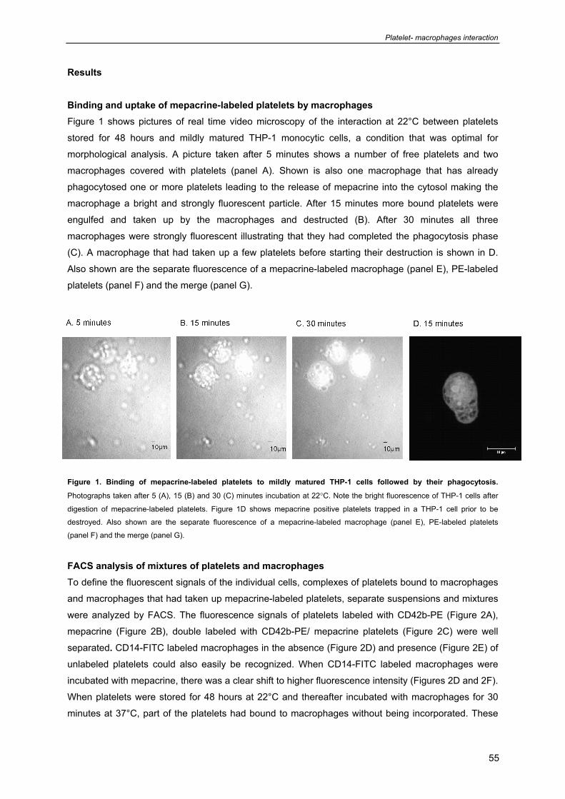
Platelet- macrophages interaction
55
Results Binding and uptake of mepacrine-labeled platelets by macrophages Figure 1 shows pictures of real time video microscopy of the interaction at 22°C between platelets
stored for 48 hours and mildly matured THP-1 monocytic cells, a condition that was optimal for
morphological analysis. A picture taken after 5 minutes shows a number of free platelets and two
macrophages covered with platelets (panel A). Shown is also one macrophage that has already
phagocytosed one or more platelets leading to the release of mepacrine into the cytosol making the
macrophage a bright and strongly fluorescent particle. After 15 minutes more bound platelets were
engulfed and taken up by the macrophages and destructed (B). After 30 minutes all three
macrophages were strongly fluorescent illustrating that they had completed the phagocytosis phase
(C). A macrophage that had taken up a few platelets before starting their destruction is shown in D.
Also shown are the separate fluorescence of a mepacrine-labeled macrophage (panel E), PE-labeled
platelets (panel F) and the merge (panel G).
Figure 1. Binding of mepacrine-labeled platelets to mildly matured THP-1 cells followed by their phagocytosis.
Photographs taken after 5 (A), 15 (B) and 30 (C) minutes incubation at 22°C. Note the bright fluorescence of THP-1 cells after
digestion of mepacrine-labeled platelets. Figure 1D shows mepacrine positive platelets trapped in a THP-1 cell prior to be
destroyed. Also shown are the separate fluorescence of a mepacrine-labeled macrophage (panel E), PE-labeled platelets
(panel F) and the merge (panel G). FACS analysis of mixtures of platelets and macrophages To define the fluorescent signals of the individual cells, complexes of platelets bound to macrophages
and macrophages that had taken up mepacrine-labeled platelets, separate suspensions and mixtures
were analyzed by FACS. The fluorescence signals of platelets labeled with CD42b-PE (Figure 2A),
mepacrine (Figure 2B), double labeled with CD42b-PE/ mepacrine platelets (Figure 2C) were well
separated. CD14-FITC labeled macrophages in the absence (Figure 2D) and presence (Figure 2E) of
unlabeled platelets could also easily be recognized. When CD14-FITC labeled macrophages were
incubated with mepacrine, there was a clear shift to higher fluorescence intensity (Figures 2D and 2F). When platelets were stored for 48 hours at 22°C and thereafter incubated with macrophages for 30
minutes at 37°C, part of the platelets had bound to macrophages without being incorporated. These
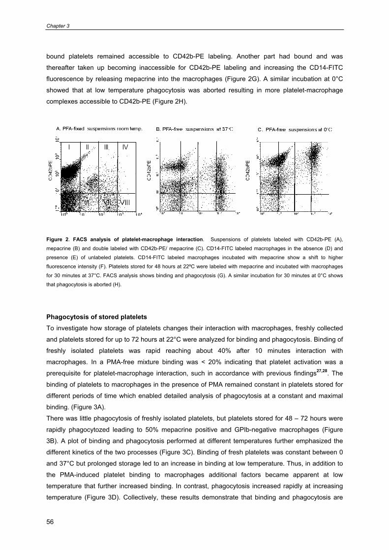
Chapter 3
56
bound platelets remained accessible to CD42b-PE labeling. Another part had bound and was
thereafter taken up becoming inaccessible for CD42b-PE labeling and increasing the CD14-FITC
fluorescence by releasing mepacrine into the macrophages (Figure 2G). A similar incubation at 0°C
showed that at low temperature phagocytosis was aborted resulting in more platelet-macrophage
complexes accessible to CD42b-PE (Figure 2H).
Figure 2. FACS analysis of platelet-macrophage interaction. Suspensions of platelets labeled with CD42b-PE (A),
mepacrine (B) and double labeled with CD42b-PE/ mepacrine (C). CD14-FITC labeled macrophages in the absence (D) and
presence (E) of unlabeled platelets. CD14-FITC labeled macrophages incubated with mepacrine show a shift to higher
fluorescence intensity (F). Platelets stored for 48 hours at 22ºC were labeled with mepacrine and incubated with macrophages
for 30 minutes at 37°C. FACS analysis shows binding and phagocytosis (G). A similar incubation for 30 minutes at 0°C shows
that phagocytosis is aborted (H).
Phagocytosis of stored platelets To investigate how storage of platelets changes their interaction with macrophages, freshly collected
and platelets stored for up to 72 hours at 22°C were analyzed for binding and phagocytosis. Binding of
freshly isolated platelets was rapid reaching about 40% after 10 minutes interaction with
macrophages. In a PMA-free mixture binding was < 20% indicating that platelet activation was a
prerequisite for platelet-macrophage interaction, such in accordance with previous findings27,28. The
binding of platelets to macrophages in the presence of PMA remained constant in platelets stored for
different periods of time which enabled detailed analysis of phagocytosis at a constant and maximal
binding. (Figure 3A).
There was little phagocytosis of freshly isolated platelets, but platelets stored for 48 – 72 hours were
rapidly phagocytozed leading to 50% mepacrine positive and GPIb-negative macrophages (Figure
3B). A plot of binding and phagocytosis performed at different temperatures further emphasized the
different kinetics of the two processes (Figure 3C). Binding of fresh platelets was constant between 0
and 37°C but prolonged storage led to an increase in binding at low temperature. Thus, in addition to
the PMA-induced platelet binding to macrophages additional factors became apparent at low
temperature that further increased binding. In contrast, phagocytosis increased rapidly at increasing
temperature (Figure 3D). Collectively, these results demonstrate that binding and phagocytosis are
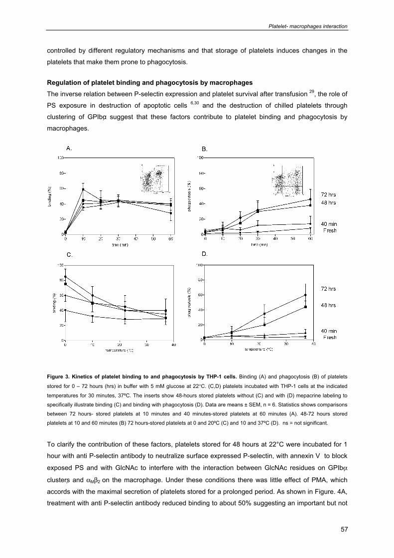
Platelet- macrophages interaction
57
controlled by different regulatory mechanisms and that storage of platelets induces changes in the
platelets that make them prone to phagocytosis.
Regulation of platelet binding and phagocytosis by macrophages The inverse relation between P-selectin expression and platelet survival after transfusion 29, the role of
PS exposure in destruction of apoptotic cells 6,30 and the destruction of chilled platelets through
clustering of GPIbα suggest that these factors contribute to platelet binding and phagocytosis by
macrophages.
Figure 3. Kinetics of platelet binding to and phagocytosis by THP-1 cells. Binding (A) and phagocytosis (B) of platelets
stored for 0 – 72 hours (hrs) in buffer with 5 mM glucose at 22°C. (C,D) platelets incubated with THP-1 cells at the indicated
temperatures for 30 minutes, 37ºC. The inserts show 48-hours stored platelets without (C) and with (D) mepacrine labeling to
specifically illustrate binding (C) and binding with phagocytosis (D). Data are means ± SEM, n = 6. Statistics shows comparisons
between 72 hours- stored platelets at 10 minutes and 40 minutes-stored platelets at 60 minutes (A). 48-72 hours stored
platelets at 10 and 60 minutes (B) 72 hours-stored platelets at 0 and 20ºC (C) and 10 and 37ºC (D). ns = not significant.
To clarify the contribution of these factors, platelets stored for 48 hours at 22°C were incubated for 1
hour with anti P-selectin antibody to neutralize surface expressed P-selectin, with annexin V to block
exposed PS and with GlcNAc to interfere with the interaction between GlcNAc residues on GPIbα
clusters and αMβ2 on the macrophage. Under these conditions there was little effect of PMA, which
accords with the maximal secretion of platelets stored for a prolonged period. As shown in Figure. 4A,
treatment with anti P-selectin antibody reduced binding to about 50% suggesting an important but not
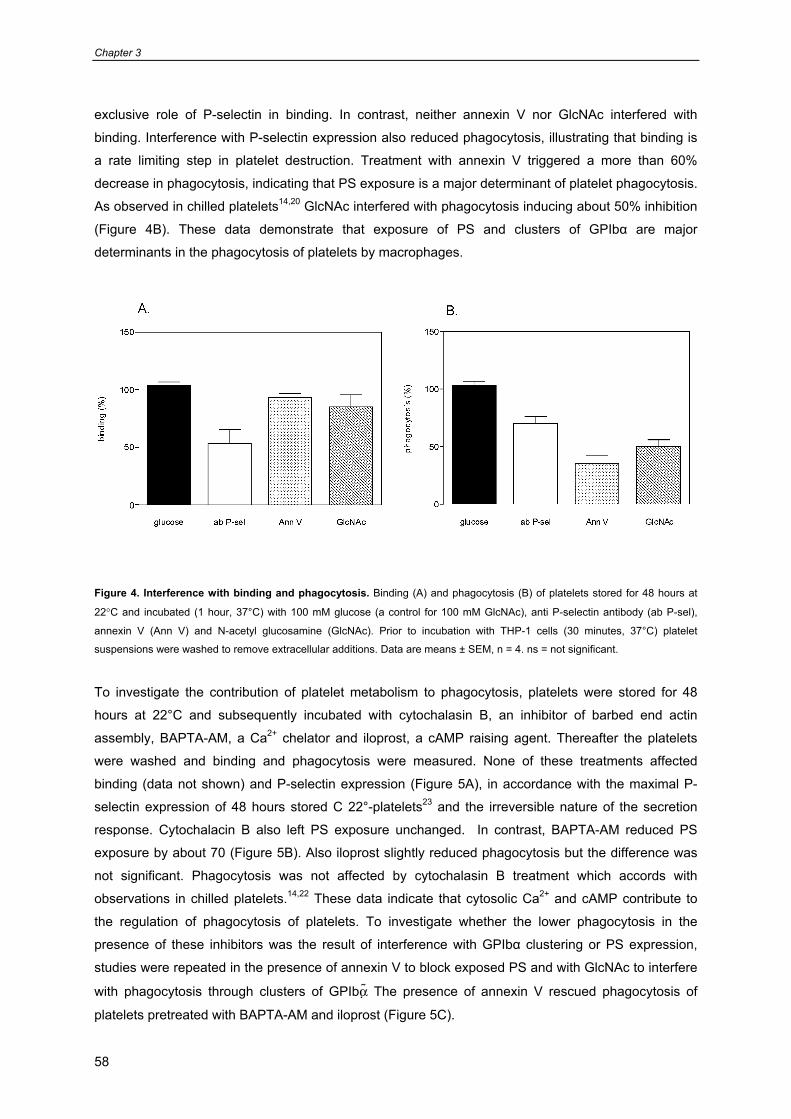
Chapter 3
58
exclusive role of P-selectin in binding. In contrast, neither annexin V nor GlcNAc interfered with
binding. Interference with P-selectin expression also reduced phagocytosis, illustrating that binding is
a rate limiting step in platelet destruction. Treatment with annexin V triggered a more than 60%
decrease in phagocytosis, indicating that PS exposure is a major determinant of platelet phagocytosis.
As observed in chilled platelets14,20 GlcNAc interfered with phagocytosis inducing about 50% inhibition
(Figure 4B). These data demonstrate that exposure of PS and clusters of GPIbα are major
determinants in the phagocytosis of platelets by macrophages.
Figure 4. Interference with binding and phagocytosis. Binding (A) and phagocytosis (B) of platelets stored for 48 hours at
22°C and incubated (1 hour, 37°C) with 100 mM glucose (a control for 100 mM GlcNAc), anti P-selectin antibody (ab P-sel),
annexin V (Ann V) and N-acetyl glucosamine (GlcNAc). Prior to incubation with THP-1 cells (30 minutes, 37°C) platelet
suspensions were washed to remove extracellular additions. Data are means ± SEM, n = 4. ns = not significant.
To investigate the contribution of platelet metabolism to phagocytosis, platelets were stored for 48
hours at 22°C and subsequently incubated with cytochalasin B, an inhibitor of barbed end actin
assembly, BAPTA-AM, a Ca2+ chelator and iloprost, a cAMP raising agent. Thereafter the platelets
were washed and binding and phagocytosis were measured. None of these treatments affected
binding (data not shown) and P-selectin expression (Figure 5A), in accordance with the maximal P-
selectin expression of 48 hours stored C 22°-platelets23 and the irreversible nature of the secretion
response. Cytochalacin B also left PS exposure unchanged. In contrast, BAPTA-AM reduced PS
exposure by about 70 (Figure 5B). Also iloprost slightly reduced phagocytosis but the difference was
not significant. Phagocytosis was not affected by cytochalasin B treatment which accords with
observations in chilled platelets.14,22 These data indicate that cytosolic Ca2+ and cAMP contribute to
the regulation of phagocytosis of platelets. To investigate whether the lower phagocytosis in the
presence of these inhibitors was the result of interference with GPIbα clustering or PS expression,
studies were repeated in the presence of annexin V to block exposed PS and with GlcNAc to interfere
with phagocytosis through clusters of GPIbα The presence of annexin V rescued phagocytosis of
platelets pretreated with BAPTA-AM and iloprost (Figure 5C).
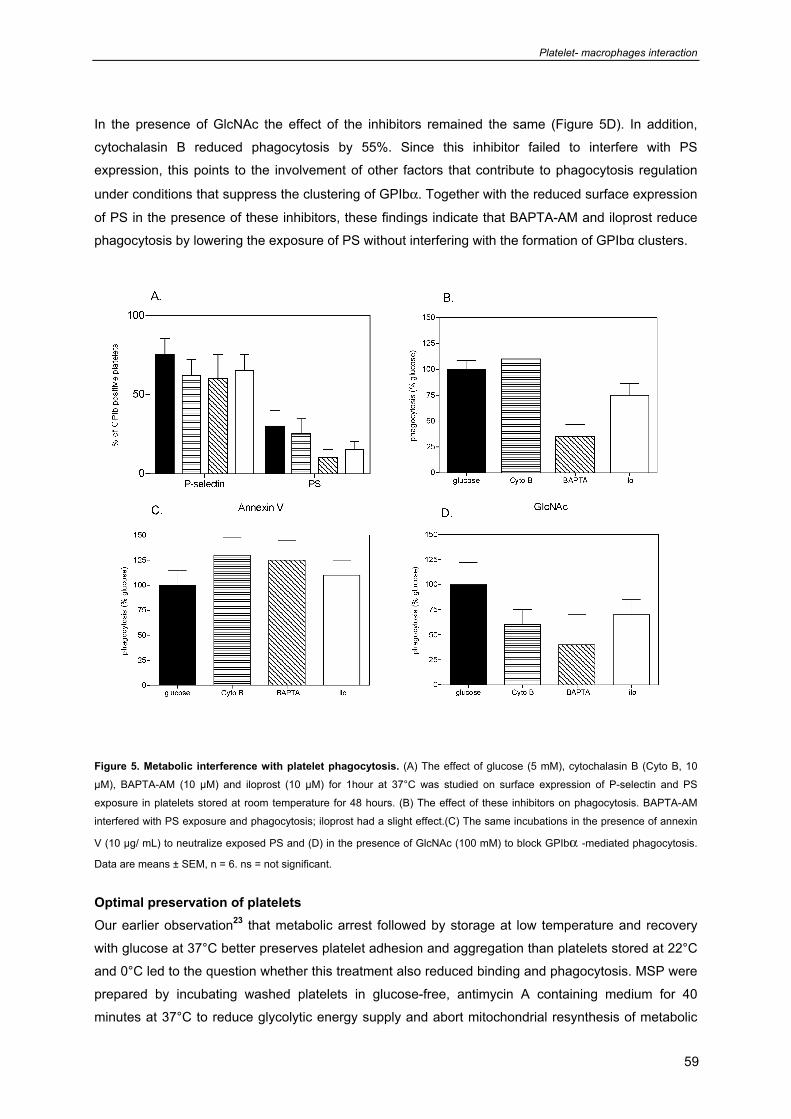
Platelet- macrophages interaction
59
In the presence of GlcNAc the effect of the inhibitors remained the same (Figure 5D). In addition,
cytochalasin B reduced phagocytosis by 55%. Since this inhibitor failed to interfere with PS
expression, this points to the involvement of other factors that contribute to phagocytosis regulation
under conditions that suppress the clustering of GPIbα. Together with the reduced surface expression
of PS in the presence of these inhibitors, these findings indicate that BAPTA-AM and iloprost reduce
phagocytosis by lowering the exposure of PS without interfering with the formation of GPIbα clusters.
Figure 5. Metabolic interference with platelet phagocytosis. (A) The effect of glucose (5 mM), cytochalasin B (Cyto B, 10
μM), BAPTA-AM (10 μM) and iloprost (10 μM) for 1hour at 37°C was studied on surface expression of P-selectin and PS
exposure in platelets stored at room temperature for 48 hours. (B) The effect of these inhibitors on phagocytosis. BAPTA-AM
interfered with PS exposure and phagocytosis; iloprost had a slight effect.(C) The same incubations in the presence of annexin
V (10 μg/ mL) to neutralize exposed PS and (D) in the presence of GlcNAc (100 mM) to block GPIbα -mediated phagocytosis.
Data are means ± SEM, n = 6. ns = not significant.
Optimal preservation of platelets Our earlier observation23 that metabolic arrest followed by storage at low temperature and recovery
with glucose at 37°C better preserves platelet adhesion and aggregation than platelets stored at 22°C
and 0°C led to the question whether this treatment also reduced binding and phagocytosis. MSP were
prepared by incubating washed platelets in glucose-free, antimycin A containing medium for 40
minutes at 37°C to reduce glycolytic energy supply and abort mitochondrial resynthesis of metabolic
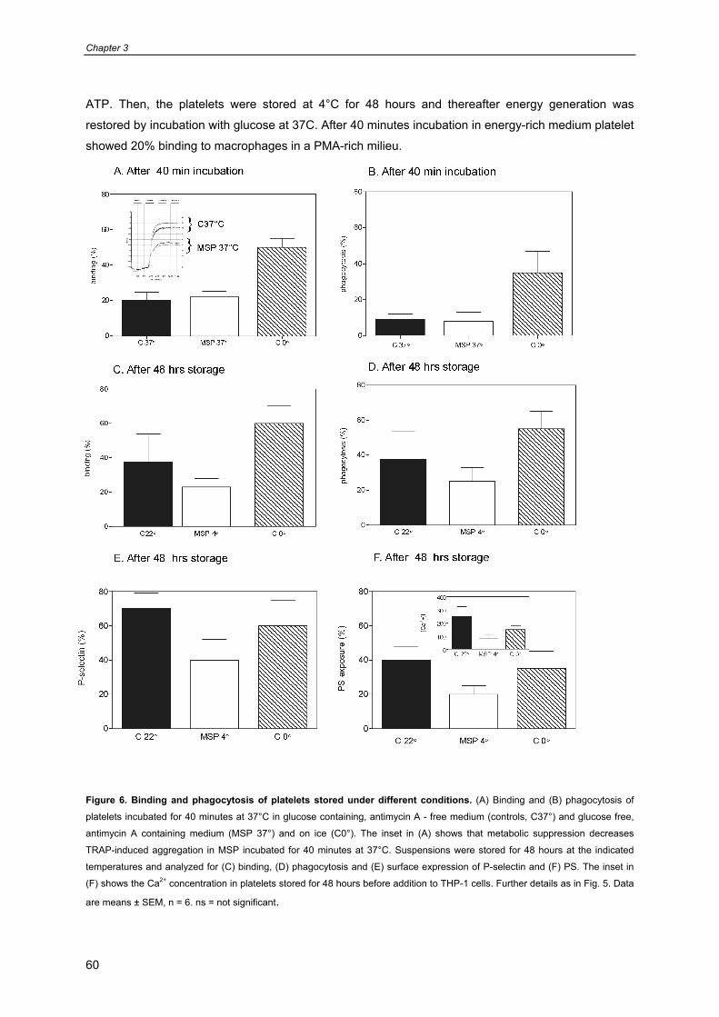
Chapter 3
60
ATP. Then, the platelets were stored at 4°C for 48 hours and thereafter energy generation was
restored by incubation with glucose at 37C. After 40 minutes incubation in energy-rich medium platelet
showed 20% binding to macrophages in a PMA-rich milieu.
Figure 6. Binding and phagocytosis of platelets stored under different conditions. (A) Binding and (B) phagocytosis of
platelets incubated for 40 minutes at 37°C in glucose containing, antimycin A - free medium (controls, C37°) and glucose free,
antimycin A containing medium (MSP 37°) and on ice (C0°). The inset in (A) shows that metabolic suppression decreases
TRAP-induced aggregation in MSP incubated for 40 minutes at 37°C. Suspensions were stored for 48 hours at the indicated
temperatures and analyzed for (C) binding, (D) phagocytosis and (E) surface expression of P-selectin and (F) PS. The inset in
(F) shows the Ca2+ concentration in platelets stored for 48 hours before addition to THP-1 cells. Further details as in Fig. 5. Data
are means ± SEM, n = 6. ns = not significant.
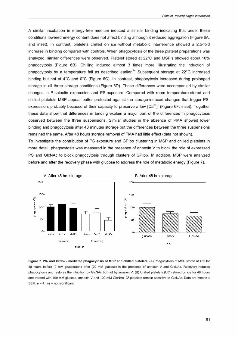
Platelet- macrophages interaction
61
A similar incubation in energy-free medium induced a similar binding indicating that under these
conditions lowered energy content does not affect binding although it reduced aggregation (Figure 6A,
and inset). In contrast, platelets chilled on ice without metabolic interference showed a 2.5-fold
increase in binding compared with controls. When phagocytosis of the three platelet preparations was
analyzed, similar differences were observed. Platelet stored at 22°C and MSP’s showed about 10%
phagocytosis (Figure 6B). Chilling induced almost 3 times more, illustrating the induction of
phagocytosis by a temperature fall as described earlier.14 Subsequent storage at 22°C increased
binding but not at 4°C and 0°C (Figure 6C). In contrast, phagocytosis increased during prolonged
storage in all three storage conditions (Figure 6D). These differences were accompanied by similar
changes in P-selectin expression and PS-exposure. Compared with room temperature-stored and
chilled platelets MSP appear better protected against the storage-induced changes that trigger PS-
expression, probably because of their capacity to preserve a low [Ca2+]i (Figure 6F, inset). Together
these data show that differences in binding explain a major part of the differences in phagocytosis
observed between the three suspensions. Similar studies in the absence of PMA showed lower
binding and phagocytosis after 40 minutes storage but the differences between the three suspensions
remained the same. After 48 hours storage removal of PMA had little effect (data not shown).
To investigate the contribution of PS exposure and GPIbα clustering in MSP and chilled platelets in
more detail, phagocytosis was measured in the presence of annexin V to block the role of expressed
PS and GlcNAc to block phagocytosis through clusters of GPIbα. In addition, MSP were analyzed
before and after the recovery phase with glucose to address the role of metabolic energy (Figure 7).
Figure 7. PS- and GPIbα - mediated phagocytosis of MSP and chilled platelets. (A) Phagocytosis of MSP stored at 4°C for
48 hours before (0 mM glucose)and after (20 mM glucose) in the presence of annexin V and GlcNAc. Recovery reduces
phagocytosis and restores the inhibition by GlcNAc but not by annexin V. (B) Chilled platelets (C0°) stored on ice for 48 hours
and treated with 100 mM glucose, annexin V and 100 mM GlcNAc. Cº platelets remain sensitive to GlcNAc. Data are means ±
SEM, n = 4. ns = not significant.
Chapter 3
62
MSP that were still in the energy-depleted phase were phagocytosed by macrophages as seen with
MSP after the recovery phase. However, neither annexin V nor GlcNAc interfered with phagocytosis of
these cells, indicating that other processes than PS exposure and GPIbα clustering were responsible
for destruction. Recovery with glucose induced a 30% lower phagocytosis. This number was
unchanged in the presence of annexin V but the GlcNAc induced a fall in phagocytosis as observed in
22°-platelets. Platelets chilled to 0°C and stored for 48 hours showed lower phagocytosis in the
presence of GlcNAc which is similar as seen with platelets stored at room temperature.
Platelet- macrophages interaction
63
Discussion The present study shows that binding of platelets to macrophages and the phagocytosis that follows
are regulated by different mechanisms. Freshly collected platelets in the absence of platelet activators
show little binding and PMA treatment is required to induce rapid and complete interaction with
macrophages. The PMA-induced binding remains constant when platelets are stored at 22°C which
makes it possible to investigate the regulation of phagocytosis. The binding appears for a major part
mediated through P-selectin expressed on the platelet surface since (i) it is absent in platelets with
blocked secretion by prostacyclin treatment7, (ii) present in platelets which express P-selectin29,31 and,
(iii) inhibited by an anti P-selectin antibody (this study). Inhibition by anti P-selectin is incomplete
suggesting that an additional mechanism might be involved that has yet to be identified. In fact,
platelets survive normally in P-selectin deficient mice indicating that P-selectin is not an exclusive
mediator of platelet-macrophage interaction2. Candidates for P-selectin independent binding to
macrophages are CD36, the αvβ3 vitronectin receptor32 and the ligand-receptor pair CD40-CD40L.33
P-selectin and PSGL-1 regulate the initial interaction between the vessel wall and leukocytes and
between activated platelets and leukocytes. P-selectin is a transmembrane protein present in Weibel-
Palade bodies of endothelial cells and α-granules of platelets and rapidly translocates to the cell
surface after cell activation.34 Together with αMβ2 integrin, PSGL-1 regulates leukocyte adhesion to the
endothelium.35 PSGL-1 is also present on matured THP-1 cells36 and the coupling with P-selectin
might be a first step in the process that lead to platelet destruction. PSGL-1 mediated monocyte
activation leads to conformational activation of αMβ2, synthesis and release of various cytokines,
chemokines and reactive oxygen species and tissue factor expression.37 It also triggers surface
expression of PS contributing to PS-mediated activation of the coagulation system37. If a similar
mechanism is operational in macrophages, stimulation of PSGL-1 would initiate both release of
chemokines and phagocytosis of platelets.
Phagocytosis of platelets depends on surface exposure of PS and clustering of GPIbα and neither one
appears involved in binding. Also the binding of lymphocytes, Jurkat cells and neutrophils to
macrophages is independent of PS.38 Hoffmann et al. 6 postulated that regardless of the receptors
engaged on the phagocyte, ingestion does not occur in the absence of exposed PS. The present study
shows that neutralization of exposed PS by annexin V leads to more than 50% inhibition of platelet
phagocytosis, indicating that exposed PS is a crucial component in the phagocytotic pathway such in
agreement with findings in lymphocytes or neutrophils38,3,30. The asymmetric phospholipid distribution
in plasma membranes is normally maintained by energy-dependent lipid transporters that translocate
different phospholipids from one monolayer to the other against their respective concentration
gradients30. When platelets are activated or enter apoptosis, lipid asymmetry might be perturbed by
scramblases that shuttle phospholipids non-specifically between the two monolayers30 in a process
depending on ATP and [Ca2+]i. In stored platelets PS exposure may function in the same way
generating a signal for ingestion.6,14 Recognition of PS depends on PS receptor6 on the macrophage.
The scavenger receptors LOX-139, CD36 40, SRB-141 and integrin αvβ3 may serve a role in PS
recognition. The signalling cascades initiated by the PS counter receptor may involve the small
GTPases Rac42 and cdc4243 which are implicated in membrane ruffling.6
Chapter 3
64
GPIbα clustering through sugar linkages has been demonstrated in platelets that were rapidly chilled
on ice and is considered a main cause for the short survival of cold-stored platelets after transfusion.
The present data show that GPIbα-mediated phagocytosis by macrophages also occurs during
prolonged storage at room temperature, which is daily practice in blood banks. Clustering of GPIbα is
independent of barbed end actin assembly since it occurs in the presence of cytochalacin B such in
agreement with earlier observations.14,22 Interference with platelet metabolism after prolonged storage
did not change the binding properties but reduced phagocytosis. Since GPIbα clustering appears not
to be affected by intracellular mediators,20 it is likely that the reduced phagocytosis in the presence of
BAPTA-AM and iloprost reflect interference with PS exposure. BAPTA binds cytosolic Ca2+ and
suppresses Ca2+ rises after platelet activation44 and storage. BAPTA left binding unchanged but
reduced phagocytosis by more than 50%. Earlier studies have demonstrated that PS exposure is a
reversible process although the mechanism that restores the positioning of PS on the inner leaflet of
the plasma membrane is unexplained45. Iloprost is a stable prostacyclin derivative and induced a slight
inhibition of phagocytosis but did not interfere with binding. Prostacyclin is known to raise cAMP,
which through protein kinase A inhibits many steps in the platelet activation cascades. It also
suppresses an increase in [Ca2+]i and the decrease in phagocytosis induced by iloprost might well
reflect the inhibition of Ca2+ signaling thereby lowering PS expression46.
Storage at room temperature also led to a decrease in CD42b-PE fluorescence. A similar decrease
was found in platelets stored at 4ºC in the presence of proteolysis inhibitors (data not shown). Whether
this change in antibody binding reflects changes in GPIbα involved in phagocytosis has yet to be
investigated.
To investigate how different storage conditions induced binding and phagocytosis, C 22°-platelets
were compared with C 0°-platelets and platelets with metabolic arrest (MSP). At the end of the 40
minute starvation period at 37°C, binding and phagocytosis of MSP was not different from platelets
incubated in a normal milieu. In contrast, chilling of platelets to 0°C increased binding and
phagocytosis. Chilling induced phagocytosis has been reported earlier14 and the present findings
suggest that increased binding contributes to the high phagocytosis of these cells. Subsequent
storage for 48 hours further increased binding of C 22°-platelets but not of C 0°-platelets. Analysis of
P-selectin expression confirmed the differences in binding between these suspensions. In contrast,
after 48 hours storage all suspensions showed more phagocytosis. Again, differences in phagocytosis
between suspensions were accompanied with similar differences in PS exposure confirming the role of
PS as a major phagocytotic signal. Thus, the advantage of low temperature storage is suppression of
binding and phagocytosis signals during storage. The advantage of metabolic suppression lies in the
protection against cell alterations inflicted during the fall in temperature. Separate analysis of PS- and
GPIbα-mediated phagocytosis learned that metabolic suppression attenuated the rise in [Ca2+]i during
platelet storage, resulting in a lower PS-expression and PS-mediated phagocytosis compared with
22°-and 0°-platelets. GPIbα-mediated phagocytosis remained present provided that energy
metabolism was restored. In addition to PS-mediated phagocytosis chilled preserved PS-mediated
phagocytosis in accordance with the higher level of [Ca2+]i and exposed PS seen in these cells. A
limitation of this study that should be acknowledged is its restriction to in vitro conditions. Thus,

Platelet- macrophages interaction
65
although the relevance of our findings for platelet survival in vivo remains unanswered, they may help
to define conditions that improve the survival of platelets after transfusion.
ACKNOWLEDGEMENTS The authors thank Drs. A. Martens and H. Rozenmuller, Dept. of Haematology, UMCU, for helpful
discussions.
Chapter 3
66
Reference List
1. Rinder HM, Bonan JL, Rinder CS, Ault KA, Smith BR. Dynamics of leukocyte-platelet adhesion in whole blood. Blood 1991 Oct 1;78(7):1730-7.
2. Berger G, Hartwell DW, Wagner DD. P-Selectin and platelet clearance. Blood 1998 Dec 1;92(11):4446-52.
3. Brown SB, Clarke MC, Magowan L, Sanderson H, Savill J. Constitutive death of platelets leading to scavenger receptor-mediated phagocytosis. A caspase-independent cell clearance program. J.Biol.Chem. 2000 Feb 25;275(8):5987-96.
4. Stuart MC, Bevers EM, Comfurius P, Zwaal RF, Reutelingsperger CP, Frederik PM. Ultrastructural detection of surface exposed phosphatidylserine on activated blood platelets. Thromb.Haemost. 1995 Oct;74(4):1145-51.
5. Watala C, Waczulikova I, Wieclawska B, Rozalski M, Gresner P, Gwozdzinski K, Mateasik A, Sikurova L. Merocyanine 540 as a fluorescent probe of altered membrane phospholipid asymmetry in activated whole blood platelets. Cytometry 2002 Nov 1;49(3):119-33.
6. Hoffmann PR, deCathelineau AM, Ogden CA, Leverrier Y, Bratton DL, Daleke DL, Ridley AJ, Fadok VA, Henson PM. Phosphatidylserine (PS) induces PS receptor-mediated macropinocytosis and promotes clearance of apoptotic cells. J.Cell Biol. 2001 Nov 12;155(4):649-59.
7. Elias M, Heethuis A, Weggemans M, Bom V, Blom N, McShine RL, Halie MR, Smit Sibinga CT. Stabilization of standard platelet concentrates and minimization of the platelet storage lesion by a prostacyclin analogue. Ann.Hematol. 1992 Jun;64(6):292-8.
8. Oury C, Sticker E, Cornelissen H, De VR, Vermylen J, Hoylaerts MF. ATP augments von Willebrand factor-dependent shear-induced platelet aggregation through Ca2+-calmodulin and myosin light chain kinase activation. J.Biol.Chem. 2004 Jun 18;279(25):26266-73.
9. Verhoeven AJ, Mommersteeg ME, Akkerman JW. Kinetics of energy consumption in human platelets with blocked ATP regeneration. Int.J.Biochem. 1986;18(11):985-90.
10. Solum NO. Procoagulant expression in platelets and defects leading to clinical disorders. Arterioscler.Thromb.Vasc.Biol. 1999 Dec;19(12):2841-6.
11. Andrews RK, Berndt MC. Platelet physiology: in cold blood. Curr.Biol. 2003 Apr 1;13(7):R282-R284.
12. Winokur R, Hartwig JH. Mechanism of shape change in chilled human platelets. Blood 1995 Apr 1;85(7):1796-804.
13. Hoffmeister KM, Falet H, Toker A, Barkalow KL, Stossel TP, Hartwig JH. Mechanisms of cold-induced platelet actin assembly. J.Biol.Chem. 2001 Jul 6;276(27):24751-9.
14. Hoffmeister KM, Felbinger TW, Falet H, Denis CV, Bergmeier W, Mayadas TN, von Andrian UH, Wagner DD, Stossel TP, Hartwig JH. The clearance mechanism of chilled blood platelets. Cell 2003 Jan 10;112(1):87-97.
15. Andrews RK, Gardiner EE, Shen Y, Whisstock JC, Berndt MC. Glycoprotein Ib-IX-V. Int.J.Biochem.Cell Biol. 2003 Aug;35(8):1170-4.
16. Huizinga EG, Tsuji S, Romijn RA, Schiphorst ME, de Groot PG, Sixma JJ, Gros P. Structures of glycoprotein Ibalpha and its complex with von Willebrand factor A1 domain. Science 2002 Aug 16;297(5584):1176-9.
Platelet- macrophages interaction
67
17. Andrews RK, Lopez JA, Berndt MC. Molecular mechanisms of platelet adhesion and activation. Int.J.Biochem.Cell Biol. 1997 Jan;29(1):91-105.
18. Kovacsovics TJ, Hartwig JH. Thrombin-induced GPIb-IX centralization on the platelet surface requires actin assembly and myosin II activation. Blood 1996 Jan 15;87(2):618-29.
19. Wu YP, Vink T, Schiphorst M, van Zanten GH, Ijsseldijk MJ, de Groot PG, Sixma JJ. Platelet thrombus formation on collagen at high shear rates is mediated by von Willebrand factor-glycoprotein Ib interaction and inhibited by von Willebrand factor-glycoprotein IIb/IIIa interaction. Arterioscler.Thromb.Vasc.Biol. 2000 Jun;20(6):1661-7.
20. Hoffmeister KM, Josefsson EC, Isaac NA, Clausen H, Hartwig JH, Stossel TP. Glycosylation restores survival of chilled blood platelets. Science 2003 Sep 12;301(5639):1531-4.
21. Akkerman JW, Gorter G. Relation between energy production and adenine nucleotide metabolism in human blood platelets. Biochim.Biophys.Acta 1980 Mar 7;590(1):107-16.
22. Josefsson EC, Gebhard HH, Stossel TP, Hartwig JH, Hoffmeister KM. The macrophage alphaMbeta2 integrin alphaM lectin domain mediates the phagocytosis of chilled platelets. J.Biol.Chem. 2005 May 6;280(18):18025-32.
23. Badlou BA, Ijseldijk MJ, Smid WM, Akkerman JW. Prolonged platelet preservation by transient metabolic suppression. Transfusion 2005 Feb;45(2):214-22.
24. Tibbles HE, Navara CS, Hupke MA, Vassilev AO, Uckun FM. Thrombopoietin induces p-selectin expression on platelets and subsequent platelet/leukocyte interactions. Biochem.Biophys.Res.Commun. 2002 Apr 12;292(4):987-91.
25. Grynkiewicz G, Poenie M, Tsien RY. A new generation of Ca2+ indicators with greatly improved fluorescence properties. J.Biol.Chem. 1985 Mar 25;260(6):3440-50.
26. Wu YP, Bloemendal HJ, Voest EE, Logtenberg T, de Groot PG, Gebbink MF, de Boer HC. Fibrin-incorporated vitronectin is involved in platelet adhesion and thrombus formation through homotypic interactions with platelet-associated vitronectin. Blood 2004 Aug 15;104(4):1034-41.
27. Caron A, Theoret JF, Mousa SA, Merhi Y. Anti-platelet effects of GPIIb/IIIa and P-selectin antagonism, platelet activation, and binding to neutrophils. J.Cardiovasc.Pharmacol. 2002 Aug;40(2):296-306.
28. Gutensohn K, Geidel K, Brockmann M, Siemensen M, Krueger W, Kroeger N, Kuehnl P. Binding of activated platelets to WBCs in vivo after transfusion. Transfusion 2002 Oct;42(10):1373-80.
29. Leytin V, Allen DJ, Gwozdz A, Garvey B, Freedman J. Role of platelet surface glycoprotein Ibalpha and P-selectin in the clearance of transfused platelet concentrates. Transfusion 2004 Oct;44(10):1487-95.
30. Fadok VA, Henson PM. Apoptosis: giving phosphatidylserine recognition an assist--with a twist. Curr.Biol. 2003 Aug;13(16):R655-R657.
31. Fijnheer R, Modderman PW, Veldman H, Ouwehand WH, Nieuwenhuis HK, Roos D, de Korte D. Detection of platelet activation with monoclonal antibodies and flow cytometry. Changes during platelet storage. Transfusion 1990 Jan;30(1):20-5.
32. Monks J, Rosner D, Geske FJ, Lehman L, Hanson L, Neville MC, Fadok VA. Epithelial cells as phagocytes: apoptotic epithelial cells are engulfed by mammary alveolar epithelial cells and repress inflammatory mediator release. Cell Death.Differ. 2005 Feb;12(2):107-14.
33. Langer F, Ingersoll SB, Amirkhosravi A, Meyer T, Siddiqui FA, Ahmad S, Walker JM, Amaya M, Desai H, Francis JL. The role of CD40 in CD40L- and antibody-mediated platelet activation. Thromb.Haemost. 2005 Jun;93(6):1137-46.
Chapter 3
68
34. Merten M, Thiagarajan P. P-selectin in arterial thrombosis. Z.Kardiol. 2004 Nov;93(11):855-63.
35. Vandendries ER, Furie BC, Furie B. Role of P-selectin and PSGL-1 in coagulation and thrombosis. Thromb.Haemost. 2004 Sep;92(3):459-66.
36. del Conde, I, Nabi F, Tonda R, Thiagarajan P, Lopez JA, Kleiman NS. Effect of P-selectin on phosphatidylserine exposure and surface-dependent thrombin generation on monocytes. Arterioscler.Thromb.Vasc.Biol. 2005 May;25(5):1065-70.
37. del Conde, I, Shrimpton CN, Thiagarajan P, Lopez JA. Tissue factor-bearing microvesicles arise from lipid rafts and fuse with activated platelets to initiate coagulation. Blood 2005 Mar 1;.
38. Savill J, Smith J, Sarraf C, Ren Y, Abbott F, Rees A. Glomerular mesangial cells and inflammatory macrophages ingest neutrophils undergoing apoptosis. Kidney Int. 1992 Oct;42(4):924-36.
39. Oka K, Sawamura T, Kikuta K, Itokawa S, Kume N, Kita T, Masaki T. Lectin-like oxidized low-density lipoprotein receptor 1 mediates phagocytosis of aged/apoptotic cells in endothelial cells. Proc.Natl.Acad.Sci.U.S.A 1998 Aug 4;95(16):9535-40.
40. Tait JF, Smith C. Phosphatidylserine receptors: role of CD36 in binding of anionic phospholipid vesicles to monocytic cells. J.Biol.Chem. 1999 Jan 29;274(5):3048-54.
41. Linton MF, Fazio S. Macrophages, inflammation, and atherosclerosis. Int.J.Obes.Relat Metab Disord. 2003 Dec;27 Suppl 3:S35-40.:S35-S40.
42. Park JB. Phagocytosis induces superoxide formation and apoptosis in macrophages. Exp.Mol.Med. 2003 Oct 31;35(5):325-35.
43. Sumita C, Yamane M, Matsuda T, Maeda M, Nariai T, Fujio Y, Azuma J. Platelet activating factor induces cytoskeletal reorganization through Rho family pathway in THP-1 macrophages. FEBS Lett. 2005 Jul 18;579(18):4038-42.
44. Haslam RJ, Coorssen JR. Evidence that activation of phospholipase D can mediate secretion from permeabilized platelets. Adv.Exp.Med.Biol. 1993;344:149-64.:149-64.
45. Comfurius P, Senden JM, Tilly RH, Schroit AJ, Bevers EM, Zwaal RF. Loss of membrane phospholipid asymmetry in platelets and red cells may be associated with calcium-induced shedding of plasma membrane and inhibition of aminophospholipid translocase. Biochim.Biophys.Acta 1990 Jul 24;1026(2):153-60.
46. Perrotta PL, Perrotta CL, Snyder EL. Apoptotic activity in stored human platelets. Transfusion 2003 Apr;43(4):526-35.
Glycoprotein Ib and phagocytosis of platelets
69
Chapter 4
ROLE OF GLYCOPROTEIN Ibα IN PHAGOCYTOSIS OF PLATELETS BY MACROPHAGES
B. A. Badlou1,2,4, G. Spierenburg3, W. M. Smid4 and J.W. N. Akkerman1,2
1Thrombosis and Haemostasis Laboratory, Department of Haematology UMC-Utrecht and 2Institute
for Biomembranes Utrecht University, 3Department of Immunology, UMC-Utrecht, and 4Sanquin Blood
Bank Region North-West, Utrecht, The Netherlands,
Submitted
Chapter 4
70
BACKGROUND: Platelet storage at 0 – 4 °C suppresses bacterial multiplication, but induces clusters
of glycoprotein Ibα (GPIbα) that trigger their phagocytosis by macrophages and reduce their survival
after transfusion. We investigated whether cold storage affected the binding of an anti-GPIb� antibody
and a possible relation with platelet binding and phagocytosis by macrophages in vitro.
STUDY DESIGN AND METHODS: Human platelets were isolated and stored for 1 - 48 hrs at 0°C.
Binding of AN51-PE antibody to amino acids (AA) 1-35 on GPIbα (CD42b) was compared with
binding of platelets to matured monocytic THP-1 cells and phagocytosis of mepacrine-labelled
platelets by these cells analyzed by FACS.
RESULTS: Fresh platelets detected as AN51-PE positive particles showed normal binding and < 5%
phagocytosis. Cold-storage decreased AN51-PE binding and increased phagocytosis. N-
acetylglucosamine, known to interfere with macrophage recognition of GPIbα clusters, restored
AN51-PE binding and suppressed phagocytosis.
CONCLUSIONS: We conclude that binding of an antibody against AA 1-35 on GPIbα reflects changes
in GPIbα that make platelets targets for phagocytosis in vitro by macrophages.
ABBREVIATIONS: moAb: monoclonal antibody; AN51: moAb CD42b (GPIbα) clone R7014; AA:
amino acids; GlcNAc= N-acetyl glucosamine
Glycoprotein Ib and phagocytosis of platelets
71
Introduction
The storage of platelet concentrates at room temperature facilitates microbial multiplication and
introduces changes in platelets indicative for activation and apoptosis.1-3 Improvements have been
sought in lowering the storage temperature thereby reducing bacterial growth and opening ways to
prolong the storage period.4-7 A disadvantage of cold storage is that it induces the so called cold-
induced platelet lesion which is accompanied by changes in glycoprotein (GP) Ib,8 shape change and
actin assembly,9;9myosin activation and an increase in [Ca2+]i.10 The main drawback of cold storage is
the induction of clusters of the von Willebrand Factor (VWF) receptor GPIbα, which are recognized by
integrin αMβ2 (Mac-1, CR3) receptors on liver macrophages triggering rapid platelet destruction in vitro
and their removal from the circulation after transfusion.5;8;11 GPIbα clusters expose N-
acetylglucosamine (GlcNAc) to which αMβ2 binds through the integrin’s affinity for N-linked glycans.
Enzymatic galactosylation of chilled platelets blocks αMβ2 recognition and prolongs the circulation of
cold stored platelets.
GPIbα is a member of the GPIb-V-IX complex and a major sialoglycoprotein that contains different N-
and O-linked oligosaccharides.12;13 The complex is formed in the endoplasmic reticulum and
transported to the golgi for further modification.14 GPIbα serves in the binding of platelets to activated
VWF at sites of vascular damage slowing down their velocity in flowing blood and enabling other
receptors to firmly attach platelets to the wound.13;15 Although chilled-rewarmed platelets form clusters
of GPIbα, this property does not change the haemostatic functions of the receptor. 8 The VWF binding
site is located within amino acids (AA) 36-200 at the N-terminal part of GPIbα, which contains the 7-
leucine-rich repeats. This is part of glycocalicin, a region of GPIbα rapidly released by proteolytic
degradation.16;17 It is also the site for factor XI binding on the activated platelet.17;18 Cleavage of
glycocalicin impairs factor XI binding and affects the cessation of bleeding.18
The expression of GPIb on the platelet surface is readily detected by FACS analysis following
incubation with a PE- or FITC-labelled monoclonal antibody (moAb).19 Storage at room temperature is
known to inflict changes on the platelet surface that decrease antibody binding to GPIbα leading to a
shift to lower fluorescence intensity.19 This is generally sought to reflect proteolytic cleavage of GPIb
through the metalloproteinase tumor necrosis factor-α converting enzyme (TACE) or ADAMS-17. 20,21
ADAMS-17 is a plasma constituent and present in platelet concentrates prepared in mixtures of
additive solution and plasma.21 GPIbα shares the sensitivity to proteolytic damage with other platelet
surface receptors such as P-selectin, CD40 ligand, and GPV. Glycocalicin, which is released from
GPIbα, and GPV are present in plasma, where they may serve as feedback inhibitors limiting the
development of thrombi. 22 Proteolysis decreases sharply at lowering the temperature and one would
expect GPIb to remain intact in cold-stored platelets. However, GPIbα expression decreases after
prolonged storage at 4°C. In the course of our studies on platelet storage we found that the binding of
a PE-labelled moAb directed against amino acids (AA) 1-35 in the N-terminal flank of the leucine-rich
region of GPIbα termed AN51 decreases during storage at 4°C. This is a condition in which platelets
express properties that make them targets for phagocytosis by macrophages. In the present study we
Chapter 4
72
investigated the nature of the affinity loss for AN51 and its possible impact on the mechanisms that
control the binding of platelets to macrophages and their subsequent destruction.
Materials and Methods We obtained phorbol 12-myristate 13-acetate (PMA), mepacrine (Quinacrine), mannose, N-
acetylglucosamine (GlcNAc) from Sigma Chemicals (Mannheim, FRG, Germany). The anti human
CD42b (GPIb�PE (R7014 clone AN51 directed against AA1-35 of the N-terminal flank of GPIb�),
unlabelled AN51, anti-human CD14-FITC and FITC-labeled IgG as a negative control were from Dako
A/S (Glusdorp, Denmark). Serum free cell culture media RPMI1640 was from Corning Inc. (Corning,
NY, USA), fetal calf serum (FCS) from Cambrex (Viers, Belgium), penicillin, streptomycin sulfate and
trypsin from Gibco invitrogen corporation (Grand Island, N.Y. USA), prostacyclin (PGI2) from Cayman
Chemical Company (Ann Arbor, MI, USA). The FACSAria sorter was from BD Biosciences
Pharmingen (USA). The stable prostacyclin analogue iloprost was a kind gift from Schering A.G.
(Berlin, Germany). N (R)-[2-(hydroxyaminocarbonyl)methyl]-4-methylpentanoyl-L-alanine amine TNF-
� protease inhibitor (TAPI) was from Calbiochem (Louisville, KT, USA). D-glucose was from BDH
analaR (Poole Dorset, UK). The moAb 6D1 against AA 104-128 of the VWF binding domain was a
generous gift of Dr. Ruggeri from the Department of Molecular and Experimental Medicine, The
Scripps Research Institute (La Jolla, CA, USA). MoAb 6B4 directed against AA 201-268 partially
overlapping the binding sites for thrombin and the VWF-ristocetin complex and moAb 10H9 against
AA 276-282 adjacent to these binding sites have been described.23;24
Platelet isolation and storage Freshly drawn venous blood from healthy volunteers (40 mL) was collected with informed consent into
1:10 v/v 130 mmol/L trisodium citrate. The donors claimed not to have taken any medication during
two weeks prior to blood collection. Platelet-rich plasma was prepared by centrifugation (200g, 15
minutes, 20 °C). ACD (0.1 volume of 2.5 g tri-sodium citrate, 1.5 g citric acid and 2.0 g D-glucose in
100 mL distilled water) was added to lower the pH to 6.0 and prevent platelet activation during further
isolation. The suspensions were centrifuged (330g, 15 minutes, 22 oC) and resuspended in Hepes-
Tyrode (137 mM NaCl, 2.68 mM KCl, 0.42 mM NaH2PO4, 1.7 mM MgCl2, and 11.9 mM NaHCO3, pH
7.2) with 5 mM glucose to a final concentration of 2 x 10⁸ platelets /mL. Platelet count was measured
on a Cell counter AL871 (Molab, Hilden, Germany). Platelets were stored on ice (termed C0
suspension) for up to 48 hours, a condition known to induce clustering of GPIbα and binding to and
phagocytosis by macrophages.8 The suspensions were stored in closed tubes impermeable for gas
exchange without agitation. Prior to each measurement, the C0 suspensions were re-warmed for 15
minutes at 37°C, as described.25
Analysis of GPIb binding to AN51-PE antibody The expression of GPIbα was measured by incubating 0.1 mL platelet suspension (2 x 10⁸ platelets
/mL) with PE-labelled AN51 antibody for 15 minutes at 37°C at the indicated concentrations.
Subsequently, 300 μl PBS was added and 10000 events were analyzed on a FACScalibur (BD
Glycoprotein Ib and phagocytosis of platelets
73
Biosciences, USA). The distribution of AN51-PE fluorescence on fresh platelets was gated and termed
M1 fraction. Loss of AN51-PE binding during storage resulted in a shift to lower fluorescence, termed
M2 fraction. Quantitative measurements of changes in GPIbα fluorescence were evaluated by WinMDI
software. Some experiments were performed after prior incubation for 30 minutes, 37°C, with 5 mM
EDTA, 110 nM TAPI, 100 mM GlcNAc, 100 mM mannose and 100 mM glucose. To investigate which
regions of GPIbα contributed to affinity changes for AN51-PE binding, platelets were pre-incubated
with 10 μg/mL of the following moAb’s: AN51 (unlabelled), 6D1, 6B4 and 10H9 without and with
GlcNAc for 60 minutes, 37°C, prior to incubation with 2 μg/L AN51-PE.
Binding and phagocytosis assay The binding and phagocytosis assay carried out as described. THP-1 monocytic cell lines were
cultured to a density of (2 - 4) .105 cells/mL in RPMI 1640 containing 10% FCS, 2 mM glutamine,
penicillin (10 U/L) and streptomycin (1 µg/L) at 37°C.26,28 THP-1 cells were counted in a Burker-Turk
counting chamber and 1 mL suspension containing 1.106 cells was added to a well of a 48 wells plate
(Corning incorporated, Corning, NY, USA). Maturation of monocytes to macrophages was induced by
incubation with 500 nM PMA for 24 hours at 37°C. Before the binding and phagocytosis experiments,
100 μL platelet suspension was labeled with 1 µM mepacrine in Hepes-Tyrode (pH 7.2, 5 minutes,
22°C).25 Mepacrine is a fluorescent polyphenolic compound, which emits at 519 nm that is in the range
of the emission of FITC (530 ±15 nm). Free mepacrine was removed by a washing step (5 minutes,
350g, 22°C, with soft mode) while preventing platelet activation with 10 ng/mL PGI2. Then the pellets
were resuspended in 25 µl Hepes-Tyrode buffer (pH 7.2) containing 2 .105 platelets/mL were added to
1.106 macrophages in 1 mL per well and 2 mM CaCl2 and MgCl2 were added (diluted in HBSS) and
incubated for 30 min, 37°C. Subsequently, all wells were gently washed 3 times with HBSS buffer (0.3
mM KH2PO4, 13.7 mM NaCl, 417 mM NaHCO3, 31 mM Na2HPO4 and 0.5 mM KCl in aqua dest) . To
each well 200 µL HBSS was added followed by EDTA (5 mM diluted in HBSS) and incubated for 15
min on ice. Samples were incubate with MoAb AN51-PE for 15 min, 37°C. To all samples 300 µL PBS
was added and immediately 20,000 events were measured on a FACScalibur. Quantitative analysis
was performed using WinMDI software. Binding of platelets to macrophages was expressed as the
percentage of CD42b/CD14 positive particles of the total number of CD42b and/or CD14 positive
particles. Phagocytosis of platelets by PMA matured THP-1 cells was measured by FACS analysis of
mepacrine positive CD14 cells that were inaccessible to the anti CD42b-PE antibody and expressed
as percentage of total number of CD14 positive, CD42b negative particles.
Chapter 4
74
Sorting of platelets with high- and low affinity AN51-binding To separate suspensions with high- and low AN51-binding, platelets were stored at 0°C for 48 hours.
Then, suspensions were incubated with AN51-PE for 15 minutes at 37ºC and 10x106 platelets
platelets were sorted on a BDFACSAria cell sorter. Suspensions were captured in 15 ml plastic tubes
containing Hepes-Tyrode buffer pH 6.5 to prevent platelet activation. The distribution of AN51-PE
positive platelets was analyzed as defined above. For analysis of the interaction with macrophages,
aliquots of sorted suspensions were first incubated with mepacrine (1 µg/mL,
5 minutes, 22ºC) followed by a wash step in the presence of PGI2 (10 ng/ml). The pellets were
resuspended in Hepes Tyrode buffer (pH 7.2) containing 5 mM glucose. Platelets were incubated with
THP-1 macrophages for 30 minutes at 37ºC, for analysis of binding and phagocytosis, as described.25 Statistics Data are expressed as means ± SEM with number of observations, n. Statistical analysis was based
on a paired t-test or One-way ANOVA (with post t-test) for comparison between 2 and more groups.
Differences were considered significant at a p value of less than 0.05.
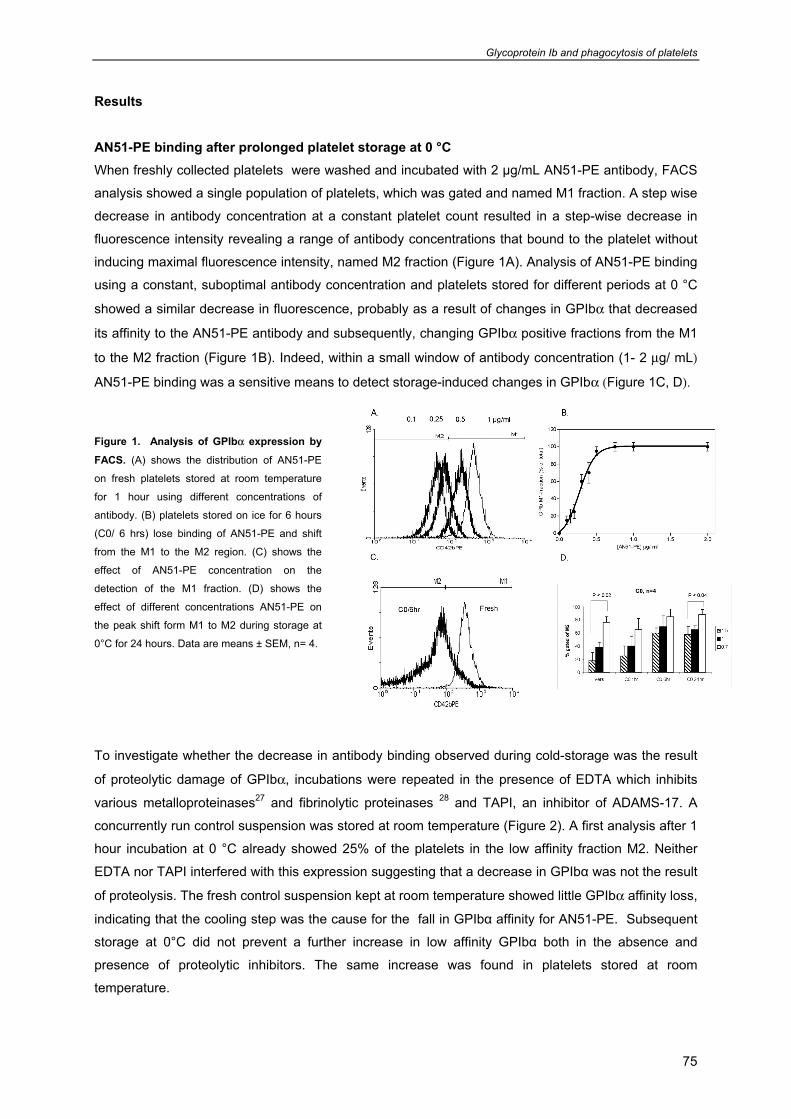
Glycoprotein Ib and phagocytosis of platelets
75
Results AN51-PE binding after prolonged platelet storage at 0 °C When freshly collected platelets were washed and incubated with 2 µg/mL AN51-PE antibody, FACS
analysis showed a single population of platelets, which was gated and named M1 fraction. A step wise
decrease in antibody concentration at a constant platelet count resulted in a step-wise decrease in
fluorescence intensity revealing a range of antibody concentrations that bound to the platelet without
inducing maximal fluorescence intensity, named M2 fraction (Figure 1A). Analysis of AN51-PE binding
using a constant, suboptimal antibody concentration and platelets stored for different periods at 0 °C
showed a similar decrease in fluorescence, probably as a result of changes in GPIbα that decreased
its affinity to the AN51-PE antibody and subsequently, changing GPIbα positive fractions from the M1
to the M2 fraction (Figure 1B). Indeed, within a small window of antibody concentration (1- 2 μg/ mL)
AN51-PE binding was a sensitive means to detect storage-induced changes in GPIbα (Figure 1C, D).
Figure 1. Analysis of GPIbα expression by
FACS. (A) shows the distribution of AN51-PE
on fresh platelets stored at room temperature
for 1 hour using different concentrations of
antibody. (B) platelets stored on ice for 6 hours
(C0/ 6 hrs) lose binding of AN51-PE and shift
from the M1 to the M2 region. (C) shows the
effect of AN51-PE concentration on the
detection of the M1 fraction. (D) shows the
effect of different concentrations AN51-PE on
the peak shift form M1 to M2 during storage at
0°C for 24 hours. Data are means ± SEM, n= 4.
To investigate whether the decrease in antibody binding observed during cold-storage was the result
of proteolytic damage of GPIbα, incubations were repeated in the presence of EDTA which inhibits
various metalloproteinases27 and fibrinolytic proteinases 28 and TAPI, an inhibitor of ADAMS-17. A
concurrently run control suspension was stored at room temperature (Figure 2). A first analysis after 1
hour incubation at 0 °C already showed 25% of the platelets in the low affinity fraction M2. Neither
EDTA nor TAPI interfered with this expression suggesting that a decrease in GPIbα was not the result
of proteolysis. The fresh control suspension kept at room temperature showed little GPIbα affinity loss,
indicating that the cooling step was the cause for the fall in GPIbα affinity for AN51-PE. Subsequent
storage at 0°C did not prevent a further increase in low affinity GPIbα both in the absence and
presence of proteolytic inhibitors. The same increase was found in platelets stored at room
temperature.
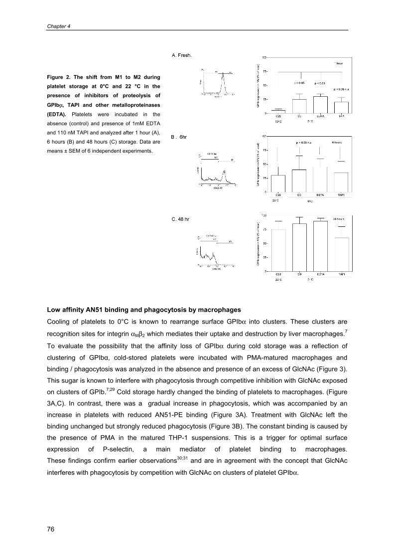
Chapter 4
76
Figure 2. The shift from M1 to M2 during platelet storage at 0°C and 22 °C in the presence of inhibitors of proteolysis of
GPIbα, TAPI and other metalloproteinases
(EDTA). Platelets were incubated in the
absence (control) and presence of 1mM EDTA
and 110 nM TAPI and analyzed after 1 hour (A),
6 hours (B) and 48 hours (C) storage. Data are
means ± SEM of 6 independent experiments.
Low affinity AN51 binding and phagocytosis by macrophages
Cooling of platelets to 0°C is known to rearrange surface GPIbα into clusters. These clusters are
recognition sites for integrin αMβ2 which mediates their uptake and destruction by liver macrophages.7
To evaluate the possibility that the affinity loss of GPIbα during cold storage was a reflection of
clustering of GPIbα, cold-stored platelets were incubated with PMA-matured macrophages and
binding / phagocytosis was analyzed in the absence and presence of an excess of GlcNAc (Figure 3).
This sugar is known to interfere with phagocytosis through competitive inhibition with GlcNAc exposed
on clusters of GPIb.7;29 Cold storage hardly changed the binding of platelets to macrophages. (Figure
3A,C). In contrast, there was a gradual increase in phagocytosis, which was accompanied by an
increase in platelets with reduced AN51-PE binding (Figure 3A). Treatment with GlcNAc left the
binding unchanged but strongly reduced phagocytosis (Figure 3B). The constant binding is caused by
the presence of PMA in the matured THP-1 suspensions. This is a trigger for optimal surface
expression of P-selectin, a main mediator of platelet binding to macrophages.
These findings confirm earlier observations30;31 and are in agreement with the concept that GlcNAc
interferes with phagocytosis by competition with GlcNAc on clusters of platelet GPIbα.
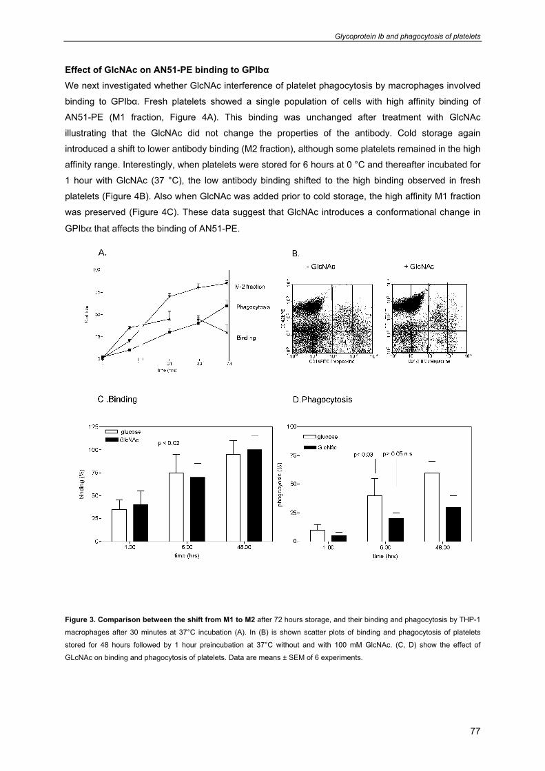
Glycoprotein Ib and phagocytosis of platelets
77
Effect of GlcNAc on AN51-PE binding to GPIbα We next investigated whether GlcNAc interference of platelet phagocytosis by macrophages involved
binding to GPIbα. Fresh platelets showed a single population of cells with high affinity binding of
AN51-PE (M1 fraction, Figure 4A). This binding was unchanged after treatment with GlcNAc
illustrating that the GlcNAc did not change the properties of the antibody. Cold storage again
introduced a shift to lower antibody binding (M2 fraction), although some platelets remained in the high
affinity range. Interestingly, when platelets were stored for 6 hours at 0 °C and thereafter incubated for
1 hour with GlcNAc (37 °C), the low antibody binding shifted to the high binding observed in fresh
platelets (Figure 4B). Also when GlcNAc was added prior to cold storage, the high affinity M1 fraction
was preserved (Figure 4C). These data suggest that GlcNAc introduces a conformational change in
GPIbα that affects the binding of AN51-PE.
Figure 3. Comparison between the shift from M1 to M2 after 72 hours storage, and their binding and phagocytosis by THP-1
macrophages after 30 minutes at 37°C incubation (A). In (B) is shown scatter plots of binding and phagocytosis of platelets
stored for 48 hours followed by 1 hour preincubation at 37°C without and with 100 mM GlcNAc. (C, D) show the effect of
GLcNAc on binding and phagocytosis of platelets. Data are means ± SEM of 6 experiments.
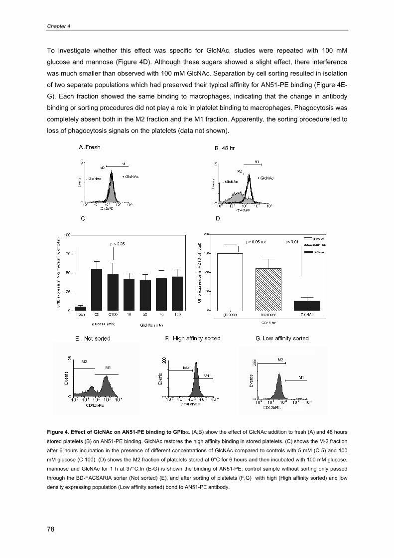
Chapter 4
78
To investigate whether this effect was specific for GlcNAc, studies were repeated with 100 mM
glucose and mannose (Figure 4D). Although these sugars showed a slight effect, there interference
was much smaller than observed with 100 mM GlcNAc. Separation by cell sorting resulted in isolation
of two separate populations which had preserved their typical affinity for AN51-PE binding (Figure 4E-
G). Each fraction showed the same binding to macrophages, indicating that the change in antibody
binding or sorting procedures did not play a role in platelet binding to macrophages. Phagocytosis was
completely absent both in the M2 fraction and the M1 fraction. Apparently, the sorting procedure led to
loss of phagocytosis signals on the platelets (data not shown).
Figure 4. Effect of GlcNAc on AN51-PE binding to GPIbα. (A,B) show the effect of GlcNAc addition to fresh (A) and 48 hours
stored platelets (B) on AN51-PE binding. GlcNAc restores the high affinity binding in stored platelets. (C) shows the M-2 fraction
after 6 hours incubation in the presence of different concentrations of GlcNAc compared to controls with 5 mM (C 5) and 100
mM glucose (C 100). (D) shows the M2 fraction of platelets stored at 0°C for 6 hours and then incubated with 100 mM glucose,
mannose and GlcNAc for 1 h at 37°C.In (E-G) is shown the binding of AN51-PE; control sample without sorting only passed
through the BD-FACSARIA sorter (Not sorted) (E), and after sorting of platelets (F,G) with high (High affinity sorted) and low
density expressing population (Low affinity sorted) bond to AN51-PE antibody.
M2 M1
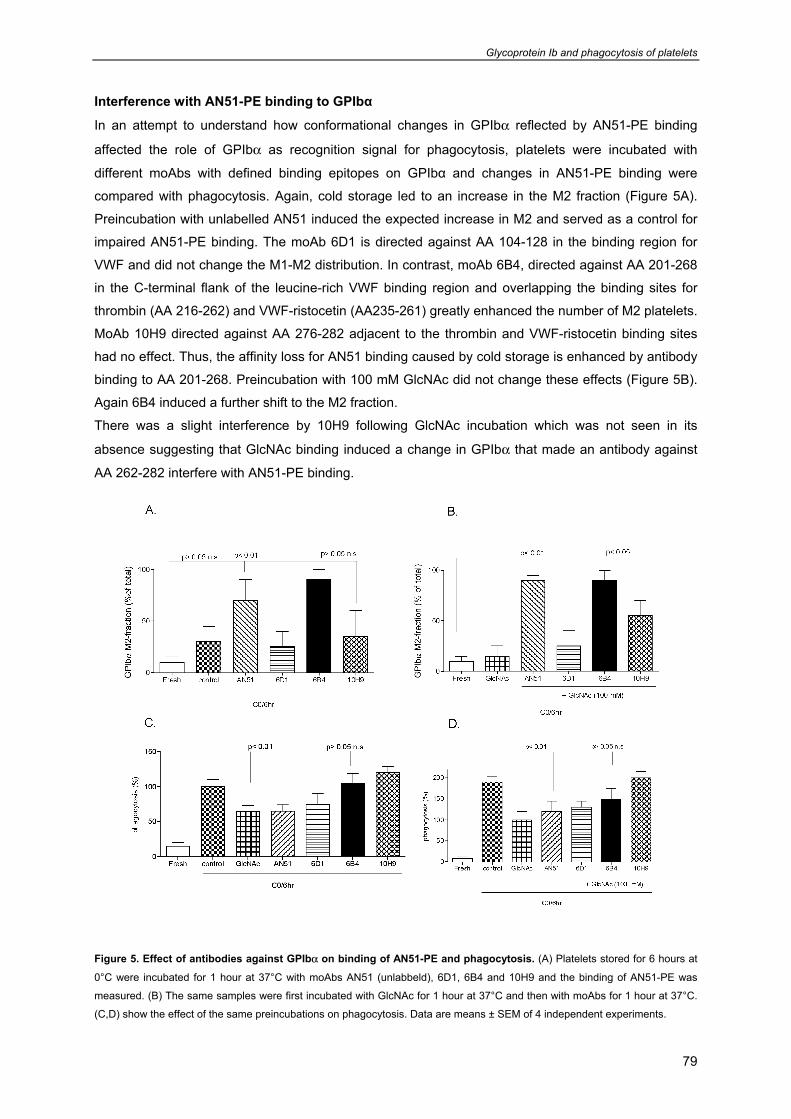
Glycoprotein Ib and phagocytosis of platelets
79
Interference with AN51-PE binding to GPIbα
In an attempt to understand how conformational changes in GPIbα reflected by AN51-PE binding
affected the role of GPIbα as recognition signal for phagocytosis, platelets were incubated with
different moAbs with defined binding epitopes on GPIbα and changes in AN51-PE binding were
compared with phagocytosis. Again, cold storage led to an increase in the M2 fraction (Figure 5A).
Preincubation with unlabelled AN51 induced the expected increase in M2 and served as a control for
impaired AN51-PE binding. The moAb 6D1 is directed against AA 104-128 in the binding region for
VWF and did not change the M1-M2 distribution. In contrast, moAb 6B4, directed against AA 201-268
in the C-terminal flank of the leucine-rich VWF binding region and overlapping the binding sites for
thrombin (AA 216-262) and VWF-ristocetin (AA235-261) greatly enhanced the number of M2 platelets.
MoAb 10H9 directed against AA 276-282 adjacent to the thrombin and VWF-ristocetin binding sites
had no effect. Thus, the affinity loss for AN51 binding caused by cold storage is enhanced by antibody
binding to AA 201-268. Preincubation with 100 mM GlcNAc did not change these effects (Figure 5B).
Again 6B4 induced a further shift to the M2 fraction.
There was a slight interference by 10H9 following GlcNAc incubation which was not seen in its
absence suggesting that GlcNAc binding induced a change in GPIbα that made an antibody against
AA 262-282 interfere with AN51-PE binding.
Figure 5. Effect of antibodies against GPIbα on binding of AN51-PE and phagocytosis. (A) Platelets stored for 6 hours at
0°C were incubated for 1 hour at 37°C with moAbs AN51 (unlabbeld), 6D1, 6B4 and 10H9 and the binding of AN51-PE was
measured. (B) The same samples were first incubated with GlcNAc for 1 hour at 37°C and then with moAbs for 1 hour at 37°C.
(C,D) show the effect of the same preincubations on phagocytosis. Data are means ± SEM of 4 independent experiments.
Chapter 4
80
Concurrent analysis of phagocytosis demonstrated that treatments in the absence, the antibodies had
little effect AN51 and 6D1 slightly reduced phagocytosis to the range also found with GlcNAc, whereas
6B4 and 10H9 proserved phagocytosis in the range of the controls (Figure 5C). The unlabelled AN51
antibody did not induce more phagocytosis suggesting that the recognition signals for apoptosis are
close to AA 1-35. When the platelets were first treated with GlcNAc and thereafter with the antibodies,
the difference between the lower phagocytosis induced by GlcNAc and the higher phagocytosis
induced by the antibodies became larger illustrating that these antibodies opposed the mechanisms
that reduced the phagocytotic signals on GPIbα (Figure 5D). Again AN51 had no effect. Treatment
with antibodies prior to GlcNAc resulted in similar findings as treatment with antibodies alone,
suggesting that the binding of the antibodies is sufficiently strong to resist interference by GlcNAc
(data not shown).
Glycoprotein Ib and phagocytosis of platelets
81
Discussion
Here we show that cooling of platelets and subsequent storage at 0°C induces a change in GPIbα that
leads to a decrease in binding affinity for a moAb directed against AA 1-35 in the N-terminal flank.
High affinity binding is restored by GlcNAc which is known to inhibit phagocytosis of cold stored
platelets by macrophages by competing with GlcNAc-exposing GPIbα clusters for binding to integrin
αMβ2 on macrophages.7;29 Detection of these changes in GPIbα depends on a critical antibody
concentration as an excess fails to detect a shift from the M1 to the M2 fraction (data not shown),
whereas a suboptimal concentration detects all fresh platelets in M2 preventing a further increase
upon cold-storage. The observation that GlcNAc restores high affinity antibody binding of cold-stored
platelets suggests that in addition to its capacity to interfere with GPIbα - αMβ2 interaction, the sugar
introduces a conformational change in GPIbα that affects its affinity to AN51-PE. Interestingly, the
same treatment reduces phagocytosis, making AN51-PE binding a marker for phagocytosis signals on
GPIbα.
Attempts to evaluate separate M1 and M2 populations for there capacity to become phagocytozed
were unsuccesfull. Both populations could be clearly separated and showed a similar binding to THP-
1 cells matured by preincubation with a high concentration of PMA. Previous studies have shown that
surface expressed P-selectin is a major intermediate in platelet binding to macrophages suggesting
that M1 and M2 platelets showed a similar degree of α-granule secretion, which is the source of P-
selectin expression. This might be the result of activation by PMA in the cell suspensions, which is a
potent inducer of the platelet secretion response.32-34 Initially, the presence of AN51-PE on the
platelets during sorting was thought to be the cause of the abolished phagocytosis. However, addition
of AN51 antibody prior to platelet-macrophage contact in stead of the usual addition of antbody at the
end of the phagocytosis assay showed that AN51 did not reduced with platelet destruction. Probably,
the duration of the sorting procedure and the strain to which platelets are submitted destroys the
phagocytosis signals in GPIbα. The finding that AN51-PE binding recognizes phagocytosis signals in
GPIbα but does not block with this process indicates that its binding domain AA 1-35 is not the site
where changes induced by cold storage lead to recognition by macrophages.
Apparently, loss of AN51-PE binding is a sensitive marker for changes elsewhere in GPIbα that
become targets for macrophage recognition. These changes are enhanced by moAb 6B4 directed
against AA 201-268 overlapping the binding sites for thrombin and the VWF-ristocetin complex and
10H9 against AA 276-282 in the anionic sulfated sequence adjacent to these binding sites. In contrast,
moAbs 6D1 against AA 104-128 of the VWF binding domain had no effect. In freshly collected platelet
suspensions moAb 6B4 is known to inhibit VWF binding to its binding domain in region in AA 36 – 200 23;35suggesting a spatial orientation of GPIbα that brings regions AA 201 – 268 and AA 36 – 200 in
close proximity. The present data with cold-preserved platelets suggest such a proximity for the
regions AA 201 - 268 recognized by 6B4 and AA 1- 35 to which AN51 binds and factors that interfere
with such an interaction such as the binding of an antibody affect the generation of phagocytosis
signals. Also moAb 10H9 interfered with AN51-PE binding and phagocytosis although its binding
epitope is adjacent of the 6B4 binding site (Figure 6).
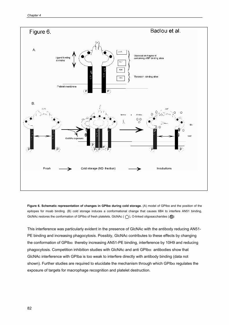
Chapter 4
82
Figure 6. Schematic representation of changes in GPIbα during cold storage. (A) model of GPIbα and the position of the
epitopes for moab binding. (B) cold storage induces a conformational change that causes 6B4 to interfere AN51 binding,
GlcNAc restores the conformation of GPIbα of fresh platelets. GlcNAc ( ), O-linked oligosaccharides ( )
This interference was particularly evident in the presence of GlcNAc with the antibody reducing AN51-
PE binding and increasing phagocytosis. Possibly, GlcNAc contributes to these effects by changing
the conformation of GPIbα thereby increasing AN51-PE binding, interference by 10H9 and reducing
phagocytosis. Competition inhibition studies with GlcNAc and anti GPIbα antibodies show that
GlcNAc interference with GPIba is too weak to interfere directly with antibody binding (data not
shown). Further studies are required to elucidate the mechanism through which GPIbα regulates the
exposure of targets for macrophage recognition and platelet destruction.

Glycoprotein Ib and phagocytosis of platelets
83
Acknowledgment This work was supported by Sanquin Blood Supply Foundation (grant nr. PPO 01.019). JWNA is
supported by the Netherlands Thrombosis Foundation.
Chapter 4
84
References
1. Leytin V, Freedman J. Platelet apoptosis in stored platelet concentrates and other models.
Transfus.Apheresis.Sci. 2003;28:285-295.
2. Perrotta PL, Perrotta CL, Snyder EL. Apoptotic activity in stored human platelets. Transfusion
2003;43:526-535.
3. Wadhawan V, Karim ZA, Mukhopadhyay S et al. Platelet storage under in vitro condition is
associated with calcium-dependent apoptosis-like lesions and novel reorganization in platelet
cytoskeleton. Arch.Biochem.Biophys. 2004;422:183-190.
4. Badlou BA, Ijseldijk MJ, Smid WM, Akkerman JW. Prolonged platelet preservation by transient
metabolic suppression. Transfusion 2005;45:214-222.
5. Stepanian A, Ribba AS, Lavergne JM et al. A new mutation, S1285F, within the A1 loop of von
Willebrand factor induces a conformational change in A1 loop with abnormal binding to platelet
GPIb and botrocetin causing type 2M von Willebrand disease. Br.J.Haematol. 2003;120:643-
651.
6. Snyder EL, Rinder HM. Platelet storage--time to come in from the cold? N.Engl.J.Med.
2003;348:2032-2033.
7. Hoffmeister KM, Felbinger TW, Falet H et al. The clearance mechanism of chilled blood
platelets. Cell 2003;112:87-97.
8. Kawasaki T, Fujimura Y, Usami Y et al. Complete amino acid sequence and identification of the
platelet glycoprotein Ib-binding site of jararaca GPIb-BP, a snake venom protein isolated from
Bothrops jararaca. J.Biol.Chem. 1996;271:10635-10639.
9. Hartwig JH. Mechanisms of actin rearrangements mediating platelet activation. J.Cell Biol.
1992;118:1421-1442.
10. Omokawa S, Notoya T, Kumagai M et al. Status of platelet collection and platelet transfusion.
Ther.Apher. 2001;5:17-21.
11. Andrews RK, Gardiner EE, Shen Y, Whisstock JC, Berndt MC. Glycoprotein Ib-IX-V.
Int.J.Biochem.Cell Biol. 2003;35:1170-1174.
12. Canobbio I, Balduini C, Torti M. Signalling through the platelet glycoprotein Ib-V-IX complex.
Cell Signal. 2004;16:1329-1344.
13. Huizinga EG, Tsuji S, Romijn RA et al. Structures of glycoprotein Ibalpha and its complex with
von Willebrand factor A1 domain. Science 2002;297:1176-1179.
14. Chou CF, Omary MB. Mitotic arrest with anti-microtubule agents or okadaic acid is associated
with increased glycoprotein terminal GlcNAc's. J.Cell Sci. 1994;107:1833-1843.
15. Wu YP, Vink T, Schiphorst M et al. Platelet thrombus formation on collagen at high shear rates
is mediated by von Willebrand factor-glycoprotein Ib interaction and inhibited by von Willebrand
factor-glycoprotein IIb/IIIa interaction. Arterioscler.Thromb.Vasc.Biol. 2000;20:1661-1667.
Glycoprotein Ib and phagocytosis of platelets
85
16. Blann AD, Lip GY, Fijnheer R. Significance of soluble P-selectin, von Willebrand factor, and
other adhesion molecules in hypercholesterolemia and peripheral artery disease. Circulation
1999;99:2478-2479.
17. Adelman B, Michelson AD, Greenberg J, Handin RI. Proteolysis of platelet glycoprotein Ib by
plasmin is facilitated by plasmin lysine-binding regions. Blood 1986;68:1280-1284.
18. Baglia FA, Shrimpton CN, Emsley J et al. Factor XI interacts with the leucine-rich repeats of
glycoprotein Ibalpha on the activated platelet. J.Biol.Chem. 2004;%19;279:49323-49329.
19. Barnard MR, Macgregor H, Ragno G et al. Fresh, liquid-preserved, and cryopreserved platelets:
adhesive surface receptors and membrane procoagulant activity. Transfusion 1999;39:880-888.
20. Dormann D, Clemetson KJ, Kehrel BE. The GPIb thrombin-binding site is essential for thrombin-
induced platelet procoagulant activity. Blood 2000;96:2469-2478.
21. Bergmeier W, Burger PC, Piffath CL et al. Metalloproteinase inhibitors improve the recovery and
hemostatic function of in vitro-aged or -injured mouse platelets. Blood 2003;102:4229-4235.
22. Fox JE. Shedding of adhesion receptors from the surface of activated platelets. Blood
Coagul.Fibrinolysis 1994;5:291-304.
23. Cauwenberghs N, Vanhoorelbeke K, Vauterin S et al. Epitope mapping of inhibitory antibodies
against platelet glycoprotein Ibalpha reveals interaction between the leucine-rich repeat N-
terminal and C-terminal flanking domains of glycoprotein Ibalpha. Blood 2001;98:652-660.
24. Deckmyn H, Cauwenberghs N, Wu D, Depraetere H, Vanhoorelbeke K. Development of
antibodies that interfere with the collagen-VWF-GPIb axis as new antithrombotics.
Verh.K.Acad.Geneeskd.Belg. 2005;67:55-65.
25. Badlou BA, Wu YP, Smid WM, Akkerman JW: Regulation of platelet phagocytosis by
macrophages. Transfusion In press.
26. Muta T, Yamazaki S, Eto A, Motoyama M, Takeshige K. IkappaB-zeta, a new anti-inflammatory
nuclear protein induced by lipopolysaccharide, is a negative regulator for nuclear factor-kappaB.
J.Endotoxin.Res. 2003;9:187-191.
27. Byrd CM, Hruby DE. Development of an in vitro cleavage assay system to examine vaccinia
virus I7L cysteine proteinase activity. Virol.J. 2005;2:63.:63.
28. Bello CA, Hermogenes AL, Magalhaes A et al. Isolation and biochemical characterization of a
fibrinolytic proteinase from Bothrops leucurus (white-tailed jararaca) snake venom. Biochimie
2006;88:189-200.
29. Josefsson EC, Gebhard HH, Stossel TP, Hartwig JH, Hoffmeister KM. The macrophage
alphaMbeta2 integrin alphaM lectin domain mediates the phagocytosis of chilled platelets.
J.Biol.Chem. 2005;280:18025-18032.
30. Fernandes LS, Conde ID, Wayne SC et al. Platelet-monocyte complex formation: effect of
blocking PSGL-1 alone, and in combination with alphaIIbbeta3 and alphaMbeta2, in coronary
stenting. Thromb.Res. 2003;111:171-177.
31. Berger G, Hartwell DW, Wagner DD. P-Selectin and platelet clearance. Blood 1998;92:4446-
4452.
Chapter 4
86
32. Whiss PA, Andersson RG, Srinivas U. Kinetics of platelet P-selectin mobilization: concurrent
surface expression and release induced by thrombin or PMA, and inhibition by the NO donor
SNAP. Cell Adhes.Commun. 1998;6:289-300.
33. Krishnamurthi S, Joseph S, Kakkar VV. Effect of phorbol 12-myristate 13-acetate (PMA) on
agonist-induced arachidonate release and 5-hydroxytryptamine secretion in human platelets.
Dependence of effects on agonist type and time of incubation with PMA. Biochim.Biophys.Acta
1987;927:429-436.
34. Chen R, Liang N. Cytoskeletal changes in platelets induced by thrombin and phorbol myristate
acetate (PMA). Cell Biol.Int. 1998;22:429-435.
35. Cauwenberghs N, Meiring M, Vauterin S et al. Antithrombotic effect of platelet glycoprotein Ib-
blocking monoclonal antibody Fab fragments in nonhuman primates.
Arterioscler.Thromb.Vasc.Biol. 2000;20:1347-1353.
Surface markers for platelet binding and phagocytosis
87
Chapter 5
Role of surface markers for binding and phagocytosis of platelets by macrophages
B.A. Badlou1, 2,3, W. M. Smid2 and J.W.N. Akkerman1,3
From the Thrombosis and Haemostasis Laboratory, Department of Haematology,
University Medical Centre Utrecht1, the Sanquin Blood Bank Region North-West Amsterdam2, and
The Institute for Biomembranes, Utrecht University, Utrecht3, The Netherlands.
Submitted
Chapter 5
88
Abstract Introduction: Using an in vitro assay for analysis of the interaction between platelets and
macrophages, we previously identified surface-expressed platelet P-selectin as an intermediate in
binding and exposure of phosphatidylserine (PS) combined with conformational changes in
glycoprotein (GP) Ibα as intermediates in phagocytosis. There is little insight in the relative contribution
of these regulators of platelet destruction by macrophages.
Objectives: The aim of this study was to evaluate the quantitative relationships between these
markers for platelet destruction by macrophages.
Methods and study design: Platelets in buffer were stored at 22°C for different periods to obtain a
wide range of surface expressed P-selectin and -PS measured by FACS. The storage-induced
conformational change in GPIbα was deduced from decreased binding of an anti GPIbα antibody, also
by FACS. Data from the same platelets were compared with binding and phagocytosis by PMA-
matured monocytic THP-1 cells to determine the relative contribution of these parameters to platelet
destruction. Correlations established in room temperature stored platelets were then compared with
platelets stored at 0°C and metabolic suppressed platelets (MSPs) stored at 4°C to investigate
whether changes in storage conditions affected the relation between surface markers and platelet
destruction.
Results: Between 0 and 80%, P-selectin expression correlated linearly with binding, which then
leveled off. PS exposure showed a threshold of 20% below which phagocytosis was < 10%, but
between 20 and 50% PS exposure correlated linearly with phagocytosis. GPIbα conformational
change showed a threshold of 30% below which phagocytosis was < 10%, but between 30 and 90%
GPIbα change correlated linearly with phagocytosis. A comparison between room temperature-stored,
cold-stored and metabolic suppressed platelets (MSPs) showed that at a given degree of PS exposure
and GPIbα change, cold stored platelets showed more phagocytosis than the other two suspensions.
Conclusions: These in vitro correlations may help to predict the extent of platelet binding to
macrophages based on P-selectin expression, and the extent of phagocytosis based on PS-
expression and GPIbα change. If platelet destruction parameters measured in vitro correlate with
platelet destruction in vivo, the quality of transfused platelets can be predicted on the basis of these
relatively simple FACS analyses.
Surface markers for platelet binding and phagocytosis
89
Introduction Most investigators in the field of platelet transfusion favor the idea that optimal platelet storage in vitro
correlates with optimal haemostatic effectiveness and survival in vivo.1,2 There is indeed evidence that
optimal preservation of platelet reactivity results in better platelet functions after transfusion with little
binding to white blood cells3 and phagocytosis by macrophages.4-6 Nevertheless, there is a need for
platelet tests that under laboratory conditions predict how well platelets behave in circulating blood. A
test that predicts the efficacy of transfusions protocols for platelet concentrates (PCs) would have a
clear medical benefit and save money. Different markers have been tested as indicators of the so
called platelet storage lesion (PSL), such as P-selectin expression (PSE),7 expression of CD40/CD40
ligand 8 and glycoprotein (GP)IIbIIIa;9,10 changes in mean platelet volume, sphere to disc changes;11-15
actin assembly;16,17 serotonin release;14,18 changes in GPIb-V-IX and GPVI expressions;19,20 PS
exposure;21,22 loss of swirling;23 osmotic pressure reponse24-27 and changes in platelet count caused
by agglutination.18,28,28-30 Although each marker provides insight in the activation state of the platelets,
none of them is a perfect predictor of the quality of transfused platelets.2 An additional problem is the
lack of comparative studies with data on sensitivity and specificity of the different markers.
Prolonged storage induces an gradual increase in PSE 24,31-33 and PS exposure 5,34-40 and a decrease
in GPIbα expression.6,41-44 Hoffmeister et al45,46 described that cold-induced GPIb clustering correlates
positively with phagocytosis and that galactosylation of GPIbα restores normal platelet survival. Leytin
et al.6 described that PSE showed a positive and GPIb expression a negative correlation with
phagocytosis of platelets in vivo.
Previously we provided evidence that binding of platelets to macrophages is mediated by surface
expressed P-selectin and phagocytosis by exposed PS and changes in GPIbα, the receptor for von
Willebrand factor. Changes in GPIbα were detected as a decrease in the binding of a PE-labeled
moAb against AA 1 – 35 on the N-terminal flank and thought to be related to the clustering of GPIbα
observed after cooling of platelets.45,46
The aim of this study was to investigate how PSE, PS exposure and changes in the conformation of
GPIbα that occur during platelet storage correlate with binding and phagocytosis by macrophages in
an in vitro assay. The results show that each of these parameters contribute in a different manner to
the processes that lead to destruction of platelets.
Chapter 5
90
Materials and Method Platelet preparation Platelets were prepared as previously described.44 In short, freshly drawn venous blood (40 ml) from
healthy volunteers was collected with informed consent into 1:10 v/v 130 mmol/L trisodium citrate. The
donors claimed not to have taken any medication during two weeks prior to blood collection. Platelet-
rich plasma (PRP) was prepared by centrifugation (200 g, 15 minutes, 22°C). ACD (0.1 volume) was
added to lower the pH to 6.5 and prevent platelet activation during further isolation. The suspension
was centrifuged and resuspended in Hepes-Tyrode (pH 7.2). Platelet count was measured on a
Cellcounter AL871 (Molab, Hilden, Germany). The platelet number was adjusted to 200 000 cells/µl
before the experiments. Metabolic suppressed platelets (MSP) were prepared by incubating the cells
for 40 minutes at 37°C in glucose-free Hepes-Tyrode (pH 7.2) in the presence of 20 μM antimycin A.
At the times indicated, 100 μl samples were collected, incubated with 20 mM glucose in Hepes tyrode
buffer for 1 hr at 37°C to restore energy generation. Controls were platelet suspensions in Hepes-
Tyrode pH 7.2 containing 5 mM glucose stored at 22°C (C 22°C) or immediately cooled on ice (C
0°C). C 22°C was incubated with 20 mM glucose (1 hr, 37°C) prior to the aggregation tests to account
for a possible glucose shortage.
Platelet P-selectin, PS exposure and GPIbα expression measurements by FACS
P-selectin and GPIbα expression was measured as described.44 In short, at the indicated time points,
100 µl sample was incubated in 1 ml plastic tubes with moAb CD62p-FITC and CD42b-PE (R70, clone
AN51). For analysis of PS exposure, platelets were incubated with a combination of annexin V-FITC
and AN51-PE as described.47,48 To all samples 300 µl PBS was added and immediately 10,000 events
were measured on a FACScalibur. Quantitative analysis was done using WinMDI software.
Analysis of platelet binding and phagocytosis by macrophages The binding of platelets to macrophages was measured as described by Hoffmeister et al.46 with some
significant modifications.47 In short, monocytic THP-1 cell lines divided in 24x NUNC multiwell plates (1
ml/ well) matured by 500 nM PMA incubated overnight at 37°C, 5% CO2, in the cell incubator. 2x10⁶
platelets in Hepes-Tyrode buffer (pH 7.2) were added to a well containing 1x10⁶ PMA-matured THP-1
cells in 1 mL RPMI 1640 medium and the mixed suspensions were incubated at 37°C without agitation
Wells were gently washed with HBSS buffer (0.3 mM KH2PO4, 13.7 mM NaCl, 417 mM NaHCO3, 31
mM Na2HPO4 and 0.5 mM KCl in aqua dest) and free platelets collected in the wash medium were
isolated by centrifugation-resuspension under protection of PGI2. Wells were incubated with 200 μL
HBSS buffer containing 5 mM EDTA for 15 minutes at 0°C, reconstituted with free platelets removed
during the wash step and thereafter 100 µL suspension was incubated with 2 µg/mL anti-human
CD42b-PE and 2 µg/mL anti-human CD14-FITC antibody for 15 minutes at 37°C. Then, HBSS buffer
was added and 20 000 particles were measured by flowcytometry (FACS Calibur, Becton-Dickinson,
San Jose, CA, USA). FACS data were analyzed with WinMDI software. Binding of platelets to
macrophages was expressed as the percentage of CD42b/CD14 positive particles of the total number
Surface markers for platelet binding and phagocytosis
91
of CD42b and/or CD14 positive particles. Phagocytosis of platelets by PMA matured THP-1 cells was
measured by FACS analysis of mepacrine positive CD14 cells that were inaccessible to the anti
CD42b-PE antibody and expressed as percentage of total number of CD14 positive, CD42b negative
particles.
Analysis of correlations
To compare PSE, PE expression and GPIbα changes with binding and phagocytosis, platelets from 6
to 9 different donors were stored under different conditions for up to 48 hours as indicated in the
“results” section.44,47,48 The surface markers together with data on binding and phagocytosis from the
same donor were expressed as means ± SEM and curve fitted according to the nonlinear regression
(curve fit), the sigmoidal dose-response with the Graph pad program.
Statistics Data are expressed as means ± SEM with number of observations n. Statistical analysis was by
nonparametric Spearman correlation analysis, two-tails with confidence intervals of 95%. Differences
were considered significant at a p value of less than 0.05.
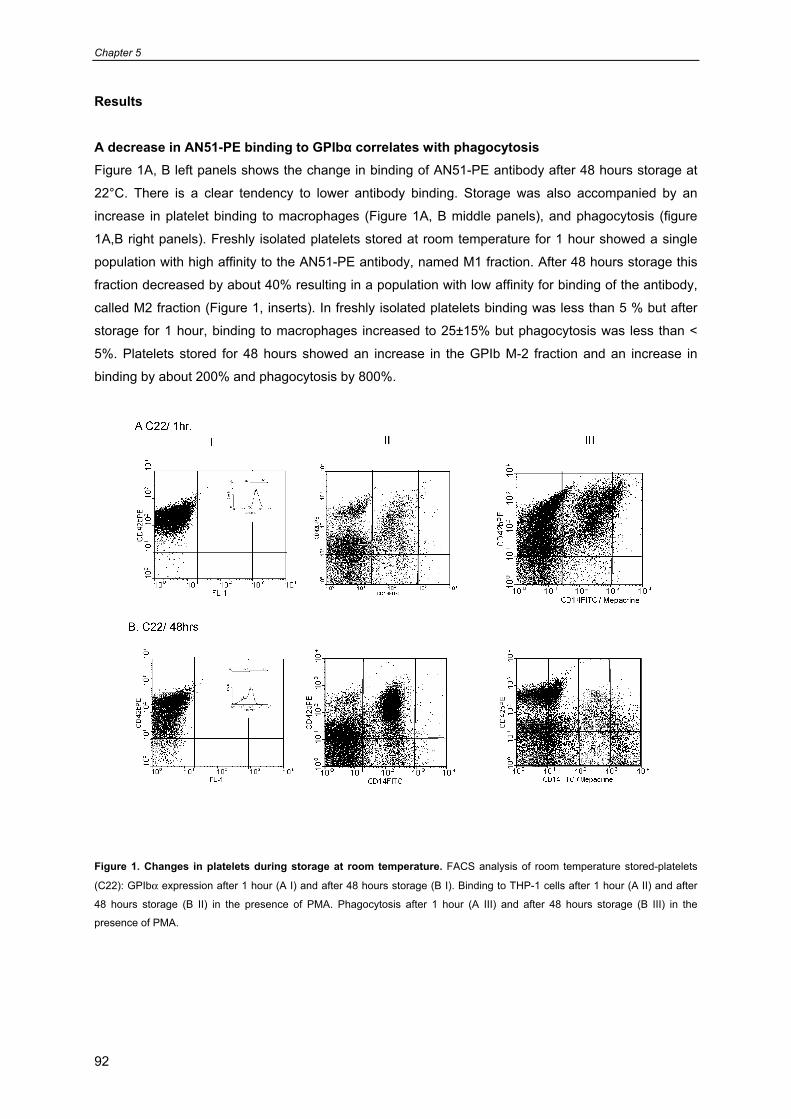
Chapter 5
92
Results A decrease in AN51-PE binding to GPIbα correlates with phagocytosis Figure 1A, B left panels shows the change in binding of AN51-PE antibody after 48 hours storage at
22°C. There is a clear tendency to lower antibody binding. Storage was also accompanied by an
increase in platelet binding to macrophages (Figure 1A, B middle panels), and phagocytosis (figure
1A,B right panels). Freshly isolated platelets stored at room temperature for 1 hour showed a single
population with high affinity to the AN51-PE antibody, named M1 fraction. After 48 hours storage this
fraction decreased by about 40% resulting in a population with low affinity for binding of the antibody,
called M2 fraction (Figure 1, inserts). In freshly isolated platelets binding was less than 5 % but after
storage for 1 hour, binding to macrophages increased to 25±15% but phagocytosis was less than <
5%. Platelets stored for 48 hours showed an increase in the GPIb M-2 fraction and an increase in
binding by about 200% and phagocytosis by 800%.
Figure 1. Changes in platelets during storage at room temperature. FACS analysis of room temperature stored-platelets
(C22): GPIbα expression after 1 hour (A I) and after 48 hours storage (B I). Binding to THP-1 cells after 1 hour (A II) and after
48 hours storage (B II) in the presence of PMA. Phagocytosis after 1 hour (A III) and after 48 hours storage (B III) in the
presence of PMA.
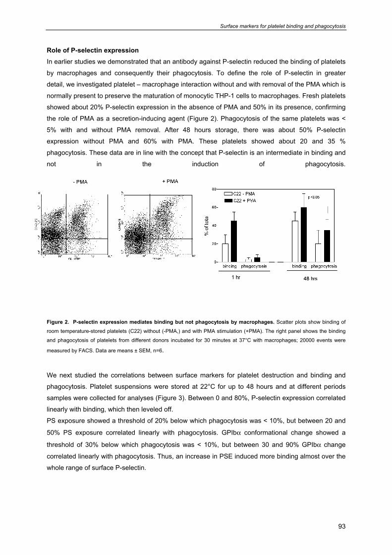
Surface markers for platelet binding and phagocytosis
93
Role of P-selectin expression In earlier studies we demonstrated that an antibody against P-selectin reduced the binding of platelets
by macrophages and consequently their phagocytosis. To define the role of P-selectin in greater
detail, we investigated platelet – macrophage interaction without and with removal of the PMA which is
normally present to preserve the maturation of monocytic THP-1 cells to macrophages. Fresh platelets
showed about 20% P-selectin expression in the absence of PMA and 50% in its presence, confirming
the role of PMA as a secretion-inducing agent (Figure 2). Phagocytosis of the same platelets was <
5% with and without PMA removal. After 48 hours storage, there was about 50% P-selectin
expression without PMA and 60% with PMA. These platelets showed about 20 and 35 %
phagocytosis. These data are in line with the concept that P-selectin is an intermediate in binding and
not in the induction of phagocytosis.
Figure 2. P-selectin expression mediates binding but not phagocytosis by macrophages. Scatter plots show binding of
room temperature-stored platelets (C22) without (-PMA,) and with PMA stimulation (+PMA). The right panel shows the binding
and phagocytosis of platelets from different donors incubated for 30 minutes at 37°C with macrophages; 20000 events were
measured by FACS. Data are means ± SEM, n=6. We next studied the correlations between surface markers for platelet destruction and binding and
phagocytosis. Platelet suspensions were stored at 22°C for up to 48 hours and at different periods
samples were collected for analyses (Figure 3). Between 0 and 80%, P-selectin expression correlated
linearly with binding, which then leveled off.
PS exposure showed a threshold of 20% below which phagocytosis was < 10%, but between 20 and
50% PS exposure correlated linearly with phagocytosis. GPIbα conformational change showed a
threshold of 30% below which phagocytosis was < 10%, but between 30 and 90% GPIbα change
correlated linearly with phagocytosis. Thus, an increase in PSE induced more binding almost over the
whole range of surface P-selectin.
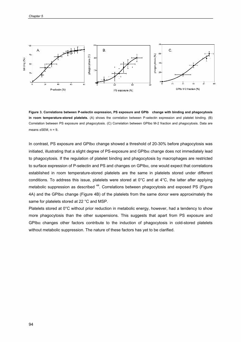
Chapter 5
94
Figure 3. Correlations between P-selectin expression, PS exposure and GPIb� change with binding and phagocytosis in room temperature-stored platelets. (A) shows the correlation between P-selectin expression and platelet binding. (B)
Correlation between PS exposure and phagocytosis. (C) Correlation between GPIbα M-2 fraction and phagocytosis. Data are
means ±SEM, n = 9.
In contrast, PS exposure and GPIbα change showed a threshold of 20-30% before phagocytosis was
initiated, illustrating that a slight degree of PS-exposure and GPIbα change does not immediately lead
to phagocytosis. If the regulation of platelet binding and phagocytosis by macrophages are restricted
to surface expression of P-selectin and PS and changes on GPIbα, one would expect that correlations
established in room temperature-stored platelets are the same in platelets stored under different
conditions. To address this issue, platelets were stored at 0°C and at 4°C, the latter after applying
metabolic suppression as described 44. Correlations between phagocytosis and exposed PS (Figure
4A) and the GPIbα change (Figure 4B) of the platelets from the same donor were approximately the
same for platelets stored at 22 °C and MSP.
Platelets stored at 0°C without prior reduction in metabolic energy, however, had a tendency to show
more phagocytosis than the other suspensions. This suggests that apart from PS exposure and
GPIbα changes other factors contribute to the induction of phagocytosis in cold-stored platelets
without metabolic suppression. The nature of these factors has yet to be clarified.
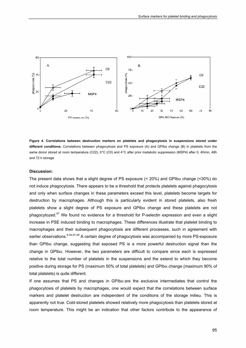
Surface markers for platelet binding and phagocytosis
95
Figure 4. Correlations between destruction markers on platelets and phagocytosis in suspensions stored under different conditions. Correlations between phagocytosis and PS exposure (A) and GPIbα change (B) in platelets from the
same donor stored at room temperature (C22), 0°C (C0) and 4°C after prior metabolic suppression (MSP4) after 0, 40min, 48h
and 72 h storage
Discussion:
The present data shows that a slight degree of PS exposure (< 20%) and GPIbα change (<30%) do
not induce phagocytosis. There appears to be a threshold that protects platelets against phagocytosis
and only when surface changes in these parameters exceed this level, platelets become targets for
destruction by macrophages. Although this is particularly evident in stored platelets, also fresh
platelets show a slight degree of PS exposure and GPIbα change and these platelets are not
phagocytozed.47 We found no evidence for a threshold for P-selectin expression and even a slight
increase in PSE induced binding to macrophages. These differences illustrate that platelet binding to
macrophages and their subsequent phagocytosis are different processes, such in agreement with
earlier observations.6,34,47,49 A certain degree of phagocytosis was accompanied by more PS-exposure
than GPIbα change, suggesting that exposed PS is a more powerful destruction signal than the
change in GPIbα. However, the two parameters are difficult to compare since each is expressed
relative to the total number of platelets in the suspensions and the extend to which they become
positive during storage for PS (maximum 50% of total platelets) and GPIbα change (maximum 90% of
total platelets) is quite different.
If one assumes that PS and changes in GPIbα are the exclusive intermediates that control the
phagocytosis of platelets by macrophages, one would expect that the correlations between surface
markers and platelet destruction are independent of the conditions of the storage milieu. This is
apparently not true. Cold-stored platelets showed relatively more phagocytosis than platelets stored at
room temperature. This might be an indication that other factors contribute to the appearance of
Chapter 5
96
phagocytic signals on the platelets. These factors have yet to be characterized but the difference
between cold-stored and room temperature stored platelets might provide a basis for further
identification of these factors.
Interestingly, metabolic suppression prior to cold storage greatly suppressed the expression of
phagocytic signals. The relation between PS exposure and GPIbα change versus phagocytosis
appears to follow the correlation established with room temperature stored-platelets, but the low PS
expression and GPIbα change hamper reliable calculations. The rational of applying metabolic
suppression prior to storage is that storage conditions trigger activation of platelets and start functions
such as secretion and PS-exposure. The activating sequences and the execution of secretion
responses in particular are energy-dependent processes. A shortage of metabolic energy would
therefore bring the platelets in a “hibernation” state that protects the cells until energy metabolism is
restored. The finding that this treatment reduces PS-exposure is not surprising since ATP-driven
reactions contribute to the preservation of PS asymmetry and PS flip flop. The finding that metabolic
suppression also reduces the change in GPIbα seen during storage is unexpected. GPIbα is the
receptor that mediates platelet adhesion to VWF.4,46 The cooling of platelets is thought to trigger
irreversible GPIbα clustering which provides a signal for rapid destruction by phagocytes.46 Hepatic
macrophages rapidly remove cold-stored platelets from the circulation through binding of αMβ2 integrin
to GlcNAc-exposing GPIbα clusters.45,46 But also storage at 22°C shows that old platelets are more
prone to phagocytosis than young platelets, suggesting that the same mechanism might control the
removal of room temperature-stored platelets after transfusion.5,6
At present, it is uncertain whether the decrease in binding of a PE-labeled antibody against AA 1-35 of
GPIbα is a reflection of the clustering observed in transmission electron micrographs described
before.46 If one assumes that such a correlation exists, our findings would imply that there is a role of
metabolic energy in the clustering of GPIbα.
It is clear that the present correlations between surface markers for platelet destruction and binding
and phagocytosis by macrophages are based on an in vitro system with matured THP-1 cells. At
present it is uncertain to which extent this set up is an accurate reflection of the destruction of platelets
under in vivo conditions. If such a correlation could be established in future studies, the sensitivity of
transfused platelets to destruction by macrophages could be predicted on the basis a few, rather
simple, FACS analysis.

Surface markers for platelet binding and phagocytosis
97
Acknowledgments
This work was supported by Sanquin Blood Supply Foundation (grant nr. PPO 01.019). JWNA is
supported by the Netherlands Thrombosis Foundation. The authors thank Dr Mark Roest and Drs.
Thomas van Himbergen from the Dept. of Haematology, UMCU, for helpful discussions.
Chapter 5
98
References
1. Holme S, Sweeney JD, Sawyer S, Elfath MD. The expression of p-selectin during collection,
processing, and storage of platelet concentrates: relationship to loss of in vivo viability.
Transfusion 1997 Jan;37(1):12-7.
2. Gurney D, Lip GY, Blann AD. A reliable plasma marker of platelet activation: does it exist?
Am.J.Hematol. 2002 Jun;70(2):139-44.
3. Gutensohn K, Geidel K, Brockmann M, Siemensen M, Krueger W, Kroeger N, Kuehnl P. Binding
of activated platelets to WBCs in vivo after transfusion. Transfusion 2002 Oct;42(10):1373-80.
4. Stepanian A, Ribba AS, Lavergne JM, Fressinaud E, Juhan-Vague I, Mazurier C, Girma JP,
Meyer D. A new mutation, S1285F, within the A1 loop of von Willebrand factor induces a
conformational change in A1 loop with abnormal binding to platelet GPIb and botrocetin causing
type 2M von Willebrand disease. Br.J.Haematol. 2003 Feb;120(4):643-51.
5. Leytin V, Freedman J. Platelet apoptosis in stored platelet concentrates and other models.
Transfus.Apheresis.Sci. 2003 Jun;28(3):285-95.
6. Leytin V, Allen DJ, Gwozdz A, Garvey B, Freedman J. Role of platelet surface glycoprotein
Ibalpha and P-selectin in the clearance of transfused platelet concentrates. Transfusion 2004
Oct;44(10):1487-95.
7. Rinder HM, Snyder EL, Bonan JL, Napychank PA, Malkus H, Smith BR. Activation in stored
platelet concentrates: correlation between membrane expression of P-selectin, glycoprotein
IIb/IIIa, and beta-thromboglobulin release. Transfusion 1993 Jan;33(1):25-9.
8. Danese S, Fiocchi C. Platelet activation and the CD40/CD40 ligand pathway: mechanisms and
implications for human disease. Crit Rev.Immunol. 2005;25(2):103-21.
9. Buensuceso C, de Virgilio M, Shattil SJ. Detection of integrin alpha IIbbeta 3 clustering in living
cells. J.Biol.Chem. 2003 Apr 25;278(17):15217-24.
10. Caron A, Theoret JF, Mousa SA, Merhi Y. Anti-platelet effects of GPIIb/IIIa and P-selectin
antagonism, platelet activation, and binding to neutrophils. J.Cardiovasc.Pharmacol. 2002
Aug;40(2):296-306.
11. Leven RM, Gonnella PA, Reeber MJ, Nachmias VT. Platelet shape change and cytoskeletal
assembly: effects of pH and monovalent cation ionophores. Thromb.Haemost. 1983 Jun
28;49(3):230-4.
12. Kunicki TJ, Tuccelli M, Becker GA, Aster RH. A study of variables affecting the quality of
platelets stored at "room temperature". Transfusion 1975 Sep;15(5):414-21.
13. Sturk A, Burt LM, Hakvoort T, ten Cate JW, Crawford N. The effect of storage on platelet
morphology. Transfusion 1982 Mar;22(2):115-20.
14. Maurer-Spurej E, Pfeiler G, Maurer N, Lindner H, Glatter O, Devine DV. Room temperature
activates human blood platelets. Lab Invest 2001 Apr;81(4):581-92.
15. Nachmias VT. Platelet and megakaryocyte shape change: triggered alterations in the
cytoskeleton. Semin.Hematol. 1983 Oct;20(4):261-81.
Surface markers for platelet binding and phagocytosis
99
16. Mohri H, Asakura Y, Fukushima J, Kawamoto S, Okubo T, Okuda K. Synthetic peptide from the
V3 loop consensus motif with a potent anti-HIV activity inhibits ristocetin-mediated vWF-GPIb
interaction. Peptides 1997;18(9):1289-93.
17. Kovacsovics TJ, Hartwig JH. Thrombin-induced GPIb-IX centralization on the platelet surface
requires actin assembly and myosin II activation. Blood 1996 Jan 15;87(2):618-29.
18. de Korte D, Gouwerok CW, Fijnheer R, Pietersz RN, Roos D. Depletion of dense granule
nucleotides during storage of human platelets. Thromb.Haemost. 1990 Apr 12;63(2):275-8.
19. Bergmeier W, Rabie T, Strehl A, Piffath CL, Prostredna M, Wagner DD, Nieswandt B. GPVI
down-regulation in murine platelets through metalloproteinase-dependent shedding.
Thromb.Haemost. 2004 May;91(5):951-8.
20. Clemetson KJ. Platelet GPIb-V-IX complex. Thromb.Haemost. 1997 Jul;78(1):266-70.
21. Fernandes LS, Conde ID, Wayne SC, Kansas GS, Snapp KR, Bennet N, Ballantyne C, McIntire
LV, O'Brian SE, Klem JA, et al. Platelet-monocyte complex formation: effect of blocking PSGL-1
alone, and in combination with alphaIIbbeta3 and alphaMbeta2, in coronary stenting.
Thromb.Res. 2003;111(3):171-7.
22. Olas B, Wachowicz B, Holmsen H, Fukami MH. Resveratrol inhibits polyphosphoinositide
metabolism in activated platelets. Biochim.Biophys.Acta 2005 Aug 15;1714(2):125-33.
23. jkstra-Tiekstra MJ, Pietersz RN, Huijgens PC. Correlation between the extent of platelet
activation in platelet concentrates and in vitro and in vivo parameters. Vox Sang. 2004
Nov;87(4):257-63.
24. Cetin M, Eser B, Er O, Unal A, Kilic E, Patiroglu T, Coskun HS, Altinbas M, Arslan D, Ilhan O.
Effects of DMSO on platelet functions and P-selectin expression during storage.
Transfus.Apheresis Sci. 2001 Jun;24(3):261-7.
25. Rinder HM, Snyder EL, Tracey JB, Dincecco D, Wang C, Baril L, Rinder CS, Smith BR.
Reversibility of severe metabolic stress in stored platelets after in vitro plasma rescue or in vivo
transfusion: restoration of secretory function and maintenance of platelet survival. Transfusion
2003 Sep;43(9):1230-7.
26. Van Der Meer PF, Gulliksson H, AuBuchon JP, Prowse C, Richter E, de Wildt-Eggen J.
Interruption of agitation of platelet concentrates: effects on in vitro parameters. Vox Sang. 2005
May;88(4):227-34.
27. Yardimci TU. The effect of osmotic shock on platelet membrane transport. Haematologica 1980
Aug;65(4):516-22.
28. Dumont LJ, VandenBroeke T. Seven-day storage of apheresis platelets: report of an in vitro
study. Transfusion 2003 Feb;43(2):143-50.
29. Hogge DE, Thompson BW, Schiffer CA. Platelet storage for 7 days in second-generation blood
bags. Transfusion 1986 Mar;26(2):131-5.
30. Wallvik J, Akerblom O. The platelet storage capability of different plastic containers. Vox Sang.
1990;58(1):40-4.
Chapter 5
100
31. Fijnheer R, Modderman PW, Veldman H, Ouwehand WH, Nieuwenhuis HK, Roos D, de Korte
D. Detection of platelet activation with monoclonal antibodies and flow cytometry. Changes
during platelet storage. Transfusion 1990 Jan;30(1):20-5.
32. Rinder HM, Snyder EL. Activation of platelet concentrate during preparation and storage. Blood
Cells 1992;18(3):445-56.
33. Wildt-Eggen J, Schrijver JG, Bins M, Gulliksson H. Storage of platelets in additive solutions:
effects of magnesium and/or potassium. Transfusion 2002 Jan;42(1):76-80.
34. Brown SB, Clarke MC, Magowan L, Sanderson H, Savill J. Constitutive death of platelets
leading to scavenger receptor-mediated phagocytosis. A caspase-independent cell clearance
program. J.Biol.Chem. 2000 Feb 25;275(8):5987-96.
35. Li J, Xia Y, Bertino AM, Coburn JP, Kuter DJ. The mechanism of apoptosis in human platelets
during storage. Transfusion 2000 Nov;40(11):1320-9.
36. Perrotta PL, Perrotta CL, Snyder EL. Apoptotic activity in stored human platelets. Transfusion
2003 Apr;43(4):526-35.
37. Rand ML, Wang H, Bang KW, Poon KS, Packham MA, Freedman J. Procoagulant surface
exposure and apoptosis in rabbit platelets: association with shortened survival and steady-state
senescence. J.Thromb.Haemost. 2004 Apr;2(4):651-9.
38. Seghatchian J, Krailadsiri P. Platelet storage lesion and apoptosis: are they related?
Transfus.Apheresis.Sci. 2001 Feb;24(1):103-5.
39. Solum NO. Procoagulant expression in platelets and defects leading to clinical disorders.
Arterioscler.Thromb.Vasc.Biol. 1999 Dec;19(12):2841-6.
40. Wadhawan V, Karim ZA, Mukhopadhyay S, Gupta R, Dikshit M, Dash D. Platelet storage under
in vitro condition is associated with calcium-dependent apoptosis-like lesions and novel
reorganization in platelet cytoskeleton. Arch.Biochem.Biophys. 2004 Feb 15;422(2):183-90.
41. Barnard MR, Macgregor H, Ragno G, Pivacek LE, Khuri SF, Michelson AD, Valeri CR. Fresh,
liquid-preserved, and cryopreserved platelets: adhesive surface receptors and membrane
procoagulant activity. Transfusion 1999 Aug;39(8):880-8.
42. Barnard MR, Macgregor H, Mercier R, Ragno G, Pivacek LE, Hechtman HB, Michelson AD,
Valeri CR. Platelet surface p-selectin, platelet-granulocyte heterotypic aggregates, and plasma-
soluble p-selectin during plateletpheresis. Transfusion 1999 Jul;39(7):735-41.
43. Bergmeier W, Burger PC, Piffath CL, Hoffmeister KM, Hartwig JH, Nieswandt B, Wagner DD.
Metalloproteinase inhibitors improve the recovery and hemostatic function of in vitro-aged or -
injured mouse platelets. Blood 2003 Dec 1;102(12):4229-35.
44. Badlou BA, Ijseldijk MJ, Smid WM, Akkerman JW. Prolonged platelet preservation by transient
metabolic suppression. Transfusion 2005 Feb;45(2):214-22.
45. Josefsson EC, Gebhard HH, Stossel TP, Hartwig JH, Hoffmeister KM. The macrophage
alphaMbeta2 integrin alphaM lectin domain mediates the phagocytosis of chilled platelets.
J.Biol.Chem. 2005 May 6;280(18):18025-32.
Surface markers for platelet binding and phagocytosis
101
46. Hoffmeister KM, Felbinger TW, Falet H, Denis CV, Bergmeier W, Mayadas TN, von Andrian UH,
Wagner DD, Stossel TP, Hartwig JH. The clearance mechanism of chilled blood platelets. Cell
2003 Jan 10;112(1):87-97.
47. Badlou BA, Wu YP, Smid WM, Akkerman JW. Platelet binding and phagocytosis by
macrophages. Transfusion 2006 in Press.
48. Badlou BA, Wu YP, Spierenburg G, Smid WM, Akkerman JW. Role of glycoprotein Ibalpha in
phagocytosis of platelets by macrophages Submitted 2006.
49. Berger G, Hartwell DW, Wagner DD. P-Selectin and platelet clearance. Blood 1998 Dec
1;92(11):4446-52.

Chapter 5
102
Metabolic energy reduction preserves PLT function
103
Chapter 6
Metabolic energy reduction by glucose deprivation and low gas exchange preserves platelet function after 48 hour storage at 4°C
B.A. Badlou1, 2,3, P.F. van der Meer1, J.W.N. Akkerman2,3, W. M. Smid1 and R.N.I.Pietersz1
1Sanquin Blood Bank North-West region Amsterdam, 2Thrombosis and Haemostasis Laboratory,
Department of Haematology, University Medical Centre Utrecht, 3The Institute for Biomembranes,
Utrecht University, Utrecht, The Netherlands.
Submitted
Chapter 6
104
Abstract
Introduction: We showed earlier that metabolic suppressed platelets (MSPs) prepared by incubation
in glucose-free, antimycin A medium at 37°C better sustain storage at 4°C than intact controls.
However, the use of the mitochondrial inhibitor antimycin A is incompatible with platelet transfusion.
Objectives: The aim of this study was to investigate how energy-reduced platelets (PLTs) could be
prepared under blood bank conditions in the absence of antimycin A. Study design and methods: PLTs in gas-impermeable bags in glucose-free medium were kept at 22°C for 4 hours to reduce
energy stores and thereafter stored at 4°C (ER22-4). Controls were ER22-4 PLTs without prior
incubation at 22°C (ER4), MSPs and PLTs in gas-permeable bags with glucose and stored at 22
(C22) and 4°C (C4). Results: After 48 h storage ER22-4 were superior to C22 with respect to pH
preservation (6.4± 0.4 vs 5.3 ± 0.9, n=4), PLT count (800± 225 vs 650± 150 x 109), TRAP-induced
aggregation (30± 15 vs 10± 5%) and glycoprotein (GP)Ibα expression (60 ± 15% vs 30 ± 15). GPIbα
expression was higher in ER22-4 than in ER4 indicating that energy suppression preserved GPIbα
during cold storage. Thus, the effect of antimycin A effect could be mimicked by storage in gas-
impermeable bags. Conclusion: Energy suppression by methods that approach blood bank
conditions is a feasible means to prevent PLT activation during low temperature storage opening ways
to improve PLT storage under conditions that attenuate bacterial multiplication.
Abbreviations: MSP4 = Metabolic Suppressed Platelets stored at 4°C, ER = Energy Reduced
platelets, C22 = room temperature stored control platelets, C4 = in refrigerator stored control platelets,
PSE = P-selectin expression, PLT = platelet
Metabolic energy reduction preserves PLT function
105
Introduction The storage of platelet concentrates (PCs) at room temperature facilitates microbial multiplication1,2
and introduces changes in platelets (PLTs) indicative for activation3-6 and initiation of apoptosis.7,8 The
main feature of the PLT storage lesion is the gradual increase in P-selectin expression,6 lactate
production with concomitant reduction in pH,9-12 and decrease in glycoprotein (GP)Ib expression.13,14
Improvements have been sought in lowering the storage temperature from 22°C to 4°C but this
condition severely reduces the survival of transfused PLTs.15,16 Attempts to reduce the surface
expression of recognition sites for PLT clearance have been based on inhibition of granule secretion
thereby reducing P-selectin expression (PSE),5,6,17-19 preservation of a neutral pH,20 and arrest of
glycolytic and oxidative energy generation.14,21 A reduction in ATP resynthesis impairs the capacity of
PLTs to respond to activating stimuli during storage, which might improve their quality after storage
provided that energy metabolism and hemostatoic functions can be fully restored. Transient metabolic
suppression can be induced by incubation of PLTs with antimycin A, an inhibitor of mitochondrial
respiration, and storage in a glucose free medium. After a period of so called “hibernation”, these
metabolic suppressed PLTs (MSPs) can be stored at low temperature and subsequent incubation in
glucose-rich medium leads to recovery of PLT functions.14
We showed previously that metabolic suppression better preserve the capacity to aggregate upon
stimulation with thrombin receptor (PAR1) activating peptide and preserve a higher GPIbα expression,
GPIb-related aggregation and adhesion to von Willebrand Factor (VWF) than controls stored at 4°C.14
An important drawback of the preparation of MSPs is the use of antimycin A, an inhibitor of
cytochrome B, and obviously incompatible with platelet transfusion. In addition, studies with MSPs
were performed in closed test tubes, a condition clearly different from the storage of PCs in blood
banks.
In the present study we searched for modifications for the preparation of MSPs that would open ways
to introduce this procedure in blood banks as a first step for further tests on the applicability of
metabolic suppression for transfusion. The results showed that antimycin A can be replaced by
storage in gas-impermeable bags. Subsequent storage at 4°C of these energy-reduced PLTs (ER22-
4) followed by recovery with glucose leads to PLTs that better preserve a number of in vitro functions
than controls stored without prior metabolic blockade.
Materials and Methods We obtained: antimycin A from Sigma Chemicals (Mannheim, FRG), moAbs, CD42b-PE (AN51 clone
R7014) was from Dako A/S (Glostrup, Denmark). FITC-labeled IgG (Dako A/S) was used as a
negative control in the FACS experiments. Paraformaldehyde was from Sigma-Aldrich, (Mannheim,
FGR).Thrombin receptor-activating peptide SFLLRN (TRAP, a PAR1 activator) was synthesized with a
semi-automatic peptide synthesizer (Labortec AG SP650, Switzerland) according to van Scharrenburg
et al.22 Sterile ACD-A was from Hemonetics (Braintree, MA, USA).The polyolefin, gas-permeable,
regular PVC and gas-impermeable bags (volumes of 100 and 600 ml) were from Fresenius Hemocare
AG (Bad Homburg, Germany).
Chapter 6
106
Platelet incubations
Platelet preparations in capped test tubes The experiments with PLTs in capped tubes were performed as previously described.14 In brief, freshly
drawn venous blood (40 ml) from healthy volunteers was collected into 1:10 v/v 130 mmol/L trisodium
citrate in test tubes. The donors claimed not to have taken any medication during two weeks prior to
blood collection. Platelet-rich plasma (PRP) was prepared by centrifugation (200 g, 15 minutes, 22°C).
ACD (2.5 g tri-sodium citrate, 1.5 g citric acid and 2.0 g D-glucose in 100 ml distilled water) was added
1:10 to lower the pH to 6.5 and prevent platelet activation during further isolation. The suspension was
centrifuged (330 g, 15 minutes, 22oC) and resuspended in Hepes-Tyrode (137 mM NaCl, 2.68 mM
KCl, 0.42 mM NaH2PO4, 1.7 mM MgCl2, and 11.9 mM NaHCO3, pH 7.2). Platelet count was measured
on a Cellcounter AL871 (Molab, Hilden, Germany). The platelet count was adjusted to 200,000 cells/
µl before the experiments started in capped test tubes.
Platelet preparations in polyolefin bags Blood in quantities of 500 ± 50 mL was collected from volunteers with informed consent, in quadruple
bag systems containing citrate phosphate dextrose (CPD) as anticoagulant and saline-adenin-
glucose-mannitol (SAGM) as additive solution for the red cells. Following fast spin centrifugation, the
blood was separated into plasma, a buffy coat (BC) and red cells. Leukocyte-reduced platelet
concentrates (PC’s) were prepared from 5 ABO-identical BCs and one unit of plasma. Following soft
spin centrifugation, the platelet-rich-plasma was transferred through a leukocyte reduction filter into a
platelet storage bag, as previously described.23 The PCs on average had a volume of about 400 mL.
Sterile ACD-A (2.5 g tri-sodium citrate, 1.5 g citric acid and 2.0 g D-glucose in 100 mL distilled water)
was added in a 1:10 ratio to PC to lower the pH to < 6.5 and prevent platelet activation during
subsequent centrifugation. One PC was divided over 4 plastic bags. To this end, the bag was under
sterile conditions connected with a connection device (Terumo, Japan) to each plastic bag and 80 to
100 mL PC was transferred to the second bags. The bags were supported in centrifuge buckets by
plastic supports to keep them in an upright position in order to collect the pellets at the bottom of bags
and centrifuged (1,250 g, 15 minutes, 22oC). Following centrifugation, the platelet poor plasma was
gently removed in a plasma extractor and to the pellets sterile Hepes-Tyrode containing 5 mM glucose
was added to the controls and Hepes-Tyrode without glucose to prepare the metabolic suppressed
suspensions. All bags were left undisturbed for a minimum of 60 minutes at room temperature. See for
further incubations Figure 1.
Incubations MSPs were prepared as described 14 by incubating washed PLTs in 2 mL capped test tubes in
glucose-free Hepes-Tyrode (pH 7.2) C for 40 minutes in the presence of 20 μM antimycin A at 37° and
without agitation. ER22-4 were prepared by first incubating PLTs in gas-impermeable bags in Hepes-
Tyrode without glucose for 4 hours at 22oC without agitation to reduce energy stores followed by
storage 4°C, again without agitation. As a control for this treatment ER22-4 were prepared without the
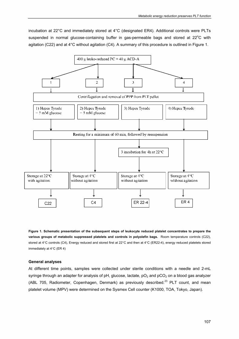
Metabolic energy reduction preserves PLT function
107
incubation at 22°C and immediately stored at 4°C (designated ER4). Additional controls were PLTs
suspended in normal glucose-containing buffer in gas-permeable bags and stored at 22oC with
agitation (C22) and at 4°C without agitation (C4). A summary of this procedure is outlined in Figure 1.
Figure 1. Schematic presentation of the subsequent steps of leukocyte reduced platelet concentrates to prepare the various groups of metabolic suppressed platelets and controls in polyolefin bags. Room temperature controls (C22),
stored at 4°C controls (C4), Energy reduced and stored first at 22°C and then at 4°C (ER22-4), energy reduced platelets stored
immediately at 4°C (ER 4)
General analyses At different time points, samples were collected under sterile conditions with a needle and 2-mL
syringe through an adapter for analysis of pH, glucose, lactate, pO2 and pCO2 on a blood gas analyzer
(ABL 705, Radiometer, Copenhagen, Denmark) as previously described.23 PLT count, and mean
platelet volume (MPV) were determined on the Sysmex Cell counter (K1000, TOA, Tokyo, Japan).
Chapter 6
108
Platelet aggregation At the beginning of the experiments, PLT suspensions, adjusted to about 1000x 109/L with Hepes
Tyrode were placed in polyolefin bags (permeable bags) and in impermeable PVC bags (600 mL). At
different time points, samples were collected under sterile conditions with a needle and 2-mL syringe
through an adapter and aggregation was measured (500 µL) in stirred suspensions (1,000 rev/
minute) at 37°C in a multi-channel aggregometer (Chronolog Havertown, PA, USA) after stimulation
with 15 µM TRAP for 15 minutes. Data were expressed as maximal aggregation after 1hour
preincubation without (-recovery), and with 20 mM glucose (+recovery).
Platelet GPIbα expression
GPIbα expression was measured by incubating a 100 µl sample with 200000 PLTs/L with 2 µg/mL
Moab CD42b-PE for 15 minutes at 37ºC. To the samples 300 µl PBS was added and immediately
10,000 events were measured on a FACScalibur (BD BioSciences, USA). Semi-quantitative analysis
of GPIbα expression was performed using WinMDI software. Fresh platelets show a single population
of CD42b-PE positive cells. The gradual decrease in GPIbα expression observed during storage was
expressed as percentage of the population measured in fresh platelets.
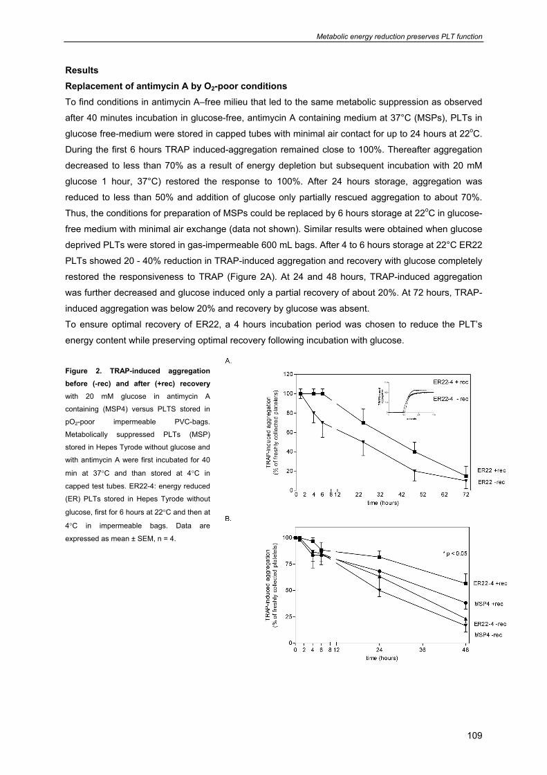
Metabolic energy reduction preserves PLT function
109
Results Replacement of antimycin A by O2-poor conditions To find conditions in antimycin A–free milieu that led to the same metabolic suppression as observed
after 40 minutes incubation in glucose-free, antimycin A containing medium at 37°C (MSPs), PLTs in
glucose free-medium were stored in capped tubes with minimal air contact for up to 24 hours at 22oC.
During the first 6 hours TRAP induced-aggregation remained close to 100%. Thereafter aggregation
decreased to less than 70% as a result of energy depletion but subsequent incubation with 20 mM
glucose 1 hour, 37°C) restored the response to 100%. After 24 hours storage, aggregation was
reduced to less than 50% and addition of glucose only partially rescued aggregation to about 70%.
Thus, the conditions for preparation of MSPs could be replaced by 6 hours storage at 22oC in glucose-
free medium with minimal air exchange (data not shown). Similar results were obtained when glucose
deprived PLTs were stored in gas-impermeable 600 mL bags. After 4 to 6 hours storage at 22°C ER22
PLTs showed 20 - 40% reduction in TRAP-induced aggregation and recovery with glucose completely
restored the responsiveness to TRAP (Figure 2A). At 24 and 48 hours, TRAP-induced aggregation
was further decreased and glucose induced only a partial recovery of about 20%. At 72 hours, TRAP-
induced aggregation was below 20% and recovery by glucose was absent.
To ensure optimal recovery of ER22, a 4 hours incubation period was chosen to reduce the PLT’s
energy content while preserving optimal recovery following incubation with glucose.
Figure 2. TRAP-induced aggregation before (-rec) and after (+rec) recovery
with 20 mM glucose in antimycin A
containing (MSP4) versus PLTS stored in
pO2-poor impermeable PVC-bags.
Metabolically suppressed PLTs (MSP)
stored in Hepes Tyrode without glucose and
with antimycin A were first incubated for 40
min at 37°C and than stored at 4°C in
capped test tubes. ER22-4: energy reduced
(ER) PLTs stored in Hepes Tyrode without
glucose, first for 6 hours at 22°C and then at
4°C in impermeable bags. Data are
expressed as mean ± SEM, n = 4.
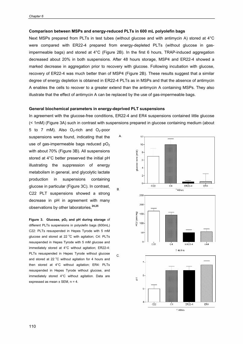
Chapter 6
110
Comparison between MSPs and energy-reduced PLTs in 600 mL polyolefin bags Next MSPs prepared from PLTs in test tubes (without glucose and with antimycin A) stored at 4°C
were compared with ER22-4 prepared from energy-depleted PLTs (without glucose in gas-
impermeable bags) and stored at 4°C (Figure 2B). In the first 6 hours, TRAP-induced aggregation
decreased about 20% in both suspensions. After 48 hours storage, MSP4 and ER22-4 showed a
marked decrease in aggregation prior to recovery with glucose. Following incubation with glucose,
recovery of ER22-4 was much better than of MSP4 (Figure 2B). These results suggest that a similar
degree of energy depletion is obtained in ER22-4 PLTs as in MSPs and that the absence of antimycin
A enables the cells to recover to a greater extend than the antimycin A containing MSPs. They also
illustrate that the effect of antimycin A can be replaced by the use of gas-impermeable bags.
General biochemical parameters in energy-deprived PLT suspensions In agreement with the glucose-free conditions, ER22-4 and ER4 suspensions contained little glucose
(< 1mM) (Figure 3A) such in contrast with suspensions prepared in glucose containing medium (about
5 to 7 mM). Also O2-rich and O2-poor
suspensions were found, indicating that the
use of gas-impermeable bags reduced pO2
with about 70% (Figure 3B). All suspensions
stored at 4°C better preserved the initial pH
illustrating the suppression of energy
metabolism in general, and glycolytic lactate
production in suspensions containing
glucose in particular (Figure 3C). In contrast,
C22 PLT suspensions showed a strong
decrease in pH in agreement with many
observations by other laboratories.24,25
Figure 3. Glucose, pO2 and pH during storage of
different PLTs suspensions in polyolefin bags (600mL)
C22: PLTs resuspended in Hepes Tyrode with 5 mM
glucose and stored at 22 oC with agitation; C4: PLTs
resuspended in Hepes Tyrode with 5 mM glucose and
immediately stored at 4°C without agitation; ER22-4:
PLTs resuspended in Hepes Tyrode without glucose
and stored at 22 oC without agitation for 4 hours and
then stored at 4°C without agitation; ER4: PLTs
resuspended in Hepes Tyrode without glucose, and
immediately stored 4°C without agitation. Data are
expressed as mean ± SEM, n = 4.
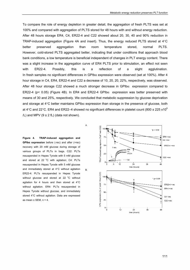
Metabolic energy reduction preserves PLT function
111
To compare the role of energy depletion in greater detail, the aggregation of fresh PLTS was set at
100% and compared with aggregation of PLTS stored for 48 hours with and without energy reduction. After 48 hours storage ER4, C4, ER22-4 and C22 showed about 20, 30, 40 and 90% reduction in
TRAP-induced aggregation (Figure 4A and insert). Thus, the energy reduced PLTS stored at 4°C
better preserved aggregation than room temperature stored, normal PLTS.
However, cold-stored PLTS aggregated better, indicating that under conditions that approach blood
bank conditions, a low temperature is beneficial independent of changes in PLT energy content. There
was a slight increase in the aggregation curve of ER4 PLTS prior to stimulation, an effect not seen
with ER22-4. Possibly, this is a reflection of a slight agglutination.
In fresh samples no significant differences in GPIbα expression were observed (set at 100%). After 4
hour storage in C4, ER4, ER22-4 and C22 a decrease of 10, 20, 20, 22%, respectively, was observed.
After 48 hour storage C22 showed a much stronger decrease in GPIbα expression compared to
ER22-4 (p< 0.05) (Figure 4B). In ER4 and ER22-4 GPIbα expression was better preserved with
means of 30 and 25%, respectively. We concluded that metabolic suppression by glucose deprivation
and storage at 4°C better maintains GPIbα expression than storage in the presence of glucose, both
at 4°C and 22°C. ER4 and ER22–4 showed no significant differences in platelet count (800 ± 225 x109
/L) and MPV (9 ± 2 fL) (data not shown). Figure 4. TRAP-induced aggregation and GPIbα expression before (-rec) and after (+rec)
recovery with 20 mM glucose during storage of
various groups of PLTs in bags. C22: PLTs
resuspended in Hepes Tyrode with 5 mM glucose
and stored at 22 oC with agitation; C4: PLTs
resuspended in Hepes Tyrode with 5 mM glucose
and immediately stored at 4°C without agitation
ER22-4: PLTs resuspended in Hepes Tyrode
without glucose and stored at 22 oC without
agitation for 4 hours and then stored at 4°C
without agitation. ER4: PLTs resuspended in
Hepes Tyrode without glucose, and immediately
stored 4°C without agitation. Data are expressed
as mean ± SEM, n = 4.
Chapter 6
112
Discussion The main finding of the present study is that metabolic suppression of PLTS stored in bags can be
induced by glucose deprivation and storage in gas-impermeable bags. Subsequent storage at 4°C
better preserved PLT function and GPIbα expression than PLTS stored under conditions that support
energy metabolism during storage at 22°C. Compared with control PLTS (C22), the ER4 and ER22-4
showed better preserved TRAP-induced aggregation, GPIbα expression, pH, platelet count and MPV.
Earlier observations showed that glucose deprivation in combination with mitochondrial blockade
reduced the generation of metabolic ATP.26 After a short incubation under these conditions, energy
generation could be restored by addition of glucose addition which led to recover PLT function.
In our previous study we demonstrated14 that for PLTS incubated in test tubes the optimal condition to
induce metabolic arrest was a 40 minutes incubation at 37°C without glucose and with antimycin A.
Responsiveness to the activating agents TRAP and VWF-ristocetin was significantly reduced during
metabolic arrest and addition of glucose induced almost complete recovery to these activating agents.
In the current study we tried to find the optimal conditions for replacement of antimycin A and adapted
the incubation conditions to procedures that were better compatible with daily procedures in blood
banks. We found that the conditions for metabolic suppression when PLT were stored in plastic
capped tubes could be mimicked in gas-impermeable plastic bags. PLTS stored in a medium
containing a minimal amount of glucose, and stored in an impermeable bag at 4°C showed a decrease
in aggregation when they were incubated for more than 24 hours, indicating that glucose deprivation
and a low PO2 reduced platelet responsiveness to activating agents. However, following glucose
addition in the first 6 hours TRAP-induced aggregation returned to 100% and between 6 to 48 hours
the responsiveness was partially restored. The preservation of platelet function and their recovery was
lost after 48 h storage. This is also seen with PLTS stored at 22°C with glucose in gas-permeable
bags.
Interestingly, when energy reduced PLTS (ER4) were stored at 4°C for 48 hours; we found almost the
same aggregation as in controls containing glucose (C4). This aggregation was better than in both
C22 PLTS and in ER22-4 PLTS indicating that low temperature storage better preserves platelet
aggregation than storage at 22°C and that following metabolic suppression the recovery is incomplete.
These data confirm the data of Hoffmeister et al. that 4°C storage better preserves aggregation than
room temperature storage.27 However, changes in GPIb expression show that energy reduction better
protects against storage-induced GP b loss than controls with glucose. Preservation of GPIbα can
prolong survival of PLTS in the circulation.13,27,28 When PLTS without glucose stored in impermeable
bags were first incubated for 4 hours at 22°C and then at 4°C GPIbα expression was still higher than
70%. Application of these conditions in the blood banks may improve PLT storage at 4°C.
Another finding was that PLTS stored without or with glucose placed immediately at 4°C showed an
increase in spontaneous agglutination, reflected in an increased lag phase of aggregation. This could
be an indication of platelet-platelet interaction, cold induced shape change,29-32 actin assembly,33,34
and an increase in intracellular calcium concentration.35 In PLTS without glucose first stored at 22°C
for 4 hour to reduce energy availability and next placed at 4°C the spontaneous agglutination was not
observed. Results of previous studies showed36,37 that early activation of PLTS promotes rapid binding
I
Metabolic energy reduction preserves PLT function
113
and phagocytosis of stored PLTS. We postulate that PLT activation by storage at 4°C should be
avoided to prevent rapid phagocytosis by liver macrophages. In conclusion metabolic suppression by
glucose deprivation and antimycin A of PLTS in capped test tubes, and incubation for 40 min at 37°C
can be replaced by glucose deprivation and incubation for 4 hours at 22°C in bags. Further studies are
required to adapt these incubations before the procedure can be introduced in blood banks.
Acknowledgment This project was supported by Sanquin Blood Supply Foundation (project PPO 01-019). JWNA is
supported by the Netherlands Thrombosis Foundation. The authors thank all technicians of the
Sanquin Blood bank region NW, department Research and Development for their collaborations.
Chapter 6
114
References
1. Blajchman MA. Bacterial contamination and proliferation during the storage of cellular blood
products. Vox Sang. 1998;74 Suppl 2:155-9.:155-9.
2. Brecher ME, Hay SN, Rothenberg SJ. Monitoring of apheresis platelet bacterial contamination
with an automated liquid culture system: a university experience. Transfusion 2003
Jul;43(7):974-8.
3. Gulliksson H. Defining the optimal storage conditions for the long-term storage of platelets.
Transfus.Med.Rev. 2003 Jul;17(3):209-15.
4. Snyder EL, Rinder HM. Platelet storage--time to come in from the cold? N.Engl.J.Med. 2003
May 15;348(20):2032-3.
5. Rinder HM, Snyder EL. Activation of platelet concentrate during preparation and storage. Blood
Cells 1992;18(3):445-56.
6. Fijnheer R, Modderman PW, Veldman H, Ouwehand WH, Nieuwenhuis HK, Roos D, de Korte
D. Detection of platelet activation with monoclonal antibodies and flow cytometry. Changes
during platelet storage. Transfusion 1990 Jan;30(1):20-5.
7. Leytin V, Freedman J. Platelet apoptosis in stored platelet concentrates and other models.
Transfus.Apheresis.Sci. 2003 Jun;28(3):285-95.
8. Wadhawan V, Karim ZA, Mukhopadhyay S, Gupta R, Dikshit M, Dash D. Platelet storage under
in vitro condition is associated with calcium-dependent apoptosis-like lesions and novel
reorganization in platelet cytoskeleton. Arch.Biochem.Biophys. 2004 Feb 15;422(2):183-90.
9. Bertolini F, Murphy S, Rebulla P, Sirchia G. Role of acetate during platelet storage in a synthetic
medium. Transfusion 1992 Feb;32(2):152-6.
10. de Korte D, Gouwerok CW, Fijnheer R, Pietersz RN, Roos D. Depletion of dense granule
nucleotides during storage of human platelets. Thromb.Haemost. 1990 Apr 12;63(2):275-8.
11. Gulliksson H, AuBuchon JP, Cardigan R, Van Der Meer PF, Murphy S, Prowse C, Richter E,
Ringwald J, Smacchia C, Slichter S, et al. Storage of platelets in additive solutions: a multicentre
study of the in vitro effects of potassium and magnesium. Vox Sang. 2003 Oct;85(3):199-205.
12. Hardisty RM. Disorders of platelet secretion. Baillieres Clin.Haematol. 1989 Jul;2(3):673-94.
13. Leytin V, Allen DJ, Gwozdz A, Garvey B, Freedman J. Role of platelet surface glycoprotein
Ibalpha and P-selectin in the clearance of transfused platelet concentrates. Transfusion 2004
Oct;44(10):1487-95.
14. Badlou BA, Ijseldijk MJ, Smid WM, Akkerman JW. Prolonged platelet preservation by transient
metabolic suppression. Transfusion 2005 Feb;45(2):214-22.
15. Josefsson EC, Gebhard HH, Stossel TP, Hartwig JH, Hoffmeister KM. The macrophage
alphaMbeta2 integrin alphaM lectin domain mediates the phagocytosis of chilled platelets.
J.Biol.Chem. 2005 May 6;280(18):18025-32.
Metabolic energy reduction preserves PLT function
115
16. Hoffmeister KM, Felbinger TW, Falet H, Denis CV, Bergmeier W, Mayadas TN, von Andrian UH,
Wagner DD, Stossel TP, Hartwig JH. The clearance mechanism of chilled blood platelets. Cell
2003 Jan 10;112(1):87-97.
17. Cetin M, Eser B, Er O, Unal A, Kilic E, Patiroglu T, Coskun HS, Altinbas M, Arslan D, Ilhan O.
Effects of DMSO on platelet functions and P-selectin expression during storage.
Transfus.Apheresis Sci. 2001 Jun;24(3):261-7.
18. Holme S, Sweeney JD, Sawyer S, Elfath MD. The expression of p-selectin during collection,
processing, and storage of platelet concentrates: relationship to loss of in vivo viability.
Transfusion 1997 Jan;37(1):12-7.
19. Wildt-Eggen J, Schrijver JG, Bins M, Gulliksson H. Storage of platelets in additive solutions:
effects of magnesium and/or potassium. Transfusion 2002 Jan;42(1):76-80.
20. Hogge DE, Thompson BW, Schiffer CA. Platelet storage for 7 days in second-generation blood
bags. Transfusion 1986 Mar;26(2):131-5.
21. Verhoeven AJ, van Oostrum IE, van Haarlem H, Akkerman JW. Impaired energy metabolism in
platelets from patients with Wiskott-Aldrich syndrome. Thromb.Haemost. 1989 Feb 28;61(1):10-
4.
22. van Scharrenburg GJ, Puijk WC, Egmond MR, de Haas GH, Slotboom AJ. Semisynthesis of
phospholipase A2. Preparation and properties of arginine-6 bovine pancreatic phospholipase
A2. Biochemistry 1981 Mar 17;20(6):1584-91.
23. Pietersz RN, Van Der Meer PF, Steneker I, Hinloopen B, Dekker WJ, van Zanten AP, Reesink
HW. Preparation of leukodepleted platelet concentrates from pooled buffy coats: prestorage
filtration with Autostop BC. Vox Sang. 1999;76(4):231-6.
24. Gulliksson H, Sallander S, Pedajas I, Christenson M, Wiechel B. Storage of platelets in additive
solutions: a new method for storage using sodium chloride solution. Transfusion 1992
Jun;32(5):435-40.
25. Pietersz RN, de KD, Reesink HW, van den EA, Dekker WJ, Roos D. Preparation of leukocyte-
poor platelet concentrates from buffy coats. III. Effect of leukocyte contamination on storage
conditions. Vox Sang. 1988;55(1):14-20.
26. Akkerman JW, Gorter G, Soons H, Holmsen H. Close correlation between platelet responses
and adenylate energy charge during transient substrate depletion. Biochim.Biophys.Acta 1983
Oct 4;760(1):34-41.
27. Tsvetkova NM, Walker NJ, Crowe JH, Field CL, Shi Y, Tablin F. Lipid phase separation
correlates with activation in platelets during chilling. Mol.Membr.Biol. 2000 Oct;17(4):209-18.
28. Tsvetkova NM, Crowe JH, Walker NJ, Crowe LM, Oliver AE, Wolkers WF, Tablin F. Physical
properties of membrane fractions isolated from human platelets: implications for chilling induced
platelet activation. Mol.Membr.Biol. 1999 Jul;16(3):265-72.
29. Tablin F, Walker NJ, Klein SD, Field CL, Crowe JH. Animal models for studies on cold-induced
platelet activation in human beings. J.Lab Clin.Med. 2000 Apr;135(4):339-46.
Chapter 6
116
30. Tablin F, Oliver AE, Walker NJ, Crowe LM, Crowe JH. Membrane phase transition of intact
human platelets: correlation with cold-induced activation. J.Cell Physiol 1996 Aug;168(2):305-
13.
31. Hoffmeister KM, Falet H, Toker A, Barkalow KL, Stossel TP, Hartwig JH. Mechanisms of cold-
induced platelet actin assembly. J.Biol.Chem. 2001 Jul 6;276(27):24751-9.
32. Winokur R, Hartwig JH. Mechanism of shape change in chilled human platelets. Blood 1995 Apr
1;85(7):1796-804.
33. Mondoro TH, Vostal JG. Cold temperatures reduce the sensitivity of stored platelets to
disaggregating agents. Platelets. 2002 Feb;13(1):11-20.
34. Badlou BA, Wu YP, Smid WM, Akkerman JW. Platelet binding and phagocytosis by
macrophages Transfusion 2006 In Press.
35. Badlou BA, Wu YP, Spierenburg G, Smid WM, Akkerman JW. Role of glycoprotein Ibalpha in
phagocytosis of platelets by macrophages 2006.
General discussion
117
Chapter 7
General discussion
- Platelet storage at room temperature 118 - Platelet storage under cold conditions 118
- Platelet storage under metabolic suppressed condition 119 - Platelet function studies 120 - Platelet function, binding and phagocytosis studies in vitro 120 - Pitfalls of preservation of platelet function and survival 121
Chapter 7
118
Different clinical studies have shown that transfusion of stored platelets results in better haemostasis
in patients with thrombocytopenia with and without a platelet function defect.1
Current preservation procedures aim to optimally preserve the metabolic status of platelets during
prolonged storage at 4°C.
This thesis describes how metabolic suppression improves the preservation of platelet function. The
mechanism of arrest of energy generation prior to cold storage is described (Chapter 2); and how
transient metabolic blockade prior to cold storage might be involved in the delay of apoptotic activities
and their correlations to platelet binding and phagocytosis (Chapter 3). Furthermore, the different
steps in the mechanism of stimulus response coupling are discussed in relation to the role of P-
selectin expression, changes in GPIbα expression and PS exposure (Chapter 4). After introduction of
metabolic suppression in absence of metabolic inhibitors we adjusted metabolic suppression to
conditions compatible with blood bank procedures (Chapter 5).
Platelet storage at room temperature
Currently most Blood banks isolate and store platelet concentrates (PC’s) at 22°C. Working at room
temperature, has several advantages. It is obviously very simple, and the survival of PC’s post
transfusion is higher than with chilled-rewarmed PLTs.2 These advantages are useful when the blood
banks and hospitals have enough patients to use freshly isolated PC’s immediately. However, there is
never a so called ‘demand-supply balance’ between the blood banks and hospitals. When more PC’s
are supplied than demanded, blood banks have to store PC’s for sometime.
Storage at room temperature has also dis-advantages. Currently, with PC’s stored at room
temperature for more than 5 days there is the problem of microbial multiplication.3-6 This is the reason
that so that the FDA limits the storage time to less than 5 days. Another pitfall of storage at room
temperature is that it introduces changes in platelets indicative for premature activation, and in GPIb
damages7 and initiation of apoptosis.8,9
Improvements have been sought in lowering the storage temperature to below 4°C (in the presence of
glucose) but this treatment severely reduces the survival of transfused platelets.2,10 A main feature of
platelet chilling-rewarming is the gradual increase in the sphere to disc shape change, actin
assembly,11 a gradual increase in [Ca2+]i concomitant with spontaneous agglutination, insensitivity to
disaggregating agents, and GPIb clustering.9-16
Platelet storage under cold conditions Previous attempts to reduce the surface expression of recognition sites for platelet destruction were
based on 1) lowering the storage temperature and freeze-dry techniques,17-19 2) room temperature
storage with inhibition of secretion using additive solutions containing 10-30% plasma,20,21 3) the use
of supplements that increased cAMP and cGMP during storage22,23 and 4) cold storage while
preventing GPIb clustering by glycosylation.10,24 Almost all of these interventions studies which were
done at room temperature and in the presence of glucose during storage, failed to preserve platelets
responsiveness. Improvements of preparation methods for PC’s and the use of sterile storage bags
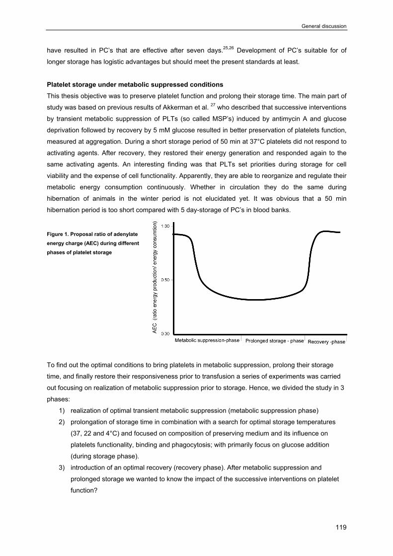
General discussion
119
have resulted in PC’s that are effective after seven days.25,26 Development of PC’s suitable for of
longer storage has logistic advantages but should meet the present standards at least.
Platelet storage under metabolic suppressed conditions This thesis objective was to preserve platelet function and prolong their storage time. The main part of
study was based on previous results of Akkerman et al. 27 who described that successive interventions
by transient metabolic suppression of PLTs (so called MSP’s) induced by antimycin A and glucose
deprivation followed by recovery by 5 mM glucose resulted in better preservation of platelets function,
measured at aggregation. During a short storage period of 50 min at 37°C platelets did not respond to
activating agents. After recovery, they restored their energy generation and responded again to the
same activating agents. An interesting finding was that PLTs set priorities during storage for cell
viability and the expense of cell functionality. Apparently, they are able to reorganize and regulate their
metabolic energy consumption continuously. Whether in circulation they do the same during
hibernation of animals in the winter period is not elucidated yet. It was obvious that a 50 min
hibernation period is too short compared with 5 day-storage of PC’s in blood banks.
Figure 1. Proposal ratio of adenylate energy charge (AEC) during different phases of platelet storage
To find out the optimal conditions to bring platelets in metabolic suppression, prolong their storage
time, and finally restore their responsiveness prior to transfusion a series of experiments was carried
out focusing on realization of metabolic suppression prior to storage. Hence, we divided the study in 3
phases:
1) realization of optimal transient metabolic suppression (metabolic suppression phase)
2) prolongation of storage time in combination with a search for optimal storage temperatures
(37, 22 and 4°C) and focused on composition of preserving medium and its influence on
platelets functionality, binding and phagocytosis; with primarily focus on glucose addition
(during storage phase).
3) introduction of an optimal recovery (recovery phase). After metabolic suppression and
prolonged storage we wanted to know the impact of the successive interventions on platelet
function?
Chapter 7
120
Platelet function studies We applied a stepwise reduction in energy supply by glucose deprivation and inhibition of
mitochondrial respiration by antimycin A. Platelets responded with a step-wise reduction in secretion
and aggregation responses. The optimal condition for metabolic suppression was 40 min at 37°C and
longer storage led into a gradual increase in P-selectin expression and loss of aggregation responses
to TRAP and ADP. Hence, we decided to use for subsequent experiments 40 min at 37°C as optimal
time and temperature to introduce metabolic suppression prior to prolonged storage, in capped tubes.
The following step was to find out which temperature optimally preserved MSP’s during prolonged
storage. Higher temperatures than 4°C, induce rapid P-selectin expression, serotonin release and loss
of responsiveness to activating agents TRAP and ristocetin.28 Hence, the optimal storage temperature
was selected at 4°C.
We confirmed earlier findings27 that complete stop in energy production did not lead to immediate cell
death. Then, we searched for ways to optimize the advantages of metabolic suppression for storage of
platelets for a prolonged period:
• We prolonged the storage time of washed platelets to at least 48 hours with minimal damage
to platelet functions measured in different aggregation, adhesion and secretion studies.
• The arrest of metabolic energy production led to irresponsiveness of platelets to the activating
stimuli, during storage at 4°C. This has major advantages for the logistics in the blood banks
for delivery of PC’s.
• We showed that metabolic suppression prior to storage at 4°C did not affect GPIb expression,
a sensitive marker for PLTs survival as described10. We confirmed that immediate cold
storage of PC’s with a normal metabolic status, induced rapid changes in GPIb that triggered
macrophages to bind and ingest chilled-rewarmed platelets.
• Although we did not measured bacterial multiplication in our samples, there is enough
evidence in literature that at 4°C microbial growth is inhibited significantly.3,29,30 By applying
metabolic suppression we make it possible to place PC’s in refrigerators.
Platelet function, binding and phagocytosis studies in vitro In chapter 2 we investigated in more detail how metabolic arrest prior to cold storage provides a
means for better preservation of platelet function during prolonged storage. MSP4 better preserved
GPIb-related functions and delayed P-selectin expression. Obviously, metabolic arrest limits the
energy sources needed functions. We speculate that limiting energy sources in platelet additive
solutions can be used to prevent the platelet storage lesion. Further studies are needed to find out
which conditions would optimally to preserve platelet functions.
General discussion
121
Pitfalls of preservation of platelet function and survival Although various publications have referred to the effect of cold on platelets as ‘activation’, chilled
platelets do not resemble platelets, activated by stimuli such as TRAP or collagen. Hence, some
research groups and blood banks consider to introduce in cold storage to optimize PC’s quality,
suppress bacterial contamination and growth while PLTs survival desired to be remained optimal.3-
5,8,24,29 PC’s prepared at room temperature show a better platelet survival after transfusion than PC’s
stored at 0°C.11,31 Hoffmeister et al.10 showed that galactosylation of GPIbα permits storage of
platelets in refrigerator and these platelets show the same survival as room temperature stored PC’s.
So far, they have not described what happens after transfusion of PC’s with synthetic galactosylated
GPIbα in humans. Are there any side effects of this galactosylation on immune response of patients?
According to literature, the side effects mainly classified under ‘unknown effects’ after blood
transfusion.32-34 Either measured as overreaction of immune response and allergic reaction or as an
increase in thrombolytic processes post transfusion.35 However, at this moment they did not it due to the complexity of the problem. First, platelets function
is a separate process compared with PLTs survival. 2 At first chilled rewarmed PLTs that function
normally do not circulate and platelets that circulate normally do not show significant haemostatic
effectiveness. Moreover, despite in-vitro and in-vivo proper responsiveness of PLTs to activating
agents, in survival studies phagocytic cells establish whether transfused PLTs may participate in
bleedings episodes or not. In other hand, the most bleeding test at this moment are not very reliable
as well.36-38 Therefore, is difficult to analyze at once what is the platelets likelihood to remain in
circulation and simultaneously, have high haemostatic effectiveness.39 Previous studies described that the in vivo viability of platelets have a positive correlation with P-
selectin expression, PS exposure,40,41 GPIb expression,2,42 platelet morphology and shape
changes11,13,24,43 although this was denied by other groups.44 Hoffmeister et al. postulated that cold
induced clustering of GPIbα is the major signal for phagocytes to remove chilled-rewarmed fresh
platelets. Interestingly, cold induced formation of GpIbα clusters leaves the haemostatic functions of
GPIbα undisturbed.2 Thus binding to activated vWF and cold-induced binding to Mac-1 of THP-1 cells
appear to be separate functions of GPIbα(Chapter 4). Suspensions containing 5mM glucose at 0 or
22°C showed a higher P-selectin expression. It is known that P-selectin plays a pivotal role in platelets
binding to phagocytic cells and we confirmed that (Chapter 3 and 6). We show further that P-selectin
is not enough for the ingestion of platelets and PS exposure and changes in GPIb expression are a
prerequisite for phagocytosis (Chapter 6), as described by other groups.2,42,45,46 Furthermore, the
major features of GPIbα changes are described focusing on the major role of GPIbα as a sensitive in
vitro-predictive marker for platelet phagocytosis. The GPIbα on the surface of the platelet exists in
linear arrays47 in a complex with GPIbβ, GPIX, and GPV, attached to submembrane actin cytoskeleton
by filamin A and filamin B.48-52 Stimulation of platelets in suspension by thrombin and other agonists
causes GPIbα to redistribute in part from the platelet surface into the open canalicular system. This
process does not lead to platelet clearance in vivo.10,53 Here rises the question as to the role of GPIbβ, GPIX, and GPV in platelets phagocytosis?
Quantitative analyzing of GPIbα expression by monoclonal antibody AN51 indicated that fresh
Chapter 7
122
platelets show a high density GPIbα expression (M1-fraction). After short storage at room temperature
or on ice the affinity of moab to bind to GPIbα decreases and a second population appears with a low
density GPIbα at their surface (M2-fraction).
The appearance of the M2-fraction more in both room temperatures stored and on ice stored platelets
populations might be caused by two possible processes induced by storage-induced lesions. First, a
fraction of the total platelets show reduced a density of GPIbα, which possibly caused by GPIbα
clustering, a concept introduced by Hoffmeister et al.10,54 Indeed it positively correlated with
phagocytosis (Chapter 5) and was significantly restored after addition of GlcNAc, as GlcNAc reduced
phagocytosis. Remarkably, GlcNAc did not have any effects on the binding to macrophages.
Obviously, binding is a separate process than phagocytosis. Second, it indicates that both C22 and C0
undergo easier deleterious changes that alter the affinity of the GPIbα binding-epitope to antibody
AN51. It is not clear for us as well. Why addition of so high GlcNAc (100mM) restores GPIbα M-1
fraction in room temperature stored and on ice stored platelets? And subsequently how protect them
against phagocytosis future studies needed elucidate the mechanism in details.
We investigated whether monosaccharides influence GPIbα with subsequent effects on binding and
phagocytosis. Further investigations needed to elucidate why other sugars did not and GlcNAc did
decrease GPIbα M2-fraction forming? And thereby reduced phagocytosis?
Transient metabolic suppression prior to cold storage has disadvantages too. The major problem was
addition of antimycin A that is not compatible with blood transfusion. To find the optimal conditions to
replace antimycin A, inhibitory effect on platelet metabolism and adjust this method to blood bank
conditions. We used gas impermeable plastic bags. We found that the conditions for metabolic
suppression when PLT were stored in capped plastic tubes could be mimicked in bags 600 mL and
storage in a glucose-poor medium for 4 hour at 22°C. Platelets stored in a medium containing minimal
glucose (<1mM) and stored in an impermeable bag at 4°C showed a decrease in aggregation when
they were incubated for more than 24 hour indicating that glucose deprivation and restricted gas
exchange reduced platelet responsiveness to activating agents. However, following glucose addition
in the first 6 hour TRAP-induced aggregation returned to 100%. Between 6 to 48 hours, the
responsiveness was partially restored. Our conclusion was that also in plastic bags energy restriction
did not affect platelet responsiveness, which was reversible up to 100% within the first 6 hour.
Furthermore, we confirmed the data of Hoffmeister et al. who described that 4°C at storage better
preserves platelet function10 although it decreases GPIbα expression. Platelets with low GPIbα
clustering expression will be rapidly removed in the body by liver macrophages. Therefore,
preservation of GPIbα can prolong survival of PLTs in the circulation.10,42,54 When platelets stored in
impermeable bags without glucose were first incubated for 4 h at 22°C and then at 4°C GPIbα
expression was still higher than 70%. Application of these conditions may improve PLT storage at 4°C
in the blood banks.
Chapter 6 describes the correlation between platelet storage lesion (PSL) that is often accompanied
by the apoptotic and morphological changes, cytoplasmic membrane shrinkage.9,23 Both phenomena,
nevertheless, are associated with the appearance of P-selectin expression and PS on the cell
surface.23,37,42 Our approach was an attempt to quantify what the correlation is between P-selectin, PS
General discussion
123
exposure and GPIbα and thereby prediction of phagocytosis of platelets based on in-vitro observation.
We hypothesized that in-vitro data of binding and phagocytosis could predict in-vivo survival of
platelets after transfusion. Markers for the storage lesion were: P-selectin expression, PS exposure,
and GPIb affinity changes. Furthermore, to evaluate the function and survival we used our suggestive
marker for GPIb clustering which was defined quantitatively by FACS and marked under M2. On the
basis of these parameters, one can predict the degree of platelet binding and phagocytosis in vitro.
We hope that by expanding this study in vivo, this model system can be used in the blood banks, for
first attempt for qualify control of PC’s.
There is evidence in the literature that show that PC’s expressing less than 10% P-selectin, PS
exposure, and GPIb clustering survive the first 24 hours of transfusion.10,42,53 Leytin et al. described
that P-selectin expression on the platelets surface during storage triggers fast P-selectin-mediated
clearance in vivo,44,53 although this was denied by other laboratories.
We and other research groups have previously shown that there is a significant correlation between P-
selectin expression and binding and between PS exposure, GPIb peak-2 formation and
phagocytosis.2,42,45,55-57 Here to verify our model system we induced P-selectin expression in washed
platelets by PMA, and investigated the binding and phagocytosis. The binding obviously has positive
correlation with high PSE but phagocytosis has not. The important les from this observation was that
the platelet binding and phagocytosis are not causally linked processes. As postulated before by
Berger et al.53 and Hoffmann PR et al.58 binding and phagocytosis of cells to macrophages are
separate processes. PS exposure also plays a pivotal role in the recognition and removal of apoptotic cells via a PS-
recognizing receptor on phagocytic cells.24 Direct correlations between PS exposure above 10% and
phagocytosis indicate that platelets should not be positive for PS exposure. A decrease of PS
exposure by binding to annexin V did not decrease binding but significantly decreased phagocytosis.
Based on our studies we hypothesize that for the binding and complex formation studies between
platelet and phagocytic cells, PS exposure is not good marker but for phagocytosis can be used
properly.
Future plans should focus on direction of cold storage and adjustments of metabolic suppression prior
to cold storage with blood bank’s compatible supplements. After preparation of sterile PC’s with limited
energy sources and no microbial contamination prolongation of storage time to more than 2 weeks
should be feasible. Pilot studies already show that the pooled Leuko-reduced platelet concentrates
stored at 4°C with metabolic reductions optimally preserved platelet function after 18 days.
Chapter 7
124
References
1. Verhoeven AJ, Verhaar R, Gouwerok EG, de Korte D. The mitochondrial membrane potential
in human platelets: a sensitive parameter for platelet quality. Transfusion 2005 Jan;45(1):82-9.
2. Kawasaki T, Fujimura Y, Usami Y, Suzuki M, Miura S, Sakurai Y, Makita K, Taniuchi Y, Hirano
K, Titani K. Complete amino acid sequence and identification of the platelet glycoprotein Ib-
binding site of jararaca GPIb-BP, a snake venom protein isolated from Bothrops jararaca.
J.Biol.Chem. 1996 May 3;271(18):10635-9.
3. Blajchman MA. Bacterial contamination and proliferation during the storage of cellular blood
products. Vox Sang. 1998;74 Suppl 2:155-9.:155-9.
4. Brecher ME, Hay SN, Rothenberg SJ. Monitoring of apheresis platelet bacterial contamination
with an automated liquid culture system: a university experience. Transfusion 2003
Jul;43(7):974-8.
5. Munksgaard L, Albjerg L, Lillevang ST, Gahrn-Hansen B, Georgsen J. Detection of bacterial
contamination of platelet components: six years' experience with the BacT/ALERT system.
Transfusion 2004 Aug;44(8):1166-73.
6. Wagner S. Transfusion-related bacterial sepsis. Curr.Opin.Hematol. 1997 Nov;4(6):464-9.
7. Barnard MR, Macgregor H, Ragno G, Pivacek LE, Khuri SF, Michelson AD, Valeri CR. Fresh,
liquid-preserved, and cryopreserved platelets: adhesive surface receptors and membrane
procoagulant activity. Transfusion 1999 Aug;39(8):880-8.
8. Snyder EL, Rinder HM. Platelet storage--time to come in from the cold? N.Engl.J.Med. 2003
May 15;348(20):2032-3.
9. Seghatchian J, Krailadsiri P. Platelet storage lesion and apoptosis: are they related?
Transfus.Apheresis.Sci. 2001 Feb;24(1):103-5.
10. Hoffmeister KM, Felbinger TW, Falet H, Denis CV, Bergmeier W, Mayadas TN, von Andrian
UH, Wagner DD, Stossel TP, Hartwig JH. The clearance mechanism of chilled blood platelets.
Cell 2003 Jan 10;112(1):87-97.
11. Hoffmeister KM, Falet H, Toker A, Barkalow KL, Stossel TP, Hartwig JH. Mechanisms of cold-
induced platelet actin assembly. J.Biol.Chem. 2001 Jul 6;276(27):24751-9.
12. Mondoro TH, Vostal JG. Cold temperatures reduce the sensitivity of stored platelets to
disaggregating agents. Platelets. 2002 Feb;13(1):11-20.
13. Italiano JE, Jr., Bergmeier W, Tiwari S, Falet H, Hartwig JH, Hoffmeister KM, Andre P,
Wagner DD, Shivdasani RA. Mechanisms and implications of platelet discoid shape. Blood
2003 Jun 15;101(12):4789-96.
14. Murphy S. Platelet storage for transfusion. Semin.Hematol. 1985 Jul;22(3):165-77.
15. Kaufman RM. Uncommon cold: could 4 degrees C storage improve platelet function?
Transfusion 2005 Sep;45(9):1407-12.
16. Johnston GI, Cook RG, McEver RP. Cloning of GMP-140, a granule membrane protein of
platelets and endothelium: sequence similarity to proteins involved in cell adhesion and
inflammation. Cell 1989 Mar 24;56(6):1033-44.
General discussion
125
17. Valeri CR. Blood components in the treatment of acute blood loss: use of freeze-preserved
red cells, platelets, and plasma proteins. Anesth.Analg. 1975 Jan;54(1):1-14.
18. Bode AP, Read MS. Lyophilized platelets: continued development. Transfus.Sci. 2000
Feb;22(1-2):99-105.
19. Tablin F, Walker NJ, Klein SD, Field CL, Crowe JH. Animal models for studies on cold-
induced platelet activation in human beings. J.Lab Clin.Med. 2000 Apr;135(4):339-46.
20. Rinder HM, Snyder EL. Activation of platelet concentrate during preparation and storage.
Blood Cells 1992;18(3):445-56.
21. Wildt-Eggen J, Schrijver JG, Bins M, Gulliksson H. Storage of platelets in additive solutions:
effects of magnesium and/or potassium. Transfusion 2002 Jan;42(1):76-80.
22. Perrotta PL, Perrotta CL, Snyder EL. Apoptotic activity in stored human platelets. Transfusion
2003 Apr;43(4):526-35.
23. Wadhawan V, Karim ZA, Mukhopadhyay S, Gupta R, Dikshit M, Dash D. Platelet storage
under in vitro condition is associated with calcium-dependent apoptosis-like lesions and novel
reorganization in platelet cytoskeleton. Arch.Biochem.Biophys. 2004 Feb 15;422(2):183-90.
24. Stepanian A, Ribba AS, Lavergne JM, Fressinaud E, Juhan-Vague I, Mazurier C, Girma JP,
Meyer D. A new mutation, S1285F, within the A1 loop of von Willebrand factor induces a
conformational change in A1 loop with abnormal binding to platelet GPIb and botrocetin
causing type 2M von Willebrand disease. Br.J.Haematol. 2003 Feb;120(4):643-51.
25. jkstra-Tiekstra MJ, Pietersz RN, Hendriks EC, Reesink HW, Huijgens PC. In vivo PLT
increments after transfusions of WBC-reduced PLT concentrates stored for up to 7 days.
Transfusion 2004 Mar;44(3):330-6.
26. Hogge DE, Thompson BW, Schiffer CA. Platelet storage for 7 days in second-generation
blood bags. Transfusion 1986 Mar;26(2):131-5.
27. Akkerman JW, Gorter G, Soons H, Holmsen H. Close correlation between platelet responses
and adenylate energy charge during transient substrate depletion. Biochim.Biophys.Acta 1983
Oct 4;760(1):34-41.
28. Badlou BA, Ijseldijk MJ, Smid WM, Akkerman JW. Prolonged platelet preservation by transient
metabolic suppression. Transfusion 2005 Feb;45(2):214-22.
29. Mohammadi T, Pietersz RN, Scholtalbers LA, Vandenbroucke-Grauls CM, Savelkoul PH,
Reesink HW. Optimal sampling time after preparation of platelet concentrates for detection of
bacterial contamination by quantitative real-time polymerase chain reaction. Vox Sang. 2005
Nov;89(4):208-14.
30. Mohammadi T, Pietersz RN, Vandenbroucke-Grauls CM, Savelkoul PH, Reesink HW.
Detection of bacteria in platelet concentrates: comparison of broad-range real-time 16S rDNA
polymerase chain reaction and automated culturing. Transfusion 2005 May;45(5):731-6.
31. Murphy S. Platelets from pooled buffy coats: an update. Transfusion 2005 Apr;45(4):634-9.
32. Eigenthaler M, Nolte C, Halbrugge M, Walter U. Concentration and regulation of cyclic
nucleotides, cyclic-nucleotide-dependent protein kinases and one of their major substrates in
Chapter 7
126
human platelets. Estimating the rate of cAMP-regulated and cGMP-regulated protein
phosphorylation in intact cells. Eur.J.Biochem. 1992 Apr 15;205(2):471-81.
33. Leopold LH, Berger MS, Feingold J. Acute and long-term toxicities associated with
gemtuzumab ozogamicin (Mylotarg) therapy of acute myeloid leukemia. Clin.Lymphoma 2002
Mar;2 Suppl 1:S29-34.:S29-S34.
34. Yamanaka M, Eda S, Beppu M. Carbohydrate chains and phosphatidylserine successively
work as signals for apoptotic cell removal. Biochem.Biophys.Res.Commun. 2005 Mar
4;328(1):273-80.
35. Sobel BE. Effects of glycemic control and other determinants on vascular disease in type 2
diabetes. Am.J.Med. 2002 Oct 28;113 Suppl 6A:12S-22S.:12S-22S.
36. Nieuwenhuis HK, Akkerman JW, Sixma JJ. Patients with a prolonged bleeding time and
normal aggregation tests may have storage pool deficiency: studies on one hundred six
patients. Blood 1987 Sep;70(3):620-3.
37. Leytin V, Freedman J. Platelet apoptosis in stored platelet concentrates and other models.
Transfus.Apheresis.Sci. 2003 Jun;28(3):285-95.
38. Munro J, Booth A, Nicholl J. Routine preoperative testing: a systematic review of the evidence.
Health Technol.Assess. 1997;1(12):i-iv.
39. Gurney D, Lip GY, Blann AD. A reliable plasma marker of platelet activation: does it exist?
Am.J.Hematol. 2002 Jun;70(2):139-44.
40. Li J, Xia Y, Bertino AM, Coburn JP, Kuter DJ. The mechanism of apoptosis in human platelets
during storage. Transfusion 2000 Nov;40(11):1320-9.
41. Solum NO. Procoagulant expression in platelets and defects leading to clinical disorders.
Arterioscler.Thromb.Vasc.Biol. 1999 Dec;19(12):2841-6.
42. Leytin V, Allen DJ, Gwozdz A, Garvey B, Freedman J. Role of platelet surface glycoprotein
Ibalpha and P-selectin in the clearance of transfused platelet concentrates. Transfusion 2004
Oct;44(10):1487-95.
43. Michelson AD, Barnard MR, Khuri SF, Rohrer MJ, Macgregor H, Valeri CR. The effects of
aspirin and hypothermia on platelet function in vivo. Br.J.Haematol. 1999 Jan;104(1):64-8.
44. jkstra-Tiekstra MJ, Pietersz RN, Huijgens PC. Correlation between the extent of platelet
activation in platelet concentrates and in vitro and in vivo parameters. Vox Sang. 2004
Nov;87(4):257-63.
45. Brown SB, Clarke MC, Magowan L, Sanderson H, Savill J. Constitutive death of platelets
leading to scavenger receptor-mediated phagocytosis. A caspase-independent cell clearance
program. J.Biol.Chem. 2000 Feb 25;275(8):5987-96.
46. Fadok VA, Savill JS, Haslett C, Bratton DL, Doherty DE, Campbell PA, Henson PM. Different
populations of macrophages use either the vitronectin receptor or the phosphatidylserine
receptor to recognize and remove apoptotic cells. J.Immunol. 1992 Dec 15;149(12):4029-35.
47. Kovacsovics TJ, Hartwig JH. Thrombin-induced GPIb-IX centralization on the platelet surface
requires actin assembly and myosin II activation. Blood 1996 Jan 15;87(2):618-29.
General discussion
127
48. Canobbio I, Balduini C, Torti M. Signalling through the platelet glycoprotein Ib-V-IX complex.
Cell Signal. 2004 Dec;16(12):1329-44.
49. Hartwig JH. Mechanisms of actin rearrangements mediating platelet activation. J.Cell Biol.
1992 Sep;118(6):1421-42.
50. Lopez I, Duprez V, Melle J, Dreyfus F, Levy-Toledano S, Fontenay-Roupie M. Thrombopoietin
stimulates cortactin translocation to the cytoskeleton independently of tyrosine
phosphorylation. Biochem.J. 2001 Jun 15;356(Pt 3):875-81.
51. Nachmias VT. Platelet and megakaryocyte shape change: triggered alterations in the
cytoskeleton. Semin.Hematol. 1983 Oct;20(4):261-81.
52. Nakamura F, Pudas R, Heikkinen O, Permi P, Kilpelainen I, Munday AD, Hartwig JH, Stossel
TP, Ylanne J. The structure of the GPIb-filamin A complex. Blood 2005 Nov 17.
53. Berger G, Hartwell DW, Wagner DD. P-Selectin and platelet clearance. Blood 1998 Dec
1;92(11):4446-52.
54. Josefsson EC, Gebhard HH, Stossel TP, Hartwig JH, Hoffmeister KM. The macrophage
alphaMbeta2 integrin alphaM lectin domain mediates the phagocytosis of chilled platelets.
J.Biol.Chem. 2005 May 6;280(18):18025-32.
55. Rand ML, Wang H, Bang KW, Poon KS, Packham MA, Freedman J. Procoagulant surface
exposure and apoptosis in rabbit platelets: association with shortened survival and steady-
state senescence. J.Thromb.Haemost. 2004 Apr;2(4):651-9.
56. Badlou BA, Wu YP, Smid WM, Akkerman JW. Regulation of platelet phagocytosis by
macrophages. Transfusion 2006 in Press.
57. Yamao T, Noguchi T, Takeuchi O, Nishiyama U, Morita H, Hagiwara T, Akahori H, Kato T,
Inagaki K, Okazawa H, et al. Negative regulation of platelet clearance and of the macrophage
phagocytic response by the transmembrane glycoprotein SHPS-1. J.Biol.Chem. 2002 Oct
18;277(42):39833-9.
58. Hoffmann PR, deCathelineau AM, Ogden CA, Leverrier Y, Bratton DL, Daleke DL, Ridley AJ,
Fadok VA, Henson PM. Phosphatidylserine (PS) induces PS receptor-mediated
macropinocytosis and promotes clearance of apoptotic cells. J.Cell Biol. 2001 Nov
12;155(4):649-59.

Chapter 7
128
129
Chapter 8
Nederlandse samenvatting
130
De laatste jaren is de behoefte en belang aan trombocyten transfusie sterk toegenomen. Bij de bloed
banken jaarlijks miljoenen donors staan hun dierbare bloed af; voor een ander die misschien binnen
kort hun leven kan redden. Indien acuut het ziekenhuis trombocyten concentraten nodig zou hebben
worden met behulp van centrifugatie techniek de rode cellen, de witte cellen, de trombocyten en het
plasma van elkaar gescheiden en de bloedplaatjes concentraten in een steriele plastic bloedcontainer
wordt gestopt en naar het ziekenhuis gestuurd. Maar ertussen als er geen behoefte zou zijn aan de trombocyten concentraten en/of de verhouding
tussen donors en patiënten zodanig wijzigt dat er de afgestane bloed moet worden bewaard dan de
bloed banken bewaren de rode cellen, de witte cellen in de koelkast, de trombocyten bij de kamer
temperatuur en het plasma bij –80ºC.
Bewaren van de trombocyten bij kamer temperatuur heeft veel voor- maar ook nadelen. Het
belangrijkste nadeel is microbiële groei and vermeerdering zodat er FDA heeft de regelingen gemaakt
dat er niet langer dan 5 dagen trombocyten concentraten mogen worden bewaard die aan de
patiënten zou worden getransfuseerd. Omdat de sterke microbiële groei voor de patiënten onder
operatie en kwaadaardige ziektes zou dodend kunnen zijn. Er is ook grotere kans dat de trombocyten
hun functionaliteit verliezen wanneer ze langer dan 5 dagen bij de kamer temperatuur worden
bewaard.
Dus meeste bloedbanken zoeken naar de mogelijkheden die microbiële contaminatie en groei te
voorkomen bijvoorbeeld door steriel te werken en geïsoleerde bloed zakjes in de koelkast te bewaren.
Maar tegelijkertijd koud bewaren van de trombocyten concentraten leiden tot vroegtijdige activatie en
clustering van GPIb-V-IX complex receptor van de trombocyten waardoor ze niet meer bruikbaar zijn.
Verder de koud bewaarde trombocyten concentraten worden na transfusie snel door lever macrofagen
verwijderd en geklaard.
Dit proefschrift beschrijft een aantal methoden om de veranderingen van de trombocyten concentraten
te meten, analyseren en hen achteruit gaan in de functionaliteit tijdens bewaren en na blootgesteld
zijn aan de koud condities en daarna aan de macrofagen te kunnen meten en evalueren voordat ze
getransfuseerd zijn. Er is getracht de geschetste problemen in kaart te brengen en zo goed mogelijk
oplossingen hiervoor aan te dragen. In dit AIO project hebben we gekozen voor het gebruik van de
metabole suppressie en verhongeren van de trombocyten tot zeker moment en pas dan ze in koelkast
ze voor langere tijd bewaren.
Het is ons gelukt om activatie vermogen, adhesie en aggregatie functie en bovenal levensduur van de
trombocyten voor het laboratoria onderzoeken verlengen van ongeveer 4 tot 6 uur naar 48 uur
(ongeveer 10 maal meer). De piloot studies laten zien dat we trombocyten concentraten bewaren
kunnen voor 18 dagen, met de bloedbank compatible condities. Verder biedt in rust brengen van de
trombocyten tijdens bewaren heel veel mogelijkheid voor logistiek en bespaard veel geld voor de
bloedbanken. Deze verlenging van bewaartijd kan wereld wijd miljarden kosten besparen voor de
bloedbanken.
In hoofdstuk 2 is beschreven hoe we trombocyten in metabole suppressie toestand brengen met
behulp van verhongeren van trombocyten in aanwezigheid van Antimycin A; een inhibitor dat
131
mitochondriale respiratie remt (Platelet Hibernation). Na lang trombocyten in koud zijn bewaard
herstllen we energie toestand van ze met behulp van 20mM glucose voordat de functie testen zijn
uitgevoerd.
In hoofdstuk 3 is beschreven we hoe viabiliteit, binding en fagocytose van de trombocyten kan
behouden blijven tijdens koud bewaren, mits ze vooraf in rust toestand zijn gebracht en metabolisch
gesuppresserd. Blijkbaar metabool gesuppreseerde trombocyten dat in koelkast zijn bewaard beter
clustering van de GPIb-V-IX complex receptor kunnen tegen gaan vergeleken met trombocyten die
hen metabole energie intact blijft maar direct in koelkast gezet en/ of kamer temperatuur bewaarde
controles.
In hoofdstuk 4 is beschreven hoe GPIb clustering eventueel werkt als fagocytose signaal.
In hoofdstuk 5 is beschreven hoe onze model systeem kan bijna elk willekeurige trombocyten
concentraat survival kans voorspellen.
Het is een poging richting opbouwen van een model systeem dat kan voorspellen wat zou de uitkomst
zijn van de zeker trombocyten concentraten na transfusie.
In hoofdstuk 6 is beschreven hoe we metabole suppressie aanpassen aan de bloed bank condities.
Uiteindelijk in hoofdstuk 7 wordt gediscussieerd hoe uiteindelijk moeten trombocyten concentraten
worden bewaard. Is het goed dat ze bij kamer temperatuur te bewaren? Is het beter als ze in koelkast
worden bewaard? Hoe kan men de voordelen van het koud bewaren gebruiken en tegelijk de nadelen
ervan te voorkomen?

132
133
Dankwoord
Beste hematologie medewerkers en AIO’s van UMCU Lab 1, 2, en 3 het was me genoegen om met
jullie te werken hoewel was het heel moeilijke jaren voor me van af het begin tot het eind. Uiteindelijk
kwam het goed. Jullie scherpe inzicht en aan- en opmerkingen tijdens mijn presentaties hielp me het
hele AIO project en opleiding anders zien. Harde aanpak was niet vriendelijk maar misschien wel
professional. Het prikkelde AIO’s om alles grondiger en anders zien. Maar ik wens mijn situatie voor
niemand anders. Als ik keuze had om AIOs-schaap nogmaals te doen, en wist ik wat me te
verwachtte stond, deed ik het nooit. Maar ja! Ik moest promoveren omdat ik eigen plannen had en
zonder PhD kon niet gerealiseerd worden.
Ondanks alle gestresst toestanden en de verschillende inzichten in het mechanisme van het bewaren
van de bloed plaatjes kon ik deze prachtige dissertatie afwerken die zonder 30 jaar onderzoek en
kennis van JanWillem nooit gerealiseerd kon worden.
Beste JW, jouw inzicht en aanhoudende instelling over realiseren van bepaalde facetten waardeer ik
het van harte. Ik had heel veel leuke, leerzame maar ook moeilijke momenten met je gehad. Maar
zonder je was het niet zon hoge gekwalificeerde proefschrift geworden. Toen zei je moeten we
fagocytose essay in elkaar zetten en nagaan hoe de plaatjes worden gebonden en gefagocyteerd
dacht ik Oei…Oie Waar gaan we naar toe? Niemand had er echt inzicht in deze materie. Voor de
bloedbankiers en mij was het heel erg moeilijk het te happen en snappen. Uiteindelijk uit deze studies
een model systeem uitgekomen die heel belangrijke voorspellende waarde kan hebben voor
bloedtransfusie wereld. Je houding dat je punt voor punt alles naging en was je niet tevreden met
goede resultaten gaf me ook inzicht dat we nooit moeten tevreden zijn als over de patiënten gaat en
maatschappij geld die graag gelooft wat wetenschappers zeggen en doen. Ik ga met zware lading van
wijze lessen van je en hoop ik dat in de toekomst ze kunnen handhaven zoals je me geleerd hebt.
Beste Flip, Ik wou heel graag je ook bedanken voor alle inzicht en lessen die je vrijdagen in de
Thrombose en haemostase lessen me hebt gegeven. Je wijze houding houdt hele afdeling overeind
en maakt onmogelijke mogelijk.
Beste Martin, dank je wel dat je me hebt geholpen met adhesie proeven en alle kleine en fijne dingen
die ik nodig had in hematologie afdeling. Soms door hoge stress dat ik had vergat ik dingen en je wijze
aanpak hielp me uiteindelijk het te redden.
Beste Yaping, dank je wel dat je wou me helpen met confocal en electron microscopy experimenten.
Het was me genoegen met je te werken en je aardige instelling was erg me bevallen. Ik hoop dat je
meer waardering krijg en meer kansen te groeien in de wetenschap- wereld.
Beste Gertie, Alvast bedankt voor alle hulp en dat je me heb ingewerkt in hematologie lab en meestal
klaar stond wanneer ik je nodig had.
Beste Suzanne we hadden vanaf het begin valse staart en maar tot het eind houden het professional.
Alvast bedankt dat je af en toe wou meedenken over alles en nog wat.
Beste Rob, bedankt voor je inzicht en ervaring die je wou met me delen. Uiteindelijk moest ik mijn
model systeem aanpassen aan klinische toestanden maar heb ik geen tijd gehad misschien volgende
jaren weer elkaar tegen komen en zien wat we samen kunnen doen.
134
Beste Mark, alvast bedankt voor jullie inzet voor meedenken van statistisch opzicht van de sommige
experimenten.
Beste Martijn en Barend, alvast bedankt dat jullie wilden jullie kennis over cross beta sheet eiwitten
met delen. Nog maals gefeliciteerd met jullie new bedrijf en veel succes veder.
Beste Coen en Thomas ik wou jullie bedanken dat wilden mijn paraneven zijn. Beste Coen dank je
wel voor je leuke discussie en steun tijdens very first experimenten dat we kon voor eerste keer laten
zien dat bloedplaatjes ook cross beta sheets kunnen vormen na activatie en/of lang bewaren. Beste
Thomas we hebben van 1994 samen begonnen met Medische Biologie en je aardige instelling was
heel charmant en vriendelijk. Veel succes met jullie promoties. En alle AIO’s en analisten van de
hematologie afdeling die hoe dan ook me hadden geholpen.
Beste Peter, Harry, en Ton ik wou jullie ook bedanken. Meeste harde aanpak en inzicht kwam van
jullie kant. Ik haat het toe te geven. Maar jullie inzicht heeft me geholpen leren mezelf open houden
voor kritiek. Het hoeft niet altijd opbouwen te zijn. Ik heb geleerd dat wat men weet is wat heeft hij
geleerd. Ik heb geleerd dat destructief commentaren in de wetenschap betekenen niet echt destructief
maar evidence-based inzicht. Dus als ik er niet mee eens ben moet ik zorgen voor tegen evidence te
verzamelen anders het accepteren als wijze les.
Beste Majid, ik wou je bijzonder bedanken voor eerste inzicht in hoe electronen en protonen in cellen
zich gedragen. Waarom pH en radicalen zijn zo belangrijk en hoe ze elkaar beïnvloeden. Verder je
inzicht in radioactieve stoffen en NMR heeft me andere invalshoek van de wetenschap laten zien. Ik
bedank je voor leuke discussies dat me hielp experimenten te bedenken die nog steeds de beste zijn
in aanpak van het plaatje transfusie. Ik hoop binnen kort kan ze uitvoeren en publiceren. Alvast
bedankt voor je wijze vriendschap waanneer ik het erg nodig had en in eerste jaren zonder je kon niet
het redden.
Beste Farhad, de man van de functionele plaatjes proteomics … we hebben zo leuke gesprekken
gehad in laatste jaren en hoe we moeilijke toestanden met AIO-schap kunnen overbruggen. Je
vriendschap hielp me overeind me te houden en hoop binnen kort kan meer over functionele
proteomic samen doen. Hou contact!
Beste Frans, je openheid en vriendelijke houding achter bibliotheek balie heeft me vanaf 1994 tot nu
toe geholpen dat ik altijd positief denk. Als ik iets niet kon vinden en einde van de wereld was nadert
kon ik op je steun rekenen. Dat we Perzische antioxidanten kunnen uiteindelijk classificeren en
registeren was te danken aan je behoorlijke inzet en interesse om me te helpen. Ik vind je een schat
voor Nederland en blijf doen wat voor me hebt gedaan. Ik had moeilijke tijden achter de rug en je
vriendschap hielp me overeind te houden.
Beste Mevrouw Cil, ik wou u ook bedanken. In laatste jaren, contact houden met Maryam en u hebt
me ook heel veel geholpen om me normaal voelen. Vriendschap met u was voor me een mijlpaal dat
ik goed bezig was.
Beste UAF en SUS alvast bedankt dat me in alle opzichten hebben geholpen. Jullie steun wanneer ik
echt nodig had vanaf het begin van mijn voorbereiding jaar voor geneeskunde studie op VU te
Amsterdam tot het eind hielp me in mezelf vertrouwen te houden dat ik het zal redden. Goed werk en
135
mijn verzoek aan jullie is om meer talentvolle buitenlandse studenten blijven steunen vooral in dit
moeilijke tijden.
Beste Sanquin Bloedbank medewerkers en analisten bedankt voor jullie help en opvang in laatste
jaren. Zonder jullie was de laatste aanpassingen van de metabole suppressie aan de bloed bank
conditie kon niet in zon korte tijd gerealiseerd worden. Ik wou Nor bedanken dat heeft me als project
leider aangenomen voor deze project maar nooit kans gehad met haar tweede kop koffie te drinken. Ik
bedank je voor je goede inzicht in plaatjes transfusie waarbij met JW zon goede project hebt in elkaar
gezet. Misschien komen we elkaar ooit tegen. Ga je goed. Beste Martin alvast bedankt dat je wou me
opvangen wanneer Nor wegging. Beste Ruby en Pieter ik wou jullie ook bedanken dat wilden laatste
tijd jullie kennis over verwerken van de plaatje concentraten en voorbereidingen ervan met me delen.
Beste Leny en Marjo ook bedankt dat eerste weken me hebben opvangen in de bloedbank Utrecht
waar en wanneer niemand wist wat met me zonder Nor moest gebeuren.
Ik wou graag alle medewerkers en medewerksters van Jordan lab bedanken. Sorry, dat soms hadden
we mindere tijden samen gehad over hoe en wanneer de FACS moesten gebruiken. Elles bedankt dat
altijd bereid was om me te helpen. Beste Tom helaas kwam niet zover dat we antigen presentatie van
de macrofagen, T-cellen en B-cellen kan meten na interacties met bloed plaatjes. Maar nog steeds
geloof ik heilig dat als fagocyterende cellen bloed plaatjes opnemen moet uiteindelijk te detecteren zijn
welke gedeelte van de plaatjes gaan ze op de MHC gedeelte presenteren. Hou contact en veel
succes met de laatste lotjes. Ik wou Anton Martin en Henk special bedanken. Jongens zonder jullie
kon ik nooit de FACS experimenten uitvoeren. Jullie kritische inzicht en wijze lessen, bereid zijn om
me te helpen leed tot prachtig model systeem op te bouwen waarbij nu kan ik zeggen da ik met FACS
kan omgaan en honderden nieuwe kwantitatief model systemen op bouwen om cel-cel interactie te
bestuderen.
Beste Robert en Wieke onze aardige bovenburen alvast bedankt dat jullie wilden me helpen met cover
van het proefschrift en hoop volgende jaren meer met elkaar omgaan.
Beste Maryam, ik wou je bedanken dat laatste tijd me heel veel gesteund. Jouw inzicht in het snel zien
wat is goed en wat niet hielp me nadenken dat meer focus op goede kanten van hele verhaal en de
rest snel vergeet. We hadden samen moeilijke AIO-periode achter de rug en hoop ik volgende jaren
kunnen we ervan genieten. Het kostte je ook heel veel energie, dat begrijp ik. Maar bedankt dat je in
me had gelooft.

136
137
Curriculum Vitea Bahram Alamdary Badlou, was born in Tehran-Iran in 1964. After high school in Iran he went to army for
two years. In May 1992 started with Medicine study at Free University of Amsterdam (VU-Amsterdam) and
after two years has chosen to go further with Medical Biology at Medicine Faculty of University Medical
Center of Utrecht and finished his MS in 1999 (Drs. Medische Biologie).
As minor study program for 6 months he studied “Alkyl-Dihydroxyacetonephosphate Synthase, a
peroxisomal enzyme involved in ether lipid synthesis” in Chemistry Faculty of Utrecht University (1998).
As major program for 9 months he studied “calcium paradox in rat heart by NMR” and his thesis was a
literature study about “the effects of Insulin during and after Heart Failure” (1999). During this period he
should develop cardioplegic solutions to prevent damages to the isolated rat heart during heart
transportation and transplantation.
He is authorized to work with radioactive materials and equipments degree 4B according to Dutch Law
TUDelft Stcrt nr.227at 4 December 1997. He is also authorized to perform animal experiments according
to Dutch law article nr. 9. Stb.1985, 336 certified at 7 March 1997. Then he worked for 6 month as
Pathology and Phatophysiology teacher in the Assay Opleidingen in Rotterdam North, The Netherlands.
Then he worked as Research Associate on a project of Unilever in association with Chemistry Faculty of
Utrecht entitled “The Bioremedy of antioxidants, coming to terms with measuring lipid peroxidation and
evaluating antioxidant efficacy” in an ex-vivo and in vitro study with C11-BODIPY 581/591 and Tecan
spectroflurometer, to prevent ageing of rat-1 Fibroblasts (1999 to 2002). After Feb 2002 till 2006 he was
hired as PHD student in Sanquin Blood bank NW to study “prolonged platelet preservation by metabolic
suppression”. His promoter was prof. dr. J.W.N. Akkerman and co-promoter was dr. W.M. Smid. He
succeeds for first time to bring platelets in hibernation condition for more than 48 hours, reversibly. His
pilot studies revealed that by his metabolic adjustments in the blood banks, platelet concentrates can be
stored for 18 days.
Publications
Full Papers 1. Bahram A. Badlou, Ya Ping WU, W.M. Smid, J.W.N. Akkerman Platelet binding and phagocytosis
by macrophages. Transfusion 2006 in press.
2. Bahram A. Badlou M.J. Ijsseldijk, W.M. Smid, J.W.N. Akkerman. Prolonged platelet preservation
by transient metabolic suppression.Transfusion. 2005 Feb;45(2):214-22.
3. Janssen M.A, Bahram A. Badlou, C.J. van Echteld, T. Ruigrok 1999 Cytosolic Ca2+ concentration
during Ca2+ depletion of isolated rat hearts. Mol Cell Biochem. 2000 Jan;203(1-2):169-75.
138
Other publications (National and International): 4. Bahram A. Badlou, Ya Ping WU, W.M. Smid, J.W.N. Akkerman. Mechanism of platelet
macrophages interaction. J. Thromb Haemost Aug 2005; vol 3: Suppl 1 (ISTH 2005 Sydney-
Australia)
5. Bahram A. Badlou, Ya Ping WU, W.M. Smid, J.W.N. Akkerman. Role of GPIbα in phagocytosis of
platelets by macrophages J. Thromb Haemost Aug 2005; vol 3: Suppl 1 (ISTH 2005 Sydney-
Australia)
6. Bahram A. Badlou, Ya Ping WU, W.M. Smid, J.W.N. Akkerman Mechanism of platelet
macrophages interaction. Platelets Oct 2005 (Also an oral presentation in XXth European Platelet
meeting 2005)
7. Bahram A. Badlou, Ya Ping WU, G. Spierenburg, W.M. Smid, J.W.N. Akkerman Role of GPIbα in
phagocytosis of platelets by macrophages Platelets Oct 2005 (Also an oral presentation inXXth
European Platelet meeting 2005)
8. Bahram A. Badlou, Ya Ping WU, W.M. Smid, J.W.N. Akkerman. Role of GPIbα in phagocytosis of
platelets by macrophages. Transfusion Aug 2005; (ISBT 2005)
9. Bahram A. Badlou, Ya Ping WU, W.M. Smid, J.W.N. Akkerman. Mechanism of platelet
macrophages interaction. NVB June 2005 (Oral presentation)
10. Bahram A. Badlou, Ya Ping WU, W.M. Smid, J.W.N. Akkerman. Platelet cross beta sheets
forming after prolonged storage time NVB June 2005(manuscript in preparation)
11. Bahram A. Badlou, Ya Ping WU, W.M. Smid, J.W.N. Akkerman. Mechanism of platelet
macrophages interaction. Blood December 2004 (ASH 2004 San Diego- USA)
12. Bahram A. Badlou, Ya Ping WU, W.M. Smid, J.W.N. Akkerman. Prolonged platelet preservation
by transient metabolic suppression. Platelets 2004; 15 (8): 479 (Also an oral presentation in XIXth
European Platelet meeting)
13. Bahram A. Badlou, M.J. Ijsseldijk,W.M. Smid, J.W.N. Akkerman, J. Thromb Haemost 2003; Suppl.
I (Also an oral presentation in ISTH 2003)
14. Bahram A. Badlou, W.M. Smid, J.W.N. Akkerman Platelets 2003; 15(3): 187-197.
15. Bahram A. Badlou , W.M. Smid, J.W.N. Akkerman Platelets 2002; 13(8): 499.
16. G. Drummen Bahram A. Badlou, J.A. Post, K. Wirtz, E. Pap Bioremediation of antioxidant 2002
(in Thesis; Chemistry Faculty, Utrecht University, The Netherlands).


Copyright © 2022 FDOKUMEN