How macrophages respond to two-dimensional materials
77
HAL Id: hal-03388506 https://hal.archives-ouvertes.fr/hal-03388506 Submitted on 20 Oct 2021 HAL is a multi-disciplinary open access archive for the deposit and dissemination of sci- entific research documents, whether they are pub- lished or not. The documents may come from teaching and research institutions in France or abroad, or from public or private research centers. L’archive ouverte pluridisciplinaire HAL, est destinée au dépôt et à la diffusion de documents scientifiques de niveau recherche, publiés ou non, émanant des établissements d’enseignement et de recherche français ou étrangers, des laboratoires publics ou privés. How macrophages respond to two-dimensional materials: a critical overview focusing on toxicity Hazel Lin, Zhengmei Song, Alberto Bianco To cite this version: Hazel Lin, Zhengmei Song, Alberto Bianco. How macrophages respond to two-dimensional materials: a critical overview focusing on toxicity. Journal of Environmental Science and Health, Part B, Taylor & Francis, 2021, 56 (4), pp.333-356. hal-03388506
-
Upload
khangminh22 -
Category
Documents
-
view
4 -
download
0
Transcript of How macrophages respond to two-dimensional materials

HAL Id: hal-03388506https://hal.archives-ouvertes.fr/hal-03388506
Submitted on 20 Oct 2021
HAL is a multi-disciplinary open accessarchive for the deposit and dissemination of sci-entific research documents, whether they are pub-lished or not. The documents may come fromteaching and research institutions in France orabroad, or from public or private research centers.
L’archive ouverte pluridisciplinaire HAL, estdestinée au dépôt et à la diffusion de documentsscientifiques de niveau recherche, publiés ou non,émanant des établissements d’enseignement et derecherche français ou étrangers, des laboratoirespublics ou privés.
How macrophages respond to two-dimensional materials:a critical overview focusing on toxicity
Hazel Lin, Zhengmei Song, Alberto Bianco
To cite this version:Hazel Lin, Zhengmei Song, Alberto Bianco. How macrophages respond to two-dimensional materials:a critical overview focusing on toxicity. Journal of Environmental Science and Health, Part B, Taylor& Francis, 2021, 56 (4), pp.333-356. �hal-03388506�

1
How macrophages respond to two-dimensional materials: a critical overview 1
focusing on toxicity 2
3
Hazel Lin, Zhengmei Song, Alberto Bianco1 4
1CNRS, Immunology, Immunopathology and Therapeutic Chemistry, UPR 3572, 5
University of Strasbourg, ISIS, 67000 Strasbourg, France 6
7
*Corresponding author: Alberto Bianco 8
E-mail: [email protected] 9
10
With wider use of graphene-based materials and other two-dimensional (2D) materials in 11
various fields, including electronics, composites, biomedicine, etc., 2D materials can 12
trigger undesired effects at cellular, tissue and organ level. Macrophages can be found in 13
many organs. They are one of the most important cells in the immune system and they 14
are relevant in the study of nanomaterials as they phagocytose them. Nanomaterials have 15
multi-faceted effects on phagocytic immune cells like macrophages, showing signs of 16
inflammation in the form of pro-inflammatory cytokine or reactive oxidation species 17
production, or upregulation of activation markers due to the presence of these foreign 18
bodies. This review is catered to researchers interested in the potential impact and toxicity 19
of 2D materials, particularly in macrophages, focusing on few-layer graphene, graphene 20
oxide, graphene quantum dots, as well as other promising 2D materials containing 21
molybdenum, manganese, boron, phosphorus and tungsten. We describe applications 22
relevant to the growing area of 2D materials research, and the possible risks of ions and 23
molecules used in the production of these promising 2D materials, or those produced by 24
the degradation and dissolution of 2D materials. 25
26
Keywords: Immune cells; graphene; molybdenum; manganese; boron; nanomaterials; 2D 27
materials; cytokines; phosphorus; tungsten 28
29
30

2
Introduction 1
The immune system comprises of mainly two groups of cells: lymphocytes and myeloid 2
cells. Lymphocytes can be subdivided into B cells, T cells, natural killer (NK) cells, and 3
NK-T cells. Myeloid cells can be subdivided into platelets, erythrocytes, and cells of the 4
granulocyte lineage such as neutrophils, monocytes, macrophages, eosinophils, 5
basophils, and mast cells. [1] Neutrophils and monocytes are the most prominent 6
phagocytes in the blood and are the body’s first defense against foreign organisms or 7
materials, while macrophages are the main immune cells which process nanoparticles. 8
These phagocytes are therefore more relevant to immune cell interaction with 2D 9
materials. [2,3] Of these, macrophages have a longer lifespan and are mostly used in in 10
vitro studies. 11
Macrophages can be found in all tissues of the body and are fundamental in host defence. 12
They phagocytose dead cells and debris, shape inflammatory response and modulate 13
adaptive immunity. [4] Macrophages are one of the first cells which encounter 14
nanomaterials, and promptly produce pro-inflammatory cytokines such as IL-6 and TNF-15
α to initiate a down-stream immune response upon foreign particle recognition. [5] 16
Macrophages also secrete anti-bacterial and proteolytic enzymes, chemokines, and anti-17
inflammatory cytokines, such as IL-10 and TGF-β, and produce reactive oxidative species 18
(ROS), nitrogen, and arachidonate metabolites. [6] 19
Although macrophages are able to recognize and internalize nanomaterials, it is generally 20
unknown how exactly nanoparticle recognition occurs, with respect to specific cell-21
surface receptors and membrane cholesterol. [7] Nanomaterials interact with the biological 22
molecules by coating their surface and forming the protein corona. [8] In fact, the protein 23
corona dictates nanoparticle interaction with macrophages through mediation of 24
3
recognition and uptake into these cells. [9] Nanoparticle–macrophage interactions are also 1
dependent on particle properties, physicochemical characteristics such as size, shape, 2
charge, and colloidal stability. As a rough guide, small positively charged nanoparticles 3
are in general more toxic than big negatively charged ones. [10] 4
The nanoparticles have the potential to affect macrophage polarization, and therefore 5
internalization. Macrophage polarization is an activation process following micro-6
environmental signals. At the end of this process, macrophage phenotypes can be 7
categorized into two groups: 1) pro-inflammatory M1, and 2) anti-inflammatory M2. 8
Polyurethane nanoparticles were found to inhibit polarization toward M1 phenotype but 9
not M2, decreasing production of M1 cytokines TNF-α and IL-1β. [11] Silicon 10
nanoparticles were found to have higher uptake in M1 compared to M2 RAW 264.7 11
macrophages, [12] although contradictory results were observed in another study using 12
primary human macrophages. [13] 13
The nanoparticle exposure at subtoxic concentrations can result in ROS production, 14
increased secretion of pro-inflammatory cytokines and upregulation of activation markers 15
in macrophages. [14] At higher concentrations or in certain experimental conditions, 16
nanoparticles can exert macrophage toxicity in various ways, which may or may not be 17
indirectly linked, such as endoplasmic reticulum stress, [15] autophagic cell death, [16] 18
mitochondrial dysfunction, [17] lysosomal dysfunction [18] or oxidative damage. [19] ROS 19
in particular can be highly relevant in genotoxicity, which may be related to nanoparticle 20
surface properties, presence of transition metals, intracellular iron mobilization, particle 21
uptake, interaction and lipid peroxidation. [20] 22
The class of 2D nanomaterials covers many types of materials, including monolayered 23
elements going from graphene and phosphorene (also known as black phosphorus) to 24
4
dichalcogenides to layered silicate minerals. [21] Graphene safety has been extensively 1
reviewed in many cell types, including macrophages. [22] Nitrides such as hexagonal 2
boron nitride (hBN), an isomorph of graphene, and transition metal dichalcogenides such 3
as molybdenum disulfide, tungsten disulfide, hold much promise in the semiconductor 4
industry. [23] (Fig.1) 2D nanomaterials have also vast potential for use in electronics, 5
sensing, spintronics, photonics, thermoelectrics and energy systems. [24] They have been 6
explored in biomedicine, for example in bioimaging, cancer theranostics, biosensing and 7
antimicrobials, although little is still known about their toxicity. [25] 8
Given the potential to synergise their unique benefits in the 2D structure, there have been 9
several combinations involving 2D nanomaterials in fields ranging from electronics to 10
oncology. Boron has versatile bonding configurations and can intercalate with graphene 11
despite a crystallographic lattice and symmetry mismatch. [26] Boron-doped graphene 12
nanoribbons, [27] boron-doped nanographene and boron-doped graphene-MoS2 13
nanohybrids [28] have also emerged in the battery and semi-conductor industry. [29] Black 14
phosphorus-MoS2 nanocomposites have been utilized in dye decomposition, [30] while 15
black phosphorus-hBN-rhenium diselenide heterojunction diodes have found use in 16
electronics. [31] 17
In biology and chemistry, tungsten-doped manganese dioxide has been reported to be 18
exploited in formaldehyde removal, [32] while a black phosphorus-manganese dioxide 19
nanoplatform has been used in oxygen monitoring and in photodynamic therapy. [33] 20
MoS2-graphene oxide (GO) nanocomposites have shown efficacy in lung cancer therapy 21
[34] and GO nanosheets decorated with copper oxide-WO3 nanoparticles were used to 22
detect cancer cells. [35] 23

5
In this review, we will focus on the impact of the most representative groups of 2D 1
nanomaterials on macrophages. Quantum dots and nanoparticles have been included as 2
well, as they are considered a subset of 2D materials, as seen in many recent publications 3
reporting production methods reminiscent of 2D materials and resultant 2D properties. 4
[36-38] We will describe the effects of graphene, as sub-divided into few-layer graphene 5
(FLG), GO and graphene quantum dots (GQDs). We will also cover MoS2 and other 6
forms of molybdenum, MnO2 and other forms of manganese, hBN and other forms of 7
boron, black phosphorus, tungsten trioxide and other forms of tungsten. The scope for 8
upcoming and less explored non-graphene 2D materials includes other forms of the same 9
element and non-macrophage cells to provide a clearer picture of elemental and molecular 10
toxicity. This will hopefully enable the reader to better understand the predicted 11
macrophage toxicity of these new materials. 12
Graphene 13
Graphene, the first true 2D crystalline material, was isolated by Geim and Novoselov in 14
2004. [39] Graphene consists of single layer sp2-hybridized carbon atoms arranged in a 15
hexagonal lattice, with a carbon-carbon distance of 1.42 Å. The large π conjugation in 16
graphene results in its exceptional electrical, thermal, optical, and mechanical properties. 17
These properties can be altered as well by appropriate chemical modifications. The 18
graphene family is huge and includes FLG, GO and GQDs (Fig.2), which we will cover 19
in this review. Due to their physicochemical properties, graphene family nanomaterials 20
have attracted considerable attention in a myriad of fields [40] such as biomedicine, [41] 21
electronics, [42] photonics, [43,44] composite materials, [45] sensors and metrology. [46] In 22
view of the broad spectrum of applications and the increasing use of graphene family 23
nanomaterials in different industrial sectors, it is crucial to understand their impact on 24
6
cells and tissues, especially the interactions with the immune system and in particular in 1
macrophages. [47] In this section, we discuss in detail the effects of FLG, GO and GQDs 2
on macrophages. [21] 3
Few-layer graphene 4
FLG refers to graphene materials with less than 4-10 layers of nanosheets, [48] When the 5
number of atomic layers increases, the material becomes more metallic [49] and the 6
thermal conductivity decreases. [50] FLG is gaining importance in fields like 7
nanomedicine, because it is much easier to obtain high quantities and its colloidal 8
properties are still maintained in biological media. [51] In this context it is important to 9
consider the effects of FLG on macrophages. 10
It has been demonstrated that FLG is able to induce cytotoxicity in RAW 264.7 11
macrophages by decreasing mitochondrial membrane potential (MMP), causing the 12
accumulation of intracellular ROS, and triggering apoptosis through activation of the 13
mitochondrial pathway. The mitogen-associated protein kinases (MAPKs) and TGF-β-14
related signaling pathways may also be involved. [52] It was observed that pristine 15
graphene nanosheets produce holes in the membranes of RAW 264.7 macrophages, 16
reducing cell viability. This was due to strong interactions between pristine graphene and 17
membrane phospholipid tails. [53] It was also reported that FLG could stimulate the 18
secretion of cytokines like IL-1α, IL-6, IL-10, TNF-α and GM-CSF and chemokines such 19
as MCP-1, MIP-1a, MIP-1b and RANTES on both primary murine macrophages and 20
immortalized macrophages. This effect was linked to the toll-like receptor (TLR)-21
mediated and NF-κB pathways. [54] Our group however, showed that primary human M1 22
and M2 macrophage viability and activation were mainly found to be unaffected by 24 h 23
treatment with FLG at doses up to 50 µg/mL. [55] We also found high cell viability of 24

7
RAW 264.7 cells after exposure to FLG for 24 h and no in vivo hematotoxicity in Balb/c 1
mice at 300 µg/mouse up to 30 days. [56] 2
Recently, Cristo et al. presented the detailed toxicity mechanism of low-dose (2.5 and 5 3
µg/cm2) of 265 nm FLG in RAW 264.7 macrophages. The results of this study revealed 4
that FLG induced inflammation by oxidative stress, triggering endoplasmic reticulum 5
stress-mediated autophagy. [57] On the contrary, our group reported that FLG of 100-1600 6
nm lateral size did not induce inflammatory responses nor cell toxicity in mouse primary 7
bone marrow-derived macrophages. The cellular stress and the basal level of autophagic 8
activity were not affected at any dose of FLG (3-100 µg/mL). [58] The study showed that 9
the material was internalized mainly through phagocytosis and partly by passive 10
diffusion. No significant increased secretion of inflammation-related cytokines such as 11
IL-1β, IL-6 and TNF-α was observed. The results are in agreement with another work, 12
[59] where pristine graphene did not induce autophagy after being phagocytosed by human 13
primary macrophages (from peripheral blood mononuclear cells, PBMCs). Similarly, it 14
was demonstrated that pristine graphene cannot induce immune stimulation and toxic 15
effects in vitro. [60] In another study, [61] pristine graphene nanosheets stabilized by flavin 16
mononucleotide of two different sizes (PG-FMN, 200-400 nm and 100-200 nm) enhanced 17
the release of nitric oxide with metabolic alterations. Interestingly, the smaller PG-FMN 18
increased the levels of succinate, itaconate, phosphocholine in RAW 264.7 macrophage, 19
which was not observed in cells incubated with larger PG-FMN nanosheets. 20
Other studies compared the toxicity and cellular uptake of FLG and functionalized FLG. 21
[62,63] The interaction of pristine graphene (corresponding to FLG) and carboxyl-22
functionalized graphene (FLG-COOH) in RAW 264.7 macrophages and PBMCs showed 23
relatively high intracellular uptake of FLG-COOH compared to FLG, which was found 24

8
to be mainly retained on the cell surface and induced stress effects above 50 µg/mL 1
through the induction of ROS-mediated apoptosis. In contrast, the FLG-COOH rendered 2
better cytocompatibility with no stress effects up to 75 µg/mL. Studies focusing on pro-3
inflammatory cytokine expression (e.g., IL-1β, IL-6, IL-8, IL-10, TNF-α, and IL-12p70) 4
showed that FLG-treated PBMCs expressed relatively higher levels of IL-8 and IL-6 5
compared to FLG-COOH samples, thus indicating the inflammatory potential of the 6
former. [63] These results demonstrated that highly hydrophobic pristine graphene was 7
more toxic than hydrophilic, functionalized graphene. 8
Biodegradation is important during the study of biomedical applications, to ascertain 9
eventual material safety within the body. Aggregates (60 µg/mL) of phagocytosed 10
pristine graphene (200 nm lateral size) were found in RAW 264.7 macrophages within 11
24 h as observed by confocal Raman spectroscopy. Macrophage-engulfed graphene was 12
shown to result in time-dependent degraded material reiterating the role of macrophages 13
in biodegradation. (Fig.3) [64] The same group also compared the 3-month toxicity, organ 14
biodistribution and immune response of FLG, FLG-COOH and FLG-PEG of 100-200 nm 15
in Swiss albino mice at 20 mg/kg. The results showed that all the materials were mostly 16
retained in the lung, spleen and liver, with FLG and FLG-COOH inducing significant 17
cellular and structural damages to lungs, liver, spleen, and kidney. In contrast, FLG-PEG-18
administered animals showed no significant abnormalities and normal biochemical 19
markers. In addition, FLG-PEG evidenced clear signs of biodegradation using Raman 20
confocal imaging. [65] 21
Overall, FLG could decrease cell viability, damage cell membrane, induce apoptosis and 22
increase cytokine production in macrophages, with the main mechanism of cytotoxicity 23
related to MMP reduction and ROS increase. Addition of functional groups on the surface 24

9
of FLG can modulate cytotoxicity. As FLG is easily taken up by macrophages and widely 1
used in biomedicine, future studies could be focused on in vitro and in vivo 2
biodegradation of FLG, a neglected area of research. 3
Graphene oxide 4
GO is the oxidized form of graphene. It is made of 2D carbon obtained from graphite 5
sheets under strong acid conditions, which thereby introduces oxygenated groups onto 6
carbon-carbon double bonds. [66-68] On the surface of GO we can identify mainly hydroxyl 7
and epoxy groups, while at the edges there are few carboxylic and carbonyl functions. 8
The presence of these groups accounts for a high hydrophilicity, a superior water 9
dispersibility, a good colloidal stability and an easy surface functionalization. Owning to 10
these properties, GO is currently the most widely investigated graphene family materials 11
for biology-related applications, [69,70] including drug delivery, [71,72] cancer therapy and 12
viral infections, [73] tissue engineering, [74] bioimaging [75] and biosensing. [76] Here, we 13
present a summary of the research efforts to elucidate the bioeffects of GO on 14
macrophages including cytotoxicity, cellular uptake, inflammatory effects and 15
macrophage polarization. 16
Cytotoxicity studies in macrophages 17
Many studies revealed that GO can damage the membrane and cytoskeleton of 18
macrophages. For instance, single-layer GO nanosheets with a lateral size ranging from 19
200 nm to 700 nm can reduce cell viability by producing holes in the membranes of RAW 20
264.7 macrophages. [53] Similarly, monolayer GO within the range of 100-300 nm 21
provoked plasma membrane and cytoskeleton damage in J774A.1 macrophages at 22
sublethal concentrations (20 µg/mL) without inducing significant cell death. The 23
interactions of GO with membrane integrin was found to activate the integrin-FAK-Rho-24

10
ROCK pathway and to suppress the expression of integrin, resulting in a compromised 1
cell membrane and cytoskeleton. [77] 2
Recently, lysosomal dysfunction emerged as a potential mechanism of nanomaterial 3
toxicity. [78] Additionally, a lysosome-based process known as autophagy was recognized 4
as an important pathway of cell death. [79] Several studies have revealed that GO could 5
induce autophagy in macrophages. The autophagy was triggered by GO in a 6
concentration-dependent manner, as evidenced by the appearance of autophagic vacuoles 7
and activation of autophagic marker proteins. With a higher concentration of GO, an 8
increase in autophagic vacuoles was observed. It was also shown that autophagy was at 9
least partly regulated by the TLR pathway. [80] GO induced autophagosome accumulation 10
and the conversion of LC3-I into LC3-II, inhibiting the degradation of the autophagic 11
substrate p62 protein. [81] It was also observed that GO exerted a concentration-dependent 12
increase in membrane rafts and the production of phagosomes. GO exposure induced cell 13
necrosis, inflammatory responses, increase in the oxidative stress response and autophagy 14
in RAW 264.7 cells. ROS was also found to induce autophagy by the ROS-Nrf2-p62 15
pathway. [82] 16
It is worth noting that the oxidation states of GO may also affect toxicity in macrophages. 17
The reduced GO (rGO) was more toxic than GO in both bone marrow-derived 18
macrophages and J774A.1 cells. [83] In addition, it was also found that hydrated GO, a 19
material with high density of carbon radicals, was responsible for cell death in THP-1 20
cells as a consequence of lipid peroxidation of the surface membrane and membrane lysis. 21
[84] 22
Cellular uptake of GO in macrophages 23

11
The majority of studies on GO-mediated cellular uptake have been carried out on 1
macrophages. It was evident that the phagocytic capacity of macrophages can be altered 2
after internalizing GO. GO accumulation inside cells causes significant morphological 3
modifications and reduction of macrophage phagocytic ability. [85] In a study aimed to 4
understand the effect of GO when macrophages encounter microbial pathogens, the 5
uptake of GO by macrophages could modulate their capability to phagocytose yeasts. In 6
particular, it was found that the ingestion of heat-killed yeasts was increased by murine 7
peritoneal macrophages after GO treatment. [86] Other studies have shown that, following 8
uptake, GO accumulates primarily in the cytoplasm [85] and the lysosomes. [81] It was 9
found that GO nanosheets were localized on F-actin filaments inducing cell-cycle 10
alterations, apoptosis and oxidative stress in RAW 264.7 cells. [87] In another study, GO 11
sheets were observed within vesicles as well as in the cytoplasm of carp leukocyte cells 12
(CLC), a surrogate cell type for carp macrophages. [88] In RAW 264.7 macrophages the 13
mechanism of GO internalization is dependent on clathrin-coated membrane 14
invagination. [89] Different sizes of GO with BSA functionalization culminated in 15
different pathways. GO of 500 nm lateral size mainly penetrated the cell through clathrin-16
mediated endocytosis, while larger sheets (1 µm lateral size) were internalized by a 17
combination of clathrin-mediated endocytosis and phagocytosis. [90] 18
A certain number of studies have shown that the size of GO plays an important role in 19
determining the efficiency of macrophage cellular uptake, with smaller GO nanoparticles 20
being better internalized. [61,91-94] Our group showed that small lateral size GO internalizes 21
better and induced stronger changes in the physiological functions of human and murine 22
primary macrophages. [95] Similar results were observed in murine peritoneal 23
macrophages. In contrast, other researchers reported that the lateral size of GO does not 24
affect cellular uptake. [96,97] It has been demonstrated that the cell uptake of GO of 2 µm 25

12
and 350 nm penetrate in the same way and accumulate in similar amounts in murine 1
J774.1A1 macrophages and peritoneal macrophages. This was attributed to similar 2
antibody opsonization and active Fcλ receptor-mediated phagocytosis. [98] Using smaller 3
GO (e.g., 89 nm and 277 nm), [99] the uptake into macrophages was again independent of 4
GO size and incubation time. 5
Surface charge also affects the cellular uptake of GO. Luo et. al. [98] synthesized ∼200 6
nm of GO functionalized with PEG, bovine serum albumin (BSA), and 7
poly(ethyleneimine) (PEI). The authors found that decoration with PEG and BSA 8
inactivated endocytosis, whereas the positively charged GO-PEI facilitated endocytosis 9
only initially. They hypothesized that after cellular internalization, GO-PEI disrupts the 10
physiological potential and integrity of mitochondria and subsequently alters the levels 11
of ROS and cytochrome C. Similarly, in RAW 264.7 cells, [99] PEI-functionalized GO 12
conjugate with a positive zeta-potential was much easily internalized than GO 13
functionalized with a 6-armed PEG with a negative zeta-potential, although the cellular 14
uptake pathways were the same. This is probably because GO sheets with a positive 15
potential surface were able to better attach to the cell membrane leading to cell 16
internalization. It was indeed observed that the nanomaterials were first transferred to the 17
cell membranes, and then underwent invagination and vesicle formation. 18
The other two parameters that influence the cellular uptake of macrophages are the 19
dispersibility and functionalization of GO. Our group recently demonstrated that reducing 20
GO agglomeration in the presence of proteins and obtaining stable GO dispersions in cell 21
culture media allows faster and more efficient internalization in RAW 264.7 22
macrophages. [100] Several reports showed that the cell penetration of 1-arm PEGylated 23
GO nanosheets was higher than GO modified with a 6-arm PEG. [101,102] The possible 24

13
reason is that the latter GO needs a stronger driving force and more energy to cross the 1
cell membrane. The polymer-GO nanosheets functionalized by either amide bond 2
(amPEG-PEI-GO) or disulfide linkage (ssPEG-PEI-GO) could reduce the non-specific 3
uptake and clearance by RAW 264.7 macrophages, increasing their accumulation in 4
targeted cells. [103] Pi et al. [104] prepared the mannosylated and PEGylated GO 5
nanoplatform (GO-PEG-MAN), which showed significantly increased human THP-1-6
derived macrophages uptake through an improved mannose receptor-mediated 7
endocytosis in vitro. GO-PEG-MAN loaded with rifampicin was reported to increase 8
cellular uptake of the drug, extending its effect. This suggested that GO-PEG-MAN 9
would be a good candidate for drug delivery. In addition, the oxidation states of GO may 10
also affect macrophage uptake, with GO having greater cell membrane affinity compared 11
to rGO. Although GO was found to induce expression of antioxidative enzymes and 12
inflammatory factors, rGONPs had surprisingly higher cellular uptake and higher NF-κB 13
expression. Both GO and rGO were shown to damage F-actin cytoskeleton. [105] 14
Inflammation and macrophage polarization 15
Macrophages play an important role in pro- and anti-inflammation and can decrease the 16
immune reactions through the production of cytokines. Several studies have evaluated the 17
cytokine release induced by GO in macrophages. GO (with two different sizes of ∼2.4 18
µm and ∼200 nm) enhanced the production of IL-2, IL-10, IFN-γ and TNF-α in a dose-19
dependent manner. The treatment of RAW 264.7 macrophages with GO stimulated toll-20
like receptor (TLR) signaling and triggered cytokine responses. [80] Other studies reported 21
that GO can induce cellular necrosis mediated by activation of TLR4 and production of 22
autocrine tumor necrosis factor receptor (TNF-R). [83] In addition, PEG-modified GO 23

14
significantly enhanced the secretion of TNF-α by RAW 264.7 macrophages without 1
changing the levels of IL-6 and IL-1β. [102] 2
Several factors can affect cytokine expression including GO concentration. IL-6 3
expression in RAW 264.7 cells was increased with 15.6 and 31.25 µg/mL of GO, while 4
no influence was observed at the concentration higher than 62.5 µg/mL. Similarly, low 5
concentration of GO increased the synthesis of MIP-1α and MIP-1β, but high 6
concentration of GO decreased their synthesis. [106] Low concentration of GO can 7
stimulate the pro-inflammatory response in RAW 264.7 macrophages. The level of TNF-8
α and IL-8 increased rapidly at the GO concentration of 0.01 μg/mL and then decreased 9
at 0.1 and 1.0 μg/mL. In addition, the content of malondialdehyde, glutathione and 10
superoxide dismutase increased in a dose-dependent manner following treatment with 11
GO. [107] 12
The lateral size of GO is also important in cytokine expression. For example, small and 13
thin GO (lateral dimensions ranged between 50 nm and 2 µm) dose-dependently inhibited 14
the release of IL-1β and IL-6 but not TNF-α, while NLRP3 inflammasome and caspase-15
1 activation were not affected. This happened because small GO had profound effects on 16
the immunometabolism of the cells, leading to activation of the transcription factor 17
nuclear factor-erythroid 2 related factor 2, which inhibited the expression of IL-1β and 18
IL-6. [108] The groups of Fadeel and Kostarelos prepared the small (50-300 nm) and large 19
(10-40 μm) GO samples of one or two layers’ thickness (1-2 nm). [109] The results showed 20
that GO did not trigger size-dependent effects in primary human macrophages, or induce 21
the secretion of Th1 cytokines (e.g., TNF-α, IL-6, or IL-1β) and Th2 cytokines (e.g., IL-22
4, IL-5, and IL-13), but significantly suppressed several LPS-induced cytokines, 23

15
including the anti-inflammatory cytokine, IL-10. GO elicited also canonical NLRP3-1
ASC-caspase-1-dependent IL-1β secretion in LPS-primed cells. [109] 2
In addition, surface functionalization is another factor that can influence cytokine 3
expression. The immune responses of branched PEI and 6-armed PEG functionalized GO 4
conjugates were studied in RAW 264.7 macrophages. The results indicated that GO-PEG 5
stimulated the macrophage more by improving the secretion of IL-6. [98] On the other 6
hand, another work showed that although PEGylated GO was not internalized by 7
peritoneal macrophages, integrin β8-related signaling and cytokine responses were still 8
enhanced. [110] These results point to the conclusion that surface passivation does not 9
always prevent immunological responses to GO nanomaterials. 10
Several studies have evaluated macrophage polarization induced by GO treatment. For 11
example, GO treatment promoted J774A.1 macrophage polarization to the M1 phenotype, 12
with large GO (750-1300 nm) eliciting higher M1 macrophage induction than small GO 13
(50-350 nm) (Fig.4). [94] Fluorescent-PEG-GO nanosheets (FITC-PEG-GO) were 14
effectively absorbed by peritoneal macrophages, increasing yeast phagocytosis by pro-15
inflammatory M1 and reparative M2 macrophages. Treatments with GO enhanced M1 16
macrophage activation, which is important for the eradication of pathogens, and 17
diminished alternative activation of M2 macrophages, which decreases fungal persistence 18
and chronic infectious diseases. [111] In addition, a macrophage-targeting/polarizing GO 19
complex (MGC) decreased ROS in immune-stimulated macrophages to attenuate 20
inflammatory polarization of macrophages (M1) Furthermore, it was found that GO 21
functionalized with IL-4 plasmid DNA could polarize M1 to M2 macrophages for the 22
synergistic treatment of myocardial infarction. [112] 23

16
In conclusion, the studies conducted in the past several years have clearly evidenced the 1
biological effects of GO on macrophages. GO can reduce cell viability, can be taken up 2
by macrophages and can affect cytokine expression, all these effects being influenced by 3
several factors, such as lateral size, surface charge, dispersibility and functionalization. 4
However, more research is required on macrophage polarization to better understand the 5
possible inflammation risks of GO in macrophages. 6
Graphene quantum dots 7
Graphene quantum dots are small graphitic domains with lateral dimensions less than 10 8
nm (average 5 nm). [47,113] Owing to their high surface area, strong photoluminescent 9
properties, excellent electrical properties, superior chemical inertness and 10
biocompatibility, [114,115] GQDs have potential applications in photovoltaics, [116] anti-11
microbials, [117-119] bioimaging, [120,121,46] biosensing [122,123] and drug delivery. [124-126] 12
With such vast potential uses of GQDs, the study of their cellular effects and toxicity is 13
essential. 14
GQDs were shown to have little effect on cell viability and membrane integrity of 15
activated THP-1-derived macrophages, while significantly increasing ROS, apoptosis, 16
autophagy, and inflammatory responses. [127] Furthermore, GQDs significantly increased 17
the phosphorylation of p38 MAPK and p65, and promoted NF-κB. An increased 18
expression of TNF-α, IL-1, and IL-8 was observed at low concentrations (10 and 50 19
µg/mL), whereas high concentrations (100 and 200 µg/mL) of GQDs led to opposite 20
effects on cytokine production. It was reported that large (40 nm) GQDs were able to 21
inhibit splenocyte IFN-γ production and to modulate MAPKs in J774.1 macrophages. [128] 22
Functionalization of GQDs also affected the interactions with macrophages. For instance, 23
thiol functionalized GQDs significantly increased the efflux of oxidized-low density 24

17
lipoprotein, down-regulated cell scavenger receptors, and efficiently recovered ROS 1
levels in RAW 264.7 cells. [129,130] GQDs have pure sp2 carbon crystalline structure, while 2
various oxygen functional groups were found in abundance on the surface of graphene 3
oxide quantum dots (GOQDs), which are small fragments of water-soluble GO. [131,132] 4
Another study confirmed that folic acid-linked GOQDs were non-toxic to J774.A1 5
macrophages even after prolonged exposure and high concentrations. [133] 6
A comprehensive investigation on the uptake pathways, intracellular and nuclear 7
localization and distribution of aminated graphene QDs (AG-QDs) in NR8383 rat 8
alveolar macrophages showed internalization mainly by energy-dependent endocytosis, 9
phagocytosis and caveolae-mediated endocytosis. However, the fluorescence 10
spectrophotometry method used for testing cellular uptake is semi-quantitative, and 11
requires supporting data from alternative methods. The internalized AG-QDs were shown 12
to accumulate in the nucleus (Fig.5), causing nuclear damage and DNA disruption by 13
oxidative stress, direct contact, up-regulation of caspase genes as well as generation of 14
ROS. [133] AG-QDs at 100 μg/mL were also able to trigger genotoxicity. However, the 15
induced DNA damage was not permanent and could be repaired by removing the material 16
and re-incubating the cells in fresh medium. [134] 17
Finally, N-doped GQD carriers were developed to enhance the delivery of the promising 18
therapeutic molecule sodium 10-amino-2-methoxyundecanoate into the cells for 19
alleviation of inflammatory diseases. The composite used at the relatively high 20
concentration of 1 mg/mL up to 24 h showed anti-inflammatory potential in 21
lipopolysaccharide (LPS)-activated RAW 264.7 macrophages with improved down-22
regulation of COX-2, iNOS, TNF-α, NF-κB, IL-1α, IL-1β, IL-4, and IL-6, in comparison 23
to the cells treated with the molecule alone. [135] 24

18
In general, GQDs are less toxic in macrophages compared to other GO-based materials. 1
Due to the excellent properties, GQDs can easily enter macrophages through different 2
pathways. The possibility of DNA damage and inflammatory response can be mainly 3
attributed to the uptake of GQDs. However, further systematic investigations involving 4
long-term impact, including the study on exocytosis are necessary. 5
2D materials beyond graphene 6
Based on their unique physical properties, 2D transition metal dichalcogenides (TMDCs) 7
such as MoS2, WS2, MoSe2 and WSe2 have been used in various fields ranging from 8
electronic and optoelectronic devices, batteries, sensing and catalysis. [136,137] In this 9
section, beside 2D structures we will also describe the materials in their elemental form 10
as these can be liberated from the different 2D flakes containing them, during processes 11
such as aging and degradation, through processes such as photochemical transformations, 12
oxidation and reduction, dissolution, precipitation, adsorption and desorption, 13
combustion, abrasion and biotransformation. [138] These different forms of elemental 14
material can vary in toxicity. We have chosen a few up-and-coming materials that have 15
been already investigated in electronics and energy storage in lieu of their potential 16
applications in biology. Although few studies have been conducted on macrophages for 17
some of these 2D materials, we have reviewed the effects on similar compounds 18
containing these elements. 19
Molybdenum disulfide and other forms of molybdenum 20
A trace element existing in various oxidation states, molybdenum is widely used in many 21
industries to make superalloys, nickel-based alloys, lubricants, chemicals, electronics due 22
to its low coefficient of thermal expansion and high thermal conductivity. These 23
properties enable it to enhance material strength, weldability, corrosion resistance and 24

19
improve high-temperature creep deformation. [139] Molybdenum can be found naturally 1
in all plants and animals as an enzyme co-factor, and in the environment naturally in the 2
form of molybdenite (MoS2), or released from mining activities. [140] Although 3
molybdenum at high doses was found to be toxic in animals, studies in humans have 4
found no long-term danger at doses of up to 1500 µg. [141,142] 5
Different types of molybdenum compounds have various effects in human and rodent 6
cells. Co-Cr-Mo alloys are commonly used in orthopaedic implants and toxicological 7
studies have been conducted to elucidate the effects of wear and corrosion. Macrophages 8
contact the implant soon after insertion, and have been often used as a cellular model. Co-9
Cr-Mo alloys have been found to increase IL-6 and M-CSF, and to decrease MCP-1 10
secretion in mouse macrophage J774A.1 cells. [143] In a separate study in MLO-Y4 11
osteocytes, Co-Cr-Mo alloy particles were found to induce TNF-α after 24 h but 12
downregulated IL-6 after 6 h. [144] Most of the toxicity however has been attributed to Co 13
and Cr, due to increased serum and synovial levels of these ions. [145] Conversely, another 14
study in RAW 264.7 macrophages found that Co-Cr-Mo alloys release Co, Cr, Mo ions 15
to host tissues after 3 days, with Co resulting in the highest amount of released ions. The 16
same study also reported that Cr-Co-Mo alloy increased IL-1β secretion. [146] 17
MoCl5 was found to induce IL-1β dose-dependently in THP-1 cells and in primary human 18
monocytes, an effect that was found to be caspase 1- and ASC-dependent. [147] The same 19
authors also found that spherical and smooth 1 μm Co-Cr-Mo alloy particles did not affect 20
macrophage IL-1β, while irregular 1 μm Co-Cr-Mo alloy particles increased IL-1β. This 21
was carried out in PBMC-derived macrophages and THP-1 cells, and was found to be 22
cathepsin B-dependent. [148] 23
20
A more recent study showed that commercial 99.5% pure molybdenum particles dose-1
dependently increased IL-1β secretion in primary human macrophages. These particles 2
were also found to increase TNF and IL-6 and activate the NLRP3 inflammasome. [149] A 3
well-characterized 2D molybdenum-based material, MoS2 is the most abundant form of 4
molybdenum and has been thoroughly investigated over the last years. Aggregated MoS2 5
is commonly known to induce strong pro-inflammatory and pro-fibrogenic responses 6
(increasing IL-8, TNF and IL-1β in THP-1 cells), so exfoliation is currently used to 7
decrease its toxicity, [150] although the caveat is that toxicity of MoS2 can also increase 8
with increasing degree of exfoliation. [151] 9
The effects of MoS2 can be determined to be mainly through cellular uptake as seen from 10
RAW 264.7 cells and mice [152] as shown in Figure 6. MoS2 accumulates mostly in the 11
liver and spleen but shows no toxicity in RAW 264.7 cells. MoS2 can be oxidized into 12
water-soluble molybdate species (Mo VI), which could explain its total excretion from 13
the body within a month. [153] MoS2 nanoflowers were shown to modulate anti-14
inflammation in RAW 264.7 macrophages and human bone marrow stem cells, especially 15
when PEGylated and loaded with the TNF-α inhibitor etanercept (ET). ET-loaded 16
MoS2@PEG were non-toxic and inhibited pro-inflammatory markers TNF-α, CD86 and 17
iNOS, while promoting anti-inflammatory markers Arg1, CD206 and IL-10. In fact, the 18
addition of PEG to MoS2 was found to evoke stronger cytokine response (e.g., IL-6, TNF-19
α, IFN-γ, MCP-1) than MoS2 alone due to a stronger membrane adsorption and a slower 20
and prolonged membrane penetration. [154] 21
Lastly, a study in differentiated THP-1 cells found that MoS2 was internalized within 4 h 22
and partially degraded by 72 h, leading to an increase in intracellular lipid bodies as a 23
mechanism of defence in response to MoS2. MoS2 interaction with proteins could be 24

21
detected, implying a potentially relevant direct impact to other signalling pathways. [155] 1
Proven extensively to be non-toxic when not overly-exfoliated, MoS2 evokes 2
inflammatory response although this can be circumvented by adjusting its adjuvants in 3
complex compounds. (Table 1) 4
Our group has very recently found MoS2 to be minimally toxic in human macrophages 5
with slight alterations in cell stress and inflammatory responses. [55] A few years ago we 6
also found that cytotoxicity of MoS2 only emerged after 24 h upon incubation with the 7
products of MoS2 degradation recovered after 14 d at concentrations of 50 µg/mL. [156] 8
Manganese dioxide and other forms of manganese 9
Manganese is the fifth most abundant metal, with manganese dioxide the most common 10
naturally-occurring form. Manganese is used in the manufacturing of fireworks, dry-cell 11
batteries, fertilizer, paints, gasoline additives, medical imaging and cosmetics. [157] 12
Manganese is important in enzymes involved in cholesterol, amino acid and carbohydrate 13
metabolism. [158] Manganese is very important physiologically as it is crucial in 14
connective tissue, bones, blood-clotting factors, and sex hormones. Manganese also plays 15
a role in fat and carbohydrate metabolism, calcium absorption, regulation of cellular 16
energy, and blood sugar regulation, and is required for normal brain function. [159] 17
Manganese was shown to induce iNOS expression in RAW 264.7 macrophages via 18
activation of both MAPK and PI3K/Akt. [160] Mn2+ ions can enter cells through the natural 19
resistance-associated macrophage protein (Nramp) transporters, [161] which are expressed 20
at the phagosomal membrane of macrophages and neutrophils, and also mediate Fe2+ and 21
Co2+ uptake. [162] Manganese particles of 40 nm and agglomerates ranging from 200 nm 22
to over 16 microns were reported to be internalized by rat alveolar macrophages and other 23
cells including BRL 3A rat liver cells and PC-12 rat neuron-like cells. [163] In rat bone 24

22
marrow-derived macrophages, PEGylated MnO2 nanoparticles of 15 nm were non-toxic 1
and did not trigger inflammatory cascades and down-regulated TNF-α secretion when 2
used at 5-100 µg/mL. [164] 3
MnO2 nanoparticles were reported to almost completely enter guinea pig alveolar 4
macrophages within an hour, compared to other particles such as TiO2. The uptake also 5
induced chemotaxin production. [165] Lastly, hyaluronic acid-coated, mannan-conjugated 6
MnO2 particles (Man-HA-MnO2) were found to prime anti-inflammatory, pro-tumour 7
M2 RAW 264.7 macrophages to a pro-inflammatory M1 form. This enhances the ability 8
of MnO2 to modulate chemoresistance due to down-regulation of hypoxia-inducible 9
factor-1α (HIF-1α) and vascular endothelial growth factor (VEGF). (Fig.7). [166] In short, 10
manganese as an element easily enters cells, inducing cell stress responses. (Table 2) With 11
the bulk of toxicity research conducted on the brain and lung, much remains unknown 12
about the effects of manganese on macrophages in other organs, or in other immune cells 13
in general. Likewise, 2D MnO2 has been barely studied in vitro but its biological effects 14
on cells has been shown to be mainly strong absorption with ssDNA and intrinsic oxidase 15
activity [167] and even antimicrobial activity. [168] We would like to see more studies in 16
future on immune cells, as this will help us better understand the impact of 2D MnO2 in 17
particular. 18
Hexagonal boron nitride and other forms of boron 19
Boron-containing compounds are predicted to have potent biological activity as boron 20
atoms could interact with a target protein through strong hydrogen bonds and also through 21
covalent bonds. [169] Boron-containing compounds have current applications in 22
biomedicine as anti-fungals, [170] dipeptidyl peptidase-IV inhibitors, [171] antibiotics, [172] 23
antivirals [173] and in radiopharmaceuticals [174] Industrially, boron is used to harden steel 24

23
and is used for refining nonferrous metals. It is also an additive to enhance semiconductor 1
control and has been used in making glass, food preservatives, cleaning products, 2
antiseptics and agrochemicals. [175] 3
Hexagonal boron nitride (hBN) is a form where boron and nitrogen atoms are covalently 4
bound in a hexagonal structure and their layers are stacked and interact through van der 5
Waals forces. [176] Contrary to graphene, whose strength significantly decreases with 6
increasing layers, the mechanical strength of boron nitride is unaffected by increasing 7
thickness. [177] As such, it has been used in the pharmaceutical industry as a tablet 8
lubricant [178] and in the electronics industry as a wide bandgap semiconductor with high 9
thermal and chemical stability. [179] 10
Given their association with bone mineral but not connective tissues, [180] boron nitride 11
nanotubes and nanoplatelets have been used as polymeric matrix reinforcement in bone 12
tissue engineering. [181] In oncology, controlled release boron nitride nanospheres were 13
used in prostate cancer treatment through adjusting treatment temperature and nanosphere 14
crystallinity. [182] 15
Few studies involving boron compounds have been conducted on macrophages in 16
comparison to more deeply studied materials such as graphene and molybdenum 17
disulfide. In C3H/HeJ mouse peritoneal macrophages, boron enhanced Fc-receptor 18
expression and IL-6 production. [183,184] Boron derivatives such as acyclic amine-19
carboxyboranes were found to inhibit 5’lipoxygenase activity in J774A mouse 20
macrophages and RMPI 1788 human leukocytes, at levels similar to conventional anti-21
inflammatory drugs such as indomethacin. These boron compounds were also effective 22
enzyme inhibitors of lysosomal acid phosphatase, cathepsins and aryl sulfatase. [185] 23

24
In THP-1-derived human macrophages, boron nitride nanotubes (BNNTs) were 1
demonstrated to cause lysosomal destabilization, pyroptosis and inflammasome 2
activation, as seen by an increase in cathepsin B, caspase 1, IL-1β and IL-18, via the 3
NLRP3 pathway. The macrophage phagocytic capacity was also suppressed (Fig.8). [186] 4
In peritoneal macrophages from BALB/c mice, boron induces lymphocyte proliferation 5
and further stimulated secretion of TNF-α, IL-6, IL-1β, NO and expression of iNOS. [187] 6
Pectin-coated boron nitride nanotubes were reported to be non-toxic in RAW 264.7 7
macrophages at concentrations up to 50 µg/mL for 24 h and were internalized without 8
impairing cell structures or triggering release of inflammatory cytokines (IL-6, IL-10, 9
TNF-α), apoptosis and oxidative stress. These nanoparticles were confined within the 10
endoplasmic compartment and failed to localize with lysosomes. Interestingly, these 11
nanotubes were shown to down-regulate the pro-inflammatory cytokine IL-1β, although 12
more studies from different labs need to be conducted to confirm this contrasting finding. 13
[188] 14
In short, boron has shown to be non-toxic in general, possibly due to its suppression of 15
macrophage phagocytosis, although it has been shown to induce inflammatory responses 16
which could be indirectly linked to its inhibitory effects on cellular uptake. (Table 3) As 17
most of the studies conducted are on non-BN materials, it would be interesting to 18
investigate the effect of 2D hBN, which is rapidly increasing in use in materials science 19
and biomedicine, on macrophages. 20
Other 2D materials 21
There are other promising 2D materials such as black phosphorus and tungsten, which 22
have not been as popular as the earlier-mentioned examples of molybdenum, manganese 23
and boron, but have come into view as scientists explore their unique properties. These 24

25
materials may not be as well-studied and more research is needed to better manipulate 1
and produce materials with favorable stability and toxicity profiles. These can then be 2
subsequently used for various biomedical purposes. 3
Black phosphorus 4
Thin layer black phosphorus (BP) is a versatile semi-conductor, having a tunable direct 5
bandgap and high carrier mobilities. [189] Most 2D materials are good photodetectors in 6
the visible range and black phosphorus is one of the few that can extend the spectral range 7
to mid-infrared. [190] Black phosphorus is unfortunately easily degraded under 8
environmental conditions, reacting with oxygen in water even in the absence of light, 9
decomposing into PO2, PO3-, and PO43-. [191] This is advantageous, given that phosphorus 10
is already a main component in DNA and RNA, the building blocks of life. [192] 11
Hinging on their use as field-effect transistors, black phosphorus quantum dots have been 12
used as chemiluminescence emitters to detect copper, [193] or in the form of nanosheets to 13
detect H2O2, [194] microRNA, [195] or as a metal-free co-catalyst for photocatalytic nitrogen 14
fixation. [196] Relevant to biomedicine, black phosphorus quantum dots have been found 15
to reduce the thermal stability of human serum albumin by decreasing the α-helix 16
structure but increasing the β-sheets. [197] Black phosphorus nanosheets were shown to 17
bind to BSA and bovine haemoglobin (BHB), leading to the partial destruction of certain 18
segments on BHB through alteration of tertiary structure. [198] 19
Although no data in macrophages is currently available, layered black phosphorus was 20
found to be toxic only above 50 µg/mL in A549 human lung cancer cells. This toxicity 21
was lower than graphene oxides but higher than exfoliated transition-metal 22
dichalcogenides such as MoS2, WS2, WSe2. [199] Notably, the mechanisms of toxicity of 23
black phosphorus was linked to ROS production and disruption of cell membrane 24

26
integrity. Interestingly, large layered black phosphorus (~884 nm ± 102.2 nm) was 1
identified to have higher cytotoxicity than small ones (~208.5 nm ± 46.9 nm). [200] 2
Black phosphorus quantum dots with titanium sulphonate ligands (derived from addition 3
of p-toluenesulfonic acid to Ti(OiPr)4 and subsequently heated to remove EtOH) to 4
improve biocompatibility resulted in upregulation of the macrophage and lymphocyte 5
inflammation marker CD68+ cells in mouse lungs, with no toxicity reported in RAW 6
264.7 macrophages. These quantum dots escaped macrophage uptake and induced low 7
ROS production. Black phosphorus quantum dots without the titanium ligand showed a 8
decrease in ATP production in J774A.1 macrophages and lysosomal swelling, and 9
increased neutrophil generation in treated mice, implying inflammatory response. [201] 10
Lastly, black phosphorus quantum dots and nanosheets induce immunotoxicity and 11
immune perturbation in differentiated THP-1-derived macrophages in the presence of a 12
plasma corona. It is well-known that the protein corona affects nanomaterial/cell 13
interactions. In this case, it was found that the corona influenced cellular uptake, activated 14
NF-κB and increased NOS and TNF secretion. In THP-1-derived macrophages, there was 15
increased IL−1β, IL-6, IL-8 and IFN-λ, while in human peripheral blood macrophages, 16
there was increased level of IL-1β, IL-6, IL-8, IL-9 and IL-10. Both materials were also 17
found to be slightly toxic in H1299 human lung cancer cells, L0-2 human hepatic cells, 18
293T human embryonic kidney cells, THP-1 macrophages and human peripheral blood 19
macrophages. (Fig.9) [202] 20
The same authors reported also that black phosphorus nanosheet–corona complexes of 21
207 nm promoted M1 polarization of RAW 264.7 macrophages and interacted with 22
calmodulin to facilitate Ca2+ influx in this type of cells, which thereafter induced 23
activation of the p38-MAPK and p65-NF-κB pathways, with no apparent involvement of 24

27
JNK and ERK pathways. Black phosphorus–corona complex-exposed macrophages 1
upregulated expression of M1-related markers TNF-α, iNOS, IL-12p40 and CD16. 2
Interestingly, the presence of the corona on the black phosphorus was sufficient to 3
promote phagocytosis of cancer cells by macrophages, in a co-culture. mRNA levels of 4
M2-related genes IL-10, CD206 and arginase-I were decreased in agreement with M1 5
polarization. [203] In general, black phosphorus can induce inflammatory effects and leads 6
to pro-M1 macrophage phenotypes, with toxicity remaining cell-type-dependent. (Table 7
4) 8
Tungsten nanomaterials 9
Naturally found in rocks and soils, tungsten is made into strong and flexible alloys that 10
conduct electricity well. Tungsten is a component of light bulb filaments, ceramic 11
pigments, fabric fire-retardant coatings and fade-resistant dyes, turbine blades, welding 12
electrodes, fishing weights, golf clubs and bullets. [204] Many tungsten compounds have 13
been explored in various applications. Tungsten oxide (WO) mesoporous silica 14
nanoparticles were linked to the pro-apoptotic gene Bax for cancer photothermal therapy 15
(PTT). [205] WO nanoparticles were incorporated into cloth as a flexible pH sensor. [206] 16
Tetrathiotungstate has been investigated as an anti-copper drug, [207] and WSe2 nanosheets 17
were used as a glucose sensor. [208] 18
The effects of tungsten are multi-faceted and have been mostly investigated in terms of 19
genetic changes, oxidative stress and cytokine production. WO3 nanoparticles were found 20
to increase rat liver enzymes and to cause DNA damage in peripheral blood leukocytes 21
and liver. [209] The same authors reported cell cycle inhibition and induced apoptotic death 22
with WO3 nanoparticles in A549 human lung cancer cells, with toxicity seen only at high 23

28
concentrations above 200 µg/mL. [210] Toxicity in macrophages can therefore be 1
extrapolated by considering the trend in other cell types such as A549 cells. 2
The immune effects of tungsten are complex. Oral tungstate (NaW) up to 125 mg/kg per 3
day for up to 70 days showed preferential uptake in rat immune organs, including the 4
femur, spleen and thymus. [211] In rats, tungstate was reported to decrease general 5
cytotoxic T cell activity in one study [212] but activate spleen cytotoxic and helper T cells, 6
with immunosuppressive effects linked to co-exposure to immune stress. The percentage 7
of monocytes was also found to be lower at higher tungstate concentrations. [213] 8
In PBMCs, tungsten carbide-cobalt (WC-Co) particles of < 1 µm, 99.5% purity, 100 µg 9
/mL, were shown to activate p38 and stabilize HIF-1a and p53, while expressing the 10
oxidase stress response gene HMOX1. [214] In JB6 cell and rat lung macrophages, WC-Co 11
nanoparticles were seen to induce apoptosis and ROS production. [215] 12
Although few studies exclusively on macrophages have been conducted, tungsten carbide 13
has been found to be effective bio-cargo for macrophages in photothermal therapy. [216] 14
A THP-1 cell and Beas-2B lung epithelium co-culture with WC–Co nanoparticles with a 15
WC grain size of 80 nm reported increased IL-1β, IL-12 and decreased TNF-α. Toxicity 16
was already observed from 10 µg/mL, with increased expression of CD40. [217] The anti-17
inflammatory polyoxotungstate-1 (3Na2WO4·9WO3·H2O) at up to 100 µM was found to 18
prevent TNF-α and nitric oxide release from LPS-treated murine macrophages, and to 19
decrease ATP-induced IL-1β release. [218] In RAW 264.7 macrophages, it was reported 20
that tungstate nanoparticles led to ROS production without leading to DNA damage nor 21
production of IL-6, IL-8 or TNF-α. Cells engulfing these nanoparticles are as shown in 22
Figure 10. [219] 23

29
Tungstate (Na2WO4) reduced LPS-induced IL-10, TNF-α and IL-6 in THP-1 cells and 1
altered cell cycle progression. [220] An intra-tracheal rat study showed neither acute local 2
pulmonary inflammation nor IL-6 production with WC-Co nanoparticles. There was also 3
no increase in alveolar macrophage or activation despite nanoparticle phagocytosis. [221] 4
In short, despite contradicting reports about its toxicity, tungsten compounds have proven 5
to be inflammatory in some cell types without increasing TNF-α, but it is clear that they 6
induce oxidative stress, which could culminate in compensatory intracellular stress 7
response mechanisms. (Table 5) There have been sparse in vitro data on the effects of 2D 8
tungsten compounds in immune cells although it has been found that human skin 9
fibroblasts preferentially adhere to tungsten compared to silicon oxide in a 2D tungsten-10
silicon oxide composite [222] and that no histological abnormalities were reported in mouse 11
heart, liver, spleen, lungs or kidney after 16 days of 2D tungsten nitride nanosheets. [223] 12
Another paper also reported no histological organ abnormalities after 30 days although 13
high levels of 2D WS2-PEG nanosheets were found in the liver and spleen. [153] We 14
included this element in our review as it is an emerging 2D nanomaterial. In fact, we hope 15
there will be more studies in future on immune cells, as this will help us better understand 16
the biological impact of 2D tungsten compounds. 17
Summary and future outlook 18
Macrophages are efficient phagocytes and their main interaction with 2D materials is 19
related to uptake, which includes mechanisms such as phagocytosis, endocytosis and 20
direct trans-membrane transport. Once in the cell, these materials end up in various 21
intracellular locations such as endosomes, lysosomes or the cytosol. [224,225] This 22
heterogenous uptake makes it contentious to pinpoint material interaction with specific 23
30
mechanisms. Our review has therefore focused on the effects of macrophages after they 1
encounter various 2D materials. 2
Our review has summarized macrophage studies on various 2D materials and found the 3
bulk of the effects to be related to inflammation. Graphene family materials have in 4
general been found to affect inflammatory cytokines in macrophages. Individually, FLG 5
was found to induce apoptosis, damage cell membrane and decrease viability, while GO 6
can polarize macrophages to a pro-inflammatory form. In contrast, GQDs had little effect 7
on viability and membrane integrity but increased ROS, inflammation and apoptosis. 8
TMDCs exhibited less toxicity and inflammation than graphene materials in general, with 9
MoS2 increasing intracellular lipids and potentially interacting with proteins, and with 10
MnO2 increasing cell stress and polarizing macrophages to a pro-inflammatory form. 11
Other 2D materials such as boron nitride could increase inflammation despite lower 12
toxicity due to decreased macrophage phagocytosis while black phosphorus was less 13
toxic than GO, but more than TMDCs due to ROS- and membrane disruption-linked 14
mechanisms. Black phosphorus also promoted macrophage polarization to pro-15
inflammatory subtypes. Lastly, tungsten, despite having sparse data on 2D forms, had 16
contrasting effects on macrophage toxicity in different studies and increased 17
inflammation and ROS without displaying genotoxicity (Fig.11). 18
Low-dimensional nanomaterials (0D to 2D) could mechanically affect plasma and 19
lysosomal membranes, leading to frustrated phagocytosis and cytotoxicity. In general, 20
mechanical stress or damage occurs when cells try to pack rigid structures into spherical 21
lysosomes. [21] Using graphene family members as an example, these materials can be 22
classified as 0D fullerenes and carbon nanodots, 1D carbon nanotubes, 2D graphene, 23
graphene oxides and graphene nanoribbons, and 3D nanodiamonds. [226] With fullerene 24

31
as an exception for size (the smallest 0D material), toxicity may increase with material 1
dimension in macrophages, given that 3D nanodiamonds were found to cause no immune 2
response [227] while 0D, 1D and 2D materials were found to be taken up by macrophages 3
and can cause cytotoxicity. [55,228] It is however important to note that the mechanisms of 4
material toxicity are very complex, with additional factors such as lateral size and rigidity 5
coming into play. This makes it difficult to identify a particular mechanism or interaction 6
type that could be responsible for material toxicity. 7
Nanomaterial rigidity can also affect toxicity, with rigid non functionalized CNTs found 8
to induce more inflammation than flexible functionalized CNTs. [229, 230] In the case of 2D 9
materials, the intrinsic structure of the material and resultant physicochemical properties, 10
such as material flexibility and ease of stacking, allow toxicity prediction. Rigidity also 11
increases in general with thickness, which impedes completion of material phagocytosis. 12
[231] A phenomenon of incomplete uptake of large foreign material relative to cell size, 13
frustrated phagocytosis has been extensively reported with 1D materials such as CNTs in 14
macrophages, with longer CNTs causing greater effects. [232] However, 2D graphene 15
nanoplatelets have also been found to cause frustrated phagocytosis in macrophages due 16
to their aerodynamic properties and consequent rigidity. [231,233] Unsurprisingly, frustrated 17
phagocytosis is also affected by material lateral size. It has been reported that 18
macrophages are at higher risk of frustrated phagocytosis in the presence of graphene 19
materials with a lateral size of more than 20 µm. [233,234] However, with the bulk of 20
macrophage 2D material research carried out with sub-micrometer materials, frustrated 21
phagocytosis may not be as prominent as that observed with 1D materials. 22
Different 2D materials have potentially different effects in the cells of different 23
individuals and the various methods of synthesizing 2D materials and measuring 24

32
inflammation may make it difficult to compare results from different labs. Additionally, 1
2D materials may impact cells differently in the presence of different cell culture media, 2
and have different dispersion stability due to the peripheral protein corona effect. [235] In 3
some cases such as manganese and tungsten which are not as well-studied as graphene, 4
we have covered the biological effects of non-2D forms of the same elemental material 5
in macrophages in an attempt to provide a starting point for understanding and predicting 6
its effect in 2D form. 7
Most work on 2D materials have been conducted short-term, and on specific subsets of 8
macrophages or cell lines, and may not be fully transferable to in vivo human research 9
which consists of much greater complexity. In a number of newer 2D materials, this 10
research has been conducted mainly in target organs such as the brain and lung, where 11
side effects have been predicted to occur. It is also pertinent to investigate the long-term 12
effects of 2D materials, and to include consequent data on material biodegradation if 13
possible. 14
At this point, it is unknown if material uptake is required for toxicity effects and if toxicity 15
is an indirect effect of macrophage activation. Much also depends on various factors such 16
as material, time-point and dose, which may differ from study to study. It is important to 17
note that many factors impact uptake and therefore toxicity, such as surface charge, [98] 18
dispersibility [100] and functionalization. [103] However, it is difficult to correlate 19
nanomaterial properties to toxicity and to complicate matters, some of these properties 20
such as charge, inertness and colloidal stability may be linked. [10] Seeing the huge range 21
in lateral size of 2D materials used in these macrophage studies, it may be challenging to 22
generalize trends. Despite this, it may be possible to predict increasing nanoparticle 23
toxicity with smaller materials (<100 nm) potentially due to increased uptake. [236] 24

33
Knowing which and how 2D materials affect the body is crucial to better design new 1
materials with minimal toxicity. The roadmap ahead may include many more innovative 2
2D materials that have yet emerged from anonymity, which would require extensive 3
safety testing in immune cells before widespread commercial use. We could even see the 4
advent of up-and-coming 2D materials such as arsenene, antimonene, germanene, 5
stanene, and silicene, which have already made inroads into electronic applications. [22] 6
This would make the current work in macrophages a solid foundation on which to better 7
investigate future 2D materials. 8
Acknowledgments 9
The authors gratefully acknowledge the financial support from the EU Graphene Flagship 10
project (no. 881603). This work was partly supported the Agence Nationale de la 11
Recherche (ANR) through the LabEx project Chemistry of Complex Systems (ANR-10-12
LABX-0026_CSC). We wish to acknowledge the Centre National de la Recherche 13
Scientifique (CNRS) and the International Center for Frontier Research in Chemistry 14
(icFRC). 15
Disclosure statement 16
No potential competing interest was reported by the authors. 17
ORCID 18
Alberto Bianco https://orcid.org/0000-0002-1090-296X 19
20
References 21
[1] Chaplin, D.D. Overview of the Immune Response. Allergy Clin Immunol. 2010. 22
125(2 Suppl 2), S3–23. 23
34
[2] Safari, H.; Kelley, W.J.; Saito, E.; Kaczorowski, N.; Carethers, L.; Shea, L.D.; 1
Eniola-Adefeso, O. Neutrophils preferentially phagocytose elongated particles-An 2
opportunity for selective targeting in acute inflammatory diseases. Sci Adv. 2020. 3
6(24), eaba1474. 4
[3] Gustafson, H.H.; Holt-Casper, D.; Grainger, D.W.; Ghandehari, H. Nanoparticle 5
Uptake: The Phagocyte Problem. Nano Today. 2015. 10(4), 487–510. 6
[4] Weissleder, R.; Nahrendorf, M.; Pittet, M.J. Imaging macrophages with 7
nanoparticles. Nat. Mater. 2014. 13(2), 125-38. 8
[5] Murray, P.J.; Wynn, T.A. Protective and pathogenic functions of macrophage 9
subsets. Nat. Rev. Immunol. 2011. 11(11), 723-737. 10
[6] Gordon, S.; Martinez, F.O. Alternative activation of macrophages: mechanism and 11
functions. Immunity. 2010. 32(5), 593-604. 12
[7] Nakayama, M. Macrophage recognition of crystals and nanoparticles. Front 13
Immunol. 2018. 9,103. 14
[8] Borgognoni, C.F.; Kim, J.H.; Zucolotto, V.; Fuchs, Riehemann, H.K. Human 15
macrophage responses to metal-oxide nanoparticles: a review. Artif. Cells Nanomed. 16
Biotechnol. 2018. 46(Suppl 2), 694-703. 17
[9] Saha, K.; Rahimi, M.; Yazdani, M.; Kim, S.T.; Moyano, D.F.; Hou, S.; Das, R.; 18
Mout, R.; Rezaee, F.; Mahmoudi, M.; Rotello, V.M. Regulation of macrophage 19
recognition through the interplay of nanoparticle surface functionality and protein 20
corona. ACS Nano. 2016. 10(4), 4421-4430. 21
[10] Rivera-Gil, P.; de Aberasturi, D.J.; Wulf,V.; Pelaz, B.; del Pino, P.; Zhao, Y.; de 22
la Fuente, J.M.; de Larramendi, I.R.; Rojo, T.; Liang, X.J.; Parak, W.J. The 23
challenge to relate the physicochemical properties of colloidal nanoparticles to their 24
cytotoxicity. Acc. Chem. Res. 2013. 46(3), 743-749. 25
[11] Huang, Y.; Hung, K.C.; Hung, H.S.; Hsu, S.H. Modulation of macrophage 26
phenotype by biodegradable polyurethane nanoparticles: possible relation between 27
macrophage polarization and immune response of nanoparticles. ACS Appl. Mater. 28
Interfaces. 2018. 10(23), 19436-19448. 29
[12] Herd, H.; Bartlett, K.T.; Gustafson, J.A.; McGill, L.D.; Ghandehari, H. 30
Macrophage silica nanoparticle response is phenotypically dependent. Biomaterials. 31
2015. 53, 574-582. 32
[13] Hoppstadter, J.; Dembek, A.; Linnenberger, R.; Dahlem, C.; Barghash, A.; 33
Fecher-Trost, C.; Fuhrmann, G.; Koch, M.; Kraegeloh, A.; Huwer, H.; Kiemer, A.K. 34

35
Toll-Like receptor 2 release by macrophages: an anti-inflammatory program induced 1
by glucocorticoids and lipopolysaccharide. Front Immunol. 2019. 10, 1634. 2
[14] Brzicova, T.; Javorkova, E.; Vrbova, K.; Zajicova, A.; Holan, V.; Pinkas, D.; 3
Philimonenko, V.; Sikorova, J.; Klema, J.; Topinka, J.; Rossner Jr, P. Molecular 4
responses in THP-1 macrophage-like Cells exposed to diverse nanoparticles. 5
Nanomaterials. 2019. 9(5), 687. 6
[15] Yu, K.; Chang, S.H.; Park, S.J.; Lim, J.; Lee, J.; Yoon, T.J.; Kim, J.S.; Cho, M.H. 7
Titanium dioxide nanoparticles induce endoplasmic reticulum stress-mediated 8
autophagic cell death via mitochondria- associated endoplasmic reticulum 9
membrane disruption in normal lung cells. PLoS One. 2015. 10(6), 0131208. 10
[16] Yu, K.; Yoon, T.J.; Minai-Tehrani, A.; Kim, J.E.; Park, S.J.; Jeong, M.S.; Ha, 11
S.W.; Lee, J.K.; Kim, J.S.; Cho, M.H. Zinc oxide nanoparticle induced autophagic 12
cell death and mitochondrial damage via reactive oxygen species generation. 13
Toxicol. In Vitro. 2013. 27(4), 1187-1195. 14
[17] Guo, C.; Wang, J.; Jing, L.; Ma, R.; Liu, X.; Gao, L.; Cao, L.; Duan, J.; Zhou, X.; 15
Li, Y.; Sun, Z. Mitochondrial dysfunction, perturbations of mitochondrial dynamics 16
and biogenesis involved in endothelial injury induced by silica nanoparticles. 17
Environ. Pollut. 2018. 236, 926-936. 18
[18] Sipos, A.; Kim, K.J.; Sioutas, C.; Crandall, E.D. Evidence for nanoparticle-19
induced lysosomal dysfunction in lung adenocarcinoma (A549) cells. Int. J. Mol. 20
Sci. 2019. 20(21), 5253. 21
[19] Khatri, M.; Bello, D.; Pal, A.K.; Cohen, J.M.; Woskie, S.; Gassert, T.; Lan, J.; 22
Gu, A.Z.; Demokritou, P.; Gaines, P. Evaluation of cytotoxic, genotoxic and 23
inflammatory responses of nanoparticles from photocopiers in three human cell 24
lines. Part. Fibre Toxicol. 2013. 10, 42. 25
[20] Schins, R.P.F. Mechanisms of genotoxicity of particles and fibers. Inhal. Toxicol. 26
2002. 14(1), 57-78. 27
[21] Wang, Z.; Zhu, W; Qiu, Y.; Yi, X.; von dem Bussche, A.; Kane, A.; Gao, H.; 28
Koski, K.; Hurt, R. Biological and environmental interactions of emerging two-29
dimensional nanomaterials. Chem. Soc. Rev. 2016. 45(6), 1750-1780. 30
[22] Fadeel, B.; Bussy, C.; Merino, S.; Vázquez, E.; Flahaut, E.; Mouchet, F.; 31
Evariste, L.; Gauthier, L.; Koivisto, A.J.; Vogel, U. et al. Safety Assessment of 32
Graphene-Based Materials: Focus on Human Health and the Environment. ACS 33
Nano. 2018. 12(11), 10582–10620. 34
36
[23] Le, T.; Oh, Y.; Kim, H.; Yoon, H. Exfoliation of 2D materials for energy and 1
environmental applications. Chemistry. 2020. 26(29), 6360-6401. 2
[24] Glavin, N.R.; Rao, R.; Varshney, V.; Bianco, A.; Apte, A.; Roy, A.; Ringe, E.; 3
Ajayan, P.M. Emerging applications of elemental 2D materials. Adv. Mater. 2020. 4
32(7), 1904302. 5
[25] Kurapati, R.; Kostarelos, K.; Prato, M.; Bianco, A. Biomedical uses for 2D 6
materials beyond graphene: current advances and challenges ahead. Adv. Mater. 7
2016. 28(29), 6052-6074. 8
[26] Liu, X.; Hersam, M.C. Borophene-graphene heterostructures. Sci. Adv. 2019. 5 9
(10), 6444. 10
[27] Kawai, S.; Saito, S.; Osumi, S.; Yamaguchi, S.; Foster, A.S.; Spijker, P.; Meyer, 11
E. Atomically controlled substitutional boron-doping of graphene nanoribbons. Nat. 12
Commun. 2015. 6, 8098. 13
[28] Riyanto; Sahroni, I.; Bindumadhavan, K.; Chang, P.Y.; Doong, R.A. Boron 14
doped graphene quantum structure and MoS2 nanohybrid as anode materials for 15
highly reversible lithium storage. Front Chem. 2019. 7, 116. 16
[29] Osumi, S.; Saito, S.; Dou, C.; Matsuo, K.; Kume, K.; Yoshikawa, H.; Awaga, K.; 17
Yamaguchi, S. Boron-doped nanographene: ewis acidity, redox properties, and 18
battery electrode performance. Chem. Sci. 2016. 7(1), 219-227. 19
[30] Jeong, R.H.; Lee, J.W.; Kim, D.I.; Yang, J.W.; Park, S.; Boo, J.H. Black 20
phosphorus-molybdenum disulphide 2D nanocomposite with broad light absorption 21
and high stability for methylene blue decomposition photocatalyst. Nanotechnology. 22
2020. 31(15), 155704. 23
[31] Afzal, A.M.; Javed, Y.; Shad, N.A.; Iqbal, M.Z.; Dastgeer, G.; Sajid, M.M.; 24
Mumtaz, S. Tunneling-based rectification and photoresponsivity in black 25
phosphorus/ hexagonal boron nitride/rhenium diselenide van der Waals 26
heterojunction diode. Nanoscale. 2020. 12(5), 3455-3468. 27
[32] Liu, F.; Cao, R.; Rong, S.; Zhang, P. Tungsten doped manganese dioxide for 28
efficient removal of gaseous formaldehyde at ambient temperatures. Mater. Des. 29
2018. 149, 165–172. 30
[33] Liu, J.; Du, P.; Liu, T.; Córdova Wong, B.J.; Wang, W.; Ju, H.; Lei, J. A black 31
phosphorus/manganese dioxide nanoplatform: Oxygen self-supply monitoring, 32
photodynamic therapy enhancement and feedback. Biomaterials. 2019. 192, 179-33
188. 34
37
[34] Liu, Y.; Peng, J.; Wang, S.; Xu, M.; Gao, M.; Xia, T.; Weng, J.; Xu, A.; Liu, S. 1
Molybdenum disulfide/graphene oxide nanocomposites show favorable lung 2
targeting and enhanced drug loading/tumor-killing efficacy with improved 3
biocompatibility. NPG Asia Mater. 2018. 10, 458. 4
[35] Alizadeh, N.; Salimi, A.; Hallaj, R.; Fathi, F.; Soleimani, F. CuO/WO3 5
nanoparticles decorated graphene oxide nanosheets with enhanced peroxidase-like 6
activity for electrochemical cancer cell detection and targeted therapeutics. Mater. 7
Sci. Eng. C Mater. Biol. Appl. 2019. 99, 1374-1383. 8
[36] Musselman, K.P.; Ibrahim, K.H.; Yavuz, M. Research Update: Beyond 9
graphene—Synthesis of functionalized quantum dots of 2D materials and their 10
applications. APL Materials. 2018. 6, 120701. 11
[37] Hizir, M.S.; Nandu, N.; Yigit, M.V. Homologous miRNA Analyses Using a 12
Combinatorial Nanosensor Array with Two-Dimensional Nanoparticles. Anal 13
Chem. 2018. 90(10), 6300-6306. 14
[38] Cai, R.; Yang, D.; Lin, K.; Lyu, Y.; Zhu, B.; He, Z.; Zhang, L.; Kitamura,Y.; 15
Qiu, L.; Chen, X.; Zhao, Y.; Chen, Z.; Tan, W. Generalized Preparation of Two-16
Dimensional Quasi-nanosheets via Self-assembly of Nanoparticles. J Am Chem Soc. 17
2019. 141(4), 1725–1734. 18
[39] Novoselov, K.S.; Geim, A.K.; Morozov, S.V.; Jiang, D.; Zhang, Y.; Dubonos, 19
S.V.; Grigorieva1, I.V.; Firsov, A.A. Electric field effect in atomically thin carbon 20
films. Science. 2004. 306, 666-669. 21
[40] Novoselov, K.S.; Falko, V.I.; Colombo, L.; Gellert, P.R.; Schwab, M.G.; Kim, K. 22
A roadmap for graphene. Nature. 2012. 490, 192–200. doi: 10.1038/nature11458. 23
[41] Kostarelos, K.; Novoselov, K.S. Exploring the interface of graphene and biology. 24
Science. 2014. 344, 261–263. 25
[42] Pang, S.; Hernandez, Y.; Feng, X.; Müllen, K. Graphene as transparent electrode 26
material for organic electronics. Adv. Mater. 2011. 23, 2779–2795. 27
[43] Loh, K.P.; Bao, Q.; Eda, G.; Chhowalla, M. Graphene oxide as a chemically 28
tunable platform for optical applications. Nat. Chem. 2010. 2, 1015–1024. 29
[44] Wei, W.; He, T.; Teng, X.; Wu, S.; Ma, L.; Zhang, H.; Ma, J.; Yang, Y.; Chen, 30
H.; Han, Y.; Sun, H.; Huan, L. Nanocomposites of graphene oxide and upconversion 31
rare-earth nanocrystals with superior optical limiting performance. Small. 2012. 8, 32
2271–2276. 33
38
[45] Stankovich, S.; Dikin, D.A.; Dommett, H.B.G.H.B.; Kohlhaas, K.M.; Zimney, 1
E.J.; Stach, E.A.; Piner, R.D.; Nguyen, S.T.; Ruoff, R.S. Graphene-based composite 2
materials. Nature. 2006. 442, 282–286. 3
[46] Shen, J., Zhu, Y.; Yang, X.; Li, C. Graphene quantum dots: emergent nanolights 4
for bioimaging, sensors, catalysis and photovoltaic devices. Chem. Commun. 2012. 5
48, 3686–3699. 6
[47] Wick, P.; Louw-Gaume, A.E.; Kucki, M.; Krug, H.F.; Kostarelos, K.; Fadeel, B.; 7
Dawson, K.A.; Salvati, A.; Vázquez, E.; Ballerini, L.; Tretiach, M.; Benfenati, F.; 8
Flahaut, E.; Gauthier; L.; Prato, M.; Bianco, A. A classification framework for 9
graphene-based materials. Angew. Chem. Int. Ed. 2014. 53(30), 7714−7718. 10
[48] Bianco, A.; Cheng, H.M.; Enoki, T.; Gogotsi, Y.; Hurt, R.H.; Koratkar, N.; 11
Kyotani, T.; Monthioux, M.; Park, C.R.; Tascon, J.M.D.; Zhang, J. All in the 12
graphene family–A recommended nomenclature for two-dimensional carbon 13
materials. Carbon. 2013. 65, 1-6. 14
[49] Morozov, S.V.; Novoselov, K.S.; Jiang, D.; Firsov, A.A.; Dubonos, S.V.; Geim. 15
A.K. Two-dimensional electron and hole gases at the surface of graphite. Phys. Rev. 16
B. 2005. 72, 201401. 17
[50] Balandin, A. Thermal properties of graphene and nanostructured carbon 18
materials. Nat. Mat. 2011. 10, 569. 19
[51] León, V.; González-Domínguez, J.M.; Fierro, J.L.G.; Prato, M.; Vázquez, E. 20
Production and stability of mechanochemically exfoliated graphene in water and 21
culture media. Nanoscale. 2016. 8, 14548–14555. 22
[52] Li, Y.; Liu, Y.; Fu, Y.; Wei, T.; Le Guyader, L.; Gao, G.; Liu, R.S.; Chang, Y.Z.; 23
Chen, C. The triggering of apoptosis in macrophages by pristine graphene through 24
the MAPK and TGF-beta signaling pathways. Biomaterials. 2012. 33, 402-411. 25
[53] Duan, G.; Zhang, Y.; Luan, B.; Weber, J.K.; Zhou, R.W.; Yang, Z.; Zhao, L.; Xu, 26
J.; Luo, J.; Zhou, R. Graphene-induced pore formation on cell membranes. Sci. Rep. 27
2017. 7, 42767. 28
[54] Zhou, H.; Zhao, K.; Li, W.; Yang, N.; Liu, Y.; Chen, C.; Wei, T. The interactions 29
between pristine graphene and macrophages and the production of cytokines/ 30
chemokines via TLR- and NF-kappaB-related signaling pathways. Biomaterials. 31
2012. 33, 6933–6942. 32

39
[55] Lin, H.; Ji, D.K.; Lucherelli, M.A.; Reina, G.; Ippolito, S.; Samorì, P.; Bianco, A. 1
Comparative effects of graphene and molybdenum disulfide on human macrophage 2
toxicity. Small. 2020. 16(35), e2002194. 3
[56] Ruiz, A.; Lucherelli, M.A.; Murera, D.; Lamon, D.; Ménard-Moyon, C.; Bianco, 4
A. Toxicological evaluation of highly water dispersible few-layer graphene in vivo. 5
Carbon. 2020. 170, 347-360. 6
[57] Cristo, L.D., McCarthy, S.; Paton, K.; Movia, D.; Prina-Mello, A. Interplay 7
between oxidative stress and endoplasmic reticulum stress mediated- autophagy in 8
unfunctionalised few-layer graphene-exposed macrophages. 2D Mater. 2018. 5, 9
045033. 10
[58] Malanagahalli, S.; Murera, D.; Martín, C.; Lin, H.; Wadier, N.; Dumortier, H.; 11
Vázquez, E.; Bianco, A. Few layer graphene does not affect cellular homeostasis of 12
mouse macrophages. Nanomaterials. 2020. 10, 228. 13
[59] McIntyre, J.; Verma, N.K.; Smith, R.J.; Moore, C.; Nerl, H.; McEvoy, N.; 14
Berner, N.; McGovern, I.; Khan, U.; Lyons, P.; O'Neill, L.; Nicolosi, V.; Duesberg, 15
G.S.; Byrne, H.J.; Colemanc, J.; Volkov, Y. A comparison of catabolic pathways 16
induced in primary macrophages by pristine single walled carbon nanotubes and 17
pristine graphene. RSC Adv. 2016. 6, 65299. 18
[60] Lebre, F.; Hanlon, D.; Boland, J.B.; Coleman, J.; Lavelle, E.C. Exfoliation in 19
endotoxin-free albumin generates pristine graphene with reduced inflammatory 20
properties. Adv. Biosys. 2018. 2(12), 1800102. 21
[61] Cicuéndez, M.; Fernandes, M.; Ayán-Varelac, M.; Oliveira, H.; José-Feito, M.; 22
Diez-Orejas, R.; Paredes, J.I.; Villar-Rodil, S.; Vila, M.; Portolés, M.T.; Duarte. I.F. 23
Macrophage inflammatory and metabolic responses to graphene-based 24
nanomaterials differing in size and functionalization. Colloid. Surface. B. 2020. 186, 25
110709. 26
[62] Sasidharan, A.L.S.; Panchakarla, L.S.; Chandran, P.; Menon, D.; Nair, S.; Raob, 27
C.N.R.; Koyakutty, M. Differential nano-bio interactions and toxicity effects of 28
pristine versus functionalized graphene. Nanoscale. 2011. 3, 2461-2464. 29
[63] Sasidharan, A.L.S.; Panchakarla, L.S.; Sadanandan, A.R.; Ashokan, A.; 30
Chandran, P.; Girish, C.M.; Menon, D.; Nair, S.V.; Rao, C.N.R.; Koyakutty, M. 31
Hemocompatibility and macrophage response of pristine and functionalized 32
graphene. Small. 2012. 8(8), 1251-1263. 33
40
[64] Girish, C.M.; Sasidharan, A.; Gowd, G.S.; Nair, S.; Koyakutty, M. Confocal 1
raman imaging study showing macrophage mediated biodegradation of graphene in 2
vivo. Adv. Healthcare Mater. 2013. 2, 1489-1500. 3
[65] Sasidharan, A.; Swaroop, S.; Koduri, C.K.; Madathil-Girish, C.; Chandran, P.; 4
Panchakarla, L.S.; Somasundaram, V.H.; Gowd, G.S.; Nair, S.; Koyakutty, M. 5
Comparative in vivo toxicity, organ biodistribution and immune response of pristine, 6
carboxylated and PEGylated few-layer graphene sheets in Swiss albino mice: A 7
three-month study. Carbon. 2015. 95, 511-524. 8
[66] Lawal, A.T. Graphene-based nano composites and their applications. A review. 9
Biosens. Bioelectron. 2019. 141, 111384. 10
[67] Xia, M.Y.; Xie, Y.; Yu, C.H.; Chen, G.Y.; Li, Y.H.; Zhang, T.; Peng, Q. 11
Graphene-based nanomaterials: The promising active agents for antibiotics-12
independent antibacterial applications. J. Control. Release. 2019. 307, 16-31. 13
[68] De Melo-Diogo, D.; Lima-Sousa, R.; Alves, C.G.; Correia, I.J. Graphene family 14
nanomaterials for application in cancer combination photothermal therapy. 15
Biomater. Sci. 2019. 7, 3534–3551. 16
[69] Bullo, S.; Buskaran, K.; Baby, R.; Dorniani, D.; Fakurazi, S.; Hussein, M.Z. Dual 17
drugs anticancer nanoformulation using graphene oxide-PEG as nanocarrier for 18
protocatechuic acid and chlorogenic acid. Pharm. Res. 2019. 36, 91. 19
[70] Tiwari, H.; Karki, N.; Pal, M.; Basak, S.; Verma, R.K.; Bal, R.; Kandpal, N.D.; 20
Bisht, G.; Sahoo, N.G. Functionalized graphene oxide as a nanocarrier for dual drug 21
delivery applications: The synergistic effect of quercetin and gefitinib against 22
ovarian cancer cells. Colloid. Surface. B. 2019. 178, 452–459. 23
[71] Yang, K.; Feng, L.; Liu, Z. The advancing uses of nano-graphene in drug 24
delivery. Expert Opin. Drug Deliv. 2015. 12, 601–612. 25
[72] Muthoosamy, K.; Bai, R.G.; Manickam, S. Graphene and graphene oxide as a 26
docking station for modern drug delivery system. Curr. Drug Deliv. 2014. 11, 701–27
718. 28
[73] Ye, S.; Shao, K.; Li, Z.; Guo, N.; Zuo, Y.; Li, Q.; Lu, Z.; Chen, L.; He, Q.; Han, 29
H. Antiviral activity of graphene oxide: how sharp edged structure and charge 30
matter. ACS Appl. Mater. Interfaces. 2015. 7(38), 21571-21579. 31
[74] Menaa, F.; Abdelghani, A.; Menaa, B. Graphene nanomaterials as biocompatible 32
and conductive scaffolds for stem cells: impact for tissue engineering and 33
regenerative medicine. J. Tissue Eng. Regen. Med. 2015. 9, 1321-1338. 34

41
[75] Bartelmess, J.; Quinn, S.J.; Giordani, S. Carbon nanomaterials: multi-functional 1
agents for biomedical fluorescence and Raman imaging. Chem. Soc. Rev. 2015. 44, 2
4672–4698. 3
[76] Wang, Z.Y.; Dai, Z. Carbon nanomaterial-based electrochemical biosensors: an 4
overview. Nanoscale. 2015. 7, 6420–6431. 5
[77] Zhu, J.; Xu, M.; Gao, M.; Zhang, Z.; Xu, Y.; Xia, T.; Liu, S. Graphene oxide 6
induced perturbation to plasma membrane and cytoskeletal meshwork sensitize 7
cancer cells to chemotherapeutic agents. ACS Nano. 2017. 11, 2637−2651. 8
[78] Stern, S.T.; Adiseshaiah, P.P.; Crist, R.M. Autophagy and lysosomal dysfunction 9
as emerging mechanisms of nanomaterial toxicity. Part. Fibre Toxicol. 2010. 9, 20. 10
[79] Cohignac, V.; Landry, M.J.; Boczkowski, J.; Lanone, S. Autophagy as a possible 11
underlying mechanism of nanomaterial toxicity. Nanomaterials. 2014. 4, 548-582. 12
[80] Chen, G.Y.; Yang, H.J.; Lu, C.H.; Chao, Y.C.; Hwang, S.M.; Chen, C.L.; Lo, 13
K.W.; Sung, L.Y.; Luo, W.Y.; Tuan, H.Y.; Hu, Y.C. Simultaneous induction of 14
autophagy and toll-like receptor signaling pathways by graphene oxide. 15
Biomaterials. 2012. 33, 6559-6569. 16
[81] Wan, B.; Wang, Z.X.; Lv, Q.Y.; Dong, P.X.; Zhao, L.X.; Yang, Y.; Guo, L.H. 17
Single-walled carbon nanotubes and graphene oxides induce autophagosome 18
accumulation and lysosome impairment in primarily cultured murine peritoneal 19
macrophages. Toxicol. Lett. 2013. 221, 118-127. 20
[82] Yang, X.; Yang, X.; Zhang, Y.; Lai, W.; Xiang, Z.; Tu, B.; Li, D.; Nan, X.; Chen, 21
C.; Hu, Z.; Fang, Q. Proteomic profiling of RAW264.7 macrophage cells exposed to 22
graphene oxide: insights into acute cellular responses. Nanotoxicology. 2019. 13(1), 23
35–49. 24
[83] Wu, Y.; Wang, F.; Wang, S.; Ma, J.; Xu, M.; Gao, M.; Liu, R.; Chen, W.; Liu, S. 25
Reduction of graphene oxide alters its cyto-compatibility towards primary and 26
immortalized macrophages. Nanoscale. 2018. 10, 14637–14650. 27
[84] Li, R.; Guiney, L.M.; Chang,C.; Mansukhani, N.D.; Ji, Z.; Wang, X.; Liao, .P.; 28
Jiang, W.; Sun, B.; Hersam, M.; Nel, A.; Xia, T. Surface oxidation of graphene 29
oxide determines membrane damage, lipid peroxidation, and cytotoxicity in 30
macrophages in a pulmonary toxicity model. ACS Nano. 2018. 12, 1390−1402. 31
[85] Qu, G.; Liu, S.; Zhang, S.; Wang, L.; Wang, X.; Sun, B.; Yin, N.; Gao, X.; Xia, 32
T.; Chen, J.; Jiang, G. Graphene oxide induces toll-like receptor 4 (TLR4)-33
dependent necrosis in macrophages. ACS Nano. 2013. 7, 5732-5745. 34

42
[86] Diez-Orejas, R.; Feito, M.J.; Cicuéndez, M.; Rojo, J.M.; Portolés, M.T. 1
Differential effects of graphene oxide nanosheets on Candida albicans phagocytosis 2
by murine peritoneal macrophages. J. Colloid Interface Sci. 2018. 512, 665–673. 3
[87] Matesanz, C.; Vila, M.; Feito, M.J.; Linares, J.; Gonçalves, G.; Vallet-Regí, M.; 4
Marques, P.A.A.P.; Portolés, M. The effects of graphene oxide nanosheets localized 5
on F-actin filaments on cell-cycle alterations. Biomaterials. 2013. 34, 1562–1569. 6
[88] Kalman, J.; Merino, C.; Férnandez-Cruz, M.L.; Navas, J. Usefulness of fish cell 7
lines for the initial characterization of toxicity and cellular fate of graphene-related 8
materials (carbon nanofibers and graphene oxide). Chemosphere. 2019. 218, 347-9
358. 10
[89] Linares, J.; Matesanz, M.; Vila, M.; Feito, M.J.; Gonçalves, G.; Vallet-Regí, M.; 11
Marques, P.; Portolés, M.T. Endocytic mechanisms of graphene oxide nanosheets in 12
osteoblasts, hepatocytes and macrophages. ACS Appl. Mater. Interfaces. 2014. 6, 13
3697-13706. 14
[90] Mu, Q.; Su, G.; Li, L.; Gilbertson, B.O.; Yu, L.H.; Zhang, Q.; Sun, Y.; Yan, B. 15
Size-dependent cell uptake of protein-coated graphene oxide nanosheets. ACS Appl. 16
Mater. Interfaces. 2012. 4, 2259−2266. 17
[91] Russier, J.; Treossi, E.; Scarsi, A.; Perrozzi, F.; Dumortier, H.; Ottaviano, L.; 18
Meneghetti, M.; Palermo, V.; Bianco, A. Evidencing the mask effect of graphene 19
oxide: a comparative study on primary human and murine phagocytic cells. 20
Nanoscale. 2013. 5, 11234. 21
[92] Vila, M.; Portolés, M.; Marques, P.; Feito, M.J.; Matesanz, M.; Ramírez-22
Santillán, C.; Gonçalves, G.; Cruz, S.A.; Nieto, A.; Vallet-Regí, M. Cell uptake 23
survey of pegylated nanographene oxide. Nanotechnology. 2012. 23, 465103. 24
[93] Zhang, H.; Peng, C.; Yang, J.; Lv, M.; Liu, R.; He, D.; Fan, C.; Huang, Q. 25
Uniform ultrasmall graphene oxide nanosheets with low cytotoxicity and high 26
cellular uptake. ACS Appl. Mater. Interfaces. 2013. 5(5), 1761-1767. 27
[94] Ma, J.; Liu, R.; Wang, X.; Liu, Q.; Chen, Y.; Valle, R.P.; Zuo, Y.Y.; Xia, T.; Liu, 28
S. Crucial role of lateral size for graphene oxide in activating macrophages and 29
stimulating pro-inflammatory responses in cells and animals. ACS Nano. 2015. 9, 30
10498−10515. 31
[95] Rodrigues, A.F.; Newman, L.A.; Jasim, D.A.; Vacchi, I.A.; Ménard-Moyon, C.; 32
Crica, L.E.; Bianco, A.; Kostarelos, K.; Bussy, C. Immunological impact of 33

43
graphene oxide sheets in the abdominal cavity is governed by surface reactivity. 1
Arch. Toxicol. 2018. 92, 3359-3379. 2
[96] Yue, H.; Wei, W.; Yue, Z.; Wang, B.; Luo, N.; Gao, Y.; Ma, D.; Ma, G.; Su, Z. 3
The role of the lateral dimension of graphene oxide in the regulation of cellular 4
responses. Biomaterials. 2012. 33, 4013-4021. 5
[97] Mendes, R.G.; Koch, B.; Bachmatiuk, A.; Ma, X.; Sánchez, S.; Damm, C.; 6
Schmidt, O.; Gemming, T.; Eckert, J.; Rümmeli, M. A size dependent evaluation of 7
the cytotoxicity and uptake of nanographene oxide. J. Mater. Chem. B. 2015. 3, 8
2522–2529. 9
[98] Luo, N.; Ni, D.; Yue, H.; Wei, W.; Ma, G. Surface-engineered graphene navigate 10
divergent biological outcomes toward macrophages. ACS Appl. Mater. Interfaces. 11
2015. 7, 5239−5247. 12
[99] Wang, B.; Su, X.; Liang, J.; Yang, L.; Hu, Q.; Shan, X.; Wan, J.; Hu, Z. 13
Synthesis of polymer-functionalized nanoscale graphene oxide with different surface 14
charge and its cellular uptake, biosafety and immune responses in Raw264.7 15
macrophages. Mat. Sci. Eng. C-Mater. 2018. 90, 514-522. 16
[100] Reina, G.; Ruiz, A.; Murera, D.; Nishina, Y.; Bianco, A. “Ultramixing”: a 17
simple and effective method to obtain controlled and stable dispersions of graphene 18
oxide in cell culture media. ACS Appl. Mater. Interfaces. 2019. 11, 7695−7702. 19
[101] Yue, Z.G.; Wei, W.; Lv, P.; Yue, H.; Wang, L.; Su, Z.; Ma, G. Surface charge 20
affects cellular uptake and intracellular trafficking of chitosan-based nanoparticles. 21
Biomacromolecules. 2011. 12, 2440–2446. 22
[102] Feito, M.J.; Vila, M.; Matesanz, M.; Linares, J.; Gonçalves, G.; Marques, P.; 23
Vallet-Regí, M.; Rojo, J.M.; Portolés, M. 2014. In vitro evaluation of graphene 24
oxide nanosheets on immune function. J. Colloid. Interf. Sci. 2014. 432, 221–228. 25
[103] Kim, H.; Kim, J.; Lee, M.; Choi, H.C.; Kim, W.J. Stimuli-regulated 26
enzymatically degradable smart graphene-oxide-polymer nanocarrier facilitating 27
photothermal gene delivery. Adv. Healthcare Mater. 2016. 5, 1918–1930. 28
[104] Pi, J.; Shen, L.; Shen, H.; Yang, E.; Wang, W.; Wang, R.; Huang, D.; Lee, B.; 29
Hu, C.; Chen, C.Y.; Jin, H.; Cai, J.; Zeng, G.; Chen, Z.W. Mannosylated graphene 30
oxide as macrophage-targeted delivery system for enhanced intracellular 31
M.tuberculosis killing efficiency. Mat. Sci. Eng. C. 2019. 103, 109777. 32
[105] Yan, J.; Chen, L.; Huang, C.; Lung, S.C.; Yang, L.; Wang, W.; Lin, P.; Suo, G.; 33
Lin, C. Consecutive evaluation of graphene oxide and reduced graphene oxide 34

44
nanoplatelets immunotoxicity on monocytes. Colloid. Surface. B. 2017. 153, 300–1
309. 2
[106] Lategan, K.; Alghadi, H.; Bayati, M.; de Cortalezzi, M.M.F.; Pool, E.J. Effects 3
of graphene oxide nanoparticles on the immune system biomarkers produced by 4
RAW264.7 and human whole blood cell cultures. Nanomaterials. 2018. 8, 125. 5
[107] Yang, X.; Yang, Q.; Zheng, G.; Han, S.; Zhao, F.; Hu, Q.; Fu, Z. Developmental 6
neurotoxicity and immunotoxicity induced by graphene oxide in zebrafish embryos. 7
Environ. Toxicol. 2019. 34, 415-423. 8
[108] Hoyle, C.; Rivers-Auty, J.; Lemarchand, E.; Vranic, S.; Wang, E.; Buggio, M.; 9
Rothwell, N.; Allan, S.; Kostarelos, K.; Brough, D. Small, thin graphene oxide is 10
anti-inflammatory activating nuclear factor erythroid 2-related factor 2 via metabolic 11
reprogramming. ACS Nano. 2018. 12, 11949−11962. 12
[109] Mukherjee, S.P.; Kostarelos, K.; Fadeel, B. Cytokine profiling of primary 13
human macrophages exposed to endotoxin-free graphene oxide: size-independent 14
NLRP3 inflammasome activation. Adv. Healthcare Mater. 2018. 7, 1700815. 15
[110] Luo, N.; Weber, J.K.; Wang, S.; Luan, B.; Yue, H.; Xi, X.; Du, J.; Yang, Z.; 16
Wei, W.; Zhou, R.; Ma, G. PEGylated graphene oxide elicits strong immunological 17
responses despite surface passivation. Nat. commun. 2017. 8, 14537. 18
[111] Diez-Orejas, R.; Feito, M.J.; Cicuéndez, M.; Casarrubios, L.; Rojo, J.M.; 19
Portolés, M.T. Graphene oxide nanosheets increase Candida albicans killing by 20
proinflammatory and reparative peritoneal macrophages. Colloid. Surface. B. 2018. 21
171, 250–259. 22
[112] Han, J.; Kim, Y.; Lim, M.; Kim, H.Y.; Kong, S.; Kang, M.; Choo, Y.W.; Jun, J.; 23
Ryu, S.; Jeong, H.; Park, J.; Jeong, G.J.; Lee, J.C; Eom, G.H.; Y.; Kim, B Dual roles 24
of graphene oxide to attenuate inflammation and elicit timely polarization of 25
macrophage phenotypes for cardiac repair. ACS Nano. 2018. 12, 1959−1977. 26
[113] Silvestrov, P.G.; Efetov, K. Quantum dots in graphene. Phys. Rev. Lett. 2007. 27
98, 016802. 28
[114] Ponomarenko, L.; Schedin, F.; Katsnelson, M.; Yang, R.; Hill, E.H.; Novoselov, 29
K.; Geim, A.K. Chaotic dirac billiard in graphene quantum dots. Science. 2008. 320, 30
356–358. 31
[115] Li, L.; Wu, G.; Yang, G.; Peng, J.; Zhao, J.; Zhu, J. Focusing on luminescent 32
graphene quantum dots: current status and future perspectives. Nanoscale. 2013. 5, 33
4015–4039. 34

45
[116] Gupta, V.; Chaudhary, N.; Srivastava, R.; Sharma, G.D.; Bhardwaj, R.; Chand, 1
S. Luminscent graphene quantum dots for organic photovoltaic devices. J. Am. 2
Chem. Soc. 2011. 133, 9960–9963. 3
[117] Sun, H.; Gao, N.; Dong, K.; Ren, J.; Qu, X. Graphene quantum dots-band-aids 4
used for wound disinfection. ACS Nano. 2014. 8, 6202–6210. 5
[118] Ristic, B.Z.; Milenkovic, M.M.; Dakić, I.; Todorovic-Markovic, B.; 6
Milosavljević, M.; Budimir, M.D.; Paunović, V.; Dramićanin, M.; Marković, Z.; 7
Trajkovic, V. Photodynamic antibacterial effect of graphene quantum dots. 8
Biomaterials. 2014. 35, 4428–4435. 9
[119] Jiang, F.; Chen, D.; Li, R.; Wang, Y.; Zhang, G.; Li, S.; Zheng, J.; Huang, N.; 10
Gu, Y.; Wang, C.; Shu, C. Eco-friendly synthesis of size-controllable amine-11
functionalized graphene quantum dots with antimycoplasma properties. Nanoscale. 12
2013. 5, 1137–1142. 13
[120] Sun, X.; Liu, Z.; Welsher, K.; Robinson, J.; Goodwin, A.; Zaric, S.; Dai, H. 14
Nano-graphene oxide for cellular imaging and drug delivery. Nano Res. 2008. 1, 15
203–212. 16
[121] Schroeder, K.L.; Goreham, R.V.; Nann, T. Graphene quantum dots for 17
theranostics and bioimaging. Pharm. Res. 2016. 33, 2337–2357. 18
[122] Hwang, E.; Hwang, H.M.; Shin, Y.; Yoon, Y.; Lee, H.; Yang, J.; Bak, S.; Lee, 19
H. Chemically modulated graphene quantum dot for tuning the photoluminescence 20
as novel sensory probe. Sci. Rep. 2016. 6, 39448. 21
[123] Qian, Z.S.; Shan, X.Y.; Chai, L.J.; Ma, J.J.; Chen, J.R.; Feng, H. DNA 22
nanosensor based on biocompatible graphene quantum dots and carbon nanotubes. 23
Biosens. Bioelectron. 2014. 60(15), 64–70. 24
[124] Liu, Z.; Robinson, J.; Sun, X.; Dai, H. PEGylated Nanographene Oxide for 25
Delivery of Water-Insoluble Cancer Drugs. J. Am. Chem. Soc. 2008. 130, 10876–26
10877. 27
[125] Iannazzo, D.; Ziccarelli, I.; Pistone, A. Graphene quantum dots: multifunctional 28
nanoplatforms for anticancer therapy. J. Mater. Chem. B. 2017. 5, 6471–6489. 29
[126] Joshi, P.N.; Kundu, S.; Sanghi, S.; Sarkar, D. Graphene quantum dots-from 30
emergence to nanotheranostic applications. In: Sezer, A.D. (Ed.), Smart Drug 31
Delivery System. Intech. Open Limited, London. 2016. 32
[127] Qin, Y.; Zhou, Z.; Pan, S.; He, Z.; Zhang, X.; Qiu, J.; Duan, W.; Yang, T.; 33
Zhou, S. Graphene quantum dots induce apoptosis, autophagy, and inflammatory 34
46
response via p38 mitogen-activated protein kinase and nuclear factor-kappaB 1
mediated signaling pathways in activated THP-1 macrophages. Toxicology. 2015. 2
327, 62–76. 3
[128] Volarevic, V.; Paunović, V.; Marković, Z.; Markovic, B.S.; Misirkic-4
Marjanovic, M.; Todorovic-Markovic, B.M.; Bojic, S.; Vucicevic, L.M.; Jovanović, 5
S.; Arsenijević, N.; Holclajtner-Antunovic, I.; Milosavljevic, M.; Dramicanin, M.; 6
Kravic-Stevovic, T.; Ciric, D.; Lukic, M.L.; Trajkovic, V. Large graphene quantum 7
dots alleviate immune-mediated liver damage. ACS Nano. 2014. 8, 12098-12109. 8
[129] Oh, B.; Lee, C.H. Development of thiolated-graphene quantum dots for 9
regulation of ROS in macrophages. Pharm. Res. 2016. 33, 2736–2747. 10
[130] Oh, B.; Lee, Y.; Fu, M.; Lee, C.H. Computational analysis on down-regulated 11
images of macrophage scavenger receptor. Pharm. Res. 2017. 34, 2066–2074. 12
[131] Liu, F.; Jang, M.; Ha, H.D.; Kim, J.; Cho, Y.; Seo, T.S. Facile synthetic method 13
for pristine graphene quantum dots and graphene oxide quantum dots: origin of blue 14
and green luminescence. Adv. Mater. 2013. 25, 3657–3662. 15
[132] Goreham, R.; Schroeder, K.L.; Holmes, A.; Bradley, S.J.; Nann, T. 16
Demonstration of the lack of cytotoxicity of unmodified and folic acid modified 17
graphene oxide quantum dots, and their application to fluorescence lifetime imaging 18
of HaCaT cells. Microchim. Acta. 2018. 185, 128. 19
[133] Xu, L.; Dai, Y.; Wang, Z.; Zhao, J.; Li, F.; White, J.C.; Xing, B. Graphene 20
quantum dots in alveolar macrophage: uptake-exocytosis, accumulation in nuclei, 21
nuclear responses and DNA cleavage. Part. Fibre Toxicol. 2018. 15(1), 45. 22
[134] Xu, L.; Zhao, J.; Wang, Z. Genotoxic response and damage recovery of 23
macrophages to graphene quantum dots. Sci. Total Environ. 2019. 664, 536–545. 24
[135] Kumar, R.S.; Shakambari, G.; Ashokkumar, B.; Nelson, D.J.; John, S.A.; 25
Varalakshmi, P. Nitrogen-doped graphene quantum dot-combined sodium 10-26
amino-2-methoxyundecanoate: studies of proinflammatory gene expression and live 27
cell Imaging. ACS Omega. 2018. 3, 11982−11992. 28
[136] Chhowalla, M.; Shin, H.; Eda, G.; Li, L.; Loh, K.P.; Zhang, H. The chemistry of 29
two-dimensional layered transition metal dichalcogenide nanosheets. Nat. Chem. 30
2013. 5, 263. 31
[137] Miró, P.; Ghorbani-Asl, M.; Heine, T. Two dimensional materials beyond 32
MoS2: noble-transition-metal dichalcogenides. Angew. Chem. Int. Ed. 2014. 53, 33
3015. 34

47
[138] Mitrano, D.M.; Motellier, S.; Clavaguera, S.; Nowack, B. Review of 1
nanomaterial aging and transformations through the life cycle of nano-enhanced 2
products. Environ. Int. 2015. 77, 132-147. 3
[139] [IMA] International Molybdenum Association. Molybdenum Uses. [accessed 7 4
Apr 2020]. https://www.imoa.info/molybdenum-uses/molybdenum-chemistry-5
uses/molybdenum-chemistry-uses.php. 6
[140] [ATSDR] US Agency for Toxic Substances and Disease Registry. Toxicological 7
Profile for Molybdenum. Apr 2017 Version. [accessed 7 Apr 2020]. 8
https://www.atsdr.cdc.gov/ToxProfiles/tp212.pdf. 9
[141] Turnlund, J.R.; Keyes, W.; Peiffer, G. Molybdenum absorption, excretion, and 10
retention studied with stable isotopes in young men at five intakes of dietary 11
molybdenum. Am. J. Clin. Nutr. 1995. 62(4),790-796. 12
[142] [US DRI] US Dietary Reference Intakes for Vitamin A, Vitamin K, Arsenic, 13
Boron, Chromium, Copper, Iodine, Iron, Manganese, Molybdenum, Nickel, Silicon, 14
Vanadium, and Zinc. 2001. [accessed 13 Apr 2020]. http://nap.edu/10026. 15
[143] Jakobsen, S.S.; Larsen, A.; Stoltenberg, M.; Bruun, J.; Søballe, K. Effects of as-16
cast and wrought Cobalt-Chrome-Molybdenum and Titanium-Aluminium-17
Vanadium alloys on cytokine gene expression and protein secretion in J774A.1 18
macrophages. Eur. Cell Mater. 2007. 14, 45-54. 19
[144] Kanaji, A.; Caicedo, M.S.; Virdi, A.S.; Sumner, D.R.; Hallab, N.J.; Sena, K. 20
Co-Cr-Mo Alloy Particles Induce Tumor Necrosis Factor Alpha Production in 21
MLO-Y4 Osteocytes: A Role for Osteocytes in Particle Induced Inflammation. 22
Bone. 2009. 45(3), 528-533. 23
[145] Bijukumar, D.R.; Segu, A.; Souza, J.; Li, X.; Barba, M.; Mercuri, L.; Jacobs, 24
J.J.; Mathew, M.T. Systemic and local toxicity of metal debris released from hip 25
prostheses: A review of experimental approaches. Nanomedicine. 2018. 14(3), 951-26
963. 27
[146] Lin, H.Y.; Bumgardner. J.D. In vitro biocorrosion of Co-Cr-Mo implant alloy 28
by macrophage cells. J. Orthop. Res. 2004. 22(6), 1231-1236. 29
[147] Caicedo, M.S.; Desai, R.; McAllister, K.; Reddy, A.; Jacobs, J.J.; Hallab, N.J. 30
Soluble and particulate Co-Cr-Mo alloy implant metals activate the Inflammasome 31
danger signaling pathway in human macrophages: a novel mechanism for implant 32
debris reactivity. J. Orthop. Res. 2009. 27(7), 847-854. 33

48
[148] Caicedo, M.S.; Samelko, L.; McAllister, K.; Jacobs, J.J.; Hallab, N.J. Increasing 1
both CoCrMo-alloy particle size and surface irregularity induces increased 2
macrophage inflammasome activation in vitro potentially through lysosomal 3
destabilization mechanisms. J. Orthop. Res. 2013. 31(10), 1633-1642. 4
[149] Jamsen, E.; Pajarinen, J.; Kouri, V.P.; Rahikkala, A.; Goodman, S.; Manninen, 5
M.; Nordström, D.; Eklund, K.; Nurmi, K. Tumor necrosis factor primes and metal 6
particles activate the NLRP3 inflammasome in human primary macrophages. Acta 7
Biomater. 2020. 108, 347-357. 8
[150] Wang, X.; Mansukhani, N.D.; Guiney, L.M.; Ji, Z.; Chang, C.H.; Wang, M.; 9
Liao, Y.; Song, T.; Sun, B.; Li, R.; Xia, T.; Hersam, M.; Nel, A. Differences in the 10
toxicological potential of two-dimensional versus aggregated molybdenum disulfide 11
in the lung. Small. 2015. 11(38), 5079–5087. 12
[151] Chng, E.L.K.; Sofer, Z.; Pumera, M. MoS2 exhibits stronger toxicity with 13
increased exfoliation. Nanoscale. 2014. 6, 14412-14418. doi: 10.1039/c4nr04907a. 14
[152] Song, C.; Li, Z.; Chen, Y.; Zheng, C.; Hu, N.; Guo, C. Macrophage-engulfed 15
MoS2 for active targeted photothermal therapy. New J. Chem. 2019. 43, 1838. 16
[153] Hao, J.; Song, G.; Liu, T.; Yi, X.; Yang, K.; Cheng, L.; Liu, Z. In vivo long-term 17
biodistribution, excretion, and toxicology of PEGylated transition-metal 18
dichalcogenides MS2 (M = Mo, W, Ti) nanosheets. Adv. Sci. 2017. 4(1), 1600160. 19
[154] Sun, G.; Yang, S.; Cai, H.; Shu, Y.; Han, Q.; Wang, B.; Li, Z.; Zhou, L.; Gao, 20
Q.; Yin, Z. Molybdenum disulfide nanoflowers mediated anti-inflammation 21
macrophage modulation for spinal cord injury treatment. J. Colloid. Interface Sci. 22
2019. 549, 50-62. 23
[155] Moore, C.; Harvey, A.; Coleman, J.N.; Byrne, H.; McIntyre, J. In vitro 24
localisation and degradation of few-layer MoS2 submicrometric plates in human 25
macrophage-like cells: a label free Raman microspectroscopic study. 2D Mater. 26
2020. 7, 025003. 27
[156] Kurapati, R.; Muzi, L.; de Garibay, A.P.R.; Russier, J.; Voiry, D.; Vacchi, I.A.; 28
Chhowalla, M.; Bianco A. Enzymatic biodegradability of pristine and functionalized 29
transition metal dichalcogenide MoS2 nanosheets. Adv. Funct. Mat. 2017. 27(7), 30
1605176. 31
[157] Trumbo, P.; Yates, A.; Schlicker, S.; Poos, M. Dietary reference intakes: 32
vitamin A, vitamin K, arsenic, boron, chromium, copper, iodine, iron, manganese, 33

49
molybdenum, nickel, silicon, vanadium, and zinc. J. Am. Diet. Assoc. 2001. 101(3), 1
294-301. 2
[158] [ATSDR] US Agency for Toxic Substances and Disease Registry. Toxicological 3
Profile for Manganese. Sep 2012 Version. [accessed 7 Apr 2020]. 4
https://www.atsdr.cdc.gov/toxprofiles/tp151.pdf. 5
[159] Aschner, M.; Erikson, K.; Dorman, D. Manganese dosimetry: species 6
differences and implications for neurotoxicity. Crit. Rev. Toxicol. 2005. 35(1), 1-32. 7
[160] Bae, J.H.; Jang, B.; Suh, S.; Ha, E.; Shin, D. Manganese induces inducible nitric 8
oxide synthase (iNOS) expression via activation of both MAP kinase and PI3K/Akt 9
pathways in BV2 microglial cells. Neurosci. Lett. 2006. 398(1-2), 151-154. 10
[161] Papp-Wallace, K.M.; Maguire, M.E. Manganese transport and the role of 11
manganese in virulence. Annu. Rev. Microbiol. 2006. 60, 187-209. 12
[162] Forbes, J.R.; Gros, P. Iron, manganese, and cobalt transport by Nramp1 13
(Slc11a1) and Nramp2 (Slc11a2) expressed at the plasma membrane. Blood. 2003. 14
102(5), 1884-1892. 15
[163] Skebo, J.E.; Grabinski, C.; Schrand, A.; Schlager, J.; Hussain, S. Assessment of 16
metal nanoparticle agglomeration, uptake, and interaction using high-illuminating 17
system. Int. J. Toxicol. 2007. 26(2), 135-141. 18
[164] Kumar, S.; Adjei, I.M.; Brown, S.B.; Liseth, O.; Sharma, B. Manganese dioxide 19
nanoparticles protect cartilage from inflammation-induced oxidative stress. 20
Biomaterials. 2019. 224, 119467. 21
[165] Snella, M.C. Manganese dioxide induces alveolar macrophage chemotaxis for 22
neutrophils in vitro. Toxicology. 1985. 34(2),153-159. 23
[166] Song, M.; Liu, T.; Shi, C.; Zhang, X.; Chen, X. Bioconjugated manganese 24
dioxide nanoparticles enhance chemotherapy response by priming tumor-associated 25
macrophages toward M1-like phenotype and attenuating tumor hypoxia. ACS Nano. 26
2016. 10(3), 3872. 27
[167] Wen, W.; Song, Y.; Yan, X.; Zhu, C.; Du, D.; Wang, S.; Asiri, A.; Lin, Y. 28
Recent advances in emerging 2D nanomaterials for biosensing and bioimaging 29
applications. Materials Today. 2018. 21(2), 164-177. 30
[168] Alimohammadi, F.; Gh, M.S.; Attanayake, N.H.; Thenuwara, A.C.; Gogotsi, Y.; 31
Anasori, B.; Strongin, D. Antimicrobial Properties of 2D MnO2 and MoS2 32
Nanomaterials Vertically Aligned on Graphene Materials and Ti3C2 MXene. 33
Langmuir. 2018. 34, 7192−7200. 34

50
[169] Das, B.C.; Thapa, P.; Karki, R.; Schinke, C.; Das, S.; Kambhampati, S.; 1
Banerjee, S.; Van Veldhuizen, P.V.; Verma, A.; Weiss, L.; Evans, T. Boron 2
chemicals in diagnosis and therapeutics. Future Med. Chem. 2013. 5(6), 653-676. 3
[170] Baker, S.J.; Zhang, Y.; Akama, T.; Lau, A.; Zhou, H.; Hernandez, V.S.; Mao, 4
W.; Alley, M.R.; Sanders, V.; Plattner, J. Discovery of a new boron-containing 5
antifungal agent, 5-Fluoro-1,3-dihydro-1-hydroxy-2,1-benzoxaborole (AN2690), for 6
the potential treatment of qnychomycosis. J. Med. Chem. 2006. 49(15), 4447-4450. 7
[171] Connolly, B.A.; Sanford, D.G.; Chiluwal, A.K.; Healey, S.E.; Peters, D.; 8
Dimare, M.T..; Wu, W.; Liu, Y.; Maw, H.; Zhou, Y.; Li, Y.; Jin, Z.; Sudmeier, J.; 9
Lai, J.; Bachovchin, W. Dipeptide boronic acid inhibitors of dipeptidyl peptidase IV: 10
determinants of potency and in vivo efficacy and safety. J. Med. Chem. 2008. 11
51(19), 6005-6013. 12
[172] Morandi, S.; Morandi, F.; Caselli, E.; Shoichet, B.; Prati, F. Structure-based 13
optimization of Cephalothin-analogue boronic acids as β-lactamase inhibitors. 14
Bioorg. Med. Chem. 2008. 16(3), 1195-1205. 15
[173] Li, X.; Zhang, Y.; Liu, Y.; Ding, C.; Zhou,Y.; Li, Q.; Plattner, J.; Baker, S.; 16
Zhang, S.; Kazmierski, W.; Wright, L.L.; Smith, G.K.; Grimes, R.M.; Crosby, R.M.; 17
Creech, K.L.; Carballo, L.H.; Slater, M.J.; Jarvest, R.L.; Thommes, P.; Hubbard, 18
J.A.; Convery, M.A.; Nassau, P.M.; McDowell, W.; Skarzynski, T.J.; Qian, X.; Fan, 19
D.; Liao, L.; Ni, Z.; Pennicott, L.E.; Zou, W.; Wright, J. Novel macrocyclic HCV 20
NS3 protease inhibitors derived from a-amino cyclic boronates. Bioorg. Med. Chem. 21
Lett. 2010. 20(19), 5695-5700. 22
[174] Ting, R.; Harwig, C.; Auf dem Keller, U.; McCormick, S.; Austin, P.; Overall, 23
C.; Adam, M.; Ruth, T.; Perrin, D. Toward [18F]-labeled aryltrifluoroborate 24
radiotracers: in vivo positron emission tomography imaging of stable 25
aryltrifluoroborate clearance in mice. J. Am. Chem. Soc. 2008. 130(36), 12045-26
12055. 27
[175] Moseman, R.F. Chemical disposition of boron in animals and humans. Environ. 28
Health Perspect. 1994. 102(Suppl 7), 113-117. 29
[176] Sen, O.; Emanet, M.; Çulha, M. One-step synthesis of hexagonal boron nitrides, 30
their crystallinity and biodegradation. Front. Bioeng. Biotechnol. 2018. 6, 83. 31
[177] Falin, A.; Cai, Q.; Santos, E.; Scullion, D.; Qian, D.; Zhang, R.; Yang, Z.; 32
Huang, S.; Watanabe, K.; Taniguchi, T.; Barnett, M.R.; Chen, Y.; Ruoff, R.S.; Li, 33

51
L.H. Mechanical properties of atomically thin boron nitride and the role of 1
interlayer interactions. Nat. Commun. 2017. 8, 15815. 2
[178] Turkoglu, M.; Şahin, I.; San, T. Evaluation of hexagonal boron nitride as a new 3
tablet lubricant. Pharm. Dev. Technol. 2005. 10(3), 381-388. 4
[179] Wang, J.; Ma, F.; Liang, W.; Sun, M. Electrical properties and applications of 5
graphene, hexagonal boron nitride (h-BN), and graphene/h-BN heterostructures. 6
Materials Today Physics. 2017. 2, 6-34. 7
[180] Jugdaohsingh, R.; Pedro, L.D.; Watson, A.I.E.; Powell, J.J. Silicon and boron 8
differ in their localization and loading in bone. Bone. Rep. 2014. 1, 9-15. 9
[181] Farshid, B.; Lalwani, G.; Mohammadi, M.S.; Simonsen, J.; Sitharaman, B. 10
Boron nitride nanotubes and nanoplatelets as reinforcing agents of polymeric 11
matrices for bone tissue engineering. J. Biomed. Mater. Res. B Appl. Biomater. 12
2017. 105(2), 406-419. 13
[182] Li, X.; Wang, X.; Zhang, J.; Hanagata, N.; Wang, X.; Weng, Q.; Ito, A.; Bando, 14
Y.; Golberg, D. Hollow boron nitride nanospheres as boron reservoir for prostate 15
cancer treatment. Nat. Commun. 2017. 8, 13936. 16
[183] Shin, K.S;, Kiyohara, H.; Matsumoto, T.; Yamada, H. Rhamnogalacturonan II 17
from the leaves of Panax ginseng C.A. Meyer as a macrophage Fc receptor 18
expression-enhancing polysaccharide. Carbohydr. Res. 1997. 300(3), 239-249. 19
[184] Shin, K.S.; Kiyohara, H.; Matsumoto, T.; Yamada, H. Rhamnogalacturonan II 20
dimers cross-linked by borate diesters from the leaves of Panax ginseng C.A. Meyer 21
are responsible for expression of their IL-6 production enhancing activities. 22
Carbohydrate Research. 1998. 307: 97-106. 23
[185] Hall, I.H.; Burnham, B.S.; Chen, S.Y.; Sood, A.; Spielvogel, B.F.; Morse, K.W. 24
The anti-inflammatory activity of boron derivatives in rodents. Met. Based Drugs. 25
1995. 2(1), 1-12. 26
[186] Kodali, V.K.; Roberts, J.; Shoeb M.; Wolfarth, M.G.; Bishop, L.M.; Eye, T.; 27
Barger, M.; Roach, K.A.; Friend, S.; Schwegler-Berry, D.; Chen, B.T.; Stefaniak, 28
A.; Jordan, K.C.; Whitney, R.R.; Porter, D.W.; Erdely, A.D. Acute in vitro and in 29
vivo toxicity of a commercial grade boron nitride nanotube mixture. 30
Nanotoxicology. 2017. 11(8), 1040-1058. 31
[187] Routray, I.; Ali, S. Boron induces lymphocyte proliferation and modulates the 32
priming effects of lipopolysaccharide on macrophages. PLoS One. 2016. 11(3), 33
0150607. 34
52
[188] Rocca, A.; Marino, A.; Del Turco, S.; Cappello, V.; Parlanti, P.; Pellegrino, M.; 1
Golberg, D.; Mattoli, V.; Ciofani, G. Pectin-coated boron nitride nanotubes: In vitro 2
cyto-/immune-compatibility on RAW 264.7 macrophages. Biochim. Biophys. Acta. 3
2016. 1860(4), 775-784. 4
[189] Favron, A.; Gaufrès, E.; Fossard, F.; Phaneuf-L'Heureux, A.; Tang, N.Y.W.; 5
Lévesque, P.L.; Loiseau, A.; Leonelli, R.; Francoeur, S. Martel, R. Photooxidation 6
and quantum confinement effects in exfoliated black phosphorus. Nat. Mater. 2015. 7
14(8): 826-832. 8
[190] Huang, S.; Ling, X. Black phosphorus: optical characterization, properties and 9
applications. Small. 2017. 13, 38. 10
[191] Zhang, T.; Wan, Y.; Xie, H.; Mu, Y.; Du, P.; Wang, D.; Wu, X.; Ji, H.; Wan, L. 11
Degradation chemistry and stabilization of exfoliated few-layer black phosphorus in 12
water. J. Am. Chem. Soc. 2018. 140(24), 7561-7567. 13
[192] Choi, J.R.; Yong, K.W.; Choi, J.Y.; Nilghaz, A.; Lin, Y.; Xu, J.; Lu, X. Black 14
phosphorus and its biomedical applications. Theranostics. 2018. 8(4), 1005-1026. 15
[193] Chen, J.; Wang, Q.; Liu, X.; Chen, X.; Wang, L.; Yang, W. Black phosphorus 16
quantum dots as novel electrogenerated chemiluminescence emitters for the 17
detection of Cu2+. Chem. Commun. 2020. 56(34), 4680-4683. 18
[194] Ding, H.; Tang, Z.; Zhang, L.; Dong, Y. Electrogenerated chemiluminescence 19
of black phosphorus nanosheets and its application in the detection of H2O2. 20
Analyst. 2019. 144(4), 1326-1333. 21
[195] Zhou, J.; Li, Z.; Ying, M.; Liu, M.; Wang, X.; Wang, X.; Cao, L.; Zhang H.; 22
Xu, G. Black phosphorus nanosheets for rapid microRNA detection. Nanoscale. 23
2018. 10(11), 5060-5064. 24
[196] Shen, Z.K.; Yuan, Y.; Wang, P.; Bai, W.; Pei, L.; Wu, S.; Yu, Z.; Zou, Z. Few-25
layer black phosphorus nanosheets: a metal-free cocatalyst for photocatalytic 26
nitrogen fixation. ACS Appl. Mater. Interfaces. 2020. 12(15), 17343-17352. 27
[197] Huang, S.; Li, H.; Luo, H.; Yang, L.; Zhou, Z.; Xiao, Q.; Liu, Y. 28
Conformational structure variation of human serum albumin after binding 29
interaction with black phosphorus quantum dots. Int. J. Biol. Macromol. 2020. 146, 30
405-414. 31
[198] Zhang, H.; Han, Q.; Yin, X.; Wang, Y. Insights into the binding mechanism of 32
two-dimensional black phosphorus nanosheets-protein associations. Spectrochim. 33
Acta A Mol. Biomol. Spectrosc. 2020. 227, 117662. 34

53
[199] Latiff, N.M.; Teo, W.Z.; Sofer, Z.; Fisher, A.C.; Pumera, M. The cytotoxicity of 1
layered black phosphorus. Chemistry. 2015. 21(40), 13991-13995. 2
[200] Zhang, X.; Zhang, Z.; Zhang, S.; Li, D.; Ma, W.; Ma, C.; Wu, F.; Zhao, Q.; 3
Yan, Q.; Xing, B. Size effect on the cytotoxicity of layered black phosphorus and 4
underlying mechanisms. Small. 2017. 13, 32. 5
[201] Qu, G.; Liu, W.; Zhao, Y.; Gao, J; Xia, T.; Shi, J.; Hu, L.; Zhou, W.; Gao, J.; 6
Wang, H.; Luo, Q.; Zhou, Q.; Liu, S.; Yu, X.; Jiang, G. Improved biocompatibility 7
of black phosphorus nanosheets by chemical modification. Angew. Chem. Int. Ed. 8
Engl. 2017. 56(46), 14488-14493. 9
[202] Mo, J.; Xie, Q.; Wei, W.; Zhao, J. Revealing the immune perturbation of black 10
phosphorus nanomaterials to macrophages by understanding the protein corona. Nat. 11
Commun. 2018. 9(1), 2480. 12
[203] Mo, J.; Xu, Y.; Wang, X.; Wei, W.; Zhao, J. Exploiting the protein corona: 13
coating of black phosphorus nanosheets enables macrophage polarization via 14
calcium influx. Nanoscale. 2020. 12(3), 1742-1748. 15
[204] [ATSDR] US Agency for Toxic Substances and Disease Registry. Toxicological 16
Profile for Tungsten. [accessed 13 Apr 2020]. 17
https://www.atsdr.cdc.gov/ToxProfiles/tp186.pdf. 18
[205] Zheng, B.; Wang, J.; Pan, H.; Chen, H.; Ji, W.; Liao, Z.; Gong, X.; Wang, H.; 19
Chang, J. A visual guide to gene/optothermal synergy therapy nanosystem using 20
tungsten oxide. J. Colloid. Interface Sci. 2017. 506, 460-470. 21
[206] Jamal, M.; Razeeb, K.; Shao, H.; Islam, J.; Akhter, I.; Furukawa, H.; Khosla, A. 22
Development of tungsten oxide nanoparticle modified carbon fibre cloth as flexible 23
pH sensor. Sci. Rep. 2019. 9(1), 4659. 24
[207] Hou, G.; Dick, R.; Zeng, C.; Brewer, G. Antitumor and antiinflammatory effects 25
of tetrathiotungstate in comparison with tetrathiomolybdate. Transl. Res. 2007. 26
149(5), 260-264. 27
[208] Chen, T.M.; Wu, X.; Wang, J.; Yang, G.W. 2017. WSe2 few layers with enzyme 28
mimic activity for high-sensitive and high-selective visual detection of glucose. 29
Nanoscale. 2017. 9(32), 11806-11813. 30
[209] Chinde, S.; Grover, P. Toxicological assessment of nano and micron-sized 31
tungsten oxide after 28 days repeated oral administration to Wistar rats. Mutat. Res. 32
2017. 819, 1-13. 33
54
[210] Chinde, S.; Poornachandra, Y.; Panyala, A.; Kumari, S.I.; Yerramsetty, S.; 1
Adicherla, H.; Grover, P. Comparative study of cyto- and genotoxic potential with 2
mechanistic insights of tungsten oxide nano- and microparticles in lung carcinoma 3
cells. J Appl Toxicol. 2018. 38(6): 896-913. 4
[211] McInturf, S.M.; Bekkedal, M.Y.V.; Wilfong, E.; Arfsten, D.P.; Chapman, G.; 5
Gunasekar, P.G. The potential reproductive, neurobehavioral and systemic effects of 6
soluble sodium tungstate exposure in Sprague–Dawley rats. Toxicol. Appl. 7
Pharmacol. 2011. 254(2), 133-137. 8
[212] Frawley, R.P.; Smith, M.J.; White, K.L.; Elmore, S.; Herbert, R.; Moore, R.; 9
Staska, L.; Behl, M; Hooth, M.J.; Kissling, G.E.; Germolec, D.R. 2016. 10
Immunotoxic effects of sodium tungstate dihydrate on female B6C3F1/N mice when 11
administered in drinking water. J. Immunotoxicol. 2016. 13(5), 666-675. 12
[213] Osterburg, A.R.; Smith, M.J.; White, K.L.; Elmore, S.A.; Herbert, R.A.; Moore, 13
R.V.; Staska, L.M.; Behl, M.; Hooth, M.J.; Kissling, G.E.; Germolec, D.R. Oral 14
tungstate (Na2WO4) exposure reduces adaptive immune responses in mice after 15
challenge. J. Immunotoxicol. 2014. 11(2), 148-159. 16
[214] Lombaert, N.; Castrucci, E.; Decordier, I.; Hummelen, P.; Kirsch-Volders, M.; 17
Cundari, E.; Lison, D. 2013. Hard-metal (WC–Co) particles trigger a signaling 18
cascade involving p38 MAPK, HIF-1a, HMOX1, and p53 activation in human 19
PBMC. Arch. Toxicol. 2013. 87(2), 259-268. 20
[215] Zhao, J.; Bowman, L.; Magaye, R.; Leonard, S.; Castranova, V.; Ding, M. 21
Apoptosis induced by tungsten carbide-cobalt nanoparticles in JB6 cells involves 22
ROS generation through both extrinsic and intrinsic apoptosis pathways. Int. J. 23
Oncol. 2013. 42(4), 1349-1359. 24
[216] Gao, Y.; Huang, W.; Yang, C.; Liu, Z.; Meng, H.; Yang, B.; Xu, Y.; Guo, C. 25
Targeted photothermal therapy of mice and rabbits realized by macrophage-loaded 26
tungsten carbide. Biomater. Sci. 2019. 7(12), 5350-5358. 27
[217] Armstead, A.L.; Li, B. In vitro inflammatory effects of hard metal (WC–Co) 28
nanoparticle exposure. Int. J. Nanomedicine. 2016. 11, 6195-6206. 29
[218] Pimenta-dos-Reis, G.; Torres, E.J.L.; Quintana, P.G.; Vidal, L.O.; Santos, 30
B.A.F.; Lin, C.; Heise, N.; Persechini, P.M.; Schachter, J. POM-1 inhibits P2 31
receptors and exhibits anti-inflammatory effects in macrophages. Purinergic. Signal. 32
2017. 13(4), 611-627. 33

55
[219] Dunnick, K.M.; Badding, M.A.; Schwegler-Berry, D.E.; Patete, J.M.; 1
Koenigsmann, C.; Wong, S. S.; Leonard, S.S. The effect of tungstate nanoparticles 2
on reactive oxygen species and cytotoxicity in Raw 264.7 mouse monocyte 3
macrophage cells. J. Toxicol. Environ. Health A. 2014. 77(20), 1251-1268. 4
[220] Osterburg, A.R.; Robinson, C.T.; Schwemberger, S.J.; Mokashi, V.; 5
Stockelman, M.; Babcock, G.F. Sodium tungstate (Na2WO4) exposure increases 6
apoptosis in human peripheral blood lymphocytes. J. Immunotoxicol. 2010. 7(3), 7
174-182. 8
[221] Armstead, A.L.; Minarchick, V.C.; Porter, D.; Nurkiewicz, T.; Li, B. Acute 9
inflammatory responses of nanoparticles in an intra-tracheal instillation rat model. 10
PLoS One. 2015. 10(3), 0118778. 11
[222] Moussa, H.I.; Kim, G.; Tong, J.G.; Glerum, D.M.; Tsui, T.Y. Influence of 12
Antimycin A, a bacterial toxin, on human dermal fibroblast cell adhesion to 13
tungsten-silicon oxide nanocomposites. J. Exp. Nanosci. 2019. 14(1), 69-88. 14
[223] Xu, Q.; Zhao, S.; Deng, L.; Ouyang, J.; Wen, M.; Zeng, K.; Chen, W.; Zhang, 15
L.; Liu, Y. A NIR-II light responsive hydrogel based on 2D engineered tungsten 16
nitride nanosheets for multimode chemo/photothermal therapy. Chem. Commun. 17
2019. 55, 9471. 18
[224] Rees, P.; Wills, J.W.; Brown, M.R.; Barnes, C.M.; Summers, H.D. The origin of 19
heterogeneous nanoparticle uptake by cells. Nat Commun. 2019. 10(1), 2341. 20
[225] Lesniak, A.; Fenaroli, F.; Monopoli, M.P.; Åberg, C.; Dawson, K.A.; Salvati, A. 21
Effects of the presence or absence of a protein corona on silica nanoparticle uptake 22
and impact on cells. ACS Nano. 2012. 6(7), 5845-57. 23
[226] Panwar, N.; Soehartono, A.M.; Chan, K. K.; Zeng, S.; Xu, G.; Qu, J.; Coquet, 24
P.; Yong, K.; Chen, X. Nanocarbons for Biology and Medicine: Sensing, Imaging, 25
and Drug Delivery. Chem Rev. 2019. 119(16), 9559-9656. 26
[227] Huang, K. J.; Lee, C. Y.; Lin, Y. C.; Lin, C. Y.; Perevedentseva, E.; Hung, S. 27
F.; Cheng, C. L. Phagocytosis and immune response studies of Macrophage-28
Nanodiamond Interactions in vitro and in vivo. J Biophotonics. 2017. 10(10), 1315-29
1326. 30
[228] Raja, I. S ; Song, S. J. ; Kang, M. S. ; Lee, Y. B. ;, Kim, B. ; Hong, S. W. ; 31
Jeong, S. J. ; Lee, J. C. ; Han, D. W. Toxicity of Zero- and One-Dimensional Carbon 32
Nanomaterials. Nanomaterials (Basel). 2019. 9(9), 1214. 33
56
[229] Rydman, E. M.; Ilves, M.; Koivisto, A. J.; Kinaret, P. A. S.; Fortino, V.; 1
Savinko, T. S.; Lehto, M. T.; Pulkkinen, V.; Vippola, M.; Hämeri, K. J.; Matikainen, 2
S.; Wolff, H.; Savolainen, K. M.; Greco, D.; Alenius, H. Inhalation of rod-like 3
carbon nanotubes causes unconventional allergic airway inflammation. Part Fibre 4
Toxicol. 2014. 11, 48. 5
[230] Ali-Boucetta, H.; Nunes, A.; Sainz, R.; Herrero, M. A.; Tian, B.; Prato, M.; 6
Bianco, A.; Kostarelos, K. Asbestos-like pathogenicity of long carbon nanotubes 7
alleviated by chemical functionalization. Angew Chem Int Ed Engl. 2013. 52(8), 8
2274-8. 9
[231] Boyles, M. S. P.; Young, L.; Brown, D. M.; MacCalman, L.; Cowie, H.; 10
Moisala, A.; Smail, F.; Smith, P.J.W.; Proudfoot, L.; Windle, A. H.; Stone, V. 11
Multi-walled carbon nanotube induced frustrated phagocytosis, cytotoxicity and pro-12
inflammatory conditions in macrophages are length dependent and greater than that 13
of asbestos. Toxicol In Vitro. 2015. 29(7), 1513-28. 14
[232] Schinwald, A.; Murphy, F.A.; Jones, A.; MacNee, W.; Donaldson, K. 15
Graphene-based nanoplatelets: a new risk to the respiratory system as a consequence 16
of their unusual aerodynamic properties. ACS Nano. 2012. 6(1), 736-46. 17
[233] Li, Y.; Yuan, H.; von dem Bussche, A.; Creighton, M.; Hurt, R. H.; Kane, A. 18
B.; Gao, H. Graphene microsheets enter cells through spontaneous membrane 19
penetration at edge asperities and corner sites. Proc Natl Acad Sci U S A. 2013. 20
110(30), 12295-300. 21
[234] Bussy, C.; Ali-Boucetta, H.; Kostarelos, K. Safety considerations for graphene: 22
lessons learnt from carbon nanotubes. Acc Chem Res. 2013. 46(3), 692-701. 23
24
[235] Franqui, L.S.; de Farias, M.A.; Portugal, R.; Costa, C.; Domingues, R.R.; Souza 25
Filho, A.S.; Coluci, V.; Leme, A.F.P.; Martinez, D. Interaction of graphene oxide 26
with cell culture medium: evaluating the fetal bovine serum protein corona 27
formation towards in vitro nanotoxicity assessment and nanobiointeractions. Mater. 28
Sci. Eng. C Mater. Biol. Appl. 2019. 100, 363-377. 29
[236] Kusaka, T.; Nakayama, M.; Nakamura, K.; Ishimiya, M.; Furusawa, E.; 30
Ogasawara, K. Effect of silica particle size on macrophage inflammatory responses. 31
PLoS One. 2014. 9(3), 92634. 32
33
34

57
1
2
3

58
1
2
Table 1. Dose and time-dependent effects of different MoS2 materials on macrophages 3
Compound Average
Size Cell type Cytokines ROS Duration Dose Other effects Ref
MoS2 (aggregated, 2D lithiation or 2D pluronic dispersed)
- THP-1
Produced TNF-α and IL-
1β - 24 h
6.25- 50 μg/ mL
- [152]
MoS2@ PEG ET-loaded
200-300
nm RAW 264.7
Inhibited TNF-α, promoted
IL-10
- 2 h 0-150 μg/mL
Inhibited iNOS, CD86;
promoted Arg1, CD206
[154]
MoS2
120 nm THP-1-derived macrophages
- - 4, 24, 72 h 100
μg/mL
Increase in intracellular
lipids
[155]
MoS2
150 nm
Primary human macrophagesa
TNF-α and IL-6 (M1)
Produced ROS (M1)
24 h 5-50
μg/mL Decreased CD80 (M1)
[55]
MoS2 and f-MoS2
-
RAW 264.7
No significant TNF-α and IL-6
production
- 24 h 1-75
μg/mL No immune activation
[156]
a) Primary human macrophages were differentiated into M1 and M2 phenotypes. 4
5
6
7
8
9
10
11
12
13
14
15
16
17
18
19
20
21
22
23
24
25
26

59
1
Table 2. Dose and time-dependent effects of different MnO2 materials on macrophages 2
Compound Average
Size Cell type Cytokines ROS Duration Dose Other effects Ref
PEG-MnO2 NPs 15 nm
Primary rat
macrophages
Decreased TNF-α
- 24 h 5-100 μg/mL
- [164]
MnO2 nanoparticles - guinea pig
alveolar macrophages
- - 1-6 h 2.5
mg/mL
Increased neutrophil migration
[165]
Hyaluronic acid-coated, mannan-conjugated
MnO2 particles
203 nm RAW 264.7
Increased IL-
12, decreased IL-10
- 24 h 0.5-5 μM Decreased HIF-1α,
VEGF. Pro-M1
[166]
3
4
5
6
7
8
9
10
11
12
13
14
15
16
17
18
19
20
21
22
23
24
25
26
27
28

60
1
Table 3. Dose and time-dependent effects of different boron materials on macrophages 2
Compound Average
sizea) Cell type Cytokines ROS Duration Dose Other effects Ref
Boron nitride nanotubes
0.1–0.3 mm
THP-1-derived macrophages
Increased IL-1β, Il-18
- 24 h 0-100
μg/mL
Increased cathepsin B,
caspase 1
[186]
Boron - Mouse
macrophages (in vivo)
Increased TNF-α, IL-6, IL-1β, NO.
- 10 d
4.6 mg/kg
Increased iNOS [187]
Pectin-coated boron nitride
nanotubes 2.0 μm RAW 264.7
Did not release IL-6, IL-10, TNF-α.
Decreased IL-1β.
No oxidative
stress 24 h
0–50 μg/mL
- [188]
a) Length 3
4
5
6
7
8
9
10
11
12
13
14
15
16
17
18
19
20
21
22
23
24
25
26
27
28
29

61
1
Table 4. Dose and time-dependent effects of different black phosphorus materials on macrophages 2
Compound Average size Cell type Cytokines ROS Duration Dose Other effects Ref
BP quantum dots with titanium
sulphonate 3.3 nm
J774A.1 and RAW264.7
Increased TNF-α in BP alone,
return to normality with
titanium sulphonate
Low production
6, 24 h 10
μg/mL ATP decline [201]
BPquantum dots and nanosheets
Quantum dots (5 nm);
nanosheets (300 nm)
THP-1-derived macrophages
Increased IL−1β, IL-6, IL-8, IFN-λ
- 6, 24 h
0-50 μg/mL
Corona influences uptake and
toxicity
[202]
PBMCs
Increased IL-1β, IL-6, IL-8, IL-9,
IL-10
BP–corona complex 207 nm RAW 264.7 Increased TNF-
α, IL-12 - 24 h
15 μg/mL
Increased iNOS, CD16
[203]
3
4
5
6
7
8
9
10
11
12
13
14
15
16
17
18
19
20
21
22
23
24
25
26

62
1
Table 5. Dose and time-dependent effects of different tungsten materials on macrophages 2
Compound Average size Cell type Cytokines ROS Duration Dose Other effects Ref
WC-Co 95.53 nm and
39.00 μm
Rat lung macrophages (In
vivo) - Induced ROS 7-21 days
1% solution in saline
Pro-apoptotic [215]
WC-Co 98 nm THP-1 Beas-2B co-
culture
Increased IL- 1β, IL-12, decreased
TNF-α - 2-48 h
1–1,000 μg/mL
Increased CD40, pro-M1
[217]
Sodium Polyoxo-tungstate
(POM-1) -
Primary mouse macrophages
Decreased TNF-α, IL-1β
- 30 min to
24 h 100 µM
Blocks cytoplasmic Ca2+ release
[218]
CaWO4, SrWO4, BaWO4, Na2WO4 in wire & sphere
form
BaWO4
spheres (1 µm); other
spheres 400 nm. All
nanowires were 100 nm
RAW 264.7 No production of
IL-6, IL-8 or TNF-α.
Tungstate nanowires
produce ROS 24 h
50 μg/mL
No DNA damage
[219]
Na2WO4 - THP-1
Induced IL-10,
TNF-α, IL-6 - 72 h
0.01-10 mM
Altered cell cycle
progression
[220]
WC-Co 100 nm
Rat alveolar
macrophages (in vivo)
No production of IL-6
- 24 h 0-500 μg per rat
No pulmonary inflammation
[221]
3
4

63
1
2
Figure 1. 3
4
5
6
7
8
9
10
11
TMDCs
(e.g. MoS2)
TMOs
(e.g. MnO2, MoO3)
Nonmetallic family
(e.g. h-BN)Graphene family
(e.g. graphene, GO)
Black phosphorus

64
1
2
Figure 2. 3
4
5
6
7
8
9
10
11
12
13

65
1
Figure 3. 2
3
4
5
6
7
8
9
10
11
12
13
14
15
16
17

66
1
Figure 4. 2
3
4
5
6
7
8
9
10
11
12
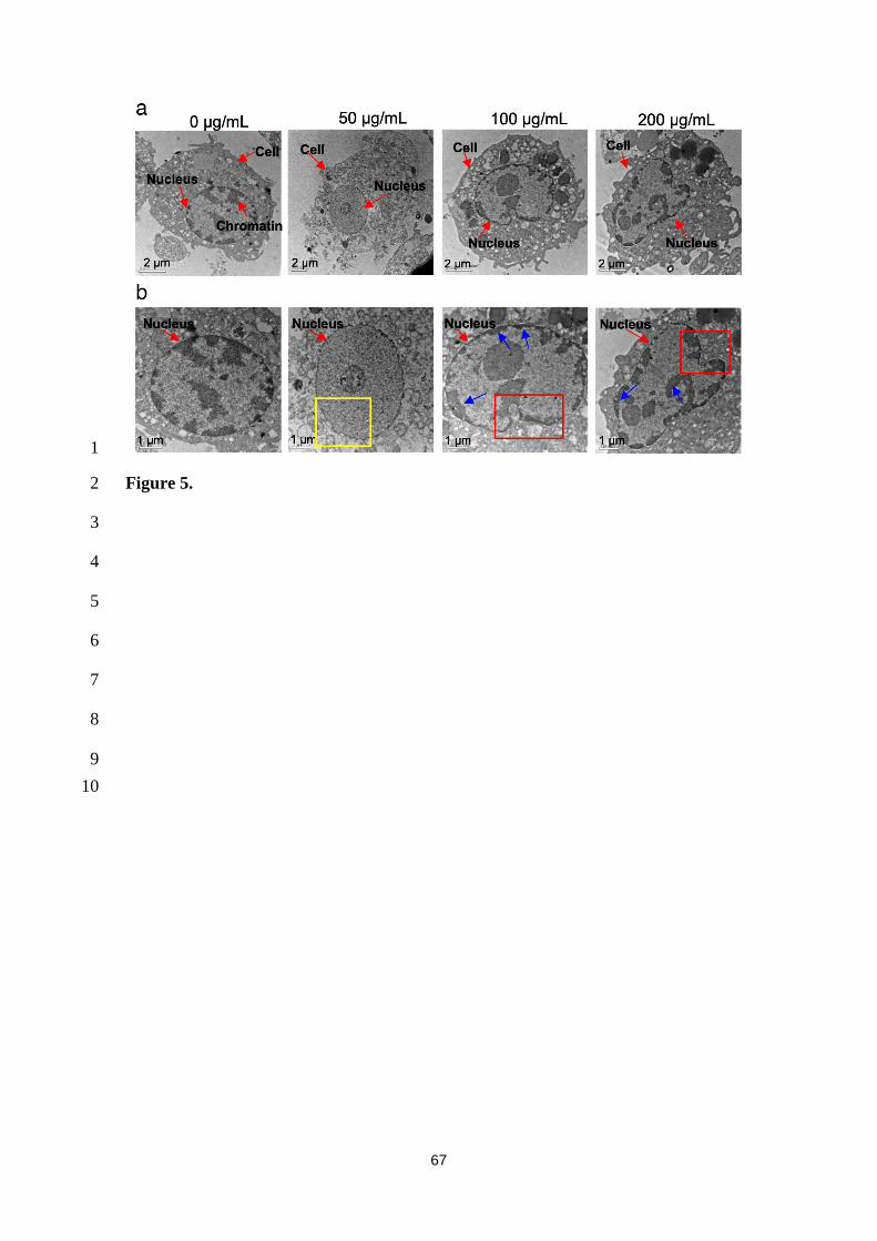
67
1
Figure 5. 2
3
4
5
6
7
8
9
10
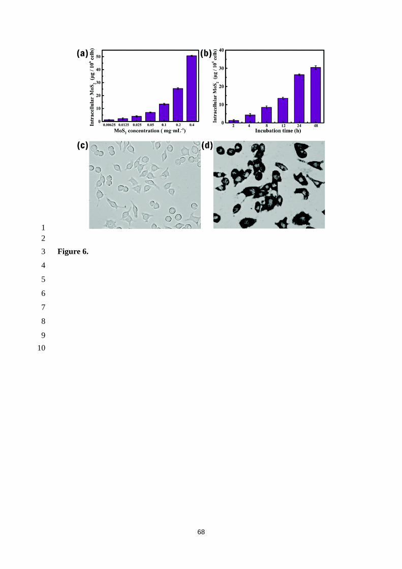
68
1
2
Figure 6. 3
4
5
6
7
8
9
10
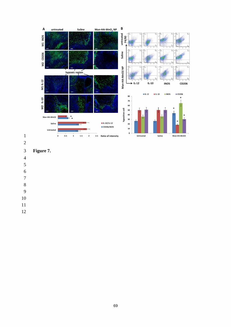
69
1
2
Figure 7. 3
4
5
6
7
8
9
10
11
12
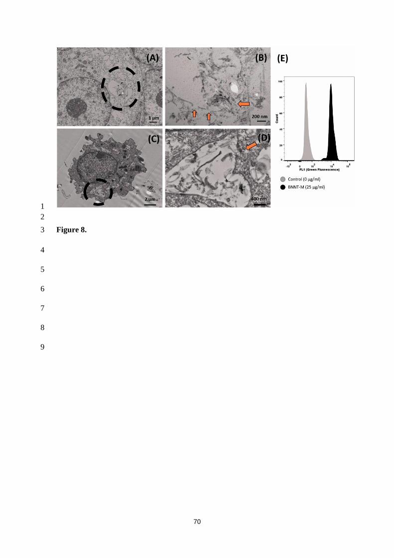
70
1
2
Figure 8. 3
4
5
6
7
8
9

71
1
Figure 9. 2
3
4
5
6
7
8
9
10
11
12
13
14
15

72
1
2
Figure 10. 3
4
5
6
7
8
9
10
11
12
13
14
15
16
17
18
19
20
21

73
1
Figure 11. 2
3
4
5
6
7
8
9
10
11
12
13
14

74
Figure captions (list) 1
2
Figure 1. Different types of 2D materials described in this review. 3
4
Figure 2. Categorization of graphene-based materials based on three parameters; C/O 5
ratio, average lateral size and number of layers. Reprinted with permission. [47] Copyright 6
2014, John Wiley & Sons, Inc. 7
8
Figure 3. (a) Bright field and (b) Raman images of graphene-RAW 264.7 after 24 h of 9
incubation. Secondary cluster maps of graphene aggregates localized within the cells at 10
24 h (c) and 7 d (d), are represented by false color codes. (e) Averaged spectra of graphene 11
from the sub-cluster regions. Dotted boxes represent the C-H stretching (2800-3050/cm) 12
from cells and O-H stretching (3100–3700/cm) from PBS solution. Reprinted with 13
permission. [64] Copyright 2013, John Wiley & Sons, Inc. 14
15
Figure 4. GO induced macrophage polarization to M1 subtype in a size-dependent 16
manner. (A) Representative histograms showing the numbers of M2 (Arg1+) and M1 17
(iNOS+) cells. J774.A1 cells were treated with small GO (S-GO) or large GO (L-GO) at 18
20 μg/mL for 24 h, followed by FACS analysis. (B) Percentages of iNOS+ cells (n = 5). 19
(C) Relative iNOS level in J774.A1 cells upon exposure to S-GO or L-GO at 20 μg/mL 20
for 24 h. HPRT1 was used as control for normalization. Reprinted with permission. [94] 21
Copyright 2015, American Chemistry Society. 22
23
Figure 5. TEM images of NR8383 nuclear morphology after exposure to AG-QDs (0, 24
50, 100, and 200 μg/mL) for 24 h. The images in panel (b) are enlarged from panel (a). 25
75
In panel (b), the yellow box indicates the shrinking of the inner nuclear envelope after 1
AG-QDs (50 μg/mL) exposure. The red boxes indicate the malformation of nuclear 2
morphology after AG-QDs (100 and 200 μg/mL) exposure. The blue arrows indicate the 3
chromatin condensation (electron-dense, black structure along nuclear membrane) 4
within the nuclei. Reprinted with permission. [133] Copyright 2018, BioMed Central 5
Ltd, Springer Nature. 6
7
Figure 6. Phagocytosis of MoS2 by macrophages. Quantitative analysis of the effect of 8
(a) MoS2 concentration and (b) incubation time on loading capacity; (c) and (d) 9
microphotographs of macrophages before and after BSA–MoS2 loading, respectively (t = 10
24 h, BSA–MoS2 is 0.2 mg/mL). Reprinted with permission. [152] Copyright 2019, The 11
Royal Society of Chemistry. 12
13
Figure 7. Man-HA-MnO2 skews TAMs M2 phenotype toward M1 phenotype. (A) 14
Representative immunofluorescence images of tumour sections stained with M1 and 15
M2 macrophage marker (green) after Man-HA-MnO2 administration. The orange dots 16
are Man-HA-MnO2. Magnification 100 ×; scale bar 100 μm. (B) Flow cytometric 17
analysis of phenotype of macrophages in tumours after administration of Man-HA-18
MnO2 (n = 5/group). Error bars are standard error of the mean. *p < 0.05 compared to 19
untreated control. Reprinted with permission. [166] Copyright 2016, American Chemical 20
Society Publications. 21
22
Figure 8. Ultrastructural evidence confirming uptake and lysosomal rupture in vitro and 23
in vivo. (A) TEM image of a differentiated THP-1 macrophage exposed to 25 mg/mL 24
(7.79 mg/cm2) of BNNT-M for 6 h (BNNT-M: mixture of BNNT, impurities of boron 25

76
and hBN). (B) High magnification image of the circled portion from Figure (A) showing 1
a ruptured lysosome (ruptured portion depicted with arrows). (C) Alveolar macrophage 2
from BALF of C57BL/6 mice exposed to BNNT-M (40 mg) for 24 h. (D) High 3
magnification image of the circled portion from Figure (C) showing a ruptured lysosome 4
(ruptured portion highlighted with arrows). (E) Pretreatment with acridine orange 5
followed by challenge with BNNT-M showed ~20-fold increase in green fluorescence 6
suggesting lysosomal membrane permeabilization. Reprinted with permission. [186] 7
Copyright 2017, Informa UK Limited. 8
9
Figure 9. Cytokine secretion of different macrophages. Macrophage-like THP-1 cells 10
were treated with 50 μg/mL of BP and an increasing concentration of corona complexes 11
(12.5, 25 and 50 μg/mL) for 6 h. Values are expressed as the means ± SDs of triplicates. 12
Statistical significance is assessed by Student’s t test. *p < 0.05, **p < 0.01. Reprinted 13
with permission. [202] Copyright 2018, Springer Nature. 14
15
Figure 10. RAW 264.7 cells engulf tungstate nanospheres. TEM analysis of RAW 264.7 16
cells exposed to tungstate nanospheres for 3 h. Reprinted with permission. [219] Copyright 17
2014, Informa UK Limited. 18
19
Figure 11. Macrophages and 2D materials mainly result in inflammation. 20