ISG15 Regulates Peritoneal Macrophages Functionality against Viral Infection
15
ISG15 Regulates Peritoneal Macrophages Functionality against Viral Infection Emilio Ya ´ ngu ¨ ez 1. , Alicia Garcı´a-Culebras 2. , Aldo Frau 2. , Catalina Llompart 3,4 , Klaus-Peter Knobeloch 5 , Sylvia Gutierrez-Erlandsson 3 , Adolfo Garcı´a-Sastre 6,7,8 , Mariano Esteban 3 , Amelia Nieto 3,4 , Susana Guerra 2 * 1 Institute of Medical Virology, University of Zurich, Zurich, Switzerland, 2 Department of Preventive Medicine and Public Health, Universidad Auto ´ noma, Madrid, Spain, 3 Department of Molecular and Cellular Biology, Centro Nacional de Biotecnologı ´a CSIC, Madrid, Spain, 4 Ciber de Enfermedades Respiratorias, Madrid, Spain, 5 Institute of Neuropathology, University Freiburg, Freiburg, Germany, 6 Department of Microbiology, Icahn School of Medicine at Mount Sinai, New York, New York, United States of America, 7 Department of Medicine, Division of Infectious Diseases, Icahn School of Medicine at Mount Sinai, New York, New York, United States of America, 8 Global Health and Emerging Pathogens Institute, Icahn School of Medicine at Mount Sinai, New York, New York, United States of America Abstract Upon viral infection, the production of type I interferon (IFN) and the subsequent upregulation of IFN stimulated genes (ISGs) generate an antiviral state with an important role in the activation of innate and adaptive host immune responses. The ubiquitin-like protein (UBL) ISG15 is a critical IFN-induced antiviral molecule that protects against several viral infections, but the mechanism by which ISG15 exerts its antiviral function is not completely understood. Here, we report that ISG15 plays an important role in the regulation of macrophage responses. ISG152/2 macrophages display reduced activation, phagocytic capacity and programmed cell death activation in response to vaccinia virus (VACV) infection. Moreover, peritoneal macrophages from mice lacking ISG15 are neither able to phagocyte infected cells nor to block viral infection in co-culture experiments with VACV-infected murine embryonic fibroblast (MEFs). This phenotype is independent of cytokine production and secretion, but clearly correlates with impaired activation of the protein kinase AKT in ISG15 knock-out (KO) macrophages. Altogether, these results indicate an essential role of ISG15 in the cellular immune antiviral response and point out that a better understanding of the antiviral responses triggered by ISG15 may lead to the development of therapies against important human pathogens. Citation: Ya ´ngu ¨ ez E, Garcı ´a-Culebras A, Frau A, Llompart C, Knobeloch K-P, et al. (2013) ISG15 Regulates Peritoneal Macrophages Functionality against Viral Infection. PLoS Pathog 9(10): e1003632. doi:10.1371/journal.ppat.1003632 Editor: Pinghui Feng, University of Texas Southwestern Medical Center, United States of America Received May 8, 2013; Accepted July 29, 2013; Published October 10, 2013 Copyright: ß 2013 Ya ´ngu ¨ ez et al. This is an open-access article distributed under the terms of the Creative Commons Attribution License, which permits unrestricted use, distribution, and reproduction in any medium, provided the original author and source are credited. Funding: This work was supported by grants from the Spanish Ministry of Health FIS2011-00127, Comunidad de Madrid UAM-CM-CCG10-4911 and UAM-Banco de Santander to SG. This work was also partly supported by NIAID grant U19AI083025 and by CRIP (Center for Research on Influenza Pathogenesis, HHSN266200700010C), a NIAID Center of Excellence for Influenza Research and Surveillance (CEIRS) to AGS. The funders had no role in study design, data collection and analysis, decision to publish, or preparation of the manuscript. Competing Interests: The authors have declared that no competing interests exist. * E-mail: [email protected] . These authors contributed equally to this work. Introduction The host innate immune response represents a critical initial line of defense against invading pathogens, and the magnitude of this early response can influence the course of disease progression. One of the earliest host responses to viral infection is the production of type I interferon (IFN-a and-b) and the subsequent upregulation of IFN-stimulated genes (ISGs) [1,2]. These ISGs generate an antiviral state and play an important role in determining the host innate and adaptive immune responses. One of the most highly induced genes in the IFN response is ISG15, which encodes a small UBL protein of 17 kDa that forms covalent conjugates with cellular proteins mediating considerable antiviral responses [3]. During viral infection in mice, ISG15 exists in three different forms: unconjugated within the cell, conjugated to target proteins and released into the serum [4]. When ISG15 is secreted, free ISG15 can function as a cytokine that modulates the immune response. For example, free ISG15 can activate natural killer (NK) and cytotoxic T-cells, stimulate IFN-c production and induce dendritic cell maturation and neutrophil recruitment [5,6]. In addition, antiviral activity associated with protein ISGylation has been described in vitro and/or in vivo for both DNA and RNA viruses, including influenza A and B viruses [7], Sindbis virus [7,8], hepatitis B virus [9], herpes simplex type-1 virus [7], vaccinia virus [10], vesicular stomatitis virus [11,12], lymphocytic choriomeningitis virus [13], respiratory syncytial virus [14], HIV-1 [15] and Ebola virus [16]. In contrast, free ISG15, but not ISGylation, promotes antiviral responses against Chikungunya virus infection [17]. Mice lacking UbE1L, the ISG15 E1 enzyme required for ISG15 conjugation, are more susceptible to influenza virus infection, indicating that ISGylation is essential for ISG15 antiviral activity [18]. ISG15 was shown to conjugate to over 160 host and viral proteins [19], especially those undergoing active translation [20]. Some of these host target proteins are downstream effectors of the interferon signaling, such as double-stranded RNA-activated protein kinase (PKR) [21], Myxovirus resistance protein A (MxA), and Retinoic acid inducible gene-I (RIG-I) [4], while PLOS Pathogens | www.plospathogens.org 1 October 2013 | Volume 9 | Issue 10 | e1003632
-
Upload
independent -
Category
Documents
-
view
4 -
download
0
Transcript of ISG15 Regulates Peritoneal Macrophages Functionality against Viral Infection

ISG15 Regulates Peritoneal Macrophages Functionalityagainst Viral InfectionEmilio Yanguez1., Alicia Garcıa-Culebras2., Aldo Frau2., Catalina Llompart3,4, Klaus-Peter Knobeloch5,
Sylvia Gutierrez-Erlandsson3, Adolfo Garcıa-Sastre6,7,8, Mariano Esteban3, Amelia Nieto3,4,
Susana Guerra2*
1 Institute of Medical Virology, University of Zurich, Zurich, Switzerland, 2 Department of Preventive Medicine and Public Health, Universidad Autonoma, Madrid, Spain,
3 Department of Molecular and Cellular Biology, Centro Nacional de Biotecnologıa CSIC, Madrid, Spain, 4 Ciber de Enfermedades Respiratorias, Madrid, Spain, 5 Institute of
Neuropathology, University Freiburg, Freiburg, Germany, 6 Department of Microbiology, Icahn School of Medicine at Mount Sinai, New York, New York, United States of
America, 7 Department of Medicine, Division of Infectious Diseases, Icahn School of Medicine at Mount Sinai, New York, New York, United States of America, 8 Global
Health and Emerging Pathogens Institute, Icahn School of Medicine at Mount Sinai, New York, New York, United States of America
Abstract
Upon viral infection, the production of type I interferon (IFN) and the subsequent upregulation of IFN stimulated genes(ISGs) generate an antiviral state with an important role in the activation of innate and adaptive host immune responses.The ubiquitin-like protein (UBL) ISG15 is a critical IFN-induced antiviral molecule that protects against several viral infections,but the mechanism by which ISG15 exerts its antiviral function is not completely understood. Here, we report that ISG15plays an important role in the regulation of macrophage responses. ISG152/2 macrophages display reduced activation,phagocytic capacity and programmed cell death activation in response to vaccinia virus (VACV) infection. Moreover,peritoneal macrophages from mice lacking ISG15 are neither able to phagocyte infected cells nor to block viral infection inco-culture experiments with VACV-infected murine embryonic fibroblast (MEFs). This phenotype is independent of cytokineproduction and secretion, but clearly correlates with impaired activation of the protein kinase AKT in ISG15 knock-out (KO)macrophages. Altogether, these results indicate an essential role of ISG15 in the cellular immune antiviral response andpoint out that a better understanding of the antiviral responses triggered by ISG15 may lead to the development oftherapies against important human pathogens.
Citation: Yanguez E, Garcıa-Culebras A, Frau A, Llompart C, Knobeloch K-P, et al. (2013) ISG15 Regulates Peritoneal Macrophages Functionality against ViralInfection. PLoS Pathog 9(10): e1003632. doi:10.1371/journal.ppat.1003632
Editor: Pinghui Feng, University of Texas Southwestern Medical Center, United States of America
Received May 8, 2013; Accepted July 29, 2013; Published October 10, 2013
Copyright: � 2013 Yanguez et al. This is an open-access article distributed under the terms of the Creative Commons Attribution License, which permitsunrestricted use, distribution, and reproduction in any medium, provided the original author and source are credited.
Funding: This work was supported by grants from the Spanish Ministry of Health FIS2011-00127, Comunidad de Madrid UAM-CM-CCG10-4911 and UAM-Bancode Santander to SG. This work was also partly supported by NIAID grant U19AI083025 and by CRIP (Center for Research on Influenza Pathogenesis,HHSN266200700010C), a NIAID Center of Excellence for Influenza Research and Surveillance (CEIRS) to AGS. The funders had no role in study design, datacollection and analysis, decision to publish, or preparation of the manuscript.
Competing Interests: The authors have declared that no competing interests exist.
* E-mail: [email protected]
. These authors contributed equally to this work.
Introduction
The host innate immune response represents a critical initial
line of defense against invading pathogens, and the magnitude of
this early response can influence the course of disease progression.
One of the earliest host responses to viral infection is the
production of type I interferon (IFN-a and-b) and the subsequent
upregulation of IFN-stimulated genes (ISGs) [1,2]. These ISGs
generate an antiviral state and play an important role in
determining the host innate and adaptive immune responses.
One of the most highly induced genes in the IFN response is
ISG15, which encodes a small UBL protein of 17 kDa that forms
covalent conjugates with cellular proteins mediating considerable
antiviral responses [3]. During viral infection in mice, ISG15 exists
in three different forms: unconjugated within the cell, conjugated
to target proteins and released into the serum [4]. When ISG15 is
secreted, free ISG15 can function as a cytokine that modulates the
immune response. For example, free ISG15 can activate natural
killer (NK) and cytotoxic T-cells, stimulate IFN-c production and
induce dendritic cell maturation and neutrophil recruitment [5,6].
In addition, antiviral activity associated with protein ISGylation
has been described in vitro and/or in vivo for both DNA and RNA
viruses, including influenza A and B viruses [7], Sindbis virus
[7,8], hepatitis B virus [9], herpes simplex type-1 virus [7],
vaccinia virus [10], vesicular stomatitis virus [11,12], lymphocytic
choriomeningitis virus [13], respiratory syncytial virus [14], HIV-1
[15] and Ebola virus [16]. In contrast, free ISG15, but not
ISGylation, promotes antiviral responses against Chikungunya
virus infection [17].
Mice lacking UbE1L, the ISG15 E1 enzyme required for ISG15
conjugation, are more susceptible to influenza virus infection,
indicating that ISGylation is essential for ISG15 antiviral activity
[18]. ISG15 was shown to conjugate to over 160 host and viral
proteins [19], especially those undergoing active translation [20].
Some of these host target proteins are downstream effectors of the
interferon signaling, such as double-stranded RNA-activated
protein kinase (PKR) [21], Myxovirus resistance protein A
(MxA), and Retinoic acid inducible gene-I (RIG-I) [4], while
PLOS Pathogens | www.plospathogens.org 1 October 2013 | Volume 9 | Issue 10 | e1003632

others are involved in the regulation of type I IFN signaling, e.g.
Janus kinase 1 (JAK1), extracellular signal-regulated kinase
(ERK1), interferon regulatory transcription factor 3 (IRF3) and
signal Transducers and Activator of transcription 1 (STAT1) [22].
The ISGylation of these cellular proteins may also contribute to
the antiviral activity of ISG15. Moreover, it has been described
that ISG15 expression blocks the virus-budding process by
different mechanisms such the blockage of Endosomal Sorting
Complexes Required for Transport (ESCRT) machinery for HIV
[15], or in the case of Ebola and other enveloped viruses
infections, inhibiting the Nedd4 E3 ubiquitin ligase [23].
Furthermore, several viral proteins have been shown to be
conjugated to ISG15, such as the NS1 protein from influenza
virus, NS5A from Hepatitis C virus [24] and gag from HIV virus
[25]. It has been proposed that conjugation to viral proteins
inhibits specific viral functions or virions assembly, causing a block
in viral infection progression [26,27]. Concerning VACV replica-
tion, previous publications from our group showed that ISG15
exerts antiviral activity against this virus, and that viral E3 protein
can bind to ISG15, counteracting its activity. Thus, a VACV
strain lacking E3 (VVDE3L) was unable to replicate in ISG15+/+cells, but was able to replicate in ISG152/2 deficient cells [10].
Moreover, infection of ISG152/2 mice with VVDE3L resulted
in significant disease and mortality, which was not observed in
ISG15+/+ mice infected with this attenuated virus [10]. Besides
the antiviral activity of ISG15, it has been recently described that a
mutation in human ISG15 correlates with ‘‘Mendelian suscepti-
bility to mycobacterial disease’’ (MSMD), a rare disorder that
manifests in severe clinical symptoms following infection with
weakly virulent mycobacterial strains and other intracellular
pathogens [28].
Although the role of ISG15 as a defense molecule against the
infection by several pathogens is accepted, the mechanisms by
which its antiviral properties are exerted and the main cellular
populations responsible for these activities are still weakly defined.
In order to characterize whether the ISG15 antiviral activity
observed in animal models is cell-type specific and to further
characterize its implication in the immune response, we have now
analyzed the impact of ISG15 deficiency on VACV replication
using different primary cell lines (fibroblasts, dendritic cells and
macrophages) derived from ISG15+/+ and ISG152/2 mice.
Here, we have observed that macrophages are key effectors of
ISG15-mediated antiviral responses during VACV infection and,
very importantly, that one of the most essential functions of the
macrophages, the phagocytosis, is dramatically diminished in the
absence of ISG15. These results provide valuable information on
the underlying mechanisms governing the suppression of viral
infection by ISG15.
Results
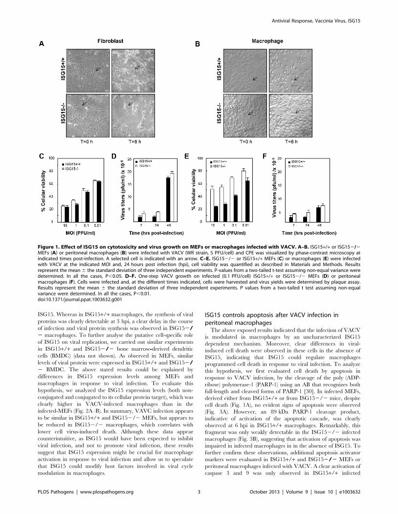
ISG152/2 fibroblasts and macrophages responddifferently to VACV infection
In order to characterize if different ISG15 deficient cell types
exhibit variable susceptibility to viral infection, we examined
VACV replication in different primary cells, derived from both
ISG15+/+ and ISG152/2 mice. As a first indicator, we analysed
the cytopathic effect (CPE) induced in fibroblasts (MEFs) or
peritoneal F4/F80 positives macrophages (Figure S1) after VACV
infection (5 PFU/cell). CPE was estimated by cell rounding and
alterations in cell morphology. While no differences in the VACV-
induced CPE were observed between wild type and ISG152/2
MEFs (Fig. 1A), VACV-induced CPE was clearly detectable in
ISG15+/+ macrophages (Fig. 1B and Movie S1), but not in
ISG152/2 cells (Fig. 1B and Movie S2). In order to confirm these
observations and to get a more quantitative result, we measured
the cellular mortality produced by VACV infection in the different
cell lines by a cellular viability assay. In agreement with the
previous results, in the case of VACV-infected MEFs (Fig. 1C), no
differences in the percentage of cellular viability at 24 hours post-
infection were observed in any case and, as expected, percentage
of cell death correlated with the multiplicity of infection used both
in for ISG15+/+ and ISG152/2 cells. However, in the infected
peritoneal macrophages (Fig. 1E), a higher percentage of
ISG152/2 cells survived to the infection, at each multiplicity of
infection (MOI) analyzed, in comparison to the results in ISG15+/
+ macrophages. In order to evaluate if the differences in cell
viability in response to VACV infection were due to variations in
the viral production in the absence of ISG15, we quantified virus
titers in both ISG15+/+ and ISG152/2 MEFs and peritoneal
macrophages infected with VACV in the same conditions. Upon
VACV-infection, no significant differences in viral titters were
observed between ISG15+/+ and ISG152/2 MEFs. Further-
more, and as expected, the viral titers increased with time (Fig. 1D)
and correlated with the observed increase in cellular mortality
(Fig. 1C). In contrast, in macrophages from both ISG152/2 or
ISG15+/+ mice, and according to previously published results
[29], the infection with VACV was abortive and viral titers did not
increase over time (Fig. 1F). In addition, viral protein synthesis in
both VACV-infected ISG15+/+ and ISG152/2 MEFs and
peritoneal macrophages was evaluated by Western blot using
specific antibodies for early p25 (E3L), intermediate p39 (A4L), and
late viral proteins p14 (A27L). Proteins encoded by E3L and A4L
genes were efficiently detected in lysates of all the infected cells
(Fig. 2A–B). However the relative levels were significantly lower in
the ISG152/2 macrophages, suggesting that viral infection could
be blocked in an early step in these cells. Regarding the late
proteins encoded by A27L genes, they were detected only in lysates
from infected MEFs (Fig. 2A) and not in infected macrophages
(Fig. 2B), confirming that VACV infection is abortive in
macrophages. To further characterize these results, the kinetics
of viral protein synthesis and the viral induced shut-off, as
indicators of the infection progression, were evaluated by
metabolic labeling at 3, 6 and 9 h after VACV infection in the
different cell lines. In ISG15+/+ and ISG152/2 MEFs, the viral
protein synthesis pattern presented similar kinetics, strongly
suggesting that ISG15 does not significantly alter VACV
replication in this cell type (Fig. 2C). However, in macrophages,
kinetics of viral gene expression was strongly affected by the lack of
Author Summary
Modification of proteins by ubiquitin (UB) and ubiquitin-like proteins (UBLs) are key regulatory processes of theinnate and adaptive immune response. Interferon (IFN)stimulated gene product 15 (ISG15) is an ubiquitin-likeprotein modifier, which is reversibly conjugated todifferent viral and cellular proteins mediating considerableantiviral responses. In turn, many viruses, includingpoxviruses, have evolved strategies to block the antiviraland inflammatory effects of the innate immune responsesto keep cells alive until virus replication is completed. Here,we describe a novel function of ISG15 in the control ofmacrophages activation, phagocytosis and apoptosis inresponse to viral infection. These processes are essentialfor the self-defense mechanism to protect animals frominfectious disease and could be crucial to understand theISG15 antiviral activity described in animal models.
Antiviral Response, Vaccinia Virus, ISG15
PLOS Pathogens | www.plospathogens.org 2 October 2013 | Volume 9 | Issue 10 | e1003632
ISG15. Whereas in ISG15+/+ macrophages, the synthesis of viral
proteins was clearly detectable at 3 hpi, a clear delay in the course
of infection and viral protein synthesis was observed in ISG152/2 macrophages. To further analyse the putative cell-specific role
of ISG15 on viral replication, we carried out similar experiments
in ISG15+/+ and ISG152/2 bone marrow-derived dendritic
cells (BMDC) (data not shown). As observed in MEFs, similar
levels of viral protein were expressed in ISG15+/+ and ISG152/2 BMDC. The above stated results could be explained by
differences in ISG15 expression levels among MEFs and
macrophages in response to viral infection. To evaluate this
hypothesis, we analyzed the ISG15 expression levels (both non-
conjugated and conjugated to its cellular protein target), which was
clearly higher in VACV-infected macrophages than in the
infected-MEFs (Fig. 2A–B). In summary, VAVC infection appears
to be similar in ISG15+/+ and ISG152/2 MEFs, but appears to
be reduced in ISG152/2 macrophages, which correlates with
lower cell virus-induced death. Although these data appear
counterintuitive, as ISG15 would have been expected to inhibit
viral infection, and not to promote viral infection, these results
suggest that ISG15 expression might be crucial for macrophage
activation in response to viral infection and allow us to speculate
that ISG15 could modify host factors involved in viral cycle
modulation in macrophages.
ISG15 controls apoptosis after VACV infection inperitoneal macrophages
The above exposed results indicated that the infection of VACV
is modulated in macrophages by an uncharacterized ISG15
dependent mechanism. Moreover, clear differences in viral-
induced cell death were observed in these cells in the absence of
ISG15, indicating that ISG15 could regulate macrophages
programmed cell death in response to viral infection. To analyze
this hypothesis, we first evaluated cell death by apoptosis in
response to VACV infection, by the cleavage of the poly (ADP-
ribose) polymerase-1 (PARP-1) using an AB that recognizes both
full-length and cleaved forms of PARP-1 [30]. In infected MEFs,
derived either from ISG15+/+ or from ISG152/2 mice, despite
cell death (Fig. 1A), no evident signs of apoptosis were observed
(Fig. 3A). However, an 89 kDa PARP-1 cleavage product,
indicative of activation of the apoptotic cascade, was clearly
observed at 6 hpi in ISG15+/+ macrophages. Remarkably, this
fragment was only weakly detectable in the ISG152/2 infected
macrophages (Fig. 3B), suggesting that activation of apoptosis was
impaired in infected macrophages in in the absence of ISG15. To
further confirm these observations, additional apoptosis activator
markers were evaluated in ISG15+/+ and ISG152/2 MEFs or
peritoneal macrophages infected with VACV. A clear activation of
caspase 3 and 9 was only observed in ISG15+/+ infected
Figure 1. Effect of ISG15 on cytotoxicity and virus growth on MEFs or macrophages infected with VACV. A–B. ISG15+/+ or ISG152/2MEFs (A) or peritoneal macrophages (B) were infected with VACV (WR strain, 5 PFU/cell) and CPE was visualized by phase-contrast microscopy atindicated times post-infection. A selected cell is indicated with an arrow. C–E. ISG152/2 or ISG15+/+ MEFs (C) or macrophages (E) were infectedwith VACV at the indicated MOI and, 24 hours post infection (hpi), cell viability was quantified as described in Materials and Methods. Resultsrepresent the mean 6 the standard deviation of three independent experiments. P-values from a two-tailed t-test assuming non-equal variance weredetermined. In all the cases, P,0.05. D–F. One-step VACV growth on infected (0.1 PFU/cell) ISG15+/+ or ISG152/2 MEFs (D) or peritonealmacrophages (F). Cells were infected and, at the different times indicated, cells were harvested and virus yields were determined by plaque assay.Results represent the mean 6 the standard deviation of three independent experiments. P values from a two-tailed t test assuming non-equalvariance were determined. In all the cases, P,0.01.doi:10.1371/journal.ppat.1003632.g001
Antiviral Response, Vaccinia Virus, ISG15
PLOS Pathogens | www.plospathogens.org 3 October 2013 | Volume 9 | Issue 10 | e1003632

macrophages, which correlated with lower levels of the anti-
apoptotic factor B-cell lymphoma 2 (Bcl-2). In order to get a more
quantitative and physiological indicator of the apoptosis activation
in these cells, the Caspase-Glo 3/7 assay kit was used following
manufacturer’s instructions. As shown in Fig. 3C, VACV-induced
apoptosis was almost undetected in ISG15+/+ and ISG152/2
MEFs, further validating the observations described above.
However, when cells were treated with the apoptosis activators
epopside and staurosporine, apoptosis in ISG15+/+ and
ISG152/2 treated-MEFs reached similar levels, confirming that
ISG15 does not play any role in the activation of the apoptosis
cascade in MEFs. In contrast, VACV-induced apoptosis activation
was only detected in ISG15+/+ cells but not in macrophages
lacking ISG15. In order to exclude that ISG15 generally regulates
macrophage apoptosis rather than controlling apoptosis induction
upon viral infection, we again used the general apoptosis activators
epopside and staurosporine. A slight diminution of the apoptosis
induced by these compounds was detected in the absence of
ISG15, indicating that, although it could be implicated in general
apoptosis, ISG15 mainly regulates the apoptosis in response to
viral infection (Fig. 3D). Although these data might be explained
by a higher level of viral infection and viral protein synthesis in
ISG15+/+ macrophages, resulting in enhanced apoptosis as
compared to ISG152/2 macrophages, these results also suggest
that in macrophages but not in MEFs ISG15 is involved in the
specific activation of programmed cell death upon VACV
infection.
ISG15 regulates viral infection in macrophagesThe data presented above suggest that the virus infection itself
could be the signal that triggers apoptosis in macrophages, as the
VACV cycle appears to be prematurely inhibited in the absence of
ISG15 in these cells. Given that ISG152/2 infected macrophages
showed an increase in cellular survival, accompanied by a
reduction in viral protein levels and a delayed kinetic of viral
protein synthesis, we decided to analyze whether VACV entry was
Figure 2. VACV protein synthesis on ISG15+/+ or ISG152/2 MEFs or macrophages. A–B. Viral protein expression during VACV infection ofISG15+/+ or ISG152/2 MEFs or macrophages. ISG15+/+ or ISG152/2 MEFs (A) or macrophages (B) were infected (106 cells/time post-infection;3 PFU/cell) with VACV (WR strain) and, at the indicated times post-infection, cell extracts were prepared and equal amounts of proteins werefractionated by SDS-PAGE, transferred to nitrocellulose, and detected with specific antibodies virus early (p25), intermediate (p39) and late proteins(p14). ISG15 expression levels were detected by Western blot using specific antibodies. Actin was measured as protein loading control. Proteinstandards are indicated. Uninfected cells (Mock) served as control. C–D. De novo protein synthesis in VACV-infected ISG15+/+ or ISG152/2 MEFs (C)or macrophages (D). ISG15+/+ or ISG152/2 cells were infected (106 cells/time post-infection; 3 PFU/cell) with VACV (WR strain) and labeled with 35S-methionine (50 mCi/ml, 30 min) at the different times indicated. Cellular lysates were analyzed by 12% SDS-PAGE, transferred to nitrocellulosemembranes and visualized by autoradiography. Protein standards are indicated. Uninfected cells (Mock) served as control. In the same membranes,the expression of ISG15 or tubuline (protein loading control) was detected by Western blot using specific antibodies.doi:10.1371/journal.ppat.1003632.g002
Antiviral Response, Vaccinia Virus, ISG15
PLOS Pathogens | www.plospathogens.org 4 October 2013 | Volume 9 | Issue 10 | e1003632

impaired in ISG152/2 macrophages. For that, we used a
recombinant virus in which the viral structural protein A3L was
labeled with the yellow fluorescent protein (YFP), allowing the
visualization of the viral particles by time-lapse microscopy. When
macrophages were infected with the VACV-YFP, we observed a
delay in the entry of the virions inside the cell in the absence of
ISG15 (Fig. 4). While in wild type (WT) macrophages the
fluorescent signal increased with time inside the cell (Fig. 4A), in
ISG152/2 macrophages, the signal was localized mainly outside
the cell even after 2 hours of adsorption (Fig. 4B). These results
indicate that, in the absence of ISG15, infection was blocked at an
early step, which might also be the reason for the delay in the
kinetics observed in the viral protein radiolabeling experiment
(Fig. 2D). These results suggest that, in the absence of ISG15, the
virus entry is impaired and made us to consider whether this
mechanism was exclusive for VACV or if it also observed upon
infection with other viruses. Therefore, we performed similar
experiments using FluV, a completely different virus with different
cellular receptor. In a first approach, we infected ISG15+/+ and
ISG152/2 MEFs or peritoneal macrophages with FluV (5 PFU/
cell) and, as above described for VACV, evaluated de novo viral
protein synthesis using 35S-Met at different times post-infection. As
observed for VACV infection, there are no variations in viral
protein synthesis in MEFs in the absence of ISG15 (Fig. 5A), but a
clear delay in the overall viral protein synthesis is observed in the
infected ISG152/2 macrophages (Fig. 5B, compare 3 hpi in
ISG152/2 and ISG15 +/+ macrophages). Although no viral
production was observed in both ISG15+/+ and ISG152/2
macrophages (Figure S2), when we visualized the effect of FluV
infection by phase-contrast microscopy, a clear CPE was evident
in the ISG15+/+ macrophages with the course of infection,
whereas the ISG152/2 macrophages remained unaltered with
no obvious signs of viral infection-induced cell death (Fig. 5C).
Since the block in infection and the differences in viral induced
CPE were observed with two very distinct viruses, we next
investigated whether other cellular entry processes, such as
phagocytosis, might be regulated by ISG15 in macrophages.
ISG15 regulates macrophage phagocytosisA major function of macrophages is the phagocytosis of
pathogens, antigens, and infected or apoptotic cells, which is
critical for innate as well as for adaptive immunity. Taking into
account that in ISG152/2 macrophages the early events in the
infection cycle of two completely different viruses, VACV or
FLuV, were aborted, and that this was at the level of virus
endocytosis, at least for VACV, we considered that other cellular
entry processes inherent to macrophage function, such as
phagocytosis, could be controlled by ISG15. Latex beads are a
Figure 3. ISG15 controls apoptosis in macrophages after VACV infections. A–B. Time course of apoptosis markers during VACV infection inISG15+/+ or ISG152/2 MEFs (A) or peritoneal macrophages (B). ISG15+/+ or ISG152/2 cells were mock-infected (M) or infected with VACV (WRstrain, 10 PFU/cell). At the indicated times post-infection, cells were harvested and total proteins were separated by SDS-PAGE, transferred tonitrocellulose and immunoblotted with anti-PARP-1, anti-caspase-9, anti-activated-capsase-3 and anti bcl-2 specific antibodies. Actin was measuredas protein loading control. Protein standards are indicated. C–D. Apoptosis activation in ISG15+/+ or ISG152/2 MEFs (C) or peritoneal macrophages(D). Apoptosis was measured using the Caspase-Glo 3/7 assay kit at 24 hpi Results represent the mean 6 the standard deviation of threeindependent experiments. P-values from a two-tailed t test assuming non-equal variance were determined. In all the cases, P,0.05.doi:10.1371/journal.ppat.1003632.g003
Antiviral Response, Vaccinia Virus, ISG15
PLOS Pathogens | www.plospathogens.org 5 October 2013 | Volume 9 | Issue 10 | e1003632

common model substrate in biochemical studies of macrophage
phagosome composition and maturation [31]. To determine
whether ISG15 is critical for macrophage phagocytosis capacity,
we analyzed the intake of GFP labelled latex beads in ISG15+/+and ISG152/2 macrophages by confocal and time-lapse
microscopy, as described in Material and Methods. To
potentially enhance the impact of ISG15, which is an IFN
induced protein, macrophages were treated with type I IFN
alpha (100 units/ml for 16 hours) or left untreated [32]. The
time-lapse microscopy images showed a marked decrease of
latex beads phagocytosis in ISG152/2 macrophages when
compared to the ISG15+/+ cells. This difference was even more
evident upon IFN treatment, further implicating a role of ISG15
in this process (Fig. 6A–C and Movie S3-S4-S5-S6). Quantifi-
cation of these results revealed that, after IFN incubation, the
phagocytic capacity of ISG15+/+ cells was about 100 times
higher than that observed in ISG152/2 cells (Figs. 6B).
Moreover, and pointing out the biological relevance of ISG15 in
phagocytosis, IFN treatment increased the latex bead uptake in
ISG15+/+ macrophages but not in ISG152/2 cells (Fig. 6B).
Representative confocal immunofluorescence images of macro-
phages with internalized beads are shown in Fig. 6D. Further-
more, to check if the treatment with IFN also enhance the entry
of VACV in macrophages, we monitored by immunofluores-
cence the amount of virus inside the cell after 2 hpi in
permeabilized ISG15+/+ and ISG152/2 cells treated or not
with IFN. A clear increase of viral entry was observed after IFN
treatment in ISG15+/+ infected macrophages (Figure S3). All
together, these results strongly suggest that ISG15 has an
important role in the phagocytic activity of macrophages that is
in correspondence with an increase of viral entry, and that IFN
enhances both processes through the induction of ISG15.
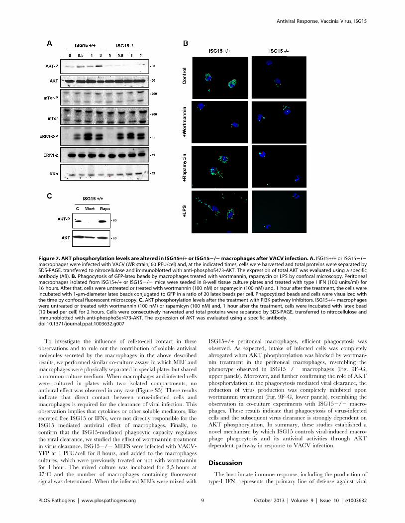
ISG15 plays a critical role in AKT induced phagocytosisThe activation of Phosphoinositide 3-kinase/(PI3K)- A protein-
serine/threonine kinase (AKT) signaling has previously been
shown to be required for macrophage phagocytosis [31,33].
Therefore, we examined whether the activation of this signaling
cascade could be affected in the absence of ISG15. The
phosphorylation of AKT, mammalian Target of Rapamycin (m-
Tor) and ERK1-2 in ISG15+/+ and ISG152/2 macrophages in
response to VACV infection was monitored by Western blot.
ISG15+/+ macrophages have some basal levels of AKT
phosphorylation under the used conditions, which were increased
at early times upon exposure to VACV. By contrast, basal levels of
P-AKT were clearly reduced in ISG152/2 macrophages, when
compared to ISG15+/+ cells, and these did not increased upon
exposure to VACV (Fig. 7A). In contrast, phosphorylation levels of
m-Tor or ERK1-2 were similar in both types of cell populations.
Similar results were obtained when macrophages were incubated
with latex beads (data not shown), indicating that the underlying
ISG15-dependent mechanism of phagocytosis/endocytosis activa-
tion by both particles and viruses could be similar and related to
AKT. To analyze whether the reduction in AKT phosphorylation
observed in ISG152/2 macrophages is involved in the phago-
cytosis blockage, the phagocytosis capacity of WT macrophages
was analyzed by confocal immunofluorescence using different
inhibitors of the PI3K pathway. Treatment of IFN-exposed
ISG15+/+ macrophages with wortmannin, an inhibitor of AKT
phosphorylation (Fig. 7C), considerably reduced their phagocytic
Figure 4. ISG15 control the VACV entry. Peritoneal macrophages isolated from ISG15+/+ or ISG152/2 mice were seeded in 8-well tissue cultureplates and treated with type I interferon (IFN) (100units/ml) for 16 hours. After that, the cells infected with VACV-YFP (60 PFU/cell). Infected-cells werevisualized with the time by fluorescent and phase contrast microscopy. Representative fields are shown at a magnification of 406 (left panels).doi:10.1371/journal.ppat.1003632.g004
Antiviral Response, Vaccinia Virus, ISG15
PLOS Pathogens | www.plospathogens.org 6 October 2013 | Volume 9 | Issue 10 | e1003632

capacity (Fig. 7B and Movie S8) in comparison to untreated cells
(Fig. 7B and Movie S7). In contrast, the mTOR inhibitor
rapamycin, which preserves AKT phosphorylation (Fig. 7C), had
no effect on the phagocytosis activity. We also checked if treatment
with lipopolysaccharide (LPS), a well characterized macrophage
activator, affected phagocytosis in both ISG152/2 and ISG15+/
+ macrophages (without previous interferon treatment). The WT
macrophages activated with LPS showed similar phagocytosis
levels as controls (Fig. 7B and Movie S10). Strikingly, in ISG152/
2 macrophages, no activation of phagocytosis was observed after
LPS treatment (Fig. 7B). In addition to the confocal analyses, time-
lapse microscopy was performed and representative images of the
different treatments are shown in Figure 8. Only wortmanin
treatment clearly reduced phagocytosis capacity of ISG15+/+macrophages, as indicated by the high number of non-internalized
and surface-bound beads in these cells. Collectively, these results
indicate that ISG15 plays a critical role in AKT-phosphorylation,
and that this pathway is essential to ensure proper phagocytosis in
macrophages. These findings describe a novel function of ISG15
promoting macrophage phagocytosis.
ISG15 promotes the antiviral activity of macrophages andthe phagocytosis of VACV-infected cells
As ISG15 plays a critical role in latex beads and virus entry in
macrophages, we wanted to analyze if ISG15 also participates in
the phagocytosis of infected cells by macrophages. A function of
ISG15 in this process would also at least partially explain the high
susceptibility of ISG152/2 mice to viral infections. To assess this
possibility, ISG152/2 MEFS were infected with VACV-YFP at
1 PFU/cell for 8 hours, and subsequently added to macrophages
cultures at a ratio of one MEF to four macrophages. The mixed
culture was incubated for 2,5 hours at 37uC and the number of
macrophages containing fluorescent signal was determined. When
VACV-YFP-infected MEFs were mixed with ISG15+/+ perito-
neal macrophages, efficient phagocytosis was observed (Fig. 9A).
In contrast, a massive decrease in phagocytosis of infected cells was
found when infected MEFs were mixed with ISG152/2
macrophages (Fig. 9A). Quantification of these results revealed
that the phagocytic capacity of the ISG15+/+ macrophages was
about 20 times higher than that observed in ISG152/2 (Fig. 9F).
Figure 5. Effect of ISG15 on cytotoxicity and infection progression in ISG15+/+ or ISG152/2 MEFs or macrophages infected withinfluenza virus (FluV). A–B. De novo protein synthesis in FluV- infected ISG15+/+ or ISG152/2 MEFs (A) or peritoneal macrophages (B). ISG15+/+or ISG152/2 cells were infected (106 cells/time post-infection; 3 PFU/cell) with FluV (A/WSN/1933 strain) and labeled with 35S-methionine (50 mCi/ml,30 min) at the times indicated. Cellular lysates were analyzed by 12% SDS-PAGE, transferred to nitrocellulose membranes and visualized byautoradiography. Protein standards are indicated. Uninfected cells (Mock) served as control. In the same membranes, the expression of ISG15 ortubuline (protein loading control) was detected by Western blot using specific antibodies. C. ISG15+/+ or ISG152/2 peritoneal macrophages wereinfected with FluV (A/WSN/1933 strain, 5 PFU/cell) and CPE was visualized by phase-contrast microscopy at indicated times post-infection. A selectedcell is indicated with an arrow.doi:10.1371/journal.ppat.1003632.g005
Antiviral Response, Vaccinia Virus, ISG15
PLOS Pathogens | www.plospathogens.org 7 October 2013 | Volume 9 | Issue 10 | e1003632

More than 40% of the ISG15+/+ macrophages incorporated
virus-infected cells, whereas only about 2% of the ISG152/2
macrophages phagocyted VACV infected cells. These results show
that in macrophages ISG15 is necessary to phagocytize VACV-
infected cells. To study whether the phagocytosis is or not
exclusively due to the macrophages in these co-cultures experi-
ments, we decided monitored the phagocytosis capacity in MEFs
using GFP-latex. A clear absence of latex bead was observed inside
the cells indicating that MEFs do not exhibit phagocytic ability
(Figure S4). However, although the MEFs were not able to
phagocyte latex beads the infection with the VACV-YFP showed
that virus entry occurred normally in these cells (Figure S4).
Phagocytosis of infected cells could contribute to the virus
clearance and, if during this process the macrophage gets infected,
it could commit suicide, blocking further possible propagation of
the virus infection. Thus, we decided to examine whether virus
growth in MEFs is affected in the presence of macrophages, as a
simulation of macrophage mediated viral clearance. For this
purpose, ISG152/2 MEFs were infected with VACV and added
to a macrophage culture after 8 hpi. The co-culture was further
maintained and the virus titer in the culture medium was
determined at 24 and 48 h after infection. The virus titer in the
infected cells of the co-cultures that contains ISG15 +/+macrophages gradually decreased, while that in the control culture
with DMEM and in the co-culture with ISG152/2 macrophages
clearly increased the time (Fig. 9B). Furthermore, in the co-culture
experiments with ISG15+/+ macrophages, the reduction in viral
titer was accompanied with a reduction in VACV viral protein
expression (Fig. 9C), an increase in ISG15 levels (conjugated and
non-conjugated) (Fig. 9D) and a clear increase in the AKT
phosphorylation level (Fig. 9-E). These results clearly indicate that
the production of VACV into the cells is inhibited in the presence
of high levels of ISG15 in macrophages and strongly suggest that
this was due to phagocytosis of infected cells. However, the
possibility remained that soluble factors, secreted from macro-
phages and could cause a decrease in viral replication.
Figure 6. Phagocytosis of GFP-latex beads by ISG15 +/+ or ISG15 2/2 macrophages. A–C. Peritoneal macrophages isolated for ISG15+/+or ISG152/2 mice were seeded in 8-well tissue culture plates and treated with type I IFN (100units/ml) for 16 hours (A) or not (C). After that, the cellswere incubated with 1-mm-diameter latex beads conjugated to GFP in a ratio of 10 latex beads per cell. Phagocytized beads and cells were visualizedwith the time by fluorescent and phase contrast microscopy. B. Quantification of phagocytic cells in type I IFN alpha treated or not treatedmacrophages. Cells were quantified by IF in three independent experiments. Black bars represent ISG15+/+ macrophages, white bars representsISG152/2 macrophages. Results represent the mean 6 the standard deviation of three independent experiments. P values from a two-tailed t-testassuming non-equal variance were determined. In all the cases, P,0.05. D. Phagocytosis of GFP-latex beads by ISG15+/+ and ISG152/2macrophages detected by immunofluorescence. Peritoneal macrophages were cultured on coverslips and incubated for 1 h with GFP-latex beads (20beads per cell) and then washed three times with phosphate-buffered saline (PBS) and incubated with Dulbecco’s medium (DMEM) medium for anadditional hour. Cells were fixed with 4% PFA and processed for immunofluorescence analysis. Phagocytized beads and cells were visualized byconfocal fluorescent microscopy. Representative fields are shown at a magnification of 406 (left panels) and 1006 (right panels).doi:10.1371/journal.ppat.1003632.g006
Antiviral Response, Vaccinia Virus, ISG15
PLOS Pathogens | www.plospathogens.org 8 October 2013 | Volume 9 | Issue 10 | e1003632
To investigate the influence of cell-to-cell contact in these
observations and to rule out the contribution of soluble antiviral
molecules secreted by the macrophages in the above described
results, we performed similar co-culture assays in which MEF and
macrophages were physically separated in special plates but shared
a common culture medium. When macrophages and infected cells
were cultured in plates with two isolated compartments, no
antiviral effect was observed in any case (Figure S5). These results
indicate that direct contact between virus-infected cells and
macrophages is required for the clearance of viral infection. This
observation implies that cytokines or other soluble mediators, like
secreted free ISG15 or IFNs, were not directly responsible for the
ISG15 mediated antiviral effect of macrophages. Finally, to
confirm that the ISG15-mediated phagocytic capacity regulates
the viral clearance, we studied the effect of wortmannin treatment
in virus clearance. ISG152/2 MEFS were infected with VACV-
YFP at 1 PFU/cell for 8 hours, and added to the macrophages
cultures, which were previously treated or not with wortmannin
for 1 hour. The mixed culture was incubated for 2,5 hours at
37uC and the number of macrophages containing fluorescent
signal was determined. When the infected MEFs were mixed with
ISG15+/+ peritoneal macrophages, efficient phagocytosis was
observed. As expected, intake of infected cells was completely
abrogated when AKT phosphorylation was blocked by wortman-
nin treatment in the peritoneal macrophages, resembling the
phenotype observed in ISG152/2 macrophages (Fig. 9F–G,
upper panels). Moreover, and further confirming the role of AKT
phosphorylation in the phagocytosis mediated viral clearance, the
reduction of virus production was completely inhibited upon
wortmannin treatment (Fig. 9F–G, lower panels), resembling the
observation in co-culture experiments with ISG152/2 macro-
phages. These results indicate that phagocytosis of virus-infected
cells and the subsequent virus clearance is strongly dependent on
AKT phosphorylation. In summary, these studies established a
novel mechanism by which ISG15 controls viral-induced macro-
phage phagocytosis and its antiviral activities through AKT
dependent pathway in response to VACV infection.
Discussion
The host innate immune response, including the production of
type-I IFN, represents the primary line of defense against viral
Figure 7. AKT phosphorylation levels are altered in ISG15+/+ or ISG152/2 macrophages after VACV infection. A. ISG15+/+ or ISG152/2macrophages were infected with VACV (WR strain, 60 PFU/cel) and, at the indicated times, cells were harvested and total proteins were separated bySDS-PAGE, transferred to nitrocellulose and immunoblotted with anti-phosphoS473-AKT. The expression of total AKT was evaluated using a specificantibody (AB). B. Phagocytosis of GFP-latex beads by macrophages treated with wortmannin, rapamycin or LPS by confocal microscopy. Peritonealmacrophages isolated from ISG15+/+ or ISG152/2 mice were seeded in 8-well tissue culture plates and treated with type I IFN (100 units/ml) for16 hours. After that, cells were untreated or treated with wortmannin (100 nM) or rapamycin (100 nM) and, 1 hour after the treatment, the cells wereincubated with 1-mm-diameter latex beads conjugated to GFP in a ratio of 20 latex beads per cell. Phagocytized beads and cells were visualized withthe time by confocal fluorescent microscopy. C. AKT phosphorylation levels after the treatment with PI3K pathway inhibitors. ISG15+/+ macrophageswere untreated or treated with wortmannin (100 nM) or rapamicyn (100 nM) and, 1 hour after the treatment, cells were incubated with latex bead(10 bead per cell) for 2 hours. Cells were consecutively harvested and total proteins were separated by SDS-PAGE, transferred to nitrocellulose andimmunoblotted with anti-phosphoSer473-AKT. The expression of AKT was evaluated using a specific antibody.doi:10.1371/journal.ppat.1003632.g007
Antiviral Response, Vaccinia Virus, ISG15
PLOS Pathogens | www.plospathogens.org 9 October 2013 | Volume 9 | Issue 10 | e1003632

pathogens. Of the hundreds of IFN-stimulated genes (ISGs)
discovered to date, ISG15 was one of the firstly identified and
shown to encode an ubiquitin-like protein modifier [34]. ISG15
knock-out mice are more susceptible to infection by several viruses,
pointing out the relevance of this molecule in the antiviral response
in animal models [35]. However, the underlying causes of the
enhanced susceptibility of ISG152/2 mice and the cell types
involved are only weakly defined. In order to clarify these
questions, we evaluated the role of ISG15 in VACV replication in
different cells types. Both MEFs and dendritic cells (not shown)
VACV-infected were unaffected by the lack of ISG15 in the course
of infection. In contrast, ISG152/2 peritoneal macrophages
showed a clear resistance to VACV-induced cell death in
comparison with wild type control macrophages where VACV
replication is abortive and infectious progeny is not released [29].
Whereas WT macrophages became apoptotic and die after
infection, in ISG152/2 macrophages, in which VACV infection
is also abortive, no apoptosis was observed, as evidenced by the
absence of PARP cleavage and CPE (Fig. 2). Apoptosis after
infection with many types of viruses is generally considered as a
self-defense mechanism [36], as loss of host cell activity should
impair virus propagation. For instance, apoptotic cells are engulfed
and digested in lysosomes of phagocytes [36]. Moreover, a higher
increase in the expression of ISG15 and the protein ISGylation
levels were observed in ISG15+/+ infected macrophages when
compared to infected MEFs, indicating that the factor involved in
the different phenotype among VACV infection in these cells
could be modified by ISG15.
In addition to the differences in the VACV-induced apoptosis
between ISG15+/+ or ISG152/2 macrophages, we observed
clear differences in the phagocytosis ability of the macrophages.
Apoptosis and phagocytosis are two interconnected pathways,
which are very important for the efficacy of the innate immune
response against pathogen infection [36]. Phagocytic elimination
of invading microbes represents an important innate immune
mechanism, as it contributes to the clearance of the virus.
Moreover, if during this process the macrophage gets infected, it
will commit suicide by apoptosis, blocking further possible
propagation of the viral infection. In contrast, viruses appear to
resist this host action by inhibiting apoptosis using anti-apoptotic
proteins encoded by viral genes [37]. Moreover, the phagocytosis
of apoptotic bodies seems to be an important mechanism to cross-
prime DCs, as extracellular antigens gain access to MHC I
molecules for priming of cytolytic T-cells [38]. Some microbes
including viruses are phagocytized not directly but indirectly as
microbe-infected cells [33]. Phagocytosis of immune complexes,
opsonized virus, and infected host cells represents another
important connection between the adaptive and innate immune
systems, with potential roles both in priming of the adaptive
immune response and in clearance of virus. Phagocytosis not only
may rapidly remove virus or virally infected cells from the
circulation but it could also affect immune complex-induced
inflammation, which is implicated in driving disease progression.
Importantly, there is evidence that disease susceptibility and
severity in numerous autoimmune diseases and infectious diseases
are regulated by phagocytosis [39]. While ISG152/2 mice were
not more susceptible to VACV wild type virus infection, this might
be explained by the capacity for VACV to inhibit IFN signaling
and therefore ISG15 induction during infection in vivo. In fact, a
VACV with enhanced IFN induction due to the deletion of E3L
displayed higher pathogenicity in ISG152/2 mice [10]. It is
attractive to speculate that the increased susceptibility of ISG152/
Figure 8. Wortmannin treatment inhibits phagocytosis of GFP-latex beads by ISG15+/+ macrophages. Peritoneal macrophages isolatedfrom ISG15+/+ mice were seeded in 8-well tissue culture plates and treated with type I IFN (100 units/ml) for 16 hours. After that, were untreated ortreated with wortmannin (100 nM) or rapamycin (100 nM) for 1 hour and subsequently incubated with 1-mm-diameter latex beads conjugated toGFP in a ratio of 20 latex beads per cell. Phagocytized beads and cells were visualized with the time by fluorescent and phase contrast microscopy.Representative fields are shown at a magnification of 406 (left panels) and 1006 (right panels).doi:10.1371/journal.ppat.1003632.g008
Antiviral Response, Vaccinia Virus, ISG15
PLOS Pathogens | www.plospathogens.org 10 October 2013 | Volume 9 | Issue 10 | e1003632

2 mice to several virus infections, including herpesvirus and
influenza virus, is mediated at least in part by a defect in
macrophage phagocytosis. Further experimentation is required to
demonstrate that this is the case.
As we mentioned before, our results reflect a diminution in the
apoptosis and in the phagocytosis of ISG152/2 macrophages.
Interestingly, type I IFN increases the phagocytic ability of
macrophages in an ISG15 dependent manner, strongly suggesting
that the ability of IFN to activate phagocytosis by macrophages is
mediated by upregulation of ISG15.
The phosphatidylinositol 3-kinase (PI3K) pathway is an
important signaling pathway that modulates diverse cellular
activities, including cell survival, growth, proliferation, metabo-
lism, migration, and apoptosis [40]. A great number of viruses
utilize the PI3K-AKT cell signaling pathway to promote various
steps in their replication cycle, such as the regulation of gene
expression and the genome replication. Some bacteria and a few
non-enveloped viruses also utilize this pathway to trigger their
invasion and phagocytosis into cells [41–43]. Recently, it has been
published that VACV induces AKT phosphorylation to allow viral
entry in an integrin b1-dependent manner, suggesting that integrin
b1-mediates PI3K/AKT activation induced by VACV [44].
Interestingly, when we examined the phagocytic capacity of
ISG152/2 macrophages, the deficiency in the uptake of latex
beads, VACV and VACV-infected MEFs was accompanied by a
decrease in the phosphorylation levels of AKT. Moreover, the
inhibition of the PI3K/AKT pathway in macrophages leaded to a
reduction of phagocytosis and virus clearing capacity. These
results further confirm that the initial transient activation of AKT
is required for macrophages antiviral activities.
Currently, there is increasing evidence of the involvement of
AKT pathway in the biological effects of IFNs [45]. In this sense,
Figure 9. ISG15 regulates the phagocytosis and the clearance of VACV-infected MEFS regulating AKT phosphorylation. A. ISG152/2MEFS were infected with VACV-YFP at 1 PFU/cell for 8 hours, and added to the ISG15+/+ or ISG152/2 macrophages culture with a ratio of one MEFcells to four macrophages. The mixed culture was monitored for 2,5 hours at 37uC by time-lapse microscopy and representative images wereillustrated. B. Inhibition of VACV growth in the presence of ISG15+/+ macrophages. (A) ISG152/2 MEFS were infected with VACV-YFP at 1 PFU/cellfor 8 hours and further cultured with DMEM (as negative control) or macrophages (ISG15+/+ or ISG152/2) with a ratio of 1:2 using 105 macrophages.Infected cells were harvested at different times postinfection and virus yields were determined by plaque assay. Results represent the mean 6 thestandard deviation of three independent experiments. P values from a two-tailed t test assuming non-equal variance were determined. In all thecases, P,0.05. C-D-E. Viral protein expression (C) ISG15 (D) and AKT phosphorylation levels (E) were measured in the above described co-cultures.Total proteins from cells described above were separated by SDS-PAGE, transferred to nitrocellulose and immunoblotted with anti-ISG15 or anti-phosphoSer473-AKT. The expression of AKT was evaluated using a specific antibody. Actin was measured as protein loading control. Proteinstandards are indicated. F–G Quantification of phagocytic cell, expressed as relative amount of the total number of macrophages, and the viral titerwas determined in the absence (F) or presence of 100 nM wortmannin (G). Means and standard deviations of a typical example from threeindependent experiments are presented. P values from a two-tailed t test assuming non-equal variance were determined. In all the cases, P,0.05.doi:10.1371/journal.ppat.1003632.g009
Antiviral Response, Vaccinia Virus, ISG15
PLOS Pathogens | www.plospathogens.org 11 October 2013 | Volume 9 | Issue 10 | e1003632

our data indicate a clear connection between this pathway and
ISG15 in the control of phagocytosis and antiviral defense. In
particular, we concluded that the regulation of the macrophage
antiviral response by ISG15 is dependent on AKT phosphoryla-
tion. Consequently, our results suggest that the interconnection
between IFN response and the AKT pathway could be at the base
of the increased susceptibility to viral infection in the absence of
ISG15. Optimal phagocytosis by macrophages is likely necessary
to exert the antiviral action and to allow the clearance of the virus
to take place. These mechanisms could be crucial for the ISG15
antiviral activity in animal models. Our results clearly indicate that
a physical contact between the macrophages and the infected cells
is required for antiviral effects, suggesting that intracellular ISG15
regulate this process. However, ISG15 is also secreted by
neutrophils, monocytes and lymphocytes, and this released
ISG15 controls T and natural killer (NK) lymphocytes, principal
inductors of interferon (IFN)-c [28]. In addition, the lack of
secreted ISG15 is associated with severe mycobacterial disease in
both mice and humans [28]. Taking into account that the
intracellular survival of Mycobacterium tuberculosis depend on its
ability to arrest phagolysosome biogenesis blocking the efficient
antigen processing and presentation in macrophages [46], it is
likely that this novel mechanism described here might also play a
role in the increased susceptibility of ISG15 deficient humans to
bacterial infections, although further investigations will be needed
to evaluate this possibility.
In summary, we have identified a key pathway that links the
macrophage phagocytosis capacity with a post-phagocytic signal-
ing event. This, in turn, could be required for the macrophage-
mediated antiviral activity essential for the virus clearance and the
immune response following infection. These novel findings further
underline the importance of ISG15 in the control of innate
immunity. Further investigations on how these different processes
are regulated by ISG15 and how these events affect downstream
immune functions in vivo will be required to understand the role of
ISG15 in the generation of tissue protective responses during
infection with different pathogens.
Materials and Methods
Cells, viruses, and infectionsISG152/2 MEFs and their wild type counterpart were
generated by Osiak et al. [13] and cultured in DMEM with
10% fetal calf serum (FCS). VACV wild-type Western Reserve
strain (WR) was grown on monkey BSC-40 cells (African green
monkey kidney cells, ATCC number CRL-2761), purified by
sucrose gradient banding and titrated in BSC-40 cells as described
[47]. VACV-YFP , a generous gift of Michael Way, was grown as
described [48]. Influenza virus A/Wilson Smith N(WSN)/
1933strain was grown on MDCK cells (ATCC number CCL-34)
and titrated in MDCK cells as described [49].
ISG15+/+ or ISG152/2 peritoneal macrophages isolationResident peritoneal macrophages were isolated from mice by
peritoneal lavage using 10 ml DMEM. Lavage fluid was
centrifuged (5006g, 5 min) and cells were cultured in Petri dishes
in DMEM containing 10% fetal bovine serum, 100 U/ml
penicillin and 100 mg/ml streptomycin (3 h, 37uC, 5% CO2).
Non-adherent cells were removed by extensive washing with
DMEM as described [50], this protocol showed that the purity of
peritoneal macrophage was $95% and their immunophenotype
profile was typical of F4/80 positive (see Figure S1). All animals
were handled in strict accordance with good animal practice as
defined by the relevant national, international, and/or local
animal welfare bodies, and with the Spanish Royal Decree (RD
1201/2005). All animal work was approved by the Ethical
Committee of Animal Experimentation (CEEA-CNB) of the
Centro Nacional de Biotecnologıa (CNB-CSIC).
Cellular viability assayCells were grown to confluence in 96-well plates and infected
with VACV virus at the indicated multiplicity of infection (MOI)
from 0.01 to 10 PFU/cell. 24 hours post-infection (hpi), the
medium was removed and cytolysis was determined by crystal
violet staining as described previously [51]. The percentage of
viable cells after infection was calculated assuming the survival rate
of uninfected cells to be 100%.
Western blot analysisISG15+/+ and ISG152/2 fibroblast or peritoneal macro-
phages were infected (106 cells/time post-infection; 10 PFU/cell)
with VACV and collected at the indicated times post-infection (0,
2, 6, and 16 hpi). Cell extracts were obtained using lysis buffer
(50 mM Tris-HCl, 0.5 M NaCl, 10% NP-40, 1% SDS) and
protein extraction was performed by 5 min incubation on ice.
Protein lysates (100 mg) were fractionated by 14% or 8% SDS-
PAGE, transferred to nitrocellulose membranes, and incubated
with anti-ISG15 [12], anti-tubuline (Sigma), anti-AKT (Cell
signaling), anti-phosphoS473-AKT (Cell signaling), anti-caspase
3 (Oncogene), anti-caspase 9 (Oncogene), anti-bcl2 (Santa Cruz),
anti-eIF-2a (Santa Cruz), anti-phospho-T202/Y204- Erk1/2
(Santacruz, kindly provided by S. Alemany) anti-anti Erk1/2
(Santa Cruz), anti-anti-S35-eIF2a (Invitrogen) and anti-PARP
(Cell Signaling) antibodies, followed by secondary antibodies
(mouse and rabbit peroxidase conjugates from Sigma). Protein
expression was detected using ECL reagents (Amersham).
Analysis of 35S-methionine labeled proteinsISG15+/+ and ISG152/2 fibroblast or peritoneal macro-
phages were infected (106 cells/time postinfection; 3 PFU/cell)
with VACV (Western Reserve strain) or FluV (WSN, A/WSN/
1933). At indicated hpi, cells were washed with methionine-free
medium and incubated with methionine-free medium containing
50 mCi/ml of 35S-methionine (30 min, 37uC). Cells extracts were
prepared as described in lysis buffer (50 mMTris-HCl, 0.5 M
NaCl, 10% NP-40, 1% sodium dodecyl sulfate [SDS]), fraction-
ated by 12% SDS- polyacrylamide gel electrophoresis (PAGE) and
developed by autoradiography.
Determination of caspase 3/7 activationApoptosis quantification was carried out using the Caspase-Glo
3/7 assay kit (Promega), following the protocol recommended by
the supplier. Briefly, ISG15+/+ and ISG152/2 fibroblast or
peritoneal macrophages monolayers grown in 96 well plates were
infected at the indicated MOI. At the specified times post-
infection, 100 ml of Caspase-Glo 3/7 reagent was added to the
wells under study. Plates were gently shaken and then incubated in
the dark at 20uC for 60 min before recording the luciferase activity
using an Orion microplate luminometer (Berthold technologies).
Latex bead phagocytosisISG15+/+ and ISG152/2 peritoneal macrophages or MEFS
were seeded in 8-well m-Slide microscopy chambers (Ibidi,
Germany) and filmed after the incubation with 1-mm-diameter
latex beads conjugated to green fluorescent protein (GFP) (Sigma),
in a ratio of 10 beads per cell. Alternatively cells were cultured on
coverslips and incubated for 1 h with the beads (20 or 10 beads per
Antiviral Response, Vaccinia Virus, ISG15
PLOS Pathogens | www.plospathogens.org 12 October 2013 | Volume 9 | Issue 10 | e1003632

cell, depending of the experiment), washed three times with PBS
and incubated with DMEM medium for an additional hour as
described [31]. Cells were fixed with 4% PFA and processed for
immunofluorescence analysis. Briefly, cells were washed with
phosphate-buffered saline (PBS), fixed with 4% paraformaldehyde,
and permeabilized with 0.1% Triton X-100 in PBS (room
temperature, 10 min), DNA was stained with ToPro 3 (Life
Technologies). Images were obtained using a Bio-Rad Radiance
2100 confocal laser microscope.
Phagocytosis of VACV-infected MEFs by macrophagesThe phagocytosis assay with mouse peritoneal macrophages was
conducted as previously described [52]. ISG15+/+ and ISG152/2
peritoneal macrophages were seeded in 8-well m-Slide microscopy
chambers (Ibidi, Germany) during 48 hours and washed to remove
the non-adherent cells. ISG152/2 MEFS were infected with
VACV-YFP at 1 PFU/cell for 14 hours, and added to the
macrophages culture at ratio of one MEF cells to four
macrophages. The mixed culture was incubated for two hours
at 37uC and the number of macrophages contained fluorescent
signal was determined and expressed relative to the total
number of macrophages as the phagocytic index.
Live cell imagingImages from live ISG15+/+ or ISG152/2 macrophages or
MEFS infected with VACV-YFP or treated with latex beads were
collected every 10 minutes for 2 hours using a Leica Plan APO
206/0.70 objective mounted on a Leica TCS SP5 confocal system
as described previously [53].
Supporting Information
Figure S1 Presence of macrophages in murine perito-neal lavages. ISG15+/+ or ISG152/2 peritoneal macrophages
were cultured on coverslips and incubated for 72 h, non-adherent
cells were removed by extensive washing with DMEM. Cells were
fixed with 4% PFA and processed for immunofluorescence
analysis using an anti-F4/80 specific AB. Representative fields
are shown at a magnification of 406.
(TIF)
Figure S2 Restrictive infection of peritoneal macro-phages ISG15+/+ or ISG152/2 to influenza virus.ISG15+/+ or ISG152/2 macrophages were infected (106 cells/
time post-infection; 3 PFU/cell) with FluV (A/WSN/1933 strain)
and at the different times indicated, cells were harvested and virus
yields were determined by plaque assay. Results represent the
mean 6 the standard deviation of three independent experiments.
P values from a two-tailed t test assuming non-equal variance were
determined. In all the cases, P,0.01.
(TIF)
Figure S3 IFN treatment enhances the VACV entry inmacrophages. ISG15+/+ and ISG152/2 peritoneal macro-
phages were treated with type I IFN alpha (100 units/ml for
16 hours) or left untreated and infected with VACV (60 PFU/cell)
and at 2 hours postinfection cells were fixed with PFA 4%, washed
one in PBS, permeabilized and labeled with anti-WR antibodies,
followed by the appropriate fluorescent secondary AB and ToPro
reagent. The cells were analyzed by confocal immunofluorescence
microscopy. Representative fields are shown at a magnification of
1006.
(TIF)
Figure S4 In vivo imaging of latex beads phagocytosisand VACV-YFP infection in MEFs. A. Phagocytosis of GFP-
latex beads by ISG15 +/+ MEFs. Cells were seeded in 8-well tissue
culture plates and treated with type I IFN (100units/ml) for 16 hours.
After that, the cells were incubated with 1-mm-diameter latex beads
conjugated to GFP in a ratio of 10 latex beads per cell. Phagocytized
beads and cells were visualized with the time by fluorescent and phase
contrast microscopy. Representative fields are shown at a magnifi-
cation of 406 (left panels). B. MEFs isolated from ISG15+/+ mice
were seeded in 8-well tissue culture plates and treated with type I IFN
(100units/ml) for 16 hours. After that, the cells infected with VACV-
YFP (60 PFU/cell). Infected-cells were visualized with the time by
fluorescent and phase contrast microscopy. Representative fields are
shown at a magnification of 406 (left panels).
(TIF)
Figure S5 ISG15 requires direct contact between virus-infected cells and macrophages for the regulation of thephagocytosis and the clearance of VACV-infected MEFS.ISG152/2 MEFS were infected with VACV-YFP at 1 PFU/cell
for 8 hours, and were co-cultivated in a 2 chambers plate with
DMEM (as negative control) or macrophages (ISG15+/+ or
ISG152/2). Infected cells were harvested at different times
postinfection and virus yields were determined by plaque assay.
Results represent the mean 6 the standard deviation of three
independent experiments. P values from a two-tailed t test assuming
non-equal variance were determined. In all the cases, P,0.05.
(TIF)
Movie S1 The movie shows the CPE produced afterVACV infection in ISG15+/+ macrophages. The film
started at 8 hpi and finished at 9 hpi (time stamp indicates hours
and minutes).
(AVI)
Movie S2 The movie shows the CPE produced afterVACV infection in ISG152/2 macrophages. The film
started at 8 hpi and finished at 9 hpi (time stamp indicates hours
and minutes).
(AVI)
Movie S3 The movie shows the phagocytosis producedafter the incubation of ISG15+/+ macrophages withGFP-labeled latex beads. The film started at t = 0 and finishes
2 hours later.
(AVI)
Movie S4 The movie shows the phagocytosis producedafter the incubation of ISG152/2 macrophages withGFP-labeled latex beads. The film started at t = 0 and finishes
2 hours later.
(AVI)
Movie S5 The movie shows the phagocytosis producedafter the incubation of IFN treated ISG15+/+ macro-phages with GFP-labeled latex beads. The film started at
t = 0 and finishes 2 hours later.
(AVI)
Movie S6 The movie shows the phagocytosis producedafter the incubation of IFN treated ISG152/2 macro-phages with GFP-labeled latex beads. The film started at
t = 0 and finishes 2 hours later.
(AVI)
Movie S7 The movie shows the phagocytosis producedafter the incubation of ISG15+/+ macrophages IFNtreated with GFP-labeled latex beads. The film started at
t = 0 and finishes 2 hours later.
(AVI)
Antiviral Response, Vaccinia Virus, ISG15
PLOS Pathogens | www.plospathogens.org 13 October 2013 | Volume 9 | Issue 10 | e1003632

Movie S8 The movie shows the phagocytosis producedafter the incubation of wortmanin treated (100 nM)ISG15+/+ IFN exposed macrophages with GFP-labeledlatex beads. The film started at t = 0 and finishes 2 hours
later.
(AVI)
Movie S9 The movie shows the phagocytosis producedafter the incubation of rapamycin treated (100 nM)ISG15+/+ IFN exposed macrophages with GFP-labeledlatex beads. The film started at t = 0 and finishes 2 hours later.
(AVI)
Movie S10 The movie shows the phagocytosis producedafter the incubation of LPS treated (100 nM) ISG15+/+
macrophages with GFP-labeled latex beads. The film
started at t = 0 and finishes 2 hours later.
(AVI)
Acknowledgments
We thank the expert technical assistance of Beatriz Martın and Richard
Cadagan for his help with mice. We are grateful for the people who have
kindly provided reagents indicated in the methods section.
Author Contributions
Conceived and designed the experiments: SG EY AGC CL AN AGS.
Performed the experiments: SG EY AGC CL AF SGE. Analyzed the data:
SG EY AGC SGE AGS AN. Contributed reagents/materials/analysis
tools: SG KPK ME. Wrote the paper: SG.
References
1. Sledz CA, Holko M, de Veer MJ, Silverman RH, Williams BR (2003)
Activation of the interferon system by short-interfering RNAs. Nat Cell Biol5: 834–839.
2. Sadler AJ, Williams BR (2008) Interferon-inducible antiviral effectors. Nat RevImmunol 8: 559–568.
3. Au WC, Moore PA, Lowther W, Juang YT, Pitha PM (1995) Identification of a
member of the interferon regulatory factor family that binds to the interferon-
stimulated response element and activates expression of interferon-inducedgenes. Proc Natl Acad Sci U S A 92: 11657–11661.
4. Zhao C, Denison C, Huibregtse JM, Gygi S, Krug RM (2005) Human ISG15
conjugation targets both IFN-induced and constitutively expressed proteins functioning
in diverse cellular pathways. Proc Natl Acad Sci U S A 102: 10200–10205.
5. D’Cunha J, Knight E, Jr., Haas AL, Truitt RL, Borden EC (1996)Immunoregulatory properties of ISG15, an interferon-induced cytokine. Proc
Natl Acad Sci U S A 93: 211–215.
6. Recht M, Borden EC, Knight E, Jr. (1991) A human 15-kDa IFN-induced
protein induces the secretion of IFN-gamma. J Immunol 147: 2617–2623.
7. Lenschow DJ, Lai C, Frias-Staheli N, Giannakopoulos NV, Lutz A, et al. (2007)
IFN-stimulated gene 15 functions as a critical antiviral molecule againstinfluenza, herpes, and Sindbis viruses. Proc Natl Acad Sci U S A 104: 1371–
1376.
8. Giannakopoulos NV, Arutyunova E, Lai C, Lenschow DJ, Haas AL, et al. (2009)
ISG15 Arg151 and the ISG15-conjugating enzyme UbE1L are important forinnate immune control of Sindbis virus. J Virol 83: 1602–1610.
9. Kim JH, Luo JK, Zhang DE (2008) The level of hepatitis B virus replication isnot affected by protein ISG15 modification but is reduced by inhibition of
UBP43 (USP18) expression. J Immunol 181: 6467–6472.
10. Guerra S, Caceres A, Knobeloch KP, Horak I, Esteban M (2008) Vaccinia virus
E3 protein prevents the antiviral action of ISG15. PLoS Pathog 4: e1000096.
11. Ritchie KJ, Hahn CS, Kim KI, Yan M, Rosario D, et al. (2004) Role of ISG15protease UBP43 (USP18) in innate immunity to viral infection. Nat Med 10:
1374–1378.
12. Knobeloch KP, Utermohlen O, Kisser A, Prinz M, Horak I (2005)
Reexamination of the role of ubiquitin-like modifier ISG15 in the phenotypeof UBP43-deficient mice. Mol Cell Biol 25: 11030–11034.
13. Osiak A, Utermohlen O, Niendorf S, Horak I, Knobeloch KP (2005) ISG15, aninterferon-stimulated ubiquitin-like protein, is not essential for STAT1 signaling
and responses against vesicular stomatitis and lymphocytic choriomeningitisvirus. Mol Cell Biol 25: 6338–6345.
14. Moore EC, Barber J, Tripp RA (2008) Respiratory syncytial virus (RSV)attachment and nonstructural proteins modify the type I interferon response
associated with suppressor of cytokine signaling (SOCS) proteins and IFN-stimulated gene-15 (ISG15). Virol J 5: 116.
15. Okumura A, Lu G, Pitha-Rowe I, Pitha PM (2006) Innate antiviral responsetargets HIV-1 release by the induction of ubiquitin-like protein ISG15. Proc Natl
Acad Sci U S A 103: 1440–1445.
16. Okumura A, Pitha PM, Harty RN (2008) ISG15 inhibits Ebola VP40 VLP
budding in an L-domain-dependent manner by blocking Nedd4 ligase activity.Proc Natl Acad Sci U S A 105: 3974–3979.
17. Werneke SW, Schilte C, Rohatgi A, Monte KJ, Michault A, et al. (2011) ISG15
is critical in the control of Chikungunya virus infection independent of UbE1L
mediated conjugation. PLoS Pathog 7: e1002322.
18. Lai C, Struckhoff JJ, Schneider J, Martinez-Sobrido L, Wolff T, et al. (2009)
Mice lacking the ISG15 E1 enzyme UbE1L demonstrate increased susceptibilityto both mouse-adapted and non-mouse-adapted influenza B virus infection.
J Virol 83: 1147–1151.
19. Giannakopoulos NV, Luo JK, Papov V, Zou W, Lenschow DJ, et al. (2005)
Proteomic identification of proteins conjugated to ISG15 in mouse and humancells. Biochem Biophys Res Commun 336: 496–506.
20. Durfee LA, Lyon N, Seo K, Huibregtse JM (2010) The ISG15 conjugation
system broadly targets newly synthesized proteins: implications for the antiviral
function of ISG15. Mol Cell 38: 722–732.
21. Okumura F, Okumura AJ, Uematsu K, Hatakeyama S, Zhang DE, et al. (2012)
Activation of double-stranded RNA-activated protein kinase (PKR) by interferonstimulated gene 15 (ISG15) modification down-regulates protein translation.
J Biol Chem 288(4):2839–47.
22. Malakhov MP, Kim KI, Malakhova OA, Jacobs BS, Borden EC, et al. (2003)
High-throughput immunoblotting. Ubiquitiin-like protein ISG15 modifies keyregulators of signal transduction. J Biol Chem 278: 16608–16613.
23. Malakhova OA, Zhang DE (2008) ISG15 inhibits Nedd4 ubiquitin E3 activity
and enhances the innate antiviral response. J Biol Chem 283: 8783–8787.
24. Kim MJ, Yoo JY (2010) Inhibition of hepatitis C virus replication by IFN-
mediated ISGylation of HCV-NS5A. J Immunol 185: 4311–4318.
25. Bratosin D, Mazurier J, Slomianny C, Aminoff D, Montreuil J (1997) Molecularmechanisms of erythrophagocytosis: flow cytometric quantitation of in vitro
erythrocyte phagocytosis by macrophages. Cytometry 30: 269–274.
26. Tang Y, Zhong G, Zhu L, Liu X, Shan Y, et al. (2010) Herc5 attenuates
influenza A virus by catalyzing ISGylation of viral NS1 protein. J Immunol 184:5777–5790.
27. Zhao C, Hsiang TY, Kuo RL, Krug RM (2010) ISG15 conjugation system
targets the viral NS1 protein in influenza A virus-infected cells. Proc Natl AcadSci U S A 107: 2253–2258.
28. Bogunovic D, Byun M, Durfee LA, Abhyankar A, Sanal O, et al. (2012)Mycobacterial disease and impaired IFN-gamma immunity in humans with
inherited ISG15 deficiency. Science 337: 1684–1688.
29. Melkova Z, Esteban M (1994) Interferon-gamma severely inhibits DNAsynthesis of vaccinia virus in a macrophage cell line. Virology 198: 731–735.
30. Soldani C, Scovassi AI (2002) Poly(ADP-ribose) polymerase-1 cleavage duringapoptosis: an update. Apoptosis 7: 321–328.
31. Marti-Lliteras P, Regueiro V, Morey P, Hood DW, Saus C, et al. (2009)
Nontypeable Haemophilus influenzae clearance by alveolar macrophages is
impaired by exposure to cigarette smoke. Infect Immun 77: 4232–4242.
32. Hsiang TY, Zhao C, Krug RM (2009) Interferon-induced ISG15 conjugationinhibits influenza A virus gene expression and replication in human cells. J Virol
83: 5971–5977.
33. Aderem A, Underhill DM (1999) Mechanisms of phagocytosis in macrophages.
Annu Rev Immunol 17: 593–623.
34. Au WC, Moore PA, LaFleur DW, Tombal B, Pitha PM (1998) Characterizationof the interferon regulatory factor-7 and its potential role in the transcription
activation of interferon A genes. J Biol Chem 273: 29210–29217.
35. Skaug B, Chen ZJ (2010) Emerging role of ISG15 in antiviral immunity. Cell
143: 187–190.
36. Barber GN (2001) Host defense, viruses and apoptosis. Cell Death Differ 8: 113–126.
37. Teodoro JG, Branton PE (1997) Regulation of apoptosis by viral gene products.
J Virol 71: 1739–1746.
38. Joffre OP SE, Savina A, Amigorena S. (2012) Cross-presentation by dendritic
cells. Nat Rev Immunol 12: 557–569.
39. Smith KG, Clatworthy MR (2010) FcgammaRIIB in autoimmunity andinfection: evolutionary and therapeutic implications. Nat Rev Immunol 10:
328–343.
40. Hemmings BA, Restuccia DF (2012) PI3K-PKB/Akt pathway. Cold Spring
Harb Perspect Biol 4: a011189.
41. Li E, Stupack D, Klemke R, Cheresh DA, Nemerow GR (1998) Adenovirusendocytosis via alpha(v) integrins requires phosphoinositide-3-OH kinase. J Virol
72: 2055–2061.
42. Kierbel A, Gassama-Diagne A, Mostov K, Engel JN (2005) The phosphoino-
sitol-3-kinase-protein kinase B/Akt pathway is critical for Pseudomonasaeruginosa strain PAK internalization. Mol Biol Cell 16: 2577–2585.
43. Dunn EF, Connor JH (2012) HijAkt: The PI3K/Akt pathway in virus
replication and pathogenesis. Prog Mol Biol Transl Sci 106: 223–250.
44. Izmailyan R HJ, Chung CS, Chen CH, Hsu PW, Liao CL, et al(2012) Integrin
b1 mediates vaccinia virus entry through activation of PI3K/Akt signaling.J Virol 86: 10.
Antiviral Response, Vaccinia Virus, ISG15
PLOS Pathogens | www.plospathogens.org 14 October 2013 | Volume 9 | Issue 10 | e1003632

45. Kaur S, Sassano A, Dolniak B, Joshi S, Majchrzak-Kita B, et al. (2008) Role of
the Akt pathway in mRNA translation of interferon-stimulated genes. Proc Natl
Acad Sci U S A 105: 4808–4813.
46. Deretic V, Singh S, Master S, Harris J, Roberts E, et al. (2006) Mycobacterium
tuberculosis inhibition of phagolysosome biogenesis and autophagy as a host
defence mechanism. Cell Microbiol 8: 719–727.
47. Guerra S, Lopez-Fernandez LA, Pascual-Montano A, Munoz M, Harshman K,
et al. (2003) Cellular gene expression survey of vaccinia virus infection of human
HeLa cells. J Virol 77: 6493–6506.
48. Arakawa Y, Cordeiro JV, Schleich S, Newsome TP, Way M (2007) The release
of vaccinia virus from infected cells requires RhoA-mDia modulation of cortical
actin. Cell Host Microbe 1: 227–240.
49. Tobita K, Sugiura A, Enomote C, Furuyama M (1975) Plaque assay and
primary isolation of influenza A viruses in an established line of canine
kidney cells (MDCK) in the presence of trypsin. Med Microbiol Immunol
162: 9–14.50. Guerra S, Aracil M, Conde R, Bernad A, Esteban M (2005) Wiskott-Aldrich
syndrome protein is needed for vaccinia virus pathogenesis. J Virol 79: 2133–
2140.51. Garcia MA, Guerra S, Gil J, Jimenez V, Esteban M (2002) Anti-apoptotic and
oncogenic properties of the dsRNA-binding protein of vaccinia virus, E3L.Oncogene 21: 8379–8387.
52. Fujimoto I, Pan J, Takizawa T, Nakanishi Y (2000) Virus clearance through
apoptosis-dependent phagocytosis of influenza A virus-infected cells bymacrophages. J Virol 74: 3399–3403.
53. Cordeiro JV, Guerra S, Arakawa Y, Dodding MP, Esteban M, et al. (2009)F11-mediated inhibition of RhoA signalling enhances the spread of vaccinia
virus in vitro and in vivo in an intranasal mouse model of infection. PLoSOne 4: e8506.
Antiviral Response, Vaccinia Virus, ISG15
PLOS Pathogens | www.plospathogens.org 15 October 2013 | Volume 9 | Issue 10 | e1003632