Cyclooxygenase2 Mediates Dialysate-Induced Alterations of the Peritoneal Membrane
11
Cyclooxygenase-2 Mediates Dialysate-Induced Alterations of the Peritoneal Membrane Luiz S. Aroeira,* † Enrique Lara-Pezzi,* ‡ Jesu ´ s Loureiro,* ‡ Abelardo Aguilera,* § Marta Ramı´rez-Huesca,* ‡ Guadalupe Gonza ´ lez-Mateo,* † M. Luisa Pe ´ rez-Lozano,* ‡ Patricia Albar-Vizcaı´no,* ‡ M-Auxiliadora Bajo,* † Gloria del Peso,* † Jose ´ Antonio Sa ´ nchez-Tomero,* § Jose ´ Antonio Jime ´ nez-Heffernan,* Rafael Selgas,* † and Manuel Lo ´ pez-Cabrera* ‡¶ *Instituto Reina Sofı´a de Investigaciones Nefrolo ´ gicas, † Unidad de Investigacio ´n and Servicio de Nefrologı´a, Hospital Universitario La Paz, ‡ Unidad de Biologı´a Molecular and § Servicio de Nefrologı´a, Hospital Universitario de la Princesa, Servicio de Anatomı´a Patolo ´ gica, Hospital Universitario Puerta de Hierro, and ¶ Centro de Biologı´a Molecular Severo Ochoa, Consejo Superior de Investigaciones Cientı´ficas-Universidad Auto ´ noma de Madrid, Cantoblanco, Madrid, Spain ABSTRACT During peritoneal dialysis (PD), exposure of the peritoneal membrane to nonphysiologic solutions causes inflammation, ultimately leading to altered structure and function. Myofibroblasts, one of the cell types that contribute to dysfunction of the peritoneal membrane, can originate from mesothelial cells (MCs) by epithelial-to-mesenchymal transition (EMT), a process that has been associated with an increased rate of peritoneal transport. Because cyclooxygenase-2 (COX-2) is induced by inflamma- tion, we studied the role of COX-2 in the deterioration of the peritoneal membrane. We observed that nonepithelioid MCs found in peritoneal effluent expressed higher levels of COX-2 than epithe- lioid MCs. The mass transfer coefficient for creatinine correlated with MC phenotype and with COX-2 levels. Although COX-2 was upregulated during EMT of MCs in vitro, COX-2 inhibition did not prevent EMT. In a mouse model of PD, however, COX-2 inhibition with Celecoxib resulted in reduced fibrosis and in partial recovery of ultrafiltration, outcomes that were associated with a reduction of inflammatory cells. Furthermore, PD fluid with a low content of glucose degradation products did not induce EMT or COX-2; the peritoneal membranes of mice treated with this fluid showed less worsening than mice exposed to standard fluid. In conclusion, upregulation of COX-2 during EMT may mediate peritoneal inflammation, suggesting COX-2 inhibition as a potential strategy to ameliorate peritoneal deterioration in PD patients. J Am Soc Nephrol 20: 582–592, 2009. doi: 10.1681/ASN.2008020211 Peritoneal dialysis (PD) is a therapeutic option for the treatment of ESRD and is based on the use of the peritoneum as a semipermeable membrane across which ultrafiltration and diffusion take place. 1,2 Continuous exposure to nonphysiologic PD solu- tions and episodes of peritonitis or hemoperito- neum cause inflammation and injury to the perito- neal membrane (PM), which undergoes fibrosis, angiogenesis, and hyalinizing vasculopathy. 3 These morphologic alterations seem to be associated with increased small-solute transport rate and with ul- trafiltration dysfunction of the PM. 2,3 Resident myofibroblasts and infiltrating inflammatory cells Received February 22, 2008. Accepted October 27, 2008. Published online ahead of print. Publication date available at www.jasn.org. Correspondence: Dr. Manuel Lo ´ pez-Cabrera, Unidad de Biolo- gı´a Molecular, Hospital Universitario de la Princesa, C/ Diego de Leo ´ n No. 62, 28006-Madrid, Spain. Phone: 34-91-5202334; Fax: 34-91-5202374; E-mail: [email protected] Copyright 2009 by the American Society of Nephrology BASIC RESEARCH www.jasn.org 582 ISSN : 1046-6673/2003-582 J Am Soc Nephrol 20: 582–592, 2009
-
Upload
independent -
Category
Documents
-
view
1 -
download
0
Transcript of Cyclooxygenase2 Mediates Dialysate-Induced Alterations of the Peritoneal Membrane

Cyclooxygenase-2 Mediates Dialysate-InducedAlterations of the Peritoneal Membrane
Luiz S. Aroeira,*† Enrique Lara-Pezzi,*‡ Jesus Loureiro,*‡ Abelardo Aguilera,*§
Marta Ramırez-Huesca,*‡ Guadalupe Gonzalez-Mateo,*† M. Luisa Perez-Lozano,*‡
Patricia Albar-Vizcaıno,*‡ M-Auxiliadora Bajo,*† Gloria del Peso,*†
Jose Antonio Sanchez-Tomero,*§ Jose Antonio Jimenez-Heffernan,*� Rafael Selgas,*† andManuel Lopez-Cabrera*‡¶
*Instituto Reina Sofıa de Investigaciones Nefrologicas, †Unidad de Investigacion and Servicio de Nefrologıa,Hospital Universitario La Paz, ‡Unidad de Biologıa Molecular and §Servicio de Nefrologıa, Hospital Universitario dela Princesa, �Servicio de Anatomıa Patologica, Hospital Universitario Puerta de Hierro, and ¶Centro de BiologıaMolecular Severo Ochoa, Consejo Superior de Investigaciones Cientıficas-Universidad Autonoma de Madrid,Cantoblanco, Madrid, Spain
ABSTRACTDuring peritoneal dialysis (PD), exposure of the peritoneal membrane to nonphysiologic solutions causesinflammation, ultimately leading to altered structure and function. Myofibroblasts, one of the cell typesthat contribute to dysfunction of the peritoneal membrane, can originate from mesothelial cells(MCs) by epithelial-to-mesenchymal transition (EMT), a process that has been associated with anincreased rate of peritoneal transport. Because cyclooxygenase-2 (COX-2) is induced by inflamma-tion, we studied the role of COX-2 in the deterioration of the peritoneal membrane. We observedthat nonepithelioid MCs found in peritoneal effluent expressed higher levels of COX-2 than epithe-lioid MCs. The mass transfer coefficient for creatinine correlated with MC phenotype and withCOX-2 levels. Although COX-2 was upregulated during EMT of MCs in vitro, COX-2 inhibition didnot prevent EMT. In a mouse model of PD, however, COX-2 inhibition with Celecoxib resulted inreduced fibrosis and in partial recovery of ultrafiltration, outcomes that were associated with areduction of inflammatory cells. Furthermore, PD fluid with a low content of glucose degradationproducts did not induce EMT or COX-2; the peritoneal membranes of mice treated with this fluidshowed less worsening than mice exposed to standard fluid. In conclusion, upregulation of COX-2during EMT may mediate peritoneal inflammation, suggesting COX-2 inhibition as a potentialstrategy to ameliorate peritoneal deterioration in PD patients.
J Am Soc Nephrol 20: 582–592, 2009. doi: 10.1681/ASN.2008020211
Peritoneal dialysis (PD) is a therapeutic option forthe treatment of ESRD and is based on the use of theperitoneum as a semipermeable membrane acrosswhich ultrafiltration and diffusion take place.1,2
Continuous exposure to nonphysiologic PD solu-tions and episodes of peritonitis or hemoperito-neum cause inflammation and injury to the perito-neal membrane (PM), which undergoes fibrosis,angiogenesis, and hyalinizing vasculopathy.3 Thesemorphologic alterations seem to be associated withincreased small-solute transport rate and with ul-
trafiltration dysfunction of the PM.2,3 Residentmyofibroblasts and infiltrating inflammatory cells
Received February 22, 2008. Accepted October 27, 2008.
Published online ahead of print. Publication date available atwww.jasn.org.
Correspondence: Dr. Manuel Lopez-Cabrera, Unidad de Biolo-gıa Molecular, Hospital Universitario de la Princesa, C/ Diego deLeon No. 62, 28006-Madrid, Spain. Phone: 34-91-5202334; Fax:34-91-5202374; E-mail: [email protected]
Copyright � 2009 by the American Society of Nephrology
BASIC RESEARCH www.jasn.org
582 ISSN : 1046-6673/2003-582 J Am Soc Nephrol 20: 582–592, 2009

have been considered the main entities responsible of thestructural and functional alterations of the peritoneum. Morerecently, however, it has been shown that mesothelial cells(MCs) may also play an active role in PM alteration.4 It hasbeen demonstrated that during PD-induced inflammatory andrepair responses, MCs show a progressive loss of epithelial phe-notype and acquire myofibroblast-like characteristics by anepithelial-mesenchymal transition (EMT).2,5,6 The myofibro-blastic conversion of MCs has been confirmed in an animalmodel based on intraperitoneal overexpression of TGF-�1.7
The EMT of MCs is a complex process that is characterized bythe increased expression of the transcriptional repressor Snail,which negatively regulates the expression of E-cadherin andother adhesion molecules, resulting in disruption of intercel-lular junctions. Then, MCs adopt a front– back polarity andacquire increased capacity to invade the submesothelial com-
pact zone, where they contribute to inflammatory responses,fibrosis, and angiogenesis that ultimately lead to PM fail-ure.2,4,8
Cyclooxygenases (COX) are the rate-limiting enzymes thatare involved in the synthesis of prostaglandins by oxidation ofarachidonic acid.9 Whereas COX-1 is constitutively expressedand is involved in homeostatic functions, COX-2 expression isinducible and is involved in a number of pathologic processes,including inflammation, angiogenesis, and tumor growth.9 –11
In regard to the role of COX-2 in fibrosis, both profibrotic andantifibrotic functions have been described for this enzyme.12–15
Whether COX-2 is involved in PM deterioration during PDhas not been explored in depth. It has been shown that prosta-glandins are locally produced in the peritoneal cavity of PDpatients and that their synthesis increases during peritonitis,which results in enhanced peritoneal permeability to macro-
Figure 1. COX-2 and Snail expression in omen-tum- and PD effluent–derived MCs. (A) Phase-contrast microscopy shows different morpho-logic characteristics of MCs. (Left) Typicalphenotype of human peritoneal MCs. (Middleand Right) MCs with epithelioid and nonepithe-lioid phenotypes. (B) Differences in COX-2mRNA expression in MCs with different pheno-types. (C) Differences in Snail mRNA expressionin MCs with different phenotypes. (D) Statisticallysignificant linear correlation between COX-2 andSnail mRNA expression in effluent-derived MC.(E) Differences in Snail mRNA expression in MCsfrom patients treated with standard PD fluidswith high content of GDP (High GDPs) or bio-compatible PD fluids with low content of GDPs(Low GDPs) or receiving one exchange per daywith icodextrin-containing solution (Icodextrin).(F) Differences in COX-2 mRNA expression inMCs from patients treated with different PD flu-ids. Bars represent means � SE. Symbols showstatistical differences between groups.
BASIC RESEARCHwww.jasn.org
J Am Soc Nephrol 20: 582–592, 2009 COX-2 and Peritoneal Dialysis 583

molecules.16,17 The most possible sources of prostaglandins re-leased into the PD dialysate are macrophages and MCs. Exper-iments in vitro have demonstrated that exposure of monocytesto advanced glycation end products results in increased COX-2expression and prostaglandin E2 (PGE2) secretion18 and thathigh glucose concentration increases PGE2 synthesis in MCs.19
Herein, we demonstrate that COX-2 is upregulated duringthe EMT of MC and that peritoneal transport rate correlateswith COX-2 expression ex vivo. We also show that COX-2 in-hibition does not prevent EMT in vitro but ameliorates PMworsening in vivo in a mouse model of PD fluid exposure. Thedata point to COX-2 as key player in the setting and mainte-nance of peritoneal inflammation and reveal anti-inflamma-tory therapy as a strategy to preserve PM integrity in PD pa-tients.
RESULTS
Upregulation of COX-2 Expression during the EMT ofMCs in PD PatientsWe analyzed the possible association of COX-2 upregulationwith the EMT of MCs, a key process in PM dysfunction.2 Ef-fluent-derived MC from 23 clinically stable PD patients weregrouped in epithelioid and nonepithelioid phenotypes accord-ing to their morphology at confluence (Figure 1A) and expres-sion patterns of epithelial or mesenchymal markers.2 Omen-tum-derived MC from five nonuremic donors were used as acontrol. The baseline characteristics of the patients and thedifferences between the subgroups according to the phenotypeof effluent MCs are shown in Table 1. Quantitative reversetranscription–PCR analysis showed a progressive and signifi-cant upregulation of COX-2 mRNA expression ex vivo as EMTproceeded, with maximum increase in nonepithelioid MCs(Figure 1B). The expression of the EMT marker Snail also
showed a progressive increase along the transdifferentiationprocess (Figure 1C). The expression of COX-2 and Snail mR-NAs presented a significant correlation, indicating that COX-2expression was augmented in cells that underwent an EMT(Figure 1D).
Interestingly, the phenotype of effluent MCs was associatedwith the PD fluids used in patients. The distribution of patientsaccording to PD fluids is shown in Supplemental Table S1. Allof the patients (seven of seven) treated with standard PD fluids,with high content of glucose degradation products (GDPs),contained nonepithelioid MCs in their effluents. In contrast,patients treated with PD fluids containing low GDP concen-tration and patients treated with standard fluids and receivingone exchange per day with icodextrin-containing solutionshowed nonepithelioid MCs in 33% (three of nine) and 0%(zero of seven) of the cases, respectively (two-tail Fisher test,high GDPs versus low GDPs, P � 0.01; high GDPs versus ico-dextrin, P � 0.001). Furthermore, the expression of Snailshowed correlation with the type of PD fluid, its expressionbeing significantly higher in patients treated with standard PDfluids when compared with patients treated with low GDP so-lutions and patients receiving one icodextrin exchange (Figure1E). The expression of COX-2 was also significantly higher inthe standard PD fluids group when compared with the icodex-trin group, but it did not reach statistical significance whencompared with the low GDP group (Figure 1F).
Upregulation of COX-2 Expression during the EMT ofMC In VitroThe induction of COX-2 during EMT was confirmed in vitrousing various stimuli. As shown in Figure 2A, omentum MCstimulated with TGF-�1 plus IL-1� showed a rapid and tran-sient induction of COX-2 mRNA, which paralleled the expres-sion pattern of Snail mRNA. As mentioned, the expression ofCOX-2 and Snail mRNAs showed a significant correlation
Table 1. Baseline characteristics of PD patients with different phenotypes of MCa
ParameterStudied Population
(n � 23)
Effluent MC Phenotypes
PEpithelioid(n � 13)
Nonepithelioid(n � 10)
Age (yr) 67.88 � 14.60 65.20 � 11.10 68.20 � 16.00 NSTime on PD (mo) 9.47 � 7.44 8.80 � 5.00 11.30 � 8.40 NSEPO (U/kg per wk) 88.80 � 54.10 80.10 � 46.20 93.50 � 52.10 NSInflammation (d)b 1.80 � 0.840 (1.00 to 3.00) 0.00 � 0.00 1.80 � 0.84 0.0001Glucose load (kg) 44.10 � 28.10 42.20 � 22.60 48.50 � 31.10 NSUrea-MTC (ml/min) 22.10 � 3.30 20.80 � 3.23 23.92 � 2.60 NSCr-MTC (ml/min) 10.80 � 7.30 9.70 � 1.00 12.43 � 2.60 0.0600 (NS)UF 3.86% (ml)c 641.70 � 89.30 650.00 � 57.70 631.00 � 121.00 0.0800 (NS)CCr (ml/min) 1.30 � 3.40 1.50 � 2.90 0.90 � 3.70 NSCOX-2 mRNA (RU) 3.56 � 2.75 1.70 � 1.00 5.98 � 2.33 0.0001Snail mRNA (RU) 2.00 � 1.40 1.43 � 0.40 2.75 � 1.87 0.0120aData are means � SD. CCr, clearance of creatinine; EPO, recombinant human erythropoietin; UF, ultrafiltration.bThe phenotypes of MC showed an association with episodes of peritonitis or hemoperitoneum. None of the patients with epithelioid MC in their effluentsexperienced these pathologic conditions, whereas 50% (five of 10) of patients with nonepithelioid MC had episodes of peritonitis (n � 3) or hemoperitoneum(n � 2; P � 0.05, two-tail Fisher exact test).cPeritoneal exchange with glucose 3.86% during 4 h.
BASIC RESEARCH www.jasn.org
584 Journal of the American Society of Nephrology J Am Soc Nephrol 20: 582–592, 2009
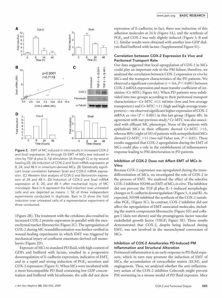
(Figure 2B). The treatment with the cytokines also resulted inincreased COX-2 protein expression in parallel with the mes-enchymal marker fibronectin (Figure 2C). The upregulation ofCOX-2 during MC transdifferentiation was further verified inwound-healing experiments in which EMT was triggered bymechanical injury of confluent omentum-derived cell mono-layers (Figure 2D).
Exposure of MCs to standard PD fluid, with high content ofGDPs and buffered with lactate, resulted in a progressivedownregulation of E-cadherin expression, indicative of EMT,and in a rapid and strong induction of PGE2 secretion andCOX-2 expression (Figure 3). When MCs were incubated witha more biocompatible PD fluid containing low GDP concen-tration and buffered with bicarbonate, the cells did not show
repression of E-cadherin; in fact, there was induction of thisadhesion molecules at 24 h (Figure 3A), and the synthesis ofPGE2 and COX-2 was only slightly induced (Figure 3, B andC). Similar results were obtained with another low-GDP dial-ysis fluid buffered with lactate (Supplemental Figure S1).
Correlation between COX-2 Expression Ex Vivo andPeritoneal Transport RateOur data suggested that local upregulation of COX-2 in MCscould play an important role in the PM failure; therefore, weanalyzed the correlation between COX-2 expression ex vivo byMCs and the transport characteristics of the PD patients. Weobserved a significant correlation (r � 0.6, P � 0.001) betweenCOX-2 mRNA expression and mass transfer coefficient of cre-atinine (Cr-MTC; Figure 4A). When PD patients were subdi-vided into two groups according to their peritoneal transportcharacteristics—Cr-MTC �11 ml/min (low and low-averagetransporters) and Cr-MTC �11 (high and high-average trans-porters)—we observed significant higher expression of COX-2mRNA ex vivo (P � 0.001) in this last group (Figure 4B). Inagreement with our previous study,4 Cr-MTC was also associ-ated with effluent MC phenotype. None of the patients withepithelioid MCs in their effluents showed Cr-MTC �11,whereas 80% (eight of 10) of patients with nonepithelioid MCsshowed Cr-MTC �11 (two-tail Fisher test, P � 0.01). Theseresults suggested that COX-2 upregulation during the EMT ofMCs could play a role in the establishment of inflammatoryresponse leading to PM deterioration in PD patients.
Inhibition of COX-2 Does not Affect EMT of MCs InVitroBecause COX-2 expression was upregulated during the trans-differentiation of MCs, we investigated the role of COX-2 inthe process of EMT. We analyzed the effect of the selectiveCOX-2 inhibitor NS398 on EMT of MCs in vitro. The inhibitordid not prevent the TGF-� plus IL-1–induced morphologicchanges or E-cadherin downregulation (Figure 5, A and B). Asexpected, NS398 inhibited the synthesis of the COX-2 metab-olite PGE2 (Figure 5C). In contrast, COX-2 inhibitor did notaffect the upregulation of EMT-associated molecules, includ-ing the matrix components fibronectin (Figure 5D) and colla-gen I (data not shown) and the proangiogenic factor vascularendothelial growth factor (VEGF; Figure 5E). These resultsdemonstrated that COX-2, despite being induced duringEMT, was not involved in the mesenchymal conversion ofMCs.
Inhibition of COX-2 Ameliorates PD-Induced PMInflammation and Structural AlterationPeritoneal inflammation is an early response to PD fluid expo-sure, which in turn may promote the induction of EMT ofMCs, the accumulation of extracellular matrix (ECM), andangiogenesis. Thus, we analyzed whether the anti-inflamma-tory action of the COX-2 inhibitor Celecoxib might preventPM worsening in a mouse model of PD fluid exposure. Mice
Figure 2. EMT of MC induced in vitro results in increased COX-2and Snail expression. (A through D) EMT of MCs was induced invitro by TGF-� plus IL-1� stimulation (A through C) or by woundhealing (D). (A) Induction of COX-2 and Snail mRNA expression at8, 24, and 48 h in omentum-derived MCs. (B) Statistically signifi-cant linear correlation between Snail and COX-2 mRNA expres-sion. (C) Western blot analysis of COX-2 and fibronectin expres-sion at 24 and 48 h. (D) Induction of COX-2 and Snail mRNAexpression at 8, 24, and 48 h after mechanical injury of MCmonolayer. Bars in A represent the fold induction over untreatedcells and are depicted as means � SE of three independentexperiments conducted in duplicate. Bars in D show the foldinduction over untreated cells of a representative experiment ofthree conducted.
BASIC RESEARCHwww.jasn.org
J Am Soc Nephrol 20: 582–592, 2009 COX-2 and Peritoneal Dialysis 585

were daily instilled via catheters with saline or standard lactate-based PD fluid and treated with Celecoxib or vehicle by oralroute for 5 wk. The histologic analysis of parietal peritoneumbiopsies showed that PD fluid exposure resulted in increasedinflammation, ECM accumulation, and thickness comparedwith saline-treated groups. The administration of the COX-2inhibitor significantly reduced all of these morphologicchanges. No significant differences were observed betweenmice that were instilled with saline and treated or not withCelecoxib (Figure 6, A and B). To test the effect of Celecoxib onPD fluid-induced angiogenesis, we stained blood vessels of pa-rietal peritoneum with an anti-CD31 antibody. PD fluid–in-stilled mice showed a significant increase of vessel numbercompared with saline-instilled groups. The administration ofthe COX-2 inhibitor to PD fluid–instilled mice resulted only ina slight tendency to reduce angiogenesis, but it did not reachstatistical significance (Figure 6, C and D). To analyze the func-tional relevance of the observed morphologic changes of theperitoneum, we performed a 90-min peritoneal equilibriumtest in each group of mice at the last day of treatment. As shownin Figure 6E, the volumes recovered from animals treated withPD were lower than those from saline-treated mice. A partialincrease of recovered volumes was obtained in PD fluid– ex-posed mice treated with Celecoxib.
To evaluate the effect of COX-2 inhibition on the early in-flammatory response of the peritoneum, we exposed mice toPD fluid and either treated them or not with Celecoxib for 15 d.The analysis of peritoneal influx of inflammatory cells showeda significant reduction of total cell counts in Celecoxib-treatedmice. The decrease of cell infiltration was more evident in mac-rophages (CD11b�), because the reduction of T cells (CD3�)did not reach statistical differences (Figure 6F). These results
Figure 3. Effects of standard and low-GDPsolutions on EMT of MCs and on COX-2 ex-pression in vitro. Omentum-derived MCswere incubated for 24 or 48 h with controlmedium (C), standard PD fluid containinghigh GDPs (HGDP), or solution containinglow GDPs (LGDP) diluted one half with cul-ture medium. Cells were also treated withTGF-� plus IL-1� (TGF). (A) Expression of E-cadherin was determined by Western blot.(B) The synthesis of PGE2 was measured inculture medium supernatant by ELISA, andresults are depicted as picograms per milli-gram of total cellular proteins. (C) The ex-pression of COX-2 was analyzed by Westernblot. Bars in A and C represent the fold in-duction of E-cadherin and COX-2 expressionover the control after normalization with tu-bulin expression. The experiment was re-peated at least three times, and a represen-tative experiment is shown.
Figure 4. COX-2 expression in PD effluent MCs and peritonealtransport rate. (A) Linear correlation between COX-2 mRNA ex-pression and Cr-MTC in the PD patient group. (B) Differences inCOX-2 mRNA expression in MCs obtained from patients with lowand low-average versus high and high-average (Cr-MTC �11versus �11 ml/min) peritoneal transport rates. Bars representmeans � SE.
BASIC RESEARCH www.jasn.org
586 Journal of the American Society of Nephrology J Am Soc Nephrol 20: 582–592, 2009

demonstrated that COX-2 inhibition ameliorated the deleteri-ous effects of PD fluid exposure of PM by reducing inflamma-tion and fibrosis, which in turn resulted in improved ultrafil-tration. The data also indicated that inflammation was anupstream event in peritoneal fibrosis and probably in vesselpermeability, and that inflammation-independent mediatorsoperated in new vessel formation.
Exposure to Low-GDP Solution Results in DecreasedPM Inflammation and Structural AlterationBecause the incubation of MCs with low-GDP solutions hadlittle impact on EMT and on the expression of COX-2, weanalyzed whether the exposure of mouse PM to biocompatiblefluids resulted in ameliorated PM worsening. Mice were dailyinstilled with standard PD fluid or with a solution containinglow GDP concentration. The inflammatory response was ana-lyzed at days 7 and 35, and PM thickness was determined at day35. The analysis of peritoneal influx of inflammatory cells dur-ing the time course showed that total cell counts increased inthe standard fluid–treated group but not in the group instilledwith low-GDP fluid. The number of infiltrating cells in thegroup treated with low-GDP fluid was significantly smallerthan in the standard fluid–treated group at day 35 (Figure 7A).The initial percentage of recruited T lymphocytes (CD3� cells)at day 7 was significantly reduced in the low-GDP fluid–treatedgroup, whereas at day 35, there was no difference amonggroups (Figure 7B). The recruitment of macrophages(CD11b� cells) was reduced in the group treated with low-GDP fluid at both time points, but the differences among
groups reached statistical significance only at day 35 (Figure7C). Finally, at day 35, the low-GDP fluid–treated groupshowed a significant decrease of submesothelial fibrosis com-pared with the standard fluid–treated group (Figure 7D).These findings demonstrated that mice exposed to PD fluidwith low content of GDPs showed less PM worsening.
DISCUSSION
The possible involvement of COX-2 in PM deterioration dur-ing PD has remained elusive. Prostaglandins are produced inthe peritoneal cavity of PD patients, mostly during peritonitis,and are implicated in increased peritoneal permeability tomacromolecules.16,17 Interestingly, whereas the inhibition ofprostaglandin synthesis, by intraperitoneal administration ofindomethacin, results in a decrease of hyperpermeability tomacromolecules during peritonitis, it has no effect on perme-ability in clinically stable and uncomplicated PD patients.20,21
These findings suggest that only the augmented prostaglan-dins, probably those produced by the inducible COX-2, areinvolved in peritoneal transport dysfunction.
Herein, we demonstrate that effluent MCs with nonepithe-lioid phenotype show increased expression of COX-2 and thatperitoneal transport rate correlates with COX-2 expressionlevels, suggesting that MCs that have undergone an EMT are animportant source of prostaglandins and thus contribute toperitoneal inflammatory response in PD patients. We alsoshow that effluent MC phenotypes and the expression of Snail
Figure 5. COX-2 inhibition does not affectin vitro EMT of MCs induced by TGF-� plusIL-1�. Omentum-derived MCs were treatedfor 24 or 48 h with TGF-� plus IL-1�, in thepresence of COX-2 inhibitor (NS398) or vehi-cle (DMSO). (A) Phase-contrast microscopyshows that COX-2 inhibitor does not preventnonepithelioid phenotype acquisition of MCat 48 h. (B) Western blot analysis shows thatCOX-2 inhibition does not prevent E-cad-herin downregulation. (C) COX-2 inhibitorprevents PGE2 upregulation. (D) COX-2 in-hibitor does not block the synthesis of fi-bronectin. (E) COX-2 inhibitor does not affectthe production of VEGF. Bars in C through Edepict values obtained by ELISA.
BASIC RESEARCHwww.jasn.org
J Am Soc Nephrol 20: 582–592, 2009 COX-2 and Peritoneal Dialysis 587

and COX-2 are associated with the PD fluids used in patients.Treatments with standard PD fluids have a greater impact onEMT of MCs and on the expression of Snail and COX-2 than
treatments with low-GDP solutions or with a com-bination of standard and icodextrin-based fluids.
That COX-2 upregulation in MCs paralleled thatof the EMT inducer Snail prompted us to analyzewhether it could play a role in the transdifferentia-tion process of these cells; however, in vitro experi-ments demonstrated that inhibition of COX-2 didnot prevent TGF-� plus IL-1–induced EMT of MCsand did not affect the expression of EMT-associatedmolecules. In contrast, it was recently shown that inMCs stimulated with high glucose, the inhibition ofCOX-2 prevented TGF-� and ECM upregulation.22
An explanation to these apparent discrepanciescould be that the inhibition of COX-2 in vitro pre-vents the indirect effect of high glucose on ECM pro-duction, via inhibition of TGF-� synthesis,22 but notthe direct effect of inflammatory cytokines on theEMT of MCs.
We demonstrate that Celecoxib treatment pre-serves the PM of PD fluid–instilled mice. Becauseinflammation is an early response of peritoneum toPD fluid–mediated insult, the beneficial effect ofCelecoxib could be a consequence of its anti-inflam-matory properties.9 It is known that tissue injuries inadult mammals caused by traumas or surgeries in-duce inflammation and scar formation as a conse-quence of an imperfect healing process.23 In con-trast, during the fetal period or soon after birth,injury-induced inflammatory response is weak andthe healing takes place without scar formation.23 Inaddition, it has been shown that mice deficient inmacrophages, neutrophils, and mast cells (PU.1 nullmice) are impaired to mount a standard inflamma-tory response and show scar-free healing.24 Thus, theparticipation of inflammation in tissue repair doesnot seem to be essential.25 It is tempting to speculatethat the inhibition of inflammation, by Celecoxibtreatment, contributes to the reduction of peritonealfibrosis in mice exposed to PD fluid. Our data indi-cate that PD fluid–induced angiogenesis is onlyslightly reduced in mice treated with Celecoxib. Thismay be explained by the fact that not only inflamma-tory cells but also injured MCs are able to producedifferent proangiogenic factors26,27; therefore,whereas Celecoxib treatment may block the produc-tion of angiogenic factors by inflammatory cells, ithas no effect on the synthesis of these factors by MCs.In addition, under stress conditions, resident perito-neal fibroblasts may account for the production ofangiogenic factors.28,29 The partial recovery of PMfunction in animals exposed to PD fluid and treatedwith Celecoxib might be explained by the inhibition
of PGE2 synthesis, which results in decreased vascular perme-ability.8 Our findings are further supported by the fact thatmice exposed to low-GDP fluid, which does not induce EMT
Figure 6. Inhibition of COX-2 ameliorates PD-induced PM worsening. (Aand B) Standard PD fluid exposure results in an increase of ECM deposition(A) and thickness (B) of the PM, and Celecoxib treatment significantly reducesthese effects, as measured by Masson’s trichrome staining. (C and D) CD31staining of peritoneal samples reveals that PD fluid exposure induces angio-genesis and that Celecoxib treatment does not significantly affect the in-crease in blood vessel formation. (E) Celecoxib treatment improves PMfunction (ultrafiltration) of PD fluid–instilled mice. (F) Celecoxib reduces peri-toneal inflammation of mice exposed to PD fluid. (A through E) Mice receiveda daily instillation of PD fluid or saline for 5 wk. After that period, sampleswere prepared and analyzed as described in the Concise Methods section.(F) For the study of the effect of Celecoxib on PD fluid–induced inflammation,mice were exposed to PD fluid for 15 d. At the end of the experiment, thedrained cells were counted and analyzed by flow cytometry. CD3 representsintraperitoneal T lymphocytes and CD11b myeloid cell population. Box plotsrepresent 25th and 75th percentiles and median, minimum, and maximumvalues.
BASIC RESEARCH www.jasn.org
588 Journal of the American Society of Nephrology J Am Soc Nephrol 20: 582–592, 2009

of MCs and COX-2 expression in vitro, show fewer infiltratinginflammatory cells and decreased fibrosis than mice treatedwith standard fluid, demonstrating the benefit of using bio-compatible PD solutions.
The results presented in this work indicate that inflamma-tion is an early response to PD fluid exposure, which in turnleads to peritoneal fibrosis, whereas angiogenesis does notseem to be entirely dependent on inflammatory reaction. Theinhibition of COX-2 or the prevention of its expression byusing biocompatible PD fluids may ameliorate the deleteriouseffect of PD on PM, by reducing inflammation, ECM accumu-lation, and thickness, which results in improved ultrafiltration.We are aware that COX-2 inhibitors cannot be easily used inclinical practice, at least for long-term treatments, because ma-jor concerns about the cardiovascular safety of these drugshave been raised.30 This issue is particularly important in pa-tients with ESRD, which have prevalent cardiovascular risk31;however, we believe that our results provide evidence about thefeasibility of considering inflammation as a potential thera-peutic target to ameliorate PD-related PM deterioration. Fur-ther studies of peritoneal inflammatory reaction will provide
more specific interventions with minimum adverseeffects on cardiovascular risk.
CONCISE METHODS
PatientsWe included 23 clinically stable PD patients: 12 men and
11 women. The causes of renal failure were nephrosclerosis
(n � 6), glomerulonephritis (n � 6), diabetes (n � 4),
chronic pyelonephritis (n � 2), polycystic kidney disease
(n � 1), unknown cause (n � 2), and other causes (n � 2).
The baseline characteristics of the patients and the differ-
ences between the subgroups according to the phenotype
of effluent MCs are shown in Table 1. The age of patients
ranged from 21 to 83 yr (mean 67.88 � 14.60). The mean
period PD was 9.47 � 7.44 mo (range 3 to 25). Most pa-
tients (21 of 23) received recombinant human erythropoi-
etin during this study. The duration of active peritoneal
inflammation was defined as the time (days) from eleva-
tion of cell count in PD effluent until normalization of cell
count. Three patients showed peritonitis, and two experi-
enced hemoperitoneum. All of the patients who experi-
enced peritonitis or hemoperitoneum drained nonepithe-
lioid MCs in the effluents. For these patients, at least 3 mo
passed after the resolution of the pathologic conditions
before sampling the effluent-derived MC. Peritoneal glu-
cose load was calculated by the sum of glucose contained in
each PD-fluid bag during the whole time on PD. urea-
MTC and Cr-MTC were measured using standard meth-
ods.32 Ultrafiltration capacity was defined by a peritoneal
exchange of 4 h using 3.86% glucose.33
At the moment of effluent-derived MC sampling, seven
patients were on continuous ambulatory peritoneal dialy-
sis (CAPD) and 16 were on APD. Fourteen patients (five on CAPD
and nine on APD) were treated with standard solution based on glu-
cose and lactate, containing high GDP concentration (Dianeal; Baxter
Healthcare Corp., Deerfield, IL). Seven of these patients (five on
CAPD and two on APD) received one long dwell per day (generally
overnight) with icodextrin-containing solution (Extraneal; Baxter).
Nine patients (two on CAPD and seven on APD) were treated with
low-GDP solutions buffered with lactate in five cases (Balance; Frese-
nius Medical Care, Bad Homburg, Germany) or bicarbonate in four
cases (BicaVera; Fresenius). The distribution of patients according to
PD techniques and PD fluids is shown in Supplemental Table S1.
This study abides by the Declaration of Helsinki and was approved
by the ethics committee of Hospital Universitario de la Princesa (Ma-
drid, Spain). Written consent was obtained from the PD patients in-
cluded in this study to use effluent samples. Oral informed consent
was obtained from omentum donors submitted to elective surgeries.
Culture of MCs and ReagentsMCs were obtained from PD effluents and from omentum samples
using the methods described previously.34 To standardize effluent
MC harvesting in the high-GDP group (n � 14), we obtained the cells
Figure 7. Exposure to low-GDP solution results in decreased PM worsening.Mice (11 per group) were daily instilled with 1.5 ml of low GDP solution (�)or standard PD fluid containing high GDPs (u), both composed of 4.25%glucose. (A) Total peritoneal influx of inflammatory cells was analyzed by flowcytometry at days 7 (five mice per group) and 35 (six mice per group). (B andC) The percentages of CD3� cells (T lymphocytes) and CD11b� cells (mac-rophages) were also determined by flow cytometry at days 7 and 35. (D)Peritoneal thickness was measured at day 35, and results are depicted aspercentage of fibrosis induction, 100% being the mean value of the high-GDP group. Box plots represent 25th and 75th percentiles and median,minimum, and maximum values.
BASIC RESEARCHwww.jasn.org
J Am Soc Nephrol 20: 582–592, 2009 COX-2 and Peritoneal Dialysis 589

from a long dwell (generally overnight) with a PD fluid containing
2.27% glucose (Dianeal; Baxter). Effluent MCs from the low-GDP
group were isolated from a long dwell with fluids containing 2.3%
glucose and buffered with lactate (n � 5; Balance; Fresenius) or with
bicarbonate (n � 4; BicaVera; Fresenius). From each patient, at least
three independent effluent MC samples were obtained in a period of 2
mo, which showed phenotype stability, and one representative MC
culture per patient was used in the study. All cells were cultured in
Earle’s M199 medium, supplemented with 20% FCS, 50 U/ml peni-
cillin, 50 �g/ml streptomycin, and 2% Biogro-2 (Biologic Industries,
Ashrat, Israel). The purity of effluent and omentum-derived MC cul-
tures was determined by the expression of standard mesothelial mark-
ers: Intercellular adhesion molecule 1, cytokeratins, and calretinin.
These MC cultures were negative for von Willebrand factor excluding
endothelial cell contamination.34
To induce EMT in vitro, we treated omentum-derived MC for 24
or 48 h with a combination of human recombinant TGF-� (0.5 ng/
ml) and IL-1� (2 ng/ml; R&D Systems, Minneapolis, MN), which has
been proved to be a good model of EMT in vitro.4,6 When indicated,
the selective COX-2 inhibitor NS398 (Alexis Biochemicals, San Diego,
CA) was used at a final concentration of 20 �M. Wound-healing
studies were carried out as described previously by performing a me-
chanical injury with a 1.5-mm cell scraper.6 To analyze the effect of
GDPs on EMT and COX-2 expression, omentum-derived MC were
incubated for 24 or 48 h with standard PD fluid composed of 4.25%
glucose and buffered with lactate (Stay Safe; Fresenius) or low-GDP
solutions composed of 4.25% glucose and buffered with lactate or
bicarbonate (Balance or BicaVera; Fresenius) diluted one half with
culture medium
RNA Extraction and Reverse Transcription–PCRTotal RNA was extracted by using RNAwiz (Ambion, Austin, TX),
and the complementary DNA was obtained from 1 �g of total RNA by
reverse transcription following the manufacturer’s recommendation
(Applied Biosystems, Cheshire, UK). COX-2 mRNA was amplified
with a LightCycler using a SYBR Green Kit (Roche Diagnostics
GmbH, Mannheim, Germany) and the specific primer set 5�-
TTCAAATGAGATTGTGGGAAAATTGCT-3� and 5�-AGATCA-
TCTCTGCCTGAGTATCTT-3� as described previously.35 Snail
mRNA was amplified using similar conditions and the following
primer set: 5�-CACATCCTTCTCACTGCCATG-3� and 5�-GCAT-
CTAAACTCTAGTCTGC-3�. �-Actin was amplified using commer-
cial primers (Clontech, Palo Alto, CA). Annealing temperatures for
the COX-2, Snail, and �-actin PCR were 56, 53, and 60°C, respec-
tively. All samples were normalized with respect to the value obtained
for �-actin.
Western Blot Analysis and Enzyme-LinkedImmunoassaysFor Western blotting, MC cultures were lysed in a buffer (1% sodium
deoxycholate and 0.1% SDS), and total protein was quantified using a
total protein assay kit (Pierce, Cambridge, MA). MC proteins (50 �g)
were resolved in 8 to 10% SDS-polyacrylamide gels. Proteins were
transferred onto nitrocellulose membranes, which were blocked with
fat-free milk and then incubated with specific antibodies against
COX-2, fibronectin, collagen I, E-cadherin, and �-tubulin (Becton
Dickinson, Franklin Lakes, NJ). Membranes were incubated with goat
anti-mouse IgG antibody conjugated with peroxidase (Pharmingen,
San Diego, CA) and developed with enhanced chemiluminescence
detection kit (Amersham Biosciences, Freiburg, Germany). Blot im-
ages were acquired with an LAS-1000 charged coupled device camera
(Fujifilm, Cedex, France).
For the detection of VEGF and PGE2 in culture supernatants, the
media of MCs cultured under the various conditions were replaced,
and 18 h later supernatants were collected and stored at �80°C until
their analysis. The concentrations of VEGF and PGE2 in supernatants
were assessed by a standard ELISA kit (R&D Systems). The produc-
tion of fibronectin under various conditions was measured in cell
lysates by ELISA as recommended by the manufacturer (Biomedical
Technologies, Stoughton, MA)
Peritoneal Exposure Model in MiceA total of 112 female C57BL/6 mice with age between 12 and 16 wk
were used in this study (Harlan Interfauna Iberica, Barcelona, Spain).
The experimental protocol was in accordance with the National In-
stitutes of Health Guide for Care and Use of Laboratory Animals and
was approved by the Animal Ethics Committee of the Unidad de
Cirurgía Experimental of Hospital Universitario La Paz.
A customized vascular access port (Access Technologies, Skokie,
IL) was implanted subcutaneously, and the catheter end was intro-
duced into the peritoneal cavity by a chirurgic procedure. During the
week of recovery, animals received daily 0.2 ml of saline solution
containing 1 U/ml heparin. Thereafter, mice were subdivided into
different groups. For histologic and functional analysis, 70 mice were
used: 20 were daily instilled with 1.5 ml of standard PD fluid com-
posed of 4.25% glucose and buffered with lactate (Stay Safe; Frese-
nius) and orally treated with vehicle (PDF group), 20 were instilled
with PD fluid and orally treated with Celecoxib (2 �g/g body wt; PDF
� Cel group), 15 were daily instilled with 1.5 ml of physiologic saline
and orally treated with vehicle (Sal group), and 15 were instilled with
physiologic saline and orally treated with Celecoxib (Sal � Cel
group). Mice were treated with different conditions for 5 wk. The
whole study was performed in four separate experiments. In one ex-
periment, a peritoneal equilibrium test was performed during the last
day of treatment with the various conditions. Mice were instilled with
1.5 ml of PD solution; 90 min later, animals were killed and the total
peritoneal volumes were collected. The peritoneal net volume was
determined by the difference between instilled and recovered vol-
umes. For histologic analyses, specimens of the parietal peritoneum
were collected. Sample were fixed in neutral-buffered formalin, em-
bedded in paraffin, cut into 5-�m sections,and stained either with
hematoxylin and eosin or Masson’s trichrome. The thickness of sub-
mesothelial tissue was determined by blinded microscope analysis
using a metric ocular. The presence of blood vessels was determined
by immunostaining with an anti-CD31 mAb (Becton Dickinson).
Blood vessels of five fields per mouse sample were counted, and the
median was used for statistical analysis.
To analyze the effect of Celecoxib in early inflammatory response
to PD fluid exposure, we used 20 mice. Mice (10 per group) were daily
instilled with 1.5 ml of standard PD solution (Stay Safe; Fresenius)
BASIC RESEARCH www.jasn.org
590 Journal of the American Society of Nephrology J Am Soc Nephrol 20: 582–592, 2009

and orally treated with Celecoxib or vehicle alone during 15 d. Then,
the peritoneal influx of inflammatory cells was analyzed by flow cy-
tometry using anti-CD11b and anti-CD3 mAb (Becton Dickinson).
During these experiments, the percentages of dropouts were sim-
ilar in all of the groups (approximately 40%), the main cause being
catheter damage produced by the mouse itself and not as a conse-
quence of treatments. There were no significant differences in weight
gain among the various groups.
To compare the effects of PD fluids with different contents of
GDPs on PM inflammation and fibrosis, we used 22 mice. Mice (11
per group) were daily instilled with 1.5 ml of standard PD fluid com-
posed of 4.25% glucose and lactate (Stay Safe; Fresenius) or with
low-GDP solution composed of 4.25% glucose and bicarbonate (Bi-
caVera; Fresenius). The peritoneal influx of inflammatory cells was
analyzed by flow cytometry at days 7 (five mice per group) and 35 (six
mice per group). Peritoneal thickness was also determined at day 35.
In this experiment, no dropout was observed as a result of technical
improvement of surgical implantation of catheters.
Statistical AnalysisResults are given as means � SD (Table 1). Figures 1, B through F, 2A,
and 4B represent means � SE. Comparisons between data groups
were performed using the nonparametric Mann-Whitney rank sum U
test. Linear correlation was determined by Spearman regression anal-
ysis (Figures 1D, 2B, and 4A). �2 and two-tailed Fisher exact tests were
used to compare qualitative variables. P � 0.05 was considered statis-
tically significant. We used SPSS 14.5 (Chicago, IL) and GraphPad
Prism 4.0 (La Jolla, CA).
ACKNOWLEDGMENTS
This work was supported by grants SAF2007-61201 and PET2006-
0256 from Ministerio de Educacion y Ciencia to M.L.-C. and FIS PI
06/0098 and RETICS 06/0016 from Fondo Investigaciones Sanitarias
to R.S. This work was also partially supported by Fresenius Medical
Care and Gambro Europe.
We thank the nurses from the peritoneal dialysis units for help in
recompilation of peritoneal effluents and omental samples. We also
thank Javier Benito de la Víbora, DVM, and Carlota Largo Aramburu,
DVM, PhD, for the assistance with mouse care.
DISCLOSURESNone.
REFERENCES
1. Krediet RT: The peritoneal membrane in chronic peritoneal dialysis.Kidney Int 55: 341–356, 1999
2. Aroeira LS, Aguilera A, Sanchez-Tomero JA, Bajo MA, del Peso G,Jimenez-Heffernan JA, Selgas R, Lopez-Cabrera M: Epithelial to mes-enchymal transition and peritoneal membrane failure in peritonealdialysis patients: Pathologic significance and potential therapeuticinterventions. J Am Soc Nephrol 18: 2004–2013, 2007
3. Williams JD, Craig KJ, Topley N, Von Ruhland C, Fallon M, NewmanGR, Mackenzie RK, Williams GT: Morphologic changes in the perito-neal membrane of patients with renal disease. J Am Soc Nephrol 13:470–479, 2002
4. Aroeira LS, Aguilera A, Selgas R, Ramirez-Huesca M, Perez-LozanoML, Cirugeda A, Bajo MA, del Peso G, Sanchez-Tomero JA, Jimenez-Heffernan JA, Lopez-Cabrera M: Mesenchymal conversion of me-sothelial cells as a mechanism responsible for high solute transportrate in peritoneal dialysis: Role of vascular endothelial growth factor.Am J Kidney Dis 46: 938–948, 2005
5. Jimenez-Heffernan JA, Aguilera A, Aroeira LS, Lara-Pezzi E, Bajo MA,del Peso G, Ramirez M, Gamallo C, Sanchez-Tomero JA, Alvarez V,Lopez-Cabrera M, Selgas R: Immunohistochemical characterization offibroblast subpopulations in normal peritoneal tissue and in peritonealdialysis-induced fibrosis. Virchows Arch 444: 247–256, 2004
6. Yanez-Mo M, Lara-Pezzi E, Selgas R, Ramirez-Huesca M, Dominguez-Jimenez C, Jimenez-Heffernan JA, Aguilera A, Sanchez-Tomero JA,Bajo MA, Alvarez V, Castro MA, del Peso G, Cirujeda A, Gamallo C,Sanchez-Madrid F, Lopez-Cabrera M: Peritoneal dialysis and epitheli-al-to-mesenchymal transition of mesothelial cells. N Engl J Med 348:403–413, 2003
7. Margetts PJ, Bonniaud P, Liu L, Hoff CM, Holmes CJ, West-Mays JA,Kelly MM: Transient overexpression of TGF-beta1 induces epithelialmesenchymal transition in the rodent peritoneum. J Am Soc Nephrol16: 425–436, 2005
8. Yang AH, Chen JY, Lin JK: Myofibroblastic conversion of mesothelialcells. Kidney Int 63: 1530–1539, 2003
9. Turini ME, DuBois RN: Cyclooxygenase-2: A therapeutic target. AnnuRev Med 53: 35–57, 2002
10. Gately S, Li WW: Multiple roles of COX-2 in tumor angiogenesis: Atarget for antiangiogenic therapy. Semin Oncol 31: 2–11, 2004
11. Khanapure SP, Garvey DS, Janero DR, Letts LG: Eicosanoids in inflam-mation: Biosynthesis, pharmacology, and therapeutic frontiers. CurrTop Med Chem 7: 311–340, 2007
12. Bonner JC, Rice AB, Ingram JL, Moomaw CR, Nyska A, Bradbury A,Sessoms AR, Chulada PC, Morgan DL, Zeldin DC, Langenbach R:Susceptibility of cyclooxygenase-2-deficient mice to pulmonary fibro-genesis. Am J Pathol 161: 459–470, 2002
13. Cheng HF, Wang CJ, Moeckel GW, Zhang MZ, McKanna JA, HarrisRC: Cyclooxygenase-2 inhibitor blocks expression of mediators ofrenal injury in a model of diabetes and hypertension. Kidney Int 62:929–939, 2002
14. Wilborn J, Crofford LJ, Burdick MD, Kunkel SL, Strieter RM, Peters-Golden M: Cultured lung fibroblasts isolated from patients with idio-pathic pulmonary fibrosis have a diminished capacity to synthesizeprostaglandin E2 and to express cyclooxygenase-2. J Clin Invest 95:1861–1868, 1995
15. Yamamoto H, Kondo M, Nakamori S, Nagano H, Wakasa K, Sugita Y,Chang-De J, Kobayashi S, Damdinsuren B, Dono K, Umeshita K,Sekimoto M, Sakon M, Matsuura N, Monden M: JTE-522, a cycloox-ygenase-2 inhibitor, is an effective chemopreventive agent against ratexperimental liver fibrosis. Gastroenterology 125: 556–571, 2003
16. Steinhauer HB, Schollmeyer P: Prostaglandin-mediated loss of pro-teins during peritonitis in continuous ambulatory peritoneal dialysis.Kidney Int 29: 584–590, 1986
17. Zemel D, Koomen GC, Hart AA, ten Berge IJ, Struijk DG, Krediet RT:Relationship of TNF-alpha, interleukin-6, and prostaglandins to peri-toneal permeability for macromolecules during longitudinal follow-upof peritonitis in continuous ambulatory peritoneal dialysis. J Lab ClinMed 122: 686–696, 1993
18. Shanmugam N, Kim YS, Lanting L, Natarajan R: Regulation of cyclo-oxygenase-2 expression in monocytes by ligation of the receptor foradvanced glycation end products. J Biol Chem 278: 34834–34844,2003
19. Sitter T, Haslinger B, Mandl S, Fricke H, Held E, Sellmayer A: Highglucose increases prostaglandin E2 synthesis in human peritoneal
BASIC RESEARCHwww.jasn.org
J Am Soc Nephrol 20: 582–592, 2009 COX-2 and Peritoneal Dialysis 591

mesothelial cells: role of hyperosmolarity. J Am Soc Nephrol 9: 2005–2012, 1998
20. Douma CE, de Waart DR, Zemel D, Struijk DG, Krediet RT: Prosta-glandin inhibition by intraperitoneal indomethacin has no effect onperitoneal permeability during stable CAPD. Nephrol Dial Transplant16: 803–808, 2001
21. Zemel D, Struijk DG, Dinkla C, Stolk LM, ten Berge IJ, Krediet RT:Effects of intraperitoneal cyclooxygenase inhibition on inflammatorymediators in dialysate and peritoneal membrane characteristics duringperitonitis in continuous ambulatory peritoneal dialysis. J Lab ClinMed 126: 204–215, 1995
22. Liu H, Peng Y, Liu F, Li J, Chen X, Liu Y, Zhang H: A selectivecyclooxygenase-2 inhibitor decreases transforming growth factor-be-ta1 synthesis and matrix production in human peritoneal mesothelialcells. Cell Biol Int 31: 508–515, 2007
23. Ferguson MW, O’Kane S: Scar-free healing: From embryonic mecha-nisms to adult therapeutic intervention. Philos Trans R Soc Lond B BiolSci 359: 839–850, 2004
24. Martin P, D’Souza D, Martin J, Grose R, Cooper L, Maki R, McKercherSR: Wound healing in the PU.1 null mouse: Tissue repair is not de-pendent on inflammatory cells. Curr Biol 13: 1122–1128, 2003
25. Martin P, Leibovich SJ: Inflammatory cells during wound repair: Thegood, the bad and the ugly. Trends Cell Biol 15: 599–607, 2005
26. Lai KN, Lai KB, Lam CW, Chan TM, Li FK, Leung JC: Changes ofcytokine profiles during peritonitis in patients on continuous ambula-tory peritoneal dialysis. Am J Kidney Dis 35: 644–652, 2000
27. Lai KN, Tang SC, Leung JC: Mediators of inflammation and fibrosis.Perit Dial Int 27[Suppl 2]: S65–S71, 2007
28. Giatromanolaki A, Kotsiou S, Koukourakis MI, Sivridis E: Angiogenicfactor expression in hepatic cirrhosis. Mediators Inflamm 2007: 67187,2007
29. Tunyogi-Csapo M, Koreny T, Vermes C, Galante JO, Jacobs JJ, GlantTT: Role of fibroblasts and fibroblast-derived growth factors inperiprosthetic angiogenesis. J Orthop Res 25: 1378–1388, 2007
30. Harris RC, Breyer MD: Update on cyclooxygenase-2 inhibitors. ClinJ Am Soc Nephrol 1: 236–245, 2006
31. Garcia-Lopez E, Carrero JJ, Suliman ME, Lindholm B, Stenvinkel P:Risk factors for cardiovascular disease in patients undergoing perito-neal dialysis. Perit Dial Int 27[Suppl 2]: S205–S209, 2007
32. Selgas R, Fernandez-Reyes MJ, Bosque E, Bajo MA, Borrego F, Jime-nez C, Del Peso G, De Alvaro F: Functional longevity of the humanperitoneum: How long is continuous peritoneal dialysis possible? Re-sults of a prospective medium long-term study. Am J Kidney Dis 23:64–73, 1994
33. Ho-dac-Pannekeet MM, Atasever B, Struijk DG, Krediet RT: Analysis ofultrafiltration failure in peritoneal dialysis patients by means of stan-dard peritoneal permeability analysis. Perit Dial Int 17: 144–150, 1997
34. Lopez-Cabrera M, Aguilera A, Aroeira LS, Ramirez-Huesca M, Perez-Lozano ML, Jimenez-Heffernan JA, Bajo MA, del Peso G, Sanchez-Tomero JA, Selgas R: Ex vivo analysis of dialysis effluent-derivedmesothelial cells as an approach to unveiling the mechanism of peri-toneal membrane failure. Perit Dial Int 26: 26–34, 2006
35. Lara-Pezzi E, Gomez-Gaviro MV, Galvez BG, Mira E, Iniguez MA,Fresno M, Martinez AC, Arroyo AG, Lopez-Cabrera M: The hepatitis Bvirus X protein promotes tumor cell invasion by inducing membrane-type matrix metalloproteinase-1 and cyclooxygenase-2 expression.J Clin Invest 110: 1831–1838, 2002
Supplemental information for this article is available online at http://www.jasn.org/.
BASIC RESEARCH www.jasn.org
592 Journal of the American Society of Nephrology J Am Soc Nephrol 20: 582–592, 2009