40. Dialysis | Infection Prevention for Specialty Care Populations
n engl j med
348;5
www.nejm.org january
30, 2003
The
new england journal
of
medicine
403
original article
Peritoneal Dialysis and Epithelial-to-Mesenchymal Transition of Mesothelial Cells
María Yáñez-Mó, Ph.D., Enrique Lara-Pezzi, Ph.D., Rafael Selgas, Ph.D., M.D., Marta Ramírez-Huesca, B.S., Carmen Domínguez-Jiménez, Ph.D.,
José A. Jiménez-Heffernan, M.D., Abelardo Aguilera, M.D., José A. Sánchez-Tomero, Ph.D., M.D., M. Auxiliadora Bajo, Ph.D., M.D.,
Vincente Álvarez, Ph.D., M.D., M. Angeles Castro, Ph.D., Gloria del Peso, Ph.D., M.D., Antonio Cirujeda, M.D., Carlos Gamallo, Ph.D., M.D.,
Francisco Sánchez-Madrid, Ph.D., and Manuel López-Cabrera, Ph.D.
From the Servicio de Inmunología (M.Y.-M.,M.R.-H., C.D.-J., F.S.-M.), Biología Molecu-lar (E.L.-P., C.G., M.L.-C.), and Nefrología(R.S., A.A., J.A.S.-T., V.A., A.C.), HospitalUniversitario de la Princesa, UniversidadAutónoma de Madrid; the Servicio de Ne-frología, Hospital Universitario La Paz(M.A.B., M.A.C., G.P.); and the InstitutoReina Sofía de Investigaciones Nefrológi-cas (M.Y.-M., E.L.-P., R.S., M.R.-H., C.D.-J.,J.A.J.-H., A.A., J.A.S.-T., M.A.B., V.A., M.A.C.,G.P., A.C., C.G., F.S.-M., M.L.-C.) — all inMadrid; the Servicio de Anatomía Patológi-ca, Hospital Universitario de Guadalajara,Guadalajara, Spain (J.A.J.-H.). Address re-print requests to Dr. López-Cabrera at theDepartamento de Biología Molecular, Hos-pital Universitario de la Princesa, C/Diegode León no. 62, 28006 Madrid, Spain, or [email protected].
Drs. Yáñez-Mó and Lara-Pezzi contrib-uted equally to the article.
N Engl J Med 2003;348:403-13.
Copyright © 2003 Massachusetts Medical Society.
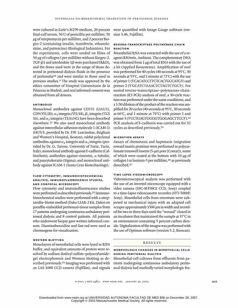
background
During continuous ambulatory peritoneal dialysis, the peritoneum is exposed to bioin-compatible dialysis fluids that cause denudation of mesothelial cells and, ultimately,tissue fibrosis and failure of ultrafiltration. However, the mechanism of this process hasyet to be elucidated.
methods
Mesothelial cells isolated from effluents in dialysis fluid from patients undergoingcontinuous ambulatory peritoneal dialysis were phenotypically characterized by flowcytometry, confocal immunofluorescence, Western blotting, and reverse-transcriptasepolymerase chain reaction. These cells were compared with mesothelial cells fromomentum and treated with various stimuli in vitro to mimic the transdifferentiation ob-served during continuous ambulatory peritoneal dialysis. Results were confirmed invivo by immunohistochemical analysis performed on peritoneal-biopsy specimens.
results
Soon after dialysis is initiated, peritoneal mesothelial cells undergo a transition froman epithelial phenotype to a mesenchymal phenotype with a progressive loss of epithe-lial morphology and a decrease in the expression of cytokeratins and E-cadherin throughan induction of the transcriptional repressor
snail.
Mesothelial cells also acquire a mi-gratory phenotype with the up-regulation of expression of
a
2
integrin. In vitro analysespoint to wound repair and profibrotic and inflammatory cytokines as factors that initiatemesothelial transdifferentiation. Immunohistochemical studies of peritoneal-biopsyspecimens from patients undergoing continuous ambulatory peritoneal dialysis dem-onstrate the expression of the mesothelial markers intercellular adhesion molecule1 and cytokeratins in fibroblast-like cells entrapped in the stroma, suggesting that thesecells stemmed from local conversion of mesothelial cells.
conclusions
Our results suggest that mesothelial cells have an active role in the structural and func-tional alteration of the peritoneum during peritoneal dialysis. The findings suggest po-tential targets for the design of new dialysis solutions and markers for the monitor-ing of patients.
abstract
Copyright © 2003 Massachusetts Medical Society. All rights reserved. Downloaded from www.nejm.org at UNIVERSIDAD AUTONOMA FACULTAD DE MED BIB on December 28, 2007 .
n engl j med
348;5
www.nejm.org january 30,
2003
The
new england journal
of
medicine
404
ontinuous ambulatory perito-
neal dialysis is an alternative to hemodial-ysis for the treatment of end-stage renal
disease.
1
The peritoneal membrane is lined with amonolayer of mesothelial cells that have some char-acteristics of epithelial cells, act as a permeabilitybarrier, and secrete various substances involved inthe regulation of peritoneal permeability and localhost defense.
1,2
Unfortunately, long-term exposureto the hyperosmotic, hyperglycemic, and acidic so-lutions used in dialysis often causes low-grade,chronic inflammation of and injury to the peritone-um, which progressively becomes denuded of mes-othelial cells and undergoes fibrosis.
1
Such struc-tural alterations are considered to be the principalcause of failure of ultrafiltration, which affects upto 20 percent of patients undergoing continuousambulatory peritoneal dialysis.
3
This functional de-cline of the peritoneum may be accelerated by re-current or severe episodes of peritonitis or hemo-peritoneum.
3,4
The pathophysiology of peritoneal impairmentduring long-term continuous ambulatory peritonealdialysis is not well understood. Peritoneal mesen-chymal stem cells entrapped in the stroma have his-torically been considered to be the primary cells in-volved in the development of peritoneal fibrosis.
5
However, a possible direct involvement of meso-thelial cells in this phenomenon has not been ex-amined. In this context, cultured mesothelial cellshave the capacity to change their morphologic fea-tures and produce extracellular-matrix componentsin response to a variety of stimuli.
6-12
In addition,treatment of mesothelial cells in vitro with medi-ums that have a high glucose concentration or withinflammatory cytokines induces the expression oftransforming growth factor
b
(TGF-
b
)
13
and de-creases the expression of E-cadherin.
14
The rele-vance of the profibrotic growth factor TGF-
b
15
inthe failure of ultrafiltration induced by continuousambulatory peritoneal dialysis was recently under-scored in a rat model in which the
TGF-
b
gene wastransduced to the peritoneum, where it was associ-ated with a decrease in peritoneal function.
16
In the present study, we demonstrate in vivo andex vivo that mesothelial cells undergo a transitionfrom an epithelial phenotype to a mesenchymalphenotype — a transition also called transdifferen-tiation — when they are subjected to peritoneal di-alysis. Transdifferentiation is a complex and gener-ally reversible process that starts with the disruptionof intercellular junctions and loss of the apical–
basolateral polarity typical of epithelial cells, withthe cells then transformed into fibroblast-likecells with pseudopodial protrusions and increasedmigratory, invasive, and fibrogenic features.
17
Al-though transdifferentiation can be induced in mostcultured epithelial cells with a wide variety of treat-ments, this process occurs in vivo only duringembryonic development and in some pathologicprocesses such as wound healing and tumor pro-gression.
17,18
The intercellular adhesion moleculeE-cadherin appears to have a central role in the con-trol of the epithelial-to-mesenchymal transition,since the loss of E-cadherin expression or functioncorrelates with the ability of epithelial cells to adopta mesenchymal migratory and invasive pheno-type.
19,20
The transcription factor
snail
is a strongrepressor of E-cadherin transcription and an induc-er of transdifferentiation.
21-23
Thus, phenotypicchanges of the mesothelial cells during continuousambulatory peritoneal dialysis may be directly relat-ed to the failure of peritoneal membrane function.
patients and cells
Human mesothelial cells from effluent (mean [±SE]number of cells per bag, 25,569±2971) were ob-tained by centrifugation of dialysis fluid taken ran-domly from 54 clinically stable patients undergoingnocturnal exchanges with dialysis solutions con-taining 2.27 percent glucose and 1.25 to 1.75 mmolof calcium per liter. After 10 to 15 days, culturesreached confluence and were split (in a ratio of 1:2)two to three times. The morphologic features ofcells in confluent cultures were compared and re-mained stable during the two to three cell passages.Eighty-five percent of the cultures were obtainedbefore a first episode of peritonitis occurred. Of the116 effluent cultures evaluated, 62 had cobblestonemorphology, 28 contained transitional mesothelialcells, 20 contained fibroblast-like mesothelial cells,and 6 contained a mixed population of cells.
Omental mesothelial cells were obtained by di-gestion of samples of omentum from 30 patientswho were not undergoing continuous ambulatoryperitoneal dialysis but were undergoing unrelatedabdominal surgery; the samples were digested with0.05 percent trypsin and 0.02 percent EDTA. Omen-tal fibroblasts were obtained from three differentsamples of omentum by extensive treatment withtrypsin after the removal of mesothelial cells (three20-minute rounds of exposure to trypsin). All cells
c
methods
Copyright © 2003 Massachusetts Medical Society. All rights reserved. Downloaded from www.nejm.org at UNIVERSIDAD AUTONOMA FACULTAD DE MED BIB on December 28, 2007 .
n engl j med
348;5
www.nejm.org january
30, 2003
epithelial-to-mesenchymal transition in peritoneal dialysis
405
were cultured in Earle’s M199 medium, 20 percentfetal-calf serum, 50 U of penicillin per milliliter, 50μg of streptomycin per milliliter, and 2 percent Bio-gro-2 (containing insulin, transferrin, ethanola-mine, and putrescine) (Biological Industries). Forthe experiments, cells were seeded on films of50 μg of collagen I per milliliter without Biogro-2.TGF-
b
1 and interleukin-1
b
were purchased (R&D),and the doses used were in the range of those de-tected in peritoneal-dialysis fluids in the presenceof peritonitis
24
and were similar to those used inprevious studies.
9
The study was approved by theethics committee of Hospital Universitario de laPrincesa in Madrid, and oral informed consent wasobtained from all donors.
antibodies
Monoclonal antibodies against CD151 (LIA1/1),CD9 (VJ1/20),
a
3
integrin (VJ1/18),
b
1
integrin (TS2/16), and
a
2
integrin (TEA1/41) have been describedelsewhere.
25
We also used monoclonal antibodyagainst intercellular adhesion molecule 1 (ICAM-1)(HU5/3, provided by Dr. F.W. Luscinskas, Brighamand Women’s Hospital, Boston); rabbit polyclonalantibodies against
a
2
integrin and
a
3
integrin (pro-vided by Dr. G. Tarone, University of Turin, Turin,Italy); monoclonal antibody against E-cadherin (Cal-biochem); antibodies against vimentin,
a
tubulin,and pancytokeratin (Sigma); and monoclonal anti-body against ICAM-1 (Santa Cruz Biotechnology).
flow cytometry, immunohistochemical
analysis, immunofluorescence studies,
and confocal microscopy
Flow cytometry and immunofluorescence studieswere performed as described previously.
25
Immuno-histochemical studies were performed with a strep-tavidin–biotin method (Dako LSAB-2 Kit, Dako) onparaffin-embedded peritoneal-tissue samples from17 patients undergoing continuous ambulatory peri-toneal dialysis and 8 control patients. All patientswho underwent biopsy gave written informed con-sent. Diaminobenzidine and fast red were used aschromogens for visualization.
western blotting
Monolayers of mesothelial cells were lysed in RIPAbuffer, and equivalent amounts of protein were re-solved by sodium dodecyl sulfate–polyacrylamide-gel electrophoresis and Western blotting as de-scribed previously.
25
Imaging was performed withan LAS-1000 CCD camera (Fujifilm), and signals
were quantified with Image Gauge software (ver-sion 3.46, Fujifilm).
reverse-transcriptase polymerase chain
reaction
Mesothelial RNA was extracted with the use of a re-agent (RNAwiz, Ambion). The complementary DNAwas obtained from 1 μg of total RNA with the use ofa kit (Applied Biosystems). Amplification of
snail
was performed for 40 cycles (40 seconds at 95°C, 30seconds at 53°C, and 1 minute at 72°C) with the useof primer 1 (5'CACATCCTTCTCACTGCCATG3') andprimer 2 (5'GCATCTAAACTCTAGTCTGC3'). Fornested reverse-transcriptase–polymerase-chain-reaction (RT-PCR) analysis of
snail,
a 30-cycle reac-tion was performed under the same conditions, anda 1:50 dilution of the product of the reaction was am-plified for 20 cycles (40 seconds at 95°C, 30 secondsat 60°C, and 1 minute at 70°C) with primer 1 andprimer 3 (5'CCTGAGTGGGGTGGGAGCTTCC3').
22
PCR analysis of E-cadherin was carried out for 32cycles as described previously.
22
migration assays
Assays of chemotaxis and haptotaxis (migrationtoward matrix proteins) were performed in polycar-bonate transwell inserts (5-μm pore [Costar]), someof which were coated at the bottom with 10 μg ofcollagen I or laminin-5 per milliliter,
26
as previouslydescribed.
27
time-lapse videomicroscopy
Videomicroscopical analysis was performed withthe use of an inverted microscope equipped with avideo camera (SSC-M350CE CCD, Sony) coupledto a time-lapse videocassette recorder (SVT-5000P,Sony). Mesothelial cells from omentum were sub-jected to mechanical injury with an adapted cellscraper approximately 1500 μm in width and record-ed for two to three days until the “wound” closed inan incubator that maintained the sample at 37°C inan environment containing 5 percent carbon diox-ide. Digitalization of the images was performed withthe use of Optimas software (version 5.2, Bioscan).
morphologic changes in mesothelial cells
during peritoneal dialysis
Mesothelial-cell cultures from effluents from pa-tients undergoing continuous ambulatory perito-neal dialysis had markedly varied morphologic fea-
results
Copyright © 2003 Massachusetts Medical Society. All rights reserved. Downloaded from www.nejm.org at UNIVERSIDAD AUTONOMA FACULTAD DE MED BIB on December 28, 2007 .
n engl j med
348;5
www.nejm.org january 30,
2003
The
new england journal
of
medicine
406
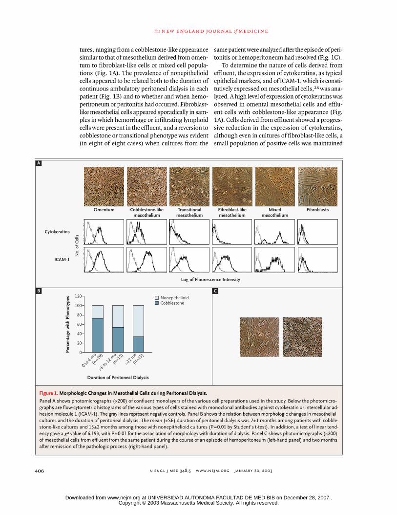
tures, ranging from a cobblestone-like appearancesimilar to that of mesothelium derived from omen-tum to fibroblast-like cells or mixed cell popula-tions (Fig. 1A). The prevalence of nonepithelioidcells appeared to be related both to the duration ofcontinuous ambulatory peritoneal dialysis in eachpatient (Fig. 1B) and to whether and when hemo-peritoneum or peritonitis had occurred. Fibroblast-like mesothelial cells appeared sporadically in sam-ples in which hemorrhage or infiltrating lymphoidcells were present in the effluent, and a reversion tocobblestone or transitional phenotype was evident(in eight of eight cases) when cultures from the
same patient were analyzed after the episode of peri-tonitis or hemoperitoneum had resolved (Fig. 1C).
To determine the nature of cells derived fromeffluent, the expression of cytokeratins, as typicalepithelial markers, and of ICAM-1, which is consti-tutively expressed on mesothelial cells,
28
was ana-lyzed. A high level of expression of cytokeratins wasobserved in omental mesothelial cells and efflu-ent cells with cobblestone-like appearance (Fig.1A). Cells derived from effluent showed a progres-sive reduction in the expression of cytokeratins,although even in cultures of fibroblast-like cells, asmall population of positive cells was maintained
Figure 1. Morphologic Changes in Mesothelial Cells during Peritoneal Dialysis.
Panel A shows photomicrographs (¬200) of confluent monolayers of the various cell preparations used in the study. Below the photomicro-graphs are flow-cytometric histograms of the various types of cells stained with monoclonal antibodies against cytokeratin or intercellular ad-hesion molecule 1 (ICAM-1). The gray lines represent negative controls. Panel B shows the relation between morphologic changes in mesothelial cultures and the duration of peritoneal dialysis. The mean (±SE) duration of peritoneal dialysis was 7±1 months among patients with cobble-stone-like cultures and 13±2 months among those with nonepithelioid cultures (P=0.01 by Student’s t-test). In addition, a test of linear tend-ency gave a
x
2
value of 6.193, with P=0.01 for the association of morphology with duration of dialysis. Panel C shows photomicrographs (¬200) of mesothelial cells from effluent from the same patient during the course of an episode of hemoperitoneum (left-hand panel) and two months after remission of the pathologic process (right-hand panel).
A
B C
Omentum
No.
of C
ells
Cytokeratins
ICAM-1
Cobblestone-likemesothelium
Transitionalmesothelium
Log of Fluorescence Intensity
Fibroblast-likemesothelium
Mixedmesothelium
Fibroblasts
Perc
enta
ge w
ith P
heno
type
s
Duration of Peritoneal Dialysis
0 to 6
mo
(n=29
)
>6 to 12
mo
(n=15
)
>12 m
o
(n=15
)
120
60
80
100
40
20
0
NonepithelioidCobblestone
Copyright © 2003 Massachusetts Medical Society. All rights reserved. Downloaded from www.nejm.org at UNIVERSIDAD AUTONOMA FACULTAD DE MED BIB on December 28, 2007 .
n engl j med
348;5
www.nejm.org january
30, 2003
epithelial-to-mesenchymal transition in peritoneal dialysis
407
(Fig. 2). Two peaks of keratin expression were ob-served in mixed cultures, whereas keratin expres-sion was absent from fibroblasts from omentum.However, all cells from effluent, even in mixed cul-tures, had a high level of homogeneous expressionof ICAM-1 that was independent of their morpho-logic features. In contrast, ICAM-1 expression wasnegligible on fibroblasts taken directly from bothomentum and skin (Fig. 1A), supporting the theorythat fibroblastoid cells in effluent have a mesothe-lial origin and their presence is not the result ofcontamination by fibroblasts.
epithelial-to-mesenchymal transition
in vivo
The morphologic changes and down-regulation ofkeratin in mesothelial cells derived from effluentcould be indicative of an epithelial-to-mesenchy-mal transition.
17
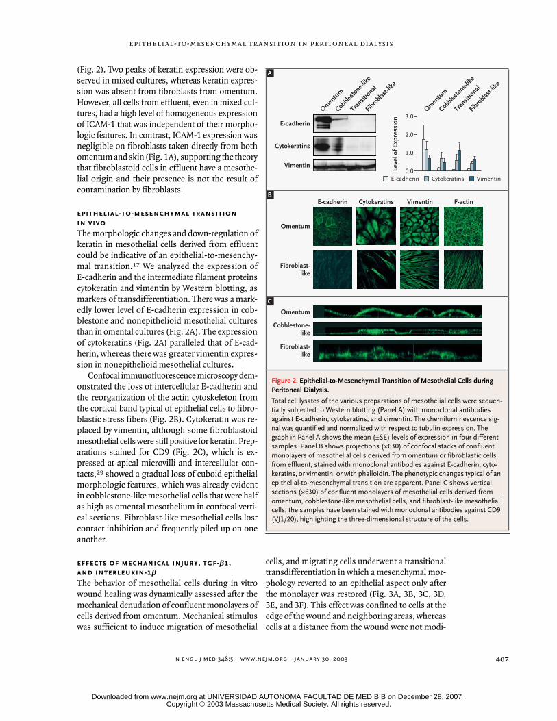
We analyzed the expression ofE-cadherin and the intermediate filament proteinscytokeratin and vimentin by Western blotting, asmarkers of transdifferentiation. There was a mark-edly lower level of E-cadherin expression in cob-blestone and nonepithelioid mesothelial culturesthan in omental cultures (Fig. 2A). The expressionof cytokeratins (Fig. 2A) paralleled that of E-cad-herin, whereas there was greater vimentin expres-sion in nonepithelioid mesothelial cultures.
Confocal immunofluorescence microscopy dem-onstrated the loss of intercellular E-cadherin andthe reorganization of the actin cytoskeleton fromthe cortical band typical of epithelial cells to fibro-blastic stress fibers (Fig. 2B). Cytokeratin was re-placed by vimentin, although some fibroblastoidmesothelial cells were still positive for keratin. Prep-arations stained for CD9 (Fig. 2C), which is ex-pressed at apical microvilli and intercellular con-tacts,
29
showed a gradual loss of cuboid epithelialmorphologic features, which was already evidentin cobblestone-like mesothelial cells that were halfas high as omental mesothelium in confocal verti-cal sections. Fibroblast-like mesothelial cells lostcontact inhibition and frequently piled up on oneanother.
effects of mechanical injury, tgf-
b
1,
and interleukin-1
b
The behavior of mesothelial cells during in vitrowound healing was dynamically assessed after themechanical denudation of confluent monolayers ofcells derived from omentum. Mechanical stimuluswas sufficient to induce migration of mesothelial
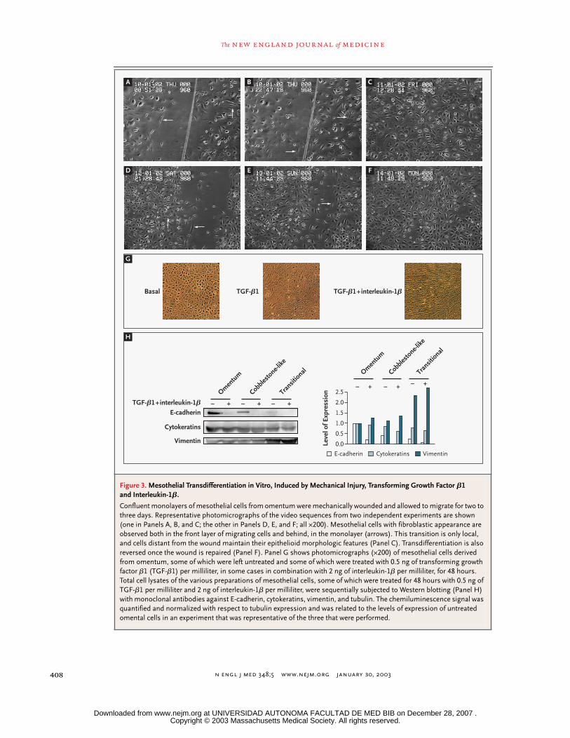
cells, and migrating cells underwent a transitionaltransdifferentiation in which a mesenchymal mor-phology reverted to an epithelial aspect only afterthe monolayer was restored (Fig. 3A, 3B, 3C, 3D,3E, and 3F). This effect was confined to cells at theedge of the wound and neighboring areas, whereascells at a distance from the wound were not modi-
Figure 2. Epithelial-to-Mesenchymal Transition of Mesothelial Cells during Peritoneal Dialysis.
Total cell lysates of the various preparations of mesothelial cells were sequen-tially subjected to Western blotting (Panel A) with monoclonal antibodies against E-cadherin, cytokeratins, and vimentin. The chemiluminescence sig-nal was quantified and normalized with respect to tubulin expression. The graph in Panel A shows the mean (±SE) levels of expression in four different samples. Panel B shows projections (¬630) of confocal stacks of confluent monolayers of mesothelial cells derived from omentum or fibroblastic cells from effluent, stained with monoclonal antibodies against E-cadherin, cyto-keratins, or vimentin, or with phalloidin. The phenotypic changes typical of an epithelial-to-mesenchymal transition are apparent. Panel C shows vertical sections (¬630) of confluent monolayers of mesothelial cells derived from omentum, cobblestone-like mesothelial cells, and fibroblast-like mesothelial cells; the samples have been stained with monoclonal antibodies against CD9 (VJ1/20), highlighting the three-dimensional structure of the cells.
Omentum
E-cadherin Cytokeratins Vimentin F-actin
Fibroblast-like
Omentum
Cobblestone-like
Fibroblast-like
A
E-cadherin
Cobble
stone-l
ike
Fibro
blast-
like
Transit
ional
Omen
tum
Cobble
stone-l
ike
Fibro
blast-
like
Transit
ional
Omen
tum
Cytokeratins
Vimentin
E-cadherin Cytokeratins Vimentin
Leve
l of E
xpre
ssio
n 3.0
2.0
1.0
0.0
B
C
Copyright © 2003 Massachusetts Medical Society. All rights reserved. Downloaded from www.nejm.org at UNIVERSIDAD AUTONOMA FACULTAD DE MED BIB on December 28, 2007 .
n engl j med
348;5
www.nejm.org january 30,
2003
The
new england journal
of
medicine
408
Figure 3. Mesothelial Transdifferentiation in Vitro, Induced by Mechanical Injury, Transforming Growth Factor
b
1 and Interleukin-1
b
.
Confluent monolayers of mesothelial cells from omentum were mechanically wounded and allowed to migrate for two to three days. Representative photomicrographs of the video sequences from two independent experiments are shown (one in Panels A, B, and C; the other in Panels D, E, and F; all ¬200). Mesothelial cells with fibroblastic appearance are observed both in the front layer of migrating cells and behind, in the monolayer (arrows). This transition is only local, and cells distant from the wound maintain their epithelioid morphologic features (Panel C). Transdifferentiation is also reversed once the wound is repaired (Panel F). Panel G shows photomicrographs (¬200) of mesothelial cells derived from omentum, some of which were left untreated and some of which were treated with 0.5 ng of transforming growth factor
b
1 (TGF-
b
1) per milliliter, in some cases in combination with 2 ng of interleukin-1
b
per milliliter, for 48 hours. Total cell lysates of the various preparations of mesothelial cells, some of which were treated for 48 hours with 0.5 ng of TGF-
b
1 per milliliter and 2 ng of interleukin-1
b
per milliliter, were sequentially subjected to Western blotting (Panel H) with monoclonal antibodies against E-cadherin, cytokeratins, vimentin, and tubulin. The chemiluminescence signal was quantified and normalized with respect to tubulin expression and was related to the levels of expression of untreated omental cells in an experiment that was representative of the three that were performed.
A
D
G
H
E F
B C
¡ + ¡ + ¡ +TGF-b1+interleukin-1b
Cobble
stone-l
ike
Transit
ional
Omen
tum
¡ +¡ + ¡ +
Cobble
stone-l
ike
Transit
ional
Omen
tum
Basal TGF-b1 TGF-b1+interleukin-1b
E-cadherin Cytokeratins Vimentin
Leve
l of E
xpre
ssio
n 2.5
2.0
1.0
1.5
0.5
0.0
E-cadherin
Cytokeratins
Vimentin
Copyright © 2003 Massachusetts Medical Society. All rights reserved. Downloaded from www.nejm.org at UNIVERSIDAD AUTONOMA FACULTAD DE MED BIB on December 28, 2007 .
n engl j med
348;5
www.nejm.org january
30, 2003
epithelial-to-mesenchymal transition in peritoneal dialysis
409
fied, reinforcing the theory that the mechanicalstimulus was sufficient to induce transdifferentia-tion. Complete time-lapse video sequences appearin Supplementary Appendix 1 (available with thefull text of this article at http://www.nejm.org).
To determine whether TGF-
b
1 and interleu-kin-1
b
, two cytokines detected in effluents frompatients undergoing continuous ambulatory peri-toneal dialysis primarily during episodes of perito-nitis,
24
could reproduce the phenotypic changes ob-served ex vivo, cultured mesothelial cells derivedfrom omentum were treated with TGF-
b
1 alone orin combination with interleukin-1
b
. An additivemorphologic effect of both stimuli could be ob-served (Fig. 3G). E-cadherin expression was almostcompletely abolished (Fig. 3H), and its localizationat intercellular junctions could hardly be detectedby immunofluorescence. Cytokeratin expressionwas also diminished, and an additive effect withinterleukin-1
b
was observed. In contrast, thesetreatments were associated with an increment invimentin expression.
expression of
snail
in mesothelial cells
undergoing epithelial-to-mesenchymal
transition
Recently, a transcription factor called
snail
has beendescribed as a potent repressor of E-cadherin expres-sion and an inducer of epithelial-to-mesenchymaltransition.
21-23
To determine whether
snail
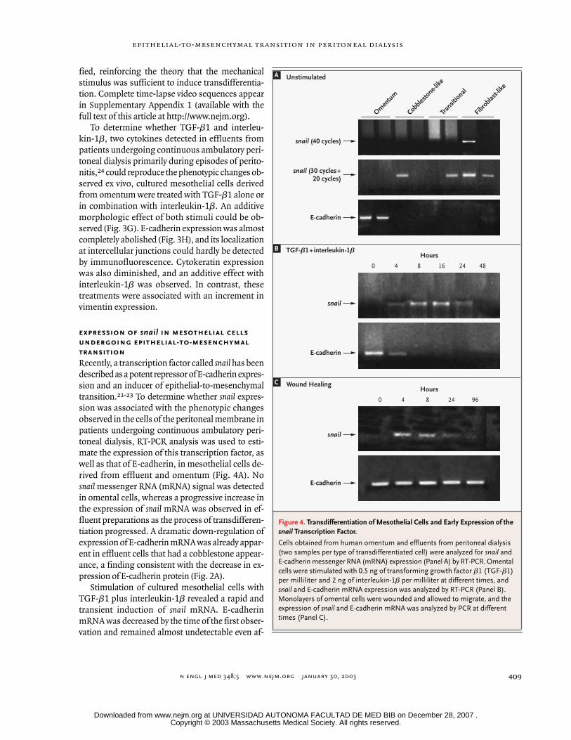
expres-sion was associated with the phenotypic changesobserved in the cells of the peritoneal membrane inpatients undergoing continuous ambulatory peri-toneal dialysis, RT-PCR analysis was used to esti-mate the expression of this transcription factor, aswell as that of E-cadherin, in mesothelial cells de-rived from effluent and omentum (Fig. 4A). No
snail
messenger RNA (mRNA) signal was detectedin omental cells, whereas a progressive increase inthe expression of
snail
mRNA was observed in ef-fluent preparations as the process of transdifferen-tiation progressed. A dramatic down-regulation ofexpression of E-cadherin mRNA was already appar-ent in effluent cells that had a cobblestone appear-ance, a finding consistent with the decrease in ex-pression of E-cadherin protein (Fig. 2A).
Stimulation of cultured mesothelial cells withTGF-
b
1 plus interleukin-1
b
revealed a rapid andtransient induction of
snail
mRNA. E-cadherinmRNA was decreased by the time of the first obser-vation and remained almost undetectable even af-
Figure 4. Transdifferentiation of Mesothelial Cells and Early Expression of the
snail
Transcription Factor.
Cells obtained from human omentum and effluents from peritoneal dialysis (two samples per type of transdifferentiated cell) were analyzed for
snail
and E-cadherin messenger RNA (mRNA) expression (Panel A) by RT-PCR. Omental cells were stimulated with 0.5 ng of transforming growth factor
b
1 (TGF-
b
1) per milliliter and 2 ng of interleukin-1
b
per milliliter at different times, and
snail
and E-cadherin mRNA expression was analyzed by RT-PCR (Panel B). Monolayers of omental cells were wounded and allowed to migrate, and the expression of
snail
and E-cadherin mRNA was analyzed by PCR at different times (Panel C).
A
Omen
tum
snail (40 cycles)
snail (30 cycles+20 cycles)
E-cadherin
Cobble
stone-l
ike
Transit
ional
Fibro
blast-
like
Hours
Wound Healing
snail
E-cadherin
0 4 8 16 24 48
Hours
snail
E-cadherin
0 4 8 24 96
TGF-b1+interleukin-1bB
C
Unstimulated
Copyright © 2003 Massachusetts Medical Society. All rights reserved. Downloaded from www.nejm.org at UNIVERSIDAD AUTONOMA FACULTAD DE MED BIB on December 28, 2007 .
n engl j med 348;5 www.nejm.org january 30, 2003
The new england journal of medicine
410
ter snail transcription had declined (Fig. 4B). Simi-larly, after in vitro wound healing, a transientinduction of snail mRNA was observed, which prob-ably corresponded to the transitional process in thecells next to the wound. Since the majority of the
cells were not involved in the wound-healing proc-ess, no down-regulation of E-cadherin was observedin the total population (Fig. 4C).
up-regulation of a2222 integrin
and acquisition of a migratory phenotype
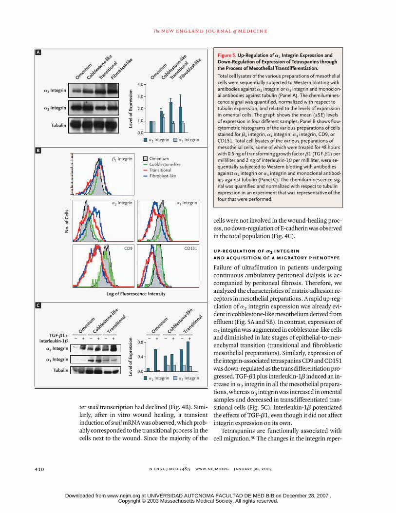
Failure of ultrafiltration in patients undergoingcontinuous ambulatory peritoneal dialysis is ac-companied by peritoneal fibrosis. Therefore, weanalyzed the characteristics of matrix-adhesion re-ceptors in mesothelial preparations. A rapid up-reg-ulation of a2 integrin expression was already evi-dent in cobblestone-like mesothelium derived fromeffluent (Fig. 5A and 5B). In contrast, expression ofa3 integrin was augmented in cobblestone-like cellsand diminished in late stages of epithelial-to-mes-enchymal transition (transitional and fibroblasticmesothelial preparations). Similarly, expression ofthe integrin-associated tetraspanins CD9 and CD151was down-regulated as the transdifferentiation pro-gressed. TGF-b1 plus interleukin-1b induced an in-crease in a2 integrin in all the mesothelial prepara-tions, whereas a3 integrin was increased in omentalsamples and decreased in transdifferentiated tran-sitional cells (Fig. 5C). Interleukin-1b potentiatedthe effects of TGF-b1, even though it did not affectintegrin expression on its own.
Tetraspanins are functionally associated withcell migration.30 The changes in the integrin reper-
A
Omen
tum
a2 Integrin
a3 Integrin
Tubulin
Cobble
stone-l
ike
Transit
ional
Fibro
blast-
like
Omen
tum
Cobble
stone-l
ike
Transit
ional
Fibro
blast-
like
b1 Integrin
a2 Integrin a3 Integrin
CD9 CD151
No.
of C
ells
Log of Fluorescence Intensity
4.0
2.0
1.0
0.0
0.0
a2 Integrin a3 Integrin
a2 Integrin a3 Integrin
Leve
l of E
xpre
ssio
n
0.8
0.4
¡ + ¡ + ¡ +
a2 Integrin
TGF-b1+interleukin-1b
Cobble
stone-l
ike
Transit
ional
Omen
tum
¡ + ¡ + ¡ +Cob
blesto
ne-like
Transit
ional
Omen
tum
a3 Integrin
Tubulin
OmentumCobblestone-likeTransitionalFibroblast-like
B
C
Leve
l of E
xpre
ssio
n
3.0
Figure 5. Up-Regulation of a2 Integrin Expression and Down-Regulation of Expression of Tetraspanins through the Process of Mesothelial Transdifferentiation.
Total cell lysates of the various preparations of mesothelial cells were sequentially subjected to Western blotting with antibodies against a2 integrin or a3 integrin and monoclon-al antibodies against tubulin (Panel A). The chemilumines-cence signal was quantified, normalized with respect to tubulin expression, and related to the levels of expression in omental cells. The graph shows the mean (±SE) levels of expression in four different samples. Panel B shows flow-cytometric histograms of the various preparations of cells stained for b1 integrin, a2 integrin, a3 integrin, CD9, or CD151. Total cell lysates of the various preparations of mesothelial cells, some of which were treated for 48 hours with 0.5 ng of transforming growth factor b1 (TGF-b1) per milliliter and 2 ng of interleukin-1b per milliliter, were se-quentially subjected to Western blotting with antibodies against a2 integrin or a3 integrin and monoclonal antibod-ies against tubulin (Panel C). The chemiluminescence sig-nal was quantified and normalized with respect to tubulin expression in an experiment that was representative of the four that were performed.
Copyright © 2003 Massachusetts Medical Society. All rights reserved. Downloaded from www.nejm.org at UNIVERSIDAD AUTONOMA FACULTAD DE MED BIB on December 28, 2007 .
n engl j med 348;5 www.nejm.org january 30, 2003
epithelial-to-mesenchymal transition in peritoneal dialysis
411
toire and the switch from a keratin-based to a vi-mentin-based cytoskeleton could also affect themigratory capacity of mesothelial cells. We have ob-served that the transdifferentiation process was ac-companied by a higher overall migratory capacityof mesothelial cells. Treatment with TGF-b1 plusinterleukin-1b enhanced the haptotaxis to collagen,the main ligand for a2b1 integrin. Migration towardlaminin-5 followed the changes in the expressionof its receptor, a3b1 integrin; it was enhanced inepithelioid cultures and reduced in transitional andfibroblastic cells (data not shown).
evidence of epithelial-to-mesenchymal
transition of mesothelial cells
in peritoneal tissue
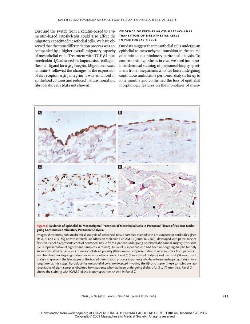
Our data suggest that mesothelial cells undergo anepithelial-to-mesenchymal transition in the courseof continuous ambulatory peritoneal dialysis. Toconfirm this hypothesis in vivo, we used immuno-histochemical staining of peritoneal-biopsy speci-mens from nine patients who had been undergoingcontinuous ambulatory peritoneal dialysis for up tonine months and confirmed the loss of epithelialmorphologic features on the monolayer of meso-
Figure 6. Evidence of Epithelial-to-Mesenchymal Transition of Mesothelial Cells in Peritoneal Tissue of Patients Under-going Continuous Ambulatory Peritoneal Dialysis.
Images show immunohistochemical analysis of peritoneal-tissue samples stained with anticytokeratin antibodies (Pan-els A, B, and C, ¬150) or with intercellular adhesion molecule 1 (ICAM-1) (Panel D, ¬180), developed with peroxidase or fast red. Panel A represents control peritoneal tissue from a patient undergoing unrelated abdominal surgery (this sam-ple is representative of eight tissue samples examined). In Panel B, a patient who had been undergoing dialysis for only six months already has a loss of mesothelial-cell polarity (this sample is representative of nine samples from patients who had been undergoing dialysis for nine months or less). Panel C (8 months of dialysis) and the inset (34 months of dialysis) represent the late stages of the transdifferentiation process in patients who have been undergoing dialysis for a long time; at this stage, fibroblast-like mesothelial cells are detected invading the fibrotic tissue (these samples are rep-resentative of eight samples obtained from patients who had been undergoing dialysis for 8 to 77 months). Panel D shows the staining with ICAM-1 of the biopsy specimen shown in Panel C.
A B
C D
Copyright © 2003 Massachusetts Medical Society. All rights reserved. Downloaded from www.nejm.org at UNIVERSIDAD AUTONOMA FACULTAD DE MED BIB on December 28, 2007 .
n engl j med 348;5 www.nejm.org january 30, 2003
The new england journal of medicine
412
thelial cells in the early stages of this type of dialy-sis (Fig. 6B). In biopsy specimens from eight pa-tients who had undergone such dialysis for 8 to 77months, the monolayer of mesothelial cells disap-peared, and elongated mesothelial cells positive forcytokeratin and ICAM-1 were found embedded inthe fibrotic tissue (Fig. 6C and 6D); these specimenscorresponded to cultures of nonepithelioid meso-thelial cells from effluent.
Peritoneal dialysis is an increasingly common al-ternative to hemodialysis. However, the proceduresubjects mesothelial cells to high osmotic pressureand bioincompatible substances. Studies usingstandard histologic techniques on peritoneum frompatients undergoing continuous ambulatory peri-toneal dialysis show a complete loss of the mono-layer of mesothelial cells and fibrosis, which mightbe responsible for the ultimate functional failure ofthe peritoneal membrane.1 Our data show that mes-othelial cells undergo a transition from an epithe-lial phenotype to a mesenchymal phenotype duringperitoneal dialysis, with the induction of snail ex-pression and a dramatic down-regulation of E-cad-herin expression. Moreover, these findings are evi-dence of a direct and active role of mesothelial cellsin the tissue fibrosis and failure of ultrafiltration inthis process, generating new fibroblastic cells andleading to peritoneal fibrosis.
Previous studies have characterized the cobble-stone-like mesothelial cells from peritoneal efflu-ents as indistinguishable from mesothelial cellsderived from omentum.10 However, even early in
continuous ambulatory peritoneal dialysis, a lossof cuboid morphology is observed both in vivo andex vivo, accompanied by an induction of snail ex-pression that down-regulates E-cadherin expres-sion, even when cells retain an epithelioid appear-ance. If peritoneal dialysis is continued, long-termexposure to mechanical denudation, profibrotic fac-tors such as TGF-b, and inflammatory cytokinesmay induce a complete transition of mesothelialcells, which could be responsible for tissue fibrosisand failure of ultrafiltration. Patients with recurrentepisodes of peritonitis have high levels of expres-sion of TGF-b24 and have accelerated failure of ul-trafiltration.4
The fact that mesothelial cells undergo epithe-lial-to-mesenchymal transition during continuousambulatory peritoneal dialysis may change our viewof the pathophysiology of ultrafiltration failure. Ourdata reveal a series of markers such as snail, E-cad-herin, and a2 integrin that are already modified inthe early phases of the transdifferentiation process.In addition, ICAM-1 appears to be a potential mark-er that discriminates between mesothelial cells andfibroblasts. All these markers may be useful in thefollow-up of patients undergoing peritoneal dialy-sis and in the development of new solutions forperitoneal dialysis. Furthermore, these data suggestnew therapeutic targets that might ultimately pre-vent the fibrosis associated with continuous ambu-latory peritoneal dialysis.
Supported by Fresenius Medical Care; by grants (01/0063-02 toDr. Selgas and 00/0602 to Dr. López-Cabrera) from the Fondo de In-vestigaciones Sanitarias; and by a grant (02-00536 to Dr. Sánchez-Madrid) from the Programa de Biología Molecular y Celular.
We are indebted to Angela Nieto for critical discussion and toFrancisco Rodríguez for statistical analysis of the data.
discussion
references
1. Krediet RT. The peritoneal membranein chronic peritoneal dialysis patients. Kid-ney Int 1999;55:341-56.2. Brulez HF, Verbrugh HA. First-linedefense mechanisms in the peritoneal cavityduring peritoneal dialysis. Perit Dial Int 1995;15:Suppl:S24-S33.3. Chaimovitz C. Peritoneal dialysis. Kid-ney Int 1994;45:1226-40. [Erratum, KidneyInt 1994;46:285.]4. Selgas R, Fernández-Reyes MJ, BosqueE, et al. Functional longevity of the humanperitoneum: how long is continuous perito-neal dialysis possible? Results of a prospec-tive medium long-term study. Am J KidneyDis 1994;23:64-73.5. Dobbie JW. Pathogenesis of peritonealfibrosing syndromes (sclerosing peritonitis)
in peritoneal dialysis. Perit Dial Int 1992;12:14-27.6. Fang CC, Yen C-J, Chen Y-M, et al. Pen-toxifylline inhibits human peritoneal mes-othelial cell growth and collagen synthesis:effects of TGF-b. Kidney Int 2000;57:2626-33.7. Medcalf JF, Walls J, Pawluczyk IZ, HarrisKPG. Effects of glucose dialysate on extra-cellular matrix production by human perito-neal mesothelial cells (HPMC): the role ofTGF-b. Nephrol Dial Transplant 2001;16:1885-92.8. Rampino T, Cancarini G, Gregorini M,et al. Hepatocyte growth factor/scatter fac-tor released during peritonitis is active onmesothelial cells. Am J Pathol 2001;159:1275-85.
9. Yang WS, Kim BS, Lee SK, Park JS, KimSB. Interleukin-1b stimulates the produc-tion of extracellular matrix in cultured humanperitoneal mesothelial cells. Perit Dial Int1999;19:211-20.10. Leavesley DI, Stanley JM, Faull RJ. Epi-dermal growth factor modifies the expres-sion and function of extracellular matrixadhesion receptors expressed by peritonealmesothelial cells from patients on CAPD.Nephrol Dial Transplant 1999;14:1208-16.11. Connell ND, Rheinwald JG. Regulationof the cytoskeleton in mesothelial cells:reversible loss of keratin and increase invimentin during rapid growth in culture.Cell 1983;34:245-53.12. Faull RJ, Stanley JM, Fraser S, Power DA,Leavesley DI. HB-EGF is produced in the
Copyright © 2003 Massachusetts Medical Society. All rights reserved. Downloaded from www.nejm.org at UNIVERSIDAD AUTONOMA FACULTAD DE MED BIB on December 28, 2007 .
n engl j med 348;5 www.nejm.org january 30, 2003
epithelial-to-mesenchymal transition in peritoneal dialysis
413
peritoneal cavity and enhances mesothelialcell adhesion and migration. Kidney Int2001;59:614-24.13. Ha H, Yu MR, Lee HB. High glucose-induced PKC activation mediates TGF-b1and fibronectin synthesis by peritonealmesothelial cells. Kidney Int 2001;59:463-70.14. Ito T, Yorioka N, Yamamoto M, KataokaK, Yamakido M. Effect of glucose on inter-cellular junctions of cultured human perito-neal mesothelial cells. J Am Soc Nephrol2000;11:1969-79.15. Massagué J. TGF-b signal transduction.Annu Rev Biochem 1998;67:753-91.16. Margetts PJ, Kolb M, Galt T, Hoff CM,Shockley TR, Gauldie J. Gene transfer oftransforming growth factor-b1 to the ratperitoneum: effects on membrane function.J Am Soc Nephrol 2001;12:2029-39.17. Hay ED. An overview of epithelio-mes-enchymal transformation. Acta Anat (Basel)1995;154:8-20.18. Birchmeier C, Birchmeier W, Brand-Saberi B. Epithelial-mesenchymal transi-tions in cancer progression. Acta Anat(Basel) 1996;156:217-26.19. Perl AK, Wilgenbus P, Dahl U, Semb H,Christofori G. A causal role for E-cadherin
in the transition from adenoma to carci-noma. Nature 1998;392:190-3.20. Takeichi M. Morphogenetic roles ofclassic cadherins. Curr Opin Cell Biol 1995;7:619-27.21. Batlle E, Sancho E, Franci C, et al. Thetranscription factor snail is a repressor ofE-cadherin gene expression in epithelialtumour cells. Nat Cell Biol 2000;2:84-9.22. Cano A, Pérez-Moreno MA, Rodrigo I, etal. The transcription factor snail controls epi-thelial-mesenchymal transitions by repress-ing E-cadherin expression. Nat Cell Biol2000;2:76-83.23. Carver EA, Jiang R, Lan Y, Oram KF,Gridley T. The mouse snail gene encodes akey regulator of the epithelial-mesenchy-mal transition. Mol Cell Biol 2001;21:8184-8.24. Lai KN, Lai KB, Lam CW, Chan TM, LiFK, Leung JC. Changes of cytokine profilesduring peritonitis in patients on continuousambulatory peritoneal dialysis. Am J KidneyDis 2000;35:644-52.25. Yáñez-Mó M, Alfranca A, Cabañas C, etal. Regulation of endothelial cell motility bycomplexes of tetraspan molecules CD81/TAPA-1 and CD151/PETA-3 with a3b1 inte-
grin localized at endothelial lateral junc-tions. J Cell Biol 1998;141:791-804.26. Rousselle P, Golbik R, van der Rest M,Aumailley M. Structural requirement for celladhesion to kalinin (laminin-5). J Biol Chem1995;270:13766-70.27. Lara-Pezzi E, Serrador JM, Montoya MC,et al. The hepatitis B virus X protein (HBx)induces a migratory phenotype in a CD44-dependent manner: possible role of HBx ininvasion and metastasis. Hepatology 2001;33:1270-81.28. Suassuna JH, Neves FCD, Hartley RB,Ogg CS, Cameron JS. Immunohistochemi-cal studies of the peritoneal membrane andinfiltrating cells in normal subjects and inpatients on CAPD. Kidney Int 1994;46:443-54.29. Yáñez-Mó M, Tejedor R, Rousselle P,Sánchez-Madrid F. Tetraspanins in intercel-lular adhesion of polarized epithelial cells:spatial and functional relationship to inte-grins and cadherins. J Cell Sci 2001;114:577-87.30. Hemler ME. Integrin associated pro-teins. Curr Opin Cell Biol 1998;10:578-85.Copyright © 2003 Massachusetts Medical Society.
electronic access to the journal’s cumulative index
At the Journal’s site on the World Wide Web (http://www.nejm.org) you can search an index of all articles published since January 1975 (abstracts 1975–1992, full-text 1993–present). You can search by author, key word, title, type of article, and date. The results will include the citations for the articles plus links to the abstracts of articles published since 1993. For nonsubscribers, time-limited access to single articles and 24-hour site access can also be ordered for a fee through the Internet (http://www.nejm.org).
Copyright © 2003 Massachusetts Medical Society. All rights reserved. Downloaded from www.nejm.org at UNIVERSIDAD AUTONOMA FACULTAD DE MED BIB on December 28, 2007 .
New England Journal of Medicine
CORRECTION
Peritoneal Dialysis and Epithelial-to-MesenchymalTransition of Mesothelial Cells
Peritoneal Dialysis and Epithelial-to-Mesenchymal Transition of
Mesothelial Cells . On page 408, Panel H of Figure 3 contains several
errors. The corrected version of this figure appears with the full text of
the article at www.nejm.org.
N Engl J Med 2005;353:2827
Copyright © 2003 Massachusetts Medical Society. All rights reserved. Downloaded from www.nejm.org at UNIVERSIDAD AUTONOMA FACULTAD DE MED BIB on December 28, 2007 .
Copyright © 2022 FDOKUMEN