![[Peritoneal pseudomyxoma: An overview emphasizing pathological assessment and therapeutic strategies].](https://static.fdokumen.com/doc/165x107/6331d121b6829c19b80bab58/peritoneal-pseudomyxoma-an-overview-emphasizing-pathological-assessment-and-therapeutic.jpg)
Production of stromal cell-derived factor 1 by mesothelial cells and effects of this chemokine on...
10
0014-2980/01/0202-350$17.50 + .50/0 © WILEY-VCH Verlag GmbH, D-69451 Weinheim, 2001 Production of stromal cell-derived factor 1 by mesothelial cells and effects of this chemokine on peritoneal B lymphocytes Arnaud Foussat 1 , Karl Balabanian 1 , Ali Amara 2 , Laurence Bouchet-Delbos 1 , Ingrid Durand-Gasselin 1 , Fran ¸ coise Baleux 3 , Jacques Couderc 1 , Pierre Galanaud 1 and Dominique Emilie 1 1 INSERM U131, Institut Paris-Sud sur les Cytokines, Clamart, France 2 Unit ´ e d’Immunologie Virale, Institut Pasteur, Paris, France 3 Unit ´ e de Chimie Organique, Institut Pasteur, Paris, France B1a lymphocytes accumulate and proliferate in the peritoneal cavity. Stromal cell-derived factor 1 (SDF-1) is a chemotactic and growth promoting factor for B cell precursors. It is required for fetal liver B cell lymphopoiesis, which generates mostly B1a lymphocytes. Using immunohistochemistry with an anti-SDF-1 monoclonal antibody, we found that SDF-1 was produced by peritoneal mesothelial cells in adult mice. Peritoneal B1a lymphocytes expressed a functional SDF-1 receptor, as shown by actin polymerization experiments. In vitro, SDF-1 stimulated migration, proliferation of a minority of peritoneal B1a lymphocytes, and prevented apoptosis in a large fraction of cells. B1a cells migrating in response to SDF-1 were largely enriched in the CD5 high CD43 high B220 – CD1d – subpopulation. In vivo, neutraliza- tion of SDF-1 for 3 weeks significantly decreased the number of peritoneal B1 cells. SDF-1 also acted on peritoneal B2 cells. These findings show that after the cessation of B cell lym- phopoiesis in the liver, around birth, the persistence of B1a cells remains SDF-1 dependent, and that SDF-1 production by mesothelial cells plays a role in the peritoneal location of B1a cells. Thus, the role of mesothelial cells for B1a cells in adults may be similar to that of SDF- 1-producing biliary ductal plate cells in the fetus, and to that of bone marrow stromal cells for B2 cell precursors. Key words: Stromal cell-derived factor 1 / Chemokine / B cell lymphopoiesis / B lymphocyte / Mesothelial cell Received 10/4/00 Revised 1/9/00 Accepted 26/10/00 [I 20782] Abbreviations: SDF-1: Stromal cell-derived factor 1 CFSE: 5(6)-Carboxyfluorescein diacetate succinimidyl ester 1 Introduction Chemokines belong to a family of cytokines defined by their ability to trigger the tissue-specific targeting of leu- kocytes. However, these cytokines also affect the matu- ration, proliferation, survival and effector responses of leukocytes, B and T cell lymphopoiesis, and organogen- esis of some non-lymphoid structures [1–6]. This broad range of activities is best illustrated by the CXC chemo- kine stromal cell-derived factor 1 (SDF-1). SDF-1 is a chemoattractant for early B cell progenitors, B and T cell subpopulations, monocytes, megakaryocytes and CD34 + hematopoietic stem cells [7–11]. SDF-1 also plays a critical role in the development of the cerebellum, the cardiac septum, the mesenteric vasculature and the B lymphocyte compartment [12–14]. SDF-1 was initially cloned from mouse bone marrow stromal cells as a pre- B cell stimulating factor [15, 16]. The functions of SDF-1 in B cell lymphopoiesis may include attraction of B cell precursors to stromal cells in addition to a growth pro- moting effect, because B cell precursors deficient in expression of the SDF-1 receptor, CXCR4, are not retained within the bone marrow microenvironment [17]. The effect of SDF-1 on B cell lymphopoiesis starts during fetal development. In the human fetus, SDF-1 is pro- duced by biliary ductal plate epithelial cells in the liver, and B cell precursors are in close contact with SDF-1- producing cells [18]. Evidence for a role for SDF-1 in antenatal B cell lymphopoiesis is also provided by the absence of liver B cell lymphopoiesis in mice with dis- rupted SDF-1 or CXCR4 genes [12, 14, 19]. In adult mice, B lymphocytes can be divided into three distinct subpopulations. B1 lymphocytes express the Mac-1 antigen and include two subsets according to expression of the CD5 antigen: B1a are constitutively 350 A. Foussat et al. Eur. J. Immunol. 2001. 31: 350–359
-
Upload
independent -
Category
Documents
-
view
0 -
download
0
Transcript of Production of stromal cell-derived factor 1 by mesothelial cells and effects of this chemokine on...

0014-2980/01/0202-350$17.50+.50/0 © WILEY-VCH Verlag GmbH, D-69451 Weinheim, 2001
Production of stromal cell-derived factor 1 bymesothelial cells and effects of this chemokine onperitoneal B lymphocytes
Arnaud Foussat1, Karl Balabanian1, Ali Amara2, Laurence Bouchet-Delbos1, IngridDurand-Gasselin1, Francoise Baleux3, Jacques Couderc1, Pierre Galanaud1 andDominique Emilie1
1 INSERM U131, Institut Paris-Sud sur les Cytokines, Clamart, France2 Unite d’Immunologie Virale, Institut Pasteur, Paris, France3 Unite de Chimie Organique, Institut Pasteur, Paris, France
B1a lymphocytes accumulate and proliferate in the peritoneal cavity. Stromal cell-derivedfactor 1 (SDF-1) is a chemotactic and growth promoting factor for B cell precursors. It isrequired for fetal liver B cell lymphopoiesis, which generates mostly B1a lymphocytes. Usingimmunohistochemistry with an anti-SDF-1 monoclonal antibody, we found that SDF-1 wasproduced by peritoneal mesothelial cells in adult mice. Peritoneal B1a lymphocytesexpressed a functional SDF-1 receptor, as shown by actin polymerization experiments. Invitro, SDF-1 stimulated migration, proliferation of a minority of peritoneal B1a lymphocytes,and prevented apoptosis in a large fraction of cells. B1a cells migrating in response to SDF-1were largely enriched in the CD5highCD43highB220–CD1d– subpopulation. In vivo, neutraliza-tion of SDF-1 for 3 weeks significantly decreased the number of peritoneal B1 cells. SDF-1also acted on peritoneal B2 cells. These findings show that after the cessation of B cell lym-phopoiesis in the liver, around birth, the persistence of B1a cells remains SDF-1 dependent,and that SDF-1 production by mesothelial cells plays a role in the peritoneal location of B1acells. Thus, the role of mesothelial cells for B1a cells in adults may be similar to that of SDF-1-producing biliary ductal plate cells in the fetus, and to that of bone marrow stromal cells forB2 cell precursors.
Key words: Stromal cell-derived factor 1 / Chemokine / B cell lymphopoiesis / B lymphocyte /Mesothelial cell
Received 10/4/00Revised 1/9/00Accepted 26/10/00
[I 20782]
Abbreviations: SDF-1: Stromal cell-derived factor 1CFSE: 5(6)-Carboxyfluorescein diacetate succinimidyl ester
1 Introduction
Chemokines belong to a family of cytokines defined bytheir ability to trigger the tissue-specific targeting of leu-kocytes. However, these cytokines also affect the matu-ration, proliferation, survival and effector responses ofleukocytes, B and T cell lymphopoiesis, and organogen-esis of some non-lymphoid structures [1–6]. This broadrange of activities is best illustrated by the CXC chemo-kine stromal cell-derived factor 1 (SDF-1). SDF-1 is achemoattractant for early B cell progenitors, B and T cellsubpopulations, monocytes, megakaryocytes andCD34+ hematopoietic stem cells [7–11]. SDF-1 alsoplays a critical role in the development of the cerebellum,the cardiac septum, the mesenteric vasculature and the
B lymphocyte compartment [12–14]. SDF-1 was initiallycloned from mouse bone marrow stromal cells as a pre-B cell stimulating factor [15, 16]. The functions of SDF-1in B cell lymphopoiesis may include attraction of B cellprecursors to stromal cells in addition to a growth pro-moting effect, because B cell precursors deficient inexpression of the SDF-1 receptor, CXCR4, are notretained within the bone marrow microenvironment [17].The effect of SDF-1 on B cell lymphopoiesis starts duringfetal development. In the human fetus, SDF-1 is pro-duced by biliary ductal plate epithelial cells in the liver,and B cell precursors are in close contact with SDF-1-producing cells [18]. Evidence for a role for SDF-1 inantenatal B cell lymphopoiesis is also provided by theabsence of liver B cell lymphopoiesis in mice with dis-rupted SDF-1 or CXCR4 genes [12, 14, 19].
In adult mice, B lymphocytes can be divided into threedistinct subpopulations. B1 lymphocytes express theMac-1 antigen and include two subsets according toexpression of the CD5 antigen: B1a are constitutively
350 A. Foussat et al. Eur. J. Immunol. 2001. 31: 350–359

Fig. 1. Transfer of peritoneal B lymphocytes to SCID mice. Peritoneal CD19+ cells from BALB/c mice were transferred to perito-neum of SCID mice, and peritoneal cells were recovered 5 weeks later and labeled for CD19, CD5 and Mac-1. (C) CD19 andMac-1 labeling in one representative of six SCID mice, in comparison with the labeling of peritoneal cells from one of ten repre-sentative BALB/c mice (A) and one representative of four SCID mice without cell transfer (B). The B1a/B1 ratio (i.e.CD5+CD19+Mac-1+/CD19+Mac-1+ ratio) in SCID mice reconstituted with peritoneal B cells was 98.0±0.7%. There were few if anyB2 cells in these mice, as shown by the virtual absence of CD19+Mac-1– cells in (C).
CD19+ Mac1+CD5+, whereas B1b are CD19+Mac1+CD5–.Conventional B2 cells are CD19+Mac1–. The B1a cellsdevelop and accumulate in the coelomic cavities. In con-trast, B1a cells are rare in blood and lymphoid organs.Mature B1a lymphocytes produce large amounts ofmultireactive IgM with weak affinity for self and non-selfantigens [20–23]. The gradual expansion of the B1a lym-phocyte population in mice results in lymphoproliferationand circulating autoantibodies in the elderly. This expan-sion is accelerated in certain mice with particular geneticbackgrounds, such as New Zealand Black (NZB) mice[24–26].
In human fetus, we recently showed that mesothelialcells from coelomic cavities produce SDF-1 [18]. This ledus to hypothesize that SDF-1 production by mesothelialcells extends beyond birth, and that it supports the localregeneration of B1a cells in coelomic cavities throughoutlife. In this work, we investigated whether SDF-1 is pro-duced by mesothelial cells in adult mice and whether thischemokine affects the migration, proliferation and sur-vival of peritoneal B1 lymphocytes.
2 Results
2.1 Peritoneal B lymphocytes from BALB/c micereconstitute the peritoneal B1a lymphocytepopulation, but not other B cell populations
To test the ability of the peritoneal cavity to support theself renewal of the distinct peritoneal B lymphocyte sub-populations, we isolated peritoneal CD19+ lymphocytesfrom BALB/c mice and transferred them to the peritonealcavity of CB.17 SCID mice. The presence of the variousB lymphocyte subpopulations was analyzed 5 weeks
after cell transfer. In SCID mice receiving peritoneal Blymphocytes, the number of CD19+ cells in the bonemarrow and in the spleen was low (2.9±0.6% and0.5±0.1%, respectively) and in the same range to that ofcontrol SCID mice to which no cells were transferred(3.1±0.3% and 0.2±0.1%, respectively). In contrast, thenumber of CD19+ cells in the peritoneal cavity of SCIDmice after cell transfer was significantly higher than incontrol SCID mice. The vast majority of these B lympho-cytes belonged to the B1a subpopulation (Fig. 1). There-fore, the peritoneal cavity provides a favorable environ-ment for the self renewal of the B1a subpopulation, butnot for other B lymphocytes. Moreover, peritoneal B lym-phocytes do not repopulate lymphoid organs of immu-nodeficient mice.
2.2 Production of SDF-1 by mesothelial cells inthe serous cavities
SDF-1 production by mesothelial cells was analyzed byimmunohistochemistry. Three mice were studied andsimilar results were obtained in each case. All mesothe-lial cells were stained with the anti-SDF-1 mAb, regard-less of their peritoneal, pericardial or pleural origin, bothfor the visceral and the parietal layer. Cells from the sub-mesothelial space were not stained with the anti-SDF-1mAb. No labeling was detected if the anti-SDF-1 mAbwas omitted (Fig. 2).
2.3 CXCR4 is functional in B1 and B2 peritoneallymphocytes
We investigated the function of CXCR4 by analyzing theability of SDF-1 to induce cytoskeleton rearrangement in
Eur. J. Immunol. 2001. 31: 350–359 SDF-1 and peritoneal B lymphocytes 351

Fig. 2. Immunochemical detection of SDF-1 in mesothelialcells. Peritoneal (A, B), pleural (C, D) and pericardial (E, F) tis-sues were analyzed for the presence of SDF-1 by immuno-histochemistry. The anti-SDF-1 mAb was used in (A, C, E)and omitted in (B, D, F). A–F ×200.
Fig. 3. SDF-1-mediated actin polymerization. The effect ofSDF-1 was evaluated by quantifying actin polymerizationfollowing chemokine addition. Peritoneal B1a (A), peritonealB1b (B), peritoneal B2 (C), and splenic B2 (D) lymphocyteswere tested. Results (mean ± SEM of three experiments) areexpressed as the variation of fluorescence intensity inducedby SDF-1 addition in function of time, as compared to base-line value, determined before chemokine addition.
Fig. 4. Chemoattraction of peritoneal B cells by SDF-1. Theeffect of SDF-1 on the migration of peritoneal B lymphocyteswas evaluated by enumerating B1a, B1b and B2 lympho-cytes migrating toward a SDF-1 gradient in a Transwellmigration assay. Results represent the number (mean ±SEM) of cells migrating in response to medium alone, or to amedium containing SDF-1 (1 ? g/ml). Seven experimentswere performed.
peritoneal B cells. We monitored actin polymerization infreshly isolated peritoneal B lymphocytes following theaddition of SDF-1. SDF-1 induced actin polymerization
in B1a, B1b and B2 peritoneal lymphocytes. In contrast,freshly isolated B2 cells from the spleen did not respondto SDF-1 (Fig. 3). Therefore, the CXCR4 receptor is func-tional in all peritoneal B lymphocyte subsets.
2.4 SDF-1 is a chemoattractant for peritoneal Bcells
We investigated, using the Transwell system, whetherCXCR4 stimulation triggered migration of peritoneal Bcells. SDF-1 induced chemotaxis of B1a, B1b and B2cells. Only a minority of freshly isolated peritoneal B cells(between 1/100 and 1/1,000) migrated in response toSDF-1. This fraction was clearly higher than in controlcultures, performed in the absence of SDF-1 (Fig. 4). Todemonstrate that SDF-1 was acting as a chemoattract-ant rather than as an enhancer of chemokinesis, SDF-1was added to both the upper and lower chambers of theTranswell system. In these conditions, the number ofperitoneal B cells migrating to the lower chamber wasless than 10% as compared to assays performed withSDF-1 only added to the lower chamber (data notshown).
We compared the efficacy of SDF-1 to trigger peritonealB lymphocyte migration with that of B lymphocyte che-moattractant. The fraction of responding cells was in thesame range for both chemokines (Table 1).
2.5 Characterization of B1a cells migrating inresponse to SDF-1
Whether the chemoattractant effect of SDF-1 wasrestricted to peculiar peritoneal B lymphocyte subsetswas analyzed by phenotyping migrating cells. No spe-cific subset of B1b and B2 cells that was electively sensi-
352 A. Foussat et al. Eur. J. Immunol. 2001. 31: 350–359

Fig. 5. Characterization of B1a cells migrating in response toSDF-1. Peritoneal cells were labeled with various mAbbefore being evaluated for SDF-1 migration in a Transwellassay. Cells recovered in the SDF-1-containing chamberwere then analyzed by flow cytometry (B, D) for CD5 (B)alone or in combination with CD43 (D), in comparison withinput cells (A, C). Results shown are from B1a cells, definedas cells expressing CD19, Mac-1 and CD5, with a CD5 fluo-rescence intensity above 101. The number in the upper cor-ner of each panel corresponds to the percentage amongB1a cells of cells contained in the indicated gate. The per-centage of CD5high B220– B1a cells in input cells and SDF-1-responding cells was 7.3 and 50.0, respectively, and that ofCD5high CD1d– B1a cells was 11.5 and 38.2, respectively.Results are from one representative out of two experiments.
Table 1. Chemotaxis assay with SDF-1 or BLC on peritonealB lymphocytesa)
B2 B1a B1b
SDF1 (ng/ml)
Exp. 1 0 155 1 2
100 7,999 692 248
1,000 12,433 1,614 403
Exp. 2 0 284 3 7
100 5,276 59 96
1,000 5,297 95 123
BLC (ng/ml)
Exp. 1 0 155 1 2
100 1,001 139 28
1,000 10,592 2,688 348
Exp. 2 0 284 3 7
100 1,662 68 43
1,000 4,803 331 126
a) Peritoneal B lymphocytes were allowed to migratetoward SDF-1 or BLC (100 or 1,000 ng/ml) in a Transwellmigration assay Results shown correspond to thenumber of cells migrating to the lower chamber
tive to SDF-1 could be identified. In contrast, the B1acells migrating in response to SDF-1 were highlyenriched in cells expressing high levels of CD5 (Fig. 5A,B). This CD5high subpopulation was defined as cells witha CD5 fluorescence intensity at least ten times higherthan that defining the B1a subset. Whereas only8.6±1.6% of input B1a cells were CD5high, such cells rep-resented 24.2±3.5% of B1a cells migrating to the SDF-1-containing chamber (mean ± SEM of ten experiments,p X 0.001, paired Student’s t-test). This enrichment didnot result from an activation of cells during the test, asthe labeling of cells preceded the migration assay.
To further characterize this CD5high B1a subset, we per-formed additional flow cytometry studies. CD5high B1acells migrating in response to SDF-1 expressed high lev-els of CD43, as compared to CD5intermediate input cells(Fig. 5C, D). Again, this phenotype did not result from anactivation of the cells during the test, as the labeling pre-ceded the migration assay. Virtually all CD5high B1a cellsexpressed no detectable levels of B220 and CD1d, incontrast to CD5intermediate cells. However, we cannotexclude that this absence of detectable expression ofB220 and CD1d resulted from their down modulationduring the migration.
2.6 In vitro proliferative effect of SDF-1 onperitoneal B cells
We investigated whether SDF-1 stimulated the prolifera-tion of peritoneal cells. We first measured thymidineincorporation into unfractionated peritoneal cells. Perito-neal cells were cultured with a series of concentrations ofSDF-1. SDF-1 induced the proliferation of peritonealcells in a dose-dependent fashion (Fig. 6A).
To determine which cell population proliferated followingSDF-1 stimulation, we used CFSE staining. SDF-1induced proliferation in a few cells of both B1 and B2subpopulations (Fig. 6B). In the three experiments per-formed, the proportion of B1 and B2 cells that dividedfollowing SDF-1 stimulation were 2.5±1.6% and2.9±1.0%, respectively. The intensity of staining of someof the SDF-1-treated cells was 1/10 that of control cells,indicating that these cells had cycled about three timesduring culture. When B1a and B1b cells were consideredindependently, SDF-1 stimulated cell division in bothsubpopulations (Fig. 6C).
Eur. J. Immunol. 2001. 31: 350–359 SDF-1 and peritoneal B lymphocytes 353
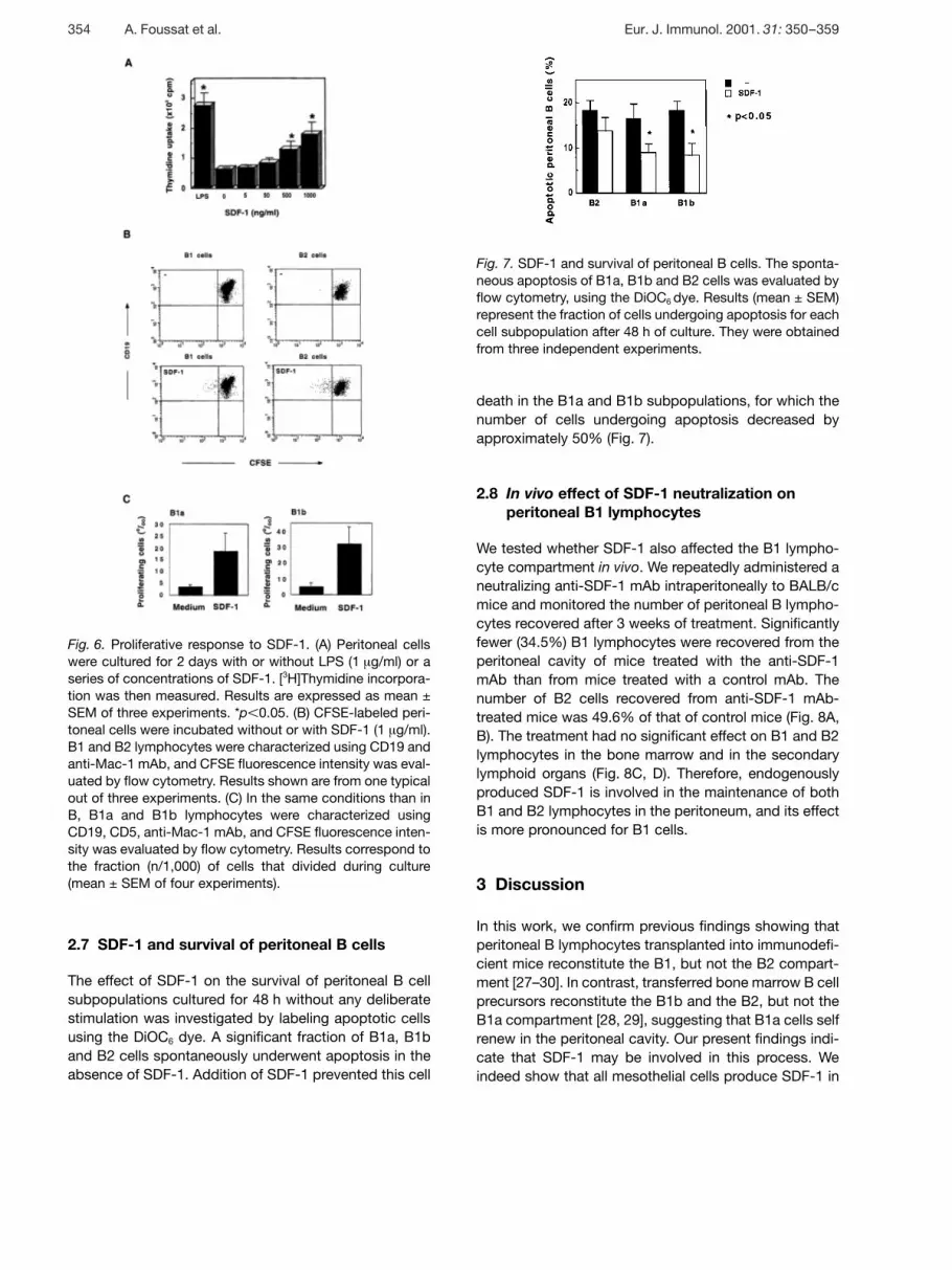
Fig. 6. Proliferative response to SDF-1. (A) Peritoneal cellswere cultured for 2 days with or without LPS (1 ? g/ml) or aseries of concentrations of SDF-1. [3H]Thymidine incorpora-tion was then measured. Results are expressed as mean ±SEM of three experiments. *p X 0.05. (B) CFSE-labeled peri-toneal cells were incubated without or with SDF-1 (1 ? g/ml).B1 and B2 lymphocytes were characterized using CD19 andanti-Mac-1 mAb, and CFSE fluorescence intensity was eval-uated by flow cytometry. Results shown are from one typicalout of three experiments. (C) In the same conditions than inB, B1a and B1b lymphocytes were characterized usingCD19, CD5, anti-Mac-1 mAb, and CFSE fluorescence inten-sity was evaluated by flow cytometry. Results correspond tothe fraction (n/1,000) of cells that divided during culture(mean ± SEM of four experiments).
Fig. 7. SDF-1 and survival of peritoneal B cells. The sponta-neous apoptosis of B1a, B1b and B2 cells was evaluated byflow cytometry, using the DiOC6 dye. Results (mean ± SEM)represent the fraction of cells undergoing apoptosis for eachcell subpopulation after 48 h of culture. They were obtainedfrom three independent experiments.
2.7 SDF-1 and survival of peritoneal B cells
The effect of SDF-1 on the survival of peritoneal B cellsubpopulations cultured for 48 h without any deliberatestimulation was investigated by labeling apoptotic cellsusing the DiOC6 dye. A significant fraction of B1a, B1band B2 cells spontaneously underwent apoptosis in theabsence of SDF-1. Addition of SDF-1 prevented this cell
death in the B1a and B1b subpopulations, for which thenumber of cells undergoing apoptosis decreased byapproximately 50% (Fig. 7).
2.8 In vivo effect of SDF-1 neutralization onperitoneal B1 lymphocytes
We tested whether SDF-1 also affected the B1 lympho-cyte compartment in vivo. We repeatedly administered aneutralizing anti-SDF-1 mAb intraperitoneally to BALB/cmice and monitored the number of peritoneal B lympho-cytes recovered after 3 weeks of treatment. Significantlyfewer (34.5%) B1 lymphocytes were recovered from theperitoneal cavity of mice treated with the anti-SDF-1mAb than from mice treated with a control mAb. Thenumber of B2 cells recovered from anti-SDF-1 mAb-treated mice was 49.6% of that of control mice (Fig. 8A,B). The treatment had no significant effect on B1 and B2lymphocytes in the bone marrow and in the secondarylymphoid organs (Fig. 8C, D). Therefore, endogenouslyproduced SDF-1 is involved in the maintenance of bothB1 and B2 lymphocytes in the peritoneum, and its effectis more pronounced for B1 cells.
3 Discussion
In this work, we confirm previous findings showing thatperitoneal B lymphocytes transplanted into immunodefi-cient mice reconstitute the B1, but not the B2 compart-ment [27–30]. In contrast, transferred bone marrow B cellprecursors reconstitute the B1b and the B2, but not theB1a compartment [28, 29], suggesting that B1a cells selfrenew in the peritoneal cavity. Our present findings indi-cate that SDF-1 may be involved in this process. Weindeed show that all mesothelial cells produce SDF-1 in
354 A. Foussat et al. Eur. J. Immunol. 2001. 31: 350–359

Fig. 8. In vivo effect of an anti-SDF-1 mAb on peritoneal Blymphocyte recovery. Mice were treated for 3 weeks with ananti-SDF-1 (empty dots and columns) or a control (full dotsand columns) mAb. Peritoneal cells (A, B) and cells in lym-phoid organs (C, D) were then recovered, enumerated andanalyzed for CD19 and Mac-1 expression. Results areexpressed as the absolute numbers of B1 (A) or B2 (B) lym-phocytes recovered, or as the fraction of B1 (C) or B2 (D)cells recovered among total cells. Mean ± SEM results fromsix mice are shown in (C) and (D) (BM: bone marrow; pLN:peripheral lymph nodes; mLN: mesenteric lymph nodes).
mice, whether they are of peritoneal, pericardial or pleu-ral origin, as we have also shown to be the case in thehuman fetus [18]. We also show that peritoneal B lym-phocytes express a functional SDF-1 receptor, as theyrespond to SDF-1 stimulation by reorganizing their cyto-skeleton.
SDF-1 stimulates the proliferation and migration of B1a,B1b and B2 peritoneal cells. This effect is observed inonly a few cells. Several mechanisms may account forthis observation. First, contact of B or T lymphocyteswith SDF-1 induces internalization of CXCR4, leading tothe transient desensitization of this receptor [31, 32]. It istherefore possible that B lymphocytes resident in theperitoneum are not fully sensitive to SDF-1 due to its per-sistent local production. Second, an optimal effect ofSDF-1 may require additional growth factor(s) not pre-sent in our experimental conditions. A synergy betweenSDF-1 and other cytokines has been observed for a
number of hematopoietic cells, such as MDC for plate-lets [33], thrombopoietin for megakaryocyte precursors[34, 35], stem cell factor/kit ligand, IL-6 and steel factorfor hematopoietic progenitors [34, 36, 37]. Preliminaryresults indicate that such a synergy between SDF-1 andother cytokines also exists for peritoneal B lymphocytes,and that IL-10 is able to potentiate some of the effects ofSDF-1 on this cell population.
An effect of SDF-1 on minor subsets of peritoneal B lym-phocytes may also explain why only a few peritonealcells migrate and proliferate in response to SDF-1.Whereas we could not characterize the minor subsets ofB1b and B2 peritoneal B lymphocytes responding toSDF-1, we observed that a large fraction of B1a cellsmigrating along a gradient of SDF-1 displayed a pheno-type distinct from that of other B1a cells. They expressedhigher levels of CD5 and CD43, and they were negativefor B220 and CD1a. Further studies on this minor subsetof peritoneal B lymphocytes should help to definewhether it has other functional specificities.
SDF-1 was recently shown to sustain survival of CD34+
hematopoietic precursors [34]. We show that such aneffect is also observed with peritoneal B lymphocytes.Prevention of apoptosis by SDF-1 occurred in a largerfraction of peritoneal B lymphocytes than proliferationand migration. This suggests that the fraction of perito-neal B lymphocytes responsive to SDF-1 is larger thanindicated by proliferation and migration assays. Alterna-tively, the protective effect of SDF-1 on apoptosis maybe indirect, through the release of yet unidentified sur-vival factors acting on a large fraction of peritoneal Blymphocytes.
Altogether, these in vitro findings suggest that SDF-1produced by mesothelial cells attracts subsets of B lym-phocytes to coelomic cavities, stimulates their local pro-liferation and prevents their death. This would explainwhy the in vivo neutralization of SDF-1 in mice signifi-cantly decreases the number of peritoneal B lympho-cytes. A unifying mechanism involving mesothelial cell-derived SDF-1 may therefore account for the accumula-tion of B1 cells in coelomic cavities, particularly in theperitoneal cavity, independently of bone marrow. The rel-ative contribution in this phenomenon of SDF-1 effect onattraction and amplification of a minor B cell subset ver-sus SDF-1-induced increase of cell survival in a largerfraction of peritoneal B cells remains to be defined.
In vitro findings have shown that SDF-1 is a chemoat-tractant and a growth factor for B cell precursors [15, 16].In vivo transfer of hematopoietic stem cells deficient inCXCR4 expression has demonstrated that SDF-1attracts B lymphocyte precursors to the bone marrow,
Eur. J. Immunol. 2001. 31: 350–359 SDF-1 and peritoneal B lymphocytes 355
but it was not clear whether it could directly stimulate theproliferation of precursors [17, 38]. With respect tomature B lymphocytes, Bleul et al. [7] showed that SDF-1triggers actin polymerization and migration of humantonsillar naive and memory B lymphocytes. In contrast,SDF-1 stimulation of freshly isolated B2 cells frommouse spleen does not trigger actin polymerization andproliferation. It is unclear whether this reflects a differ-ence in the behavior of human and mice B2 lympho-cytes, or between tonsil and spleen B lymphocytes, or adifference in experimental procedures. As mice reconsti-tuted with B cells lacking CXCR4 display no abnormalityof secondary lymphoid organs [17], SDF-1 and its recep-tor may not play a critical role in the targeting to and pro-liferation in secondary lymphoid organs of mature B2lymphocytes. This is consistent with the absence ofmodification of B2 cell content in secondary lymphoidorgans of mice treated with the anti-SDF-1 mAb and withthe normal architecture of such organs in treated mice(data not shown). There was also no change of B1 con-tent in secondary lymphoid organs of anti-SDF-1-treatedmice, suggesting that this chemokine is not involved inthe homing of B1 cells to these tissues. However, it is dif-ficult to exclude minor modifications considering the dif-ficulty in accurately quantifying B1 cells in secondarylymphoid organs due to their low frequency.
Peritoneal B2 cells, unlike freshly isolated spleen B2 lym-phocytes, are sensitive to SDF-1 stimulation. This differ-ent behavior possibly reflects a particular stage of matu-ration or a particular mechanism of preactivation of peri-toneal B2 cells, and their SDF-1 sensitivity may triggertheir targeting to the peritoneal cavity. Alternatively, peri-toneal and spleen B2 lymphocytes may not belong to thesame lineage. However, the repopulation of immunodefi-cient mice with peritoneal B cells restores the B1 but notthe B2 peritoneal compartment, indicating that perito-neal B2 cells require a constant supply from the bonemarrow for stable numbers of these cells to be main-tained throughout life.
SDF-1 is produced not only by mesothelial cells (thiswork), but also by bone marrow stromal cells [15]. It istherefore unclear why B1 cells specifically target to anddevelop within the peritoneal cavity but not in the bonemarrow, whereas the opposite applies to precursors ofB2 cells. Presumably, additional cytokines and/or recep-tor ligands specifically expressed by mesothelial/perito-neal cells or by bone marrow stromal cells account forthis difference in behavior. Potential candidates for B1cell-specific factors include IL-5, IL-9 and IL-10 [39–42].
In humans, some B lymphomas (primary effusion lym-phomas, PEL) selectively develop in body cavities, andthese tumors are unusually frequent in HIV-infected
patients [43]. It is possible that PEL represent the humanmalignant counterpart of murine B1 lymphocytes,because they share the same tropism for body cavities,and because they produce large amounts of IL-10 [44], acharacteristic of murine B1 lymphocytes [21, 22]. CXCR4is expressed by cell lines derived from PEL (unpublishedresults). This raises the hypothesis that SDF-1 producedby mesothelial cells plays a role in the emergence andmaintenance of PEL in body cavities.
This work provides new insight into the accumulationand local renewal of B1a cells in the peritoneal cavity inadults. SDF-1, which is produced by mesothelial cells,displays a number of effects on peritoneal B1a lympho-cytes. It attracts and stimulates the proliferation of aminor subpopulation, and it prevents cell death in alarger fraction of cells. These effects may explain thelong-term persistence of B1a cells in the peritoneal cav-ity. Our results also show that the peritoneal B1a popula-tions are not homogeneous, and that cells with theCD5highCD43highB220–CD1d– phenotype are the mostsensitive to the chemoattractant properties of SDF-1.Whereas this minor subset of peritoneal B lymphocytesdisplay other functional specificities remains to be deter-mined. During fetal development, the liver is the mainsite of B cell lymphopoiesis, and it produces mostly B1lymphocytes. SDF-1 production is a key event in thisfetal liver lymphopoiesis. Our present findings suggestthat, even after B cell lymphopoiesis has stopped in theliver, around birth, a similar phenomenon persiststhroughout life in the peritoneal cavity, and that this pro-cess is also dependent on the local production of SDF-1.
4 Materials and methods
4.1 Mice and cell preparation
Mice were obtained from Iffa Credo (L’Abresle, France). Allmice were 7–8-week-old BALB/c females. Peritoneal cellswere obtained by washing the peritoneum of mice with30 ml of phosphate-buffered saline (PBS). Approximately 2million peritoneal cells were recovered per mice, which rou-tinely contained 10–15% of both B1 and B2 cells. Cells fromfive mice were pooled for F-actin polymerization experi-ments, for proliferation experiments with CFSE, (MolecularProbes, Leiden, Netherlands) and for survival studies. Cellsfrom two mice were pooled for Transwell assays and thymi-dine incorporation experiments.
For CD19+ cell transfer to CB.17 SCID mice, peritoneal cellsfrom BALB/c mice were collected and purified usingstreptavidin-coated magnetic beads and a biotinylated CD19mAb (Becton Dickinson, Rungis, France). Cells (2×10⁵) wereinjected intraperitoneally to SCID mice. Peritoneal cells wererecovered 5 weeks later and phenotypically characterized.
356 A. Foussat et al. Eur. J. Immunol. 2001. 31: 350–359

4.2 Immunohistochemistry
Immunohistochemistry was performed on frozen tissues.The biotinylated anti-SDF1 K15C [18] (IgG2a) mAb was usedat a final concentration of 10 ? g/ml. Binding of this mAb wasdetected using streptavidin-alkaline phosphatase (Bioge-nex, San Ramon, CA) and the fast red substrate (Sigma,Lisle d’Abeau, France), as recommended by the manufac-turers. The sections were counterstained with Mayer hema-toxylin.
4.3 mAb for flow cytometry studies
For flow cytometry studies, peritoneal cells were stainedwith allophycocyanin-labeled rat anti-Mac1 (clone M1/70)mAb, fluorescein- or PE-cyanin5-labeled rat anti-CD5 (clone53–7.3) mAb and PE- or fluorescein-labeled rat anti-CD19(clone 1D3) mAb. We also used PE-labeled rat anti-CD43(clone S7), PE-labeled rat anti-B220 (clone RA3–6B2), PE-labeled rat anti-CD1d (clone 1B1). All mAb were purchasedfrom Becton Dickinson.
4.4 Actin polymerization
Actin polymerization experiments were performed as previ-ously described [45]. Briefly, cells were stained with fluores-cent mAb and incubated at 37°C in RPMI medium contain-ing 20 mM HEPES (6×10⁶cells/ml), with or without humanSDF-1 (250 ng/ml). Every 15 s, 100 ? l of cell suspension wasadded to 400 ? l of 10–7 M FITC-labeled phalloidin (Sigma),0.125 mg/ml l- § -lysophosphatidylcholine and 4.5% formal-dehyde in PBS. Fixed cells were analyzed by flow cytometry.
4.5 Transwell chemotaxis assays
Cell migration was evaluated using the Transwell system(Corning Costar, Brumath, France). Cells were stained withfluorescent mAb. Then, 1.5×10⁶ cells in 150 ? l RPMI mediumsupplemented with 20 mM Hepes and 1% mice serum wereadded to the upper chamber and 600 ? l of the samemedium with or without human SDF-1 (0.1 or 1 ? g/ml) wereadded to the lower chamber. The two chambers were sepa-rated by a polycarbonate membrane with 5- ? m pores. TheTranswell system was then incubated at 37°C for 2 h. Cellsrecovered in the lower chamber were pelleted, fixed andanalyzed by flow cytometry. Murine B lymphocyte chemoat-tractant (R&D Systems, Minneapolis, MN) was also tested inparallel with SDF-1.
4.6 Proliferation assays
For thymidine incorporation studies, peritoneal cells wereplated at a density of 106/ml in 100 ? l RPMI medium supple-mented with 10% fetal calf serum (FCS; Life Technology,
Cergy Pontoise, France) with or without LPS (1 ? g/ml;Sigma) or a series of concentration of human SDF-1. Cellswere incubated for 2 days at 37°C, in an atmosphere con-taining 5%CO2. 0.5 ? Ci of [methyl-3H]thymidine (Amersham,Orsay, France) was added to each well during the last 18 hof culture. Cells were then harvested and thymidine incorpo-ration was evaluated on a scintillation g -plate 1205 counter(EGG Wallac, Turku, Finland).
For single-cell proliferation evaluations, peritoneal cells wereincubated at a density of 2×10⁶/ml in 5 ? g/ml CFSE in PBSfor 10 min. Cells were washed and incubated for 4 days at adensity of 2×10⁶ cells/ml in RPMI medium supplementedwith FCS (10%) with or without human SDF-1 (1 ? g/ml) at37°C, in an atmosphere containing 5% CO2. Cells were thenwashed and stained with fluorescent mAb, fixed and ana-lyzed by flow cytometry.
4.7 Survival assay
Enumeration of apoptotic cells was performed using theDiOC6(3) (3,3’-dihexylocarbocyanine iodide) assay, as previ-ously described [46]. Briefly, peritoneal cells were cultured ata density of 2×10⁶ cells/ml in RPMI medium supplementedwith FCS (10%) with or without human SDF-1 (1 ? g/ml) at37°C, in an atmosphere containing 5% CO2. After 48 h, cellswere washed and labeled with fluorescent mAb. Cells werethen labeled with 50 nMDiOC6 and incubated in PBS for30 min at 37°C. Cells were then put at 4°C and immediatelyanalyzed by flow cytometry. Only cells contained in thesame gate than viable CD19+ cells were enumerated.
4.8 In vivo treatment of mice
BALB/c mice were injected intraperitoneally twice a week for3 weeks with 100 ? g of an anti-SDF-1 K15C mAb or 100 ? gof the A12 anti-phosphorylcholine control mAb. The anti-SDF-1 K15C mAb recognizes the N-terminal seven aminoacids of SDF-1 [18], which are conserved in human andmurine SDF-1, and it neutralizes human SDF-1 [47]. At2 days after the last injection, cells from the peritoneal cavityand from lymphoid tissues were recovered and stained withfluorescent mAb for flow cytometry. Two independent exper-iments, each involving three control mice and three anti-SDF-1 mAb-treated mice, were performed.
4.9 Statistical analysis
Unless specified, results were compared using the Mann-Whitney U test.
Acknowledgements: This work was supported by theAssociation de Recherche sur la Polyarthrite. F. Arenzana-Seisdedos is acknowledged for helpful discussion,
Eur. J. Immunol. 2001. 31: 350–359 SDF-1 and peritoneal B lymphocytes 357

N. Schrantz for advise for apoptosis experiments, and Y.Richard and R. Krzysiek for critical review of the manuscript.A.F. was a recipient from the Association de Recherche surle Cancer.
References
1 Van Snick, J., Houssiau, F., Proost, P., Van Damme J. andRenauld, J. C., I-309/T cell activation gene-3 chemokine pro-tects murine T cell lymphomas against dexamethason-inducedapoptosis. J. Immunol. 1996. 157: 2570–2576.
2 Hadida, F., Vieillard, V., Autran, B., Clark-Lewis, I., Baggiolini,M. and Debre, P., HIV-specific T cell cytotoxicity mediated byRANTES via the chemokine receptor CCR3. J. Exp. Med. 1998.188: 609–614.
3 Baggiolini, M., Chemokines and leukocyte traffic. Nature 1998.392: 565–568.
4 Sallusto, F., Lanzavecchia, A. and Mackay, C. R., Chemokinesand chemokine receptors in T cell priming and Th1/Th2-mediated responses. Immunol. Today 1998. 19: 568–574.
5 Forster, R., Mattis, A. E., Kremmer, E., Wolf, E., Brem, G. andLipp, M., A putative chemokine receptor, BLR1, directs B cellmigration to defined lymphoid organs and specific anatomiccompartments of the spleen. Cell 1996. 87: 1037–1047.
6 Butcher, E. C. and Picker, L. J., Lymphocyte homing andhomeostasis. Science 1996. 272: 60–66.
7 Bleul, C. C., Schultze, J. L. and Springer, T. A., B lymphocytechemotaxis regulated in association with microanatomic localiza-tion, differentiation state, and B cell receptor engagement. J Exp.Med. 1998. 187: 753–762.
8 Bleul, C. C., Fuhlbrigge, R. C., Casasnovas, J. M., Aiuti, A. andSpringer, T. A., A highly efficacious lymphocyte chemoattractant,stromal cell-derived factor 1 (SDF-1). J. Exp. Med. 1996. 184:1101–1109.
9 D’Apuzzo, M., Rolink, A., Loetscher, M., Hoxie, J. A., Clark-Lewis, I., Melchers, F., Baggiolini, M. and Moser, B., The che-mokine SDF-1, stromal cell-derived factor 1, attracts early stageB cell precursors via the chemokine receptor CXCR4. Eur. J.Immunol. 1997. 27: 1788–1793.
10 Aiuti, A., Webb, I. J., Springer, T. and Gutierrez-Ramos, J. C.,The chemokine SDF-1 is a chemoattractant for human CD34+hematopoietic progenitor cells and provides a new mechanism toexplain the mobilization of CD34+ progenitors to peripheralblood. J. Exp. Med. 1997. 185: 111–120.
11 Kim, C. H., Pelus, L. M., White, J. R. and Broxmeyer, H. E., Dif-ferential chemotactic behavior of developing T cells in responseto thymic chemokines. Blood 1998. 91: 4434–4443.
12 Nagasawa, T., Hiroa, S., Tachibana, K., Takakura, N., Nishi-kawa, S., Kitamura, Y., Yoshida, N., Kikutani, H. and Kishi-moto, T., Defects of B cell lymphopoiesis and bone-marrow mye-lopoiesis in mice lacking the CXC chemokine PBSF/SDF-1.Nature 1996. 382: 635–638.
13 Tachibana, K., Hirota, S., Lizasa, H., Yoshida, H., Kawabata,K., Kataoka, Y., Kitamura, Y., Matsushima, K., Yoshida, N.,Nishikawa, S., Kishimoto, T. and Nagasawa, T., The chemokinereceptor CXCR4 is essential for vascularization of the gastroin-testinal tract. Nature 1998. 393: 591–594.
14 Ma, Q., Jones, D., Borghesani, P. R., Segal, R. A., Nagasawa,T., Kishimoto, T., Bronson, R. T. and Springer, T. A., ImpairedB-lymphopoiesis, myelopoiesis, and derailed cerebellar neuron
migration in CXCR4 and SDF-1 deficient mice. Proc. Natl. Acad.Sci. USA 1998. 95: 9448–9453.
15 Nagasawa, T., Kikutani, H. and Kishimoto, T., Molecular clon-ing and structure of a pre-B cell growth stimulating factor. Proc.Natl. Acad. Sci. USA 1994. 91: 2305–2309.
16 Nagasawa, T., Nakajima, T., Tachibana, K., Lizasa, H., Bleul,C. C., Yoshie, O., Matsushima, K., Yoshida, N., Springer, T. A.and Kishimoto, T., Molecular cloning and characterization of amurine pre-B cell growth stimulating factor stromal cell derivedfactor 1 receptor, a murine homolog of the human immunodefi-ciency virus 1 entry coreceptor fusin. Proc. Natl. Acad. Sci. USA1996. 93:1426–1429.
17 Ma, Q., Jones, D. and Springer, T. A., The chemokine receptorCXCR4 is required for the retention of B lineage and granulocyticprecursors within the bone marrow microenvironment. Immunity1999. 10: 463–471.
18 Coulomb-L’Hermine, A., Amara, A., Schiff, C., Durand-Gasselin, I., Foussat, A., Delaunay, T., Chaouat, G., Capron, F.,Ledee, N., Galanaud, P., Arenzana-Seisdedos, F. and Emilie,D., Stromal cell derived factor 1 (SDF-1) and antenatal human Bcell lymphopoiesis: expression of SDF-1 by mesothelial cells andbiliary ductal plate epithelial cells. Proc. Natl. Acad. Sci. USA1999. 96: 8585–8590.
19 Zou, Y. R., Kottmann, A. H., Kuroda, M., Taniuchi, I. and Litt-man, D. R., Function of the chemokine receptor CXCR4 in hae-matopoiesis and in cerebellar development. Nature 1998. 393:595–599.
20 Youinou, P., Jamin, C. and Lydyard, P. M., CD5 expression inhuman B cell populations. Immunol. Today 1999. 20: 312–316.
21 Kantor, A. B., The development and repertoire of B-1 cells (CD5B cells). Immunol. Today 1991. 12: 389–391.
22 Tarakhovsky, A., Bar Mitzvah for B-1 cells: how will they growup? J. Exp. Med. 1997. 185: 981–984.
23 Pillai, S., The chosen few? Positive selection and the generationof naive B lymphocytes. Immunity 1999. 10: 493–502.
24 Wofsy, D. and Chiang, N. Y., Proliferation of Ly-1 B cells in auto-immune NZB and (NZB × NZW) F1 mice. Eur. J. Immunol. 1987.17: 809–814.
25 Ramachandra, S., Metcalf, R. A., Fredrickson, T., Marti, G. E.and Raveche, E., Requirement for increased IL-10 in the devel-opment of B-1 lymphoproliferative disease in a murine model ofCLL. J. Clin. Invest. 1996. 98: 1788–1793.
26 Reininger, L., Winkler, T. H., Kalberer, C. P., Jourdan, M., Mel-chers, F. and Rolink, A. G., Intrinsic B cell defects in NZB andNZW mice contribute to systemic lupus erythematosus in(NZB × NZW) F1 mice. J. Exp. Med. 1996. 184: 853–861.
27 Hayakawa, K., Hardy, R. R., Herzenberg, L. A. and Herzen-berg, L. A., Progenitors for Ly-1 B cells are distinct from progeni-tors for other B cells. J. Exp. Med. 1985. 161: 1554–1568.
28 Hardy, R. R. and Hayakawa, K., CD5 B cells, a fetal B cell line-age. Adv. Immunol. 1994. 55: 297-339.
29 Yang, Y. G., deGoma, E., Barth, R., Sergio, J. J. and Sykes, M.,B cell reconstitution and xenoreactive anti-pig natural antibodyproduction in severe combined immunodeficient mice reconsti-tuted with immunocompetent B cells from varying sources.Transplantation 1998. 66: 89–95.
30 Marcos, M. A. R., Gaspar, M. L., Malenchere, E. and Coutinho,A., Isolation of peritoneal precursors of B-1 cells in the adultmouse. Eur. J. Immunol. 1994. 24: 1033–1040.
31 Amara, A., Gall, S. L., Scwartz, O., Salamero, J., Montes, M.,Loetscher, P., Baggiolini, M., Virelizier, J. L. and Arenzana-
358 A. Foussat et al. Eur. J. Immunol. 2001. 31: 350–359

Seisdedos, F., HIV coreceptor downregulation as antiviral princi-ple: SDF-1alpha dependent internalization of the chemokinereceptor CXCR4 contributes to inhibition of HIV replication. J.Exp. Med. 1997. 186: 139–146.
32 Peled, A., Petit, I., Kollet, O., Magid, M., Ponomaryov; T., Byk,T., Nagler, A., Ben-Hur, H., Many, A., Shultz, L., Lider, O., Alon,R., Zipori, D. and Lapidot, T., Dependence of human stem cellengraftment and repopulation of NOD/SCID mice on CXCR4.Science 1999. 283: 845–848.
33 Kowasalka, M. A., Ratajczak, M. Z., Majka, M., Jin, J., Kuna-puli, S., Brass, L. and Poncz, M., Stromal cell-derived factor-1and macrophage-derived chemokine: 2 chemokines that activateplatelets. Blood 2000. 96: 50–57.
34 Lataillade, J. J., Clay, D., Dupuy, C., Rigal, S., Jasmin, C., Bou-rin, P. and Le Bousse-Kerdiles, M. C., Chemokine SDF-1enhances circulating CD34+ cell proliferation in synergy withcytokines: possible role in progenitor survival. Blood 2000. 95:756–768.
35 Hodohara, K., Fujii, N., Yamamoto, N. and Kaushansky, K.,Stromal cell-derived factor-1 (SDF-1) acts together with thrombo-poietin to enhance the development of megacaryocytic progeni-tor cells (CFU-MK). Blood 2000. 95: 769–775.
36 Dutt, P., Wang, J-F. and Groopman, J. E., Stromal cell-derivedfactor-1 § and stem cell factor. kit ligand share signalling path-ways in hemopoietic progenitors: a potential mechanism forcooperative induction of chemotaxis. J. Immunol. 1998. 161:3652–3658.
37 Kim, C. H. and Broxmeyer, H. E., In vitro behaviour of hemato-poietic progenitor cells under the influence of chemoattractants:stromal cell-derived factor-1 and Steel factor, and the bone mar-row environment. Blood 1998. 91: 100–110.
38 Kawabata, K., Ujikawa, M., Egawa, T., Kawamoto, H., Tachi-bana, K., Lizasa, H., Katsura, Y., Kishimoto, T. and Nagasawa,T., A cell-autonomous requirement for CXCR4 in long-term lym-phoid and myeloid reconstitution. Proc. Natl. Acad. Sci. USA1999. 96: 5663–5667.
39 Nisitani, S., Tsubata, T., Murakami, M. and Honjo, T., Adminis-tration of interleukin 5 or 10 activates peritoneal B-1 cells andinduces autoimmune hemolytic anemia in anti-erythrocyte auto-antibody transgenic mice. Eur. J. Immunol. 1995. 25: 3047–3052.
40 Peng, B., Mehta, N. H., Fernandes, H., Chou, C. C. and Rave-che, E., Growth inhibition of malignant CD5+B (B-1) cells by anti-sens IL-10 oligonucleotide. Leuk. Res 1995. 19: 159–167.
41 Bao, S., Beagley, K. W., Murray, A. M., Caristo, V., Matthaei, K.I., Young, I. G. and Husband, A. J., Intestinal IgA plasma cells ofthe B1 lineage are IL-5 dependent. Immunology 1998. 94:181–188.
42 Vink, A., Warnier, G., Brombacher, F. and Renauld, J. C., Inter-leukin 9 induced in vivo expansion of the B-1 lympohcyte popula-tion. J. Exp. Med. 1999. 189: 1413–1423.
43 Nador, R. G., Cesarman, E., Chadburn, A., Dawson, D. B.,Ansari, M. Q., Sald, J. and Knowles, D. M., Primary effusionlymphoma: a distinct clinicopathologic entity associated with theKaposi’s sarcoma associated herpes virus. Blood 1996. 88:645–656.
44 Foussat, A., Wijdenes, J., Bouchet, L., Gaidano, G., Neipel, F.,Balabanian, K., Galanaud, P., Couderc, J. and Emilie, D.,Human interleukin-6 is in vivo an autocrine growth factor forhuman herpesvirus-8-infected malignant B lymphocytes. Eur.Cytokine Netw. 1999. 10: 501–508.
45 Zou, W., Foussat, A., Houhou, S., Durand-Gasselin, I., Duli-oust, A., Bouchet, L., Galanaud, P., Levy, Y., and Emilie, D.,Acute upregulation of CCR-5 expression by CD4+ T lymphocytesin HIV infected patients treated with interleukin-2. AIDS 1999. 13:455–463.
46 Zamzami, N., Marchetti, P., Castedo, M., Decaudin, D.,Macho, A., Hirsch, T., Susin, S. A., Petit, M. X., Mignotte, B.and Kroemer, G., Sequential reduction of mitochondrial trans-membrane potential and generation of reactive oxygen species inearly programmed cell death. J. Exp. Med. 1995. 182: 367–377.
47 Amara, A., Lorthioir, O., Valenzuela, A., Megerus, A., Thelen,M., Montes, M., Virelizier, J. L., Delepierre, M., Baleux, F.,Lortat-Jacob, H. and Arenzana-Seisdedos, F., Stromal-Cell-Derived Factor 1 § associates with heparan sulfates through thefirst beta strand of the chemokine. J. Biol. Chem. 1999. 274:23916–23925.
Correspondence: Dominique Emilie, Institut Paris-Sud surles Cytokines, 32 rue des Carnets, F-92140 Clamart, FranceFax: +33-1-46327993e-mail: emilie — ipsc.u-psud.fr
Eur. J. Immunol. 2001. 31: 350–359 SDF-1 and peritoneal B lymphocytes 359