A nuclear-encoded protein, mTERF6, mediates transcription ...
12
1 SCIENTIFIC REPORTS | (2018) 8:11929 | DOI:10.1038/s41598-018-30166-6 www.nature.com/scientificreports A nuclear-encoded protein, mTERF6, mediates transcription termination of rpoA polycistron for plastid-encoded RNA polymerase- dependent chloroplast gene expression and chloroplast development Yi Zhang 1,2 , Yong-Lan Cui 1 , Xiao-Lei Zhang 1 , Qing-Bo Yu 1 , Xi Wang 1 , Xin-Bo Yuan 1 , Xue-Mei Qin 1 , Xiao-Fang He 1 , Chao Huang 1 & Zhong-Nan Yang 1 The expression of plastid genes is regulated by two types of DNA-dependent RNA polymerases, plastid-encoded RNA polymerase (PEP) and nuclear-encoded RNA polymerase (NEP). The plastid rpoA polycistron encodes a series of essential chloroplast ribosome subunits and a core subunit of PEP. Despite the functional importance, little is known about the regulation of rpoA polycistron. In this work, we show that mTERF6 directly associates with a 3′-end sequence of rpoA polycistron in vitro and in vivo, and that absence of mTERF6 promotes read-through transcription at this site, indicating that mTERF6 acts as a factor required for termination of plastid genes’ transcription in vivo. In addition, the transcriptions of some essential ribosome subunits encoded by rpoA polycistron and PEP-dependent plastid genes are reduced in the mterf6 knockout mutant. RpoA, a PEP core subunit, accumulates to about 50% that of the wild type in the mutant, where early chloroplast development is impaired. Overall, our functional analyses of mTERF6 provide evidence that it is more likely a factor required for transcription termination of rpoA polycistron, which is essential for chloroplast gene expression and chloroplast development. Chloroplasts are semi-autonomous organelles derived by endosymbiosis from a relative of present-day cyano- bacteria. e plastid genome is a conserved circular double-stranded DNA of 120 to 160 kbp (154,478 bp in Arabidopsis) 1,2 . e Arabidopsis plastid genome contains 87 potential protein-coding genes, 4 rRNA genes and 37 tRNA genes 1,3 . ese plastid genes are transcribed into monocistronic and polycistronic mRNAs 4 . Two types of DNA-dependent RNA polymerases, nuclear-encoded RNA polymerase (NEP) and plastid-encoded RNA poly- merase (PEP), are responsible for the transcription of plastid genes 5,6 . NEP is a single-subunit enzyme that tran- scribes housekeeping genes, including rpoA, rpoB, rpoC1 and rpoC2 7,8 . PEP possesses the major RNA polymerase activity in the chloroplast and transcribes over 80% of the plastid genes 7 . e four core subunits of PEP (α, β, β′ and β″) are encoded by the genes rpoA, rpoB, rpoC1 and rpoC2, respectively, and are translated from two polycis- trons, L23-L2-S19-L22-S3-L16-L14-S8-L36-S11-rpoA and rpoB-rpoC1-rpoC2 9 . The primary transcripts produced by NEP and PEP often undergo post-transcriptional modification, including splicing, maturation, trimming of 5′ and 3′ ends and RNA editing before translation 10 . In addition, 1 College of Life and Environmental Sciences, Shanghai Normal University, Shanghai, 200234, China. 2 Shanghai Center for Plant Stress Biology, Chinese Academy of Sciences, Shanghai, 201602, China. Yi Zhang and Yong-Lan Cui contributed equally to this work. Correspondence and requests for materials should be addressed to Z.-N.Y. (email: [email protected]) Received: 25 May 2017 Accepted: 20 April 2018 Published: xx xx xxxx OPEN
-
Upload
khangminh22 -
Category
Documents
-
view
6 -
download
0
Transcript of A nuclear-encoded protein, mTERF6, mediates transcription ...

1Scientific RepoRts | (2018) 8:11929 | DOI:10.1038/s41598-018-30166-6
www.nature.com/scientificreports
A nuclear-encoded protein, mTERF6, mediates transcription termination of rpoA polycistron for plastid-encoded RNA polymerase-dependent chloroplast gene expression and chloroplast developmentYi Zhang1,2, Yong-Lan Cui1, Xiao-Lei Zhang1, Qing-Bo Yu1, Xi Wang1, Xin-Bo Yuan1, Xue-Mei Qin1, Xiao-Fang He1, Chao Huang1 & Zhong-Nan Yang 1
The expression of plastid genes is regulated by two types of DNA-dependent RNA polymerases, plastid-encoded RNA polymerase (PEP) and nuclear-encoded RNA polymerase (NEP). The plastid rpoA polycistron encodes a series of essential chloroplast ribosome subunits and a core subunit of PEP. Despite the functional importance, little is known about the regulation of rpoA polycistron. In this work, we show that mTERF6 directly associates with a 3′-end sequence of rpoA polycistron in vitro and in vivo, and that absence of mTERF6 promotes read-through transcription at this site, indicating that mTERF6 acts as a factor required for termination of plastid genes’ transcription in vivo. In addition, the transcriptions of some essential ribosome subunits encoded by rpoA polycistron and PEP-dependent plastid genes are reduced in the mterf6 knockout mutant. RpoA, a PEP core subunit, accumulates to about 50% that of the wild type in the mutant, where early chloroplast development is impaired. Overall, our functional analyses of mTERF6 provide evidence that it is more likely a factor required for transcription termination of rpoA polycistron, which is essential for chloroplast gene expression and chloroplast development.
Chloroplasts are semi-autonomous organelles derived by endosymbiosis from a relative of present-day cyano-bacteria. The plastid genome is a conserved circular double-stranded DNA of 120 to 160 kbp (154,478 bp in Arabidopsis)1,2. The Arabidopsis plastid genome contains 87 potential protein-coding genes, 4 rRNA genes and 37 tRNA genes1,3. These plastid genes are transcribed into monocistronic and polycistronic mRNAs4. Two types of DNA-dependent RNA polymerases, nuclear-encoded RNA polymerase (NEP) and plastid-encoded RNA poly-merase (PEP), are responsible for the transcription of plastid genes5,6. NEP is a single-subunit enzyme that tran-scribes housekeeping genes, including rpoA, rpoB, rpoC1 and rpoC27,8. PEP possesses the major RNA polymerase activity in the chloroplast and transcribes over 80% of the plastid genes7. The four core subunits of PEP (α, β, β′ and β″) are encoded by the genes rpoA, rpoB, rpoC1 and rpoC2, respectively, and are translated from two polycis-trons, L23-L2-S19-L22-S3-L16-L14-S8-L36-S11-rpoA and rpoB-rpoC1-rpoC29.
The primary transcripts produced by NEP and PEP often undergo post-transcriptional modification, including splicing, maturation, trimming of 5′ and 3′ ends and RNA editing before translation10. In addition,
1College of Life and Environmental Sciences, Shanghai Normal University, Shanghai, 200234, China. 2Shanghai Center for Plant Stress Biology, Chinese Academy of Sciences, Shanghai, 201602, China. Yi Zhang and Yong-Lan Cui contributed equally to this work. Correspondence and requests for materials should be addressed to Z.-N.Y. (email: [email protected])
Received: 25 May 2017
Accepted: 20 April 2018
Published: xx xx xxxx
OPEN
www.nature.com/scientificreports/
2Scientific RepoRts | (2018) 8:11929 | DOI:10.1038/s41598-018-30166-6
nuclear-encoded factors, such as sigma factors, pTAC and pentatricopeptide repeats (PPRs), are required for reg-ulating the plastid gene expression mediated by both NEP and PEP11–14. After post-transcriptional modification, the translational events are initiated on the chloroplast ribosome15,16. Similar to bacteria, the chloroplast has the bacterial-type 70S ribosome, composed of rRNA and protein subunits and assembled into two multi-components of the 50S (large) and 30S (small) ribosomal subunits17,18. The 50S ribosomal subunit consists of 23S, 5S and 4.5 rRNA and 33 proteins, of which 24 are nuclear-encoded, whereas the 30S subunit harbors only one 16S rRNA in addition to 24 proteins, of which 12 are nuclear-encoded19. The rest of the chloroplast gene-encoded proteins of the translation machinery can be divided into essential and non-essential components19. The L23-L2-S19-L22-S3-L16-L14-S8-L36-S11-rpoA polycistron encodes 10 ribosome proteins (L23, L2, S19, L22, S3, L16, L14, S8, L36 and S11). Only L36 belongs to the non-essential translation machinery in both bacteria (Escherichia coli) and plants19. Despite the functional importance, the precise mechanisms underlying the transcriptional regulation of the rpoA polycistron remain elusive.
The mitochondrial transcription termination factor (mTERF) family was first identified to be responsible for mitochondrial transcription termination at a site adjacent to the mitochondrial 16S rRNA gene20,21. mTERF2 binds to the mitochondrial DNA (mtDNA) in a non-sequence-specific manner22. mTERF3 and mTERF2 act as positive and negative regulators in mitochondrial DNA transcription, respectively23,24. mTERF4 regulates the translation of mitochondrial genes by association with rRNA25–27. Both mterf3 and mterf4 knockout mice cause embryo lethality, and functional studies suggest their pivotal roles in regulating ribosome biogenesis23–27.
In stark contrast to mammals, in plants, the functions of mTERFs are poorly understood28. Arabidopsis con-tains 35 mTERF proteins, and at least 11 are predicted to be localized in the plastid29. SOLDAT10/mTERF1 is involved in chloroplast retrograde signaling, and absence of SOLDAT10 leads to embryo lethality30. RUG2/BSM (RUGOSA2/BELAYA SMERT)/mTERF4 encodes a chloroplast protein involved in splicing group IIa introns of plastid genes29,31. Zm-mTERF4, the maize ortholog of the Arabidopsis protein BSM/RUG2, is required for accu-mulation of plastid ribosomes and splicing of several group II introns in the chloroplast32. In addition to its direct function in regulating plastid gene expression, mTERF4/COE1 is required for regulating un-processed plastid RNA-triggered plastid retrograde signaling33. MDA1/mTERF5 functions in chloroplast development and abi-otic stress responses30. The mda1 mutant exhibits a pale pigmentation34. twr-1/mterf9 was identified as a yellow mutant35 and alters chloroplast development and tolerance to abiotic stress36. Although several mTERF members have been characterized in Arabidopsis and were emphasized to play a role in regulating organellar gene expres-sion, whether these members also involve in the transcription termination of plastid genes is unknown.
mTERF6 is dual-targeted to both chloroplast and mitochondria in Arabidopsis37. The knockout mutant mterf6 has an albino phenotype and is defective in the maturation of trnI.237. By functional characterization of mTERF6, we found that mTERF6 is required for the transcription termination of plastid genes in vivo. The decreased RpoA protein level and PEP-dependent plastid gene transcription indicated that mTERF6-dependent transcription ter-mination of the rpoA polycistron is necessary for PEP-dependent chloroplast gene expression and chloroplast development.
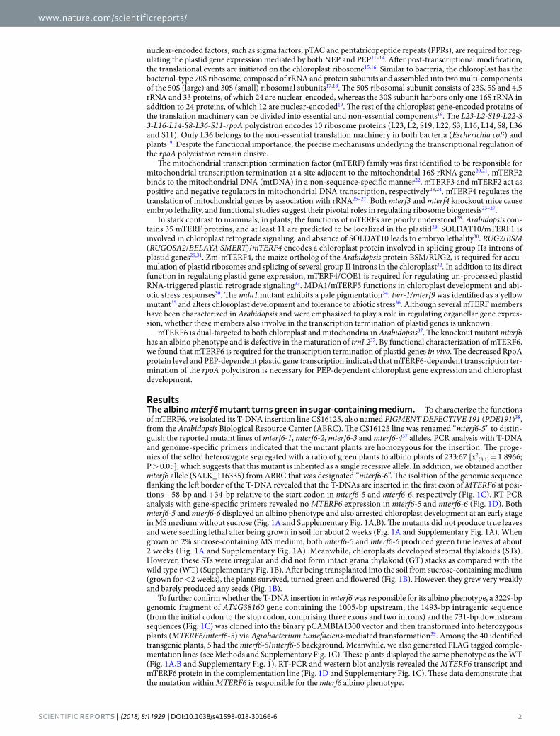
ResultsThe albino mterf6 mutant turns green in sugar-containing medium. To characterize the functions of mTERF6, we isolated its T-DNA insertion line CS16125, also named PIGMENT DEFECTIVE 191 (PDE191)38, from the Arabidopsis Biological Resource Center (ABRC). The CS16125 line was renamed “mterf6-5” to distin-guish the reported mutant lines of mterf6-1, mterf6-2, mterf6-3 and mterf6-437 alleles. PCR analysis with T-DNA and genome-specific primers indicated that the mutant plants are homozygous for the insertion. The proge-nies of the selfed heterozygote segregated with a ratio of green plants to albino plants of 233:67 [x2
(3:1) = 1.8966; P > 0.05], which suggests that this mutant is inherited as a single recessive allele. In addition, we obtained another mterf6 allele (SALK_116335) from ABRC that was designated “mterf6-6”. The isolation of the genomic sequence flanking the left border of the T-DNA revealed that the T-DNAs are inserted in the first exon of MTERF6 at posi-tions +58-bp and +34-bp relative to the start codon in mterf6-5 and mterf6-6, respectively (Fig. 1C). RT-PCR analysis with gene-specific primers revealed no MTERF6 expression in mterf6-5 and mterf6-6 (Fig. 1D). Both mterf6-5 and mterf6-6 displayed an albino phenotype and also arrested chloroplast development at an early stage in MS medium without sucrose (Fig. 1A and Supplementary Fig. 1A,B). The mutants did not produce true leaves and were seedling lethal after being grown in soil for about 2 weeks (Fig. 1A and Supplementary Fig. 1A). When grown on 2% sucrose-containing MS medium, both mterf6-5 and mterf6-6 produced green true leaves at about 2 weeks (Fig. 1A and Supplementary Fig. 1A). Meanwhile, chloroplasts developed stromal thylakoids (STs). However, these STs were irregular and did not form intact grana thylakoid (GT) stacks as compared with the wild type (WT) (Supplementary Fig. 1B). After being transplanted into the soil from sucrose-containing medium (grown for <2 weeks), the plants survived, turned green and flowered (Fig. 1B). However, they grew very weakly and barely produced any seeds (Fig. 1B).
To further confirm whether the T-DNA insertion in mterf6 was responsible for its albino phenotype, a 3229-bp genomic fragment of AT4G38160 gene containing the 1005-bp upstream, the 1493-bp intragenic sequence (from the initial codon to the stop codon, comprising three exons and two introns) and the 731-bp downstream sequences (Fig. 1C) was cloned into the binary pCAMBIA1300 vector and then transformed into heterozygous plants (MTERF6/mterf6-5) via Agrobacterium tumefaciens-mediated transformation39. Among the 40 identified transgenic plants, 5 had the mterf6-5/mterf6-5 background. Meanwhile, we also generated FLAG tagged comple-mentation lines (see Methods and Supplementary Fig. 1C). These plants displayed the same phenotype as the WT (Fig. 1A,B and Supplementary Fig. 1). RT-PCR and western blot analysis revealed the MTERF6 transcript and mTERF6 protein in the complementation line (Fig. 1D and Supplementary Fig. 1C). These data demonstrate that the mutation within MTERF6 is responsible for the mterf6 albino phenotype.
www.nature.com/scientificreports/
3Scientific RepoRts | (2018) 8:11929 | DOI:10.1038/s41598-018-30166-6
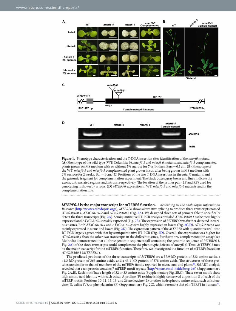
MTERF6.1 is the major transcript for mTERF6 function. According to The Arabidopsis Information Resource (http://www.arabidopsis.org/), MTERF6 shows alternative splicing to produce three transcripts named AT4G38160.1, AT4G38160.2 and AT4G38160.3 (Fig. 2A). We designed three sets of primers able to specifically detect the three transcripts (Fig. 2A). Semiquantitative RT-PCR analysis revealed AT4G38160.1 as the most highly expressed and AT4G38160.3 weakly expressed (Fig. 2B). The expression of MTERF6 was further detected in vari-ous tissues. Both AT4G38160.1 and AT4G38160.2 were highly expressed in leaves (Fig. 2C,D). AT4G38160.3 was mainly expressed in stems and leaves (Fig. 2D). The expression pattern of the MTERF6 with quantitative real-time RT-PCR largely agreed with that by semiquantitative RT-PCR (Fig. 2D). Overall, the expression was higher for AT4G38160.1 than the other two transcripts in the different tissues. Furthermore, complementation assay (see Methods) demonstrated that all three genomic sequences (all containing the genomic sequence of MTERF6.1, Fig. 2A) of the three transcripts could complement the phenotypic defects of mterf6-5. Thus, MTERF6.1 may be the major transcript for the mTERF6 function. Therefore, we investigated the function of mTERF6 based on AT4G38160.1 (MTERF6.1).
The predicted products of the three transcripts of MTERF6 are a 37.9-kD protein of 333 amino acids, a 41.3-kD protein of 363 amino acids, and a 43.1-kD protein of 378 amino acids. The structures of these pro-teins are similar to that of members of the mTERFs family reported in metazoans and plants40. SMART analysis revealed that each protein contains 7 mTERF-motif repeats (http://smart.embl-heidelberg.de/) (Supplementary Fig. 2A,B). Each motif has a length of 32 or 33 amino acids (Supplementary Fig. 2B,C). These seven motifs show high amino acid identity with each other. A proline (P) residue is highly conserved at position 8 of each of the mTERF motifs. Positions 10, 11, 15, 19, and 26 are leucine (L) or other hydrophobic amino acids, such as isoleu-cine (I), valine (V), or phenylalanine (F) (Supplementary Fig. 2C), which resemble that of mTERF1 in humans41.
Figure 1. Phenotype characterization and the T-DNA insertion sites identification of the mterf6 mutant. (A) Phenotype of the wild-type (WT; Columbia-0), mterf6-5 and mterf6-6 mutants, and mterf6-5 complemented plants grown on MS medium with or without 2% sucrose for 7 or 14 days. Bars = 0.1 cm. (B) Phenotype of the WT, mterf6-5 and mterf6-5 complemented plant grown in soil after being grown in MS medium with 2% sucrose for 2 weeks. Bar = 1 cm. (C) Positions of the two T-DNA insertions in the mterf6 mutants and the genomic fragment for complementation experiment. The black boxes, gray boxes and lines indicate the exons, untranslated regions and introns, respectively. The location of the primer pair (LP and RP) used for genotyping is shown by arrows. (D) MTERF6 expression in WT, mterf6-5 and mterf6-6 mutants and in the complementation line.
www.nature.com/scientificreports/
4Scientific RepoRts | (2018) 8:11929 | DOI:10.1038/s41598-018-30166-6
Previous studies of mTER1 revealed that these mTERF motifs are involved in nucleic acid binding21. In addition, these mTERF motifs are very conserve among plant species (Supplementary Fig. 2D).
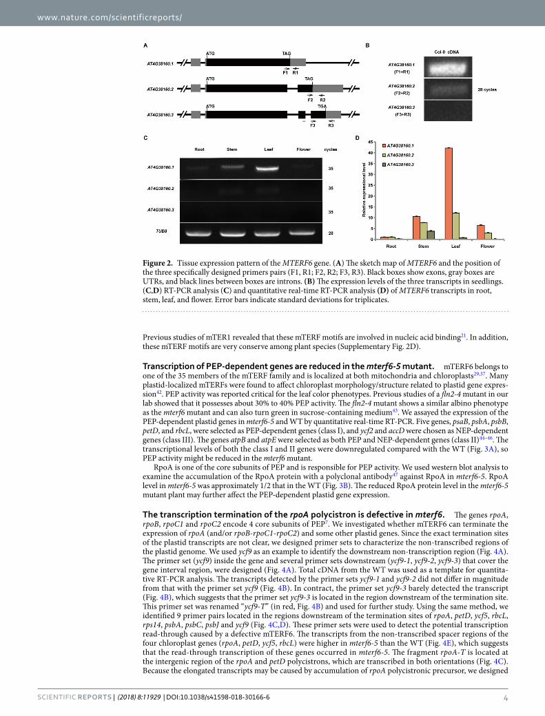
Transcription of PEP-dependent genes are reduced in the mterf6-5 mutant. mTERF6 belongs to one of the 35 members of the mTERF family and is localized at both mitochondria and chloroplasts29,37. Many plastid-localized mTERFs were found to affect chloroplast morphology/structure related to plastid gene expres-sion42. PEP activity was reported critical for the leaf color phenotypes. Previous studies of a fln2-4 mutant in our lab showed that it possesses about 30% to 40% PEP activity. The fln2-4 mutant shows a similar albino phenotype as the mterf6 mutant and can also turn green in sucrose-containing medium43. We assayed the expression of the PEP-dependent plastid genes in mterf6-5 and WT by quantitative real-time RT-PCR. Five genes, psaB, psbA, psbB, petD, and rbcL, were selected as PEP-dependent genes (class I), and ycf2 and accD were chosen as NEP-dependent genes (class III). The genes atpB and atpE were selected as both PEP and NEP-dependent genes (class II)44–46. The transcriptional levels of both the class I and II genes were downregulated compared with the WT (Fig. 3A), so PEP activity might be reduced in the mterf6 mutant.
RpoA is one of the core subunits of PEP and is responsible for PEP activity. We used western blot analysis to examine the accumulation of the RpoA protein with a polyclonal antibody47 against RpoA in mterf6-5. RpoA level in mterf6-5 was approximately 1/2 that in the WT (Fig. 3B). The reduced RpoA protein level in the mterf6-5 mutant plant may further affect the PEP-dependent plastid gene expression.
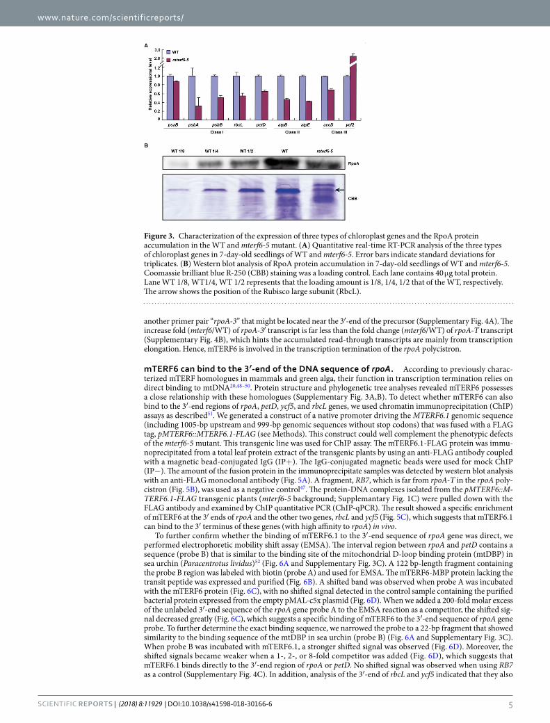
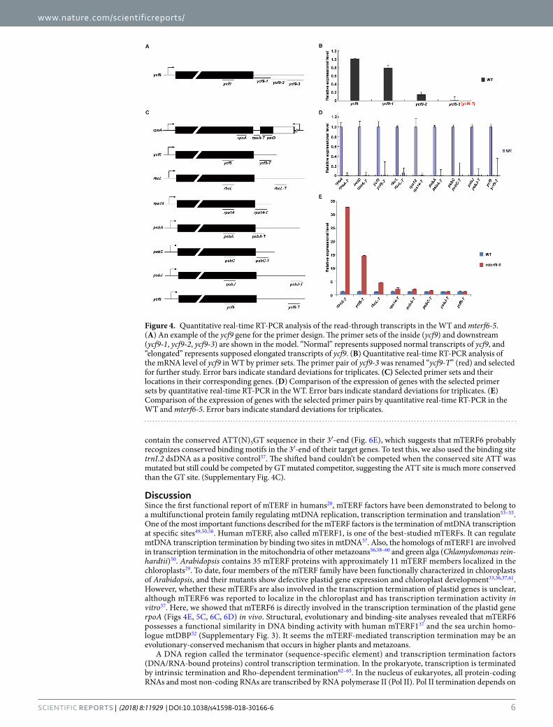
The transcription termination of the rpoA polycistron is defective in mterf6. The genes rpoA, rpoB, rpoC1 and rpoC2 encode 4 core subunits of PEP7. We investigated whether mTERF6 can terminate the expression of rpoA (and/or rpoB-rpoC1-rpoC2) and some other plastid genes. Since the exact termination sites of the plastid transcripts are not clear, we designed primer sets to characterize the non-transcribed regions of the plastid genome. We used ycf9 as an example to identify the downstream non-transcription region (Fig. 4A). The primer set (ycf9) inside the gene and several primer sets downstream (ycf9-1, ycf9-2, ycf9-3) that cover the gene interval region, were designed (Fig. 4A). Total cDNA from the WT was used as a template for quantita-tive RT-PCR analysis. The transcripts detected by the primer sets ycf9-1 and ycf9-2 did not differ in magnitude from that with the primer set ycf9 (Fig. 4B). In contract, the primer set ycf9-3 barely detected the transcript (Fig. 4B), which suggests that the primer set ycf9-3 is located in the region downstream of the termination site. This primer set was renamed “ycf9-T” (in red, Fig. 4B) and used for further study. Using the same method, we identified 9 primer pairs located in the regions downstream of the termination sites of rpoA, petD, ycf5, rbcL, rps14, psbA, psbC, psbJ and ycf9 (Fig. 4C,D). These primer sets were used to detect the potential transcription read-through caused by a defective mTERF6. The transcripts from the non-transcribed spacer regions of the four chloroplast genes (rpoA, petD, ycf5, rbcL) were higher in mterf6-5 than the WT (Fig. 4E), which suggests that the read-through transcription of these genes occurred in mterf6-5. The fragment rpoA-T is located at the intergenic region of the rpoA and petD polycistrons, which are transcribed in both orientations (Fig. 4C). Because the elongated transcripts may be caused by accumulation of rpoA polycistronic precursor, we designed
Figure 2. Tissue expression pattern of the MTERF6 gene. (A) The sketch map of MTERF6 and the position of the three specifically designed primers pairs (F1, R1; F2, R2; F3, R3). Black boxes show exons, gray boxes are UTRs, and black lines between boxes are introns. (B) The expression levels of the three transcripts in seedlings. (C,D) RT-PCR analysis (C) and quantitative real-time RT-PCR analysis (D) of MTERF6 transcripts in root, stem, leaf, and flower. Error bars indicate standard deviations for triplicates.
www.nature.com/scientificreports/
5Scientific RepoRts | (2018) 8:11929 | DOI:10.1038/s41598-018-30166-6
another primer pair “rpoA-3” that might be located near the 3′-end of the precursor (Supplementary Fig. 4A). The increase fold (mterf6/WT) of rpoA-3′ transcript is far less than the fold change (mterf6/WT) of rpoA-T transcript (Supplementary Fig. 4B), which hints the accumulated read-through transcripts are mainly from transcription elongation. Hence, mTERF6 is involved in the transcription termination of the rpoA polycistron.
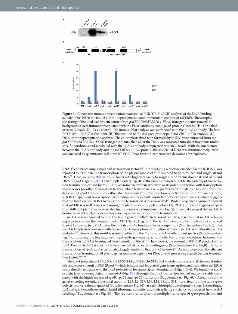
mTERF6 can bind to the 3′-end of the DNA sequence of rpoA. According to previously charac-terized mTERF homologues in mammals and green alga, their function in transcription termination relies on direct binding to mtDNA20,48–50. Protein structure and phylogenetic tree analyses revealed mTERF6 possesses a close relationship with these homologues (Supplementary Fig. 3A,B). To detect whether mTERF6 can also bind to the 3′-end regions of rpoA, petD, ycf5, and rbcL genes, we used chromatin immunoprecipitation (ChIP) assays as described51. We generated a construct of a native promoter driving the MTERF6.1 genomic sequence (including 1005-bp upstream and 999-bp genomic sequences without stop codons) that was fused with a FLAG tag, pMTERF6::MTERF6.1-FLAG (see Methods). This construct could well complement the phenotypic defects of the mterf6-5 mutant. This transgenic line was used for ChIP assay. The mTERF6.1-FLAG protein was immu-noprecipitated from a total leaf protein extract of the transgenic plants by using an anti-FLAG antibody coupled with a magnetic bead-conjugated IgG (IP+). The IgG-conjugated magnetic beads were used for mock ChIP (IP−). The amount of the fusion protein in the immunoprecipitate samples was detected by western blot analysis with an anti-FLAG monoclonal antibody (Fig. 5A). A fragment, RB7, which is far from rpoA-T in the rpoA poly-cistron (Fig. 5B), was used as a negative control47. The protein-DNA complexes isolated from the pMTERF6::M-TERF6.1-FLAG transgenic plants (mterf6-5 background; Supplemantary Fig. 1C) were pulled down with the FLAG antibody and examined by ChIP quantitative PCR (ChIP-qPCR). The result showed a specific enrichment of mTERF6 at the 3′ ends of rpoA and the other two genes, rbcL and ycf5 (Fig. 5C), which suggests that mTERF6.1 can bind to the 3′ terminus of these genes (with high affinity to rpoA) in vivo.
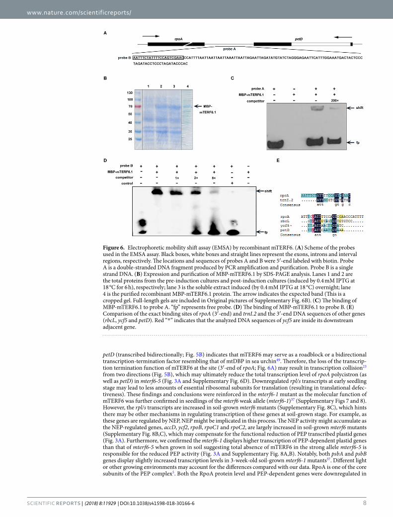
To further confirm whether the binding of mTERF6.1 to the 3′-end sequence of rpoA gene was direct, we performed electrophoretic mobility shift assay (EMSA). The interval region between rpoA and petD contains a sequence (probe B) that is similar to the binding site of the mitochondrial D-loop binding protein (mtDBP) in sea urchin (Paracentrotus lividus)52 (Fig. 6A and Supplementary Fig. 3C). A 122 bp-length fragment containing the probe B region was labeled with biotin (probe A) and used for EMSA. The mTERF6-MBP protein lacking the transit peptide was expressed and purified (Fig. 6B). A shifted band was observed when probe A was incubated with the mTERF6 protein (Fig. 6C), with no shifted signal detected in the control sample containing the purified bacterial protein expressed from the empty pMAL-c5x plasmid (Fig. 6D). When we added a 200-fold molar excess of the unlabeled 3′-end sequence of the rpoA gene probe A to the EMSA reaction as a competitor, the shifted sig-nal decreased greatly (Fig. 6C), which suggests a specific binding of mTERF6 to the 3′-end sequence of rpoA gene probe. To further determine the exact binding sequence, we narrowed the probe to a 22-bp fragment that showed similarity to the binding sequence of the mtDBP in sea urchin (probe B) (Fig. 6A and Supplementary Fig. 3C). When probe B was incubated with mTERF6.1, a stronger shifted signal was observed (Fig. 6D). Moreover, the shifted signals became weaker when a 1-, 2-, or 8-fold competitor was added (Fig. 6D), which suggests that mTERF6.1 binds directly to the 3′-end region of rpoA or petD. No shifted signal was observed when using RB7 as a control (Supplementary Fig. 4C). In addition, analysis of the 3′-end of rbcL and ycf5 indicated that they also
Figure 3. Characterization of the expression of three types of chloroplast genes and the RpoA protein accumulation in the WT and mterf6-5 mutant. (A) Quantitative real-time RT-PCR analysis of the three types of chloroplast genes in 7-day-old seedlings of WT and mterf6-5. Error bars indicate standard deviations for triplicates. (B) Western blot analysis of RpoA protein accumulation in 7-day-old seedlings of WT and mterf6-5. Coomassie brilliant blue R-250 (CBB) staining was a loading control. Each lane contains 40 μg total protein. Lane WT 1/8, WT1/4, WT 1/2 represents that the loading amount is 1/8, 1/4, 1/2 that of the WT, respectively. The arrow shows the position of the Rubisco large subunit (RbcL).
www.nature.com/scientificreports/
6Scientific RepoRts | (2018) 8:11929 | DOI:10.1038/s41598-018-30166-6
contain the conserved ATT(N)5GT sequence in their 3′-end (Fig. 6E), which suggests that mTERF6 probably recognizes conserved binding motifs in the 3′-end of their target genes. To test this, we also used the binding site trnI.2 dsDNA as a positive control37. The shifted band couldn’t be competed when the conserved site ATT was mutated but still could be competed by GT mutated competitor, suggesting the ATT site is much more conserved than the GT site. (Supplementary Fig. 4C).
DiscussionSince the first functional report of mTERF in humans20, mTERF factors have been demonstrated to belong to a multifunctional protein family regulating mtDNA replication, transcription termination and translation53–55. One of the most important functions described for the mTERF factors is the termination of mtDNA transcription at specific sites49,50,56. Human mTERF, also called mTERF1, is one of the best-studied mTERFs. It can regulate mtDNA transcription termination by binding two sites in mtDNA57. Also, the homologs of mTERF1 are involved in transcription termination in the mitochondria of other metazoans56,58–60 and green alga (Chlamydomonas rein-hardtii)50. Arabidopsis contains 35 mTERF proteins with approximately 11 mTERF members localized in the chloroplasts29. To date, four members of the mTERF family have been functionally characterized in chloroplasts of Arabidopsis, and their mutants show defective plastid gene expression and chloroplast development33,36,37,61. However, whether these mTERFs are also involved in the transcription termination of plastid genes is unclear, although mTERF6 was reported to localize in the chloroplast and has transcription termination activity in vitro37. Here, we showed that mTERF6 is directly involved in the transcription termination of the plastid gene rpoA (Figs 4E, 5C, 6C, 6D) in vivo. Structural, evolutionary and binding-site analyses revealed that mTERF6 possesses a functional similarity in DNA binding activity with human mTERF137 and the sea urchin homo-logue mtDBP52 (Supplementary Fig. 3). It seems the mTERF-mediated transcription termination may be an evolutionary-conserved mechanism that occurs in higher plants and metazoans.
A DNA region called the terminator (sequence-specific element) and transcription termination factors (DNA/RNA-bound proteins) control transcription termination. In the prokaryote, transcription is terminated by intrinsic termination and Rho-dependent termination62–65. In the nucleus of eukaryotes, all protein-coding RNAs and most non-coding RNAs are transcribed by RNA polymerase II (Pol II). Pol II termination depends on
Figure 4. Quantitative real-time RT-PCR analysis of the read-through transcripts in the WT and mterf6-5. (A) An example of the ycf9 gene for the primer design. The primer sets of the inside (ycf9) and downstream (ycf9-1, ycf9-2, ycf9-3) are shown in the model. “Normal” represents supposed normal transcripts of ycf9, and “elongated” represents supposed elongated transcripts of ycf9. (B) Quantitative real-time RT-PCR analysis of the mRNA level of ycf9 in WT by primer sets. The primer pair of ycf9-3 was renamed “ycf9-T” (red) and selected for further study. Error bars indicate standard deviations for triplicates. (C) Selected primer sets and their locations in their corresponding genes. (D) Comparison of the expression of genes with the selected primer sets by quantitative real-time RT-PCR in the WT. Error bars indicate standard deviations for triplicates. (E) Comparison of the expression of genes with the selected primer pairs by quantitative real-time RT-PCR in the WT and mterf6-5. Error bars indicate standard deviations for triplicates.
www.nature.com/scientificreports/
7Scientific RepoRts | (2018) 8:11929 | DOI:10.1038/s41598-018-30166-6
RNA 3′-end processing signals and termination factors66. In Arabidopsis, a nuclear-encoded factor, RHON1, was reported to terminate the transcription of the plastid gene rbcL67. It can bind to both mRNA and single-strand DNA67. Here, we show that mTERF6 binds with higher capacity to single strand versus double strand of 3′-end DNA of rpoA (Figs 5C, 6C,D and Supplementary Fig. 4C). The possible reason might be the polarity of transcrip-tion termination caused by mTERF6′s asymmetric protein structure or its polar interaction with transcription machineries (or other termination factor), which leads to mTERF6 prefers to terminate transcription from the direction of rpoA transcription rather than terminate from the direction of petD transcription49. Furthermore, RHON1-dependent transcription termination occurs in Arabidopsis but not rice (Oryza sativa), which suggests that the function of RHON1 in transcription termination is less conserved67. Protein sequence alignment showed that mTERF6 is well conserved among the plant species (Supplementary Fig. 2D). The 3′-end regions of rpoA from different plant species were also highly conserved (Supplementary Fig. 5). These data suggest that mTERF6 homologs in other plant species may also play a role in transcription termination.
mTERF6 was reported to bind the trnI.2 gene directly37. In terms of our data, it seems that mTERF6 bind-ing regions contain the common motif ATT(N)5GT (Fig. 6E). The ATT site seems to be much more conserved than GT showing by EMSA using the mutated trnI.2 binding sites as competitors (Supplementary Fig. 4C). This result is largely in accordance with the reduced transcription termination activity of mTERF6 in vitro after ATTG mutation37. However, this motif was not identified in the 3′ ends of rpoA in other plant species (Supplementary Fig. 5), indicating the binding sites might undergo some variations with this protein evolution. In rhon1, the transcription of rbcL is terminated largely similar to the WT67. In mterf6-5, the amount of RT-PCR product of the rpoA 3′-end (rpoA-T) is also much less than that of its corresponding gene (Supplementary Fig. 6A,B). Thus, the transcription of rpoA can be terminated largely similar to that of rbcL in rhon167. As in prokaryotes, the efficient transcription termination of plastid genes may also depend on RNA 3′-end processing signals besides termina-tion factors63,68,69.
The rpoA polycistron L23-L2-S19-L22-S3-L16-L14-S8-L36-S11-rpoA encodes some essential ribosomal subu-nits and a core subunit of PEP (RpoA)5 which is important for plastid gene transcription and translation. mTERF6 could directly associate with the rpoA polycistron for transcription termination (Figs 4, 5, 6). We found the RpoA protein level downregulated in mterf6-5 (Fig. 3B) although the rpoA transcripts turned out to be stable com-pared with the highly increased rpoB, rpoC1 and rpoC2 transcripts (Supplementary Fig. 6C). Also, most of the genes encoding essential ribosomal subunits (L23, L2, S19, L16, L14, S8 and S11) translated from the same rpoA polycistron were downregulated (Supplementary Fig. 6D) at early chloroplast development stage. Interestingly, rpl2 and rpl16 encode essential plastid ribosomal subunits, and their splicing efficiency was reduced in mterf6-5 seedlings (Supplementary Fig. 6E). The reduced transcription of multiple transcripts of rpoA polycistron and
Figure 5. Chromatin immunoprecipitation quantitative PCR (ChIP-qPCR) analysis of the DNA binding activity of mTERF6 in vivo. (A) Immunoprecipitation and immunoblot analysis of mTERF6. The samples consisting of the total leaf protein extract from pMTERF6::MTERF6.1-FLAG transgenic plants (mterf6-5 background) were immunoprecipitated with the FLAG antibody-conjugated protein G beads (IP+) or naked protein G beads (IP−) as a control. The immunoblot analysis was performed with the FLAG antibody. The lane “mTERF6.1-FLAG” is the input. (B) The position of the designed primer pairs for ChIP-qPCR analysis. (C) DNA immunoprecipitation analysis. The chloroplasts fixed with formaldehyde (FA) were extracted from the pMTERF6::MTERF6.1-FLAG transgenic plants, then all of the DNA was sonicated into short fragments under specific conditions and incubated with the FLAG antibody-conjugated protein G beads. With the interaction between the FLAG antibody and the mTERF6.1-FLAG protein, the associated DNA was immunoprecipitated and analyzed by quantitative real-time RT-PCR. Error bars indicate standard deviations for triplicates.
www.nature.com/scientificreports/
8Scientific RepoRts | (2018) 8:11929 | DOI:10.1038/s41598-018-30166-6
petD (transcribed bidirectionally; Fig. 5B) indicates that mTERF6 may serve as a roadblock or a bidirectional transcription-termination factor resembling that of mtDBP in sea urchin49. Therefore, the loss of the transcrip-tion termination function of mTERF6 at the site (3′-end of rpoA; Fig. 6A) may result in transcription collision23 from two directions (Fig. 5B), which may ultimately reduce the total transcription level of rpoA polycistron (as well as petD) in mterf6-5 (Fig. 3A and Supplementary Fig. 6D). Downregulated rpl/s transcripts at early seedling stage may lead to less amounts of essential ribosomal subunits for translation (resulting in translational defec-tiveness). These findings and conclusions were reinforced in the mterf6-1 mutant as the molecular function of mTERF6 was further confirmed in seedlings of the mterf6 weak allele (mterf6-1)37 (Supplementary Figs 7 and 8). However, the rpl/s transcripts are increased in soil-grown mterf6 mutants (Supplementary Fig. 8C), which hints there may be other mechanisms in regulating transcription of these genes at soil-grown stage. For example, as these genes are regulated by NEP, NEP might be implicated in this process. The NEP activity might accumulate as the NEP-regulated genes, accD, ycf2, rpoB, rpoC1 and rpoC2, are largely increased in soil-grown mterf6 mutants (Supplementary Fig. 8B,C), which may compensate for the functional reduction of PEP transcribed plastid genes (Fig. 3A). Furthermore, we confirmed the mterf6-1 displays higher transcription of PEP-dependent plastid genes than that of mterf6-5 when grown in soil suggesting total absence of mTERF6 in the strong allele mterf6-5 is responsible for the reduced PEP activity (Fig. 3A and Supplementary Fig. 8A,B). Notably, both psbA and psbB genes display slightly increased transcription levels in 3-week-old soil-grown mterf6-1 mutants37. Different light or other growing environments may account for the differences compared with our data. RpoA is one of the core subunits of the PEP complex5. Both the RpoA protein level and PEP-dependent genes were downregulated in
Figure 6. Electrophoretic mobility shift assay (EMSA) by recombinant mTERF6. (A) Scheme of the probes used in the EMSA assay. Black boxes, white boxes and straight lines represent the exons, introns and interval regions, respectively. The locations and sequences of probes A and B were 5′-end labeled with biotin. Probe A is a double-stranded DNA fragment produced by PCR amplification and purification. Probe B is a single strand DNA. (B) Expression and purification of MBP-mTERF6.1 by SDS-PAGE analysis. Lanes 1 and 2 are the total proteins from the pre-induction cultures and post-induction cultures (induced by 0.4 mM IPTG at 18 °C for 6 h), respectively; lane 3 is the soluble extract induced (by 0.4 mM IPTG at 18 °C) overnight; lane 4 is the purified recombinant MBP-mTERF6.1 protein. The arrow indicates the expected band (This is a cropped gel. Full-length gels are included in Original pictures of Supplementary Fig. 6B). (C) The binding of MBP-mTERF6.1 to probe A. “fp” represents free probe. (D) The binding of MBP-mTERF6.1 to probe B. (E) Comparison of the exact binding sites of rpoA (3′-end) and trnI.2 and the 3′-end DNA sequences of other genes (rbcL, ycf5 and petD). Red “*” indicates that the analyzed DNA sequences of ycf5 are inside its downstream adjacent gene.
www.nature.com/scientificreports/
9Scientific RepoRts | (2018) 8:11929 | DOI:10.1038/s41598-018-30166-6
mterf6-5 seedlings (Fig. 3). Low amounts of RpoA may lead to reduced PEP activity. In addition, the reduced translation efficiency may also lead to less RpoA protein (Supplementary Fig. 9).
The knockout mutants of mTERF6 are seedling lethal (Fig. 1), with severe chloroplast developmental defects33 (Supplementary Fig. 1B). A certain degree of PEP-dependent plastid transcription is critical for chloroplast devel-opment. PEP-defect mutants, such as ptac14 and trx z, with very low transcription, are completely albino70,71. In fln2-4, the transcription of the PEP-dependent plastid gene is approximately 30% to 40% that of the WT and the mutant shows an albino phenotype that turns green in the sucrose-containing medium43. The ecb2-2 and ys1 mutants are also defective in the transcription of PEP-dependent plastid genes (with about 40% to 60% the transcription abundance of the WT) and have yellow leaves, which can later turn green43. The transcription lev-els of the PEP-dependent plastid genes in mterf6-5 are similar to those of ecb2-2 and ys143 (Fig. 3A). However, mterf6-5 has an albino phenotype that turns green in sucrose-containing medium (Fig. 1A), which is similar to the fln2 phenotype43. These results suggest that the PEP-dependent plastid gene transcription might not be the only limiting factor for seedling growth. Seed-reserve and photosynthesis CO2 fixation are the two primary energy sources for seedling growth. In fln2-4, with a defect in chloroplast development, the seeds cannot provide sufficient energy before the biogenesis of chloroplasts72. The external sucrose in the medium supports its sustain-able development into a functional chloroplast72. In mterf6-5, loss of mTERF6 function results in reduced trans-lational capacity and therefore decreased photosynthesis-related protein accumulation (Supplementary Fig. 9). Thus, besides the defect in PEP activity, the reduced translational ability in chloroplasts of mterf6 might also account for its sucrose-dependent greening phenotype.
Materials and MethodsPlant materials and growing conditions. Wild-type (Columbia ecotype, Col-0) and mutant Arabidopsis plants were grown on Murashige and Skoog (MS) medium containing 0.7% (w/v) agar at 22 °C with a day/night cycle of 16 h/8 h at 120 μmol m−2 s−1. The T-DNA insertion lines CS16125 and SALK_116335 were obtained from the Arabidopsis Biological Resource Center (ABRC; http://abrc.osu.edu/). The mterf6-1 (GABI_152G06) mutant was kindly provided by Dr. Tatjana Kleine.
Identification of T-DNA insertional lines and complementation of the mterf6 mutant phenotype. For the insertion sites of the T-DNA inserted mutants, the shared LB1 primer (5′-GCCTTTTCAGAAATGGA TAAATAGC-3′) was designed using the left sequence of the T-DNA. The specific primers, containing an LP and an RP, were designed by using the upstream and downstream sequences of the insertion sites, respectively. For CS16125, the primers were for LP: 5′-ACTCTGCACAAGGCATCAACC-3′; and RP: 5′-TGATGCCACATCGCTTTGAGC-3′. For SALK_ 116335, they were for LP: 5′- ATTGTACCCTTGTCACGCATC-3′; and RP: 5′-CGATGCTGTAGCTGATAAGCC-3′ (Supplementary Table S1). Genomic DNA (gDNA) from each mutant was extracted as the PCR template.
As for the genetic complementation experiment, the full-length 3229-bp genomic fragment (containing the sequence 1005-bp upstream of the promoter to 2224-bp downstream from the start of transcription) was amplified by KOD plus polymerase (TOYOBO; http://www.toyobo.co.jp) and was fused to the pCAMBIA1300 vector. The primer pairs were for MTERF6-Sac-F: 5′-gagctcGTGGGGACGTTAGGAGAGTGACCAG-3′; and MTERF6-Sal-R: 5′ -gtcgacTGATTACCTGCGATTATAGCCATTC-3′ (Supplementary Table S1). The recombinant DNA was introduced into the heterozygous plants (MTERF6/mterf6) by Agrobacterium tumefaciens-mediated transformation39. Then, transgenic plants were screened by PNS culture medium with 80 mg L−1 hygromycin and further identified with the genomic-specific primers F: 5′-ACTCTGCACAAGGCATCAACC-3′; and R: 5′-TGATGCCACATCGCTTTGAGC-3′.
Phenotype characterization and transgenic plants generation. For phenotype characterization, plants grown on MS medium were photographed by using an anatomic microscope. The small leaf segments were collected from 7- and 14-day-old Col-0 and mterf6-5 mutant plants and from 14-day-old complemented plants grown on MS medium (with/without 2% sucrose) under normal culture conditions. Later, a Hitachi H7500 trans-mission electron microscope (Hitachi High-Technologies, Tokyo) was used to observe the ultrastructure of chlo-roplasts73. For generation of transgenic plants, the 2004-bp MTERF6.1 genomic sequence before the stop codon of MTERF6.1 was amplified with the primer set MTERF6-Sac-F/MTERF6.1-Sal-R and cloned into the pCAM-BIA1300 vector, following FLAG and 39-Nos sequences. Furthermore, the genomic sequences of MTERF6.2 (2375 bp) and MTERF6.3 (2495 bp) were cloned for construction by using the same method (for primer design, see Supplementary Table S1). The constructs were transformed into MTERF6/mterf6-5 heterozygous plants by Agrobacterium-mediated transformation39. These transgenic plants (pMTERF6::MTERF6-FLAG in the mterf6-5 mutant background) were screened and identified by using the same method as for the complementation lines mentioned above.
RNA Isolation, cDNA Synthesis, RT-PCR, and Quantitative Real-Time RT-PCR. The proce-dures for the purification of total RNA, for cDNA synthesis, RT-PCR, and quantitative real-time RT-PCR were described previously46. The specific primers are in Supplementary Table S1.
Western blot analysis. Total protein from a 7-day-old seedling was extracted as described74. The anti-RpoA polyclonal antibody was raised against a synthetic peptide of RpoA by the GL Biochem company (http://www.glbiochem.com)47.
Protein Purification and Electrophoresis obility Shift Assays (EMSA). To obtain the mTERF6 pro-tein in vitro, cDNA of mTERF6 without the region of transit peptide, was cloned into the pMAL-c5X vector in-frame with the maltose-binding protein (MBP) gene. Nde I and BamH I sites were introduced into forward and reverse primers, respectively (see Supplementary Table S1). The expression and purification of the recombinant
www.nature.com/scientificreports/
1 0Scientific RepoRts | (2018) 8:11929 | DOI:10.1038/s41598-018-30166-6
MBP-mTERF6 involved the pMAL Protein Fusion and Purification System (New England Biolabs). Various pairs of primers covering several sites of interest of chloroplast DNA (cpDNA) were labeled with biotin at their 5′ ends (Supplementary Table S1). The PCR products were amplified as probes by using ExTaq polymerase (Takara), and the unlabeled products were used as competitors. The double-stranded DNA probes were purified and extracted by using an Ultra-Sep Gel Extraction Kit (150) (OMEGA D2510-01). The procedures for the EMSA are available online (Light Shift Chemiluminescent EMSA Kit, Thermo Scientific; http://www.thermoscientific.com). Each 20-μL binding reaction, containing 2 μL binding buffer, 5 mM MgCl2, 1 mM EDTA, 2.5% glycerol, 0.05% NP-40, 50 ng/μL poly (dI-dC), 20 fmal probe and 0.5 μg recombinant protein, was performed and incubated at room temperature for 20 min.
Chromatin immunoprecipitation (ChIP) experiments. The complemented T2 transgenic plants (pMTERF6::MTERF6.1-FLAG in the mterf6-5/mterf6-5 background) were identified and used as material for ChIP experiments. The ChIP assay was performed as previously described51. Briefly, 3 to 5 g young leaves har-vested from 3-week-old plants planted in soil were immersed in ~100 mL of ice-cold TBS (20 mM Tris, pH 7.6, and 200 mM NaCl) supplemented a final concentration of 0.01% (v/v) Silwet L-77 and 1% (v/v) formaldehyde. Leaves were fixed by vacuum-infiltration and washed with fresh TBS solution containing 0.3 M glycine at 4 °C. Leaves were ground in liquid nitrogen. The powder was resuspended in 3 mL of lysis buffer (50 mM Hepes-KOH, pH 7.5, 140 mM NaCl, 1 mM EDTA, 1% Triton X-100, 0.1% sodium deoxycholate, protease inhibitor cock-tails, and 10% glycerol) and homogenized by vortexing. The resultant slurry was filtered through two layers of Miracloth, then the crude extracts in the filtrate were sonicated on ice with a sonifier Bioruptor (UCD-200, Diagenode). The sample was centrifuged at 15000 g for 20 min at 4 °C. The supernatant was immunoprecipitated using antibody (anti-FLAG)-coupled magnetic protein G beads (Life Technologies) (IP+) and using protein G beads (IP−) as a control. The DNA was isolated from immunoprecipitated products and used for ChIP-qPCR analysis.
Accession numbers. Sequence data from this study can be found in the Arabidopsis Genome Initiative or GenBank/EMBL databases under the following accession numbers: mTERF6 (AT4G38160), RpoA (ATCG00740), PetD (ATCG00730), RbcL (ATCG00490), Ycf5 (ATCG01040), TUB8 (AT5G23860), L23 (ATCG01300), L2 (ATCG00830), S19 (ATCG00820), L22 (ATCG00810), S3 (ATCG00800), L16 (ATCG00790), L14 (ATCG00780), S8 (ATCG00770), L36 (ATCG00760), S11 (ATCG00750), PsaB (ATCG00340), PsbA (ATCG00020), PsbB (ATCG00680), Ycf2 (ATCG00860) and AccD (ATCG00500), AtpB (ATCG00480), AtpE (ATCG00470), Rps14 (ATCG00330), PsbC (ATCG00280), PsbJ (ATCG00550), Ycf9 (ATCG00300), RpoB (ATCG00190), RpoC1 (ATCG00180), RpoC2 (ATCG00170) TrnI.2 (ATCG00930).
References 1. Sato, S., Nakamura, Y., Kaneko, T., Asamizu, E. & Tabata, S. Complete structure of the chloroplast genome of Arabidopsis thaliana.
DNA Res. 6, 283–90 (1999). 2. Lo´pez-Juez, E. & Pyke, K. A. Plastids unleashed: their development and their integration in plant development. International
Journal of Developmental Biology 49, 557–577 (2005). 3. Abdallah, F., Salamini, F. & Leister, D. A prediction of the size and evolutionary origin of the proteome of chloroplasts of Arabidopsis.
Trends in Plant Sci. 5, 141–142 (2000). 4. Sugita, M. & Sugiura, M. Regulation of gene expression in chloroplasts of higher plants. Plant Mol. Biol. 32, 315–326 (1996). 5. Hess, W. R. & Börner, T. Organellar RNA polymerases of higher plants. Int. Rev. Cytol. 190, 1–59 (1999). 6. Börner, T., Aleynikova, A. Y., Zubo, Y. O. & Kusnetsov, V. V. Chloroplast RNA polymerases: Role in chloroplast biogenesis. Biochim.
Biophys. Acta. 1847, 761–769 (2015). 7. Hajdukiewicz., P. T., Allison, L. A. & Maliga, P. The two RNA polymerases encoded by the nuclear and the plastid compartments
transcribe distinct groups of genes in tobacco plastids. EMBO J. 16, 4041–4048 (1997). 8. Hedtke, B., Borner, T. & Weihe, A. Mitochondrial and chloroplast phage-type RNA polymerases in Arabidopsis. Science 277,
809–811 (1997). 9. Zhelyazkova, P. et al. The primary transcriptome of barley chloroplasts: numerous noncoding RNAs and the dominating role of the
plastid-encoded RNA polymerase. Plant Cell 24, 123–136 (2012). 10. Barkan, A. Expression of plastid genes: organelle-specific elaborations on a prokaryotic scaffold. Plant Physiol. 155, 1520–1532
(2011). 11. Hammani, K. et al. Helical repeats modular proteins are major players for organelle gene expression. Biochimie 100, 141–150 (2014). 12. Yu, Q. B., Huang, C. & Yang, Z. N. Nuclear-encoded factors associated with the chloroplast transcription machinery of higher plants.
Front Plant Sci. 5, 316 (2014). 13. Chi, W., He, B., Mao, J., Jiang, J. & Zhang, L. Plastid sigma factors: Their individual functions and regulation in transcription.
Biochim. Biophys. Acta. 1847, 770–778 (2015). 14. Shikanai, T. RNA editing in plants: Machinery and flexibility of site recognition. Biochim. Biophys. Acta. 1847, 779–785 (2015). 15. Gillham, N. W., Boynton, J. E. & Hauser, C. R. Translational regulation of gene expression in chloroplasts and mitochondria. Annu.
Rev. Genet. 28, 71–93 (1994). 16. Danon, A. Translational regulation in the chloroplast. Plant Physiol. 115, 1293–1298 (1997). 17. Beligni, M., Yamaguchi, K. & Mayfield, S. The translational apparatus of Chlamydomonas reinhardtii chloroplast. Photosynth. Res.
82, 315–325 (2004). 18. Marín-Navarro, J., Manuell, A., Wu, J. & Mayfield, P. S. Chloroplast translation regulation. Photosynth. Res. 94, 359–374 (2007). 19. Tiller, N. & Bock, R. The translational apparatus of plastids and its role in plant development. Mol. Plant 7, 1105–1120 (2014). 20. Kruse, B., Narasimhan, N. & Attardi, G. Termination of transcription in human mitochondria: identification and purification of a
DNA-binding protein factor that promotes termination. Cell 58, 391–397 (1989). 21. Yakubovskaya, E., Mejia, E., Byrnes, J., Hambardjieva, E. & Garcia-Diaz, M. Helix unwinding and base flipping enable human
MTERF1 to terminate mitochondrial transcription. Cell 141, 982–993 (2010). 22. Pellegrini, M. et al. MTERF2 is a nucleoid component in mammalian mitochondria. Biochim. Biophys. Acta. 1787, 296–302 (2009). 23. Park, C. B. et al. MTERF3 is a negative regulator of mammalian mtDNA transcription. Cell 130, 273–285 (2007). 24. Wredenberg, A. et al. MTERF3 regulates mitochondrial ribosome biogenesis in invertebrates and mammals. PLoS Genet. 9, p.
e1003178 (2013).
www.nature.com/scientificreports/
1 1Scientific RepoRts | (2018) 8:11929 | DOI:10.1038/s41598-018-30166-6
25. Cámara, Y. et al. MTERF4 regulates translation by targeting the methyltransferase NSUN4 to the mammalian mitochondrial ribosome. Cell Metab. 13, 527–539 (2011).
26. Yakubovskaya, E. et al. Structure of the essential MTERF4:NSUN4 protein complex reveals how an MTERF protein collaborates to facilitate rRNA modification. Structure 20, 1940–1947 (2012).
27. Spahr, H., Habermann, B., Gustafsson, C. M., Larsson, N. G. & Hallberg, B. M. Structure of the human MTERF4-NSUN4 protein complex that regulates mitochondrial ribosome biogenesis. Proc. Natl. Acad. Sci. USA 109, 15253–15258 (2012).
28. Kleine, T. Arabidopsis thaliana mTERF proteins: evolution and functional classification. Front Plant Sci. 3, 233 (2012). 29. Babiychuk, E. et al. Plastid gene expression and plant development require a plastidic protein of the mitochondrial transcription
termination factor family. Proc. Natl Acad. Sci. USA 108, 6674–6679 (2011). 30. Meskauskiene, R. et al. A mutation in the Arabidopsis mTERF-related plastid protein SOLDAT10 activates retrograde signaling and
suppresses1O2-induced cell death. Plant J. 60, 399–410 (2009). 31. Quesada, V. et al. Arabidopsis RUGOSA2 encodes an mTERF family member required for mitochondrion, chloroplast and leaf
development. Plant J. 68, 738–753 (2011). 32. Hammani, K. & Barkan, A. An mTERF domain protein functions in group II intron splicing in maize chloroplasts. Nucleic Acids Res.
42, 5033–5042 (2014). 33. Sun, X., Xu, D., Liu, Z., Kleine, T. & Leister, D. Functional relationship between mTERF4 and GUN1 in retrograde signaling. J. Exp.
Bot. 67, 3909–3924 (2016). 34. Robles, P., Micol, J. L. & Quesada, V. Arabidopsis MDA1, a Nuclear-Encoded Protein, Functions in Chloroplast Development and
Abiotic Stress Responses. PLoS ONE 7, e42924 (2012). 35. Mokry, M. et al. Identification of factors required for meristem function in Arabidopsis using a novel next generation sequencing fast
forward genetics approach. BMC Genomics 12, 256 (2011). 36. Robles, P., Micol, J. L. & Quesada, V. Mutations in the plant-conserved MTERF9 alter chloroplast gene expression, development and
tolerance to abiotic stress in Arabidopsis thaliana. Physiol. Plant 154, 297–323 (2015). 37. Romani, I. et al. mTERF6, a member of the Arabidopsis mitochondrial transcription termination factor family, is required for
maturation of chloroplast tRNAIle (GAU). Plant Physiol. 169, 627–646 (2015). 38. Tzafrir, I. et al. Identification of genes required for embryo development in Arabidopsis. Plant Physiol. 135, 1206–1220 (2004). 39. Clough, S. J. & Bent, A. F. Floral dip: a simplified method for Agrobacterium-mediated transformation of Arabidopsis thaliana. Plant
J. 16, 735–743 (1998). 40. Linder, T. et al. A family of putative transcription termination factors shared amongst metazoans and plants. Curr. Genet. 48,
265–269 (2005). 41. Fernandez-Silva, P., Martinez-Azorin, F., Micol, V. & Attardi, G. The human mitochondrial transcription termination factor
(mTERF) is a multizipper protein but binds to DNA as a monomer, with evidence pointing to intramolecular leucine zipper interactions. EMBO J. 16, 1066–1079 (1997).
42. Quesada, V. The roles of mitochondrial transcription termination factors (MTERFs) in plants. Physiologia Plantarum 157, 389–399 (2016).
43. Huang, C. et al. The reduced plastid-encoded polymerase-dependent plastid gene expression leads to the delayed greening of the Arabidopsis fln2 mutant. PLoS ONE 8, e73092 (2013).
44. Chateigner-Boutin, A. L. et al. CLB19, a pentatricopeptide repeat protein required for editing of rpoA and clpP chloroplast transcripts. Plant J. 56, 590–602 (2008).
45. Myouga, F. et al. A heterocomplex of iron superoxide dismutases defends chloroplast nucleoids against oxidative stress and is essential for chloroplast development in Arabidopsis. Plant Cell 20, 3148–3162 (2008).
46. Yu, Q. B., Jiang, Y., Chong, K. & Yang, Z. N. AtECB2, a pentatricopeptide repeat protein, is required for chloroplast transcript accD RNA editing and early chloroplast biogenesis in Arabidopsis thaliana. Plant J. 59, 1011–1023 (2009).
47. Yin, Q. Q. et al. The Arabidopsis pentatricopeptide repeat protein PDM1 is associated with the intergenic sequence of S11-rpoA for rpoA monocistronic RNA cleavage. Chin. Sci. Bull. 57, 3452–3459 (2012).
48. Roberti, M., Mustich, A., Gadaleta, M. N. & Cantatore, P. Identification of two homologous mitochondrial DNA sequences, which bind strongly and specifically to a mitochondrial protein of Paracentrotus lividus. Nucleic Acids Res. 19, 6249–6254 (1991).
49. Fernandez-Silva, P. et al. Sea urchin mtDBP is a two-faced transcription termination factor with a biased polarity depending on the RNA polymerase. Nucleic Acids Res. 29, 4736–4743 (2001).
50. Wobbe, L. & Peter, J. N. The mTERF protein MOC1 terminates mitochondrial DNA transcription in the unicellular green alga Chlamydomonas reinhardtii. Nucleic Acids Research 10, 1–15 (2013).
51. Hanaoka, M., Kato, M., Anma, M. & Tanaka, K. SIG1, a sigma factor for the chloroplast RNA polymerase, differently associates with multiple DNA regions in the chloroplast chromosomes in vivo. Int. J. Mol. Sci. 13, 12182–12194 (2012).
52. Polosa, P. L. et al. Cloning and characterisation of mtDBP, a DNA-binding protein which binds two distinct regions of sea urchin mitochondrial DNA. Nucleic Acids Res. 27, 1890–1899 (1999).
53. Roberti, M. et al. The MTERF family proteins: mitochondrial transcription regulators and beyond. Biochim. Biophys. Acta. 1787, 303–311 (2009).
54. Roberti, M. et al. MTERF factors: a multifunction protein family. BioMol. Concepts 1, 215–224 (2010). 55. Guja, K. E. & Garcia-Diaz, M. Hitting the brakes: Termination of mitochondrial transcription. Biochim. Biophys. Acta. 1819,
939–947 (2012). 56. Roberti, M. et al. In vitro transcription termination activity of the Drosophila mitochondrial DNA-binding protein DmTTF. Biochem.
Biophys. Res. Commun. 331, 357–362 (2005). 57. Martin, M., Cho, J., Cesare, A. J., Griffith, J. D. & Attardi, G. Termination factor-mediated DNA loop between termination and
initiation sites drives mitochondrial rRNA synthesis. Cell 123, 1227–1240 (2005). 58. Roberti, M. et al. DmTTF, a novel mitochondrial transcription termination factor that recognises two sequences of Drosophila
melanogaster mitochondrial DNA. Nucleic Acids Res. 31, 1597–1604 (2003). 59. Roberti, M., Bruni, F., Polosa, P. L., Gadaleta, M. N. & Cantatore, P. The Drosophila termination factor DmTTF regulates in vivo
mitochondrial transcription. Nucleic Acids Res. 34, 2109–2116 (2006). 60. Polosa, P. L. et al. Cloning of the sea urchin mitochondrial RNA polymerase and reconstitution of the transcription termination
system. Nucleic Acids Res. 35, 2413–2427 (2007). 61. Robles, P., Micol, J. L. & Quesada, V. Unveiling Plant mTERF Functions. Mol. Plant 5, 294–296 (2012). 62. Platt, T. Transcription termination and the regulation of gene expression. Ann. Rev. Biochem. 55, 339–72 (1986). 63. Henkin, T. M. Control of transcription termination in prokaryotes. Annu Rev Genet. 30, 35–57 (1996). 64. John, P. R. Loading Rho to Terminate Transcription. Cell 114, 157–159 (2003). 65. Ciampi, M. S. Rho-dependent terminators and transcription termination. Microbiology 152, 2515–2528 (2006). 66. Richard, P. & Manley, J. L. Transcription termination by nuclear RNA polymerases. Genes Dev. 23, 1247–69 (2009). 67. Chi, W. et al. RHON1 mediates a Rho-Like activity for transcription termination in plastids of Arabidopsis thaliana. Plant Cell 26,
4918–4932 (2014). 68. Stern, D. B., Goldschmidt-Clermont, M. & Hanson, M. R. Chloroplast RNA metabolism. Annu. Rev. Plant Biol. 61, 125–155 (2010). 69. Kleine, T. & Leister, D. Emerging functions of mammalian and plant mTERFs. Biochim. Biophys. Acta. 1847, 786–797 (2015).
www.nature.com/scientificreports/
1 2Scientific RepoRts | (2018) 8:11929 | DOI:10.1038/s41598-018-30166-6
70. Gao, Z. P. et al. A functional component of the transcriptionally active chromosome complex, Arabidopsis pTAC14, interacts with pTAC12/HEMERA and regulates plastid gene expression. Plant Physiol. 157, 1733–1745 (2011).
71. Arsova, B. et al. Plastidial Thioredoxin z interacts with two fructokinase-like proteins in a thiol-dependent manner: evidence for an essential role in chloroplast development in Arabidopsis and Nicotiana benthamiana. Plant Cell 22, 1498–1515 (2010).
72. Huang, C. et al. Rubisco accumulation is important for the greening of the fln2-4 mutant in Arabidopsis. Plant Sci. 236, 185–194 (2015).
73. Motohashi, R. et al. An essential role of a TatC homologue of a ▵pH-dependent protein transporter in thylakoid membrane formation during chloroplast development in Arabidopsis thaliana. Proc. Natl. Acad. Sci. USA 98, 10499–10504 (2001).
74. Cui, Y. L. et al. The GDC1 gene encodes a novel ankyrin domain-containing protein that is essential for grana formation in Arabidopsis. Plant Physiol. 155, 130–141 (2011).
AcknowledgementsThis study was supported by the National Natural Science Foundation of China (31470337, 31370271). We thank the ABRC for providing the Col-0, CS16125, and SALK_116335 seeds. We also thank Dr. Tatjana Kleine for providing the mterf6-1 (GABI_152G06) seeds.
Author ContributionsResearch was designed by Z.-N.Y. Research was performed by Y.Z., Y.-L.C., X.-L.Z., Q.-B.Y., X.W., X.-B.Y., X.-M.Q., X.-F.H. and C.H. Research data was analyzed by Y.Z., Y.-L.C. and Z.-N.Y. The manuscript was prepared by Y.Z., Y.-L.C. and Z.-N.Y. The whole study was supervised by Z.-N.Y.
Additional InformationSupplementary information accompanies this paper at https://doi.org/10.1038/s41598-018-30166-6.Competing Interests: The authors declare no competing interests.Publisher's note: Springer Nature remains neutral with regard to jurisdictional claims in published maps and institutional affiliations.
Open Access This article is licensed under a Creative Commons Attribution 4.0 International License, which permits use, sharing, adaptation, distribution and reproduction in any medium or
format, as long as you give appropriate credit to the original author(s) and the source, provide a link to the Cre-ative Commons license, and indicate if changes were made. The images or other third party material in this article are included in the article’s Creative Commons license, unless indicated otherwise in a credit line to the material. If material is not included in the article’s Creative Commons license and your intended use is not per-mitted by statutory regulation or exceeds the permitted use, you will need to obtain permission directly from the copyright holder. To view a copy of this license, visit http://creativecommons.org/licenses/by/4.0/. © The Author(s) 2018