Spying on cancerMolecular imaging in vivo with genetically encoded reporters
11
CANCER CELL : JANUARY 2005 • VOL. 7 • COPYRIGHT ' 2005 ELSEVIER INC. DOI 10.1016/j.ccr.2004.12.011 5 Introduction to reporter genes and molecular imaging Novel medical therapies, diagnostics, and cures previously not imagined are anticipated to emerge from the refined map of the human genome. The application of genomics has provided new means for recognizing the molecular basis of numerous dis- eases and identifying novel drug targets for pharmacological intervention and diagnostics. Routinely used methodologies to study biological processes are based on destructive sampling of biological material, thus allowing the researcher to witness only a static snapshot taken at the respective experimental end- point. Introduction of green fluorescent protein (GFP) and its analogs and the evolution of live-cell fluorescence microscopy revolutionized our understanding of many processes at the molecular and cellular levels. With the development of suitable probes and instrumentation for functional imaging in vivo, our ability to identify and measure biological processes in real time has progressively extended to the whole organism.This new set of molecular probes, detection technologies, and imaging strategies, collectively termed molecular imaging (Sharma et al., 2002; Weissleder, 2002; Blasberg and Tjuvajev, 2003; Contag and Bachmann, 2002; Gelovani Tjuvajev and Blasberg, 2003; Herschman, 2003; Massoud and Gambhir, 2003; Piwnica-Worms et al., 2004), is providing biologists with exciting new opportunities to perform noninvasive and longitudinal stud- ies of dynamic biological processes in intact cells and living ani- mals. In the last five years, a series of groundbreaking studies have demonstrated that molecular imaging is a powerful tool that enables visualization of gene expression, biochemical reactions, signal transduction, and regulatory pathways in whole organisms in vivo. The ability to image fundamental processes such as transcriptional regulation, signal transduc- tion, protein-protein interactions, oncogenic transformation, cell trafficking, and targeted drug action provides ample reason to incorporate imaging reporters into cancer research. Imaging reagents can comprise injectable radiopharmaceu- ticals and contrast agents, with or without activation strategies (Bremer et al., 2001; Louie et al., 2000; Ntziachristos et al., 2002; Piwnica-Worms et al., 2004), or genetically encoded reporters (Blasberg and Tjuvajev, 2003; Gelovani Tjuvajev and Blasberg, 2003; Sharma et al., 2002). Both types of reagents are useful in biological studies, but injectable agents have the potential to directly translate to the clinic. Except in the context of gene therapy, genetically encoded reporters are less likely to be used in humans, but possess a fundamental advantage in basic research, in that once validated, a genetically encoded reporter can theoretically be cloned into a variety of vectors, and a broad array of regulatory pathways can be interrogated with the same validated reporter. For radiopharmaceuticals, this eliminates constraints inherent to traditional routes of synthesiz- ing, labeling, and validating a new and different radioligand for every new receptor or protein of interest. Genetically encoded imaging reporters, representing the focus of this review, also provide the potential for a stable source of signal enabling longi- tudinal studies in living organisms with high temporal and, in some cases, high spatial resolution. Regardless of the examined process, any strategy for imag- ing genetically encoded reporters is comprised of three major components: (1) a reporter gene that generates an imagable sig- nal, (2) a regulatory element governing the activity of the reporter gene and therefore generating contrast (e.g., a constitu- tive or inducible promoter, an upstream cis-regulatory sequence, or a polypeptide fused in frame with the reporter gene and there- by posttranscriptionally regulating its activation), and (3) a detec- tion device able to noninvasively sense and quantify the signal produced by the reporter gene within the intact cell or organism. Genetically encoded reporters can produce signal (1) intrinsical- ly by the reporter (e.g., fluorescent proteins), (2) through enzy- matic activation of an inactive substrate by the reporter (e.g., firefly luciferase that catalyses the light-producing reaction from the substrate D luciferin in the presence of O 2 and Mg 2+ -ATP), (3) by enzymatic modification of an active (e.g., radiolabeled) sub- strate producing selective retention in reporter cells (e.g., selec- tive retention of [ 18 F]FHBG by herpes simplex virus 1 thymidine kinase [HSV1-TK]; Luker et al., 2002b), or (4) by direct binding or import of an active substrate (e.g., binding of radiolabeled somatostatin to somatostatin receptor type 2 [SSTR2]-express- ing cells; Rogers et al., 2000). Table 1 lists common reporter genes used in molecular imaging. Specialized detection devices for imaging small animals more or less coevolved during the late 1990s. The imaging R E V I E W Spying on cancer: Molecular imaging in vivo with genetically encoded reporters Shimon Gross 1 and David Piwnica-Worms 1,2 1 Molecular Imaging Center, Mallinckrodt Institute of Radiology, and Department of Molecular Biology and Pharmacology, Washington University Medical School, St. Louis, Missouri 63110 2 Correspondence: [email protected] Genetically encoded imaging reporters introduced into cells and transgenic animals enable noninvasive, longitudinal studies of dynamic biological processes in vivo. The most common reporters include firefly luciferase (bioluminescence imaging), green fluorescence protein (fluorescence imaging), herpes simplex virus-1 thymidine kinase (positron emission tomography), and variants with enhanced spectral and kinetic properties. When cloned into promoter/enhancer sequences or engineered into fusion proteins, imaging reporters allow transcriptional regulation, signal transduction, protein-protein interactions, oncogenic transformation, cell trafficking, and targeted drug action to be spatiotemporally resolved in vivo. Spying on cancer with genetically encoded imaging reporters provides insight into cancer-specific molecular machinery within the context of the whole animal.
-
Upload
independent -
Category
Documents
-
view
1 -
download
0
Transcript of Spying on cancerMolecular imaging in vivo with genetically encoded reporters
CANCER CELL : JANUARY 2005 · VOL. 7 · COPYRIGHT © 2005 ELSEVIER INC. DOI 10.1016/j.ccr.2004.12.011 5
Introduction to reporter genes and molecular imagingNovel medical therapies, diagnostics, and cures previously notimagined are anticipated to emerge from the refined map of thehuman genome. The application of genomics has provided newmeans for recognizing the molecular basis of numerous dis-eases and identifying novel drug targets for pharmacologicalintervention and diagnostics. Routinely used methodologies tostudy biological processes are based on destructive samplingof biological material, thus allowing the researcher to witnessonly a static snapshot taken at the respective experimental end-point. Introduction of green fluorescent protein (GFP) and itsanalogs and the evolution of live-cell fluorescence microscopyrevolutionized our understanding of many processes at themolecular and cellular levels. With the development of suitableprobes and instrumentation for functional imaging in vivo, ourability to identify and measure biological processes in real timehas progressively extended to the whole organism.This new setof molecular probes, detection technologies, and imagingstrategies, collectively termed molecular imaging (Sharma etal., 2002; Weissleder, 2002; Blasberg and Tjuvajev, 2003;Contag and Bachmann, 2002; Gelovani Tjuvajev and Blasberg,2003; Herschman, 2003; Massoud and Gambhir, 2003;Piwnica-Worms et al., 2004), is providing biologists with excitingnew opportunities to perform noninvasive and longitudinal stud-ies of dynamic biological processes in intact cells and living ani-mals. In the last five years, a series of groundbreaking studieshave demonstrated that molecular imaging is a powerful toolthat enables visualization of gene expression, biochemicalreactions, signal transduction, and regulatory pathways inwhole organisms in vivo. The ability to image fundamentalprocesses such as transcriptional regulation, signal transduc-tion, protein-protein interactions, oncogenic transformation, celltrafficking, and targeted drug action provides ample reason toincorporate imaging reporters into cancer research.
Imaging reagents can comprise injectable radiopharmaceu-ticals and contrast agents, with or without activation strategies(Bremer et al., 2001; Louie et al., 2000; Ntziachristos et al.,2002; Piwnica-Worms et al., 2004), or genetically encodedreporters (Blasberg and Tjuvajev, 2003; Gelovani Tjuvajev andBlasberg, 2003; Sharma et al., 2002). Both types of reagents
are useful in biological studies, but injectable agents have thepotential to directly translate to the clinic. Except in the contextof gene therapy, genetically encoded reporters are less likely tobe used in humans, but possess a fundamental advantage inbasic research, in that once validated, a genetically encodedreporter can theoretically be cloned into a variety of vectors,and a broad array of regulatory pathways can be interrogatedwith the same validated reporter. For radiopharmaceuticals, thiseliminates constraints inherent to traditional routes of synthesiz-ing, labeling, and validating a new and different radioligand forevery new receptor or protein of interest. Genetically encodedimaging reporters, representing the focus of this review, alsoprovide the potential for a stable source of signal enabling longi-tudinal studies in living organisms with high temporal and, insome cases, high spatial resolution.
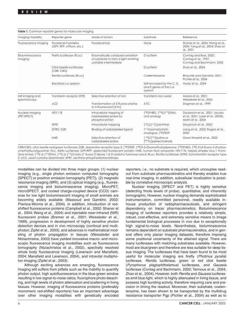
Regardless of the examined process, any strategy for imag-ing genetically encoded reporters is comprised of three majorcomponents: (1) a reporter gene that generates an imagable sig-nal, (2) a regulatory element governing the activity of thereporter gene and therefore generating contrast (e.g., a constitu-tive or inducible promoter, an upstream cis-regulatory sequence,or a polypeptide fused in frame with the reporter gene and there-by posttranscriptionally regulating its activation), and (3) a detec-tion device able to noninvasively sense and quantify the signalproduced by the reporter gene within the intact cell or organism.Genetically encoded reporters can produce signal (1) intrinsical-ly by the reporter (e.g., fluorescent proteins), (2) through enzy-matic activation of an inactive substrate by the reporter (e.g.,firefly luciferase that catalyses the light-producing reaction fromthe substrate D luciferin in the presence of O2 and Mg2+-ATP), (3)by enzymatic modification of an active (e.g., radiolabeled) sub-strate producing selective retention in reporter cells (e.g., selec-tive retention of [18F]FHBG by herpes simplex virus 1 thymidinekinase [HSV1-TK]; Luker et al., 2002b), or (4) by direct binding orimport of an active substrate (e.g., binding of radiolabeledsomatostatin to somatostatin receptor type 2 [SSTR2]-express-ing cells; Rogers et al., 2000). Table 1 lists common reportergenes used in molecular imaging.
Specialized detection devices for imaging small animalsmore or less coevolved during the late 1990s. The imaging
R E V I E W
Spying on cancer: Molecular imaging in vivo with geneticallyencoded reportersShimon Gross1 and David Piwnica-Worms1,2
1Molecular Imaging Center, Mallinckrodt Institute of Radiology, and Department of Molecular Biology and Pharmacology, Washington University Medical School, St. Louis, Missouri 63110
2Correspondence: [email protected]
Genetically encoded imaging reporters introduced into cells and transgenic animals enable noninvasive, longitudinal studiesof dynamic biological processes in vivo. The most common reporters include firefly luciferase (bioluminescence imaging),green fluorescence protein (fluorescence imaging), herpes simplex virus-1 thymidine kinase (positron emission tomography),and variants with enhanced spectral and kinetic properties. When cloned into promoter/enhancer sequences or engineeredinto fusion proteins, imaging reporters allow transcriptional regulation, signal transduction, protein-protein interactions,oncogenic transformation, cell trafficking, and targeted drug action to be spatiotemporally resolved in vivo. Spying on cancerwith genetically encoded imaging reporters provides insight into cancer-specific molecular machinery within the context ofthe whole animal.
6 CANCER CELL : JANUARY 2005
modalities can be divided into three major groups: (1) nuclearimaging (e.g., single photon emission computed tomography[SPECT] or positron emission tomography [PET]), (2) magneticresonance imaging (MRI), and (3) optical imaging (e.g., fluores-cence imaging and bioluminescence imaging). MicroPET,microSPECT, and cooled charge-coupled device (CCD) cam-eras for low light bioluminescent imaging of small animals arebecoming widely available (Massoud and Gambhir, 2003;Piwnica-Worms et al., 2004). In addition, introduction of red-shifted fluorescence proteins (Campbell et al., 2002; Shaner etal., 2004; Wang et al., 2004) and injectable near-infrared (NIR)fluorescent probes (Bremer et al., 2001; Weissleder et al.,1999), progression in development of highly sensitive photondetection devices and in vivo microscopy (confocal and multi-photon; Zipfel et al., 2003), and advances in mathematical mod-eling of photon propagation in tissues (Weissleder andNtziachristos, 2003) have yielded innovative macro- and micro-scopic fluorescence imaging modalities such as fluorescencetomography (Ntziachristos et al., 2002), spectrally resolvedwhole body fluorescence imaging (Levenson and Mansfield,2004; Mansfield and Levenson, 2004), and intravital multipho-ton imaging (Zipfel et al., 2003).
Although exciting advances are emerging, fluorescenceimaging still suffers from pitfalls such as the inability to quantifyphoton output, high autofluorescence in the blue-green windowresulting in low signal-to-noise ratios, fluorophore photo-bleach-ing, and high levels of photon attenuation and scattering in livingtissues. However, imaging of fluorescence proteins (preferablymonomeric red-shifted reporters) has an important advantageover other imaging modalities with genetically encoded
reporters, i.e., no substrate is required, which uncouples read-out from substrate pharmacokinetics and thereby enables truereal-time imaging. In addition, subcellular localization is possi-ble by correlative microscopic analysis.
Nuclear imaging (SPECT and PET) is highly sensitive(detecting fmole levels of probe), quantitative, and inherentlytomographic. However, nuclear imaging demands sophisticatedinstrumentation, committed personnel, readily available in-house production of radiopharmaceuticals, and stringentdependency on tracer pharmacokinetics. Bioluminescenceimaging of luciferase reporters provides a relatively simple,robust, cost-effective, and extremely sensitive means to imagefundamental biological processes in vivo due to exceptionallyhigh signal-to-noise levels. Nevertheless, bioluminescenceremains dependent on substrate pharmacokinetics, and in gen-eral offers only planar imaging datasets, therefore imposingsome positional uncertainty of the attained signal. There aremany luciferases with matching substrates available. However,most are blue/green and therefore are less suitable for deep tis-sue imaging. The luciferases that have been found to be mostuseful for molecular imaging are firefly (Photinus pyralis)luciferase, Renilla luciferase, green or red click beetle(Pyrophorus plagiophthalamus) luciferases, and Gaussialuciferase (Contag and Bachmann, 2002; Tannous et al., 2004;Zhao et al., 2004). However, both Renilla and Gaussia luciferas-es emit blue light, which is highly attenuated in living tissue, andpossess high bursting activity, therefore requiring care and pre-cision in timing the readout. Moreover, their substrate, coelen-terazine, has been shown to be transported by the multidrugresistance transporter Pgp (Pichler et al., 2004) as well as to
R E V I E W
Table 1. Common reporter genes for molecular imaging
Imaging modality Reporter gene Mode of action Substrate Reference
Fluorescence imaging Fluorescent proteins Fluorescence None Shaner et al., 2004; Wang et al.,(GFP, RFP, mPlum, etc.) 2004; Yang et al., 2004; Zhao et
al., 2001
Bioluminescence Firefly luciferase (FLuc) Enzymatically catalyzed oxidation D luciferin Contag and Ross, 2002;imaging of substrate to form a light emitting Contag et al., 1997;
unstable intermediate Contag and Bachmann, 2002
Click beetle luciferases D luciferin Zhao et al., 2004(CBR, CBG)
Renilla luciferase (RLuc) Coelenterazine Bhaumik and Gambhir, 2001; Pichler et al., 2004
Bacterial Lux operon Self-encoded by the C, D, Hardy et al., 2004and E genes of the Luxoperon
MR imaging and Transferrin receptor (ETR) Selective retention of iron Transferrin-iron oxide Moore et al., 2001; spectroscopy Weissleder et al., 2000
yCD Transformation of 5-fluorocytosine 5-FC Stegman et al., 1999to 5-fluorouracil (5-FU)
Nuclear imaging HSV1-TK Intracellular trapping of [18F]FHBG, [124I]/[131I]FIAU, Doubrovin et al., 2001; Jacobs (PET,SPECT) radiolabeled probe by and analogs et al., 2001; Luker et al., 2002b,
phosphorylation Morin et al., 2004
XPRT Intracellular trapping [14C]/[11C]xanthine Doubrovin et al., 2003
SSTR2, D2R Binding of radiolabeled ligand [111In]somatostatin Liang et al., 2002; Rogers et al.,analogue, [18F]FESP 2000
hNIS Selective retention of [ 124I]/[131I]iodine or Groot-Wassink et al., 2002radiolabeled probe [99mTc]pertechnetate
CBR/CBG, click beetle red/green luciferase; D2R, dopamine receptor type 2; [18F]FESP, [18F]3-2-(fluoroethyl)spiperone; [18F]FHBG, [18F] 9-[4-fluoro-3-(hydrox-ymethyl)butyl]guanine; Fluc, firefly luciferase; GFP/RFP, green/red fluorescent protein; hNIS, human Na/I symporter; HSV1-TK, herpes simplex virus-1 thymi-dine kinase; [124I] or [131I]FIAU, [124I] or [131I]5-iodo-2�-fluoro-2�deoxy-1-β-D-arabino-furanosyl-uracil; RLuc, Renilla luciferase; SSTR2, Somatostatin receptor type2; yCD, yeast cytosine deaminase; XPRT, xanthine-phosphoribotransferase.
CANCER CELL : JANUARY 2005 7
interact efficiently with superoxide anion and peroxynitrate inlight-producing reactions (Tarpey et al., 1999), thereby compli-cating applications of Renilla and Gaussia luciferases in vivo.
As mentioned above, each of the imaging modalities (MR,nuclear, or optical) has its own strengths and weaknesses (i.e.,tradeoffs of spatial and temporal resolution, depth of signaldetection, acquisition time, cost, ease of operation, and thepotential for clinical translation) and should therefore be select-ed primarily according to the examined biological process. Thisreview is not intended for critical assessment or comparison ofthe technical merits of the various modalities, detection devices,or instruments, and therefore, the reader is referred to recentcomprehensive reviews covering these topics (Bremer et al.,2003; Contag and Bachmann, 2002; Massoud and Gambhir,2003; Rudin and Weissleder, 2003; Sharma et al., 2002). Nosingle modality addresses all aspects of molecular imaging, andtherefore, there is increasing interest in constructing fusionreporters that combine the positive attributes of different modal-ities (Doubrovin et al., 2003; Jacobs et al., 2003; Luker et al.,2002b; Ray et al., 2003, 2004). Herein we will focus on the reg-ulatory and biochemical elements that govern activation ofimaging reporter genes with an emphasis on cancer, regardlessof their emission characteristics or the imaging modality used todetect their signal. Different strategies to regulate geneticallyencoded reporter activation and thereby detect and dynamical-ly monitor various components of the cell machinery (transcrip-tional, posttranscriptional, translational, and posttranslational)in intact cells and small animal models are summarized in Table2 and further discussed in the following sections.
Transcriptional regulation of reporter activityThe simplest way to regulate activity of a reporter gene is bypromoter-driven transcription. The earliest applications ofgenetically encoded molecular imaging reporters were intendedfor studying cell trafficking and engraftment, bacterial or viraldistribution, transgene expression, or analyzing tumor burdenand metastatic activity by expressing reporter genes under thecontrol of constitutive promoters (e.g., viral promoters such aspCMV, pSV40, etc.). Typically, cells transfected ex vivo or trans-duced with engineered viruses enabled reporter monitoring ofspatiotemporal changes in signal after implantation of the cellsor vectors in an intact animal. For example, the ability to monitorprimary tumor burden and response to therapy was studied by bioluminescence imaging and crossvalidated by MRI in an orthotopically implanted 9L glioma tumor, constitutivelyexpressing firefly luciferase (Rehemtulla et al., 2000). In thisstudy, excellent correlation was found between the two imagingmodalities for the kinetics of both tumor growth and drug-induced cytotoxicity by 1,3-bis (2-chloroethyl)-1-nitrosourea(BCNU). Historically, in the case of internal orthotopic tumormodels, animal survival was the dominant strategy for monitor-ing tumor growth and response to therapy prior to small animalimaging (Contag and Ross, 2002), and thus, imaging strategiesprovide more refined readouts of response. While MRI providesaccurate three-dimensional measurements of tumor size, biolu-minescence imaging of luciferase expression is potentiallysuperior in reporting the quantity of viable cells.
Monitoring gene delivery and optimizing gene therapy pro-tocols are other queries for application of simple constitutivepromoters to regulate imaging reporter genes. Constitutiveexpression (by an immediate early CMV promoter) of tworeporter genes (D2R and HSV1-TK) within a bicistronic (IRES)
transcriptional unit was used to image adenoviral-mediatedgene delivery by PET imaging for evaluation and optimization ofgene therapy protocols (Liang et al., 2002). It was demonstratedin this study that the two genes coexpressed primarily in theliver (the main site for adenoviral infection in mice) over a 3month period and over a 7- to 10-fold concentration range. Inthe case of cancer gene therapy, HSV1-TK is a particularlyattractive reporter gene, since it has the advantage of beingboth a therapeutic and a reporter gene (by using ganciclovirtreatment and the appropriate radiolabeled substrate, respec-tively) (Qiao et al., 2002;Tjuvajev et al., 1999). A variation of thisstrategy thereby enabled direct monitoring of inducible suicidegene therapy for controlling graft versus host disease after allo-geneic bone marrow transplantation (Rettig et al., 2004).Tumoral accumulation of HSV-based oncolytic viruses also hasbeen imaged with an HSV1-TK reporter (Bennett et al., 2001;Jacobs et al., 2001).
Reporter genes transfected in eukaryotic cells have beenwidely used to study cis-regulatory sequences or trans-actingfactors that modulate the transcriptional activity of target pro-moters. By transducing cells in vivo with an expression cassettethat contains a reporter gene under the control of a transcrip-tionally regulated sequence, it is now feasible to monitor tran-scriptional regulation in a living animal. As an example, aretroviral vector containing the dual reporter gene HSV1-TK-GFP regulated by an upstream p53 response element(p53→TK-GFP) was used to transduce U87 glioma (p53+/+) andSaOS osteosarcoma (p53−/−) cells that were implanted into ratsto establish tumor xenografts (Doubrovin et al., 2001). Whole-body PET imaging and fluorescence microscopy demonstratedDNA damage-induced upregulation of TK-GFP in a p53-depen-dent manner, and this increase in activity correlated with upreg-ulation of downstream p53-regulated genes as measured byindependent assays.
Anatomical and temporal changes in transcriptional regula-tion have also been shown to be resolved by reporter geneimaging in transgenic mouse models. For instance, a transgenicmouse has been generated wherein firefly luciferase isexpressed under the regulation of an NF-κB response elementand used to study time- and organ-dependent changes in biolu-minescence after administration of classical stressors such astumor necrosis factor α (TNFα), interleukin-1α (IL-1α), orlipopolysaccharide (LPS), or after inducing genotoxic stress byUV irradiation (Carlsen et al., 2002). It was demonstrated in thisstudy that in the absence of extrinsic stimuli, strong NF-kBactivity was evident in cervical lymph nodes, thymus, andPeyer’s patches. However, treatment with TNFα, IL-1α, or LPSincreased the NF-kB-dependent bioluminescent signal in anorgan-specific manner, with the strongest activity observed inskin, lungs, spleen, Peyer’s patches, and the wall of the smallintestines. It was further shown that induction of chronic inflam-mation resembling rheumatoid arthritis produced a strong sig-nal in affected joints.
To analyze the dynamics of estrogen receptor (ER) tran-scriptional activity in vivo, a transgenic mouse model was generated (Ciana et al., 2003) wherein firefly luciferase isexpressed under transcriptional control of the ER (ERE→FLuc).As expected, in reproductive organs and in the liver, luciferaseactivity paralleled circulating estrogen levels, peaking atproestrus. However, in non-reproductive organs such as boneand brain, peak transcriptional activity of estrogen receptorswas observed in diestrus. It was further demonstrated that
R E V I E W

8 CANCER CELL : JANUARY 2005
R E V I E W
Table 2. Strategies for regulating imaging reporter gene activity
Level of regulation Examined process Strategies Examples References
Transcriptional Transcriptional regulation The promoter of interest drives ERE→FLuc Ciana et al., 2003the reporter gene
NF-κB→FLuc Carlsen et al., 2002p53→HSV1-TK-EGFP Doubrovin et al., 2001AP1→FLuc Huang et al., 1997Grp78→HSV1-TK Dong et al., 2004HRE→HSV1-TK-GFP Serganova et al., 2004
Transactivation PSA→ Gal4BD-VP16 + Gal4→FLuc Zhang et al., 2002a
Posttranscriptional RNA splicing Spliceosome-mediated RNA trans-splicing Trans-splicing-mediated Bhaumik et al., 2004and translational reconstitution of hRLuc mRNA
Translational regulation Fusing the reporter with a protein that LTR→DHFR-HSV1-TK Mayer-Kuckuk et al., regulates its own translation 2002, 2003
Posttranslational Protein-protein interaction Functional complementation of a split CMV→FRB-NFLuc + Luker and Piwnica-reporter (with or without intein-mediated CMV→CFLuc-FKBP Worms, 2004; Luker et reporter reconstitutuion) al., 2004
NF-κB→NFLuc-ID + Paulmurugan et al., 2002CMV→MyoD-CFLucNF-κB→NFLuc-DnaEN-ID + Paulmurugan et al., 2002CMV→MyoD-DnaEC-CFLuc
Two-hybrid system Gal4BD-p53←TetRE→VP16-TAg + Luker et al., 2002b,4xGal4→HSV1-TK-EGFP 2003b
Energy transfer (FRET, BRET) CMV→β-arrestin-RLuc + Jares-Erijman and Jovin,CMV→Ub-GFP 2003; Perroy et al., 2004
Proteasomal degradation N-terminal fusion of tetraubiquitin to CMV→4xUb-FLuc Gross and Piwnica-(total) the reporter Worms, 2005; Luker et
al., 2003aβ-actin→4xUb-EGFP Lindsten et al., 2003
Proteasomal degradation Fusion of the substrate of interest to SV40→p27-FLuc Zhang et al., 2004(substrate-specific) the reporter
CMV→IκBα-FLuc Gross and Piwnica-Worms, 2004, 2005
CMV→p53-FLuc Rehemtulla et al., 2004Protease activation Introduction of a protease recognition AMLP→ER-DEVD-FLuc-DEVD-ER Laxman et al., 2002
motif that, when cut, activates the reporterNuclear transport Intein-mediated reconstruction of CMV→DnaEC-CRLuc-AR + Kim et al., 2004
nucleocytoplasmic separated CMV→FLAG-NRLuc-DnaEN- NLSreporter fragments
Cellular Monitoring tumor burden, Constitutive expression of the reporter CMV→FLuc in 9L glioma Rehemtulla et al., 2000cell trafficking, stem cell gene in the target cells/virusesengraftment, pathogen infection, multidrug resistance
LTR→HSV1-TK in T cells Koehne et al., 2003UL29→FLuc + UL30→RLuc in HSV Luker et al., 2002aLTR→FLuc/GFP in αMBP-CD4+ T cells Costa et al., 2001LP→lux in L. Monocytogenes Hardy et al., 2004CMV→RLuc Pichler et al., 2004
Combined Conditional organ-specific Cre-loxP recombination β-actin→lox-GFP-lox-FLuc Lyons et al., 2003reporter expression
ROSA26→lox-stop-lox-FLuc Safran et al., 2003Spontaneous tumorigenesis Coupling reporter activation with POMC→Cre + POMC→FLuc Vooijs et al., 2002
transformation in a conditional and crossed with a conditionaloptionally organ-specific manner lox-Rb-lox knockout
E2F1→FLuc crossed with Nestin→ Uhrbom et al., 2004tv-a and infected with RCAS-PDGFB virus
AMLP, adenoviral major late promoter; AP1, affector protein 1; AR, androgen receptor; Cre, Cre-recombinase; DEVD, asp-glu-val-asp; E2F1, E2F1 promoter; EGFP,enhanced green fluorescence protein; ERE or ER, estrogen receptor; FKBP, FK506-binding protein type 12; FRB, rapamycin-binding domain of mTOR; Gal4BD,Gal4 DNA binding domain; Grp78, glucose-regulated protein-78; HRE, hypoxia response element; HSV1-TK, herpes simplex virus-1 thymidine kinase; IκBα, inhibitorof nuclear factor κB type α; LP, listerial promoter; LTR, long terminal repeat; MBP, myelin binding protein; N/C FLuc, N- or C-terminal fragments of firefly luciferase;N/C RLuc, N- or C-terminal fragments of Renilla luciferase; NF-κB, nuclear factor κB; N-tv-a, nestin promoter driving TV-a avian virus receptor; NLS, nuclear local-ization signal; PDGFB, platelet-derived growth factor type B; POMC, lobe-specific pituitary promoter; PSA, prostate-specific antigen; Rb, retinoblastoma tumorsuppressor gene; RCAS, replication-competent avian sarcoma and leucosis virus long terminal splice acceptor; ROSA26, ubiquitous ROSA26 promoter; TAg, simi-an virus large T antigen; TetRE, tetracycline-responsive element; Ub, ubiquitin; VP16, VP16 transactivator; →, promoter regulation; -, fusion gene; +, coexpression.
CANCER CELL : JANUARY 2005 9
these tissue-specific responses are masked when mice under-go conventional hormone treatment, and that estrogen recep-tors are transcriptionally active even in immature mice (beforegonadal production of sex hormones) and in ovariectomizedmice. Overall, this study emphasizes the importance of estro-gen-independent activation of ER, especially in non-reproduc-tive organs, and provides far-reaching implications for hormonereplacement therapy and cancer risk.
Imaging posttranscriptional molecular eventsImaging posttranscriptional events, such as translational regu-lation, protein-protein interactions, protein processing, or pro-tein degradation, is primarily performed by fusing the reportergene, a partial reporter fragment, or an upstream transactivatorto the protein of interest, thereby generating a molecular sensorthat activates (or deactivates) the reporter in response to agiven protein interaction or modification. Fundamentally, thedetection of physical interaction among two or more proteinscan be assisted if association between the interactive partnersleads to the production of a readily observed biological or phys-ical readout. At present, three general strategies are feasible forimaging interacting protein pairs in cellulo or in vivo: (1) protein-protein interaction-dependent reporter gene transactivation orrepression (two hybrid system; recruitment of signal transduc-tion cascades), (2) reporter complementation, achieved by fus-ing inactive reporter fragments to interacting proteins, therebybringing the fragments into close proximity and restoringreporter activity (Luker et al., 2004; Paulmurugan et al., 2002,2004), and (3) energy transfer techniques such as Forster reso-nance energy transfer (FRET) (Jares-Erijman and Jovin, 2003)and bioluminescence resonance energy transfer (BRET)(Perroy et al., 2004). However, energy transfer strategies havenot yet been demonstrated in living animals.Reporter transactivationAs an example of noninvasive molecular imaging of protein-pro-tein interactions in vivo by PET and fluorescence imaging, afusion reporter gene was engineered comprising a mutant
HSV1-TK and EGFP (mNLS-sr39TK-EGFP) under regulation ofa concatenated Gal4 promoter (Luker et al., 2002b, 2003b).Thep53 tumor suppressor was fused to the Gal4 DNA bindingdomain (p53-Gal4BD), and simian virus-associated large T anti-gen (TAg) was fused to the transactivator VP16 (TAg-VP16).Expression of p53-Gal4BD and TAg-VP16 was regulated by abidirectional, tetracycline-responsive promoter. Thus, upontreatment with doxycycline, transcription of the reporter genewas regulated by the known high-affinity interaction betweenp53 and TAg. Visualization of the reporter was accomplishedwith [18F]FHBG and microPET imaging. Based on region-of-interest values from the microPET images, the uptake of[18F]FHBG was 5.5-fold higher than in control xenograftsexpressing polyoma virus coat protein fused to VP16 (CP-VP16) instead of TAg-VP16.
The interaction of two other proteins, ID and MyoD, wasalso interrogated in vivo by a similar strategy applied to biolumi-nescence (Ray et al., 2002). To modulate the expression ofthese two proteins, the NF-κB promoter was used to regulatetranscription of the ID-Gal4 and MyoD-VP16 hybrid proteins,while TNFα was used to induce activation of the NF-κB promot-er. Firefly luciferase regulated by five repetitive Gal4 elementswas used as the reporter gene.Thus, upon treatment with TNFαto induce expression of the interacting hybrids, biolumines-cence was correlated with interaction of ID and MyoD and wastransiently higher than when induced by a noninteracting pro-tein pair (MyoD and p53).Reporter fragment complementation and reconstitutionLimitations of transactivation strategies for studying protein-pro-tein interactions are the requirement for protein translocationand stable protein interactions in the nucleus, as well as tempo-ral delays inherent to transcriptional readouts. To circumventthese limitations, other techniques have been developed. A splitreporter protein approach can be used through either comple-mentation or reconstitution strategies. Complementation strate-gies do not require the formation of an intact protein from splitfragments, as opposed to reconstitution strategies that attempt
R E V I E W
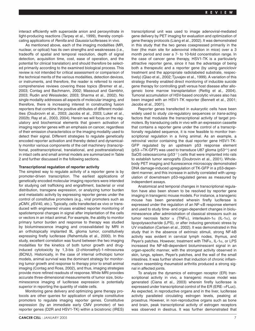
Figure 1. Optimization of firefly luciferaseprotein-fragment complementation imaging(LCI)
A: Schematic representation of the optimizedN- and C-terminal fragments of luciferase (asrevealed by screening of incremental trunca-tion libraries), fused to FRB and FKBP-12,respectively. B: Schematic of LCI. Rapamycin-induced asso-ciation of proteins FRB and FKBP-12 bringsinactive fragments of luciferase into closeproximity, thereby producing bioluminescenceactivity. C: Monitoring rapamycin-induced FRB/FKBP12association in live cells. HEK-293 cells transfect-ed with FRB-NFLuc + CFLuc-FKBP-12 (upper) orS2035I FRB-NFLuc + CFLuc-FKBP-12 (lower) weretreated for 6 hr with 50 nM rapamycin. Notethat the S2035I mutation of mTOR/FRB is knownto abrogate the rapamycin-induced associa-tion of FRB and FKBP-12. A pseudocolor IVISbioluminescence image of live cells in a 96-well plate is shown. D: Luciferase complementation imaging oftwo representative nu/nu mice, one implantedwith HEK-293 cells expressing FRB-NFLuc +CFLuc-FKBP (upper) and the other with cells
expressing mutant S2035I FRB-NFLuc + CFLuc-FKBP-12 (lower). Images were taken 18 hr before treatment with rapamycin (left) and 2.5 hr after receivinga single dose of rapamycin (4.5 mg/kg, i.p., right).
10 CANCER CELL : JANUARY 2005
to reconstitute the mature reporter protein. Reporter comple-mentation is based on the principle that reporter activity (e.g.,enzymatic activity) is regained when its split fragments arebrought into close proximity due to a specific protein-proteininteraction. Protein-protein interactions can also drive reporterreconstitution by intein-mediated protein autosplicing.
The interaction of ID and MyoD was demonstrated in vitroand in vivo by firefly luciferase complementation and reconstitu-tion (Paulmurugan et al., 2002). However, in this study, fireflyluciferase fragments suffered from constitutive activity of the N-terminal fragment. Reporter complementation was also demon-strated using Renilla luciferase split fragments (Kim et al., 2004;Paulmurugan and Gambhir, 2003; Paulmurugan et al., 2004),though the blue-green emission spectrum of Renilla luciferasepenetrates tissues poorly, thereby precluding general use.Furthermore, coelenterazine, the bioluminescent substrate forRenilla luciferase, was shown to be transported by the multidrugresistance transporter Pgp (see Pichler et al., 2004, and below),complicating applications of Renilla luciferase in vivo.Consequently, to facilitate the study of regulated protein-proteininteractions in cells and living animals, an optimized fireflyluciferase protein fragment complementation system was devel-oped by screening incremental truncation libraries of N- and C-terminal fragments of firefly luciferase. The initial seeds for thescreen were inactive firefly luciferase fragments fused to therapamycin binding domain (FRB) of the kinase mammalian targetof rapamycin (mTOR) and FK506 binding protein 12 (FKBP),respectively (Luker and Piwnica-Worms, 2004; Luker et al.,2004). The optimized FRB-NLuc/CLuc-FKBP pair (containing2–416 N-terminal and 398–550 C-terminal overlapping luciferasefragments; Figure 1A) generated luciferase activity in cells uponsingle-site binding of rapamycin in an FK506-competitive manner(Figure 1B).The inducibility range of optimized luciferase comple-mentation imaging (LCI) was robust, with drug-specific inductionof bioluminescence reaching 1,200-fold over background,exceeding currently available systems (Figures 1C and 1D). Thisproperty enabled monitoring of lower affinity protein-protein inter-actions, such as homodimerization of nonphosphorylated STAT1and the phosphorylation-dependent interaction between humanCdc25C with 14-3-3ε in vivo (Luker et al., 2004).
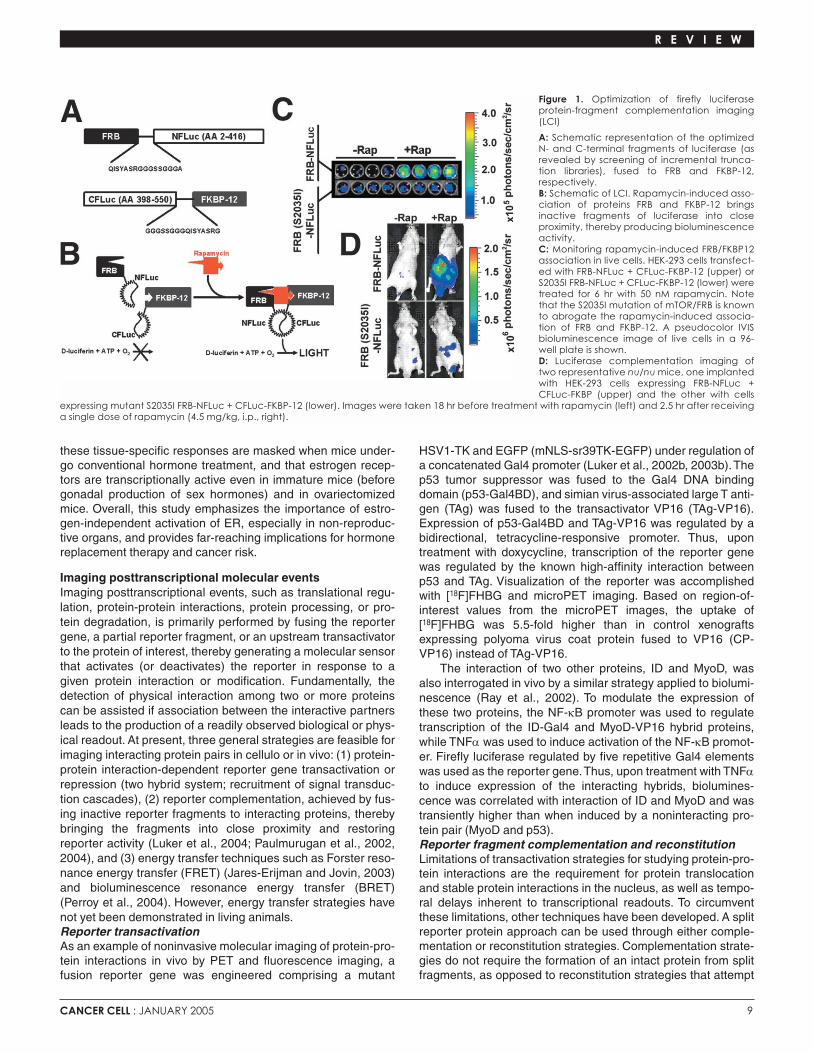
Imaging total- and substrate-specific proteasomal degradationThe ubiquitin-proteasome pathway is the central mediator of reg-ulated proteolysis, an instrumental switch for a variety of signal-ing cascades. Deregulation of proteasomal activity or impropersubstrate recognition and processing by the ubiquitin-protea-some machinery may lead to cancer, stroke, chronic inflam-mation, and neurodegenerative diseases. To monitor totalproteasomal activity, an ubiquitin-luciferase bioluminescenceimaging reporter was developed by fusing the N terminus of fire-fly luciferase to four copies of a mutant ubiquitin (UbG76V, Figure2A, upper panel) (Luker et al., 2003a). The tetraubiquitin fusiondegradation motif has been shown to significantly destabilizeheterologous proteins in cultured cells (Stack et al., 2000), whilethe glycine to valine substitution at the C terminus of ubiquitinlimits cleavage by ubiquitin hydrolases (Johnson et al., 1992;Stack et al., 2000). Both in cultured cells and in tumor xenografts,the 4xUb-FLuc reporter was degraded rapidly under steady-state conditions and stabilized in a concentration- and time-dependent manner in response to various proteasome inhibitors(Figure 2A, lower panel). Bioluminescence imaging revealed thatproteasome function in tumor xenografts was blocked as soonas 30 min after administration of a single dose of the chemother-apeutic proteasome inhibitor bortezomib and returned to nearlybaseline by 46 hr. However, after a two-week regimen of borte-zomib, imaging of target tumors showed significantly enhancedproteasome inhibition that no longer returned to baseline.
Similar to 4xUb-FLuc for imaging total proteasome activity,fusing a proteasomal substrate to the N terminus of FLuc gen-erates a reporter responsive to degradation of that specific pro-teasomal substrate. For example, it was recently demonstratedthat bioluminescence imaging of inhibitor of κB (IκBα)-specificproteasomal degradation using an IκBα-FLuc fusion reportercan monitor in real time nuclear factor-κB (NF-κB) activationand pharmacological modulation (Gross and Piwnica-Worms,2004, 2005), independent of transcriptional or translationalevents. Reporter degradation was monitored both in culturedcells after treatment with tumor necrosis factor (TNFα) and inresponse to treatment with lipopolysaccharide (LPS) in vivousing a liver inflammation model, wherein the IκBα-FLuc
R E V I E W
Figure 2. Imaging total- and substrate-specific proteasome activity
A: In vivo bioluminescence imaging of Ub-FLuc monitors total proteasome function and inhibition in living mice. Upper panel: schematic of the Ub-FLucreporter. Lower panel: mice bearing size-matched tumors were imaged one day before (−) and four hours after tail vein injection of the indicated doses ofbortezomib. Unfused FLuc, Ub-FLuc, and vector control tumors are denoted by black arrows, yellow arrows, and asterisks, respectively. B: Real-time bioluminescence imaging of IκBα-specific proteasomal degradation in a somatic gene transfer mouse model. Upper panel: schematic of theIκBα-FLuc reporter. Lower panel: representative bioluminescence images of RLuc (left two panels) and IκBα-FLuc (right two panels) taken before or 1 hr afterinduction of acute liver inflammation by intravenous injection of LPS (4 µg/g BW). All images correspond to an individual mouse. Note that plasmids encod-ing RLuc and IκBα-FLuc were delivered to the liver by high-volume intravenous injection.

CANCER CELL : JANUARY 2005 11
reporter was delivered to liver hepatocytes by hydrodynamicsomatic gene transfer (Figure 2B). Moreover, pretreatment withproteasome inhibitors or inhibitors of IκB-kinase (IKK) wasshown to abrogate ligand-induced reporter degradation.
A similar strategy was used to monitor cell cycle-regulateddegradation of the cyclin-dependent kinase 2 (cdk2) inhibitorp27 using a p27-FLuc fusion reporter (Zhang et al., 2004). Itwas shown in this study that reporter activity is regulated by itsE3-ligase Skp2 in a cell cycle-dependent manner. Blockade ofcdk2 activity by drugs, inhibitory proteins, peptides, or smallinterfering RNA (siRNA) induced reporter accumulation andincreases in bioluminescence. Elevation in reporter activity dueto pharmacological modulation of cdk2 was also documented invivo in human tumor xenografts.Imaging caspase-3 activationThe cysteine protease caspase-3 is an effector caspase acti-vated during apoptotic cell death by upstream initiator cas-pases (i.e., caspases 8, 9, 10, and 12). Once activated,caspase-3 executes apoptosis by cleaving cellular proteins at
a specific DEVD consensus motif. To enable noninvasive andrepetitive imaging of apoptosis in living animals, a reporterwas engineered (Laxman et al., 2002) for bioluminescenceimaging wherein the estrogen receptor regulatory domain(ER) was fused to FLuc, thereby sterically silencing FLuc cat-alytic activity. Inclusion of a DEVD sequence between thesetwo moieties allowed for caspase-3-mediated restoration ofluciferase activity, enabling real-time monitoring of apoptoticactivation. Using this reporter, the investigators demonstratedactivation of caspase-3 in intact cells and living animals inresponse to treatment with TNFα-related apoptosis-inducingligand (TRAIL). Furthermore, ZVAD-fmk, a general caspaseinhibitor, was shown to abrogate TRAIL-induced reporter acti-vation, thus confirming the role of caspases for regulatingactivity of this reporter.Imaging multidrug resistanceCoelenterazine, the bioluminescent substrate of the reportergene Renilla luciferase, is a substrate for the multidrug trans-porter Pgp (Pichler et al., 2004). In cultured living cells, stably
R E V I E W
Table 3. Examples of transgenic mouse models expressing reporter genes suitable for molecular imaging applications
Reporter gene Activity Transgene References
Firefly luciferase AP-1-dependent transcription AP-1→FLuc Huang et al., 1997
NF-κB-dependent transcription κB→FLuc Carlsen et al., 2002
Activation of estrogen receptor (ER) ERE→FLuc Ciana et al., 2003
Circadian gene expression mPer1→FLuc Wilsbacher et al., 2002
Neuronal damage GFAP→FLuc Zhu et al., 2004
Drug metabolism CYP3A4→FLuc Zhang et al., 2003
Bilirubin synthesis HO1→FLuc Zhang et al., 2002b
Bone repair and development hOC→FLuc Iris et al., 2003
Cre-mediated activation β-actin →lox-GFP-lox-FLuc; Lyons et al., 2003;
ROSA26→lox-stop-lox-FLuc Safran et al., 2003
E2F1-dependent transcription E2F1→FLuc Uhrbom et al., 2004
Pituitary spontaneous tumorigenesis POMC→Cre + POMC→Fluc Vooijs et al., 2002
Angiogenesis VEGFR2→FLuc Zhang et al., 2004
Fluorescent proteins Neuronal development nestin→GFP Yamaguchi et al., 2000
Embyonic germ cell migration Oct4→GFP Anderson et al., 2000
Ureteric bud development Hoxb7→GFP Srinivas et al., 1999
Angiogenesis VEGF→GFP Fukumura et al., 1998
Lymphocyte development Gfi1→GFP Yucel et al., 2004
X chromosome inactivation CMV→GFP in X chromosome Hadjantonakis et al., 2003
Synaptic formation and plasticity Thy1→GFP, Thy1→YFP, Feng et al., 2000; Sakai et al., 2004; Thy1→CFP, TetRE→PKCγ-GFP Trachtenberg et al., 2002
Proteasome activity β-actin→Ub-GFP Lindsten et al., 2003
Embryonic perfusion ε-globin→GFP Jones et al., 2002
Ubiquitous GFP expression β-actin→GFP Yang et al., 2004
HSV1-TK Endothelial damage VE-cad→HSV1-TK Dancer et al., 2003
Liver targeting Alb→Cre + CAG→lox-stop-lox- Sundaresan et al., 2004HSV1-TK adenovirus
Germ cell ablation Inhα→HSV1-TK Ahtiainen et al., 2004
GVH disease control CD2→∆CD34-HSV1-TK Rettig et al., 2004
Alb, albumin promoter; AP1, affector protein 1; CAG, cytomegalovirus immediate early gene 1 enhancer; CD2, T cell locus promoter; CFP, cyan fluorescentprotein; CMV, cytomegalovirus promoter; CYP3A4, cytochrome P450-3A promoter; Cre, Cre-recombinase; E2F1, E2F1 promoter; ERE, estrogen receptor;FLuc, firefly luciferase; GFAP, glial fibrillary acidic protein promoter; Gfi1, growth factor independence promoter; GFP, green fluorescent protein; GVH, graftversus host; HO1, heme-oxygenase 1; hOC, human osteocalcin promoter; Hoxb7, homeobox B7 promoter; HSV1-TK, herpes simplex virus-1 thymidine kinase;Inhα, inhibin-alpha promoter; κB, κB response element; mPer1, mammalian period-1 promoter; Oct4, POU5F1 promoter; PKCγ, protein kinase Cγ; POMC,lobe-specific pituitary promoter; TetRE, tetracycline-responsive element; Thy-1, a mature neuronal marker promoter; Ub, ubiquitin; VE-cad, vascularendothelial cadherin promoter; VEGF, vascular endothelial growth factor promoter; VEGFR2, VEGF receptor type 2 promoter; YFP, yellow fluorescent pro-tein; →, promoter regulation; −, fusion gene; +, coexpression.
12 CANCER CELL : JANUARY 2005
transfected with a codon-humanized Renilla luciferase, it wasshown that low baseline coelenterazine-mediated biolumines-cence could be fully enhanced (reversed) to non-Pgp matchedcontrol levels with potent and selective Pgp inhibitors.Therefore, using coelenterazine and noninvasive biolumines-cence imaging in vivo, tumor-specific Pgp transport activity andinhibition could be monitored in living mice. This study empha-sizes the role of coelenterazine as a Pgp substrate, but at thesame time raises concerns regarding the indiscriminative use ofRenilla luciferase and aequorin as reporters in intact cells andtransgenic animals, since Pgp-mediated alterations in coelen-terazine permeability may impact results.
Imaging oncogenic transformation and spontaneoustumorigenesisThe use of transgenic animal models of human diseasespromises to extend our understanding of the mechanisms ofpathogenesis by placing target genes and processes in theappropriate physiological milieu. However, until recently,analysis of these animal models was limited by the ability tomonitor only obvious phenotypic changes or perform destruc-tive analyses at defined time points. In cancer research, thispitfall becomes a major drawback, because almost all aspectsof tumorigenesis, tumor growth, invasion, metastatic potential,and response to therapy are dynamic in time and space.Moreover, while it is clear that the closest approximation ofhuman cancers is attained by spontaneous transformationmodels, the stochastic nature of spontaneous tumors compli-cates and thereby severely limits the application of these mod-els. Consequently, recent advances in small animal imaginginstrumentation, molecular genetics, and reporter genedesign have yielded the ability to integrate an imagablereporter capacity into transgenic models of human diseases.Such aptitude not only refines the data by allowing each ani-mal to serve as its own control, but also permits in vivo high-throughput analyses of drugs for preclinical trials. Consecutiveanalysis of the same animal means that fewer animals areneeded for each study and experimental uncertainties arisingfrom inter-animal variations are greatly reduced (Herschman,2003). Table 3 provides detailed information on available
transgenic mouse models expressingsuch imagable reporters.
Of particular interest is a transgenicmouse model (Vooijs et al., 2002) whereinboth firefly luciferase and Cre recombi-nase were expressed solely in the pitu-itary gland under the control of the
intermediate lobe-specific POMC promoter. These mice werecrossed with mice carrying conditional lox-Rb-lox alleles, therebycoupling luciferase activation to deletion of Rb and developmentof pituitary-specific melanotrophic tumors. This sophisticatedmodel allowed the researchers to monitor, by bioluminescenceimaging, tumor onset, progression, and response to antineoplas-tic therapy, thereby generating temporally resolved, statisticallysignificant data from a relatively small cohort of animals.
There is no doubt that conditional activation or deletion ofan oncogene or a tumor suppressor gene, coupled with reportergene expression (e.g., by using Cre/loxP conditional recombi-nation technology) is indispensable for longitudal studies of therole of a specific transformation event for tumorigenesis.However, to limit the need to generate, optimize, and validatenovel independent transgenic luciferase mouse strains for eachconditional transformation model, a ubiquitously expressingconditional luciferase reporter mouse was developed that canbe used to render a wide range of Cre/loxP mouse tumor mod-els for bioluminescence imaging (Lyons et al., 2003). Herein, aβ-actin promoter drives FLuc in a Cre-dependent manner. Toillustrate the usefulness of this model, the investigators coupledluciferase activation with lung tumorigenesis, induced byKras2v12 (a constitutively active ras mutant) in a preexistingmouse model of non-small cell lung cancer, and followed onsetand progression of the spontaneously generated lung tumors.An improved version of a conditional loxP-luciferase mouse wasrecently described (Safran et al., 2003), where a true knockinwas generated by introducing a lox-stop-lox cassette upstreamto firefly luciferase cDNA under the control of the ubiquitousROSA26 promoter. Another advantage of this model over previ-ous attempts (Lyons et al., 2003) is the use of codon-optimizedfirefly luciferase, thereby increasing its activity by two orders ofmagnitude.
In the studies mentioned above (Lyons et al., 2003;Vooijs etal., 2002), luciferase activation was regionally coupled to Cre-mediated Rb knockout (Vooijs et al., 2002) or Cre-mediatedKras2v12 expression (Lyons et al., 2003), but not biochemicallydependent on the molecular transformation event; i.e.,luciferase did not serve as a downstream target reporter of Rb
R E V I E W
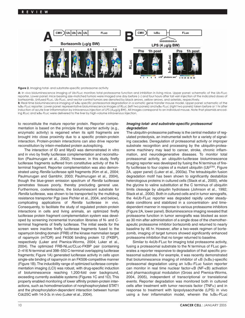
Figure 3. Imaging tumor burden and response totherapy in spontaneous glioma tumorigenesismodal
A: Generation of Ef-FLuc + N-tv-a double trans-genic mice (see text for further details). B: Approximate correlation between BLI outputand tumor size. Upper panel: luciferase activityin tumor-bearing Ef-FLuc + N-tv-a transgenicmice. Lower panel: Whole-mount histologicanalysis of the brains from the same mice asimaged in the upper panel. Note that tumor sizecorrelates with the amount of emitted light. C: Longitudinal imaging of one tumor-bearingmouse treated with the PDGFR inhibitorPTK787/ZK222584 daily for 6 days. NT, biolumi-nescence before drug treatment. Modifiedfrom Uhrbom et al. (2004) with permission fromNature Medicine (http://www.nature.com/nm).

CANCER CELL : JANUARY 2005 13
or K-ras function in Vooijs et al. (2002) and Lyons et al. (2003),respectively. In contrast, a recent transgenic mouse model tostudy gliomagenesis by bioluminescence imaging was reported(Uhrbom et al., 2004), wherein luciferase activation is not onlycoupled, but also dependent upon platelet-derived growth factor(PDGF)-induced loss of Rb. This mouse (Ef-FLuc) expressesluciferase under the control of the E2F1 promoter, which is neg-atively regulated by Rb under normal conditions, and thusluciferase activity increases upon loss of Rb in tumors, regard-less of mitotic status.These mice were crossed with N-tv-a micethat express the viral receptor tv-a from the nestin promoter,thereby restricting retroviral transactivation of E2F1→FLuc toglial progenitor cells using viral PDGF-RCAS vectors (Figure3A) (Holland, 2001).This strategy enables spontaneous glioma-genesis and tumor progression to be followed noninvasivelyand repetitively over time (Figure 3B). Furthermore, the biolumi-nescent signal correlates in this model to both tumor cell num-ber and loss of Rb control, thereby enabling analyses of thepotency and pharmacodynamics of drugs that interfere withtumor maintenance and proliferation (i.e., PDGFR and mTORinhibitors) as well as cytotoxic drugs (see Figure 3C for aPDGFR inhibitor).
Concluding remarksIntegration of genetically encoded imaging reporters into intactcells and small animal models of disease has provided powerfultools to monitor cancer-associated molecular, biochemical, andcellular pathways in vivo. These types of studies are gainingwidespread acceptance within the scientific community andtherefore could be considered in some cases to be the methodof choice for deciphering complex biological responses in a liv-ing animal. We predict that more researchers will continue totake advantage of these new capabilities, thus allowing them tononinvasively “spy” on cancer-specific molecular and regulatorycascades in the intact animal.
Acknowledgments
The authors would like to thank colleagues in the Washington UniversityMolecular Imaging Center for their insightful discussions contributing to thisreview. Supported by NIH P50 CA94056.
References
Ahtiainen, M., Toppari, J., Poutanen, M., and Huhtaniemi, I. (2004). IndirectSertoli cell-mediated ablation of germ cells in mice expressing the inhibin-alphapromoter/herpes simplex virus thymidine kinase transgene. Biol. Reprod. 71,1545–1550.
Anderson, R., Copeland, T.K., Scholer, H., Heasman, J., and Wylie, C. (2000).The onset of germ cell migration in the mouse embryo. Mech. Dev. 91, 61–68.
Bennett, J., Tjuvajev, J., Johnson, P., Doubrovin, M., Akhurst, T., Malholtra, S.,Hackman, T., Balatoni, J., Finn, R., Larson, S., et al. (2001). Positron emissiontomography imaging for herpes virus infection: Implications for oncolytic viraltreatments of cancer. Nat. Med. 7, 859–863.
Bhaumik, S., and Gambhir, S. (2001). Optical imaging of Renilla luciferasereporter gene expression in living mice. Proc. Natl. Acad. Sci. USA 99,377–382.
Bhaumik, S., Walls, Z., Puttaraju, M., Mitchell, L.G., and Gambhir, S.S. (2004).Molecular imaging of gene expression in living subjects by spliceosome-medi-ated RNA trans-splicing. Proc. Natl. Acad. Sci. USA 101, 8693–8698.
Blasberg, R.G., and Tjuvajev, J.G. (2003). Molecular-genetic imaging: Currentand future perspectives. J. Clin. Invest. 111, 1620–1629.
Bremer, C., Tung, C., and Weissleder, R. (2001). In vivo molecular target
assessment of matrix metalloproteinase inhibition. Nat. Med. 7, 743–748.
Bremer, C., Ntziachristos, V., and Weissleder, R. (2003). Optical-based molecu-lar imaging: Contrast agents and potential medical applications. Eur. Radiol. 13,231–243.
Campbell, R., Tour, O., Palmer, A., Steinbach, P., Baird, G., Zacharias, D., andTsien, R. (2002). A monomeric red fluorescent protein. Proc. Natl. Acad. Sci.USA 99, 7877–7882.
Carlsen, H., Moskaug, J.O., Fromm, S.H., and Blomhoff, R. (2002). In vivoimaging of NF-kappaB activity. J. Immunol. 168, 1441–1446.
Ciana, P., Raviscioni, M., Mussi, P., Vegeto, E., Que, I., Parker, M., Lowik, C.,and Maggi, A. (2003). In vivo imaging of transcriptionally active estrogen recep-tors. Nat. Med. 9, 82–86.
Contag, C.H., and Bachmann, M.H. (2002). Advances in vivo bioluminescenceimaging of gene expression. Annu. Rev. Biomed. Eng. 4, 235–260.
Contag, C., and Ross, B. (2002). It’s not just about anatomy: In vivo biolumines-cence imaging as an eyepiece into biology. J. Magn. Reson. Imaging 16,378–387.
Contag, C., Spilman, S., Contag, P., Oshiro, M., Eames, B., Dennery, P.,Stevenson, D., and Benaron, D. (1997). Visualizing gene expression in livingmammals using a bioluminescent reporter. Photochem. Photobiol. 66,523–531.
Costa, G., Sandora, M., Nakajima, A., Nguyen, E., Taylor-Edwards, C., Slavin,A., Contag, C., Fathman, C., and Benson, J. (2001). Adoptive immunotherapyof experimental autoimmune encephalomyelitis via T cell delivery of the IL-12p40 subunit. J. Immunol. 167, 2379–2387.
Dancer, A., Julien, S., Bouillot, S., Pointu, H., Vernet, M., and Huber, P. (2003).Expression of thymidine kinase driven by an endothelial-specific promoterinhibits tumor growth of Lewis lung carcinoma cells in transgenic mice. GeneTher. 10, 1170–1178.
Dong, D., Dubeau, L., Bading, J., Nguyen, K., Luna, M., Yu, H., Gazit-Bornstein,G., Gordon, E., Gomer, C., Hall, F., et al. (2004). Spontaneous and controllableactivation of suicide gene expression driven by the stress-inducible grp78 pro-moter resulting in eradication of sizable human tumors. Hum. Gene Ther. 15,553–561.
Doubrovin, M., Ponomarev, V., Beresten, T., Balatoni, J., Bornmann, W., Finn,R., Humm, J., Larson, S., Sadelain, M., Blasberg, R., and Tjuvajev, J. (2001).Imaging transcriptional regulation of p53-dependent genes with positron emis-sion tomography in vivo. Proc. Natl. Acad. Sci. USA 98, 9300–9305.
Doubrovin, M., Ponomarev, V., Serganova, I., Soghomonian, S., Myagawa, T.,Beresten, T., Ageyeva, L., Sadelain, M., Koutcher, J., Blasberg, R.G., andTjuvajev, J.G. (2003).Development of a new reporter gene system–dsRed/xan-thine phosphoribosyltransferase-xanthine for molecular imaging of processesbehind the intact blood-brain barrier. Mol. Imaging 2, 93–112.
Feng, G., Mellor, R.H., Bernstein, M., Keller-Peck, C., Nguyen, Q.T., Wallace,M., Nerbonne, J.M., Lichtman, J.W., and Sanes, J.R. (2000). Imaging neuronalsubsets in transgenic mice expressing multiple spectral variants of GFP.Neuron 28, 41–51.
Fukumura, D., Xavier, R., Sugiura, T., Chen, Y., Park, E.C., Lu, N., Selig, M.,Nielsen, G., Taksir, T., Jain, R.K., and Seed, B. (1998).Tumor induction of VEGFpromoter activity in stromal cells. Cell 94, 715–725.
Gelovani Tjuvajev, J., and Blasberg, R.G. (2003). In vivo imaging of molecular-genetic targets for cancer therapy. Cancer Cell 3, 327–332.
Groot-Wassink, T., Aboagye, E.O., Glaser, M., Lemoine, N.R., and Vassaux, G.(2002). Adenovirus biodistribution and noninvasive imaging of gene expressionin vivo by positron emission tomography using human sodium/iodide symporteras reporter gene. Hum. Gene Ther. 13, 1723–1735.
Gross, S., and Piwnica-Worms, D. (2004). Real-time imaging of ligand-inducedIκBα degradation in intact cells and living mice. Mol. Imaging 3, 194.
Gross, S., and Piwnica-Worms, D. (2005). Monitoring proteasome activity incellulo and in living animals by bioluminescence imaging: Techincal considera-tions for design and use of genetically-encoded reporters. Methods Enzymol.,in press.
Hadjantonakis, A.K., Dickinson, M.E., Fraser, S.E., and Papaioannou, V.E.(2003). Technicolour transgenics: Imaging tools for functional genomics in the
R E V I E W

14 CANCER CELL : JANUARY 2005
mouse. Nat. Rev. Genet. 4, 613–625.
Hardy, J., Francis, K.P., DeBoer, M., Chu, P., Gibbs, K., and Contag, C.H.(2004). Extracellular replication of Listeria monocytogenes in the murine gallbladder. Science 303, 851–853.
Herschman, H.R. (2003). Molecular imaging: Looking at problems, seeing solu-tions. Science 302, 605–608.
Holland, E.C. (2001). Gliomagenesis: Genetic alterations and mouse models.Nat. Rev. Genet. 2, 120–129.
Huang, C., Ma, W.Y., Dawson, M.I., Rincon, M., Flavell, R.A., and Dong, Z.(1997). Blocking activator protein-1 activity, but not activating retinoic acidresponse element, is required for the antitumor promotion effect of retinoic acid.Proc. Natl. Acad. Sci. USA 94, 5826–5830.
Iris, B., Zilberman, Y., Zeira, E., Galun, E., Honigman, A., Turgeman, G.,Clemens, T., Gazit, Z., and Gazit, D. (2003). Molecular imaging of the skeleton:Quantitative real-time bioluminescence monitoring gene expression in bonerepair and development. J. Bone Miner. Res. 18, 570–578.
Jacobs, A., Tjuvajev, J., Dubrovin, M., Akhurst, T., Balatoni, J., Beattie, B., Joshi,R., Finn, R., Larson, S., Herrlinger, U., et al. (2001). Positron emission tomogra-phy-based imaging of transgene expression mediated by replication-condition-al, oncolytic herpes simplex virus type 1 mutant vectors in vivo. Cancer Res. 61,2983–2995.
Jacobs, A., Winkeler, A., Hartung, M., Slack, M., Dittmar, C., Kummer, C.,Knoess, C., Galldiks, N., Vollmar, S., Wienhard, K., and Heiss, W. (2003).Improved herpes simplex virus type 1 amplicon vectors for proportional coex-pression of positron emission tomography marker and therapeutic genes. Hum.Gene Ther. 14, 277–297.
Jares-Erijman, E.A., and Jovin, T.M. (2003). FRET imaging. Nat. Biotechnol. 21,1387–1395.
Johnson, E., Bartel, B., Seufert, W., and Varshavsky, A. (1992). Ubiquitin as adegradation signal. EMBO J. 11, 497–505.
Jones, E.A., Crotty, D., Kulesa, P.M., Waters, C.W., Baron, M.H., Fraser, S.E.,and Dickinson, M.E. (2002). Dynamic in vivo imaging of postimplantation mam-malian embryos using whole embryo culture. Genesis 34, 228–235.
Kim, S.B., Ozawa, T., Watanabe, S., and Umezawa, Y. (2004). High-throughputsensing and noninvasive imaging of protein nuclear transport by using recon-stitution of split Renilla luciferase. Proc. Natl. Acad. Sci. USA 101,11542–11547.
Koehne, G., Doubrovin, M., Doubrovina, E., Zanzonico, P., Gallardo, H.,Ivanova, A., Balatoni, J., Teruya-Feldstein, J., Heller, G., May, C., et al. (2003).Serial in vivo imaging of the targeted migration of human HSV-TK-transducedantigen-specific lymphocytes. Nat. Biotechnol. 21, 405–413.
Laxman, B., Hall, D., Bhojani, M., Hamstra, D., Chenevert, T., Ross, B., andRehemtulla, A. (2002). Noninvasive real-time imaging of apoptosis. Proc. Natl.Acad. Sci. USA 99, 16551–16555.
Levenson, R., and Mansfield, J. (2004). Small-animal fluorescence detectionusing the Maestro multispectral imaging system. Mol. Imaging 3, 227.
Liang, Q., Gotts, J., Satyamurthy, N., Barrio, J., Phelps, M.E., Gambhir, S.S.,and Herschman, H.R. (2002). Noninvasive, repetitive, quantitative measure-ment of gene expression from a bicistronic message by positron emissiontomography, following gene transfer with adenovirus. Mol.Ther. 6, 73–82.
Lindsten, K., Menendez-Benito, V., Masucci, M.G., and Dantuma, N.P. (2003).A transgenic mouse model of the ubiquitin/proteasome system. Nat.Biotechnol. 21, 897–902.
Louie, A., Huber, M., Ahrens, E., Rothbacher, U., Moats, R., Jacobs, R., Fraser,S., and Meade, T. (2000). In vivo visualization of gene expression using mag-netic resonance imaging. Nat. Biotechnol. 18, 321–325.
Luker, G., Bardill, J., Prior, J., Pica, C., Piwnica-Worms, D., and Leib, D.(2002a). Noninvasive bioluminescence imaging of herpes simplex virus type 1infection and therapy in living mice. J.Virol. 76, 12149–12161.
Luker, G., Sharma, V., Pica, C., Dahlheimer, J., Li, W., Ochesky, J., Ryan, C.,Piwnica-Worms, H., and Piwnica-Worms, D. (2002b). Noninvasive imaging ofprotein-protein interactions in living animals. Proc. Natl. Acad. Sci. USA 99,6961–6966.
Luker, K., and Piwnica-Worms, D. (2004). Optimizing luciferase protein frag-
ment complementation for bioluminescent imaging of protein-protein interac-tions in live cells and animals. Methods Enzymol. 385, 349–360.
Luker, G., Pica, C., Song, J., Luker, K., and Piwnica-Worms, D. (2003a).Imaging 26S proteasome activity and inhibition in living mice. Nat. Med. 9,969–973.
Luker, G., Sharma, V., Pica, C., Prior, J., Li, W., and Piwnica-Worms, D.(2003b). Molecular imaging of protein-protein interactions: Controlled expres-sion of p53 and large T antigen fusion proteins in vivo. Cancer Res. 63,1780–1788.
Luker, K.E., Smith, M.C., Luker, G.D., Gammon, S.T., Piwnica-Worms, H., andPiwnica-Worms, D. (2004). Kinetics of regulated protein-protein interactionsrevealed with firefly luciferase complementation imaging in cells and living ani-mals. Proc. Natl. Acad. Sci. USA 101, 12288–12293.
Lyons, S.K., Meuwissen, R., Krimpenfort, P., and Berns, A. (2003).The genera-tion of a conditional reporter that enables bioluminescence imaging ofCre/loxP-dependent tumorigenesis in mice. Cancer Res. 63, 7042–7046.
Mansfield, J., and Levenson, R. (2004). Fluorescence dye multiplexing and tis-sue autofluorescence removal using multispectral imaging. Mol. Imaging 3,231.
Massoud, T., and Gambhir, S. (2003). Molecular imaging in living subjects:Seeing fundamental biological processes in a new light. Genes Dev. 17,545–580.
Mayer-Kuckuk, P., Banerjee, D., Malhotra, S., Doubrovin, M., Iwamoto, M.,Akhurst, T., Balatoni, J., Bornmann, W., Finn, R., Larson, S., et al. (2002). Cellsexposed to antifolates show increased cellular levels of proteins fused to dihy-drofolate reductase: A method to modulate gene expression. Proc. Natl. Acad.Sci. USA 99, 3400–3405.
Mayer-Kuckuk, P., Doubrovin, M., Gusani, N.J., Gade, T., Balatoni, J., Akhurst,T., Finn, R., Fong, Y., Koutcher, J.A., Larson, S., et al. (2003). Imaging of dihy-drofolate reductase fusion gene expression in xenografts of human liver metas-tases of colorectal cancer in living rats. Eur. J. Nucl. Med. Mol. Imaging 30,1281–1291.
Moore, A., Josephson, L., Bhorade, R., Basilion, J., and Weissleder, R. (2001).Human transferrin receptor gene as a marker gene for MR imaging. Radiology221, 244–250.
Morin, K., Duan, W., Xu, L., Zhou, A., Moharram, S., Knaus, E., McEwan, A.,and Wiebe, L. (2004). Cytotoxicity and cellular uptake of pyrimidine nucleosidesfor imaging herpes simplex type-1 thymidine kinase (HSV-1 TK) expression inmammalian cells. Nucl. Med. Biol. 31, 623–630.
Ntziachristos, V., Tung, C., Bremer, C., and Weissleder, R. (2002).Fluorescencemolecular tomography resolves protease activity in vivo. Nat. Med. 8, 757–761.
Paulmurugan, R., and Gambhir, S. (2003). Monitoring protein-protein interac-tions using split synthetic Renilla luciferase protein-fragment-assisted comple-mentation. Anal. Chem. 75, 1584–1589.
Paulmurugan, R., Umezawa, Y., and Gambhir, S.S. (2002). Noninvasive imag-ing of protein-protein interactions in living subjects by using reporter proteincomplementation and reconstitution strategies. Proc. Natl. Acad. Sci. USA 99,15608–15613.
Paulmurugan, R., Massoud, T., Huang, J., and Gambhir, S. (2004). Molecularimaging of drug-modulated protein-protein interactions in living subjects.Cancer Res. 64, 2113–2119.
Perroy, J., Pointer, S., Charest, P., Aubry, M., and Bouvier, M. (2004). Real-timemonitoring of ubiquitination in living cells by BRET. Nat Meth 1, 203–208.
Pichler, A., Prior, J., and Piwnica-Worms, D. (2004). Imaging reversal of mul-tidrug resistance in living mice with bioluminescence: MDR1 P-glycoproteintransports coelenterazine. Proc. Natl. Acad. Sci. USA 101, 1702–1707.
Piwnica-Worms, D., Schuster, D., and Garbow, J. (2004). Molecular imaging ofhost-pathogen interactions in intact small animals. Cell. Microbiol. 6, 319–331.
Qiao, J., Doubrovin, M., Sauter, B., Huang, Y., Guo, Z., Blatoni, J., Akhurst, T.,Blasberg, R., Tjuvajev, J., Chen, S.-H., and Woo, S. (2002).Tumor-specific tran-scribptional targeting of suicide gene therapy. Gene Ther. 9, 168–175.
Ray, P., Pimenta, H., Paulmurugan, R., Berger, F., Phelps, M., Iyer, M., andGambhir, S. (2002). Noninvasive quantitative imaging of protein-protein interac-tions in living subjects. Proc. Natl. Acad. Sci. USA 99, 3105–3110.
R E V I E W
CANCER CELL : JANUARY 2005 15
Ray, P., Wu, A.M., and Gambhir, S.S. (2003). Optical bioluminescence andpositron emission tomography imaging of a novel fusion reporter gene in tumorxenografts of living mice. Cancer Res. 63, 1160–1165.
Ray, P., De, A., Min, J.J., Tsien, R.Y., and Gambhir, S.S. (2004). Imaging tri-fusion multimodality reporter gene expression in living subjects. Cancer Res.64, 1323–1330.
Rehemtulla, A., Stegman, L.D., Cardozo, S.J., Gupta, S., Hall, D.E., Contag,C.H., and Ross, B.D. (2000). Rapid and quantitative assessment of cancertreatment response using in vivo bioluminescence imaging. Neoplasia 2,491–495.
Rehemtulla, A., Taneja, N., and Ross, B.D. (2004). Bioluminescence detectionof cells having stabilized p53 in response to a genotoxic event. Mol. Imaging 3,63–68.
Rettig, M.P., Ritchey, J.K., Prior, J.L., Haug, J.S., Piwnica-Worms, D., andDiPersio, J.F. (2004).Kinetics of in vivo elimination of suicide gene-expressing Tcells affects engraftment, graft-versus-host disease, and graft-versus-leukemiaafter allogeneic bone marrow transplantation. J. Immunol. 173, 3620–3630.
Rogers, B.E., Zinn, K.R., and Buchsbaum, D.J. (2000). Gene transfer strategiesfor improving radiolabeled peptide imaging and therapy. Q. J. Nucl. Med. 44,208–223.
Rudin, M., and Weissleder, R. (2003). Molecular imaging in drug discovery anddevelopment. Nat. Rev. Drug Discov. 2, 123–131.
Safran, M., Kim, W.Y., Kung, A.L., Horner, J.W., DePinho, R.A., and Kaelin,W.G., Jr. (2003). Mouse reporter strain for noninvasive bioluminescent imagingof cells that have undergone Cre-mediated recombination. Mol. Imaging 2,297–302.
Sakai, N., Tsubokawa, H., Matsuzaki, M., Kajimoto, T., Takahashi, E., Ren, Y.,Ohmori, S., Shirai, Y., Matsubayashi, H., Chen, J., et al. (2004). Propagation ofgammaPKC translocation along the dendrites of Purkinje cell in gammaPKC-GFP transgenic mice. Genes Cells 9, 945–957.
Serganova, I., Doubrovin, M., Vider, J., Ponomarev, V., Soghomonyan, S.,Beresten, T., Ageyeva, L., Serganov, A., Cai, S., Balatoni, J., et al. (2004).Molecular imaging of temporal dynamics and spatial heterogeneity of hypoxia-inducible factor-1 signal transduction activity in tumors in living mice. CancerRes. 64, 6101–6108.
Shaner, N.C., Campbell, R.E., Steinbach, P.A., Giepmans, B.N., Palmer, A.E.,and Tsien, R.Y. (2004). Improved monomeric red, orange and yellow fluores-cent proteins derived from Discosoma sp. red fluorescent protein. Nat.Biotechnol. 22, 1567–1572.
Sharma, V., Luker, G., and Piwnica-Worms, D. (2002). Molecular imaging ofgene expression and protein function in vivo with PET and SPECT. J. Magn.Reson. Imaging 16, 336–351.
Srinivas, S., Goldberg, M.R., Watanabe, T., D’Agati, V., al-Awqati, Q., andCostantini, F. (1999). Expression of green fluorescent protein in the ureteric budof transgenic mice: A new tool for the analysis of ureteric bud morphogenesis.Dev. Genet. 24, 241–251.
Stack, J., Whitney, M., Rodems, S., and Pollok, B. (2000). A ubiquitin-basedtagging system for controlled modulation of protein stability.Nat. Biotechnol. 18,1298–1302.
Stegman, L.D., Rehemtulla, A., Beattie, B., Kievit, E., Lawrence, T.S., Blasberg,R.G., Tjuvajev, J.G., and Ross, B.D. (1999). Noninvasive quantitation of cyto-sine deaminase transgene expression in human tumor xenografts with in vivomagnetic resonance spectroscopy. Proc. Natl. Acad. Sci. USA 96, 9821–9826.
Sundaresan, G., Paulmurugan, R., Berger, F., Stiles, B., Nagayama, Y., Wu, H.,and Gambhir, S. (2004). MicroPET imaging of Cre-loxP-mediated conditionalactivation of a herpes simplex virus type 1 thymidine kinase reporter gene.Gene Ther. 11, 609–618.
Tannous, B., Kim, D., and Weissleder, R. (2004). Novel luciferases for in vitroand in vivo imaging. Mol. Imaging 3, 227.
Tarpey, M., White, C., Suarez, E., Richardson, G., Radi, R., and Freeman, B.(1999). Chemiluminescent detection of oxidants in vascular tissue. Lucigeninbut not coelenterazine enhances superoxide formation. Circ. Res. 84,1203–1211.
Tjuvajev, J., Chen, S., Joshi, A., Joshi, R., Guo, Z., Balatoni, J., Ballon, D.,Koutcher, J., Finn, R., Woo, S., and Blasberg, R. (1999). Imaging adenoviral-
mediated herpes virus thymidine kinase gene transfer and expression in vivo.Cancer Res. 59, 5186–5193.
Trachtenberg, J.T., Chen, B.E., Knott, G.W., Feng, G., Sanes, J.R., Welker, E.,and Svoboda, K. (2002). Long-term in vivo imaging of experience-dependentsynaptic plasticity in adult cortex. Nature 420, 788–794.
Uhrbom, L., Nerio, E., and Holland, E.C. (2004). Dissecting tumor maintenancerequirements using bioluminescence imaging of cell proliferation in a mouseglioma model. Nat. Med. 10, 1257–1260.
Vooijs, M., Jonkers, J., Lyons, S., and Berns, A. (2002). Noninvasive imaging ofspontaneous retinoblastoma pathway dependent tumors in mice. Cancer Res.62, 1862–1867.
Wang, L., Jackson, W.C., Steinbach, P.A., and Tsien, R.Y. (2004). Evolution ofnew nonantibody proteins via iterative somatic hypermutation. Proc. Natl. Acad.Sci. USA 101, 16745–16749.
Weissleder, R. (2002). Scaling down imaging: Molecular mapping of cancer inmice. Nat. Rev. Cancer 2, 1–8.
Weissleder, R., and Ntziachristos, V. (2003). Shedding light on live moleculartargets. Nat. Med. 9, 123–128.
Weissleder, R., Tung, C., Mahmood, U., and Bogdanov, A. (1999). In vivo imag-ing of tumors with protease activated near-infrared fluorescent probes. Nat.Biotechnol. 17, 375–378.
Weissleder, R., Moore, A., Mahmood, U., Bhorade, R., Benveniste, H.,Chicocca, E., and Basilion, J. (2000). In vivo magnetic resonance imaging oftransgene expression. Nat. Med. 6, 351–355.
Wilsbacher, L.D., Yamazaki, S., Herzog, E.D., Song, E.J., Radcliffe, L.A., Abe,M., Block, G., Spitznagel, E., Menaker, M., and Takahashi, J.S. (2002). Photicand circadian expression of luciferase in mPeriod1-luc transgenic mice invivo.Proc. Natl. Acad. Sci. USA 99, 489–494.
Yamaguchi, M., Saito, H., Suzuki, M., and Mori, K. (2000).Visualization of neu-rogenesis in the central nervous system using nestin promoter-GFP transgenicmice. Neuroreport 11, 1991–1996.
Yang, M., Reynoso, J., Jiang, P., Li, L., Moossa, A.R., and Hoffman, R.M.(2004). Transgenic nude mouse with ubiquitous green fluorescent proteinexpression as a host for human tumors. Cancer Res. 64, 8651–8656.
Yucel, R., Kosan, C., Heyd, F., and Moroy, T. (2004). Gfi1:green fluorescent pro-tein knock-in mutant reveals differential expression and autoregulation of thegrowth factor independence 1 (Gfi1) gene during lymphocyte development. J.Biol. Chem. 279, 40906–40917.
Zhang, L., Adams, J., Billick, E., Ilagan, R., Iyer, M., Le, K., Smallwood, A.,Gambhir, S., Carey, M., and Wu, L. (2002a). Molecular engineering of a two-step transcription amplification (TSTA) system for transgene delivery inprostate cancer. Mol.Ther. 5, 223–232.
Zhang, W., Contag, P.R., Hardy, J., Zhao, H., Vreman, H.J., Hajdena-Dawson,M., Wong, R.J., Stevenson, D.K., and Contag, C.H. (2002b). Selection of poten-tial therapeutics based on in vivo spatiotemporal transcription patterns of hemeoxygenase-1. J. Mol. Med. 80, 655–664.
Zhang, W., Purchio, A.F., Chen, K., Wu, J., Lu, L., Coffee, R., Contag, P.R., andWest, D.B. (2003). A transgenic mouse model with a luciferase reporter forstudying in vivo transcriptional regulation of the human CYP3A4 gene. DrugMetab. Dispos. 31, 1054–1064.
Zhang, G.J., Safran, M., Wei, W., Sorensen, E., Lassota, P., Zhelev, N.,Neuberg, D.S., Shapiro, G., and Kaelin, W.G., Jr. (2004). Bioluminescent imag-ing of Cdk2 inhibition in vivo. Nat. Med. 10, 643–648.
Zhao, M., Yang, M., Baranov, E., Wang, X., Penman, S., Moossa, A.R., andHoffman, R.M. (2001). Spatial-temporal imaging of bacterial infection andantibiotic response in intact animals.Proc.Natl.Acad.Sci.USA 98, 9814–9818.
Zhao, H., Doyle, T., Coquoz, O., Kalish, F., Rice, B., and Contag, C.H. (2004).Spectral characterization of firefly, click beetle and Renilla luciferases in mam-malian cells and living mice. Mol. Imaging 3, 229.
Zhu, L., Ramboz, S., Hewitt, D., Boring, L., Grass, D.S., and Purchio, A.F.(2004). Non-invasive imaging of GFAP expression after neuronal damage inmice. Neurosci. Lett. 367, 210–212.
Zipfel, W.R., Williams, R.M., and Webb, W.W. (2003). Nonlinear magic:Multiphoton microscopy in the biosciences. Nat. Biotechnol. 21, 1369–1377.
R E V I E W