Cyclooxygenase2 Mediates Dialysate-Induced Alterations of the Peritoneal Membrane
Upload
independentCategory
view
4download
0
10.1128/MCB.21.3.952-965.2001.
2001, 21(3):952. DOI:Mol. Cell. Biol. A. Hemmings and Michael E. GreenbergAnne Brunet, Jongsun Park, Hien Tran, Linda S. Hu, Brian Transcription Factor FKHRL1 (FOXO3a)Signals by Phosphorylating the Forkhead Protein Kinase SGK Mediates Survival
http://mcb.asm.org/content/21/3/952Updated information and services can be found at:
These include:
REFERENCEShttp://mcb.asm.org/content/21/3/952#ref-list-1at:
This article cites 50 articles, 32 of which can be accessed free
CONTENT ALERTS more»articles cite this article),
Receive: RSS Feeds, eTOCs, free email alerts (when new
http://journals.asm.org/site/misc/reprints.xhtmlInformation about commercial reprint orders: http://journals.asm.org/site/subscriptions/To subscribe to to another ASM Journal go to:
on Septem
ber 5, 2014 by guesthttp://m
cb.asm.org/
Dow
nloaded from
on Septem
ber 5, 2014 by guesthttp://m
cb.asm.org/
Dow
nloaded from
MOLECULAR AND CELLULAR BIOLOGY,0270-7306/01/$04.0010 DOI: 10.1128/MCB.21.3.952–965.2001
Feb. 2001, p. 952–965 Vol. 21, No. 3
Copyright © 2001, American Society for Microbiology. All Rights Reserved.
Protein Kinase SGK Mediates Survival Signals by Phosphorylatingthe Forkhead Transcription Factor FKHRL1 (FOXO3a)ANNE BRUNET,1 JONGSUN PARK,2 HIEN TRAN,1 LINDA S. HU,1 BRIAN A. HEMMINGS,2
AND MICHAEL E. GREENBERG1*
Division of Neuroscience, Children’s Hospital and Department of Neurobiology, Harvard Medical School,Boston, Massachusetts 02115,1 and Friedrich Miescher Institute, CH-4058, Basel, Switzerland2
Received 14 June 2000/Returned for modification 31 July 2000/Accepted 19 October 2000
Serum- and glucocorticoid-inducible kinases (SGKs) form a novel family of serine/threonine kinases that areactivated in response to a variety of extracellular stimuli. SGKs are related to Akt (also called PKB), aserine/threonine kinase that plays a crucial role in promoting cell survival. Like Akt, SGKs are activated by thephosphoinositide-3 kinase (PI3K) and translocate to the nucleus upon growth factor stimulation. However thephysiological substrates and cellular functions of SGKs remained to be identified. We hypothesized that SGKsregulate cellular functions in concert with Akt by phosphorylating common targets within the nucleus. Thebest-characterized nuclear substrates of Akt are transcription factors of the Forkhead family. Akt phosphor-ylates Forkhead transcription factors such as FKHRL1, leading to FKHRL1’s exit from the nucleus and theconsequent shutoff of FKHRL1 target genes. We show here that SGK1, like Akt, promotes cell survival and thatit does so in part by phosphorylating and inactivating FKHRL1. However, SGK and Akt display differenceswith respect to the efficacy with which they phosphorylate the three regulatory sites on FKHRL1. While bothkinases can phosphorylate Thr-32, SGK displays a marked preference for Ser-315 whereas Akt favors Ser-253.These findings suggest that SGK and Akt may coordinately regulate the function of FKHRL1 by phosphory-lating this transcription factor at distinct sites. The efficient phosphorylation of these three sites on FKHRL1by SGK and Akt appears to be critical to the ability of growth factors to suppress FKHRL1-dependenttranscription, thereby preventing FKHRL1 from inducing cell cycle arrest and apoptosis. These findingsindicate that SGK acts in concert with Akt to propagate the effects of PI3K activation within the nucleus andto mediate the biological outputs of PI3K signaling, including cell survival and cell cycle progression.
Serum- and glucocorticoid-induced kinases (SGKs) belongto a new family of serine/threonine kinases that are regulatedat both the transcriptional and posttranslational levels by ex-ternal stimuli. The mRNA encoding SGK1, the best-studiedmember of the SGK family, is rapidly induced in response to avariety of stimuli, including growth factors (51, 52), steroid andpeptide hormones (3, 51, 52), cytokines (15, 50), changes in cellvolume (49), and brain injury (24).
The SGK gene is conserved from yeast to human, and theSGK protein is expressed in a variety of tissues and cell lines inmammals (10, 51, 52). Although it has been proposed thatSGK may play a role in cell cycle progression (8) or sodiumhomeostasis control (4, 12), the cellular functions of SGK arelargely uncharacterized, and to date no in vivo SGK substrateshave been identified.
Within the protein kinase superfamily, SGK is closely re-lated to Akt (also called PKB), another serine/threonine ki-nase that is activated in response to growth and survival factorsand plays a critical role in promoting cell survival (16, 20).Several recent reports have shown that growth and survivalfactors also trigger SGK activation (28, 41). The activation ofSGK is dependent upon phosphoinositide 3-kinase (PI3K) ac-tivity and requires the phosphorylation of two regulatory sites,Thr-256 and Ser-422, that lie in the activation loop and the
C-terminal domain of SGK, respectively (28, 41). The proteinkinase PDK1, which had previously been identified as a PI3K-dependent serine/threonine kinase that phosphorylates andactivates Akt, is also likely to be responsible for the phosphor-ylation of SGK at Thr-256 (28, 41). The kinase that phosphor-ylates Ser-422 has yet to be identified.
Upon activation by growth factors, endogenous SGK trans-locates rapidly into the nucleus, where it may encounter nu-clear substrates (8). SGK and Akt are likely to phosphorylaterelated substrates, as they share a similar consensus phosphor-ylation site (RXRXXS/T) (1, 28, 41).
Despite their similarity, SGK and Akt display unique fea-tures. First, unlike Akt, whose expression appears not to beregulated by extracellular stimuli, SGK protein expression isinduced upon treatment of cells with extracellular stimuli, in-cluding growth factors. Second, in contrast to Akt, SGK doesnot have a pleckstrin homology domain and appears not tobe recruited to the plasma membrane prior to its activation.Third, the consensus sequence that is phosphorylated by SGKis not identical to the site phosphorylated by Akt. For example,SGK is more effective than Akt at phosphorylating a consensuspeptide substrate in which the serine phosphoacceptor site isreplaced with a threonine. Moreover, SGK, in contrast to Akt,is capable of phosphorylating peptide substrates that do nothave a bulky hydrophobic amino acid immediately C terminalto the phosphoacceptor site (28). These differences betweenSGK and Akt suggested that these two kinases might havecomplementary rather than redundant functions.
The observation that SGK translocates to the nucleus fol-
* Corresponding author. Mailing address: Division of Neuroscience,Children’s Hospital and Department of Neurobiology, Harvard Med-ical School, Boston, MA 02115. Phone: (617) 355-8344. Fax: (617)738-1542. E-mail: [email protected].
952
on Septem
ber 5, 2014 by guesthttp://m
cb.asm.org/
Dow
nloaded from
lowing its activation by PI3K led us to hypothesize that SGKmay act in concert with Akt to phosphorylate critical targetswithin the nucleus. The best-characterized nuclear substratesof Akt are transcription factors of the Forkhead family: FKHR,FKHRL1, and AFX (for a review, see reference 30). Accord-ing to the new nomenclature for Forkhead transcription fac-tors, these three proteins have been assigned to the FOXO(named for “Forkhead box, group O”) subfamily of Forkheadtranscription factors (26).
When Akt is inactive, FOXO family members are dephos-phorylated and localized in the nucleus, where they activatetranscription. Upon growth factor stimulation, Akt is activatedand FOXOs are phosphorylated at three key regulatory phos-phorylation sites (Thr-32, Ser-253, and Ser-315 for FKHRL1).The phosphorylation of FOXOs promotes their exit from thenucleus, resulting in inhibition of FOXO-dependent transcrip-tion (6, 7, 23, 31, 38, 43, 45, 46). Akt has been reported tophosphorylate the second regulatory site of FOXOs (Ser-253for FKHRL1) with greater affinity than the first and third sites(37, 43). However, it is not clear whether protein kinases inaddition to Akt phosphorylate the first and third FOXO reg-ulatory sites with high affinity or what the respective contribu-tion of each site to the control of FOXO-dependent geneexpression and FOXO biological functions is.
We report here that the protein kinase SGK mediates thebiological effects of PI3K in parallel with Akt. Activated SGKis capable of promoting cell survival in part by phosphorylatingand inactivating the Forkhead transcription factor FKHRL1(also called FOXO3a). SGK, like Akt, phosphorylates FKHRL1,thereby leading to FKHLR1 translocation from the nucleus tothe cytoplasm and to the inhibition of FKHRL1-dependenttranscription. However, SGK and Akt, when expressed at phys-iological levels, display differences with respect to the efficacywith which they phosphorylate the regulatory sites on FKHRL1:Thr-32 is phosphorylated by both protein kinases, but SGKprefers Ser-315 whereas Akt favors Ser-253. The efficient phos-phorylation of all three regulatory sites of FKHRL1 appears tobe required for the ability of growth factors to completelyrepress FKHRL1-dependent transcription, thereby preventingthis transcription factor from inducing apoptosis and/or cellcycle arrest. As mediators of PI3K signaling within the nucleus,SGK and Akt are likely to be important regulators of a varietyof PI3K-dependent cellular responses, including cell prolifer-ation and survival.
MATERIALS AND METHODS
Material. Insulin-like growth factor I (IGF-I) and insulin were purchased,respectively, from R&D Systems and Roche Biochemicals. LY 294002 (LY) wasobtained from Calbiochem. The antihemagglutinin (anti-HA) antibody (12CA5)was purchased from Roche Biochemicals, and the anti-M2 antibody was pur-chased from Sigma. The antibody against cleaved poly(ADP-ribose) polymerase(PARP) was purchased from Cell Signaling Technology. Purified SGK was ob-tained from Upstate Biotechnology.
Constructs. The constructs encoding wild-type (WT) SGK and mutants withmutations of the phosphorylation sites were described previously (41). Theconstructs encoding WT Akt, the constitutively active (CA) mutant (myristylatedAkt lacking the pleckstrin homology domain), and the kinase dead mutant (Aktwith a K197M mutation) have been described previously (17). The constructsencoding glutathione S-transferase (GST)–FKHRL1, HA-FKHRL1, M2-FKHRL1,and the Forkhead responsive element (FHRE)-driven luciferase reporter weredescribed previously (7). The constructs encoding the catalytic subunit of PKA(35), WT RSK2 (53), and the active forms of protein kinase C (PKC) z and l (14,47) have been described previously.
Antibodies. The anti-FKHRL1, anti-phospho-T32, and anti-phospho-S253 an-tibodies were described previously (7). For the anti-phospho-S315 antibody, aphosphopeptide with the sequence CFRSRTNpSNATVS was synthesized (TuftsSynthesis Facility, Tufts Medical School, Boston, Mass.) and coupled to keyholelimpet hemocyanin (KLH) (Pierce). The KLH-coupled peptide was injected intoNew Zealand White rabbits (Covance Research Products, Denver, Pa.). Seraobtained from immunized rabbits were purified by passage over a proteinA-Sepharose column (Pharmacia), followed by elution of the bound phospho-antibodies with 100 mM glycine (pH 2.5). The eluate was then passed over anagarose-iodoacetyl column to which was coupled the nonphosphorylated form ofthe peptide antigen. The flowthrough from the column contained the antiphos-phopeptide antibodies used for the analysis of FKHRL1 phosphorylation.
For the anti-SGK1 antibody, a peptide with the sequence CVLHKQPYDRTVDWW was synthesized (Tufts Synthesis Facility, Tufts Medical School) andcoupled to KLH (Pierce). The KLH-coupled peptide was injected into NewZealand White rabbits (Covance Research Products). Sera obtained from im-munized rabbits were purified by passage over an agarose-iodoacetyl column towhich was coupled the peptide antigen, followed by elution of the bound phos-phoantibodies with 100 mM glycine (pH 2.5).
Cell culture. Cells of the human embryonic kidney cell line 293 (HEK 293), thederivative HEK 293T, and the Chinese hamster lung fibroblast cell line CCL39(American Type Culture Collection) were cultured in Dulbecco’s modified Eaglemedium (DMEM) (Gibco) supplemented with 10% fetal calf serum (FCS)(Gibco) and antibiotics (50 U of penicillin and 50 mg of streptomycin/ml) at 37°Cin an atmosphere of 95% air–5% CO2. Cerebellar granule cells were obtainedfrom P6 Long Evans rats and cultured in BME (Sigma) supplemented with 25mM KCl, 10% heat-inactivated calf serum (HyClone), and antibiotics as previ-ously described (21).
In vitro kinase assay. HEK 293 cells were seeded at 106 cells per 10-cm dishand were transfected the following day by a modified calcium phosphate methodwith 2 mg of plasmid DNA. The transfection mixture was removed after 16 h ofincubation and cells were serum starved for 24 h before stimulation for 15 minwith 100 nM insulin. Cells were placed on ice and extracted with lysis buffercontaining 50 mM Tris-HCl (pH 7.5), 1% Nonidet P-40 (vol/vol), 120 mM NaCl,25 mM NaF, 40 mM b-glycerophosphate, 0.1 mM sodium orthopervanadate, 1mM phenylmethylsulfonyl fluoride, 1 mM benzamidine, and 2 mM microcystin-LR. Lysates were centrifuged for 15 min at 12,000 3 g, and the HA-conjugatedSGK protein was immunoprecipitated from 400 mg of cell extracts with theanti-HA epitope 12CA5 monoclonal antibody coupled to protein A-Sepharose(Pharmacia Biotech). The immune complexes were washed once with lysis buffercontaining 0.5 M NaCl, then with lysis buffer, and finally with kinase assay buffer(50 mM Tris-HCl [pH 7.5], 0.1% [vol/vol] 2-mercaptoethanol). In vitro kinaseassays were performed for 60 min at 30°C in 50 ml of reaction mixture containing30 ml of immunoprecipitate in kinase buffer, 5 mg of WT GST-FKHRL1 as asubstrate, 10 mM MgCl2, 1 mM protein kinase A inhibitor peptide (Bachem),and 100 mM [g-32P]ATP (Amersham; 1,000 to 2,000 cpm/pmol). All reactionswere stopped by adding Laemmli sample buffer. HEK 293 cell extracts andimmunoprecipitates were resolved by sodium dodecyl sulfate–12% polyacryl-amide gel electrophoresis (SDS–12% PAGE) and transferred to Immobilon Pmembranes (Millipore). The filters were blocked for 30 min in 13 phosphate-buffered saline (PBS) containing 5% skim milk, 0.5% Triton X-100, and 0.5%Tween 20, followed by a 2-h incubation with the anti-FKHRL1 antibody or thevarious anti-phospho-FKHRL1 antibodies diluted in the same blocking solution.The secondary antibody was alkaline phosphatase-conjugated anti-rabbit immu-noglobulin G (IgG; Sigma) diluted 2,500-fold in the blocking buffer. The detec-tion of proteins was carried out by using the alkaline phosphatase color devel-opment reagents from Bio-Rad.
Immunoblotting. CCL39 fibroblasts were seeded in six-well plastic dishes at adensity of 5 3 105/well. They were transfected by the calcium phosphate tech-nique with 5 mg of the WT HA-FKHRL1 construct and 5 mg of the relevantconstructs. Twenty-four hours after transfection, cells were incubated in serum-free DMEM for 20 h and treated with 10 mM LY for 1 h. Extracts were obtainedby lysing the cells in lysis buffer (50 mM Tris-HCl [pH 8], 100 mM NaCl, 2 mMEGTA, 10 mM NaF, 40 mM b-glycerophosphate, 0.5% Triton X-100, 2 mMdithiothreitol, aprotinin, 1 mM phenylmethylsulfonyl fluoride). Proteins wereresolved by SDS-PAGE (8 to 6%; 29:1, acrylamide/bisacrylamide ratio) andtransferred to polyvinylidene difluoride or nitrocellulose membranes. Themembranes were incubated with anti-HA, anti-FKHRL1, anti-phospho-Thr-32, anti-phospho-Ser-253, or anti-phospho-Ser-315 antibodies for 2 h at roomtemperature. The primary antibody was visualized using horseradish peroxi-dase-conjugated anti-mouse or anti-rabbit IgG secondary antibodies and en-hanced chemiluminescence.
VOL. 21, 2001 PHOSPHORYLATION AND REGULATION OF FKHRL1 BY SGK 953
on Septem
ber 5, 2014 by guesthttp://m
cb.asm.org/
Dow
nloaded from
Immunolocalization. CCL39 cells were plated onto glass coverslips at a densityof 102/well in 12-well dishes. The cells were transfected by the calcium phosphatetechnique with 1 mg of M2-FKHRL1 and 3 mg of the relevant constructs. The dayafter transfection, cells were rendered quiescent by incubation in serum-freemedium for 16 h and then treated with 20 mM LY for 1 h. The cells were thenfixed for 15 min in 4% formaldehyde–2% sucrose at room temperature andpermeabilized with 0.1% Triton X-100 for 10 min. Coverslips were washed withPBS, and nonspecific antibody binding sites were blocked by incubation with PBScontaining 3% bovine serum albumin (BSA). The coverslips were then incubatedwith primary antibody diluted in PBS-BSA (anti-M2, 1/2,000) for 2 h and washedfive times with PBS. Cells were then incubated for 1 h with a secondary antibody(goat anti-mouse IgG Cy3 conjugated: 1/500) diluted in PBS-BSA. After exten-sive washes in PBS, coverslips were mounted in Aquamount and examined underepifluorescent illumination. For quantification, 50 to 100 cells per coverslip werecounted.
Luciferase assay. CCL39 cells were seeded in 24-well plates at a density of1.5 3 105/well and were cotransfected with 0.25 mg of empty vector (pECE) orWT HA-FKHRL1 or with 0.5 mg of the luciferase reporter gene, 0.75 mg of theEF-LacZ construct, 1 mg of carrier DNA (pBluescript), 0.25 to 0.5 mg of the SGKconstruct, and 0.05 to 0.01 mg of the Akt construct in order to achieve similarlevels of expression between these two kinases. Two days after transfection, cellswere lysed in 100 ml of lysis buffer and the luciferase activity of 1/5 of the sampleswas assayed according to the Promega protocol. b-Galactosidase activity wasassayed as previously described (34).
RT-PCR analysis. The expression of endogenous SGK was determined byreverse transcription (RT) of total RNA followed by PCR analysis. Total RNAwas extracted using RNAzol (Telstat). Two micrograms of total RNA was re-verse transcribed by extension of random hexamer primers using Superscript IIreverse transcriptase (Life Technology) according to the manufacturer’s proto-col. PCR of the cDNA was performed using AdvanTaq (Life Technology) withthe following pairs of primers: human SGK1-1 (hSGK1-1) (forward; GGAGCCTGAGCTTATGAATGCCAAC) and hSGK1-2 (reverse; TGCCACAGAAGGTGGATGTTGTGC); hSGK1-3 (forward; CCTTGTGGATATGCTGTGTGAACCG) and hSGK1-4 (reverse; TGGGGCATTGGTCCATAAAAACC). ThePCR program used was as follows: 1 min at 95°C; 32 cycles of 30 s at 95°C, 1 minat 68°C, and 1 min at 72°C; and a 5-min extension at 72°C.
A PCR was also performed on total RNA that had not been reverse tran-scribed to control for the absence of genomic DNA in the RNA preparation (seeFig. 4A). The products of the PCRs were resolved on a 2% agarose gel.
BrdU incorporation. CCL39 fibroblasts were seeded onto glass coverslips in12-well plates at a density of 65,000/well and were transfected with 4 mg of theconstruct of interest. At 8 h after transfection, cells were starved for 24 h inDMEM and then stimulated with 20% FCS in the presence of bromodeoxyuri-dine (BrdU) for 24 h. Cells were fixed in 100% methanol for 15 min at 220°C.Coverslips were incubated with 2 N HCl for 10 min at 37°C and washed exten-sively with PBS. Nonspecific antibody binding sites were blocked by incubationwith PBS containing 8% BSA and 10% FCS. Coverslips were then incubated withprimary antibodies (anti-FKHRL1, 1/2,000; antibromodeoxyuridine [anti-BrdU],1/500) for 2 h and washed five times with PBS. Cells were then incubated for 1 hwith a secondary antibody (goat anti-rabbit IgG, Cy3 conjugated, 1/500; goatanti-rat IgG, biotinylated, 1/300), followed by a 30-min incubation with Cy2-conjugated streptavidin (1/200). Coverslips were mounted in Aquamount andexamined under epifluorescent illumination. For quantification, 50 to 100 cellsper coverslip were counted.
Apoptosis assay. Cerebellar granule neurons were seeded onto glass coverslipsin 24-well plates at a density of 106 cells/well and at 5 or 6 days in vitro werecotransfected with 2 mg of the construct of interest and 0.5 mg of the plasmidencoding green fluorescent protein (GFP) under the control of the cytomegalo-virus promoter, as described elsewhere (21). At 16 h after transfection, themedium was changed for BME containing IGF-I (50 ng/ml) for 24 h and thenincubated in BME alone for an additional 8 h or not incubated. The transfectedneurons were then fixed in 4% paraformaldehyde–2% sucrose. Nuclei werestained with Hoechst 33258 (2.5 mg/ml). Apoptotic nuclei of GFP-positive neu-rons were counted in a blinded manner.
RESULTS
SGK phosphorylates the Forkhead transcription factorFKHRL1 and displays a preference for Thr-32 and Ser-315 invitro. Within the protein kinase superfamily, the serine/threo-nine kinase SGK stands out as a good candidate for the phos-phorylation of FOXO family members since (i) SGK is acti-
vated by growth and survival factors in a PI3K-dependentmanner (28, 41), (ii) following growth factor stimulation, SGKtranslocates into the nucleus, where it might phosphorylatetranscription factors (8), and (iii) the sequence of peptides thatare selectively phosphorylated by SGK is compatible with theamino acid regions that surround the three known phosphor-ylation sites of FKHRL1 (28, 41).
To determine if SGK can phosphorylate FKHRL1, a WTSGK, a kinase-inactive (KN) form of SGK (K127Q), or a CAform of SGK (S422D) (41) was expressed in HEK 293 cells andactivated by exposure of the cells to insulin or IGF-I. In vitrokinase assays of immunoprecipitated SGK were then per-formed using bacterially purified FKHRL1 as a substrate. Wefound that in response to insulin or IGF-I, WT SGK, but nota KN mutant of SGK, phosphorylated FKHRL1 in vitro (Fig.1A, left panel). The CA form of SGK efficiently phosphory-lated FKHRL1 in vitro, in the absence or presence of IGF-I(Fig. 1A, right panel).
To identify the regulatory sites of FKHRL1 that are phos-phorylated by SGK, in vitro kinase assays were performedusing as substrates WT FKHRL1 or a series of mutants inwhich the three FKHRL1 phosphorylation sites (Thr-32, Ser-253, and Ser-315) had been replaced by alanines either indi-vidually or in combination (Fig. 1B). We found that SGK nolonger phosphorylated the T32A/S315A double mutant butstill phosphorylated the S253A single mutant, indicating thatThr-32 and Ser-315 are the major FKHRL1 sites phosphory-lated by SGK in vitro. In contrast, Akt still phosphorylated theFKHRL1 T32A/S315A double mutant but no longer phos-phorylated the FKHRL1 S253A mutant. These experimentsshow that in contrast to Akt, which prefers Ser-253, SGKfavors Thr-32 and Ser-315 in vitro.
To confirm directly the difference in the efficacy with whichSGK and Akt phosphorylate the three regulatory sites ofFKHRL1, we performed immunoblot analyses on the samplesobtained by kinase assay, using antibodies that specifically rec-ognize each of the three phosphorylation sites of FKHRL1(Fig. 1C). Consistent with the results obtained by kinase assays,we found that SGK favored the phosphorylation of Thr-32 andSer-315 of FKHRL1 whereas Akt markedly preferred Ser-253.These results are consistent with previous studies whichshowed that although Akt can phosphorylate all three FOXOregulatory sites when highly active, it has a preference for thesecond site of phosphorylation under conditions that are withinthe linear range of activity for this kinase (7, 37, 43).
These experiments show that SGK, like Akt, phosphorylatesFKHRL1 in vitro but that SGK and Akt display a difference inthe efficacy with which they phosphorylate the three FKHRL1phosphorylation sites. Our results are consistent with the ob-servation that the amino acid sequences surrounding Thr-32and Ser-315 correspond more closely to the SGK consensusphosphorylation sequence than to the Akt consensus phos-phorylation site (28). In contrast, Ser-253 is surrounded byamino acids that form a better consensus Akt phosphorylationsite (1).
SGK phosphorylates the Forkhead transcription factorFKHRL1 and displays a preference for Ser-315 within cells.We next determined whether SGK also induces the phosphor-ylation of FKHRL1 within cells and whether the difference inthe efficacy with which SGK and Akt phosphorylate FKHRL1
954 BRUNET ET AL. MOL. CELL. BIOL.
on Septem
ber 5, 2014 by guesthttp://m
cb.asm.org/
Dow
nloaded from

regulatory sites is also observed in vivo. To this end, we co-transfected fibroblasts or HEK 293T cells with constructs ex-pressing CA SGK or Akt together with FKHRL1 and per-formed immunoblotting with phosphoantibodies to FKHRL1.Overexpression of CA versions of Akt or SGK within cells ledto the phosphorylation of all three regulatory sites of FKHRL1(data not shown). However, when the amount of the expres-sion plasmids transfected into cells was reduced so that thelevels of expressed SGK and Akt were lower and closer to theendogenous levels, we found that SGK was clearly more potentthan Akt at inducing the phosphorylation of FKHRL1 at Ser-315 (Fig. 2A). In contrast, Akt was more efficient than SGKat phosphorylating FKHRL1 at Ser-253. Thr-32, althoughpreferred in vitro by SGK, appeared to be phosphorylatedby both Akt and SGK within cells. These findings corrobo-rate the specificity of SGK and Akt for FKHRL1 observed invitro. These results also indicate that although when over-
expressed, Akt and SGK can phosphorylate all three phos-phorylation sites of FKHRL1, when expressed at lower,potentially more physiological levels, these two kinases dis-play a marked preference for particular regulatory sites ofFKHRL1.
To determine if endogenous FKHRL1 is phosphorylatedwithin cells at the preferred SGK phosphorylation sites inresponse to extracellular stimuli, we used primary cultures ofcerebellar granule neurons, which express high levels of en-dogenous FKHRL1 (A. Brunet and M. E. Greenberg, unpub-lished data). The neurons were incubated in the presence ofsurvival factors (FCS or IGF-I) or in the presence of a chem-ical inhibitor that blocks the PI3K pathway (LY). The levels ofendogenous FKHRL1 protein remained constant in the ab-sence or presence of extracellular stimulation (Fig. 2B). Incontrast, immunoblots with phosphoantibodies to FKHRL1showed that endogenous FKHRL1 was phosphorylated at the
FIG. 1. SGK phosphorylates FKHRL1 in vitro. (A) HEK 293 cells were transfected with WT SGK, a KN mutant of SGK (K127Q), or a CAmutant of SGK (S422D) and stimulated with insulin or IGF-I for 15 min. SGK was immunoprecipitated from cell lysates with the anti-HA antibodyand incubated in the presence of [g-32P]ATP with WT GST-FKHRL1 (upper panels). The total level of GST-FKHRL1 was analyzed byimmunoblotting with an antibody directed against total FKHRL1 (lower panels). (B) HEK 293 cells were transfected with a CA mutant of Akt.Akt was immunoprecipitated from cell lysates with the anti-HA antibody and incubated in the presence of [g-32P]ATP with WT GST-FKHRL1or a series of phosphorylation mutants of FKHRL1 (upper panel). Purified CA SGK was obtained from Upstate Biotechnology and incubated inthe presence of [g-32P]ATP with WT GST-FKHRL1 or a series of phosphorylation mutants of FKHRL1 (middle panel). The total levels ofGST-FKHRL1 were analyzed by immunoblotting with an antibody directed against total FKHRL1 (lower panel). CTL, control; TM, triple mutantof FKHRL1 (T32A/S253A/S315A). The numbers represent the percentage of phosphorylation of FKHRL1 mutants compared to wild-typeFKHRL1. (C) Extracts obtained for panel B with WT-FKHRL1 were analyzed by immunoblotting with antibodies directed against phospho-T32,phospho-S253, and phospho-S315 or with the antibody directed against total FKHRL1.
VOL. 21, 2001 PHOSPHORYLATION AND REGULATION OF FKHRL1 BY SGK 955
on Septem
ber 5, 2014 by guesthttp://m
cb.asm.org/
Dow
nloaded from
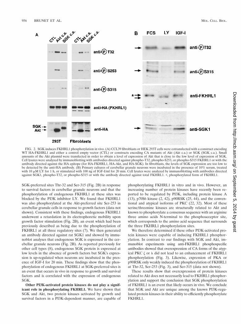
SGK-preferred sites Thr-32 and Ser-315 (Fig. 2B) in responseto survival factors in cerebellar granule neurons and that thephosphorylation of endogenous FKHRL1 at these sites wasblocked by the PI3K inhibitor LY. We found that FKHRL1was also phosphorylated at the Akt-preferred site Ser-253 incerebellar granule cells in response to growth factors (data notshown). Consistent with these findings, endogenous FKHRL1underwent a retardation in its electrophoretic mobility upongrowth factor stimulation (Fig. 2B), an event which had beenpreviously described as being due to the phosphorylation ofFKHRL1 at all three regulatory sites (7). We then generatedan antibody directed against rat SGK1 and showed by immu-noblot analyses that endogenous SGK is expressed in the cer-ebellar granule neurons (Fig. 2B). As reported previously forother cell types (8), endogenous SGK protein is expressed atlow levels in the absence of growth factors but SGK’s expres-sion is up-regulated when neurons are incubated in the pres-ence of IGF-I for 20 min. These findings show that the phos-phorylation of endogenous FKHRL1 at SGK-preferred sites isan event that occurs in vivo in response to growth and survivalfactors and is correlated with the expression of endogenousSGK.
Other PI3K-activated protein kinases do not play a signif-icant role in phosphorylating FKHRL1. We have shown thatSGK and Akt, two protein kinases activated by growth andsurvival factors in a PI3K-dependent manner, are capable of
phosphorylating FKHRL1 in vitro and in vivo. However, anincreasing number of protein kinases have recently been re-ported to be regulated by PI3K, including protein kinase A(13), p70S6 kinase (2, 42), p90RSK (25, 44), and the conven-tional and atypical isoforms of PKC (22, 32). Most of theseserine/threonine kinases are structurally related to Akt andknown to phosphorylate a consensus sequence with an argininethree amino acids N-terminal to the phosphoacceptor site,which corresponds to the amino acid sequence that surroundsthe three FKHRL1 phosphorylation sites.
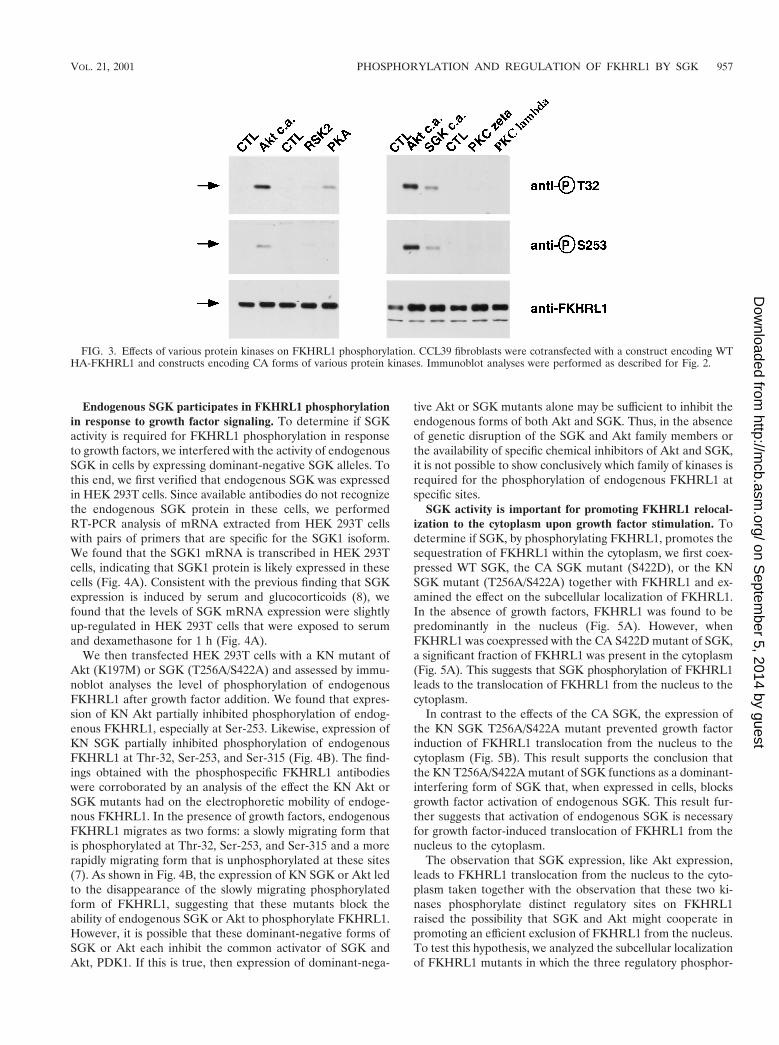
We therefore determined if these other PI3K-activated pro-tein kinases were capable of inducing FKHRL1 phosphor-ylation. In contrast to our findings with SGK and Akt, im-munoblot experiments using anti-FKHRL1 phosphospecificantibodies showed that overexpression of CA forms of the atyp-ical PKC z or l did not lead to an enhancement of FKHRL1phosphorylation (Fig. 3). Likewise, expression of PKA orp90RSK only weakly induced the phosphorylation of FKHRL1at Thr-32, Ser-253 (Fig. 3), and Ser-315 (data not shown).
These results show that overexpression of protein kinasesrelated to Akt does not necessarily lead to FKHRL1 phosphor-ylation and support the conclusion that SGK phosphorylationof FKHRL1 is an event that likely occurs in vivo. We concludethat SGK and Akt are unique among the known PI3K-regu-lated protein kinases in their ability to efficiently phosphorylateFKHRL1.
FIG. 2. SGK induces FKHRL1 phosphorylation in vivo. (A) CCL39 fibroblasts or HEK 293T cells were cotransfected with a construct encodingWT HA-FKHRL1 and either a control empty vector (CTL) or constructs encoding CA mutants of Akt (Akt c.a.) or SGK (SGK c.a.). Smallamounts of the Akt plasmid were transfected in order to obtain a level of expression of Akt that is close to the low level of expression of SGK.Cell lysates were analyzed by immunoblotting with antibodies directed against phospho-T32, phospho-S253, or phospho-S315 FKHRL1 or with theantibody directed against the HA epitope (for HA-FKHRL1, HA-Akt, and HA-SGK). In fibroblasts, the levels of SGK expression are too low tobe detected by the anti-HA antibody. (B) Primary cultures of cerebellar granule neurons were incubated in the presence of 10% serum, treatedwith 10 mM LY for 1 h, or stimulated with 100 ng of IGF-I/ml for 20 min. Cell lysates were analyzed by immunoblotting with antibodies directedagainst SGK1, phospho-T32, or phospho-S315 or with the antibody directed against total FKHRL1. p, phosphorylated form of FKHRL1.
956 BRUNET ET AL. MOL. CELL. BIOL.
on Septem
ber 5, 2014 by guesthttp://m
cb.asm.org/
Dow
nloaded from
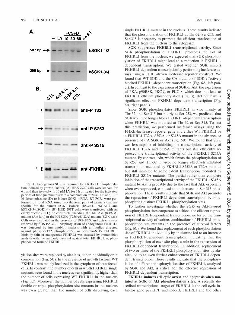
Endogenous SGK participates in FKHRL1 phosphorylationin response to growth factor signaling. To determine if SGKactivity is required for FKHRL1 phosphorylation in responseto growth factors, we interfered with the activity of endogenousSGK in cells by expressing dominant-negative SGK alleles. Tothis end, we first verified that endogenous SGK was expressedin HEK 293T cells. Since available antibodies do not recognizethe endogenous SGK protein in these cells, we performedRT-PCR analysis of mRNA extracted from HEK 293T cellswith pairs of primers that are specific for the SGK1 isoform.We found that the SGK1 mRNA is transcribed in HEK 293Tcells, indicating that SGK1 protein is likely expressed in thesecells (Fig. 4A). Consistent with the previous finding that SGKexpression is induced by serum and glucocorticoids (8), wefound that the levels of SGK mRNA expression were slightlyup-regulated in HEK 293T cells that were exposed to serumand dexamethasone for 1 h (Fig. 4A).
We then transfected HEK 293T cells with a KN mutant ofAkt (K197M) or SGK (T256A/S422A) and assessed by immu-noblot analyses the level of phosphorylation of endogenousFKHRL1 after growth factor addition. We found that expres-sion of KN Akt partially inhibited phosphorylation of endog-enous FKHRL1, especially at Ser-253. Likewise, expression ofKN SGK partially inhibited phosphorylation of endogenousFKHRL1 at Thr-32, Ser-253, and Ser-315 (Fig. 4B). The find-ings obtained with the phosphospecific FKHRL1 antibodieswere corroborated by an analysis of the effect the KN Akt orSGK mutants had on the electrophoretic mobility of endoge-nous FKHRL1. In the presence of growth factors, endogenousFKHRL1 migrates as two forms: a slowly migrating form thatis phosphorylated at Thr-32, Ser-253, and Ser-315 and a morerapidly migrating form that is unphosphorylated at these sites(7). As shown in Fig. 4B, the expression of KN SGK or Akt ledto the disappearance of the slowly migrating phosphorylatedform of FKHRL1, suggesting that these mutants block theability of endogenous SGK or Akt to phosphorylate FKHRL1.However, it is possible that these dominant-negative forms ofSGK or Akt each inhibit the common activator of SGK andAkt, PDK1. If this is true, then expression of dominant-nega-
tive Akt or SGK mutants alone may be sufficient to inhibit theendogenous forms of both Akt and SGK. Thus, in the absenceof genetic disruption of the SGK and Akt family members orthe availability of specific chemical inhibitors of Akt and SGK,it is not possible to show conclusively which family of kinases isrequired for the phosphorylation of endogenous FKHRL1 atspecific sites.
SGK activity is important for promoting FKHRL1 relocal-ization to the cytoplasm upon growth factor stimulation. Todetermine if SGK, by phosphorylating FKHRL1, promotes thesequestration of FKHRL1 within the cytoplasm, we first coex-pressed WT SGK, the CA SGK mutant (S422D), or the KNSGK mutant (T256A/S422A) together with FKHRL1 and ex-amined the effect on the subcellular localization of FKHRL1.In the absence of growth factors, FKHRL1 was found to bepredominantly in the nucleus (Fig. 5A). However, whenFKHRL1 was coexpressed with the CA S422D mutant of SGK,a significant fraction of FKHRL1 was present in the cytoplasm(Fig. 5A). This suggests that SGK phosphorylation of FKHRL1leads to the translocation of FKHRL1 from the nucleus to thecytoplasm.
In contrast to the effects of the CA SGK, the expression ofthe KN SGK T256A/S422A mutant prevented growth factorinduction of FKHRL1 translocation from the nucleus to thecytoplasm (Fig. 5B). This result supports the conclusion thatthe KN T256A/S422A mutant of SGK functions as a dominant-interfering form of SGK that, when expressed in cells, blocksgrowth factor activation of endogenous SGK. This result fur-ther suggests that activation of endogenous SGK is necessaryfor growth factor-induced translocation of FKHRL1 from thenucleus to the cytoplasm.
The observation that SGK expression, like Akt expression,leads to FKHRL1 translocation from the nucleus to the cyto-plasm taken together with the observation that these two ki-nases phosphorylate distinct regulatory sites on FKHRL1raised the possibility that SGK and Akt might cooperate inpromoting an efficient exclusion of FKHRL1 from the nucleus.To test this hypothesis, we analyzed the subcellular localizationof FKHRL1 mutants in which the three regulatory phosphor-
FIG. 3. Effects of various protein kinases on FKHRL1 phosphorylation. CCL39 fibroblasts were cotransfected with a construct encoding WTHA-FKHRL1 and constructs encoding CA forms of various protein kinases. Immunoblot analyses were performed as described for Fig. 2.
VOL. 21, 2001 PHOSPHORYLATION AND REGULATION OF FKHRL1 BY SGK 957
on Septem
ber 5, 2014 by guesthttp://m
cb.asm.org/
Dow
nloaded from
ylation sites were replaced by alanines, either individually or incombination (Fig. 5C). In the presence of growth factors, WTFKHRL1 was mainly localized in the cytoplasm of transfectedcells. In contrast, the number of cells in which FKHRL1 singlemutants were found in the nucleus was significantly higher thanthe number of cells expressing WT FKHRL1 in the nucleus(Fig. 5C). Moreover, the number of cells expressing FKHRL1double or triple phosphorylation site mutants in the nucleuswas even greater than the number of cells displaying each
single FKHRL1 mutant in the nucleus. These results indicatethat the phosphorylation of FKHRL1 at Thr-32, Ser-253, andSer-315 is necessary to promote the efficient translocation ofFKHRL1 from the nucleus to the cytoplasm.
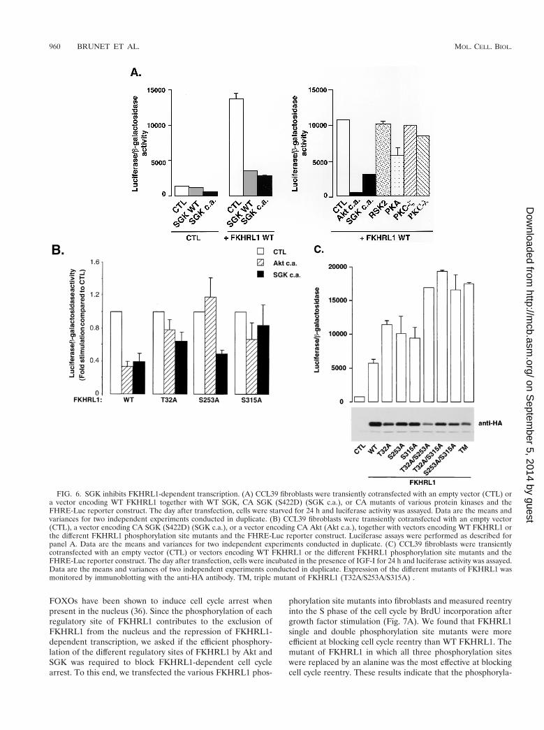
SGK suppresses FKHRL1 transcriptional activity. SinceSGK phosphorylation of FKHRL1 promotes the exit ofFKHRL1 from the nucleus, we expected that SGK phosphor-ylation of FKHRL1 might lead to a reduction in FKHRL1-dependent transcription. We tested whether SGK inhibitsFKHRL1-dependent transcription by performing luciferase as-says using a FHRE-driven luciferase reporter construct. Wefound that WT SGK and the CA mutants of SGK effectivelyblocked FKHRL1-dependent transcription (Fig. 6A, left pan-el). In contrast to the expression of SGK or Akt, the expressionof PKA, p90RSK, PKC z, or PKC l, which does not lead toFKHRL1 efficient phosphorylation (Fig. 3), did not have asignificant effect on FKHRL1-dependent transcription (Fig.6A, right panel).
Since SGK phosphorylates FKHRL1 in vivo mainly atThr-32 and Ser-315 but poorly at Ser-253, we predicted thatSGK would no longer block FKHRL1-dependent transcriptionwhen FKHRL1 was mutated at Thr-32 or Ser-315. To testthis prediction, we performed luciferase assays using theFHRE-luciferase reporter gene and either WT FKHRL1 ora FKHRL1 T32A, S253A, or S315A mutant in the absence orpresence of CA SGK or Akt (Fig. 6B). We found that SGKwas less capable of inhibiting the transcriptional activity ofFKHRL1 T32A and S315A mutants but still efficiently re-pressed the transcriptional activity of the FKHRL1 S253Amutant. By contrast, Akt, which favors the phosphorylation ofSer-253 and Thr-32 in vivo, no longer effectively inhibitedtranscription mediated by FKHRL1 S253A or T32A mutantsbut still inhibited to some extent transcription mediated byFKHRL1 S315A mutants. The partial rather than completeinhibition of the transcriptional activity of the FKHRL1 S315Amutant by Akt is probably due to the fact that Akt, especiallywhen overexpressed, can lead to an increase in Ser-315 phos-phorylation. These results indicate that SGK and Akt promotethe repression of FKHRL1-dependent transcription by phos-phorylating distinct FKHRL1 phosphorylation sites.
To further investigate whether the SGK- or Akt-favoredphosphorylation sites cooperate to achieve the efficient repres-sion of FKHRL1-dependent transcription, we tested the tran-scriptional activity of various combinations of FKHRL1 phos-phorylation site mutants in the presence of survival factors(Fig. 6C). We found that replacement of each phosphorylationsite of FKHRL1 individually by an alanine led to an increasein FKHRL1-dependent transcription, indicating that thephosphorylation of each site plays a role in the repression ofFKHRL1-dependent transcription. In addition, replacementof two or three of the FKHRL1 phosphorylation sites by ala-nine led to an even further enhancement of FKHRL1-depen-dent transcription. These results indicate that the phosphory-lation of different phosphorylation sites of FKHRL1, catalyzedby SGK and Akt, is critical for the effective repression ofFKHRL1-dependent transcription.
FKHRL1 induces cell cycle arrest and apoptosis when mu-tated at SGK or Akt phosphorylation sites. A recently de-scribed transcriptional target of FKHRL1 is the cell cycle in-hibitor gene p27KIP1, and indeed, FKHRL1 and the other
FIG. 4. Endogenous SGK is required for FKHRL1 phosphoryla-tion induced by growth factors. (A) HEK 293T cells were starved for6 h and then treated with 10 mM LY for 1 h or treated for the indicatedperiods of time (in minutes) with a combination of 20% FCS and 1024
M dexamethasone (D) to induce SGK1 mRNA. RT-PCRs were per-formed on total RNA using two different pairs of primers that arespecific for the human SGK1 isoform (hSGK1-1–hSGK1-2 andhSGK1-3–hSGK1-4). (B) HEK 293T cells were transfected with anempty vector (CTL) or constructs encoding the KN Akt (K197M)mutant (Akt k.n.) or the KN SGK (T256A/S422A) mutant (SGK k.n.).Cells were incubated in the presence of 10% FCS, and extracts wereresolved by SDS-PAGE. Phosphorylation of endogenous FKHRL1was detected by immunoblot analysis with antibodies directedagainst phospho-T32, phospho-S253, or phospho-S315 FKHRL1.Mobility shift of endogenous FKHRL1 was assessed by immunoblotanalysis with the antibody directed against total FKHRL1. p, phos-phorylated form of FKHRL1.
958 BRUNET ET AL. MOL. CELL. BIOL.
on Septem
ber 5, 2014 by guesthttp://m
cb.asm.org/
Dow
nloaded from
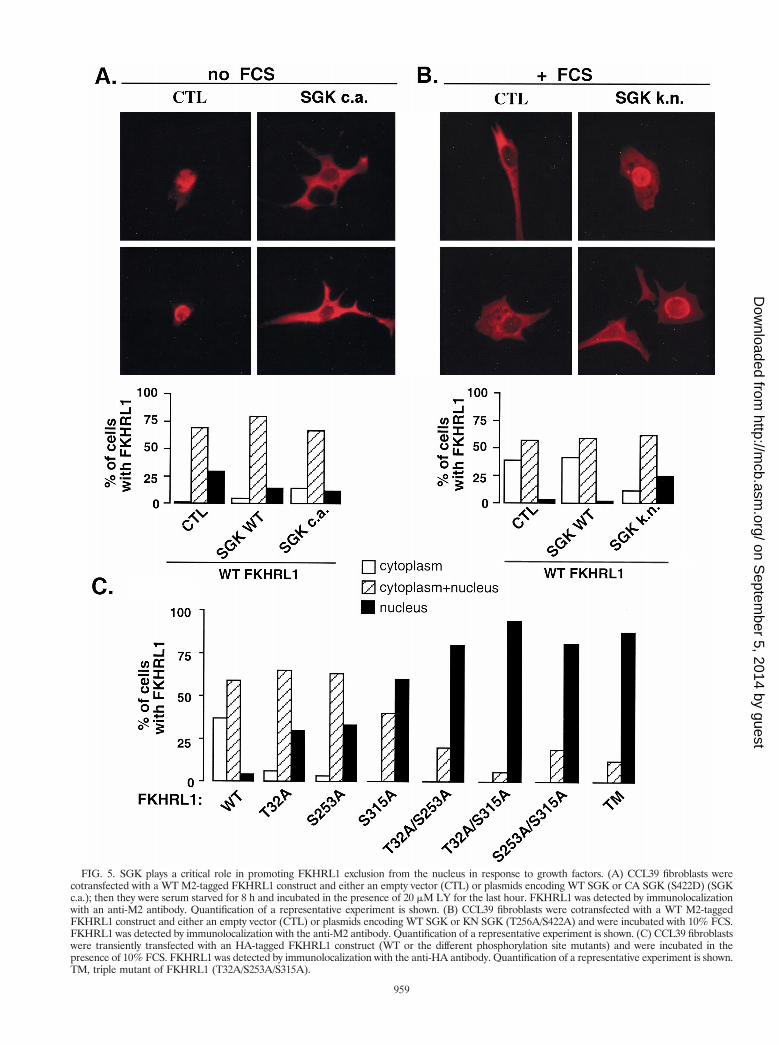
FIG. 5. SGK plays a critical role in promoting FKHRL1 exclusion from the nucleus in response to growth factors. (A) CCL39 fibroblasts werecotransfected with a WT M2-tagged FKHRL1 construct and either an empty vector (CTL) or plasmids encoding WT SGK or CA SGK (S422D) (SGKc.a.); then they were serum starved for 8 h and incubated in the presence of 20 mM LY for the last hour. FKHRL1 was detected by immunolocalizationwith an anti-M2 antibody. Quantification of a representative experiment is shown. (B) CCL39 fibroblasts were cotransfected with a WT M2-taggedFKHRL1 construct and either an empty vector (CTL) or plasmids encoding WT SGK or KN SGK (T256A/S422A) and were incubated with 10% FCS.FKHRL1 was detected by immunolocalization with the anti-M2 antibody. Quantification of a representative experiment is shown. (C) CCL39 fibroblastswere transiently transfected with an HA-tagged FKHRL1 construct (WT or the different phosphorylation site mutants) and were incubated in thepresence of 10% FCS. FKHRL1 was detected by immunolocalization with the anti-HA antibody. Quantification of a representative experiment is shown.TM, triple mutant of FKHRL1 (T32A/S253A/S315A).
959
on Septem
ber 5, 2014 by guesthttp://m
cb.asm.org/
Dow
nloaded from
FOXOs have been shown to induce cell cycle arrest whenpresent in the nucleus (36). Since the phosphorylation of eachregulatory site of FKHRL1 contributes to the exclusion ofFKHRL1 from the nucleus and the repression of FKHRL1-dependent transcription, we asked if the efficient phosphory-lation of the different regulatory sites of FKHRL1 by Akt andSGK was required to block FKHRL1-dependent cell cyclearrest. To this end, we transfected the various FKHRL1 phos-
phorylation site mutants into fibroblasts and measured reentryinto the S phase of the cell cycle by BrdU incorporation aftergrowth factor stimulation (Fig. 7A). We found that FKHRL1single and double phosphorylation site mutants were moreefficient at blocking cell cycle reentry than WT FKHRL1. Themutant of FKHRL1 in which all three phosphorylation siteswere replaced by an alanine was the most effective at blockingcell cycle reentry. These results indicate that the phosphoryla-
FIG. 6. SGK inhibits FKHRL1-dependent transcription. (A) CCL39 fibroblasts were transiently cotransfected with an empty vector (CTL) ora vector encoding WT FKHRL1 together with WT SGK, CA SGK (S422D) (SGK c.a.), or CA mutants of various protein kinases and theFHRE-Luc reporter construct. The day after transfection, cells were starved for 24 h and luciferase activity was assayed. Data are the means andvariances for two independent experiments conducted in duplicate. (B) CCL39 fibroblasts were transiently cotransfected with an empty vector(CTL), a vector encoding CA SGK (S422D) (SGK c.a.), or a vector encoding CA Akt (Akt c.a.), together with vectors encoding WT FKHRL1 orthe different FKHRL1 phosphorylation site mutants and the FHRE-Luc reporter construct. Luciferase assays were performed as described forpanel A. Data are the means and variances for two independent experiments conducted in duplicate. (C) CCL39 fibroblasts were transientlycotransfected with an empty vector (CTL) or vectors encoding WT FKHRL1 or the different FKHRL1 phosphorylation site mutants and theFHRE-Luc reporter construct. The day after transfection, cells were incubated in the presence of IGF-I for 24 h and luciferase activity was assayed.Data are the means and variances of two independent experiments conducted in duplicate. Expression of the different mutants of FKHRL1 wasmonitored by immunoblotting with the anti-HA antibody. TM, triple mutant of FKHRL1 (T32A/S253A/S315A) .
960 BRUNET ET AL. MOL. CELL. BIOL.
on Septem
ber 5, 2014 by guesthttp://m
cb.asm.org/
Dow
nloaded from
tion of the three regulatory sites of FKHRL1 is important toprevent FKHRL1 from inducing cell cycle arrest.
In addition to inducing cell cycle arrest in fibroblasts, an-other function of the FOXO subfamily of Forkhead transcrip-tion factors is to trigger apoptosis (7, 36, 45, 46). To assesswhether the phosphorylation of FKHRL1 at the different reg-ulatory sites is required to prevent this transcription factorfrom inducing apoptosis, we transfected the various FKHRL1phosphorylation site mutants into HEK 293T cells in the pres-ence of the survival factor IGF-I. We assessed apoptosis of thetransfected cells by performing immunoblot analyses with anantibody that specifically binds the cleaved product of PARP(Fig. 7B). In living cells, PARP is intact and thus not recog-nized by the antibody, whereas in cells undergoing apoptosis,caspases are activated and cleave PARP, one of whose cleav-age products is recognized by the antibody. We found thatwhile WT FKHRL1 did not induce PARP cleavage, the mu-tants of FKHRL1 with single and especially double and triplephosphorylation site mutations triggered PARP cleavage.These results indicate that the efficient phosphorylation ofFKHRL1 at all three regulatory sites is critical to preventFKHRL1 from inducing cell death.
SGK plays a role in the promotion of growth factor-inducedcell survival. Our observation that SGK suppresses FKHRL1-dependent transcription and that the phosphorylation ofFKHRL1 at all three regulatory sites is important for theprevention of FKHRL1-induced cell cycle arrest and apoptosissuggested that SGK may regulate these two biological func-tions. Indeed, a role for SGK in cell cycle progression had beensuggested in a recent study (8).
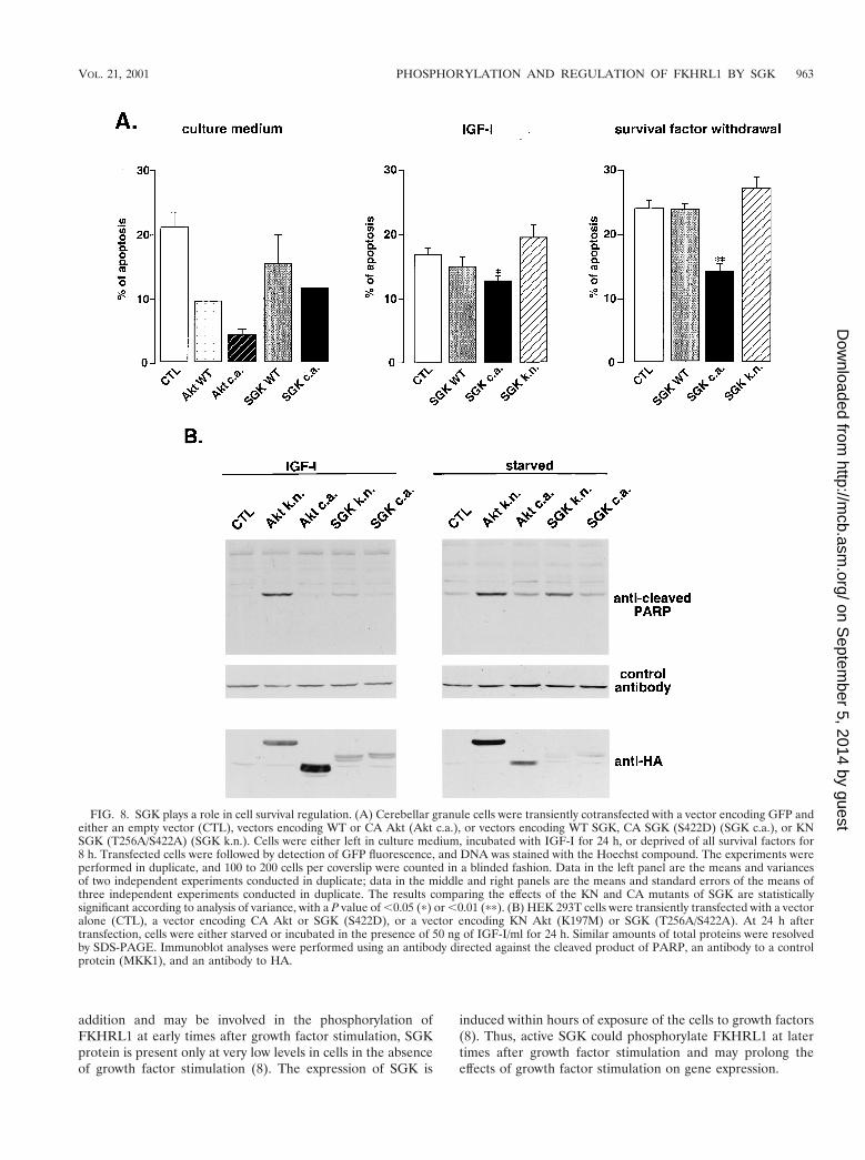
To determine whether SGK plays a critical role in regulatingcell survival, we first employed primary cultures of cerebellargranule neurons, a population of neurons that is dependentboth on PI3K and on the repression of FKHRL1-dependenttranscription for cell survival (7, 21). As shown in Fig. 2B, theseneurons express an endogenous form of SGK as well as anendogenous form of FKHRL1 that is phosphorylated in re-sponse to survival factors. After transfecting cerebellar granulecells with various forms of SGK and counting the number ofapoptotic and surviving cells among the transfected cells, wethen asked whether SGK could promote the survival of neu-rons. The expression of a CA form of SGK, as with Akt, leadsto a significant reduction of apoptotic neurons whether thecerebellar neurons are grown in the presence or absence ofsurvival factors (with a P value of ,0.01 according to analysisof variance) (Fig. 8A). In contrast, the expression of KN SGK(T256A/S422A) in cerebellar neurons led to a small increase inthe number of cells undergoing apoptosis (Fig. 8A), which isconsistent with ability of this SGK mutant to act in a dominant-interfering manner to prevent endogenous SGK from phos-phorylating FKHRL1 and relocalizing it to the cytoplasm.
To further determine if SGK activity is critical for mediatingcell survival, we expressed KN mutants of SGK or Akt in HEK293T cells in the presence or absence of IGF-I and monitoredapoptosis by the appearance of the cleaved form of PARP (Fig.8B). We found that the KN forms of SGK and Akt in HEK293T cells induced the cleavage of PARP, while the CA formsdid not.
It has to be noted that in cerebellar neurons as well as inHEK 293T cells, the effects of Akt mutants on cell survival
were more pronounced than the effects of SGK mutants. How-ever this difference could be due to a difference in the expres-sion or activity of the mutants of Akt or SGK, making itdifficult to determine which kinase, Akt or SGK, plays a morecritical role in mediating cell survival.
Taken together, these findings indicate that SGK, when ex-pressed and activated, may together with Akt mediate survivaleffects of PI3K, in part by phosphorylating FKHRL1 and re-pressing its transcriptional activity. The inhibition of SGK andAkt activity may trigger the dephosphorylation of FKHRL1,the translocation of FKHRL1 from the cytoplasm to the nu-cleus, and the activation of FKHRL1-regulated death genes,thereby inducing apoptosis.
DISCUSSION
In this report we describe experiments demonstrating thatthe PI3K-regulated kinase SGK1 phosphorylates the FOXOtranscription factor FKHRL1, thereby inducing the exit ofFKHRL1 from the nucleus and the repression of FKHRL1-dependent transcription. Our experiments identify FKHRL1as a physiological substrate of SGK and suggest a biologicalrole for this protein kinase in the regulation of cell survival andcell cycle progression. Our findings suggest that SGK functionsas a critical link between growth factor activation of the PI3Kand the regulation of gene expression events that are criticalfor biological responses such as cell survival and cell cyclereentry.
SGK and Akt may cooperate in promoting a variety of bio-logical responses by phosphorylating common substrates atoverlapping but different regulatory sites. SGK and Akt differwith respect to the efficacy with which they phosphorylatespecific sites on FKHRL1, since SGK preferentially phosphor-ylates Ser-315 whereas Akt favors Ser-253. The coordinatedphosphorylation of FKHRL1 at three sites by SGK and Aktcould involve the sequential phosphorylation of the differentsites of FKHRL1. In a recent study of the FKHRL1 familymember FKHR, the site equivalent to FKHRL1 Ser-253 wassuggested to act as a “gatekeeper,” since the phosphorylationof Ser-253 was found to be required to release a negativeconstraint on the FKHR molecule so that FKHR could then bephosphorylated at additional sites (37). Nakae and colleaguessuggested that while Akt is responsible for phosphorylatingFKHR at Ser-253, other unidentified protein kinases that aredistinct from Akt may phosphorylate the two other sites onFKHR (37). Given the findings in the present study, it istempting to speculate that Akt may first phosphorylateFKHRL1 at Ser-253 and that this phosphorylation event mayinduce a conformational change in FKHRL1 that allows SGKto phosphorylate FKHRL1 at Ser-315. Alternatively, the phos-phorylation of FKHRL1 at Ser-253 by Akt may allow thebinding to FKHRL1 of 14-3-3 proteins, which then act ascofactors that promote the phosphorylation of FKHRL1 bySGK. A role for 14-3-3 proteins as cofactors that promotesequential protein phosphorylation was recently demonstratedfor another Akt target, the Bcl-2 family member BAD (18).
The efficient phosphorylation of the three regulatory sites ofFKHRL1 appears to be required to ensure the effective trans-location of FKHRL1 from the nucleus to the cytoplasm andthe effective repression of FKHRL1-dependent transcription.
VOL. 21, 2001 PHOSPHORYLATION AND REGULATION OF FKHRL1 BY SGK 961
on Septem
ber 5, 2014 by guesthttp://m
cb.asm.org/
Dow
nloaded from

The phosphorylation of FKHRL1 at each of the regulatorysites may involve different mechanisms that cooperate to pro-mote the efficient exclusion of FKHRL1 from the nucleus.FKHRL1 Ser-315, which is the phosphorylation site favored bySGK, is located close to the nuclear export signal (NES) ofFOXOs (6). One possibility is that SGK phosphorylation ofSer-315 unmasks the NES of FKHRL1, thereby rendering theNES accessible to the nuclear export machinery. Thus, SGKphosphorylation of Ser-315 may play a primary role in export-ing nuclear FKHRL1 to the cytoplasm upon activation of thePI3K-SGK signaling pathway. The phosphorylation of theother two sites on FKHRL1, Thr-32 and Ser-253, is catalyzedmore effectively by Akt and is required for FKHRL1 binding to14-3-3 proteins (7). When bound to FKHRL1, 14-3-3 proteinsmay participate in the nuclear export of FKHRL1, since the14-3-3 proteins have been shown to contain a NES and to beinvolved in the coexport of their binding partners (33). 14-3-3protein binding to FKHRL1 may also ensure the retention ofFKHRL1 in the cytoplasm. As FKHRL1 Ser-253 is locatedwithin the nuclear localization signal of FKHRL1, Akt phos-phorylation of Ser-253 may also inhibit the function of thenuclear localization signal of FKHRL1, thereby preventing thereentry of cytoplasmic FKHRL1 into the nucleus.
The regulation of FKHRL1 subcellular localization by phos-phorylation is reminiscent of the regulation of the yeast tran-scription factor Pho4 (27, 29, 40). Pho4 is phosphorylated atseveral regulatory sites by different kinases and the multiplephosphorylation events cooperate to promote the nuclear ex-port of Pho4 and to prevent the nuclear import of Pho4, thusresulting in the exclusion of this transcription factor from thenucleus (29). The sequential phosphorylation of transcriptionfactors, such as Pho4 or FKHRL1, at several sites that have
overlapping but distinct roles in regulating nuclear export maytherefore provide a general and efficient mechanism that en-sures the effective translocation of these transcription factorsfrom the nucleus to the cytoplasm.
The primary consequence of FKHRL1 phosphorylation atits regulatory sites appears to be the translocation of FKHRL1from the nucleus to the cytoplasm and the inhibition ofFKHRL1-dependent transcription. However, a question thatremains to be answered is that of whether there are kinase-dependent mechanisms in addition to the regulation of thesubcellular localization of FKHRL1 that contribute to the con-trol of FKHRL1 function. Given that Ser-315, the amino acidresidue that is preferentially phosphorylated by SGK, is lo-cated within the transactivation domain of FKHRL1, it is pos-sible that the phosphorylation of Ser-315 may also regulate thetransactivation function of FKHLR1. In addition, phosphory-lation of FKHRL1 at different phosphorylation sites may fa-cilitate FKHRL1 binding to distinct protein partners that haveyet to be identified but could prove to be important forFKHRL1 function.
It is not yet clear why there are two families of PI3K-regu-lated kinases that phosphorylate FKHRL1. One possibility isthat when expressed at physiological levels, SGK and Akt dis-play a strict preference for one FKHRL1 site over the others,so that SGK exclusively phosphorylates FKHRL1 at Ser-315while Akt selectively phosphorylates FKHRL1 at Ser-253. Thisis consistent with the observation that when expressed at lowlevels, SGK selectively phosphorylated Ser-315 while Akt wasmore effective at phosphorylating Ser-253. Another possibilityis suggested by the observation that Akt and SGK are differ-entially expressed in cells in response to environmental stimuli.Whereas Akt is activated within seconds upon growth factor
FIG. 7. The phosphorylation of FKHRL1 at the three regulatory sites is required to prevent FKHRL1-induced cell cycle arrest and apoptosis.(A) CCL39 fibroblasts were transfected with the various phosphorylation site mutants of FKHRL1, starved for 24 h, and stimulated for 24 h with20% FCS in the presence of BrdU. Transfected cells and the incorporation of BrdU were identified by immunofluorescence with an anti-FKHRL1antibody and an anti-BrdU antibody, respectively. Data are the means and variances of two independent experiments. CTL, control; TM, triplemutant of FKHRL1 (T32A/S253A/S315A). (B) HEK 293T cells were transfected with the various FKHRL1 phosphorylation site mutants andstimulated for 24 h with IGF-I. Immunoblot analyses were performed with an antibody directed against the cleaved product of PARP, a controlantibody to a protein whose levels remain constant (MKK1), and an antibody to HA. TM, triple mutant of FKHRL1 (T32A/S253A/S315A) .
962 BRUNET ET AL. MOL. CELL. BIOL.
on Septem
ber 5, 2014 by guesthttp://m
cb.asm.org/
Dow
nloaded from
addition and may be involved in the phosphorylation ofFKHRL1 at early times after growth factor stimulation, SGKprotein is present only at very low levels in cells in the absenceof growth factor stimulation (8). The expression of SGK is
induced within hours of exposure of the cells to growth factors(8). Thus, active SGK could phosphorylate FKHRL1 at latertimes after growth factor stimulation and may prolong theeffects of growth factor stimulation on gene expression.
FIG. 8. SGK plays a role in cell survival regulation. (A) Cerebellar granule cells were transiently cotransfected with a vector encoding GFP andeither an empty vector (CTL), vectors encoding WT or CA Akt (Akt c.a.), or vectors encoding WT SGK, CA SGK (S422D) (SGK c.a.), or KNSGK (T256A/S422A) (SGK k.n.). Cells were either left in culture medium, incubated with IGF-I for 24 h, or deprived of all survival factors for8 h. Transfected cells were followed by detection of GFP fluorescence, and DNA was stained with the Hoechst compound. The experiments wereperformed in duplicate, and 100 to 200 cells per coverslip were counted in a blinded fashion. Data in the left panel are the means and variancesof two independent experiments conducted in duplicate; data in the middle and right panels are the means and standard errors of the means ofthree independent experiments conducted in duplicate. The results comparing the effects of the KN and CA mutants of SGK are statisticallysignificant according to analysis of variance, with a P value of ,0.05 (p) or ,0.01 (pp). (B) HEK 293T cells were transiently transfected with a vectoralone (CTL), a vector encoding CA Akt or SGK (S422D), or a vector encoding KN Akt (K197M) or SGK (T256A/S422A). At 24 h aftertransfection, cells were either starved or incubated in the presence of 50 ng of IGF-I/ml for 24 h. Similar amounts of total proteins were resolvedby SDS-PAGE. Immunoblot analyses were performed using an antibody directed against the cleaved product of PARP, an antibody to a controlprotein (MKK1), and an antibody to HA.
VOL. 21, 2001 PHOSPHORYLATION AND REGULATION OF FKHRL1 BY SGK 963
on Septem
ber 5, 2014 by guesthttp://m
cb.asm.org/
Dow
nloaded from

The overexpression of KN mutants of SGK or Akt in cellsreduces the phosphorylation of FKHRL1 and inhibits FKHRL1cytoplasmic relocalization in response to growth factors, sug-gesting that endogenous SGK and Akt may both be requiredfor the phosphorylation of FKHRL1 and for FKHRL1 cyto-plasmic retention. In addition, overexpression of KN mutantsof SGK or Akt in cells promotes apoptosis, raising the possi-bility that endogenous SGK and Akt may both play an impor-tant role in mediating PI3K-dependent cell survival. However,a potential limitation to the use of the dominant-interferingSGK and Akt mutants to selectively inhibit their endogenouscounterparts is that SGK and Akt are both activated by thesame upstream kinase, PDK1 (28, 41). Thus, the expression ofhigh levels of dominant-interfering mutants of SGK may leadto inhibition of PDK1 and consequently may prevent the acti-vation of both SGK and Akt (48). Conversely, it is possible thatthe overexpression of dominant-negative mutants of Akt mayinterfere with endogenous SGK function. Therefore, many ofthe data that had been generated in the past few years usingdominant-negative mutants of Akt to block endogenous Aktfunction may have to be reexamined, as the results obtainedmay reflect the inhibition of both endogenous Akt and SGK.The identification of chemical inhibitors that specifically blockSGK or Akt will be particularly useful for resolving this issue.
In our present study, we have defined a new function forSGK in mediating survival in response to survival factors. Itwill be important to determine whether the survival function ofSGK can be generalized to cells that have been challenged witha range of apoptotic stimuli, such as DNA damage, matrix de-tachment, or cell cycle discordance. The transcription factorFKHRL1 is also likely just one of a number of SGK substratesthat mediate the survival function of SGK. Other previously char-acterized targets of Akt that had been implicated in the regu-lation of cell survival, such as BAD and caspase 9 (9, 17, 19),may also be found to be phosphorylated and regulated by SGK.
The physiological roles of SGK are not likely to be restrictedto the promotion of cell survival. Previous studies have indi-cated that SGK is present in the nucleus during the S phase ofthe cell cycle, suggesting that SGK plays an active role in cellcycle progression (8). Importantly, FOXO transcription factorshave recently been shown to promote cell cycle arrest by in-ducing the expression of the cell cycle inhibitor p27KIP1 (36).Consistent with a role for SGK phosphorylation of FKHRL1 incell cycle progression, we find that mutating any one of thethree phosphorylation sites of FKHRL1 generates a form ofFKHRL1 that promotes cell cycle arrest. These results suggestthat SGK and Akt may together promote cell cycle progressionby phosphorylating and inactivating FOXOs, thereby decreas-ing p27KIP1 levels.
Given the importance of SGK and Akt for cell survival andproliferation, mutations in these kinases are likely to have animpact on cancer development in vertebrates. CA versions ofAkt and PI3K are transduced by oncogenic retroviruses andcan trigger cell transformation (5, 11). It will be interesting todetermine whether the CA versions of SGK will also be foundto be tumorigenic. The ability of CA forms of Akt and SGK toinduce cell transformation may be a consequence of the inap-propriate suppression of FOXO-dependent transcription, dueto SGK and/or Akt phosphorylation of FOXO transcriptionfactors. One attractive model is that when cells are exposed to
deleterious conditions that inhibit PI3K signaling, FOXOs willtranslocate to the nucleus and induce the expression of celldeath or cell cycle arrest genes. CA SGK and Akt may shut offthe expression of these cell death and arrest genes by excludingFOXOs from the nucleus, thereby triggering abnormal prolif-eration and survival and ultimately cell transformation.
Members of the PI3K pathway are conserved throughoutevolution. In particular, SGK homologues are present in yeast,nematodes, flies, and mammals. In the nematode Caenorhab-ditis elegans, the PI3K-Akt pathway plays an important role inthe regulation of insulin metabolism and organismal aging bysuppressing the activity of a Forkhead transcription factortermed Daf16 (39). Since in addition to Akt, there exists anSGK counterpart in the nematode, it will be interesting todetermine whether the nematode SGK contributes to the sup-pression of Daf16 function and participates in the control ofinsulin metabolism and organismal aging.
In summary, the protein kinase SGK appears to integratethe effects of different extracellular stimuli and to regulate avariety of biological functions by phosphorylating key sub-strates including the FOXO transcription factor FKHRL1. Theidentification of the array of biological functions and substratesof SGK will be of critical importance in understanding how thePI3K signaling pathway orchestrates cellular responses to var-ious extracellular cues.
ACKNOWLEDGMENTS
We thank G. Firestone, A. Toker, and F. Uberall for kindly provid-ing reagents. We thank S. R. Datta, M. Z. Lin, K. F. Tolias, and J. Ziegfor critical reading of the manuscript. We also thank S. R. Datta forstimulating discussion.
This research was supported by an NIH grant (CA43855), a MentalRetardation Research Center grant (HD 18655), and a grant fromDaiichi Pharmaceuticals to M.E.G. A.B. is supported by a HumanFrontier Long Term Fellowship.
ADDENDUM IN PROOF
While this ipaper was in review, Sonyang and colleagues(Curr. Biol. 10:1233–1236, 2000) demonstrated that SGK-3(also known as CISK) can both promote cell survival anddirectly phosphorylate and inactivate the proapoptotic proteinsFKHR and BAD.
REFERENCES
1. Alessi, D. R., F. B. Caudwell, M. Andjelkovic, B. A. Hemmings, and P.Cohen. 1996. Molecular basis for the substrate specificity of protein kinase B;comparison with MAPKAP kinase-1 and p70 S6 kinase. FEBS Lett. 399:333–338.
2. Alessi, D. R., M. T. Kozlowski, Q. P. Weng, N. Morrice, and J. Avruch. 1998.3-Phosphoinositide-dependent protein kinase 1 (PDK1) phosphorylates andactivates the p70 S6 kinase in vivo and in vitro. Curr. Biol. 8:69–81.
3. Alliston, T. N., A. C. Maiyar, P. Buse, G. L. Firestone, and J. S. Richards.1997. Follicle stimulating hormone-regulated expression of serum/glucocor-ticoid-inducible kinase in rat ovarian granulosa cells: a functional role for theSp1 family in promoter activity. Mol. Endocrinol. 11:1934–1949.
4. Alvarez de la Rosa, D., P. Zhang, A. Naray-Fejes-Toth, G. Fejes-Toth, andC. M. Canessa. 1999. The serum and glucocorticoid kinase sgk increases theabundance of epithelial sodium channels in the plasma membrane of Xeno-pus oocytes. J. Biol. Chem. 274:37834–37839.
5. Bellacosa, A., J. R. Testa, S. P. Staal, and P. N. Tsichlis. 1991. A retroviraloncogene, akt, encoding a serine threonine kinase containing a SH2-likeregion. Science 254:274–277.
6. Biggs, W. H. I., J. Meisenhelder, T. Hunter, W. K. Cavenee, and K. C. Arden.1999. Protein kinase B/Akt-mediated phosphorylation promotes nuclear ex-clusion of the winged helix transcription factor FKHR1. Proc. Natl. Acad.Sci. USA 96:7421–7426.
964 BRUNET ET AL. MOL. CELL. BIOL.
on Septem
ber 5, 2014 by guesthttp://m
cb.asm.org/
Dow
nloaded from
7. Brunet, A., A. Bonni, M. J. Zigmond, M. Z. Lin, P. Juo, L. S. Hu, M. J.Anderson, K. C. Arden, J. Blenis, and M. E. Greenberg. 1999. Akt promotescell survival by phosphorylating and inhibiting a Forkhead transcriptionfactor. Cell 96:857–868.
8. Buse, P., S. H. Tran, E. Luther, P. T. Phu, G. W. Aponte, and G. L. Firestone.1999. Cell cycle and hormonal control of nuclear-cytoplasmic localization ofthe serum- and glucocorticoid-inducible protein kinase, Sgk, in mammarytumor cells. A novel convergence point of anti-proliferative and proliferativecell signaling pathways. J. Biol. Chem. 274:7253–7263.
9. Cardone, H. M., N. Roy, H. R. Stennicke, G. S. Salvesen, T. F. Franke, E.Stanbridge, S. Frisch, and J. C. Reed. 1998. Regulation of cell death pro-tease caspase-9 by phosphorylation. Science 282:1318–1321.
10. Casamayor, A., P. D. Torrance, T. Kobayashi, J. Thorner, and D. R. Alessi.1999. Functional counterparts of mammalian protein kinases PDK1 andSGK in budding yeast. Curr. Biol. 9:186–197.
11. Chang, H. W., M. Aoki, D. Fruman, K. R. Auger, A. Bellacosa, P. N. Tsichlis,L. C. Cantley, T. M. Roberts, and P. K. Vogt. 1997. Transformation ofchicken cells by the gene encoding the catalytic subunit of PI 3-kinase.Science 276:1848–1850.
12. Chen, S. Y., A. Bhargava, L. Mastroberardino, O. C. Meijer, J. Wang, P.Buse, G. L. Firestone, F. Verrey, and D. Pearce. 1999. Epithelial sodiumchannel regulated by aldosterone-induced protein sgk. Proc. Natl. Acad. Sci.USA 96:2514–2519.
13. Cheng, X., Y. Ma, M. Moore, B. A. Hemmings, and S. S. Taylor. 1998. Phos-phorylation and activation of cAMP-dependent protein kinase by phosphoi-nositide-dependent protein kinase. Proc. Natl. Acad. Sci. USA 95:9849–9854.
14. Chou, M. M., W. Hou, J. Johnson, L. K. Graham, M. H. Lee, C. S. Chen,A. C. Newton, B. S. Schaffhausen, and A. Toker. 1998. Regulation of proteinkinase C zeta by PI 3-kinase and PDK-1. Curr. Biol. 8:1069–1077.
15. Cowling, R. T., and H. C. Birnboim. 2000. Expression of serum- and glu-cocorticoid-regulated kinase (sgk) mRNA is up-regulated by GM-CSF andother proinflammatory mediators in human granulocytes. J. Leukoc. Biol.67:240–248.
16. Datta, S. R., A. Brunet, and M. E. Greenberg. 1999. Cellular survival: a playin three Akts. Genes Dev. 13:2905–2927.
17. Datta, S. R., H. Dudek, X. Tao, S. Masters, H. Fu, Y. Gotoh, and M. E.Greenberg. 1997. Akt phosphorylation of BAD couples survival signals to thecell-intrinsic death machinery. Cell 91:231–241.
18. Datta, S. R., A. Katsov, L. Hu, A. Petros, S. W. Fesik, M. B. Yaffe, and M. E.Greenberg. 2000. 14-3-3 proteins and survival kinases cooperate to inactivateBAD by BH3 domain phosphorylation. Mol. Cell 6:41–51.
19. del Peso, L., M. Gonzalez-Garcia, C. Page, R. Herrera, and G. Nunez. 1997.Interleukin-3-induced phosphorylation of BAD through the protein kinaseAkt. Science 278:687–689.
20. Downward, J. 1998. Mechanisms and consequences of activation of proteinkinase B/Akt. Curr. Opin. Cell Biol. 10:262–267.
21. Dudek, H., S. R. Datta, T. F. Franke, M. Birnbaum, R. Yao, G. M. Cooper,R. A. Segal, D. R. Kaplan, and M. E. Greenberg. 1997. Regulation ofneuronal survival by the serine-threonine protein kinase Akt. Science 275:661–665.
22. Dutil, E. M., A. Toker, and A. C. Newton. 1998. Regulation of conventionalprotein kinase C isozymes by phosphoinositide-dependent kinase 1 (PDK1).Curr. Biol. 8:1366–1375.
23. Guo, S., G. Rena, S. Cichy, X. He, P. Cohen, and T. Unterman. 1999.Phosphorylation of serine 256 by protein kinase B disrupts transactivation byFKHR and mediates effects of insulin on IGF binding protein-1 promoteractivity through a conserved insulin response sequence. J. Biol. Chem. 274:17184–17192.
24. Imaizumi, K., M. Tsuda, A. Wanaka, M. Tohyama, and T. Takagi. 1994.Differential expression of sgk mRNA, a member of the Ser/Thr proteinkinase gene family, in rat brain after CNS injury. Brain Res. Mol. Brain Res.26:189–196.
25. Jensen, C. J., M. B. Buch, T. O. Krag, B. A. Hemmings, S. Gammeltoft, andM. Frodin. 1999. 90-kDa ribosomal S6 kinase is phosphorylated and acti-vated by 3-phosphoinositide-dependent protein kinase-1. J. Biol. Chem. 274:27168–27176.
26. Kaestner, K. H., W. Knochel, and D. E. Martinez. 2000. Unified nomenclaturefor the winged helix/forkhead transcription factors. Genes Dev. 14:142–146.
27. Kaffman, A., N. M. Rank, and E. K. O’Shea. 1998. Phosphorylation regulatesassociation of the transcription factor Pho4 with its import receptor Pse1/Kap121. Genes Dev. 12:2673–2683.
28. Kobayashi, T., and P. Cohen. 1999. Activation of serum- and glucocorticoid-regulated protein kinase by agonists that activate phosphatidylinositide 3-ki-nase is mediated by 3-phosphoinositide-dependent protein kinase-1 (PDK1)and PDK2. Biochem. J. 339:319–328.
29. Komeili, A., and E. K. O’Shea. 1999. Roles of phosphorylation sites inregulating activity of the transcription factor Pho4. Science 284:977–980.
30. Kops, G. J., and B. M. Burgering. 1999. Forkhead transcription factors: new
insights into protein kinase B (c-akt) signaling. J. Mol. Med. 77:656–665.31. Kops, G. J., N. D. de Ruiter, A. M. De Vries-Smits, D. R. Powell, J. L. Bos,
and B. M. Burgering. 1999. Direct control of the Forkhead transcriptionfactor AFX by protein kinase B. Nature 398:630–634.
32. Le Good, J. A., W. H. Ziegler, D. B. Parekh, D. R. Alessi, P. Cohen, and P. J.Parker. 1998. Protein kinase C isotypes controlled by phosphoinositide 3--kinase through the protein kinase PDK1. Science 281:2042–2045.
33. Lopez-Girona, A., B. Furnari, O. Mondesert, and P. Russell. 1999. Nuclearlocalization of Cdc25 is regulated by DNA damage and a 14-3-3 protein.Nature 397:172–175.
34. Maniatis, T., E. F. Fritsch, and J. Sambrook. 1989. Molecular cloning: alaboratory manual, 2nd ed. Cold Spring Harbor Laboratory Press, ColdSpring Harbor, N.Y.
35. Maurer, R. A. 1989. Both isoforms of the cAMP-dependent protein kinasecatalytic subunit can activate transcription of the prolactin gene. J. Biol.Chem. 264:6870–6873.
36. Medema, R. H., G. J. Kops, J. L. Bos, and B. M. Burgering. 2000. AFX-likeForkhead transcription factors mediate cell-cycle regulation by Ras and PKBthrough p27kip1. Nature 404:782–787.
37. Nakae, J., V. Barr, and D. Accili. 2000. Differential regulation of geneexpression by insulin and IGF-1 receptors correlates with phosphorylation ofa single amino acid residue in the forkhead transcription factor FKHR.EMBO J. 19:989–996.
38. Nakae, J., B. C. Park, and D. Accili. 1999. Insulin stimulates phosphorylationof the forkhead transcription factor FKHR on serine 253 through a wort-mannin-sensitive pathway. J. Biol. Chem. 274:15982–15985.
39. Ogg, S., S. Paradis, S. Gottlieb, G. I. Patterson, L. Lee, H. A. Tissenbaum, andG. Ruvkun. 1997. The Forkhead transcription factor DAF-16 transduces insulin-like metabolic and longevity signals in C. elegans. Nature 389:994–999.
40. O’Neill, E. M., A. Kaffman, E. R. Jolly, and E. K. O’Shea. 1996. Regulationof PHO4 nuclear localization by the PHO80-PHO85 cyclin-CDK complex.Science 271:209–212.
41. Park, J., M. L. Leong, P. Buse, A. C. Maiyar, G. L. Firestone, and B. A.Hemmings. 1999. Serum and glucocorticoid-inducible kinase (SGK) is atarget of the PI 3-kinase-stimulated signaling pathway. EMBO J. 18:3024–3033.
42. Pullen, N., P. B. Dennis, M. Andjelkovic, A. Dufner, S. C. Kozma, B. A.Hemmings, and G. Thomas. 1998. Phosphorylation and activation of p70s6kby PDK1. Science 279:707–710.
43. Rena, G., S. Guo, S. Cichy, T. G. Unterman, and P. Cohen. 1999. Phosphor-ylation of the transcription factor forkhead family member FKHR by proteinkinase B. J. Biol. Chem. 274:17179–17183.
44. Richards, S. A., J. Fu, A. Romanelli, A. Shimamura, and J. Blenis. 1999.Ribosomal S6 kinase 1 (RSK1) activation requires signals dependent on andindependent of the MAP kinase ERK. Curr. Biol. 9:810–820.
45. Takaishi, H., H. Konishi, H. Matsuzaki, Y. Ono, Y. Shirai, N. Saito, T.Kitamura, W. Ogawa, M. Kasuga, U. Kikkawa, and Y. Nishizuka. 1999.Regulation of nuclear translocation of forkhead transcription factor AFX byprotein kinase B. Proc. Natl. Acad. Sci. USA 96:11836–11841.
46. Tang, E. D., G. Nunez, F. G. Barr, and K.-L. Guan. 1999. Negative regulationof the Forkhead transcription factor FKHR by Akt. J. Biol. Chem. 274:16741–16746.
47. Uberall, F., K. Hellbert, S. Kampfer, K. Maly, A. Villunger, M. Spitaler, J.Mwanjewe, G. Baier-Bitterlich, G. Baier, and H. H. Grunicke. 1999. Evi-dence that atypical protein kinase C-lambda and atypical protein kinaseC-zeta participate in Ras-mediated reorganization of the F-actin cytoskele-ton. J. Cell Biol. 144:413–425.
48. Vanhaesebroeck, B., and D. R. Alessi. 2000. The PI3K-PDK1 connection:more than just a road to PKB. Biochem. J. 346:561–576.
49. Waldegger, S., P. Barth, G. Raber, and F. Lang. 1997. Cloning and charac-terization of a putative human serine/threonine protein kinase transcription-ally modified during anisotonic and isotonic alterations of cell volume. Proc.Natl. Acad. Sci. USA 94:4440–4445.
50. Waldegger, S., K. Klingel, P. Barth, M. Sauter, M. L. Rfer, R. Kandolf, andF. Lang. 1999. h-sgk serine-threonine protein kinase gene as transcriptionaltarget of transforming growth factor beta in human intestine. Gastroenter-ology 116:1081–1088.
51. Webster, K. M., L. Goya, and G. L. Firestone. 1993. Immediate-early tran-scriptional regulation and rapid mRNA turnover of a putative serine/threo-nine protein kinase. J. Biol. Chem. 268:11482–11485.
52. Webster, K. M., L. Goya, Y. Ge, A. C. Maiyar, and G. L. Firestone. 1993.Characterization of sgk, a novel member of the serine/threonine proteinkinase gene family which is transcriptionally induced by glucocorticoids andserum. Mol. Cell. Biol. 13:2031–2040.
53. Xing, J., D. D. Ginty, and M. E. Greenberg. 1996. Coupling of the RAS-MAPK pathway to gene activation by RSK2, a growth factor-regulatedCREB kinase. Science 273:959–963.
VOL. 21, 2001 PHOSPHORYLATION AND REGULATION OF FKHRL1 BY SGK 965
on Septem
ber 5, 2014 by guesthttp://m
cb.asm.org/
Dow
nloaded from
Copyright © 2022 FDOKUMEN