Posteriorization by FGF, Wnt, and Retinoic Acid Is Required for Neural Crest Induction
Upload
independentCategory
view
0download
0
MOLECULAR AND CELLULAR BIOLOGY, July 2005, p. 5616–5625 Vol. 25, No. 130270-7306/05/$08.00�0 doi:10.1128/MCB.25.13.5616–5625.2005Copyright © 2005, American Society for Microbiology. All Rights Reserved.
Lack of the Central Nervous System- and Neural Crest-ExpressedForkhead Gene Foxs1 Affects Motor Function and
Body WeightMikael Heglind,1 Anna Cederberg,1 Jorge Aquino,2 Guilherme Lucas,2 Patrik Ernfors,2
and Sven Enerback1*Medical Genetics, Department of Medical Biochemistry, Goteborg University, Box 440, SE 405 30 Goteborg,1 and
Unit of Molecular Neurobiology, Department of Medical Biochemistry and Biophysics,Karolinska Institute, SE 171 77 Stockholm,2 Sweden
Received 21 December 2004/Returned for modification 9 March 2005/Accepted 31 March 2005
To gain insight into the expression pattern and functional importance of the forkhead transcription factorFoxs1, we constructed a Foxs1–�-galactosidase reporter gene “knock-in” (Foxs1�-gal/�-gal) mouse, in which thewild-type (wt) Foxs1 allele has been inactivated and replaced by a �-galactosidase reporter gene. Staining for�-galactosidase activity reveals an expression pattern encompassing neural crest-derived cells, e.g., cranial anddorsal root ganglia as well as several other cell populations in the central nervous system (CNS), mostprominently the internal granule layer of cerebellum. Other sites of expression include the lachrymal gland,outer nuclear layer of retina, enteric ganglion neurons, and a subset of thalamic and hypothalamic nuclei. Inthe CNS, blood vessel-associated smooth muscle cells and pericytes stain positive for Foxs1. Foxs1�-gal/�-gal
mice perform significantly better (P < 0.01) on a rotating rod than do wt littermates. We have also noted alower body weight gain (P < 0.05) in Foxs1�-gal/l�-gal males on a high-fat diet, and we speculate that dorsomedialhypothalamic neurons, expressing Foxs1, could play a role in regulating body weight via regulation of sym-pathetic outflow. In support of this, we observed increased levels of uncoupling protein 1 mRNA in Foxs1�-gal/�-gal
mice. This points toward a role for Foxs1 in the integration and processing of neuronal signals of importance forenergy turnover and motor function.
When the gene responsible for the Drosophila homeoticmutant fork head was isolated (55) and compared to HNF3�, �,and �, a common highly conserved DNA-binding domain ofsome 100 amino acids was identified (54), hence the nameforkhead. According to a widely adopted nomenclature, “Fox”(Forkhead box) is used as the unifying root symbol for allforkhead genes (31). Today, we know of some forty genesencoding this highly conserved DNA-binding region (8, 33). Inmany instances, forkhead genes such as Foxa2 and Foxc2 playdifferent roles during embryogenesis, e.g., guiding morphoge-netic events (2, 28, 56), than they do in the adult organism, e.g.,regulating metabolic pathways (11, 57). A number of forkheadgenes have been inactivated by homologous recombinationusing embryonic stem (ES) cells, providing evidence of impor-tant roles in development (2, 26, 27, 38, 56). Furthermore,mutations in forkhead genes have been linked to human dis-ease (15, 18, 42, 58). Fox genes have been shown to be directlyinvolved in cellular differentiation (6, 21) and in regulation ofgene expression in fully differentiated cells/tissues, e.g., theliver and pancreas (reviewed by Kaestner in reference 30).There is also extensive evidence that these proteins are com-ponents of different signal transduction pathways, includingthose downstream of insulin, activin, and other transforminggrowth factor �-related ligands (1, 13, 14, 44, 47).
In a screen for forkhead genes expressed in human adipose
tissue, we identified a gene, which we called FKHL18 (10), withmany similarities to what had been reported for the mousegene Fkh3 (32). Despite extensive efforts, we were not able toconfirm its expression in either isolated adipocytes or adiposetissue. Hybridization to a panel of RNA from 50 differenttissues identified FKHL18 transcripts only in the aorta and, toa lesser extent, also in the kidney (10). This is different fromwhat had been published for Fkh3, with a reportedly strongexpression in the lung, ovary, and testis and weaker expressionin the heart, thymus, and spleen (32). With these contradictorydata at hand, we decided to make an Fkh3 �-galactosidasereporter gene knock-in mouse targeting the Fkh3 locus. In thisway, �-galactosidase activity can be used as a marker for Fkh3expression. This approach also allows for assessment of anyparticular phenotype associated with a lack of Fkh3. A moreextensive sequence analysis showed that the mouse orthologueof the human gene FKHL18 is indeed Fkh3. Recently, thenomenclature committee has approved FOXS1 and Foxs1 asthe new names for the human and mouse genes, respectively.
Using Foxs1�-gal/�-gal mice, we have been able to identifyexpression of Foxs1 in neural crest-derived cells, particularlycranial sensory, dorsal root, and enteric ganglia. Furthermore,we have identified Foxs1 expression in regions of the centralnervous system (CNS) of importance for integration and pro-cessing of balance, hearing, and motor functions. Foxs1 is alsoexpressed in hypothalamic nuclei known to be involved inregulating energy balance. Functional tests show that Foxs1appears to be implicated in regulating events important formotor function as well as body weight regulation.
* Corresponding author. Mailing address: Medical Genetics, Deptof Medical Biochemistry, Goteborg University, Medicinareg. 9A, Box440, SE 405 30 Goteborg, Sweden. Phone: 46 31 7733334. Fax: 46 31416108. E-mail: [email protected].
5616
on March 4, 2016 by guest
http://mcb.asm
.org/D
ownloaded from

MATERIALS AND METHODS
Targeting construct. Foxs1 clones were isolated from a mouse 129/SvJgenomic library (Stratagene) using a Foxs1-specific probe, and correct identity ofthe clones was confirmed by sequencing. To construct the targeting vector, twofragments of the genomic DNA flanking the DNA-binding region were sub-cloned at convenient restriction sites into the pPNT vector (52). The targetingvector contained 1.5 and 8.1 kb of homologous genomic DNA on either side ofa neomycin resistance cassette. The 1.5-kb short arm was fused in frame with anNLS (nuclear localization signal)-LacZ cassette obtained from the pCH110-nlsplasmid, leaving the first 16 amino acids of the Foxs1 open reading frame intact.
ES cells and Foxs1–NLS–�-galactosidase (Foxs1�-gal/�-gal) mutant mice. R1ES cells (2 � 107) were electroporated in Dulbecco’s modified Eagle mediumcontaining linearized targeting construct (30 �g). Selection was performed onneomycin-resistant mouse embryonic fibroblasts with G418 (300 �g/ml) andganciclovir (2 �M). Double-resistant colonies were screened by Southern blotanalysis. DNA samples were either digested with EcoRV and probed with a 5�external probe (RsaI-RsaI, 169 bp, genomic fragment) or digested with SmaI andprobed with a 3� external probe (SacI, 444 bp, genomic fragment). Homologousrecombination was detected in 1% (13/1,128) of the ES cell clones screened.Four correctly targeted ES cell clones were injected into C57BL/6 host blasto-cysts to generate chimeric animals. Male chimeras were bred with C57BL/6females, and two of the cell lines were found to generate germ line transmission,determined by Southern blot analysis after EcoRV digestion. Heterozygous micewere bred for six generations onto a C57BL/6 background. Subsequent progenyand embryos were genotyped by PCR. The PCR primers were designed to use acommon 5� primer located upstream of the ATG (5�-CAGATCGCCAGCTCTGAATA-3�) and specific primers for the lacZ gene (5�-CGGGGTCTTCTACCTTTCTCTT-3�) and the Foxs1 open reading frame (5�-CCCATGATGTAGCGGTAGATG-3�). These primers generate products of 148 bp specific for themutant allele and 206 bp for the wild type (wt). Mice were given either standardchow with 4% fat content or a high-fat diet containing 36% fat.
Functional testing of Foxs1�-gal/�-gal mice. (i) Open-field activity. Locomotoractivity of mice in the open field (40 by 40 cm) was detected by 5 photocells by5 photocells at two different levels from the floor. Beam breaks were quantifiedby a computer connected to the system. Activity was measured during 1 h, andthe number of beam breaks was summed into 12 5-minute blocks.
(ii) Rotarod. Rotarod testing was done essentially as previously described (3).In brief, mice were initially put through a 3-day training program and then givenfour 10-min acceleration trials over 2 days. Mean results from four trials wereranked and subjected to the Mann-Whitney nonparametric test.
(iii) Balance beam. Balance skills were tested by letting the mice traverse agraded series of narrow beams essentially as described previously (9).
(iv) Reflex responses. The Preyer reflex and reaching response were assessedas described previously (27).
(v) Mechanosensitivity. Mechanosensitivity was assessed by application ofcalibrated von Frey hairs to the plantar surface of the hindpaws of mice. The50% withdrawal threshold was determined by gradual increases or decreases ofthe stimulus strength as described previously (12).
(vi) Tail flick. Focused light of two different intensities was directed at the tail3 cm from the tip. Time was automatically measured until the tail was twitchedaway from the heat stimulus, and 15 s was used as a cutoff to prevent tissue injury.
(vii) Capsaicin injection. Capsaicin (5 �g/20 �l, dissolved in 5% ethanol, 5%Tween 80, and 90% saline) was injected into the dorsal surface of the hindpaw,and the time spent attending the paw, biting or licking, during the following 15min was measured using a stop watch.
(viii) Water maze. Swimming abilities, spatial learning, and memory weretested in a water task as described previously (53). Swimming paths and timeswere recorded and processed by use of the Water 2020 system (HVS Image).
Histology. For detection of �-galactosidase expression, whole embryos orfree-floating sections were incubated in 5-bromo-4-chloro-3-indolyl-�-D-galacto-pyranoside (X-Gal) staining solution [5 mM K3Fe(CN)6, 5 mM K4Fe(CN)6, 5mM EGTA, 0.01% deoxycholate, 0.02% NP-40, 2 mM MgCl2, and 1 mg/ml ofX-Gal in phosphate-buffered saline (PBS)] at 37°C. Whole embryos were incu-bated overnight, and sections were incubated for 5 to 6 h. �-Galactosidase-expressing tissues were located and photographed under a dissecting microscope.To aid in identifying �-galactosidase-expressing nuclei in the postnatal day 14(P14) brain, a number of X-Gal-stained 20-�m sections were attached to slides,counterstained for 5 min in 0.1% cresyl violet acetate in distilled H2O, andwashed for 15 min in 96% ethanol and for 5 min in PBS. Double-stained sectionswere examined and photographed using an Eclipse E800 microscope (Nikon).For dorsal root ganglia (DRG) immunohistochemistry, P7 mice were transcar-dially perfused with 4% paraformaldehyde. DRG were postfixed in the same
fixative, embedded in Tissue Tek, and cryosectioned at 14 �m. Embryos and P14brains for immunohistochemistry or histology were fixed by immersion in 4%paraformaldehyde. For adequate penetration of fixative in embryonic day 15.5(E15.5) and E18.5 embryos, skin was partly removed and the peritoneum andskulls were opened up. Embryos and brains for immunostaining were embeddedin acrylamide and vibratome sectioned at 100 �m. Tissues that were to be usedfor X-Gal and cresyl violet (Nissl) staining were embedded in a mixture of 25%bovine serum albumin (BSA) and 0.4% gelatin in PBS. The solution was hard-ened by addition of glutaraldehyde to a final concentration of 2.5%, and theresulting tissue blocks were serial sectioned on a vibratome at 20, 100, or 200 �m.Whole-mount RNA in situ hybridization was performed using an InsituPro robot(Inatavis AG) as described previously (39). The probe consisted of a HindIII/ApaI fragment constituting nucleotides 936 to 1268 of the Foxs1 coding sequence(GenBank accession no. NM010226). In an effort to reduce nonspecific back-ground due to substrate entrapment, the head of the embryo was carefullyperforated with a fine cannula.
Immunohistochemistry. Cryosections were permeabilized and blocked in 1%BSA and 0.2% Triton X-100 in PBS and then incubated for 1 h with primaryantibodies. After washes in PBS, sections were incubated for another hour withsecondary antibodies. All incubations were at room temperature. Vibratomefloating sections were blocked and permeabilized in either 1% BSA or 5%donkey serum together with 0.5% Triton X-100 in PBS. Incubation with primaryantibody was overnight followed by washes in PBS and incubation with secondaryantibody for 6 h. Sections for isolectin B4 staining were, after blocking but beforeantibody staining, incubated for 1 h in PBLEC (1% Triton X-100, 0.1 mM CaCl2,0.1 mM MgCl2, 0.1 mM MnCl2 in PBS, pH 6.8) and then incubated overnight inisolectin B4-biotin (200 ng/�l stock solution; Sigma) diluted 1:10 in PBLEC. Allincubations with floating sections were at 4°C. For nuclear counterstaining,Topro3 (1:1,000; Molecular Probes) was either added to secondary antibodysolution or diluted in PBS and added separately after antibody incubations.Sections were mounted in ProLong antifade (Molecular Probes). Primary anti-bodies used were rabbit anti-�-galactosidase (1:3,000; Cappel), guinea pig anti-calcitonin gene-related peptide (anti-CGRP, 1:1,000; Peninsula Laboratories),guinea pig anti-substance P (1:1,000; Peninsula Laboratories), rabbit anti-par-valbumin (1:1,000; Swant), mouse anti-�-tubulin III (1:500; Covance), mouseanti-neurofilament 160 (1:100; Developmental Studies Hybridoma Bank), mouseanti-calbindin D28k (1:2,000; Sigma), goat anti-GABAA receptor �6 (1:100;Santa Cruz), and fluorescein isothiocyanate-conjugated anti-smooth muscle actin(1:100; Sigma). Species-specific secondary antibodies were from Jackson Immu-noresearch and Molecular Probes. For double staining with two rabbit antibod-ies, the Zenon labeling kit (Molecular Probes) was used according to the man-ufacturer’s protocol. Sections were visualized using an LSM 510 Meta laserconfocal microscope (Carl Zeiss).
DNA sequences and real-time PCR. To establish that the mouse gene Foxs1/Fkh3/Fkhl18 is a true orthologue of the human gene FKHL18, we compared thefollowing FKHL18 (cDNA GenBank accession no. NM004118; genomic Gen-Bank accession no. AL160175) and Foxs1/Fkhl18 (cDNA GenBank accession no.NM0102266; genomic GenBank accession no. AL833801) sequences with otherreported forkhead sequences in GenBank (http://www.ncbi.nlm.nih.gov). Forquantitative real-time PCR, total RNA was extracted from brown adipose tissueusing Trizol (Invitrogen) according to the manufacturer’s protocol and first-strand cDNA was generated with the random hexamer primer in the first-strandcDNA synthesis kit for reverse transcription-PCR (Roche). The mRNA levels ofuncoupling protein 1 (ucp1) were analyzed by real-time PCR in a ABI Prism7900HT detection system with 36B4 as an internal control. Samples were run intriplicate using the SYBR Green PCR master mix (Applied Biosystems). Theprimers used were as follows: Ucp1 forward, 5�-CACCTTCCCGCTGGACACT-3�; Ucp1 reverse, 5�-CCCTAGGACACCTTTATACCTAA-3�; 36B4 forward,5�-GAGGAATCAGATGAGGATATGGGA-3; 36B4 reverse, 5�-AAGCAGGCTGACTTGGTTGC-3�.
RESULTS
Construction of a Foxs1 knock-in �-galactosidase reporter(Foxs1�-gal/�-gal) mouse. The coding sequence of Foxs1, con-tained within a single exon, is well suited for an experimentalstrategy in which a �-galactosidase reporter gene is fused intothe open reading frame of Foxs1 to report of its expression. Weconstructed a targeting vector that recombined with the wild-type allele in such a way that the �-galactosidase reporter wasfused downstream of the 16 most N-terminal amino acids of
VOL. 25, 2005 Foxs1 IS EXPRESSED IN NEURAL CREST-DERIVED CELL TYPES 5617
on March 4, 2016 by guest
http://mcb.asm
.org/D
ownloaded from

Foxs1 (Fig. 1). An NLS was ligated to the N terminus of�-galactosidase to ensure a distinct nuclear staining of �-ga-lactosidase-positive cells. Localization of �-galactosidase to thelow-volume nuclear compartment will, as opposed to a cyto-plasmic stain, increase the sensitivity of detection. In this waya Foxs1–NLS–�-galactosidase fusion protein was created withthe potential to faithfully report of Foxs1 expression levels, sincetranscription start, cis elements, and exon/intron structure will bevery similar to that of the wt Foxs1 allele. To ensure this, wecompared Foxs1 staining patterns of wt and Foxs1�/�-gal em-bryos (Fig. 2A and D) using a Foxs1 cRNA probe for the wt(Fig. 2A) and X-Gal (a �-galactosidase substrate) forFoxs1�/�-gal embryos (Fig. 2D) (see Materials and Methods).As can be deduced from Fig. 2D, the staining patterns areessentially the same in E11.5 Foxs1�/�-gal X-Gal-stained em-bryos compared with wt E11.5 embryos stained using cRNA insitu whole-mount hybridization (Fig. 2A). In both embryos,staining is seen in trunk and cranial nerve ganglia, e.g., thetrigeminal ganglion. This finding supports the idea that thestaining pattern in Foxs1�/�-gal mice reflects the Foxs1 mRNAexpression pattern of wt mice. There is some unspecific back-ground activity in embryos subjected to whole-mount cRNAexperiments (Fig. 2A), derived from cephalic vesicles and limbbuds, also found in the sense control (not shown). In spite ofcareful comparisons between Foxs1�/�-gal and Foxs1�-gal/�-gal,we have not been able to detect any difference in the pattern of
X-Gal staining or immunoreactivity in sections or whole-mount analysis.
Expression of Foxs1 during embryogenesis. The pattern ofstaining of Foxs1�/�-gal embryos (Fig. 2B to F) is consistentwith the observation that most neural crest cells acquire Foxs1expression after migration, when they have reached the gan-glia. Foxs1-positive cells in the trigeminal and facio-acousticganglia of cranial nerves V and VII-VIII, respectively, wereevident already at E9.5 (Fig. 2B). We have not been able todetect any �-galactosidase activity at stages earlier than E9.5,i.e., at E8.5 (not shown). At E10.5 (Fig. 2C and F), expressionis seen in the nasal region and along the anterior posterior axisincluding petrosal and nodose ganglia of the glossopahryngeal(IX) and vagal (X) nerves, respectively. Staining of the supe-rior and jugular ganglia of cranial nerves IX and X is observedat E11.5 (Fig. 2D). This expression pattern is compatible withrecruitment of Foxs1-positive cells from both neural crest andplacodal cell populations (20, 46). In the E12.5 embryo, Foxs1reporter gene activity is also found along major cranial bloodvessels (Fig. 2E). At a later stage, E15.5, Foxs1-specific stainingis found in the external granule layer (EGL) of the developingcerebellum and in the deep fastigial nucleus (Fig. 3A). Stainingremains strong in the sensory divisions of cranial ganglia (V,VII, VIII, and IX) and DRG (Fig. 3B and C). Interestingly,cells along the ventral ramus of the spinal nerve, most likelyglia cells, stain positive for Foxs1 (Fig. 3C). Whereas at E18.5,
FIG. 1. Strategy for disruption of the Foxs1 gene. (A) Schematic representation of the Foxs1 locus, the targeting vector, and the recombinantallele. The targeting vector carried a neomycin resistance gene (neor) and an Escherichia coli lacZ gene fitted with a nuclear localization signal(nls-lacZ) that had been fused in frame with the Foxs1 coding sequence 16 amino acids downstream of its initiation codon. Homologousrecombination resulted in a mutant allele where most of the Foxs1 gene, including the DNA binding region (Box), had been deleted. Arrowsindicate the sizes of restriction fragments detected by the short arm (s.a.) probe in the wt and recombinant alleles. Southern blot analysis ofEcoRV-digested DNA from wild-type and targeted ES cells (B) and mice (C). The short arm probe detected a 5.5-kb band for the wild-type alleleand a 4-kb fragment for the targeted allele. The ES cell clone denoted 39P was used for production of mice.
5618 HEGLIND ET AL. MOL. CELL. BIOL.
on March 4, 2016 by guest
http://mcb.asm
.org/D
ownloaded from

cells of the cartilage primordium of nasal septum cells stain forFoxs1, there are, at this stage, no �-galactosidase-expressingcells to be found in the retina (Fig. 3D), as opposed to thesituation in the adult retina (see Fig. 7B). Furthermore, ex-pression is found in the cochlear nucleus and in cells lining thecochlear and vestibular nerves, extending from the vestibulo-cochlear ganglion (VIII) (Fig. 3E). Foxs1-positive cells, mostlikely glia cells, are located along a trigeminal branch thatextends toward the follicles of vibrissae (Fig. 3F). It also ap-pears that, in contrast to the situation at E15.5 (Fig. 3C),�-galactosidase activity is found in the gray matter of the spinalcord (Fig. 3G).
Postnatal Foxs1 expression in CNS. To investigate the ex-pression pattern of Foxs1 in postnatal brains (P14), we usedvibratome sections that were double stained: X-Gal was usedto identify Foxs1-positive cells and cresyl violet (Nissl stain)was then applied to visualize CNS nuclei (see Materials andMethods). Foxs1-positive neurons were seen in the anteroven-tral and anteromedial thalamic nuclei (Fig. 4A) as well as inthe posterior regions of the hypothalamus, the posterior hypo-thalamic area, and dorsomedial hypothalamic nucleus (Fig.4B). Cortical and presubicular layer VI neurons and periaque-ductal gray matter also display neurons positive for Foxs1 (Fig.
4C). A layer of neurons bordering the white matter in thelateral and medial entorhinal cortices (Fig. 4D) and the fasti-gial and cochlear nuclei (Fig. 4E) are all positive for Foxs1. Inthe cerebellum, the internal granule layer, a subpopulation ofPurkinje cells, and the vestibular nuclei (Fig. 4F) display re-porter gene activity. We can also identify Foxs1-expressingneurons in the external cuneate and lateral reticular nuclei(Fig. 4G). Taken together, Foxs1 appears to be expressed in amultitude of CNS neurons, forming a complex pattern of ex-pression.
Cell type-specific expression of Foxs1. To examine particularcell types that express Foxs1, as indicated by �-galactosidaseactivity, we used a set of markers for various cell types ofinterest and compared the staining pattern of these markerswith that of �-galactosidase. To study the cranial ganglia (Fig.2, 3) in greater detail, we used antibodies against neurofila-ment 160 and �-galactosidase together with a nuclear counter-stain (see Materials and Methods). This immunostaining pro-cedure showed that most if not all of the neurons at E11.5express Foxs1 at the following locations: the trigeminal gan-glion (Fig. 5A), vestibulocochlear ganglion (Fig. 5B), proximaland distal ganglia of glossopharyngeal and vagal nerves (Fig.5C), and DRG (Fig. 5D). The fact that Foxs1-negative cells canhardly be found in these ganglia indicate that glia cells mightalso express Foxs1. We have not been able to identify Foxs1expression in neural crest cell populations prior to delamina-tion nor in migrating neural crest cells (not shown). Thus,Foxs1 seems to mark neural crest cells that have reached theirfinal migratory destination, e.g., cranial sensory and dorsal rootganglia. Expression appears to persist throughout embryonicand early postnatal development and is localized to DRG andother neural crest- and placode-derived cranial sensory gan-glia, suggesting that Foxs1-positive cells are recruited from theventral route of neural crest migration, cells that eventually willgive rise to neurons and glia and form DRG and cranial sen-sory ganglia (41). At this stage, cells of neural crest and pla-code origin form sensory cranial ganglia. Most likely, placodalcells start to express Foxs1 when the ganglion is formed, sincewe have not seen any placodal expression of �-galactosidase.This is also supported by the fact that we have not been able todetect �-galactosidase-positive cells during migration nor havewe seen any Foxs1-positive cells in the skin of Foxs1�/�-gal orFoxs1�-gal/�-gal mice. This is in agreement with a ventral routeof migration for neural crest cells that at a later stage willexpress Foxs1. Within the DRG at P7, Foxs1 expression colo-calizes with several markers for different sensory modalities,such as CGRP expressed by trkA-positive neurons, predomi-nately unmyelinated and small myelinated neurons, involved innociception (Fig. 5E) (19, 50). Foxs1 expression is also foundin trkA-positive neurons expressing substance P, nonmyeli-nated nociceptive neurons (Fig. 5F) (19, 50). Furthermore,Foxs1 is expressed in parvalbumin-positive trkC, large-diame-ter neurons that convey proprioceptive information (Fig. 5G)(22, 24, 34). Thus, several different types of DRG neuronsexpress Foxs1. In the outer perimeters of DRG and connectingrami, the glia cell population (Schwann and satellite cells) wasexamined, and a subpopulation of such cells was shown toexpress Foxs1 (Fig. 5H) (20). Cerebellar expression of Foxs1,at P14, is restricted to the internal granule layer (Fig. 5I); here,virtually all cells analyzed express both Foxs1 and GABAA
FIG. 2. Early embryonic expression pattern of Foxs1. (A) Whole-mount Foxs1 in situ hybridization using an Foxs1 cRNA probe. Tran-scripts were detected in the trigeminal (V), facio-acoustic (VII-VIII),superior (IXs), and jugular (Xj) ganglia and also in DRG. (B to E)Whole-mount X-Gal staining of Foxs1�/�-gal heterozygotic embryos ofages ranging from E9.5 to E12.5. Expression of Foxs1 is initiated afterE8.5 and by E9.5 (B), X-Gal staining can be detected in the trigeminalganglion and the facio-acoustic ganglia and in a few DRG. By E10.5(C), expression has extended to DRG along the rostro-caudal axis andto the petrosal (IXp) and nodose (Xn) ganglia as well as in the nasalregion of the embryo (arrows in panel C). In the E11.5 embryo (D),Foxs1 expression can be detected in the superior and jugular ganglia,and by E12.5 (E), Foxs1 expression can be detected along majorcephalic blood vessels (arrow in panel E). (F) Staining in all cranialsensory ganglia evident in transverse section of E10.5 embryo. Thelevel of the transverse section is indicated by the red line in panel C.
VOL. 25, 2005 Foxs1 IS EXPRESSED IN NEURAL CREST-DERIVED CELL TYPES 5619
on March 4, 2016 by guest
http://mcb.asm
.org/D
ownloaded from

receptor �6, a marker for granule cells (40), with the exceptionof a subpopulation of Purkinje cells, defined as positive forcalbindin D28K, that appear to express Foxs1 (Fig. 5J). Wehave examined cerebellar sections from earlier time points(E15.5) (Fig. 3A), and here we can demonstrate �-galactosi-dase activity in the external granule layer. To assess a potentialloss of neurons, serial sections of dorsal root ganglia wereprobed with antibodies directed against markers for DRG neu-ron subfamilies, e.g., CGRP, parvalbumin, and substance P,together with anti-�-tubulin III, a general neuronal marker(29). Cell counts revealed no differences between wt andFoxs1�-gal/�-gal mice in DRG total neuron count nor for any ofthe specific markers (not shown).
Blood vessels located in the cephalic region of vertebrateshave been shown to be of neural crest origin (16, 17). In theFoxs1�-gal/�-gal and Foxs1�/�-gal mice �-galactosidase-positivecells are found in cephalic vessels, such as the brain surfacevessels (Fig. 6A and B). Using a confocal technique togetherwith markers for anti-smooth muscle actin and endothelium,we were able to localize �-galactosidase expression to pericytesand vascular smooth muscle cells (vSMCs) (Fig. 6C and D).Other sites of Foxs1 expression include lachrymal glands (Fig.7A) and the outer nuclear layer of the retina as well as some ofits ganglion cells (Fig. 7B). �-Galactosidase activity can also befound in enteric ganglion neurons, based on colocalization withperipherin, a marker for autonomic and peripheral sensoryneurons (Fig. 7C) (48).
Foxs1�-gal/�-gal mice outperform wt mice on the rotarod. Interms of fertility, lifespan, and general behavior, we have notnoted any difference between wt, Foxs1�/�-gal, and Foxs1�-gal/�-gal
mice. To further investigate possible phenotypes associated withDRG, cranial sensory ganglia, and cerebellar function, wt andFoxs1�-gal/�-gal mice were compared in a series of functional testsdesigned to reveal altered sensory afference and/or motor effer-ence. In the following tests, open-field activity (general behavior),balance beam performance (motor function/balance), Preyer re-flex (inner ear, hearing), reaching response (inner ear, vestibularfunction), mechanosensitivity, tail flick (nociception), capsaicininjections (nociception), and water maze (balance, spatial learn-ing) (see Materials and Methods), there were no differences be-tween wt and Foxs1�-gal/�-gal mice. These findings rule out anymajor cerebellar or DRG/sensory cranial ganglia-related pheno-type. However, Foxs1�-gal/�-gal mice perform significantly better(P � 0.01) than wt littermates on a rotating rod, i.e., a rotarodtest, in which the time mice can stay on an accelerating rotatingrod is measured (Fig. 8A). The rotarod is a widely employed testof a rodent’s ability to sustain complex coordinated movementsover time. There are other examples of gene-targeting experi-ments in which null mutants outperform wt mice on a rotarodtest, e.g., mice lacking the dopamine D4 receptor (49) or thegrowth factor neuregulin1 (25).
Male Foxs1�-gal/�-gal mice on a high-fat diet gain less weightthan wt counterparts. Based on the fact that the dorsomedialhypothalamic nuclei have been shown to be involved in body
FIG. 3. Late embryonic expression of Foxs1. X-Gal staining of 100-�m vibratome sections of E15.5 (A to C) and E18.5 (D to G) embryos isshown. (A) Foxs1 expression is present in the EGL of the forming cerebellum and in the deep fastigial nucleus (FN). Staining remains strong insensory division of cranial ganglia (B) as well as in DRG, but at this stage, Foxs1 is also expressed in peripherally located Schwann cells along theventral ramus of the spinal nerve (C). In the E18.5 embryo, cells just beneath the nasal epithelium (arrows in panel D), in the cartilage primordiumof the nasal septum, stain positive for �-galactosidase. At this stage, the retina (ret) is negative for �-galactosidase (D). Expression is evident inthe cochlear nucleus (CN) and in Schwann cells of the cochlear (cn) and vestibular (vn) nerves extending from the vestibulocochlear ganglion(VIII) (E) and also in Schwann cells of a branch of the trigeminal nerve (nV) extending toward follicles of vibrissae (fvib) (F). (G) Foxs1 expressionhas been initiated in the gray matter of the spinal cord (sc).
5620 HEGLIND ET AL. MOL. CELL. BIOL.
on March 4, 2016 by guest
http://mcb.asm
.org/D
ownloaded from

weight regulation (4), particularly in response to high-fat diets(5), we set out to study weight curves for Foxs1�-gal/�-gal and wtmice on a high-fat diet. A significantly lower weight gain wasseen in male Foxs1�-gal/�-gal mice, while no difference was ob-served when female Foxs1�-gal/�-gal and wt were compared (Fig.8B and C). Since differences in sympathetic outflow to brownadipose tissue (BAT) have been postulated as a mechanismresponsible for this (7), we measured ucp1 mRNA levels inBAT from wt and Foxs1�-gal/�-gal mice (Fig. 8D). Whereas ucp1mRNA levels were significantly up-regulated in Foxs1�-gal/�-gal
male mice compared to male wt mice (P � 0.05) (Fig. 8D), nodifference was observed in females (Fig. 8D), a fact that agreeswell with their weight curves (Fig. 8B and C). There was nodifference in the amount of food eaten between Foxs1�-gal/�-gal
and wt mice (not shown).
DISCUSSION
Granule cells, the most numerous neuron in CNS, arethought to originate from the rostral portion of the rhombo-meric lip. Granule cell precursors migrate over the cerebellarsurface and then accumulate to form the EGL. From this site,a second wave of mitosis within the EGL initiates a new mi-gratory movement into the cerebellar cortex forming the in-ternal granule layer (IGL). Foxs1 appears to be expressed
during the formation of the EGL (Fig. 3A) as well as that ofthe IGL (Fig. 5I and J). �-Galactosidase activity was also foundin more ventrally located regions, such as the fastigial, co-chlear, vestibular, external cuneate, and lateral reticular nuclei(Fig. 4). At present, it is unclear whether these nuclei share acommon origin with granule cells. Even though there is over-whelming evidence for a rostral rhombomeric lip origin ofgranule cell precursors, some authors claim that this by nomeans excludes contribution to more ventrally located precer-ebellar nuclei, outside the cerebellum proper (43).
In the truncal neural crest, Foxs1 is first present at E9.5, andat the same time, in the cephalic neural crest, Foxs1 is ex-pressed in the trigeminal ganglion (Fig. 2B). The Foxs1 expres-sion seen at E9.5 along the neural tube at the position ofcondensing DRG and the persistent expression at E10.5 andE11.5 is consistent with a coincident expression in postmigra-tory cells (Fig. 2B to D). We have not been able to identify anyFoxs1-positive cells at E8.5. This is compatible with a view inwhich most Foxs1-positive cells follow a ventral route of mi-gration and that, in most cells, Foxs1 is turned on in neuralcrest cells that have reached their destination in the dorsal rootor cranial sensory ganglia. This is further supported by the factthat we have not been able to identify Foxs1-positive cells inmelanocytes of epidermis (not shown). Differences in gene
FIG. 4. Sites of prominent Foxs1 expression in the P14 brain. To the left are low-magnification pictures of X-Gal-stained 200-�m coronalsections indicating the approximate level of the higher-magnification pictures, to the right, of 20-�m sections stained with both X-Gal and cresylviolet. Close-ups are depicted in black and white frames referring to sections marked in low-magnification pictures. (A) Anteroventral (AV) andanteromedial (AM) nuclei of thalamus. (B) Posterior hypothalamic area (PHA) and dorsomedial nucleus (DMH) of hypothalamus. (C) Cortical(Cx) and presubicular (PRS) layer VI neurons and periaqueductal gray matter (PAG). (D) Layer bordering the white matter in medial (MEnt)and lateral (LEnt) entorhinal cortices. (E) Fastigial (FN) and cochlear (CN) nuclei. (F) Cerebellar IGL and a subpopulation of Purkinje cells (PC,arrows) and vestibular nuclei (VN). (G) External cuneate (ECu) and lateral reticular (LRN) nuclei. Bars, 50 �m.
VOL. 25, 2005 Foxs1 IS EXPRESSED IN NEURAL CREST-DERIVED CELL TYPES 5621
on March 4, 2016 by guest
http://mcb.asm
.org/D
ownloaded from

expression between various neural crest cell populations havebeen described, e.g., TRP2/DT, a melanoblast marker ex-pressed by neural crest cells of the dorsolateral route (51). Inthis context, Foxs1 could be regarded as a marker for neuralcrest cells that have used the ventral migratory route. At P7,Foxs1 expression is found in the mature DRG (Fig. 5E to G).Foxs1 expression is not restricted to any specific neuronal sub-type (Fig. 5E to G). Furthermore, it appears that some neuralcrest cells migrating along the ventral route and eventuallyending up in the enteric ganglia also turn on Foxs1 expression(Fig. 7C). Foxs1 is also expressed in nonneural crest-derivedcell types/organs, e.g., the outer nuclear layer of retina andlachrymal glands (Fig. 7A and B). In the cephalic neural crest,the situation is analogous, Foxs1 is expressed from the timepoint when the cranial nerve ganglia are formed and appear topersist in these locations at least to P7 (Fig. 2, 3, 5). In sensoryportions of cranial ganglia, neurons of both placodal and neu-ral crest origin appear to express Foxs1 (Fig. 2, 3, 5). Neuralcrest-derived glia cells express Foxs1 in the nerves extendingfrom the ventral rami (Fig. 3C), trigeminal branches (Fig. 3F),and DRG (Fig. 5H). Thus, both neural crest and rostral rhom-bomeric lip cells start to express Foxs1 at their destination, e.g.,
DRG and EGL, respectively. Future efforts will be directedtoward exploring whether these two migratory cell populationshave more in common than temporal similarity in Foxs1 ex-pression.
While essentially all nuclei of pericytes and vSMCs of ce-phalic blood vessels stain distinctly for �-galactosidase (Fig. 6),vessels from other locations, e.g., limbs and abdomen, lack�-galactosidase activity (not shown). Based on this, we suggestthat Foxs1 should be regarded as a marker for cephalic neuralcrest-derived pericytes and vSMCs in contrast to vasculature ofmesodermal origin. No phenotypic difference has been ob-served between Foxs1�/�-gal, Foxs1�-gal/�-gal, and wt mice withregard to macro- and microscopic morphology of cephalic ves-sels. Based on experiments using quail chick chimeras, it hasbeen proposed that two distinct cell populations give rise topericytes and vSMCs in the vasculature of CNS. Neural crest-derived pericytes/vSMCs are found in the forebrain (telen-cephalon and diencephalon), and pericytes/vSMCs of meso-dermal origin are localized to the veins and arteries thatirrigate dorsal/posterior parts of the head and neck. Theboundary is located in the circle of Willis; here, the anteriorpart (e.g., internal carotid artery) has been shown to be of
FIG. 5. Cellular localization of Foxs1 expression. (A to D) Immunostaining of sagittal sections of E11.5 Foxs1�-gal/�-gal embryos with antibodiesagainst �-galactosidase (orange), neurofilament 160 (green), and a nuclear counterstain (red). Foxs1 is expressed in most if not all of the neuronslocated in the trigeminal (A), vestibulocochlear (B), proximal and distal ganglia of glossopharyngeal and vagal nerves (C), and DRG (D). (E toH) Foxs1 expression in P7 dorsal root ganglia. Immunofluorescence with anti-�-galactosidase antibody (green) and a nuclear counterstain (red)reveals that Foxs1 is not restricted to neurons of a specific sensory modality but colocalizes with several different markers in P7 Foxs1�-gal/�-gal DRG.Colocalization is seen with CGRP (orange) (E), substance P (orange) (F), and parvalbumin (orange) (G). (H) �-Galactosidase-positive cells arealso seen along the nerve sheet extending from the DRG. (I and J) Cerebellar expression of Foxs1 in P14 brain. (I) Low-magnification view ofX-Gal-stained sagittal section identifying Foxs1 in the internal granule layer. (J) Immunofluorescence analysis with antibodies against �-galacto-sidase (green), calbindin D28K (red, a marker of Purkinje cells), and GABAA receptor �6 (orange, a marker of granule cells) confirms Foxs1expression in most or all granule cells as well as in a subpopulation of Purkinje cells (arrows). Bars, 100 �m (A to D), 20 �m (E to H), 50 �m (J).
5622 HEGLIND ET AL. MOL. CELL. BIOL.
on March 4, 2016 by guest
http://mcb.asm
.org/D
ownloaded from
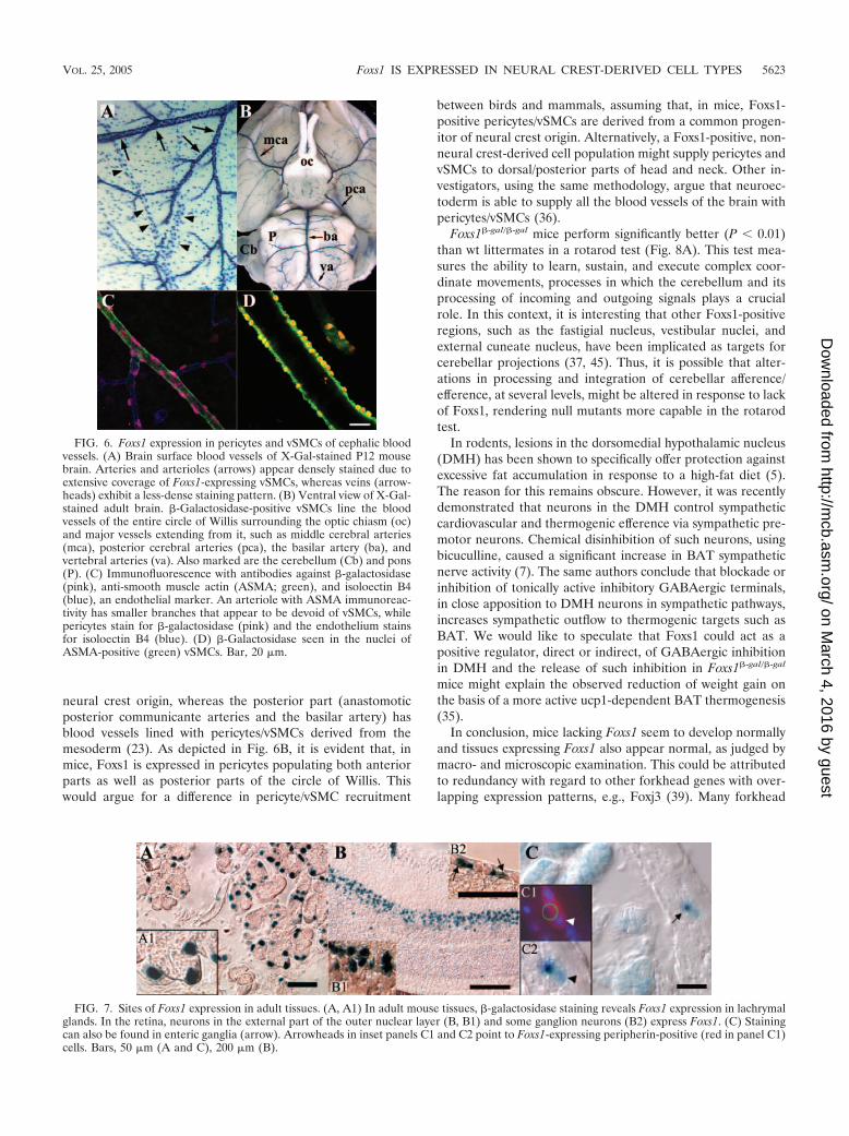
neural crest origin, whereas the posterior part (anastomoticposterior communicante arteries and the basilar artery) hasblood vessels lined with pericytes/vSMCs derived from themesoderm (23). As depicted in Fig. 6B, it is evident that, inmice, Foxs1 is expressed in pericytes populating both anteriorparts as well as posterior parts of the circle of Willis. Thiswould argue for a difference in pericyte/vSMC recruitment
between birds and mammals, assuming that, in mice, Foxs1-positive pericytes/vSMCs are derived from a common progen-itor of neural crest origin. Alternatively, a Foxs1-positive, non-neural crest-derived cell population might supply pericytes andvSMCs to dorsal/posterior parts of head and neck. Other in-vestigators, using the same methodology, argue that neuroec-toderm is able to supply all the blood vessels of the brain withpericytes/vSMCs (36).
Foxs1�-gal/�-gal mice perform significantly better (P � 0.01)than wt littermates in a rotarod test (Fig. 8A). This test mea-sures the ability to learn, sustain, and execute complex coor-dinate movements, processes in which the cerebellum and itsprocessing of incoming and outgoing signals plays a crucialrole. In this context, it is interesting that other Foxs1-positiveregions, such as the fastigial nucleus, vestibular nuclei, andexternal cuneate nucleus, have been implicated as targets forcerebellar projections (37, 45). Thus, it is possible that alter-ations in processing and integration of cerebellar afference/efference, at several levels, might be altered in response to lackof Foxs1, rendering null mutants more capable in the rotarodtest.
In rodents, lesions in the dorsomedial hypothalamic nucleus(DMH) has been shown to specifically offer protection againstexcessive fat accumulation in response to a high-fat diet (5).The reason for this remains obscure. However, it was recentlydemonstrated that neurons in the DMH control sympatheticcardiovascular and thermogenic efference via sympathetic pre-motor neurons. Chemical disinhibition of such neurons, usingbicuculline, caused a significant increase in BAT sympatheticnerve activity (7). The same authors conclude that blockade orinhibition of tonically active inhibitory GABAergic terminals,in close apposition to DMH neurons in sympathetic pathways,increases sympathetic outflow to thermogenic targets such asBAT. We would like to speculate that Foxs1 could act as apositive regulator, direct or indirect, of GABAergic inhibitionin DMH and the release of such inhibition in Foxs1�-gal/�-gal
mice might explain the observed reduction of weight gain onthe basis of a more active ucp1-dependent BAT thermogenesis(35).
In conclusion, mice lacking Foxs1 seem to develop normallyand tissues expressing Foxs1 also appear normal, as judged bymacro- and microscopic examination. This could be attributedto redundancy with regard to other forkhead genes with over-lapping expression patterns, e.g., Foxj3 (39). Many forkhead
FIG. 6. Foxs1 expression in pericytes and vSMCs of cephalic bloodvessels. (A) Brain surface blood vessels of X-Gal-stained P12 mousebrain. Arteries and arterioles (arrows) appear densely stained due toextensive coverage of Foxs1-expressing vSMCs, whereas veins (arrow-heads) exhibit a less-dense staining pattern. (B) Ventral view of X-Gal-stained adult brain. �-Galactosidase-positive vSMCs line the bloodvessels of the entire circle of Willis surrounding the optic chiasm (oc)and major vessels extending from it, such as middle cerebral arteries(mca), posterior cerebral arteries (pca), the basilar artery (ba), andvertebral arteries (va). Also marked are the cerebellum (Cb) and pons(P). (C) Immunofluorescence with antibodies against �-galactosidase(pink), anti-smooth muscle actin (ASMA; green), and isoloectin B4(blue), an endothelial marker. An arteriole with ASMA immunoreac-tivity has smaller branches that appear to be devoid of vSMCs, whilepericytes stain for �-galactosidase (pink) and the endothelium stainsfor isoloectin B4 (blue). (D) �-Galactosidase seen in the nuclei ofASMA-positive (green) vSMCs. Bar, 20 �m.
FIG. 7. Sites of Foxs1 expression in adult tissues. (A, A1) In adult mouse tissues, �-galactosidase staining reveals Foxs1 expression in lachrymalglands. In the retina, neurons in the external part of the outer nuclear layer (B, B1) and some ganglion neurons (B2) express Foxs1. (C) Stainingcan also be found in enteric ganglia (arrow). Arrowheads in inset panels C1 and C2 point to Foxs1-expressing peripherin-positive (red in panel C1)cells. Bars, 50 �m (A and C), 200 �m (B).
VOL. 25, 2005 Foxs1 IS EXPRESSED IN NEURAL CREST-DERIVED CELL TYPES 5623
on March 4, 2016 by guest
http://mcb.asm
.org/D
ownloaded from

genes display a high degree of similarity in their DNA bindingdomains, and as a consequence hereof, they are likely to in-teract with identical cis elements. The phenotype observedmight be related to cell types in which Foxs1 has a uniquenonexchangeable role to play.
ACKNOWLEDGMENTS
pCH110-nls plasmid was a kind gift from Jean-Jacques Panthier. EScell culture work and blastocyst injections were performed by theTransgenic Core Facility at Goteborg University.
This work was supported by the Swedish Research Council (grantsK2002-04X-03522-31D and K2002-31X-12186-06A to S.E. and K2004-32X-11283-10A to P.E.), EU grants (QLK3-CT-2002-02149 andLSHM-CT-2003-503041 to S.E.), The Arne and IngaBritt Foundationand The Soderberg Foundation (to S.E.), and Petrus and AugustaHedlunds Foundation (to P.E.).
REFERENCES
1. Accili, D., and K. C. Arden. 2004. FoxOs at the crossroads of cellular me-tabolism, differentiation, and transformation. Cell 117:421–426.
2. Ang, S. L., and J. Rossant. 1994. HNF-3 beta is essential for node andnotochord formation in mouse development. Cell 78:561–574.
3. Bergquist, F., H. N. Shahabi, and H. Nissbrandt. 2003. Somatodendriticdopamine release in rat substantia nigra influences motor performance onthe accelerating rod. Brain Res. 973:81–91.
4. Bernardis, L. L. 1975. The dorsomedial hypothalamic nucleus in autonomicand neuroendocrine homeostasis. Can. J. Neurol. Sci. 2:45–60.
5. Bernardis, L. L., and L. L. Bellinger. 1991. Brown (BAT) and white (WAT)adipose tissue in high-fat junk food (HFJF) and chow-fed rats with dorso-medial hypothalamic lesions (DMNL rats). Behav. Brain Res. 43:191–195.
6. Blomqvist, S. R., H. Vidarsson, S. Fitzgerald, B. R. Johansson, A. Ollerstam,R. Brown, A. E. Persson, G. G. Bergstrom, and S. Enerback. 2004. Distalrenal tubular acidosis in mice that lack the forkhead transcription factorFoxi1. J. Clin. Investig. 113:1560–1570.
7. Cao, W. H., W. Fan, and S. F. Morrison. 2004. Medullary pathways medi-
ating specific sympathetic responses to activation of dorsomedial hypothal-amus. Neuroscience 126:229–240.
8. Carlsson, P., and M. Mahlapuu. 2002. Forkhead transcription factors: keyplayers in development and metabolism. Dev. Biol. 250:1–23.
9. Carter, R. J., L. A. Lione, T. Humby, L. Mangiarini, A. Mahal, G. P. Bates,S. B. Dunnett, and A. J. Morton. 1999. Characterization of progressive motordeficits in mice transgenic for the human Huntington’s disease mutation.J. Neurosci. 19:3248–3257.
10. Cederberg, A., R. Betz, S. Lagercrantz, C. Larsson, M. Hulander, P. Carls-son, and S. Enerback. 1997. Chromosome localization, sequence analysis,and expression pattern identify FKHL 18 as a novel human forkhead gene.Genomics 44:344–346.
11. Cederberg, A., L. M. Gronning, B. Ahren, K. Tasken, P. Carlsson, and S.Enerback. 2001. FOXC2 is a winged helix gene that counteracts obesity,hypertriglyceridemia, and diet-induced insulin resistance. Cell 106:563–573.
12. Chaplan, S. R., F. W. Bach, J. W. Pogrel, J. M. Chung, and T. L. Yaksh. 1994.Quantitative assessment of tactile allodynia in the rat paw. J. Neurosci.Methods 53:55–63.
13. Chen, X., M. J. Rubock, and M. Whitman. 1996. A transcriptional partnerfor MAD proteins in TGF-beta signalling. Nature 383:691–696. (Erratum,384:648.)
14. Chen, X., E. Weisberg, V. Fridmacher, M. Watanabe, G. Naco, and M.Whitman. 1997. Smad4 and FAST-1 in the assembly of activin-responsivefactor. Nature 389:85–89.
15. Clifton-Bligh, R. J., J. M. Wentworth, P. Heinz, M. S. Crisp, R. John, J. H.Lazarus, M. Ludgate, and V. K. Chatterjee. 1998. Mutation of the geneencoding human TTF-2 associated with thyroid agenesis, cleft palate andchoanal atresia. Nat. Genet. 19:399–401.
16. Couly, G., P. Coltey, A. Eichmann, and N. M. Le Douarin. 1995. The angio-genic potentials of the cephalic mesoderm and the origin of brain and headblood vessels. Mech. Dev. 53:97–112.
17. Couly, G. F., P. M. Coltey, and N. M. Le Douarin. 1992. The developmentalfate of the cephalic mesoderm in quail-chick chimeras. Development 114:1–15.
18. Crisponi, L., M. Deiana, A. Loi, F. Chiappe, M. Uda, P. Amati, L. Bisceglia,L. Zelante, R. Nagaraja, S. Porcu, M. S. Ristaldi, R. Marzella, M. Rocchi, M.Nicolino, A. Lienhardt-Roussie, A. Nivelon, A. Verloes, D. Schlessinger, P.Gasparini, D. Bonneau, A. Cao, and G. Pilia. 2001. The putative forkhead
FIG. 8. (A) Rotarod performance. Bars represent mean fall latencies for wt (black bars) and Foxs1�-gal/�-gal (gray bars) male (n 7 pergenotype) and female mice (n 8 per genotype). Minutes on the rotating rod differed significantly: *, P � 0.05; **, P � 0.01 (Mann-Whitney Utest). Weight curves for female (B) and male (C) wt (squares) and Foxs1�-gal/�-gal (triangles) mice. Mice were put on a high-fat diet for 13 weeks.Repeated measures of analysis of variance indicate a significantly (P � 0.05) lower percent weight gain in Foxs1�-gal/�-gal (n 7) male mice thanin wt (n 5) male mice. No difference was seen for female mice (n 8 per genotype). (D) Relative mRNA levels of ucp1 were measured usingquantitative real-time PCR. A small but significant difference was seen between wt (n 4) (black bars) and Foxs1�-gal/�-gal (n 3) (gray bars) malemice, whereas there was no difference in female mice (n 4 per genotype). *, P � 0.05 (Student’s t test). Error bars (all panels) indicate standarderrors of the means.
5624 HEGLIND ET AL. MOL. CELL. BIOL.
on March 4, 2016 by guest
http://mcb.asm
.org/D
ownloaded from

transcription factor FOXL2 is mutated in blepharophimosis/ptosis/epican-thus inversus syndrome. Nat. Genet. 27:159–166.
19. Crowley, C., S. D. Spencer, M. C. Nishimura, K. S. Chen, S. Pitts-Meek,M. P. Armanini, L. H. Ling, S. B. MacMahon, D. L. Shelton, A. D. Levinson,et al. 1994. Mice lacking nerve growth factor display perinatal loss of sensoryand sympathetic neurons yet develop basal forebrain cholinergic neurons.Cell 76:1001–1011.
20. D’Amico-Martel, A., and D. M. Noden. 1983. Contributions of placodal andneural crest cells to avian cranial peripheral ganglia. Am. J. Anat. 166:445–468.
21. Enerback, S., B. G. Ohlsson, L. Samuelsson, and G. Bjursell. 1992. Char-acterization of the human lipoprotein lipase (LPL) promoter: evidence oftwo cis-regulatory regions, LP-alpha and LP-beta, of importance for thedifferentiation-linked induction of the LPL gene during adipogenesis. Mol.Cell. Biol. 12:4622–4633.
22. Ernfors, P., K. F. Lee, J. Kucera, and R. Jaenisch. 1994. Lack of neurotro-phin-3 leads to deficiencies in the peripheral nervous system and loss of limbproprioceptive afferents. Cell 77:503–512.
23. Etchevers, H. C., C. Vincent, N. M. Le Douarin, and G. F. Couly. 2001. Thecephalic neural crest provides pericytes and smooth muscle cells to all bloodvessels of the face and forebrain. Development 128:1059–1068.
24. Farinas, I., K. R. Jones, C. Backus, X. Y. Wang, and L. F. Reichardt. 1994.Severe sensory and sympathetic deficits in mice lacking neurotrophin-3.Nature 369:658–661.
25. Gerlai, R., P. Pisacane, and S. Erickson. 2000. Heregulin, but not ErbB2 orErbB3, heterozygous mutant mice exhibit hyperactivity in multiple behav-ioral tasks. Behav. Brain Res. 109:219–227.
26. Hulander, M., A. E. Kiernan, S. R. Blomqvist, P. Carlsson, E. J. Samuelsson,B. R. Johansson, K. P. Steel, and S. Enerback. 2003. Lack of pendrinexpression leads to deafness and expansion of the endolymphatic compart-ment in inner ears of Foxi1 null mutant mice. Development 130:2013–2025.
27. Hulander, M., W. Wurst, P. Carlsson, and S. Enerback. 1998. The wingedhelix transcription factor Fkh10 is required for normal development of theinner ear. Nat. Genet. 20:374–376.
28. Iida, K., H. Koseki, H. Kakinuma, N. Kato, Y. Mizutani-Koseki, H. Ohuchi,H. Yoshioka, S. Noji, K. Kawamura, Y. Kataoka, F. Ueno, M. Taniguchi, N.Yoshida, T. Sugiyama, and N. Miura. 1997. Essential roles of the wingedhelix transcription factor MFH-1 in aortic arch patterning and skeletogen-esis. Development 124:4627–4638.
29. Ji, R. R., Q. Zhang, P. Y. Law, H. H. Low, R. Elde, and T. Hokfelt. 1995.Expression of mu-, delta-, and kappa-opioid receptor-like immunoreactivi-ties in rat dorsal root ganglia after carrageenan-induced inflammation.J. Neurosci. 15:8156–8166.
30. Kaestner, K. H. 2000. The hepatocyte nuclear factor 3 (HNF3 or FOXA)family in metabolism. Trends Endocrinol. Metab. 11:281–285.
31. Kaestner, K. H., W. Knochel, and D. E. Martinez. 2000. Unified nomencla-ture for the winged helix/forkhead transcription factors. Genes Dev. 14:142–146.
32. Kaestner, K. H., K. H. Lee, J. Schlondorff, H. Hiemisch, A. P. Monaghan,and G. Schutz. 1993. Six members of the mouse forkhead gene family aredevelopmentally regulated. Proc. Natl. Acad. Sci. USA 90:7628–7631.
33. Kaufmann, E., and W. Knochel. 1996. Five years on the wings of fork head.Mech. Dev. 57:3–20.
34. Klein, R., I. Silos-Santiago, R. J. Smeyne, S. A. Lira, R. Brambilla, S. Bryant,L. Zhang, W. D. Snider, and M. Barbacid. 1994. Disruption of the neuro-trophin-3 receptor gene trkC eliminates la muscle afferents and results inabnormal movements. Nature 368:249–251.
35. Kopecky, J., G. Clarke, S. Enerback, B. Spiegelman, and L. P. Kozak. 1995.Expression of the mitochondrial uncoupling protein gene from the aP2 genepromoter prevents genetic obesity. J. Clin. Investig. 96:2914–2923.
36. Korn, J., B. Christ, and H. Kurz. 2002. Neuroectodermal origin of brainpericytes and vascular smooth muscle cells. J. Comp. Neurol. 442:78–88.
37. Kotchabhakdi, N., and F. Walberg. 1978. Cerebellar afferent projectionsfrom the vestibular nuclei in the cat: an experimental study with the methodof retrograde axonal transport of horseradish peroxidase. Exp. Brain Res.31:591–604.
38. Labosky, P. A., G. E. Winnier, T. L. Jetton, L. Hargett, A. K. Ryan, M. G.Rosenfeld, A. F. Parlow, and B. L. Hogan. 1997. The winged helix gene, Mf3,
is required for normal development of the diencephalon and midbrain,postnatal growth and the milk-ejection reflex. Development 124:1263–1274.
39. Landgren, H., and P. Carlsson. 2004. FoxJ3, a novel mammalian forkheadgene expressed in neuroectoderm, neural crest, and myotome. Dev. Dyn.231:396–401.
40. Laurie, D. J., P. H. Seeburg, and W. Wisden. 1992. The distribution of 13GABAA receptor subunit mRNAs in the rat brain. II. Olfactory bulb andcerebellum. J. Neurosci. 12:1063–1076.
41. Le Douarin, N. M., and M. A. Teillet. 1974. Experimental analysis of themigration and differentiation of neuroblasts of the autonomic nervous sys-tem and of neurectodermal mesenchymal derivatives, using a biological cellmarking technique. Dev. Biol. 41:162–184.
42. Lehmann, O. J., N. D. Ebenezer, T. Jordan, M. Fox, L. Ocaka, A. Payne, B. P.Leroy, B. J. Clark, R. A. Hitchings, S. Povey, P. T. Khaw, and S. S. Bhatta-charya. 2000. Chromosomal duplication involving the forkhead transcriptionfactor gene FOXC1 causes iris hypoplasia and glaucoma. Am. J. Hum.Genet. 67:1129–1135.
43. Lin, J. C., L. Cai, and C. L. Cepko. 2001. The external granule layer of thedeveloping chick cerebellum generates granule cells and cells of the isthmusand rostral hindbrain. J. Neurosci. 21:159–168.
44. Nakae, J., W. H. Biggs III, T. Kitamura, W. K. Cavenee, C. V. Wright, K. C.Arden, and D. Accili. 2002. Regulation of insulin action and pancreaticbeta-cell function by mutated alleles of the gene encoding forkhead tran-scription factor Foxo1. Nat. Genet. 32:245–253.
45. Newlands, S. D., J. T. Vrabec, I. M. Purcell, C. M. Stewart, B. E. Zimmer-man, and A. A. Perachio. 2003. Central projections of the saccular andutricular nerves in macaques. J. Comp. Neurol. 466:31–47.
46. Noden, D. M. 1993. Spatial integration among cells forming the cranialperipheral nervous system. J. Neurobiol. 24:248–261.
47. Ogg, S., S. Paradis, S. Gottlieb, G. I. Patterson, L. Lee, H. A. Tissenbaum,and G. Ruvkun. 1997. The Fork head transcription factor DAF-16 trans-duces insulin-like metabolic and longevity signals in C. elegans. Nature389:994–999.
48. Parysek, L. M., and R. D. Goldman. 1988. Distribution of a novel 57 kDaintermediate filament (IF) protein in the nervous system. J. Neurosci. 8:555–563.
49. Rubinstein, M., T. J. Phillips, J. R. Bunzow, T. L. Falzone, G. Dziewczapol-ski, G. Zhang, Y. Fang, J. L. Larson, J. A. McDougall, J. A. Chester, C. Saez,T. A. Pugsley, O. Gershanik, M. J. Low, and D. K. Grandy. 1997. Micelacking dopamine D4 receptors are supersensitive to ethanol, cocaine, andmethamphetamine. Cell 90:991–1001.
50. Smeyne, R. J., R. Klein, A. Schnapp, L. K. Long, S. Bryant, A. Lewin, S. A.Lira, and M. Barbacid. 1994. Severe sensory and sympathetic neuropathiesin mice carrying a disrupted Trk/NGF receptor gene. Nature 368:246–249.
51. Steel, K. P., D. R. Davidson, and I. J. Jackson. 1992. TRP-2/DT, a new earlymelanoblast marker, shows that steel growth factor (c-kit ligand) is a survivalfactor. Development 115:1111–1119.
52. Tybulewicz, V. L., C. E. Crawford, P. K. Jackson, R. T. Bronson, and R. C.Mulligan. 1991. Neonatal lethality and lymphopenia in mice with a homozy-gous disruption of the c-abl proto-oncogene. Cell 65:1153–1163.
53. Weeber, E. J., M. Levy, M. J. Sampson, K. Anflous, D. L. Armstrong, S. E.Brown, J. D. Sweatt, and W. J. Craigen. 2002. The role of mitochondrialporins and the permeability transition pore in learning and synaptic plastic-ity. J. Biol. Chem. 277:18891–18897.
54. Weigel, D., and H. Jackle. 1990. The fork head domain: a novel DNA bindingmotif of eukaryotic transcription factors? Cell 63:455–456.
55. Weigel, D., G. Jurgens, F. Kuttner, E. Seifert, and H. Jackle. 1989. Thehomeotic gene fork head encodes a nuclear protein and is expressed in theterminal regions of the Drosophila embryo. Cell 57:645–658.
56. Winnier, G. E., L. Hargett, and B. L. Hogan. 1997. The winged helix tran-scription factor MFH1 is required for proliferation and patterning of parax-ial mesoderm in the mouse embryo. Genes Dev. 11:926–940.
57. Wolfrum, C., D. Q. Shih, S. Kuwajima, A. W. Norris, C. R. Kahn, and M.Stoffel. 2003. Role of Foxa-2 in adipocyte metabolism and differentiation.J. Clin. Investig. 112:345–356.
58. Yildirim-Toruner, C., K. Subramanian, L. El Manjra, E. Chen, S. Goldstein,and E. Vitale. 2004. A novel frameshift mutation of FOXC2 gene in a familywith hereditary lymphedema-distichiasis syndrome associated with renal dis-ease and diabetes mellitus. Am. J. Med. Genet. 131:281–285.
VOL. 25, 2005 Foxs1 IS EXPRESSED IN NEURAL CREST-DERIVED CELL TYPES 5625
on March 4, 2016 by guest
http://mcb.asm
.org/D
ownloaded from
Copyright © 2022 FDOKUMEN