The Special Developmental Biology of Craniofacial Tissues ...
Upload
khangminh22Category
view
4download
0
5.1 Introduction
The neural crest is a temporary embryonic structurethat is composed of a population of multipotent cellsthat delaminate from the ectoderm by epitheliomes-enchymal tranformation (EMT; Duband et al. 1995;Hay 1995; Le Douarin and Kalcheim 1999; Francis-West et al. 2003). Neural-crest-derived cells are calledmesectodermal or ectomesenchymal cells (mesoder-mal cells of ectodermal origin) that have arisenthrough EMT. The neural crest was first described by His (1868) in the chick embryo as a Zwischen-strang, a strip of cells lying between the dorsal ecto-derm and the neural tube. Classic contributions inamphibians identified interactions between tissuesthat lead to neural crest formation, and were re-viewed by Hörstadius (1950). Cell labelling tech-niques, particularly the quail-chick chimeric marker(Le Douarin 1969, 1973), showed that the neural crestcontributes to a large number of structures in theavian embryo (Le Douarin and Kalcheim 1999; LeDouarin 2004), including the spinal, cranial and auto-nomic ganglia, the medulla of the adrenal gland, themelanocytes and many of the skeletal and connectivetissues of the head. The whole facial and visceralskeleton and part of the neurocranium are formedfrom the neural crest. Many species-related discrep-ancies are present in the literature on neural crest cellmigration and their targets. In contrast to chick em-bryos, the neural crest in mammalian embryos is aless distinct structure and can be defined as the tran-sition zone between the neuroectoderm and the pre-sumptive epidermis from neural plate stages onwards(O’Rahilly and Müller 1999). In presomite murineembryos, the whole ectoderm, including the pre-sumptive neural crest, is able to produce mesectoder-mal cells (Smits-van Prooije 1986; Smits-van Prooijeet al. 1988). These EMT cells proliferate shortly aftertheir migration into the mesodermal compartment(Vermeij-Keers and Poelmann 1980). In somitestages during the transformation of the cranial neu-roectoderm of the head folds via the neural grooveinto the neural tube, there is a balance between theoutgrowth of the neuroectoderm and the productionof EMT cells by the neural crest; therefore, in mam-malian embryos, only short-distance migration ofEMT cells occurs.
The cranial neural crest provides the precursorsof cartilage, bone, muscles and connective tissue of
the head (Vermeij-Keers 1990; Sulik 1996; LaBonneand Bronner-Fraser 1999; Le Douarin and Kalcheim1999; Sperber 2002; Knecht and Bronner-Fraser 2002;Francis-West et al. 2003; Santagati and Rijli 2003). Inaddition, during later developmental stages multipleplaces of EMT are recognized as well. In the head–neck area, for example, the neurogenic placodes andthe optic neural crest are such areas. Neurogenic pla-codes, specialized regions of the embryonic ecto-derm, are the major source of primary sensory neu-rons in the head (Johnston and Bronsky 1995; Gra-ham and Begbie 2000). The vasculature of the head isderived from mesoderm-derived endothelial precur-sors, while the neural crest provides the pericytes andsmooth muscle cells of the vessels of the face and theforebrain (Etchevers et al. 2001).
Deficiencies in migration, proliferation and differ-entiation of neural-crest-derived tissue account for awide range of craniofacial malformations, i.e. the so-called neurocristopathies, manifested in a variety ofsyndromes (Jones 1990; Gorlin et al. 2001; Wilkie andMorriss-Kay 2001; Johnston and Bronsky 1995, 2002;Cohen 2002). Abnormalities in form, function orapoptosis of neural crest cells may range from vonRecklinghausen’s neurofibromatosis through Treach-er Collins to DiGeorge and Waardenburg syndromes(Dixon et al. 2000). Neurocristopathies may be ac-companied by developmental disorders of the CNS.Recently, however, the idea that neural crest abnor-malities underly the pathogenesis of the DiGeorgeand Treacher Collins syndromes has been challenged(Sect. 5.5). Major craniofacial malformations are alsofound in holoprosencephaly (HPE) and the cran-iosynostoses. Holoprosencephaly is an early disorderof pattern formation that may lead to closely relatedforebrain and facial malformations. In the fetal peri-od, craniosynostoses are frequent (approximately onein 2,500 children) craniofacial malformations due toagenesis or premature ossification of the cranial su-tures, caused by mutations in FGFR and other genes(Wilkie 1997; Cohen and MacLean 2000; Gorlin et al.2001; Jabs 2002), that may interfere with normal braindevelopment to varying degrees.
In this chapter, the neural crest and its derivativesand craniofacial development will be discussed, fol-lowed by an overview of the neurocristopathies, HPEand abnormal development of the skull, leading toCNS malformations. The neuropathology of HPE willbe discussed in Chap. 9.
The Neural Crest and Craniofacial MalformationsHans J. ten Donkelaar and Christl Vermeij-Keers
Chapter 5

5.2 Induction of the Neural Crest
Neural crest cells are induced at the border betweenthe neuroectoderm and the non-neural or surface ec-toderm (Fig. 5.1). During the formation of the neuraltube in the chick embryo, neural crest progenitorscome to lie in or directly adjacent to the dorsal neur-al tube (Le Douarin and Kalcheim 1999). Dependingon the species, neural crest cells leave the neuroep-ithelium before, during or after neural tube closure,and ‘migrate’ throughout the body. To leave the neu-roepithelium, neural crest cells must lose their ep-ithelial characteristics and take on the properties ofmigratory mesenchymal cells. Wheat germ agglu-tinin labelling experiments in mouse embryos(Smits-van Prooije et al. 1988) and 1,1’-dioctadecyl-3,3,3’,3’-tetramethylindocarbocyanine perchlorate(DiI) labelling in chick embryos (Kulesa and Fraser1998) have shown that migration of neural crest cellsis random. Other studies have suggested that thereare cell-free spaces underneath the neuroectodermand surface ectoderm resulting in specific pathwaysfor migrating neural crest cells (Le Douarin 1969,1973). In quail-chick chimeras, however, cell-freespaces may be created artificially. More recent studiessuggest a role for chemoattractive signals such asfibroblast growth factor (FGF) in the control of neur-al crest cell migration (Kubota and Ito 2000; Francis-West et al. 2003).
Induction of the neural crest appears to be a com-plex multistep process that involves many genes(LaBonne and Bronner-Fraser 1999; Aybar and May-or 2002; Knecht and Bronner-Fraser 2002; Gammilland Bronner-Fraser 2003; Wu et al. 2003). The gener-ation of neural crest cells appears to result from in-ductive interactions shared with the neural plate andthe early epidermis. The neural–epidermal boundarycan be distinguished by the expression of molecularmarkers such as the transcription factors of the Snailfamily, Snail and Slug, in Xenopus, zebrafish, chickand mouse (Mayor et al. 1995; Sefton et al. 1998; Link-er et al. 2000; Aybar et al. 2003). The timing and ex-pression pattern of these markers differs betweenvertebrates. Bone morphogenetic protein (BMP) sig-nalling specifies the formation of dorsal neural tis-sues and the neural plate border (Sasai and DeRobertis 1997; Weinstein and Hemmati-Brivanlou1999). Low or absent BMP signalling leads to neuralinduction, whereas a high level of BMP signallinginduces epidermis.An intermediate level of BMP sig-nalling may determine the neural plate border (Wil-son et al. 1997; Nguyen et al. 1998, 2000). In zebrafish,BMP signalling is required for the development of theneural crest (Nguyen et al. 2000), but in Xenopusintermediate BMP levels alone cannot induce theneural crest. Additional factors, Wnt proteins in par-ticular, are required to induce the neural crest
(LaBonne and Bronner-Fraser 1998; Gammill andBronner-Fraser 2003; Wu et al. 2003).
The epitheliomesenchymal tranformation (EMT)of emerging neural crest cells is accompanied by theexpression of the zinc-finger transcription factorSlug. In chick and Xenopus embryos, its expression ismaintained during the phase of crest cell migration(Nieto et al. 1994; LaBonne and Bronner-Fraser2000). Slug mutant mice, however, do not show de-fects in either neural crest or mesodermal tissues(Jiang et al. 1998). In mice, another family member,
Chapter 5 Craniofacial Malformations192
Fig. 5.1 The early development of the human neural crest ina Carnegie stage 10 embryo. At rostral levels (a), crest materialis formed before closure of the neural groove, whereas atmore caudal levels (b–d) closure of the neural folds preceedsmigration of crest material. (After Müller and O’Rahilly 1985)

Snail, rather than Slug is expressed in the regions un-dergoing EMT (Cano et al. 2000; Locascio and Nieto2001; Knecht and Bronner-Fraser 2002; Gammill andBronner-Fraser 2003). Snail mutant mice die at gas-trulation as a result of defects in mesoderm forma-tion arising from deficient EMT.
5.3 Derivatives of the Neural Crest
The neural crest is the major source of mesenchymalcells in the head and neck, and in addition gives riseto sensory ganglia, autonomic and enteric ganglia aswell as to pigment cells of the skin. In the postcranialregion, mesenchyme forming the connective tissuesis mesodermal in origin (Table 5.1). The chick neuralcrest can be divided into four main domains (LeDouarin and Kalcheim 1999): (1) the cranial orcephalic neural crest, giving rise to the craniofacialmesenchyme in particular; (2) the trunk neuralcrest, giving rise to the dorsal root and sympatheticganglia; (3) the vagal and sacral neural crest, gener-ating the parasympathetic (enteric) ganglia of thegut; and (4) the cardiac neural crest, located betweenthe cranial and trunk neural crests. In chick embryos,the cardiac neural crest produces the musculocon-nective tissue for the large arteries arising from theheart and contributes to the separation of the trun-cus arteriosus into the pulmonary artery and aorta(Le Lièvre and Le Douarin 1975; Kirby 1987; Kirbyand Waldo 1990). The cranial neural crest is the onlypart of the neural crest that is able to produce carti-lage and bone.
5.3.1 The Cranial Neural Crest
During neural tube closure in the chick embryo,cranial neural crest cells migrate into the underlyingtissues as mesenchyme (ectomesenchyme), forminga lineage of pluripotential stem cells that give rise todiverse tissues (Le Douarin and Kalcheim 1999). Incontrast, in mice the entire ectoderm of early pre-somite embryos and the ectoderm of the head foldsin late presomite and early somite embryos are ableto deposit ectomesenchymal cells into mesodermalcompartments (Vermeij-Keers and Poelmann 1980;Smits-van Prooije et al. 1985; Tan and Morriss-Kay1985; Smits-van Prooije 1986; Boshart et al. 2000).From four-somite up to 20-somite murine embryos,i.e. after the closure of the rostral neuropore, the cra-nial neural crest is active. In human embryos ofstage 9 (one to three somites) up to stage 10 (four to12 somites), ectomesenchymal cells arise from partsof the head folds, probably the rostral and facial crest (Müller and O’Rahilly 1983,1985;Vermeij-Keers1990). In chick embryos, however, the embryonic
prominences of the face and neck (the so-called fron-tonasal process and the pharyngeal arches) are de-pendent on mesencephalic and rhombencephalicneural crest tissue, migrating as ectomesenchyme in-to ventral regions of the future skull, face and neck(Fig. 5.2; Couly and Le Douarin 1990). In the humanhead, neural crest cells also arise from the externallayer of the optic vesicle, i.e. the optic neural crest(O’Rahilly and Müller 2001).
In chick embryos, cranial neural crest cells mi-grate from regions rostral to rhombomere 6, takingone of three major migratory pathways (Lumsdenand Guthrie 1991; Sechrist et al. 1993; Kulesa andFraser 2000; Fig. 5.3): (1) cells from the first and sec-
5.3 Derivatives of the Neural Crest 193
Table 5.1 Derivatives of neural crest cells (after LeDouarinand Kalcheim 1999; Sperber 2001)
Connective tissues Ectomesenchyme of facial promi-nences and pharyngeal arches
Bones and cartilage of facial and visceral skeleton
Dermis of face and ventral aspect of neck
Stroma of salivary, thymus, thyroid,parathyroid and pituitary glands
Corneal mesenchyme
Sclera and choroid optic coats
Blood vessel walls;aortic arch arteries
Dental papilla; part of periodontal ligament; cementum
Muscle tissues Ciliary muscles
Covering connective tissues of pharyngeal-arch muscles (masticatory, facial, faucial, laryngeal)
Nervous tissues Supporting tissues: leptomeninges of prosencephalon and part of the mesencephalon; glia;Schwann’s sheath cells
Sensory ganglia: spinal dorsal root ganglia; and sensory ganglia of trigeminal, facial, glosso-pharyngeal and vagal nerves
Autonomic nervous system:sympathetic ganglia and plexuses;parasympathetic ganglia
Endocrine tissues Adrenomedullary cells and adrenergic paraganglia
Calcitonin parafollicular cells of thyroid gland
Carotid body
Pigment cells Melanocytes in all tissues
Melanophores of iris
ond rhombomeres migrate to the first pharyngeal(mandibular) arch, forming the mandibula, incusand malleus, and contributing to the trigeminal gan-glion; these cells also generate the facial skeleton; (2)cells from the fourth rhombomere invade the secondpharyngeal arch and form the hyoid cartilage and thefacial and vestibulo-acoustic ganglia; and (3) cellsfrom the sixth rhombomere migrate into the thirdand fourth pharyngeal arches and pouches to formthe thymus, the parathyroid and thyroid glands, and
the superior and jugular ganglia. Neural crest cellsfrom the third and fifth rhombomeres enter the mi-grating streams of neural crest cells of the adjacentrhombomeres. Those that do not enter the variousmigratory streams will die (Graham et al. 1993, 1994;Sechrist et al. 1993). The apoptotic elimination ofneural crest cells in the third and fifth rhombomeresinvolves the induction of high-level BMP expressionin the neural crest, which stimulates expression of thehomeobox gene Msx2 (Graham et al. 1994). In mice, a
194 Chapter 5 Craniofacial Malformations
Fig. 5.2 Fate map of the ecto-dermal territories in the chickembryo from the three-somitestage (a) to the 8-day embryo(c). b The rhombomeres (r1, r3, r6,r8) and the cranial nerves sup-plying the pharyngeal arches(1–4). pmax maxillary promi-nence, V trigeminal nerve,VII facial nerve, IX glossopharyn-geal nerve, X vagus nerve.(After Couly and Le Douarin1990)
Fig. 5.3 Migratory pathways ofcranial neural crest cells to thepharyngeal arches (I–IV, VI), andthe segmented expression ofHox genes in the hindbrain andpharyngeal arches of the chickembryo. fp floor plate, gV trigem-inal ganglion, MHB midbrain–hindbrain boundary, nV3mandibular nerve, nVII facialnerve, nIX glossopharyngealnerve, nX vagus nerve, ov oticvesicle, sc spinal cord, vm vis-ceromotor nucleus of facialnerve, IV–VII, IX, X, XII cranialnerve nuclei, 1–8 rhombomeres

similar pattern of neural crest migration pathwayshas been found (Golding et al. 2000; Trainor et al.1994, 2002; Trainor and Krumlauf 2000). The separatestreams are kept apart by ephrins (Golding et al.2000; Trainor and Krumlauf 2000).
Experimental studies using transposition of theavian neural folds (Noden 1978a, b, 1983a) led to theconcept that the spatial organization of cranial struc-tures is the result of a prepatterning mechanism be-fore cells migrate from the neural crest. The ordereddomains of Hox gene expression in the neural tubeand neural crest were later assumed to be a molecu-lar correlate of the prepatterning mechanism. Re-cently, however, plasticity of Hox gene expression hasbeen observed in the hindbrain and postotic neuralcrest of chick, mouse and zebrafish embryos (Trainorand Krumlauf 2000; Gammill and Bronner-Fraser2003; Santagati and Rijli 2003). Although the neuralcrest plays an important role in arch patterning, thereare also patterning mechanisms that are establishedindependently in the pharynx. Therefore, the forma-tion of the pharyngeal apparatus must result from anintegration between these patterning systems (Gra-ham and Smith 2001; Graham et al. 2004). The neuralcrest most probably plays a more prominent role inpatterning the rostral, preotic arches but a lesser rolein the caudal, postotic arches. The development ofthe branchial arches involves retinoic acid dependentmechanisms. Neural crest cells migrating intobranchial arches 3–6,however,are not the primary tar-gets of retinoic acid,but pharyngeal epithelial cells are.Retinoic acid signalling is indispensable for the devel-opment of the pharyngeal epithelia of these branchialarches (Niederreither et al. 2003; Mark et al. 2004).
The segmented expression of Hox genes in thehindbrain is reflected in the neural crest cells whichexpress a complement of Hox genes characteristic fortheir level of origin (Hunt et al. 1991; Capecchi 1997;Favier and Dollé 1997; Rijli et al. 1998; Trainor andKrumlauf 2000; Santagati and Rijli 2003; Fig. 5.3).Each rhombomere and pharyngeal arch is character-ized by its own complement of Hox genes, its Hoxcode. In human embryos, Vieille-Grosjean et al.(1997) showed that the pattern of HOX gene expres-sion in the rhombomeres and pharyngeal arches issimilar to that observed in mice. The mouse Hoxb2gene, which is expressed in the hindbrain up to theboundary between the second and third rhom-bomeres, has an expression domain that extends cau-dally from the second pharyngeal arch (Fig. 5.3).Hoxb3 is expressed as far rostrally as the rhom-bomere 4–rhombomere 5 boundary and from thethird pharyngeal arch caudally. Hox genes are re-quired for the normal morphogenesis of arch-de-rived skeletal elements. Mice with targeted disrup-tions in the paralogous genes Hoxa3 and Hoxd3,which are expressed in the third and fourth arches,
display defects in the laryngeal cartilages (Condieand Capecchi 1994). Mice that lack the Hoxa2 geneshow a transformation of second arch skeletal ele-ments into components of the first arch (Gendron-Maguire et al. 1993; Rijli et al. 1993), suggesting thatspecification of crest cells to first arch componentsrequires the downregulation of Hoxa2. Hoxa3 knock-outs show specific deletions or hypoplasias of struc-tures derived from the third arch, resembling DiGe-orge syndrome. They lack a thymus and parathyroidglands, have a reduced thyroid gland and show mal-formations of the laryngeal cartilages and muscles,and of the heart (Chisaka and Capecchi 1991; Manleyand Capecchi 1995). Decreased embryonic retinoicacid synthesis also results in a DiGeorge syndromephenotype in newborn mice (Vermot et al. 2003;Mark et al. 2004).
5.3.2 The Trunk Neural Crest
The migratory pathways of trunk neural crest cellsare stereotyped and have been defined by varioustypes of labelling experiments using grafts of Japan-ese quail crest into chick embryos, immunostainingfor cell surface markers such as HNK-1 or fluorescentdyes such as DiI (Le Douarin and Kalcheim 1999)and, more recently, using time-lapse techniques(Kulesa and Fraser 1998, 2000). Two main directionsof movement were found (Fig. 5.4): a dorsolateralpathway for crest cells that differentiate into
5.3 Derivatives of the Neural Crest 195
Fig. 5.4 Migratory pathways of chick trunk neural crest cells.adm adrenal medulla (chromaffin cells), ao aorta, drg dorsalroot ganglion cells, intg intestinal ganglion cells, mcmelanocytes, nc neural crest, nch notochord, nt neural tube,pag paravertebral ganglion cells, prg prevertebral ganglioncells, som somite

melanocytes, and a ventral pathway between neuraltube and somites that gives rise to the dorsal root andsympathetic ganglia, to Schwann cells and to chro-maffin cells for the adrenal gland. Crest cells passonly via the anterior part of the sclerotome. The mi-gration path taken by trunk neural crest cells is guid-ed by extracellular matrix molecules surrounding theneural tube. Fibronectin, laminin, tenascin and pro-teoglycans promote their migration (Newgeen et al.1986), whereas ephrin proteins present in the poste-rior part of the sclerotome restrict neural crest mi-gration to the anterior part of the sclerotome(O’Leary and Wilkinson 1999). In human embryos,the trunk neural crest participates at stage 12 in theformation of the pia mater, the spinal ganglia and thesympathetic trunk and ganglia. At later stages(13–14), they participate in the formation of thesheaths of the dorsal and ventral roots (O’Rahilly andMüller 1999).
5.4 Craniofacial Development
Prior to a discussion of neurocristopathies and othercraniofacial malformations involving the CNS, thedevelopment of the face, the pharyngeal arches andthe skull will be briefly discussed. Knowledge of thenormal developmental events that shape the cranio-facial region is necessary for understanding thechanges that result in malformations of this region.The following description is largely based on Hin-richsen (1985, 1990) and Vermeij-Keers (1990).
5.4.1 Early Development of the Face
The early development of the forebrain and face ischaracterized by the formation and subsequenttransformation of the so-called head folds (Vermeij-Keers 1990): first, the neural walls with the eye pri-mordia, then the otic disc, the pharyngeal arches and,finally, the lens placode and the nasal placode. Thesetransformations show a rather basic pattern of devel-opment, outgrowth of swellings forming and sur-rounding a cavity or groove that is subsequentlyclosed partly or totally as a result of fusion of theswellings. The first slightly lordotic shape of the hu-man embryo facilitates the initial fusion process ofthe neural walls transforming the neural groove intothe neural tube (Chap. 4). Later, the lordosis changesinto a kyphosis, enabling the transformation of thepharyngeal arches and the formation of the neck. Thedevelopment of the head or cephalic folds in humanand murine embryos is comparable (van Oostrom1972; Vermeij-Keers 1990; Sulik 1996). In human em-bryos, the development of the head folds takes placeduring stages 8–9 (O’Rahilly 1973; O’Rahilly and
Müller 1981; Müller and O’Rahilly 1983), and inmouse embryos around 7.5 days post coitum (E7.3–E7.7), at which time the head folds are covered withcolumnar epithelium (van Oostrom 1972; Smits-vanProoije 1986). The border of the head folds is thetransition of the surface ectoderm and the amnion.At stage 8, O’Rahilly and Müller (1981) described thehead folds already as neural folds. They are the firstrostral structures to appear in the embryo, and con-tinue caudally as the neural plate. In four- to seven-somite mouse embryos, the head folds grow outrapidly, and a separation into lateral, thin (surfaceectoderm of the head–neck region) and medial, thick(neuroectoderm of the forebrain) parts becomesevident. At the transition of both parts, the cranialneural crest, the neuroectoderm produces ectomes-enchymal cells.
In amphibians, Adelmann (1936) showed thatwithin the rostral neural plate a median populationof cells, the eye field, segregates into two lateralprimordia, the future optic vesicles. Experimentalremoval of the prechordal plate resulted in failure ofseparation of the midline structures of the rostralneural plate, leading to cyclopia as is also found in themost severe forms of HPE. The separation of the eyefield is dependent upon interactions with the under-lying mesoderm. The resulting degree of bilateraliza-tion of the eyes and forebrain has a profound effecton the subsequent morphogenesis of the face. In am-phibian and chick embryos, the primordia of the eyesare found in the rostral neural plate, just caudal to theanterior neural ridge (Couly and Le Douarin 1988;Eagleson and Harris 1990; Eagleson et al. 1995;Fig. 2.8). Cyclopic animals such as the zebrafish mu-tant cyclops (Hatta et al. 1991, 1994; Schier 2001) havea defect in the development of the optic stalk and chi-asm. Mice lacking the Shh gene demonstrate cyclopiaand abnormal axial patterning (Chiang et al. 1996).Secreted factors such as Sonic hedgehog (SHH; Chi-ang et al. 1996) and FGF8 (Sun et al. 1999) play impor-tant roles before and during patterning of the neuralplate (Chap. 9). In chick embryos, FGF8 and SHH sig-nalling pathways are also required during early mor-phogenesis of the forebrain and frontonasal process(Schneider et al. 2001). The Distal-less related geneDlx5 is also required for normal development of thefrontonasal process (Acampora et al. 1999). In humanembryos, O’Rahilly and Müller (1999) suggested asingle eye field at stage 8 that is situated directlyabove the prechordal plate. The prechordal plate sup-presses the median part, so that bilateral optic pri-mordia are formed, presumably still in stage 8 em-bryos. Instead of a single optic primordium,Vermeij-Keers et al. (1987) found evidence for the existence ofbilateral optic primordia in human embryos.
During the transformation of the head folds intothe cranial neural walls or neural folds, the optic pri-
196 Chapter 5 Craniofacial Malformations

mordia (optic sulci) develop within the neuroecto-derm as two separate shallow grooves (Fig. 5.5a, b;mouse, seven-somite embryo: Smits-van Prooije et al.1985; man, stage 10: Müller and O’Rahilly 1985).Close to the margin of the neuroectoderm the oticdisc develops as a concavity of columnar epithelium
in the surface ectoderm (three-somite mouse em-bryo: Verwoerd and van Oostrom 1979; man, stage 9:Müller and O’Rahilly 1983). Meanwhile, the caudalpart of the neural folds transforms via the neuralgroove into the neural tube. The initial contact be-tween the neural walls takes place in the caudal partof the rhombencephalic folds or in the upper cervicalneural folds (mouse, five-somite embryo: Smits-van Prooije et al. 1985; man, stage 10: Müller andO’Rahilly 1985; Nakatsu et al. 2000). Rostrally, in thehead other points of closure occur in the mouse em-bryo (Geelen and Langman 1977; Sakai 1989; Goldenand Chernoff 1993) as well as in the human embryo(Müller and O’Rahilly 1985; Golden and Chernoff1995; Nakatsu et al. 2000; Chap. 4). The final closureof the rostral neuropore is located between two areasof ectodermal, cuboidal or columnar epithelium, thenasal fields (the Nasenfelder of His 1885), in mice in 15-somite embryos (Vermeij-Keers et al. 1983;Fig. 5.5c) and in human embryos at stage 11(O’Rahilly and Gardner 1971; Müller and O’Rahilly1986). This location corresponds with the presump-tive internasal groove (Vermeij-Keers 1990).
In human embryos, the first visible appearance ofthe bilateral optic primordia is seen in eight-somiteembryos (Bartelmez and Blount 1954; O’Rahilly1966) as a thickened area of each neural wall in whicha shallow sulcus is present. Subsequently, each opticsulcus widens and changes into an optic vesicle afterclosure of the neural walls. Within each nasal fieldtwo local thickenings develop, the lens placode(mouse: E8.8, 15–16-somite embryos; stage 13 humanembryos: Müller and O’Rahilly 1988a) and the nasalplacode (mouse: E9.8; stage 14 human embryos: Hin-richsen 1985; Müller and O’Rahilly 1988b). The lensplacode is adjacent to the optic vesicle and is trans-formed into the lens vesicle, whereas the optic vesiclebecomes the optic cup. In stage 15 human embryos,the lens vesicle detaches from the surface ectoderm(O’Rahilly 1966). The cavity of the optic vesicle com-municates via the lumen of the short optic stalk withthat of the forebrain. During the formation of the op-tic cup, its external layer grows out into the directionof the internal layer. This process is influenced by thelens placode (Chap. 9).
The mesodermal component of the head region isnot only supplied by the cranial neural crest but alsoby cells deposited by the surface ectoderm or neuro-genic placodes: the nasal placodes (Verwoerd andvan Oostrom 1979), the acoustic or otic placode (Bat-ten 1958) and the epibranchial placodes of the pha-ryngeal arches (Adelmann 1925; Graham and Begbie2000). Neurogenic placodes are specialized regions ofthe cephalic embryonic ectoderm from which amongothers neuroblasts for the cranial sensory systems aregenerated. Neurons in cranial sensory ganglia have adual origin from the neural crest as well as from pla-
5.4 Craniofacial Development 197
Fig. 5.5 Scanning electron micrographs of the developingface of mouse embryos in frontal view (the heart was re-moved in a and c): a E8.0 embryo, arrows indicate the opticsulci as bilateral grooves in the head folds; b E8.3 embryo,showing the prosencephalon with the evaginating optic vesicles; c E8.7 embryo in which the prosencephalon is fusedexcept for the rostral neuropore (arrow) which separates thetwo nasal fields (nf)

codes (Noden 1991a, b; Le Douarin and Kalcheim1999). The origin of neurogenic precursors and thepattern of expansion of the surrounding surface ec-toderm were defined using the quail-chick chimeratechnique (D’Amico-Martel and Noden 1983; Coulyand Le Douarin 1985, 1987, 1990).
The otic disc is the first ill-defined appearance ofthe developing ear (three- to five-somite mouse em-bryo: Verwoerd and van Oostrom 1979, Smits-vanProoije 1986; stage 9 human embryo: Müller andO’Rahilly 1983). At the end of stage 10, the otic discbecomes incorporated and transforms via the otic pitinto the otic vesicle or otic cyst (O’Rahilly 1983;Müller and O’Rahilly 1985; Van De Water et al. 1988).Subsequently, the otic vesicle becomes separatedfrom the surface ectoderm by apoptosis, and inducesthe condensation of its surrounding mesenchymeinto the otic capsule. The otic vesicle forms the mem-branous labyrinth and the otic capsule the osseouslabyrinth (Streeter 1906, 1918; Chap. 7). Placodalcells, given off from the walls of the otic vesicle, formthe vestibular and possibly the cochlear (spiral) gan-glia (O’Rahilly and Müller 2001). The cavity of themiddle ear develops from the tubotympanic recessof the first pharyngeal pouch (Kanagasuntheram1967). The origin of the auditory ossicles is not en-tirely clear (Anson et al. 1948, 1960; Hanson et al.1962; O’Rahilly and Müller 2001). Presumably, thehead of the malleus and the body and short crus ofthe incus arise from the first pharyngeal arch, where-as the handle of the malleus, possibly the long crus ofthe incus and the head and crura of the stapes devel-op from the second arch. The base of the stapes ap-pears in the lateral wall of the otic capsule. The exter-nal ear arises from a series of small swellings, the au-ricular hillocks, around the first pharyngeal groove(Streeter 1922).
5.4.2 Development of the Pharyngeal Arches
A prominent feature of human embryos is the pres-ence of a series of bulges on the lateral surface of thehead and neck, the pharyngeal arches (Fig. 5.6). Eacharch has an outer covering of ectoderm, an inner cov-ering of endoderm and a mesenchymal core derivedfrom the neural crest, and most likely the surfaceectoderm and mesoderm. Between the arches, theectoderm and endoderm are in close apposition andform the pharyngeal membranes. The externally sit-uated pharyngeal grooves have internal counter-parts, the pharyngeal pouches. During the process of outgrowth of the embryo, the wide lumen ofthe foregut is transformed into pharyngeal pouchesby outgrowing swellings into this lumen. The firstpharyngeal pouch develops in the early somite
stages. Between the first pharyngeal pouch and itscomplementary pharyngeal groove the first pharyn-geal membrane develops. Initially, the groove is shal-low and wide but deepens subsequently during theoutgrowth of the first pharyngeal arch. The differentgerm layers generate distinct components of thepharynx. The ectoderm produces the epidermis andthe sensory neurons of the arch-associated ganglia(Verwoerd and van Oostrom 1979; D’Amico-Marteland Noden 1983; Couly and Le Douarin 1990), where-as the endoderm gives rise to the epithelial cells lin-ing the pharynx and the endocrine glands that formfrom the pharyngeal pouches. The neural crest formsthe connective and skeletal tissues (Noden 1983b;Couly et al. 1993), whereas the mesoderm forms themusculature and the endothelial cells of the archarteries (Noden 1983b; Noden et al. 1991a, b; Couly etal. 1992; Trainor et al. 1994; Francis-West et al. 2003).The development of the craniofacial muscles is dis-cussed in Chap.7.
In human embryos, five pharyngeal or visceralarches (also known as branchial or aortic arches)develop successively. Four pairs are visible on theoutside of the human embryo at 4 weeks and are sep-arated from each other by three grooves (Fig. 5.6).More caudally, their arrangement is less clear-cut butit is customary to label a fifth and even a sixth arch.The first pharyngeal arch is the biggest, develops cra-nial to the heart primordium of the embryo andforms only the mandibular processes of the facialswellings (Vermeij-Keers et al. 1983). The maxillaryprominence expands around the stomodeum below
198 Chapter 5 Craniofacial Malformations
Fig. 5.6 Scanning electron micrograph of the developmentof the human pharyngeal arches at stage 15 (from Jirásek2001, with permission)

the optic vesicle and is unrelated to the pharynx (Ver-meij-Keers 1990; Noden 1991a, b). The mandibularprocesses keep their positions, grow out and form agroove in the midline between the two swellings.They actually do not fuse but merge because no ep-ithelial plate is formed. The second arch grows cau-dally and laterally from the side of the second pha-ryngeal groove, covering the third and fourth arches.The third pharyngeal arch does the same with re-spect to the fourth. The retrobranchial ridge, incor-porating the fifth arch, grows rostrally into the direc-tion of the second arch (Starck 1975). Via contactswith the fourth and third arches this swelling adheres
and fuses with the second pharyngeal arch. Initially,slit-like cavities, remnants of the pharyngeal grooves,remain present between these contact places. Insidethe embryo, the slit-like cavities are obliterated andectodermal epithelial plates are formed at the contactplaces between the swellings. These plates disappearby apoptosis. The outgrowing pharyngeal arches andtheir subsequent shifting do not only cause transfor-mations of the pharyngeal grooves but also of theircorresponding pouches. The successive organs devel-oping from the pharyngeal pouches (Fig. 5.7; Table5.2), except for the first one, do not migrate to theirdefinite position but attain their positions during theoutgrowth and transformation of the pharyngealarch system (Vermeij-Keers 1990). Later, they differ-entiate and form, among others, the thymus (deriva-tive of the third pouch) and the superior (fourthpouch) and inferior (third pouch) parathyroidglands (Weller 1933; Norris 1937, 1938).
5.4.3 Further Development of the Face
The further development of the human face is usual-ly described as a process of merging of five swellingsor growth centres surrounding the stomodeum,termed processes or prominences: bilateral maxillaryand mandibular processes, and the frontonasalprominence (His 1885; Hochstetter 1891; Bardeen1910; Politzer 1952; Hinrichsen 1985, 1990; Sulik1996; Fig. 5.8). The outgrowth of the maxillaryprocesses is not coupled with changes in the shape ofthe first pharyngeal arch. Therefore, the maxillaryprocesses represent separate swellings and do notform part of the mandibular arches (Vermeij-Keers1972, 1990). The term frontonasal prominence is useddifferently. In some studies it is used as an equivalentfor the mittlere Stirnfortsatz of His (1885), but in
5.4 Craniofacial Development 199
Fig. 5.7 Development of the pharyngeal pouches (I–IV) in a4-week-old human embryo.The stippled line caudal to the sto-modeal depression (std) indicates the position of the buc-copharyngeal membrane before its resorption. ir infundibularrecess, ltb laryngotracheal bud, mes mesencephalon, nch no-tochord, oe oesophagus, Rp Rathke’s pouch, tel telencephalon,thd thyroid diverticulum. (After Weller 1933)
Table 5.2 Derivatives of the pharyngeal arches, grooves, and pouches (after Sperber 2001)
Pharyngeal Ectodermal Endodermal Skeleton Muscles Nervesarch groove pouch
First External Auditory tube; Meckel’s cartilage: Masticatory, tensor nV(mandibular) acoustic meatus; tympanic membrane malleus, incus, tympani, mylohyoid,
ear hillocks; mandibula template anterior belly pinna digastric
Second Disappears Tonsillar fossa Reichert’s cartilage: Facial, stapedius, nVII(hyoid) stapes, styloid process; stylohyoid, posterior
superior part body hyoid belly digastric
Third Disappears Inferior parathyroid Inferior part body hyoid; Stylopharyngeus nIXglands; thymus greater cornu hyoid
Fourth Disappears Superior Thyroid and laryngeal Pharyngeal constrictors, nXparathyroid glands cartilages palate muscles,
cricothyroid
Sixth Disappears Ultimopharyngeal Cricoid, arytenoid, Laryngeal, pharyngeal nXbody corniculate cartilages constrictors

many recent textbooks on human embryology thefrontonasal prominence covers the nasal fields. Ineach nasal field a nasal placode develops, separatedby the interplacodal area (Vermeij-Keers et al. 1983;Vermeij-Keers 1990). Around each nasal placodethree facial swellings, defined as mesenchymal prolif-erations covered by ectoderm and separated bygrooves, will grow out. At the medial side of each pla-code the medial nasal process and, laterally, the later-al nasal and maxillary processes develop. The nasalplacodes, thereby, evaginate and are turned over bythe outgrowth of the lateral nasal and maxillaryprocesses (Vermeij-Keers 1972, 1990). The first con-tact between the facial swellings is between the max-illary and medial nasal processses. Later, the lateralnasal process will contact the medial process. At thesite of contact between the three facial swellings theepithelial plate of Hochstetter (1891) or nasal fin de-velops. Cell death occurs before, during and after for-mation of the epithelial plate. Subsequently, the firstdisruption of the plate appears halfway, right abovethe frontally expanded stomodeum that is now calledthe primitive oral cavity. Cell death continues andgradually the fusion of the three swellings becomesobvious. By 7 weeks of development, the facialprocesses are no longer separable.
The three facial swellings transform the nasal pla-code via the nasal groove into the nasal tube, leadingto the formation of the primary palate and primitive
oral cavity. This transformation is not only accompa-nied by considerable morphogenetic changes in thedeveloping facial region itself, but also by changes inthe nasal lumen and the anlage of the nasolacrimalduct. This duct develops from a narrow nasolacrimalgroove between the lateral nasal and maxillaryprominences. The posterior part of each nasal tube isinitially separated from the oral cavity by theoronasal or bucconasal membrane (part of Hochstet-ter’s epithelial plate) at the end of the fifth week,which disintegrates by apoptosis at the end of theseventh week to form the primitive choanae. Failureof membrane disintegration leads to choanal atresia,one of the common congenital nasal anomalies (ap-proximately 1 in 8,000 births; Sperber and Gorlin1997; Sperber 2002). Both medial nasal processesgrow out into the interplacodal area, separated by asmall groove, the internasal groove (Vermeij-Keers etal. 1983). From these structures, the tip and dorsumof the nose, the nasal septum, the columella and thephiltrum are formed. In early developmental stagesthe nose can be considered as two separate organs,which may develop asymmetrically. This asymmetri-cal development of the nose is expressed perfectly inthe unilateral (in)complete clefting of the lip. Cleftingof the upper lip and/or alveolus (cheilognathoschisis)is one of the most frequent of all congenital anom-alies. The anomaly appears more common in malesand has been ascribed to inadequate neural crest tis-
200 Chapter 5 Craniofacial Malformations
Fig. 5.8 Early development of the human face in Carnegie stages 14 (a), 16 (b) and 17 (c), shown in frontal views. Arrows indi-cate the interplacodal area. (From the Kyoto Collection of Human Embryos; courtesy Kohei Shiota)

sue migration to the lip/alveolus area. A role for thesurface ectoderm including its placodes has alsobeen suggested (Boshart et al. 2000). The degree ofclefting varies enormously. Lip/alveolus clefts maycoincidentally be associated with cleft palate, whichis inherited separately (Wyszinski 2002). Complete orincomplete clefts of the palate concern a defect in thefusion process of the palatine processes with eachother and/or the nasal septum and the primarypalate. This fusion process of the secondary palatetakes place in fronto-occipital direction after forma-tion of the primary palate. Other nasal malforma-tions may vary from a simple depression to completeseparation of the nostrils (Sperber and Gorlin 1997;Sperber 2002), and various degrees of aplasia of thewings of the nose and atresia of the nasal cavities(Nishimura 1993).
Severe nasal clefting and abnormal embryonicapoptosis was found in Alx3/Alx4 double-mutantmice (Beverdam et al. 2001). Aristaless-like home-obox genes form a distinct gene family, characterizedby a paired-type homeobox and the presence of asmall conserved C-terminal domain in the proteinsencoded, known as aristaless or OAR domain (Mei-jlink et al. 1999). During embryogenesis, a subset ofthese genes, including Alx3, Alx4, Prx1, Prx2 andCart1, are expressed in neural-crest-derived mes-enchyme of developing craniofacial regions and inthe mesenchyme of developing limbs (Leussink et al.1995; Qu et al. 1997; ten Berge et al. 1998a, b). Micewith Alx4 mutations have strong preaxial polydacty-ly, and mild craniofacial abnormalities in the rostral
skull base and parietal and frontal bones (Qu et al.1997). In double Alx3/Alx4 mutants, most facialbones and many other neural-crest-derived skull ele-ments are malformed, truncated or absent (Bever-dam et al. 2001). Cart1 mutant mice have major cra-nial defects including acrania and meroanencephaly(Zhao et al. 1996). These severe malformations arediscussed in Chap. 4. In man, ALX4 haploinsufficien-cy is associated with ossification defects in the pari-etal bones (Wu et al. 2000; Wuyts et al. 2000a; Mavro-giannis et al. 2001).
After rupture of the buccopharyngeal membrane,the stomodeum communicates with the foregut. Thelargest part of the primitive oral cavity is derivedfrom the stomodeum. The roof of the stomodeummakes contact with the floor of the prosencephalon,just in front of the still intact buccopharyngeal mem-brane. After its rupture, initiated by apoptosis, thesurrounding tissues grow out into the primitive oralcavity, leading to the formation of Rathke’s pouch(Chap. 9). Subsequently, the walls of the pouch makecontact and form a solid stalk that disappearsthrough apoptosis. The primitive mouth opening be-comes reduced by proliferating ectomesenchyme,fusing the maxillary and mandibular prominences toform the corners of the definitive mouth. Inadequatefusion results in macrostomia (unilateral or bilater-al), whereas tension of the fusion process may pro-duce microstomia. The lower lip is rarely defective,but if so, it is clefted in the midline (Oostrom et al.1996).
5.4 Craniofacial Development 201
Fig. 5.9 Early development of the human face in Carnegie stages 15 (a), 16 (b) and 21 (c), shown in oblique–lateral views (fromthe Kyoto Collection of Human Embryos; courtesy Kohei Shiota)

Apart from the nasolacrimal and internasalgrooves, the interorbital groove develops graduallyby outgrowth of the lateral nasal processes and therapidly growing telencephalic vesicles (Vermeij-Keers 1972; Vermeij-Keers et al. 1984; Fig. 5.9). Thisgroove runs from one eye cup to the other over thenow-visible nasal root, and connects the future medi-al angles of the eyes. The distance between the eyesshows a relative decrease owing to a relative lag intransverse growth. Insufficient relative decrease ofthe midfacial part leads to hypertelorism (Fig. 5.10).The most common form of human hypertelorism isfound in frontonasal dysplasia or median cleft syn-drome (Gorlin et al. 2001). Mutations in the GLI3gene cause Greig cephalopolysyndactyly syndrome, arare form of hypertelorism that is associated withpolysyndactyly (Mo et al. 1997; Shin et al. 1999; Gor-lin et al. 2001).
5.4.4 Development of the Skull
The development of the skull is influenced by envi-ronmental as well as genetic factors (Kjaer et al. 1999;Sperber 2001, 2002). Its development is greatly influ-enced by brain growth. Initial neurocranial develop-ment is dependent on the formation of a membranesurrounding the neural tube, whose prior existence isessential for normal development. Persistence of thecranial neural fold stage (anencephaly) results inacalvaria (Sperber et al. 1986). The surroundingmembrane subdivides into an outer ectomeninx andan inner endomeninx. The ectomeninx produces anouter osteogenic layer, in which bone forms, and aninner dura mater. The endomeninx subdivides intothe outer arachnoid and the inner pia mater. Theskull consists of the neurocranium, surrounding thebrain, and the facial and visceral skeleton, whichform the bones of the face and the lower jaw and the
auditory ossicles, respectively. The skull developsfrom paraxial mesoderm (the sphenoid), the cranialneural crest, occipital somites (parts of the occipitalbone) and, presumably, mesenchyme from the pre-chordal plate. The skull ossifies in part endochon-drally and in part intramembranously.
The neurocranium comprises the vault of theskull, i.e. the calvaria, and the cranial base (Fig. 5.11).The chondrocranium forms the cartilaginous base ofthe embryonic and fetal skull, in which endochondralossification occurs. It arises from mesenchymal condensations. At stage 17, the first cartilage of theneurocranium develops around the membranouslabyrinth and forms the otic capsule (Müller andO’Rahilly 1980). The occipital component of thechondrocranium also arises very early, and corre-sponds to the first four sclerotomes (Müller andO’Rahilly 1994; O’Rahilly and Müller 2001). Otherprimary chondrogenic centres are the area of thefuture clivus (basi-occipital) and the sphenoid, pre-sented by the hypophysial fossa, the dorsum sellaeand the greater and lesser wings. The foramen mag-num arises at the end of the embryonic period.Intramembranously ossified components of the neu-rocranium (desmocranium) are the bony plates ofthe skull such as the frontal and parietal bones. Theossification centres that develop in the membraneform the frontal, parietal, squamous temporal andsquamous occipital bones. In general, each of thesebones develops out of one bone centre. The parietalbone, however, develops from two bone centres thatfuse with each other and subsequently function asone centre. The intervening areas form fibrous su-tures and fontanelles, termed anterior, posterior,anterolateral and posterolateral. Defects of calvarialintramembranous ossification are recognized as cra-nium bifidum and foramina parietalia permagna,and are due to mutations in the ALX4 and MSX2genes (Cargile et al. 2000; Wuyts et al. 2000a, b). The
202 Chapter 5 Craniofacial Malformations
Fig. 5.10 The face of a stage 17/18 human embryo (a), anadult face (b) and the face of an adolescent with orbital hyper-telorism (c), showing the relative positions of the eyes andnose components. The internasal groove is indicated by a
small arrow and the interorbital groove by a larger arrow. Thefacial prominences are numbered: 1 medial nasal process;2 lateral nasal process; 3 maxillary prominence; 4 mandibularprominence. (From Vermeij-Keers et al. 1984)
calvarial sutures are the sites at which the skull ex-pands to accommodate itself to the enlarging brain.Growth takes place in the direction perpendicular tothe sutures (Smith and Töndury 1978; Vermeij-Keers1990; Mathijssen 2000; Opperman 2000). Most vol-ume expansion of the skull occurs in utero and with-in the first 2 years of life, although most sutures donot ossify before adulthood (Sperber 2001, 2002).
The facial skeleton can be subdivided into an up-per third, predominantly of neurocranial composi-
tion and incorporating the orbita, a middle portionincorporating the nasal complex, maxillae, zygomataand temporal bones, and a lower third, composed ofthe mandibula, i.e. part of the viscerocranium. Thefacial skeleton develops intramembranously (des-mocranium) from ossification centres in the ec-tomesenchyme of the facial prominences (Fig. 5.12).During the third intrauterine month, centres appearfor the nasal, lacrimal, palatine, zygomatic and pre-maxillary bones (Sandikcioglu et al. 1994; Kjaer et al.
5.4 Craniofacial Development 203
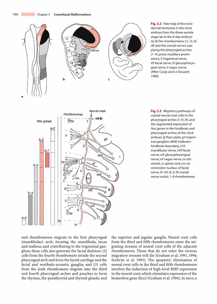
Fig. 5.11 Development of the skull base from above: a chon-drocranium at the end of the embryonic period; b the calvariaat birth. cl clivus, cp (site of ) cribriform plate, d dens, eth eth-moid, fm foramen magnum, fr frontal bone,gw greater wing ofsphenoid, hc hypoglossal canal, hf hypophysial fossa, iam in-ternal acoustic meatus, jf jugular foramen, lw lesser wing ofsphenoid, nc nasal capsule, nch notochord, ns nasal septum,oc otic capsule, occ occipital bone, opc optic canal, par parietalbone, pt petrous part of temporal bone, so supra-occipital,sph sphenoid, sqt squamous part of temporal bone. (AfterO’Rahilly and Müller 1996)
Fig. 5.12 Development of the human desmocranium: a a 40-mm embryo; b an 80-mm embryo (after Gray’s Anatomy 1995).The contours of the CNS are indicated by broken lines.Chondral elements (red): A nasal capsule; B orbitosphenoid;C postsphenoid; D otic capsule; E exoccipital; F supra-occipital;G alisphenoid; H Meckel’s cartilage; I carilage of malleus;
J styloid cartilage; K hyoid cartilage; L thyroid cartilage;M cricoid cartilage; N arytenoid cartilage. Dermal elements(light red): 1 frontal bone; 2 nasal bone; 3 squama of temporalbone; 4 squama of occipital bone; 5 parietal bone; 6 maxilla;7 tympanic ring; 8 mandibula; 9 zygomatic bone

1999). Mesenchymal precursors such as those for theauditory ossicles of the visceral skeleton are presentearly and later become cartilaginous. They are partlyreplaced by intramembranously ossified bone. Thecartilage of the first pharyngeal arch (Meckel’s carti-lage) is largely replaced by the mandibula. Themandibula ossifies intramembranously from a singlecentre on each side (Mérida-Velasco et al. 1993). Thefacial skeleton is largely laid down in mesenchyme atthe end of the embryonic period (O’Rahilly andMüller 2001).
Deficient mandibular development (microg-nathia) is characteristic of the Pierre Robin se-quence, and several syndromes such as cri du chat,Treacher Collins and Down syndromes. In the PierreRobin sequence, the underdeveloped mandibulausually demonstrates catch-up growth in the child.In Treacher Collins syndrome, deficiency of themandibula is maintained throughout growth (Sper-ber 2001). Hemifacial microsomia (Goldenhar syn-drome) also becomes more severe with retardedgrowth. The Prx1/Prx2 genes play a role in mandibu-lar arch morphogenesis (ten Berge et al. 2001). Ag-nathia is found in the otocephalies, a series of malfor-mations, ranging from milder forms in which deriva-tives of the skeletal and dental portions of the firstarch are absent to the more severe form in which lit-tle more than external ears (‘ear head’) are apparent(Duhamel 1966; Vermeij-Keers 1990; Fig. 5.17).
Agnathia-otocephaly was described as a lethal de-velopmental field complex, characterized by extremehypoplasia or absence of the mandibula, astomia,aglossia and synotia (Bixler et al. 1985). It is mostlikely caused by a persistent buccopharyngeal mem-brane (Vermeij-Keers 1990) and is frequently associ-ated with HPE (Pauli et al. 1983; Siebert et al. 1990;Cohen and Sulik 1992). In inbred strains of guineapigs, otocephaly is probably a neural crest problem(Wright and Wagner 1934). In a substrain of C57B1mice with a balanced chromosomal translocation,Juriloff and co-workers (1985) found that in the lessseverely affected embryos the first evidence of celldeath was in the mesodermal cores of the first pha-ryngeal arch. The balanced translocation may hastencell death. In more severe cases, cell death was alsofound in the mesoderm underlying the neural tube.Otx2 heterozygous mouse mutants display oto-cephalic phenotypes, the severity of which is depen-dent on the genetic background of a C57BL/6 strain(Hide et al. 2002). Otx2 is not only expressed in theforebrain and the mesencephalon (Acampora et al.1995, 1998), but also in the cephalic mesenchyme, in-cluding mesencephalic neural crest cells which aredistributed to the mandibula (Kimura etal. 1997).Therefore, Otx2 heterozygous mutant defects relateprimarily to Otx2 function in the formation of mes-encephalic neural crest (Kimura et al. 1997). Most
Otx2+/– mutant mice also display HPE (Matsuo et al.1995), but a role for OTX2 in human HPE has notbeen found so far.
5.5 Neurocristopathies
A number of craniofacial malformations have majorneural crest involvement, and are usually referred toas neurocristopathies (Jones 1990; Johnston andBronsky 1995, 2002). The concept neurocristopathywas introduced by Bolande (1974) to explain thedevelopmental relationships among a number ofdysgenetic, hamartomatous and neoplastic disor-ders, including pheochromocytoma, von Reckling-hausen’s neurofibromatosis, Hirschsprung’s agan-glionic megacolon and the multiple endocrine ade-nomatoses. A neurocristopathy was defined as acondition arising from aberrations in the early mi-gration, growth and differentiation of neural crestcells. Subsequently, an increasing number of disor-ders such as retinoic acid syndrome (RAS), hemi-facial microsomia, Treacher Collins syndrome,DiGeorge sequence, cleft lip and palate, frontonasaldysplasia and Waardenburg syndrome have beenincluded into the neurocristopathies (Table 5.3). Re-cently, the idea that neural crest abnormalities under-lie the pathogenesis of the DiGeorge sequence hasbeen challenged (Sect. 5.5.4). The same holds forTreacher Collins syndrome (Sect. 5.5.3). Two syn-dromes specifically affect derivatives of the face andthe first and second branchial arches. TreacherCollins syndrome affects bilaterally the ear, zygoma,lower eyelid and mandibula. Hemifacial microsomiashows unilateral malformation of nearly the samestructures. Some patients who were exposed tothalidomide during a restricted period of develop-ment had a malformation similar to mandibulofacialdysostosis and hemifacial microsomia (Kleinsasserand Schlothane 1964; Jacobson and Granström 1997;Gorlin et al. 2001).
5.5.1 Retinoic Acid Syndrome
Retinoic acid syndrome (RAS) malformations firstappeared shortly after the introduction of Accutane(13-cis-retinoic acid), a drug used for the treatmentof severe cystic acne (Lammer et al. 1985). Althoughthe retinoids (the normal biologically active retinoicacid and related compounds such as vitamin A, thedietary precursor of retinoic acid) had long beenknown to be potent teratogens, and the drug Accu-tane was not to be taken during pregnancy, in theUSA many accidental exposures occurred, resultingin a surprisingly high incidence of very severe mal-formations involving craniofacial structures. Terato-
204 Chapter 5 Craniofacial Malformations

genic doses of retinoic acid given to mice at earlystages of neurulation yielded craniofacial malforma-tions that are strikingly similar to those in childrenwith retinoic acid embryopathy (Webster et al. 1986;Willhite et al. 1986; Sulik et al. 1988; Sulik 1996; Mor-riss-Kay and Ward 1999). The defects observed inchildren with RAS include abnormalities of the exter-nal and middle ear, sometimes underdevelopment ofthe mandibula and cleft palate, facial nerve paralysis,cerebellar defects, outflow tract defects of the cardio-vascular system, and defects of the thymus andparathyroid glands. Such defects are usually fatalwithin the first years of life (Johnston and Bronsky1995, 2002). The unexpectedly severe nature of RASmalformations relates to the very poor ability of hu-mans to clear retinoic acid metabolites (Webster et al.1986). Teratogenic studies in mice (Goulding andPratt 1986; Webster et al. 1986; Pratt et al. 1987) sug-gested that the timing of exposure for the most severefacial malformations coincided with the onset andperiod of migration of first and second arch crestcells (about day 21 in human embryos), whereas thesensitive period for cardiovascular malformationscoincided with the migration of third and fourth archcrest cells (about day 23 in man).
Local retinoid signalling coordinates forebrainand facial morphogenesis by maintaining FGF8 andSHH expression (Schneider et al. 2001). FGF8 andSHH act as survival factors in the brain and facial pri-mordia (Ahlgren and Bronner-Fraser 1999; Helms etal. 1997; Hu and Helms 1999). Experiments in chick
embryos (Schneider et al. 2001) show that, in the ab-sence of an intact retinoid signalling pathway, FGF8and SHH expression is lost, cells fail to proliferate andundergo apoptosis, and the forebrain and frontonasalprocess cease their morphogenesis. These experi-ments demonstrate that there is a critical period inwhich morphogenesis of the forebrain and the fron-tonasal process is dependent upon retinoid signallingcorrelated with the timing of retinoic acid produc-tion in the frontonasal ectoderm. Forebrain and fron-tonasal process-derived tissue are sensitive to dis-ruptions in retinoid signalling during early develop-ment, but later become insensitive.
5.5.2 Oculoauriculo-vertebral Spectrum
The predominant defects in the non-random associ-ation of anomalies known as the oculoauriculo-ver-tebral spectrum are problems in the morphogenesisof the face, the first and second pharyngeal arches,sometimes accompanied by vertebral anomalies(most commonly cervical hemivertebrae or hypopla-sia of vertebrae) and/or ocular anomalies (Jones1997; Gorlin et al. 2001). The association with epibul-bar dermoid and vertebral anomaly is known asGoldenhar syndrome (Fig. 5.13a–c), and the predom-inantly unilateral occurrence is known as hemifacialmicrosomia.The occurrence of various combinationsand gradations of these anomalies, both unilateraland bilateral, with or without epibulbar dermoid and
5.5 Neurocristopathies 205
Table 5.3 Some neurocristopathies and related disorders (after Sperber and Gorlin 1997; Johnston and Bronsky 2002)
Syndrome Ear abnor- Facial Cardio- Pharyngeal Facial Other malities bone vascular glands clefts associated
malfor- malfor- malforma-mations mations tions
Retinoic Microtia/anotia; Mandibular Conotruncal Thymus deficient Cleft palate Brain acid low-set ears; (micrognathia) defects or absent (8%) (particularly syndrome stenosis external and other cerebellum)
meatus deficiencies
DiGeorge Low-set ears Variable maxillary Conotruncal Thymus deficient Cleft lip Brainsyndrome and mandibular defects or absent and/or
deficiencies palate (10%)(micrognathia)
Hemifacial Microtia; acces- Usually asymmetri- Conotruncal No reported Cleft lip Eye, brain microsomia sory auricles; cal deficiencies defects abnormalities and/or palate (in severe
abnormal ear of mandibula, (7–22%) cases),ossicles squamous temporal vertebrae in
and other bones oculo-auri-culo-verte-bral variant
Treacher Pinna anomalies; Symmetrical No increase No reported Cleft palate Rarely with Collins abnormal ear deficiencies or in cardiovas- abnormalities (35%) limb defects,syndrome ossicles; hypo- absence of zygoma, cular mal- e.g. Nager
plasia or atresia underdevelopment formations syndromeexternal meatus of posterior maxilla
and mandibula
vertebral anomaly, suggested that hemifacial micro-somia and the Goldenhar syndrome may simply rep-resent gradations in severity of a similar disorder ofmorphogenesis. Their frequency of occurrence isestimated to be 1 in 3,000 to 1 in 5,000, with a slight(3:2) male predominance (Jones 1997; Gorlin et al.2001). CNS malformations include mental deficiency,hydrocephalus, Chiari type II malformation, occipitalencephalocele, facial nerve paralysis, agenesis, hypo-plasia and lipoma of the corpus callosum, and hypo-plasia of the septum pellucidum (Aleksic et al. 1984;Jacobson and Granström 1997). Severe abnormalitiesof the pons were found in two infants with Goldenharsyndrome (Pane et al. 2004). The syndrome can bedetected by prenatal ultrasound examination be-cause of the frequent presence of a lipoma on the cor-pus callosum (Jeanty et al. 1991; Wong et al. 2001).
The main facial features of hemifacial microsomiainclude a small lower jaw, sometimes with an absentjaw joint, and a malformed or absent external earwith accessory tags and facial clefts (Cousley andCalvert 1997). Experimental studies in rodents sug-gest that this pattern of malformation is often causedby bleeding in the region of the stapedial artery,transitorily supplying the second pharyngeal arch(Poswillo 1973). Such events are usually sporadic,but genetic predisposition can occur, as shown by theHfm (hemifacial microsomia) mouse, in which achromosome 10 transgene integration is associatedwith a small ear or an asymmetric jaw in 25% of prog-eny heterozygous for the transgene (Naora et al.1994).At E9.5, rupture of the dorsal vasculature of the sec-ond pharyngeal arch has been found in Hfm+/– mu-tants. In man, genetic linkage to chromosome 14q32was reported in a family with hemifacial microsomiain which first-arch abnormalities segregate with un-usually high penetrance (Kelberman et al. 2000).
5.5.3 Treacher Collins Syndrome
Treacher Collins syndrome (Treacher Collins 1900) ormandibulofacial dysostosis (Franceschetti and Klein1949) is an autosomal dominant inherited syndromethat is localized on chromosome 5q32-33.1. Its inci-dence is approximately 1 in 50,000 live births and itsclinical features include the following (Fig. 5.13d–f):(1) abnormalities of the external ears, atresia of theexternal auditory canals and malformation of themiddle ear ossicles, resulting in bilateral conductivehearing loss (Phelps et al. 1981); (2) lateral downwardsloping of the palpebral fissures, frequently withcolobomas of the lower eyelids and a paucity of lidlashes medial to the defect; (3) hypoplasia of themandibula, maxilla and zygoma; and (4) cleft palate(Dixon et al. 1994; Marres et al. 1995; Jacobson andGranström 1997; Jones 1997; Gorlin et al. 2001; Marsh
and Dixon 2001). The Treacher Collins SyndromeCollaborative Group (1996) identified the molecularbasis of this rare disorder by positional cloning. Al-though the causative gene (TCOF1 after TreacherCollins–Franceschetti syndrome) has a somewhatvariable penetrance, malformations are usually veryconsistent. TCOF1 appears to be poorly conservedamong mammals compared with other developmen-tal genes. Mouse Tcof1 shows only 62% amino acididentity with the human protein (Dixon et al. 1997).Tcof1 is widely expressed, most highly at the edges ofthe neural folds (Dixon et al. 1997). Heterozygousmice show exencephaly associated with extensiveapoptosis in the prefusion neural folds (Dixon et al.2000). The Treacher Collins syndrome has not beenassociated with neural tube defects. This discrepancymay be due to species-specific differences betweenmice and man.
Studies on the pathogenesis of RAS defects in micehave also provided data relevant to Treacher Collinssyndrome (Poswillo 1975; Wiley et al. 1983; Websteret al. 1986; Sulik et al. 1987; Osumi-Yamachita et al.1992; Evrard et al. 2000). Treacher Collins syndromemay result from abnormal development of the firstand second branchial arch ectodermal placodesrather than as a direct result of primary interferencewith neural crest cells. Initial abnormalities are large-ly limited to the distal part of the trigeminal gan-glion, which corresponds to that part of the ganglionderived from the ectodermal placodes. Cell deathinterfered with the development of the surroundingcrest cell mesenchyme, which gives rise to the zygo-ma and posterior parts of the maxillary prominenceand mandibular arch, thereby producing defects sim-ilar to those in Treacher Collins syndrome; therefore,Treacher Collins syndrome most likely is not a neuro-cristopathy.
5.5.4 DiGeorge Sequence and Related Disorders
The DiGeorge sequence or syndrome (DiGeorge1965) variably includes defects of development of thethymus, parathyroids and great vessels (Conley et al.1979; Lammer and Opitz 1986; Jones 1997; Gorlin etal. 2001), i.e. tissues of the third and fourth branchialarches, and their associated pouches. Hypoplasia oraplasia of the thymus leads to a deficit in cellular im-munity allowing severe infectious diseases. Hypopla-sia to absence of the parathyroids results in severehypocalcemia and seizures in early infancy. Commoncardiovascular malformations are aortic arch anom-alies, including right aortic arch, interrupted aorta,conotruncal anomalies such as truncus arteriosusand ventricular septal defect, patent ductus arterio-sus and tetralogy of Fallot. Specific to partial mono-
206 Chapter 5 Craniofacial Malformations

somy 22q are lateral displacement of inner canthiwith short palpebral fissures, short philtrum, microg-nathia and ear anomalies. The DiGeorge sequence isaetiologically heterogeneous. It has been associatedwith prenatal exposure to alcohol and Accutane, anda variety of chromosome abnormalities (Gorlin et al.2001; Johnston and Bronsky 2002). The majority ofcases, however, result from partial monosomy of theproximal arm of chromosome 22 owing to a mi-crodeletion of 22q11.2 (Fig. 5.14), detectable by mol-ecular or fluorescence in situ hybridization analysis(Lindsay 2001). Therefore, the DiGeorge sequenceand related malformations with chromosome 22deletions such as the velocardiofacial or Shprintzensyndrome (Shprintzen et al. 1978; Goldberg et al.
1993; also known as Sedlacková syndrome: Sed-lacková 1967) and conotruncal anomaly face syn-drome are combined with the chromosome 22 dele-tion (del22q11) syndrome (Driscoll et al. 1992a, b;Emanuel et al. 2001; Lindsay 2001). The velocardio-facial syndrome is characterized by hypoplasia or acleft of the secondary palate, cardiac defects, a typicalface, microcephaly and hearing and learning disabil-ities. The occurrence of the del22q11 syndrome is es-timated as 1 in 4,000 live births (Scambler 1994, 2000;Lindsay 2001). The symptoms of del22q11 syndromeare diverse. Distinct features can show variable ex-pressivity and incomplete penetrance. Depending onthe key symptoms, two phenotypes may be distin-guished (Ryan et al. 1997; Scambler 2000; Lindsay
5.5 Neurocristopathies 207
Fig. 5.13 Craniofacial dysmorphology syndromes in neuro-cristopathies: a–c Goldenhar syndrome in a 2-year old boy,showing bilateral microtia (left lobar type; right concha type),conductive hearing loss, left facial nerve paresis, left-sidedmandibular hypoplasia, craniosynostosis, defects of vertebraeand ribs and various heart defects: ventricular septal defect,atrial septal defect, pulmonary stenosis and patent ductus ar-
teriosus; d–f, Treacher Collins syndrome in a 10-year-old boywith the following bilateral craniofacial abnormalities: micro-tia, agenesis of external acoustic meatus, conductive deaf-ness, dysplasia of os petrosum, aplasia of zygoma, hypoplasiaand crowding of maxilla and mandibula, hypoplasia of mas-toid and maxillary sinus, and colobomata of lower eyelids.(Courtesy Michiel Vaandrager, Rotterdam)

2001). The ‘pharyngeal’ phenotype encompasses themost characteristic features of del22q11 syndrome,including congenital cardiovascular defects, cranio-facial anomalies and aplasia or hypoplasia of thethymus and parathyroids. The ‘neurobehavioural’phenotype becomes manifest in early childhood aslearning difficulties, cognitive defects and attention-deficit disorder. In adolescence and adulthood, somepatients develop various psychiatric disorders, in-cluding schizophrenia, schizoaffective and bipolardisorders. The basis of the neurobehavioural pheno-type is unknown. All patients with the del22q11 syn-drome manifest at least some components of the pha-ryngeal and neurobehavioural phenotypes withvarying degrees of severity. In contrast to the clinicalheterogeneity of this syndrome, the del22q11 geneticlesion is remarkably homogeneous in affected indi-viduals.Approximately 90% of patients have a typicaldeleted region of some 3 Mb, which encompasses anestimated 30 genes, whereas about 8% of patientshave a smaller deletion of some 1.5 Mb, encompass-ing 24 genes (Lindsay 2001).
Treatment of pregnant mice with ethanol at thetime of migration of the first and second arch crestcells leads to massive cell death, resulting in malfor-mations very similar to those of the DiGeorge se-quence (Daft et al. 1986; Sulik et al. 1986). This sug-gested that the DiGeorge sequence may be secondaryto an abnormality in the neural crest (Scambler1994). A number of gene knockouts produce the
DiGeorge sequence. Hoxa3 knockout mice showsome resemblance to the DiGeorge sequence (Chisa-ka and Capecchi 1991; Manley and Capecchi 1995;Capecchi 1997). They lack a thymus and parathyroidglands, have a reduced thyroid and show malforma-tions of the laryngeal cartilages and muscles, and ofthe heart. Lindsay and co-workers (Lindsay et al.1999; Lindsay and Baldini 2001) developed a mousemodel heterozygous for the 22q11.2 deletion. Neuralcrest migration into the pharyngeal arches appearedto be normal but there was severe underdevelopmentof the fourth arch aortic vessels in all of the embryosstudied. The gene content of the human 22q11 regionwas found to be highly conserved in a region ofmouse chromosome 16. The first mouse deletiongenerated, Df1 (Lindsay et al. 1999), encompassingmouse homologues of 18 out of the 24 genes that aredeleted in patients with the 1.5-Mb deletion, led tocardiovascular defects similar to those in patients.The T-box transcription factor Tbx1 plays a crucialrole in the fourth arch abnormality in mice (Lindsay2001; Lindsay et al. 2001). Tbx1-null mice die at birthand have a persistent truncus arteriosus, a hypoplas-tic pharynx, lack a thymus and parathyroids and haveear, jaw and vertebral anomalies. The embryologicalbasis of these abnormalities is maldevelopment ofthe pharyngeal arches and arch arteries 2–6, and ofthe pharyngeal pouches 2–4 (Lindsay et al. 2001). Theseverity and extent of the embryological lesion indi-cate that Tbx1 may be required for the segmentation
208 Chapter 5 Craniofacial Malformations
Fig. 5.14 a–c Craniofacial dysmorphology in a 10-year-oldgirl with a 22q11 deletion. At birth, the patient showed a flatocciput, short neck, wide but closed palate, tetralogy of Fallotand slight dysmorphic toes. In 1994, a velopharyngeal insuffi-ciency was found, followed by DNA analysis, and the diagno-sis of a 22q11 deletion in 1996. Apart from bilateral hearing
loss, due to chronic inflammation of the middle ears, no otherabnormalities of the derivatives of the pharyngeal archeswere found. A general developmental delay of the patient re-quired special education. (Courtesy Jeannette M. Hooge-boom, Rotterdam)

of the pharyngeal endoderm, an event that initiatesthe development of the entire pharyngeal apparatus(Lindsay 2001). Chordin secreted by the mesendo-derm is required for the correct expression of Tbx1and other transcription factors involved in the devel-opment of the pharyngeal region (Bachiller et al.2003). Chordin mutant mice either die early duringdevelopment or die perinatally, showing an extensivearray of malformations that encompass most featuresof DiGeorge syndrome and velocardiofacial syn-drome in humans. FGF8 is also required for pharyn-geal arch and cardiovascular development in mice(Abu-Issa et al. 2002; Frank et al. 2002). Fgf8 mutantsresemble Tbx1–/– mouse embryos, probably owing toa common signalling pathway (Vitelli et al. 2002).
The hypothesis that neural crest abnormalities un-derlie the pathogenesis of the DiGeorge sequenceseems unlikely since pharyngeal patterning is not af-fected in chick embryos in which the neural crest hasbeen ablated (Bockman et al. 1989; Veitch et al. 1999).Moreover, Tbx1 is not expressed in the neural-crest-derived mesenchyme of the pharyngeal arches. Neur-al crest cells may, however, play a secondary role inthe disorder as targets of Tbx1-driven signalling(Lindsay 2001). Recently, Yagi et al. (2003) showedthat a TBX1 mutation is responsible for five majorphenotypes in del22q11 syndrome. In view of thesedata, the DiGeorge sequence should not be viewedany longer as a neurocristopathy.
5.5.5 Waardenburg Syndrome
The term Waardenburg syndrome, originally describ-ed by Waardenburg (1951), is used for a heteroge-neous set of auditory–pigmentary syndromes, theprimary cause of which is a patchy lack of melanocytes
in the hair, eyes, skin and stria vascularis (Read 2001;Spritz et al. 2003). Four subtypes can be distin-guished: (1) type 1 with dystopia canthorum, causedby mutations in the PAX3 gene (Fig. 5.15); (2) type 2without dystopia is heterogeneous, some cases aredue to changes in the MITF and SLUG genes; (3) therare type 3, resembling type 1 but with additionalcontractures or hypoplasia of the upper-limb jointsand muscles, also results from PAX3 mutations; and(4) type 4 with Hirschsprung’s disease, again hetero-geneous and due to mutations in the EDN3, EDNRBand SOX10 genes. Most forms are inherited as auto-somal dominant traits. The hearing loss in all types iscongenital, sensorineural and non-progressive(Chap. 7). Waardenburg syndrome types 1, 3, and 4are neurocristopathies, affecting more than one neur-al crest derivative. Type 2 appears to be melanocyte-specific.Auditory–pigmentary syndromes and mousemodels are discussed further in Chap. 7.
5.6 Holoprosencephaly
The holoprosencephalies encompass a range of phe-notypes that vary in severity and involve malforma-tions of the brain and upper face among the midline(DeMyer et al. 1963, 1964; Cohen 1989a, b; Cohen andSulik 1992; Norman et al. 1995; Golden 1998; Muenkeand Beachy 2001; Cohen and Shiota 2002). HPE isaetiologically extremely heterogeneous. Its forma-tion may depend on the interaction of both geneticand environmental factors. Specific teratogens suchas maternal diabetes increase the risk for HPE 200-fold (Norman et al. 1995; Cohen and Shiota 2002).About 1–2% of newborn infants of diabetic mothersdevelop HPE. Numerous other teratogens are knownto cause HPE in various animal models (Cohen and
5.6 Holoprosencephaly 209
Fig. 5.15 a, b Waardenburg syn-drome in a 32-year-old male patient with a 558-559delCA mutation in exon 4 of the PAX3gene, compatible with Waarden-burg type 1.The patient showstypical craniofacial dysmorpholo-gy, early temporal greying, hassevere bilateral congenital hear-ing loss, mental retardation andno verbal communication.(Courtesy Jeannette M. Hooge-boom, Rotterdam)

Sulik 1992; Cohen and Shiota 2002). The incidence ofHPE in live-born children with normal chromo-somes has been estimated to be 0.48–0.88 per 10,000.In contrast, the rate among human abortuses was es-timated at 40 per 10,000, indicating a very high rate ofembryonic and fetal loss (Matsunaga and Shiota1977; Shiota 1993). In a large epidemiologic study ina Californian population, Croen et al. (1996) ob-served an overall prevalence of 1.2 per 10,000 livebirths and fetal deaths (121 HPE cases in 1,035,386live births/fetal death deliveries), whereas the preva-lence for live births was 0.88 per 10,000. In anotherperinatal study from Scotland, Whiteford and Tolmie(1996) found 50 HPE cases in 694,950 live births andstill births (a prevalence of 0.7 per 10,000).
Although the majority of HPE cases are sporadic,familial HPE has been described in pedigrees, sug-gesting autosomal dominant, autosomal recessive,and possibly X-linked inheritance. The clinical vari-ability can be striking even within a single pedigree.
In pedigrees with clinically unaffected parents andmultiple affected siblings autosomal recessive inher-itance is suggested. Since abnormal HPE genes arenot fully penetrant and germline mosaicism mayhappen, some of these cases may actually be autoso-mal dominant (Nanni et al. 1999). The causes of HPEin man are summarized in Table 5.4. Certain chromo-somes, chromosome 13 in particular, display recur-rent involvement in HPE. HPE may be present in asmany as 70% of trisomy 13 cases (Taylor 1968; Cohenand Sulik 1992; Norman et al. 1995). Cytogeneticallyverified chromosome abnormalities in perinatalstudies range from 34% in Scotland (Whiteford andTolmie 1996) to 37% in California (Croen et al. 1996).A similar frequency was observed in prenatally diag-nosed HPE (Berry et al. 1990; Blaas et al. 2002). Basedon non-random cytogenetic rearrangements, geneticstudies of HPE have revealed at least 12 putative loci(HPE1-12) on 11 different chromosomes, which maycontain genes that play a role in the pathogenesis of
210 Chapter 5 Craniofacial Malformations
Table 5.4 Aetiology of human holoprosencephaly (after Cohen and Sulik 1992; Norman et al. 1995; Blaas et al. 2002; Cohen andShiota 2002)
Causes Examples Notes
Chromosomal Most frequently involved:abnormalities Chromosomes 13 and 18
Trisomy 13 In 70% HPE (Taylor 1968)Trisomy 18Numerous deletions, Examples of deletions and duplications (in order of frequency):duplications, del(13)(q22), del(18p), del(7)(q36), dup(3)(p24-pter), del(2)(p21),and ring chromosomes del(21)(q22.3); for further data see Schinzel (1983),
Cohen and Sulik (1992) and Norman et al. (1995)
Identified genes HPE3 (7q36): SHH See text for explanationHPE2 (2p21): SIX3HPE5 (13q32): ZIC2HPE4 (18p): TGIFPatched/PTCHGLI2DCHR7 Smith–Lemli–Opitz syndrome (Kelley and Hennekam 2001)
Teratogens Diabetic embryopathy HPE in 1–2% of newborn infants of diabetic mothers
Ethyl alcohol (alcohol abuse) In 28 autopsies of patients with HPE, Jellinger et al. (1981) found 1 case in which the mother had a history of alcohol abuse;Ronen and Andrews (1991) found HPE in 3 such cases
Retinoic acid HPE has been noted (Lammer et al. 1985; Rosa et al. 1994)
Syndromes with HPE Meckel syndrome May have HPE with median or lateral cleft lip (Hsia et al. 1971)
Pallister–Hall syndrome Congenital hypothalamic hamartoblastoma hypopituitarism,other anomalies including HPE (Hall et al. 1980), due to mutations in GLI3 (Kang et al. 1997)
Lambotte syndrome Microcephaly, mental retardation, ocular hypotelorism (Verloes et al. 1990)
Velocardiofacial syndrome HPE in 1 out of 61 cases (Wraith et al. 1985)
Aicardi syndrome Flexion spasms, mental retardation and agenesis of corpus callosum; arhinencephaly in several instances;rarely HPE (Sato et al. 1987; Donnenfeld et al. 1989)
Associations Anencephaly Cases with holoprosencephalic facies (Lemire et al. 1981)
Frontonasal dysplasia Median cleft syndrome (DeMyer 1967; Sedano et al. 1970)
Agnathia-otocephaly Cyclopia/HPE may occur (Pauli et al. 1983; Siebert et al. 1990)
HPE holoprosencephaly

HPE (Roessler and Muenke 1998; Nanni et al. 2000).Identifiable genetic causes account for 15–20% of allcases of HPE.Several HPE genes have been identified:SHH (also known as HPE3: Belloni et al. 1996;Roessler et al. 1996; Nanni et al. 1999), SIX3 (HPE2:Wallis et al. 1999), ZIC2 (HPE5: Brown et al. 1998),TGIF (HPE4: Gripp et al. 2000), Patched/PTCH (Mingand Muenke 1998), GLI2 (Roessler et al. 2003) andDHCR7 (Kelley et al. 1996). Of these defective genesin human HPE, three exhibit a ventrodorsal gradientof expression (SHH, SIX3 and TGIF) and two adorsoventral gradient (GLI2 and ZIC2). Patients withHPE3 (SHH) and HPE2 (SIX3) mutations exhibit theHPE spectrum with major facial malformations((Wallis et al. 1999), whereas ZIC2 mutations showonly minimal facial malformations, such as slantingof the frontal bones as a result of microcephaly(Brown et al. 1998). Heterozygous carriers for muta-tions in either SHH or SIX3 can appear phenotypical-ly normal, whereas other heterozygous mutation car-riers within the same family may be severely affected(Nanni et al. 1999). Nanni and co-workers identifiedthree HPE patients with an SHH mutation and an ad-ditional ZIC2 or TGIF mutation. The first patient ex-hibited microcephaly due to semilobar HPE but withnormal midfacial dimensions and, therefore, this cor-responds best with the ZIC2 phenotype. The othertwo cases showed major craniofacial abnormalitiesmatching the SHH HPE spectrum. HPE may occur in
a large number of syndromes and associations(Siebert et al. 1990; Cohen and Sulik 1992; Norman etal. 1995; see Table 5.4 for some examples). Syndromal(not chromosomal) conditions with HPE includesuch diverse disorders as Meckel–Gruber, Pallister–Hall, Smith–Lemli–Opitz and Aicardi syndromes.The Smith–Lemli–Opitz syndrome is due to a defi-ciency in the final step of cholesterol biosynthesis(Tint et al. 1994; Kelley et al. 1996; Kelley and Hen-nekam 2001; Chap. 3).
The brain malformations in HPE will be discussedin Chap. 9. In brief, forebrain malformations rangefrom the alobar, complete form with one single ven-tricle, undivided thalami and corpora striata, and ab-sence of the olfactory bulbs and corpus callosum, tothe semilobar, incomplete form, in which hypoplasticcerebral lobes with an interhemispheric posterior fis-sure and a hypoplastic corpus callosum may be pre-sent, to the lobar type, in which a distinct interhemi-spheric fissure is present with some midline continu-ity and the olfactory bulbs may vary from normal toabsent. HPE can be detected prenatally by ultrasound(Kurtz et al. 1980; Blaas et al. 2000, 2002; ClinicalCase 5.1). In the Californian study (Croen et al. 1996),46% of HPEs were of the alobar type, 20% semilobarand 5% lobar.
The facial anomalies in HPE are usually catego-rized into four main types (DeMyer et al. 1964; Cohenand Sulik 1992; Figs. 5.16, 5.17): (1) cyclopia with a
5.6 Holoprosencephaly 211
Fig. 5.16 Holoprosencephaly,main types of facial malforma-tion: a cyclopia; b, c single medi-an eye with various degrees of doubling of ocular structures;d ethmocephaly; e cebocephaly;f median cleft lip with arhinen-cephaly (after Duhamel 1966)

single eye or various degrees of doubling of the eyeanlage, with or without a proboscis; (2) ethmo-cephaly with ocular hypotelorism and proboscis lo-cated between the eyes; (3) cebocephaly with ocularhypotelorism and a single-nostril nose; and (4) me-dian cleft lip and palate (premaxillary agenesis) and
ocular hypotelorism. Less severe facial dysmor-phism, microsigns such as a single central incisorand/or ocular hypotelorism, and HPE without facialmalformations are also found (DeMyer et al. 1964;Cohen and Sulik 1992; Brown et al. 1998). Five out ofthe six cases shown in Fig. 5.17 exhibited major facial
212 Chapter 5 Craniofacial Malformations
Fig. 5.17 Holoprosencephaly, spectrum of craniofacial mal-formations. a Alobar or complete holoprosencephaly was di-agnosed by ultrasound at 22 weeks of gestation, after whichpregnancy was terminated at 24 weeks.At autopsy,the femalefetus showed agenesis of both eyes and orbits,arhinia and mi-crostomia.b Frontal view of a cyclopic face of a female fetus ofapproximately 35 weeks of gestation. The fetus had a singleeye in a single orbit and fused optic nerves, arhinia withoutproboscis, and agnathia with astomia and synotia. The brainshowed a complete holoprosencephaly. c Frontal view of amale fetus of 32 weeks of gestation with synorbitism, arhiniawith proboscis, agnathia with astomia and synotia. The brainshowed a semilobar or incomplete holoprosencephaly with a
dorsal sac (Chap. 9). d Frontal view of a female fetus of32 weeks of gestation with hypotelorism, arhinia with a sep-tated proboscis, agnathia with astomia and synotia.The brainshowed an incomplete holoprosencephaly. e A hypoteloricmale fetus of 34 weeks of gestation with a flat nose, cleftpalate and agenesis of the premaxillae, the prolabium and thenasal septum. An incomplete holoprosencephaly was found.f A case of alobar, complete holoprosencephaly with a normalface. (a Courtesy Annemarie Potters, Deventer; b–e from thecollection of the Museum of Anatomy, University of Leiden;courtesy Christl Vermeij-Keers; f courtesy Raoul Hennekam,Amsterdam)

malformations, varying from agenesis of the eyes tohypotelorism and were holoprosencephalic and, as aconsequence, microcephalic. In these cases the devel-oping forebrain is too narrow, indicating a lack ofoutgrowth of the ventral neurectoderm during earlyembryogenesis (Müller and O’Rahilly 1989). As a re-sult the interplacodal area (the area between thenasal placodes) is either absent (resulting in agenesisof the eyes and orbits, cyclopia, synophthalmia, syn-orbitism, hypotelorism without a nasal septum) ortoo narrow (hypotelorism with a nasal septum). Dur-ing normal embryonic development, both medialnasal processes grow out from the interplacodal area,and give rise to the premaxilla, the prolabium, thevomer and other parts of the nasal septum. The me-sectoderm of this area originates from the cranialneural crest and surface ectodermal placodes (Smits-van Prooije et al. 1988).
HPE with major facial malformations may be theresult of insufficient outgrowth of the prosencephal-ic neuroectoderm and as a consequence insufficientEMT in presomite stages (Vermeij-Keers 1990). Theoutcome is an abnormally narrow prosencephalon,particularly along its ventral midline, causing agene-sis or non-separation of the eye vesicles, non-separa-tion of the thalami and lack of proper outgrowth ofthe telencephalic hemispheres, leading to agenesis ofthe olfactory bulbs and the corpus callosum. This isin keeping with experimental studies in animals onthe teratogenic effects of drugs and other chemicals(Cohen and Sulik 1992; Cohen and Shiota 2002) anddata from mice lacking Shh (Chiang et al. 1996). Shhexpression has been detected in the mouse pre-chordal plate and the ventral neuroectoderm. Inchick embryos, Shh expression in the ectoderm of thecraniofacial primordia is essential for outgrowth ofthe facial swellings (Helms et al. 1997; Hu and Helms1999). Moreover, Six3 participates in midline fore-brain and eye formation (Bovolenta et al. 1996). Incontrast, in mice Zic2 is expressed along the dorsalneuroectoderm up to the rostral end of the futuretelencephalon, and reduction of Zic2 expressioncauses a neurulation delay and inhibition of EMT,which results in HPE and various neural tube defects(Nagai et al. 1997, 2000). These different expressionpatterns, Shh and Six3 in the ventral neuroectodermand Zic2 in the dorsal neuroectoderm, suggest thatthese genes affect the outgrowth and differentiationof the forebrain in different ways. Mutations of genesexpressed dorsally in the neural tube give rise toeither inappropriate division of the prosencephaloninto cerebral hemispheres with agenesis of the telen-cephalic roof plate, resulting in HPE with normalmidfacial dimensions (Brown et al. 1998; Fig. 5.17f),or defects in the fusion process of the prosencephalicneural walls, causing exencephaly and anencephaly(Nagai et al. 2000).
5.7 Abnormal Development of the Skullwith CNS Manifestations
Developmental defects of the cranial vault and faceare relatively common, whereas congenital defects ofthe skull base and sensory capsules (nasal and otic)are relatively rare (Sperber 2002). During postnatalgrowth of congenital craniofacial defects, threegeneral patterns of development may occur: mainte-nance of the defective growth pattern; catch-upgrowth, minimizing the defect; or marked worseningof the derangement with increasing age. Of great sig-nificance to facial development is the normal closingof the foramen caecum in the anterior cranial fossa atthe fronto-ethmoid junction (Hoving 1993). Abnor-mal patency of this foramen, due to non-separationof the neuroectoderm and surface ectoderm duringneural tube formation, allows a pathway for neuraltissue to herniate into the nasal region, providing thebasis of encephaloceles, gliomas and dermoid cyststhat cause gross disfigurement of facial structures.
5.7.1 The Craniosynostoses
Premature closure of calvarial sutures causes the ces-sation of growth and leads to isolated and syndromicforms of craniosynostoses. Many craniosynostosissyndromes are due to genetic mutations of the FGFreceptors (FGFRs 1, 2 and 3), and the TWIST andMSX2 genes (Cohen and MacLean 2000; Warren et al.2001; Jabs 2002; Johnston and Bronsky 2002). FGFRson mesenchymal cells enhance their differentiationinto osteoblasts and increase apoptosis when theybind FGFs, and are repressed by TWIST. MSX2 alsoenhances the differentiation into osteoblasts and isinvolved in cell death,which may be important in ear-ly suture closure. In mice, Msx2 and Twist coopera-tively control the development of the neural-crest-derived skeletogenic mesenchyme of the skull vault(Ishii et al. 2003). Depending on which of the suturesare obliterated prematurely, the skull can develop acharacteristic shape (Fig. 5.19). Synostosis of thecoronal sutures leads to brachycephaly or short-headedness.Variant head shapes are acrocephaly andoxycephaly (pointed head), or turricephaly (tower-shaped head). Unilateral coronal synostosis gives riseto anterior plagiocephaly, which is associated withasymmetry of the face and skull. Dolichocephaly(long-headedness) or scaphocephaly occurs afterpremature fusion of the sagittal suture. Trigono-cephaly (triangular-shaped head) results from pre-mature closure of the metopic suture. Posterior pla-giocephaly is mostly non-synostotic and due to ab-normal mechanical forces. Compound synostosis, inwhich two or more sutures are involved, can lead tocomplex head shapes with severe skull deformities
5.7 Abnormal Development of the Skull with CNS Manifestations 213

214 Chapter 5 Craniofacial Malformations
Clinical Case 5.1 Alobar Holoprosencephaly
Alobar holoprosencephaly (HPE) and other forms ofHPE can be detected prenatally by ultrasound.Blaas etal. (2000) presented a case of alobar HPE with cyclop-ia in an embryo with a gestational age of 9 weeks and2 days with a crown–rump length (CRL) of 22 mm.Three-dimensional ultrasound improved the imaging(see Case Report).
Case Report. A 31-year-old gravida 6para 1 was referred to a university hospital because ofhabitual abortion. Her husband had a balanced chro-mosomal translocation (46,XY,t(8;14)(p21.1;q24.1). Atthe first examination at the gestational age of 9 weeksand 2 days, ultrasound examination showed an em-bryo of 22-mm CRL with abnormal development ofthe brain with a small monoventricular forebrain(Fig. 5.18a) and a proboscis (Fig. 5.18b). Seven dayslater, CRL was 33 mm (Fig. 5.18d). Three-dimensional
Fig. 5.18 Alobar holoprosencephaly at 9 weeks of gesta-tion visualized by two- and three-dimensional ultrasound:a–c ultrasound sections showing the non-separation of theforebrain (asterisks in a and b) of the 9-week and 2-day-oldembryo (crown–rump length, CRL, 22 mm), and three-di-mensional reconstruction of the embryo with volume pre-sentation of body and brain cavities (c); d three-dimension-al reconstruction 7 days later, the arrow points at the cyclop-ia; e coronal section through the face of a normal fetus (CRL30 mm), showing the normal hypertelorism of the eyes
(arrows) at that age; f fetus with holoprosencephaly (CRL33 mm) with the two eye anlagen lying close together (arrows); g three-dimensional reconstruction of the samefetus with the holosphere (yellow) and the cavities of thediencephalon (D,green), the mesencephalon (M, red) and therhombencephalon (Rh, blue), and the eye anlagen (yellow,arrows); h postabortem photograph of the fetus with cy-clopia, two eye anlagen and a proboscis (reproduced withpermission from Blaas et al.2000, Ultrasound Obstet.Gynecol.15:62–65; copyright 2000, John Wiley & Sons Ltd.)

such as the cloverleaf (Kleeblattschädel) malforma-tion found in thanatophoric dysplasia.
Agenesis and premature ossification of one ormore cranial sutures are the most common develop-mental disorders of the skull vault, affecting 1 in2,500 individuals (Cohen and MacLean 2000; Mathi-jssen 2000; Muenke and Wilkie 2001; Jabs 2002).These disorders can be associated with increased in-tracranial pressure, difficulties with vision, hearingand breathing, and facial deformities. In agenesis, di-rect fusion of bone centres takes place at locationswhere normally sutures arise. This can be observedin isolated and syndromic forms of craniosynostosis,for example in scaphocephaly and Apert syndrome,respectively (Mathijssen 2000). Non-syndromic cran-
iosynostosis, in which craniosynostosis is an isolatedfeature, is more frequent than syndromic forms.Sagittal synostosis is the most common of the non-syndromic synostoses (birth prevalence: 1 in 5,000).In isolated sagittal stenosis, the overall scaphocephal-ic skull shape is mirrored in anteroposterior expan-sion of the brain, primarily in a posterior direction(Aldridge et al. 2002). The classical craniosynostosissyndromes are inherited as autosomal dominanttraits and include Apert, Pfeiffer, Saethre–Chotzenand Crouzon syndromes (Figs. 5.20, 5.21). Most auto-somal dominant craniosynostosis syndromes involvethe coronal suture. Syndromic craniosynostoses areusually associated with facial, limb, ear and/or heartmalformations.Recently, the molecular bases of theseclassical craniosynostoses and of several rare cran-iosynostosis syndromes have been identified. Domi-nant mutations in one of three FGFR genes (FGFR1,FGFR2 and FGFR3) or in the transcription factorTWIST account for some 20% of cases of craniosyn-ostosis. Many FGFR mutations encode highly recur-rent missense substitutions, which arise exclusivelyfrom the father and are associated with increased pa-ternal age (Crow 2000; Wilkie and Morriss-Kay 2001).Several different syndromes can be associated withthe same mutations in FGFR genes (Table 5.5), indi-cating that the mutations might act by distinct mech-anisms. Furthermore, the same syndrome, for exam-ple Pfeiffer syndrome, is due to different mutations inFGFR1 and FGFR2. Heterogeneous mutations inFGFR2 also cause Apert, Crouzon, Jackson–Weiss andBeare–Stevenson syndromes. Muenke syndrome isdue to a specific mutation in FGFR3. Cytogeneticdeletions and translocations involving 7p21.1 andvarious heterozygous TWIST mutations causeSaethre–Chotzen syndrome. A specific heterozygousmutation in the MSX2 gene, which has been observedin only one family, causes Boston-type craniosynos-
5.7 Abnormal Development of the Skull with CNS Manifestations 215
Fig. 5.19 Premature ossification of the cranial sutures leadsto an abnormal head shape: a normal skull; b trigonocephaly;c brachycephaly; d anterior plagiocephaly; e posterior plagio-cephaly; f dolichocephaly (after Cohen and MacLean 2000)
reconstructions were made from both examinations(Fig. 5.18c, g). The body, including the proboscis andthe brain cavities, was outlined by manual segmenta-tion and given different colours. Two eye anlagen,lying closely together below the proboscis, could beidentified at 19 weeks of gestation (Fig. 5.18f ). Therewas a small monoventricular holosphere, connectedto the diencephalon by a narrow duct (Fig. 5.18c, d, f ).Chorion villus biopsy at 10.5 weeks revealed the samebalanced translocation as that of the father. The pa-tient was informed about the diagnosis of alobar HPEat the first visit. With the informed consent of the pa-tient, the pregnancy continued until 12.5 weeks ofgestation before it was terminated. This was done toconfirm the diagnosis by ultrasound, by karyotyping
and by postabortem autopsy. The autopsy confirmedthe diagnosis of alobar HPE, associated with cyclopiawith two eye anlagen, a proboscis and a smallmonoventricular holosphere (Fig. 5.18h).
References
Blaas H-GK, Eik-Nes SH, Berg S, Torp H (1998) In-vivo three-di-mensional ultrasound reconstructions of embryos and earlyfetuses. Lancet 352:1182–1186
Blaas H-GK, Eik-Nes SH, Vainio T, Isaksen CV (2000) Alobar holo-prosencephaly at 9 weeks gestational age visualized by two- and three-dimensional ultrasound. Ultrasound ObstetGynecol 15:62–65

tosis. The diagnosis of craniosynostosis syndromes isby clinical examination and imaging of the skull,hands and feet. The more common syndromic cran-iosynostoses will be briefly discussed.
Apert syndrome or acrocephalosyndactyly (Apert1906) is characterized by bilateral coronal suture syn-ostosis (Fig. 5.20a, b) due to fusion of bone centres ofthe frontal and parietal bones, resulting a wide-opengap between the frontal bones instead of the frontalsuture at birth, exorbitism, strabismus, midfacial hy-poplasia, joint ankylosis and symmetrical complexsyndactyly of the hands and feet (Cohen and Krei-borg 1993, 1995). Mental retardation is common andoften severe. Deafness and optic atrophy are fre-quent. Non-progressive ventriculomegaly and agene-sis of the corpus callosum are the most frequent CNSmalformations (Cohen and Kreiborg 1990; ClinicalCase 5.2). Almost all cases with Apert syndrome areassociated with either of two FGFR2 mutations,Ser252Trp or Pro253Ag (Park et al. 1995; Wilkie et al.1995).
Crouzon syndrome (Crouzon 1912; Kreiborg 1981)is characterized by bicoronal craniosynostosis inwhich eventually all calvarial sutures can be involved(Fig. 5.20c, d), midfacial hypoplasia, exorbitism,
epilepsy and seemingly unaffected limbs. Subtleradiographic abnormalities such as fusion of carpalbones have been described (Anderson et al. 1997;Murdoch-Kinch and Ward 1997). The gene forCrouzon syndrome has been mapped to 10q25-q26(Preston et al. 1994) and has been shown to codeFGFR2. Several mutations have been demonstrated(Jabs et al. 1994; Reardon et al. 1994; Passos-Bueno et al. 1999). Pfeiffer syndrome (Pfeiffer 1964) includesbicoronal synostosis (Fig. 5.20e, f), hypertelorism,and broad and deviated thumbs and halluces (Cohen1993).
Typical characterics of the Saethre–Chotzen syn-drome (Saethre 1931; Chotzen 1932) are bilateralcoronal synostosis (Fig. 5.21), hypertelorism, unilat-eral or bilateral ptosis of the eyelids, low-set hairline,hearing loss, brachydactyly and soft-tissue syndacty-ly. Saethre–Chotzen syndrome has been linked to theTWIST gene on chromosome 7p21.1. Mutations inand variably sized deletions of this gene can be foundin patients with clinical features of Saethre–Chotzensyndrome. The clinical spectrum of genetic ab-normalities of the TWIST gene is highly variable. In alarge five-generation family with characteristics ofSaethre–Chotzen syndrome as well as of the ble-
216 Chapter 5 Craniofacial Malformations
Table 5.5 Overview of the more common syndromic craniosynostoses (after Aicardi 1998; Jabs 2002)
Craniosynostosis Main features Proportion Inheritance Gene defectsyndrome/OMIM of cases number with cranio-
synostosis
Apert syndrome/ Mental retardation, midface deficiency, Almost all AD FGFR2,101200 proptosis, down-slanting palpebral fissures, (coronal sutures) (most cases exons IIIa
complex symmetrical syndactyly of hands new muta- and IIIcand feet (either of digits I–V or of digits II–IV) tions)
Crouzon syndrome/ Hypoplasia of maxilla, shallow orbits, Almost all AD FGFR2,123500 proptosis (coronal, exons IIIa
then all sutures) and IIIc
Pfeiffer syndrome/ Strabismus, proptosis, hypertelorism, All cases AD FGFR1, FGFR2,101600 broad thumbs and toes, mild variable (coronal) FGFR3,
cutaneous syndactyly of fingers and toes exons IIIa and IIIc
Saethre–Chotzen Facial asymmetry, low-set frontal hairline, All cases AD TWISTsyndrome/101400 ptosis, deviated nasal septum, variable (coronal, uni-
brachydactyly and cutaneous syndactyly, or bilateral)mental retardation
Muenke syndrome/ Variable, uni- or bicoronal synostosis, AD FGFR3,602849, 600593 brachydactyly exon IIIa
Boston-type cranio- Variable craniosynostosis; AD MSX2synostosis/12310.001 supraorbital recession; short first metatarsals
Jackson–Weiss Variable craniosynostosis, maxillary hypoplasia, AD FGFR1,syndrome/123150 broad great toe and medial deviation, exons IIIa
syndactyly, bony fusion and IIIc)FGFR2,exon IIIa
Beare–Stevenson Variable craniosynostosis, cutis gyrata, Usually FGFR2syndrome/123790 acanthosis nigricans sporadic
mutations
AD autosomal dominant
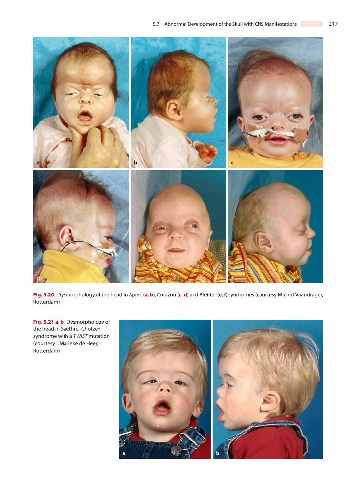
5.7 Abnormal Development of the Skull with CNS Manifestations 217
Fig. 5.20 Dysmorphology of the head in Apert (a, b), Crouzon (c, d) and Pfeiffer (e, f) syndromes (courtesy Michiel Vaandrager,Rotterdam)
Fig. 5.21 a, b Dysmorphology of the head in Saethre–Chotzensyndrome with a TWIST mutation(courtesy I. Marieke de Heer,Rotterdam)

218 Chapter 5 Craniofacial Malformations
Clinical Case 5.2 Apert Syndrome
Apert syndrome, described by Apert (1906) as acro-cephalosyndactyly, is characterized by bilateral coro-nal suture synostosis, related skull malformations,mental retardation, syndactyly of the hands and feet,and frequently deafness and optic atrophy (Apert1906; Cohen and Kreiborg 1995). The most frequentlyassociated cerebral malformations are hypoplasia ofthe corpus callosum and the septum pellucidum, andventricular enlargement (Cohen and Kreiborg 1990;see Case Report).
Case Report. After an uneventful preg-nancy, a boy was born as the seventh child in a familywith six healthy children. He was born at term with abirth weight of 3,724 g. He had a dysmorphic headwith a prominent forehead, retraction of the glabellaand upper nose bridge, midfacial hypoplasia and low-implanted ears. There was bilateral choanal atresia.The posterior part of the head was flat with a broadneck. There was symmetric syndactyly of fingers 2–5of the hands and of toes 1–5.The boy died from severe
cardiorespiratory failure due to the following complexmalformations of the heart: abnormal venous returnof systemic vessels with a persistent left vena cava su-perior draining through the coronary sinus into theright atrium; hypoplasia of the left ventricle and aorta;bicuspid aortic valve; aortic coarctation, and a persis-tent patent ductus arteriosus. Head circumferencewas 36.5 cm (P50) with a shorter-than-normal antero-posterior diameter. This was reflected in a shortenedbrain with relatively small parieto-occipital lobes(Fig. 5.22a). There was partial agenesis of the corpuscallosum, the posterior part of which was absent.Moreover, the septum pellucidum was absent.The pyramids were hypoplastic, and were flanked by rather large, somewhat plump inferior olives(Fig. 5.22b).
References
Apert E (1906) De l’acrocéphalosyndactylie. Bull Soc Méd (Paris)23:1310-1330
Cohen MM Jr, Kreiborg S (1990) The central nervous system inthe Apert syndrome. Am J Med Genet 35:36–45
Cohen MM Jr, Kreiborg S (1995) Hands and feet in the Apert syn-drome. Am J Med Genet 57:82–96
Fig. 5.22 Apert syndrome:a medial view of the brain,show-ing the short anteroposterior diameter of the brain, a rela-tively small parietal-occipital lobe, incomplete corpus callo-sum and absent septum pellucidum; b Luxol Fast Blue
stained section through the medulla oblongata, showinglarge inferior olives and hypoplastic pyramids (courtesyPieter Wesseling, Nijmegen)

5.7 Abnormal Development of the Skull with CNS Manifestations 219
Clinical Case 5.3 Thanatophoric Dysplasia
Thanatophoric dysplasia is the most common formof lethal skeletal dysplasia. The name is derived fromthe Greek thanatus (death) and phorus (seeking).Holtermüller and Wiedemann (1960) described a formof craniosynostosis with trilocular configuration of
the skull and severe internal hydrocephalus as Klee-blattschädel (cloverleaf skull) as an isolated anomalybut also in combination with generalized skeletal dys-plasia.Different mutations at the FGFR3 locus give riseto distinct phenotypes (Cohen 1997; Hall and Lopez-Rangel 1997). Abnormalities in the CNS mainly affectthe temporal lobe (see Case Report).
Case Report. This girl was the secondchild of a 31-year-old healthy mother. After an un-eventful pregnancy, apart from breech position, shewas born by Caesarean section at 41 weeks of gesta-tion. Birth weight was 4,200 g and crown–heel lengthwas 42 cm (much less than P3). The skull was broadand large with frontal bossing (Fig. 5.23a, b; head cir-cumference was 40 cm; much greater than P97).Therewas micromelia with phonehorn-formed long bones,a small and short thorax, and hypoplastic lungs. Thebaby died after 3 days. The babygram showed thetypical configuration of thanatophoric dysplasia.The brain was megalencephalic and weighed 551 g(normal 420±33 g). Both hippocampi lacked normalenrolling (Fig. 5.24). A leptomeningeal neuroglial ec-topia was found in the left hippocampal region.
References
Cohen MM Jr (1997) Short-limb skeletal dysplasias and cranio-synostosis – what do they have in common. Pediatr Radiol27:442–446
Hall JG, Lopez-Rangel M (1997) Bone dysplasias, nontraditionalmechanisms of inheritance and monozygotic twins. PediatrRadiol 27:422–427
Holtermüller K, Wiedemann HR (1960) Kleeblattschädel Syn-drom. Med Monatsschr 14:439–446
Fig. 5.23 Thanatophoric dys-plasia: frontal (a) and lateral(b) views of the full-termneonate (courtesy MartinLammens, Nijmegen)
Fig. 5.24 Transverse section through the brain showing thelack of normal enrolling of the hippocampus on both sides(courtesy Martin Lammens, Nijmegen)

pharophimosis–ptosis–epicanthus inversus syn-drome,only two patients of the family had craniosyn-ostosis (de Heer et al. 2004). TWIST deletions oftenalso include part of the surrounding chromosome 7pand, strikingly, all patients with large TWIST dele-tions suffer from mental retardation, a rare finding inpatients with Saethre–Chotzen syndrome (de Heer2004). In Saethre–Chotzen patients, in whom neithera mutation nor a deletion of TWIST has been found,the FGFR3 P250R mutation was detected in some cas-es (Muenke et al. 1997; de Heer et al. 2005). This mu-tation has been specifically linked to Muenke syn-drome, which is characterized by unicoronal or bi-coronal synostosis and slight facial dysmorphology,but a Saethre–Chotzen phenotype can apparently al-so result from this mutation.
Infants suffering from thanatophoric dwarfism ordysplasia usually die shortly after birth, at least par-tially because of respiratory insufficiency. The pa-tients show severe skeletal abnormalities, including aform of craniosynostosis with trilocular configura-tion of the skull known as cloverleaf skull (Klee-blattschädel) and typical facial dysmorphology(Clinical Case 5.3). Temporal lobe abnormalities are aconsistent abnormality (Knisely and Ambler 1988;Norman et al. 1995).
5.7.2 Cranial Base Abnormalities
Abnormalities of the cranial base, and of the cervico-occipital junction in particular, account for some se-rious neurological problems in infants and children(Aicardi 1998). The Chiari malformations are dis-cussed in Chap. 4. Most cases of basilar impressionare congenital and may be familial (Bull et al. 1955;Coria et al. 1983), but acquired cases occur. The de-gree of impression is variable. In platybasia, the baseof the skull is flat. Partial forms exist and are often as-sociated with various abnormalities of the atlas andcondylar processes (Bull et al. 1955; Wackenheim1967). Underlying neural anomalies are hydro-syringomyelia, fibrous bands compressing the lowerbrain stem, abnormal vessels and kinking of themedulla. Decompression of the posterior fossa canconsiderably improve the anatomy of the spinal cord(Menezes et al. 1980). A narrow foramen magnum iscommon in achondroplasia (Reid et al. 1987; Nelsonet al. 1988). Cervical vertebral blocks occur mostcommonly in Klippel–Feil syndrome (Chap. 6).
References
Abu-Issa R, Smyth G, Smoak I,Yamamura K, Meyers EN (2002) Fgf8is required for pharyngeal arch and cardiovascular develop-ment in the mouse. Development 129:4613–4625
Acampora D, Mazan S, Lallemand Y, Avantaggiato V, Maury M,Simeone A, Brûlet P (1995) Forebrain and midbrain regions aredeleted in Otx2–/– mutants due to a defective anterior neu-roectoderm specification during gastrulation. Development121:3279–3290
Acampora D, Avantaggiato V,Tuorte F, Briata P, Corte G, Simeone A(1998) Visceral endoderm-restricted translation of Otx1 medi-ates recovery of Otx2 requirements for specification of anteri-or neural plate and normal gastrulation. Development125:5091–5104
Acampora D,Merlo GR,Paleari L,Zerega B,Postiglione MP,ManteroS, Bober E, Barbieri O, Simeone A, Levi S (1999) Craniofacial,vestibular and bone defects in mice lacking the Distal-less-re-lated gene Dlx5. Development 126:3795–3809
Adelmann HB (1925) The development of the neural folds and cra-nial ganglia in the rat. J Comp Neurol 39:19–171
Adelmann HB (1936) The problem of cyclopia. Q Rev Biol11:116–182 and 284–304
Ahlgren SC,Bronner-Fraser M (1999) Inhibition of sonic hedgehogsignaling in vivo results in craniofacial neural crest cell death.Curr Biol 9:1304–1314
Aicardi J (1998) Diseases of the Nervous System in Childhood, 2nded. Mac Keith, London
Aldridge K, Marsh JL, Govier D, Richtmeier JT (2002) Central ner-vous system phenotypes in craniosynostosis. J Anat (Lond)201:31–39
Aleksic S, Budzilovich G, Greco MA, McCarthy J, Reuben R, Margo-lis S,Epstein F,Feigin I,Pearson J (1984) Intracranial lipomas,hy-drocephalus and other CNS anomalies in oculoauriculo-verte-bral dysplasia (Goldenhar-Gorlin syndrome). Child’s Brain11:285–297
Anderson PJ, Hall CM, Evans RD, Jones BM, Hayward RD (1997)Hand anomalies in Crouzon syndrome. Skelet Radiol 26:113–115
Anson BJ, Bast TH, Cauldwell EW (1948) The development of theauditory ossicles, the otic capsule and the extracapsular tis-sues. Ann Otol Rhinol Laryngol 57:603–632
Anson BJ, Hanson JS, Richany SF (1960) Early embryology of theauditory ossicles and associated structures in relation to cer-tain anomalies observed clinically. Ann Otol Rhinol Laryngol69:427–447
Apert E (1906) De l’acrocéphalosyndactylie. Bull Soc Méd (Paris)23:1310–1330
Aybar MJ, Mayor R (2002) Early induction of neural crest cells:Lessons learned from frog, fish and chick. Curr Opin Genet Dev12:452–458
Aybar MJ, Nieto MA, Mayor R (2003) Snail precedes Slug in the ge-netic cascade required for the specification and migration ofXenopus neural crest. Development 130:483–494
Bachiller D, Klingensmith J, Shneyder N, Tran U, Anderson R,Rossant J, De Robertis EM (2003) The role of chordin/Bmp sig-nals in mammalian pharyngeal development and DiGeorgesyndrome. Development 130:3567–3578
Bardeen R (1910) Die Entwicklung des Schädels, des Zungenbeinsund des Kehlkopfskeletts. In: Keibel F, Mall FP (Hrsg) Handbuchder Entwicklungsgeschichte des Menschen, I. Band. Hirzel,Leipzig, pp 402–456
Bartelmez GW, Blount MP (1954) The formation of neural crestfrom the primary optic vesicle in man. Contrib EmbryolCarnegie Instn 35:55–91
220 Chapter 5 Craniofacial Malformations

Batten EH (1958) The origin of the acoustic ganglion in the sheep.J Embryol Exp Morphol 6:597–615
Belloni E, Muenke M, Roessler E, Traverso G, Siegel-Bartelt J,Frumkin A, Mitchell HF, Donis-Keller H, Helms C, Hing AV, et al.(1996) Identification of Sonic hedgehog as a candidate generesponsible for holoprosencephaly. Nat Genet 14:353–356
Berry SM, Gosden C, Snijders RJM, Nicolaides KH (1990) Fetal holo-prosencephaly. Associated malformations and chromosomaldefects. Fetal Diagn Ther 5:92–99
Beverdam A, Brouwer A, Reijnen M, Korving J, Meijlink F (2001) Se-vere nasal clefting and abnormal embryonic apoptosis inAlx3/Alx4 double mutant mice. Development 128:3975–3986
Bixler D, Ward R, Gale DD (1985) Agnathia-holoprosencephaly: Adevelopmental field complex involving face and brain. Reportof 3 cases. J Craniofac Genet Dev Biol, Suppl1:241–249
Blaas H-GK, Eik-Nes SH, Vainio T, Isaksen CV (2000) Alobar holo-prosencephaly at 9 weeks gestational age visualized by two- and three-dimensional ultrasound. Ultrasound ObstetGynecol 15:62–65
Blaas H-GK, Eriksson AG, Salvesen KÅ, Isaksen CV, Christensen B,Møllerløkken G, Eik-Nes SH (2002) Brains and faces in holo-prosencephaly: Pre-and postnatal description of 30 cases.Ultrasound Obstet Gynecol 19:24–38
Bockman DE, Redmond M, Kirby ML (1989) Alteration of early vas-cular development after ablation of cranial neural crest. AnatRec 225:209–217
Bolande RP (1974) The neurocristopathies: A unifying concept ofdisease arising in neural crest development. Hum Pathol4:409–429
Boshart L, Vlot EA, Vermeij-Keers C (2000) Epithelio-mesenchymaltransformation in the embryonic face: Implications for cranio-facial malformations. Eur J Plast Surg 23:217–223
Bovolenta P, Mallamaci A, Boncinelli E (1996) Cloning and charac-terization of two chick homeobox genes, members of theSix/sine oculis family, expressed during eye development. Int JDev Biol 1(Suppl):738–748
Brown SA, Warburton D, Brown LY, Yu C, Roeder ER, Stengel-Rutkowski S, Hennekam RCM, Muenke M (1998) Holoprosen-cephaly due to mutation in ZIC2, a homologue of Drosophilaodd-paired. Nat Genet 20:180–183
Bull JS, Nixon WL, Pratt RT (1955) Radiological criteria and familialoccurrence of primary basilar impression. Brain 78:229–247
Cano A, Perez-Moreno MA, Rodrigo I, Locascio A, Blanco MJ, delBarrio MG, Portillo F, Nieto MA (2000) The transcription factorsnail controls epithelial-mesenchymal transitions by repress-ing E cadherin expression. Nat Cell Biol 2:78–83
Capecchi MR (1997) The role of Hox genes in hindbrain develop-ment. In: Cowan WM, Jessell TM, Zipursky SL (eds) Molecularand Cellular Approaches to Neural Development. Oxford Uni-versity Press, New York, pp 334–355
Cargile CB, McIntosh I, Clough MV, Rutberg J,Yaghmai R, GoodmanBK, Chen XN, Korenberg JR, Thomas GH, Geraghty MT (2000)Delayed membranous ossification of the cranium associatedwith familial translocation (2;3) (p15;q12). Am J Med Genet92:328–335
Chiang C, Litingtung Y, Lee E, Young KE, Corden JL, Westphal H,Beachy PA (1996) Cyclopia and defective axial patterning inmice lacking Sonic hedgehog gene function. Nature 383:407–413
Chisaka O, Capecchi MR (1991) Regionally restricted developmen-tal defects resulting from targeted disruption of the mousehomeobox gene Hox-1.5. Nature 350:473–479
Chotzen F (1932) Eine eigenartige familiäre Entwicklungsstörung(Akrocephalosyndactylie, Dysostosis craniofacialis und Hyper-telorismus). Monatschr Kinderheilk 55:97–122
Cohen MM Jr (1989a) Perspectives on holoprosencephaly: Part I.Epidemiology, genetics, and syndromology. Teratology 40:211–235
Cohen MM Jr (1989b) Perspectives on holoprosencephaly: Part III.Spectra, distinctions, continuities, and discontinuities. Am JMed Genet 34:271–288
Cohen MM Jr (1993) Pfeiffer syndrome update, clinical subtypes,and guidelines for differential diagnosis. Am J Med Genet45:300–307
Cohen MM Jr (2002) Malformations of the craniofacial region:Evo-lutionary, embryonic, genetic, and clinical perspectives. Am JMed Genet 115:245–268
Cohen MM Jr, Kreiborg S (1990) The central nervous system in theApert syndrome. Am J Med Genet 35:36–45
Cohen MM Jr, Kreiborg S (1993) An updated pediatric perspectiveon the Apert syndrome. Am J Dis Child 147:989–993
Cohen MM Jr, Kreiborg S (1995) Hands and feet in the Apert syn-drome. Am J Med Genet 57:82–96
Cohen MM Jr, MacLean RE, eds (2000) Craniosynostosis: Diagnosis,Evaluation, and Management, 2nd ed. Oxford University Press,New York
Cohen MM Jr,Shiota K (2002) Teratogenesis of holoprosencephaly.Am J Med Genet 109:1–15
Cohen MM Jr, Sulik KK (1992) Perspectives on holoprosencephaly:Part II. Central nervous system, craniofacial anatomy, syn-drome commentary, diagnostic approach, and experimentalstudies. J Craniofac Genet Dev Biol 12:196–244
Condie BG, Capecchi MR (1994) Mice with targeted disruptions inthe paralogous genes hoxa-3 and hoxd-3 reveal synergistic in-teractions. Nature 370:304–307
Conley ME, Beckwith JB, Mancer JFK, Tenckhoff L (1979) The spec-trum of the DiGeorge syndrome. J Pediatr 94:883–890
Coria F, Quintana F, Rebollo M, Combarras O, Berciano J (1983)Occipital dysplasia and Chiari type I deformity in a family. Clin-ical and radiological study of three generations. J Neurol Sci62:147–158
Couly GF, Le Douarin NM (1985) Mapping of the early neural pri-mordium in quail-chick chimeras. I. Developmental relation-ships between placodes, facial ectoderm, and prosen-cephalon. Dev Biol 110:422–439
Couly GF, Le Douarin NM (1987) Mapping of the early neural pri-mordium in quail-chick chimeras. II.The prosencephalic neuralplate and neural folds: Implications for the genesis of cephalichuman congenital abnormalities. Dev Biol 120:198–214
Couly GF,Le Douarin NM (1988) The fate map of the cephalic neur-al primordium at the presomitic to the 3-somite stage in theavian embryo. Development (Suppl) 103:101–113
Couly GF, Le Douarin NM (1990) Head morphogenesis in embry-onic avian chimeras: Evidence for a segmental pattern in theectoderm corresponding to the neuromeres. Development108:543–558
Couly GF, Coltey PM, Le Douarin NM (1992) The developmentalfate of the cephalic mesoderm in quail-chick chimeras. Devel-opment 114:1–15
Couly GF,Coltey PM,Le Douarin NM (1993) The triple origin of skullin higher vertebrates: A study in quail-chick chimeras. Devel-opment 117:409–429
Cousley RRJ, Calvert ML (1997) Current concepts in the under-standing of hemifacial mocrosomia. Br J Past Surg 50:536–551
Croen LA, Shaw GM, Lammer EJ (1996) Holoprosencephaly: Epi-demiologic and clinical characterists of a California popula-tion. Am J Med Genet 64:465–472
Crouzon O (1912) Dysostose craniofaciale héréditaire. Bull MedSoc Med Hôp (Paris) 33:545–555
References 221

Crow JF (2000) The origins, patterns and implications of humanspontaneous mutation. Nat Rev Genet 1:40–47
Daft PA, Johnston MC, Sulik KK (1986) Abnormal heart and greatvessel development following acute ethanol exposure in mice.Teratology 33:93–104
D’Amico-Martel A, Noden DM (1983) Contributions of placodaland neural crest cells to avian cranial peripheral ganglia. Am JAnat 66:445–468
de Heer IM (2004) Saethre-Chotzen Syndrome.Craniofacial anom-alies caused by genetic changes in the TWIST gene. Thesis,Erasmus University Rotterdam
de Heer IM, Hoogeboom AJM, Eussen HJ, Vaandrager JM, de KleinA (2004) Deletion of the TWIST gene in a large five-generationfamily. Clin Genet 65:396–399
de Heer IM, de Klein A, van den Ouweland AM, Vermeij-Keers C,Wouters CH, Vaandrager JM, Hovius SER, Hoogeboom AJM(2005) Clinical and genetic analysis of patients with theSaethre-Chotzen syndrome. Plast Reconstr Surg 115:1894–1902
DeMyer WE (1967) The median-cleft syndrome. Differential diag-nosis of cranium bifidum occultum, hypertelorism, and medi-an cleft nose, lip, and palate. Neurology 17:961–971
DeMyer WE, Zeman W, Palmer CG (1963) Familial holoprosen-cephaly (arhinencephaly) with median cleft palate. Neurology13:913–918
DeMyer WE, Zeman W, Palmer CG (1964) The face predicts thebrain: Diagnostic significance of median facial anomalies forholoprosencephaly (arhinencephaly). Pediatrics 34:256–263
DiGeorge AM (1965) Discussion on a new concept of the cellularbasis of immunology. J Pediatr 67:907
Dixon J, Hovanes K, Shiang R, Dixon MJ (1997) Sequence analysis,identification of evolutionary conserved motifs and expres-sion analysis of murine tcof1 provide further evidence for a po-tential function for the gene and its human homologue,TCOF1. Hum Mol Genet 6:727–737
Dixon J, Brakebusch C, Fässler R, Dixon MJ (2000) Increased levelsof apoptosis in the prefusion neural folds underlie the cranio-facial disorder, Treacher Collins syndrome. Hum Molec Genet9:1473–1480
Dixon MJ, Marres HAM, Edwards SJ, Dixon J, Cremers CWRJ (1994)Treacher Collins syndrome: Correlation between clinical andgenetic linkage studies. Clin Dysmorphol 3:96–103
Donnenfeld AE, Packer RJ, Zackai EH, Chee CM, Sellinger B,Emanuel BS (1989) Clinical, cytogenetic, and pedigree findingsin 18 cases of Aicardi syndrome. Am J Med Genet 32:461–467
Driscoll DA, Burdoff ML, Emanuel BS (1992a) A genetic etiology forthe DiGeorge syndrome: Consistent deletions and microdele-tions of 22q11. Am J Hum Genet 50:924–933
Driscoll DA, Spinner ND, Burdoff ML (1992b) Deletions and mi-crodeletions of 22q11.2 in velo-cardiofacial syndrome. Am JMed Genet 44:261–268
Duband JL, Monier F, Delannet M, Newgreen D (1995) Epithelium-mesenchyme transition during neural cell development. ActaAnat (Basel) 154:63–78
Duhamel B (1966) Morphogenèse Pathologique. Masson, ParisEagleson GW, Harris WA (1990) Mapping of the presumptive brain
regions in the neural plate of Xenopus laevis. J Neurobiol21:427–440
Eagleson HW,Ferreiro B,Harris WA (1995) Fate of the anterior neur-al ridge and the morphogenesis of the Xenopus brain. J Neuro-biol 28:146–158
Emanuel BS, McDonald-McGinn D, Saitta SC, Zackai EH (2001) The22q11.2 deletion syndrome. Adv Pediatr 48:39–73
Etchevers HC, Vincent C, Le Douarin NM, Couly GF (2001) Thecephalic neural crest provides pericytes and smooth musclecells to all blood vessels of the face and forebrain. Develop-ment 128:1059–1068
Evrard L, Vanmuylder N, Dourov N, Hermans C, Biermans J, Werry-Huet A, Rooze M, Louryan S (2000) Correlation of HSP110expression with all-trans retinoic acid-induced apoptosis.J Craniofac Genet Dev Biol 20:183–192
Favier B, Dollé P (1997) Developmental functions of mammalianHox genes. Mol Hum Reprod 3:115–131
Franceschetti A, Klein D (1949) Mandibulo-facial dysostosis: Newhereditary syndrome. Acta Ophthalmol 27:144–224
Francis-West PH,Robson L,Evans DJR (2003) Craniofacial Develop-ment:The tissue and molecular interactions that control devel-opment of the head. Advances in Anatomy Embryology andCell Biology, Vol 169. Springer, Berlin Heidelberg New York
Frank DU, Fotheringham LK, Brewer JA, Muglia LJ, Tristani-FirouziM, Capecchi MR, Moon AM (2002) An Fgfg8 mouse mutantphenocopies human 22q11 deletion syndrome. Development129:4591–4603
Gammill LS, Bronner-Fraser M (2003) Neural crest specification:Migrating into genomics. Nat Rev Neurosci 4:795–805
Geelen JAG, Langman J (1977) Closure of the neural tube in thecephalic region of the mouse embryo. Anat Rec 189:625–640
Gendron-Maguire M, Mallo M, Zhang M, Gridley T (1993) Hoxa-2mutant mice exhibit homeotic transformation of skeletal ele-ments derived from cranial neural crest. Cell 75:1317–1331
Goldberg R,Motzkin B,Marion R,Scambler PJ,Shprintzen RJ (1993)Velocardiofacial syndrome. A review of 120 patients. Am J MedGenet 45:313–319
Golden JA (1998) Holoprosencephaly: A defect in brain pattern-ing. J Neuropathol Exp Neurol 57:991–999
Golden JA, Chernoff GF (1993) Intermittent pattern of neural tubeclosure in two strains of mice.Teratology 47:73–80
Golden JA, Chernoff GF (1995) Multiple sites of anterior neuraltube closure in humans: Evidence from anterior neural tubedefects (anencephaly). Pediatrics 95:506–510
Golding J, Trainor P, Krumlauf R, Gassman M (2000) Defects inpathfinding by cranial neural crest cells in mice lacking theNeuregulin receptor ErbB4. Nat Cell Biol 2:103–109
Gorlin RJ, Cohen MM Jr, Hennekam RCM, eds (2001) Syndromes ofthe Head and Neck, 4th ed. Oxford University Press, Oxford
Goulding EH, Pratt RM (1986) Isotretinoin teratogenicity in mousewhole embryo culture. J Craniofac Genet Dev Biol 6:99–112
Graham A,Begbie J (2000) Neurogenic placodes: A common front.Trends Neurosci 23:313–316
Graham A, Smith A (2001) Patterning the pharyngeal arches.BioEssays 23:54–61
Graham A, Heyman I, Lumsden A (1993) Even-numbered rhom-bomeres control the apoptotic elimination of neural crest cellsfrom odd-numbered rhombomeres in the chick hindbrain.De-velopment 119:233–245
Graham A, Francis-West P, Brickell P, Lumsden A (1994) The sig-nalling molecule BMP4 mediates apoptosis in the rhomben-cephalic neural crest. Nature 372:684–686
Graham A, Begbie J, McGonnell I (2004) Significance of the cranialneural crest. Dev Dyn 229:5–13
Gray’s Anatomy (1995) 38th Edition (Williams PL et al., eds).Churchill Livingstone, New York
Gripp KW, Wotton D, Edwards MC, Roessler E, Ades L, Meinecke P,Richieri-Costa A, Zackai EH, Massague J, Muenke M, Elledge SJ(2000) Mutations in TGIF cause holoprosencephaly and linkNODAL signalling to human neural axis determination. NatureGenet 25:205–208
222 Chapter 5 Craniofacial Malformations

Hall JG, Pallister PD, Clarren SK, Beckwith JB,Wiglesworth FW, Fras-er FC,Cho S,Benke PJ,Reed SD (1980) Congenital hypothalam-ic hamartoblastoma, hypopituitarism, imperforate anus, andpostaxial polydactyly: A new syndrome? I. Clinical, causal, andpathogenetic considerations. Am J Med Genet 7:47–74
Hanson JR,Anson BJ,Strickland EM (1962) Branchial sources of theauditory ossicles in man. Arch Otolaryngol 76:100–122 and200–215
Hatta K, Kimmel CB, Ho RK (1991) The cyclops mutation blocksspecification on the floor plate of the zebrafish central nervoussystem. Nature 350:339–341
Hatta K,Puschel AW,Kimmel CB (1994) Midline signaling in the pri-mordium of the zebrafish anterior CNS. Proc Natl Acad Sci USA91:2061–2065
Hay ED (1995) An overview of epithelio-mesenchymal transfor-mation. Acta Anat (Basel) 154:8–20
Helms JA, Kim CH, Hu D, Minkoff R,Thaller C, Eichele G (1997) Son-ic hedgehog participates in craniofacial morphogenesis and isdown-regulated by teratogenic doses of retinoic acid. Dev Biol187:25–35
Hide T, Hatakeyama J, Kimura-Yoshida C, Tian E, Takeda N, Ushio Y,Shinoishi T, Aizawa S, Matsuo I (2002) Genetic modifiers of oto-cephalic phenotypes in Otx2 heterozygous mutant mice. De-velopment 129:4347–4357
Hinrichsen K (1985) The early development of morphology andpatterns of the face in the human embryo. Adv Anat EmbryolCell Biol 98:1–79
Hinrichsen KV (1990) Gesichtsentwicklung. In: Hinrichsen KV(Hrsg) Humanembryologie. Springer, Berlin Heidelberg NewYork, pp 650–692
His W (1868) Untersuchungen über die erste Anlage des Wirbelth-ierleibes. Die erste Entwickelung des Hünchens im Ei. Vogel,Leipzig
His W (1885) Anatomie menschlicher Embryonen, III: ZurGeschichte der Organe.Vogel, Leipzig
Hochstetter F (1891) Ueber die Bildung der inneren Nasengängeoder primitiven Choanen. Verh Anat Ges (Anat Anz Suppl)6:145–151
Hörstadius S (1950) The Neural Crest. Oxford University Press,Oxford
Hoving EW (1993) Frontoethmoidal Encephaloceles. A study of their pathogenesis. Thesis, University of Groningen, TheNetherlands
Hsia YE, Bratu M, Herbordt A (1971) Genetics of the Meckel syn-drome (dysencephalia splanchnocystica). Pediatrics 48:237–247
Hu D, Helms JA (1999) The role of sonic hedgehog in normal and abnormal craniofacial morphogenesis. Development 126:4873–4884
Hunt P, Gulisano M, Cook M, Sham MH, Faiella A, Wilkinson D,Boncinelli E, Krumlauf R (1991) A distinct Hox code for thebranchial region of the vertebrate head. Nature 353:861–864
Ishii M, Merrill AE, Chan Y-S, Gitelman I, Rice DPC, Sucov HM, Max-son RE (2003) Msx2 and Twist cooperatively control the devel-opment of the neural crest-derived skeletogenic mesenchymeof the murine skull vault. Development 130:6131–6142
Jabs EW (2002) Genetic etiologies of craniosynostosis. In: MooneyMP, Siegel MI (eds) Understanding Craniofacial Anomalies:Theetiopathogenesis of craniosynostoses and facial clefting.Wiley-Liss, New York, pp 125–146
Jabs EW, Li X, Scott AF, Meyers G, Chen W, Eccles M, Mao JI, CharnasLR, Jackson CE, Jayne M (1994) Jackson-Weiss and Crouzonsyndromes are allelic with mutations in fibroblast growth fac-tor receptor 2. Nat Genet 8:275–279
Jacobson C, Granström G (1997) Clinical appearance of sponta-neous and induced first and second branchial arch syndromes.Scand J Plast Reconstr Hand Surg 31:125–136
Jeanty P, Zaleski W, Fleischer AC (1991) Prenatal sonographic diag-nosis of lipoma of the corpus callosum in a fetus with Golden-har syndrome. Am J Perinatol 8:89–90
Jellinger K, Gross H, Kaltenbäck E, Grisold W (1981) Holoprosen-cephaly and agenesis of the corpus callosum: Frequency of as-sociated malformations. Acta Neuropathol (Berl) 55:1–10
Jiang R, Lan Y, Norton CR, Sundberg JP, Gridley T (1998) The sluggene is not essential for mesoderm or neural crest develop-ment in mice. Dev Biol 198:277–285
Jirásek JE (2001) An Atlas of the Human Embryo and Fetus.Parthenon, New York
Johnston MC, Bronsky PT (1995) Prenatal craniofacial develop-ment: New insights on normal and abnormal mechanisms. CritRev Oral Biol Med 6:368–422
Johnston MC, Bronsky PT (2002) Craniofacial embryogenesis: Ab-normal developmental mechanisms. In: Mooney MP, Siegel MI(eds) Understanding Craniofacial Anomalies: The etiopatho-genesis of craniosynostoses and facial clefting.Wiley-Liss, NewYork, pp 61–124
Jones KL (1997) Smith’s Recognizable Patterns of Human Malfor-mation, 5th ed. Saunders, Philadelphia
Jones MC (1990) The neurocristopathies: Reinterpretation basedupon the mechanism of abnormal morphogenesis. Cleft PalatJ 27:136–140
Juriloff DM, Sulik KK, Roderick TH, Hogan BK (1985) Genetic anddevelopmental studies of a new mouse mutation that pro-duces otocephaly. J Craniofac Genet Dev Biol 5:121–145
Kanagasuntheram R (1967) A note on the development of thetubotympanic recess in the human embryo. J Anat (Lond)101:731–741
Kang S, Graham JM, Haskins-Olney A, Biesecker LG (1997) Gli3frameshift mutations cause autosomal dominant Pallister-Hallsyndrome. Nat Genet 15:266–268
Kelbermann D, Tyson J, McInemey AM, Malcolm S, Winter RM, Bit-ner-Glindricz M (2000) Mapping of a locus for autosomal dom-inant hemifacial microsomia. J Med Genet 37(Suppl 1):S76
Kelley RI, Hennekam RCM (2001) Smith-Lemli-Opitz syndrome. In:Scriver CR, Beaudet AL, Sly WS,Valle D (eds) The Metabolic andMolecular Bases of Inherited Disease,8th ed.McGraw-Hill, NewYork, pp 6183–6201
Kelley RI, Roessler E, Hennekam RCM, Feldman GL, Kosaki K, JonesMC, Palumbos JC, Muenke M (1996) Holoprosencephaly inRSH/Smith-Lemli-Opitz syndrome: Does abnormal cholesterolmetabolism affect the function of Sonic Hedgehog? Am J MedGenet 66:478–484
Kirby ML (1987) Cardiac morphogenesis: Recent research ad-vances. Pediatr Res 21:219–224
Kirby ML, Waldo KL (1990) Role of neural crest in congenital heartdisease. Circulation 82:332–340
Kimura C, Takeda N, Suzuki M, Oshimura M, Aizawa S, Matsuo I(1997) Cis-acting elements conserved between mouse andpufferfish Otx2 genes govern the expression in mesencephal-ic neural crest cells. Development 124:3929–3941
Kjaer I, Keeling JW, Fischer-Hansen B (1999) The Prenatal HumanCranium – Normal and Pathologic Development. Munksgaard,Copenhagen
Kleinsasser O, Schlothane R (1964) Die Ohrenbildung im Rahmender Thalidomide-Embryopathie. Z Laryngol Rhinol Otol 43:344–367
Knecht AK, Bronner-Fraser M (2002) Induction of the neural crest:A multigenic process. Nat Rev Genet 3:453–461
References 223

Knisely AS, Ambler MW (1988) Temporal-lobe abnormalities inthanatophoric dysplasia. Pediatr Neurosci 14:169–176
Kreiborg S (1981) Crouzon syndrome. A clinical and roentgen-cephalometric study. Scand J Plast Reconstr Surg 18(Sup-pl):1–198
Kubota Y, Ito K (2000) Chemotactic migration of mesencephalicneural crest cells in the mouse. Dev Dyn 217:170–179
Kulesa PM, Fraser SE (1998) Neural crest cell dynamics revealed bytime-lapse video microscopy of whole embryo chick explantcultures. Dev Biol 204:327–344
Kulesa PM, Fraser SE (2000) In ovo time-lapse analysis after dorsalneural tube ablation shows rerouting of chick hindbrain neur-al crest. Development 127:2843–2852
Kurtz AB,Wapner RJ,Rubin CS,Cole-Beuglet C,Ross D,Goldberg BB(1980) Ultrasound criteria for in utero diagnosis of micro-cephaly. J Clin Ultrasound 8:11–16
LaBonne C, Bronner-Fraser M (1998) Neural crest induction inXenopus: Evidence for a two-signal model. Development125:2403–2414
LaBonne C, Bronner-Fraser M (1999) Molecular mechanisms ofneural crest formation. Annu Rev Cell Dev Biol 15:81–112
LaBonne C, Bronner-Fraser M (2000) Snail-related transcriptionalrepressors are required in Xenopus for both the induction ofthe neural crest and its subsequent migration. Dev Biol221:195–205
Lammer EJ, Opitz JM (1986) The DiGeorge anomaly as a develop-mental field defect. Am J Med Genet (Suppl) 2:113–127
Lammer EJ, Chen DT, Hoar RM, Agnish ND, Benke PJ, Braun JT,Curry CJ, Fernhoff PM, Grix AW, Lott IT (1985) Retinoic acidembryopathy. New Engl J Med 313:837–841
Le Douarin NM (1969) Particularités du noyau interphasique chezla caille japonaise (Coturnix coturnix japonica). Utilisation deces particularités comme ‘marquage biologique’ dans lesrecherches sur les interactions tissulaires et les migrations cel-lulaires au cours de l’ontogenèse.Bull Biol Fr Belg 103:435–452
Le Douarin NM (1973) A Feulgen-positive nucleolus. Exp Cell Res77:459–468
Le Douarin NM (2004) The avian embryo as a model to study thedevelopment of the neural crest: a long and still ongoing sto-ry. Mech Dev 121:1089–1102
Le Douarin NM, Kalcheim C (1999) The Neural Crest, 2nd ed. Cam-bridge University Press, Cambridge
Le Lièvre CS, Le Douarin NM (1975) Mesenchymal derivatives ofthe neural crest: Analysis of chimaeric quail and chick em-bryos. J Embryol Exp Morphol 34:125–154
Lemire RJ, Cohen MM Jr, Beckwith JB, Kokich VG, Siebert JR (1981)The facial features of holoprosencephaly in anencephalic hu-man specimens. I. Historical review and associated malforma-tions.Teratology 23:297–303
Leussink B, Brouwer A, El Khattabi M, Poelmann RE, Gittenberger-de Groot AC, Meijlink F (1995) Expression patterns of thepaired-related homeobox genes MHox/Prx1 and S8/Prx2 sug-gest roles in development of the heart and the forebrain.MechDev 52:51–64
Lindsay EA (2001) Chromosomal microdeletions: Dissectingdel22q11 syndrome. Nat Rev Genet 2:858–868
Lindsay EA, Baldini A (2001) Recovery from arterial growth delayreduces penetrance of cardiovascular defects in mice deletedfor the DiGeorge syndrome region. Hum Mol Genet 10:997–1002
Lindsay EA, Botta A, Jurcic V, Carattini-Rivera S, Cheah YC, Rosen-blatt HM, Bradley A, Baldini A (1999) Congenital heart diseasein mice deficient for the DiGeorge syndrome region. Nature401:379–383
Lindsay EA,Vitelli F, Su H, Morishima M, Huyuh T, Pramparo T, JurcicV, Ogunrinu G, Sutherland HF, Scambler PJ, et al. (2001) Tbx1haploinsufficiency in the DiGeorge syndrome region causesaortic arch defects in mice. Nature 410:97–101
Linker C, Bronner-Fraser M, Mayor R (2000) Relationship betweengene expression domains of Xsnail, Xslug and Xtwist and cellmovement in the prospective neural crest of Xenopus. Dev Bi-ol 224:215–225
Locascio A, Nieto MA (2001) Cell movements during vertebratedevelopment: Integrated tissue behaviour versus individualcell migration. Curr Opin Genet Dev 11:464–469
Lumsden A, Guthrie S (1991) Alternating patterns of cell surfaceproperties and neural crest cell migration during segmenta-tion of the chick embryo. Development (Suppl) 2:9–15
Manley NR, Capecchi MR (1995) The role of Hoxa-3 in mouse thy-mus and thyroid development. Development 121:1989–2003
Mark M, Ghyselinck NB, Chambon P (2004) Retinoic acid signallingin the development of branchial arches. Curr Opin Genet Dev14:591–598
Marres HAM, Cremers CWRJ, Dixon MJ, Huygen PLM, Joosten FBM(1995) The Treacher Collins syndrome: A clinical, radiologicaland genetic linkage study of two pedigrees. Arch OtolaryngolHead Neck Surg 121:509–514
Marsh KL, Dixon MJ (2001) Treacher Collins syndrome. In: ScriverCR, Beaudet AL, Sly WS, Valle D (eds) The Metabolic & Molecu-lar Bases of Inherited Disease, 8th ed. McGraw-Hill, New York,pp 6147–6152
Mathijssen IMJ (2000) Craniosynostosis: Clinical and fundamentalaspects.Thesis, Erasmus University Rotterdam
Matsunaga E, Shiota K (1977) Holoprosencephaly in human em-bryos: Epidemiologic studies of 150 cases. Teratology 16:261–272
Matsuo I, Kuratani S, Kimura C, Takeda N, Aizawa S (1995) MouseOtx2 functions in the formation and patterning of rostral head.Genes Dev 9:2646–2658
Mavrogiannis LA, Antonopoulou I, Baxova A, Kutilek S, Kim C,A,Sugayama SM, Salamanca A, Wall SA, Morriss-Kay GM, WilkieAO (2001) Haploinsufficiency of the human homeobox geneALX4 causes skull ossification defects. Nat Genet 27:17–18
Mayor R, Morgan R, Sargent MG (1995) Induction of the prospec-tive neural crest of Xenopus. Development 121:767–777
Menezes AH, VanGilder JC, Graf CJ, McDonnell DE (1980) Cranio-cervical abnormalities: A comprehensive surgical approach.J Neurosurg 53:444–455
Mérida-Velasco JA, Sánchez-Montesinos I, Espín-Ferra J, García-García JD, Roldan-Schilling V (1993) Developmental differ-ences in the ossification process of the human corpus andramus mandibulae. Anat Rec 235:319–324
Meijlink F, Beverdam A, Brouwer A, Oosterveen TC, ten Berge D(1997) Vertebrate aristaless-related genes. Int J Dev Biol43:651–663
Ming JE, Muenke M (1998) Holoprosencephaly: From Homer tohedgehog. Clin Genet 53:155–163
Mo R,Freer AM,Zinyk DL,Crackower MA,Michaud J,Heng HH,ChikKW, Shi XM,Tsui LC, Cheng SH, et al. (1997) Specific and redun-dant functions of Gli2 and Gli3 zinc finger genes in skeletalpatterning and development. Development 124:113–123
Morriss-Kay GM, Ward SJ (1999) Retinoids and mammalian devel-opment. Int Rev Cytol 188:73–131
Muenke M, Beachy PA (2001) Holoprosencephaly. In: Scriver CR,Beaudet AL, Sly WS, Valle D (eds) The Metabolic & MolecularBases of Inherited Disease, 8th ed. McGraw-Hill, New York,pp 6203–6230
224 Chapter 5 Craniofacial Malformations

Muenke M, Wilkie AOM (2001) Craniosynostosis syndromes. In:Scriver CR, Beaudet AL, Sly WS, Valle D (eds) The Metabolic &Molecular Bases of Inherited Disease,8th ed.McGraw-Hill, NewYork, pp 6117–6146
Muenke M, Gripp KW, McDonald-McGinn DM, Gaudenz K, Whitak-er LA, Barlett SP, Markowitz RI, Robin NH, Nwokoro N, MulvihillJJ, et al. (1997) A unique point mutation in the fibroblastgrowth factor receptor 3 gene (FGFR3) defines a new cranio-synostosis syndrome. Am J Hum Genet 60:555–564
Müller F,O’Rahilly R (1980) The human chondrocranium at the endof the embryonic period, proper, with particular reference tothe nervous system. Am J Anat 159:33–58
Müller F, O’Rahilly R (1983) The first appearance of the major divi-sions of the human brain at stage 9. Anat Embryol (Berl)168:419–432
Müller F, O’Rahilly R (1985) The first appearance of the neural tubeand optic primordium in the human embryo at stage 10. AnatEmbryol (Berl) 172:157–169
Müller F, O’Rahilly R (1986) The development of the human brainand the closure of the rostral neuropore at stage 11. AnatEmbryol (Berl) 175:205–222
Müller F, O’Rahilly R (1988a) The development of the human brainfrom a closed neural tube at stage 13. Anat Embryol (Berl)177:203–224
Müller F, O’Rahilly R (1988b) The first appearance of the futurecerebral hemispheres in the human embryo at stage 14. AnatEmbryol (Berl) 177:495–511
Müller F, O’Rahilly R (1989) Mediobasal prosencephalic defects, in-cluding holoprosencephaly and cyclopia, in relation to the de-velopment of the human forebrain. Am J Anat 185:391–414
Müller F, O’Rahilly R (1994) Occipitocervical segmentation instaged human embryos. J Anat (Lond) 185:251–258
Murdoch-Kinch CA, Ward RE (1997) Metacarpophalangeal analy-sis in Crouzon syndrome: Additional evidence for phenotypicconvergence with the acrocephalosyndactyly syndromes. AmJ Med Genet 73:61–66
Nagai T,Aruga J,Takada S,Günther T,Spörle R,Shughart K,Mikoshi-ba K (1997) The expression of the mouse Zic1, Zic2, and Zic3genes suggests an essential role for Zic genes in body patternformations. Dev Biol 182:299–313
Nagai T, Aruga J, Minowa O, Sugimoto T, Ohno Y, Noda T, MikoshibaK (2000) Zic2 regulates the kinetics of neurulation. Proc NatAcad Sci USA 97:1618–1623
Nakatsu T, Uwabe C, Shiota K (2000) Neural tube closure in hu-mans initiates at multiple sites: Evidence from human em-bryos and implications for the pathogenesis of neural tubedefects. Anat Embryol (Berl) 201:455–466
Nanni L, Ming JE, Bocian M, Steinhaus K, Bianchi DW, de Die-Smul-ders C, Giannotti A, Imaizumi K, Jones KL, Del Campo M, et al.(1999) The mutational spectrum of the Sonic hedgehog gene inholoprosencephaly: SHH mutations cause a significant pro-portion of autosomal dominant holoprosencephaly. Hum MolGenet 8:2479–2488
Nanni L, Croen LA, Lammer EJ, Muenke M (2000) Holoprosen-cephaly: Molecular study of a Californi population. Am J MedGenet 90:315–319
Naora H, Kimura M, Otani H, Yokoyama M, Koizumi I, Katiuki M,Tanaka O (1994) Transgenic mouse model of hemifacial micro-somia: Cloning and characterization of insertional mutationregion on chromosome 10. Genomics 23:515–519
Nelson FN, Hecht JT, Horton WA, Butler IJ, Goldie WD, Miner M(1988) Neurological basis of respiratory complications inachondroplasia. Ann Neurol 24:89–93
Newgeen DF, Scheel M, Kaster V (1986) Morphogenesis of sclero-tome and neural crest cells in avian embryos: In vivo and in vit-ro studies on the role of notochordal extracellular material.Cell Tissue Res 244:299–313
Nguyen VH, Schmid B, Trout J, Connors SA, Ekker M, Mullins MC(1998) Ventral and lateral regions of the zebrafish gastrula,including the neural crest progenitors, as established by abmp2/swirl pathway of genes. Dev Biol 199:103–110
Nguyen VH,Trout J, Connors SA, Andermann P,Weinberg E,MullinsMC (2000) Dorsal and intermediate neuronal cell types of thespinal cord are established by a BMP signaling pathway.Devel-opment 127:1209–1220
Niederreither K,Vermot J, Le Roux I, Schuhbaur B, Chambon P, Dol-lé P (2003) The regional pattern of retinoic acid synthesis byRALDH2 is essential for the development of the posterior pha-ryngeal arches and the enteric nervous system. Develolment130:2525–2534
Nieto MA,Sargent MG,Wilkinson DG,Cooke J (1994) Control of cellbehavior during vertebrate development by slug, a zinc fingergene. Science 264:835–839
Nishimura Y (1993) Embryological study of nasal cavity develop-ment in human embryos with reference to congenital nostrilatresia. Acta Anat (Basel) 147:140–144
Noden DM (1978a) The control of avian cephalic neural crest cyto-differentiation. I. Skeletal and connective tissues. Dev Biol67:296–312
Noden DM (1978b) The control of avian cephalic neural crest cyto-differentiation. II. Neural tissues. Dev Biol 67:313–329
Noden DM (1983a) The role of the neural crest in patterning ofavian cranial skeletal, connective, and muscle tissues. Dev Biol96:144–165
Noden DM (1983b) The embryonic origins of avian craniofacialmuscles and associated connective tissues. Am J Anat 186:257–276
Noden DM (1991a) Cell movements and control of patterned tis-sue assembly during craniofacial development. J CraniofacGenet Dev Biol 11:192–213
Noden DM (1991b) Vertebrate craniofacial development:The rela-tion between ontogenetic process and morphological out-come. Brain Behav Evol 38:190–225
Norman MG, McGillivray BC, Kalousek DK, Hill A, Poskitt PJ (1995)Congenital Malformations of the Brain. Pathologic, embry-ologic, clinical, radiologic and genetic aspects. Oxford Univer-sity Press, New York
Norris EH (1937) The parathyroid glands and lateral thyroid inman: Their morphogenesis, histogenesis, topographic anato-my and prenatal growth. Contrib Embryol Carnegie Instn26:247–294
Norris EH (1938) The morphogenesis and histogenesis of the thy-mus gland in man: In which the origin of Hassall’s corpuscle ofthe human thymus is discovered. Contrib Embryol CarnegieInstn 27:191–207
O’Leary DDM, Wilkinson DG (1999) Eph receptors and ephrins inneural development. Curr Opin Neurobiol 9:65–73
Oostrom CAM, Vermeij-Keers C, Gilbert PM, van der Meulen JC(1996) Median cleft of the lower lip and mandible: Case re-ports, a new embryological hypothesis and subdivision. PlastReconstr Surg 97:313–319
Opperman LA (2000) Cranial sutures as intramembranous growthsites. Dev Dyn 219:472–485
O’Rahilly R (1966) The early development of the eye in stagedhuman embryos. Contrib Embryol Carnegie Instn 38:1–42
O’Rahilly R (1973) Developmental stages in human embryos. PartA: Embryos of the first three weeks (stages 1 to 9). CarnegieInstitution of Washington Publication 631, Washington, DC
References 225

O’Rahilly R (1983) The timing and sequence of events in the devel-opment of the human eye and ear. Anat Embryol (Berl) 168:87–99
O’Rahilly R,Gardner E (1971) The timing and sequence of events inthe development of the human nervous system during theembryonic period proper. Z Anat Entw Gesch 134:1–12
O’Rahilly R, Müller F (1981) The first appearance of the human ner-vous system at stage 8. Anat Embryol (Berl) 163:1–13
O’Rahilly R, Müller F (1999) The Embryonic Human Brain. An atlasof developmental stages, 2nd ed.Wiley-Liss, New York
O’Rahilly R, Müller F (2001) Human Embryology & Teratology, 3rded.Wiley-Liss, New York
Osumi-Yamashita N, Iseki S, Noji S, Nohno T, Koyama E,Taniguchi S,Doi H,Eto K (1992) Retinoic acid treatment induces the ectopicexpression of retinoic acid receptor b gene and excessive cell death in the embryonic mouse face. Dev Growth Diff34:199–209
Pane M, Baranello G, Battaglia D, Donvito V, Carnevale F, StefaniniMC, Guzetta F, Mercuri E, Bertini E (2004) Severe abnormalitiesof the pons in two infants with Goldenhar syndrome. Neuro-pediatrics 35:234–238
Park WJ, Theda C, Maestri NE, Meyers GA, Fryburg JS, Dufresne C,Cohen MM Jr, Jabs EW (1995) Analysis of phenotypic featuresand FGFR2 mutations in Apert syndrome. Am J Hum Genet57:321–328
Passos-Bueno MR, Wilcox WR, Jabs EW, Sertie AL, Alonso LG, KitohH (1999) Clinical spectrum of fibroblast growth factor receptormutations. Hum Mut 14:115–125
Pauli RM, Pettersen JC, Arya S, Gilbert EF (1983) Familial agnathia-holoprosencephaly. Am J Med Genet 14:677–698
Pfeiffer RA (1964) Dominant erbliche Akrocephalosyndactylie.Z Kinderheilk 90:310–320
Phelps PD, Poswillo D, Lloyd GAS (1981) The ear deformities inmandibulofacial synostosis. Clin Otolaryngol 6:15–28
Politzer G (1952) Zur normalen und abnormen Entwicklung desmenschlichen Gesichtes. Z Anat Entw Gesch 116:332–347
Poswillo D (1973) The pathogenesis of the first and secondbranchial arch syndrome. Oral Surg 35:302–328
Poswillo D (1975) The pathogenesis of the Treacher Collins syn-drome (mandibulofacial dysostosis). Br J Oral Surg 13:1–26
Pratt RM, Goulding EH, Abbott BD (1987) Retinoic acid inhibitsmigration of cranial neural crest cells in the cultured mouseembryo. J Craniofac Genet Dev Biol 7:205–217
Preston RA, Post JC, Keats BJB, Aston CE, Ferrell RE, Priest J, Nouri N,Losken HW, Morris CA, Hurtt MR, et al. (1994) A gene forCrouzon craniofacial dysostosis maps to the long arm of chro-mosome 10. Nat Genet 7:149–153
Qu S, Niswender KD, Ji QS, van der Meer R, Keeney D, MagnusonMA, Wisdom R (1997) Polydactyly and ectopic ZPA formationin Alx-4 mutant mice. Development 124:3999–4008
Read AP (2001) Waardenburg syndrome. In: Scriver CR, BeaudetAL, Sly WS, Valle D (eds) The Metabolic & Molecular Bases ofInherited Disease. McGraw-Hill, New York, pp 6097–6116
Reardon W, Winter RM, Rutland P, Pulleyn LJ, Jones BM, Malcolm S(1994) Mutations in the fibroblast growth factor receptor 2gene cause Crouzon syndrome. Nat Genet 8:98–103
Reid CS, Pyeritz RE, Kopits SE, Maria BL, Wang H, McPherson RW,Hurko O, Phillips JA, Rosenbaum AE (1987) Cervicomedullarycompression in young patients with achondroplasia: Value ofcomprehensive neurologic and respiratory evaluation. J Pedi-atr 110:522–530
Rijli FM, Mark M, Lakharaju S, Dierich A, Dollé P, Chambon P (1993)A homeotic transformation is generated in the rostralbranchial region of the head by disruption of Hoxa-2, whichacts as a selector gene. Cell 75:1333–1349
Rijli FM, Gavalas A, Chambon P (1998) Segmentation and specifi-cation in the branchial region of the head: The role of the Hoxselector genes. Int J Dev Biol 42:393–401
Roessler E, Muenke M (1998) Holoprosencephaly: A paradigm forthe complex genetics of brain development. J Inher Metab Dis21:481–497
Roessler E, Belloni E, Gaudenz K, Jay P, Berta P, Scherer SW,Tsui L-C,Muenke M (1996) Mutations in the human Sonic Hedgehoggene cause holoprosencephaly. Nat Genet 14:357–360
Roessler E, Du Y-Z, Muller JL, Casas E, Allen WP, Gillessen-KaesbachG,Raeder ER,Ming JE,Ruiz i Altaba A,Muenke M (2003) Loss-of-function mutations in the human GLI2 gene are associatedwith pituitary anomalies and holoprosencephaly-like features.Proc Nat Acad Sci USA 100:13424–13429
Ronen GM, Andrews WL (1991) Holoprosencephaly as a possibleembryonic alcohol effect. Am J Med Genet 40:151–154
Rosa F, Piazza-Hepp T, Goetsch R (1994) Holoprosencephaly with1st trimester topical isotretinoin.Teratology 49:418–419
Roubicek M, Spranger J, Wende S (1981) Frontonasal dysplasia asan expression of holoprosencephaly. Eur J Pediatr 137:229–231
Ryan AK, Goodship JA, Wilson DI, Philip N, Levy A, Seidel H, Schuf-fenbauer S, Oechsler H, Belohradsky B, Prieur M, et al. (1997)Spectrum of clinical features associated with interstitial chro-mosome 22q11 deletions: A European collaborative study.J Med Genet 34:798–804
Saethre H (1931) Ein Beitrag zum Turmschädelproblem (Patho-genese, Erblichkeit, und Symptomologie). Dtsch Z Nervenheilk117:533–555
Sakai Y (1989) Neurulation in the mouse: Manner and timing ofneural tube closure. Anat Rec 223:194–203
Sandikcioglu M, Molsted K, Kjaer I (1994) The prenatal develop-ment of the human nasal and vomeronasal bones. J CraniofacGenet Dev Biol 14:124–134
Santagati F, Rijli FM (2003) Cranial neural crest and the building ofthe vertebrate head. Nat Rev Neurosci 4:806–818
Sasai Y, De Robertis EM (1997) Ectodermal patterning in verte-brate embryos. Dev Biol 182:5–20
Sato N, Matsuishi T, Utsunomiya N,Yamashita Y, Horikoshi T, Okud-era T, Hashimoto T (1987) Aicardi syndrome with holoprosen-cephaly and cleft lip and palate. Pediatr Neurol 3:114–116
Scambler PJ (1994) DiGeorge syndome and related birth defects.Sem Dev Biol 5:303–310
Scambler PJ (2000) The 22q11 deletion syndromes. Hum MolGenet 9:2421–2426
Schier AF (2001) Axis formation and patterning in zebrafish. CurrOpin Genet Dev 11:393–404
Schinzel A (1983) Catalogue of Unbalanced Chromosome Aberra-tions in Man. De Gruyter, Berlin
Schneider RA, Hu D, Rubinstein JLR, Maden M, Helms JA (2001) Lo-cal retinoid signaling coordinates forebrain and facial mor-phogenesis by maintaining FGF8 and SHH. Development128:2755–2767
Sechrist J, Serbedzija GN, Scherson T, Fraser SE, Bronner-Fraser M(1993) Segmental migration of the hindbrain neural crest doesnot arise from its segmental generation. Development 118:691–703
Sedano HO, Cohen MM Jr, Jirásek JE, Gorlin RJ (1970) Frontonasaldysplasia. J Pediatr 76:906–913
Sedlacková E (1967) The syndrome of the congenitally shortenedvelum. The dual innervation of the soft palate. Folia PhoniatrLogop 19:441–443
Sefton M, Sánchez S, Nieto MA (1998) Conserved and divergentroles for members of the Snail family of transcription factors inthe chick and mouse embryo. Development 125:3111–3121
226 Chapter 5 Craniofacial Malformations

Shin SH, Kogerman P, Lindstom E, Toftgard R, Biesecker LG (1999)GLI3 mutations in human disorders mimic Drosophila cubitusinterruptus protein functions and localization. Proc Nat AcadSci USA 96:288–304
Shiota K (1993) Teratothanasia: Prenatal loss of abnormal concep-tuses and the prevalence of various malformations duringhuman gastrulation. Birth Defects 29:189–199
Shprintzen RJ, Goldberg RB, Lewin HL, Sidoti EJ, Berkman MD,Argamaso RV (1978) A new syndrome involving cleft palate,cardiac anomalies, typical facies,and learning disabilities:Velo-cardio-facial syndrome. Cleft Palate J 15:56–62
Siebert J, Cohen MM Jr, Sulik KK, Shaw C-M, Lemire RJ (1990) Holo-prosencephaly: An overview and atlas of cases.Wiley-Liss, NewYork
Smith DW,Töndury G (1978) Origin of the calvaria and its sutures.Am J Dis Child 132:662–666
Smits-van Prooije AE (1986) Processes Involved in Normal and Ab-normal Fusion of the Neural Walls in Murine Embryos. Thesis,University of Leiden
Smits-van Prooije AE, Vermeij-Keers C, Poelmann RE, MentinkMMT, Dubbeldam JA (1985) The neural crest in presomite to40-somite murine embryos. Acta Morphol Neerl-Scand23:99–114
Smits-van Prooije AE, Vermeij-Keers C, Poelmann RE, MentinkMMT, Dubbeldam JA (1988) The formation of mesoderm andmesectoderm in 5- to 41-somite rat embryos cultured in vitro,using WGA-Au as a marker. Anat Embryol (Berl) 177:245–256
Sperber GH (2001) Craniofacial Development. Decker, HamiltonSperber GH (2002) Craniofacial embryogenesis: Normal develop-
mental mechanisms. In: Mooney MP, Siegel MI (eds) Under-standing Craniofacial Anomalies: The etiopathogenesis ofcraniosynostosis and facial clefting. Wiley-Liss, New York,pp 31–59
Sperber GH, Gorlin RJ (1997) Head and neck. In: Gilbert-Barness E(ed) Potter’s Pathology of the Fetus and Infant.Mosby,St.Louis,MI, pp 1541–1579
Sperber GH, Honore LH, Johnson ES (1986) Acalvaria, holoprosen-cephaly and facial dysmorphia syndrome. In: Melnick M (ed)Current Concepts in Craniofacial Anomalies. Liss, New York,pp 318–329
Spritz RA, Chiang P-W, Oiso N, Alkhateeb A (2003) Human andmouse disorders of pigmentation. Curr Opin Genet Dev13:284–289
Starck D (1975) Embryologie, 3rd ed.Thieme, Stuttgart Streeter GL (1906) On the development of the membranous
labyrinth and the acoustic and facial nerves in the human em-bryo. Am J Anat 6:139–165
Streeter GL (1918) The histogenesis and growth of the otic cap-sule and its contained periotic tissue-spaces in the humanembryo. Contrib Embryol Carnegie Instn 7:5–54
Streeter GL (1922) Development of the auricle in the humanembryo. Contrib Embryol Carnegie Instn 14:111–138
Sulik KK (1996) Craniofacial development. In: Turvey TA, Vig KWL,Fonseca RJ (eds) Facial Clefts and Synostosis – Principles andManagement. Saunders, Philadelphia, PA, pp 3–27
Sulik KK,Johnston MC,Daft PA,Russell WE (1986) Fetal alcohol syn-drome and DiGeorge anomaly: Critical ethanol exposure peri-ods for craniofacial malformations as illustrated in an animalmodel. Am J Med Genet 2:191–194
Sulik KK, Johnston MC, Smiley SJ, Speight HS, Jarvis BE (1987)Mandibulofacial dysostosis (Treacher Collins syndrome): Anew proposal for its pathogenesis. Am J Med Genet 27:359–372
Sulik KK, Cook CS, Webster WS (1988) Teratogens and craniofacialmalformations: Relationship to cell death. Development103:213–231
Sun X, Meyers EN, Lewandoski M, Martin GR (1999) Targeted dis-ruption of Fgf8 causes failure of cell migration in the gastrulat-ing mouse embryo. Genes Dev 13:1834–1846
Tan S-S,Morriss-Kay G (1985) The development and distribution ofthe cranial neural crest in the rat embryo. Cell Tissue Res240:403–416
Taylor AI (1968) Autosomal trisomy syndrome: A detailed study of27 cases of Edwards’ syndrome and 27 cases of Patau’s syn-drome. J Med Genet 5:227–252
ten Berge D, Brouwer A, El Bahi S, Guénet JL, Robert B, Meijlink F(1998a) Mouse Alx3: an aristaless-like homeobox gene ex-pressed during embryogenesis in ectomesenchyme and later-al plate mesoderm. Dev Biol 199:11–25
ten Berge D,Brouwer A,Korving J,Martin JF,Meijlink F (1998b) Prx1and Prx2 in skeletogenesis: Roles in the craniofacial region,inner ear and limbs. Development 125:3831–3842
ten Berge D,Brouwer A,Korving J,Reijnen MJ,van Raaij EJ,VerbeekF, Gaffield W, Meijlink F (2001) Prx1 and Prx2 are upstream reg-ulators of sonic hedgehog and control cell proliferation duringmandibular arch morphogenesis. Development 128:2929–2938
The Treacher Collins Syndrome Collaborative Group (1996) Posi-tional cloning of a gene involved in the pathogenesis ofTreacher Collins syndrome. Nat Genet 12:130–136
Tint OS, Irons M, Elias ER, Batta AK, Frieden R, Chen TS, Salen G(1994) Defective cholesterol biosynthesis associated withSmith-Lemli-Opitz syndrome. N Engl J Med 330:107–113
Trainor PA, Krumlauf R (2000) Patterning the cranial neural crest:Hindbrain segmentation and Hox gene plasticity. Nature RevNeurosci 1:116–124
Trainor PA,Tan S-S,Tam PPL (1994) Cranial paraxial mesoderm: Re-gionalisation of cell fate and impact on craniofacial develop-ment in mouse embryos. Development 120:2397–2408
Trainor PA, Sobieszczuk D, Wilkinson D, Krumlauf R (2002) Sig-nalling between the hindbrain and paraxial tissues dictatesneural crest migration pathways. Development 129:433–442
Treacher Collins E (1900) Cases with symmetrical congenitalnotches in the outer part of each lower lid and defective devel-opment of the malar bones. Trans Ophthalmol Soc UK 20:190–192
Van De Water TR, Noden DM, Maderson PFA (1988) Embryology ofthe ear: Outer, middle and inner. Otol Med Surg 1:3–27
van Oostrom CG (1972) De initiële regionale ectoderm-ontwikkel-ing in het kopgebied bij de muis. Thesis, University of Amster-dam (in Dutch)
Veitch E,Begbie J,Schilling TF,Smith MM,Graham A (1999) Pharyn-geal arch patterning in the absence of neural crest. Curr Biol9:1481–1484
Verloes A, Dodinval P, Beco L, Bonnivert J, Lambotte C (1990) Lam-botte syndrome: Microcephaly, holoprosencephaly, intrauter-ine growth retardation, facial anomalies, and early lethality – anew sublethal multiple congenital anomaly/mental retarda-tion syndrome in four sibs. Am J Med Genet 37:119–123
Vermeij-Keers C (1972) Transformations in the facial region of thehuman embryo. Adv Anat Embryol Cell Biol 46:1–30
Vermeij-Keers C (1990) Craniofacial embryology and morphogen-esis: Normal and abnormal. In: Stricker M, van der Meulen JC,Raphael B, Mazzola R,Tolhurst DE (eds) Craniofacial Malforma-tions. Churchill Livingstone, Edinburgh, pp 27–60
Vermeij-Keers C, Poelmann RE (1980) The neural crest: A study oncell degeneration and the improbability of cell migration inmouse embryo. Neth J Zool 30:74–81
References 227
Vermeij-Keers C, Mazzola RF, van der Meulen JC, Stricker M,Raphael B (1983) Cerebro-craniofacial and craniofacial malfor-mations: An embryological analysis. Cleft Palate J 20:128–145
Vermeij-Keers C, Poelmann RE, Smits-van Prooije AE, van derMeulen JC (1984) Hypertelorism and the median cleft facesyndrome. An embryological analysis. Ophth Pediatr Genet4:97–105
Vermeij-Keers C, Poelmann RE, Smits-van Prooije AE (1987) 6.5-mm Human embryo with a single nasal placode: Cyclopia orhypotelorism? Teratology 36:1–6
Vermot J, Niederreither K, Garnier JM, Chambon P, Dollé P (2003)Decreased embryonic retinoic acid synthesis results in a DiGe-orge syndrome phenotype in newborn mice. Proc Natl AcadSci USA 100:1763–1768
Verwoerd CDA, van Oostrom CG (1979) Cephalic neural crest andplacodes. Adv Anat Embryol Cell Biol 58:1–75
Vieille-Grosjean I, Hunt P, Gulisano M, Boncinelli E, Thorogood P(1997) Branchial HOX gene expression and human craniofacialdevelopment. Dev Biol 183:49–60
Vitelli F, Taddei I, Morishima M, Meyers EN, Lindsay Ea, Baldini A(2002) A genetic link between Tbx1 and fibroblast growth fac-tor signaling. Development 129:4605–4611
Waardenburg PJ (1951) A new syndrome combining develop-mental anomalies of the eyelids, eyebrows and nose root withpigmentary defects of the iris and head hair and with congen-ital deafness. Am J Hum Genet 3:195–253
Wackenheim A (1967) Les dysplasies des condyles occipitaux.AnnRadiol 11:535–543
Wallis DE, Roessler E, Hehr U, Nanni L, Wiltshire T, Richieri-Costa A,Gillessen-Kaesbach G, Zackai EH, Rommens J, Muenke M(1999) Mutations in the homeodomain of the human SIX3gene cause holoprosencephaly. Nat Genet 22:196–198
Warren SM, Greenwald JA, Spector JA, Bouletreau P, Mehrara BJ,Longaker MT (2001) New developments in cranial sutureresearch. Plast Reconstr Surg 107:523–540
Webster WS, Johnston MC, Lammer EJ, Sulik KK (1986) Isotretinoinembryopathy and the cranial neural crest: An in vivo and invitro study. J Craniofac Genet Dev Biol 6:211–222
Weinstein DC, Hemmati-Brivanlou A (1999) Neural induction.Annu Rev Cell Dev Biol 15:411–433
Weller GL Jr (1933) Development of the thyroid, parathyroid andthymus glands in man. Contrib Embryol Carnegie Instn24:93–142
Whiteford ML, Tolmie JL (1996) Holoprosencephaly in the west ofScotland 1975-1994. J Med Genet 33:578–584
Wiley MJ, Cauwenbergs P, Taylor IM (1983) Effects of retinoic acidon the development of the facial skeleton in hamsters; earlychanges involving neural crest cells. Acta Anat 116:180–192
Wilkie AOM (1997) Craniosynostosis: Genes and mechanisms.Hum Mol Genet 6:1647–1656
Wilkie AOM, Morriss-Kay GM (2001) Genetics of craniofacial devel-opment and malformation. Nat Rev Neurosci 2:458–468
Wilkie AO, Slaney SF, Oldridge M, Poole MD, Ashworth GJ, HockleyAD, Hayward RD, David DJ, Pulleyn LJ, Rutland P, et al. (1995)Apert syndrome results from localized mutations of FGFGR2and is allelic with Crouzon syndrome. Nat Genet 9:165–172
Willhite CC, Hill RM, Irving AW (1986) Isotretinoin-induced cranio-facial malformations in humans and hamsters. J CraniofacGenet Dev Biol 6:193–209
Wilson PA, Lagna G, Suzuki A, Hemmati-Brivanlou A (1997) Con-centration-dependent patterning of the Xenopus ectoderm byBMP4 and its signal transducer Smad1. Development 124:3177–3194
Wong GB, Mulliken JB, Benacerraf BR (2001) Prenatal diagnosis ofmajor craniofacial anomalies. Plast Reconstr Surg 108:1316–1333
Wraith JE, Super M,Watson GH, Phillips M (1985) Velo-cardio-facialsyndrome presenting as holoprosencephaly. Clin Genet 27:408–410
Wright S, Wagner K (1934) Types of subnormal development ofthe head from inbred strains of guinea pigs and their bearingon the classification and interpretation of vertebrate mon-sters. Am J Anat 54:383
Wu J, Saint-Jeannet J-P, Klein PS (2003) Wnt-frizzled signaling inneural crest formation.Trends Neurosci 26:40–45
Wu YQ,Badano JL,McCaskill C,Vogel H,Potocki L,Shaffer LG (2000)Haploinsufficiency of ALX4 as a potential cause of parietalforamina in the 11p11.2 contiguous gene-deletion syndrome.Am J Hum Genet 67:1327–1332
Wuyts W, Cleiren E, Homfray T, Rasore-Quartino A,Vanhoenacker F,Van Hul W (2000a) The ALX4 homeobox gene is mutated inpatients with ossification defects of the skull. J Med Genet37:916–920
Wuyts W, Reardon W, Preis S, Homfrey T, Rasore-Quartino A, Chris-tians H, Willems PJ, Van Hul W (2000b) Identification of muta-tions in the MSX2 homeobox gene in families affected withforamina parietalia permagna. Hum Mol Genet 9:1251–1255
Wyszinski DF, ed (2002) Cleft Lip and Palate: From origin to treat-ment. Oxford University Press, Oxford
Yagi H, Furutani Y, Hamada H, Sasaki T, Asakawa S, Minoshima S,Ichida F, Joo K, Kimura M, Imamura S-I, et al. (2003) Role of TBX1in human del22q11.2 syndrome. Lancet 362:1366–1373
Zhao Q,Behringer RR,DeCrombrugghe B (1996) Prenatal folic acidtreatment suppresses acrania and meroanencephaly in micemutant for the Cart1 homeobox gene. Nat Genet 13:275–283
228 Chapter 5 Craniofacial Malformations
Copyright © 2022 FDOKUMEN