The crest phenotype in chicken is associated with ectopic expression of HOXC8 in cranial skin

The
Jour
nal o
f Cel
l Bio
logy
©
The Rockefeller University Press, 0021-9525/2004/05/483/9 $8.00The Journal of Cell Biology, Volume 165, Number 4, May 24, 2004 483–491http://www.jcb.org/cgi/doi/10.1083/jcb.200402105
JCB
Article
483
Calcineurin initiates smooth muscle differentiation in neural crest stem cells
Kris M. Mann, Jenna Lynn Ray, Edward S. Moon, Kristin M. Sass, and Mark R. Benson
Cardiovascular Research Center, University of Michigan, Ann Arbor, MI 48109
he process of vascular smooth muscle cell (vSMC)differentiation is critical to embryonic angiogenesis.However, despite its importance, the vSMC differen-
tiation program remains largely undefined. Murine genedisruption studies have identified several gene productsthat are necessary for vSMC differentiation, but thesemethodologies cannot establish whether or not a factor issufficient to initiate the differentiation program. A gain-of-function system consisting of normal vSMC progenitorcells would serve as a useful complement to whole ani-
T
mal loss-of-function studies. We use such a system here,namely freshly isolated rat neural crest stem cells (NCSCs),to show that activation of the calcineurin signaling pathwayis sufficient to drive these cells toward a smooth musclefate. In addition, we present data suggesting that transforminggrowth factor (TGF)-
�
1, which also causes NCSCs to differ-entiate into smooth muscle, activates calcineurin signalingin NCSCs, leading to a model in which activation of cal-cineurin signaling is the mechanism by which TGF-
�
1causes SMC differentiation in these cells.
Introduction
Differentiation of vascular smooth muscle cells (vSMCs)is critical to arteriogenesis in the embryo. The first step inarteriogenesis is vasculogenesis, during which endothelialcells proliferate and form thin-walled tubules. Smoothmuscle progenitor cells are then recruited to ensheath theseendothelial tubules and are induced to differentiate intosmooth muscle. The endothelial tubules are unstable andprone to regression until they are ensheathed by differenti-ated smooth muscle, so differentiation of smooth muscle isnecessary for the development of stable, mature arteries(Carmeliet, 2000).
A comprehensive understanding of the mechanisms ofsmooth muscle differentiation has proven to be elusive,although a role in smooth muscle cell (SMC) differentiationhas been proposed for several factors. For example, a varietyof experimental results indicate that transforming growthfactor (TGF)-
�
1 is involved in smooth muscle differentiation(Folkman and D’Amore, 1996; Perrella et al., 1998). Dis-ruption of the endoglin gene, which encodes an endothelialcell surface protein that regulates TGF signaling, inhibitsvascular smooth muscle differentiation (Li et al., 1999).Also, mice deficient for the activin-like kinase 1 TGF receptoralso exhibit poor vSMC differentiation (Oh et al., 2000). In
addition, diverse cell types differentiate into smooth musclewhen treated with TGF-
�
1. These include bovine endothelialcells, the 10T1/2 line of murine embryonal fibroblasts,murine embryonic stem cells, and rat neural crest stem cells(NCSCs; Arciniegas et al., 1992; Shah et al., 1996; Drab etal., 1997; Hirschi et al., 1998). This finding strongly suggeststhat one of the normal, physiological effects of TGF-
�
1 is toinduce SMC differentiation.
Several factors other than TGF-
�
1 are also thought toregulate smooth muscle differentiation. These factors includeGATA-6 (Morrisey et al., 1996; Mano et al., 1999), CRP1and CRP2 (Chang et al., 2003), dHAND (Yamagishi et al.,2000), and myocardin, which seems to act in concert withserum response factor (Wang et al., 2001; Du et al., 2003).However, the interplay between these factors in the differenti-ation process, and their relative importance to differentiation,is poorly understood. Most of the evidence that these factorsare important is derived from loss-of-function analyses,chiefly murine gene disruption studies. These experimentsprovide valuable information but cannot answer certainimportant questions, like whether a given gene acts in a cell-autonomous manner or whether it acts proximally enoughin the differentiation program to be sufficient to initiatesmooth muscle differentiation in progenitor cells.
Address correspondence to M.R. Benson, 7301E MSRB III, 1150 W.Medical Center Dr., Ann Arbor, MI 48109. Tel.: (734) 936-9548. Fax:(734) 936-2641. email: [email protected]
Key words: vascular smooth muscle; cell differentiation; transforminggrowth factor-
�
; physiologic angiogenesis; transcription factors
Abbreviations used in this paper: GSK, glycogen synthase kinase; NCSC,neural crest stem cell; PE, phycoerythrin; SMA, smooth muscle
�
-actin;SMC, smooth muscle cell; TGF, transforming growth factor; vSMC,vascular SMC.
on October 26, 2014
jcb.rupress.orgD
ownloaded from
Published May 17, 2004
484 The Journal of Cell Biology
|
Volume 165, Number 4, 2004
Purified normal SMC progenitor cells capable of differen-tiating into smooth muscle in vitro would provide a systemsensitive to gain-of-function effects and would be extremelyuseful for dissecting the pathways important for smoothmuscle differentiation. Such a progenitor cell type—the10T1/2 cell line—was critically important in the initialidentification of MyoD as a master regulator of skeletal mus-cle differentiation (Olson, 1990). There are a few reports ofcells that have the capacity to become smooth muscle invitro in response to particular stimuli, but these cells eitherdo not acquire a fully differentiated phenotype or only asmall fraction of cells actually differentiate (Arciniegas et al.,1992; Drab et al., 1997; Hirschi et al., 1998). In addition,these various cell types have all been subjected to long-termcell culture, increasing the risk of artifactual results.
NCSCs can be purified from rat embryos and have the ca-pacity to differentiate into smooth muscle (Shah et al.,1996; Morrison et al., 1999). This is an excellent systemwith which to study SMC differentiation—neural crest cellsare normal progenitors of vascular smooth muscle, and ifstem cells are freshly isolated for each experiment so thattime in culture is limited to relatively brief periods, artifac-tual results due to culture should be minimized. Thus, thissystem is likely to be much more physiologically relevantthan other in vitro SMC differentiation systems.
The work described here examines the effect of calcineurinon NCSCs. Calcineurin is a calcium/calmodulin-activatedprotein phosphatase, the activation of which results in de-phosphorylation of a set of substrates, the best-characterizedof which are four NFAT transcription factors, NFATc1–c4(Rao et al., 1997; Crabtree and Olson, 2002). Phosphory-lated, inactive NFATs are localized to the cytoplasm. De-phosphorylation of NFATs results in exposure of a nuclearlocalization signal and translocation to the nucleus of the cell(Shibasaki et al., 1996; Rao et al., 1997; Crabtree and Olson,2002). There the NFATs bind to gene regulatory sequencesand, through cooperative interactions with other transcrip-tion factors, influence gene activity. Nuclear NFATs are re-phosphorylated by several kinases, including glycogen syn-thase kinase (GSK)-3
�
, resulting in export from the nucleus.Therefore, GSK-3
�
is an antagonist of calcineurin signaling(Antos et al., 2002; Crabtree and Olson, 2002; Fig. 1, a dia-gram of the calcineurin signaling pathway).
Here, we describe experiments showing that calcineurin di-rects NCSCs to a smooth muscle fate, as does the calcineurin-activated transcription factor NFATc1. Two negative regulatorsof calcineurin signaling, GSK-3
�
and the calcineurin inhibi-tor MCIP-1, both decrease smooth muscle differentiation.This work provides strong evidence that calcineurin promotessmooth muscle differentiation in cells of the neural crest lineage.In addition, we present data suggesting that TGF-
�
1, which alsocauses NCSCs to differentiate into smooth muscle, appears todo so, at least in part, by activating calcineurin signaling.
Results
A calcineurin target gene is up-regulated in NCSCs that are differentiating in response to TGF-
�
1
Rat NCSCs differentiate into smooth muscle in response toTGF-
�
1 (Shah et al., 1996). In an attempt to identify fac-
tors that act downstream of TGF-
�
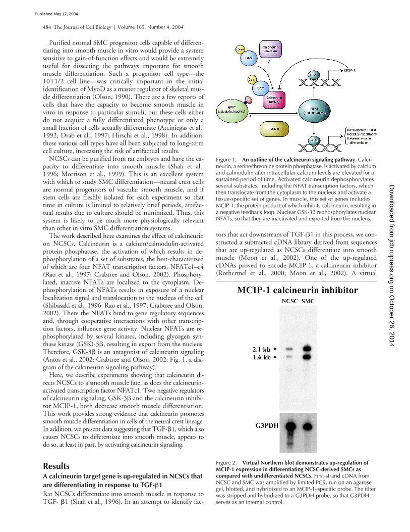
1 in this process, we con-structed a subtracted cDNA library derived from sequencesthat are up-regulated as NCSCs differentiate into smoothmuscle (Moon et al., 2002). One of the up-regulatedcDNAs proved to encode MCIP-1, a calcineurin inhibitor(Rothermel et al., 2000; Moon et al., 2002). A virtual
Figure 1. An outline of the calcineurin signaling pathway. Calci-neurin, a serine/threonine protein phosphatase, is activated by calcium and calmodulin after intracellular calcium levels are elevated for a sustained period of time. Activated calcineurin dephosphorylates several substrates, including the NFAT transcription factors, which then translocate from the cytoplasm to the nucleus and activate a tissue-specific set of genes. In muscle, this set of genes includes MCIP-1, the protein product of which inhibits calcineurin, resulting in a negative feedback loop. Nuclear GSK-3� rephosphorylates nuclear NFATs, so that they are inactivated and exported from the nucleus.
Figure 2. Virtual Northern blot demonstrates up-regulation of MCIP-1 expression in differentiating NCSC-derived SMCs ascompared with undifferentiated NCSCs. First-strand cDNA from NCSC and SMC was amplified by limited PCR, run on an agarose gel, blotted, and hybridized to an MCIP-1–specific probe. The filter was stripped and hybridized to a G3PDH probe, so that G3PDH serves as an internal control.
on October 26, 2014
jcb.rupress.orgD
ownloaded from
Published May 17, 2004

Calcineurin initiates SMC differentiation |
Mann et al. 485
Northern blot confirmed up-regulation of MCIP-1 in neu-ral crest-derived differentiating smooth muscle as comparedwith pluripotent NCSCs (Fig. 2).
Calcineurin activity is chiefly regulated at the posttransla-tional level (Rao et al., 1997). MCIP-1 gene expression is in-duced by activated calcineurin (Yang et al., 2000), and soMCIP-1 imposes negative feedback on calcineurin at highlevels of calcineurin activity (Fig. 1). Therefore, we reasonedthat the increased MCIP-1 mRNA levels in differentiatingSMCs might be a surrogate marker for high levels of cal-cineurin activity in those cells. There is considerable evi-dence that calcineurin has effects on cardiac muscle hyper-trophy, on skeletal muscle fiber type, and on promotingskeletal muscle differentiation (Friday et al., 2000; Rommelet al., 2001; Crabtree and Olson, 2002), so the possibilitythat calcineurin signaling was activated in differentiatingsmooth muscle was of great interest.
Activation of the calcineurin signaling pathway increases the SMC fraction of an NCSC population, and inhibition of the calcineurin signaling pathway decreases the SMC fraction of an NCSC population
To evaluate the effect of calcineurin on the differentiation ofNCSCs into SMCs, we developed a highly quantitative flowcytometric technique for quickly assessing the fraction ofSMCs in a NCSC population. NCSCs were transducedwith the pMIG retroviral vector (Van Parijs et al., 1999),which expresses GFP, and were exposed to 20 pM TGF-
�
1for 4 d to induce SMC differentiation. These cells were fixedand stained with an antibody directed against smooth mus-cle
�
-actin (SMA), a marker for smooth muscle differentia-tion. An increase in SMC differentiation and a decrease intotal cell number was observed for NCSCs treated withTGF-
�
1, as described previously (Shah et al., 1996; Moonet al., 2002). The fraction of cells that differentiated intoNCSCs as measured by high levels of SMA expression wasapproximately three times that of controls (Fig. 3 A).
We used pMIG to express a constitutively active truncatedform of calcineurin in NCSCs. pMIG produces a bicistronictranscript consisting of the inserted sequence as well as se-quences encoding GFP, so that transduced cells can be iden-tified by virtue of GFP expression. 6 d after transduction,the cells were fixed and stained with antibody against SMA.We analyzed the cell population by flow cytometry andfound that the fraction of SMCs was more than twice ashigh in cells transduced with the calcineurin construct ascompared with cells transduced with vector alone (Fig. 3 B).In addition, we noted that increased GFP correlates with in-creased SMA expression in the calcineurin-expressing cells.GFP and calcineurin are encoded on the same transcript,and increases in GFP expression should correlate with in-creased levels of calcineurin expression, as has been demon-strated for other bicistronic transcripts (Collier et al., 1998;Mizuguchi et al., 2000). Therefore, these experimental re-sults suggest that calcineurin promotes smooth muscle dif-ferentiation in a dose-dependent manner (Fig. 3 C).
To determine whether or not inhibitors of calcineurin sig-naling would suppress smooth muscle differentiation, we ex-ploited the fact that NCSCs spontaneously differentiate intosmooth muscle at a low frequency. We found that there was
a small but consistent inhibition of spontaneous SMC dif-ferentiation by MCIP-1 (Fig. 3 B). We also examined the ef-fect of expression of the protein kinase GSK-3
�
on sponta-neous SMC differentiation. GSK-3
�
has diverse targets,including nuclear NFATs, which it phosphorylates and in-activates. Thus, GSK-3
�
serves to oppose calcineurin’seffects (Antos et al., 2002; Crabtree and Olson, 2002). Ex-pression of constitutively active GSK-3
�
in NCSCs con-sistently resulted in a lower rate of spontaneous smoothmuscle differentiation (Fig. 3 B), which is consistent withthe notion that inhibition of the calcineurin pathway in-hibits SMC differentiation. Attempts to use the pharma-cological calcineurin inhibitor cyclosporine were unsuc-cessful because of marked toxicity to NCSCs, even at lowconcentrations.
Calcineurin acts in an instructive manner to initiate smooth muscle differentiation
The flow cytometry data suggest that calcineurin-expressingcells are more likely to be SMCs than control cells, but itdoes not tell us anything about the mechanism of that effect.Specifically, flow cytometry does not allow us to discrimi-nate between a selective effect, by which calcineurin is eitherdetrimental to the growth of non-SMC types or promotesproliferation of SMCs, and an instructive effect, by whichcalcineurin directs progenitor cells to the SMC differentia-tion pathway. To discriminate between these possibilities,we analyzed the calcineurin effect using clonal analysis.
Freshly isolated NCSCs were transduced with retroviralconstructs, and after 24 h the transduced cells were replatedat clonal density. Examination 4 h later confirmed that thereplated cells were almost exclusively single (91% in threeexperiments, SD of 7%). 7 d later, the resulting cultureswere stained for SMA and each colony was characterizedwith respect to its SMC content. Each of these colonies hasarisen from a single cell, and will therefore consist of cellsthat are identical in terms of retroviral transduction—all thecells in a colony are either untransduced or transduced, andif transduced, all cells have the same number and chromo-somal position of retroviral insertions. The resulting relativehomogeneity of phenotype facilitates characterization ofeach colony and also allows a more rigorous analysis of cellfate decisions. Using clonal analysis, the effect of calcineurinwas even more striking than in the flow cytometry experi-ments (Fig. 4). Clonal analysis of calcineurin-expressing col-onies showed a more than eightfold increase in coloniescomposed entirely of smooth muscle as compared withcontrols (Fig. 4 B, M). In addition, expression of calcineu-rin resulted in a more than threefold decrease in the num-ber of colonies that contained no SMCs (Fig. 4 B, U andGU). The ratio of colony number on calcineurin-transducedplates as compared with vector-transduced plates was 1.17,with a SD of 0.04, so there was no evidence for a selective ef-fect that was detrimental to non-SMCs (Fig. 4 B). SMC col-onies on calcineurin plates were similar in size to SMC colo-nies on control plates (unpublished data), so there was noevidence for a selective effect that acts by promoting SMCgrowth. Our interpretation of this result is that calcineurinacts primarily by instructive means and directs NCSCs to-ward a smooth muscle fate.
on October 26, 2014
jcb.rupress.orgD
ownloaded from
Published May 17, 2004

486 The Journal of Cell Biology
|
Volume 165, Number 4, 2004
To characterize the degree of differentiation that cal-cineurin is capable of inducing, we stained clonal cal-cineurin-expressing NCSC colonies with antibodies againstsmooth muscle myosin heavy chain and against calponin,two proteins that are specific for SMCs. Cells in these colo-nies express both of these proteins, demonstrating that cal-cineurin induces a well-differentiated SMC phenotype (Fig.4 C). We did not detect a qualitative difference betweenSMCs that arose spontaneously in control cultures and cal-
cineurin-expressing SMCs; both were reactive with thesetwo SMC-specific antibodies. We conclude from this exper-iment that calcineurin causes NCSCs to differentiate intocells that are smooth muscle as judged by their morphologyand by the expression of multiple smooth muscle-specificproteins.
One intriguing aspect of the flow cytometric profile of cal-cineurin-expressing cells is that a larger fraction of cells areuntransduced as compared with flow profiles of MCIP-1
Figure 3. Calcineurin activation, like TGF-�1 treatment, increases the proportion of smooth muscle in the neural crest cell population. (A) TGF-mediated smooth muscle differentiation as measured by flow cytometry. Approximately 18,000 NCSCs per well isolated from sciatic nerve were transduced with the pMIG retroviral vector, and 2 d later 20 pM TGF-�1 was added to half of the wells. After four additional days, the cells were fixed and stained with a primary antibody directed against SMA and a PE-conjugated secondary antibody, and PE and GFP fluorescence were determined by flow cytometry. The horizontal and vertical lines on each plot denote the upper boundary of GFP-negative and SMA-negative NCSCs, respectively. Cells to the right of the vertical line are GFP-positive and thus successfully transduced, and cells above the horizontal line are SMA-positive and thus are SMCs. The percentage of cells in each quadrant is indicated. Increased SMC content is evident in wells treated with TGF. (B) Calcineurin expression increases the percentage of SMCs in NCSC cultures, and inhibitors of calcineurin activity decrease the percentage of SMCs in NCSC cultures. NCSCs were transduced the day after isolation, and then grown for 6 d. At that point, they were fixed and stained with a primary antibody directed against SMA and a PE-conjugated secondary antibody, and PE and GFP fluorescence were determined by flow cytometry. The horizontal and vertical lines on each plot denote the upper boundary of GFP-negative and SMA-negative NCSCs, respectively. Cells to the right of the vertical line are GFP-positive and thus successfully transduced, and cells above the horizontal line are SMA-positive and thus are SMCs. The percentage of transduced cells that has differentiated into smooth muscle is indicated in the top right of each plot. Note that the NCSCs in the calcineurin plot that express the highest GFP levels, and therefore presumably the highest calcineurin levels, are almost uniformly smooth muscle. Calcineurin/vector ratio of percentage of SMCs from three independent experiments was 2.4, with a SD of 0.06; for MCIP-1, the mean ratio was 0.6, with a SD of 0.26; and for GSK-3�, the ratio was 0.5, with a SD of 0.15. (C) Increased expression of GFP correlates with increased SMC differentiation. Three independent flow cytometric analyses of calcineurin-transduced cells were evaluated for a correlation between actin expression (i.e., SMC identity) and GFP expression. Because GFP and calcineurin are on a bicistronic message, we expect calcineurin expression to increase in tandem with GFP expression. The level of actin expression at each level of intensity of GFP fluorescence is expressed as fold above a pMIG retroviral vector control � SD.
on October 26, 2014
jcb.rupress.orgD
ownloaded from
Published May 17, 2004

Calcineurin initiates SMC differentiation |
Mann et al. 487
and GSK-3
�
(Fig. 3 B). The results of other work with thecalcineurin-expressing retrovirus led us to doubt that thiswas simply due to low rates of transduction. Rather, we sus-pected that this was a result of a slowed mitotic rate in cal-cineurin-expressing stem cells that have begun to differenti-ate, so that the proportion of untransduced, undifferentiatedcells in the population increased disproportionately over theseveral days that the cells were cultured. Clonal analysis con-firmed this finding; the proportion of GFP-positive colonieson calcineurin plates was equal to that of control plates,demonstrating that transduction rates were equal, andsmooth muscle colonies contained many fewer cells than un-differentiated colonies (Fig. 4 A and not depicted), presum-ably because differentiated SMCs divide more slowly thanundifferentiated cells. These data supported our idea thatthe large number of untransduced cells in flow cytometricstudies was due to relative overgrowth of untransduced cellsrather than to poor transduction rates.
The calcineurin-activated transcription factor NFATc1 also causes smooth muscle differentiation
The predominant and best studied mechanism by which cal-cineurin exerts its effects is by dephosphorylating and acti-vating the four calcineurin-regulated NFAT transcriptionfactors. We chose to investigate whether or not calcineu-rin mediates SMC differentiation via an NFAT-dependent
mechanism. We performed RT-PCR experiments to deter-mine which NFAT transcripts are expressed in NCSCs, andfound that NFATc1 and NFATc3 are expressed in thesecells (unpublished data). Therefore, we constructed a retro-viral plasmid with a FLAG-tagged NFATc1 and transducedNCSCs with NFATc1 virus. Fig. 5 shows a photomicro-graph of NCSCs expressing NFATc1, and shows thatNFATc1 promotes SMC differentiation of NCSCs. In thiscase, the virus does not coexpress GFP, so although it is notpossible to identify transduced cells by virtue of GFP expres-sion, as was done for the experiments shown in Fig. 3, anNFATc1-dependent differentiation effect is clearly evident.This case provides strong evidence that calcineurin causesSMC differentiation in an NFAT-mediated manner.
TGF-
�
1 activates transcription of the NFATc1 gene
The fact that TGF-
�
1 activates the expression of MCIP-1,a calcineurin target gene, and the fact that expression of ei-ther calcineurin or NFATc1 phenocopies the TGF-
�
1 effect(i.e., causes smooth muscle differentiation), suggests thatTGF-
�
1 may cause SMC differentiation by activating cal-cineurin signaling. This is a compelling model because it al-lows TGF-
�
1 and calcineurin, two factors known to play arole in SMC differentiation, to be placed in an epistatic rela-tionship in the same differentiation pathway. To further testthis model, we quantitated the expression level of another
Figure 4. Colonies of calcineurin-transduced NCSCs demonstrate a marked increase in SMC differentiation and assume a highly differentiated phenotype. NCSCs were transduced 24 h after isolation and replated at clonal density on the following day. They were allowed to grow for 10 d, and then stained with DAPI and the indicated antibodies. (A) Calcineurin- and pMIG-transduced colonies stained with anti-SMA antibody are shown. They were photographed sequentially, in the order that they were encountered on the plate. (B) Colony characterization of calcineurin- and pMIG-transduced colonies. Colonies were stained with cell type–specific antibodies and characterized with respect to their content of undifferentiated (U), smooth muscle (M), or glial (G) content. Cn, calcineurin; Vec, pMIG. Means of three independent experiments plus SD are shown. (C) Calcineurin- or pMIG-transduced SMCs express the highly specific SMC markers calponin and smooth muscle myosin heavy chain, indicating a highly differentiated pheno-type. Note that although the degree of magnification may appear to be higher in the photographs of calcineurin-transduced cells, magnification of parallel photomi-crographs in A and C is in fact the same. The nuclei of SMCs are larger than those of undifferentiated NCSCs, giving the incorrect impression of different degrees of magnification.
on October 26, 2014
jcb.rupress.orgD
ownloaded from
Published May 17, 2004

488 The Journal of Cell Biology
|
Volume 165, Number 4, 2004
calcineurin/NFAT target gene, NFATc1 itself (Zhou et al.,2002), after TGF-
�
1 treatment. Using quantitative real-time PCR, we found that TGF-
�
1 treatment results in a4.5-fold increase in NFATc1 transcript levels in NCSCs(Fig. 6). This is further evidence that the calcineurin path-way may be activated in TGF-
�
1–treated NCSCs.
Discussion
Using freshly isolated cells of the neural crest lineage as asmooth muscle differentiation system responsive to gain-of-function effects, we have shown that expression of cal-cineurin or of the calcineurin-activated transcription factorNFATc1 causes NCSCs to differentiate into smooth muscleand that expression of MCIP-1 or GSK-3
�
, each of whichantagonizes calcineurin signaling, inhibits SMC differentia-tion in these cells. Clonal analysis indicates that calcineurinexerts its effect on NCSCs by a primarily instructive mecha-nism rather than a selective mechanism. We found in theclonal analysis experiments that it was necessary to wait 24 hto replate transduced NCSCs because of markedly deleteri-ous effects on cell viability and transduction rate when thecells were replated sooner after transduction. This period oftime may allow some selection to take place, but there areseveral reasons why we think that a significant selective effectis unlikely. First, expression of the retroviral constructs isminimal at 24 h after transduction, and overt differentiationin response to factors expressed from transduced retroviralconstructs is not evident until 5–6 d after transduction (un-
published data). Therefore, it seems unlikely that a signifi-cant selective effect could be exerted so early after transduc-tion. In addition, comparison of experimental plates withcontrols shows no evidence of a selective effect. Therefore,we conclude that the effect of calcineurin on NCSCs is pri-marily, and perhaps exclusively, instructive.
The fact that calcineurin is sufficient to cause SMC differ-entiation shows that it is capable of initiating the smoothmuscle differentiation program. This is a conclusion thatcould not have been made from whole animal loss-of-func-tion experiments alone and demonstrates the value of aphysiologically relevant in vitro differentiation system.
The observation that calcineurin plays a role in smoothmuscle differentiation is not entirely surprising because theimportance of calcineurin signaling to cardiac and skeletalmuscle has already been established. Several groups haveshown that calcineurin is capable of causing cardiac hypertro-phy, skeletal muscle fiber type switching, and skeletal muscledifferentiation (Chin et al., 1998; Friday et al., 2000; Kegleyet al., 2001; Crabtree and Olson, 2002). In addition, thephenotypes of mice deficient for calcineurin pathway func-tion are consistent with a role for calcineurin signaling inSMC differentiation (Graef et al., 2001). Mice with a dis-rupted calcineurin regulatory subunit (CnB) gene and micewith two disrupted NFAT genes (NFATc3 and NFATc4)have noticeable abnormalities in vSMC differentiation. Inthese mice, aortic SMCs are in many cases not tightly associ-ated with the vessel wall. In CnB knockout mice, immunore-activity of vSMCs with smooth muscle-specific antibodies is
Figure 5. Expression of NFATc1 in NCSCs causes smooth muscle differentiation. (top) Photomicrographs of NCSCs transduced with MSCV vector-derived or FLAG-NFATc1–expressing retrovirus and stained with FLAG antibody. (bottom) Representative example of flow cytometry of NCSCs transduced with MSCV virus (solid black peak) or NFATc1 virus (open peak with gray outline). Cells were fixed and stained for SMA expression 6 d after transduction. NFATc1/vector ratio of percentage of SMCs from three independent experiments was 2.0, with a SD of 0.1.
on October 26, 2014
jcb.rupress.orgD
ownloaded from
Published May 17, 2004

Calcineurin initiates SMC differentiation |
Mann et al. 489
dramatically decreased. The authors speculate that there is nointrinsic, cell-autonomous defect in SMC differentiation be-cause somitic vessel SMCs look normal, but this findingcould also be explained by differing sensitivities of SMCsfrom different lineages to defects in calcineurin signaling.Vertebrate vascular smooth muscle is derived from multiplecell types. SMCs in the proximal aorta, aortic arch, and pul-monary trunk are derived from neural crest, whereas smoothmuscle in the descending aorta is derived from mesodermand probably also from transdifferentiated endothelium (Git-tenberger-de Groot et al., 1999; Jiang et al., 2000). Smoothmuscle in the coronary arteries is derived from epicardium(Gittenberger-de Groot et al., 1999). SMCs from differentprogenitor cell types have different properties, and thus vas-cular smooth muscle in one part of the animal is differentfrom vascular smooth muscle in another part (Topouzis andMajesky, 1996). Our experiments, which show that cal-cineurin initiates SMC differentiation in purified NCSCs,support a model of smooth muscle differentiation whereinthe calcineurin requirement is cell-autonomous in nature. Atenable alternative interpretation of the whole animal loss-of-function experiments is that the calcineurin requirement iscell-autonomous but that different progenitor cell types havediffering sensitivities to loss of calcineurin signaling.
Loss-of-function mutations in the gene encoding theMEF2C transcription factor also result in poor vSMC dif-ferentiation (Lin et al., 1998). The fact that MEF familymembers are activated by calcineurin in cardiac and skeletalmuscle (Olson and Williams, 2000; Zhang et al., 2002) andare known to interact with NFAT transcription factors attarget promoters constitutes further evidence of a role forcalcineurin in smooth muscle differentiation. It will be im-portant to examine potential relationships between cal-cineurin and MEF2C in smooth muscle differentiation.Similarly, potential relationships between calcineurin andother factors likely to be involved in smooth muscle differ-entiation, like TGF-
�
1, GATA-6, and SRF (Folkman andD’Amore, 1996; Parmacek, 2001), will be of great interest.
The mechanisms of NFAT-mediated calcineurin signalingwere elucidated primarily in immune cells (Rao et al., 1997;Crabtree and Olson, 2002). Interestingly, the actions of cal-cineurin in muscle and in T cells are mediated in strikinglysimilar ways (Crabtree and Olson, 2002). For example, cal-cineurin signaling in lymphocytes seems to require a cooper-ative interaction between NFATs and other transcriptionfactors, like members of the MEF family and AP-1 (Liu etal., 1997; Rao et al., 1997). Calcineurin-mediated skeletalmuscle fiber type switching also seems to act via an associa-tion between NFAT and MEF transcription factors (Calvoet al., 1999; Wu et al., 2000). This finding suggests that themechanisms of NFAT-mediated calcineurin signaling arelikely to be conserved, in general outline and perhaps in sig-nificant detail, in NCSCs.
Activation of the calcineurin signaling pathway in re-sponse to TGF-
�
1 has not been previously described, yetour experiments suggest that it occurs in NCSCs. We seeup-regulation of MCIP-1 and NFATc1, both of which arecalcineurin target genes, in response to TGF-
�
1. In addi-tion, expression of either calcineurin or NFATc1 mimics theeffects of TGF-
�
1 on NCSCs. This suggests an epistatic re-lationship between TGF-
�
1 and the calcineurin pathway insmooth muscle differentiation, a relationship in whichTGF-
�
1 activates calcineurin signaling to initiate the differ-entiation program. The discovery of this potential relation-ship between the TGF-
�
1 and calcineurin pathways high-lights the strengths of the NCSC system as a tool to studySMC differentiation and demonstrates how NCSCs can beused to better define relationships between various factorsknown to be important for SMC differentiation.
TGF-
�
1 might activate calcineurin by any one of a varietyof mechanisms. It has been demonstrated that in some celltypes, TGF-
�
1 is capable of increasing intracellular calciumto levels adequate for calcineurin activation (Alevizopouloset al., 1997). Therefore, direct activation of calcineurin byTGF-
�
1 is one plausible mechanism. Activation of cal-cineurin would lead to dephosphorylation and activation ofNFATs, and then to transcriptional activation of NFAT tar-get genes, including MCIP-1 and NFATc1. Other possiblemechanisms include direct activation of genes encodingmore downstream components of the calcineurin signalingpathway, such as NFATc1, by TGF-
�
1, in a Smad-depen-dent or -independent manner.
The convincing demonstration that activation of the cal-cineurin pathway by TGF-
�
1 is necessary for TGF-
�
1–induced smooth muscle differentiation, and a precise defini-tion of the mechanism by which TGF-
�
1 activates thecalcineurin signaling pathway, would be of great interest.Experiments to accomplish these aims are ongoing in ourlaboratory.
Materials and methods
NCSC isolation
NCSC isolation has been described in detail previously (Morrison et al.,1999; Bixby et al., 2002; Kruger et al., 2002; Moon et al., 2002). In brief,rat E14.5 sciatic nerves or gut (stomach and intestine) were dissected. Dis-aggregated sciatic nerve cells were stained with antibodies to p75 and P0,and cells with high p75 and low P0 were isolated by FACS
®
. Disaggre-gated gut cells were stained with antibodies to p75 and
�
-4, and cells with
Figure 6. NFATc1 transcript levels increase in TGF-treated NCSCs. NCSCs were treated with TGF-�1 for 24–48 h, and then total RNA was isolated and quantitative real-time PCR was performed using NFATc1-specific primers. Both GAPDH and �-actin transcripts were also quantitated, and levels of NFATc1 transcript were normalized to either GAPDH or �-actin. The result was virtually identical using either GAPDH or �-actin to normalize (mean of 4.6, SD of 0.4 or mean of 4.5, SD of 0.5, respectively, three independent experiments).
on October 26, 2014
jcb.rupress.orgD
ownloaded from
Published May 17, 2004

490 The Journal of Cell Biology
|
Volume 165, Number 4, 2004
high p75 and high
�
-4 were isolated by FACS
®
. NCSCs were cultured at alltimes in low oxygen chambers (1% oxygen, 5% CO
2
). Gut and sciaticnerve stem cells responded in an identical manner to transduction withcalcineurin, MCIP-1, and GSK-3
�
.
Generation of a subtracted library specific for differentiating smooth muscle
This generation was described previously (Moon et al., 2002). In brief, sci-atic nerve stem cells were treated with 20 pM TGF-
�
1 for 24 h, and thenmRNA was isolated and cDNA made. This cDNA library was subtractedwith cDNA from pluripotent NCSCs that had not been treated with TGF-
�
1.
Virtual Northern blot of MCIP-1
Virtual Northern blot of MCIP-1 was described previously (Moon et al.,2002). Full-length first-strand cDNA from untreated, pluripotent NCSCs orfrom NCSCs treated with TGF-
�
1 for 24 h was amplified for a limited num-ber of cycles using the Advantage system (CLONTECH Laboratories, Inc.),which should result in proportional amplification. The cDNA was run outon an agarose gel, blotted, and hybridized to an MCIP-1–specific probe us-ing standard procedures. Blots were stripped and reprobed with GAPDHas an internal control.
Cloning of constitutively active calcineurin, NFATc1, GSK-3
�
, and MCIP-1 into the pMIG vector
High fidelity PCR was used to obtain cDNAs encoding MCIP-1, a trun-cated calcineurin coding sequence (1–398 aa), and S9A GSK-3
�
from ratcDNA. The calcineurin and GSK proteins produced by these cDNAs areconstitutively active (O’Keefe et al., 1992; Ohteki et al., 2000). All PCRproducts were sequenced to ensure that no PCR-induced mutations hadoccurred and were cloned into pMIG using standard techniques. A cDNAencoding human NFATc1 was subcloned from a construct provided by G.Crabtree (Stanford University, Stanford, CA).
Production of retrovirus and transduction of NCSCs
The BOSC packaging cell line (Pear et al., 1993; Pear, 1996) was trans-fected with pMIG constructs using standard calcium phosphate tech-niques. 250 ng of the pHCMV-G plasmid (a gift from N. Hopkins, Mas-sachusetts Institute of Technology, Cambridge, MA), which encodesvesicular stomatitis virus glycoprotein, was cotransfected with each pMIGconstruct. Media were changed the next day, and 18 h later the mediawere taken off and used as vesicular stomatitis virus glycoprotein–pseudo-typed viral stock (Hopkins, 1993). NCSCs were transduced by removinghalf the medium in each well and replacing it with viral stock. Polybrenewas also added, to a final concentration of 1
�
g/ml. Cells were incubatedfor 4 h, whereupon the media were replaced.
Flow cytometry of transduced NCSCs
NCSCs that had been transduced with retrovirus were fixed with the Cyto-fix/Cytoperm kit (BD Biosciences). After fixation, cells were stained withan antibody to SMA (anti-SMA; A2547; Sigma-Aldrich) and a phycoeryth-rin (PE)-conjugated secondary antibody (115-116-146; Jackson Immu-noResearch Laboratories). 10,000 cells from each sample were assessedfor GFP and PE fluorescence by a FACScan flow cytometer (Becton Dickin-son). Mock-transduced cells were used as a GFP-negative control, andcells stained with nonspecific isotype-matched IgG (554126; BD Bio-sciences) in place of anti-SMA were used as an actin-negative control.
Clonal analysis
300–400 NCSCs were sorted to each well of a 12-well plate, and weretransduced on the following day. The day after transduction, the cells wereremoved with trypsin and immediately replated. Each well of a 12-wellplate was replated to 6 wells of a 6-well plate. The cells were grown for6–10 d, whereupon they were inspected by fluorescence microscopy andGFP-positive colonies were identified. In most experiments, all or verynearly all colonies were GFP positive, indicating that they had been ef-fectively transduced. After confirming successful transduction, the plateswere stained with anti-SMA, anti-glial fibrillary acidic protein (anti-GFAP;G3893; Sigma-Aldrich), a PE-conjugated antibody reactive with anti-GFAP(Southern Biotechnology Associates 1070–09), a FITC-conjugated anti-body reactive against anti-SMA (1080-02; Southern Biotechnology Associ-ates, Inc.), and DAPI. Colonies were visualized on an epifluorescence mi-croscope (model Diaphot 300; Nikon) and photographed with a 35-mmcamera (model FX-35DX; Nikon), using NPZ 800 ISO film (Fuji), with thecamera set at 1600 ISO. Photographs were scanned into Photoshop using ascanner (model Expression 1680; Epson).
Immunostaining of transduced colonies
Cells were fixed with acid/ethanol (Kruger et al., 2002) and stained withanti-SMA, anti-GFAP, and DAPI as described in the preceding section. Inthe experiment shown in Fig. 4 C, cells were stained with antibody againstsmooth muscle myosin heavy chain (M7786; Sigma-Aldrich) or anticalpo-nin (C2687; Sigma-Aldrich). The same secondary antibody (1070-09;Southern Biotechnology Associates, Inc.) was used for both of these pri-mary antibodies. Colonies were examined, characterized, and photo-graphed under a fluorescence microscope as described for the clonal anal-ysis experiments.
Quantitative real-time PCR
PCR primers that amplified a fragment of 80–160 bp were designed andwere shown to target a unique sequence by using BLAST searches of therat genome. 500–1,500 NCSCs (from gut or sciatic nerve) were sorted toeach well of a 12-well plate and grown for 5–6 d, at which time TGF-
�
1was added to a final concentration of 20 pM. 24–48 h later, total RNA wasisolated using the RNAqueous-Micro kit (Ambion). First-strand cDNA wasmade from total RNA by random priming using the SuperScript First StrandSynthesis System (GIBCO BRL), yielding a final volume of 20
�
l cDNA.Quantitative real-time PCR reactions were performed on a LightCycler(Roche), using FastStart DNA Master SYBR Green I reagents (Roche). A 1-
�
lvolume of cDNA (either undiluted or diluted 1:5 with water) was used ineach quantitative real-time PCR reaction. Cycle number for each reactionwas determined by taking the crossing point of a tangent to the log-linearportion of the curve of cycle number versus fluorescence. Relativeamounts of cDNA (target compared with internal control) were determinedusing an estimated efficiency of 1.8-fold amplification per cycle. Meltingcurve analysis was performed for each reaction to rule out primer dimerformation and to ensure that a unique product was generated.
We thank Kathy Collins, Sean Morrison, Dennis Thiele, Steve Weiss, KevinStruhl, and Deneen Wellik for suggesting important experiments, for criti-cal reading of the manuscript, and for unflagging encouragement. We aregrateful to Anjana Rao and Gerald Crabtree for providing reagents, and wethank Patrick Hogan and T.J. Murphy for thoughtful advice concerningNFAT biology.
The authors have no conflicts of interest related to this work and haveno commercial affiliations. This work was supported by a Scientist Devel-opment Grant awarded to M.R. Benson by the Midwest Affiliate of theAmerican Heart Association.
Submitted: 18 February 2004Accepted: 15 April 2004
References
Alevizopoulos, A., Y. Dusserre, U. Ruegg, and N. Mermod. 1997. Regulation ofthe transforming growth factor beta-responsive transcription factor CTF-1by calcineurin and calcium/calmodulin-dependent protein kinase IV.
J. Biol.Chem.
272:23597–23605.Antos, C.L., T.A. McKinsey, N. Frey, W. Kutschke, J. McAnally, J.M. Shelton,
J.A. Richardson, J.A. Hill, and E.N. Olson. 2002. Activated glycogen syn-thase-3 beta suppresses cardiac hypertrophy in vivo.
Proc. Natl. Acad. Sci.USA.
99:907–912.Arciniegas, E., A.B. Sutton, T.D. Allen, and A.M. Schor. 1992. Transforming
growth factor beta 1 promotes the differentiation of endothelial cells intosmooth muscle-like cells in vitro.
J. Cell Sci.
103:521–529.Bixby, S., G.M. Kruger, J.T. Mosher, N.M. Joseph, and S.J. Morrison. 2002. Cell-
intrinsic differences between stem cells from different regions of the periph-eral nervous system regulate the generation of neural diversity.
Neuron.
35:643–656.
Calvo, S., P. Venepally, J. Cheng, and A. Buonanno. 1999. Fiber-type-specifictranscription of the troponin I slow gene is regulated by multiple elements.
Mol. Cell. Biol.
19:515–525.Carmeliet, P. 2000. Mechanisms of angiogenesis and arteriogenesis.
Nat. Med.6:389–395.
Chang, D.F., N.S. Belaguli, D. Iyer, W.B. Roberts, S.P. Wu, X.R. Dong, J.G.Marx, M.S. Moore, M.C. Beckerle, M.W. Majesky, and R.J. Schwartz.2003. Cysteine-rich LIM-only proteins CRP1 and CRP2 are potent smoothmuscle differentiation cofactors. Dev. Cell. 4:107–118.
Chin, E.R., E.N. Olson, J.A. Richardson, Q. Yang, C. Humphries, J.M. Shelton,H. Wu, W. Zhu, R. Bassel-Duby, and R.S. Williams. 1998. A calcineurin-
on October 26, 2014
jcb.rupress.orgD
ownloaded from
Published May 17, 2004

Calcineurin initiates SMC differentiation | Mann et al. 491
dependent transcriptional pathway controls skeletal muscle fiber type. GenesDev. 12:2499–2509.
Collier, A.J., S. Tang, and R.M. Elliott. 1998. Translation efficiencies of the 5� un-translated region from representatives of the six major genotypes of hepatitisC virus using a novel bicistronic reporter assay system. J. Gen. Virol. 79:2359–2366.
Crabtree, G.R., and E.N. Olson. 2002. NFAT signaling: choreographing the sociallives of cells. Cell. 109(Suppl):S67–S79.
Drab, M., H. Haller, R. Bychkov, B. Erdmann, C. Lindschau, H. Haase, I. Mo-rano, F.C. Luft, and A.M. Wobus. 1997. From totipotent embryonic stemcells to spontaneously contracting smooth muscle cells: a retinoic acid anddb-cAMP in vitro differentiation model. FASEB J. 11:905–915.
Du, K.L., H.S. Ip, J. Li, M. Chen, F. Dandre, W. Yu, M.M. Lu, G.K. Owens, andM.S. Parmacek. 2003. Myocardin is a critical serum response factor cofactorin the transcriptional program regulating smooth muscle cell differentiation.Mol. Cell. Biol. 23:2425–2437.
Folkman, J., and P.A. D’Amore. 1996. Blood vessel formation: what is its molecu-lar basis? Cell. 87:1153–1155.
Friday, B.B., V. Horsley, and G.K. Pavlath. 2000. Calcineurin activity is requiredfor the initiation of skeletal muscle differentiation. J. Cell Biol. 149:657–666.
Gittenberger-de Groot, A.C., M.C. DeRuiter, M. Bergwerff, and R.E. Poelmann.1999. Smooth muscle cell origin and its relation to heterogeneity in develop-ment and disease. Arterioscler. Thromb. Vasc. Biol. 19:1589–1594.
Graef, I.A., F. Chen, L. Chen, A. Kuo, and G.R. Crabtree. 2001. Signals trans-duced by Ca2�/calcineurin and NFATc3/c4 pattern the developing vascula-ture. Cell. 105:863–875.
Hirschi, K., S. Rohovsky, and P. D’Amore. 1998. PDGF, TGF�, and heterotypiccell–cell interactions mediate endothelial cell-induced recruitment of 10T1/2cells and their differentiation to a smooth muscle fate. J. Cell Biol. 141:805–814.
Hopkins, N. 1993. High titers of retrovirus (vesicular stomatitis virus) pseudo-types, at last. Proc. Natl. Acad. Sci. USA. 90:8759–8760.
Jiang, X., D.H. Rowitch, P. Soriano, A.P. McMahon, and H.M. Sucov. 2000. Fateof the mammalian cardiac neural crest. Development. 127:1607–1616.
Kegley, K.M., J. Gephart, G.L. Warren, and G.K. Pavlath. 2001. Altered primarymyogenesis in NFATC3�/� mice leads to decreased muscle size in the adult.Dev. Biol. 232:115–126.
Kruger, G.M., J.T. Mosher, S. Bixby, N. Joseph, T. Iwashita, and S.J. Morrison.2002. Neural crest stem cells persist in the adult gut but undergo changes inself-renewal, neuronal subtype potential, and factor responsiveness. Neuron.35:657–669.
Li, D.Y., L.K. Sorensen, B.S. Brooke, L.D. Urness, E.C. Davis, D.G. Taylor, B.B.Boak, and D.P. Wendel. 1999. Defective angiogenesis in mice lacking en-doglin. Science. 284:1534–1537.
Lin, Q., J. Lu, H. Yanagisawa, R. Webb, G.E. Lyons, J.A. Richardson, and E.N.Olson. 1998. Requirement of the MADS-box transcription factor MEF2Cfor vascular development. Development. 125:4565–4574.
Liu, S., P. Liu, A. Borras, T. Chatila, and S.H. Speck. 1997. Cyclosporin A-sensi-tive induction of the Epstein-Barr virus lytic switch is mediated via a novelpathway involving a MEF2 family member. EMBO J. 16:143–153.
Mano, T., Z. Luo, S.L. Malendowicz, T. Evans, and K. Walsh. 1999. Reversal ofGATA-6 downregulation promotes smooth muscle differentiation and in-hibits intimal hyperplasia in balloon-injured rat carotid artery. Circ. Res. 84:647–654.
Mizuguchi, H., Z. Xu, A. Ishii-Watabe, E. Uchida, and T. Hayakawa. 2000. IRES-dependent second gene expression is significantly lower than cap-dependentfirst gene expression in a bicistronic vector. Mol. Ther. 1:376–382.
Moon, E.S., J.L. Ray, R.L. Leach, and M.R. Benson. 2002. A set of genes activatedin differentiating smooth muscle is also activated in smooth muscle from in-jured arteries or atherosclerotic lesions. Gene Function & Disease. 3:77–86.
Morrisey, E.E., H.S. Ip, M.M. Lu, and M.S. Parmacek. 1996. GATA-6: a zinc fin-ger transcription factor that is expressed in multiple cell lineages derivedfrom lateral mesoderm. Dev. Biol. 177:309–322.
Morrison, S.J., P.M. White, C. Zock, and D.J. Anderson. 1999. Prospective iden-tification, isolation by flow cytometry, and in vivo self-renewal of multipo-tent mammalian neural crest stem cells. Cell. 96:737–749.
Oh, S.P., T. Seki, K.A. Goss, T. Imamura, Y. Yi, P.K. Donahoe, L. Li, K. Miya-zono, P. ten Dijke, S. Kim, and E. Li. 2000. Activin receptor-like kinase 1
modulates transforming growth factor-beta 1 signaling in the regulation ofangiogenesis. Proc. Natl. Acad. Sci. USA. 97:2626–2631.
Ohteki, T., M. Parsons, A. Zakarian, R.G. Jones, L.T. Nguyen, J.R. Woodgett,and P.S. Ohashi. 2000. Negative regulation of T cell proliferation and inter-leukin 2 production by the serine threonine kinase GSK-3. J. Exp. Med. 192:99–104.
O’Keefe, S.J., J. Tamura, R.L. Kincaid, M.J. Tocci, and E.A. O’Neill. 1992. FK-506- and CsA-sensitive activation of the interleukin-2 promoter by cal-cineurin. Nature. 357:692–694.
Olson, E.N. 1990. MyoD family: a paradigm for development? Genes Dev.4:1454–1461.
Olson, E.N., and R.S. Williams. 2000. Remodeling muscles with calcineurin.Bioessays. 22:510–519.
Parmacek, M.S. 2001. Transcriptional programs regulating vascular smooth mus-cle cell development and differentiation. Curr. Top. Dev. Biol. 51:69–89.
Pear, W. 1996. Transient transfection methods for preparation of high-titer retro-viral supernatants. In Current Protocols in Molecular Biology. Vol. 2. F.M.Ausubel, R. Brent, R.E. Kingston, D.D. Moore, J.G. Seidman, J.A. Smith,and K. Struhl, editors. John Wiley & Sons, Inc., New York. 9.11.
Pear, W.S., G.P. Nolan, M.L. Scott, and D. Baltimore. 1993. Production of high-titer helper-free retroviruses by transient transfection. Proc. Natl. Acad. Sci.USA. 90:8392–8396.
Perrella, M.A., M.K. Jain, and M.E. Lee. 1998. Role of TGF-beta in vascular de-velopment and vascular reactivity. Miner. Electrolyte Metab. 24:136–143.
Rao, A., C. Luo, and P.G. Hogan. 1997. Transcription factors of the NFAT fam-ily: regulation and function. Annu. Rev. Immunol. 15:707–747.
Rommel, C., S.C. Bodine, B.A. Clarke, R. Rossman, L. Nunez, T.N. Stitt, G.D.Yancopoulos, and D.J. Glass. 2001. Mediation of IGF-1-induced skeletalmyotube hypertrophy by PI(3)K/Akt/mTOR and PI(3)K/Akt/GSK3 path-ways. Nat. Cell Biol. 3:1009–1013.
Rothermel, B., R.B. Vega, J. Yang, H. Wu, R. Bassel-Duby, and R.S. Williams.2000. A protein encoded within the Down syndrome critical region is en-riched in striated muscles and inhibits calcineurin signaling. J. Biol. Chem.275:8719–8725.
Shah, N.M., A.K. Groves, and D.J. Anderson. 1996. Alternative neural crest cellfates are instructively promoted by TGFbeta superfamily members. Cell. 85:331–343.
Shibasaki, F., E.R. Price, D. Milan, and F. McKeon. 1996. Role of kinases and thephosphatase calcineurin in the nuclear shuttling of transcription factor NF-AT4. Nature. 382:370–373.
Topouzis, S., and M.W. Majesky. 1996. Smooth muscle lineage diversity in thechick embryo. Two types of aortic smooth muscle cell differ in growth andreceptor-mediated transcriptional responses to transforming growth factor-beta. Dev. Biol. 178:430–445.
Van Parijs, L., Y. Refaeli, J.D. Lord, B.H. Nelson, A.K. Abbas, and D. Baltimore.1999. Uncoupling IL-2 signals that regulate T cell proliferation, survival,and Fas-mediated activation-induced cell death. Immunity. 11:281–288.
Wang, D., P.S. Chang, Z. Wang, L. Sutherland, J.A. Richardson, E. Small, P.A.Krieg, and E.N. Olson. 2001. Activation of cardiac gene expression bymyocardin, a transcriptional cofactor for serum response factor. Cell. 105:851–862.
Wu, H., F.J. Naya, T.A. McKinsey, B. Mercer, J.M. Shelton, E.R. Chin, A.R. Si-mard, R.N. Michel, R. Bassel-Duby, E.N. Olson, and R.S. Williams. 2000.MEF2 responds to multiple calcium-regulated signals in the control of skele-tal muscle fiber type. EMBO J. 19:1963–1973.
Yamagishi, H., E.N. Olson, and D. Srivastava. 2000. The basic helix-loop-helixtranscription factor, dHAND, is required for vascular development. J. Clin.Invest. 105:261–270.
Yang, J., B. Rothermel, R.B. Vega, N. Frey, T.A. McKinsey, E.N. Olson, R. Bas-sel-Duby, and R.S. Williams. 2000. Independent signals control expressionof the calcineurin inhibitory proteins MCIP1 and MCIP2 in striated mus-cles. Circ. Res. 87:E61–E68.
Zhang, C.L., T.A. McKinsey, S. Chang, C.L. Antos, J.A. Hill, and E.N. Olson.2002. Class II histone deacetylases act as signal-responsive repressors of car-diac hypertrophy. Cell. 110:479–488.
Zhou, B., R.Q. Cron, B. Wu, A. Genin, Z. Wang, S. Liu, P. Robson, and H.S.Baldwin. 2002. Regulation of the murine Nfatc1 gene by NFATc2. J. Biol.Chem. 277:10704–10711.
on October 26, 2014
jcb.rupress.orgD
ownloaded from
Published May 17, 2004
Copyright © 2022 FDOKUMEN