Membrane Potential-regulated Transcription of the Resting K+ Conductance TASK-3 via the Calcineurin...
10
Michel Lazdunski and Amanda Patel Duprat, Michel Mazzuca, Florian Lesage, Marc Zanzouri, Inger Lauritzen, Fabrice Pathway Conductance TASK-3 via the Calcineurin + Transcription of the Resting K Membrane Potential-regulated and Biogenesis: Membrane Transport, Structure, Function, doi: 10.1074/jbc.M606092200 originally published online July 24, 2006 2006, 281:28910-28918. J. Biol. Chem. 10.1074/jbc.M606092200 Access the most updated version of this article at doi: . JBC Affinity Sites Find articles, minireviews, Reflections and Classics on similar topics on the Alerts: When a correction for this article is posted • When this article is cited • to choose from all of JBC's e-mail alerts Click here Supplemental material: http://www.jbc.org/content/suppl/2006/07/25/M606092200.DC1.html http://www.jbc.org/content/281/39/28910.full.html#ref-list-1 This article cites 55 references, 30 of which can be accessed free at at BIBLIO DE L'UNIV DE NICE on December 31, 2013 http://www.jbc.org/ Downloaded from at BIBLIO DE L'UNIV DE NICE on December 31, 2013 http://www.jbc.org/ Downloaded from
-
Upload
landaverde -
Category
Documents
-
view
2 -
download
0
Transcript of Membrane Potential-regulated Transcription of the Resting K+ Conductance TASK-3 via the Calcineurin...

Michel Lazdunski and Amanda PatelDuprat, Michel Mazzuca, Florian Lesage, Marc Zanzouri, Inger Lauritzen, Fabrice PathwayConductance TASK-3 via the Calcineurin
+Transcription of the Resting KMembrane Potential-regulatedand Biogenesis:Membrane Transport, Structure, Function,
doi: 10.1074/jbc.M606092200 originally published online July 24, 20062006, 281:28910-28918.J. Biol. Chem.
10.1074/jbc.M606092200Access the most updated version of this article at doi:
.JBC Affinity SitesFind articles, minireviews, Reflections and Classics on similar topics on the
Alerts:
When a correction for this article is posted•
When this article is cited•
to choose from all of JBC's e-mail alertsClick here
Supplemental material:
http://www.jbc.org/content/suppl/2006/07/25/M606092200.DC1.html
http://www.jbc.org/content/281/39/28910.full.html#ref-list-1
This article cites 55 references, 30 of which can be accessed free at
at BIB
LIO
DE
L'U
NIV
DE
NIC
E on D
ecember 31, 2013
http://ww
w.jbc.org/
Dow
nloaded from
at BIB
LIO
DE
L'U
NIV
DE
NIC
E on D
ecember 31, 2013
http://ww
w.jbc.org/
Dow
nloaded from
Membrane Potential-regulated Transcription of the RestingK� Conductance TASK-3 via the Calcineurin Pathway*□S
Received for publication, June 26, 2006, and in revised form, July 24, 2006 Published, JBC Papers in Press, July 24, 2006, DOI 10.1074/jbc.M606092200
Marc Zanzouri1, Inger Lauritzen1, Fabrice Duprat, Michel Mazzuca, Florian Lesage, Michel Lazdunski,and Amanda Patel2
From the Institut de Pharmacologie Moleculaire et Cellulaire, Centre National de la Recherche Scientifique, UMR 6097, Universitede Nice-Sophia Antipolis, Valbonne 06560, France
The 2P domain K� channel TASK-3 is highly expressed incerebellar granule neurons where it has been proposed tounderlie the K� leak conductance, IKso. In a previous work weshowed that expression of TASK-3 increases in cerebellar gran-ule neurons as theymature in culture. Here we show that withinthe cerebellum, levels of TASK-3 mRNA increase as granuleneurons migrate to their adult positions and receive excitatorymossy fiber input. To understand the mechanism of thisincrease inTASK-3 expressionwe used an in vitromodel cultur-ing the neurons in either depolarizing conditions mimickingneuronal activity (25K, 25 mM KCl) or in conditions whichapproach deafferentation (5K, 5 mM KCl). An importantincrease in TASK-3 mRNA is uniquely observed in 25K and isspecific since other background K� channel levels remainunchanged or decrease. The rise in TASK-3 mRNA leads to anincrease in TASK-3 protein and the IKso conductance resultingin hyperpolarization. Blocking L-type calcium channels or theirdownstream effector calcineurin, abrogates TASK-3 expressionand IKso, leading to hyperexcitability. This is the first studydemonstrating that depolarization-induced Ca2� entry candirectly regulate cellular excitability by dynamically regulatingthe transcription of a resting K� conductance. The appearanceof this conductancemay play an important role in the transitionof depolarized immature neurons to their mature hyperpolar-ized state during neuronal development.
Cerebellar granule neurons (CGNs)3 are the excitatory glu-tamatergic interneurons which transmit mossy fiber input tothe Purkinje cells (1–3). The standing outward K� current,IKso, plays an important role in controlling CGN excitability
since it behaves as a leak conductance and is inhibited by theactivation of muscarinic acetylcholine receptors (4, 5). Forexample, in cultured rat CGNs the application of muscarineincreases the input resistance and enhances cell excitability byreducing IKso (4). Thismechanismhas also been shown to havea clear physiological role in vivo. In the vestibule cerebellum,inhibition of IKso by acetylcholine released from cholinergicmossy fibers increases the firing frequency of CGNs, which inturn enhances the spontaneous excitatory post-synaptic cur-rents (EPSCs) in Purkinje neurons (6).The molecular components of IKso have been identified as
TASK-1 and TASK-3, members of the tandem pore family ofK� channels (7–11). TASK channels are constitutively activeleak or background K� channels (7–9). They are highly sensi-tive to external pH, such that acidic conditions reduce whilealkaline conditions enhance their activity (7–9). They arereversibly inhibited by a variety of peptide hormones and neu-rotransmitters acting via Gq-coupled receptors (10, 12–15).CGNs express two subtypes of currents corresponding to eitherTASK-3 homomeric or TASK-1/-3 heteromeric channels (4,10, 16–18). These channels are slightly different, in that theyshow distinct sensitivities to external pH, and only homomericTASK-3 channels are blocked by certain agents, such as ruthe-nium red (19, 20) and zinc (21).It is well documented that elevated concentrations of exter-
nal potassium, or other depolarizing conditions increase intra-cellular calcium levels and promote long term survival of CGNsin culture (22–24). It is believed that it mimics the glutamater-gic innervation that CGNs receive in vivo frommossy fibers bypost-natal day 12 (25). Several studies have reported that neu-rotransmitter or ion channel subunit expression depends onthe culture conditions (26–29). For instance, levels of theGABAA receptor �6 subunit, the principle subunit of the cere-bellum, are lower in CGNs when they are cultured in high K�
versus physiological concentrations of K� in the presence ofgrowth factors that inhibit apoptosis (28, 29). In contrast, theGABAA � subunit mRNA increases in high K� cultures and itsin vivo expression coincideswith the formation of synaptic con-tacts of GNs with afferent excitatory mossy fibers within theinternal granule layer (IGL). These findings strongly suggestthat synaptic depolarization plays a role in the expression of thissubunit (28, 29).Mature neonatal cultures of CGNs express particularly high
levels of TASK-3, and most of the currents in 1-week-old cul-tures correspond to TASK-3 homomeric channels (17, 18).
* This work was funded by La Ligue Nationale contre le Cancer (Equipe labe-lise) and Association Pour La Recherche Sur le Cancer (Subvention number3331) and the Institute Paul Hamel and by the CNRS. The costs of publica-tion of this article were defrayed in part by the payment of page charges.This article must therefore be hereby marked “advertisement” in accord-ance with 18 U.S.C. Section 1734 solely to indicate this fact.
□S The on-line version of this article (available at http://www.jbc.org) containssupplemental primer information and Fig. 1.
1 These authors contributed equally to this work.2 To whom correspondence should be addressed: IPMC-CNRS, UMR6097,
660 Route des Lucioles, Sophia Antipolis, 06560 Valbonne, France. Tel.:33-493957730; Fax: 33-493957704; E-mail: [email protected].
3 The abbreviations used are: CGN, cerebellar granule neurons; EGL, externalgranule layer; IGL, internal granule layer; EPSC, excitatory post-synapticcurrent; RR, ruthenium red; MEF-2, myocyte enhancer factor 2; GABA,�-aminobutyric acid; BDNF, brain-derived neurotrophic factor; CNQX,cyano-7-nitroquinoxaline-2,3-dione.
THE JOURNAL OF BIOLOGICAL CHEMISTRY VOL. 281, NO. 39, pp. 28910 –28918, September 29, 2006© 2006 by The American Society for Biochemistry and Molecular Biology, Inc. Printed in the U.S.A.
28910 JOURNAL OF BIOLOGICAL CHEMISTRY VOLUME 281 • NUMBER 39 • SEPTEMBER 29, 2006
at BIB
LIO
DE
L'U
NIV
DE
NIC
E on D
ecember 31, 2013
http://ww
w.jbc.org/
Dow
nloaded from
Indeed, this increased expression of TASK-3 is largely respon-sible for K�-dependent apoptosis observed inCGNs (11, 17). Inthe present work, we took advantage of the CGN cell culturemodel to understand the mechanism of TASK-3 up-regulationand determine that depolarization-induced Ca2� influx isinvolved in the regulation of TASK-3 expression.
EXPERIMENTAL PROCEDURES
Cell Cultures and Treatments—Cerebellar granule neuronswere prepared from 7–8-day-old Wistar rats (Charles RiverFrance) decapitated under anesthesia with isoflurane. Cultureswere prepared as described previously (17). 2 � 106 neuronswere seeded on poly-L-lysine (50 �g/ml)-coated or Biocoat�35-mm dishes (Falcon) and cultured in Eagle’s minimal essen-tial medium (Sigma) supplemented with 10% fetal bovineserum (Invitrogen), 2 mM glutamine, BDNF (50 ng/ml), and0.5% penicillin-streptomycin at 37 °C in a humidified atmo-sphere containing 5% CO2. To prevent growth of glial cells,cytosine arabinoside (Ara-C) (10 �M) was added to the cultures1 day following plating. Where indicated 20 mM KCl (25Kmedium) or the same volume of 20 mM NaCl as control (5Kmedium) were added 3 h following plating. Nifedipine (10 �M),FK506 (2.5 �g/ml), cyclosporin A (1 �M), or KN93 (5 �M), allpurchased from Sigma, were added 3 h after plating. Actino-mycin D (10 �g/ml) and cycloheximide (10 �g/ml) wereadded 1–2 days after plating. Corresponding solvents wereadded in the control conditions.
Hippocampal and cortical neu-rons were prepared from 18–19-day-old fetal Wistar rats (CharlesRiver France). 2� 106 neurons wereseeded on poly-D-lysine-coatedplates and cultured in Eagle’s mini-mal essential medium supple-mented with 10% horse serum(Sigma), 2 mM glutamine, and 0.5%penicillin-streptomycin at 37 °C in ahumidified environment containing5% CO2. To prevent growth of glialcells, cytosine arabinoside (Ara-C)(10 �M) was added to the cultures 1day following plating. Where indi-cated 20 mM KCl (25K) or 20 mMNaCl (5K) were added 3 h followingplating. As indicated, nifedipine (10�M)was added 3 h following plating.COS-7 cells were cultured inDul-
becco’s modified Eagle’s medium(Invitrogen) supplemented with10% fetal bovine serum and 0.5%penicillin-streptomycin at 37 °C in ahumidified environment containing5%CO2. Cells were transfected withrat TASK-3 or rat TASK-1 cDNAsusing the DEAE-dextran method.These cells were used for eitherWestern blot analysis orimmunocytochemistry.
Total RNA Extraction, Reverse Transcription, and Quantita-tive PCR—Total RNAs from hippocampal, cortical, and cere-bellar granule neurons cultured in 35-mm dishes wereextracted with the Nucleospin RNA II Kit (Macherey-Nagel).Reverse transcription was performed with 2 �g of total RNAusing Superscript II reverse transcriptase (Invitrogen). Real-time PCR assays for each gene target were performed on cDNAsamples using SYBR Green Mastermix Plus (Eurogentec) in96-well optical plates on anABI Prism 7700 sequence detectionsystem (PE Biosystems). PCR data were captured usingSequence Detector Software. The data were analyzed using thecomparative CTmethod where the amount of target is normal-ized to an endogenous reference (cyclophylin D) (User BulletinNumber 2, Applied Biosystems). Experiments were performedin triplicate. Primer sequences, which were used for quantita-tive PCR, are provided in the supplemental information. Stand-ard curves were generated for each set of primers using serialdilutions of rat brain cDNA to ensure a similar efficiency ofamplification.In Situ Hybridization—For in situ hybridization, post-natal
day 6, 12, and 30 (P6, P12, and P30) Wistar rats were anesthe-tized and decapitated. Brains were removed and frozen at�40 °C. Horizontal and coronal brain sections (12 �m) werecut in a cryostat (Leica), thaw-mounted onto charged slides(Superfrost Plus), and immediately fixed for 5 min in 4% phos-phate-buffered paraformaldehyde, pH 7.4. Prehybridizationand hybridization were performed as described previously (30,
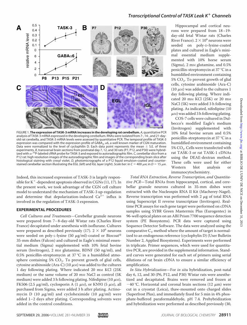
FIGURE 1. The expression of TASK-3 mRNA increases in the developing rat cerebellum. A, quantitative PCRanalysis of TASK-3 mRNA expressed in the developing cerebellum. RNAs were isolated from 7-, 14-, and 21-day-old rat cerebella, and TASK-3 mRNA levels were assessed by quantitative PCR. The temporal profile of TASK-3expression was compared with the expression profile of GABAA �6, a well known marker of CGN maturation.Data were normalized to the level of cyclophilin D. Each data point represents the mean � S.E. of threeexperiments. B, transversal brain sections from postnatal day 7, 12, and 30 rats (P7, P12, and P30) were hybrid-ized with �-33P-labeled cRNA probe for TASK-3 and exposed to autoradiographic film. C, cerebellar slice from aP12 rat: high resolution images of the autoradiographic film and images of the corresponding brain slice afterhistological staining with cresyl violet. D, photomicrographs of a P12 liquid emulsion-coated and counter-stained cerebellar section illustrating the EGL (left) and IGL layer (right). Scale bar: in C � 400 �M; in D � 15 �M.
Transcriptional Control of TASK Leak K� Channels
SEPTEMBER 29, 2006 • VOLUME 281 • NUMBER 39 JOURNAL OF BIOLOGICAL CHEMISTRY 28911
at BIB
LIO
DE
L'U
NIV
DE
NIC
E on D
ecember 31, 2013
http://ww
w.jbc.org/
Dow
nloaded from
31) using identical �-33P-labeled sense and antisense ribo-probes. After hybridization, slides were washed and exposed tofilm (BioMax, Eastman Kodak Co.) for about 6–7 days. Theresulting autoradiograms were imaged using a film scanner at3000 dpi resolution and processed in Adobe Photoshop.Selected slides were then covered with photographic emulsion(Hypercoat RPN40, Amersham Biosciences) and left to exposeat 4 °C for 5 weeks. After development, slides were stained withcresyl violet.Preparation and Characterization of a Polyclonal Rabbit
TASK-3Antibody—Anti-TASK-3antibodieswere raised against agluthatione S-transferase fusion protein containing the carboxyl-terminal 55 amino acids ofmouseTASK-3 (fromThr348 to Ile402,GenBankTM accession number NP001029048). The prepara-tion of fusion proteins and rabbit immunization were per-formed as described previously (32).Immunocytochemistry—Cerebellar granule neurons or COS
transfected cells were fixed in 4% phosphate-bufferedparaformaldehyde for 20 min, treated with 50 mM NH4Cl, andpermeabilized in 0.1% Triton X-100 for 5 min. After blockingfor 1 h in phosphate-buffered saline containing 5% goat serumand0.1%Tween 20, cellswere incubatedwithTASK-3 antibody(1:500 to 1:1000) overnight at 4 °C. After washes, cells wereincubated in goat anti-rabbit Alexa Fluor� 488 (1/800) for 1 h atroom temperature. Cells immunoreactive for TASK-3 werevisualized using an epifluorescence microscope (Axioplan 2,Carl Zeiss) equipped with 25� and 63� oil immersion objec-tives. Images were recorded with a cooled CCD camera (Cool-snap HQ, Photometrics) driven by Metavue software.Electrophysiological Recordings—Currents were recorded in
the perforated patch configuration or in the standardwhole cellconfiguration. The external solution contained (in mM) 120NaCl, 5 KCl, 2 MgCl2, 0.5 CaCl2, 5 glucose, 10 HEPES, pH 7.4with NaOH. In the depolarizing solution, 20 mM of NaCl wasreplaced with KCl. The perforated patch pipette solution con-tained 240 �g/ml amphotericin B, and (in mM) 125 KCl, 5MgCl2, 0.1 EGTA, and 5 HEPES, pH 7.2 with KOH. The wholecell patch pipette solution contained (inmM) 140 KCl, 2MgCl2,2 K-ATP, 2.7 CaCl2, 5 EGTA, and 10 mM HEPES, pH 7.4 withKOH. Cells were continuously superfused with a microperfu-sion system during the time course of the experiments (0.1ml/min). Experiments were performed at room temperature.Acidic pH of the external solution was obtained with HCl.CNQX stock solution (20 mM, distilled water) were stored at�20 °C and used on the day of experiment. Zinc and rutheniumred (Sigma) were directly dissolved in the external medium.The synaptic activity was analyzed using Mini-analysis pro-gram (Synaptosoft, Fort Lee, NJ), the threshold for detectionwas 15 pA.
RESULTS
During cerebellar development, CGNs initially localizedin the external granule layer (EGL) migrate across the molec-ular layer to reach the IGL. Within the IGL, GNs synapsewith afferent glutamatergic mossy fibers (1–3).We have pre-viously shown that the level of TASK-3 mRNA increases as afunction of cerebellar development (17). In this work, weshow that the highest increase in TASK-3 mRNA occurs in
the second week after birth. The temporal expression profileof TASK3 mRNA parallels that of the GABAA �6 receptorsubunit mRNA, a well known marker of mature CGNs (33)
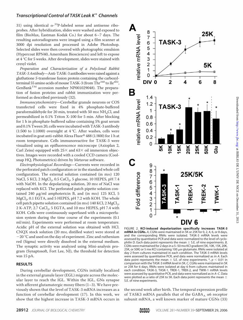
FIGURE 2. KCl-induced depolarization specifically increases TASK-3mRNA in CGNs. A, CGNs were maintained in 5K or 25K for 0, 2, 4, 6, or 8 days,and the corresponding RNAs were isolated. TASK-3 mRNA levels wereassessed by quantitative PCR and data were normalized to the level of cyclo-philin D. Each data point represents the mean � S.E. of nine experiments. B,CGNs were maintained for 2 days in a 5–50 mM KCl gradient (5K, 10K, 15K, 20K,25K, or 50K) or 5 mM KCl containing 100 �M glutamate. RNAs were isolated atday 2 from cultures maintained in each condition. The TASK-3 mRNA levelswere assessed by quantitative PCR, and data were normalized as in A. Eachdata point represents the mean � S.E. of nine experiments. *, p � 0.01 incomparison with the TASK-3 mRNA level in 5K. C, CGNs were maintained in 5Kor 25K for 6 days. RNAs were isolated at day 6 from cultures maintained ineach condition. TASK-3, TASK-1, TREK-1, TREK-2, and TWIK-1 mRNA levelswere assessed by quantitative PCR, and data were normalized as in A–C. Datawere plotted as a ratio of 25K to 5K. Each data point represents the mean �S.E. of nine experiments.
Transcriptional Control of TASK Leak K� Channels
28912 JOURNAL OF BIOLOGICAL CHEMISTRY VOLUME 281 • NUMBER 39 • SEPTEMBER 29, 2006
at BIB
LIO
DE
L'U
NIV
DE
NIC
E on D
ecember 31, 2013
http://ww
w.jbc.org/
Dow
nloaded from
(Fig. 1A). In situ hybridization analysis of rat brain slices atdifferent developmental stages using a TASK-3 specific anti-sense riboprobe was used to study TASK-3 expression indetail. Little or no labeling was observed with the corre-sponding sense probe (data not shown). At postnatal day 7(P7), before the migration of CGNs, a weak expression ofTASK-3 was observed in the EGL (Fig. 1B). At postnatal day12 (P12), neurons that had reached the IGL were stronglylabeled compared with granule neurons remaining in the EGL(Fig. 1, B–D). In adult (P30), the hybridization signal was slightlyincreased compared with the signal in the IGL at P12. As previ-ously described, TASK-3mRNA is strongly expressed in the adultcerebellum but is also abundant in many other brain structuressuch as the cerebral cortex, hippocampus, striatum, and brainstem (30). Taken together, these findings show that theincrease of TASK-3 mRNA levels in granule neurons coincideswith their arrival in the IGL and suggest that depolarization due
to synaptic activity may contributeto the increase in TASK-3 mRNAexpression.To test the hypothesis that neuro-
nal depolarization plays a role inincreasing TASK-3 expression, cul-tured CGNs were grown in amedium containing either a physio-logical (5 mM, 5Kmedium) or depo-larizing (25 mM, 25K medium)concentrations of KCl. Granuleneurons cultivated in 5K die by apo-ptosis during the first days in cul-ture. To prevent apoptosis, BDNFwas added to the culture medium.BDNF by itself did not modifyTASK-3mRNAexpression in either5K or 25K (data not shown). Asshown in Fig. 2A, TASK-3 mRNAexpression increases significantly indepolarizing versus physiologicalconditions depending on the num-ber of days in culture (Fig. 2A). TheTASK-3 mRNA levels increase as afunction of external potassium con-centration withmaximal levels at 20mM KCl (Fig. 2B). Since K�-dependent depolarization isthought to mimic glutamatergicexcitatory mossy fiber stimulationin vivo (25), we also examined theeffect of glutamate on TASK-3expression. Treating the cells with100 �M glutamate for 48 h, similarlyincreased TASK-3 mRNA levels tothose obtained with exposure to 25mM KCl (Fig. 2B). Given that otherbackground K� channels are alsoexpressed in the cerebellum, wetested whether their expression wasalso regulated by KCl-induced
depolarization. Among the two pore K� channels tested, onlythe mRNA for TASK-3 is up-regulated, while the others areeither down-regulated or remain unchanged (Fig. 2C). Interest-ingly, TASK-1, which can associate with TASK-3 to form het-eromeric channels (17 ,19, 20), was found to be significantlydown-regulated by KCl.To follow whether the rise in TASK-3 mRNA is reflected by
an increase in TASK-3 protein, we generated an antibodyagainst the carboxyl terminus of mTASK-3. Anti-TASK-3 spe-cifically detects a duplex in lysates from COS cells transfectedwith rTASK-3, while no bands were detected in the controlconditions (supplemental Fig. 1A). The antibody was alsocharacterized using immunocytochemistry on COS cellstransfected with the rTASK-3 subunit. Only cells expressingrTASK-3 were labeled, while cells transfected with therTASK-1 subunit or the vector alonewere not fluorescent (sup-plemental Fig. 1B). Immunocytochemistry was also performed
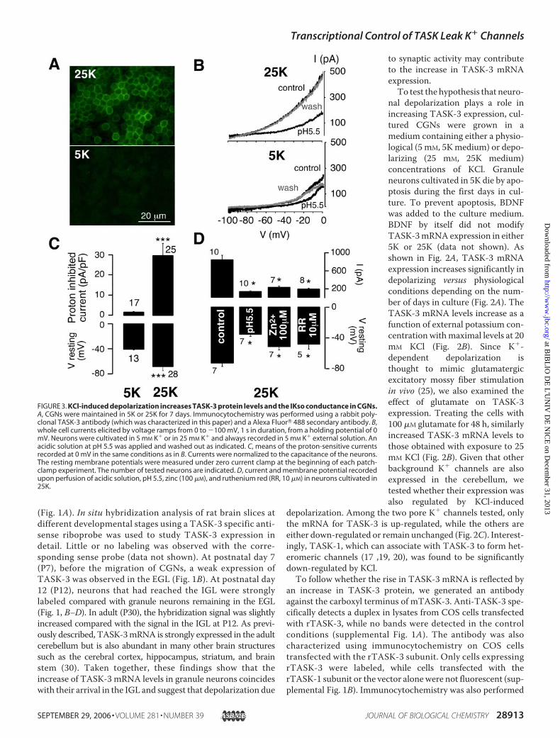
FIGURE 3. KCl-induced depolarization increases TASK-3 protein levels and the IKso conductance in CGNs.A, CGNs were maintained in 5K or 25K for 7 days. Immunocytochemistry was performed using a rabbit poly-clonal TASK-3 antibody (which was characterized in this paper) and a Alexa Fluor� 488 secondary antibody. B,whole cell currents elicited by voltage ramps from 0 to �100 mV, 1 s in duration, from a holding potential of 0mV. Neurons were cultivated in 5 mM K� or in 25 mM K� and always recorded in 5 mM K� external solution. Anacidic solution at pH 5.5 was applied and washed out as indicated. C, means of the proton-sensitive currentsrecorded at 0 mV in the same conditions as in B. Currents were normalized to the capacitance of the neurons.The resting membrane potentials were measured under zero current clamp at the beginning of each patch-clamp experiment. The number of tested neurons are indicated. D, current and membrane potential recordedupon perfusion of acidic solution, pH 5.5, zinc (100 �M), and ruthenium red (RR, 10 �M) in neurons cultivated in25K.
Transcriptional Control of TASK Leak K� Channels
SEPTEMBER 29, 2006 • VOLUME 281 • NUMBER 39 JOURNAL OF BIOLOGICAL CHEMISTRY 28913
at BIB
LIO
DE
L'U
NIV
DE
NIC
E on D
ecember 31, 2013
http://ww
w.jbc.org/
Dow
nloaded from
on CGNs cultured in either 5K or 25K for 7 days. A specificmembrane labeling was observed for TASK-3 only under depo-larizing conditions (Fig. 3A) demonstrating that the increase inTASK-3 mRNA leads to a concomitant increase in protein.We and others (4, 17, 18) have previously shown that the
TASK channels underlie IKso in CGNs cultured in 25K. Adetailed electrophysiological comparison between granule neu-rons cultured in 5K versus 25K was performed. Depolarizingmedium significantly increases the amplitude of the pH-sensi-tive IKso component (29.3 � 6.0 pA, n � 25, p � 0.0005) ascompared with the current observed in neurons cultured in 5K(1.6� 0.3 pA,n� 17, p� 0.0005) for a period of 6–10 days (Fig.3B/C). Conditions that increase IKso current lead tomembranehyperpolarization (Fig. 3C). The resting membrane potentialmeasured in physiological K� concentration was �40.9 � 3.4mV (n� 13) as compared with�68.4� 2.9mV in depolarizingconditions (n � 28, p � 10�5).
We used a pharmacological approach to distinguish theTASK-3 homomer from TASK-1 homomeric and TASK-1/-3heteromeric channels. Zinc (100 �M) and ruthenium red (10�M) are known to selectively inhibit TASK-3 homomers (19, 20,21). Zn2� (100 �M) and ruthenium red (10 �M) reduced thecurrent to 25.7 � 3.4% (n � 7, p � 0.001) and 23.1 � 2.6% (n �8, p � 0.0006), respectively, which is the same degree of inhibi-tion observed with acidosis (pH 5.5), which was 16.7 � 2.3%(n � 10, p � 10�4) (Fig. 3D). The application of Zn2� andruthenium red leads to membrane depolarization (25K:�73.6� 4.2mV,n� 7; 25K, pH5.5:�35.9� 2.5mV,n� 7,p�10�6; 25K � Zn2�: �51.9 � 4.3 mV, n � 7, p � 0.0002; 25K �RR: �48.8 � 6.2 mV, n � 5, p � 0.0005). These results confirmthat the TASK-3 homomeric channel is the principle compo-nent of IKso recorded in 25K. The difference observed betweencurrent inhibition obtainedwith external acidosis and that withZn2�/RR could be due to TASK-1 homomers and/or TASK-1/-3 heteromers, since both channels are resistant to inhibitionby Zn2� and RR. Indeed, PCR quantification experimentsreveal that TASK-3 mRNA levels strongly increase in 25K,while levels of TASK-1 mRNAs decline, which might explainthe low levels of TASK-1/-3 heteromeric channels and/orTASK-1 homomeric channels under such culture conditions.Potassium depolarization of granule neurons leads to the
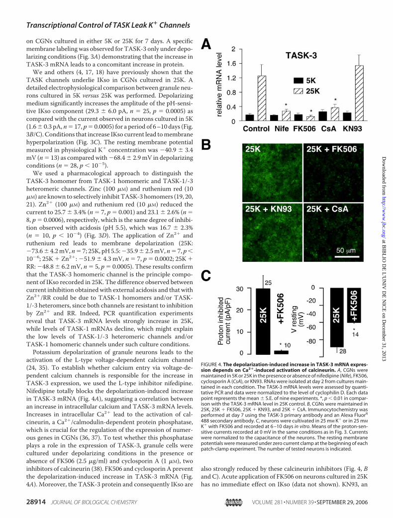
activation of the L-type voltage-dependent calcium channel(24, 35). To establish whether calcium entry via voltage-de-pendent calcium channels is responsible for the increase inTASK-3 expression, we used the L-type inhibitor nifedipine.Nifedipine totally blocks the depolarization-induced increasein TASK-3 mRNA (Fig. 4A), suggesting a correlation betweenan increase in intracellular calcium and TASK-3 mRNA levels.Increases in intracellular Ca2� lead to the activation of cal-cineurin, a Ca2�/calmodulin-dependent protein phosphatase,which is crucial for the regulation of the expression of numer-ous genes in CGNs (36, 37). To test whether this phosphataseplays a role in the expression of TASK-3, granule cells werecultured under depolarizing conditions in the presence orabsence of FK506 (2.5 �g/ml) and cyclosporin A (1 �M), twoinhibitors of calcineurin (38). FK506 and cyclosporin A preventthe depolarization-induced increase in TASK-3 mRNA (Fig.4A). Moreover, the TASK-3 protein and consequently IKso are
also strongly reduced by these calcineurin inhibitors (Fig. 4, Band C). Acute application of FK506 on neurons cultured in 25Khas no immediate effect on IKso (data not shown). KN93, an
FIGURE 4. The depolarization-induced increase in TASK-3 mRNA expres-sion depends on Ca2�-induced activation of calcineurin. A, CGNs weremaintained in 5K or 25K in the presence or absence of nifedipine (Nife), FK506,cyclosporin A (CsA), or KN93. RNAs were isolated at day 2 from cultures main-tained in each condition. The TASK-3 mRNA levels were assessed by quanti-tative PCR and data were normalized to the level of cyclophilin D. Each datapoint represents the mean � S.E. of nine experiments. *, p � 0.01 in compar-ison with the TASK-3 mRNA level in 25K control. B, CGNs were maintained in25K, 25K � FK506, 25K � KN93, and 25K � CsA. Immunocytochemistry wasperformed at day 7 using the TASK-3 primary antibody and an Alexa FluorR
488 secondary antibody. C, neurons were cultivated in 25 mM K� or in 25 mM
K� with FK506 and recorded at 6 –10 days in vitro. Means of the proton-sen-sitive currents recorded at 0 mV in the same conditions as in Fig. 3. Currentswere normalized to the capacitance of the neurons. The resting membranepotentials were measured under zero current clamp at the beginning of eachpatch-clamp experiment. The number of tested neurons is indicated.
Transcriptional Control of TASK Leak K� Channels
28914 JOURNAL OF BIOLOGICAL CHEMISTRY VOLUME 281 • NUMBER 39 • SEPTEMBER 29, 2006
at BIB
LIO
DE
L'U
NIV
DE
NIC
E on D
ecember 31, 2013
http://ww
w.jbc.org/
Dow
nloaded from
inhibitor of CaM kinases, which are also downstream effectorsof Ca2� signaling (39), was without effect on either TASK-3mRNA or protein in 25K (Fig. 4, A and B). Furthermore, cellsgrown in 25K with FK506 display depolarized resting mem-brane potentials with values close to those recorded in cellsgrown in 5K conditions (Fig. 4C, right panel) indicating thatFK506 is acting at the level of TASK-3 expression to decreaseIKso.Ca2�-dependentTASK-3 expression could occur at the tran-
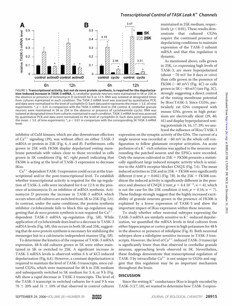
scriptional and/or the post-transcriptional level. To establishwhether transcriptional activity is required for the up-regula-tion of TASK-3, cells were incubated for 6 or 12 h in the pres-ence of actinomycin D, an inhibitor of mRNA synthesis. Acti-nomycin D prevents the increase in TASK-3 mRNA whichoccurswhen cell cultures are switched from5K to 25K (Fig. 5A).In contrast, under the same conditions, the protein synthesisinhibitor cycloheximide fails to block this up-regulation sug-gesting that de novo protein synthesis is not required for Ca2�-dependent TASK-3 mRNA up-regulation (Fig. 5B). Whileapplication of cycloheximide does lead to a decrease in TASK-3mRNA levels (Fig. 5B), this occurs in both 5K and 25K, suggest-ing that de novo protein synthesis is necessary for stabilizing themessenger but in a calcineurin-independent manner (Fig. 5B).To determine the kinetics of the response of TASK-3mRNA
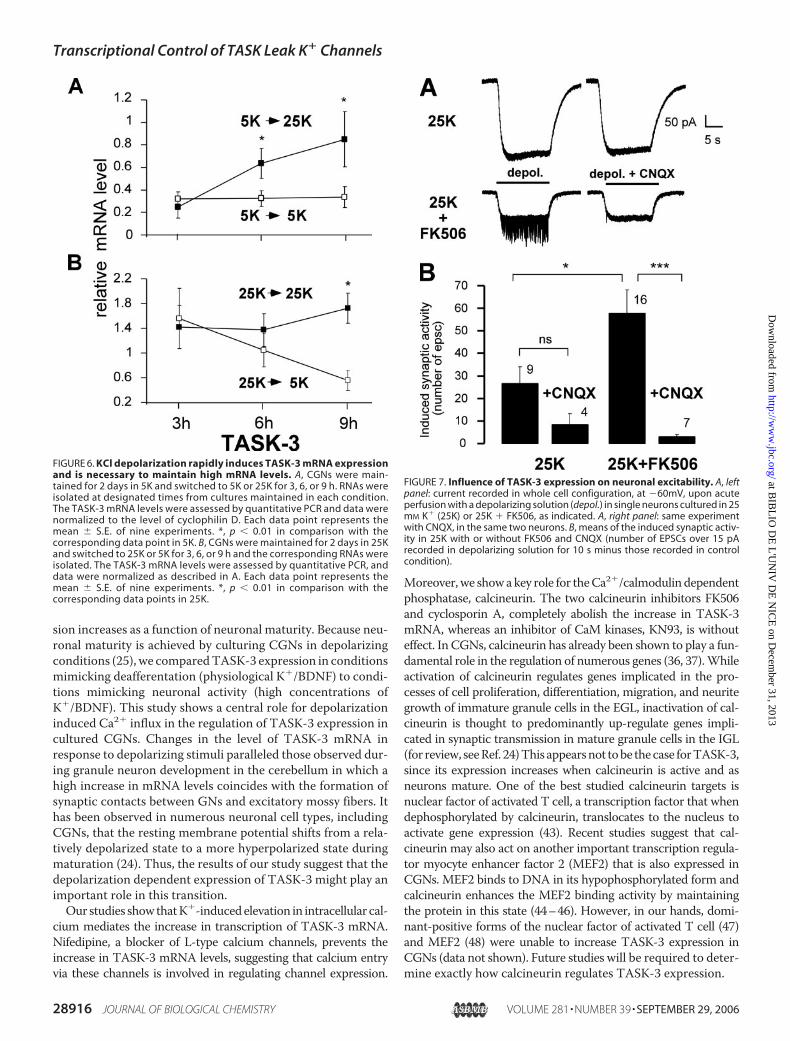
expression, 48-h-old cultures grown in 5K were either main-tained in 5K or switched to 25K. A significant increase inTASK-3 mRNA levels is observed within 6 h of KCl-induceddepolarization (Fig. 6A). However, a constant depolarization isrequired tomaintain the level of TASK-3 transcripts, since cul-tured CGNs, which were maintained for 48 h in 25K mediumand subsequently switched to 5K medium for 3, 6, or 9 h (Fig.6B) show a rapid decrease in TASK-3 transcripts. The level ofthe TASK-3 transcript in switched cultures for 6 and 9 h was76 � 20% and 31 � 10% of that observed in control cultures
maintained in 25Kmedium, respec-tively (p� 0.01). These results dem-onstrate that cultured CGNsrequire the continued presence ofdepolarizing conditions to maintainexpression of the TASK-3 subunitmRNA and that this regulation isdynamic.As mentioned above, cells grown
in 25K, i.e. expressing high levels ofTASK-3, are more hyperpolarized(about �70 mV for 8 days in vitro)than cells grown in the presence ofFK506 (�40 mV) (Fig. 4C) or cellsgrown in 5K (�40mV) (see Fig. 3C),strongly suggesting a direct controlof the resting membrane potentialby IKso/TASK-3. Since CGNs, par-ticularly rat GNs compared withmouse GNs, grown in high potas-sium are electrically silent (29, 40,41) and display hyperpolarized rest-ing potentials (4, 16, 17, 29), we ana-lyzed the influence of IKso/TASK-3
expression on the synaptic activity of the GNs. The current of asingle neuron was recorded at �60 mV in the whole cell con-figuration to follow glutamate receptor activation. An acuteperfusion of a K�-rich solution was applied to the neurons sur-rounding the patched neuron and the EPSCs were recorded.Only the neuron cultivated in 25K � FK506 presents a statisti-cally significant large induced synaptic activity which is sensi-tive to the AMPA receptor blocker CNQX (Fig. 7A). The meaninduced activities in 25K and in 25K� FK506were significantlydifferent (t test: p � 0.041) (Fig. 7B). In the 25K � FK506 con-dition the induced activity is significantly different in the pres-ence and absence of CNQX (t test; p � 4.4 10�4, n � 4), whichis not the case for the 25K condition (t test; p � 0.16, n � 7).These findings strongly suggest that the highly increased excit-ability of granule neurons grown in the presence of FK506 isexplained by a lower expression of TASK-3 and show theimportant impact of IKso expression within these neurons.To study whether other neuronal subtypes expressing the
TASK-3 mRNA are similarly sensitive to K�-induced depolar-ization, we quantified the mRNA levels in fetal cultures fromeither hippocampus or cortex grown in high potassium for 48 hin the absence or presence of nifedipine (Fig. 8). Both neuronalsubtypes show a nifedipine-sensitive increase in TASK-3 tran-scripts. However, the level of Ca2�-induced TASK-3 transcriptis significantly lower than that observed in cerebellar granuleneurons, approaching levels observed in 5K. Nevertheless,these findings demonstrate that transcriptional regulation ofTASK-3 by intracellular Ca2� is not unique to CGNs and sug-gest that this regulation may be an important mechanismthroughout the brain.
DISCUSSION
Since the resting K� conductance IKso is largely encoded byTASK-3 (17, 18), we wanted to determine howTASK-3 expres-
FIGURE 5. Transcriptional activity, but not de novo protein synthesis, is required for the depolariza-tion-induced increase in TASK-3 mRNA. A, cerebellar granule neurons were maintained in 5K or 25K inthe absence or presence of Actinomycin D (actinoD) for 6 or 12 h. RNA was isolated at designated timesfrom cultures maintained in each condition. The TASK-3 mRNA level was assessed by quantitative PCR,and data were normalized to the level of cyclophilin D. Each data point represents the mean � S.E. of nineexperiments. *, p � 0.01 in comparison with the TASK-3 mRNA level in 25K control. B, cerebellar granuleneurons were maintained in 5K or 25K in the absence or presence of cycloheximide (cyclo). RNA wasisolated at designated times from cultures maintained in each condition. TASK-3 mRNA level was assessedby quantitative PCR and data were normalized to the level of cyclophilin D. Each data point representsthe mean � S.E. of nine experiments. *, p � 0.01 in comparison with the corresponding 5K TASK-3 mRNAlevel.
Transcriptional Control of TASK Leak K� Channels
SEPTEMBER 29, 2006 • VOLUME 281 • NUMBER 39 JOURNAL OF BIOLOGICAL CHEMISTRY 28915
at BIB
LIO
DE
L'U
NIV
DE
NIC
E on D
ecember 31, 2013
http://ww
w.jbc.org/
Dow
nloaded from
sion increases as a function of neuronal maturity. Because neu-ronal maturity is achieved by culturing CGNs in depolarizingconditions (25), we comparedTASK-3 expression in conditionsmimicking deafferentation (physiological K�/BDNF) to condi-tions mimicking neuronal activity (high concentrations ofK�/BDNF). This study shows a central role for depolarizationinduced Ca2� influx in the regulation of TASK-3 expression incultured CGNs. Changes in the level of TASK-3 mRNA inresponse to depolarizing stimuli paralleled those observed dur-ing granule neuron development in the cerebellum in which ahigh increase in mRNA levels coincides with the formation ofsynaptic contacts between GNs and excitatory mossy fibers. Ithas been observed in numerous neuronal cell types, includingCGNs, that the resting membrane potential shifts from a rela-tively depolarized state to a more hyperpolarized state duringmaturation (24). Thus, the results of our study suggest that thedepolarization dependent expression of TASK-3 might play animportant role in this transition.Our studies showthatK�-inducedelevation in intracellular cal-
cium mediates the increase in transcription of TASK-3 mRNA.Nifedipine, a blocker of L-type calcium channels, prevents theincrease in TASK-3 mRNA levels, suggesting that calcium entryvia these channels is involved in regulating channel expression.
Moreover,we showakey role for theCa2�/calmodulindependentphosphatase, calcineurin. The two calcineurin inhibitors FK506and cyclosporin A, completely abolish the increase in TASK-3mRNA, whereas an inhibitor of CaM kinases, KN93, is withouteffect. In CGNs, calcineurin has already been shown to play a fun-damental role in the regulation of numerous genes (36, 37).Whileactivation of calcineurin regulates genes implicated in the pro-cesses of cell proliferation, differentiation, migration, and neuritegrowth of immature granule cells in the EGL, inactivation of cal-cineurin is thought to predominantly up-regulate genes impli-cated in synaptic transmission in mature granule cells in the IGL(for review, seeRef. 24)This appearsnot tobe thecase forTASK-3,since its expression increases when calcineurin is active and asneurons mature. One of the best studied calcineurin targets isnuclear factor of activated T cell, a transcription factor that whendephosphorylated by calcineurin, translocates to the nucleus toactivate gene expression (43). Recent studies suggest that cal-cineurin may also act on another important transcription regula-tor myocyte enhancer factor 2 (MEF2) that is also expressed inCGNs. MEF2 binds to DNA in its hypophosphorylated form andcalcineurin enhances the MEF2 binding activity by maintainingthe protein in this state (44–46). However, in our hands, domi-nant-positive forms of the nuclear factor of activated T cell (47)and MEF2 (48) were unable to increase TASK-3 expression inCGNs (data not shown). Future studies will be required to deter-mine exactly how calcineurin regulates TASK-3 expression.
FIGURE 6. KCl depolarization rapidly induces TASK-3 mRNA expressionand is necessary to maintain high mRNA levels. A, CGNs were main-tained for 2 days in 5K and switched to 5K or 25K for 3, 6, or 9 h. RNAs wereisolated at designated times from cultures maintained in each condition.The TASK-3 mRNA levels were assessed by quantitative PCR and data werenormalized to the level of cyclophilin D. Each data point represents themean � S.E. of nine experiments. *, p � 0.01 in comparison with thecorresponding data point in 5K. B, CGNs were maintained for 2 days in 25Kand switched to 25K or 5K for 3, 6, or 9 h and the corresponding RNAs wereisolated. The TASK-3 mRNA levels were assessed by quantitative PCR, anddata were normalized as described in A. Each data point represents themean � S.E. of nine experiments. *, p � 0.01 in comparison with thecorresponding data points in 25K.
FIGURE 7. Influence of TASK-3 expression on neuronal excitability. A, leftpanel: current recorded in whole cell configuration, at �60mV, upon acuteperfusion with a depolarizing solution (depol.) in single neurons cultured in 25mM K� (25K) or 25K � FK506, as indicated. A, right panel: same experimentwith CNQX, in the same two neurons. B, means of the induced synaptic activ-ity in 25K with or without FK506 and CNQX (number of EPSCs over 15 pArecorded in depolarizing solution for 10 s minus those recorded in controlcondition).
Transcriptional Control of TASK Leak K� Channels
28916 JOURNAL OF BIOLOGICAL CHEMISTRY VOLUME 281 • NUMBER 39 • SEPTEMBER 29, 2006
at BIB
LIO
DE
L'U
NIV
DE
NIC
E on D
ecember 31, 2013
http://ww
w.jbc.org/
Dow
nloaded from

Interestingly, the expression of both TASK-1 and TASK-3mRNAs is regulated by calcium. Whereas the level of TASK-3mRNA is increased by calcium, the level of TASK-1 mRNA isreduced. These findings may explain the differences observedbetween CGNs grown in high K� (10, 17, 18) and cultures grownin physiological concentrations of K� in the presence of growthfactors and after a short stimulation of glutamate (16, 49). In thelatter papers both homomeric TASK-3 channels and heteromericTASK-1/-3 channels contribute to IKso. In high K�, TASK-3homomers are largely the main contributors to IKso, in accord-ance with an increased TASK-3 and a decreased TASK-1 mRNAexpression. The calcium-dependent transcriptional regulation ofthesechannel subunitswouldgiveCGNsthe facility toregulate therelative levels of hetero- and homomeric channels with their dis-tinct sensitivities to pH changes and neurotransmitter responses(19).While, the temporal profile of TASK-3mRNA expression inCGNs in vitro follows that observed in vivo, this is not the case forTASK-1. TASK-1 mRNA is reduced in neurons cultured in highK�, while it is increased in vivoduring cerebellar development (17,50). A similar situation is observedwith theGABAA receptor (51).The expression of both the �1 and �6 receptor subunits increaseduring cerebellar development in parallel with the establishmentof synaptic contacts in the internal granule layer.However, K�-in-duced calcium entry in cultured neurons only increases the �1subunit mRNA (51).The calcineurin inhibitor FK506 potently blocks TASK-3
transcription and as a consequence diminishes IKso. Since IKso
is a key determinant of CGNexcitability (4), the electrical prop-erties of CGNs grown in culture conditions favoring expressionof high levels of TASK-3 were compared with those grown inthe presence of FK506. 8–9 days in vitro CGNs display hyper-polarized restingmembrane potentials (about�70mV) and areelectrically silent. It requires a depolarizing stimulus (see“Experimental Procedures”) for some CNQX-sensitive EPSCsto be recorded. On the other hand, cells grown in high K� andFK506 are more depolarized (about �40 mV) and are highlysensitive to a depolarizing stimulus. Taken together, theseresults demonstrate a close correlation between the Ca2�-de-pendent activation of calcineurin and the expression of IKsoleading to a change inCGNexcitability. Indeed, the importanceof IKso in adaptive regulation of excitability was divulged in aknock-out mouse model of the GABAA �6 receptor subunit(50), wherein the removal of a tonic inhibitory conductanceleads to a long term increase in the leak conductance IKso (50).Interestingly, recent work on a mouse knock-out model ofTASK-1 demonstrated that the lack of TASK-1 did not affectthe resting membrane potential of CGNs and that the IKsoconductancewas carried by TASK-3 homomeric channels (18).In the future it will be important to determine the electricalproperties of CGNs derived from TASK-3 and TASK-1/TASK-3 knock-out mice.The physiological importance of TASK-like background activ-
ity has clearly been demonstrated in several neuronal subtypesbesides cerebellar granule neurons, such as thalamocortical neu-rons (52), hypoglossalmotoneurons (53), brainstemmotoneurons(54), and serotonergic raphe neurons (31). For example, inthalamocortical relay neurons, TASK channels control activitymodes during the sleep-wake cycle (52). In motoneurons andaminergic brainstem neurons, the immobilizing and soporificeffects of anesthetics have been attributed to anesthetic activationof TASK channels (54–56). In serotonergic raphe neurons, thepH-dependent inhibition of IKso channels has been suggested tocontribute to ventilatory and arousal reflexes associated withextracellular acidosis (31). TASK channels are also thought tounderlie the oxygen-sensitive background K� channel in the che-moreceptor carotid body Type I cells (34, 42). Hypoxic inhibitionofTASKcurrents and subsequent depolarization of these oxygen-sensing cells signals the respiratory center in the brainstem, lead-ing toa reflex increase inbreathing (34). In all cases, the fine tuningof these channels are key elements in the regulation of neuronalexcitability.Our work reveals a new and important mechanism for the
control of the leak K� conductance IKso. This is the first studydemonstrating that depolarization-induced Ca2� entry can, viacalcineurin, directly regulate cellular excitability by dynami-cally regulating the transcription of a resting K� conductance.Future work will determine what significance Ca2�-dependenttranscriptional regulation of TASK-3 has in vivo.
Acknowledgments—We thank Dr. E. Honore for critical commentsand fruitful discussion during this study, Martine Jodar for excellenttechnical assistance, and Ron Prywes for providing the dominant-positive of MEF2 and Anjana Rao for the dominant-positive of thenuclear factor of activated T cell.
FIGURE 8. KCl-induced depolarization increases TASK-3 mRNA in fetal rathippocampal and cortical neurons. Neurons were isolated from fetal rathippocampus and cortex and maintained in 5K, 25K, or 25K � nifedipine for48 h. The corresponding RNAs were isolated and TASK-3 mRNA levels wereassessed by quantitative PCR. Data were normalized to the level of cyclophilinD. Each data point represents the mean � S.E. of three experiments. *, p �0.01 in comparison with the TASK-3 mRNA level in 25K.
Transcriptional Control of TASK Leak K� Channels
SEPTEMBER 29, 2006 • VOLUME 281 • NUMBER 39 JOURNAL OF BIOLOGICAL CHEMISTRY 28917
at BIB
LIO
DE
L'U
NIV
DE
NIC
E on D
ecember 31, 2013
http://ww
w.jbc.org/
Dow
nloaded from
REFERENCES1. Ross, C. A., Bredt, D., and Snyder, S. H. (1990) Trends Neurosci. 13,
216–2222. Voogd, J., and Glickstein, M. (1998) Trends Neurosci. 21, 370–3753. Herrup, K., and Kuemerle, B. (1997) Annu. Rev. Neurosci. 20, 61–904. Watkins, C. S., and Mathie, A. (1996) J. Physiol. (Lond.) 492, 401–4125. Mathie, A., Clarke, C. E., Ranatunga, K. M., and Veale, E. L. (2003) Cere-
bellum 2, 11–256. Takayasu, Y., Iino, M., Furuya, N., and Ozawa, S. (2003) J. Neurosci. 23,
6200–62087. Duprat, F., Lesage, F., Fink, M., Reyes, R., Heurteaux, C., and Lazdunski,
M. (1997) EMBO J. 16, 5464–54718. Leonoudakis, D., Gray, A. T., Winegar, B. D., Kindler, C. H., Harada, M.,
Taylor, D. M., Chavez, R. A., Forsayeth, J. R., and Yost, C. S. (1998) J. Neu-rosci. 18, 868–877
9. Kim, Y., Bang, H., and Kim, D. (2000) J. Biol. Chem. 275, 9340–934710. Millar, J. A., Barratt, L., Southan, A. P., Page, K. M., Fyffe, R. E., Robertson,
B., and Mathie, A. (2000) Proc. Natl. Acad. Sci. U. S. A. 97, 3614–361811. Patel, A. J., and Lazdunski, M. (2004) Pfluegers Arch. 448, 261–27312. Talley, E. M., Lei, Q., Sirois, J. E., and Bayliss, D. A. (2000) Neuron 25,
399–41013. Czirjak, G., Petheo, G. L., Spat, A., and Enyedi, P. (2001) Am. J. Physiol.
281, C700–C70814. Czirjak, G., and Enyedi, P. (2002)Mol. Endocrinol. 16, 621–62915. Chemin, J., Girard, C., Duprat, F., Lesage, F., Romey,G., and Lazdunski,M.
(2003) EMBO J. 22, 5403–541116. Han, J., Truell, J., Gnatenco, C., and Kim, D. (2002) J. Physiol. (Lond.) 542,
431–44417. Lauritzen, I., Zanzouri, M., Honore, E., Duprat, F., Ehrengruber, M. U.,
Lazdunski, M., and Patel, A. J. (2003) J. Biol. Chem. 278, 32068–3207618. Aller, M. I., Veale, E. L., Linden, A.M., Sandu, C., Schwaninger, M., Evans,
L. J., Korpi, E. R.,Mathie, A.,Wisden,W., and Brickley, S. G. (2005) J. Neu-rosci. 25, 11455–11467
19. Czirjak, G., and Enyedi, P. (2002) J. Biol. Chem. 277, 5426–543220. Czirjak, G., and Enyedi, P. (2003)Mol. Pharmacol. 63, 646–65221. Clarke, C. E., Veale, E. L., Green, P. J., Meadows, H. J., and Mathie, A.
(2004) J. Physiol. (Lond.) 560, 51–6222. Gallo, V., Kingsbury, A., Balazs, R., and Jorgensen, O. S. (1987) J. Neurosci.
7, 2203–221323. Bessho, Y., Nawa, H., and Nakanishi, S. (1994) Neuron 12, 87–9524. Nakanishi, S., and Okazawa, M. (2006) J. Physiol. (Lond.), in press25. Balazs, R., Gallo, V., and Kingsbury, A. (1988) Brain Res. 468, 269–27626. Resink, A., Villa, M., Benke, D., Mohler, H., and Balazs, R. (1995) J. Neu-
rochem. 64, 558–56527. Resink, A., Villa, M., Boer, G. J., Mohler, H., and Balazs, R. (1995) Eur.
J. Neurosci. 7, 1700–170628. Gault, L. M., and Siegel, R. E. (1997) J. Neurosci. 17, 2391–239929. Engblom, A. C., Johansen, F. F., and Kristiansen, U. (2003) J. Biol. Chem.
278, 16543–1655030. Talley, E. M., Solorzano, G., Lei, Q., Kim, D., and Bayliss, D. A. (2001)
J. Neurosci. 21, 7491–750531. Washburn, C. P., Sirois, J. E., Talley, E. M., Guyenet, P. G., and Bayliss,
D. A. (2002) J. Neurosci. 22, 1256–126532. Lesage, F., Reyes, R., Fink, M., Duprat, F., Guillemare, E., and Lazdunski,
M. (1996) EMBO J. 15, 6400–640733. Laurie, D. J., Wisden, W., and Seeburg, P. H. (1992) J. Neurosci. 12,
4151–417234. Buckler, K., Williams, B., and Honore, E. (2000) J. Physiol. (Lond.) 525,
135–14235. Carafoli, E., Genazzani, A., and Guerini, D. (1999) Biochem. Biophys. Res.
Commun. 266, 624–63236. Kramer, D., Fresu, L., Ashby, D. S., Freeman, T. C., and Genazzani, A. A.
(2003)Mol. Cell. Neurosci. 23, 325–33037. Sato, M., Suzuki, K., Yamazaki, H., and Nakanishi, S. (2005) Proc. Natl.
Acad. Sci. U. S. A. 102, 5874–587938. Liu, J., Farmer, J. D., Jr., Lane, W. S., Friedman, J., Weissman, I., and
Schreiber, S. L. (1991) Cell 66, 807–81539. Marley, P. D., andThomson, K.A. (1996)Biochem. Biophys. Res. Commun.
221, 15–1840. Lossi, L., Mioletti, S., and Merighi, A. (2002) Neuroscience 112, 509–52341. Mellor, J. R., and Randall, A. D. (1998) J. Physiol. (Lond.) 506, 377–39042. Buckler, K. J. (1997) J. Physiol. (Lond.) 498, 649–66243. Clipstone, N. A., and Crabtree, G. R. (1992) Nature 357, 695–69744. Mao, Z., and Wiedmann, M. (1999) J. Biol. Chem. 274, 31102–3110745. Olson, E. N., and Williams, R. S. (2000) BioEssays 22, 510–51946. Blaeser, F., Ho, N., Prywes, R., and Chatila, T. A. (2000) J. Biol. Chem. 275,
197–20947. Macian, F., Garcia-Cozar, F., Im, S. H., Horton, H. F., Byrne, M. C., and
Rao, A. (2002) Cell 109, 719–73148. Han, T. H., and Prywes, R. (1995)Mol. Cell. Biol. 15, 2907–291549. Kang, D., Han, J., Talley, E. M., Bayliss, D. A., and Kim, D. (2004) J. Physiol.
(Lond.) 554, 64–7750. Brickley, S. G., Revilla, V., Cull-Candy, S. G., Wisden, W., and Farrant, M.
(2001) Nature 409, 88–9251. Zheng, T., Santi, M.-R., Bovolin, P., Marlier, L. N.-L., and Grayson, D. R.
(1993) Dev. Brain Res. 75, 91–10352. Meuth, S. G., Budde, T., Kanyshkova, T., Broicher, T., Munsch, T., and
Pape, H. C. (2003) J. Neurosci. 23, 6460–646953. Berg, A. P., Talley, E.M.,Manger, J. P., and Bayliss, D. A. (2004) J. Neurosci.
24, 6693–670254. Sirois, J. E., Lynch, C., 3rd, and Bayliss, D. A. (2002) J. Physiol. (Lond.) 541,
717–72955. Sirois, J. E., Lei, Q., Talley, E. M., Lynch, C., III, and Bayliss, D. A. (2000)
J. Neurosci. 20, 6347–635456. Talley, E. M., and Bayliss, D. A. (2002) J. Biol. Chem. 277, 17733–17742
Transcriptional Control of TASK Leak K� Channels
28918 JOURNAL OF BIOLOGICAL CHEMISTRY VOLUME 281 • NUMBER 39 • SEPTEMBER 29, 2006
at BIB
LIO
DE
L'U
NIV
DE
NIC
E on D
ecember 31, 2013
http://ww
w.jbc.org/
Dow
nloaded from