Essential role of non-canonical Wnt signalling in neural crest migration
11
2587 Introduction Neural crest cells give rise to a variety of cell types, including neurons and glial cells in the peripheral nervous system, and connective tissues of the craniofacial structures. The neural crest is initially formed at the junction of the epidermal and neural ectoderm by mutual interaction between these tissues, and by signals from the mesoderm. Several molecules have been implicated in neural crest induction, including BMPs, Wnts, FGF, Notch and Retinoic Acid (for reviews, see Aybar and Mayor, 2002; Basch et al., 2004; Dorsky et al., 2000; Heeg-Truesdell and LaBonne, 2004; Huang and Saint-Jeannet, 2004; Knecht and Bronner-Fraser, 2002; Mayor and Aybar, 2001). Once the neural crest is induced at the border of the neural plate, its cells delaminate and move along specific routes to their destination in the embryo. A number of molecules are known to participate in neural crest delamination and migration, such as cadherins, Rho GTPases, Noggin and several extracellular matrix molecules (Borchers et al., 2001; Bronner-Fraser et al., 1992; Henderson et al., 2000; Hoffmann and Balling, 1995; Kimura et al., 1995; Liu and Jessell, 1998; Nakagawa and Takeichi, 1995; Nakagawa and Takeichi, 1998; Perris and Perissinotto, 2000; Pla et al., 2001; Sela-Donenfeld and Kalcheim, 1999; Sela-Donenfeld and Kalcheim, 2000; Takeichi et al., 2000; Vallin et al., 1998; Van de Putte et al., 2003; Yagi and Takeichi, 2000). However, the mechanisms by which extracellular signals are integrated with cell adhesion and cytoskeletal modification to orchestrate the cell movements underlying delamination and movement of the neural crest are still unclear. Mesoderm is another tissue that undergoes extensive cell movement. In recent years, evidence has accumulated from studies in zebrafish and Xenopus embryos that supports the notion that the migration of mesodermal cells during gastrulation is dependent on factors similar to those involved in planar cell polarity (PCP) in Drosophila, which are activated by non-canonical Wnt signalling (for reviews, see Keller, 2002; Mlodzik, 2002; Myers et al., 2002; Ueno and Greene, 2003; Veeman et al., 2003b; Wallingford et al., 2002). Non-canonical Wnt signalling (Planar Cell Polarity or Wnt- Ca 2+ ) affects convergent extension movements through a pathway similar to the Drosophila PCP pathway. One element in this pathway is the protein Dishevelled (Dsh); a domain of this protein is required for PCP and for convergent extension in vertebrates (Axelrod et al., 1998; Boutros et al., 1998; Heisenberg et al., 2000; Tada and Smith, 2000). Perturbation of non-canonical Wnt signalling disrupts the mediolateral elongation and alignment of dorsal mesodermal cells, and the mediolateral stabilization of cell protrusions (Wallingford et al., 2000). In addition, interference with the non-canonical Wnt Migration of neural crest cells is an elaborate process that requires the delamination of cells from an epithelium and cell movement into an extracellular matrix. In this work, it is shown for the first time that the non-canonical Wnt signalling [planar cell polarity (PCP) or Wnt-Ca 2+ ] pathway controls migration of neural crest cells. By using specific Dsh mutants, we show that the canonical Wnt signalling pathway is needed for neural crest induction, while the non-canonical Wnt pathway is required for neural crest migration. Grafts of neural crest tissue expressing non-canonical Dsh mutants, as well as neural crest cultured in vitro, indicate that the PCP pathway works in a cell-autonomous manner to control neural crest migration. Expression analysis of non-canonical Wnt ligands and their putative receptors show that Wnt11 is expressed in tissue adjacent to neural crest cells expressing the Wnt receptor Frizzled7 (Fz7). Furthermore, loss- and gain-of-function experiments reveal that Wnt11 plays an essential role in neural crest migration. Inhibition of neural crest migration by blocking Wnt11 activity can be rescued by intracellular activation of the non-canonical Wnt pathway. When Wnt11 is expressed opposite its normal site of expression, neural crest migration is blocked. Finally, time-lapse analysis of cell movement and cell protrusion in neural crest cultured in vitro shows that the PCP or Wnt- Ca 2+ pathway directs the formation of lamellipodia and filopodia in the neural crest cells that are required for their delamination and/or migration. Key words: Neural crest, Cell migration, Wnt, Wnt11, Fz7, Non- canonical, PCP Summary Essential role of non-canonical Wnt signalling in neural crest migration Jaime De Calisto 1,2 , Claudio Araya 1,2 , Lorena Marchant 1,2 , Chaudhary F. Riaz 1 and Roberto Mayor 1,2,3, * 1 Department of Anatomy and Developmental Biology, University College London, Gower Street, London WC1E 6BT, UK 2 Millennium Nucleus in Developmental Biology, Facultad de Ciencias, Universidad de Chile, Casilla 653, Santiago, Chile 3 Fundacion Ciencia para la Vida, Zanartu 1482, Santiago, Chile *Author for correspondence (e-mail: [email protected]) Accepted 8 April 2005 Development 132, 2587-2597 Published by The Company of Biologists 2005 doi:10.1242/dev.01857 Research article Development
Transcript of Essential role of non-canonical Wnt signalling in neural crest migration

2587
IntroductionNeural crest cells give rise to a variety of cell types, includingneurons and glial cells in the peripheral nervous system, andconnective tissues of the craniofacial structures. The neuralcrest is initially formed at the junction of the epidermal andneural ectoderm by mutual interaction between these tissues,and by signals from the mesoderm. Several molecules havebeen implicated in neural crest induction, including BMPs,Wnts, FGF, Notch and Retinoic Acid (for reviews, see Aybarand Mayor, 2002; Basch et al., 2004; Dorsky et al., 2000;Heeg-Truesdell and LaBonne, 2004; Huang and Saint-Jeannet,2004; Knecht and Bronner-Fraser, 2002; Mayor and Aybar,2001).
Once the neural crest is induced at the border of the neuralplate, its cells delaminate and move along specific routes totheir destination in the embryo. A number of molecules areknown to participate in neural crest delamination andmigration, such as cadherins, Rho GTPases, Noggin andseveral extracellular matrix molecules (Borchers et al., 2001;Bronner-Fraser et al., 1992; Henderson et al., 2000; Hoffmannand Balling, 1995; Kimura et al., 1995; Liu and Jessell, 1998;Nakagawa and Takeichi, 1995; Nakagawa and Takeichi, 1998;Perris and Perissinotto, 2000; Pla et al., 2001; Sela-Donenfeldand Kalcheim, 1999; Sela-Donenfeld and Kalcheim, 2000;Takeichi et al., 2000; Vallin et al., 1998; Van de Putte et al.,
2003; Yagi and Takeichi, 2000). However, the mechanisms bywhich extracellular signals are integrated with cell adhesionand cytoskeletal modification to orchestrate the cellmovements underlying delamination and movement of theneural crest are still unclear.
Mesoderm is another tissue that undergoes extensive cellmovement. In recent years, evidence has accumulated fromstudies in zebrafish and Xenopus embryos that supports thenotion that the migration of mesodermal cells duringgastrulation is dependent on factors similar to those involvedin planar cell polarity (PCP) in Drosophila, which are activatedby non-canonical Wnt signalling (for reviews, see Keller, 2002;Mlodzik, 2002; Myers et al., 2002; Ueno and Greene, 2003;Veeman et al., 2003b; Wallingford et al., 2002).
Non-canonical Wnt signalling (Planar Cell Polarity or Wnt-Ca2+) affects convergent extension movements through apathway similar to the Drosophila PCP pathway. One elementin this pathway is the protein Dishevelled (Dsh); a domain ofthis protein is required for PCP and for convergent extensionin vertebrates (Axelrod et al., 1998; Boutros et al., 1998;Heisenberg et al., 2000; Tada and Smith, 2000). Perturbationof non-canonical Wnt signalling disrupts the mediolateralelongation and alignment of dorsal mesodermal cells, and themediolateral stabilization of cell protrusions (Wallingford etal., 2000). In addition, interference with the non-canonical Wnt
Migration of neural crest cells is an elaborate process thatrequires the delamination of cells from an epithelium andcell movement into an extracellular matrix. In this work, itis shown for the first time that the non-canonical Wntsignalling [planar cell polarity (PCP) or Wnt-Ca2+]pathway controls migration of neural crest cells. By usingspecific Dsh mutants, we show that the canonical Wntsignalling pathway is needed for neural crest induction,while the non-canonical Wnt pathway is required forneural crest migration. Grafts of neural crest tissueexpressing non-canonical Dsh mutants, as well as neuralcrest cultured in vitro, indicate that the PCP pathwayworks in a cell-autonomous manner to control neural crestmigration. Expression analysis of non-canonical Wntligands and their putative receptors show that Wnt11 isexpressed in tissue adjacent to neural crest cells expressing
the Wnt receptor Frizzled7 (Fz7). Furthermore, loss- andgain-of-function experiments reveal that Wnt11 plays anessential role in neural crest migration. Inhibition of neuralcrest migration by blocking Wnt11 activity can be rescuedby intracellular activation of the non-canonical Wntpathway. When Wnt11 is expressed opposite its normal siteof expression, neural crest migration is blocked. Finally,time-lapse analysis of cell movement and cell protrusion inneural crest cultured in vitro shows that the PCP or Wnt-Ca2+ pathway directs the formation of lamellipodia andfilopodia in the neural crest cells that are required for theirdelamination and/or migration.
Key words: Neural crest, Cell migration, Wnt, Wnt11, Fz7, Non-canonical, PCP
Summary
Essential role of non-canonical Wnt signalling in neural crestmigrationJaime De Calisto1,2, Claudio Araya1,2, Lorena Marchant1,2, Chaudhary F. Riaz1 and Roberto Mayor1,2,3,*1Department of Anatomy and Developmental Biology, University College London, Gower Street, London WC1E 6BT, UK 2Millennium Nucleus in Developmental Biology, Facultad de Ciencias, Universidad de Chile, Casilla 653, Santiago, Chile3Fundacion Ciencia para la Vida, Zanartu 1482, Santiago, Chile*Author for correspondence (e-mail: [email protected])
Accepted 8 April 2005
Development 132, 2587-2597Published by The Company of Biologists 2005doi:10.1242/dev.01857
Research article
Dev
elop
men
t

2588
signalling pathway of zebrafish, Xenopus or mouse embryos,either genetically or by the use of morpholinos, producesdefects in convergent extension of the mesoderm and failuresin neural tube closure (Carreira-Barbosa et al., 2003; Curtin etal., 2003; Darken et al., 2002; Goto and Keller, 2002;Heisenberg et al., 2000; Jessen et al., 2002; Kibar et al., 2001;Kilian et al., 2003; Park and Moon, 2002; Rauch et al., 1997;Takeuchi et al., 2003; Veeman et al., 2003b).
Here, we present evidence that the non-canonical Wntpathway regulates neural crest migration. We conclude fromthe effect of expressing different mutants of the Dsh proteinsin Xenopus embryos that canonical (β-catenin-mediated) Wntsignalling participates in neural crest induction, whereas thenon-canonical (PCP or Wnt-Ca2+) pathway controls neuralcrest migration. Grafts of cells containing fluorescent markersand expressing specific Dsh mutants show that non-canonicalWnt signalling is essential for neural crest migration. We showthat Wnt11 is expressed in the ectoderm of Xenopus embryosin a region adjacent to the neural crest cells that expresses theWnt receptor Fz7. Loss- and gain-of-function experiments ofWnt11 indicate that this ligand is required for neural crestmigration in vivo. In addition, localized overexpression ofWnt11 in Xenopus embryos provokes an abnormal migrationof the neural crest cells towards the region of high Wntexpression. Finally, by performing time-lapse analysis, weshow that the non-canonical Wnt signal controls neural crestmigration on a fibronectin substrate by stabilizing theprotrusions of the migrating neural crest cells.
Materials and methodsXenopus embryos, micromanipulation and whole-mountin situ hybridizationXenopus embryos and dissections were obtained as describedpreviously (Mancilla and Mayor, 1996), and embryos were stagedaccording to Nieuwkoop and Faber (Nieuwkoop and Faber, 1967). Forin situ hybridization antisense RNA probes, digoxigenin or fluorescein(Boehringer Mannheim) was used as a label. Specimens were stainedusing the method of (Harland, 1991). For neural crest dissection,we confirmed that the dissected tissue contained neural crest byperforming an in situ hybridization for a neural crest marker in thedonor embryo.
In vitro RNA synthesis and microinjection of mRNAsAll cDNA was linearized and transcribed, as described by Harlandand Weintraub (Harland and Weintraub, 1985) (New EnglandBiolabs). For injections and lineage tracing, mRNA was resuspendedin DEPC-water, and co-injected into two- or eight-cell-stage embryoswith fluorescein dextran or rhodamine dextran (FDX, RDX;Molecular Probes) using 8-12 nl needles as described by Aybar et al.(Aybar et al., 2003). The constructs used were Slug (Mayor et al.,1995); Wnt11 (Ku and Melton, 1993); Fz7 (Medina et al., 2000); dd1and dd2 (Sokol, 1996); and Dsh-∆N, Dsh-DEP+ and dnWnt11 (Tadaand Smith, 2000).
In vitro culture, time lapse and immunostaining of neuralcrest cellsIn vitro culture of neural crest cells was performed as described byAlfandari et al. (Alfandari et al., 2003). For time-lapse recordings ofmigrating neural crest, images were collected on a Nikon EclipseE1000 Microscope using a Jenoptik/Jena cam. Images were collectedevery 2 minutes and time-lapse stacks were assembled and viewed inOpenLab software. Protrusive activity was quantified by counting newprotrusions extending, existing protrusions withdrawn, or stable
protrusions (present in both the first frame and the last frame of themovie). Phalloidin-rhodamine and microtubule staining wasperformed by incubating with phalloidin-rhodamine (Sigma-Aldrich),or with a monoclonal antibody against α-tubulin (Sigma-Aldrich) for1 hour; the secondary antibody used was IgG-FITC (Sigma-Aldrich).Lamellipodia were counted as large when they occupied more thanone-third of the cell border, and as normal when they were smallerthan one-third of the cell border.
Scanning electron microscopy (SEM)Embryos were microinjected and their neural crest cultured in vitroas described above. They were then fixed in 0.2 M cacodylate bufferand 1.5% glutaraldehyde, and rinsed in 0.1 M cacodylate buffer, asdescribed previously (Sadaghiani and Thiebaud, 1987). Critical pointdrying was performed by using ethanol and liquid nitrogen.
ResultsNeural crest induction requires canonical Wntsignalling, whereas non-canonical Wnt signalling isrequired for neural crest migrationCanonical and non-canonical Wnt pathways are dependent onthe Dishevelled (Dsh) protein, but specific deletion mutants ofDsh allow the two kinds of signalling properties of the proteinto be uncoupled. Fig. 1A shows the different Dsh mutants usedin this study and their activities, as described for mesodermdevelopment (Axelrod et al., 1998; Boutros and Mlodzik,1999; Boutros et al., 1998; Smith et al., 2000; Sokol, 1996;Tada and Smith, 2000). dd1 and dd2 mutants disrupt bothcanonical Wnt (β-catenin) and non-canonical (PCP or Wnt-Ca2+) signalling pathways in Xenopus, whereas Dsh-∆N andDsh-DEP+ do not affect canonical Wnt signalling but dointerfere with PCP/Wnt-Ca2+ signalling (Rothbacher et al.,2000; Sokol, 1996; Wallingford et al., 2000). To examine therole of the PCP/Wnt-Ca2+ pathway on neural crest migration,we needed to be certain that the Dsh mutants used in this studyspecifically affect the PCP/Wnt-Ca2+ pathway without havingan effect on the canonical Wnt pathway that might affect neuralcrest induction. The animal pole of one blastomere of a two-cell-stage embryo was injected with 1 ng of mRNA for dd2 ordd1. The embryos were then fixed at a premigratory stage(stage 17) and the expression of the neural crest marker Sluganalyzed by in situ hybridization. The injected cells wereidentified by the co-injection and immunostaining of FLDx.Inhibition of canonical Wnt signalling by dd2 and dd1dramatically inhibited neural crest induction on the injectedside (Fig. 1B-E). By contrast, injection of 1 ng mRNA codingfor Dsh-∆N or Dsh-DEP+ produced no effect in the expressionof the neural crest marker Slug (Fig. 1F-I). Importantly Dsh-DEP+, a specific inhibitor of the PCP/Wnt-Ca2+ pathway, hadno effect on neural crest induction as assessed by Slugexpression (Fig. 1G,I), suggesting that inhibition of non-canonical Wnt signalling does not affect neural crest induction.
Based on these results, we decided to use Dsh-DEP+ andDsh-∆N to study neural crest migration. Embryos wereinjected as described, but fixed at stages when neural crestmigration is taking place (stages 24-25). Injection of eitherconstruct produced a dramatic effect on neural crest migrationas visualized by Slug expression (Fig. 2A,B). On the injectedside, Slug-expressing cells were seen in a group on the surfaceof the embryo with no indication of cell migration (whitearrowheads in Fig. 2A,B), whereas the uninjected side (the
Development 132 (11) Research article
Dev
elop
men
t

2589Non-canonical Wnt in neural crest migration
control) showed the normal streams of cephalic neural crestcell migration (red arrowheads in Fig. 2A,B). To inhibit thePCP/Wnt-Ca2+ pathway specifically in neural crest cells, the
following experiment was performed (Fig. 2C). FDX wasinjected at the one-cell stage, either alone or together withmRNA encoding Dsh-DEP+. Then, at the early neurula stage,the prospective neural crest was grafted into a normal embryo.Host embryos were then cultured to stage 26, when thedistribution of the fluorescent neural crest cells was examined.Grafts of control neural crest cells show a normal distribution,
Fig. 1. Neural crest induction is dependent on canonical, but not non-canonical, Wnt signalling. (A) Several dishevelled (Dsh) mutantswere used to distinguish between canonical (β-cat) and non-canonical (PCP) Wnt signalling. DN, dominant negative; –, no effect;+, activation. (B-I) mRNA coding for each of these mutants wasinjected at the two-cell stage into the animal region fated to becomeectoderm, the embryos were cultured until the equivalent of stage 17and the expression of the neural crest marker Slug was analyzed.B,C,F and G are dorsal views; D,E,H and I are sections; anterior is tothe top. The injected side (arrowhead) was identified by the lineagemarker FDX (pale green). (B,D) Embryo injected with 1 ng of dd2mRNA. Strong inhibition of the neural crest marker on the injectedside is observed (35% of embryos showed inhibition of Slugexpression, n=65; embryos with gastrulation defects were notincluded). (C,E) Embryo injected with 1 ng of dd1 mRNA. Stronginhibition of the neural crest marker on the injected side is observed(37% of embryos showed inhibition of Slug expression, n=85;embryos with gastrulation defects were not included). (F,H) Embryoinjected with 1 ng of Dsh-∆N mRNA. Normal expression of theneural crest marker is observed on the injected side. Some embryosexhibited a weak inhibition of the expression of Slug (12% ofembryos showed inhibited Slug expression, n=85). (G,I) Embryoinjected with 1 ng of Dsh-DEP+ mRNA. No effect on the expressionof the neural crest marker is observed (n=55).
Fig. 2. Neural crest migration is dependent on normal non-canonicalWnt signalling. (A,B) Embryos were injected into the animalblastomeres at the 8-cell stage with 1 ng of mRNA coding for Dsh-∆N (A) or Dsh-DEP+ (B). The embryos were cultured until stage 24,when the expression of the neural crest marker Slug was analyzed atpostmigratory stages; the injected side (white arrowhead) wasidentified by FDX expression (pale green). The uninjected sideshows the normal pattern of cephalic neural crest migration, which isindicated by the red arrowheads, each one pointing to themandibular, hyoid and branchial neural crest, respectively. Theinjection of Dsh-∆N and Dsh-DEP+ led to a dramatic inhibition ofneural crest migration (white arrowhead in A,B; 40%, n=60, and45%, n=55, of embryos showed inhibition of neural crest migration,respectively). (C) One-cell-stage embryos were injected with mRNAcoding for Dsh-DEP+, together with the fluorescent lineage tracerFDX (green). At the early neurula stage, the prospective cephalicneural crest were taken from the injected embryos and grafted into anormal uninjected neurula embryo. The migration of the neural crestwas analyzed in vivo by following the fluorescence label until stage26, when the cephalic neural crest has reached its final destination.(D,F) Control embryo showing the normal pattern of cephalic crestmigration; 95% of grafted embryos exhibited normal migration,n=30. (E,G) Embryo grafted with neural crest taken from an embryoexpressing Dsh-DEP+. No migration of the neural crest is observedon the operated side. Only 5% of grafted embryos showed normalmigration, n=20.
Dev
elop
men
t

2590
with typical streams of migrating cephalic neural crest cells(red arrowheads in Fig. 2D,F). However, grafts of cellsexpressing Dsh-DEP+ showed complete inhibition ofmigration of the neural crest (white arrowhead in Fig. 2E,G),consistent with the phenotype shown in Fig. 2B.
Wnt11 is involved in controlling neural crestmigrationNext, we analyzed the possible ligand of the non-canonicalWnt signalling involved in neural crest migration. Severalmembers of the Wnt family (Wnt4, Wnt5a and Wnt11) havebeen proposed as activators of the non-canonical Wntsignalling pathway (reviewed by Kuhl, 2002). We have focusedon Wnt11 as a possible candidate for a ligand that controls crestmigration.
The expression of Wnt11 was compared with that of theneural crest marker Slug at different times during development(Fig. 3). Our results show that just before migration of theneural crest (stage 17) Wnt11 is expressed adjacent to theprospective migrating cells (Fig. 3A-C,E-G). The prospectiveneural crest, defined by expression of Slug (Fig. 3A,E), isadjacent to a continuous band of Wnt11-expressing cellsflanking the prospective pathway of migration (Fig. 3B,F).Double in situ hybridization for Slug and Wnt11 shows Wnt11expression at the most lateral side of Slug expression (Fig.3C,G). The continuous band of Wnt11 that borders the cephalicneural crest is not uniform; there are regions where Wnt11seems to be expressed more strongly or in a larger populationof cells (compare black and white arrows in Fig. 3C). Onceneural crest cells start to migrate (Fig. 3I), the Wnt11-expressing cells do not move, instead they remain on the dorsalaspect of the neural tube (Fig. 3J) while the neural crest cellsmove underneath them (Fig. 3K).
Although no specific Wnt11 receptor has been identified,
there is some evidence that suggests that PCP Wnt signallinginvolves Fz7 (Carreira-Barbosa et al., 2003; Djiane et al., 2000;Medina et al., 2000; Sumanas and Ekker, 2001; Winklbauer etal., 2001). We examined the distribution of the Fz7 receptorin neural crest cells. Our results show expression of Fz7 indifferent regions of the neural ectoderm, as has been describedpreviously (Djiane et al., 2000; Wheeler and Hoppler, 1999),including in the pre-migratory neural crest (Fig. 3D,H) and themigrating crest cells (Fig. 3L). A comparison of Fz7 and Slugexpression indicates that Fz7 is expressed in a subpopulationof neural crest cells located adjacent to the Wnt11-expressingcells in the ectoderm (Fig. 3E,G,H). Interestingly, these cellsare probably the first cells to delaminate. In summary, early inneural crest migration, Wnt11 is present in cells adjacent to thefirst migrating cells, which also express the receptor Fz7 (Fig.3M). Once the neural tube closes, the early migrating crestcells move away and beneath the Wnt11-expressing cells, sothat later migrating cells come into contact with the source ofWnt11 signalling (Fig. 3N).
As the expression pattern of Wnt11 occurs in the right placeand at the right time to perform a key role in controlling neuralcrest migration, we investigated the effect of gain and loss offunction. This was done by injecting the mRNA of Wnt11 anda dominant-negative form of it (Tada and Smith, 2000) (Fig.4A). Embryos were injected into two dorsal blastomeres of aneight-cell stage embryo with 1 ng of Wnt11 and 2 ng ofdominant-negative Wnt11 (dnWnt11) mRNA, and the neuralcrest marker Slug was analyzed before or after neural crestmigration. Injected cells were identified by the pale blue colourwhich results from the immunostaining of the lineage markerFLDx. Injection of any of these constructs did not affect theexpression of the neural crest marker Slug at the early neurulastage, even in those cases in which gastrulation was affected(stage 17, Fig. 4B,D), indicating that Wnt11 is not involved in
neural crest induction. All further neural crest migrationexperiments were conducted only with embryos thatshowed normal blastopore closure. Inhibition of neuralcrest migration was observed after injection of Wnt11(Fig. 4C) or dnWnt11 (Fig. 4E), with a similar phenotype
Development 132 (11) Research article
Fig. 3. Wnt11 is expressed adjacent to the migrating neuralcrest. Simple and double in situ hybridization were performedfor the neural crest marker gene Slug, Wnt11 and Fz7, asindicated at the top of the figure. (A-D) Dorsal (A,B) andlateral (C,D) views of stage 16-17 embryos; dashed linesindicate the sections shown in E-H; white arrow, weakerWnt11 expression; black arrow, stronger Wnt11 expression.(E-H) Sections of the embryos shown in A-D. Purplearrowhead indicates the region where Wnt11 is detected. n,notochord; bracket, region of Slug and Fz7 expression; dashedline marks the endomesoderm. (I-K) Dorsal (I,J) and lateral(K,L) views of stage 23 embryos. e, eye; purple arrowheadindicates the region where Wnt11 is detected; greenarrowhead shows the three streams of migrating neural crest.(M,N) Summary of Slug, Fz7 and Wnt11 expression.(M) Premigratory stages. Wnt11 is expressed in the ectodermadjacent to the neural crest just before migration starts. Asubpopulation of the neural crest cells expresses Fz7.(N) Migratory stages. During neural crest migration, Wnt11 isexpressed next to the migrating neural crest. np, neural plate;nc, neural crest; s, somite; n, notochord; nt, neural tube; e,epidermis.
Dev
elop
men
t

2591Non-canonical Wnt in neural crest migration
to that obtained when Dsh mutants were used to block thePCP/Wnt-Ca2+ pathway (Fig. 2A,B). As overexpression andinhibition of Wnt11 results in the same phenotype, weperformed a rescue experiment by co-injecting these twomolecules. Injection of Wnt11 and of dnWnt11 leads to aninhibition of neural crest migration in 30% and 35% of theembryos, respectively, whereas co-injection of both mRNAs
reduces that inhibition to 10%, indicating a rescue in aboutone-third of the injected embryos (Fig. 4F,G). Taken together,these results support the conclusion that Wnt11 controls neuralcrest migration.
Our observations that Wnt11 activity is required for neuralcrest migration and that Wnt11 is expressed adjacent to theearly migrating neural crest cells prompted us to test thehypothesis that Wnt11 influences neural crest migration by‘attracting’ cells to regions with high Wnt11 levels. In order toanalyze this proposition, the following experiment wasperformed in which Wnt11 was overexpressed in a localizedmanner opposite from its endogenous expression (Fig. 5A).Embryos were injected with 1 ng of Wnt11 mRNA. To have anaive ectoderm expressing Wnt11, the animal cap wasdissected at stage 9, but because at this stage the ectoderm iscompetent to respond to neural crest induction, the animal capwas cultured in vitro under a coverslip until the equivalent ofstage 14, when the competence for neural crest induction hasbeen lost (Bastidas et al., 2004; Mancilla and Mayor, 1996).With the purpose of generating a localized source of Wnt11signalling, the cultured ectoderm was grafted into the neuralplate region of a normal stage 14 embryo, but adjacent to theprospective neural crest cells, and neural crest migration wasanalyzed. Control uninjected grafts had no effect on migrationof the neural crest (Fig. 5B,D); however, grafts that expressedWnt11 block neural crest migration (dashed line in Fig. 5C,E;87% of embryos showed an inhibited pattern of neural crestmigration, n=22). When larger grafts were used, the mostcommon outcome was the localization of the neural crest at thebase of the graft, with a slight tendency to move into the graft(data not shown). The overexpression of Wnt11 in the normalneural crest migration pathway (Fig. 5F) leads to a localpromotion of cell movement (Fig. 5G-N), but usually the cellsare not able to continue their migration along the normalpathway; instead, they become stacked under the Wnt11-expressing graft or they invade the graft (Fig. 5K-N).
Finally, we analyzed whether the inhibition of neural crestmigration by dnWnt11 could be rescued by intracellularactivation of the PCP/Wnt-Ca2+ pathway in the neural crestcells (Fig. 6). The normal migration of the neural crest (Fig.6B) was inhibited by the expression of dnWnt11 (Fig. 6C);however, when these embryos received a graft of neural cresttaken from an embryo injected with Dsh-∆N, an activator ofthe non-canonical pathway (Tada and Smith, 2000), completerescue of neural crest migration was observed (Fig. 6D,E).
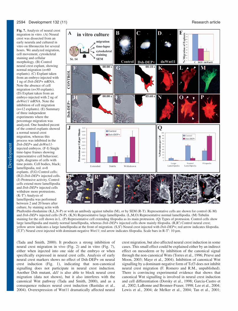
Non-canonical Wnt signalling controls cellprotrusion in the migrating neural crest cellsThe non-canonical Wnt pathway controls convergent extensionof mesoderm during gastrulation movements by instigating adirectional activity of the lamellipodia that favours movementin one direction (Carreira-Barbosa et al., 2003; Wallingford etal., 2000). Although no similar directionality of individualmigratory neural crest cells has been described, we decided toinvestigate whether we could identify similar cell behaviour,controlled by non-canonical Wnt signalling. We culturedneural crest cells in vitro and analyzed their behaviour (Fig.7A). In control explants, neural crest cells migrated normally(Fig. 7B), as described previously (Alfandari et al., 2003);however, in explants taken from embryos injected with 1 ng ofDsh-DEP+ mRNA or 2 ng of dnWnt11, migration of cells was
Fig. 4. Wnt11 activity is required for neural crest migration. (A) 1 ngof Wnt11 or 2 ng of its dominant-negative mRNA were injected inone animal blastomere of an eight-cell embryo. (B-G) Embryos werecultured until the premigratory (B,D; indicated as stage 17) ormigratory (C,E,G; indicated as stage 24) neural crest stages, whenthe expression of Slug was analyzed. All of the embryos are shown indorsal view, anterior to the top. The injected side is indicated by awhite arrowhead. Normal neural crest migration is indicated by redarrowheads. (B,C) Wnt11 overexpression. (B) 90% of embryosshowed normal Slug expression, n=55. (C) 30% of embryos showedinhibited neural crest migration, n=55. (D,E) Expression ofdominant-negative Wnt11. (D) 90% of embryos showed normal Slugexpression, n=65. (E) 35% of embryos showed inhibited neural crestmigration; n=65. (F) Summary of inhibition of neural crest migrationafter injecting Wnt11 (w11), dominant-negative Wnt11 (dnw11), orboth (w11+dnw11). (G) Co-injection of Wnt11 and its dominant-negative form. 90% of embryos showed normal neural crestmigration, n=50.
Dev
elop
men
t

2592
strongly inhibited (Fig. 7C-E). To examine the effect at acellular level, we performed a time-lapse analysis of themigrating neural crest cells. The number and shape of cellprotrusions was counted in control and Dsh-DEP+ expressingcells in frames from time-lapse video movies (Fig. 7F-I). Ourresults indicate that in explants from Dsh-DEP+ expressingembryos, there were less cell protrusions than in control cells.The frequency of crest cells withdrawing rather than extendingcell processes is greater in the Dsh-DEP+ cells than in thecontrol cells (Fig. 7J). To extend these observations, wevisualised actin microfilaments with phalloidin-rhodamine,and microtubules by immunostaining, and then analyzed thesize and types of lamellipodia (Fig. 7K-P). In control cells,lamellipodia were larger and more polarized than in the Dsh-DEP+ expressing cells, whereas the Dsh-DEP+ expressingcells exhibited more filopodia than the control neural crest cells(Fig. 7Q). A typical control cell is shown in Fig. 7L,M (morethan 50% of cells), while typical Dsh-DEP+ expressing cells
are shown in Fig. 7O,P (more than 90% of cells, although mostof the cells were found forming groups and very few wereisolated). We also analyzed the morphology of the migratingneural crest by SEM (Fig. 7R-T). Control migrating cellsexhibited large lamellipodia at the front of migration (yellowarrows in Fig. 7R,R′), while cells injected with Dsh-DEP+(Fig. 7S,S′) or dnWnt11 (Fig. 7T,T′) exhibited long filopodiathat frequently were connecting the more packed cells (redarrows).
DiscussionThis study reveals that PCP/Wnt-Ca2+ signalling is involved inneural crest migration. We have not analyzed whether the PCPor Wnt-Ca2+ pathway is controlling neural crest migrationbecause both pathways are inhibited by the same Dsh mutants(Sheldahl et al., 2003). Although there are several similaritieswith other instances in which PCP participates in cell
Development 132 (11) Research article
Fig. 5. Effect of localized overexpression of Wnt11 mRNAon neural crest migration. (A) One-cell stage embryoswere injected with Wnt11 mRNA and FDX. At stage 9, theanimal cap was dissected and cultured under a coverslipuntil the equivalent of stage 14. The ectoderm was graftedadjacent to the neural crest of a host embryo (stage 14),but into the neural plate side. Grafted embryos werecultured until stage 23-26, when neural crest migrationwas examined by Slug expression. Purple arrow indicatesthe normal migration of the neural crest cells. (B) Controlside showing normal cephalic neural crest migration(dashed line), and the three streams of migration (blackarrows). (C) Grafted side of the embryo shown in B. Notethe inhibition in neural crest migration (dashed line andwhite arrows). The green arrowhead indicates the graft.(D,E) Sections from grafted embryos. Red bars indicatethe ventral limit of neural crest migration; greenarrowhead indicates the graft. (D) Graft of ectoderminjected with FDX; no effect on neural crest migration isobserved. (E) Section of the embryo shown in B,C. Notethe shorter distance of migration in the grafted side. Thestronger signal in the grafted side is likely to be aconsequence of all of the neural crest cells grouping in thedorsal region. Forty-five percent of embryos grafted withWnt11-expressing cells showed inhibition of neural crestmigration (n=26). (F) Similar experiment to the onedescribed in A, but the Wnt11-expressing cells weregrafted onto the normal pathway of neural crest migration.Grafted embryos were cultured until stage 26 when neuralcrest migration was examined by Slug expression. Purplearrow indicates the normal migration of the neural crestcells. (G-N) Anterior to the right. E, eye; dashed lineindicates the limit of the graft; arrow indicates themigrating neural crest. (G,H,K,L) The operated (H,L) andcontrol (G,K) sides of the same embryo are shown.(I,M) Sections of grafted embryos. (J,N) Summary ofcephalic neural crest migration. Pink, branchial; blue,hyoid; purple, mandibular neural crest. Mixed coloursindicate abnormal neural crest migration. The Wnt11-expressing graft is shown in green. Arrows indicate theproposed route of abnormal neural crest migration.(G-J) Control embryo that received an uninjected graft.(K-N) Embryos that received a Wnt11-expressing graft;80% of the embryos with a Wnt11-expressing graftexhibited this phenotype, n=28.
Dev
elop
men
t

2593Non-canonical Wnt in neural crest migration
migration, we also found some important differences. Invertebrates, there is compelling evidence that the non-canonical Wnt pathway controls aspects of gastrulation,cochlear hair cell morphology, heart induction, dorsoventralpatterning, tissue separation, neuronal migration and cancer(for reviews, see Fanto and McNeill, 2004; Kuhl, 2002;Mlodzik, 2002; Strutt, 2003; Veeman et al., 2003a). However,the simplest and best-understood role of PCP is in theorganization of hairs in the wing of Drosophila. Disruption ofthe PCP signal produces different phenotypes in which theorientation and the subcellular localization of the hair areaffected (Eaton and Cohen, 1996; Gubb and Garcia-Bellido,1982; Held et al., 1986; Strutt et al., 1997; Winter et al., 2001;Wong and Adler, 1993). An important feature of the PCP
molecular mechanism is its effects on asymmetrically localizedmolecules. This has been particularly well studied in the wing,where Flamingo, Diego, Frizzled, Dishevelled, Strabismus andPrickle become localized along the proximodistal axis of thecells (Axelrod, 2001; Bastock et al., 2003; Feiguin et al., 2001;Shimada et al., 2001; Tree et al., 2002; Usui et al., 1999). Alarge body of evidence suggests that Fz has a key role insensing positional information; although what is upstream ofFz remains unknown. It has been proposed that ft and ds controlPCP upstream of Fz/Dsh pathways, and that a gradient of Ftactivity sets up the initial asymmetrical localization (Ma et al.,2003; Rawls et al., 2002; Yang et al., 2001), but the origins ofthe gradient of Ft activity are unknown.
One of the most appealing models of positional informationis based on the presence of a morphogen-like molecule thatwould activate Fz in a dose-dependent manner (Adler et al.,2000; Fanto et al., 2003). It has been suggested that such amolecule could belong to the Wnt family, as it binds to the Fzreceptor and should be produced in a localized fashion. Nosuch molecule has been found so far in the Drosophila PCPpathway, or in any vertebrate system. Our results show a clearlocalized expression of Wnt11, just adjacent to premigratoryneural crest and just before the onset of crest delamination (Fig.3). Other Wnt family members that participate in the non-canonical pathway (such as Wnt4 and Wnt5a) do not show asimilar pattern of expression using in situ hybridization (J.D.C.and R.M., unpublished) (Torres et al., 1996; McGrew et al.,1992). Our functional studies using dnWnt11 also support thenotion that this molecule is essential for neural crest migration,while the rescue of the effect of dnWnt11 by co-expression ofWnt11 mRNA provides evidence for the specificity of thisdominant-negative construct (Fig. 4F,G). Furthermore, theeffect on neural crest migration of the dnWnt11 can be rescuedby specific activation of non-canonical Dsh (Fig. 6).
Several observations suggest that the localized expression ofWnt11 adjacent to the premigratory neural crest is essential forits function. First, overexpression of Wnt11 in one half of theembryo completely blocks normal neural crest migration (Fig.4) and suggests a requirement for localized expression for thismolecule. Second, when Wnt11 was expressed in a localizedmanner opposite to its normal expression, the neural crest cellsmigration was blocked (Fig. 5). Third, when Wnt11 wasexpressed in the normal route of crest migration the cellsmigrated actively under the Wnt11-expressing cells (Fig. 5).Taken together, these results show that localized expression ofWnt11 is required to activate the PCP/Wnt-Ca2+ pathway andto control neural crest migration. We do not know themolecular function of this localized Wnt expression in theneural crest cells, but the situation in Drosophila PCP suggeststhat a gradient of Wnt11 could determine the asymmetricexpression of PCP molecules that direct crest migration. Noasymmetrical localization of PCP proteins has been observedin any vertebrate system.
In addition to the evidence suggesting that Wnt11 is requiredfor neural crest migration, our data based on Dsh mutants showthat non-canonical Wnt signalling participates in neural crestmigration. Dsh-DEP+, a dominant-negative form of Dsh thatcontains the DEP domain and lacks the DIX and PDZ domains,has been an incisive reagent for analysing the role of the non-canonical pathway in neural crest migration. This blocks thePCP/Wnt-Ca2+ pathway without affecting canonical signalling
Fig. 6. Inhibition of neural crest migration by dominant-negativeWnt11 is rescued by activated Dsh. (A) Two-cell stage embryos wereinjected with dnWnt11 mRNA and RDX (red), or with Dsh-∆N andFDX (green). At stage 14, neural crest cells were taken from theDsh-∆N-injected embryo and grafted into the dnWnt11-injectedembryo. Analysis of neural crest migration was performed byexamining Slug expression and fluorescence. (B) Control side ofembryo injected with dnWnt11. Arrowheads indicate normal neuralcrest migration. (C) Injected side of the embryo shown in B. Asteriskindicates the absence of neural crest migration. (Inset) RDXfluorescence. (D,E) Graft of Dsh-∆N/FDX-expressing cells into anembryo injected with dnWnt11/RDX. (Inset) RDX fluorescence.Note the normal migration of the FDX-expressing cells. Seventy-fivepercent of embryos showed rescued neural crest migration; n=20.
Dev
elop
men
t
2594
(Tada and Smith, 2000). It produces a strong inhibition ofneural crest migration in vivo (Fig. 2) and in vitro (Fig. 7),either when injected into one side of the embryo or whenspecifically expressed in neural crest cells. Analysis of earlyneural crest markers shows no effect of Dsh-DEP+ on neuralcrest induction (Fig. 1), indicating that non-canonicalsignalling does not participate in neural crest induction.Another Dsh mutant, dd1 is also able to block neural crestmigration (data not shown), but it also interferes with thecanonical Wnt pathway (Tada and Smith, 2000), and as aconsequence reduces neural crest induction (Bastidas et al.,2004). Overexpression of Wnt11 dramatically affected neural
crest migration, but also affected neural crest induction in somecases. This small effect could be explained either by an indirecteffect on mesoderm or by inhibition of the canonical signalthrough the non-canonical Wnts (Torres et al., 1996; Prieve andMoon, 2003; Maye et al., 2004). Inhibition of canonical Wntsignalling by a dominant-negative form of Tcf3 does not inhibitneural crest migration (F. Romero and R.M., unpublished).There is convincing experimental evidence that shows thatcanonical Wnt signalling is involved in neural crest inductionand cell differentiation (Dorsky et al., 1998; Garcia-Castro etal., 2002; LaBonne and Bronner-Fraser, 1998; Lee et al., 2004;Lewis et al., 2004; de Melker et al., 2004; Tan et al., 2001;
Development 132 (11) Research article
Fig. 7. Analysis of neural crestmigration in vitro. (A) Neuralcrest was dissected from anearly neurula and cultured invitro on fibronectin for severalhours. We analyzed migration,cell movement, cytoskeletalstaining and cellularmorphology. (B) Controlneural crest explant, showingnormal migration (n=60explants). (C) Explant takenfrom an embryo injected with1 ng of Dsh-DEP+ mRNA.Note the absence of cellmigration (n=30 explants).(D) Explant taken from anembryo injected with 2 ng ofdnWnt11 mRNA. Note theinhibition of cell migration(n=12 explants). (E) Summaryof three independentexperiments where thepercentage migration wasanalyzed. One hundred pecentof the control explants showeda normal neural crestmigration, whereas thisprocess was inhibited in theDsh-DEP+ and dnWnt11-injected embryos. (F-I) Singletime-lapse frames showingrepresentative cell behaviour;right, diagrams of cells withtime points. Cell bodies, black;lamellipodia, red. n=8explants. (F,G) Control cells;(H,I) Dsh-DEP+ injected cells.(J) Protrusive activity. Controlcells extend more lamellipodiaand Dsh-DEP+ injected cellswithdraw more protrusions.(K-T′) Analysis oflamellipodia was performedbetween 2 and 20 hours afterculture, by staining actin withPhalloidin-rhodamine (K,L,N-P) or with an antibody against tubulin (M), or by SEM (R-T). Representative cells are shown for control (K-M)and Dsh-DEP+ injected cells (N-P). (K,N) Representative large lamellipodia. (L,M,O) Representative normal lamellipodia. (M) Tubulinstaining for the cell shown in L. (P) Representative cell extending filopodia as its main protrusion. (Q) Types of protrusion. Control cells showlarge lamellipodia and mainly normal lamellipodia, whereas Dsh-DEP+ injected cells show mainly filopodia. (R,R′) Control neural crest;yellow arrow indicates a large lamellipodia at the front of migration. (S,S′) Neural crest injected with Dsh-DEP+; red arrow indicates filopodia.(T,T′) Neural crest injected with dominant-negative Wnt11; red arrow indicates filopodia. Scale bars in R-T′: 10 µm.
Dev
elop
men
t

2595Non-canonical Wnt in neural crest migration
Villanueva et al., 2002). Wnt signalling evidently plays acrucial role in neural crest development, in the canonicalpathway in induction and in the non-canonical pathway inneural crest migration, as we have shown here.
If Wnt11 activates the non-canonical Wnt pathway in neuralcrest cells, these cells should express the appropriate Wntreceptor. Although the specific Wnt11 receptor is unknown,there is evidence for the involvement of Fz7 and Fz2 in non-canonical Wnt signalling, and of Fz7 in gastrulation (Carreira-Barbosa et al., 2003; Djiane et al., 2000; Kuhl et al., 2000;Medina et al., 2000; Sumanas and Ekker, 2001; Wang andMalbon, 2004; Winklbauer et al., 2001). We have shown thatFz7 is expressed in the premigratory neural crest just beforemigration, as well as in the migrating neural crest. Comparisonof the expression of Fz7 with that of the neural crest markerSlug indicates that only a subpopulation of neural crest cellsexpresses the Wnt receptor. The Fz7-expressing cellscorrespond to those cells nearest to the Wnt11-expressing cells,which are likely to be the first cells to start migration. Theexpression pattern of Fz7 correlates well with the proposal thatthis is the receptor of the Wnt11 signalling involved in neuralcrest migration; however, another receptor could also beinvolved. We have not investigated whether neural crest cellsnot adjacent to the Wnt11-expressing cells are able to receivethe Wnt11 signal. Most reports suggest that Wnt works asshort-range signalling molecule, but that depending on the cellcontext and the proteins involved, the range can be extended(reviewed by Christian, 2000; Arias, 2003).
The participation of PCP on cell movements duringgastrulation has been very well characterized, although howWnt controls cell movement remains unknown. Migration ofthe neural crest cells requires an epithelial-mesenchymaltransition (EMT), an elaborate process that occurs in manysteps. There is an initial delamination step that is essential forthe second step of neural crest migration. Our results using Dshand Wnt11 mutants, which show inhibition of cell movementboth in vivo and in vitro, are compatible with an inhibition ofdelamination or posterior cell movement of the neural crestcells. Localized expression of Wnt11, by a graft of Wnt11-expressing ectoderm, shows an effect on crest migration that isdependent on the position of the graft. We propose that Wnt11can trigger a cellular activity required for cell movement duringdelamination and/or cell migration, and that the crest cellsrequire additional cues to translate this into an effective cellmigration. The possibility that neural crest was induced in thegraft is ruled out, as competence for neural crest induction islost at the stage at which the tissue was transplanted (Mancillaand Mayor, 1996; Bastidas et al., 2004). It is still possible thatWnt11 promotes cell proliferation (Ouko et al., 2004),although this seems unlikely as cell numbers in our in vitrocultures did not increase after stimulating Wnt11 signalling.We, therefore, favour an effect of Wnt11 on neural crestmigration, instead of cell proliferation. A recent report showsthat PCP signalling controls the orientation of cell divisionduring gastrulation (Gong et al., 2004). No analysis of celldivision orientation during neural crest migration has beenreported, but inhibition of the cell cycle blocks neural crestmigration (Burstyn-Cohen and Kalcheim, 2002; Saka andSmith, 2001). Thus, it is possible that Wnt11 signallingcontrols cell migration by controlling cell divisions.
Neural crest migration in vitro and in vivo is blocked by Dsh
and Wnt11 mutants, to a similar extent (Fig. 7). The ability toblock cell migration in vitro suggests that the neural crest cellshave already responded to Wnt11 signalling at the time of thedissection. This is possible, as the Wnt11-expressing cells areadjacent to the neural crest and it would be difficult to excludethem from an in vitro culture. Analysis of cell protrusions inmigrating crest cells in vitro shows that non-canonical Wntsignalling is required to stabilize the lamellipodia. Inhibitionof the PCP pathway increases the number of cells withfilopodia with a less-polarized phenotype than the controlneural crest cells. Similar functions for the PCP pathway andWnt11 have been described during gastrulation in Xenopus andzebrafish embryos (Ulrich et al., 2003; Wallingford et al.,2000). We propose that Wnt11 controls cytoskeletal behaviouror cell adhesion properties in neural crest migration, and thatit is required to generate the cell protrusions necessary forlocomotion.
We thank Masazumi Tada for useful comments and reagents duringthe course of this work. We also thank Claudio Stern, Michael Sargentand Claudia Linker for comments on the manuscript, Sergei Sokol,Christian Gonzalez, Miguel Concha and Herbert Steinbeisser forreagents and equipment, and Florencio Espinoza for technicalassistance. This investigation was supported by an InternationalResearch Scholar Award from the Howard Hughes Medical Instituteto R.M., and by grants from the MRC, Fondecyt (#1020688) and theMillennium Program (P99-137F and ICM P02-050).
ReferencesAdler, P. N., Taylor, J. and Charlton, J. (2000). The domineering non-
autonomy of frizzled and van Gogh clones in the Drosophila wing is aconsequence of a disruption in local signaling. Mech. Dev. 96, 197-207.
Alfandari, D., Cousin, H., Gaultier, A., Hoffstrom, B. G. and DeSimone,D. W. (2003). Integrin alpha5beta1 supports the migration of Xenopuscranial neural crest on fibronectin. Dev. Biol. 260, 449-464.
Arias, A. M. (2003). Wnts as morphogens? The view from the wing ofDrosophila. Nat. Rev. Mol. Cell Bio. 4, 321-325.
Axelrod, J. D. (2001). Unipolar membrane association of Dishevelledmediates Frizzled planar cell polarity signaling. Genes Dev. 15, 1182-1187.
Axelrod, J. D., Miller, J. R., Shulman, J. M., Moon, R. T. and Perrimon,N. (1998). Differential recruitment of Dishevelled provides signalingspecificity in the planar cell polarity and Wingless signaling pathways.Genes Dev. 12, 2610-2622.
Aybar, M. J. and Mayor, R. (2002). Early induction of neural crest cells:lessons learned from frog, fish and chick. Curr. Opin. Genet. Dev. 12, 452-458.
Aybar, M. J., Nieto, M. A. and Mayor, R. (2003). Snail precedes slug in thegenetic cascade required for the specification and migration of the Xenopusneural crest. Development 130, 483-494.
Basch, M. L., Garcia-Castro, M. I. and Bronner-Fraser, M. (2004).Molecular mechanisms of neural crest induction. Birth Defects Res. CEmbryo Today 72, 109-123.
Bastidas, F., De Calisto, J. and Mayor, R. (2004). Identification of neuralcrest competence territory: role of Wnt signaling. Dev. Dyn. 229, 109-117.
Bastock, R., Strutt, H. and Strutt, D. (2003). Strabismus is asymmetricallylocalised and binds to Prickle and Dishevelled during Drosophila planarpolarity patterning. Development 130, 3007-3014.
Borchers, A., David, R. and Wedlich, D. (2001). Xenopus cadherin-11restrains cranial neural crest migration and influences neural crestspecification. Development 128, 3049-3060.
Boutros, M. and Mlodzik, M. (1999). Dishevelled: at the crossroads ofdivergent intracellular signaling pathways. Mech. Dev. 83, 27-37.
Boutros, M., Paricio, N., Strutt, D. I. and Mlodzik, M. (1998). Dishevelledactivates JNK and discriminates between JNK pathways in planar polarityand wingless signaling. Cell 94, 109-118.
Bronner-Fraser, M., Wolf, J. J. and Murray, B. A. (1992). Effects ofantibodies against N-cadherin and N-CAM on the cranial neural crest andneural tube. Dev. Biol. 153, 291-301.
Dev
elop
men
t

2596
Burstyn-Cohen, T. and Kalcheim, C. (2002). Association between the cellcycle and neural crest delamination through specific regulation of G1/Stransition. Dev. Cell 3, 383-395.
Carreira-Barbosa, F., Concha, M. L., Takeuchi, M., Ueno, N., Wilson, S.W. and Tada, M. (2003). Prickle 1 regulates cell movements duringgastrulation and neuronal migration in zebrafish. Development 130, 4037-4046.
Christian, J. L. (2000). BMP, Wnt and Hedgehog signals: how far can theygo? Curr. Opin. Cell Biol. 12, 244-249.
Curtin, J. A., Quint, E., Tsipouri, V., Arkell, R. M., Cattanach, B., Copp,A. J., Henderson, D. J., Spurr, N., Stanier, P., Fisher, E. M. et al. (2003).Mutation of Celsr1 disrupts planar polarity of inner ear hair cells and causessevere neural tube defects in the mouse. Curr. Biol. 13, 1129-1133.
Darken, R. S., Scola, A. M., Rakeman, A. S., Das, G., Mlodzik, M.and Wilson, P. A. (2002). The planar polarity gene strabismusregulates convergent extension movements in Xenopus. EMBO J. 21, 976-985.
de Melker, A. A., Desban, N. and Duband, J. L. (2004). Cellular localizationand signaling activity of beta-catenin in migrating neural crest cells. Dev.Dyn. 230, 708-726.
Djiane, A., Riou, J., Umbhauer, M., Boucaut, J. and Shi, D. (2000). Roleof frizzled 7 in the regulation of convergent extension movements duringgastrulation in Xenopus laevis. Development 127, 3091-3100.
Dorsky, R. I., Moon, R. T. and Raible, D. W. (1998). Control of neural crestcell fate by the Wnt signalling pathway. Nature 396, 370-373.
Dorsky, R. I., Moon, R. T. and Raible, D. W. (2000). Environmental signalsand cell fate specification in premigratory neural crest. BioEssays 22, 708-716.
Eaton, S. and Cohen, S. (1996). Wnt signal transduction: more than one watto skin a (beta-) cat? Trends Cell Biol. 6, 287-290.
Fanto, M. and McNeill, H. (2004). Planar polarity from flies to vertebrates.J. Cell Sci. 117, 527-533.
Fanto, M., Clayton, L., Meredith, J., Hardiman, K., Charroux, B.,Kerridge, S. and McNeill, H. (2003). The tumor-suppressor and celladhesion molecule Fat controls planar polarity via physical interactions withAtrophin, a transcriptional co-repressor. Development 130, 763-774.
Feiguin, F., Hannus, M., Mlodzik, M. and Eaton, S. (2001). The ankyrinrepeat protein Diego mediates Frizzled-dependent planar polarization. Dev.Cell 1, 93-101.
Garcia-Castro, M. I., Marcelle, C. and Bronner-Fraser, M. (2002).Ectodermal Wnt function as a neural crest inducer. Science 297, 848-851.
Gong, Y., Mo, C. and Fraser, S. E. (2004). Planar cell polarity signallingcontrols cell division orientation during zebrafish gastrulation. Nature 430,689-693.
Goto, T. and Keller, R. (2002). The planar cell polarity gene strabismusregulates convergence and extension and neural fold closure in Xenopus.Dev. Biol. 247, 165-181.
Gubb, D. and Garcia-Bellido, A. (1982). A genetic analysis of thedetermination of cuticular polarity during development in Drosophilamelanogaster. J. Embryol. Exp. Morphol. 68, 37-57.
Harland, R. M. (1991). In situ hybridization: an improved whole-mountmethod for Xenopus embryos. Methods Cell Biol. 36, 685-695.
Harland, R. and Weintraub, H. (1985). Translation of mRNA injected intoXenopus oocytes is specifically inhibited by antisense RNA. J. Cell Biol.101, 1094-1099.
Heeg-Truesdell, E. and LaBonne, C. (2004). A slug, a fox, a pair of sox:transcriptional responses to neural crest inducing signals. Birth Defects Res.C Embryo Today 72, 124-139.
Heisenberg, C. P., Tada, M., Rauch, G. J., Saude, L., Concha, M. L.,Geisler, R., Stemple, D. L., Smith, J. C. and Wilson, S. W. (2000).Silberblick/Wnt11 mediates convergent extension movements duringzebrafish gastrulation. Nature 405, 76-81.
Held, L. I., Jr, Duarte, C. M. and Derakhshanian, K. (1986). Extra jointsand misoriented bristles on Drosophila legs. Prog. Clin. Biol. Res. 217A,293-296.
Henderson, D. J., Ybot-Gonzalez, P. and Copp, A. J. (2000). RhoB isexpressed in migrating neural crest and endocardial cushions of thedeveloping mouse embryo. Mech. Dev. 95, 211-214.
Hoffmann, I. and Balling, R. (1995). Cloning and expression analysis of anovel mesodermally expressed cadherin. Dev. Biol. 169, 337-346.
Huang, X. and Saint-Jeannet, J. P. (2004). Induction of the neural crest andthe opportunities of life on the edge. Dev. Biol. 275, 1-11.
Jessen, J. R., Topczewski, J., Bingham, S., Sepich, D. S., Marlow, F.,Chandrasekhar, A. and Solnica-Krezel, L. (2002). Zebrafish trilobite
identifies new roles for Strabismus in gastrulation and neuronal movements.Nat. Cell Biol. 4, 610-615.
Keller, R. (2002). Shaping the vertebrate body plan by polarized embryoniccell movements. Science 298, 1950-1984.
Kibar, Z., Vogan, K. J., Groulx, N., Justice, M. J., Underhill, D. A. andGros, P. (2001). Ltap, a mammalian homolog of Drosophila Strabismus/VanGogh, is altered in the mouse neural tube mutant Loop-tail. Nat. Genet. 28,251-255.
Kilian, B., Mansukoski, H., Barbosa, F. C., Ulrich, F., Tada, M. andHeisenberg, C. P. (2003). The role of Ppt/Wnt5 in regulating cell shape andmovement during zebrafish gastrulation. Mech. Dev. 120, 467-476.
Kimura, Y., Matsunami, H., Inoue, T., Shimamura, K., Uchida, N., Ueno,T., Miyazaki, T. and Takeichi, M. (1995). Cadherin-11 expressed inassociation with mesenchymal morphogenesis in the head, somite, and limbbud of early mouse embryos. Dev Biol 169, 347-358.
Knecht, A. K. and Bronner-Fraser, M. (2002). Induction of the neural crest:a multigene process. Nat. Rev. Genet. 3, 453-461.
Ku, M. and Melton, D. A. (1993). Xwnt-11: a maternally expressed Xenopuswnt gene. Development 119, 1161-1173.
Kuhl, M. (2002). Non-canonical Wnt signaling in Xenopus: regulation of axisformation and gastrulation. Semin. Cell Dev. Biol. 13, 243-249.
LaBonne, C. and Bronner-Fraser, M. (1998). Neural crest induction inXenopus: evidence for a two-signal model. Development 125, 2403-2414.
Lee, H. Y., Kleber, M., Hari, L., Brault, V., Suter, U., Taketo, M. M.,Kemler, R. and Sommer, L. (2004). Instructive role of Wnt/beta-catenin insensory fate specification in neural crest stem cells. Science 303, 1020-1023.
Lewis, J. L., Bonner, J., Modrell, M., Ragland, J. W., Moon, R. T., Dorsky,R. I. and Raible, D. W. (2004). Reiterated Wnt signaling during zebrafishneural crest development. Development 131, 1299-1308.
Liu, J. P. and Jessell, T. M. (1998). A role for rhoB in the delamination ofneural crest cells from the dorsal neural tube. Development 125, 5055-5067.
Ma, D., Yang, C. H., McNeill, H., Simon, M. A. and Axelrod, J. D. (2003).Fidelity in planar cell polarity signalling. Nature 421, 543-547.
Mancilla, A. and Mayor, R. (1996). Neural crest formation in Xenopus laevis:mechanisms of Xslug induction. Dev. Biol. 177, 580-589.
Maye, P., Zheng, J., Li, L. and Wu, D. (2004). Multiple mechanisms forWnt11-mediated repression of the canonical Wnt signaling pathway. J. Biol.Chem. 279, 24659-24665.
Mayor, R. and Aybar, M. J. (2001). Induction and development of neuralcrest in Xenopus laevis. Cell Tissue Res. 305, 203-209.
Mayor, R., Morgan, R. and Sargent, M. G. (1995). Induction of theprospective neural crest of Xenopus. Development 121, 767-777.
McGrew, L. L., Otte, A. P. and Moon, R. T. (1992). Analysis of Xwnt-4 inembryos of Xenopus laevis: a Wnt family member expressed in the brainand floor plate. Development 115, 463-473.
Medina, A., Reintsch, W. and Steinbeisser, H. (2000). Xenopus frizzled 7can act in canonical and non-canonical Wnt signaling pathways:implications on early patterning and morphogenesis. Mech. Dev. 92, 227-237.
Mlodzik, M. (2002). Planar cell polarization: do the same mechanismsregulate Drosophila tissue polarity and vertebrate gastrulation? Trends Genet18, 564-571.
Myers, D. C., Sepich, D. S. and Solnica-Krezel, L. (2002). Convergence andextension in vertebrate gastrulae: cell movements according to or in searchof identity? Trends Genet 18, 447-455.
Nakagawa, S. and Takeichi, M. (1995). Neural crest cell-cell adhesioncontrolled by sequential and subpopulation-specific expression of novelcadherins. Development 121, 1321-1332.
Nakagawa, S. and Takeichi, M. (1998). Neural crest emigration from theneural tube depends on regulated cadherin expression. Development 125,2963-2971.
Nieuwkoop, P. D. and Faber, J. (1967). Normal Table of Xenopus laevis(Daudin). Amsterdam: Elsevier-North Holland Publishing.
Ouko, L., Ziegler, T. R., Gu, L. H., Eisenberg, L. M. and Yang, V. W.(2004). Wnt11 signaling promotes proliferation, transformation, andmigration of IEC6 intestinal epithelial cells. J. Biol. Chem. 279, 26707-26715.
Park, M. and Moon, R. T. (2002). The planar cell-polarity gene stbmregulates cell behaviour and cell fate in vertebrate embryos. Nat. Cell Biol.4, 20-25.
Perris, R. and Perissinotto, D. (2000). Role of the extracellular matrix duringneural crest cell migration. Mech. Dev. 95, 3-21.
Pla, P., Moore, R., Morali, O. G., Grille, S., Martinozzi, S., Delmas, V. and
Development 132 (11) Research article
Dev
elop
men
t

2597Non-canonical Wnt in neural crest migration
Larue, L. (2001). Cadherins in neural crest cell development andtransformation. J. Cell Physiol. 189, 121-132.
Prieve, M. G. and Moon, R. T. (2003). Stromelysin-1 and mesothelin aredifferentially regulated by Wnt-5a and Wnt-1 in C57mg mouse mammaryepithelial cells. BMC Dev. Biol. 3, 2.
Rauch, G. J., Hammerschmidt, M., Blader, P., Schauerte, H. E., Strahle,U., Ingham, P. W., McMahon, A. P. and Haffter, P. (1997). Wnt5 isrequired for tail formation in the zebrafish embryo. Cold Spring HarborSymp. Quant. Biol. 62, 227-234.
Rawls, A. S., Guinto, J. B. and Wolff, T. (2002). The cadherins fat anddachsous regulate dorsal/ventral signaling in the Drosophila eye. Curr. Biol.12, 1021-1026.
Rothbacher, U., Laurent, M. N., Deardorff, M. A., Klein, P. S., Cho, K. W.and Fraser, S. E. (2000). Dishevelled phosphorylation, subcellularlocalization and multimerization regulate its role in early embryogenesis.EMBO J. 19, 1010-1022.
Sadaghiani, B. and Thiebaud, C. H. (1987). Neural crest development in theXenopus laevis embryo, studied by interspecific transplantation andscanning electron microscopy. Dev Biol 124, 91-110.
Saka, Y. and Smith, J. C. (2001). Spatial and temporal patterns of cell divisionduring early Xenopus embryogenesis. Dev. Biol. 229, 307-318.
Sela-Donenfeld, D. and Kalcheim, C. (1999). Regulation of the onset ofneural crest migration by coordinated activity of BMP4 and Noggin in thedorsal neural tube. Development 126, 4749-4762.
Sela-Donenfeld, D. and Kalcheim, C. (2000). Inhibition of noggin expressionin the dorsal neural tube by somitogenesis: a mechanism for coordinatingthe timing of neural crest emigration. Development 127, 4845-4854.
Sheldahl, L. C., Slusarski, D. C., Pandur, P., Miller, J. R., Kuhl, M. andMoon, R. T. (2003). Dishevelled activates Ca2+ flux, PKC, and CamKII invertebrate embryos. J. Cell Biol. 161, 769-777.
Shimada, Y., Usui, T., Yanagawa, S., Takeichi, M. and Uemura, T. (2001).Asymmetric colocalization of Flamingo, a seven-pass transmembranecadherin, and Dishevelled in planar cell polarization. Curr. Biol. 11, 859-863.
Smith, J. C., Conlon, F. L., Saka, Y. and Tada, M. (2000). Xwnt11 and theregulation of gastrulation in Xenopus. Philos. Trans. R. Soc. London Ser. B355, 923-930.
Sokol, S. Y. (1996). Analysis of Dishevelled signalling pathways duringXenopus development. Curr. Biol. 6, 1456-1467.
Strutt, D. (2003). Frizzled signalling and cell polarisation in Drosophila andvertebrates. Development 130, 4501-4513.
Strutt, D. I., Weber, U. and Mlodzik, M. (1997). The role of RhoA in tissuepolarity and Frizzled signalling. Nature 387, 292-295.
Sumanas, S. and Ekker, S. C. (2001). Xenopus frizzled-7 morphant displaysdefects in dorsoventral patterning and convergent extension movementsduring gastrulation. Genesis 30, 119-122.
Tada, M. and Smith, J. C. (2000). Xwnt11 is a target of Xenopus Brachyury:regulation of gastrulation movements via Dishevelled, but not through thecanonical Wnt pathway. Development 127, 2227-2238.
Takeichi, M., Nakagawa, S., Aono, S., Usui, T. and Uemura, T. (2000).Patterning of cell assemblies regulated by adhesion receptors of the cadherinsuperfamily. Philos. Trans. R. Soc. London Ser. B 355, 885-890.
Takeuchi, M., Nakabayashi, J., Sakaguchi, T., Yamamoto, T. S.,Takahashi, H., Takeda, H. and Ueno, N. (2003). The prickle-related genein vertebrates is essential for gastrulation cell movements. Curr. Biol. 13,674-679.
Tan, C., Deardorff, M. A., Saint-Jeannet, J. P., Yang, J., Arzoumanian, A.and Klein, P. S. (2001). Kermit, a frizzled interacting protein, regulatesfrizzled 3 signaling in neural crest development. Development 128, 3665-3674.
Torres, M. A., Yang-Snyder, J. A., Purcell, S. M., DeMarais, A. A.,McGrew, L. L. and Moon, R. T. (1996). Activities of the Wnt-1 class ofsecreted signaling factors are antagonized by the Wnt-5A class and by adominant negative cadherin in early Xenopus development. J. Cell Biol. 133,1123-1137.
Tree, D. R., Shulman, J. M., Rousset, R., Scott, M. P., Gubb, D. andAxelrod, J. D. (2002). Prickle mediates feedback amplification to generateasymmetric planar cell polarity signaling. Cell 109, 371-381.
Ueno, N. and Greene, N. D. (2003). Planar cell polarity genes and neural tubeclosure. Birth Defects Res. C Embryo Today 69, 318-324.
Ulrich, F., Concha, M. L., Heid, P. J., Voss, E., Witzel, S., Roehl, H., Tada,M., Wilson, S. W., Adams, R. J., Soll, D. R. et al. (2003). Slb/Wnt11controls hypoblast cell migration and morphogenesis at the onset ofzebrafish gastrulation. Development 130, 5375-5384.
Usui, T., Shima, Y., Shimada, Y., Hirano, S., Burgess, R. W., Schwarz, T.L., Takeichi, M. and Uemura, T. (1999). Flamingo, a seven-passtransmembrane cadherin, regulates planar cell polarity under the control ofFrizzled. Cell 98, 585-595.
Vallin, J., Girault, J. M., Thiery, J. P. and Broders, F. (1998). Xenopuscadherin-11 is expressed in different populations of migrating neural crestcells. Mech. Dev. 75, 171-174.
Van de Putte, T., Maruhashi, M., Francis, A., Nelles, L., Kondoh, H.,Huylebroeck, D. and Higashi, Y. (2003). Mice lacking ZFHX1B, the genethat codes for Smad-interacting protein-1, reveal a role for multiple neuralcrest cell defects in the etiology of Hirschsprung disease-mental retardationsyndrome. Am. J. Hum. Genet. 72, 465-470.
Veeman, M. T., Axelrod, J. D. and Moon, R. T. (2003a). A second canon.Functions and mechanisms of beta-catenin-independent Wnt signaling. Dev.Cell 5, 367-377.
Veeman, M. T., Slusarski, D. C., Kaykas, A., Louie, S. H. and Moon, R. T.(2003b). Zebrafish prickle, a modulator of noncanonical Wnt/Fz signaling,regulates gastrulation movements. Curr. Biol. 13, 680-685.
Villanueva, S., Glavic, A., Ruiz, P. and Mayor, R. (2002). Posteriorizationby FGF, Wnt, and retinoic acid is required for neural crest induction. Dev.Biol. 241, 289-301.
Wallingford, J. B., Rowning, B. A., Vogeli, K. M., Rothbacher, U., Fraser,S. E. and Harland, R. M. (2000). Dishevelled controls cell polarity duringXenopus gastrulation. Nature 405, 81-85.
Wallingford, J. B., Fraser, S. E. and Harland, R. M. (2002). Convergentextension: the molecular control of polarized cell movement duringembryonic development. Dev. Cell 2, 695-706.
Wang, H. Y. and Malbon, C. C. (2004). Wnt-frizzled signaling to G-protein-coupled effectors. Cell Mol. Life Sci. 61, 69-75.
Wheeler, G. N. and Hoppler, S. (1999). Two novel Xenopus frizzled genesexpressed in developing heart and brain. Mech. Dev. 86, 203-207.
Winklbauer, R., Medina, A., Swain, R. K. and Steinbeisser, H. (2001).Frizzled-7 signalling controls tissue separation during Xenopus gastrulation.Nature 413, 856-860.
Winter, C. G., Wang, B., Ballew, A., Royou, A., Karess, R., Axelrod, J. D.and Luo, L. (2001). Drosophila Rho-associated kinase (Drok) linksFrizzled-mediated planar cell polarity signaling to the actin cytoskeleton.Cell 105, 81-91.
Wong, L. L. and Adler, P. N. (1993). Tissue polarity genes of Drosophilaregulate the subcellular location for prehair initiation in pupal wing cells. J.Cell Biol. 123, 209-221.
Yagi, T. and Takeichi, M. (2000). Cadherin superfamily genes: functions,genomic organization, and neurologic diversity. Genes Dev. 14, 1169-1180.
Yang, W., Lin, Q., Zhao, J., Guan, J. L. and Cerione, R. A. (2001). Thenonreceptor tyrosine kinase ACK2, a specific target for Cdc42 and anegative regulator of cell growth and focal adhesion complexes. J. Biol.Chem. 276, 43987-43993.
Dev
elop
men
t