Curve interpolation based on the canonical arc length parametrization
Characterization of the Rad53:Dbf4 interface
1
A NOVEL, NON-CANONICAL FHA BINDING INTERFACE MEDIATES THE INTERACTION BETWEEN Rad53 AND Dbf4*
Lindsay A. Matthews1, Rajeevan Selvaratnam2,4, Darryl R. Jones3,4, Madoka Akimoto2, Brendan J.
McConkey3, Giuseppe Melacini1,2, Bernard P. Duncker3 and Alba Guarné1,*
1From the Department of Biochemistry and Biomedical Sciences and 2Department of Chemistry and Chemical Biology
McMaster University, Hamilton, Ontario L8S 4K1 3From the Department of Biology, University of Waterloo, Waterloo, Ontario N2L 3H1, Canada.
4These two authors contributed equally to the work
*Running Title: Characterization of the Rad53:Dbf4 interface
To whom correspondence should be addressed: Alba Guarné, Department of Biochemistry and Biomedical Sciences, McMaster University, Hamilton, Ontario L8S 4K1, Canada, Tel: (905) 525-9140 ext. 26394; FAX (905) 522-9033; e-mail: [email protected] Keywords: DNA replication; DNA damage response; checkpoint control; protein-protein interactions; protein domains; NMR; yeast genetics; bioinformatics. Background: The interaction between the checkpoint kinase Rad53 and Dbf4 is critical to suppress late origin firing and stabilize stalled forks during replication stress. Results: The FHA1 domain of Rad53 interacts with the BRCT domain of Dbf4 through a novel, non-canonical interface. Conclusion: FHA domains gain specificity by engaging multiple binding interfaces. Significance: Understanding how FHA domains interact with their binding partners is key to elucidate how they relay information along signaling pathways.
ABSTRACT ForkHead Associated (FHA) and BRCA-1
C-terminal (BRCT) domains are overrepresented in DNA damage and replication stress response proteins. They primarily function as phosphoepitope-recognition modules, but can also mediate non-canonical interactions. The latter are rare and only a few have been studied at a molecular level. We have identified a crucial, non-canonical interaction between the N-terminal FHA1 domain of the checkpoint effector kinase Rad53 and the BRCT domain of the regulatory subunit of the Dbf4-dependent kinase, which is critical to suppress late origin firing and stabilize stalled forks during replication stress.
The Rad53:Dbf4 interaction is phosphorylation-independent and involves a novel, non-canonical interface on the FHA1 domain. Mutations within this surface result in hypersensitivity to genotoxic stress. Importantly, this surface is not conserved in the FHA2 domain of Rad53, suggesting that the FHA domains of Rad53 gain specificity by engaging additional interaction interfaces beyond their phosphoepitope-binding site. In general, our results point to FHA domains functioning as complex logic gates rather than mere phosphoepitope targeting modules.
ForkHead Associated (FHA) domains are
ubiquitous phosphoepitope-binding modules present in a number of proteins that play critical roles in the DNA damage and replication stress response (1). The replication checkpoint preserves the genomic integrity of the cell by stabilizing stalled forks, boosting DNA repair enzyme levels and pausing the cell cycle (2). Rad53 (the budding yeast homolog of the tumor suppressor Chk2) is an effector kinase with integral roles in the replication checkpoint (3). In contrast to other FHA-containing proteins, Rad53 has two FHA domains (FHA1 and FHA2) that mediate independent interactions of Rad53 with upstream and downstream branches of the checkpoint. One of the binding partners of FHA1 is Dbf4 (4), the
http://www.jbc.org/cgi/doi/10.1074/jbc.M113.517060The latest version is at JBC Papers in Press. Published on November 27, 2013 as Manuscript M113.517060
Copyright 2013 by The American Society for Biochemistry and Molecular Biology, Inc.
by guest on September 12, 2016
http://ww
w.jbc.org/
Dow
nloaded from
by guest on September 12, 2016
http://ww
w.jbc.org/
Dow
nloaded from
by guest on September 12, 2016
http://ww
w.jbc.org/
Dow
nloaded from
by guest on September 12, 2016
http://ww
w.jbc.org/
Dow
nloaded from

Characterization of the Rad53:Dbf4 interface
2
regulatory subunit of the initiator kinase Cdc7 (5,6). The association of Rad53 and Dbf4 mediates the Rad53-dependent phosphorylation of Dbf4 and consequent inhibition of Cdc7, thus preventing late origin firing (7-10). This interaction involves the N-terminal region of Dbf4 that folds as a modified BReast cancer-1 C-Terminal (BRCT) domain (4,11-13).
BRCT domains are also commonly found in DNA damage and replication stress response proteins where they occur as single or multiple repeats (14). Tandem BRCT repeats are phosphoepitope-binding modules with the phosphoepitope binding pocket residing at the interface between the two domains (15). Conversely, single BRCT domains have diverse functions and their mechanisms cannot be extrapolated from one protein to another due to low sequence identity. Furthermore their specific functions remain elusive because many BRCT domains, like the one found in Dbf4, require additional structural elements to become functional (12,16,17). The BRCT domain of Dbf4 is immediately preceded by an α-helix that stabilizes the domain and, thus, it has been previously referred to as HBRCT (12,18). The HBRCT domain of Dbf4 is necessary and sufficient for the interaction with Rad53 and, yet, it does not contain any threonine residue that could serve as the phosphorylation site recognized by Rad53 (7,9,12). A phosphorylated peptide derived from the N-terminal sequence of the HBRCT domain of Dbf4 including the canonical pT-X-X-E FHA-binding motif can interact weakly with the FHA1 domain of Rad53 (19), however mutation of this threonine in full-length Dbf4 does not disrupt the interaction with the FHA1 domain of Rad53 (12,19).
Phospho-threonine (pThr) binding to FHA domains has been extensively studied using short phosphopeptides (20). However, there is some evidence indicating that interactions with full-length partners may involve additional interfaces. The FHA domain of Saccharomyces cerevisiae Dun1 requires the presence of a second pThr for specific binding (21), prompting the comparison of FHA domains to logic gates (22); whereas that of M. tuberculosis Rv1827 uses its pThr-binding pocket to mediate both phosphorylation-dependent and phosphorylation-independent interactions (23). Dbf4 interacts preferentially with the FHA1
domain of Rad53 (4) while Rad9 interacts with its FHA2 domain (24), indicating that defined features in each FHA domain determine their binding specificities. In the present study, we exploited this characteristic of Rad53 to analyze how FHA domains increase binding specificity for their targets. We have uncovered a novel interface defined by one of the lateral surfaces of the FHA1 domain and characterized its interaction with the HBRCT domain of Dbf4. We find that the FHA1 domain of Rad53 can bind simultaneously to the HBRCT domain of Dbf4 and a pThr-containing phosphoepitope, suggesting that a bipartite interaction may modulate the interaction between Rad53 and Dbf4 in vivo. This dual binding mechanism and its functional implications for the replication checkpoint are discussed. EXPERIMENTAL PROCEDURES Cloning, Expression and Purification – A codon optimized version of the HBRCT domain of Dbf4 (amino acids 105-220) was synthesized by GeneArt® (Invitrogen) and subcloned into an expression vector including a (His)6-SUMO tag. The HBRCT-W116D/M120A was also subcloned in the pSUMO vector. Plasmids encoding wild type and (His)6-SUMO-tagged HBRCT variants were transformed in RosettaTM cells (Novagen). Cultures were grown to OD600 of 0.7, induced by addition 1 mM isopropyl β-D-1-thiogalactopyranoside (IPTG) and incubated overnight at 16°C with orbital agitation.
Two variants of the FHA1 domain of Rad53, including amino acids 14-164 and 4-165, were subcloned in pPROEX HTa vectors (Invitrogen). The two variants were produced in RosettaTM cells (Novagen). Protein expression was induced with 1 mM IPTG and the cultures were subsequently incubated for 4 hours at 30°C with orbital agitation. Cell pellets were resuspended in 20 mL of buffer A (20 mM Tris pH 8.0, 500 mM NaCl, 1.4 mM β-mercaptoethanol, and 5% glycerol) supplemented with 15 mM imidazole and lysed by sonication. Lysates were cleared by centrifugation at 39,000 g and the supernatants were loaded onto HiTrap Ni-chelating HP columns (GE Healthcare). (His)6-SUMO-HBRCT and (His)6-FHA1 variants were eluted with linear gradients to 300 mM imidazole. Tags were removed with TEV protease and aliquots of the purified HBRCT and FHA1 were stored at -80 °C in 20 mM Tris pH 8.0, 100
by guest on September 12, 2016
http://ww
w.jbc.org/
Dow
nloaded from

Characterization of the Rad53:Dbf4 interface
3
mM NaCl, 1 mM EDTA, 5 mM DTT, and 5% glycerol.
NMR Spectroscopy – All experiments were conducted using a 700 MHz AV Bruker NMR spectrometer with a TCI cryoprobe. HBRCT samples were uniformly labeled by expressing the domain (residues 105-220) in M9 media containing 15NH4Cl and 13C-D-glucose (15N,13CHBRCT) or 15NH4Cl alone (15NHBRCT). Labeled HBRCT was purified and stored as described earlier (12). All experiments with labeled HBRCT were conducted at 306 K in 20 mM TRIS pH 8.0, 100 mM NaCl, 5 mM DTT, 1 mM EDTA, and 5% glycerol. The FHA1 domain of Rad53 (residues 14-164) was similarly labeled to produce 13C,15NFHA1 or 15NFHA1. The protein was purified as described above and stored in 10 mM sodium phosphate buffer pH 6.5, 1 mM EDTA, and 5 mM DTT. All experiments with labeled FHA1 were conducted at 293 K as described previously (25). Experiments including both 15NFHA1 and HBRCT (or BRCT) were also performed in 10 mM sodium phosphate buffer pH 6.5, 1 mM EDTA, and 5 mM DTT.
Assignment of resonances for 13C,15NHBRCT (0.5 mM), was done using standard triple resonance experiments as described elsewhere (26). The 13C,15NFHA1 sample (0.3 mM) was first mixed with an equimolar amount of unlabelled HBRCT prior to collecting triple resonance experiments. All data were processed with NMRPipe (27) and resonance assignment was carried out using SPARKY (http://www.cgl.ucsf.edu/home/sparky/).
1H15N NOE experiments were conducted using a 0.1 mM sample of 15NHBRCT using a 3 second recycle delay, with and without proton saturation in alternate scans (28). The 15N dimension was digitized with 256 complex points for a spectral width of 31.82 ppm and the 1H dimension was digitized with 1024 complex points for a spectral width of 14.28 ppm. After 16 dummy scans, a total of 128 scans were accumulated for each data set, with and without proton saturation. The steady state NOE values were computed as the ratio of the intensities in saturated to unsaturated spectra. The error for the NOE values were gauged based on the standard deviation between fit heights in replicate spectra. Select cross peaks were not included in the relaxation analyses due to line broadening and/or overlap.
Peptide titration experiments – Lyophilized phosphorylated peptide derived from Cdc7 (pPEP: 480DGESpTDEDDVVS491) and a nonphosphorylated control (PEP: 480DGESTDEDDVVS491) were synthesized by Biomatik. The peptides were resuspended in 10 mM sodium phosphate buffer pH 6.5 and titrated into a sample containing 0.12 mM 15NFHA1 with an equimolar amount of HBRCT. HSQC spectra were collected after each addition and the titrant volume of peptide added was < 20% of the sample volume.
Trypsin Proteolysis – Nine µL of HBRCT (20 µM in storage buffer) were mixed with 1 µL of 50 mM MgCl2 and 1 µL of trypsin (0.195-12.5 µg/mL) and incubated for 1 hour at room temperature. The reaction products were subsequently resolved in 15% SDS-polyacrylamide gels.
Statistical Analysis – Peak intensity losses for 15NFHA1 in the HSQC spectrum after addition of HBRCT were analyzed based on a method published previously (29). The peak fit height was measured with SPARKY using a Lorentzian fit to integrate the peaks. Overlapping peaks were rejected (E26, I32, T39, K57, K64, C74, L99, Q114, S120, and N158). The remaining values were normalized using the most intense peak (Arg164), prior to being compared with a similarly analyzed data set from a sample lacking HBRCT. The peak height ratio was expressed as a percentage. The average peak height ratio was computed excluding Arg164 to avoid bias. In addition, Ile163 was also excluded as its value exceeded the average beyond four standard deviations. Perturbed residues were defined as having a peak height ratio below one standard deviation from the average.
Chemical Crosslinking – The buffer of the HBRCT variants, as well as the FHA1 variant (4-165), was exchanged on a Superdex-75 column (GE Healthcare) equilibrated with crosslinking buffer (20 mM HEPES pH 7.7, 100 mM NaCl, 1 mM EDTA, 5 mM DTT, and 5% glycerol). Crosslinking reactions (10 µL) consisted of 4 µL of each protein at 45 µM (or buffer for control reactions), 1 µL of bis[sulfosuccinimidyl] suberate (BS3) at 0.75 mM and 1 µL of crosslinking buffer. The effect of a phosphorylated peptide was assessed by adding 1 µL (1.44 mM, 0.72 mM or 0.36 mM) of either pPEP or PEP. Reactions were
by guest on September 12, 2016
http://ww
w.jbc.org/
Dow
nloaded from

Characterization of the Rad53:Dbf4 interface
4
incubated for 30 minutes at room temperature, quenched by adding 50 mM TRIS and the reaction products were resolved on 18% SDS-polyacrylamide gels.
Bioinformatics analysis – To identify patterns of amino acid conservation in the FHA1 and FHA2 domains of Rad53, representative protein sequences were retrieved using PSI-BLAST (30) run with an E-value cutoff of E=1e-6. Full length Rad53 from Saccharomyces cerevisiae (Uniprot ID: P22216) was used to query the UniRef90 database using PSI-BLAST, and sequences containing both FHA1 and FHA2 domains were retained. A total of 28 sequences were identified (Uniprot IDs: P2216, G3AR58, G3BDW6, H8X5Q1, B9WDY3, C5MJH1, B5RTA3, G8YJ57, C4Y319, A5DJY0, F2QUG7, C4R6G9, K0KML2, J7SAK5, Q6CKF2, G8JNR2, Q75CE9, I2GYM1, C5DF05, Q6FK01, H2AZ96, A7TKN4, G8BVR8, G8ZMW0, C5DXS2, G0WBA8, G0VCB8, E7NNR4), with no further sequences meeting the criteria after three PSI-BLAST iterations. The multiple sequence alignment produced by PSI-BLAST was edited to contain only regions corresponding to the FHA1 and FH2 domains. The Consurf server (http://consurf.tau.ac.il) was used to visualize conserved surface residues (31). A single PDB file containing both FHA1 and FHA2 domains was created by combining PDB files 1G6G and 1J4K. The combined file was used to map patterns of residue conservation onto the protein surfaces using the same conservation scale. Residue conservation is shown as standard deviations from the overall mean residue variation, with more conserved residues being plotted as negative numbers.
Yeast two-hybrid analysis – Two-hybrid analysis was carried out as previously described (12) using pEG-Dbf4-FL as bait and indicated pJG-FHA1 variants as prey. Point mutations within FHA1 were generated by QuikChange (Invitrogen) using pJG-Rad53 as template. Sequence encoding amino acids 1-165 of the resulting constructs was then subcloned into the NcoI and EcoRI sites of pJG4-6 to create FHA1 variant prey vectors containing desired point mutations. All constructs were verified by DNA sequencing (Robarts Research Institute).
Genotoxicity assays – The RAD53 open reading frame, along with 500bp each of flanking
upstream and downstream sequence, was PCR amplified using genomic DNA from yeast strain DY-27 (MATa/alpha, his3Δ1/his3Δ1, leu2Δ0/leu2Δ0, lys2Δ0/LYS2, met15Δ0/MET15, ura3Δ0/ura3Δ0) as template and cloned into the XbaI and PstI sites of the CEN vector pRS315 to create pRS315-Rad53. The SexAI to BglII fragment of the pJG-FHA1 variants, pJG-FHA1NE, pJG-FHA1NEVF, and pJG-FHA1VF, were used to replace the equivalent fragments in pRS315-Rad53 to generate pRS315-Rad53NE, pRS315-Rad53NEVF, and pRS315-Rad53VF, respectively. pRS315-Rad53 variants along with empty pRS315 were transformed into yeast strain DY-147 (MATa, ade2-1, can1-100, his3-11,15, leu2-3, trp1-1, ura3-1, sml1::HIS3, rad53::URA3), while yeast strain DY-145 (MATa, ade2-1, can1-100, his3-11,15, leu2-3, trp1-1, ura3-1, sml1::HIS3 ) was transformed with empty vector pRS315. Growth spotting assays were performed as described previously (32). Protein expression levels in whole cell extracts were analyzed by Western blot using goat anti-Rad53 antibody (Santa Cruz Biotech #SC6749) and Alexa Fluor 488 donkey anti-goat secondary antibody (Invitrogen #A11055). RESULTS Characterization of the Dbf4 region that interacts with Rad53 – The minimal fragment of Dbf4 necessary and sufficient for the interaction with the FHA1 domain of Rad53 includes residues Thr105 to His220 (12). Its crystal structure revealed that this fragment of Dbf4 folds as a modified BRCT domain that we named HBRCT domain because it includes an additional N-terminal helix (Fig. 1A). In the crystal structure, this additional helix is stabilized by crystal contacts (12). The secondary chemical shifts measured for 15N,13CHBRCT indicated that residues 106-119 form a helix (Fig. 1B). Furthermore, this helix exhibited HN-NOE values close to the theoretical maximum expected in the absence of fast (ps-ns) internal dynamics (Fig. 1C), confirming that this region of the domain indeed forms a helix in solution.
We then checked whether the N-terminal helix is an integral part of the domain. Since the loop connecting this helix to the BRCT core includes several positively charged residues, its
by guest on September 12, 2016
http://ww
w.jbc.org/
Dow
nloaded from
Characterization of the Rad53:Dbf4 interface
5
susceptibility to trypsin proteolysis should vary depending on whether α0 is anchored to the domain or free to move. We treated the HBRCT, HBRCT-W116D/M120A and HBRCT-L109A/W112S variants with increasing amounts of trypsin. We chose these variants because point mutations at these positions disrupt the interaction with Rad53, presumably by destabilizing the interaction between α0 and the BRCT core (12). We found that the HBRCT was degraded homogeneously, whereas the HBRCT-W116D/M120A and HBRCT-L109A/W112S variants were degraded stepwise (Fig. 1D). The first step resulted in the formation of a fragment of similar size to a BRCT domain. The second step degraded the domain without the accumulation of any intermediate fragments, thereby confirming that the additional N-terminal helix is anchored to the BRCT core of the domain.
The HBRCT domain of Dbf4 and the FHA1 domain of Rad53 interact in vitro – The interaction between Rad53 and Dbf4 is weak and transient, and it has only been studied by yeast-two hybrid analysis (4,12,19). However, yeast-two hybrid does not provide any information as to whether an interaction is direct or mediated by a cellular adaptor. To test whether the interaction between Rad53 and Dbf4 is direct, we sought to characterize the interaction using purified proteins.
Once we determined that the HBRCT was a stable domain in solution, we over-expressed and purified the HBRCT (105-220) and the FHA1 (residues 14-164) domains. We collected HSQC spectra of uniformly labeled 15NFHA1 on its own or in the presence of equimolar amounts of unlabelled HBRCT domain. The main effect caused by the addition of the HBRCT domain was peak broadening for several 15NFHA1 residues, which is often a reliable indicator of binding, as previously shown for other weakly interacting protein:protein complexes (29,33,34).
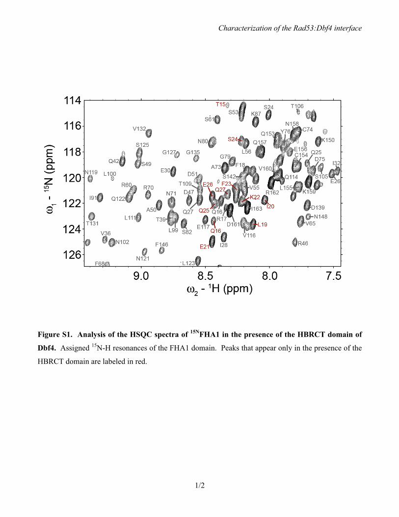
Additionally, ten new peaks appeared in the HSQC spectrum of the complex. We assigned these peaks and found that they corresponded to amino acids Gln16, Leu19, Ile20, Glu21, Lys22, Phe23, Ser24, Gln25, Glu26 and Gln27, which are all located at the N-terminus of the domain (Fig. 2A and Fig. S1). Importantly, these peaks were absent in the HSQC spectrum of 15NFHA1 collected in the presence of equimolar amounts of the BRCT fragment of Dbf4 (residues 120-220),
which lacks the helix α0 and does not interact with Rad53 (Fig. 2B and (12)). Therefore, we concluded that the new peaks in the HSQC spectrum of 15NFHA1:HBRCT are caused by the direct and specific interaction between the two proteins.
HSQC cross-peak intensity losses are useful to map the interface of protein:protein complexes (35,36). To determine whether the FHA1 residues involved in the interaction interface with Dbf4 could be identified this way, we compared the height of equivalent peaks in the 15NFHA1 and 15NFHA1:HBRCT HSQC spectra. We then integrated known peaks in the 15NFHA1 and 15NFHA1:HBRCT spectra and found that peak height was reduced to about 46% upon addition of the HBRCT domain, whereas it maintained 95% of the original height upon addition of the non-interacting BRCT domain, thereby confirming that peak broadening was caused by the interaction of the two domains.
We integrated known peaks in the 15NFHA1 and 15NFHA1:HBRCT spectra and calculated their intensity loss (Fig. 2C). We defined perturbed residues as those whose peak height was reduced by at least one standard deviation (S.D.) from the mean. Nine residues in the core of the FHA1 domain met this criterion and they were located on strands β1 (Ile37), β7 (Asn112), β9 (Asn121, Leu123), β10 (Gly127), and β11 (Phe146, Ile147, Asn148). With the exception of Asn121 and Leu123, all these residues reside on the same lateral surface of the FHA1 domain (Fig. 2D). Residues Thr15, Phe18 and Gln27, located at the N-terminus of the FHA1 domain also met this criterion, but they are not present in the FHA1 fragment used for the crystallization studies (20), and thus they are not shown in Fig. 2D. Therefore, the comparative HSQC analyses suggested that the FHA1:HBRCT interaction affected two main areas of the FHA1 domain, namely its N-terminus and one of the lateral surfaces.
The HBRCT domain of Dbf4 interacts with the lateral surface of the FHA1 domain – We decided to explore whether either of these two surfaces defined the interface of the FHA1:HBRCT complex. If Rad53 interacts with Dbf4 through one of these surfaces, we would expect this area to be conserved among Rad53 homologues. Since
by guest on September 12, 2016
http://ww
w.jbc.org/
Dow
nloaded from

Characterization of the Rad53:Dbf4 interface
6
Dbf4 binds preferentially to the FHA1 domain of Rad53 (4), we postulated that any conserved features of this interface should be present in FHA1, but not FHA2 domains.
We retrieved representative protein sequences homologous to yeast Rad53 from the non-redundant UniRef90 database using PSI-BLAST (30). Twenty-eight full-length Rad53 homologues containing both FHA1 and FHA2 domains were retained and used to compare the degree of conservation of residues in FHA1 and FHA2. In addition to residues associated with the phosphoepitope binding sites on FHA1 and FHA2, we found a second conserved surface patch on FHA1 (Fig. 3). This conserved patch spans strands β1 (Arg35, Thr39, Gly41), β7 (Asn112), β10 (Gln126, Gly127, Asp128, Glu129), and β11 (Val144, Phe146), consistent with residues identified by NMR on the lateral surface of the domain. Residues Val144 and Phe146 in strand β11 are not strictly conserved, however these two positions are always occupied by bulky hydrophobic amino acids. Therefore, we included them in the conserved surface. A corresponding conserved region was not found in FHA2, suggesting that this conserved surface may mediate FHA1-specific interactions.
Given the agreement between the NMR and bioinformatics analyses, we probed the in vivo biological relevance of this surface using yeast two-hybrid experiments. For these analyses, we used a construct of Rad53 including residues 1 to 165, instead of the shorter fragment (14-164) used for the NMR studies. Given the extent of the conserved lateral surface, we were concerned that the effect of single point mutations might result in changes too subtle to produce noticeable effects in the yeast two-hybrid assay and thus we generated variants of the FHA1 domain including both single and multiple mutations. Indeed, single point mutations on this surface had minimal effect (Table S1). Of the three mutation pairs (I37A/T39A, V144N/F146A, N112A/E129A) generated, only the N112A/E129A pair weakened the interaction with Dbf4 significantly (Fig. 4A and Table S1). However, combination of the N112A/E129A pair with any of the other two pairs completely abrogated the interaction (Fig. 4B and Table S1). Since all FHA1 variants had comparable expression levels (Fig. 4A-B), we
attributed the reduced β-galactosidase activity to a defect in the Rad53:Dbf4 interaction.
We then examined the ability of these Rad53 variants to rescue the growth defects of a rad53Δ strain when exposed to agents that cause replicative stress. In the presence of hydroxyurea (HU), a ribonucleotide reductase inhibitor that impairs DNA replication fork progression and induces the replication checkpoint, the rad53Δ strain had no detectable growth at either HU concentration. However, growth could be completely rescued by a plasmid encoding wild type Rad53 or variants of Rad53 including the paired mutations V144N/F146A (Rad53-NE) or N112A/E129A (Rad53-VF) at 50 mM HU, and partially rescued at 150 mM HU (Fig. 4D). Conversely, the Rad53-N112A/E129A/V144N/F146A (Rad53-NEVF) only complemented the growth defect at 50 mM HU, but failed to rescue at higher HU concentrations. We obtained similar results when we examined growth in the presence of methyl methanesulfonate (MMS), a DNA alkylating agent that causes fork stalling. The growth defects observed in the presence of HU or MMS were not related to lower protein expression levels of these variants (Fig. 4D), supporting the idea that this surface of the FHA1 domain is important for checkpoint function.
Additionally, we tested the effect of the loop connecting the β10 and β11 strands and found that mutation of V134D/I140A weakened the interaction with Dbf4 significantly (Fig. 4C and Table S1). Since these two residues reside on a loop rather than one of the strands, it is possible that their conformation is more sensitive to point mutations and hence the increased effect. However, alternative explanations cannot be ruled out at this point.
Although mutations on the lateral surface of the domain abrogated the interaction with Dbf4 (Fig. 4), we wanted to confirm that the N-terminal region of the FHA1 domain (residues 15-29) was not necessary for the interaction with Dbf4. Therefore, we generated a variant of Rad53 corresponding to the minimum FHA1 fold (residues 22-165). This FHA1 variant interacted with the HBRCT domain of Dbf4 similarly to longer fragments of Rad53, confirming that the region immediately preceding the FHA1 domain is
by guest on September 12, 2016
http://ww
w.jbc.org/
Dow
nloaded from

Characterization of the Rad53:Dbf4 interface
7
not necessary for the FHA1:HBRCT (Table S1). Collectively, these results indicate that Rad53 likely interacts with Dbf4 through the lateral surface of the FHA1 domain defined by the β-sheet composed of strands β2-β1-β11-β10-β7-β8 (Fig. 2D).
The pThr-binding pocket of FHA1 is not required for the interaction with Dbf4 – It has been shown that a point mutation (Arg70Ala) in the phosphoepitope binding pocket of the FHA1 domain abrogates the interaction with Dbf4 through yeast two-hybrid analysis (4). The phosphorylated peptide consisting of residues 98-113 from Dbf4 (including a phosphorylated threonine, pThr105) interacts with the FHA1 domain weakly (19). However, mutation of Thr105 –or any other threonine within the HBRCT domain– does not disrupt the interaction with Rad53 (12), suggesting that the HBRCT domain does not contain a phosphoepitope targeted by Rad53. To probe the effect of the Arg70Ala mutation on the FHA1:HBRCT interaction, we compared the HSQC spectra of 15NFHA1 and 15NFHA1-R70A. Although a number of peaks exhibited chemical shift changes, addition of the HBRCT domain to the 15NFHA1-R70A sample still caused an overall decrease in intensity and the appearance of the ten peaks indicative of complex formation (Fig. 5), thereby confirming that FHA1-R70A interacts with the HBRCT domain of Dbf4 in vitro. It is possible, however, that Rad53 interacts with Dbf4 through a bipartite interface involving both the recognition of a phosphoepitope and the interaction of the HBRCT domain with the lateral surface of the FHA1 domain.
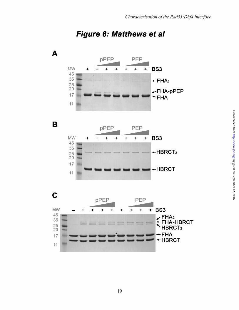
Among many other phosphorylated substrates, the FHA1 domain of Rad53 recognizes a phospoepitope within Cdc7 (37). We reasoned that Cdc7 might modulate the interaction between Rad53 and Dbf4, and thus we tested whether a Cdc7-derived peptide (480DGESpTDEDDVVS491, herein referred to as pPEP) could outcompete the interaction of Rad53 with Dbf4. To this end, we monitored complex formation using chemical crosslinking with the amine-reactive crosslinker bis[sulfsuccinimidyl] suberate (BS3). Upon treatment of the individual proteins with BS3, we could detect HBRCT dimers and, to a much lesser extent, FHA1 dimers (Fig. 6, control lanes). Treatment of FHA1:HBRCT mixtures with BS3
resulted in three crosslinked products. Two of them corresponded to the HBRCT and FHA1 dimers identified when each protein was treated with BS3 individually, whereas the third was only found when both proteins were present and had a molecular weight consistent with complex formation. The presence of both proteins in this crosslinked species was confirmed by LC-MS/MS (data not shown). The Cdc7 phosphorylated peptide (pPEP), but not its non-phosphorylated form (480DGESTDEDDVVS491, PEP), could be crosslinked to the FHA1 domain of Rad53 (Fig. 6A). Conversely, neither pPEP nor PEP could be crosslinked to the HBRCT domain of Dbf4 (Fig. 6B), indicating that BS3-crosslinking captured a specific and phosphorylation-dependent interaction between the FHA1 domain and the peptide. Titration of pPEP (or PEP) into equimolar mixtures of FHA1:HBRCT did not prevent the appearance of the crosslinked product corresponding to the FHA1:HBRCT complex (Fig. 6C). This result indicates that pPEP and HBRCT can bind the FHA1 domain simultaneously, thereby suggesting that they bind different regions of the FHA1 domain.
We confirmed these findings using NMR. To this end, we incubated a mixture of 15NFHA1 and HBRCT with increasing amounts of pPEP. We collected HSQC spectra at increasing concentrations of peptide corresponding to increasing ratios of pPEP:FHA1. We found that the presence of pPEP caused chemical shift perturbations consistent with phosphopeptide binding, while the non-phosphorylated version of the peptide (PEP) did not induce any changes in the 15NFHA1 HSQC spectrum (Fig. 7). Importantly, the peaks indicative of the interaction between FHA1 and HBRCT were present at all pPEP concentrations, although the peak corresponding to Gln16 could not be monitored in this experiment because it overlapped with the peak corresponding to Glu117 during the titration. Collectively, these experiments demonstrate that Rad53 interacts with a phosphoepitope and the HBRCT domain of Dbf4 through different surfaces of the FHA1 domain and that both interactions can occur simultaneously.
DISCUSSION We have shown that the HBRCT domain of Dbf4 interacts with the lateral surface of the Rad53
by guest on September 12, 2016
http://ww
w.jbc.org/
Dow
nloaded from

Characterization of the Rad53:Dbf4 interface
8
FHA1 domain, rather than its phosphoepitope-binding surface. This lateral surface is not conserved in the FHA2 domain of Rad53, indicating that the FHA1 domain of Rad53 gains specificity by engaging additional interaction surfaces. Additionally, FHA1 can simultaneously bind a phosphopeptide and the HBRCT domain. Therefore, the Rad53:Dbf4 interaction in vivo may occur through a bipartite interaction. The phosphorylated peptide consisting of 98-113 of Dbf4 includes a canonical pT105-X-X-E108 FHA-binding motif and interacts with the FHA1 domain weakly (19). Mutation of Thr105 in the context of full-length Dbf4, however, does not disrupt the interaction between Dbf4 and Rad53 (12). Chen and co-workers also showed that point mutations on the surface of the HBRCT domain defined by helices α0 and α3 abrogate the interaction with Rad53 suggesting that Thr105 may contribute to defining this interface rather than providing a pThr target site. Indeed, this surface of the HBRCT domain has good shape and charge complementarity with the lateral surface of the FHA1 domain (Almawi and Guarné, unpublished).
Several FHA domains have been shown to mediate bipartite interactions. For instance, the FHA domain of Dun1 requires the simultaneous interaction with two phosphorylated threonine residues (pThr) to recognize its target (21) and KIF13B interacts simultaneously with the ArfGAP and PH1 domains of CENTA1 (38). Therefore, FHA domains have been compared to logic gates able to integrate multiple inputs and generate a single output (22). When phosphopeptide binding is considered as the output, the FHA1 domain from Rad53 acts as an ‘OR’ logic gate because it can only bind to one pThr within a multiply phosphorylated peptide. In contrast, the FHA domain from Dun1 is considered an ‘AND’ logic gate because it must bind to two pThr simultaneously (21). If we extend the logic gate model to DDK targeting, the FHA1 domain of Rad53 also behaves as an ‘AND’ logic gate, requiring simultaneous binding of a phosphoepitope and engagement of the HBRCT domain of Dbf4 through its lateral interface for their normal physiological interaction (Fig. 8). An ‘AND’ logic gate would also explain why disabling either contact point abrogates the Rad53:Dbf4 interaction in a yeast two-hybrid assay and would have important ramifications
during the checkpoint response. DDK-mediated phosphorylation of Mcm2 has a role in the response to replicative stress, likely as a result of replication fork stabilization (39). Therefore, one of the logic gate inputs may guarantee the specificity of Rad53 targeting the HBRCT domain of Dbf4, while the second may ensure the stress-dependency of the interaction by recognizing a phosphoepitope generated by a stress-activated kinase.
Logic gates have emerged as critical components of biological signaling networks, thereby implying that FHA domains function as highly specialized modules that integrate complex input signals (22,40). The Dbf4:Rad53 interface unveiled in this work is reminiscent of the BRCA1:Chk2 interaction, where the tandem BRCT repeat of BRCA-1 must recognize two distal surfaces in the FHA domain of Chk2 simultaneously (41). In the BRCA1:Chk2 complex, the interaction involves the pThr-binding site and a conserved hydrophobic patch on one of the lateral surfaces of the FHA domain. Disruption of either contact point prevents the interaction, and mutation of the hydrophobic patch has been linked to Li-Fraumeni syndrome, which predisposes patients to multi-organ cancers (41). However, Dbf4 and BRCA-1 do not interact with the same lateral surface of the FHA domains of Rad53 and Chk2. Furthermore, the hydrophobic patch found in the FHA domain of Chk2 is not conserved in FHA1 from Rad53 revealing the extreme plasticity of FHA domains to enhance binding specificity. The interactions of Rad53 with Dbf4 or Chk2 with BRCA-1 lead to inhibition of late origin firing and double strand break repair, respectively. Therefore, it is not surprising that cells have developed strict selectivity mechanisms to modulate these two interactions. However, while the need for bipartite interactions increases Rad53 and Chk2 binding specificities, it also underlines the experimental difficulty in detecting dual input FHA domains as eliminating one binding site abrogates the interaction, effectively masking the presence of a second binding site.
Our work suggests that FHA domains may have a broader spectrum of interactions that has remained elusive due to the intrinsic limitations of studying ‘AND’ logic gates and it provides a novel
by guest on September 12, 2016
http://ww
w.jbc.org/
Dow
nloaded from

Characterization of the Rad53:Dbf4 interface
9
approach to study functional interactions mediated by this highly prevalent signaling domain.
ACKNOWLEDGEMENTS We thank Ahmad Almawi and other members of the Guarné laboratory for helpful discussions. This work has been funded by the Canadian Institutes of Health Research (MOP-67189 to A.G., and MOP-68897 to G.M.), the Natural
Sciences and Engineering Research Council (RGPIN 238392 to B.P.D.) and the McMaster-Waterloo Bioinformatics Program (to B.P.D., B.J.M. and A.G.). L.A.M. was supported by a CIHR doctoral scholarship. D.R.J. and R.S. were recipients of Ontario Graduate Scholarship doctoral awards.
REFERENCES
1. Durocher, D., and Jackson, S. P. (2002) The FHA domain. FEBS Lett 513, 58-66 2. Labib, K., and De Piccoli, G. (2011) Surviving chromosome replication: the many roles of the S-
phase checkpoint pathway. Philos Trans R Soc Lond B Biol Sci 366, 3554-3561 3. Branzei, D., and Foiani, M. (2006) The Rad53 signal transduction pathway: Replication fork
stabilization, DNA repair, and adaptation. Exp Cell Res 312, 2654-2659 4. Duncker, B. P., Shimada, K., Tsai-Pflugfelder, M., Pasero, P., and Gasser, S. M. (2002) An N-
terminal domain of Dbf4p mediates interaction with both origin recognition complex (ORC) and Rad53p and can deregulate late origin firing. Proc Natl Acad Sci U S A 99, 16087-16092
5. Kitada, K., Johnston, L. H., Sugino, T., and Sugino, A. (1992) Temperature-sensitive cdc7 mutations of Saccharomyces cerevisiae are suppressed by the DBF4 gene, which is required for the G1/S cell cycle transition. Genetics 131, 21-29
6. Jackson, A. L., Pahl, P. M., Harrison, K., Rosamond, J., and Sclafani, R. A. (1993) Cell cycle regulation of the yeast Cdc7 protein kinase by association with the Dbf4 protein. Mol Cell Biol 13, 2899-2908
7. Duch, A., Palou, G., Jonsson, Z. O., Palou, R., Calvo, E., Wohlschlegel, J., and Quintana, D. G. (2011) A Dbf4 mutant contributes to bypassing the Rad53-mediated block of origins of replication in response to genotoxic stress. J Biol Chem 286, 2486-2491
8. Kihara, M., Nakai, W., Asano, S., Suzuki, A., Kitada, K., Kawasaki, Y., Johnston, L. H., and Sugino, A. (2000) Characterization of the yeast Cdc7p/Dbf4p complex purified from insect cells. Its protein kinase activity is regulated by Rad53p. J Biol Chem 275, 35051-35062
9. Lopez-Mosqueda, J., Maas, N. L., Jonsson, Z. O., Defazio-Eli, L. G., Wohlschlegel, J., and Toczyski, D. P. (2010) Damage-induced phosphorylation of Sld3 is important to block late origin firing. Nature 467, 479-483
10. Zegerman, P., and Diffley, J. F. (2010) Checkpoint-dependent inhibition of DNA replication initiation by Sld3 and Dbf4 phosphorylation. Nature 467, 474-478
11. Gabrielse, C., Miller, C. T., McConnell, K. H., DeWard, A., Fox, C. A., and Weinreich, M. (2006) A Dbf4p BRCA1 C-terminal-like domain required for the response to replication fork arrest in budding yeast. Genetics 173, 541-555
12. Matthews, L. A., Jones, D. R., Prasad, A. A., Duncker, B. P., and Guarne, A. (2012) Saccharomyces cerevisiae Dbf4 has a unique fold necessary for the interaction with the Rad53 kinase. J Biol Chem 287, 2378-2387
13. Varrin, A. E., Prasad, A. A., Scholz, R. P., Ramer, M. D., and Duncker, B. P. (2005) A mutation in Dbf4 motif M impairs interactions with DNA replication factors and confers increased resistance to genotoxic agents. Mol Cell Biol 25, 7494-7504
14. Leung, C. C., and Glover, J. N. (2011) BRCT domains: easy as one, two, three. Cell Cycle 10, 2461-2470
15. Williams, R. S., Lee, M. S., Hau, D. D., and Glover, J. N. (2004) Structural basis of phosphopeptide recognition by the BRCT domain of BRCA1. Nat Struct Mol Biol 11, 519-525
by guest on September 12, 2016
http://ww
w.jbc.org/
Dow
nloaded from

Characterization of the Rad53:Dbf4 interface
10
16. Kobayashi, M., Ab, E., Bonvin, A. M., and Siegal, G. (2010) Structure of the DNA-bound BRCA1 C-terminal region from human replication factor C p140 and model of the protein-DNA complex. J Biol Chem 285, 10087-10097
17. Li, X., Liu, K., Li, F., Wang, J., Huang, H., Wu, J., and Shi, Y. (2012) Structure of C-terminal tandem BRCT repeats of Rtt107 protein reveals critical role in interaction with phosphorylated histone H2A during DNA damage repair. J Biol Chem 287, 9137-9146
18. Matthews, L. A., and Guarne, A. (2013) Dbf4: The whole is greater than the sum of its parts. Cell Cycle 12, 1180-1188
19. Chen, Y. C., Kenworthy, J., Gabrielse, C., Hanni, C., Zegerman, P., and Weinreich, M. (2013) DNA Replication Checkpoint Signaling Depends on a Rad53-Dbf4 N-terminal Interaction in Saccharomyces cerevisiae. Genetics 194, 389-401
20. Durocher, D., Taylor, I. A., Sarbassova, D., Haire, L. F., Westcott, S. L., Jackson, S. P., Smerdon, S. J., and Yaffe, M. B. (2000) The molecular basis of FHA domain:phosphopeptide binding specificity and implications for phospho-dependent signaling mechanisms. Mol Cell 6, 1169-1182
21. Lee, H., Yuan, C., Hammet, A., Mahajan, A., Chen, E. S., Wu, M. R., Su, M. I., Heierhorst, J., and Tsai, M. D. (2008) Diphosphothreonine-specific interaction between an SQ/TQ cluster and an FHA domain in the Rad53-Dun1 kinase cascade. Mol Cell 30, 767-778
22. Zhang, W., and Durocher, D. (2008) Dun1 counts on rad53 to be turned on. Mol Cell 31, 1-2 23. Nott, T. J., Kelly, G., Stach, L., Li, J., Westcott, S., Patel, D., Hunt, D. M., Howell, S., Buxton, R. S.,
O'Hare, H. M., and Smerdon, S. J. (2009) An intramolecular switch regulates phosphoindependent FHA domain interactions in Mycobacterium tuberculosis. Sci Signal 2, ra12
24. Sun, Z., Hsiao, J., Fay, D. S., and Stern, D. F. (1998) Rad53 FHA domain associated with phosphorylated Rad9 in the DNA damage checkpoint. Science 281, 272-274
25. Yongkiettrakul, S., Byeon, I. J., and Tsai, M. D. (2004) The ligand specificity of yeast Rad53 FHA domains at the +3 position is determined by nonconserved residues. Biochemistry 43, 3862-3869
26. Selvaratnam, R., Mazhab-Jafari, M. T., Das, R., and Melacini, G. (2012) The auto-inhibitory role of the EPAC hinge helix as mapped by NMR. PLoS One 7, e48707
27. Delaglio, F., Grzesiek, S., Vuister, G. W., Zhu, G., Pfeifer, J., and Bax, A. (1995) NMRPipe: a multidimensional spectral processing system based on UNIX pipes. J Biomol NMR 6, 277-293
28. Farrow, N. A., Muhandiram, R., Singer, A. U., Pascal, S. M., Kay, C. M., Gish, G., Shoelson, S. E., Pawson, T., Forman-Kay, J. D., and Kay, L. E. (1994) Backbone dynamics of a free and phosphopeptide-complexed Src homology 2 domain studied by 15N NMR relaxation. Biochemistry 33, 5984-6003
29. Diaz-Moreno, I., Diaz-Quintana, A., Molina-Heredia, F. P., Nieto, P. M., Hansson, O., De la Rosa, M. A., and Karlsson, B. G. (2005) NMR analysis of the transient complex between membrane photosystem I and soluble cytochrome c6. J Biol Chem 280, 7925-7931
30. Altschul, S. F., Madden, T. L., Schaffer, A. A., Zhang, J., Zhang, Z., Miller, W., and Lipman, D. J. (1997) Gapped BLAST and PSI-BLAST: a new generation of protein database search programs. Nucleic Acids Res 25, 3389-3402
31. Ashkenazy, H., Erez, E., Martz, E., Pupko, T., and Ben-Tal, N. (2010) ConSurf 2010: calculating evolutionary conservation in sequence and structure of proteins and nucleic acids. Nucleic Acids Res 38, W529-533
32. Jones, D. R., Prasad, A. A., Chan, P. K., and Duncker, B. P. (2010) The Dbf4 motif C zinc finger promotes DNA replication and mediates resistance to genotoxic stress. Cell Cycle 9, 2018-2026
33. Hagn, F., Lagleder, S., Retzlaff, M., Rohrberg, J., Demmer, O., Richter, K., Buchner, J., and Kessler, H. (2011) Structural analysis of the interaction between Hsp90 and the tumor suppressor protein p53. Nat Struct Mol Biol 18, 1086-1093
34. Gunasekara, N., Sykes, B., and Hugh, J. (2012) Characterization of a novel weak interaction between MUC1 and Src-SH3 using nuclear magnetic resonance spectroscopy. Biochem Biophys Res Commun 421, 832-836
by guest on September 12, 2016
http://ww
w.jbc.org/
Dow
nloaded from
Characterization of the Rad53:Dbf4 interface
11
35. Matsuo, H., Walters, K. J., Teruya, K., Tanaka, T., Gasser, S. M., Lippard, S. J., Kyogoku, Y., and Wagner, G. (1999) Identification by NMR spectroscopy of residues at contact surfaces in large, slowly exchanging macromolecular complexes. J. Am. Chem. Soc. 121, 9903-9904
36. Walters, K. J., Ferentz, A. E., Hare, B. J., Hidalgo, P., Jasanoff, A., Matsuo, H., and Wagner, G. (2001) Characterizing protein-protein complexes and oligomers by nuclear magnetic resonance spectroscopy. Methods Enzymol 339, 238-258
37. Aucher, W., Becker, E., Ma, E., Miron, S., Martel, A., Ochsenbein, F., Marsolier-Kergoat, M. C., and Guerois, R. (2010) A strategy for interaction site prediction between phospho-binding modules and their partners identified from proteomic data. Mol Cell Proteomics 9, 2745-2759
38. Tong, Y., Tempel, W., Wang, H., Yamada, K., Shen, L., Senisterra, G. A., MacKenzie, F., Chishti, A. H., and Park, H. W. (2010) Phosphorylation-independent dual-site binding of the FHA domain of KIF13 mediates phosphoinositide transport via centaurin alpha1. Proc Natl Acad Sci U S A 107, 20346-20351
39. Stead, B. E., Brandl, C. J., Sandre, M. K., and Davey, M. J. (2012) Mcm2 phosphorylation and the response to replicative stress. BMC Genet 13, 36
40. Hasty, J., McMillen, D., and Collins, J. J. (2002) Engineered gene circuits. Nature 420, 224-230 41. Li, J., Williams, B. L., Haire, L. F., Goldberg, M., Wilker, E., Durocher, D., Yaffe, M. B., Jackson,
S. P., and Smerdon, S. J. (2002) Structural and functional versatility of the FHA domain in DNA-damage signaling by the tumor suppressor kinase Chk2. Mol Cell 9, 1045-1054
by guest on September 12, 2016
http://ww
w.jbc.org/
Dow
nloaded from
Characterization of the Rad53:Dbf4 interface
12
FIGURE LEGENDS
FIGURE 1. The N-terminal domain of Dbf4 folds as a stable HBRCT in solution. (A) Ribbon diagram of the HBRCT domain of Dbf4 (PDB ID: 3QBZ) with α helices and β strands shown in yellow and teal, respectively. The side chains of the residues latching the α0 helix to the BRCT core are shown as color-coded sticks and labeled. (B) Secondary structure probabilities (Pα and Pβ) of the HBRCT fragment of Dbf4 as a function of residue number. Positive values indicate α-helices and negative values indicate β-strands. (C) 1H-15N NOE values as a function of residue number. Closed circles represent the experimentally measured values at 306 K. The solid horizontal line close to 0.8 indicates the theoretical 1H-15N NOE values computed using HydroNMR based on the overall tumbling of HBRCT (PDB: 3QBZ) in the absence of internal ps-ns motions. The black dotted line represents the secondary structure of the crystal structure and is labeled as in (A). (D) Limited trypsin proteolysis of the HBRCT (left), the HBRCT-W116D/M120A (center), and the HBRCT-L109A/W112S (right) domains. From left to right each gel has molecular weight markers (MW, indicated in kDa), the HBRCT variant in the absence of trypsin, the HBRCT variant treated with increasing concentrations of trypsin (0.195 – 12.5 µg/mL) and the purified BRCT domain of Dbf4 in the absence of trypsin as a molecular weight reference.
FIGURE 2. The FHA1 domain of Rad53 interacts with the HBRCT domain of Dbf4 in vitro. (A) Detailed view of the HSQC spectra of 15NFHA1 collected in the absence (orange) or presence (blue) of the HBRCT domain of Dbf4. The interaction is identified by residue-specific intensity losses, as well as by the appearance of ten new peaks (labeled in black) in the spectra. (B) Same as in (A) but with the HBRCT domain replaced by the BRCT domain of Dbf4. This control experiment confirms that the FHA1:HBRCT interaction detected by NMR specifically requires the N-terminal helix α0. (C) The peak height from the HSQC spectrum of 15NFHA1 in the presence of the HBRCT domain measured as a percentage of the peak height of 15NFHA1 alone. The black line marks the mean percentage (46.0%) and the red line marks one standard deviation below this value (33.8%). Peaks reduced beyond one standard deviation are labeled. (D) Ribbon diagram of the FHA1 domain of Rad53 (PDB ID: 1G6G), with the residues whose peak intensities were reduced beyond one standard deviation in the presence of the HBRCT domain shown as green sticks and labeled. The pThr-containing peptide is shown as an orange ribbon with the side chains of pT and pT+3 labeled.
FIGURE 3. The FHA1 domain of Rad53 has a conserved lateral surface. (A-B) Surface representation of the FHA1 domain of Rad53 (PDB ID: 1G6G) with residue conservation shown as standard deviations from the mean where blue indicates conserved and yellow indicates variable. The FHA1 domain has two highly conserved areas on opposite faces of the molecule: the phosphopeptide binding pocket (A) and the lateral β-sheet composed of strands β2-β1-β11-β10-β7-β8 (B). The view in (B) is a 180° rotation from panel (A). (C) Ribbon diagram of the FHA1 domain of Rad53 (PDB ID: 1G6G), shown in the same orientation as in (B) and colored as in Fig. 2D. Residues defining the conserved lateral region of FHA1 domain are shown as purple sticks and labeled.
FIGURE 4. The lateral surface of the FHA1 domain of Rad53 is required for the interaction with the HBRCT domain of Dbf4. (A-B) Yeast two-hybrid analysis using Dbf4 as bait and either the FHA1 domain of Rad53 (WT), empty vector (Empty) or variants of the FHA1 containing double (A) or multiple (B) mutations within the interaction surface predicted from NMR and bioinformatics analysis as preys. The double mutants and multiple mutants are labeled as: IT (I37A/T39A), VF (V144N/F146A), NE (N112A/E129A), NEIT (N112A/E129A/I37A/T39A), NEVF (N112A/E129A/V144N/F146A) and ITVF
by guest on September 12, 2016
http://ww
w.jbc.org/
Dow
nloaded from

Characterization of the Rad53:Dbf4 interface
13
(I37A/T39A/V144N/F146A). The interaction is shown as β-galactosidase activity units and in each case represents the mean of three independent measurements. Error bars represent standard deviation. (C) Yeast two-hybrid analysis using Dbf4 as bait and either the FHA1 domain of Rad53 (WT), empty vector (Empty) or the FHA1-V134D/I140A variant containing a double mutant in the β10-β11 loop (VI). To control for the two-hybrid bait and prey expression levels in A-C, whole cell extracts were prepared from transformants following prey induction and analyzed by Western blot using rabbit anti-LexA antibody (bait) and a mouse monoclonal anti-HA antibody (prey), along with Alexa Fluor 647 goat anti-rabbit and 488 goat anti-mouse secondary antibodies, respectively. Prior to detection, the membrane was stained with Ponceau S to assess relative protein loading. (D) Sensitivity of Rad53 variants to genotoxic stress was tested in the presence of hydroxyurea (HU) or methyl methanesulfonate (MMS). The growth of a rad53Δ, sml1Δ strain in the presence of plasmids encoding Rad53, Rad53-NE, Rad53-NEVF and Rad53-VF was compared on SC-Leu plates containing increasing concentrations of HU or MMS by spotting 10-fold serial dilutions. RAD53, sml1Δ and rad53Δ, sml1Δ strains transformed with an empty plasmid were used as controls for normal sensitivity or hypersensitivity to genotoxic agents, respectively. Rad53 expression was assessed using a goat anti-rad53 antibody (Santa Cruz Biotech) and Alexa Fluor 488 donkey anti-goat secondary antibody.
FIGURE 5. The 15NFHA1-R70A variant interacts with the HBRCT domain. Overlay of the HSQC spectra of 15NFHA1-R70A (black) and 15NFHA1-R70A in the presence of unlabeled HBRCT (red). Peaks indicative of the FHA1:HBRCT interaction are labeled in red.
FIGURE 6. The FHA1 domain can interact simultaneously with a phosphopeptide and the HBRCT domain of Dbf4. (A) BS3-crosslinking of the FHA1 domain to a pThr-containing peptide (pPEP) and its non-phosphorylated counterpart (PEP). The appearance of a new crosslinked product in the presence of pPEP, but not PEP, confirms that BS3 captures the specific interaction between the FHA1 domain and the phosphopeptide. (B) Control experiment showing that neither pPEP nor PEP interact with the HBRCT domain of Dbf4. (C) The presence of pPEP (or PEP) does not impair the interaction between the HBRCT and FHA1 domains. For A-C, the BS3-crosslinked products are resolved in 18% SDS-polyacrylamide gels stained with Coomassie brilliant blue and the molecular weight (MW) markers are indicated in kDa. The presence/absence of BS3 is indicated with ‘+’ or ‘-’ signs.
FIGURE 7. The HBRCT domain of Dbf4 does not bind to the phosphoepitope-binding pocket of the FHA1 domain. HSQC spectra monitoring the formation of the complex between 15NFHA1 and HBRCT at a 1:1 ratio in the presence of stoichiometric amounts of either DGESpTDEDDVVS (A) or DGESTDEDDVVS (B). The ten peaks indicating the interaction between the two domains are labeled in red. As a reference, some of the peaks corresponding to residues that undergo significant chemical shift perturbations or line broadening upon pPEP binding are labeled in blue in both panels.
FIGURE 8. Model of the interaction between Rad53 and DDK. Upon activation of the replication checkpoint, Rad53 (blue) is activated through hyperphosphorylation mediated both by Rad53 in trans, as well as additional kinases including DDK (shown in yellow (Dbf4) and red (Cdc7)). Reciprocally, Dbf4 is one of Rad53 targets during the checkpoint response, causing a significant reduction in Cdc7 kinase, thereby averting late origin firing. The Rad53:DDK interaction is mediated by two independent interfaces on the FHA1 domain. One of the lateral surfaces of the FHA1 domain recognizes the Dbf4 HBRCT domain while the phosphoepitope binding site binds to a second epitope present in DDK. These two surfaces must both be engaged for Rad53 to avert late origin firing and stabilize replication forks.
by guest on September 12, 2016
http://ww
w.jbc.org/
Dow
nloaded from

Characterization of the Rad53:Dbf4 interface
14
by guest on September 12, 2016
http://ww
w.jbc.org/
Dow
nloaded from

Characterization of the Rad53:Dbf4 interface
15
by guest on September 12, 2016
http://ww
w.jbc.org/
Dow
nloaded from

Characterization of the Rad53:Dbf4 interface
16
by guest on September 12, 2016
http://ww
w.jbc.org/
Dow
nloaded from

Characterization of the Rad53:Dbf4 interface
17
by guest on September 12, 2016
http://ww
w.jbc.org/
Dow
nloaded from

Characterization of the Rad53:Dbf4 interface
18
by guest on September 12, 2016
http://ww
w.jbc.org/
Dow
nloaded from
Characterization of the Rad53:Dbf4 interface
19
by guest on September 12, 2016
http://ww
w.jbc.org/
Dow
nloaded from

Characterization of the Rad53:Dbf4 interface
20
by guest on September 12, 2016
http://ww
w.jbc.org/
Dow
nloaded from
Characterization of the Rad53:Dbf4 interface
1/2
Figure S1. Analysis of the HSQC spectra of 15NFHA1 in the presence of the HBRCT domain of
Dbf4. Assigned 15N-H resonances of the FHA1 domain. Peaks that appear only in the presence of the
HBRCT domain are labeled in red.

Characterization of the Rad53:Dbf4 interface
2/2
Table S1: Role of the lateral surface of the FHA1 domain on the interaction with Dbf4
Mutant Residual Interaction (%) STD (%)
Rad53(1-165) 100 --
Rad53(1-165)-N112A 75 3
Rad53(1-165)-E129A 94 9
Rad53(1-165)-IT 91 10
Rad53(1-165)-VF 107 8
Rad53(1-165)-NE 47 6
Rad53(1-165)-NEIT 11 4
Rad53(1-165)-NEVF 10 1
Rad53(1-165)-ITVF 64 14
Rad53(1-165)-NEITVF 11 5
FHA1(ΔN)* 95 6
* The fragment referred to as FHA1(ΔN) includes residues 22-165
J. McConkey, Giuseppe Melacini, Bernard P. Duncker and Alba GuarneLindsay A. Matthews, Rajeevan Selvaratnam, Darryl R. Jones, Madoka Akimoto, Brendan
and DBF4A novel, non-canonical fha binding interface mediates the interaction between RAD53
published online November 27, 2013J. Biol. Chem.
10.1074/jbc.M113.517060Access the most updated version of this article at doi:
Alerts:
When a correction for this article is posted•
When this article is cited•
to choose from all of JBC's e-mail alertsClick here
Supplemental material:
http://www.jbc.org/content/suppl/2013/11/27/M113.517060.DC1.html
http://www.jbc.org/content/early/2013/11/27/jbc.M113.517060.full.html#ref-list-1
This article cites 0 references, 0 of which can be accessed free at
by guest on September 12, 2016
http://ww
w.jbc.org/
Dow
nloaded from
Copyright © 2022 FDOKUMEN