Negative regulation of autoimmune demyelination by the inhibitory receptor CLM-1
Upload
independentCategory
view
6download
0
IDO Mediates TLR9-Driven Protection from ExperimentalAutoimmune Diabetes1
Francesca Fallarino,2 Claudia Volpi,2 Teresa Zelante, Carmine Vacca, Mario Calvitti,Maria C. Fioretti, Paolo Puccetti, Luigina Romani, and Ursula Grohmann3
Originally predicated on the recognition of an increasing prevalence of allergy, the hygiene hypothesis was later found to accom-modate the contrasting epidemiologic trends in developed countries for infectious vs autoimmune diseases. Experimentally, re-duced exposure to infections will increase the risk of disease in several models of experimental autoimmunity. Although TLRs wereinitially considered as stimulatory molecules capable of activating early defense mechanisms against invading pathogens, emergingdata suggest that they can also exert a regulatory function. In the present study, we evaluated whether TLR3 and TLR9, recog-nizing microbial dsDNA and CpG-containing DNA sequences, respectively, play a role in the protection from experimentalautoimmune diabetes induced in C57BL/6 mice by streptozotocin. In wild-type animals, the disease was accompanied by up-regulation of IDO in pancreatic lymph nodes and would be greatly exacerbated by in vivo administration of an IDO inhibitor.Conversely, administration of a CpG-containing oligodeoxynucleotide greatly attenuated the disease in an IDO-dependent fashion.TLR9-, but not TLR3-deficient mice developed a more robust disease, an event accompanied by lack of IDO induction in pan-creatic lymph nodes. Thus, our data suggest that the TLR9-IDO axis may represent a valuable target in the prevention/therapyof type 1 diabetes. The Journal of Immunology, 2009, 183: 6303–6312.
U nlike the majority of organs and tissues in the humanbody, the immune system requires systematic environ-mental pressure to develop properly. Animals raised in a
germfree environment acquire poorly efficient immunoregulatorymechanisms and are at a greater risk of diseases associated withimmune dysfunction (1). The incidence of major infectious dis-eases has significantly decreased in developed countries over thelast three decades. In parallel, the incidence of most autoimmunediseases, including type 1 diabetes (T1D),4 has steadily been in-creasing in Europe and North America (2, 3). Therefore, both ex-perimental and epidemiological data suggest that certain microor-ganisms will induce a state of protective tolerance in mammalstoward autoimmune diseases such as T1D (4–6).
TLRs represent the early molecular sensors of invading micro-organisms and link innate with adaptive immune responses (7–9).To date, 11 members of TLR have been identified in humans, and13 in mice, and a series of genetic studies revealed their respectiveligands. Mammalian TLRs can be expressed on the cell membrane(i.e., TLR1, TLR2, TLR5, and TLR6), intracellularly (TLR3,TLR7, TLR8, and TLR9), or both (TLR4). The distinct localiza-
tion correlates with a different nature of stimulatory ligands—withtransmembrane TLRs primarily sensing microbial membrane mol-ecules, such as LPS and lipopeptides (TLR1, TLR2, TLR5, andTLR6), and intracellular forms recognizing microbial nucleic ac-ids, such as CpG-containing DNA sequences (TLR9) and viralssRNA (TLR7 and TLR8) or dsDNA (TLR3) (9), or a differentsignaling pathway (TLR4) (10).
The genetically diabetes-prone NOD mouse strain represents aprototypic experimental model of human T1D. In accordance withthe principle of protective tolerance, decontamination of NODmice from microbes does increase diabetes frequency. Conversely,deliberate infection of those mice with various microorganismstotally prevents diabetes onset if infection occurs early in life (5).An outstanding study has recently demonstrated that the interac-tion of intestinal microbes with the innate immune system is acritical epigenetic factor modifying T1D predisposition in NODmice (11). Nonetheless, the precise mechanisms of induction oftolerance by the microbiota remain to be elucidated.
IDO is expressed in dendritic cells (DCs) in response to inflam-matory stimuli, including IFN-� and CpG oligodeoxynucleotide(ODN), and represents an important physiological mechanismcapable of controlling both inflammation and autoimmunity(12–14). IDO catalyzes the first and rate-limiting step of tryp-tophan catabolism along the kynurenine pathway, which pro-duces a series of tryptophan catabolites collectively known askynurenines. We have previously demonstrated that IDO activ-ity, hence, tryptophan catabolism, is not inducible by IFN-� inDCs from NOD mice (15). This defect impairs tolerogenesis toa � cell-specific autoantigen also dominant in human T1D. Al-though maneuvers aimed at correcting the IDO defect will re-store autoantigen-specific tolerogenesis (16), it is not currentlyknown whether tryptophan catabolism plays a role in the de-velopment of the disease.
In the present study, we investigated whether TLRs and IDO areinvolved in the control of autoimmune diabetes in mice nonge-netically prone to the disease. We found that both TLR9 and IDOare required for protection from an inflammatory/prodiabetic insult
Department of Experimental Medicine and Biochemical Sciences, University of Pe-rugia, Perugia, Italy
Received for publication May 21, 2009. Accepted for publication September 7, 2009.
The costs of publication of this article were defrayed in part by the payment of pagecharges. This article must therefore be hereby marked advertisement in accordancewith 18 U.S.C. Section 1734 solely to indicate this fact.1 This work was supported in part by the Associazione per l’Aiuto ai Giovani conDiabete dell’Umbria (to U.G.).2 F.F. and C.V. contributed equally to this work.3 Address correspondence and reprint requests to Dr. Ursula Grohmann, Departmentof Experimental Medicine and Biochemical Sciences, Via del Giochetto, 06126 Pe-rugia, Italy. E-mail address: [email protected] Abbreviations used in this paper: T1D, type 1 diabetes; 1-MT, 1-methyl-DL-trypto-phan; DC, dendritic cell; IKK, I�B kinase; ODN, oligodeoxynucleotide; pDC, plas-macytoid DC; PLN, pancreatic lymph node; SPF, specific pathogen free; STZ, strep-tozotocin; Treg, T regulatory; WT, wild type.
Copyright © 2009 by The American Association of Immunologists, Inc. 0022-1767/09/$2.00
The Journal of Immunology
www.jimmunol.org/cgi/doi/10.4049/jimmunol.0901577

in healthy conditions. However, engagement of TLR9 by CpG-ODN did not efficiently induce IDO expression and activity inlymphoid tissues from NOD mice. Thus, our data: 1) confirm theprotective role of TLRs in the prevention of T1D, 2) identify IDOas the critical TLR9 downstream effector in regulating inflamma-tion/autoimmunity, and 3) suggest that successful prevention/ther-apy of T1D in genetically diabetes-prone subjects may requiremultiple and integrated approaches, capable of restoring an IDO-mediated, physiologically protective tolerance.
Materials and MethodsMice
Four- and 8-wk-old C57BL/6 mice of either sex were obtained fromCharles River Breeding Laboratories. Mice homozygous for the TLR3(Tlr3�/�) and TLR9 (Tlr9�/�) targeted mutation raised on the C57BL/6background were generated as described (17, 18), and bred at the breedingfacilities of the University of Perugia. Female NOD/Mrk mice, 4 wk of age,were purchased from Taconic Farms. All animals were housed and fed underspecific pathogen-free (SPF) conditions. All in vivo studies were in compli-ance with national (Italian Approved Animal Welfare Assurance A-3143-01)and Perugia University Animal Care and Use Committee guidelines.
In vivo treatments
For the induction of experimental autoimmune diabetes, wild-type (WT),Tlr3�/�, or Tlr9�/� C57BL/6 mice were injected i.p. for 5 consecutivedays with 40 mg/kg body weight of freshly made streptozotocin (STZ;Sigma-Aldrich) in 0.01 mol/L citrate buffer (pH 4.5). Day 1 was that of thefirst STZ injection. Control mice received vehicle alone. Nonfasting glu-cose levels in tail vein blood samples were monitored 1–2 times/wk. Micewith a blood glucose level of �250 mg/dl for �2 consecutive days wereconsidered diabetic. To inhibit IDO activity in vivo, groups of WT micewere treated with slow-release pellets of 1-methyl-DL-tryptophan (1-MT;150 mg/pellet, 7-day release at 0.9 mg/h) or placebo (both from InnovativeResearch of America) implanted on day 0 under the dorsal skin of therecipients, as described (19). In selected experiments, C57BL/6 WT micewere treated with 25 �g of CpG-ODN 1826 5�-TCCATGACGTTCCTGACGTT-3� (custom, phosphorothioate; Invitrogen Life Technologies) ondays 7 and 9, whereas 4-wk-old NOD mice received the same dose ofCpG-ODN twice per week for 3 wk.
Histopathology and immunohistochemistry
Paraffin-embedded sections (3–4 �m) of pancreata (five per organ) werestained with H&E and analyzed by light microscopy. Insulitis scoring wasaccording to the following criteria: severe insulitis (score 3), 50% or higherof the islet area is infiltrated; mild insulitis (score 2), �50% of the islet areais infiltrated; peri-insulitis (score 1), infiltration is restricted to the periphery ofislets; and no insulitis (score 0), absence of cell infiltration. Results are pre-sented as the percentage of islets per mouse in each category. At least 40 isletswere counted per mouse blindly by two observers. In immunostaining forinsulin, 4-�m sections were cut from paraffin blocks and captured on electri-cally charged slides (Sigma-Aldrich). Sections were dewaxed in zylene andstained with primary pig anti-mouse insulin Abs (DakoCytomation) for 1 h,washed in PBS, and incubated with goat anti-guinea pig tetramethyl rhoda-mine isothiocyanate conjugate for 45 min. Nuclei were counterstained with4�,6-diamidino-2-phenylindole and mounted in 1,4-diazabicyclo[2.2.2]octanesolution (all from Sigma-Aldrich). Slides were examined using a BX 41 ap-paratus in conjuction with F-View software (both from Olympus).
Cell isolation from pancreatic lymph nodes (PLNs) andperipheral lymphoid organs
Purification of CD4� T cells from pooled peripheral lymph nodes (with theexception of PLNs; see below) was conducted as described (20–22). Forcytokine induction, cells (1 � 106/ml) were cultured in the presence ofplate-bound anti-CD3� (145-2C11) and anti-CD28 (PV-1) (both at the con-centration of 1 �g/ml) for 48 h. Purification of PLN lymphocytes involvedtreating the organ with Complete Mini Protease inhibitors (Roche AppliedScience), followed by digestion with collagenase type IV (Sigma-Aldrich)in the presence of bovine pancreatic DNase (Sigma-Aldrich) for 30 min at37°C. The digested pancreata were further disrupted by gently pushing thetissue through a nylon screen, and pancreatic lymphocytes were separatedon a Percoll gradient (Sigma-Aldrich). Purified PLN cells in STZ-treatedmice (day 21) were made up of �10% lymphocytes (mostly CD4�), 3%monocytes, and 5% CD11c�Ia� cells. In prediabetic NOD mice, the
corresponding percentages were 16, 25, and 6%. Splenic DCs were purifiedby magnetic-activated sorting using CD11c MicroBeads and MidiMacs(Miltenyi Biotec), in the presence of EDTA to disrupt DC-T cell com-plexes, as described (15, 19). Cytokine production from PLN lymphocytesand splenic DCs was measured in culture supernatants harvested after 24-hcell incubation with medium alone. For the purification of plasmacytoidDCs (pDCs), CD11c� cells were further fractionated using mPDCA-1MicroBeads (Miltenyi Biotec), as described (21).
Flow cytometry
In all FACS analyses, cells were treated with rat anti-CD16/32 (2.4G2) for 30min at 4°C for blockade of Fc receptors before assaying on an EPICS flowcytometer using EXPO 32 ADC software (Beckman Coulter). CD4, CD25,CTLA-4, and CD62L expressions were analyzed as described (21). For intra-cellular Foxp3, cells were stained with anti-CD4 (GK1.5)-PE (BD Pharmin-gen), fixed, permeabilized, and stained with Alexa Fluor 488 anti-mouseFoxp3 (MF-14; Biolegend) or isotype control Alexa Fluor 488 rat IgG2b.
T regulatory (Treg) cell purification and suppression assay
CD4�CD25� and CD4�CD25� T cells were isolated from lymph nodes byMACS, as described (20, 23). The purity of either T cell fraction was morethan 95%. For polyclonal Treg suppression assay, CD4�CD25� cells werecocultured with irradiated T cell-depleted splenocyte samples andCD4�CD25� cells for 3 days in the presence of soluble anti-CD3 (20, 23).Proliferation was measured by incorporation of [3H]thymidine, according tostandard procedures.
ELISA and TGF-� bioassay
Nonfasting blood insulin levels were measured by a mouse insulin ELISAkit (Mercodia AB). Cytokines (IL-2, IL-4, IL-6, IL-10, IL-17A, IL-23, andIFN-�) were measured in culture supernatants by ELISA using specific kitsor previously described reagents and procedures (21, 24). An ELISA-basedTransAM Flexi NF-�B Family Kit (Active Motif) was used to monitoractivity of NF-�B family members, as described (21, 24). Active TGF-�was measured, as described (25), using CCL-64 mink lung epithelial in-dicator cells (American Type Culture Collection), which do not appear toactivate inactive TGF-� precursor molecules, but are extremely sensitive togrowth inhibition induced by the biologically active cytokine.
Real-time PCR
Real-time PCR analysis was performed, as described (26), using Ido1-specificprimers (S, 5�-GAAGGATCCTTGAAGACCAC-3�; AS, 5�-GAAGCTGCGATTTCCACCAA-3�). For all panels, bars represent the ratio of gene toGapdh expression, as determined by the relative quantification method (��cycle threshold; mean SD of triplicate determination).
Western blotting and IDO functional analysis
IDO expression was investigated in cells cultured overnight with completemedium either alone or in the presence of 1 �g/ml CpG-ODN by immu-noblot with a rabbit anti-mouse IDO mAb raised in our laboratory (22).Anti-�-actin Ab (Sigma-Aldrich) was used as a normalizer. For measuringIDO functional activity, pDCs were stimulated either with 200 U/ml IFN-�(R&D Systems) or 1 �g/ml CpG-ODN and, after 18 h, L-kynurenine, themain IDO product, was measured in culture supernatants by HPLC, asdescribed (19). In PLN lymphocytes, treatment with IFN-�, but not CpG-ODN, greatly reduced the cell viability, and thus, could not be used.
Nuclear extracts and EMSA
Nuclear extract preparation and EMSA were performed, as described (27).Briefly, DNA-binding reactions were conducted in 20 min at room tem-perature in a final volume of 20 �l. The reactions were started by adding10 �g of nuclear protein extract to a reaction mix containing �20,000 cpmof [�-32P]ATP-labeled NF-�B dsDNA ODN (5�-AGAGGGGACTTTCCGAGAGGC-3�) (27). Cold competitor ODN were added to the reactionmix before the radiolabeled probe (data not shown). For supershift exper-iments, protein extracts were incubated with anti-p65 or anti-RelB Ab(Santa Cruz Biotechnology) for 30 min at room temperature after the ad-dition of the radiolabeled probe. Whole samples were then loaded on a 5%native polyacrylamide gel in Tris-borate-EDTA buffer.
Statistical analysis
In the in vivo experiments, glycemia data were analyzed by Kaplan-Meierplots. Paired data were evaluated by Student’s t test. All in vitro determi-nations are means SD from at least three independent experiments, un-less otherwise indicated. All n values were computed by power analysis, soas to yield a power of at least 80% with an �-level of 0.05.
6304 TLR9 AND IDO IN EXPERIMENTAL AUTOIMMUNE DIABETES

ResultsTlr9�/� mice are highly susceptible to experimentalautoimmune diabetes
To investigate the role of TLR3 and TLR9 in the pathogenesis ofchemically induced autoimmune diabetes, we injected Tlr3�/� andTlr9�/� mice and their C57BL/6 WT counterparts with multiplelow doses of STZ. From 14 days, average were significantly higherin Tlr9�/�, but not Tlr3�/� mice, as compared with WT animals,and further increased thereafter (Fig. 1A). In addition, whereas atleast 40% WT and Tlr3�/� animals remained normoglycemic atday 40, all Tlr9�/� mice became diabetic as early as day 20(Fig. 1, B and C). Although serum insulin levels were generally
decreased in STZ-treated as compared with vehicle-injectedcounterparts, insulinemia in STZ-treated Tlr9�/� mice was sig-nificantly lower when compared with STZ-treated WT mice(Fig. 1D). Groups of mice injected with STZ were sacrificed atdifferent times, and 5-�m-thick sections of pancreas werestained with H&E or immunostained for insulin. The majorityof pancreatic islets of WT and Tlr3�/� mice were either normalor mildly infiltrated by leukocytes and clearly positive for in-sulin (Fig. 1E, day 40; Fig. 1F, days 14 and 40). In contrast, asmuch as 17% (at day 14) or 50% (at day 40) of the islets inTlr9�/� mice were characterized by intrainsulitis and low in-sulin. Thus, our data suggest that Tlr9�/�, but not Tlr3�/�
FIGURE 1. Higher susceptibility to STZ-induced diabetes in Tlr9�/� mice. WT (n 12), Tlr3�/� (n 10), and Tlr9�/� (n 10) C57BL/6 mice wereinjected with STZ from day 1, and blood glucose was monitored over time (in A–C, one experiment is depicted of two with similar results). Diabetes wasdiagnosed in mice with blood glucose level �250 mg/dl. Mice were sacrificed at different times and analyzed for insulinemia (D) and pancreas histology(E and F) and immunohistochemistry (E). A, Average blood glucose in different groups is plotted over time. Data are presented as mean glucose levels SD. �, p � 0.01 (Tlr9�/� vs WT mice). B, Blood glucose concentrations over time in individual WT and Tlr9�/� mice. C, Incidence of diabetes over timein TLR-deficient and WT mice. p � 0.01 (Tlr9�/� vs WT mice). D, Blood insulin was measured by ELISA at day 30. Control, control animals treatedwith vehicle alone. Data are means SD of three experiments. �, p � 0.05 (STZ-treated Tlr9�/� vs WT mice). E and F, Pancreatic tissues were processedfor H&E staining to evaluate insulitis (E and F) and immunostained for insulin (red, E). In fluorescent images, nuclei were counterstained with 4�,6-diamidino-2-phenylindole (blue). Representative islet area for each group (E) at day 40 and the percentages of islets per mouse with different score (F, seeMaterials and Methods) of lymphocyte infiltration (days 14 and 40) are shown. Percentages represented number of islets of a given score (see Materialsand Methods) over total number of islets (30–40 per pancreas). �, p � 0.01 and ��, p � 0.001 (Tlr9�/� vs WT mice).
6305The Journal of Immunology
C57BL/6 mice develop an earlier and more severe form of ex-perimental autoimmune diabetes than WT counterparts.
Lack of induction of Foxp3� Treg cells and higher productionof proinflammatory IL-6 and IL-17A characterize diabetes inTlr9�/� mice
Although Th1 cells appear to play a major role in autoimmunediabetes via IFN-� production, discordant observations have beenfound on NOD mice expressing targeted mutations in either IFN-�or its receptor (28–30). More recently, Th17 cells, clearly involvedin many autoimmune diseases, such as experimental autoimmuneencephalomyelitis, rheumatoid arthritis, and myocarditis, havebeen shown to promote pancreatic inflammation, although theyinduce diabetes efficiently in NOD/SCID mice only after conver-sion into Th1 cells (31, 32). In contrast, there is compelling evi-dence indicating that the development of diabetes in NOD mice istightly controlled by Treg cells, via production of TGF-� andIL-10 (33). In experimental diabetes induced by STZ, the patternof pathogenetic vs protective cells and cytokines is less clear, al-though administration of IL-23, a Th17-sustaining cytokine,greatly exacerbates the disease (34).
To examine whether TLR9 expression would affect the balanceof Th and Treg responses in chemically induced diabetes, we usedflow cytometry to evaluate the percentage and phenotype of CD4�
T cells from lymphoid organs, i.e., spleen and lymph nodes, andPLN lymphocytes at day 21 of STZ treatment in WT, Tlr3�/�, andTlr9�/� mice. Mice injected with vehicle alone were used as acontrol. Table I shows that STZ treatment did not significantlymodify the basal frequency of gated CD4�CD25� T cells coex-pressing the T cell activation marker CD62L in any groups. Incontrast, the percentages of CD4�CD25� T cells coexpressingeither CTLA-4 or Foxp3, the Treg cell lineage transcription factor,significantly increased upon STZ treatment in samples from WTand Tlr3�/�, but not Tlr9�/�, mice. Foxp3 staining of CD4� Tcells gated from lymph nodes further confirmed no significantmodulation in CD4�Foxp3� cells from TLR9-deficient micetreated with STZ (Fig. 2, A and B). However, although disparate intheir occurrence, the suppressive activity of Treg cells purifiedfrom STZ-treated Tlr9�/� mice did not differ from that of Tregcells from diabetic WT or Tlr3�/� animals (Fig. 2C).
We next analyzed the pattern of Th1, Th2, Th17, and Treg-associated cytokine production by activated CD4� T cells purifiedfrom peripheral lymph nodes. In addition, cytokine production byCD11c� DCs purified from the spleen and by PLN lymphocytes,both types of cell under culture for 24 h without added stimuli, wasalso evaluated (Fig. 2D). In CD4� T cell cultures, we found that invivo treatment with STZ induced a similar trend for the majority ofthe tested cytokines in both WT and TLR-deficient mice, withcomparable increase in IL-2, IL-10, and TGF-� and a decrease inIFN-� production, whereas IL-4 production was not modified. Incontrast, the increase in IL-17A production in response to the drug
was significantly higher in CD4� cell cultures from Tlr9�/�, butnot Tlr3�/� mice as compared with WT mice. In DC cultures,IL-23 increased equally in all STZ-treated groups, although a dra-matic increase in IL-6 was found in supernatants from TLR9-de-ficient cells. In parallel, IL-6 and IL-17A levels were also signif-icantly higher in PLN lymphocytes from STZ-treated Tlr9�/�, butnot Tlr3�/� mice, as compared with WT animals. Thus, our datasuggest that lack of TLR9 expression may lead to a higher pro-duction of proinflammatory IL-6 in diabetic Tlr9�/� mice that,combined with high levels of IL-23, may impede the TGF-�-me-diated expansion of Foxp3� Treg cells and skew the balance to-ward the development and differentiation of pathogenic Th17 cells.
IDO is not expressed in PLNs of diabetic Tlr9�/� mice
IDO has a primary role in the peripheral generation of Treg cells,under physiological or pathological conditions (14). Furthermore,in experimental pathogenic inflammation, IDO helps to tame over-zealous and exaggerated inflammatory response driven by IL-23and IL-17 (22). In contrast, these cytokines can down-regulatetryptophan catabolism (35, 36). Signaling through TLR9 inducesIDO expression in splenic DCs, particularly in the plasmacytoidsubset (pDC), and functional tryptophan catabolism is necessaryfor TLR9-driven immunosuppressive effects (14, 37). We there-fore became interested in ascertaining whether a defect in IDOexpression could be involved in the exacerbated disease inducedby STZ in Tlr9�/� mice. To this purpose, we analyzed IDO ex-pression and activity in both splenic pDCs and PLN cells purifiedfrom mice at day 21 of STZ treatment (Fig. 3). Mice injected withvehicle alone were used as control. We found that IDO transcript(Fig. 3A) and protein expression greatly increased in splenic pDCsupon in vivo STZ treatment of WT and Tlr3�/� mice, whereas inpDCs from animals lacking TLR9 the induction was barely de-tectable. In PLNs, IDO was expressed in samples from WT andTlr3�/� mice and further increased following STZ treatment. Incontrast, IDO expression could not be detected under basal con-ditions nor after in vivo treatment of Tlr9�/� mice with STZ (Fig.3, B and C). IDO expression was accompanied by significantkynurenine production by splenic pDCs from WT and Tlr3�/�
mice after in vivo treatment with STZ and further increased inresponse to in vitro restimulation with IFN-�, whereas a signifi-cantly lower level of the tryptophan metabolite was present inparallel culture supernatants from Tlr9�/� pDCs, either untreatedor treated with the cytokine (Fig. 3D). Thus, our data suggest thatdeficient TLR9 expression may determine defective IDO inductionin response to pancreatic inflammation.
In vivo treatment with CpG-ODN protects mice fromSTZ-induced diabetes in an IDO-dependent fashion
To evaluate the functional role of IDO as induced in the course ofdiabetes development and its potential link with TLR9 activation,WT recipients were administered 1-MT, the gold standard in IDO
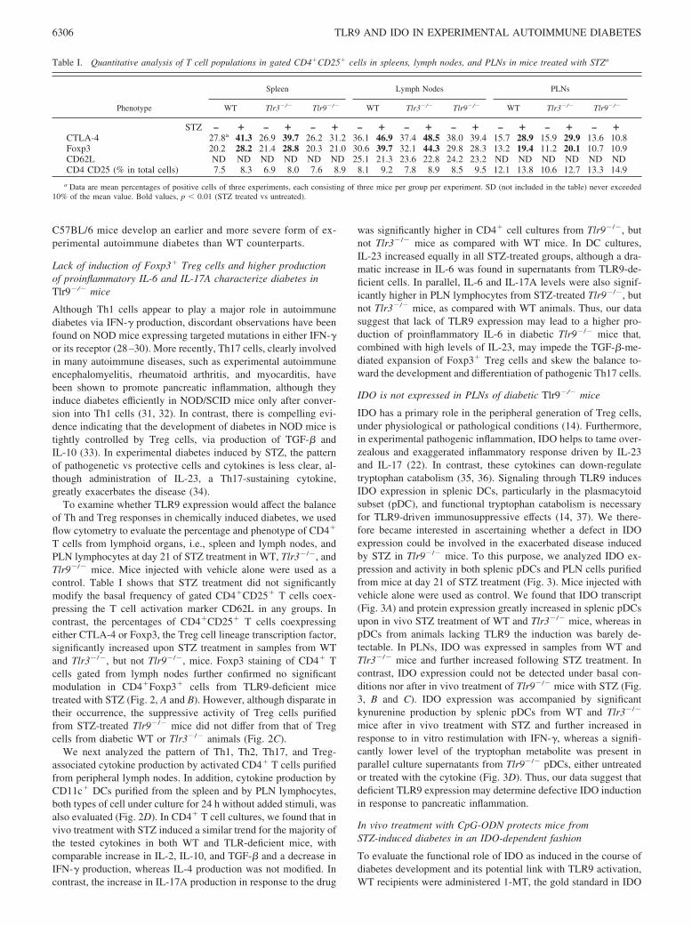
Table I. Quantitative analysis of T cell populations in gated CD4�CD25� cells in spleens, lymph nodes, and PLNs in mice treated with STZa
Phenotype
Spleen Lymph Nodes PLNs
WT Tlr3�/� Tlr9�/� WT Tlr3�/� Tlr9�/� WT Tlr3�/� Tlr9�/�
STZ – � – � – � – � – � – � – � – � – �CTLA-4 27.8a 41.3 26.9 39.7 26.2 31.2 36.1 46.9 37.4 48.5 38.0 39.4 15.7 28.9 15.9 29.9 13.6 10.8Foxp3 20.2 28.2 21.4 28.8 20.3 21.0 30.6 39.7 32.1 44.3 29.8 28.3 13.2 19.4 11.2 20.1 10.7 10.9CD62L ND ND ND ND ND ND 25.1 21.3 23.6 22.8 24.2 23.2 ND ND ND ND ND NDCD4 CD25 (% in total cells) 7.5 8.3 6.9 8.0 7.6 8.9 8.1 9.2 7.8 8.9 8.5 9.5 12.1 13.8 10.6 12.7 13.3 14.9
a Data are mean percentages of positive cells of three experiments, each consisting of three mice per group per experiment. SD (not included in the table) never exceeded10% of the mean value. Bold values, p � 0.01 (STZ treated vs untreated).
6306 TLR9 AND IDO IN EXPERIMENTAL AUTOIMMUNE DIABETES

inhibition, in the form of slow release pellets implanted on day 0,i.e., 1 day before STZ treatment. Control mice received placebopellets. All mice treated with STZ and 1-MT developed hypergly-cemia (Fig. 4, A and B) with a kinetic pattern comparable to thatmanifested by Tlr9�/� mice treated with STZ alone (Fig. 1B).Conversely, administration of CpG-ODN determined a reducedfrequency of hyperglycemic animals, an effect that could be re-versed by cotreatment with 1-MT, but not placebo pellets (Fig.4B). The therapeutic effect of CpG-ODN was accompanied by asignificant increase in the percentage of CD4�Foxp3� cells inlymph nodes (�50% increase in three experiments; p � 0.001,
CpG-ODN vs PBS), whereas the opposite pattern could be ob-served in mice treated with 1-MT, either alone or combined withCpG-ODN (�50% decrease in three experiments; p � 0.001,1-MT vs prospective placebo) (Fig. 4C). In addition, IL-17A andIL-6/IL-23 productions by lymph node CD4� cells and by splenicDCs, respectively, further increased in mice treated with STZ and1-MT CpG-ODN, but decreased after in vivo treatment withSTZ in combination with CpG-ODN and placebo. In PLNs, CpG-ODN treatment significantly reduced the production of both IL-6and IL-17A in an IDO-dependent fashion (Fig. 4D). Our datatherefore indicate that an intact tryptophan catabolism is required
FIGURE 2. CD4/Foxp3 expres-sion, Treg-suppressive activity, andcytokine production in diabeticTlr9�/� mice. Groups of mice treatedas in Fig. 1 were sacrificed at day 21.As controls, prospective recipientswere treated with vehicle alone. A,Upper part, Representative FACS dotplots for CD4 and Foxp3 expressionin permeabilized total lymph nodecells from vehicle (control)- and STZ-injected WT animals are shown.Lower part, Dot plots for gated CD4T cells. Numbers indicate the percent-age of Foxp3 expression in gatedCD4� cells. Number indicates per-centage of double-positive cells. Dataare representative of three experi-ments. B, The scattergram reflects thepercentages of Foxp3-expressingCD4� cells in individual mice (n 6). �, p � 0.001 (STZ-treated vs con-trol mice). C, Cocultures ofCD4�CD25� cells (1 � 105) fromnaive WT mice were established withvarious numbers of CD4�CD25�
cells (horizontal axis) obtained fromSTZ-treated WT or TLR-deficientmice in the presence of soluble anti-CD3 Ab (1 �g/ml) and APCs. Dataare means SD of triplicate samplesand are from one experiment repre-sentative of three. D, CD4� T cellspurified from pooled peripherallymph nodes (LN) were cultured inthe presence of plate-bound anti-CD3and anti-CD28 and, after 48 h, super-natants were assayed for cytokinecontent by ELISA. TGF-� was quan-tified by either ELISA (data notshown) or bioassay with similar re-sults. Cytokine production was alsoassessed in supernatants from splenicDCs and PLN cells cultured in me-dium alone for 24 h. Cytokine levelsare expressed in pg/ml with the ex-ception of IL-2, expressed in U/ml.Results are means SD of three ex-periments. �, p � 0.01 (STZ-treatedTlr9�/� vs WT mice).
6307The Journal of Immunology

for mitigating the inflammatory/diabetogenic effects of STZ inC57BL/6 mice, and that activation of TLR9 in vivo can protectfrom the pathogenic inflammatory response at the pancreas level inan IDO-dependent fashion.
CpG-ODN does not induce IDO expression and activity inPLNs of NOD mice
The NOD strain of mice has become a prototypic model of auto-immune diabetes (3). Most female mice die of hyperglycemia, re-flecting the onset of insulitis at 4 wk of age and the consequent Tcell-mediated destruction of pancreatic ß cells. The immune dys-regulation in NOD mice has been ascribed to many causes, includ-
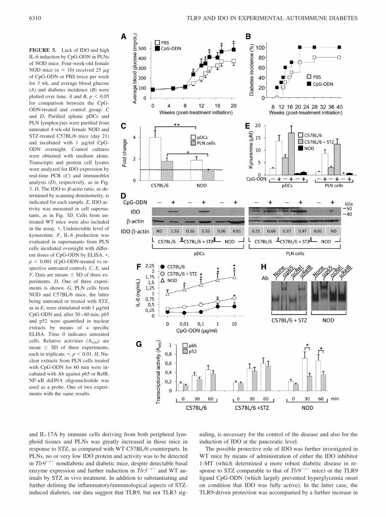
ing aberrant APC function, reduced suppressive activity of Tregcells (38), and, more recently, resistance to regulation in T celleffectors (39). In 2003, our group provided the first evidence thatIFN-� fails to induce IDO, and hence, tolerizing properties insplenic DCs from NOD female mice early in prediabetes (15).Prompted by the results obtained in the STZ diabetes model, weadministered multiple doses of CpG-ODN to 4-wk-old NOD fe-males. No protection, but rather acceleration of diabetes develop-ment could be observed in CpG-ODN-treated mice as comparedwith vehicle-treated counterparts (Fig. 5, A and B), despite con-siderable expression of Tlr9 transcripts in both splenocytes andPLN cells, which was comparable to that of C57BL/6 mice ofsame sex and age (data not shown). Lack of protection from dia-betes was accompanied by very low induction of IDO expressionin both splenic pDCs and PLN cells by in vitro incubation withCpG-ODN of cells from NOD as compared with STZ-treatedC57BL/6 mice (Fig. 5, C and D). Accordingly, IDO activity wasalso poorly observed in the former cells (Fig. 5E). CpG-ODN,however, was capable of inducing high production of IL-6 in NODPLNs as compared with parallel samples from C57BL/6 mice, ei-ther untreated or treated with STZ (Fig. 5F), in the face of basallyhigher levels of the cytokine. Because IL-6 can antagonize tryp-tophan catabolism via induction of proteasomal degradation ofIDO (40), our data suggest that an aberrant TLR9 signaling pro-ducing high levels of IL-6 may underlie the exacerbating effects ofCpG-ODN in NOD mice.
We have recently demonstrated that IDO expression is contin-gent on the noncanonical pathway of NF-�B activation (14, 21,26). Recent molecular dissection of NF-�B activation has shownthat NF-�B can be induced by the so-called canonical (classical)and noncanonical (alternative) signaling pathways, leading to dis-tinct patterns in the individual NF-�B subunits that are activatedand the downstream genetic responses that are induced. The proin-flammatory canonical pathway involves activation of the I�B ki-nase (IKK)-�, which leads to phosphorylation-induced proteolysisof the inhibitor I�B� and consequent nuclear translocation of thep65 subunit in the form of p50-p65 dimers. In the noncanonicalpathway, activation of IKK� by NF-�B-inducing kinase results inthe processing of p100 to p52 and consequent formation of p52-RelB dimers, which translocate into the nucleus and activate ananti-inflammatory gene program (13, 14). In pDCs, triggering ofTLR9 can activate both IKK�- and IKK�-mediated pathways (41).To investigate whether the inability of CpG-ODN to induce IDO inNOD mice could be due to altered signaling, PLN lymphocytesfrom both WT C57BL/6 and NOD mice were stimulated in vitrowith CpG-ODN and, after 30–60 min, NF-�B family activationwas quantified by means of an ELISA kit specific for p65 and p52(Fig. 5G). In WT C57BL/6 cells, either untreated or treated withSTZ, we found that CpG-ODN activated nuclear translocation ofp65 and p52 to a similar extent. In contrast, in NOD cells, thenuclear translocation of p65 was significantly higher than that ofp52 as early as at 30 min, suggesting the occurrence of a dominant,proinflammatory response mediated by canonical NF-�B in NODimmune cells upon engagement of TLR9. The preferential activa-tion of p65 in NOD PLN cells in response to TLR9 stimulationwas further confirmed by EMSA (Fig. 5H).
DiscussionIDO is a metabolic enzyme conserved through the last 600 millionyears of evolution. Initially confined to the regulation of trypto-phan availability in local tissue microenvironments, IDO is nowconsidered to play a wider role, which extends to homeostasis andplasticity of the immune system, with implications for many as-pects of immunopathology, including chronic inflammation and
FIGURE 3. Lack of IDO induction in diabetic Tlr9�/� mice. Groups ofmice treated with STZ as in Fig. 1 were sacrificed at day 21 and pDCs werepurified from the spleen. Lymphocytes from PLNs were also harvested. A,mRNA levels of Ido1 were quantified by real-time PCR using Gapdh nor-malization. Data (means SD of three experiments) are presented as nor-malized transcript expression in the samples (cells from STZ-treated mice)relative to normalized expression in the respective controls, i.e., cells fromuntreated mice (fold change 1; dotted line). B, IDO protein expressionwas assayed by immunoblot analysis of whole-cell lysates. Blots werestripped and reprobed with anti-�-actin Ab. One of three experiments. C,Data obtained as in B (means SD from three experiments) were analyzedby scanning densitometry and are presented as IDO protein expressionrelative to �-actin. �, Undetectable IDO protein densitometric value. ��,p � 0.01. D, Functional IDO activity was measured in terms of L-kynure-nine levels in supernatants from splenic pDCs treated overnight withIFN-�. Data are means SD of three experiments. �, p � 0.01 and ��, p �0.001.
6308 TLR9 AND IDO IN EXPERIMENTAL AUTOIMMUNE DIABETES

autoimmunity (14). Its immunoregulatory effects are mainly me-diated by DCs and involve not only tryptophan deprivation, butalso the production of kynurenines, which act on IDO� DCs, thusrendering an otherwise stimulatory DC capable of regulatory ef-fects (42), as well as on T cells. As a result, IDO� DCs mediatemultiple effects on T lymphocytes, including inhibition of prolif-eration, apoptosis, and differentiation toward a regulatory pheno-type (12–14). In addition to much evidence substantiating a criticalrole for tryptophan catabolism in immune regulation at the DC/Tcell level, nonimmune cells such as pancreatic � cells from healthypatients as well as other islet cells will express the enzyme inresponse to IFN-� (43). Furthermore, transfection of Ido1, the genecoding for mouse IDO, into � cells prolongs graft survival in NODmice (44). Thus, in inflammatory conditions, an effective trypto-phan catabolism at the pancreatic level may participate in isletprotection in individuals nongenetically prone to autoimmunediabetes.
TLRs play a central role in the generation of innate and adaptiveantimicrobial immune responses through recognition of conservedpathogen-associated molecular patterns. The majority of TLRs sig-nal through the MyD88 adaptor protein, except for TLR4 andTLR3, which can or must signal by means of TIR domain-con-taining adaptor inducing IFN-� (7–9). After proper elimination ofthe invading microorganisms, anti-inflammatory signals are nec-essary for the restoration of the homeostatic balance, and hence,host protection from the deleterious effects of overwhelming in-flammation. TLR signaling is involved not only in the primaryinduction of inflammation, but also in secondary regulatory mech-anisms (6, 45, 46). Among these, IDO may represent the down-stream effector for at least some TLRs, including the nucleic acid-binding TLR3 and TLR9. Although the TLR3 ligand poly(I:C) caninduce IDO in nonimmune cells such as astrocytes (47) and gin-gival fibroblasts (48), CpG-ODN is known to induce the enzyme
activity in immune cells, particularly pDCs, promoting strong im-munoregulatory effects (37, 49–51).
Because of the crucial role of IDO in immune tolerance and thepossible link between tryptophan catabolism and protective toler-ance by pathogen-associated molecular patterns (6), we sought toexamine whether TLR3 and TLR9 could play a role in the controlof autoimmune diabetes and whether IDO would be involved insuch effects. To this purpose, we examined experimental diabetesas induced by multiple low doses of STZ. Although not exten-sively characterized, this model is widely considered autoimmunein nature, because disease is prevented by administration of anti-Tcell mAb and can be adoptively transferred using splenocytes fromdiabetic animals. Furthermore, the overt disease is preceded byislet inflammation, characterized by infiltration of immune cells asearly as 3–4 days after the last STZ injection (52).
We found that hyperglycemia was induced in �60% of WTC57BL/6 mice by STZ and was accompanied by significantly re-duced insulinemia, mild islet infiltration, and a cytokine patterndominated by IL-6, IL-23, TGF-�, and IL-17A, suggesting theoccurrence of a Th17-mediated response. However, the increasednumber of T cells with a regulatory phenotype (CD4�Foxp3�) inperipheral lymphoid organs and in pancreas-associated lymphoidtissue, in addition to the high level of TGF-� produced by acti-vated CD4� T cells, indicates the existence of counterregulatorymechanisms possibly acting to dampen the potent inflammatoryeffects of STZ. Although both receptors are potentially capable oftransducing IDO-activating signals, TLR9- but not TLR3-deficientmice manifested an exacerbated diabetic syndrome. Lack of TLR9determined an earlier appearance of hyperglycemia in all animalsand a higher percentage of infiltrated islets, events accompanied bya lack of induction of CD4�Foxp3� Treg cells in lymphoid organsand pancreata. In addition, the production of proinflammatory IL-6
FIGURE 4. IDO-dependent protection by CpG-ODN from STZ-induced diabetes. Groups of WT mice (n 10) were treated with STZ alone (days 1–5)or in combination with CpG-ODN (days 7 and 9) and/or 1-MT (day 0). Controls received STZ, PBS (days 7 and 9), and placebo pellets (day 0). A andB, Blood glucose levels were monitored over time, and average blood glucose (A) and diabetes incidence (B) were plotted over time, as in Fig. 1. B, p �0.05 (PBS � 1-MT or CpG-ODN � placebo vs control � placebo) and p � 0.001 (CpG-ODN � 1-MT vs CpG-ODN � placebo). Parallel groups of micewere sacrificed at day 21 and analyzed for expression of Foxp3 in gated CD4� cells (C) and cytokine production (D), as in Fig. 2. CD4� T cells werepurified from pooled peripheral lymph nodes and pDCs from spleens. PLN cells were also assayed. A–C, One experiment is shown of three. D, Data (pg/ml)are means SD of three experiments.
6309The Journal of Immunology
and IL-17A by immune cells deriving from both peripheral lym-phoid tissues and PLNs was greatly increased in those mice inresponse to STZ, as compared with WT C57BL/6 counterparts. InPLNs, no or very low IDO protein and activity was to be detectedin Tlr9�/� nondiabetic and diabetic mice, despite detectable basalenzyme expression and further induction in Tlr3�/� and WT an-imals by STZ in vivo treatment. In addition to substantiating andfurther defining the inflammatory/immunological aspects of STZ-induced diabetes, our data suggest that TLR9, but not TLR3 sig-
naling, is necessary for the control of the disease and also for theinduction of IDO at the pancreatic level.
The possible protective role of IDO was further investigated inWT mice by means of administration of either the IDO inhibitor1-MT (which determined a more robust diabetic disease in re-sponse to STZ comparable to that of Tlr9�/� mice) or the TLR9ligand CpG-ODN (which largely prevented hyperglycemia onseton condition that IDO was fully active). In the latter case, theTLR9-driven protection was accompanied by a further increase in
FIGURE 5. Lack of IDO and highIL-6 induction by CpG-ODN in PLNsof NOD mice. Four-week-old femaleNOD mice (n 10) received 25 �gof CpG-ODN or PBS twice per weekfor 3 wk, and average blood glucose(A) and diabetes incidence (B) wereplotted over time. A and B, p � 0.05for comparison between the CpG-ODN-treated and control group. Cand D, Purified splenic pDCs andPLN lymphocytes were purified fromuntreated 4-wk-old female NOD andSTZ-treated C57BL/6 mice (day 21)and incubated with 1 �g/ml CpG-ODN overnight. Control cultureswere obtained with medium alone.Transcripts and protein cell lysateswere analyzed for IDO expression byreal-time PCR (C) and immunoblotanalysis (D), respectively, as in Fig.3. D, The IDO to �-actin ratio, as de-termined by scanning densitometry, isindicated for each sample. E, IDO ac-tivity was measured in cell superna-tants, as in Fig. 3D. Cells from un-treated WT mice were also includedin the assay. �, Undetectable level ofkynurenine. F, IL-6 production wasevaluated in supernatants from PLNcells incubated overnight with differ-ent doses of CpG-ODN by ELISA. �,p � 0.001 (CpG-ODN-treated vs re-spective untreated control). C, E, andF, Data are means SD of three ex-periments. D, One of three experi-ments is shown. G, PLN cells fromNOD and C57BL/6 mice, the latterbeing untreated or treated with STZ,as in E, were stimulated with 1 �g/mlCpG-ODN and, after 30–60 min, p65and p52 were quantified in nuclearextracts by means of a specificELISA. Time 0 indicates untreatedcells. Relative activities (A450) aremean SD of three experiments,each in triplicate. �, p � 0.01. H, Nu-clear extracts from PLN cells treatedwith CpG-ODN for 60 min were in-cubated with Ab against p65 or RelB.NF-�B dsDNA oligonucleotide wasused as a probe. One of two experi-ments with the same results.
6310 TLR9 AND IDO IN EXPERIMENTAL AUTOIMMUNE DIABETES
CD4�Foxp3� Treg cells as compared with controls, whereas pro-duction of proinflammatory IL-6 and IL-17A from PLN lympho-cytes was reduced. Therefore, our data further extend the protec-tive potential of the TLR9-IDO axis from animal models ofallergic disorders (14) to experimental autoimmune diabetes in-duced in mice nongenetically prone to the disease, and reveal anessential role of IDO expression in PLNs for the control of insulitisand hyperglycemia. Interestingly, this axis is already known to beimportant in the generation of human adaptive Treg cells (53),whereas either CpG-ODN (54) or kynurenines (36, 55) can controlTh17-mediated inflammatory responses in murine disease models.Although TLR9 and IDO have not been directly linked yet, it isinteresting to note that experimental autoimmune encephalomyeli-tis can be exacerbated by either lack of TLR9 expression (56) or ofIDO activity (57).
However, in the NOD mouse, the situation is far more complex.Wong et al. (58) provided evidence that the incidence of diabetesin Tlr9�/�, but not Tlr3�/� NOD mice maintained in SPF condi-tions is significantly lower when compared with their heterozygous(Tlr9�/�) littermates. In addition, Wen’s group (11) demonstratedthat NOD mice lacking MyD88, but not TLR3, exhibit loss ofdiabetes development when maintained in SPF conditions, despitethe development of robust diabetes in a germfree environment.However, colonization of germfree MyD88�/� NOD mice with adefined microbial consortium attenuates the disease (11). Overall,the bulk of available data suggests that, in the NOD mouse, TLR9-MyD88 signaling does not protect against, but rather sustainspathogenesis. Nonetheless, alternative microbial signals, appar-ently not engaging TLR3, can exert protective effects. Paradoxi-cally, STZ treatment both prevents and reverts islet destructiveautoimmunity in NOD mice (59). Considering that TLR9 can beactivated also by host unmethylated CpG-containing DNA andnon-CpG DNA (46) and that STZ can induce multiple DNA mod-ifications, it might be speculated that the STZ treatment is capableof generating endogenous, regulatory TLR9 ligands. In agreementwith data from knockout NOD animals, our current data indicatethat multiple i.p. injections of SPF NOD mice with CpG-ODNaccelerate diabetes development. However, it has been previouslyreported that i.m. vaccination of SPF NOD mice with CpG-ODNsignificantly reduces the incidence of diabetes (60). Although s.c.vs i.v. CpG-ODN administration has already been shown to de-termine opposite effects (50), it is worth mentioning that the suc-cessful i.m. administration of CpG-ODN was preceded by the in-jection of cardiotoxin, a molecular adjuvant particularly effectivein i.m. DNA vaccinations. In this specific case, cardiotoxin pre-treatment may have reset the dysregulated immune system of theNOD mouse (38), allowing CpG-ODN to exert an effective im-munosuppressive effect. Thus, the inability of TLR9 to induce aregulatory pathway in NOD mice may rely on the absence of ap-propriate endogenous TLR9 ligands and/or the existence of sig-naling defects.
In evaluating the capacity of individual TLRs to promote eitherinflammation/autoimmunity or protective tolerance, the domi-nance of specific signaling pathways should be taken into account(46). TLR9 is known to signal via the MyD88 adapter, which, inpDCs, can activate two distinct NF-�B pathways, namely the clas-sical or canonical pathway (in which IKK� kinase plays a pivotalrole) and the alternative or noncanonical pathway (which relies onIKK� activation). Although TLR9-IKK� signaling leads to thenuclear translocation of p50-p65 dimers and production of classi-cal proinflammatory cytokines, activation of IKK� by TLR9 trig-gering induces type I IFNs via nuclear translocation of p52-RelBcomplexes (41). We have recently found that noncanonical NF-�Bactivation is necessary for the induction of IDO-mediated thera-
peutic effects in a model of airway allergic inflammation (21).Furthermore, type I IFNs, although less potent than IFN-�, canalso act as IDO inducers (13, 14). Our current data indicate that theCpG-ODN treatment, either in vivo (data not shown) or in vitro,does not induce IDO protein and activity in NOD splenic pDCsand PLNs, despite a considerable induction in parallel samplesfrom diabetic WT C57BL/6 mice. Lack of IDO induction is ac-companied by overactivation of the proinflammatory p65 as com-pared with anti-inflammatory p52 subunit, whereas in cells fromdiabetic and nondiabetic C57BL/6 mice the amounts of p65 andp52 activated by CpG-ODN are quite similar. Although we cannotpresently exclude the absence of appropriate TLR9 ligands, ourdata indicate that a dysfunctional TLR9 signaling may underlie thepathogenic/proinflammatory effect of this receptor in spontaneousautoimmune diabetes and may impede the effective induction ofcounterregulatory mechanisms mediated by IDO.
In conclusion, the identification of the molecular targets andmechanisms through which microbial ligands exert their protectiveeffects may lead to a better understanding of the early pathogenesisprocesses involved in T1D, may provide new markers for an earlydiagnosis of the disease, and can ultimately lead to the generationof drugs mimicking protective microbial entities that can be usedin either the prevention and/or treatment of T1D patients. Thepresent study suggests that activation of TLR9, but not TLR3, caninduce IDO, and thus control early inflammatory attacks to � cells.Harmonious TLR9 signaling, however, seems to be mandatory forfully protective activity of tryptophan catabolism. The latter, in-deed, appears to be defective not only systematically, but alsospecifically in pancreas-associated lymph nodes of NOD mice.Thus, TLR9 and IDO may represent innovative and valuable mo-lecular targets in the prevention/therapy of T1D. Although we arestill currently evaluating the existence of a possible IDO defect inT1D patients, our data support the recent hypothesis that only com-bination therapies, capable of correcting the dysregulated immunesignaling at multiple levels, will enable the permanent preventionand curing of T1D (61).
AcknowledgmentsWe thank G. Andrielli for technical assistance and digital art.
DisclosuresThe authors have no financial conflict of interest.
References1. Feleszko, W., J. Jaworska, and E. Hamelmann. 2006. Toll-like receptors: novel
targets in allergic airway disease (probiotics, friends and relatives). Eur. J. Phar-macol. 533: 308–318.
2. Gale, E. A. 2002. The rise of childhood type 1 diabetes in the 20th century.Diabetes 51: 3353–3361.
3. Bach, J. F. 2002. The effect of infections on susceptibility to autoimmune andallergic diseases. N. Engl. J. Med. 347: 911–920.
4. Strachan, D. P. 1989. Hay fever, hygiene, and household size. B.M.J. 299:1259–1260.
5. Bach, J. F. 2005. Infections and autoimmune diseases. J. Autoimmun. 25(Suppl.):74–80.
6. Romani, L., and P. Puccetti. 2006. Protective tolerance to fungi: the role of IL-10and tryptophan catabolism. Trends Microbiol. 14: 183–189.
7. Medzhitov, R. 2001. Toll-like receptors and innate immunity. Nat. Rev. Immunol.1: 135–145.
8. Iwasaki, A., and R. Medzhitov. 2004. Toll-like receptor control of the adaptiveimmune responses. Nat. Immunol. 5: 987–995.
9. Akira, S., S. Uematsu, and O. Takeuchi. 2006. Pathogen recognition and innateimmunity. Cell 124: 783–801.
10. Kagan, J. C., T. Su, T. Horng, A. Chow, S. Akira, and R. Medzhitov. 2008.TRAM couples endocytosis of Toll-like receptor 4 to the induction of interfer-on-�. Nat. Immunol. 9: 361–368.
11. Wen, L., R. E. Ley, P. Y. Volchkov, P. B. Stranges, L. Avanesyan,A. C. Stonebraker, C. Hu, F. S. Wong, G. L. Szot, J. A. Bluestone, et al. 2008.Innate immunity and intestinal microbiota in the development of type 1 diabetes.Nature 455: 1109–1113.
12. Grohmann, U., F. Fallarino, and P. Puccetti. 2003. Tolerance, DCs and trypto-phan: much ado about IDO. Trends Immunol. 24: 242–248.
6311The Journal of Immunology
13. Mellor, A. L., and D. H. Munn. 2004. IDO expression by dendritic cells: toler-ance and tryptophan catabolism. Nat. Rev. Immunol. 4: 762–774.
14. Puccetti, P., and U. Grohmann. 2007. IDO and regulatory T cells: a role forreverse signalling and non-canonical NF-�B activation. Nat. Rev. Immunol. 7:817–823.
15. Grohmann, U., F. Fallarino, R. Bianchi, C. Orabona, C. Vacca, M. C. Fioretti, andP. Puccetti. 2003. A defect in tryptophan catabolism impairs tolerance in nono-bese diabetic mice. J. Exp. Med. 198: 153–160.
16. Fallarino, F., R. Bianchi, C. Orabona, C. Vacca, M. L. Belladonna, M. C. Fioretti,D. V. Serreze, U. Grohmann, and P. Puccetti. 2004. CTLA-4-Ig activates fork-head transcription factors and protects dendritic cells from oxidative stress innonobese diabetic mice. J. Exp. Med. 200: 1051–1062.
17. Heer, A. K., A. Shamshiev, A. Donda, S. Uematsu, S. Akira, M. Kopf, andB. J. Marsland. 2007. TLR signaling fine-tunes anti-influenza B cell responseswithout regulating effector T cell responses. J. Immunol. 178: 2182–2191.
18. Romani, L., F. Bistoni, K. Perruccio, C. Montagnoli, R. Gaziano, S. Bozza,P. Bonifazi, G. Bistoni, G. Rasi, A. Velardi, et al. 2006. Thymosin �1 activatesdendritic cell tryptophan catabolism and establishes a regulatory environment forbalance of inflammation and tolerance. Blood 108: 2265–2274.
19. Grohmann, U., C. Orabona, F. Fallarino, C. Vacca, F. Calcinaro, A. Falorni,P. Candeloro, M. L. Belladonna, R. Bianchi, M. C. Fioretti, and P. Puccetti. 2002.CTLA-4-Ig regulates tryptophan catabolism in vivo. Nat. Immunol. 3:1097–1101.
20. Fallarino, F., U. Grohmann, S. You, B. C. McGrath, D. R. Cavener, C. Vacca,C. Orabona, R. Bianchi, M. L. Belladonna, C. Volpi, et al. 2006. The combinedeffects of tryptophan starvation and tryptophan catabolites down-regulate T cellreceptor �-chain and induce a regulatory phenotype in naive T cells. J. Immunol.176: 6752–6761.
21. Grohmann, U., C. Volpi, F. Fallarino, S. Bozza, R. Bianchi, C. Vacca,C. Orabona, M. L. Belladonna, E. Ayroldi, G. Nocentini, et al. 2007. Reversesignaling through GITR ligand enables dexamethasone to activate IDO in allergy.Nat. Med. 13: 579–586.
22. Romani, L., F. Fallarino, A. De Luca, C. Montagnoli, C. D’Angelo, T. Zelante,C. Vacca, F. Bistoni, M. C. Fioretti, U. Grohmann, et al. 2008. Defective tryp-tophan catabolism underlies inflammation in mouse chronic granulomatous dis-ease. Nature 451: 211–215.
23. Orabona, C., U. Grohmann, M. L. Belladonna, F. Fallarino, C. Vacca, R. Bianchi,S. Bozza, C. Volpi, B. L. Salomon, M. C. Fioretti, et al. 2004. CD28 inducesimmunostimulatory signals in dendritic cells via CD80 and CD86. Nat. Immunol.5: 1134–1142.
24. Montagnoli, C., F. Fallarino, R. Gaziano, S. Bozza, S. Bellocchio, T. Zelante,W. P. Kurup, L. Pitzurra, P. Puccetti, and L. Romani. 2006. Immunity and tol-erance to Aspergillus involve functionally distinct regulatory T cells and trypto-phan catabolism. J. Immunol. 176: 1712–1723.
25. Spaccapelo, R., L. Romani, L. Tonnetti, E. Cenci, A. Mencacci, G. Del Sero,R. Tognellini, S. G. Reed, P. Puccetti, and F. Bistoni. 1995. TGF-� is importantin determining the in vivo patterns of susceptibility or resistance in mice infectedwith Candida albicans. J. Immunol. 155: 1349–1360.
26. Belladonna, M. L., C. Volpi, R. Bianchi, C. Vacca, C. Orabona, M. T. Pallotta,L. Boon, S. Gizzi, M. C. Fioretti, U. Grohmann, and P. Puccetti. 2008. Cuttingedge: autocrine TGF-� sustains default tolerogenesis by IDO-competent dendriticcells. J. Immunol. 181: 5194–5198.
27. Grohmann, U., M. L. Belladonna, R. Bianchi, C. Orabona, E. Ayroldi,M. C. Fioretti, and P. Puccetti. 1998. IL-12 acts directly on DC to promotenuclear localization of NF-�B and primes DC for IL-12 production. Immunity 9:315–323.
28. Wang, B., I. Andre, A. Gonzalez, J. D. Katz, M. Aguet, C. Benoist, andD. Mathis. 1997. Interferon-� impacts at multiple points during the progressionof autoimmune diabetes. Proc. Natl. Acad. Sci. USA 94: 13844–13849.
29. Hultgren, B., X. Huang, N. Dybdal, and T. A. Stewart. 1996. Genetic absence of�-interferon delays but does not prevent diabetes in NOD mice. Diabetes 45:812–817.
30. Serreze, D. V., C. M. Post, H. D. Chapman, E. A. Johnson, B. Lu, andP. B. Rothman. 2000. Interferon-� receptor signaling is dispensable in the de-velopment of autoimmune type 1 diabetes in NOD mice. Diabetes 49:2007–2011.
31. Martin-Orozco, N., Y. Chung, S. H. Chang, Y. H. Wang, and C. Dong. 2009.Th17 cells promote pancreatic inflammation but only induce diabetes efficientlyin lymphopenic hosts after conversion into Th1 cells. Eur. J. Immunol. 39:216–224.
32. Bending, D., H. De La Pena, M. Veldhoen, J. M. Phillips, C. Uyttenhove,B. Stockinger, and A. Cooke. 2009. Highly purified Th17 cells fromBDC2.5NOD mice convert into Th1-like cells in NOD/SCID recipient mice.J. Clin. Invest. 119: 565–572.
33. You, S., M. A. Alyanakian, B. Segovia, D. Damotte, J. Bluestone, J. F. Bach, andL. Chatenoud. 2008. Immunoregulatory pathways controlling progression of au-toimmunity in NOD mice. Ann. NY Acad. Sci. 1150: 300–310.
34. Mensah-Brown, E. P., A. Shahin, M. Al-Shamisi, X. Wei, and M. L. Lukic. 2006.IL-23 leads to diabetes induction after subdiabetogenic treatment with multiplelow doses of streptozotocin. Eur. J. Immunol. 36: 216–223.
35. Zelante, T., A. De Luca, P. Bonifazi, C. Montagnoli, S. Bozza, S. Moretti,M. L. Belladonna, C. Vacca, C. Conte, P. Mosci, et al. 2007. IL-23 and the Th17pathway promote inflammation and impair antifungal immune resistance. Eur.J. Immunol. 37: 2695–2706.
36. Romani, L., T. Zelante, A. De Luca, F. Fallarino, and P. Puccetti. 2008. IL-17 andtherapeutic kynurenines in pathogenic inflammation to fungi. J. Immunol. 180:5157–5162.
37. Fallarino, F., C. Orabona, C. Vacca, R. Bianchi, S. Gizzi, C. Asselin-Paturel,M. C. Fioretti, G. Trinchieri, U. Grohmann, and P. Puccetti. 2005. Ligand andcytokine dependence of the immunosuppressive pathway of tryptophan catabo-lism in plasmacytoid dendritic cells. Int. Immunol. 17: 1429–1438.
38. Delovitch, T. L., and B. Singh. 1997. The nonobese diabetic mouse as a modelof autoimmune diabetes: immune dysregulation gets the NOD. Immunity 7:727–738.
39. D’Alise, A. M., V. Auyeung, M. Feuerer, J. Nishio, J. Fontenot, C. Benoist, andD. Mathis. 2008. The defect in T-cell regulation in NOD mice is an effect on theT-cell effectors. Proc. Natl. Acad. Sci. USA 105: 19857–19862.
40. Orabona, C., M. T. Pallotta, C. Volpi, F. Fallarino, C. Vacca, R. Bianchi,M. L. Belladonna, M. C. Fioretti, U. Grohmann, and P. Puccetti. 2008. SOCS3drives proteasomal degradation of indoleamine 2,3-dioxygenase (IDO) and an-tagonizes IDO-dependent tolerogenesis. Proc. Natl. Acad. Sci. USA 105:20828–20833.
41. Kaisho, T., and T. Tanaka. 2008. Turning NF-�B and IRFs on and off in DC.Trends Immunol. 29: 329–336.
42. Belladonna, M. L., U. Grohmann, P. Guidetti, C. Volpi, R. Bianchi,M. C. Fioretti, R. Schwarcz, F. Fallarino, and P. Puccetti. 2006. Kynureninepathway enzymes in dendritic cells initiate tolerogenesis in the absence of func-tional IDO. J. Immunol. 177: 130–137.
43. Sarkar, S. A., R. Wong, S. I. Hackl, O. Moua, R. G. Gill, A. Wiseman,H. W. Davidson, and J. C. Hutton. 2007. Induction of indoleamine 2,3-dioxyge-nase by interferon-� in human islets. Diabetes 56: 72–79.
44. Alexander, A. M., M. Crawford, S. Bertera, W. A. Rudert, O. Takikawa,P. D. Robbins, and M. Trucco. 2002. Indoleamine 2,3-dioxygenase expression intransplanted NOD islets prolongs graft survival after adoptive transfer of diabe-togenic splenocytes. Diabetes 51: 356–365.
45. Netea, M. G., J. W. Van der Meer, and B. J. Kullberg. 2004. Toll-like receptorsas an escape mechanism from the host defense. Trends Microbiol. 12: 484–488.
46. Ehlers, M., and J. V. Ravetch. 2007. Opposing effects of Toll-like receptor stim-ulation induce autoimmunity or tolerance. Trends Immunol. 28: 74–79.
47. Suh, H. S., M. L. Zhao, M. Rivieccio, S. Choi, E. Connolly, Y. Zhao,O. Takikawa, C. F. Brosnan, and S. C. Lee. 2007. Astrocyte indoleamine 2,3-dioxygenase is induced by the TLR3 ligand poly(I:C): mechanism of inductionand role in antiviral response. J. Virol. 81: 9838–9850.
48. Mahanonda, R., N. Sa-Ard-Iam, P. Montreekachon, A. Pimkhaokham,K. Yongvanichit, M. M. Fukuda, and S. Pichyangkul. 2007. IL-8 and IDO ex-pression by human gingival fibroblasts via TLRs. J. Immunol. 178: 1151–1157.
49. Mellor, A. L., B. Baban, P. R. Chandler, A. Manlapat, D. J. Kahler, andD. H. Munn. 2005. Cutting edge: CpG oligonucleotides induce splenic CD19�
dendritic cells to acquire potent indoleamine 2,3-dioxygenase-dependent T cellregulatory functions via IFN type 1 signaling. J. Immunol. 175: 5601–5605.
50. Wingender, G., N. Garbi, B. Schumak, F. Jungerkes, E. Endl, D. von Bubnoff,J. Steitz, J. Striegler, G. Moldenhauer, T. Tuting, et al. 2006. Systemic applicationof CpG-rich DNA suppresses adaptive T cell immunity via induction of IDO.Eur. J. Immunol. 36: 12–20.
51. Fallarino, F., and P. Puccetti. 2006. Toll-like receptor 9-mediated induction of theimmunosuppressive pathway of tryptophan catabolism. Eur. J. Immunol. 36:8–11.
52. Kim, Y. T., and C. Steinberg. 1984. Immunologic studies on the induction ofdiabetes in experimental animals: cellular basis for the induction of diabetes bystreptozotocin. Diabetes 33: 771–777.
53. Chen, W., X. Liang, A. J. Peterson, D. H. Munn, and B. R. Blazar. 2008. Theindoleamine 2,3-dioxygenase pathway is essential for human plasmacytoid den-dritic cell-induced adaptive T regulatory cell generation. J. Immunol. 181:5396–5404.
54. Andersson, A., M. Isaksson, J. Wefer, A. Norling, A. Flores-Morales,F. Rorsman, O. Kampe, R. A. Harris, and A. Lobell. 2008. Impaired autoimmuneT helper 17 cell responses following DNA vaccination against rat experimentalautoimmune encephalomyelitis. PLoS One 3: e3682.
55. Platten, M., P. P. Ho, S. Youssef, P. Fontoura, H. Garren, E. M. Hur, R. Gupta,L. Y. Lee, B. A. Kidd, W. H. Robinson, et al. 2005. Treatment of autoimmuneneuroinflammation with a synthetic tryptophan metabolite. Science 310:850–855.
56. Marta, M., A. Andersson, M. Isaksson, O. Kampe, and A. Lobell. 2008. Unex-pected regulatory roles of TLR4 and TLR9 in experimental autoimmune enceph-alomyelitis. Eur. J. Immunol. 38: 565–575.
57. Kwidzinski, E., J. Bunse, O. Aktas, D. Richter, L. Mutlu, F. Zipp, R. Nitsch, andI. Bechmann. 2005. Indolamine 2,3-dioxygenase is expressed in the CNS anddown-regulates autoimmune inflammation. FASEB J. 19: 1347–1349.
58. Wong, F. S., C. Hu, L. Zhang, W. Du, L. Alexopoulou, R. A. Flavell, and L. Wen.2008. The role of Toll-like receptors 3 and 9 in the development of autoimmunediabetes in NOD mice. Ann. NY Acad. Sci. 1150: 146–148.
59. Koulmanda, M., A. Qipo, H. Auchincloss, Jr., and R. N. Smith. 2003. Effects ofstreptozotocin on autoimmune diabetes in NOD mice. Clin. Exp. Immunol. 134:210–216.
60. Quintana, F. J., A. Rotem, P. Carmi, and I. R. Cohen. 2000. Vaccination withempty plasmid DNA or CpG oligonucleotide inhibits diabetes in nonobese dia-betic mice: modulation of spontaneous 60-kDa heat shock protein autoimmunity.J. Immunol. 165: 6148–6155.
61. Von Herrath, M. 2009. Can we learn from viruses how to prevent type 1 diabetes?The role of viral infections in the pathogenesis of type 1 diabetes and the devel-opment of novel combination therapies. Diabetes 58: 2–11.
6312 TLR9 AND IDO IN EXPERIMENTAL AUTOIMMUNE DIABETES
Copyright © 2022 FDOKUMEN