Elevated ATG5 expression in autoimmune demyelination and multiple sclerosis
Upload
independentCategory
view
3download
0
Foodalle
rgy,derm
ato
logic
dise
ase
s,andanaphylaxis
High prevalence of autoimmune urticariain children with chronic urticaria
Luigia Brunetti, MD,a Ruggiero Francavilla, MD, PhD,a Vito L. Miniello, MD,a
Michael H. Platzer, MSc,b Domenica Rizzi, MD,a Maria Letizia Lospalluti, MD,a
Lars K. Poulsen, PhD,b Lucio Armenio, MD, PhD,a and Per Stahl Skov, MD, PhDb
Bari, Italy, and Copenhagen, Denmark
Background: The etiology of chronic urticaria (CU) in
childhood often remains unrecognized. Recently, in adults it
has been shown that approximately 40% of patients with CU
have autoimmune urticaria (AU); however, no data are
available in children.
Objective: To determine the prevalence and possible risk
factors for AU in children with CU.
Methods: Ninety-three consecutive children (52 male; median
age, 7.8 years) with CU were evaluated for AU by means of
autologous serum skin test (ASST) in all and serum-induced
basophil histamine release (HR-urticaria test) in 52. All other
known causes of CU were excluded as appropriate.
Results: A cause for CU was identified in 44 children (47%),
whereas 49 (53%) remained idiopathic. ASSTand HR-urticaria
test had positive results in 22 of 49 (45%) and in 16 of 31 (52%)
children with idiopathic CU compared with 1 of 44 (2%) and 5
of 21 (24%) with CU of a known cause, respectively (P < .00001;
P = .09). Sensitivity, specificity, and positive and negative
predictive values of the ASST for diagnosing AU are 78%,
85%, 74%, and 88%. The prevalence of AU in childhood is
31% (15/52; 95% CI, 24%-51%). None of the variables studied
were predictive for development of AU.
Conclusion: Our results demonstrate for the first time that
children have the same ability as adults to produce functionally
active autoantibodies directed against IgE or IgE receptor and
that AU occurs in children in as many as 30% of cases. The
addition of screening for AU dramatically decreases the rate of
the idiopathic form from 52% to 20%. (J Allergy Clin Immunol
2004;114:922-7.)
Key words: Chronic idiopathic urticaria, childhood, autologousserum skin test, serum induced basophil histamine release, autoim-
mune urticaria
Chronic urticaria (CU) is a common skin disordercharacterized by recurrent, transitory, itchy wheals withindividual lesions lasting less than 24 hours and affectingpatients for 6 weeks or longer.1-3 This condition is thought
From athe Department of Biomedicina dell’Eta Evolutiva, University of Bari;
and bthe National University Hospital, Laboratory of Medical Allergology,
Copenhagen.
Supported by the University of Bari.
Received for publication April 20, 2004; revised June 27, 2004; accepted for
publication July 1, 2004.
Reprint requests: Luigia Brunetti, MD, Clinica Pediatrica I, University of Bari,
Piazza Giulio Cesare, 11-70125 Bari, Italy. E-mail: l.brunetti@pediatria3.
uniba.it.
0091-6749/$30.00
� 2004 American Academy of Allergy, Asthma and Immunology
doi:10.1016/j.jaci.2004.07.042
922
to affect at least 0.1% of the population,4 and often it canbe severe and difficult to treat.5
The pathogenesis of CU is not completely understood,but mast cell degranulation and histamine release arethought to play a central role.4 It is known that the bindingof an antigen (allergen) to antigen-specific IgE on mastcells and basophils causes cell degranulation resulting inthe release of histamine and other vasoactive mediators,responsible of clinical symptoms. However, most patientswith CU have no specific allergic trigger for mast cell orbasophil activation, and when no cause can be identified,the final diagnosis is chronic idiopathic urticaria (CIU).1
Recently it has been shown that, in patients with severeCIU, the intradermal injection of autologous serum elicitsan immediate wheal-and-flare response and mast celldegranulation,1,6 and it is now established that approxi-mately one third of patients with CIU have circulatingautoantibodies directed against epitopes in the a-chain ofthe high-affinity receptor (FceRI) or against IgE.6,7 Theseautoantibodies are functionally active, causing histaminerelease from basophils of healthy donors and from dermalmast cells in vitro.8 The presence of anti-FceRI autoanti-bodies in CIU has been confirmed by Western blotanalysis and ELISA in adult series.8,9
Few and anecdotal data are published on FceRIautoantibodies in children with CU.10 The current in-vestigation was aimed to determine the frequency andpattern of autoantibodies directed against IgE or IgEreceptor in children with CU.
METHODS
Participants
The Paediatric Immuno-Allergology Clinic of the University of
Bari is the tertiary referral center for the diagnosis and follow-up of
allergic diseases of childhood in Southern Italy (Puglia, Calabria, and
Basilicata), covering an estimated population of 1.5 million children,
Abbreviations used
ASST: Autologous serum skin test
AU: Autoimmune urticaria
CIU: Chronic idiopathic urticaria
CU: Chronic urticaria
HP: Helicobacter pyloriHR-urticaria test: Serum induced basophil histamine
release
J ALLERGY CLIN IMMUNOL
VOLUME 114, NUMBER 4
Brunetti et al 923
Foodallerg
y,derm
ato
logic
disease
s,andanaphylaxis
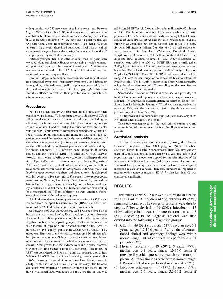
with approximately 350 new cases of urticaria every year. Between
August 2000 and October 2002, 680 new cases of urticaria were
admitted to the clinic, most of which were acute. Among these, a total
of 93 consecutive children (14%; 52 boys and 41 girls; median age,
7.8 years; range, 9 months to 16 years) with CU, defined as recurrent
(at least twice a week), short-lived cutaneous wheal with or without
accompanying angioedema and occurring formore than 2months,1,10
were prospectively enrolled in the study.
Patients younger than 6 months or older than 16 years were
excluded. None had chronic diseases or was taking steroids or immu-
nosuppressive therapy at the time of investigation. Antihistamine
treatment was stopped at least 15 days before skin testing was
performed or serum sample collected.
Familial (atopy, autoimmune diseases), clinical (age at onset,
gastrointestinal symptoms, respiratory symptoms), and laboratory
(hemoglobin, white cell, neutrophil, lymphocyte, eosinophil, baso-
phil, and monocyte cell count, IgG, IgE, IgA, IgM) data were
carefully collected to evaluate their possible role as predictors of
autoimmune urticaria.
Procedures
Full past medical history was recorded and a complete physical
examination performed. To investigate the possible cause of CU, all
children underwent extensive laboratory evaluations, including the
following: (1) blood tests for complete blood count, erythrocyte
sedimentation rate, blood chemistry, liver function tests, antistrepto-
lysin antibody, serum levels of complement components C3 and C4,
free thyroxin, thyroid stimulating hormone, and total serum IgE; (2)
autoimmune panel (antinuclear antibody and anti-DNA antibody [if
antinuclear antibody–positive], anti–smooth muscle antibodies, anti-
parietal cell antibodies, antithyroid peroxidase antibodies, antithyr-
eoglobulin antibodies), (3) infective panel (hepatitis B surface
antigen, antibody titers for hepatitis B virus and C virus, TORCH
[toxoplasmosis, other, rubella, cytomegalovirus, and herpes simplex
virus], Epstein-Barr virus, 13C-urea breath test for the diagnosis of
Helicobacter pylori (HP), stool culture, urine analysis and culture,
nasal, throat and vulvar swabs for Streptococcus b-hemolyticus and
Staphylococcus aureus); (4) chest and sinus x-rays; (5) skin prick
tests for cypress, olive tree, grass, Parietaria, Dermatophagoidespteronyssinus, Dermatophagoides farinae, Alternaria, dog and cat
dandruff, cereals, egg, fish, meat, fruits, legumes, solancee, milk, and
soy; and (6) ice cube test for cold-induced urticaria and skin stroking
for dermatographism.11 If any of these tests were abnormal, further
evaluations were performed as appropriate.
All children underwent autologous serum skin tests (ASSTs), and
serum-induced basophil histamine release (HR-urticaria test) was
performed in 52 children for whom serum was available.
Skin testing with autologous serum. ASST was performed while
the urticaria was active. Briefly, 50 mL autologous serum, histamine
(10 mg/mL in saline; positive control) and 0.9% sterile saline
(negative control) were separately injected into the dermis of the
volar forearm at gaps of 5 cm between injecting sites. Areas of
previous involvement by spontaneous wheals were avoided. The 2
orthogonal diameters of the wheals were measured 30 minutes after
the injection. According to Sabroe,12 a positive skin test was defined
as the presence of a serum-inducedwheal with ameanwheal diameter
at least 1.5 mm greater than that induced by saline (D wheal diameter
>1.5 mm). In the absence of a positive response to histamine, the
ASST was considered not informative and was repeated after at least
24 hours. All ASSTs were performed by a single investigator (L.B.).
HR-urticaria test. One adult donor whose basophils responded to
anti-IgE with a release >30% was used in the assay. The basophil
leukocytes were prepared by dextran sedimentation (5 mL freshly
drawn heparinized blood was added to 1 mL 5.0% dextran and 0.25
mL0.2mol/L EDTA (pH 7.0) and allowed to sediment for 45minutes
at 5�C. The basophil-containing layer was washed once with
piperazine-1,4-bis(2-ethanesulfonic acid) containing 0.036% human
serum albumin (PIPES-HSA) and the cells resuspended in 2 mL
PIPES-HSA containing 500 pg/mL recombinant human IL-3 (R&D
Systems, Minneapolis, Minn). Samples of 40 mL cell suspension
were incubated in filterplates (Whatman, Brentford, United
Kingdom) for 60 minutes at 37�C with serum diluted 1:4 and 1:8 in
duplicate (final reaction volume, 80 mL). After incubation, all
samples were added to 200 mL PIPES-HSA and centrifuged at
2000g for 5 minutes at 5�C to remove serum proteins and released
histamine. The cell sediment trapped on the filter was lysed by using
20 mL of a 7%HClO4. Then 200 mL PIPES buffer was added and the
samples filtered by centrifugation to collect the histamine from the
lysed basophils. The histamine content in the filtrate wasmeasured by
using the glass fiber method13,14 according to the manufacturer
(RefLab, Copenhagen, Denmark).
Serum-induced histamine release is expressed as a percentage of
total histamine content. Spontaneous release in all experiments was
less than 10% and was subtracted to determine serum specific release.
Serum from healthy individuals (n = 79) induced histamine release as
much as 16%, and the HR-urticaria test is therefore considered
positive only at a release >16.5%.
The diagnosis of autoimmune urticaria (AU) was made only if the
HR-urticaria test had a positive result.15
The study was approved by the local ethical committee, and
a written informed consent was obtained for all patients from both
parents.
Statistical analysis
The statistical analysis was performed by using the Number
Cruncher Statistical System 6.0.1 program (NCSS Statistical
Software, Kaysville, Utah). Nonparametric Mann-Whitney test was
used for comparison of continuous data (Wilcoxon rank-sum test). A
regression stepwise model was applied for the identification of the
independent predictors of outcome (AU). Spearman rank correlation
was used for examining linear association between percentage of
histamine release and D wheal diameter. Numbers are reported as
median with a range or mean 6 SD. A P value less than .05 was
considered significant.
RESULTS
The extensive work-up allowed us to establish a causefor CU in 44 of 93 children (47%), whereas 49 (53%)remained idiopathic. The causes of urticaria were distrib-uted as follows: physical in 19 (20%), infectious in 17(18%), allergic in 3 (3%), and more than one cause in 5(5%). According to the diagnosis, children were thendivided into the following 4 diagnostic groups:
(1) CIU (n = 49 (52%); 30 male (61%); median age, 6.1years; range, 1.2-16.6 years) if all of the aforemen-tioned clinical and laboratory findings were withinnormal range. HR-urticaria test was performed in 31patients (63%).
(2) Physical urticaria (n = 19 (20%); 9 male (47%);median age, 6.1 years; range, 1.0-15.6 years) ifprovoked by cold or pressure or exercise or dermogra-phism. All other findings were within normal range.HR-urticaria test was performed in 7 patients (37%).
(3) Infectious urticaria (n = 17 (18%); 10 male (59%);median age, 5.5 years; range, 3.3-13.2 years) if
J ALLERGY CLIN IMMUNOL
OCTOBER 2004
924 Brunetti et al
Foodalle
rgy,derm
ato
logic
dise
ase
s,andanaphylaxis
TABLE I. Demographic characteristics of children enrolled
CIU (n = 49) Physical (n = 19) Infectious (n = 17) Miscellaneous* (n = 8)
Age, y� 6.1 (1.2-16.6) 6.1 (1-15.6) 5.5 (3.3-13.2) 9.2 (2.1-11.7)
Female/male 19/30 10/9 7/10 2/6
Age at onset, y� 3.6 (0.1-12) 4 (0.2-14.4) 4.6 (0.3-9) 3.5 (0.4-11)
IgE sensitization 13 (27%) 2 (10%) 4 (23%) 4 (50%)
Angioedema 11 (22%) 6 (32%) 7 (41%) 3 (38%)
Gastrointestinal symptoms 19 (39%) 1 (5%) 7 (41%) 4 (50%)
Recurrent respiratory infections 3 (6%) — — 1 (13%)
Atopy in the family 28 (57%) 7 (37%) 5 (29%) 5 (63%)
*Allergic (n = 3); allergic1 physical (n = 2); physical 1 infective (n = 2); allergic1 physical 1 infective.
�Age at referral in our unit.
�Age at first presentation of urticaria.
caused by Epstein-Barr virus (n = 7), S aureus(n = 6), S b-hemolyticus (n = 4), or HP (n = 2). HR-urticaria test was performed in 9 patients (53%).
(4) Miscellaneous urticaria (n = 8 (10%); 6 male (75%);median age, 9.2 years; range, 2.1-11.7 years) causedby either allergy (n = 3) or more than one cause(n = 5). HR-urticaria test was performed in 5 patients(63%).
Patients in the different diagnostic categories were notdifferent in demographic characteristics (Table I).
Correlation between ASST and HR-urticariatest
Overall, 52 children (32 male; median age, 6.8 years;range, 1.3-16.6 years) underwent both ASST and HR-urticaria test.
Autologous serum skin test had a positive result in 22 of49 children with CIU (D wheal diameter, 4 6 1.5 mm),compared with 1 of 44 children with CU of a known cause(45% vs 2%; P < .00001). In particular, no children withphysical or infectious urticaria had a positive ASST result,compared with children with CIU (P < .001 and P < .002,respectively). Only 1 patient in the miscellaneous urticariagroup (allergic 1 physical) was ASST-positive (D whealdiameter, 5 mm).
Serum-induced basophil histamine release was positivein 16 of 31 children with CIU (mean percent histaminerelease, 21.8 6 4.9) compared with 5 of 21 children withCU of a known cause (52% vs 24%;P = .09). In particular,HR-urticaria test was positive in 3 children with mis-
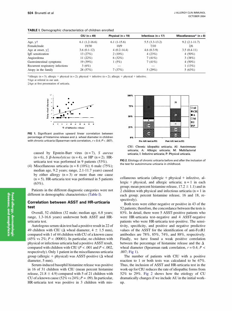
FIG 1. Significant positive upward linear correlation between
percentage of histamine release and D wheal diameter in children
with chronic urticaria (Spearman rank correlation, r = 0.4; P < .007).
cellaneous urticaria (allergic 1 physical 1 infective, al-lergic 1 physical, and allergic urticaria; n = 1 in eachgroup; mean percent histamine release, 17.26 1.1) and in2 children with physical and infectious urticaria (n = 1 ineach group; percent histamine release, 16 and 18, re-spectively).
Both tests were either negative or positive in 43 of the52 patients; therefore, the concordance between the tests is83%. In detail, there were 5 ASST-positive patients whowere HR-urticaria test–negative and 4 ASST-negativepatients who were HR-urticaria test–positive. The sensi-tivity, specificity, and positive and negative predictivevalues of the ASST for the identification of anti-FceR1antibodies are 78%, 85%, 74%, and 88%, respectively.Finally, we have found a weak positive correlationbetween the percentage of histamine release and the D
wheal diameter (Spearman rank correlation, r = 0.4; P <.007; Fig 1).
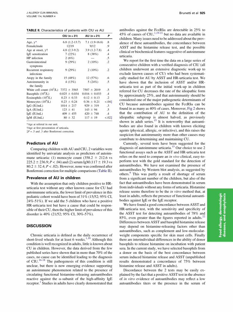
The number of patients with CIU with a positivereaction to 1 or both tests was calculated to be 67%.Thus, the inclusion of ASST and HR-urticaria test in thework-up for CIU reduces the rate of idiopathic forms from52% to 29%. Fig 2 shows how the etiology of CUdramatically changes if we include AU in the initial work-up.
FIG 2. Etiology of chronic urticaria before and after the inclusion of
the test for autoimmune urticaria in childhood.
J ALLERGY CLIN IMMUNOL
VOLUME 114, NUMBER 4
Brunetti et al 925
Foodallerg
y,derm
ato
logic
disease
s,andanaphylaxis
Predictors of AU
Comparing children with AU andCIU, 2 variables wereidentified by univariate analysis as predictors of autoim-mune urticaria: (1) monocyte count (358.2 6 212.6 vs225.26 236.9;P< .04) and (2) serum IgM (1176 19.3 vs80.26 32.4; P < .02). However, none was confirmed afterBonferroni correction for multiple comparisons (Table II).
Prevalence of AU in children
With the assumption that only children positive to HR-urticaria test without any other known cause for CU hadautoimmune urticaria, the lower limit of prevalence in thispediatric cohort would have been of 31% (15/52; 95% CI,24%-51%). If we add the 5 children who have a positiveHR-urticaria test but have a cause that could be respon-sible of their CU, then the higher limit of prevalence of thisdisorder is 40% (21/52; 95% CI, 30%-57%).
DISCUSSION
Chronic urticaria is defined as the daily occurrence ofshort-lived wheals for at least 6 weeks.1,10 Although thiscondition is well recognized in adults, little is known aboutCU in children. However, the data derived from the fewpublished series have shown that in more than 70% of thecases, no cause can be identified leading to the diagnosisof CIU.16-18 The pathogenesis of this condition is stillunclear, but there is now emerging evidence supportingan autoimmune phenomenon related to the presence ofcirculating functional histamine-releasing autoantibodiesreactive against the a-subunit of the high-affinity IgEreceptor.1 Studies in adults have clearly demonstrated that
TABLE II. Characteristics of patients with CIU vs AU
CIU (n = 31) AU (n = 21) P
Age, y* 6.8 (1.2-13.7) 7.1 (1.9-16.6) .8
Female/male 12/19 9/12 .9
Age at onset, y� 4.8 (2.3-9.2) 3.9 (1.2-7.8) .4
IgE sensitization 7 (22%) 8 (38%) .4
HP infection 2 (6%) — .5
Gastrointestinal
symptoms
9 (29%) 2 (10%) .2
Recurrent respiratory
infections
9 (29%) 2 (10%) .2
Atopy in the family 15 (48%) 12 (57%) .6
Autoimmunity in
the family
4 (13%) 5 (24%) .5
White cell count (103/L) 7372 6 3565 7507 6 2019 .5
Basophils (109/L) 0.025 6 0.034 0.016 6 0.033 .4
Eosinophils (109/L) 0.23 6 0.33 0.12 6 0.13 .2
Monocytes (109/L) 0.23 6 0.24 0.36 6 0.21 <.04�IgG (IU/mL) 1014 6 217 929 6 319 .3
IgA (IU/mL) 123 6 41 143 6 94 .5
IgE (IU/mL) 409 6 455 420 6 748 .4
IgM (IU/mL) 80 6 32 117 6 19 <.02�
*Age at referral in our unit.
�Age at first presentation of urticaria.
�P = .5 and .3 after Bonferroni correction.
antibodies against the FceRIa are detectable in 25% to45% of causes of CIU,1,19-21 but no data are available inchildren.Many issues need to be addressed about the prev-alence of these autoantibodies, the concordance betweenASST and the histamine release test, and the possibleclinical or biochemical features suggestive of autoimmuneurticaria.
We report for the first time the data on a large series ofconsecutive children with a verified diagnosis of CIU (allchildren underwent an extensive diagnostic work-up toexclude known causes of CU) who had been systemati-cally studied for AU by ASST and HR-urticaria test. Wehave shown that the inclusion of ASST and/or HR-urticaria test as part of the initial work-up in childrenreferred for CU decreases the rate of the idiopathic formby approximately 25%, and that autoimmunity should beconsidered one of the major pathogenetic determinants ofCU because autoantibodies against the FceRIa can befound in as many as 40% of cases. Moreover, Fig 2 showsthat the contribution of AU to the definition of theidiopathic subgroup is almost halved, as previouslyshown in adult series.15 It is noteworthy that autoanti-bodies are also found in children with known elicitingagents (physical, allergic, or infective), and this raises thesuspicion that autoimmunity more than other causes maycontribute to determining and maintaining CU.
Currently, several tests have been suggested for thediagnosis of autoimmune urticaria.15 Our choice to use 2functional assays such as the ASST and HR-urticaria testrelies on the need to compare an in vivo clinical, easy-to-perform test with the gold standard for the detection ofautoantibodies. We have not examined the presence ofautoantibodies by Western blot analysis, as suggested byothers.9 This was partly a result of shortage of serumfrom a significant number of the children, but also of thefact that autoantibodies have been demonstrated in serumfrom individuals without any forms of urticaria. Histaminerelease seems therefore to be the in vitro method that, atleast in adults, reflects the presence of functional autoanti-bodies against IgE or the IgE receptor.
We have found a good concordance between ASST andHR-urticaria test, with the sensitivity and specificity ofthe ASST test for detecting autoantibodies of 78% and85%, even greater than the figures reported in adults.22
Differences betweenASST and basophil histamine releasemay depend on histamine-releasing factors other thanautoantibodies, such as complement and low-molecular-weight components specific for skin mast cells. Finallythere are interindividual differences in the ability of donorbasophils to release histamine on incubation with patientsera. In the current study, we have selected basophils froma donor on the basis of the best concordance betweenserum induced histamine release and ASST (unpublishedresults demonstrated a concordance of 75% betweenhistamine release and ASST in adults).
Discordance between the 2 tests may be easily ex-plained by the fact that a positive ASST test in the absenceof in vitro evidence of autoantibodies may reflect a lowautoantibodies titers or the presence in the serum of
J ALLERGY CLIN IMMUNOL
OCTOBER 2004
926 Brunetti et al
Foodalle
rgy,derm
ato
logic
dise
ase
s,andanaphylaxis
histamine releasing substances,1,20,21 whereas a negativeASST test with a positive HR-urticaria test may reflect theinjection of the autologous serum in a refractory sitesecondary to a recent wheal.12 Another factor relevant forthe outcome of ASST in children is age, as describedregarding skin prick test to both histamine and allergen.12
A stratification of ASST responses in children wouldtherefore be relevant to perform in the current study, butthe number of positive reactions within each age group istoo low to perform a valid statistical analysis.
Autologous serum skin test is cheap, is easy to perform,and, if performed as appropriate,12 has good sensitivityand even better specificity at detecting autoantibodies alsoin children; therefore, it can be used as a predictive clinicaltest to diagnose AU, especially in places where thebasophil histamine-releasing test is not available.
Themain concern iswhen to consider a child affected byautoimmune urticaria. Indeed, in contrastwith adults, thereis little experience with this condition, and it is possiblethat the cut-off value of HR-urticaria test in children mightbe different from that of adults.Moreover, theHR-urticariatest and ASST identify 2 different phenomena. The firstshows the presence of autoantibodies, whereas the lattershows that serum is able to induce histamine release fromskin mast cells, without knowing whether this is inducedby autoantibodies in all cases.23 To distinguish betweenautoantibodies against IgE and the IgE receptor, pre-viously published protocols assessed basophils of healthylow and high IgE donors.19 Our own unpublished pilotstudies using basophils from different donors showed thatthe serum-induced histamine release was not dependingonly on the level of serum IgE.A future alternative to usingbasophils from high and low IgE donors might be to useintact and IgE deprived cells from the same donor.However, further studies are needed to establish theoptimal cell source. We have therefore chosen to use onlyone source of basophils, as discussed.
We believe that both tests provide important informa-tion when assessing a child with CU other than AU, thedefinition of which we reserve for children with autoanti-bodies.
When we have tried to assess whether it was possible tosuspect AU on the basis of clinical and laboratoristic data,we could not find any difference between the autoimmuneand nonautoimmune groups except for monocyte countand serum IgM; however, the significance was lost aftercorrection for multiple testing. The lack of clinical andlaboratoristic differences between CU with and withoutautoantibodies indicates that the reaction to a cause ratherthan the cause itself plays a major role in the clinicalpresentation. In contrast with adult studies, none of thechildren with AU had another concomitant autoimmunedisease,24 signs of thyroid autoimmunity,25 celiac dis-ease,26,27 or HP infection.28 We are aware that the likeli-hood of having more than one autoimmune diseaseincreases with age, and therefore, it is possible that theabsence of associated autoimmune conditions depends onthe pediatric age of our series; however, the follow-up ofthis cohort of children will clarify the issue.
We have determined that the prevalence of AU inchildren is not less than 30%. The achievement of a correctdiagnosis is of paramount importance, especially in thecase of AU, in which plasmapheresis29 or immunosup-pressive treatment30 may be indicated. These treatmentsshould be used only in the presence of a definitivediagnosis.
REFERENCES
1. Hide M, Francis DM, Grattan CE, Hakimi J, Kochan JP, Greaves MW.
Autoantibodies against the high affinity IgE receptor as a cause of
histamine release in chronic urticaria. N Engl J Med 1993;328:1599-604.
2. Greaves MW. Chronic urticaria. N Engl J Med 1995;332:1767-72.
3. O’Donnell BF, O’Neill CM, Francis DM, Niimi N, Barr RM, Barlow RJ,
et al. Human leucocyte antigen class II associations in chronic idiophatic
urticaria. Br J Dermatol 1999;140:853-8.
4. Sabroe RA, Seed PT, Francis DM, Barr RM, Black AK, Greaves MW.
Chronic idiopathic urticaria: comparison of the clinical features of
patients with and without anti-FcepsilonRI or anti-IgE autoantibodies.
J Am Acad Dermatol 1999;40:443-50.
5. O’Donnell BF, Lawlor F, Simpson J, Morgan M, Greaves MW. The
impact of chronic urticaria on the quality of life. Br J Dermatol 1997;136:
197-201.
6. Grattan CEH, Francis DM, Hide M, Greaves MW. Detection of
circulating histamine releasing autoantibodies with functional properties
of anti-IgE in chronic urticaria. Clin Exp Allergy 1991;21:695-704.
7. Gruber BL, Baeza ML, Marchese MJ, Agnello V, Kaplan AP.
Prevalence and functional role of anti-IgE autoantibodies in urticarial
syndromes. J Invest Dermatol 1988;90:213-7.
8. Sabroe RA, Francis DM, Barr RM, Greaves MW. Anti-FceRI autoanti-bodies and basophil histamine releasability in chronic idiopathic
urticaria. J Allergy Clin Immunol 1998;102:651-8.
9. Greaves MW. Chronic urticaria. J Allergy Clin Immunol 2000;105:
664-72.
10. Greaves MW. Chronic urticaria in childhood. Allergy 2000;55:309-20.
11. Kobza Black A, Lawlor F, Greaves MW. Consensus meeting on the
definition of physical urticarias and urticarial vasculitis. Clin Exp
Dermatol 1996;21:424-6.
12. Sabroe RA, Grattan CE, Francis DM, Barr RM, Kobza Black A, Greaves
MW. The autologous serum skin test: a screening test for autoantibodies
in chronic idiopathic urticaria. Br J Dermatol 1999;140:446-52.
13. Skov PS, Mosbech H, Norn S, Weeke B. Sensitive glass microfibre based
histamine analysis for allergy testing in washed blood cells. Allergy
1985;40:213-8.
14. Nolte H, Storm K, Schiotz PO. Diagnostic value of a glass fibre based
histamine analysis for allergy testing in children. Allergy 1990;45:213-23.
15. Grattan CE, Sabroe RA, Greaves MW. Chronic urticaria. J Am Acad
Dermatol 2002;46:645-57.
16. Harris A, Twarog FJ, Geha RS. cronich urticaria in childhood: natural
course and etiology. Ann Allergy 1983;51:161-5.
17. Volonakis M, Katasarou-Katasari A, Stratigos J. Etiologic factors in
childhood chronic urticari. Ann Allergy 1992;69:61-5.
18. Ehlers I, Niggemann B, Binder C, Zuberbier T. Role of nonallergic
hypersensitivity reactions in children with chronic urticaria. Allergy
1998;53:1074-7.
19. Niimi N, Francis DM, Kermani F, O’Donnell BF, Hide M, Kobza-Black
A, et al. Dermal mast cell activation by autoantibodies against the high
affinity IgE receptor in chronic urticaria. J Invest Dermatol 1996;106:
1001-6.
20. Tong LJ, Balakrishnan G, Kochan JP, Kinet JP, Kaplan AP. Assessment
of autoimmunity in patients with chronic urticaria. J Allergy Clin
Immunol 1997;99:461-5.
21. Ferrer M, Kinet JP, Kaplan AP. Comparative studies of functional
and binding assays for IgG anti-Fc(epsilon)RIalpha (alpha-subunit)
in chronic urticaria. Allergy Clin Immunol 1998;101:672-6.
22. Sabroe RA, Fiebiger E, Francis DM, Maurer D, Seed PT, Grattan CE,
et al. Classification of anti-FcepsilonRI and anti-IgE autoantibodies
in chronic idiopathic urticaria and correlation with disease severity.
J Allergy Clin Immunol 2002;110:492-9.

J ALLERGY CLIN IMMUNOL
VOLUME 114, NUMBER 4
Brunetti et al 927
23. Kikuchi Y, Kaplan AP. Mechanisms of autoimmune activation of
basophils in chronic urticaria. J Allergy Clin Immunol 2001;107:1056-62.
24. Gaig P, Garcia-Ortega P, Enrique E, Richart C. Successful treatment
of chronic idiopathic urticaria associated with thyroid autoimmunity.
J Invest Allergol Clin Immunol 2000;10:342-5.
25. Ryhal B, DeMera RS, Shoenfeld Y, Peter JB, Gershwin ME. Are
autoantibodies present in patients with subacute and chronic urticaria?
J Investig Allergol Clin Immunol 2001;11:16-20.
26. Levine A, Dalal I, Bujanover Y. Celiac disease associated with familial
chronic urticaria and thyroid autoimmunity in a child. Pediatrics 1999;
104:e25.
27. Meneghetti R, Gerarduzzi T, Barbi E, Ventura A. Chronic urticaria and
coeliac disease. Arch Dis Child 2004;89:293.
28. Bakos N, Hillander M. Comparison of chronic autoimmune urticaria with
chronic idiopathic urticaria. Int J Dermatol 2003;42:613-5.
29. Grattan CE, Francis DM, Slater NG, Barlow RJ, Greaves MW.
Plasmapheresis for severe, unremitting, chronic urticaria. Lancet 1992;
339:1078-80.
30. Grattan CE, O’Donnell BF, Francis DM, Niimi N, Barlow RJ,
Seed PT, et al. Randomized double-blind study of cyclosporin in chronic
‘‘idiopathic’’ urticaria. Br J Dermatol 2000;143:365-72.
Foodallerg
y,derm
ato
logic
disease
s,andanaphylaxis
Copyright © 2022 FDOKUMEN