Retinoid Therapy for Autoimmune Diseases
10
302 Current Alzheimer Research, 2009, 6, 302-311 1567-2050/09 $55.00+.00 ©2009 Bentham Science Publishers Ltd. Towards Retinoid Therapy for Alzheimer’s Disease K. Shudo 1, *, H. Fukasawa 2 , M. Nakagomi 1 and N. Yamagata 1 1 Research Foundation ITSUU Laboratory, 2 Institute of Medicinal Molecular Design, Inc., Molecular and Functional Bioscience, Japan Abstract: Alzheimer’s disease(AD) is associated with a variety of pathophysiological features, including amyloid plaques, inflammation, immunological changes, cell death and regeneration processes, altered neurotransmission, and age- related changes. Retinoic acid receptors (RARs) and retinoids are relevant to all of these. Here we review the pathology, pharmacology, and biochemistry of AD in relation to RARs and retinoids, and we suggest that retinoids are candidate drugs for treatment of AD. Keywords: Alzheimer’s disease, retinoid, retinoic acid receptors, inflammation, immunology, regulatory T cell, neurodegen- eration. TOWARDS RETINOID THERAPY FOR ALZHEI- MER’S DISEASE The management of Alzheimer’s disease (AD) remains a challenge, even as our knowledge and understanding of AD continue to grow at an unprecedented rate [1-3]. Research that bridges the gap between basic science and clinical appli- cation is extremely important for the development of new therapeutics. AD is a neurodegenerative disease, and has many pathological, biochemical and immunological features in common with Parkinson’s disease (PD), amyotrophic lat- eral sclerosis (ALS), multiple sclerosis (MS) and spinal code injury (SCI). This paper deals mainly with AD in relation to retinoic acid receptors (RARs: RAR , and ) and their ligands (retinoids), such as the endogenous RAR ligand all- trans-retinoic acid (RA), taking into account knowledge about PD, ALS and other neurodegenerative diseases. It is important to note that factors leading to the onset of these diseases are still poorly understood, and so there is a great deal of scope for novel therapeutic approaches. Recent find- ings indicate that the window of opportunity for enhancing or normalizing the growth of neuronal cells and promoting recovery from neurodegenerative diseases may be larger than previously thought. AD is associated with a variety of pathophysiological features, including amyloid plaques, inflammation, immu- nological changes, cell death and regeneration processes, altered neurotransmission, and age-related changes. Here, we will review how retinoids could be involved in all these fea- tures of AD, together with the brief summary of their biol- ogy, pharmacology, and medicinal chemistry. Fig. (1) shows the overview of the various potential therapeutic targets by retinoids discussed in this paper. MEDICINAL CHEMISTRY OF RETINOIDS Retinoids are analogues of retinoic acid, an active me- tabolite of vitamin A, and are specific modulators of cell *Address correspondence to this author at the Research Foundation ITSUU Laboratory, 2-28-10 tamagawa, setagaya-ku, Tokyo 158-0094, Japan; E-mail: [email protected] proliferation, differentiation, and morphogenesis in verte- brates. There are many excellent reviews of the medicinal chemistry of retinoids [4-10]. The term retinoid is used for substances which may be defined as (1) vitamin A related compounds (including vitamin A (retinol) and its biological precursor carotenoids, (2) RA (vitamin A metabolite), which activates RAR , , and , and synthetic analogs which bind with RARs with high affinity in an agonistic (similar bio- logical activities to RA) or antagonistic manner, (3) com- pounds which activate RXR , and , which are nuclear receptors different from RARs, (4) compounds which mod- ify the activities of RA by influencing metabolism, biosyn- thesis or other pathways acting on co-factors, without bind- ing to RARs or RXRs. We prefer to use the term retinoid in a strict sense, that is, compounds in category (2). It should be noted that the activity of vitamin A (retinol) is essentially due to RA generated by metabolism in vivo, except for the participation in vision via rhodopsin [11-13]. Normally, a low level of RA is present (~10 –9~10 M), although a much larger amount (~10 -6 M) of vitamin A (retinol) is found in serum. The endogenous active retinoid is RA, which binds to and activates RAR , , and . Isomeric 13-cis-retinoic acid may also be an endogenous retinoid, but is less abundant than RA. 9-cis-Retinoic acid is synthesized from, or in equi- librium with, RA, and it binds to and activates RAR , , and , and in addition RXR , , and . Many synthetic analogs with a variety of structures have been prepared, and show different receptor selectivities and elicit different pharmacol- ogical effects from RA, including different adverse effects. Some act as antagonists which inhibit the activity of RA. BIOLOGICAL AND PHARMACOLOGICAL ACTIVI- TIES OF RETINOIDS The biological importance of RARs has been thoroughly documented elsewhere [14]. A number of genes are regu- lated directly through the RAR or RXR- response elements through retinoic acid or retinoids binding to RAR of the RAR/RXR heterodimers, or indirectly, plausibly through the participation of the directly-regulated genes [15]. The bio- logical or pharmacological activities of interest here are
-
Upload
independent -
Category
Documents
-
view
6 -
download
0
Transcript of Retinoid Therapy for Autoimmune Diseases
302 Current Alzheimer Research, 2009, 6, 302-311
1567-2050/09 $55.00+.00 ©2009 Bentham Science Publishers Ltd.
Towards Retinoid Therapy for Alzheimer’s Disease
K. Shudo1,*, H. Fukasawa
2, M. Nakagomi
1 and N. Yamagata
1
1Research Foundation ITSUU Laboratory,
2Institute of Medicinal Molecular Design, Inc., Molecular and Functional
Bioscience, Japan
Abstract: Alzheimer’s disease(AD) is associated with a variety of pathophysiological features, including amyloid
plaques, inflammation, immunological changes, cell death and regeneration processes, altered neurotransmission, and age-
related changes. Retinoic acid receptors (RARs) and retinoids are relevant to all of these. Here we review the pathology,
pharmacology, and biochemistry of AD in relation to RARs and retinoids, and we suggest that retinoids are candidate
drugs for treatment of AD.
Keywords: Alzheimer’s disease, retinoid, retinoic acid receptors, inflammation, immunology, regulatory T cell, neurodegen-eration.
TOWARDS RETINOID THERAPY FOR ALZHEI-
MER’S DISEASE
The management of Alzheimer’s disease (AD) remains a challenge, even as our knowledge and understanding of AD continue to grow at an unprecedented rate [1-3]. Research that bridges the gap between basic science and clinical appli-cation is extremely important for the development of new therapeutics. AD is a neurodegenerative disease, and has many pathological, biochemical and immunological features in common with Parkinson’s disease (PD), amyotrophic lat-eral sclerosis (ALS), multiple sclerosis (MS) and spinal code injury (SCI). This paper deals mainly with AD in relation to retinoic acid receptors (RARs: RAR , and ) and their ligands (retinoids), such as the endogenous RAR ligand all-trans-retinoic acid (RA), taking into account knowledge about PD, ALS and other neurodegenerative diseases. It is important to note that factors leading to the onset of these diseases are still poorly understood, and so there is a great deal of scope for novel therapeutic approaches. Recent find-ings indicate that the window of opportunity for enhancing or normalizing the growth of neuronal cells and promoting recovery from neurodegenerative diseases may be larger than previously thought.
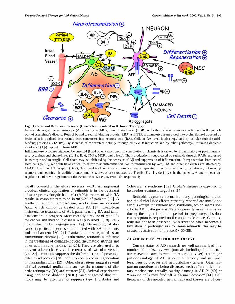
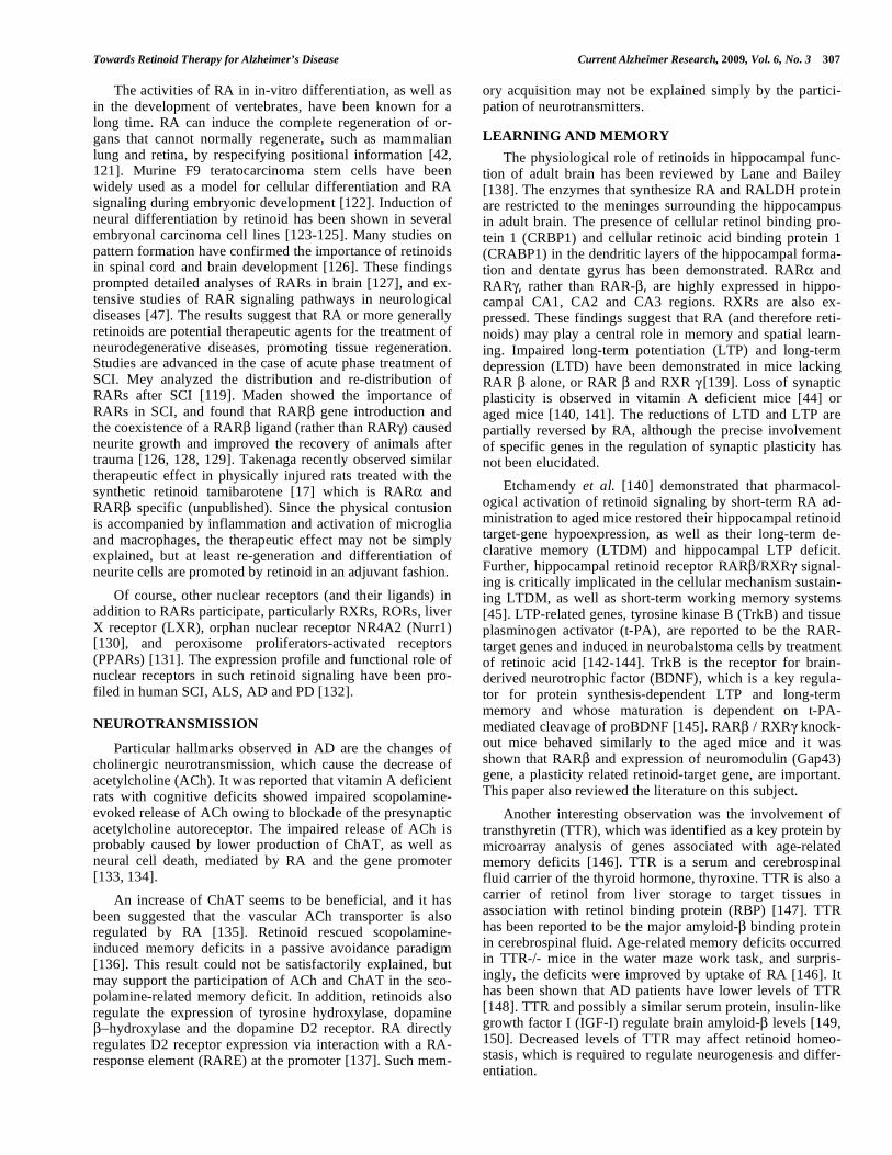
AD is associated with a variety of pathophysiological features, including amyloid plaques, inflammation, immu-nological changes, cell death and regeneration processes, altered neurotransmission, and age-related changes. Here, we will review how retinoids could be involved in all these fea-tures of AD, together with the brief summary of their biol-ogy, pharmacology, and medicinal chemistry. Fig. (1) shows the overview of the various potential therapeutic targets by retinoids discussed in this paper.
MEDICINAL CHEMISTRY OF RETINOIDS
Retinoids are analogues of retinoic acid, an active me-tabolite of vitamin A, and are specific modulators of cell
*Address correspondence to this author at the Research Foundation ITSUU
Laboratory, 2-28-10 tamagawa, setagaya-ku, Tokyo 158-0094, Japan;
E-mail: [email protected]
proliferation, differentiation, and morphogenesis in verte-brates. There are many excellent reviews of the medicinal chemistry of retinoids [4-10]. The term retinoid is used for substances which may be defined as (1) vitamin A related compounds (including vitamin A (retinol) and its biological precursor carotenoids, (2) RA (vitamin A metabolite), which activates RAR , , and , and synthetic analogs which bind with RARs with high affinity in an agonistic (similar bio-logical activities to RA) or antagonistic manner, (3) com-pounds which activate RXR , and , which are nuclear receptors different from RARs, (4) compounds which mod-ify the activities of RA by influencing metabolism, biosyn-thesis or other pathways acting on co-factors, without bind-ing to RARs or RXRs. We prefer to use the term retinoid in a strict sense, that is, compounds in category (2). It should be noted that the activity of vitamin A (retinol) is essentially due to RA generated by metabolism in vivo, except for the participation in vision via rhodopsin [11-13]. Normally, a low level of RA is present (~10
–9~10 M), although a much
larger amount (~10-6
M) of vitamin A (retinol) is found in serum. The endogenous active retinoid is RA, which binds to and activates RAR , , and . Isomeric 13-cis-retinoic acid may also be an endogenous retinoid, but is less abundant than RA. 9-cis-Retinoic acid is synthesized from, or in equi-librium with, RA, and it binds to and activates RAR , , and , and in addition RXR , , and . Many synthetic analogs
with a variety of structures have been prepared, and show different receptor selectivities and elicit different pharmacol-ogical effects from RA, including different adverse effects. Some act as antagonists which inhibit the activity of RA.
BIOLOGICAL AND PHARMACOLOGICAL ACTIVI-
TIES OF RETINOIDS
The biological importance of RARs has been thoroughly documented elsewhere [14]. A number of genes are regu-lated directly through the RAR or RXR- response elements through retinoic acid or retinoids binding to RAR of the RAR/RXR heterodimers, or indirectly, plausibly through the participation of the directly-regulated genes [15]. The bio-logical or pharmacological activities of interest here are
Towards Retinoid Therapy for Alzheimer’s Disease Current Alzheimer Research, 2009, Vol. 6, No. 3 303
mostly covered in the above reviews [4-10]. An important practical clinical application of retinoids is in the treatment of acute promyelocytic leukemia (APL): treatment with RA results in complete remission in 90-95% of patients [16]. A synthetic retinoid, tamibarotene, works even on relapsed APL, which cannot be treated with RA [17]. Long-term maintenance treatments of APL patients using RA and tami-barotene are in progress. More recently a review of retinoids for cancer and metabolic disease was published [18]. Reti-noids also inhibit angiogenesis [19]. Dermatological dis-eases, in particular psoriasis, are treated with RA, etretinate, and tamibarotene [20, 21]. Psoriasis is now regarded as an autoimmune disease [22]. Furthermore, retinoids is effective in the treatment of collagen-induced rheumatoid arthritis and other autoimmune models [23-25]. They are also useful to prevent atherosclerosis and restenosis of vascular vessels [26, 27]. Retinoids suppress the differentiation of preadipo-cytes to adipocytes [28], and promote alveolar regeneration in mammalian lungs [29]. Other observations suggest several clinical potential applications such as the treatment of dia-betic retinopathy [30] and cataract [31]. Animal experiments using non-obese diabetic (NOD) mice suggested that reti-noids may be effective to suppress type I diabetes and
Schoegren’s syndrome [32]. Crohn’s disease is expected to be another treatment target [33, 34].
Retinoids appear to normalize many pathological states, and the clinical side effects presently reported are mostly not serious except for retinoic acid syndrome, which seems spe-cific to APL pathogenesis. Teteratogenicity remains an issue during the organ formation period in pregnancy: absolute contraception is required until complete clearance. Genotox-icity has not been observed. Skin irritation often becomes a limitation in prolonged use for some retinoids; this may be caused by activation of the RAR [35-38].
ALZHEIMER’S PATHOPHYSIOLOGY
Current status of AD research are well summarized in a number of books, reviews, journals including this journal, and elsewhere such as web site reports [1-3, 39]. The major pathophysiology of AD is cerebral atrophy and neuronal loss, neuritic plaques and neurofibrillary tangles. Other im-portant questions are being discussed such as “are inflamma-tory mechanisms actually causing damage in AD ?” [40] or “immune cells may fend off Alzheimer desease” [41]. Cell therapies of degenerated neural cells and tissues are of cur-
Fig. (1). Retinoid Dramatis Personae (Characters Involved in Retinoid Therapy).
Neuron, dameged neuron, astrocyte (AS), microglia (MG), blood brain barrier (BBB), and other cellular members participate in the pathol-
ogy of Alzheimer's disease. Retinol bound to retinol-binding protein (RBP) and TTR is transported from blood into brain. Retinol uptaked by
brain cells is oxidised into retinal, then converterd into retinoic acid (RA). Cellular RA level is also regulated by cellular retinoic acid-
binding proteins (CRABPs). By increase of -secretase activity through ADAM10 induction and by other pathaways, retinoids decrease
amyloid- (A ) deposition from APP.
Inflammatory response triggered by amyloid- and other causes such as xenobiotics or chemicals is drived by inflammatory or proinflamma-
tory cytokines and chemokines (IL-1b, IL-6, TNFa, MCP1 and others). Their production is suppressed by retinoids through RARs expressed
in astrocyte and microglia. Cell death may be inhibited by the decrease of A and suppression of inflammation. In regeneration from neural
stem cells (NSC), retinoids have critical roles for their differentiation. Neurotransmission by Ach, DA and other molecules are affected by
ChAT, dopamine D2 receptor (D2R), TrkB and t-PA which are transcriptionally regulated directly or indirectly by retinoid, influencing
memory and learning. In addition, autoimmune pathways are regulated by T cells (Fig. 2 vide infra). In the scheme, + and - mean up-
regulation and down-regulation of the events or activities, by retinoids, respectively.
304 Current Alzheimer Research, 2009, Vol. 6, No. 3 Shudo et al.
rent interests, but relating to that, regeneration by retinoids looks actual and now well-understood [42, 43]. Age-related decline of cognition which is a serious feature of AD, could also be a target phenomenon of a retinoid [44, 45]. Several evidence for the relationships between the late onset Alz-heimer’s disease and retinoid defective signaling are shown [46, 47]. Now AD and other neurodegenerative diseases may be likely the therapeutic targets of retinoid.
AMYLOID- HYPOTHESIS
AD is characterized by progressive memory deficits, cognitive impairment and personality changes due to neu-ronal cell death in the hippocampus and frontal cortex. The histopathological hallmark of AD is senile plaques, which consist of insoluble amyloid- . Activated microglia and as-trocytes (either proinflammatory or anti-inflammatory) sur-round the amyloid plaques, and appear to be associated with the lesions. Reduction of amyloid- could be accomplished by inhibiting its production or aggregation, or promoting its degradation and removal, so active or passive immunization may have therapeutic potential. It has been shown that amy-loid- specific antibodies can clear amyloid assemblies and amyloid- deposits in amyloid precursor protein (APP)-transgenic mice. Amyloid- -specific antibodies have been extensively studied as a mediator of amyloid clearance [48].
APP is processed and fragmented by -secretase and -secretase to generate amyloid- . However, the nonamyloi-dogenic pathway of processing precursor proteins involves cleavage within the amyloid- peptide sequence. The identi-fication of a member of the disintegrin and metalloprotease family (ADAM10) as an -secretase, whose expression can be regulated by retinoic acid, represents another therapeutic opportunity. Endogenous ADAM10 mRNA levels and the ADAM10 promoter activities were increased on RA treat-ment in neuroblastoma cells: thus, retinoic acid works as an activator of the -secretase [49-51].
Dietary deficiency of vitamin A disrupts the retinoid sig-naling pathway in adult rats, leading to deposition of amy-loid- in the cerebral blood vessels via down-regulation of RAR- in the forebrain neurons and loss of choline acetyl-transferase (ChAT) expression, and these changes were re-versed by administration of retinoic acid [52, 53]. Pathologi-cal samples from AD patients showed a similar RAR defi-cit and deposition of amyloid- in the surviving neurons [52].
NEUROINFLAMMATION
For many years, the central nervous system was consid-ered to be immunologically privileged, being neither suscep-tible to, nor contributing to, inflammation. However, the Neuroinflammation Working Group suggested a relationship between inflammation and AD [40]. At least 600 reports indicate that the brain is by no means an immunologically isolated organ, and may have unique immunologic proper-ties. Microglial-cell activation and migration, participation of astrocytes, and participation of various cytokines in AD have been confirmed. Microglia secrete proteolytic enzymes that degrade amyloid- , and play a neuroprotective role in AD. One of the chemokine receptors, CCR2 deficiency impairs microglial accumulation and accelerates the progression of
AD-like disease in a model mouse, Tg2576 [41, 54]. More recently, Beers et al. showed CD4+ T cells, which can be recruited by MCP-1/CCR2 signaling, provides supportive neuroprotection by modulating the trophic/cytotoxic balance of glia in ALS mouse mode [55].
Epidemiological studies indicate a reduced prevalence of AD among chronic users of nonsteroidal anti-inflamatory drugs, and clinical trials suggested a beneficial effect of some cycloxygenase (cox) inhibitors, although the effect is not related to the cox-inhibitory activity. Synthetic glucocor-ticoids are potent ant-inflammatory agents that act by an-tagonizing AP1 and NF- B promoter elements that regulate the transcription of inflammatory molecules. They are useful for the treatment of inflammation, but various biochemical, systemic and behavioral side effects and other contraindica-tions exist for use in AD. In their review [40], Akiyama et al. concluded that “no more than nonsteroidal anti-inflammatory drugs (NSAIDs) cure arthritis will anti-inflammatory drugs cure AD. However, if AD neuroinflammation is approached with realistic expectations and rational drug design, AD pa-tients should significantly benefit from anti-inflammatory treatment”. Suppression of inflammation in the brain is im-portant in the treatment of many neurodegenerative diseases.
Other aspects of inflammation and immunology of neuro-degenerative diseases have also recently been reviewed [56-58]. Cytokines and chemokines, such as IL1 , IL1 , IL6, IL4, IL10, IL13 and MCP-1 are also important in AD. Though the action of IL6 generally depends on cellular and environmental conditions, unlike that of IL1 , which pre-dominantly has a proinflammatory effect, the cytokine may generally exert a damaging proinflammatory effect in the central nervous system, although it is present in normal brain tissues [59]. IL6 dysregulation is involved in many age-related diseases, including autoimmune diseases. Chronic inflammation and astrocytosis are histopathological hall-marks of AD patients, and astrocytes and microglia produce IL6 in response to amyloid- induced injury, thereby further promoting plaque formation. The induction of IL6 mRNA in the hippocampus and cortex of APPsw transgenic mice Tg2576 may be crucial in the early onset of AD [60]. The association of –174G/C and –572G/C mutations of IL6 pro-moters with AD has been discussed, but remains contentious [61, 62].
Since retinoids strongly suppress the production of IL6 [63, 64], the suppression of IL6 by retinoids may be benefi-cial for the treatment of AD. Retinoids also inhibit LPS- or amyloid- -induced TNF- production, and expression of inducible NO synthase (iNOS) in activated microglia; these effects may be mediated via the inhibition of NF- B nuclear translocation. Retinoids inhibit many aspects of microglial activation [65, 66]. Thus, retinoids seem to have consider-able potential from the standpoint of suppression of inflam-mation in neurodegenerative diseases.
AUTOIMMUNE FEATURES
Immune system alterations during aging are complex and pleiotropic. Generally, changes of the T cell component are age-dependent. It is always difficult to determine whether changes are associated with aging itself or a result (or a cause) of disease. Although immunological studies of the
Towards Retinoid Therapy for Alzheimer’s Disease Current Alzheimer Research, 2009, Vol. 6, No. 3 305
brain have been relatively limited because of the supposed immune privilege of the brain, the role of immunology can-not be neglected in AD (or PD), as well as in the representa-tive autoimmune neurodegenerative disease, MS. It is, how-ever, interesting that up-regulation of major histocompatibil-ity complex (MHC) class I and II on glial and neural cells occurs in AD, in addition to production of inflammatory cy-tokines and limited T cell infiltration [40, 67], and the gen-eration of autoreactive antibodies and T cells against amy-loid- [68] . Similar findings were reported in PD and ALS [69-72]. Thus, there is a possibility that an appropriate modulation of autoimmune response may prevent disease development. In other words, changes in the activity or population of T cells may influence the progression of AD, PD and other such diseases.
Recently, IL-17-producing helper T cell subset, Th17 is at the center of attention in autoimmune research [73, 74]. The importance of Th17 cells in central nervous system in-flammation in MS patients and experimental autoimmune myasthenia gravis in C57BL/6 mice has been shown [75, 76]. IL17 and IL22 receptors are expressed on blood-brain-barrier (BBB) endothelial cells in MS lesions, and these cy-tokines disrupt BBB tight junctions in vivo and in vitro. Thus, Th17 lymphocytes transmigrate efficiently across the BBB, killing human neurons and causing central nervous system inflammation through CD4+ lymphocyte recruitment. It was also shown that the BBB induces differentiation of migrating monocytes into Th17-polarizing dendritic cells [77]. The abundance of dendritic cells promotes secretion of IL12p70, TGF and IL6, as well as the proliferation and expansion of distinct populations of IFN -secreting Th1, and IL-17-secreting CD4+ Th17 lymphocytes. Such cells are closely associated with microvascular BBB endothelial cells within acute MS lesions. It has also been shown that im-pairment of the function of tight junctions facilitates the in-vasion of inflammatory cells prior to motor neuronal degen-eration in ALS-causing SOD1 mutant mice [78]. Similar BBB disruption may be involved in the early onset of AD, as well as other neurodegenerative disorders. The suppression of Th17 or the restoration or maintenance of the function of tight junctions has a favorable effect, slowing the progres-sion of autoimmune diseases. It is noteworthy that experi-mental autoimmune encephalomyelitis (EAE) mice or rats, an animal model of MS, has been successfully treated with retinoid [79-81]. Psoriasis, which is an important clinical target of retinoid, is now regarded as an autoimmune disease [22].
T CELL DIFFERENTIATION
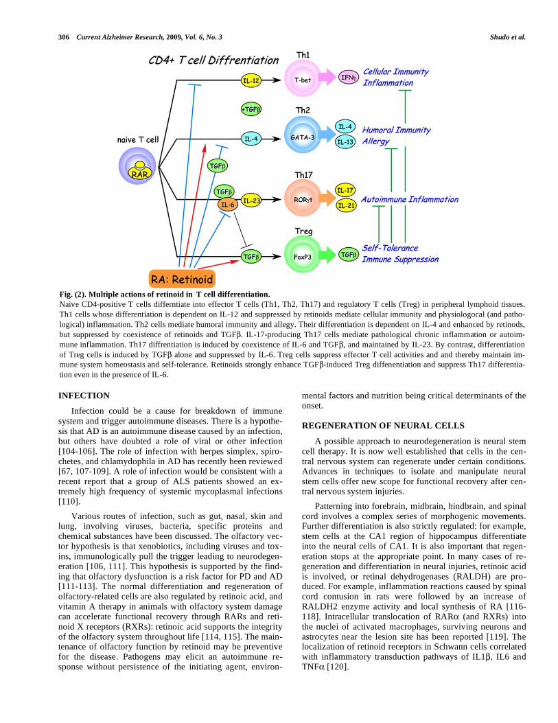
If T cells play a significant role in neurodegenerative diseases, their differentiation status must be important (Fig. 2). For two decades, it has been known that the CD4+ cells among the naïve T cells can differentiate into two types of helper T cells, Th1 cells, which express the transcription factor T-bet (activated by IFN ), and Th2 cells, which ex-press the transcription factor GATA3 (activated by IL4). In this classical Th1/Th2 paradigm, which is revised by recent emergence of Th17, Th1 activation appears particularly im-portant in tissue-specific autoimmunity such as EAE, and Th2 seems to be important in more systemic autoimmunity such as systemic lupas erythematosus (SLE) and allergic
disease, though the key point is likely to be the balance be-tween Th1 and Th2. It is noteworthy that the Th1 pathway is suppressed by RA (or retinoids), and the Th2 pathway is somewhat activated [82]. Retinoids are often beneficial for the treatment of autoimmune disease depending on the char-acteristics of the retinoid and the Th1/Th2 ratio of the dis-ease [83]. A striking recent finding is that CD4+ naïve T cells, independently of Th1- and Th2-cell development, can differentiate to another helper T cell subset, Th17, which produces IL17 and expresses transcription factor retinoid-related orphan receptor t (ROR t) [84]. Th17 cells are im-plicated in the pathogenesis of various autoimmune condi-tions, as mentioned in previous section, and supposed to be the major cause of rheumatoid arthritis, Crohn’s disease, psoriasis, MS and other diseases.
These effector T cells, Th1, Th2, and Th17 are sup-pressed by another CD4+ T cell subset of reguratory Tcells, Treg [85, 86]. A specific marker for Treg is the Foxp3 [87, 88]. The role of Treg cells in self-tolerance, autoimmune diseases and anti-tumor immunity has been actively studied in recent years. The decrease of Treg function may cause or progress breakdown of immune homeostasis. For example, the involvement of Treg has been demonstrated in MS [89], myasthenia gravis [90], and human T-lymphotropic virus 1 (HTLV-I) associated neuroimmunological disease [91]. Therapies for activation or proliferation of Treg may be use-ful for these autoimmune diseases.
This is also the case in the neurodegenerative diseases. Rosenkranz et al reported the involvement of Treg in AD and PD [92]. Analysis of Treg from AD and PD patients, as well as non-affected individuals, revealed that the frequency of Treg (CD4+ Foxp3+) increases with age and the increase is accompanied with intensified suppressive activity of Treg in patients. This may reflect a biological homeostatic re-sponse to the disease. Rosenkranz et al speculated that changes in Treg might affect disease-related immune mecha-nisms, since it has been hypothesized that neuroinflamma-tion may be a response that is aimed at blocking disease de-velopment. The important role of Treg in PD was experi-mentally demonstrated in a MTPT (N-methyl-4-phenyl-1,2,3,6-tetrahydropyridine)-treated mouse model: Treg that highly expressed Foxp3 and IL10 was found to mediate neu-roprotection through suppression of microglial activation [93].
In these respects, it is striking that CD4+ Foxp3+ T cell development from naïve CD4+ T cells is strongly induced by co-treatment with TGF and retinoid, including RA and syn-thetic retinoids such as Am580, and is suppressed by the retinoid antagonist LE540 [94-102] (Fig. 1). It is claimed that Treg is induced at the expense of IL17-secreting T cells by retinoic acid via RAR [102]. However, it is not yet clear whether Th17 cells are convertible to Treg, and also whether or not Foxp3-positive cells induced by RA and retinoids are true Treg. Much remains unclear about the roles of the vari-ous T cell subsets, but the importance of retinoids in T cell differentiation seems now clear. And, there is a proposal as to whether oral tolerance is all due to RA [103], where the author expects the use of Treg cells in the clinic and new RAR agonists other than RA with less side effects for in vivo uses.
306 Current Alzheimer Research, 2009, Vol. 6, No. 3 Shudo et al.
INFECTION
Infection could be a cause for breakdown of immune system and trigger autoimmune diseases. There is a hypothe-sis that AD is an autoimmune disease caused by an infection, but others have doubted a role of viral or other infection [104-106]. The role of infection with herpes simplex, spiro-chetes, and chlamydophila in AD has recently been reviewed [67, 107-109]. A role of infection would be consistent with a recent report that a group of ALS patients showed an ex-tremely high frequency of systemic mycoplasmal infections [110].
Various routes of infection, such as gut, nasal, skin and lung, involving viruses, bacteria, specific proteins and chemical substances have been discussed. The olfactory vec-tor hypothesis is that xenobiotics, including viruses and tox-ins, immunologically pull the trigger leading to neurodegen-eration [106, 111]. This hypothesis is supported by the find-ing that olfactory dysfunction is a risk factor for PD and AD [111-113]. The normal differentiation and regeneration of olfactory-related cells are also regulated by retinoic acid, and vitamin A therapy in animals with olfactory system damage can accelerate functional recovery through RARs and reti-noid X receptors (RXRs): retinoic acid supports the integrity of the olfactory system throughout life [114, 115]. The main-tenance of olfactory function by retinoid may be preventive for the disease. Pathogens may elicit an autoimmune re-sponse without persistence of the initiating agent, environ-
mental factors and nutrition being critical determinants of the onset.
REGENERATION OF NEURAL CELLS
A possible approach to neurodegeneration is neural stem cell therapy. It is now well established that cells in the cen-tral nervous system can regenerate under certain conditions. Advances in techniques to isolate and manipulate neural stem cells offer new scope for functional recovery after cen-tral nervous system injuries.
Patterning into forebrain, midbrain, hindbrain, and spinal cord involves a complex series of morphogenic movements. Further differentiation is also strictly regulated: for example, stem cells at the CA1 region of hippocampus differentiate into the neural cells of CA1. It is also important that regen-eration stops at the appropriate point. In many cases of re-generation and differentiation in neural injuries, retinoic acid is involved, or retinal dehydrogenases (RALDH) are pro-duced. For example, inflammation reactions caused by spinal cord contusion in rats were followed by an increase of RALDH2 enzyme activity and local synthesis of RA [116-118]. Intracellular translocation of RAR (and RXRs) into the nuclei of activated macrophages, surviving neurons and astrocytes near the lesion site has been reported [119]. The localization of retinoid receptors in Schwann cells correlated with inflammatory transduction pathways of IL1 , IL6 and TNF [120].
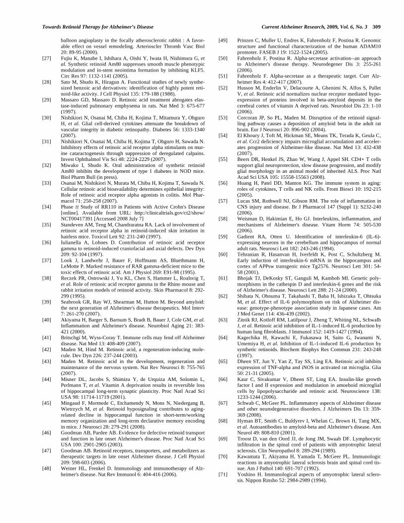
Fig. (2). Multiple actions of retinoid in T cell differentiation.
Naive CD4-positive T cells differntiate into effector T cells (Th1, Th2, Th17) and regulatory T cells (Treg) in peripheral lymphoid tissues.
Th1 cells whose differentiation is dependent on IL-12 and suppressed by retinoids mediate cellular immunity and physiologocal (and patho-
logical) inflammation. Th2 cells mediate humoral immunity and allegy. Their differentiation is dependent on IL-4 and enhanced by retinods,
but suppressed by coexistence of retinoids and TGF . IL-17-producing Th17 cells mediate pathological chronic inflammation or autoim-
mune inflammation. Th17 diffrentiation is induced by coexistence of IL-6 and TGF , and maintained by IL-23. By contrast, differentiation
of Treg cells is induced by TGF alone and suppressed by IL-6. Treg cells suppress effector T cell activities and and thereby maintain im-
mune system homeostasis and self-tolerance. Retinoids strongly enhance TGF -induced Treg diffenentiation and suppress Th17 differentia-
tion even in the presence of IL-6.
Towards Retinoid Therapy for Alzheimer’s Disease Current Alzheimer Research, 2009, Vol. 6, No. 3 307
The activities of RA in in-vitro differentiation, as well as in the development of vertebrates, have been known for a long time. RA can induce the complete regeneration of or-gans that cannot normally regenerate, such as mammalian lung and retina, by respecifying positional information [42, 121]. Murine F9 teratocarcinoma stem cells have been widely used as a model for cellular differentiation and RA signaling during embryonic development [122]. Induction of neural differentiation by retinoid has been shown in several embryonal carcinoma cell lines [123-125]. Many studies on pattern formation have confirmed the importance of retinoids in spinal cord and brain development [126]. These findings prompted detailed analyses of RARs in brain [127], and ex-tensive studies of RAR signaling pathways in neurological diseases [47]. The results suggest that RA or more generally retinoids are potential therapeutic agents for the treatment of neurodegenerative diseases, promoting tissue regeneration. Studies are advanced in the case of acute phase treatment of SCI. Mey analyzed the distribution and re-distribution of RARs after SCI [119]. Maden showed the importance of RARs in SCI, and found that RAR gene introduction and the coexistence of a RAR ligand (rather than RAR ) caused neurite growth and improved the recovery of animals after trauma [126, 128, 129]. Takenaga recently observed similar therapeutic effect in physically injured rats treated with the synthetic retinoid tamibarotene [17] which is RAR and RAR specific (unpublished). Since the physical contusion is accompanied by inflammation and activation of microglia and macrophages, the therapeutic effect may not be simply explained, but at least re-generation and differentiation of neurite cells are promoted by retinoid in an adjuvant fashion.
Of course, other nuclear receptors (and their ligands) in addition to RARs participate, particularly RXRs, RORs, liver X receptor (LXR), orphan nuclear receptor NR4A2 (Nurr1) [130], and peroxisome proliferators-activated receptors (PPARs) [131]. The expression profile and functional role of nuclear receptors in such retinoid signaling have been pro-filed in human SCI, ALS, AD and PD [132].
NEUROTRANSMISSION
Particular hallmarks observed in AD are the changes of cholinergic neurotransmission, which cause the decrease of acetylcholine (ACh). It was reported that vitamin A deficient rats with cognitive deficits showed impaired scopolamine-evoked release of ACh owing to blockade of the presynaptic acetylcholine autoreceptor. The impaired release of ACh is probably caused by lower production of ChAT, as well as neural cell death, mediated by RA and the gene promoter [133, 134].
An increase of ChAT seems to be beneficial, and it has been suggested that the vascular ACh transporter is also regulated by RA [135]. Retinoid rescued scopolamine-induced memory deficits in a passive avoidance paradigm [136]. This result could not be satisfactorily explained, but may support the participation of ACh and ChAT in the sco-polamine-related memory deficit. In addition, retinoids also regulate the expression of tyrosine hydroxylase, dopamine
hydroxylase and the dopamine D2 receptor. RA directly regulates D2 receptor expression via interaction with a RA-response element (RARE) at the promoter [137]. Such mem-
ory acquisition may not be explained simply by the partici-pation of neurotransmitters.
LEARNING AND MEMORY
The physiological role of retinoids in hippocampal func-tion of adult brain has been reviewed by Lane and Bailey [138]. The enzymes that synthesize RA and RALDH protein are restricted to the meninges surrounding the hippocampus in adult brain. The presence of cellular retinol binding pro-tein 1 (CRBP1) and cellular retinoic acid binding protein 1 (CRABP1) in the dendritic layers of the hippocampal forma-tion and dentate gyrus has been demonstrated. RAR and RAR , rather than RAR- , are highly expressed in hippo-campal CA1, CA2 and CA3 regions. RXRs are also ex-pressed. These findings suggest that RA (and therefore reti-noids) may play a central role in memory and spatial learn-ing. Impaired long-term potentiation (LTP) and long-term depression (LTD) have been demonstrated in mice lacking RAR alone, or RAR and RXR [139]. Loss of synaptic plasticity is observed in vitamin A deficient mice [44] or aged mice [140, 141]. The reductions of LTD and LTP are partially reversed by RA, although the precise involvement of specific genes in the regulation of synaptic plasticity has not been elucidated.
Etchamendy et al. [140] demonstrated that pharmacol-ogical activation of retinoid signaling by short-term RA ad-ministration to aged mice restored their hippocampal retinoid target-gene hypoexpression, as well as their long-term de-clarative memory (LTDM) and hippocampal LTP deficit. Further, hippocampal retinoid receptor RAR /RXR signal-ing is critically implicated in the cellular mechanism sustain-ing LTDM, as well as short-term working memory systems [45]. LTP-related genes, tyrosine kinase B (TrkB) and tissue plasminogen activator (t-PA), are reported to be the RAR-target genes and induced in neurobalstoma cells by treatment of retinoic acid [142-144]. TrkB is the receptor for brain-derived neurotrophic factor (BDNF), which is a key regula-tor for protein synthesis-dependent LTP and long-term memory and whose maturation is dependent on t-PA-mediated cleavage of proBDNF [145]. RAR / RXR knock-out mice behaved similarly to the aged mice and it was shown that RAR and expression of neuromodulin (Gap43) gene, a plasticity related retinoid-target gene, are important. This paper also reviewed the literature on this subject.
Another interesting observation was the involvement of transthyretin (TTR), which was identified as a key protein by microarray analysis of genes associated with age-related memory deficits [146]. TTR is a serum and cerebrospinal fluid carrier of the thyroid hormone, thyroxine. TTR is also a carrier of retinol from liver storage to target tissues in association with retinol binding protein (RBP) [147]. TTR has been reported to be the major amyloid- binding protein in cerebrospinal fluid. Age-related memory deficits occurred in TTR-/- mice in the water maze work task, and surpris-ingly, the deficits were improved by uptake of RA [146]. It has been shown that AD patients have lower levels of TTR [148]. TTR and possibly a similar serum protein, insulin-like growth factor I (IGF-I) regulate brain amyloid- levels [149, 150]. Decreased levels of TTR may affect retinoid homeo-stasis, which is required to regulate neurogenesis and differ-entiation.
308 Current Alzheimer Research, 2009, Vol. 6, No. 3 Shudo et al.
The appearance of new neurons in the hippocampus and cortex during adulthood seems to be linked to memory and learning [151]. Interestingly, in AD patients, an increased number of newly generated cells was observed in the granule cell layer, as well as the CA1 region of Ammon’s horn, where extensive cell death occurs during the disease [152]. Retinoids may have important roles in such neurogenesis.
CONCLUSION
Much research in AD is still aimed at elucidating basic pathomechanisms, although over seventy-five compounds are in clinical development for treatment of AD [39].
Retinoids are required for the maintenance of the im-mune systems, and are very potent immunomodulators. They suppressively regulate various autoimmune disease states, and being different from simple immunosuppresants, work physiologically even when applied at high pharmacological concentrations.
Retinoids are essential in the regeneration of neural cells and other tissues. Development of retinoids that are highly selective for individual RARs may contribute to the treat-ment of AD and other neurodegenerative diseases. Recently there came out a review emphasizing significant roles of retinoids for treatment of neurodegenerative diseases such as ALS, AD and schizophrenia [153]. We did not cover RXRs and RXR ligands here, though these may have a role in in-creasing the selectivity of retinoids, and they seem to have important roles in mental activities such as sleep regulation, reward-related behaviors [154-158].
Retinoids were suggested long ago to have potential for the therapy of various proliferative diseases [159]. Some applications have been realized, and our task now is to ex-tend the range of applications to neurodegenerative diseases, including AD.
ABBREVIATIONS
AD = Alzheimer’s disease
PD = Parkinson’s disease
ALS = Amyotrophic lateral sclerosis
MS = Multiple sclerosis
SCI = Spinal code injury
RARs = Retinoic acid receptors
RA = all-trans-retinoic acid
APP = Amyloid precursor protein
ChAT = Choline acetyltransferase
Treg = Regulatory T cells
EAE = Experimental autoimmune encephalomyelitis
RXRs = Retinoid X receptors
RORs = Retinoid-related orphan receptors
RALDH = Retinal dehydrogenase
Ach = Acetylcholine
CRBP1 = Cellular retinol binding protein 1
CRABP1 = Cellular retinoic acid binding protein 1
TTR = Transthyretin
APL = Acute promyelocytic leukemia
NOTE ADDED IN PROOFS
Ref. [160]
REFERENCES
[1] Cummings JL. Alzheimer's disease. N Engl J Med 351: 56-67 (2004).
[2] Roberson ED, Mucke L. 100 years and counting: prospects for defeating Alzheimer's disease. Science 314: 781-784 (2006).
[3] Hardy J. A hundred years of Alzheimer's disease research. Neuron 52: 3-13 (2006).
[4] Sporn MB, Roberts AB, Goodman AB, Eds. The Retinoids. ACADEMIC PRESS, INC. 1(2): (1984).
[5] Dawson MI, Okamura WH, Eds. Chemistry and Biology of Syn-thetic Retinoids. CRC Press, Inc. (1990).
[6] Nau H, Blaner WS, Eds. Retinoids: The biochemical and molecular basis of vitamin A and retinoid action. Springer (1999).
[7] Kagechika H, Shudo K. Synthetic retinoids: recent developments concerning structure and clinical utility. J Med Chem 48: 5875-
5883 (2005). [8] Kagechika H. Novel synthetic retinoids and separation of the plei-
otropic retinoidal activities. Curr Med Chem 9: 591-608 (2002). [9] Dawson MI. Synthetic retinoids and their nuclear receptors. Curr
Med Chem Anticancer Agents 4: 199-230 (2004). [10] de Lera AR, Bourguet W, Altucci L, Gronemeyer H. Design of
selective nuclear receptor modulators: RAR and RXR as a case study. Nat Rev Drug Discov 6: 811-820 (2007).
[11] Napoli JL. Retinoic acid: its biosynthesis and metabolism. Prog Nucleic Acid Res Mol Biol 63: 139-188 (1999).
[12] Ross AC. Retinoid production and catabolism: role of diet in regu-lating retinol esterification and retinoic Acid oxidation. J Nutr 133:
291S-296S (2003). [13] Blomhoff R, Blomhoff HK. Overview of retinoid metabolism and
function. J Neurobiol 66: 606-630 (2006). [14] Germain P, Chambon P, Eichele G, Evans RM, Lazar MA, Leid M,
et al. International Union of Pharmacology. LX. Retinoic acid re-ceptors. Pharmacol Rev 58: 712-725 (2006).
[15] Balmer JE, Blomhoff R. Gene expression regulation by retinoic acid. J Lipid Res 43: 1773-1808 (2002).
[16] Wang ZY, Chen Z. Acute promyelocytic leukemia: from highly fatal to highly curable. Blood 111: 2505-2515 (2008).
[17] Miwako I, Kagechika H. Tamibarotene. Drugs Today (Barc) 43: 563-568 (2007).
[18] Altucci L, Leibowitz MD, Ogilvie KM, de Lera AR, Gronemeyer H. RAR and RXR modulation in cancer and metabolic disease. Nat
Rev Drug Discov 6: 793-810 (2007). [19] Oikawa T, Okayasu I, Ashino H, Morita I, Murota S, Shudo K.
Three novel synthetic retinoids, Re 80, Am 580 and Am 80, all ex-hibit anti-angiogenic activity in vivo. Eur J Pharmacol 249: 113-
116 (1993). [20] Sardana K, Sehgal VN. Retinoids: fascinating up-and-coming
scenario. J Dermatol 30: 355-380 (2003). [21] Ishibashi Y. Clinical Effect of Am-80 Ointment on Psoriasis and
Pustulosis Palmaris et Plantaris (Phase Study). RinshouIyaku 11: 747-759 (1995).
[22] Nickoloff BJ. Cracking the cytokine code in psoriasis. Nat Med 13: 242-244 (2007).
[23] Kuwabara K, Shudo K, Hori Y. Novel synthetic retinoic acid inhib-its rat collagen arthritis and differentially affects serum immuno-
globulin subclass levels. FEBS Lett 378: 153-156 (1996). [24] Nagai H, Matsuura S, Bouda K, Takaoka Y, Wang T, Niwa S, et al.
Effect of Am-80, a synthetic derivative of retinoid, on experimental arthritis in mice. Pharmacology 58: 101-112 (1999).
[25] Beehler BC, Brinckerhoff CE, Ostrowski J. Selective retinoic acid receptor ligands for rheumatoid arthritis. Curr Opin Investig Drugs
5: 1153-1157 (2004). [26] Wiegman PJ, Barry WL, McPherson JA, McNamara CA, Gimple
LW, Sanders JM, et al. All-trans-retinoic acid limits restenosis after
Towards Retinoid Therapy for Alzheimer’s Disease Current Alzheimer Research, 2009, Vol. 6, No. 3 309
balloon angioplasty in the focally atherosclerotic rabbit : A favor-
able effect on vessel remodeling. Arterioscler Thromb Vasc Biol 20: 89-95 (2000).
[27] Fujiu K, Manabe I, Ishihara A, Oishi Y, Iwata H, Nishimura G, et al. Synthetic retinoid Am80 suppresses smooth muscle phenotypic
modulation and in-stent neointima formation by inhibiting KLF5. Circ Res 97: 1132-1141 (2005).
[28] Sato M, Shudo K, Hiragun A. Functional studies of newly synthe-sized benzoic acid derivatives: identification of highly potent reti-
noid-like activity. J Cell Physiol 135: 179-188 (1988). [29] Massaro GD, Massaro D. Retinoic acid treatment abrogates elas-
tase-induced pulmonary emphysema in rats. Nat Med 3: 675-677 (1997).
[30] Nishikiori N, Osanai M, Chiba H, Kojima T, Mitamura Y, Ohguro H, et al. Glial cell-derived cytokines attenuate the breakdown of
vascular integrity in diabetic retinopathy. Diabetes 56: 1333-1340 (2007).
[31] Nishikiori N, Osanai M, Chiba H, Kojima T, Ohguro H, Sawada N. Inhibitory effects of retinoic acid receptor alpha stimulants on mur-
ine cataractogenesis through suppression of deregulated calpains. Invest Ophthalmol Vis Sci 48: 2224-2229 (2007).
[32] Miwako I, Shudo K. Oral administration of synthetic retinoid Am80 inhibits the development of type 1 diabetes in NOD mice.
Biol Pharm Bull (in press). [33] Osanai M, Nishikiori N, Murata M, Chiba H, Kojima T, Sawada N.
Cellular retinoic acid bioavailability determines epithelial integrity: Role of retinoic acid receptor alpha agonists in colitis. Mol Phar-
macol 71: 250-258 (2007). [34] Phase Study of RR110 in Patients with Active Crohn's Disease
[online]. Available from URL: http://clinicaltrials.gov/ct2/show/ NCT00417391 [Accessed 2008 July 7]
[35] Standeven AM, Teng M, Chandraratna RA. Lack of involvement of retinoic acid receptor alpha in retinoid-induced skin irritation in
hairless mice. Toxicol Lett 92: 231-240 (1997). [36] Iulianella A, Lohnes D. Contribution of retinoic acid receptor
gamma to retinoid-induced craniofacial and axial defects. Dev Dyn 209: 92-104 (1997).
[37] Look J, Landwehr J, Bauer F, Hoffmann AS, Bluethmann H, LeMotte P. Marked resistance of RAR gamma-deficient mice to the
toxic effects of retinoic acid. Am J Physiol 269: E91-98 (1995). [38] Reczek PR, Ostrowski J, Yu KL, Chen S, Hammer L, Roalsvig T,
et al. Role of retinoic acid receptor gamma in the Rhino mouse and rabbit irritation models of retinoid activity. Skin Pharmacol 8: 292-
299 (1995). [39] Seabrook GR, Ray WJ, Shearman M, Hutton M. Beyond amyloid:
the next generation of Alzheimer's disease therapeutics. Mol Interv 7: 261-270 (2007).
[40] Akiyama H, Barger S, Barnum S, Bradt B, Bauer J, Cole GM, et al. Inflammation and Alzheimer's disease. Neurobiol Aging 21: 383-
421 (2000). [41] Britschgi M, Wyss-Coray T. Immune cells may fend off Alzheimer
disease. Nat Med 13: 408-409 (2007). [42] Maden M, Hind M. Retinoic acid, a regeneration-inducing mole-
cule. Dev Dyn 226: 237-244 (2003). [43] Maden M. Retinoic acid in the development, regeneration and
maintenance of the nervous system. Nat Rev Neurosci 8: 755-765 (2007).
[44] Misner DL, Jacobs S, Shimizu Y, de Urquiza AM, Solomin L, Perlmann T, et al. Vitamin A deprivation results in reversible loss
of hippocampal long-term synaptic plasticity. Proc Natl Acad Sci USA 98: 11714-11719 (2001).
[45] Mingaud F, Mormede C, Etchamendy N, Mons N, Niedergang B, Wietrzych M, et al. Retinoid hyposignaling contributes to aging-
related decline in hippocampal function in short-term/working memory organization and long-term declarative memory encoding
in mice. J Neurosci 28: 279-291 (2008). [46] Goodman AB, Pardee AB. Evidence for defective retinoid transport
and function in late onset Alzheimer's disease. Proc Natl Acad Sci USA 100: 2901-2905 (2003).
[47] Goodman AB. Retinoid receptors, transporters, and metabolizers as therapeutic targets in late onset Alzheimer disease. J Cell Physiol
209: 598-603 (2006). [48] Weiner HL, Frenkel D. Immunology and immunotherapy of Alz-
heimer's disease. Nat Rev Immunol 6: 404-416 (2006).
[49] Prinzen C, Muller U, Endres K, Fahrenholz F, Postina R. Genomic
structure and functional characterization of the human ADAM10 promoter. FASEB J 19: 1522-1524 (2005).
[50] Fahrenholz F, Postina R. Alpha-secretase activation--an approach to Alzheimer's disease therapy. Neurodegener Dis 3: 255-261
(2006). [51] Fahrenholz F. Alpha-secretase as a therapeutic target. Curr Alz-
heimer Res 4: 412-417 (2007). [52] Husson M, Enderlin V, Delacourte A, Ghenimi N, Alfos S, Pallet
V, et al. Retinoic acid normalizes nuclear receptor mediated hypo-expression of proteins involved in beta-amyloid deposits in the
cerebral cortex of vitamin A deprived rats. Neurobiol Dis 23: 1-10 (2006).
[53] Corcoran JP, So PL, Maden M. Disruption of the retinoid signal-ling pathway causes a deposition of amyloid beta in the adult rat
brain. Eur J Neurosci 20: 896-902 (2004). [54] El Khoury J, Toft M, Hickman SE, Means TK, Terada K, Geula C,
et al. Ccr2 deficiency impairs microglial accumulation and acceler-ates progression of Alzheimer-like disease. Nat Med 13: 432-438
(2007). [55] Beers DR, Henkel JS, Zhao W, Wang J, Appel SH. CD4+ T cells
support glial neuroprotection, slow disease progression, and modify glial morphology in an animal model of inherited ALS. Proc Natl
Acad Sci USA 105: 15558-15563 (2008). [56] Huang H, Patel DD, Manton KG. The immune system in aging:
roles of cytokines, T cells and NK cells. Front Biosci 10: 192-215 (2005).
[57] Lucas SM, Rothwell NJ, Gibson RM. The role of inflammation in CNS injury and disease. Br J Pharmacol 147 (Suppl 1): S232-240
(2006). [58] Weisman D, Hakimian E, Ho GJ. Interleukins, inflammation, and
mechanisms of Alzheimer's disease. Vitam Horm 74: 505-530 (2006).
[59] Gadient RA, Otten U. Identification of interleukin-6 (IL-6)-expressing neurons in the cerebellum and hippocampus of normal
adult rats. Neurosci Lett 182: 243-246 (1994). [60] Tehranian R, Hasanvan H, Iverfeldt K, Post C, Schultzberg M.
Early induction of interleukin-6 mRNA in the hippocampus and cortex of APPsw transgenic mice Tg2576. Neurosci Lett 301: 54-
58 (2001). [61] Bhojak TJ, DeKosky ST, Ganguli M, Kamboh MI. Genetic poly-
morphisms in the cathespin D and interleukin-6 genes and the risk of Alzheimer's disease. Neurosci Lett 288: 21-24 (2000).
[62] Shibata N, Ohnuma T, Takahashi T, Baba H, Ishizuka T, Ohtsuka M, et al. Effect of IL-6 polymorphism on risk of Alzheimer dis-
ease: genotype-phenotype association study in Japanese cases. Am J Med Genet 114: 436-439 (2002).
[63] Zitnik RJ, Kotloff RM, Latifpour J, Zheng T, Whiting NL, Schwalb J, et al. Retinoic acid inhibition of IL-1-induced IL-6 production by
human lung fibroblasts. J Immunol 152: 1419-1427 (1994). [64] Kagechika H, Kawachi E, Fukasawa H, Saito G, Iwanami N,
Umemiya H, et al. Inhibition of IL-1-induced IL-6 production by synthetic retinoids. Biochem Biophys Res Commun 231: 243-248
(1997). [65] Dheen ST, Jun Y, Yan Z, Tay SS, Ling EA. Retinoic acid inhibits
expression of TNF-alpha and iNOS in activated rat microglia. Glia 50: 21-31 (2005).
[66] Kaur C, Sivakumar V, Dheen ST, Ling EA. Insulin-like growth factor I and II expression and modulation in amoeboid microglial
cells by lipopolysaccharide and retinoic acid. Neuroscience 138: 1233-1244 (2006).
[67] Schwab C, McGeer PL. Inflammatory aspects of Alzheimer disease and other neurodegenerative disorders. J Alzheimers Dis 13: 359-
369 (2008). [68] Hyman BT, Smith C, Buldyrev I, Whelan C, Brown H, Tang MX,
et al. Autoantibodies to amyloid-beta and Alzheimer's disease. Ann Neurol 49: 808-810 (2001).
[69] Troost D, van den Oord JJ, de Jong JM, Swaab DF. Lymphocytic infiltration in the spinal cord of patients with amyotrophic lateral
sclerosis. Clin Neuropathol 8: 289-294 (1989). [70] Kawamata T, Akiyama H, Yamada T, McGeer PL. Immunologic
reactions in amyotrophic lateral sclerosis brain and spinal cord tis-sue. Am J Pathol 140: 691-707 (1992).
[71] Yoshino H. Immunological aspects of amyotrophic lateral sclero-sis. Nippon Rinsho 52: 2984-2989 (1994).
310 Current Alzheimer Research, 2009, Vol. 6, No. 3 Shudo et al.
[72] Hunot S, Hirsch EC. Neuroinflammatory processes in Parkinson's
disease. Ann Neurol 53 (Suppl 3): S49-58; discussion S58-60 (2003).
[73] McGeachy MJ, Cua DJ. Th17 cell differentiation: the long and winding road. Immunity 28: 445-453 (2008).
[74] Ouyang W, Kolls JK, Zheng Y. The biological functions of T helper 17 cell effector cytokines in inflammation. Immunity 28:
454-467 (2008). [75] Kebir H, Kreymborg K, Ifergan I, Dodelet-Devillers A, Cayrol R,
Bernard M, et al. Human TH17 lymphocytes promote blood-brain barrier disruption and central nervous system inflammation. Nat
Med 13: 1173-1175 (2007). [76] Wang W, Milani M, Ostlie N, Okita D, Agarwal RK, Caspi RR, et
al. C57BL/6 mice genetically deficient in IL-12/IL-23 and IFN-gamma are susceptible to experimental autoimmune myasthenia
gravis, suggesting a pathogenic role of non-Th1 cells. J Immunol 178: 7072-7080 (2007).
[77] Ifergan I, Kebir H, Bernard M, Wosik K, Dodelet-Devillers A, Cayrol R, et al. The blood-brain barrier induces differentiation of
migrating monocytes into Th17-polarizing dendritic cells. Brain 131: 785-799 (2008).
[78] Zhong Z, Deane R, Ali Z, Parisi M, Shapovalov Y, O'Banion MK, et al. ALS-causing SOD1 mutants generate vascular changes prior
to motor neuron degeneration. Nat Neurosci 11: 420-422 (2008). [79] Racke MK, Burnett D, Pak SH, Albert PS, Cannella B, Raine CS,
et al. Retinoid treatment of experimental allergic encephalomyeli-tis. IL-4 production correlates with improved disease course. J Im-
munol 154: 450-458 (1995). [80] Wang T, Niwa S, Bouda K, Matsuura S, Homma T, Shudo K, et al.
The effect of Am-80, one of retinoid derivatives on experimental allergic encephalomyelitis in rats. Life Sci 67: 1869-1879 (2000).
[81] Miyagawa N, Homma T, Kagechika H, Shudo K, Nagai H. Effect of synthetic retinoid, TAC-101, on experimental autoimmune dis-
ease. Pharmacology 67: 21-31 (2003). [82] Iwata M, Eshima Y, Kagechika H. Retinoic acids exert direct ef-
fects on T cells to suppress Th1 development and enhance Th2 de-velopment via retinoic acid receptors. Int Immunol 15: 1017-1025
(2003). [83] Fukasawa H, Kagechika H, Shudo K. Retinoid therapy for autoim-
mune diseases. Jpn J Clin Immunol 29: 114-126 (2006). [84] Ivanov, II, McKenzie BS, Zhou L, Tadokoro CE, Lepelley A,
Lafaille JJ, et al. The orphan nuclear receptor RORgammat directs the differentiation program of proinflammatory IL-17+ T helper
cells. Cell 126: 1121-1133 (2006). [85] Sakaguchi S, Sakaguchi N, Asano M, Itoh M, Toda M. Immu-
nologic self-tolerance maintained by activated T cells expressing IL-2 receptor alpha-chains (CD25). Breakdown of a single mecha-
nism of self-tolerance causes various autoimmune diseases. J Im-munol 155: 1151-1164 (1995).
[86] Tang Q, Bluestone JA. The Foxp3+ regulatory T cell: a jack of all trades, master of regulation. Nat Immunol 9: 239-244 (2008).
[87] Fontenot JD, Gavin MA, Rudensky AY. Foxp3 programs the de-velopment and function of CD4+CD25+ regulatory T cells. Nat
Immunol 4: 330-336 (2003). [88] Hori S, Nomura T, Sakaguchi S. Control of regulatory T cell de-
velopment by the transcription factor Foxp3. Science 299: 1057-1061 (2003).
[89] Viglietta V, Baecher-Allan C, Weiner HL, Hafler DA. Loss of functional suppression by CD4+CD25+ regulatory T cells in pa-
tients with multiple sclerosis. J Exp Med 199: 971-979 (2004). [90] Balandina A, Lecart S, Dartevelle P, Saoudi A, Berrih-Aknin S.
Functional defect of regulatory CD4(+)CD25+ T cells in the thy-mus of patients with autoimmune myasthenia gravis. Blood 105:
735-741 (2005). [91] Yamano Y, Takenouchi N, Li HC, Tomaru U, Yao K, Grant CW,
et al. Virus-induced dysfunction of CD4+CD25+ T cells in patients with HTLV-I-associated neuroimmunological disease. J Clin Invest
115: 1361-1368 (2005). [92] Rosenkranz D, Weyer S, Tolosa E, Gaenslen A, Berg D, Leyhe T,
et al. Higher frequency of regulatory T cells in the elderly and in-creased suppressive activity in neurodegeneration. J Neuroimmunol
188: 117-127 (2007). [93] Reynolds AD, Banerjee R, Liu J, Gendelman HE, Mosley RL.
Neuroprotective activities of CD4+CD25+ regulatory T cells in an animal model of Parkinson's disease. J Leukoc Biol 82: 1083-1094
(2007).
[94] Mucida D, Park Y, Kim G, Turovskaya O, Scott I, Kronenberg M,
et al. Reciprocal TH17 and regulatory T cell differentiation medi-ated by retinoic acid. Science 317: 256-260 (2007).
[95] Coombes JL, Siddiqui KR, Arancibia-Carcamo CV, Hall J, Sun CM, Belkaid Y, et al. A functionally specialized population of mu-
cosal CD103+ DCs induces Foxp3+ regulatory T cells via a TGF-beta and retinoic acid-dependent mechanism. J Exp Med 204:
1757-1764 (2007). [96] Kang SG, Lim HW, Andrisani OM, Broxmeyer HE, Kim CH.
Vitamin A metabolites induce gut-homing Foxp3+ regulatory T cells. J Immunol 179: 3724-3733 (2007).
[97] Elias KM, Laurence A, Davidson TS, Stephens G, Kanno Y, Shevach EM, et al. Retinoic acid inhibits Th17 polarization and
enhances Foxp3 expression through a Stat-3/Stat-5 independent signaling pathway. Blood 111: 1013-1020 (2008).
[98] Benson MJ, Pino-Lagos K, Rosemblatt M, Noelle RJ. All-trans retinoic acid mediates enhanced T reg cell growth, differentiation,
and gut homing in the face of high levels of co-stimulation. J Exp Med 204: 1765-1774 (2007).
[99] Sun CM, Hall JA, Blank RB, Bouladoux N, Oukka M, Mora JR, et al. Small intestine lamina propria dendritic cells promote de novo
generation of Foxp3 T reg cells via retinoic acid. J Exp Med 204: 1775-1785 (2007).
[100] Kim CH. Regulation of Foxp3 regulatory T cells and Th17 cells by retinoids. Clin Dev Immunol: 1-12 (2008).
[101] Takaki H, Ichiyama K, Koga K, Chinen T, Takaesu G, Sugiyama Y, et al. STAT6 Inhibits TGF-beta1-mediated Foxp3 induction
through direct binding to the Foxp3 promoter, which is reverted by retinoic acid receptor. J Biol Chem 283: 14955-14962 (2008).
[102] Schambach F, Schupp M, Lazar MA, Reiner SL. Activation of retinoic acid receptor-alpha favours regulatory T cell induction at
the expense of IL-17-secreting T helper cell differentiation. Eur J Immunol 37: 2396-2399 (2007).
[103] von Boehmer H. Oral tolerance: is it all retinoic acid? J Exp Med 204: 1737-1739 (2007).
[104] Strandberg TE, Pitkala KH, Linnavuori K, Tilvis RS. Cognitive impairment and infectious burden in the elderly. Arch Gerontol
Geriatr Suppl: 419-423 (2004). [105] D'Andrea MR. Add Alzheimer's disease to the list of autoimmune
diseases. Med Hypotheses 64: 458-463 (2005). [106] Perry VH, Cunningham C, Holmes C. Systemic infections and
inflammation affect chronic neurodegeneration. Nat Rev Immunol 7: 161-167 (2007).
[107] Miklossy J. Chronic inflammation and amyloidogenesis in Alz-heimer's disease -- role of spirochetes. J Alzheimers Dis 13: 381-
391 (2008). [108] Itzhaki RF, Wozniak MA. Herpes simplex virus type 1 in Alz-
heimer's disease: the enemy within. J Alzheimers Dis 13: 393-405 (2008).
[109] Balin BJ, Little CS, Hammond CJ, Appelt DM, Whittum-Hudson JA, Gerard HC, et al. Chlamydophila pneumoniae and the etiology
of late-onset Alzheimer's disease. J Alzheimers Dis 13: 371-380 (2008).
[110] Nicolson GL, Nasralla MY, Haier J, Pomfret J. High frequency of systemic mycoplasmal infections in Gulf War veterans and civil-
ians with Amyotrophic Lateral Sclerosis (ALS). J Clin Neurosci 9: 525-529 (2002).
[111] Doty RL. The olfactory vector hypothesis of neurodegenerative disease: is it viable? Ann Neurol 63: 7-15 (2008).
[112] Ross GW, Petrovitch H, Abbott RD, Tanner CM, Popper J, Masaki K, et al. Association of olfactory dysfunction with risk for future
Parkinson's disease. Ann Neurol 63: 167-173 (2008). [113] Tabert MH, Liu X, Doty RL, Serby M, Zamora D, Pelton GH, et al.
A 10-item smell identification scale related to risk for Alzheimer's disease. Ann Neurol 58: 155-160 (2005).
[114] Rawson NE, LaMantia AS. Once and again: retinoic acid signaling in the developing and regenerating olfactory pathway. J Neurobiol
66: 653-676 (2006). [115] Rawson NE, LaMantia AS. A speculative essay on retinoic acid
regulation of neural stem cells in the developing and aging olfac-tory system. Exp Gerontol 42: 46-53 (2007).
[116] Mey J, Morassutti DJ, Brook G, Liu RH, Zhang YP, Koopmans G, et al. Retinoic acid synthesis by a population of NG2-positive cells
in the injured spinal cord. Eur J Neurosci 21: 1555-1568 (2005). [117] Mey J. New therapeutic target for CNS injury? The role of retinoic
acid signaling after nerve lesions. J Neurobiol 66: 757-779 (2006).
Towards Retinoid Therapy for Alzheimer’s Disease Current Alzheimer Research, 2009, Vol. 6, No. 3 311
[118] Kern J, Schrage K, Koopmans GC, Joosten EA, McCaffery P, Mey
J. Characterization of retinaldehyde dehydrogenase-2 induction in NG2-positive glia after spinal cord contusion injury. Int J Dev Neu-
rosci 25: 7-16 (2007). [119] Zhelyaznik N, Mey J. Regulation of retinoic acid receptors alpha,
beta and retinoid X receptor alpha after sciatic nerve injury. Neuro-science 141: 1761-1774 (2006).
[120] Mey J, Schrage K, Wessels I, Vollpracht-Crijns I. Effects of in-flammatory cytokines IL-1beta, IL-6, and TNFalpha on the intra-
cellular localization of retinoid receptors in Schwann cells. Glia 55: 152-164 (2007).
[121] Vergara MN, Arsenijevic Y, Del Rio-Tsonis K. CNS regeneration: a morphogen's tale. J Neurobiol 64: 491-507 (2005).
[122] Strickland S, Mahdavi V. The induction of differentiation in terato-carcinoma stem cells by retinoic acid. Cell 15: 393-403 (1978).
[123] Kuff EL, Fewell JW. Induction of neural-like cells and acetylcho-linesterase activity in cultures of F9 teratocarcinoma treated with
retinoic acid and dibutyryl cyclic adenosine monophosphate. Dev Biol 77: 103-115 (1980).
[124] Liesi P, Rechardt L, Wartiovaara J. Nerve growth factor induces adrenergic neuronal differentiation in F9 teratocarcinoma cells. Na-
ture 306: 265-267 (1983). [125] McBurney MW, Jones-Villeneuve EM, Edwards MK, Anderson
PJ. Control of muscle and neuronal differentiation in a cultured embryonal carcinoma cell line. Nature 299: 165-167 (1982).
[126] Maden M. Retinoids and spinal cord development. J Neurobiol 66: 726-738 (2006).
[127] Krezel W, Kastner P, Chambon P. Differential expression of reti-noid receptors in the adult mouse central nervous system. Neuro-
science 89: 1291-1300 (1999). [128] Wong LF, Yip PK, Battaglia A, Grist J, Corcoran J, Maden M, et
al. Retinoic acid receptor beta2 promotes functional regeneration of sensory axons in the spinal cord. Nat Neurosci 9: 243-250 (2006).
[129] Corcoran J, So PL, Barber RD, Vincent KJ, Mazarakis ND, Mitro-phanous KA, et al. Retinoic acid receptor beta2 and neurite out-
growth in the adult mouse spinal cord in vitro. J Cell Sci 115: 3779-3786 (2002).
[130] Wallen-Mackenzie A, Mata de Urquiza A, Petersson S, Rodriguez FJ, Friling S, Wagner J, et al. Nurr1-RXR heterodimers mediate
RXR ligand-induced signaling in neuronal cells. Genes Dev 17: 3036-3047 (2003).
[131] Heneka MT, Landreth GE. PPARs in the brain. Biochim Biophys Acta 1771: 1031-1045 (2007).
[132] Malaspina A, Turkheimer F. A review of the functional role and of the expression profile of retinoid signaling and of nuclear receptors
in human spinal cord. Brain Res Bull 71: 437-446 (2007). [133] Kobayashi M, Matsuoka I, Kurihara K. Cholinergic differentiation
of cultured sympathetic neurons induced by retinoic acid. Induction of choline acetyltransferase-mRNA and suppression of tyrosine
hydroxylase-mRNA levels. FEBS Lett 337: 259-264 (1994). [134] Pedersen WA, Berse B, Schuler U, Wainer BH, Blusztajn JK. All-
trans- and 9-cis-retinoic acid enhance the cholinergic properties of a murine septal cell line: evidence that the effects are mediated by
activation of retinoic acid receptor-alpha. J Neurochem 65: 50-58 (1995).
[135] Berse B, Blusztajn JK. Coordinated up-regulation of choline acetyl-transferase and vesicular acetylcholine transporter gene expression
by the retinoic acid receptor alpha, cAMP, and leukemia inhibitory factor/ciliary neurotrophic factor signaling pathways in a murine
septal cell line. J Biol Chem 270: 22101-22104 (1995). [136] Shudo K, Kagechika H, Yamazaki N, Igarashi M, Tateda C. A
synthetic retinoid Am80 (tamibarotene) rescues the memory deficit caused by scopolamine in a passive avoidance paradigm. Biol
Pharm Bull 27: 1887-1889 (2004). [137] Samad TA, Krezel W, Chambon P, Borrelli E. Regulation of do-
paminergic pathways by retinoids: activation of the D2 receptor promoter by members of the retinoic acid receptor-retinoid X re-
ceptor family. Proc Natl Acad Sci USA 94: 14349-14354 (1997). [138] Lane MA, Bailey SJ. Role of retinoid signalling in the adult brain.
Prog Neurobiol 75: 275-293 (2005). [139] Chiang MY, Misner D, Kempermann G, Schikorski T, Giguere V,
Sucov HM, et al. An essential role for retinoid receptors RARbeta
and RXRgamma in long-term potentiation and depression. Neuron
21: 1353-1361 (1998). [140] Etchamendy N, Enderlin V, Marighetto A, Vouimba RM, Pallet V,
Jaffard R, et al. Alleviation of a selective age-related relational memory deficit in mice by pharmacologically induced normaliza-
tion of brain retinoid signaling. J Neurosci 21: 6423-6429 (2001). [141] Etchamendy N, Enderlin V, Marighetto A, Pallet V, Higueret P,
Jaffard R. Vitamin A deficiency and relational memory deficit in adult mice: relationships with changes in brain retinoid signalling.
Behav Brain Res 145: 37-49 (2003). [142] Lucarelli E, Kaplan DR, Thiele CJ. Selective regulation of TrkA
and TrkB receptors by retinoic acid and interferon-gamma in hu-man neuroblastoma cell lines. J Biol Chem 270: 24725-24731
(1995). [143] Bulens F, Ibanez-Tallon I, Van Acker P, De Vriese A, Nelles L,
Belayew A, et al. Retinoic acid induction of human tissue-type plasminogen activator gene expression via a direct repeat element
(DR5) located at -7 kilobases. J Biol Chem 270: 7167-7175 (1995). [144] Melchor JP, Strickland S. Tissue plasminogen activator in central
nervous system physiology and pathology. Thromb Haemost 93: 655-660 (2005).
[145] Pang PT, Teng HK, Zaitsev E, Woo NT, Sakata K, Zhen S, et al. Cleavage of proBDNF by tPA/plasmin is essential for long-term
hippocampal plasticity. Science 306: 487-491 (2004). [146] Brouillette J, Quirion R. Transthyretin: A key gene involved in the
maintenance of memory capacities during aging. Neurobiol Aging 29: 1721-1732 (2008).
[147] Monaco HL. The transthyretin-retinol-binding protein complex. Biochim Biophys Acta 1482: 65-72 (2000).
[148] Merched A, Serot JM, Visvikis S, Aguillon D, Faure G, Siest G. Apolipoprotein E, transthyretin and actin in the CSF of Alzheimer's
patients: relation with the senile plaques and cytoskeleton biochem-istry. FEBS Lett 425: 225-228 (1998).
[149] Schwarzman AL, Gregori L, Vitek MP, Lyubski S, Strittmatter WJ, Enghilde JJ, et al. Transthyretin sequesters amyloid beta protein
and prevents amyloid formation. Proc Natl Acad Sci USA 91: 8368-8372 (1994).
[150] Carro E, Trejo JL, Gomez-Isla T, LeRoith D, Torres-Aleman I. Serum insulin-like growth factor I regulates brain amyloid-beta
levels. Nat Med 8: 1390-1397 (2002). [151] Kempermann G. Why new neurons? Possible functions for adult
hippocampal neurogenesis. J Neurosci 22: 635-638 (2002). [152] Jin K, Peel AL, Mao XO, Xie L, Cottrell BA, Henshall DC, et al.
Increased hippocampal neurogenesis in Alzheimer's disease. Proc Natl Acad Sci USA 101: 343-347 (2004).
[153] Malaspina A, Michael-Titus AT. Is the modulation of retinoid and retinoid-associated signaling a future therapeutic strategy in neuro-
logical trauma and neurodegeneration? J Neurochem 104: 584-595 (2008).
[154] Goodman AB. Three independent lines of evidence suggest reti-noids as causal to schizophrenia. Proc Natl Acad Sci USA 95:
7240-7244 (1998). [155] Maret S, Franken P, Dauvilliers Y, Ghyselinck NB, Chambon P,
Tafti M. Retinoic acid signaling affects cortical synchrony during sleep. Science 310: 111-113 (2005).
[156] Sei H. Vitamin A and sleep regulation. J Med Invest 55: 1-8 (2008).
[157] Bourhis E, Maheux J, Paquet B, Kagechika H, Shudo K, Rompre PP, et al. The transcription factors Nur77 and retinoid X receptors
participate in amphetamine-induced locomotor activities. Psycho-pharmacology (Berl) 202: 635-648 (2009).
[158] Levesque D, Rouillard C. Nur77 and retinoid X receptors: crucial factors in dopamine-related neuroadaptation. Trends Neurosci 30:
22-30 (2007). [159] Sporn MB, Harris ED, Jr. Proliferative diseases. Am J Med 70:
1231-1235 (1981). [160] Ding Y, Qiao A, Wang Z, Goodwin JS, Lee E-S, Block ML, et al.
Retinoic acid attenuates -amyloid deposition and resques memory deficits in an Alzheimer's disease transgenic mouse model. J Neu-
rosci 28: 11622-11634 (2008).
Received: July 28, 2008 Revised: November 05, 2008 Accepted: November 12, 2008