Familial clustering of autoimmune diseases in patients with type 1 diabetes mellitus
24
Familial autoimmunity in T1D 1 Familial clustering of autoimmune diseases in patients with type 1 diabetes mellitus Juan-Manuel Anaya a,b,* , John Castiblanco a , Gabriel J. Tobón a , Jorge García c , Verónica Abad d , Héctor Cuervo c,e , Alejandro Velásquez d , Ivan D. Angel c , Patricia Vega a , Alvaro Arango e a Cellular Biology and Immunogenetics Unit, Corporación para Investigaciones Biológicas, Medellín, Colombia b Universidad del Rosario, Medellín, Colombia c Antioqueña de Diabetes, Medellín, Colombia d Hospital Pablo Tobón Uribe, Medellín, Colombia e Clínica Universitaria Bolivariana, School of Medicine, Universidad Pontificia Bolivariana, Medellín, Colombia * Correspondence: Juan-Manuel Anaya, MD Corporación para Investigaciones Biológicas Cra 72-A No 78-B-141 Medellín, Colombia Tel: (574) 441-08-55 Fax: (574) 441-55-14 E-mail address: [email protected]
-
Upload
independent -
Category
Documents
-
view
0 -
download
0
Transcript of Familial clustering of autoimmune diseases in patients with type 1 diabetes mellitus
Familial autoimmunity in T1D 1
Familial clustering of autoimmune diseases in patients
with type 1 diabetes mellitus
Juan-Manuel Anayaa,b,*, John Castiblancoa, Gabriel J. Tobóna, Jorge Garcíac,
Verónica Abadd, Héctor Cuervoc,e, Alejandro Velásquezd, Ivan D. Angelc,
Patricia Vegaa, Alvaro Arangoe
a Cellular Biology and Immunogenetics Unit, Corporación para Investigaciones Biológicas,
Medellín, Colombia b Universidad del Rosario, Medellín, Colombia c Antioqueña de Diabetes, Medellín, Colombia d Hospital Pablo Tobón Uribe, Medellín, Colombia e Clínica Universitaria Bolivariana, School of Medicine, Universidad Pontificia
Bolivariana, Medellín, Colombia
* Correspondence:
Juan-Manuel Anaya, MD
Corporación para Investigaciones Biológicas
Cra 72-A No 78-B-141
Medellín, Colombia
Tel: (574) 441-08-55 Fax: (574) 441-55-14
E-mail address: [email protected]

Familial autoimmunity in T1D 2
Abstract
Currently we investigated the familial aggregation of autoimmune diseases (AIDs) among
first-degree relatives (FDR) of patients with type 1 diabetes mellitus (T1D). Relatives of 98
T1D patients defined according to the guidelines diagnosis of the American Diabetes
Association and 113 matched controls without any AID, were interviewed using a
questionnaire that sought information about demographic and medical characteristics
including a list of 18 AIDs. Genetic analysis was performed using the program ASSOC
and by calculating recurrent risk ratios. In cases, 25.5% of the families had at least one
member having an AID, while in controls there were 9% (OR: 3.96, 95% CI= 1.74-9.0,
p=0.0006). An AID was registered in 8.3% of 312 FDR of patients as compared with 2.4%
of 362 FDR in controls (OR: 3.56, 95%CI = 1.64-7.73, p=0.0008). The most frequent
AIDs registered in FDR of cases were autoimmune thyroid disease (AITD) and T1D,
which disclosed coefficients of aggregation. These results indicate that AIDs cluster within
families of T1D patients adding further evidence to consider that clinically different
autoimmune phenotypes may share common susceptibility gene variants, which may act
pleiotropically as risk factors for autoimmunity.
Key Words: Aggregation, Autoimmunity, type 1 diabetes mellitus, hypothyroidism,
Genetics.

Familial autoimmunity in T1D 3
1. Introduction
Autoimmune diseases (AIDs) are chronic conditions initiated by a loss of
immunological tolerance to self-antigens. The chronic nature of many of these diseases
results in a significant impact in terms of medical care utilization, direct and indirect
economic costs, and quality of life. The estimated incidence of AIDs is about 90 per 100
000 person-year and their prevalence is about 3% of the population [1]. Almost all AIDs
disproportionately affect middle age-women women being one of the leading causes of
death among this group of patients. The older the patient, the lower the male:female ratio
becomes [1].
Although the etiology of AIDs is unknown, several factors are involved in the
development of these diseases, including genetic and environmental factors [2,3].
Population studies have established that each population holds a mutational pool, in which
most mutations individually (i.e. polymorphisms) will have mild effects; if not
undetectable, but in combination with other alleles would favor or avoid autoimmune
phenomena. Such interplay within genetic variants will generate a change in the
measurable risk of developing an autoimmune phenotype. This characteristic is the main
reason why AIDs are not inherited in a classical simple Mendelian way, but instead have a
complex or yet unknown mode of inheritance [3]. Genetic contribution to AIDs is
supported by the high rates of concordance, ranging from 15% to 60%, and by high
recurrent risk ratios (λR) [4].
There is evidence indicating that some AIDs will concentrate within families. This
includes not only cases of a single type of AID (several members having the same trait)
appearing among siblings, twins and relatives of patients [5-8], but also several different
ones (several family members with diverse AIDs) appearing [9-19], thus indicating that
autoimmune phenotypes could represent pleiotropic outcomes of non-specific disease

Familial autoimmunity in T1D 4
genes that underlie similar immunogenetic mechanisms. This common origin hypothesis
for different AIDs is also supported by results of genome–wide scans showing that several
loci may overlap in different AIDs [20,21], and by microarray expression profile studies
disclosing a similar pattern of gene expression in different AIDs [22,23]. Finally, the
multiple autoimmune syndrome, characterized by the presence of three or more AIDs in a
single individual [24] is a clear example of how diverse phenotypes may be related to a
single genotype.
Type 1 diabetes mellitus (T1D) is an organ-specific AID resulting from the damage
of insulin-producing pancreatic ß cells. This disease constitutes the earliest-onset AID,
with a male:female ratio of 1:1 [25]. Similar to other AIDs, T1D is an immune-mediated
disease that develops in genetically susceptible individuals, in whom one or more
additional AIDs may coexist, autoimmune thyroid diseases (AITD) (i.e. Grave’s disease or
Hashimoto's thyroiditis) and celiac disease (CD) being the most prevalent [26-28]. First-
degree relatives (FDR) are more predisposed to develop T1D and to have a higher
proportion of autoantibodies compared to the general population [25]. Considering the
interaction between AIDs which was summarized above and the fact that there is growing
evidence supporting a common genetic origin for these diseases, the familial aggregation
of autoimmunity within first degree relatives (FDR) of T1D patients was examined.

Familial autoimmunity in T1D 5
2. Patients and methods
2.1. Demographics of T1D patients and healthy individuals
Cases were children with T1D all of whom fulfilled the diagnostic classification
criteria proposed by the American Diabetes Association (ADA) [29] and who belonged to
our T1D cohort [30]. Their information on demographics and cumulative clinical
manifestations over the course of disease were obtained by both chart review and
discussion with the patient and was collected in a standard data collection form. A total of
98 patients with T1D were evaluated. Controls were 113 healthy children without any AID
(See Appendix 1) matched to cases by sex, age and ethnicity, and seen as outpatients by
the same physicians.
The entire groups of individuals involved in this study were of Spanish ancestry
and belonged to the population from the northwestern part of Colombia, South America
(i.e. Paisa community). This population was established in the 16th - 17th
centuries and
flourished in relative isolation until the late 19th century. The admixture between Paisa and
African or Amerindian populations has been historically documented as low [31], with an
ancestral ethnic component of 85% Caucasian and 15% Amerindian and in which the
African contribution has been estimated as being not significant. Thus, historical and
genetic evidence supports the usefulness of this population for genetic mapping [32].
2.2. Family collection
FDR of T1D patients and matched healthy individuals were interviewed
following the methodology described by Priori et al [10], using a standardized
questionnaire that incorporated demographics and medical information including a check-

Familial autoimmunity in T1D 6
point list of 18 AIDs (Appendix 1). The diagnosis of AID was only considered reliable and
consequently registered if made by a certified physician (i.e. endocrinologist,
rheumatologist or an internist) and confirmed by chart review. FDR of T1D patients as
well as FDR of the matched healthy individuals could not be considered as cases or
controls. This research was accomplished in accordance with Resolution No. 008430 of
1993 from the Ministry of Health of the Republic of Colombia, was classified as research
with minimal risk. The Ethics Committee of the CIB approved the present study.
2.3. Statistical and genetical analysis
Data was managed and stored using the SPSS program (V9.05 for Windows,
Chicago, IL). Results are presented as means ± standard deviation (SD), and in
percentages. Comparisons between means were performed by the Student’s t-test, and
those between percentages were performed by χ2 test and two–sided Fisher’s exact test as
appropriate. Crude odds ratios (OR) were calculated with 95% confidence intervals (CI). A
p–value of less than 0.05 was considered as significant.
The ASSOC program in SAGE (Statistical Analysis in Genetic Epidemiology) was
used to evaluate the existence of familial aggregation of AIDs, and to assess whether or not
the presence of an AID affected proband correlates with the presence of a FDR who is
affected by an AID, by simultaneously estimating familial variance components [33]. The
model used incorporates familial correlations and arbitrary covariates assuming the
correlation structure described by Elston, George and Severtson [34] and the regression
model described in George and Elston [35]. For each individual (j), the model predicts
parameters associated with a polygenic (Gj), family (Fj), marital (Mj), sibship (Sj) and a
random environmental effect (Ej). The model is described as h(yj)= h(βTxj)+ Gj+

Familial autoimmunity in T1D 7
F’j+Mj+Sj+Ej, where h is a transformation of the dependent variable using the
standardized Box and Cox transformation [37] with the power parameter λ1 and a shift
parameter λ2. Additionally, the polygenic effect and all random family and environmental
effects are assumed to be normally distributed random effects with zero means. For each
model, the data of one or more independent pedigrees was sampled at random to estimate
the parameters of the model by maximum likelihood (ML) assuming a generalization of
multivariate normality with or without the inclusion of a specified set of possible AIDs.
The ML of the model is determined under two hypotheses: H1 assumes the general model
including all the covariates specified; while, H0 excludes the test covariates. If L1 and L0
are the MLs under H1 y H0 respectively then the likelihood ratio statistic is 2|ln(L1)-
ln(L0)|. This joint test is asymptotically distributed as a chi-square with the number of
degrees of freedom equal to the number of test covariates.
Furthermore, familial aggregation (λR) was calculated for first–degree relatedness
(Parent/offspring and sibling/sibling pairs) using the formula λR =KRelative/K, where KRelative
(KR) was the prevalence for a specific degree of relatedness in the sample, and K was the
prevalence in the control pedigree samples or the mean prevalence in the population [37].
In other words, two approaches where taken to examine λR. First of all, a relative–pair
comparison between the prevalence of AIDs for the correspondent proband within each
pedigree was calculated on the pedigrees for both T1D and healthy individual (case-control
analysis). Secondly, previously reported prevalences of AIDs were considered
[1,4,16,17,38-41]. These prevalences were used to obtain the λR values using the calculated
prevalence for each specific degree–relative on the T1D affected proband pedigrees. Given
the fact that prevalence information about AIDs in our population was not available,
prevalences in the range of 0.1% - 0.5% were chosen [1,4,16,17,38-41]. Furthermore, 0.4%
(4/1000) individuals for each AID and 2.5% (25/1000 individuals) for all AIDs taken
Familial autoimmunity in T1D 8
together were selected as putative population prevalences [1,4,16,17,38-41]. Finally, since
there was a subgroup of AIDs in the pedigrees of T1D patients that disclosed a low
frequency, all AIDs were combined in order to determine the presence of familial
aggregation for AIDs as a trait. These methods were extended to ascertain whether or not
clustering of two or more autoimmune disorders in relatives of T1D patients increased the
probability or risk for the presence of the disorder in the proband.
3. Results
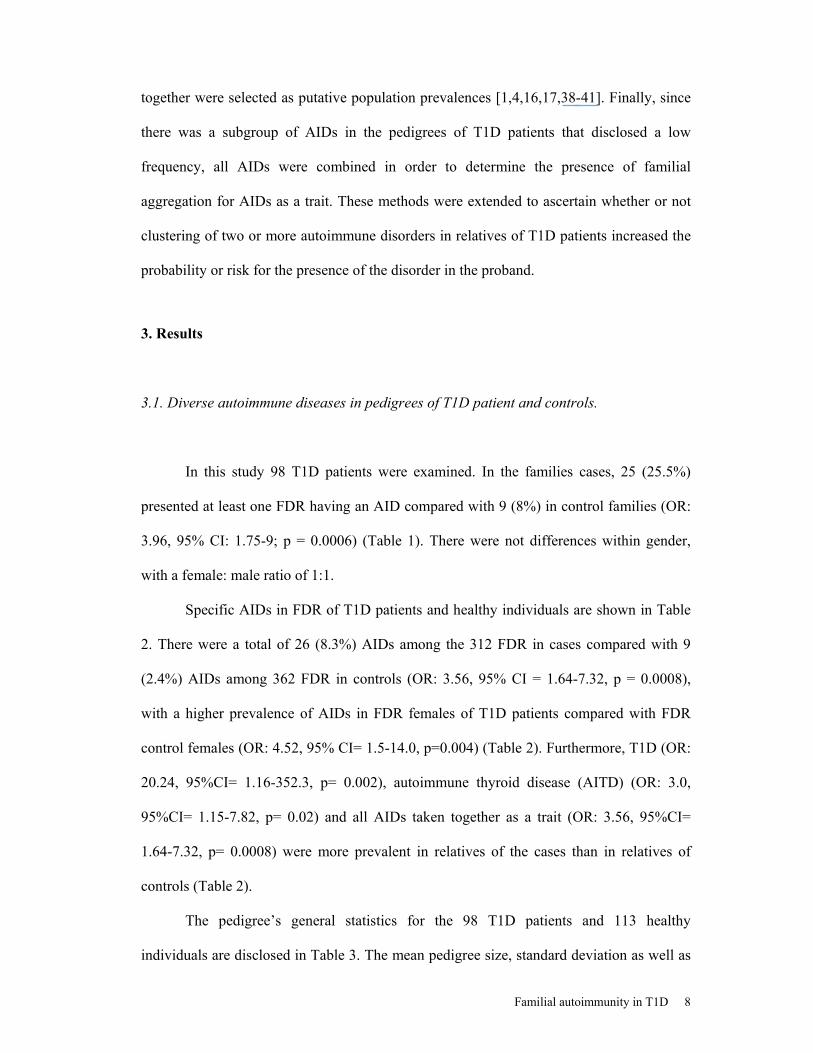
3.1. Diverse autoimmune diseases in pedigrees of T1D patient and controls.
In this study 98 T1D patients were examined. In the families cases, 25 (25.5%)
presented at least one FDR having an AID compared with 9 (8%) in control families (OR:
3.96, 95% CI: 1.75-9; p = 0.0006) (Table 1). There were not differences within gender,
with a female: male ratio of 1:1.
Specific AIDs in FDR of T1D patients and healthy individuals are shown in Table
2. There were a total of 26 (8.3%) AIDs among the 312 FDR in cases compared with 9
(2.4%) AIDs among 362 FDR in controls (OR: 3.56, 95% CI = 1.64-7.32, p = 0.0008),
with a higher prevalence of AIDs in FDR females of T1D patients compared with FDR
control females (OR: 4.52, 95% CI= 1.5-14.0, p=0.004) (Table 2). Furthermore, T1D (OR:
20.24, 95%CI= 1.16-352.3, p= 0.002), autoimmune thyroid disease (AITD) (OR: 3.0,
95%CI= 1.15-7.82, p= 0.02) and all AIDs taken together as a trait (OR: 3.56, 95%CI=
1.64-7.32, p= 0.0008) were more prevalent in relatives of the cases than in relatives of
controls (Table 2).
The pedigree’s general statistics for the 98 T1D patients and 113 healthy
individuals are disclosed in Table 3. The mean pedigree size, standard deviation as well as

Familial autoimmunity in T1D 9
the total number of relative pairs was obtained to calculate the prevalence for each AID.
The analyses were restricted to FDR.
3.2. Familial AID correlation.
By assessing if the presence of T1D in the proband correlated with the presence of
an AID in a first-degree affected relative, the association for the occurrence of each
individual AID was evaluated. Each model was weighted up every time a covariate (i.e.
AID) was added to asses if the new trait would improve the likelihood of the primary
phenotype by using the joint test, as described in the methods section. No significant
correlation was observed for any of the examined AIDs, implying that none of the
inspected models would explain the presence of the proband disease phenotype. Moreover,
when all the AIDs were considered together, as a trait, no significant correlation was
observed.
3.3. Familial aggregation (λR).
The prevalence for each AID as well as for all AIDs taken together for each pair of
relatives (Parent/offspring, Sibling/sibling and total FDR) is disclosed in Table 4.
Previously reported prevalences were also taken into account [1,4,16,17,38-41]. These
calculated prevalences were used to obtain the familial aggregation for different degrees of
relatives (Table 5). Additionally, using putative chosen prevalences (AID individually
(K=0.4%) and all AIDs together (2.5%), λR were calculated (Table 5). Values supporting
familial aggregation (λR >1.0) were observed for both groups using both the data on
pedigrees and putative chosen prevalences. Familial aggregation of AITD (λR~4.9 ± 3.0),

Familial autoimmunity in T1D 10
T1D (λR~4.21 ± 4.82) and all AIDs taken together (λR~2.21 ± 1.41) was observed in T1D
patients.
4. Discussion
These results indicate familial clustering of AIDs in patients with T1D in our
population, and are consistent with previous results showing familial autoimmunity in
other AIDs such as rheumatoid arthritis (RA) [9], systemic lupus erythematosus (SLE)
[10,11], primary Sjögren's syndrome [12,13], polymyositis [14], juvenile RA [15], multiple
sclerosis [16], vitiligo [17], and pemphigus [18].
Tait et al. [19] examined the presence of AIDs in British family members of
patients with T1D. Similarly to ours, they observed that AITD was the most common AID
among relatives. They also found that the prevalence of AIDs was higher in parents of T1D
patients than in the general population, confirming the importance of family history as a
significant risk factor for the development of T1D and supporting the hypothesis of shared
aetiological mechanisms for AIDs [19].
AITD is the most consistently associated disorder with other AIDs including T1D
[10,12,17,27,28]. Although CD may also be associated with T1D [26-28], this disease is
extremely rare among our T1D patients [30]. However, most of the cases with CD may be
subclinical or manifested at adulthood. Nevertheless, careful assessment of signs and
symptoms suggesting CD was carried out (i.e. malnutrition, growth retardation, iron
deficiency anemia). In addition, we have tested our cohort for immunoglobulin A anti-
endomysium antibodies (by an indirect immunofluorescence method) and found only one
positive case who was free of symptoms for CD [Anaya et al. Unpublished results].

Familial autoimmunity in T1D 11
Autoimmune hypothyroidism was the most common disease encountered among
FDR of our T1D patients as has been also reported in familial studies of Sjögren’s
syndrome [12,13], multiple sclerosis [16], vitiligo [17], juvenile RA [15], and SLE [10].
This finding supports previous analysis suggesting that AIDs (i.e. T1D and AITD) might
be the consequence of pleiotropic effects of a single major gene on a polygenic background
[2,9,12]. The lack of genetic information (i.e. genotypes) prevents us from drawing any
conclusion about the specific role of loci in the susceptibility of AIDs. Nonetheless, the
strong suggestions from previous studies allow us to point out the major histocompatibility
complex (MHC), including both HLA and non-HLA [3,4], as one of the central loci
contributing to T1D and AIDs. However, not all AIDs share the same genetic susceptibility
or allelic spectrum. Thus, the genetic risk factors for AIDs may well consist of two forms:
those which are common to many AIDs and those that are specific to a given disorder.
The next most commonly registered AIDs in FDR of T1D patients were T1D and
SLE. Familial correlation was not obtained for these diseases because at least one of the
maximizations was not available and thus no joint test could be performed. However, λR
was observed for T1D but not for SLE (Table 5). CD was not observed in FDR of patients
and controls. We can not exclude an ascertainment bias since CD could be asymptomatic
and test for specific antibodies or histopathological examination of the small intestinal
endoscopic biopsy was not done.
Both T1D and AITD share similar susceptibility gene polymorphisms including
HLA and non-HLA variants [4,20,42], which may account for the observed aggregation.
Shared genetic factors are in fact the most likelihood cause for familial aggregation;
however, it is important to keep in mind the fact that shared environmental factors can also
explain this aggregation. For a specified relative type, a λR greater than one suggests
familial aggregation of the disease, but does not identify whether genetic and/or

Familial autoimmunity in T1D 12
environmental factors are aggregating [43]. Thus, a major strength of this study was the
inclusion of healthy individuals matched by age, sex, origin and ethnicity, whose
environmental conditions were similar to those of the patients.
The prevalence of AIDs among FDR of control individuals was 2.5% (Table 2),
which is similar to the reported prevalence of such disorders in the general population [1]
and in FDR of controls in other studies of familial autoimmunity [16,18]. Familial
aggregation (λR) of AITD, T1D and all AIDs taken together was observed (Table 5). The
λR obtained indicates how frequently an autoimmune trait is present in the sampled
pedigrees depending on its distribution. The healthy individual’s pedigrees ought to
represent the general population given the conditions where each affected individual has
been matched with an unaffected individual by age-, sex-, origin and ethnicity, who did not
exhibit any history of AID.
Whilst the different weighted models could have predicted how a trait would
explain the presence of an AID in the proband when there is an FDR affected with an AID,
the non-significant observed familial correlation among the examined models implies that
the presence of each one of these autoimmune traits is not mathematically associated with
the proband disease phenotype (i.e. AID). However, the lack of correlation with any of the
AIDs might be explained by its familial distribution, frequency and late-age of onset. In
fact, AIDs can exist subclinically for a significant period of time [6]. Since T1D is the
earliest onset AID, prospective studies are necessary to accurately assess whether or not
familial and shared autoimmunity arise during the patient or FDR lifetime.
We did not observe a predominant paternal inheritance of T1D within families as
has been observed by others in T1D [19] as well as in other AIDs [12,19]. However, a
significant predominance of familial autoimmunity among FDR females was registered.

Familial autoimmunity in T1D 13
Consideration must be given to maternal transmission due to the high preponderance of
AIDs in females compared with the general population [1].
In summary, our results indicate familial autoimmunity in T1D and sustain a
common immunogenetic origin for diverse autoimmune phenotypes. As a corollary, results
also emphasize the importance of the autoimmunity family history as a substantial risk
factor for the development of T1D and other AIDs.
Acknowledgments
We thank all the patients and participants of this study. Some of the results of this
study were obtained by using the program package S.A.G.E., which is supported by a U.S.
Public Health Service Resource Grant (RR03655) from the National Center for Research
Resources. We also thank the two anonymous reviewers of this paper for their important
comments.

Familial autoimmunity in T1D 14
References
1. Cooper GS, Stoehla BC. The epidemiology of autoimmune diseases. Autoimmun Rev
2003;2:119-25.
2. Bias WB, Reveille JD, Beaty TH, Meyers DA, Arnett FC. Evidence that autoimmunity
in man is a Mendelian dominant trait. Am J Hum Genet 1986;39:584-602.
3. Vyse TJ, Todd JA. Genetics of autoimmune disease. Cell 1996;85:311-8.
4. Wandstrat A, Wakeland E. The genetics of complex autoimmune diseases: non-MHC
susceptibility genes. Nat Immunol 2001;9:802-9.
5. Houghton KM, Cabral DA, Petty RE, Tucker LB. Primary Sjogren's syndrome in
dizygotic adolescent twins: one case with lymphocytic interstitial pneumonia. J
Rheumatol 2005;32:1603-6.
6. Sloka S. Observations on recent studies showing increased co-occurrence of
autoimmune diseases. J Autoimmun 2002;18:251-7
7. Block SR, Winfield JB, Lockshin MD, D'Angelo WA, Christian CL. Studies of twins
with systemic lupus erythematosus. A review of the literature and presentation of 12
additional sets. Am J Med 1975;59:533-52.
8. Michel M, Johanet C, Meyer O, Frances C, Wittke F, Michel C, et al. Familial lupus
erythematosus. Clinical and immunologic features of 125 multiplex families. Medicine
(Baltimore) 2001;80:153-8.
9. Lin JP, Cash JP, Doyle SZ, Peden S, Kanik K, Amos CI, et al. Familial clustering of
rheumatoid arthritis with other autoimmune diseases. Hum Genet 1998;103:475-82.
10. Priori R, Medda E, Conti F, Cassara EAM, Danieli MG, Gerli R, et al. Familial
autoimmunity as a risk factor for systemic lupus erythematosus and vice versa: a
casecontrol study. Lupus 2003;12:735-40.
11. Reveille JD, Bias WB, Winkelstein JA, Provost TT, Dorsch CA, Arnett FC. Familial
systemic lupus erythematosus: immunogenetic studies in eight families. Medicine
(Baltimore) 1983;62:21-35.
12. Reveille JD, Wilson RW, Provost TT, Bias WB, Arnett FC. Primary Sjögren's
syndrome and other autoimmune diseases in families. Prevalence and immunogenetic
studies in six kindreds. Ann Intern Med 1984;101:748-56.
13. Anaya JM, Tobon GJ, Correa PA, Cañas C, Pineda-Tamayo R, Castiblanco J. Familial
clustering of autoimmune diseases in patients with primary Sjögren’s syndrome.
Arthritis Rheum 2005;52(suppl):S375.

Familial autoimmunity in T1D 15
14. Ginn RL, Lin JP, Plotz PH, Bale SJ, Wilder RL, Mbauya A, et al. Familial
autoimmunity in pedigrees of idiopathic inflammatory myopathy patients suggests
common genetic risk factors for many autoimmune diseases. Arthritis Rheum
1998;41:400-5.
15. Prahalad S, Shear ES, Thompson SD, Giannini EH, Glass DN. Increased prevalence of
familial autoimmunity in simplex and multiplex families with juvenile rheumatoid
arthritis. Arthritis Rheum 2002;46:1851-6.
16. Broadley SA, Deans J, Sawcer SJ, Clayton D, Compston DA. Autoimmune disease in
first-degree relatives of patients with multiple sclerosis. A UK survey. Brain
2000;123:1102-11.
17. Alkhateeb A, Fain PR, Thody A, Bennett DC, Spritz RA. Epidemiology of vitiligo and
associated autoimmune diseases in Caucasian probands and their families. Pigment
Cell Res 2003;16:208-14.
18. Firooz A, Mazhar A, Ahmed AR. Prevalence of autoimmune diseases in the family
members of patients with pemphigus vulgaris. J Am Acad Dermatol 1994;31:434-7.
19. Tait KF, Marshall T, Berman J, Carr-Smith J, Rowe B, Todd JA, et al. Clustering of
autoimmune disease in parents of siblings from the Type 1 diabetes Warren repository.
Diabet Med 2004;21:358-62.
20. Jawaheer D, Seldin MF, Amos CI, Chen WV, Shigeta R, Monteiro J, et al. A
genomewide screen in multiplex rheumatoid arthritis families suggests genetic overlap
with other autoimmune diseases. Am J Hum Genet 2001;68:927-36.
21. Becker KG, Simon RM, Bailey-Wilson JE, Freidlin B, Biddison WE, McFarland HF,
et al. Clustering of non-major histocompatibility complex susceptibility candidate loci
in human autoimmune diseases. Proc Natl Acad Sci U S A. 1998;95:9979-84.
22. Maas K, Chen H, Shyr Y, Olsen NJ, Aune T. Shared gene expression profiles in
individuals with autoimmune disease and unaffected first-degree relatives of
individuals with autoimmune disease. Hum Mol Genet 2005;14:1305-14
23. Aune TM, Parker JS, Maas K, Liu Z, Olsen NJ, Moore JH. Co-localization of
differentially expressed genes and shared susceptibility loci in human autoimmunity.
Genet Epidemiol 2004;27:162-72.
24. Humbert P, Dupond JL. Multiple autoimmune syndromes. Ann Med Interne (Paris)
1988;139:159-68.
25. Pugliese A. Genetics of type 1 diabetes. Endocrinol Metabol Clin NA 2004;33:1-16.

Familial autoimmunity in T1D 16
26. Bao F, Yu L, Babu S, Wang T, Hoffenberg EJ, Rewer SM, et al. One third of HLA
DQ2 homozygous patients with type 1 diabetes express celiac disease associated
transglutaminase autoantibodies. J Autoimmun 1999;13:143–8. 16
27. Frasier SD, Penny R, Zinder R, Goldstein I, Graves D. Antithyroid antibodies in
Hispanic patients with type I diabetes mellitus. Prevalence and significance. Am J Dis
Child 1986;40:1278-80.
28. Kordonouri O, Klinghammer A, Lang EB, Gruters-Kieslich A, Grabert M, Holl RW.
Thyroid autoimmunity in children and adolescents with type 1 diabetes: a multicenter
survey. Diabetes Care 2002;25:1346 –50.
29. The Expert Committee on the Diagnosis and Classification of Diabetes Mellitus:
Report of the Expert Committee on the Diagnosis and Classification of Diabetes
Mellitus. Diabetes Care 1997;20:1183-97
30. Tobon GJ, Arango A, Abad V, García J, Cuervo H, Velásquez A, et al. Clinical and
immunological characteristics of type 1 diabetes mellitus in a northwestern Colombian
population. Diabetes Res Clin Pract 2005 Nov 30; [Epub ahead of print].
31. Carvajal-Carmona LG, Soto ID, Pineda N, Ortiz-Barrientos D, Duque C, Ospina-
Duque J, et al. Strong Amerind/white sex bias and a possible Sephardic contribution
among the founders of a population in northwest Colombia. Am J Hum Genet
2000;67:1287-95.
32. Correa PA, Whitworth WC, Kuffner T, McNicholl J, Anaya JM. HLA-DR and DQB1
gene polymorphism in the North-western Colombian population. Tissue Antigens
2002;59:436-9.
33. S.A.G.E. Statistical Analysis for Genetic Epidemiology, Release 5.0. Computer
program package from the Department of Epidemiology and Biostatistics,
Rammelkamp Center for Education and Research, MetroHealth Campus, Case Western
Reserve University, Cleveland. 2004.
34. Elston RC, George VT, Severtson F. The Elston-Stewart algorithm for continuous
genotypes and environmental factors. Human Hered 1992;42:16-27.
35. George VT, Elston RC. Testing the association between polymorphic markers and
quantitative traits in pedigrees. Genetic Epidemiol 1987;4:193-201.
36. Box GEP, Cox DR. An analysis of transformations. J Roy Stat Soc [B] 1964;26:211-
52.

Familial autoimmunity in T1D 17
37. Risch N. Linkage strategies for genetically complex traits. I. Multilocus models. Am J
Hum Genet 1990;46:222-8.
38. Tunbridge WM, Evered DC, Hall R, Appleton D, Brewis M, Clark F, et al. The
spectrum of thyroid disease in a community: the Whickham survey. Clin Endocrinol
1977;7:481-93.
39. Jacobson DL, Gange SJ, Rose NR, Graham NM. Epidemiology and estimated
population burden of selected autoimmune diseases in the United States. Clin Immunol
Immunopathol 1997;84:223-43.
40. Hochberg MC. The application of genetic epidemiology to systemic lupus
erythematosus. J Rheumatol 1987;14:867-9.
41. Gatling W, Budd S, Walters D, Mullee MA, Goddard JR, Hill RD. Evidence of an
increasing prevalence of diagnosed diabetes mellitus in the Poole area from 1983 to
1996. Diabet Med 1998;15:1015-21.
42. Criswell LA, Pfeiffer KA, Lum RF, Gonzales B, Novitzke J, Kern M, et al. Analysis of
families in the multiple autoimmune disease genetics consortium (MADGC) collection:
the PTPN22 620W allele associates with multiple autoimmune phenotypes. Am J Hum
Genet 2005;76:561-71.
43. Laird NM, Cuenco KT. Regression methods for assessing familial aggregation of
disease.Stat Med 2003;22:1447-55.

Familial autoimmunity in T1D 18
Table 1. Characteristics of patient’s families with T1D and healthy individuals.
Characteristic
T1D patients N = 98 (%)
Controls N=113 (%)
Age (years) 8.8 ± 6.3 9.05 ± 4.22
Number of FDR 312 362
Families with ≤ 5 FDR 91 (92.3) 96 (84)
Families with 6-10 FDR 7 (7.1) 17 (15)
Families with a FDR having at least one AID Yes 25 (25.5) 9 (8)*
No 73 (74.4) 104 (92)
FDR: first degree relatives; AID: Autoimmune disease; T1D: type 1 diabetes mellitus; OR:
odds ratio; CI: confidence interval.
* Comparison between cases and controls: OR: 3.96, 95% CI = 1.74-9, p = 0.0006.

Familial autoimmunity in T1D 19
Table 2. Specific AIDs in first-degree relatives of T1D patients and healthy individuals
AIDs: Autoimmune diseases; FDR: First degree relative; T1D: Type 1 Diabetes mellitus.
F: Female, M: Male.
* Comparing frequency of T1D in FDR among cases and controls (OR: 20.24, 95%CI=
1.16-352.3, p= 0.002).
** Comparing frequency of autoimmune thyroid diseases in FDR among cases and
controls. OR: 3.0, 95%CI= 1.15-7.82, p= 0.02.
*** Comparing frequency of AID in FDR of cases and controls (OR: 3.56, 95%CI= 1.64-
7.73, p= 0.0008). 26 AID were observed in 25 FDR. There was one FDR having two
AIDs. No FDR among controls had more than one AID.
**** Comparing frequency of AID in females FDR of cases and controls. OR: 4.52,
95%CI= 1.5-14.0, p= 0.004.
AIDs in FDR of cases AIDs in FDR of controls AIDs
All F M All F M
Megaloblastic Anemia 1 1 0 0 0 0
Systemic Lupus
Erythematosus 1 0 1 0 0 0
Vitiligo 1 0 1 3 0 3
Type 1 diabetes 8 7 1 0* 0 0
Autoimmune Thyroid
disease 15 7 8 6** 4 2
Number of AID in FDR (%) 26/312***(8.3%)
15/312(4.8%)
11/312(3.5%)
9/362 (2.5%)
4/362****
(1.1%) 5/362 (1.4%)

Familial autoimmunity in T1D 20
Table 3. Pedigree’s general statistics for T1D patients and healthy individuals
Pedigrees
Descriptors T1D
Healthy
Individuals
No. of Pedigrees 98 113
Mean size ± SD
(Min, Max)
4.17± 1.35
(3, 10)
4.33 ± 1.18
(3, 8)
Pairs Parent/Offspring 426 518
Sibling/Sibling 211 246
Sister/Sister 56 13
Brother/Brother 41 34
Brother/Sister 114 41
T1D: Type 1 Diabetes Mellitus

Familial autoimmunity in T1D 21
Table 4. Prevalence of AIDs in T1D patient’s and healthy individual’s pedigrees.
AID: Autoimmune disease; T1D: type 1 diabetes mellitus; AITD: Autoimmune Thyroid
Diseases; SLE: Systemic Lupus Erythematosus; VIT: Vitiligo.
* Data is given in percentages. Prevalences are disclosed between relative pairs ([P/O],
Parent/offspring; [SIB], Sibling/Sibling; [REL] Relatives). KDM: prevalence for AID in
T1D patient pedigrees. KHI: prevalence for AID in control pedigrees. K: prevalence in the
general population.
KDM* KHI AIDs P/O SIB REL P/O SIB REL
Reported K [Population] [Ref]
T1D 0.47 3.79 1.26 0.00 0.00 0.00 0,48 [UK, USA – Caucasian] [17]
0.19 [North Americans] [39]
0.34 [UK-Caucasian)] [41]
SLE 0.23 0.00 0.16 0.00 0.00 0.00 0.024 [USA-Caucasian] [1]
0.027 [UK-Caucasian] [16]
AITD 1.64 3.79 2.35 1.16 0.00 0.65 Grave’s disease
0.65 [UK-Caucasian] [38,39]
0.80 [USA-Caucasian] [17,40]
Hashimoto thyroiditis
0.80 [UK-Caucasian] [39]
1.15 [USA-Caucasian] [40]
VIT 0.00 0.00 0.00 0.58 0.00 0.33 0.40 [USA-Caucasian] [1,17]
All AIDs 2.11 8.06 4.08 1.74 0.00 0.98 2.5 [UK-Caucasian] [16]

Familial autoimmunity in T1D 22
Table 5. Familial aggregation (λR) of AIDs in T1D patients.
λR= KDM/KHI λR= KDM/K
AID λP/O λSIB λREL λP/O λSIB λREL
T1D NA NA NA 1.17 9.47 3.15 SLE NA NA NA 0.57 0.00 0.40
AITD 1.41 NA 3.61 4.10 9.47 5.87 VIT NA NA NA 0.00 0.00 0.00
AID** 1.21 NA 4.16 0.84 3.22 1.63
* KDM: prevalence for AID in T1D patient pedigrees; KHI: prevalence for AID in Healthy
Individual’s pedigrees. K: chosen prevalence for the general population. Recurrent risk
ratio (λR= KDM/[KHI or K]), where R is the specific relative pair used (P/O:
Parent/offspring; SIB: Sibling/Sibling; REL: Relatives); the ratio was calculated by a
comparison between prevalence of T1D (KDM) in patients depending on its first–degree
relative disease or by using the chosen putative population prevalence for AID (Data from
Table 4. NA: Data not applicable. Since no prevalences were observed for KHI, a proper
appreciation of λR could not be accomplished. Nevertheless, if taking these results into
consideration the λR would disclose a high aggregation for those AIDs that were observed
in patients but not in controls pedigrees.
** When taken together the chosen population prevalence (K) for AIDs was consider as
25/1000 individuals (2.5%) and for each individual AID 4/1000 (0.4%).

Familial autoimmunity in T1D 23
Appendix 1. List of AIDs investigated in the present study
No Autoimmune disease. Reference 1 Diabetes mellitus type 1 1 2 Systemic lupus erythematosus 2 3 Antiphospholipid syndrome 3 4 Rheumatoid arthritis 4 5 Sjögren’s syndrome 5 6 Mixed connective tissue diseases 6 7 Ankylosing spondylitis 7 8 Scleroderma 8 9 Dermato-polymyositis 9-10
10 Crohn’s disease or ulcerative colitis 11 11 Megaloblastic anemia 12 12 Hypothyroidism (Hashimoto) 13 13 Hyperthyroidism (Graves) 14 14 Psoriasis 15 15 Vitiligo 15 16 17
Primary biliary cirrhosis Autoimmune hepatitis
16 17
18 Multiple sclerosis 18 19 Other
1. The Expert Committee on the Diagnosis and Classification of Diabetes Mellitus: Report of the Expert Committee on the Diagnosis and Classification of Diabetes Mellitus. Diabetes Care 20;197:1183-97.
2. Tan EM, Cohen AS, Fries JF, Masi AR, McShane DJ, Rothfield NF, et al. The 1982 revised criteria for the classification of systemic lupus erythematosus. Arthritis Rheum. 1982;25:1271-7.
3. Wilson WA, Gharavi AE, Koike T, Lockshin MD, Branch DW, Piette JC, et al. International consensus statement on preliminary classification criteria for definite antiphospholipid syndrome: report of an international workshop. Arthritis Rheum 1999;42:1309-11.
4. Arnett FC, Edworthy SM, Bloch DA, McShane DJ, Fries JF, Cooper NS, et al. The American Rheumatism Association 1987 revised criteria for the classification of rheumatoid arthritis. Arthritis Rheum 1988; 31:315-24.
5. Vitali C, Bombardieri S, Jonsson R, Moutsopoulos HM, Alexander EL, Carsons SE, et al. Classification criteria for Sjogren’s syndrome: a revised version of the European criteria proposed by the American-European Consensus Group. Ann Rheum Dis 2002;61:554-8.
6. Alarcón-Segovia D, Villareal M. Classification and diagnostic criteria for mixed connective tissue disease. En: Kasukawa R, Sharp GC (eds). Mixed Connective Tissue Disease and Anti-Nuclear Antibodies. Amsterdam. Elsevier. 1987;33-40.
7. Van der Linden S, Valkenburg HA, Cats A. Evaluation of diagnostic criteria for ankylosing spondylitis. A proposal for modification of the New York criteria. Arthritis Rheum. 1984;27:361-8.
8. Masi AT, Rodnan GP, Medsger TA, et al. Preliminary criteria for the classification of systemic sclerosis (scleroderma). Arthritis Rheum 1980;23:581-90.
9. Bohan A, Peter JB. Polymyositis and dermatomyositis. N Eng J Med 1975;292:344-7. 10. Bohan A, Peter JB. Polymyositis and dermatomyositis. N Eng J Med 1975;292:403-7. 11. Yao T, Matsui T, Hiwatashi N. Crohn’s disease in Japan: diagnostic criteria and epidemiology. Dis
Colon Rectum 2000;43:S85-93. 12. Babior BM. The megaloblastic anemias. In: Williams’ Hematology. 6th Ed. E Beutler et al (eds). New
York. McGraw-Hill. 2000. 13. Lindsay RS, Toft AD. Hypothyroidism. Lancet 1997;349:413. 14. DeGroot LJ, et al: Thyroid gland (Part III). In: Endocrinology. 4th Ed. LJ DeGroot, JL Jamenson (eds).
Philadelphia, Saunders, 2000. 15. Freedburg IM et al (eds): Fitzpatrick’s Dermatology in General Medicine, 5th ed. New York, McGraw-
Hill, 1999. 16. Kaplan MM. Primary biliary cirrhosis. N Eng J Med 1996;335:1570-80. 17. Czaja AJ. Current concepts in autoimmune hepatitis. Ann Hepatol 2005;4:6-24. 18. McDonald WI, Compston A, Edan G, Goodkin D, Hartung HP, Lublin FD, et al. Recommended
diagnostic criteria for multiple sclerosis: guidelines from de international panel on the diagnosis of multiple sclerosis. Ann Neurol. 2001;50:121-7.

Familial autoimmunity in T1D 24
19. This was a free question asked by the physicians and included vasculitis and coeliac disease (i.e. malabsorption and the presence of anti-endomysium or anti-gliadin antibodies).