Construction of miniantibodies for the in vivo study of human autoimmune diseases in animal models
10
BioMed Central Page 1 of 10 (page number not for citation purposes) BMC Biotechnology Open Access Research article Construction of miniantibodies for the in vivo study of human autoimmune diseases in animal models Roberto Di Niro †1 , Federica Ziller †1 , Fiorella Florian 1 , Sergio Crovella 2 , Marco Stebel 3 , Marco Bestagno 4 , Oscar Burrone 4 , Andrew RM Bradbury 5 , Paola Secco 6 , Roberto Marzari 1 and Daniele Sblattero* 6 Address: 1 Department of Biology, University of Trieste, 34127 Trieste, Italy, 2 Department of Reproductive and Development Science, University of Trieste, 34127 Trieste, Italy, 3 CSPA, University of Trieste, 34127 Trieste, Italy, 4 International Centre for Genetic Engineering and Biotechnology, 34012 Trieste, Italy, 5 Bioscience Division, Los Alamos National Laboratory, 87545 Los Alamos, NM, USA and 6 Department of Medical Sciences, IRCAD, University of Eastern Piedmont, 28100 Novara, Italy Email: Roberto Di Niro - [email protected]; Federica Ziller - [email protected]; Fiorella Florian - [email protected]; Sergio Crovella - [email protected]; Marco Stebel - [email protected]; Marco Bestagno - [email protected]; Oscar Burrone - [email protected]; Andrew RM Bradbury - [email protected]; Paola Secco - [email protected]; Roberto Marzari - [email protected]; Daniele Sblattero* - [email protected] * Corresponding author †Equal contributors Abstract Background: Phage display antibody libraries have been made from the lymphocytes of patients suffering from autoimmune diseases in which the antibodies are known to play a role in the pathogenesis or are important for the diagnosis of the disease. In the case of Celiac Disease, the immune response is directed against the autoantigen tissue transglutaminase. However, despite numerous studies, the role of these antibodies in the pathogenesis of this disease has not been elucidated. Results: We were able to engineer specific anti-transglutaminase antibody fragments in the form called "miniantibody". These are produced by genetic fusion of anti-tTG scFv to Human, Mouse or Rat Fc domains, making them suitable for in vivo expression. The results obtained here indicate that the miniantibody molecule is efficiently secreted, and that the reactivity to the antigen is retained even after fusion to heterologous Fc domains. Further analysis demonstrate that the molecule is secreted as homodimeric, mimicking original antibody structure. Finally, the in vivo expression in mice leads to detectable serum levels with no apparent gross immune response by the host. Conclusion: In this work we demonstrated the usefulness of a method for the in vivo expression of miniantibodies specific to transglutaminase, corresponding to the autoimmune specificity of Celiac Disease. This can be proposed as a general method to study the pathogenic role of autoimmune antibodies in autoimmune diseases. Published: 1 August 2007 BMC Biotechnology 2007, 7:46 doi:10.1186/1472-6750-7-46 Received: 6 February 2007 Accepted: 1 August 2007 This article is available from: http://www.biomedcentral.com/1472-6750/7/46 © 2007 Di Niro et al; licensee BioMed Central Ltd. This is an Open Access article distributed under the terms of the Creative Commons Attribution License (http://creativecommons.org/licenses/by/2.0 ), which permits unrestricted use, distribution, and reproduction in any medium, provided the original work is properly cited.
-
Upload
independent -
Category
Documents
-
view
1 -
download
0
Transcript of Construction of miniantibodies for the in vivo study of human autoimmune diseases in animal models

BioMed CentralBMC Biotechnology
ss
Open AcceResearch articleConstruction of miniantibodies for the in vivo study of human autoimmune diseases in animal modelsRoberto Di Niro†1, Federica Ziller†1, Fiorella Florian1, Sergio Crovella2, Marco Stebel3, Marco Bestagno4, Oscar Burrone4, Andrew RM Bradbury5, Paola Secco6, Roberto Marzari1 and Daniele Sblattero*6Address: 1Department of Biology, University of Trieste, 34127 Trieste, Italy, 2Department of Reproductive and Development Science, University of Trieste, 34127 Trieste, Italy, 3CSPA, University of Trieste, 34127 Trieste, Italy, 4International Centre for Genetic Engineering and Biotechnology, 34012 Trieste, Italy, 5Bioscience Division, Los Alamos National Laboratory, 87545 Los Alamos, NM, USA and 6Department of Medical Sciences, IRCAD, University of Eastern Piedmont, 28100 Novara, Italy
Email: Roberto Di Niro - [email protected]; Federica Ziller - [email protected]; Fiorella Florian - [email protected]; Sergio Crovella - [email protected]; Marco Stebel - [email protected]; Marco Bestagno - [email protected]; Oscar Burrone - [email protected]; Andrew RM Bradbury - [email protected]; Paola Secco - [email protected]; Roberto Marzari - [email protected]; Daniele Sblattero* - [email protected]
* Corresponding author †Equal contributors
AbstractBackground: Phage display antibody libraries have been made from the lymphocytes of patientssuffering from autoimmune diseases in which the antibodies are known to play a role in thepathogenesis or are important for the diagnosis of the disease. In the case of Celiac Disease, theimmune response is directed against the autoantigen tissue transglutaminase. However, despitenumerous studies, the role of these antibodies in the pathogenesis of this disease has not beenelucidated.
Results: We were able to engineer specific anti-transglutaminase antibody fragments in the formcalled "miniantibody". These are produced by genetic fusion of anti-tTG scFv to Human, Mouse orRat Fc domains, making them suitable for in vivo expression. The results obtained here indicatethat the miniantibody molecule is efficiently secreted, and that the reactivity to the antigen isretained even after fusion to heterologous Fc domains. Further analysis demonstrate that themolecule is secreted as homodimeric, mimicking original antibody structure. Finally, the in vivoexpression in mice leads to detectable serum levels with no apparent gross immune response bythe host.
Conclusion: In this work we demonstrated the usefulness of a method for the in vivo expressionof miniantibodies specific to transglutaminase, corresponding to the autoimmune specificity ofCeliac Disease. This can be proposed as a general method to study the pathogenic role ofautoimmune antibodies in autoimmune diseases.
Published: 1 August 2007
BMC Biotechnology 2007, 7:46 doi:10.1186/1472-6750-7-46
Received: 6 February 2007Accepted: 1 August 2007
This article is available from: http://www.biomedcentral.com/1472-6750/7/46
© 2007 Di Niro et al; licensee BioMed Central Ltd. This is an Open Access article distributed under the terms of the Creative Commons Attribution License (http://creativecommons.org/licenses/by/2.0), which permits unrestricted use, distribution, and reproduction in any medium, provided the original work is properly cited.
Page 1 of 10(page number not for citation purposes)

BMC Biotechnology 2007, 7:46 http://www.biomedcentral.com/1472-6750/7/46
BackgroundAutoimmunity is an important cause of disease inhumans, it is estimated to affect at least 3% to 5% of thehuman population and depends on a failure of the mech-anisms normally responsible for maintaining self-toler-ance (for a review see [1]). Although many factors causingthese diseases, including the genes that may predispose toautoimmunity, have been identified, the aetiology ofmost autoimmune diseases remains obscure. Much inter-est has focused on the analysis of the immune factorsleading to the tissue lesions. In some cases the cellularimmune response stimulated by lymphokines seems toplay a major role, whereas in others the humoral antibodyresponse is deemed prevalent. Functional genomics mayoffer a solution to these problems by using biological sys-tems which allow the massive interaction between anautoimmune patient's cloned antibody repertoire andindividual antigens. One of these systems is phage dis-play, a technique which involves the coupling of pheno-type to genotype in a selectable format. It has beenextensively used in molecular biology to study protein-protein interactions and one of the most successful appli-cations of phage display has been the isolation of mono-clonal antibodies to purified antigens [2-5]. In addition tolibraries from naive or immunized sources, phage anti-body libraries have also been made from patients suffer-ing from autoimmune diseases. This work has been mostextensively carried out with thyroid disease [6], systemiclupus erythematosus [7], paraneoplastic encephalomyeli-tis [8], myasthenia gravis [9] and type 1 diabetes mellitus[10]. In a recent work we described the antibody responsein Celiac Disease (CD) [11]. This is a genetic illnessstrongly linked to HLA DQ2, characterized by flatteningof the intestinal mucosa and malabsorption. The patho-genesis is precipitated by dietary exposure to wheat glutenand similar proteins in rye, barley and possibly oats [12].The disease is characterized by the presence of specificantibodies recognizing gliadins, food proteins and anendomysial autoantigen, identified as tissue transglutam-inase (tTG) [13]. We recently made and selected phageantibody libraries from the DNA isolated from CD patientlymphocytes and were able to isolate single-chain anti-body fragments (scFv) to tTG showing their specific pro-duction by intestinal lymphocytes, indicating that the siteof synthesis of these antibodies is the intestinal mucosa[11]. ScFvs isolated from different patients recognized thesame tTG epitopes and by ELISA competition experimentswe demonstrated that the number of epitopes recognizedwas restricted to two, distinguished by the ability of theantibodies to recognize mouse tTG [14]. The activities ofthese in vitro selected antibodies mimics those found inthe serum of CD patients, and preliminary work suggestssimilar biological activity indicating that in vivo studies ofthese antibodies may provide useful information on thepathogenic roles of these antibodies in CD.
The isolation of disease-specific antibodies, as well as therelated gene, is the first step in the generation of animalmodels through in vivo antibody gene expression. In vivogene expression has been implemented using a number ofdifferent techniques, including injection of naked DNAinto the muscle, with or without electroporation [15],coating gold particles with DNA and injecting them witha gene gun [16]. However, expression of recombinant pro-tein is usually transient and only low levels are reached,using these methods. Recently gene transfer systems medi-ated by vectors based on the adeno-associated virus, havebeen shown to mediate sustained and prolonged titres ofengineered antibody [17-19].
In the present work we describe the production of a seriesof miniantibody constructs composed of a humanautoimmune anti-tTG scFv combined with antibody con-stant Fc regions from human, rat and mouse. This wassought as a simple approach to rapidly obtain sustained invivo production of antibodies with specific specificities. Inaddition to providing appropriate effectors domains,fusion to constant regions also prolongs half life. This rep-resents an innovative tool for the in vivo studies of thepathogenic properties of cloned autoimmune antibodyfragments.
ResultsIn a previous paper [11] we described the isolation ofhuman IgA scFvs to tTG from phage antibody librariesobtained from the intestinal lymphocytes of CD patients.Two of these scFvs, indicated as 2.8 tTG, cross-reactive torodent tTG, and 3.7 tTG, specific for human enzyme, werethe reference antibody fragments used to make the seriesof constructs reported in Fig. 1. The construction of theseries pMB-SV5 is reported in Materials and Methods sec-tion. It is characterized by a BssHII-NheI cassette for thesubcloning of scFvs from the pDAN5 phagemid vector, aNheI-SpeI cassette for the cloning of CH2 and CH3domains of Fc from different species, and an SV5 tag at the3' end for uniform detection. Expression of the minianti-bodies was driven by a CMV promoter and a leadersequence at the N-terminus was included to allow mam-malian secretion. The two selected scFvs (2.8 tTG and 3.7tTG) were cloned into the 4 different vectors, generating atotal of 8 different constructs. All vectors were checked byDNA sequencing and the purified plasmidic DNAs weretransfected into HEK 293T cells. The secretion of the min-iantibodies in the culture medium was analyzed after 72h by ELISA on plates coated with either human or mousetTG. The results, determined by recognition of the SV5 tagat the C-terminus of the constructs (Fig. 2A), or with spe-cies specific antibodies (Fig. 2B), for the 2.8 miniantibod-ies show that they were all able to recognize both antigenswith O.D. values ranging from 0.5 (MB-HuA-2.8) to 1.6(MB-MoG-2.8). Similar results were obtained with the 3.7
Page 2 of 10(page number not for citation purposes)

BMC Biotechnology 2007, 7:46 http://www.biomedcentral.com/1472-6750/7/46
miniantibodies, except that recognition was specific forthe human enzyme, with no binding to mouse tTG (Fig.2C and 2D).
Stable cell lines for all the constructs were established bygrowing transfected cells in the selective agent for hygro-micin resistance. Supernatant from individual clones werescreened for the best ELISA reactivity and then expandedfor further experiments. The average yield of minianti-body production was in the 5–10 mg/liter range usingstandard culture flask.
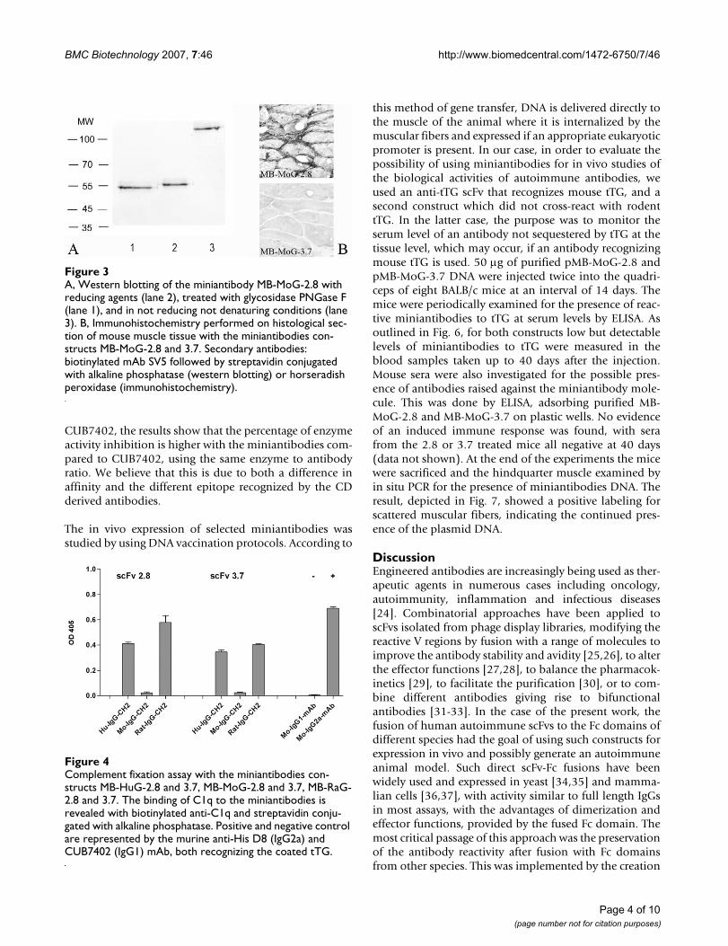
The miniantibodies were analyzed by Western blottingunder reducing and non reducing conditions and aftertreatment of the purified miniantibodies with glycosidasePNGaseF to assay the level of glycosylation. The results forthe miniantibody MB-MoG-2.8 are reported, as an exam-ple, in Fig. 3A. The predicted molecular weight of the min-iantibodies, result of the fusion of the scFv with constantdomains and SV5 tag, is about 55 kDa.
We found bands of the predicted molecular weight in thesamples treated with reducing agent and a slight increasein the electrophoretic mobility in the deglycosylated sam-ples, indicating that the miniantibodies are glycosylatedin HEK 239T cells. Under non reducing, non denaturingconditions, a high molecular weight band, explained bythe interchain disulfide bond in the Hinge region, wasobtained.
The ability of the miniantibodies to activate the classiccomplement pathway was tested with an in vitro assay, asreported in Fig. 4, in which C1q deposition was measuredusing an ELISA assay. The results showed that human andrat miniantibodies, of IgG1 and IgG2b origin respectively,were able to bind C1q, the first component of the classiccomplement cascade, as described in the literature
[20,21], whereas the mouse miniantibody of IgG1 origincould not bind C1q [22].
The miniantibodies were analyzed for their ability to rec-ognize tTG on histological sections. These experimentswere undertaken in view of the possible use of these min-iantibodies in in vivo studies. The immunolabeling of his-tological sections of mouse muscle is shown in Fig. 3B,using purified MB-MoG-2.8 and 3.7. Staining was onlydetected for MB-MoG-2.8 with specific recognition of theextracellular tTG present at the muscular endomysiumand perimysium. The same pattern was found when min-iantibodies with the human or rat Fc were used (notshown). For the same reasons, an assay on the inhibitionof tTG activity by miniantibodies was tested. We havealready shown that scFvs to tTG isolated from the intesti-nal lymphocytes of CD patients inhibit the in vitrotransamidation activity of tTG [23]. Fig. 5 shows that thecoupling of 5-(biotinamido)pentylamine to gliadin (atTG substrate) is catalyzed by mouse purified recom-binant tTG, which is inhibited by miniantibody MB-MoG-2.8, but not miniantibody MB-MoG-3.7, confirming thespecificity observed by ELISA. The inhibition closely mir-rors the values previously described for the scFv [23] in asimilar assay. In comparing the two miniantibodies withthe commercial tTG-specific monoclonal antibody
ELISA of supernatants of cultured HEK 293T cells trans-fected with the series of plasmids pMB-SV5 carrying 2.8 scFv gene (A and B) and 3.7 scFv gene (C and D) fused to CH2-CH3 domains genes from human, mouse and ratFigure 2ELISA of supernatants of cultured HEK 293T cells trans-fected with the series of plasmids pMB-SV5 carrying 2.8 scFv gene (A and B) and 3.7 scFv gene (C and D) fused to CH2-CH3 domains genes from human, mouse and rat. Antigens: human tTG, mouse tTG and BSA. Secondary antibodies: A) and C) biotinylated mAb SV5 and streptavidin conjugated with peroxidase; B) and D) goat anti human, mouse and rat IgG or IgA conjugated with peroxidase.
Schematic representation of the cloning vectorFigure 1Schematic representation of the cloning vector. The Human IgG1 CH2-CH3 domains gene in the vector pMB-SV5 could be substituted by Human IgA, Mouse and Rat Fc domain genes using restriction sites NheI and SpeI. Different scFvs can be exchanged by using restriction sites BssHII and NheI.
Page 3 of 10(page number not for citation purposes)
BMC Biotechnology 2007, 7:46 http://www.biomedcentral.com/1472-6750/7/46
CUB7402, the results show that the percentage of enzymeactivity inhibition is higher with the miniantibodies com-pared to CUB7402, using the same enzyme to antibodyratio. We believe that this is due to both a difference inaffinity and the different epitope recognized by the CDderived antibodies.
The in vivo expression of selected miniantibodies wasstudied by using DNA vaccination protocols. According to
this method of gene transfer, DNA is delivered directly tothe muscle of the animal where it is internalized by themuscular fibers and expressed if an appropriate eukaryoticpromoter is present. In our case, in order to evaluate thepossibility of using miniantibodies for in vivo studies ofthe biological activities of autoimmune antibodies, weused an anti-tTG scFv that recognizes mouse tTG, and asecond construct which did not cross-react with rodenttTG. In the latter case, the purpose was to monitor theserum level of an antibody not sequestered by tTG at thetissue level, which may occur, if an antibody recognizingmouse tTG is used. 50 μg of purified pMB-MoG-2.8 andpMB-MoG-3.7 DNA were injected twice into the quadri-ceps of eight BALB/c mice at an interval of 14 days. Themice were periodically examined for the presence of reac-tive miniantibodies to tTG at serum levels by ELISA. Asoutlined in Fig. 6, for both constructs low but detectablelevels of miniantibodies to tTG were measured in theblood samples taken up to 40 days after the injection.Mouse sera were also investigated for the possible pres-ence of antibodies raised against the miniantibody mole-cule. This was done by ELISA, adsorbing purified MB-MoG-2.8 and MB-MoG-3.7 on plastic wells. No evidenceof an induced immune response was found, with serafrom the 2.8 or 3.7 treated mice all negative at 40 days(data not shown). At the end of the experiments the micewere sacrificed and the hindquarter muscle examined byin situ PCR for the presence of miniantibodies DNA. Theresult, depicted in Fig. 7, showed a positive labeling forscattered muscular fibers, indicating the continued pres-ence of the plasmid DNA.
DiscussionEngineered antibodies are increasingly being used as ther-apeutic agents in numerous cases including oncology,autoimmunity, inflammation and infectious diseases[24]. Combinatorial approaches have been applied toscFvs isolated from phage display libraries, modifying thereactive V regions by fusion with a range of molecules toimprove the antibody stability and avidity [25,26], to alterthe effector functions [27,28], to balance the pharmacok-inetics [29], to facilitate the purification [30], or to com-bine different antibodies giving rise to bifunctionalantibodies [31-33]. In the case of the present work, thefusion of human autoimmune scFvs to the Fc domains ofdifferent species had the goal of using such constructs forexpression in vivo and possibly generate an autoimmuneanimal model. Such direct scFv-Fc fusions have beenwidely used and expressed in yeast [34,35] and mamma-lian cells [36,37], with activity similar to full length IgGsin most assays, with the advantages of dimerization andeffector functions, provided by the fused Fc domain. Themost critical passage of this approach was the preservationof the antibody reactivity after fusion with Fc domainsfrom other species. This was implemented by the creation
Complement fixation assay with the miniantibodies con-structs MB-HuG-2.8 and 3.7, MB-MoG-2.8 and 3.7, MB-RaG-2.8 and 3.7Figure 4Complement fixation assay with the miniantibodies con-structs MB-HuG-2.8 and 3.7, MB-MoG-2.8 and 3.7, MB-RaG-2.8 and 3.7. The binding of C1q to the miniantibodies is revealed with biotinylated anti-C1q and streptavidin conju-gated with alkaline phosphatase. Positive and negative control are represented by the murine anti-His D8 (IgG2a) and CUB7402 (IgG1) mAb, both recognizing the coated tTG.
A, Western blotting of the miniantibody MB-MoG-2.8 with reducing agents (lane 2), treated with glycosidase PNGase F (lane 1), and in not reducing not denaturing conditions (lane 3)Figure 3A, Western blotting of the miniantibody MB-MoG-2.8 with reducing agents (lane 2), treated with glycosidase PNGase F (lane 1), and in not reducing not denaturing conditions (lane 3). B, Immunohistochemistry performed on histological sec-tion of mouse muscle tissue with the miniantibodies con-structs MB-MoG-2.8 and 3.7. Secondary antibodies: biotinylated mAb SV5 followed by streptavidin conjugated with alkaline phosphatase (western blotting) or horseradish peroxidase (immunohistochemistry).
Page 4 of 10(page number not for citation purposes)

BMC Biotechnology 2007, 7:46 http://www.biomedcentral.com/1472-6750/7/46
of a novel mammalian expression vector in which eitherscFv or Fc domain could be easily switched using compat-ible restriction sites. As demonstrated by ELISA, all thechimeric constructs recognized the tTG antigen, and west-ern blotting showed the expected higher molecular weightbands corresponding to the dimeric form. The bindingactivity of the constructs with the scFv 2.8, crossreactive torodent tTG, was also preserved to mouse tTG tested inELISA as well as in an immunochemical assay on histolog-ical section of mouse muscle, as was the ability to inhibitthe crosslinking activity of tTG. Complement activationmeasured by C1q deposition was confirmed for thehuman and rat miniantibody constructs reflecting thepresence of the CH2 domain and correct glycosylationproduced by the transfected 293T cell.
Injection of plasmid DNA was used to induce in vivoexpression of miniantibodies. This approach derives fromextensive studies on DNA vaccination in which nakedplasmid DNA, coding for an antigenic protein, is trans-fected into muscle cells in vivo either by injection [38,39]or using a "gene gun" [40]. This results in expression ofthe vector-encoded antigen, which induces cellular andhumoral responses [41]. In a similar vein, injection ofDNA coding for an anti-tumoral scFv has also been carriedout [42]. Although other studies have demonstrated bothcellular and humoral responses against human scFvs inmice [43] with a reduction of the therapeutic potential,this feature can also be exploited to generate anti-idiotypicresponses against scFvs derived from mouse lymphomas[44,45].
Inhibitory effect of purified MB-MoG-3.7 (grey bars), MB-MoG-2.8 (black bars) and mAb CUB7402 (hatched bars) on mouse tTG activityFigure 5Inhibitory effect of purified MB-MoG-3.7 (grey bars), MB-MoG-2.8 (black bars) and mAb CUB7402 (hatched bars) on mouse tTG activity. Elisa plates coated with gliadin, a tTG substrate, are incubated with 0.2 mM 5-(biotina-mido)pentylamine and 0.25 μg of mouse tTG, with increasing amounts of purified miniantibody or mAb. The incorporation of 5-(biotinamido)pentylamine is revealed by streptavidin conjugated with peroxidase.
ELISA time course of the serum anti-tTG miniantibody aver-age titer in 8 BALB/c mice injected at 0 and 14 days with pMB-MoG-3.7 (panel A) and 2.8 (panel B) DNAFigure 6ELISA time course of the serum anti-tTG miniantibody aver-age titer in 8 BALB/c mice injected at 0 and 14 days with pMB-MoG-3.7 (panel A) and 2.8 (panel B) DNA. Serum dilu-tion 1:50. Secondary antibodies: biotinylated mAb SV5 and streptavidin conjugated with peroxidase.
In situ PCR on histological section of quadriceps muscle of a mouse injected with pMB-MoG-3.7 constructFigure 7In situ PCR on histological section of quadriceps muscle of a mouse injected with pMB-MoG-3.7 construct. PCR was per-formed after 40 days since injection. The arrows point at cells with positive reaction.
Page 5 of 10(page number not for citation purposes)

BMC Biotechnology 2007, 7:46 http://www.biomedcentral.com/1472-6750/7/46
In these experiments, the effectiveness of the anti-idio-typic response was enhanced by fusing the cloned anti-body expressed by the tumoral cells to an additional CH3antigenic region, as originally suggested by Syrengelas etal. [46]. In the present study, since a human scFv xeno-genic for mice was used, we sought to minimize the hostimmune response by using the scFv fused to a CH2-CH3syngenic mouse Fc region. The outcomes of the experi-ments have confirmed the validity of this approach, witha detectable production of miniantibodies in the serumfor at least 40 days and a peak of production after 20 days.
ConclusionThe reactivity of the serum miniantibodies in ELISAtogether with the apparent lack of humoral responseagainst the miniantibody molecule, led us to concludethat the human scFv-mouse Fc fusion miniantibodies arepoorly immunogenic in the mouse under the experimen-tal conditions used here. In conclusion, our results indi-cate that chimeric proteins generated by fusion of humanscFvs to human, murine and rat Fc regions are effectivelyproduced and secreted by cultured cells; the polypeptidesdimerize, forming disulfide bridges, so increasing thevalency of the miniantibody; the miniantibodies retainthe antigen recognition both in ELISA and immunohistol-ogy and the ability to activate complement. The inhibitoryproperties of the scFv are preserved and, upon intramus-cular injection of the plasmid, the ELISA antibody titre isstill detectable after 40 days, suggesting the absence of animmune response by the host when a syngenic Fc frag-ment is present in the construct. While this indicates thatthe approach is functional and will be useful in the in vivostudy of the role of autoimmune antibodies, in theabsence of complicating immunological factors, theperiod of expression obtained here is relatively shortwhen compared to the long time scale involved in mostautoimmune diseases, which occurs over a period ofyears. For this reason it may be appropriate to explorealternative in vivo expression methods, such as adeno-
associated virus vector, which has been shown to induceexpression over a prolonged period.
MethodsBacterial strains and enzymesDH5aF' (F'/endA1 hsdR17 (rK- mK+) supE44 thi-1 recA1gyrA (Nalr) relA1 D (lacZYA-argF)U169 deoR(F80dlacD(lacZ)M15)) strain was used for the cloning ofpDAN5, pMB-SV5 and derivates, pCDNA3.1/Hygro(+)and pTrcHisB. Molecular biology enzymes were pur-chased from New England Biolabs, Promega or Life Tech-nologies.
AntigensHuman tTG gene was cloned in pTrcHisB as described[47]. Mouse tTG gene was cloned in pTrcHisB as described[48]. Protein purification was performed as described in[14].
RNA extraction and cDNA synthesisPeripheral blood lymphocytes from a healthy donor andspleen lymphocytes from mouse and rat were separatedby density gradient centrifugation on Ficoll Hypaque(Pharmacia). Total RNA was then prepared as described[49]. cDNA was prepared using SuperScript II ReverseTranscriptase (Gibco BRL) with random hexamers.
pCDNA3.1/Hygro(+) modificationpCDNA3.1/Hygro(+) plasmid vector (Invitrogen) wasmodified as follows: NheI restriction site was exchangedwith XbaI, PmeI site was exchanged with HindIII site byinverse polymerase chain reaction (PCR) using the prim-ers Hygro-XbaI-INV antisense and Hygro-HindIII-INVsense; the primers are reported as A and B in Table 1. ScFv2.8 [11] was PCR amplified from pDAN plasmid vector bysuccessive amplification with sense primers 1-XbaI-leader,2-leader-intron and 3-BssHII-scFv (Table 1, primers 1, 2and 3), which introduce the XbaI site, a secretion leaderand a mini-intron as described in [50] and the BssHII site
Table 1: Primers. List of oligonucleotides used for vector construction and Fc region cloning.
Number Name Orientation Sequence
A Hygro-XbaI-inv Antisense AGCTTCTAGACAGCTTGGGTCTCCCTATAGB Hygro-HindIII-inv Sense AGCTAAGCTTAAACCCGCTGATCAGC1 1-XbaI-leader Sense CAGGCGTCTAGATGCCACCATGGGCTGGAGCCTGATCCTCCTGTTCCTCGTCGCTGTGGCTACAGGTAAGGG2 2-leader-intron Sense TGTGGCTACAGGTAAGGGGCTCACAGTAGCAGGCTTGAGGTCTGGACATATATATGGGTGACAATGACATCCAC3 3-BssHII-scFv Sense GGTGACAATGACATCCACTTTGCCTTTCTCTCCACAGGTGGCGCGCATGCCGACATCCGGTTGACCCAG4 scFv-NheI-HindIII Antisense CCGCTAAGCTTCGCCTGGCTAGCAAAAGCGTCCGTCGTATC5 HuGCH2-s Sense AGGCGGCTAGCGACAAAACTCACACATGCCCACCGTGCCCA6 HuGCH3-SV5-a Antisense CTGCTAAGCTTTTAAGTACTATCCAGGCCCAGCAGTGGGTTTGGGATTGGTTTGCCACTAGTTTTACCCGGGGAC
AGGGAGAG7 HuACH2-s Sense CAGGCGGCT AGCGTTCCCTCAACTCCACCTACC8 HuACH3-a Antisense CCGCTACTAGTTTTACCCGCCAAGCGGTCGAT9 MoGCH3-a Antisense CCGCTACTAGTTTTACCAGGAGAGTGGGAGAG10 MoGCH2-s Sense CAGGCG GCT AGC GGTTGTAAGCCTTGCATATGTACA11 RaGCH3-a Antisense CCGCTACTAGTTTTACCCGGAGGCCGGGAGATG12 RaGCH2-s Sense CAGGCG GCT AGC CACAAATGCCCTACATGCCCT
Page 6 of 10(page number not for citation purposes)

BMC Biotechnology 2007, 7:46 http://www.biomedcentral.com/1472-6750/7/46
at the 5' end, and antisense primer scFv-NheI-HindIII(Table 1, primer 4), which introduces the NheI and Hin-dIII sites at the 3' end. PCR fragment was cloned as XbaI –HindIII in the modified pCDNA3.1/Hygro(+) vector.
Cloning of Human Fc geneThe human IgG1 CH2 and CH3 domains gene was ampli-fied from lymphocyte cDNA by using the primer senseHuGCH2-s and antisense HuGCH3-SV5-a (Table 1, prim-ers 5 and 6), which introduce SpeI site and the SV5 tagsequence for mAb recognition [51] at the 3' end. PCR frag-ment was cloned as NheI-HindIII into the pCDNA3.1/Hygro(+) vector modified as described and carrying thescFv 2.8; the resulting vector was named pMB-HuG-2.8.The series of vector obtained by exchange of different scFvand Fc was called pMB-SV5.
Exchange of constant domains and scFvsThe set of oligonucleotide primers for amplification of Fcdomain genes was designed to comprise the CH2-CH3domains including the flexible hinge region. The CH2-CH3 domain genes were PCR amplified by using the senseprimer HuACH2-s and antisense HuACH3-a for humanIgA, MoGCH2-s and MoGCH3-a for mouse IgG1,RaGCH2-s and RaGCH3-a for rat IgG2b. All primers arereported in Table 1. The PCR fragments were cloned inpMB-HuG-2.8 vector replacing the resident Fc domaingene by cutting with NheI and SpeI and ligation.
The cloning of 3.7 scFv gene was performed by extractionof scFv gene from phagemid pDAN5 clone by cutting withBssHII and NheI and direct cloning in the series of vectorspMB-SV5 cut with the same enzymes.
HEK 293T transfection and selectionThe human kidney derived HEK 293T cell line was cul-tured in D-MEM medium (GIBCO) supplemented with10% fetal calf serum (FCS). Cells were harvested by shak-ing and plated in a 24 well microtiter plate (2 × 10⁵ cellsper well). For transient transfection, after 24 h, 1 μg ofpurified plasmid DNA resuspended in 50 μl of D-MEMwithout FCS and 2 μl of Lipofectamine 2000 (Invitrogen)in 50 μl of D-MEM were mixed, left at RT for 20 min andadded to each well of cultured cells. The cells were grownfor further 24/48 h and the supernatant inspected for min-iantibody production. Stable cell clones secreting minian-tibodies were obtained by treating the cells in the sameway as for the transient transfection, diluting the cells 1:10with fresh medium after 24 h from transfection and add-ing 400 μg/ml of antibiotic Hygromicin (Invitrogen) forthe selection of resistant cells. After 10 days of culture, theHygromicin concentration was reduced to 200 μg/ml.
ELISAELISA was performed by coating ELISA plates with puri-fied human or mouse recombinant tTG at 10 μg/mldiluted in phosphate-buffered saline (PBS) for 15 h at4°C. Wells were blocked with 2% non-fat milk in PBS(MPBS). The primary antibodies were the supernatants ofcultured HEK 293T cells diluted 1:5 with 2.5% MPBS orsera of mice injected with plasmid DNA diluted 1:50 with2% MPBS. Secondary antibodies used were biotinylatedmAb SV5 [51] recognizing the SV5 tag found at the mini-antibody C-terminus and goat anti human, mouse and ratIgG or IgA conjugated with peroxidase. The secondaryantibodies were used as following: a) biotinylated mAbSV5 diluted 1:2000 with 2% MPBS, followed by streptavi-dine conjugated with horseradish-peroxidase (HRP)(Pierce) diluted 1:2000, b) goat anti human, mouse andrat IgG or IgA conjugated with peroxidase (Dako) diluted1:1000. Each step was followed by three washes with PBSplus 0,1% Tween20 (PBST) and three washes with PBS.All the immunocomplexes were revealed with tetrame-thyl-benzidine (TMB) and read at O.D.450.
Serum-free cell cultures and Protein G purificationSerum-free supernatants for protein-G purification wereobtained as following: stable cell clones grown at conflu-ence in 25 cm2 flasks were harvested, centrifuged at 1200× g and resuspended in FCS-free D-MEM medium. Cellswere allowed to grow for further 48 h and the superna-tants collected.
Miniantibodies produced in FCS-free cell culture superna-tants were purified by using a HiTrap protein G column(GE Healthcare) following standard procedures. Briefly,50 ml of serum-free culture were passed through the pro-tein G column; the column was washed with 20 ml of 100mM Tris-HCl, pH 8.0, and 20 ml of 10 mM Tris-HCl, pH8.0. Purified miniantibodies were eluted with 50 mM Gly-cine, pH 3.0, and immediately buffered with Tris-HCl, pH8.0.
tTG inhibition assayELISA plate wells were adsorbed with 20 μg/ml purifiedgliadin for 2 h at 37°C and washed twice with PBS. Toeach well 100 μl of a solution of 5-(biotina-mido)pentylamine (Pierce) 0.2 mM, 0.25 μg of purifiedmouse tTG in NaCl 150 mM, Tris 50 mM pH 7.5 withincreasing amount of either protein G purified minianti-body or commercial tTG-specific monoclonal antibodyCUB7402 (Bio Optica, Milan), ranging from 0 to 0.5 μgper microwell, were added. After 1 h incubation at 37°C,the wells were washed three times with PBS plus 1%Tween20 and three times with PBS. 100 μl of a solution ofstreptavidin conjugated with alkaline phosphatase(Pierce) 1:2000 in PBS 2% bovine serum albumin (BSA)were added to each well and incubated for 1 h at RT. After
Page 7 of 10(page number not for citation purposes)

BMC Biotechnology 2007, 7:46 http://www.biomedcentral.com/1472-6750/7/46
extensive washing, the tTG activity, based on coupling of5-(biotinamido)pentylamine to gliadin by tTG, wasrevealed by adding 100 μl of 4-nitrophenyl phosphate(pNPP) (Sigma) and read at O.D.405.
Complement fixation assayThe complement fixation assay was performed by coatingELISA plates with recombinant human tTG at 100 μg/mlfor 15 h at 4°C. Wells were blocked with MPBS for 1 h andthen primary antibodies were incubated for 1 hr at RT.Protein G purified miniantibodies were used as primaryantibodies, at a concentration of 0,5 μg per microwell in2% MPBS. An anti-histidine tag murine monoclonal anti-body of IgG2a isotype His-probe D8 (Santa Cruz) and acommercial anti-tTG murine IgG1 CUB7402 (Neomar-ker), both recognizing the coated tTG, were used diluted1:500 in MPBS as a positive and negative control, respec-tively. After three washes with PBST and three with PBS,purified human complement component C1q (Quidel) 3μg/ml in 0,1% MPBS with 0,05% Tween20 was incubatedfor 1 hour. Following washes a biotin-labelled anti-C1qantibody (Quidel) diluted 1:3000 in the same buffer ofC1q was incubated for 1 hour, followed by alkaline phos-phatase (AP)-conjugated streptavidin (Pierce) 1:3000 inPBS with BSA 2% for 45 minutes. Reaction was revealedwith 4-nitrophenyl phosphate (pNPP) (Sigma) and readat O.D.405.
Western blottingSodium dodecyl sulphate polyacrylamide gel electro-phoresis (SDS PAGE) under reducing conditions was per-formed according to standard techniques. To perform SDSPAGE under non-reducing conditions, proteins wereloaded in sample buffer without β-mercaptoethanol. Toassess glycosylation, samples were treated with or withoutdeglycosylase PNGaseF (New England Biolabs) accordingto the manufacturer instructions. Cell culture superna-tants containing miniantibody fractions were separatedby SDS PAGE and transferred onto nitrocellulose (Amer-sham) by semi dry blotting using the Pharmacia Multi-phor II. The membrane was blocked using 2% MPBS for 1hour at room temperature. Biotinylated mAb SV5 wasused as primary antibody. After 2 h incubation at RT andextensive washing with PBS plus 0.1% Tween20, the nitro-cellulose was subsequently incubated with alkaline phos-patase conjugated streptavidin (Pierce) diluted 1:1000and revealed by the chromogenic substrate BCIP and NBT.
ImmunohistochemistryImmunoperoxidase staining was performed on histologi-cal sections of mouse muscle prepared according to stand-ard techniques. Miniantibodies from HEK 293T culturesupernatant were added to the sections, incubated for 30'at room temperature in a moist chamber, followed bybiotinylated mAb SV5 diluted 1:1500 and peroxidase con-
jugated Streptavidin (Pierce) diluted 1:2500 and diami-nobenzidine (DAB) as substrate.
DNA vaccinationEight healthy, 8 week-old, female, BALB/cAnNHsd micewere purchased from Harlan Italy. Mice were injected with50 μl of bupivacaine 0.50% in isotonic NaCl into thequadriceps muscle. Five days later, the bupivacainetreated zones were injected with 50 μg of purified pMB-MoG-2.8 and pMB-MoG-3.7 plasmidic DNA in 50 μl PBS.A second injection with the same DNA quantity was madeafter 14 days. Small volumes of blood were periodicallysampled from the mandibular artery and analyzed for thepresence of serum miniantibodies. Animal care and treat-ment were conducted in conformity with institutionalguidelines in compliance with national and internationallaws and policies (European Economic Community [EEC]Council Directive 86/609; OJL 358; December 12, 1987).
In situ PCRFrozen mouse quadriceps muscle tissue histological sec-tions (5–10 μm) fixed on SuperFrost slides, were rehy-drated to nuclease-free water through graded freshaqueous solution of ethanol (100%, 90%, 80%) then per-meabilized in a 0.01% Triton-X 100/PBS solution for 2min, and rinsed in PBS for 2 min. Primers VLPTL andVHPT2 [3] and 5 mM dUTP Cy3fluorescent nucleotides(Amersham Pharmacia) were used for direct labeling ofthe amplicon. The direct fluorescent in situ PCR was per-formed using the following cycle: 94°C, 30 s; 53°C, 60 s;72°C, 60 s, repeated 15 times. After the PCR reaction theslides were washed twice with PBS for 5 min and thencounter-stained with 4',6-Diamino-2-phenylindole(DAPI) (Vectashield, Burlingame CA) and directlyobserved under a fluorescent microscope (Olympus Opti-cal, Shinjuku-ku, Tokyo, Japan).
Detection of anti-idiotype responseELISA plates were coated with purified MB-MoG-2.8 andMB-MoG-3.7, at 10 ug/ml for 15 h at 4°C. Wells wereblocked with 2% MBPS and incubated with sera of micediluted 1:50 with 2% MPBS, for 2 h at 37°C. After exten-sive washing with PBST and PBS, secondary antibody anti-mouse Fab-specific conjugated with HRP (Jackson Immu-noresearch) diluted 1:5000 with MPBS was added andincubated for 1 h at 37°C. All the immunocomplexeswere revealed with TMB (Sigma), the reaction wasstopped with 1 M sulphuric acid and read at OD450.
Authors' contributionsRM, DS, OB and MB conceived, designed, and coordi-nated the original project and provided scientific andadministrative support. RD, FZ, PS and FF performed theconstruction of the vector series pMB-SV5, transfectionand maintaining of cell cultures, ELISAs, western blotting,
Page 8 of 10(page number not for citation purposes)

BMC Biotechnology 2007, 7:46 http://www.biomedcentral.com/1472-6750/7/46
tTG inhibition assay and immunohistochemistry. MS per-formed the in vivo injection of DNA constructs. SC per-formed the in situ PCR. DS and ARMB wrote and revisedthe manuscript. All authors read and approved the finalmanuscript.
AcknowledgementsThis work was supported in part by Fondazione Cariplo and Compagnia di San Paolo to DS and EC-Marie Curie Research Training Network, contract n. MRTN-CT-2006-036032 to RM.
We are grateful to Paolo Macor for his help for the complement fixation assay.
References1. Marrack P, Kappler J, Kotzin BL: Autoimmune disease: why and
where it occurs. Nature medicine 2001, 7(8):899-905.2. Marks JD, Hoogenboom HR, Bonnert TP, McCafferty J, Griffiths AD,
Winter G: By-passing immunization. Human antibodies fromV-gene libraries displayed on phage. J Mol Biol 1991,222(3):581-597.
3. Sblattero D, Bradbury A: Exploiting recombination in singlebacteria to make large phage antibody libraries. Nature bio-technology 2000, 18(1):75-80.
4. Bradbury A, Velappan N, Verzillo V, Ovecka M, Chasteen L, SblatteroD, Marzari R, Lou J, Siegel R, Pavlik P: Antibodies in proteomics I:generating antibodies. Trends Biotechnol 2003, 21(6):275-281.
5. Bradbury A, Velappan N, Verzillo V, Ovecka M, Chasteen L, SblatteroD, Marzari R, Lou J, Siegel R, Pavlik P: Antibodies in proteomicsII: screening, high-throughput characterization and down-stream applications. Trends Biotechnol 2003, 21(7):312-317.
6. McIntosh RS, Asghar MS, Watson PF, Kemp EH, Weetman AP: Clon-ing and analysis of IgG kappa and IgG lambda anti-thyroglob-ulin autoantibodies from a patient with Hashimoto'sthyroiditis: evidence for in vivo antigen-driven repertoireselection. J Immunol 1996, 157(2):927-935.
7. Roben P, Barbas SM, Sandoval L, Lecerf JM, Stollar BD, Solomon A,Silverman GJ: Repertoire cloning of lupus anti-DNA autoanti-bodies. J Clin Invest 1996, 98(12):2827-2837.
8. Graus YF, Verschuuren JJ, Degenhardt A, van Breda Vriesman PJ, DeBaets MH, Posner JB, Burton DR, Dalmau J: Selection of recom-binant anti-HuD Fab fragments from a phage display anti-body library of a lung cancer patient with paraneoplasticencephalomyelitis. J Neuroimmunol 1998, 82(2):200-209.
9. Graus YF, de Baets MH, Parren PW, Berrih-Aknin S, Wokke J, vanBreda Vriesman PJ, Burton DR: Human anti-nicotinic acetylcho-line receptor recombinant Fab fragments isolated from thy-mus-derived phage display libraries from myasthenia gravispatients reflect predominant specificities in serum and blockthe action of pathogenic serum antibodies. J Immunol 1997,158(4):1919-1929.
10. Jury K, Sohnlein P, Vogel M, Richter W: Isolation and functionalcharacterization of recombinant GAD65 autoantibodiesderived by IgG repertoire cloning from patients with type 1diabetes. Diabetes 2001, 50(9):1976-1982.
11. Marzari R, Sblattero D, Florian F, Tongiorgi E, Not T, Tommasini A,Ventura A, Bradbury A: Molecular dissection of the tissue trans-glutaminase autoantibody response in celiac disease. J Immu-nol 2001, 166(6):4170-4176.
12. Goggins M, Kelleher D: Celiac disease and other nutrientrelated injuries to the gastrointestinal tract. Am J Gastroenterol1994, 89(8 Suppl):S2-17.
13. Dieterich W, Ehnis T, Bauer M, Donner P, Volta U, Riecken EO,Schuppan D: Identification of tissue transglutaminase as theautoantigen of celiac disease. Nature medicine 1997,3(7):797-801.
14. Sblattero D, Florian F, Azzoni E, Zyla T, Park M, Baldas V, Not T, Ven-tura A, Bradbury A, Marzari R: The analysis of the fine specificityof celiac disease antibodies using tissue transglutaminasefragments. European journal of biochemistry/FEBS 2002,269(21):5175-5181.
15. Tjelle TE, Corthay A, Lunde E, Sandlie I, Michaelsen TE, Mathiesen I,Bogen B: Monoclonal antibodies produced by muscle afterplasmid injection and electroporation. Mol Ther 2004,9(3):328-336.
16. Adorini L: Tolerogenic Dendritic Cells Induced by Vitamin DReceptor Ligands Enhance Regulatory T Cells InhibitingAutoimmune Diabetes. Ann N Y Acad Sci 2003, 987:258-261.
17. Fang J, Qian JJ, Yi S, Harding TC, Tu GH, VanRoey M, Jooss K: Stableantibody expression at therapeutic levels using the 2A pep-tide. Nature biotechnology 2005, 23(5):584-590.
18. Noel D, Pelegrin M, Kramer S, Jacquet C, Skander N, Piechaczyk M:High in vivo production of a model monoclonal antibody onadenoviral gene transfer. Hum Gene Ther 2002,13(12):1483-1493.
19. Lewis AD, Chen R, Montefiori DC, Johnson PR, Clark KR: Genera-tion of neutralizing activity against human immunodefi-ciency virus type 1 in serum by antibody gene transfer. J Virol2002, 76(17):8769-8775.
20. Tao MH, Smith RI, Morrison SL: Structural features of humanimmunoglobulin G that determine isotype-specific differ-ences in complement activation. J Exp Med 1993,178(2):661-667.
21. Medgyesi GA, Miklos K, Kulics J, Fust G, Gergely J, Bazin H: Classesand subclasses of rat antibodies: reaction with the antigenand interaction of the complex with the complement sys-tem. Immunology 1981, 43(1):171-176.
22. Leatherbarrow RJ, Dwek RA: Binding of complement subcom-ponent C1q to mouse IgG1, IgG2a and IgG2b: a novel C1qbinding assay. Mol Immunol 1984, 21(4):321-327.
23. Esposito C, Paparo F, Caputo I, Rossi M, Maglio M, Sblattero D, NotT, Porta R, Auricchio S, Marzari R, et al.: Anti-tissue transglutam-inase antibodies from coeliac patients inhibit transglutami-nase activity both in vitro and in situ. Gut 2002, 51(2):177-181.
24. Borrebaeck CA, Carlsson R: Human therapeutic antibodies.Current opinion in pharmacology 2001, 1(4):404-408.
25. Pack P, Kujau M, Schroeckh V, Knupfer U, Wenderoth R, RiesenbergD, Pluckthun A: Improved bivalent miniantibodies, with iden-tical avidity as whole antibodies, produced by high cell den-sity fermentation of Escherichia coli. Biotechnology (N Y) 1993,11(11):1271-1277.
26. Jain M, Kamal N, Batra SK: Engineering antibodies for clinicalapplications. Trends Biotechnol 2007, 25(7):307-316.
27. Reff ME, Heard C: A review of modifications to recombinantantibodies: attempt to increase efficacy in oncology applica-tions. Crit Rev Oncol Hematol 2001, 40(1):25-35.
28. Coloma MJ, Morrison SL: Design and production of noveltetravalent bispecific antibodies. Nature biotechnology 1997,15(2):159-163.
29. Batra SK, Jain M, Wittel UA, Chauhan SC, Colcher D: Pharmacok-inetics and biodistribution of genetically engineered antibod-ies. Curr Opin Biotechnol 2002, 13(6):603-608.
30. Shan D, Press OW, Tsu TT, Hayden MS, Ledbetter JA: Characteri-zation of scFv-Ig constructs generated from the anti-CD20mAb 1F5 using linker peptides of varying lengths. J Immunol1999, 162(11):6589-6595.
31. Lu D, Zhang H, Koo H, Tonra J, Balderes P, Prewett M, Corcoran E,Mangalampalli V, Bassi R, Anselma D, et al.: A fully human recom-binant IgG-like bispecific antibody to both the epidermalgrowth factor receptor and the insulin-like growth factorreceptor for enhanced antitumor activity. The Journal of biolog-ical chemistry 2005, 280(20):19665-19672.
32. Kriangkum J, Xu B, Nagata LP, Fulton RE, Suresh MR: Bispecific andbifunctional single chain recombinant antibodies. Biomol Eng2001, 18(2):31-40.
33. Shahied LS, Tang Y, Alpaugh RK, Somer R, Greenspon D, Weiner LM:Bispecific minibodies targeting HER2/neu and CD16 exhibitimproved tumor lysis when placed in a divalent tumor anti-gen binding format. The Journal of biological chemistry 2004,279(52):53907-53914.
34. Chao H, Monahan PE, Liu Y, Samulski RJ, Walsh CE: Sustained andcomplete phenotype correction of hemophilia B mice fol-lowing intramuscular injection of AAV1 serotype vectors.Mol Ther 2001, 4(3):217-222.
35. Goudy K, Song S, Wasserfall C, Zhang YC, Kapturczak M, Muir A,Powers M, Scott-Jorgensen M, Campbell-Thompson M, Crawford JM,et al.: Adeno-associated virus vector-mediated IL-10 gene
Page 9 of 10(page number not for citation purposes)
http://www.ncbi.nlm.nih.gov/entrez/query.fcgi?cmd=Retrieve&db=PubMed&dopt=Abstract&list_uids=1748994
http://www.ncbi.nlm.nih.gov/entrez/query.fcgi?cmd=Retrieve&db=PubMed&dopt=Abstract&list_uids=1748994
http://www.ncbi.nlm.nih.gov/entrez/query.fcgi?cmd=Retrieve&db=PubMed&dopt=Abstract&list_uids=8752947
http://www.ncbi.nlm.nih.gov/entrez/query.fcgi?cmd=Retrieve&db=PubMed&dopt=Abstract&list_uids=8752947
http://www.ncbi.nlm.nih.gov/entrez/query.fcgi?cmd=Retrieve&db=PubMed&dopt=Abstract&list_uids=8752947
http://www.ncbi.nlm.nih.gov/entrez/query.fcgi?cmd=Retrieve&db=PubMed&dopt=Abstract&list_uids=8981931
http://www.ncbi.nlm.nih.gov/entrez/query.fcgi?cmd=Retrieve&db=PubMed&dopt=Abstract&list_uids=8981931
http://www.ncbi.nlm.nih.gov/entrez/query.fcgi?cmd=Retrieve&db=PubMed&dopt=Abstract&list_uids=9585817
http://www.ncbi.nlm.nih.gov/entrez/query.fcgi?cmd=Retrieve&db=PubMed&dopt=Abstract&list_uids=9585817
http://www.ncbi.nlm.nih.gov/entrez/query.fcgi?cmd=Retrieve&db=PubMed&dopt=Abstract&list_uids=9585817
http://www.ncbi.nlm.nih.gov/entrez/query.fcgi?cmd=Retrieve&db=PubMed&dopt=Abstract&list_uids=9029134
http://www.ncbi.nlm.nih.gov/entrez/query.fcgi?cmd=Retrieve&db=PubMed&dopt=Abstract&list_uids=9029134
http://www.ncbi.nlm.nih.gov/entrez/query.fcgi?cmd=Retrieve&db=PubMed&dopt=Abstract&list_uids=9029134
http://www.ncbi.nlm.nih.gov/entrez/query.fcgi?cmd=Retrieve&db=PubMed&dopt=Abstract&list_uids=8048412
http://www.ncbi.nlm.nih.gov/entrez/query.fcgi?cmd=Retrieve&db=PubMed&dopt=Abstract&list_uids=8048412
http://www.ncbi.nlm.nih.gov/entrez/query.fcgi?cmd=Retrieve&db=PubMed&dopt=Abstract&list_uids=9212111
http://www.ncbi.nlm.nih.gov/entrez/query.fcgi?cmd=Retrieve&db=PubMed&dopt=Abstract&list_uids=9212111
http://www.ncbi.nlm.nih.gov/entrez/query.fcgi?cmd=Retrieve&db=PubMed&dopt=Abstract&list_uids=8340761
http://www.ncbi.nlm.nih.gov/entrez/query.fcgi?cmd=Retrieve&db=PubMed&dopt=Abstract&list_uids=8340761
http://www.ncbi.nlm.nih.gov/entrez/query.fcgi?cmd=Retrieve&db=PubMed&dopt=Abstract&list_uids=8340761
http://www.ncbi.nlm.nih.gov/entrez/query.fcgi?cmd=Retrieve&db=PubMed&dopt=Abstract&list_uids=7251048
http://www.ncbi.nlm.nih.gov/entrez/query.fcgi?cmd=Retrieve&db=PubMed&dopt=Abstract&list_uids=7251048
http://www.ncbi.nlm.nih.gov/entrez/query.fcgi?cmd=Retrieve&db=PubMed&dopt=Abstract&list_uids=7251048
http://www.ncbi.nlm.nih.gov/entrez/query.fcgi?cmd=Retrieve&db=PubMed&dopt=Abstract&list_uids=6610105
http://www.ncbi.nlm.nih.gov/entrez/query.fcgi?cmd=Retrieve&db=PubMed&dopt=Abstract&list_uids=6610105
http://www.ncbi.nlm.nih.gov/entrez/query.fcgi?cmd=Retrieve&db=PubMed&dopt=Abstract&list_uids=6610105
http://www.ncbi.nlm.nih.gov/entrez/query.fcgi?cmd=Retrieve&db=PubMed&dopt=Abstract&list_uids=7764189
http://www.ncbi.nlm.nih.gov/entrez/query.fcgi?cmd=Retrieve&db=PubMed&dopt=Abstract&list_uids=7764189
http://www.ncbi.nlm.nih.gov/entrez/query.fcgi?cmd=Retrieve&db=PubMed&dopt=Abstract&list_uids=7764189
http://www.ncbi.nlm.nih.gov/entrez/query.fcgi?cmd=Retrieve&db=PubMed&dopt=Abstract&list_uids=9035142

BMC Biotechnology 2007, 7:46 http://www.biomedcentral.com/1472-6750/7/46
Publish with BioMed Central and every scientist can read your work free of charge
"BioMed Central will be the most significant development for disseminating the results of biomedical research in our lifetime."
Sir Paul Nurse, Cancer Research UK
Your research papers will be:
available free of charge to the entire biomedical community
peer reviewed and published immediately upon acceptance
cited in PubMed and archived on PubMed Central
yours — you keep the copyright
Submit your manuscript here:http://www.biomedcentral.com/info/publishing_adv.asp
BioMedcentral
delivery prevents type 1 diabetes in NOD mice. Proceedings ofthe National Academy of Sciences of the United States of America 2001,98(24):13913-13918.
36. Fischetti F, Durigutto P, Pellis V, Debeus A, Macor P, Bulla R, Bossi F,Ziller F, Sblattero D, Meroni P, et al.: Thrombus formationinduced by antibodies to beta2-glycoprotein I is complementdependent and requires a priming factor. Blood 2005,106(7):2340-2346.
37. Gould LH, Sui J, Foellmer H, Oliphant T, Wang T, Ledizet M,Murakami A, Noonan K, Lambeth C, Kar K, et al.: Protective andtherapeutic capacity of human single-chain Fv-Fc fusion pro-teins against West Nile virus. J Virol 2005, 79(23):14606-14613.
38. Hong W, Xiao S, Zhou R, Fang L, He Q, Wu B, Zhou F, Chen H: Pro-tection induced by intramuscular immunization with DNAvaccines of pseudorabies in mice, rabbits and piglets. Vaccine2002, 20(7–8):1205-1214.
39. Danko I, Williams P, Herweijer H, Zhang G, Latendresse JS, Bock I,Wolff JA: High expression of naked plasmid DNA in musclesof young rodents. Hum Mol Genet 1997, 6(9):1435-1443.
40. Yoshida A, Nagata T, Uchijima M, Higashi T, Koide Y: Advantage ofgene gun-mediated over intramuscular inoculation of plas-mid DNA vaccine in reproducible induction of specificimmune responses. Vaccine 2000, 18(17):1725-1729.
41. Tighe H, Corr M, Roman M, Raz E: Gene vaccination: plasmidDNA is more than just a blueprint. Immunol Today 1998,19(2):89-97.
42. Nicolet CM, Burkholder JK, Gan J, Culp J, Kashmiri SV, Schlom J, YangNS, Sondel PM: Expression of a tumor-reactive antibody-inter-leukin 2 fusion protein after in vivo particle-mediated genedelivery. Cancer Gene Ther 1995, 2(3):161-170.
43. Prasad GL, Lee HS, Iwahashi M, Milenic DE, Abrams S, Schlom J, Kash-miri SV: In vivo gene inoculation of a recombinant single-chainantitumor antibody induces anti-immunoglobulin response.Cancer Gene Ther 1997, 4(4):253-259.
44. Benvenuti F, Burrone OR, Efremov DG: Anti-idiotypic DNA vac-cines for lymphoma immunotherapy require the presence ofboth variable region genes for tumor protection. Gene therapy2000, 7(7):605-611.
45. Benvenuti F, Burrone OR: Anti-idiotypic antibodies induced bygenetic immunisation are directed exclusively against com-bined V(L)/V(H) determinants. Gene therapy 2001,8(20):1555-1561.
46. Syrengelas AD, Chen TT, Levy R: DNA immunization inducesprotective immunity against B-cell lymphoma. Nature medi-cine 1996, 2(9):1038-1041.
47. Sblattero D, Berti I, Trevisiol C, Marzari R, Tommasini A, Bradbury A,Fasano A, Ventura A, Not T: Human recombinant tissue trans-glutaminase ELISA: an innovative diagnostic assay for celiacdisease. Am J Gastroenterol 2000, 95(5):1253-1257.
48. Di Niro R, Ferrara F, Not T, Bradbury AR, Chirdo F, Marzari R, Sblat-tero D: Characterizing monoclonal antibody epitopes by fil-tered gene fragment phage display. Biochem J 2005, 388(Pt3):889-894.
49. Chomczynski P, Sacchi N: Single-step method of RNA isolationby acid guanidinium thiocyanate-phenol-chloroform extrac-tion. Anal Biochem 1987, 162(1):156-159.
50. Li E, Pedraza A, Bestagno M, Mancardi S, Sanchez R, Burrone O:Mammalian cell expression of dimeric small immune pro-teins (SIP). Protein Eng 1997, 10(6):731-736.
51. Hanke T, Szawlowski P, Randall RE: Construction of solid matrix-antibody-antigen complexes containing simian immunodefi-ciency virus p27 using tag-specific monoclonal antibody andtag-linked antigen. J Gen Virol 1992, 73(Pt 3):653-660.
Page 10 of 10(page number not for citation purposes)
http://www.ncbi.nlm.nih.gov/entrez/query.fcgi?cmd=Retrieve&db=PubMed&dopt=Abstract&list_uids=9285779
http://www.ncbi.nlm.nih.gov/entrez/query.fcgi?cmd=Retrieve&db=PubMed&dopt=Abstract&list_uids=9285779
http://www.ncbi.nlm.nih.gov/entrez/query.fcgi?cmd=Retrieve&db=PubMed&dopt=Abstract&list_uids=9509764
http://www.ncbi.nlm.nih.gov/entrez/query.fcgi?cmd=Retrieve&db=PubMed&dopt=Abstract&list_uids=9509764
http://www.ncbi.nlm.nih.gov/entrez/query.fcgi?cmd=Retrieve&db=PubMed&dopt=Abstract&list_uids=8528959
http://www.ncbi.nlm.nih.gov/entrez/query.fcgi?cmd=Retrieve&db=PubMed&dopt=Abstract&list_uids=8528959
http://www.ncbi.nlm.nih.gov/entrez/query.fcgi?cmd=Retrieve&db=PubMed&dopt=Abstract&list_uids=8528959
http://www.ncbi.nlm.nih.gov/entrez/query.fcgi?cmd=Retrieve&db=PubMed&dopt=Abstract&list_uids=9253511
http://www.ncbi.nlm.nih.gov/entrez/query.fcgi?cmd=Retrieve&db=PubMed&dopt=Abstract&list_uids=9253511
http://www.ncbi.nlm.nih.gov/entrez/query.fcgi?cmd=Retrieve&db=PubMed&dopt=Abstract&list_uids=8782465
http://www.ncbi.nlm.nih.gov/entrez/query.fcgi?cmd=Retrieve&db=PubMed&dopt=Abstract&list_uids=8782465
http://www.ncbi.nlm.nih.gov/entrez/query.fcgi?cmd=Retrieve&db=PubMed&dopt=Abstract&list_uids=2440339
http://www.ncbi.nlm.nih.gov/entrez/query.fcgi?cmd=Retrieve&db=PubMed&dopt=Abstract&list_uids=2440339
http://www.ncbi.nlm.nih.gov/entrez/query.fcgi?cmd=Retrieve&db=PubMed&dopt=Abstract&list_uids=2440339
http://www.ncbi.nlm.nih.gov/entrez/query.fcgi?cmd=Retrieve&db=PubMed&dopt=Abstract&list_uids=9278288
http://www.ncbi.nlm.nih.gov/entrez/query.fcgi?cmd=Retrieve&db=PubMed&dopt=Abstract&list_uids=9278288
http://www.ncbi.nlm.nih.gov/entrez/query.fcgi?cmd=Retrieve&db=PubMed&dopt=Abstract&list_uids=9278288
http://www.ncbi.nlm.nih.gov/entrez/query.fcgi?cmd=Retrieve&db=PubMed&dopt=Abstract&list_uids=1372038
http://www.ncbi.nlm.nih.gov/entrez/query.fcgi?cmd=Retrieve&db=PubMed&dopt=Abstract&list_uids=1372038