Olfactory Inputs to Hypothalamic Neurons Controlling Reproduction and Fertility
Upload
independentCategory
view
1download
0
Cell Metabolism
Article
Hypothalamic Fatty Acid Metabolism Mediatesthe Orexigenic Action of GhrelinMiguel Lopez,1,3,4,* Ricardo Lage,1 Asish K. Saha,6 Diego Perez-Tilve,7 Marıa J. Vazquez,1 Luis Varela,1
Susana Sangiao-Alvarellos,1 Sulay Tovar,1 Kawtar Raghay,2 Sergio Rodrıguez-Cuenca,4,5 Rosangela M. Deoliveira,6
Tamara Castaneda,7 Rakesh Datta,8 Jesse Z. Dong,8 Michael Culler,8 Mark W. Sleeman,9 Clara V. Alvarez,1
Rosalıa Gallego,2 Christopher J. Lelliott,10 David Carling,11 Matthias H. Tschop,7 Carlos Dieguez,1,3
and Antonio Vidal-Puig4,5,*1Department of Physiology2Department of Morphological Sciences3CIBER ‘‘Fisiopatologıa de la Obesidad y Nutricion,’’ Instituto de Salud Carlos IIISchool of Medicine, University of Santiago de Compostela, S. Francisco s/n, 15782 Santiago de Compostela (A Coruna), Spain4Department of Clinical Biochemistry, Addenbrooke’s Hospital, University of Cambridge, Cambridge CB2 2QR, UK5Metabolic Research Laboratories, Institute of Metabolic Science, Addenbrooke’s Hospital, University of Cambridge,
Cambridge CB2 0QQ, UK6Diabetes Research Unit, EBRC-827, Boston Medical Center, Boston, MA 02118, USA7Department of Psychiatry, University of Cincinnati Genome Research Institute, Cincinnati, OH 45237, USA8Biomeasure, Incorporated/IPSEN, Milford, MA 01757, USA9Regeneron Pharmaceuticals Inc., Tarrytown, NY 10591, USA10Department of Biosciences, AstraZeneca R&D, Molndal S-43183, Sweden11Cellular Stress Group, Medical Research Council Clinical Sciences Centre, Hammersmith Hospital, Imperial College, London W12 0NN, UK
*Correspondence: [email protected] (M.L.), [email protected] (A.V-P.)DOI 10.1016/j.cmet.2008.03.006
SUMMARY
Current evidence suggests that hypothalamic fattyacid metabolism may play a role in regulating foodintake;however,confirmationthat it isaphysiologicallyrelevant regulatory system of feeding is still incom-plete. Here, we use pharmacological and genetic ap-proaches to demonstrate that the physiological orexi-genic response to ghrelin involves specific inhibition offatty acid biosynthesis induced by AMP-activated pro-tein kinase (AMPK) resulting in decreased hypotha-lamic levels of malonyl-CoA and increased carnitinepalmitoyltransferase 1 (CPT1) activity. In addition, wealso demonstrate that fasting downregulates fattyacid synthase (FAS) in a region-specific manner andthat this effect is mediated by an AMPK and ghrelin-dependent mechanisms. Thus, decreasing AMPK ac-tivity in the ventromedial nucleus of the hypothalamus(VMH) is sufficient to inhibit ghrelin’s effectsonFASex-pression and feeding. Overall, our results indicate thatmodulation of hypothalamic fatty acid metabolismspecifically in the VMH in response to ghrelin is aphysiological mechanism that controls feeding.
INTRODUCTION
Feeding requires the integration within the central nervous sys-
tem (CNS) of multiple afferent energy-related homeostatic sig-
nals that interact with specific hypothalamic nuclei ordered into
complex neuronal circuits. These nuclei respond to changes in
energy status by altering the expression of specific neuropep-
tides and by inducing structural changes to their neuronal
networks in order to adjust food intake to whole-body energy de-
mands (Flier, 2004; Pinto et al., 2004; Pagotto et al., 2006; Plum
et al., 2006; Morton et al., 2006). It is unclear how the expression
of these neuropeptides is regulated and by which specific sig-
nals and through what efferent systems they accurately match
the bioenergetic needs of the organism. Recent evidence sug-
gests that nutrient-related metabolic pathways, such as fatty
acid metabolism, may act as direct modulators of the hypotha-
lamic control of feeding (Loftus et al., 2000; Obici et al., 2003;
He et al., 2006; Pocai et al., 2006).
The de novo fatty acid biosynthesis pathway comprises three
key enzymes, acetyl-CoA carboxylase (ACC), fatty acid synthase
(FAS), and malonyl-CoA decarboxylase (MCD), all of which are
expressed in the hypothalamus (Kahn et al., 2005; Lopez et al.,
2006). Malonyl-CoA is not only an intermediary in the biosynthe-
sis of fatty acids but also plays an important role in the metabolic
switch that controls the balance between de novo lipogenesis
and fatty acid oxidation. Levels of malonyl-CoA depend on the
balance between ACC activity that catalyzes malonyl-CoA syn-
thesis and the activation of FAS and MCD that facilitates its
clearance. AMP-activated protein kinase (AMPK) is an important
regulator of the fatty acid biosynthetic pathway. Following acti-
vation, AMPK phosphorylates and inhibits ACC, preventing the
production of malonyl-CoA. This further stimulates carnitine pal-
mitoyltransferase 1 (CPT1), ultimately facilitating mitochondrial
fatty acid oxidation (Obici et al., 2003; Kahn et al., 2005; Wolf-
gang et al., 2006). Pharmacologic and genetic evidence has
demonstrated that altered levels and activity of these enzymes
modulate feeding (Loftus et al., 2000; Obici et al., 2003; Mino-
koshi et al., 2004; Wolfgang et al., 2006; Lopez et al., 2006;
Chakravarthy et al., 2007). Since these manipulations altered
the hypothalamic pool of malonyl-CoA (Hu et al., 2003; Lopez
Cell Metabolism 7, 389–399, May 2008 ª2008 Elsevier Inc. 389

Cell Metabolism
Ghrelin and Hypothalamic Fatty Acid Metabolism
Figure 1. Fasting Influences Hypothalamic Fatty Acid Metabolism(A) Western blot; (B) hypothalamic protein levels (±SEM) of pAMPK, AMPKa1, AMPKa2, pACCa, ACCa, ACCb, and FAS; and (C) hypothalamic malonyl-CoA
levels (±SEM) in fed and 24 hr fasted rats. (D) FAS mRNA levels (±SEM) in the ARC, PVH, and VMH of fed and 24 hr fasted rats. (E) Hypothalamic FAS-IR
(403) in fed and 24 hr fasted rats. (F) Measurement of FAS-IR cells (±SEM) in the ARC, PVH, and VMH of fed and 24 hr fasted rats. (G) Expression of FAS in
the VMH (upper: 53; lower: 203), (H) FAS mRNA levels (±SEM) in the VMH, and (I) hypothalamic malonyl-CoA levels (±SEM) of the described groups are shown.
3V: third ventricle; **p < 0.01 versus fed; ***p < 0.001 versus fed or fed/vehicle; #p < 0.05 fast 24 hr/vehicle versus fast 24 hr/compound C; ###p < 0.001 fast
24 hr/vehicle versus fast 24 hr/compound C.
et al., 2006; Chakravarthy et al., 2007) and/or long-chain fatty
acids-CoA (LCFA-CoA) (Obici et al., 2003; He et al., 2006; Pocai
et al., 2006), these metabolites have been proposed as signals of
nutrient abundance able to modulate feeding.
Ghrelin (a hormone produced in the stomach with orexigenic
properties) has recently attracted enormous interest as a poten-
tial antiobesity therapeutic target (Foster-Schubert and Cum-
mings, 2006). Chronic ghrelin administration promotes weight
gain and adiposity (Tschop et al., 2000). Also, obese patients di-
agnosed with Prader-Willi syndrome have increased levels of
ghrelin (Cummings et al., 2002). Furthermore, loss-of-function
experiments using ghrelin knockout (KO) mice (Wortley et al.,
2005) or ghrelin receptor KO (GHS-R KO) mice (Zigman et al.,
2005) have shown that lack of ghrelin function protects against
early-onset obesity. Altogether, these studies indicate that ghre-
lin might be an important signal for humans and rodents to
390 Cell Metabolism 7, 389–399, May 2008 ª2008 Elsevier Inc.
prepare for meal initiation (Cummings et al., 2001; Drazen
et al., 2006).
In this study, we show that increased ghrelin levels, in the con-
text of food deprivation, promote feeding through AMPK-medi-
ated modulation of hypothalamic fatty acid metabolism, thus
leading to decreased malonyl-CoA and increased CPT1 activity.
Altogether these data demonstrate a physiological role for hypo-
thalamic fatty acid metabolism modulation as the mechanism
activated by ghrelin.
RESULTS
Fasting Induces Phosphorylation of Hypothalamic AMPKand Decreases FAS in the VMHFasting stimulated the phosphorylation of hypothalamic AMPK
and ACCa (Figures 1A and 1B), resulting in a decrease in

Cell Metabolism
Ghrelin and Hypothalamic Fatty Acid Metabolism
Figure 2. Leptin Fails to Regulate Hypothalamic Fatty Acid Metabolism
(A) Food intake (±SEM) and (B) body weight change (±SEM) of rats treated with IP injections of vehicle or leptin are shown. (C) FAS mRNA levels (±SEM) in the VMH
and (D) hypothalamic malonyl-CoA levels (±SEM) of the described groups are also shown. FAS mRNA levels (±SEM) in the VMH of ob/ob mice (E) and obese
Zucker rats (F) are shown together with lean controls. NFI: non-food intake; *p < 0.05 versus fed/vehicle; **p < 0.01 versus fed/vehicle; ***p < 0.001 versus
fed/vehicle; ###p < 0.001 versus fed/leptin.
hypothalamic malonyl-CoA levels (Figure 1C). FAS mRNA
(Figures S1A and S1B available online) and protein (Figures
S1C–S1E) were specifically detected in the arcuate (ARC), para-
ventricular (PVH), and ventromedial (VMH) nuclei, where FAS
was coexpressed with brain-derived neurotrophic factor
(BDNF) (Figure S1E). Total levels of FAS protein in the hypothal-
amus did not change after 24 hr fasting (Figures 1A and 1B).
However, 24 hr fasting selectively decreased FAS mRNA expres-
sion (Figure 1D) and FAS-immunoreactive cells in the VMH
(Figures 1E and 1F).
AMPK Signaling Regulates Fasting-Induced Inactivationof Hypothalamic ACC and FAS in the VMHTo establish a link between fasting, phosphorylation of AMPK,
and inhibition of ACC and FAS, we investigated the effect of
compound C, a nonspecific inhibitor of AMPK (Zhou et al.,
2001; Bain et al., 2007), in fasted rats. Compound C clearly in-
hibited the AMPK pathway as demonstrated by the decreased
phosphorylation of AMPK and ACCa (Figures S2A and S2B). In-
terestingly, compound C also caused a reduction in the expres-
sion of Ca2+/calmodulin-dependent protein kinase kinase
(CaMKKa), which has previously been reported to phosphorylate
AMPK in vitro (Hawley et al., 2005; Woods et al., 2005) (Figures
S2A and S2B). In contrast, compound C had no significant effect
on a number of other protein kinase pathways studied (Figures
S2A and S2B), including some of those that have recently been
reported to be inhibited by high doses of compound C in vitro
(Bain et al., 2007).
Intracerebroventricular (ICV) administration of compound C to
24 hr fasted rats prevented the previously observed fasting-
induced physiological decrease of FAS mRNA levels in the
VMH (Figures 1G and 1H). This suggests that the effect of fasting
in decreasing FAS mRNA expression in the VMH involves an
AMPK-dependent mechanism. AMPK inhibition by compound
C also resulted in activation of ACC. As a result of this, ma-
lonyl-CoA levels remained elevated at fed-state levels despite
rats being fasted for 24 hr (Figure 1I).
Decreased Leptin and Leptin Signaling Are Not Involvedin Fasting-Induced Changes of ACC and FAS in the VMHWe initially hypothesized that fasting-induced hypoleptinemia
(Table S1) may mediate a fasting-induced decrease of ACC ac-
tivity and FAS expression in hypothalamus. Fed rats treated with
leptin showed a marked decrease in both food intake and body
weight (Figures 2A and 2B). Both FAS mRNA levels in the VMH
and hypothalamic malonyl-CoA decreased by fasting, and their
levels were not restored by leptin administration (Figures 2C
and 2D). No changes in FAS mRNA levels in the VMH of fed
ob/ob mice and fed obese Zucker rats were detected when com-
pared to their respective controls (Figures 2E and 2F). These re-
sults show that FAS expression in the VMH and hypothalamic
ACC activity are not regulated by leptin, either in the fed or
fasted state.
The Orexigenic Effect of Ghrelin Is Mediatedby Hypothalamic AMPKAMPK has recently been proposed as a target of ghrelin in the
hypothalamus (Andersson et al., 2004; Kohno et al., 2008).
However, this hypothesis has not been confirmed by data link-
ing AMPK stimulation with ghrelin orexigenic activity. ICV
administration of ghrelin to fed satiated rats induced a marked
increase in their food intake (Figures 3A and S3A). Under these
Cell Metabolism 7, 389–399, May 2008 ª2008 Elsevier Inc. 391

Cell Metabolism
Ghrelin and Hypothalamic Fatty Acid Metabolism
Figure 3. Ghrelin Activates Hypothalamic AMPK and ACC
(A) Cumulative food intake (±SEM), (B) western blot, and (C) hypothalamic protein levels (±SEM) of pAMPK, AMPKa1, AMPKa2, pACCa, ACCa, and ACCb in
vehicle and ghrelin ICV-treated rats are shown. (D) shows cumulative food intake (±SEM) after ICV administration of vehicle or compound C prior to ICV admin-
istration of vehicle or ghrelin. *p < 0.05 versus vehicle or vehicle/vehicle; **p < 0.01 versus vehicle or vehicle/vehicle; ***p < 0.001 versus vehicle or vehicle/vehicle;
#p < 0.05 ghrelin/vehicle versus ghrelin/compound C; ##p < 0.01 ghrelin/vehicle versus ghrelin/compound C.
conditions ghrelin administration induced a time-dependent
increase in AMPK and ACCa phosphorylation levels, peaking
acutely at 1 (data not shown) and 2 hr and returning to normal
levels by 6 hr post-treatment (Figures 3B and 3C). To determine
whether the orexigenic effect of ghrelin was AMPK dependent
we used compound C. Our data demonstrated that ICV admin-
istration of compound C to fed rats completely prevented the
central orexigenic effect of ghrelin (Figure 3D). The same
dose of compound C when administered alone did not have
any effect on feeding, excluding the possibility of a direct
anorectic effect.
Compound C has been shown to inhibit a number of protein
kinases in vitro, suggesting its lack of specificity on AMPK
(Bain et al., 2007); thus in order to confirm the link between
392 Cell Metabolism 7, 389–399, May 2008 ª2008 Elsevier Inc.
AMPK and ghrelin orexigenic effects, we adopted a molecular
approach to inhibit AMPK, using dominant-negative forms of
AMPKa1 and a2 (a1a2 DN). Adenoviruses harboring a1a2 DN
(Woods et al., 2000) were injected stereotaxically into the VMH
(Figure 4A). Infection efficiency was demonstrated by hypotha-
lamic expression of GFP expressed by the adenovirus construct
(Figure 4B) specifically in the VMH (Figure 4C) and also by the
fact that pACC levels were significantly decreased in the hypo-
thalamus of ghrelin-treated rats injected with a1a2 DN when
compared to ghrelin-treated rats injected with control adenovi-
rus containing GFP (Ghrelin/GFP: 100 ± 10.6; Ghrelin/a1a2 DN:
63.5 ± 6.5; p < 0.01). Our data demonstrate that stereotaxic
delivery of a1a2 DN in the VMH robustly impaired the central
orexigenic effect of ghrelin (Figure 4D).

Cell Metabolism
Ghrelin and Hypothalamic Fatty Acid Metabolism
Central Administration of Ghrelin Decreases FASExpression in the HypothalamusCentral (ICV) ghrelin administration induced a time-dependent
specific increase in food intake (Figures 3A and S3A) and
a time-dependent decrease in FAS mRNA levels in the VMH in
fed rats (Figures 5A, 5B, and S3C), as well as a decrease in total
hypothalamic FAS activity (Figure 5C) and protein levels (Figures
5D and 5E) after 6 hr of treatment. No changes in FAS mRNA
levels were found either in other hypothalamic nuclei, such as
ARC and PVH, or in other brain areas, such amygdala (Amyg),
striatum, habenula (Hb), fields CA1, CA2, and CA3 of the hippo-
campus (HippCA1, 2, 3), hippocampus dentate gyrus (HippDG),
motor cortex, pyriform cortex, sensory cortex, substantia nigra
(SN), and zona incerta (thalamus, ZI), at any of the evaluated
doses (Figure S3C). The decrease in FAS expression was asso-
ciated with reduced levels of sterol regulatory element binding
protein-1 (SREBP1) (Figures 5D and 5E). Peripheral (intra-
peritoneal, IP) administration of ghrelin induced a marked
orexigenic response (Figure S3B) and reproduced the specific
inhibitory effect on FAS mRNA expression in the VMH (Figures
5F and S3D). Overall, these results suggest that peripheral ghre-
lin regulates hypothalamic FAS.
Figure 4. The Orexigenic Effect of Ghrelin Is Mediated
by AMPK in the VMH
(A) Coronal section of a rat brain at the level of the hypothala-
mus showing the injection route for a unilateral stereotaxic
microinjection of adenoviral expression vectors. The injection
route, enclosed in a red rectangle, is precisely placed in the
VMH.
(B) Western blot showing GFP protein levels in the hypothala-
mus of sham control rats (non-adenovirus-treated, negative
control), rats treatedwitha GFP-expressingadenovirus (positive
control), and rats treated with a1a2 DN adenovirus in the
VMH.
(C) Representative immunofluorescence with anti-GFP anti-
body showing GFP expression in the VMH of sham control
rats (non-adenovirus-treated, negative control) and rats treated
with a1a2 DN adenovirus in the VMH; as expected nonpositive
signal was detected in the VMH of sham control rats.
(D) Cumulative food intake (±SEM) after ICV administration of
vehicle or ghrelin to rats stereotaxically treated with a GFP-ex-
pressing adenovirus or a1a2 DN adenovirus. mt: mammillotha-
lamic tract; 3V: third ventricle; **p < 0.01 versus vehicle/GFP;
***p < 0.001 versus vehicle/GFP; #p < 0.05 ghrelin/GFP versus
ghrelin/a1a2 DN; ##p < 0.01 ghrelin/GFP versus ghrelin/a1a2 DN.
Since agouti-related protein (AgRP) and neuro-
peptide Y (NPY) expression increases after central
ghrelin administration (Seoane et al., 2003; Smith,
2005) (Figure S3E), we initially hypothesized that
the effects of ghrelin on FAS may be mediated by
the upregulation of these neuropeptides. However,
our results showed that ICV administration of AgRP
or NPY (Figure S3F) did not affect FAS mRNA. The
specificity of the ghrelin effect on FAS in the VMH
is further supported by the fact that double immu-
nocytochemistry of FAS and GHS-R revealed that
86.1% ± 1.4% immunoreactive cells in the VMH ex-
pressing FAS also coexpressed GHS-R (Figure 5G),
whereas other hypothalamic nuclei did not display
such a level of coexpression (ARC: 57.6% ± 2.4% of double-
stained cells; p < 0.001 versus VMH; PVH: 41.0% ± 2.5% of dou-
ble-stained cells; p < 0.001 versus VMH). Overall the data suggest
that ghrelin may exert a direct action on FAS-expressing neurons
in the VMH.
Ghrelin Mediates the Fasting-Induced Decreaseof FAS in the VMHWe hypothesized that the fasting-induced decrease in FAS
expression could be mediated by an elevated ghrelin tone (Table
S1) on FAS neurons. We first evaluated the effect on FAS mRNA
of a ghrelin antagonist, BIM-28163 (Halem et al., 2005), when ad-
ministered centrally in 24 hr fasted rats. Our data showed that
pharmacological blockage of ghrelin action attenuated the fast-
ing-induced decrease of FAS mRNA (Figures 5H and 5J). Sec-
ond, we examined the effects of fasting on FAS expression in
GHS-R KO characterized by a complete lack of ghrelin signaling
(Smith, 2005; Zigman et al., 2005). In response to fasting, GHS-R
KO mice did not show decreased FAS mRNA levels in the VMH
(Figures 5I and 5K). Therefore, our results are consistent with the
hypothesis that endogenous ghrelin mediates the effect of
fasting on FAS expression.
Cell Metabolism 7, 389–399, May 2008 ª2008 Elsevier Inc. 393

Cell Metabolism
Ghrelin and Hypothalamic Fatty Acid Metabolism
Figure 5. Ghrelin Inhibits FAS Expression Specifically in the VMH
(A) Expression of FAS in the VMH and (B) FAS mRNA levels (±SEM) of vehicle and ghrelin ICV-treated rats (upper: 53; lower: 203) are shown. (C) Hypothalamic
FAS activity (±SEM), (D) western blot, and (E) hypothalamic FAS and SREBP1 protein levels (±SEM) of vehicle and ghrelin ICV-treated rats are shown for 2 and
6 hr. (F) shows FAS mRNA levels (±SEM) of vehicle and ghrelin IP-treated rats. (G) Double immunohistochemistry (upper: 403; lower: 1003) shows FAS and GHS-R
coexpression in the VMH. (H and I) Expression of FAS in the VMH (upper: 53; lower: 203) and (J and K) FAS mRNA levels (±SEM) in the VMH of the described
394 Cell Metabolism 7, 389–399, May 2008 ª2008 Elsevier Inc.

Cell Metabolism
Ghrelin and Hypothalamic Fatty Acid Metabolism
Figure 6. The Orexigenic Effect of Ghrelin Is Mediated by Hypothalamic Malonyl-CoA and CPT1(A and C) Expression of FAS in the VMH (upper: 53; lower: 203) and (B and D) FAS mRNA levels (±SEM) in the VMH of the described groups are shown. Also
shown are (E) malonyl-CoA levels (±SEM), (F) CPT1 activity (±SEM), (G) western blot, and (H) CPT1c protein levels (±SEM) in the hypothalamus of vehicle and
ghrelin ICV-treated rats. (I) shows cumulative food intake (±SEM) after ICV administration of vehicle or etomoxir prior to ICV administration of vehicle or ghrelin.
3V: third ventricle; *p < 0.05 versus vehicle/vehicle; **p < 0.01 versus vehicle or vehicle/vehicle; ***p < 0.001 versus vehicle or vehicle/vehicle or vehicle/GFP;#p < 0.05 ghrelin/vehicle versus ghrelin/etomoxir; ##p < 0.01 ghrelin/vehicle versus ghrelin/compound C or ghrelin/etomoxir; ###p < 0.001 ghrelin/GFP versus
ghrelin/a1a2 DN.
AMPK Signaling Mediates Ghrelin-Induced Decreaseof FAS in the VMHOur initial data demonstrated that fasting effects on FAS expres-
sion are mediated by AMPK-dependent (Figures 1G and 1H) and
ghrelin-dependent mechanisms (Figure 5). To investigate the link
between those pathways, we analyzed the effect of AMPK inhi-
bition on hypothalamic FAS mRNA expression in ghrelin-treated
rats. AMPK inhibition by central administration of compound C to
ghrelin-treated rats prevented the ghrelin-induced decrease
of FAS mRNA expression in the VMH (Figures 6A and 6B). To
further strengthen these data, we used a1a2 DN adenovirus
(Woods et al., 2000). Our data demonstrate that stereotaxic
delivery of a1a2 DN in the VMH prevented the ghrelin-induced
decrease in FAS mRNA levels in the VMH (Figures 6C and 6D).
The data demonstrate that the ghrelin effect on FAS mRNA
expression is mediated by an AMPK-dependent mechanism.
The Orexigenic Effect of Ghrelin Is Mediatedby Decreasing Hypothalamic Content of Malonyl-CoAand Stimulation of Hypothalamic CPT1 ActivityHaving shown that ghrelin induces activation of AMPK, phos-
phorylation of ACC, and decreased FAS levels, we investigated
the net effect of these changes in malonyl-CoA. Our results
show that hypothalamic malonyl-CoA levels decrease acutely
groups are shown. 3V: third ventricle; *p < 0.05 versus vehicle; **p < 0.01 versus vehicle; ***p < 0.001 versus fed or vehicle; #p < 0.05 fast 24 hr/vehicle versus fast
24 hr/ghrelin antagonist (BIM-28163); ###p < 0.001 versus WT/fast 24 hr.
Cell Metabolism 7, 389–399, May 2008 ª2008 Elsevier Inc. 395
Cell Metabolism
Ghrelin and Hypothalamic Fatty Acid Metabolism
2 hr after ghrelin treatment, and its levels increase up to normal
values by 6 hr after ghrelin treatment (Figure 6E).
Ghrelin regulated hypothalamic CPT1 activity in a time-depen-
dent fashion, and the activity increased acutely at 1 hr (data not
shown) and 2 hr and was restored to normal levels by 6 hr after
treatment (Figure 6F). Interestingly, the timing of these changes
in CPT1 response matched the changes in malonyl-CoA levels
induced by ghrelin treatment. Moreover, no changes were de-
tected in the hypothalamic protein levels of CPT1 (Figures 6G
and 6H). This suggests that the stimulatory action of ghrelin on
CPT1 activity may be mediated by simultaneous changes in ma-
lonyl-CoA levels. To determine whether the effect of ghrelin on
food intake involved the activation of CPT1, we investigated
the effect of etomoxir, an inhibitor of CPT1 (Thupari et al.,
2002), on ghrelin orexigenic effect. ICV administration of eto-
moxir decreased the orexigenic effect of ghrelin at 2 hr (55% de-
creased compared to ghrelin-treated; p < 0.01) and 4 hr (34%
decreased compared to ghrelin-treated; p < 0.05) after its ICV
administration (Figure 6I). The selected dose of etomoxir in-
hibited CPT1 activity (vehicle-treated: 157.3 ± 18.4 pmol palmi-
tate/min/mg protein versus etomoxir-treated: 109.0 ± 13.1 pmol
palmitate/min/mg protein; p < 0.05). However, when adminis-
tered alone, this dose did not have any effect on feeding, exclud-
ing the possibility of a direct anorectic effect. Overall, the data
demonstrate that ghrelin orexigenic action is partially mediated
by stimulation of CPT1 activity.
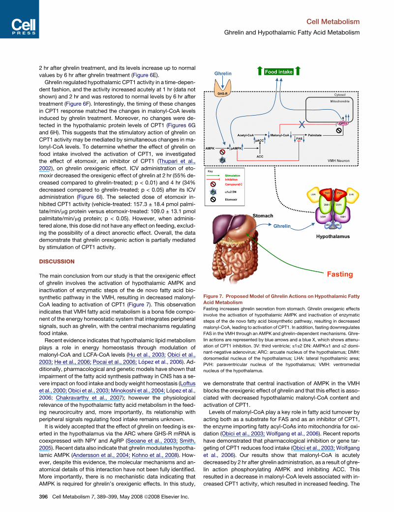
DISCUSSION
The main conclusion from our study is that the orexigenic effect
of ghrelin involves the activation of hypothalamic AMPK and
inactivation of enzymatic steps of the de novo fatty acid bio-
synthetic pathway in the VMH, resulting in decreased malonyl-
CoA leading to activation of CPT1 (Figure 7). This observation
indicates that VMH fatty acid metabolism is a bona fide compo-
nent of the energy homeostatic system that integrates peripheral
signals, such as ghrelin, with the central mechanisms regulating
food intake.
Recent evidence indicates that hypothalamic lipid metabolism
plays a role in energy homeostasis through modulation of
malonyl-CoA and LCFA-CoA levels (Hu et al., 2003; Obici et al.,
2003; He et al., 2006; Pocai et al., 2006; Lopez et al., 2006). Ad-
ditionally, pharmacological and genetic models have shown that
impairment of the fatty acid synthesis pathway in CNS has a se-
vere impact on food intake and body weight homeostasis (Loftus
et al., 2000; Obici et al., 2003; Minokoshi et al., 2004; Lopez et al.,
2006; Chakravarthy et al., 2007); however the physiological
relevance of the hypothalamic fatty acid metabolism in the feed-
ing neurocircuitry and, more importantly, its relationship with
peripheral signals regulating food intake remains unknown.
It is widely accepted that the effect of ghrelin on feeding is ex-
erted in the hypothalamus via the ARC where GHS-R mRNA is
coexpressed with NPY and AgRP (Seoane et al., 2003; Smith,
2005). Recent data also indicate that ghrelin modulates hypotha-
lamic AMPK (Andersson et al., 2004; Kohno et al., 2008). How-
ever, despite this evidence, the molecular mechanisms and an-
atomical details of this interaction have not been fully identified.
More importantly, there is no mechanistic data indicating that
AMPK is required for ghrelin’s orexigenic effects. In this study,
396 Cell Metabolism 7, 389–399, May 2008 ª2008 Elsevier Inc.
we demonstrate that central inactivation of AMPK in the VMH
blocks the orexigenic effect of ghrelin and that this effect is asso-
ciated with decreased hypothalamic malonyl-CoA content and
activation of CPT1.
Levels of malonyl-CoA play a key role in fatty acid turnover by
acting both as a substrate for FAS and as an inhibitor of CPT1,
the enzyme importing fatty acyl-CoAs into mitochondria for oxi-
dation (Obici et al., 2003; Wolfgang et al., 2006). Recent reports
have demonstrated that pharmacological inhibition or gene tar-
geting of CPT1 reduces food intake (Obici et al., 2003; Wolfgang
et al., 2006). Our results show that malonyl-CoA is acutely
decreased by 2 hr after ghrelin administration, as a result of ghre-
lin action phosphorylating AMPK and inhibiting ACC. This
resulted in a decrease in malonyl-CoA levels associated with in-
creased CPT1 activity, which resulted in increased feeding. The
Figure 7. Proposed Model of Ghrelin Actions on Hypothalamic Fatty
Acid Metabolism
Fasting increases ghrelin secretion from stomach. Ghrelin orexigenic effects
involve the activation of hypothalamic AMPK and inactivation of enzymatic
steps of the de novo fatty acid biosynthetic pathway, resulting in decreased
malonyl-CoA, leading to activation of CPT1. In addition, fasting downregulates
FAS in the VMH through an AMPK and ghrelin-dependent mechanisms. Ghre-
lin actions are represented by blue arrows and a blue X, which shows attenu-
ation of CPT1 inhibition. 3V: third ventricle; a1a2 DN: AMPKa1 and a2 domi-
nant-negative adenovirus; ARC: arcuate nucleus of the hypothalamus; DMH:
dorsomedial nucleus of the hypothalamus; LHA: lateral hypothalamic area;
PVH: paraventricular nucleus of the hypothalamus; VMH: ventromedial
nucleus of the hypothalamus.

Cell Metabolism
Ghrelin and Hypothalamic Fatty Acid Metabolism
physiological significance of ghrelin’s rapid and transient effect
on depleting malonyl-CoA levels and activating CPT1 is intrigu-
ing. However, given the potential role of peripheral (stomach-de-
rived) ghrelin in promoting meal initiation (Cummings et al., 2001;
Drazen et al., 2006), it is tempting to speculate a potential role for
the fasting-induced ghrelin-AMPK-malonyl-CPT1 hypothalamic
system as an acute signal, triggering the beginning of feeding af-
ter a period of fasting coincident with increased ghrelin levels.
Further work using a genetic ablation strategy, such as the
CPT1c KO mice (Wolfgang et al., 2006), instead of CPT1’s phar-
macological inhibition with etomoxir, might help to clarify this hy-
pothesis. One interesting point is the elucidation of the precise
roles of both hypothalamic ACC isoforms (ACCa and ACCb). De-
spite ACCb being the isoform mainly involved in the regulation of
fatty acid oxidation, at least in nonlipogenic tissues (Wolfgang
and Lane, 2006), our data suggest that in the hypothalamus,
ACCa may modulate both fatty acid synthesis and oxidation.
Another intriguing question is the output system for this mecha-
nism. Current data have demonstrated that the sympathetic
nervous system is implicated in the transmission of the ‘‘malonyl-
CoA signal’’ from the brain to skeletal muscle (Cha et al., 2005)
and also in the modulation of central ghrelin actions on white ad-
ipose tissue (WAT) lipid metabolism (Theander-Carrillo et al.,
2006). On the basis of these data, a similar mechanism may be
involved in the output response of the ghrelin-AMPK-malonyl-
CPT1 axis from the hypothalamus to peripheral organs, such
as the pancreas, liver, and WAT.
In peripheral tissues, such as liver, muscle, and WAT, the
physiological response to starvation involves a coordinated
downregulation of the lipogenic pathway with simultaneous ac-
tivation of the oxidative pathway (Brown and Goldstein, 1997).
Lipids are an important component of the CNS composition;
the brain relies heavily on lipids to maintain its metabolic and
cell membrane homeostasis (Goldberg, 2003). Thus, it would
certainly be unexpected to find that the CNS lipid metabolism
may be subjected to major changes linked to daily nutritional
changes. In fact, it is counterintuitive that physiological periods
of food deprivation may compromise lipid metabolism in the en-
tire CNS. However, it cannot be excluded that the mechanisms
involved in lipid biosynthesis may, in selected nuclei, be used
as a sensing mechanism to maintain energy balance. Our data
provide evidence that the effect of starvation on hypothalamic
fatty acid metabolism is different than in peripheral organs. For
instance, despite the activity of AMPK and ACC being increased
in whole hypothalamus during fasting (Minokoshi et al., 2004),
levels of FAS mRNA and protein levels are maintained not only
in most hypothalamic nuclei (e.g., ARC or PVH) but also in the
rest of the CNS areas we studied (cortex, hippocampus, thala-
mus, etc.). However, we have identified the VMH as the hypotha-
lamic nuclei where FAS is specifically downregulated in re-
sponse to fasting. Interestingly, our results also indicate that
the energy homeostasis-related signal sensed to regulate fatty
acid metabolism in the hypothalamus and feeding behavior
may not necessarily be a nutrient, but the stomach-secreted hor-
mone ghrelin, acting through hypothalamic AMPK. There is evi-
dence that AMPK also controls the activity of ACC and MCD and
the expression of FAS mRNA in peripheral tissues, such as liver
and adipose tissue (Saha et al., 2000; Zhou et al., 2001). How-
ever, our data, by using both pharmacological and adenovirus-
based approaches, show that hypothalamic FAS expression is
a downstream target of AMPK in the hypothalamus.
The physiological significance of ghrelin effects on FAS-ex-
pressing neurons in the VMH is intriguing. The decrease in ma-
lonyl-CoA levels in response to ghrelin is transient so that by 6 hr
after ghrelin treatment, malonyl-CoA is restored to its normal
levels. Even though the mechanism by which malonyl-CoA levels
are restored may be related to the termination of ghrelin stimula-
tion of AMPK, our data also suggest that a direct effect of ghrelin
decreasing FAS expression and activity in the VMH may also
contribute to this process. Thus, we propose that ghrelin-in-
duced and fasting-induced decreases in FAS levels in the VMH
may be a physiological adaptive mechanism that helps to pre-
vent malonyl-CoA from decreasing to deleteriously low levels
in the hypothalamus. The observation that the decrease of FAS
in response to fasting and ghrelin administration is limited to
the VMH is particularly interesting. The VMH is well placed to in-
tegrate peripherally supplied signals that regulate food intake
with the input from other neighboring nuclei and the brainstem,
connected through specific neuronal projections (Tong et al.,
2007). Thus, our data provide evidence indicating that, besides
its role of maintaining lipid biosynthesis, FAS in the VMH may
also have an additional role as a sensor of the nutritional state.
In summary, here we show that the orexigenic action of ghrelin
depends on the specific regulation of hypothalamic fatty acid
metabolism mediated by AMPK, malonyl-CoA, and CPT1 activ-
ity (Figure 7). Our data also identify the fatty acid biosynthetic
pathway in the VMH as a potentially important physiological
mediator of feeding behavior of relevance for the understanding
and treatment of obesity.
EXPERIMENTAL PROCEDURES
Animals
We used adult male Sprague-Dawley rats and lean and obese Zucker rats,
adult male GHS-R KO mice, ob/ob mice, and their wild-type (WT) littermates.
The experiments were performed in agreement with the International Law on
Animal Experimentation. We used 8–16 animals per group, apart from immu-
nohistochemistry where we used 4–6.
Treatments
Fed and fasted rats received two IP administrations of leptin (200 mg). Animals
were sacrificed 12 hr after the second injection (9:00 am, day 1), exactly 24 hr
after fasting started. Chronic (ICV) cannulae were stereotaxically implanted as
previously described (Lopez et al., 2006). Rats received an ICV administration
of ghrelin (0.25, 2, or 5 mg; Bachem), ghrelin antagonist BIM-28163 (10 nmol;
Ipsen) (Halem et al., 2005), AgRP (5 mg) (Tang-Christensen et al., 2004), NPY
(10 mg) (Tritos et al., 1998), etomoxir (10 mg), compound C (10 mg), or vehicle
(saline or DMSO). For ghrelin IP treatments rats received a single administra-
tion of ghrelin (10 nmol = 33.7 mg) (Wren et al., 2000; Andersson et al., 2004)
and were killed after 6 hr. All drugs (apart from ghrelin and BIM-28163) were
from Sigma.
Stereotaxic Microinjection of Adenoviral Expression Vectors
Rats were placed in a stereotaxic frame (David Kopf Instruments) under
ketamine/xylazine anesthesia. The VMH were targeted bilaterally using a
25-gauge needle (Hamilton) connected to a Hamilton 1 ml syringe. The injection
was directed to stereotaxic coordinates 2.3/3.3 mm posterior to the bregma
(two injections were performed in each VMH), ± 0.6 mm lateral to midline
and 10.2 mm below the surface of the skull (Figure 4A). Adenovirus vectors
were one of the following: (1) Cre-GFP (1 3 1010 pfu/ml) or (2) a1a2 DN (mix
1:1); they were delivered at a rate of 200 nl/min for 5 min (1 ml/injection site),
and the entire injector system was left in place for an additional 5 min after
Cell Metabolism 7, 389–399, May 2008 ª2008 Elsevier Inc. 397

Cell Metabolism
Ghrelin and Hypothalamic Fatty Acid Metabolism
the injections were completed. After the procedure was complete, chronic
(ICV) cannulae were stereotaxically implanted as previously described (Lopez
et al., 2006). Six days after adenovirus injection rats were ICV-treated with
ghrelin for 6 hr. After that time their brains were dissected and processed by
GPF-immunohistochemistry and FAS in situ hybridization or western blot.
We used 10 animals per group.
Blood Biochemistry
Plasma, leptin, and ghrelin levels were measured using rat ELISA kits (Crystal
Chem Inc).
In Situ Hybridization
Coronal brain sections (16 mm) were probed with specific antisense oligos (Ta-
ble S2) as previously described (Lopez et al., 2006) (see Supplemental Data).
Immunohistochemistry
Diaminobenzidine (DAB) immunohistochemistry, immunofluorescence, and
double labeling were performed as described (Lopez et al., 2006) using a rabbit
anti-GFP, anti-FAS (Abcam), and/or anti-GHS-R antibody and/or an anti-
BDNF antibody (Santa Cruz) (see Supplemental Data).
Western Blotting
Hypothalamic total protein lysates were subjected to SDS-PAGE, electro-
transferred on a PVDF membrane, and probed with the following antibodies:
ACC, pACC-Ser79, AMPKa1, AMPKa2, and pIRF-3-Ser396 (Upstate);
CaMKKa, CaMKKb, and pGP (Santa Cruz); CPT1c (Phoenix); FAS (BD),
pAMPKa-Thr172, and pSRC-Tyr527 (Cell Signaling); b-Actin and GFP (Abcam)
as previously described (Lopez et al., 2006).
Enzymatic Assays
FAS activity was performed as previously described (Lelliott et al., 2005). CPT1
activity was measured using the method of Bieber et al. (1972).
Malonyl-CoA Assay
Malonyl-CoA was measured as previously described (Lelliott et al., 2005;
Lopez et al., 2006).
Statistical Analysis
Data are expressed as mean ± SEM. Statistic significance was determined by
Student’s t test or ANOVA and post-hoc Bonferroni test. p < 0.05 was consid-
ered significant.
SUPPLEMENTAL DATA
Supplemental Data include Supplemental Experimental Procedures, two
tables, and three figures and can be found with this article online at http://
www.cellmetabolism.org/cgi/content/full/7/5/389/DC1/.
ACKNOWLEDGMENTS
We thank Dr. Ruben Nogueiras (University of Santiago de Compostela and
University of Cincinnati) for his suggestions and criticisms of this paper and
Victoria Bonnici for editing the manuscript. This work has been supported by
grants from Medical Research Council (A.V.-P. and D.C.), Welcome Trust
(A.V.-P.), Xunta de Galicia (C.D.: PGIDIT; M.L.: GRC2006/66; and R.G.:
PGIDIT07PXIB208112PR), Fondo Investigationes Sanitarias (M.L.: PI061700),
Ministerio de Educacion y Ciencia (C.D.: BFU2005), European Union (C.D.
and M.T.: LSHM-CT-2003-503041: Diabesity http://www.eurodiabesity.org;
M.L.: Marie Curie Program QLK6-CT-2002-51671; and A.V.-P.: LSHM-CT-
2005–018734: Hepadip, http://www.hepadip.org), Mutua Madrilena (C.D.
and M.L.), United States Public Health Service (A.K.S.: DK-19514 and
DK-67509).
Received: September 23, 2007
Revised: January 30, 2008
Accepted: March 11, 2008
Published: May 6, 2008
398 Cell Metabolism 7, 389–399, May 2008 ª2008 Elsevier Inc.
REFERENCES
Andersson, U., Filipsson, K., Abbott, C.R., Woods, A., Smith, K., Bloom, S.R.,
Carling, D., and Small, C.J. (2004). AMP-activated protein kinase plays a role in
the control of food intake. J. Biol. Chem. 279, 12005–12008.
Bain, J., Plater, L., Elliott, M., Shpiro, N., Hastie, C.J., McLauchlan, H.,
Klevernic, I., Arthur, J.S., Alessi, D.R., and Cohen, P. (2007). The selectivity
of protein kinase inhibitors: a further update. Biochem. J. 408, 297–315.
Bieber, L.L., Abraham, T., and Helmrath, T. (1972). A rapid spectrophotometric
assay for carnitine palmitoyltransferase. Anal. Biochem. 50, 509–518.
Brown, M.S., and Goldstein, J.L. (1997). The SREBP pathway: regulation of
cholesterol metabolism by proteolysis of a membrane-bound transcription
factor. Cell 89, 331–340.
Cha, S.H., Hu, Z., Chohnan, S., and Lane, M.D. (2005). Inhibition of hypotha-
lamic fatty acid synthase triggers rapid activation of fatty acid oxidation in skel-
etal muscle. Proc. Natl. Acad. Sci. USA 102, 14557–14562.
Chakravarthy, M.V., Zhu, Y., Lopez, M., Yin, L., Wozniak, D.W., Coleman, T.,
Hu, Z., Wolfgang, M., Vidal-Puig, A., Lane, M.D., and Semenkovich, C.F.
(2007). Brain fatty acid synthase activates PPAR-alpha to maintain energy
homeostasis. J. Clin. Invest. 117, 2539–2552.
Cummings, D.E., Purnell, J.Q., Frayo, R.S., Schmidova, K., Wisse, B.E., and
Weigle, D.S. (2001). A preprandial rise in plasma ghrelin levels suggests
a role in meal initiation in humans. Diabetes 50, 1714–1719.
Cummings, D.E., Clement, K., Purnell, J.Q., Vaisse, C., Foster, K.E., Frayo,
R.S., Schwartz, M.W., Basdevant, A., and Weigle, D.S. (2002). Elevated
plasma ghrelin levels in Prader Willi syndrome. Nat. Med. 8, 643–644.
Drazen, D.L., Vahl, T.P., D’Alessio, D.A., Seeley, R.J., and Woods, S.C. (2006).
Effects of a fixed meal pattern on ghrelin secretion: evidence for a learned
response independent of nutrient status. Endocrinology 147, 23–30.
Flier, J.S. (2004). Obesity wars: molecular progress confronts an expanding
epidemic. Cell 116, 337–350.
Foster-Schubert, K.E., and Cummings, D.E. (2006). Emerging therapeutic
strategies for obesity. Endocr. Rev. 27, 779–793.
Goldberg, J.L. (2003). How does an axon grow? Genes Dev. 17, 941–958.
Halem, H.A., Taylor, J.E., Dong, J.Z., Shen, Y., Datta, R., Abizaid, A., Diano, S.,
Horvath, T.L., and Culler, M.D. (2005). A novel growth hormone secretagogue-
1a receptor antagonist that blocks ghrelin-induced growth hormone secretion
but induces increased body weight gain. Neuroendocrinology 81, 339–349.
Hawley, S.A., Pan, D.A., Mustard, K.J., Ross, L., Bain, J., Edelman, A.M., Fren-
guelli, B.G., and Hardie, D.G. (2005). Calmodulin-dependent protein kinase ki-
nase-beta is an alternative upstream kinase for AMP-activated protein kinase.
Cell Metab. 2, 9–19.
He, W., Lam, T.K., Obici, S., and Rossetti, L. (2006). Molecular disruption of hy-
pothalamic nutrient sensing induces obesity. Nat. Neurosci. 9, 227–233.
Hu, Z., Cha, S.H., Chohnan, S., and Lane, M.D. (2003). Hypothalamic malonyl-
CoA as a mediator of feeding behavior. Proc. Natl. Acad. Sci. USA 100, 12624–
12629.
Kahn, B.B., Alquier, T., Carling, D., and Hardie, D.G. (2005). AMP-activated
protein kinase: Ancient energy gauge provides clues to modern understanding
of metabolism. Cell Metab. 1, 15–25.
Kohno, D., Sone, H., Minokoshi, Y., and Yada, T. (2008). Ghrelin raises [Ca2+]i
via AMPK in hypothalamic arcuate nucleus NPY neurons. Biochem. Biophys.
Res. Commun. 366, 388–392.
Lelliott, C.J., Lopez, M., Curtis, R.K., Parker, N., Laudes, M., Yeo, G., Jimenez-
Linan, M., Grosse, J., Saha, A.K., Wiggins, D., et al. (2005). Transcript and me-
tabolite analysis of the effects of tamoxifen in rat liver reveals inhibition of fatty
acid synthesis in the presence of hepatic steatosis. FASEB J. 19, 1108–1119.
Loftus, T.M., Jaworsky, D.E., Frehywot, G.L., Townsend, C.A., Ronnett, G.V.,
Lane, M.D., and Kuhajda, F.P. (2000). Reduced food intake and body weight in
mice treated with fatty acid synthase inhibitors. Science 288, 2379–2381.
Lopez, M., Lelliott, C.J., Tovar, S., Kimber, W., Gallego, R., Virtue, S., Blount,
M., Vazquez, M.J., Finer, N., Powles, T., et al. (2006). Tamoxifen-induced an-
orexia is associated with fatty acid synthase inhibition in the ventromedial

Cell Metabolism
Ghrelin and Hypothalamic Fatty Acid Metabolism
nucleus of the hypothalamus and accumulation of malonyl-CoA. Diabetes 55,
1327–1336.
Minokoshi, Y., Alquier, T., Furukawa, N., Kim, Y.B., Lee, A., Xue, B., Mu, J.,
Foufelle, F., Ferre, P., Birnbaum, M.J., et al. (2004). AMP-kinase regulates
food intake by responding to hormonal and nutrient signals in the hypothala-
mus. Nature 428, 569–574.
Morton, G.J., Cummings, D.E., Baskin, D.G., Barsh, G.S., and Schwartz, M.W.
(2006). Central nervous system control of food intake and body weight. Nature
443, 289–295.
Obici, S., Feng, Z., Arduini, A., Conti, R., and Rossetti, L. (2003). Inhibition of
hypothalamic carnitine palmitoyltransferase-1 decreases food intake and
glucose production. Nat. Med. 9, 756–761.
Pagotto, U., Marsicano, G., Cota, D., Lutz, B., and Pasquali, R. (2006). The
emerging role of the endocannabinoid system in endocrine regulation and en-
ergy balance. Endocr. Rev. 27, 73–100.
Pinto, S., Roseberry, A.G., Liu, H., Diano, S., Shanabrough, M., Cai, X., Fried-
man, J.M., and Horvath, T.L. (2004). Rapid rewiring of arcuate nucleus feeding
circuits by leptin. Science 304, 110–115.
Plum, L., Belgardt, B.F., and Bruning, J.C. (2006). Central insulin action in en-
ergy and glucose homeostasis. J. Clin. Invest. 116, 1761–1766.
Pocai, A., Lam, T.K., Obici, S., Gutierrez-Juarez, R., Muse, E.D., Arduini, A.,
and Rossetti, L. (2006). Restoration of hypothalamic lipid sensing normalizes
energy and glucose homeostasis in overfed rats. J. Clin. Invest. 116, 1081–
1091.
Saha, A.K., Schwarsin, A.J., Roduit, R., Masse, F., Kaushik, V., Tornheim, K.,
Prentki, M., and Ruderman, N.B. (2000). Activation of malonyl-CoA decarbox-
ylase in rat skeletal muscle by contraction and the AMP-activated protein ki-
nase activator 5-aminoimidazole-4-carboxamide-1-beta -D-ribofuranoside.
J. Biol. Chem. 275, 24279–24283.
Seoane, L.M., Lopez, M., Tovar, S., Casanueva, F., Senarıs, R., and Dieguez,
C. (2003). Agouti-related peptide, neuropeptide Y, and somatostatin-produc-
ing neurons are targets for ghrelin actions in the rat hypothalamus. Endocrinol-
ogy 144, 544–551.
Smith, R.G. (2005). Development of growth hormone secretagogues. Endocr.
Rev. 26, 346–360.
Tang-Christensen, M., Vrang, N., Ortmann, S., Bidlingmaier, M., Horvath, T.,
and Tschop, M. (2004). Central administration of Ghrelin and Agouti-related
Protein (AGRP (83–132)) increases food intake and decreases spontaneous
locomotor activity in rats. Endocrinology 145, 4645–4652.
Theander-Carrillo, C., Wiedmer, P., Cettour-Rose, P., Nogueiras, R., Perez-
Tilve, D., Pfluger, P., Castaneda, T.R., Muzzin, P., Schurmann, A., Szanto, I.,
et al. (2006). Ghrelin action in the brain controls adipocyte metabolism. J.
Clin. Invest. 116, 1983–1993.
Thupari, J.N., Landree, L.E., Ronnett, G.V., and Kuhajda, F.P. (2002). C75 in-
creases peripheral energy utilization and fatty acid oxidation in diet-induced
obesity. Proc. Natl. Acad. Sci. USA 99, 9498–9502.
Tong, Q., Ye, C., McCrimmon, R.J., Dhillon, H., Choi, B., Kramer, M.D., Yu, J.,
Yang, Z., Christiansen, L.M., Lee, C.E., et al. (2007). Synaptic glutamate re-
lease by ventromedial hypothalamic neurons is part of the neurocircuitry that
prevents hypoglycemia. Cell Metab. 5, 383–393.
Tritos, N.A., Vicent, D., Gillette, J., Ludwig, D.S., Flier, E.S., and Maratos-Flier,
E. (1998). Functional interactions between melanin-concentrating hormone,
neuropeptide Y, and anorectic neuropeptides in the rat hypothalamus. Diabe-
tes 47, 1687–1692.
Tschop, M., Smiley, D.L., and Heiman, M.L. (2000). Ghrelin induces adiposity
in rodents. Nature 407, 908–913.
Wolfgang, M.J., and Lane, M.D. (2006). Control of energy homeostasis: role of
enzymes and intermediates of fatty acid metabolism in the central nervous
system. Annu. Rev. Nutr. 26, 23–44.
Wolfgang, M.J., Kurama, T., Dai, Y., Suwa, A., Asaumi, M., Matsumoto, S.,
Cha, S.H., Shimokawa, T., and Lane, M.D. (2006). The brain-specific carnitine
palmitoyltransferase-1c regulates energy homeostasis. Proc. Natl. Acad. Sci.
USA 103, 7282–7287.
Woods, A., Azzout-Marniche, D., Foretz, M., Stein, S.C., Lemarchand, P.,
Ferre, P., Foufelle, F., and Carling, D. (2000). Characterization of the role of
AMP-activated protein kinase in the regulation of glucose-activated gene ex-
pression using constitutively active and dominant negative forms of the kinase.
Mol. Cell. Biol. 20, 6704–6711.
Woods, A., Dickerson, K., Heath, R., Hong, S.P., Momcilovic, M., Johnstone,
S.R., Carlson, M., and Carling, D. (2005). Ca(2+)/calmodulin-dependent pro-
tein kinase kinase-beta acts upstream of AMP-activated protein kinase in
mammalian cells. Cell Metab. 2, 21–33.
Wortley, K.E., Del Rincon, J.P., Murray, J.D., Garcia, K., Iida, K., Thorner, M.O.,
and Sleeman, M.W. (2005). Absence of ghrelin protects against early-onset
obesity. J. Clin. Invest. 115, 3573–3578.
Wren, A.M., Small, C.J., Ward, H.L., Murphy, K.G., Dakin, C.L., Taheri, S., Ken-
nedy, A.R., Roberts, G.H., Morgan, D.G., Ghatei, M.A., and Bloom, S.R. (2000).
The novel hypothalamic peptide ghrelin stimulates food intake and growth hor-
mone secretion. Endocrinology 141, 4325–4328.
Zhou, G., Myers, R., Li, Y., Chen, Y., Shen, X., Fenyk-Melody, J., Wu, M.,
Ventre, J., Doebber, T., Fujii, N., et al. (2001). Role of AMP-activated protein
kinase in mechanism of metformin action. J. Clin. Invest. 108, 1167–1174.
Zigman, J.M., Nakano, Y., Coppari, R., Balthasar, N., Marcus, J.N., Lee, C.E.,
Jones, J.E., Deysher, A.E., Waxman, A.R., White, R.D., et al. (2005). Mice lack-
ing ghrelin receptors resist the development of diet-induced obesity. J. Clin.
Invest. 115, 3564–3572.
Cell Metabolism 7, 389–399, May 2008 ª2008 Elsevier Inc. 399
Copyright © 2022 FDOKUMEN