Hypothalamic-pituitary function in brain death: A review
30
First published online in Journal of Intensive Care Medicine, March 31 2014 DOI: 10.1177/0885066614527410 Hypothalamic-pituitary function in brain death: A review Michael Nair-Collins, PhD 1 ; Jesse Northrup, MA 2 ; James Olcese, PhD 3 1 Department of Behavioral Sciences and Social Medicine, Florida State University College of Medicine, Tallahassee, FL, USA; 2 Pacific Science Center, Seattle, WA, USA; 3 Department of Biomedical Sciences, Florida State University College of Medicine, Tallahassee, FL, USA Michael Nair-Collins, PhD Department of Behavioral Sciences and Social Medicine, Florida State University College of Medicine 1115 West Call Street Tallahassee, FL 32306 [email protected] Jesse Northrup, MA Pacific Science Center 200 Second Avenue N Seattle, WA 98109 James Olcese, PhD Department of Biomedical Sciences, Florida State University College of Medicine 1115 West call Street Tallahassee, FL 32306 [email protected] Corresponding author’s contact information: Michael Nair-Collins, PhD Department of Medical Humanities and Social Sciences Florida State University College of Medicine 1115 West Call Street Tallahassee FL 32306 [email protected] Phone: 850 645 6564 Fax: 850 645 1773 Running Title: Hypothalamic-pituitary function in brain death
Transcript of Hypothalamic-pituitary function in brain death: A review
First published online in Journal of Intensive Care Medicine, March 31 2014
DOI: 10.1177/0885066614527410
Hypothalamic-pituitary function in brain death: A review
Michael Nair-Collins, PhD1; Jesse Northrup, MA
2; James Olcese, PhD
3
1 Department of Behavioral Sciences and Social Medicine, Florida State University College of Medicine,
Tallahassee, FL, USA; 2 Pacific Science Center, Seattle, WA, USA;
3Department of Biomedical Sciences,
Florida State University College of Medicine, Tallahassee, FL, USA
Michael Nair-Collins, PhD
Department of Behavioral Sciences and Social Medicine, Florida State University College of Medicine
1115 West Call Street
Tallahassee, FL 32306
Jesse Northrup, MA
Pacific Science Center
200 Second Avenue N
Seattle, WA 98109
James Olcese, PhD
Department of Biomedical Sciences, Florida State University College of Medicine
1115 West call Street
Tallahassee, FL 32306
Corresponding author’s contact information:
Michael Nair-Collins, PhD
Department of Medical Humanities and Social Sciences
Florida State University College of Medicine
1115 West Call Street
Tallahassee FL 32306
Phone: 850 645 6564
Fax: 850 645 1773
Running Title: Hypothalamic-pituitary function in brain death
First published online in Journal of Intensive Care Medicine, March 31 2014
DOI: 10.1177/0885066614527410
Abstract
The Uniform Determination of Death Act (UDDA) states that an individual is dead when “all functions of
the entire brain” have ceased irreversibly. However, it has been questioned whether some functions of the
hypothalamus, particularly osmoregulation, can continue after the clinical diagnosis of brain death. In
order to learn whether parts of the hypothalamus can continue to function after the diagnosis of brain
death, we performed two separate systematic searches of the Medline database, corresponding to
functions of the posterior and anterior pituitary. No meta-analysis is possible due to non-uniformity in the
clinical literature. However, some modest generalizations can reasonably be drawn from a narrative
review and from anatomic considerations that explain why these findings should be expected. We found
evidence suggesting the preservation of hypothalamic function, including secretion of hypophysiotropic
hormones, responsiveness to anterior pituitary stimulation, and osmoregulation, in a substantial
proportion of patients declared dead by neurological criteria. We discuss several possible explanations
for these findings. We conclude by suggesting that additional clinical research with strict inclusion
criteria is necessary, and further that a more nuanced and forthright public dialogue is needed, particularly
since standard diagnostic practices and the UDDA may not be entirely in accord.
First published online in Journal of Intensive Care Medicine, March 31 2014
DOI: 10.1177/0885066614527410
Introduction
The concept of brain death (BD) is accepted nearly worldwide as equivalent to death,1 yet a number of
controversies persist, leading one scholar to name it both “well-settled yet still unresolved,”2 and
prompting the United States (US) President’s Council for Bioethics to revisit the concept in a new White
Paper.3 One issue that remains controversial is the relation of standard diagnostic tests to the statutory
definition of “death” as outlined in the Uniform Determination of Death Act (UDDA), which states: “An
individual who has suffered either (1) irreversible cessation of circulatory and respiratory functions, or (2)
irreversible cessation of all functions of the entire brain, including the brain stem, is dead.”4 For example,
the editors of Nature stated in a recent editorial:
The [UDDA] seems admirably straightforward… In practice, unfortunately, physicians know that
when they declare that someone on life support is dead, they are usually obeying the spirit, but
not the letter, of this law. And many are feeling increasingly uncomfortable about it. In
particular, they struggle with three of the law’s phrases: ‘irreversible,’ ‘all functions’ and ‘entire
brain,’ knowing that they cannot guarantee full compliance… The time has come for a serious
discussion on redrafting laws that push doctors towards a form of deceit.5 (p. 570)
Evidence suggests that neuroendocrine functions may be preserved in some patients after the declaration
of BD.6 However, there are no comprehensive reviews of the clinical literature on this issue. Our
purpose, therefore, is to provide such a review. Specifically we want to know: How often is BD
accompanied by complete hypothalamic-pituitary failure? While the term “brain death” is sometimes
used to mean irreversible cessation of all functions of the entire brain, this implies that continued
hypothalamic function in BD is a contradiction in terms. Nonetheless, since our purpose in this paper is
to investigate whether hypothalamic-pituitary function remains in some patients that are declared to be
brain dead, we follow the standard usage in the clinical literature and use “brain death” to mean lack of
First published online in Journal of Intensive Care Medicine, March 31 2014
DOI: 10.1177/0885066614527410
clinical functions of the brain, as judged by apnea, brainstem areflexia, and cerebral unresponsiveness,
coupled with a known, irreversible cause of coma. We considered the functions of the posterior and
anterior pituitary separately, as befitting their distinct anatomic characteristics and embryologic origins.
Hypothalamic-pituitary function
Magnocellular neurons of the supraoptic and paraventricular nuclei of the hypothalamus secrete
the hormone arginine vasopressin (AVP) into the peripheral bloodstream via the posterior pituitary in
response to an increase in plasma osmotic pressure or hypovolemia. The half-life of AVP is relatively
short, at about 15-18 minutes, and as little as a 1% change in plasma osmolarity can induce a change in
AVP secretion. This rapid and sensitive physiologic response is part of a negative feedback system that
maintains plasma osmolarity within a narrow, roughly 3% window by controlling diuresis via the
kidneys.7 In the absence of AVP or the kidneys’ ability to respond to it, diabetes insipidus (DI) develops,
characterized by the excretion of large amounts of dilute urine, often accompanied by hypernatremia.
The clinical condition may result from primary hypothalamic dysfunction (central DI), or from the failure
of the renal collecting ducts and distal tubules to respond to AVP (nephrogenic DI).
By contrast, the hypothalamus controls the anterior pituitary indirectly by secreting
hypophysiotropic hormones into the local portal circulation. The functions of the anterior pituitary
hormones, their target organs, and the peripheral hormones they control are complex, diffuse, subject to
multiple interconnected feedback loops, and affect metabolic functions throughout the body.
We operationalized the concept of preserved hypothalamic-posterior pituitary function as the
absence of DI. In contrast with the relatively straightforward role of AVP in anti-diuresis, an inference
from one or a few physiologic variables to a conclusion regarding the preservation of hypothalamic-
anterior pituitary function would be less secure. Therefore for the functions of the anterior pituitary, the
main variables of interest are direct measurements of selected hormones or responses to anterior pituitary
stimulation tests. We used these concepts to target two literature reviews.
First published online in Journal of Intensive Care Medicine, March 31 2014
DOI: 10.1177/0885066614527410
Methods
The MEDLINE database was accessed through PubMed in September 2013. Results were
restricted to English-language articles without date restriction. For the first search we utilized MeSH
terms “brain death” in conjunction with “diabetes insipidus” and with “posterior pituitary”, yielding 26
and 1 result(s), respectively. Text words “brain death,” “diabetes insipidus,” and “posterior pituitary”
were combined in the same pattern to reveal 105 and 18 results. Reference lists were reviewed, and we
consulted organ donor management guidelines and bioethical reviews to find additional studies.
Articles were included if they reported the results of an original clinical research study that
provides evidence regarding DI or quantitative measurements of AVP in BD or brainstem death (BSD), in
a manner that allows discernment of how many individual patients had DI or not. In accordance with
common practice, we collapse BD and BSD, the irreversible cessation of all functions of the brainstem (a
legal standard for death in the United Kingdom), for the following reason. In practice, the core clinical
diagnostic tests for BD and BSD are the same: brainstem areflexia, apnea, and cerebral unresponsiveness,
coupled with a known, irreversible cause of coma.
For the second search we utilized MeSH terms “brain death” and “anterior pituitary”, yielding 24
results. Text terms “brain death” in conjunction with “endocrine failure”, and with “anterior pituitary”,
revealed 39 and 183 results respectively. Ancillary sources, as mentioned above, were consulted to find
additional citations. Articles were included if they reported the results of an original clinical research
study that provided quantitative measurements of selected hormones (discussed below) or responses to
anterior pituitary stimulation tests, in BD or BSD, reported at the individual level.
There is substantial variability in the clinical literature: The populations are not uniform in terms
of age, etiology of BD, and the use of confirmatory diagnostic tests. The variables measured and study
methodologies are not uniform, laboratory cutoff values are not standardized, and the key diagnostic
category of DI is not uniformly applied. These limitations preclude the possibility of a systematic review
First published online in Journal of Intensive Care Medicine, March 31 2014
DOI: 10.1177/0885066614527410
with meta-analysis. Instead, we provide a compilation of all studies that meet the criteria specified above,
in the context of a narrative review.
Results
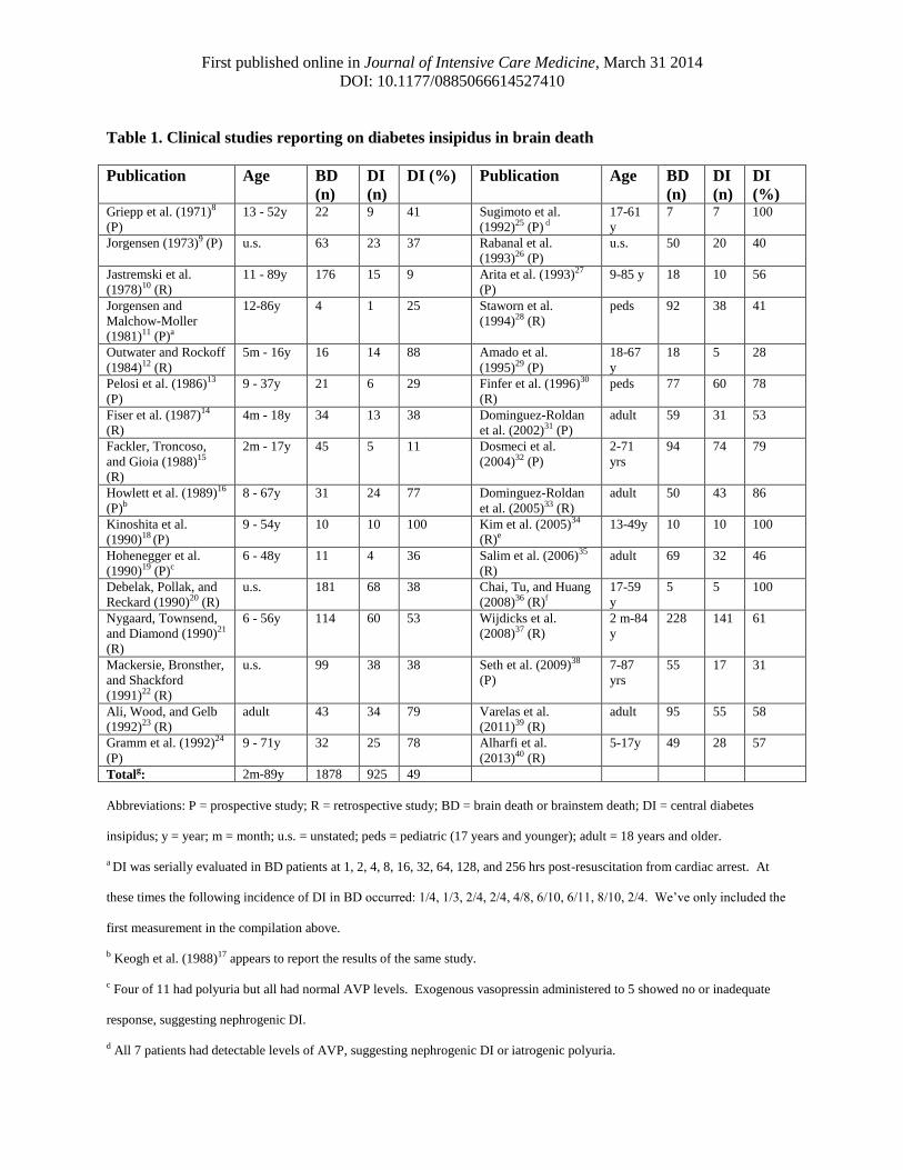
We found a total of 32 studies that meet criteria for the first search (see Table 1).8-40
Together,
the 32 studies report on 1878 patients with BD or BSD, ranging in age from 2 months to 89 years, of
which 925 (49%) were reported to have DI. The 6 studies that report exclusively on adult patients
collectively report that 200 of 334 (60%) had DI23,29,31,33,35,39
; for the pediatric population, 5 studies
collectively report that 145 of 279 (52%) had DI.12,15,28,30,40
The evidence regarding anterior pituitary function is not straightforward (see Tables 2-5). The
hypophysiotropic hormones luteinizing hormone releasing hormone (LHRH), corticotropin-releasing
hormone (CRH), and growth hormone releasing hormone (GHRH) are generally detectable in peripheral
circulation.25,27,41
The anterior pituitary hormones thyroid stimulating hormone (TSH),16,27,42-51
adrenocorticotropic hormone (ACTH),27,41,51-53
and human growth hormone (GH)16,27,41,51,53,54
are also
usually detectable, mostly within a normal range. Anterior pituitary responsiveness to stimulation is
variable. Some studies document no response to thyrotropin releasing hormone (TRH),54,55
insulin-
induced hypoglycemia,41
or arginine infusion,41
whereas others document responses to each of these
anterior pituitary stimuli as well as responses to LHRH and GHRH with appropriate hormonal
increases.25,27,42
Thyroid function appears to be impaired, though the pattern of thyroid hormones (low
triiodothyronine (T3) and to a lesser extent, low thyroxine (T4) with normal or high reverse T3 and
normal TSH) is more consistent with euthyroid sick syndrome rather than central neuroendocrine
failure16,24,46,54,56,57
(See tables 2-5 for further detail.). Adrenal function is difficult to interpret: Cortisol
levels show wide variability,16,24,27,29,46,48,56,58-60
although ACTH levels are generally preserved as
mentioned above. Diurnal variation of cortisol appears universally lost.16,27,44,61
First published online in Journal of Intensive Care Medicine, March 31 2014
DOI: 10.1177/0885066614527410
Discussion
Interpretation of results
These data are subject to limitations, and any quantitative estimate of incidence of preserved
hypothalamic function in the BD population in general should be taken as having unknown confidence.
However, this does not imply that no conclusions can be drawn.
The magnocellular neurons of the hypothalamus are osmoreceptors, directly responsive to the
osmotic pressure of their extracellular environment. While osmotically-induced depolarizations increase
the likelihood that an action potential will fire, they are not typically large enough to induce the cell to
reach its threshold voltage.62
Normal osmoregulation requires excitatory glutamatergic input from
circumventricular areas, particularly the organum vasculosum of the lamina terminalis, creating an
additive effect which allows the membrane potential to reach spike threshold, generating an action
potential down the axon, and hence inducing the secretion of AVP into the bloodstream.62
The half-life of AVP is about 15-18 minutes, which allows for a rapid physiologic response to
osmotic changes. As little as a 1% change in plasma osmolarity induces a change in AVP release, and
small changes in AVP concentration in plasma (normal range between approximately 0.5 and 5-6 pg/mL)
result in changes throughout the full range of urine osmolarity and urine volume.7 Thus, the
osmoregulation system is sensitive and rapid, with small changes in plasma osmolarity resulting in rapid
changes in AVP secretion from the hypothalamic-pituitary complex at a rate and level sufficient for
minute-to-minute changes in urine osmolarity and volume. In the absence of AVP, the distal tubules and
collecting ducts of the kidney’s nephrons are nearly impermeable to water, which prevents the
reabsorption of water back into the bloodstream, resulting in the production and release of large quantities
of dilute urine. The simplest explanation for the observation of normuria in BD patients is that the
hypothalamic osmoregulation system continues to function, in at least some patients.
First published online in Journal of Intensive Care Medicine, March 31 2014
DOI: 10.1177/0885066614527410
It is possible that an extracranial tumor could secrete AVP into the bloodstream, however, this
would not be subject to inhibitory feedback, therefore the patient would likely be oliguric or anuric. More
importantly, this is an unusual circumstance and would not explain the common finding of the absence of
DI in about half of the patients reported above. Another possibility is that AVP passively leaks from the
axonal terminals of non-viable hypothalamic cells whose perikarya have been destroyed (i.e., posterior
pituitary washout). This source of AVP is also not subject to feedback so the patient would more likely
exhibit anuria or oliguria, followed by polyuria minutes after the AVP stores are depleted. However,
depending on the rate at which AVP passively leaks, it seems plausible that passive leakage of AVP could
mimic osmoregulation at least while AVP stores last, and so this may account for some of the cases of
lack of apparent DI.
A third possibility is that glomerular filtration rate (GFR) is significantly decreased. This would
result in the diminution of the amount of fluid delivered to the distal tubule, and hence would limit renal
water excretion and prevent polyuria.63
Theoretically, this phenomenon may occur even in the absence of
AVP, therefore, the absence of polyuria does not necessarily imply function of the hypothalamic
osmoregulation system. Although this is a possibility, it cannot account for all of the cases of lack of DI
in BD patients. Fiser et al,14
for example, report no significant difference in creatinine levels between BD
patients with DI (n=13) and BD patients without DI (n=21), suggesting that decreased GFR cannot
account for the absence of DI in these patients. Finfer et al.30
reported mean maximum creatinine level of
1.02 +/- 0.7 mg/dl in their sample. Of the 17/77 without DI, decreased GFR cannot account for the
absence of polyuria. Furthermore, several studies directly measured plasma AVP, finding AVP within
the normal range for osmoregulation in some BD patients.19,24,25,27
For example, one study followed a rigorous protocol, correlating plasma AVP with plasma
osmolarity.27
Using standardized criteria for DI,64
these authors found that 10/18 patients in their sample
had DI, one had the syndrome of inappropriate anti-diuretic hormone, and the remaining 7/18 (39%)
demonstrated normal plasma osmolarity and normal AVP. It should be noted that it is unlikely that
First published online in Journal of Intensive Care Medicine, March 31 2014
DOI: 10.1177/0885066614527410
passive leakage, not subject to any feedback, would account for the normal plasma osmolarity, given the
negative feedback needed to maintain a delicate balance of plasma osmolarity within a narrow, roughly
3% window.7
Furthermore, anatomical considerations suggest that preserved hypothalamic osmoregulation may
be expected in BD. The inferior hypophyseal arteries branch off the extradural segments of the internal
carotids and thus are protected from increased intracranial pressure, and these arteries supply the posterior
pituitary, the hypophysial stalk, and parts of the hypothalamus including the median eminence.65
Pathology studies support this explanation. Walker and colleagues66
reported finding some intact regions
of the hypothalamus with relatively well preserved neurons, intermingled with neurons with lytic
changes, in patients with coma d pass (a diagnostic precursor to BD: all patients were comatose, apneic,
and with isoelectric encephalograms). In a more recent pathology study of 41 patients with BD,67
moderate to severe ischemic changes in brainstem and higher brain structures were found in only about
34-68% of cases (with variability in different brain areas). Of these 41 patients, 16 of the pituitary glands
were examined, with 55% of those showing mild or absent ischemic changes.
Polyuria alone is not sufficient evidence of dysfunctional hypothalamic control of plasma
osmolarity, yet the majority of studies did not rule out confounding factors nor corroborate urine output
with laboratory values. There are a number of iatrogenic reasons for polyuria, including diuretics or fluid
resuscitation. To illustrate, Fiser and colleagues14
report that 26/34 patients were polyuric, yet only 13
had DI, while the remaining 13 did not have hypernatremia, serum hyperosmolarity, or urine
hyposmolarity. Polyuria was determined to be due to administration of furosemide, mannitol, or contrast
solution for computed tomography. In two studies12,26
DI was found to spontaneously resolve in some
patients without administration of exogenous vasopressin, suggesting reperfusion or a decrease in edema,
or both, to the hypothalamic-pituitary complex after an initial insult that rendered the area temporarily
nonfunctional.
First published online in Journal of Intensive Care Medicine, March 31 2014
DOI: 10.1177/0885066614527410
Regarding anterior pituitary function, measurement of hypophysiotropic hormones in peripheral
circulation is not entirely reliable due to pulsatility of the hormones and the minute amounts secreted.
However, finding preserved anterior pituitary hormones, which depend on hypophysiotropic hormones
for their secretion, corroborates the finding of preserved hypophysiotropic hormones. Responsiveness to
stimulation suggests viability of the anterior pituitary as well as preserved blood flow. Thyroid and
adrenal functions, while impaired, are not absent. If there were central neuroendocrine failure, the pattern
of thyroid hormones would be very different, with significant hypothyroidism as well as undetectable
TSH. These data suggest variable preservation of some areas of the hypothalamus and anterior pituitary.
Furthermore, they serve as corroborating evidence for the preservation of hypothalamic osmoregulation,
by suggesting preservation of circulation to the pituitary and parts of the hypothalamus.
Although the data suffer limitations, we believe the following modest conclusions are justified.
At least some of the patients in these studies who were normuric had some preserved brain function
through hypothalamic osmoregulation. The various findings consistent with preservation of
hypothalamic control of the anterior pituitary corroborate this by suggesting preserved blood flow to the
area, and suggest some hypothalamic-anterior pituitary function as well, though often in the presence of
peripheral endocrine insufficiency. Anatomical considerations explain why these findings are to be
expected, and pathology studies corroborate this explanation. It is to be emphasized that this is not a
meta-analysis and no specific incidence estimates are offered for the general population of BD patients.
General Discussion
The results of this review suggest that some basal parts of the brain can continue to function in a
non-trivial proportion of patients who are declared dead on the basis of neurological criteria. Given that
the UDDA and all state laws based on it describe the statutory definition of “death” as “irreversible
cessation of all functions of the entire brain”, it follows that any patient who retains neuroendocrine
First published online in Journal of Intensive Care Medicine, March 31 2014
DOI: 10.1177/0885066614527410
regulation does not, strictly speaking, meet the statutory standard for death by neurologic criteria in the
US (nor any other nation with similar “whole-brain death” laws). Therefore we make two suggestions.
First, further clinical research is needed, with well-designed studies (including stricter inclusion
criteria) aimed at ascertaining the nature and incidence of preserved brain function in patients who meet
standard diagnostic tests for BD. In addition to neuroendocrine function, electrophysiology studies
suggest that the integrity of a variety of neural pathways may be preserved, as manifested by evoked
potentials as well as organized cortical activity.6 It is to be emphasized that the triad of unresponsiveness,
brainstem areflexia, and apnea are tests – they do not define the nosological category of BD itself.
Legally in the US, BD is the irreversible cessation of all functions of the entire brain, hence, if a patient
satisfies clinical tests while maintaining, e.g., hypothalamic osmoregulation, then that is a false positive.
Second, a forthright public dialogue is needed, which better takes into account the complexities of
this topic. This should include frank discussion of the biomedical science underlying BD and its
diagnosis, including the possibility of minimal brain function in some patients declared “dead by
neurologic criteria”. If, as it would appear, diagnostic practices and the UDDA are not entirely in accord,
then there should be an open public conversation. Whether those laws that “push doctors toward a form
of deceit” need to be redrafted, as the editors of Nature suggested,5 or whether more stringent diagnostic
practices should be used, is a question with important medical, ethical, social, and legal components, and
therefore should be publicly vetted.
For example, the diagnosis of BD has important legal and ethical implications for organ
transplantation, since brain dead organ donors remain the primary source of transplantable organs. One
well-accepted legal and ethical principle is known as the “dead donor rule,” which states that organ
donors must be dead before vital organs are procured.3 However, the results of this review suggest the
possibility that some patients who are declared dead may maintain some minimal brain function and
therefore, at least in a strict sense, do not meet legal standards for death by neurologic criteria. Were such
patients to become organ donors, adherence to the dead donor rule could be called into question. The
First published online in Journal of Intensive Care Medicine, March 31 2014
DOI: 10.1177/0885066614527410
ethical issues related to BD and organ donation are complex however, and cannot be adequately
addressed in this context (see Nair-Collins 2013 for a review of the philosophical and ethical literature on
this topic).68
It should suffice to point out that there are clear ethical, legal, and social implications of the
diagnosis of BD, which reach beyond rather technical concerns regarding AVP secretion and
osmoregulation, and therefore an informed public dialogue is needed.
One might reply that “BD is a clinical diagnosis”, and only those functions that are clinically
ascertainable are relevant to the determination of death.69,70
But this is a non sequitur. The UDDA is
clear and straightforward: An individual that has any brain function cannot be said to satisfy the UDDA’s
definition of irreversible cessation of all functions of the entire brain, regardless of whether the preserved
function is clinically observable or not. Furthermore, normuria is a clinically observable sign of brain
function just as much as pupillary constriction or spontaneous inspiration.
One might also point to the immediately following phrase in the UDDA: “A determination of
death must be made in accordance with accepted medical standards”. Since brainstem areflexia, apnea,
and cerebral unresponsiveness constitute long-accepted medical standards upon which to diagnose BD,
perhaps it follows that the use of such diagnostic standards is in accordance with the UDDA. However,
“accepted medical standards” refers to the tests to be used to identify those patients who have irreversibly
lost all functions of the entire brain, and this was made explicit by the authors of the UDDA.4 (pp.78-79)
Although it is accepted practice to not test for certain brain functions (particularly neurohormonal
functions), this does not show that doing so is consistent with the UDDA in either letter or spirit, since
both the UDDA and the President’s Commission’s report upon which it is based are explicit in insisting
that all brain functions must be lost.
Finally, we have not discussed in any depth the diagnostic tests that were used in these studies.
Given that there is variability in diagnostic practices,1,71
perhaps it is simply the case that many of the
patients reported in these studies were misdiagnosed, and that, with stricter diagnostic criteria, no patient
with neuroendocrine function would be classified as brain dead. This response is also a non sequitur,
First published online in Journal of Intensive Care Medicine, March 31 2014
DOI: 10.1177/0885066614527410
since the question of misdiagnosis is precisely the point. We wished to address, in light of continued
controversy over BD and its diagnosis, whether some patients who are in fact declared to be dead by
neurologic criteria, nonetheless maintain some brain function. The results of our review suggest an
affirmative answer, and potentially almost half of the patients reported in the cited studies should be
classified, in a strict sense, as false positives. It therefore follows that diagnostic practices – variable they
may be – might not be entirely in accord with legal standards such as the UDDA*. Whether to alter
diagnostic practices, for example by insisting on direct tests of hypothalamic function (a practice that is
not routinely used anywhere, to our knowledge) or whether to redraft the laws, is a question that should
be addressed explicitly.
We conclude by reiterating that further clinical research is needed in this area. Furthermore, a
serious and forthright public dialogue is in order, which takes into account the medical, ethical, and legal
complexity and nuance of these issues.
*For thoroughness, the absence of DI cannot be simply attributed to variable diagnostic practices: 18 of the studies
8-
10,12,14,15,18,19,21,24,25,27,29,32,35,38-40 cited in the first review explicitly listed diagnostic criteria that are in accord with
standard consensus guidelines, such as the American Academy of Neurology’s guidelines for diagnosing brain
death72
and most used additional confirmatory tests such as electroencephalography or angiography, in addition to
standard clinical criteria. Collectively those studies report that 539 of 928 patients (58%) did not have DI.
Therefore the appearance of preserved hypothalamic function cannot be dismissed as a result of diagnostic
variability or imprecision in the cited studies.
First published online in Journal of Intensive Care Medicine, March 31 2014
DOI: 10.1177/0885066614527410
Support
Dr. Nair-Collins received financial support from the Council on Research and Creativity at Florida State
University. All authors declare that they have no conflicts of interest.
Acknowledgments
The authors are grateful to Ms. Sydney Green for assistance with literature reviews, and to Dr. Les
Beitsch, MD, JD, for critical reading of the manuscript and thoughtful comments. Dr. Nair-Collins is
grateful for the support received from The Council on Research and Creativity at Florida State University.
The Council on Research and Creativity played no role in the design and conduct of the study; collection,
management, analysis, and interpretation of the data; and preparation, review, or approval of the
manuscript. All authors have had full access to all the data in the study. Dr. Nair-Collins takes full
responsibility for the integrity of the data and accuracy of analysis.
First published online in Journal of Intensive Care Medicine, March 31 2014
DOI: 10.1177/0885066614527410
References
1. Wijdicks EFM. Brain death worldwide: Accepted fact but no global consensus in
diagnostic criteria. Neurology. 2002;58(1):20-25.
2. Capron AM. Brain death--well settled yet still unresolved. N Engl J Med.
2001;344(16):1244-1246.
3. The President’s Council on Bioethics. Controversies in the determination of death: a
white paper by the President's Council on Bioethics. Washington, D.C. 2008.
4. President’s Commission for the Study of Ethical Problems in Medicine and Biomedical
and Biobehavioral Research. Defining death: a report on the medical, legal and ethical
issues in the determination of death. Washington, D.C. 1981.
5. Delimiting death. Nature. Oct 1 2009;461(7264):570.
6. Halevy A, Brody B. Brain death: reconciling definitions, criteria, and tests. Ann Intern
Med. 1993;119(6):519-525.
7. Robinson A, Verbalis J. Posterior pituitary. Williams Textbook of Endocrinology, 10th ed
Philadelphia: WB Saunders. 2003:281-329.
8. Griepp RB, Stinson EB, Clark DA, Dong E, Jr., Shumway NE. The cardiac donor. Surg
Gynecol Obstet. 1971;133(5):792-798.
9. Jorgensen EO. Spinal man after brain death. The unilateral extension-pronation reflex of
the upper limb as an indication of brain death. Acta Neurochir (Wien). 1973;28(4):259-
273.
10. Jastremski M, Powner D, Snyder J, Smith J, Grenvik A. Problems in brain death
determination. Forensic Sci. 1978;11(3):201-212.
First published online in Journal of Intensive Care Medicine, March 31 2014
DOI: 10.1177/0885066614527410
11. Jorgensen EO, Malchow-Moller A. Natural history of global and critical brain ischaemia.
Part II: EEG and neurological signs in patients remaining unconscious after
cardiopulmonary resuscitation. Resuscitation. 1981;9(2):155-174.
12. Outwater KM, Rockoff MA. Diabetes insipidus accompanying brain death in children.
Neurology. 1984;34(9):1243-1246.
13. Pelosi G, Zanghi F, Agnes S, Magalini SC. Maintenance of unstable kidney donors. Eur J
Anaesthesiol. 1986;3(3):209-217.
14. Fiser DH, Jimenez JF, Wrape V, Woody R. Diabetes insipidus in children with brain
death. Crit Care Med. 1987;15(6):551-553.
15. Fackler JC, Troncoso JC, Gioia FR. Age-specific characteristics of brain death in
children. Am J Dis Child. 1988;142(9):999-1003.
16. Howlett TA, Keogh AM, Perry L, Touzel R, Rees LH. Anterior and posterior pituitary
function in brain-stem-dead donors. A possible role for hormonal replacement therapy.
Transplantation. 1989;47(5):828-834.
17. Keogh AM, Howlett TA, Perry L, Rees LH. Pituitary function in brain-stem dead organ
donors: a prospective survey. Transplant Proc. 1988;20(5):729-730.
18. Kinoshita Y, Yahata K, Yoshioka T, Onishi S, Sugimoto T. Long-term renal preservation
after brain death maintained with vasopressin and epinephrine. Transpl Int. 1990;3(1):15-
18.
19. Hohenegger M, Vermes M, Mauritz W, Redl G, Sporn P, Eiselsberg P. Serum
vasopressin (AVP) levels in polyuric brain-dead organ donors. Eur Arch Psychiatry
Neurol Sci. 1990;239(4):267-269.
First published online in Journal of Intensive Care Medicine, March 31 2014
DOI: 10.1177/0885066614527410
20. Debelak L, Pollak R, Reckard C. Arginine vasopressin versus desmopressin for the
treatment of diabetes insipidus in the brain dead organ donor. Transplant Proc.
1990;22(2):351-352.
21. Nygaard CE, Townsend RN, Diamond DL. Organ donor management and organ
outcome: a 6-year review from a Level I trauma center. J Trauma. 1990;30(6):728-732.
22. Mackersie RC, Bronsther OL, Shackford SR. Organ procurement in patients with fatal
head injuries. The fate of the potential donor. Ann Surg. 1991;213(2):143-150.
23. Ali MJ, Wood G, Gelb AW. Organ donor problems and their managment. A four year
review of a Canadian Transplant Center. Can J Anaesth. 1992;39:A125.
24. Gramm HJ, Meinhold H, Bickel U, et al. Acute endocrine failure after brain death?
Transplantation. 1992;54(5):851-857.
25. Sugimoto T, Sakano T, Kinoshita Y, Masui M, Yoshioka T. Morphological and
functional alterations of the hypothalamic-pituitary system in brain death with long-term
bodily living. Acta Neurochir (Wien). 1992;115(1-2):31-36.
26. Rabanal JM, Teja JL, Quesada A, Cotorruelo J. Does diabetes insipidus in brain dead
organ donors protect acute tubular necrosis in renal grafts? Transplant Proc.
1993;25(6):3143.
27. Arita K, Uozumi T, Oki S, Kurisu K, Ohtani M, Mikami T. The function of the
hypothalamo-pituitary axis in brain dead patients. Acta Neurochir (Wien). 1993;123(1-
2):64-75.
28. Staworn D, Lewison L, Marks J, Turner G, Levin D. Brain death in pediatric intensive
care unit patients: incidence, primary diagnosis, and the clinical occurrence of Turner's
triad. Crit Care Med. 1994;22(8):1301-1305.
First published online in Journal of Intensive Care Medicine, March 31 2014
DOI: 10.1177/0885066614527410
29. Amado JA, Lopez-Espadas F, Vazquez-Barquero A, et al. Blood levels of cytokines in
brain-dead patients: relationship with circulating hormones and acute-phase reactants.
Metabolism. 1995;44(6):812-816.
30. Finfer S, Bohn D, Colpitts D, Cox P, Fleming F, Barker G. Intensive care management of
paediatric organ donors and its effect on post-transplant organ function. Intensive Care
Med. 1996;22(12):1424-1432.
31. Dominguez-Roldan JM, Garcia-Alfaro C, Diaz-Parejo P, Murillo-Cabezas F, Barrera-
Chacon JM, Caldera-Gonzalez A. Risk factors associated with diabetes insipidus in brain
dead patients. Transplant Proc. 2002;34(1):13-14.
32. Dosemeci L, Yilmaz M, Cengiz M, Dora B, Ramazanoglu A. Brain death and donor
management in the intensive care unit: experiences over the last 3 years. Transplant
Proc. 2004;36(1):20-21.
33. Dominguez-Roldan JM, Jimenez-Gonzalez PI, Garcia-Alfaro C, Hernandez-Hazanas F,
Fernandez-Hinojosa E, Bellido-Sanchez R. Electrolytic disorders, hypersmolar states, and
lactic acidosis in brain-dead patients. Transplant Proc. 2005;37:1987-1989.
34. Kim KA, Wang MY, McNatt SA, et al. Vector analysis correlating bullet trajectory to
outcome after civilian through-and-through gunshot wound to the head: using imaging
cues to predict fatal outcome. Neurosurgery. 2005;57(4):737-747.
35. Salim A, Martin M, Brown C, Belzberg H, Rhee P, Demetriades D. Complications of
brain death: frequency and impact on organ retrieval. Am Surg. 2006;72(5):377-381.
36. Chai CL, Tu YK, Huang SJ. Can cerebral hypoperfusion after sympathetic storm be used
to diagnose brain death? A retrospective survey in traumatic brain injury patients. J
Trauma. 2008;64(3):688-697.
First published online in Journal of Intensive Care Medicine, March 31 2014
DOI: 10.1177/0885066614527410
37. Wijdicks EF, Rabinstein AA, Manno EM, Atkinson JD. Pronouncing brain death:
Contemporary practice and safety of the apnea test. Neurology. 14 2008;71(16):1240-
1244.
38. Seth AK, Nambiar P, Joshi A, et al. First prospective study on brain stem death and
attitudes toward organ donation in India. Liver Transpl. 2009;15(11):1443-1447.
39. Varelas PN, Rehman M, Abdelhak T, et al. Single brain death examination is equivalent
to dual brain death examinations. Neurocrit Care. 2011;15(3):547-553.
40. Alharfi IM, Stewart TC, Foster J, Morrison GC, Fraser DD. Central diabetes insipidus in
pediatric severe traumatic brain injury. Pediatr Crit Care Med. 2013;14(2):203-209.
41. Kinoshita Y, Go K, Yoshioka T, Sugimoto T. Absence of response to hypothalamic
stimulation test in brain death. Neurol Med Chir (Tokyo). 1992;32(3):153-156.
42. Schrader H, Krogness K, Aakvaag A, Sortland O, Purvis K. Changes of pituitary
hormones in brain death. Acta Neurochir (Wien). 1980;52(3-4):239-248.
43. Macoviak JA, McDougall IR, Bayer MF, Brown M, Tazelaar H, Stinson EB.
Significance of thyroid dysfunction in human cardiac allograft procurement.
Transplantation. 1987;43(6):824-826.
44. Robertson KM, Hramiak IM, Gelb AW. Thyroid function and haemodynamic stability
after brain death. Can J Anaesth. 1988;35:S102.
45. Masson F, Thicoipe M, Latapie MJ, Maurette P. Thyroid function in brain-dead donors.
Transpl Int. 1990;3(4):226-233.
46. Powner DJ, Hendrich A, Lagler RG, Ng RH, Madden RL. Hormonal changes in brain
dead patients. Crit Care Med. 1990;18(7):702-708.
First published online in Journal of Intensive Care Medicine, March 31 2014
DOI: 10.1177/0885066614527410
47. Karayalin K, Umana JP, Harrison JD. Donor thyroid function does not affect outcome in
orthotopic liver transplantation. Transplantation. 1994;57(5):669-672.
48. Mariot J, Sadoune LO, Jacob F, et al. Hormone levels, hemodynamics, and metabolism in
brain dead organ donors. Transplant Proc. 1995;27(1):793-794.
49. Goarin JP, Cohen S, Riou B, et al. The effects of triiodothyronine on hemodynamic status
and cardiac function in potential heart donors. Anesth Analg. 1996;83(1):41-47.
50. Szostek M, Gaciong Z, Danielelewicz R, et al. Influence of thyroid function in brain stem
death donors on kidney allograft function. Transplant Proc. 1997;29(8):3354-3356.
51. Ishikawa T, Michiue T, Quan L, et al. Morphological and functional alterations in the
adenohypophysis in cases of brain death. Leg Med (Tokyo). 2009;11 Suppl 1(1):S234-
237.
52. Fitzgerald RD, Dechtyar I, Templ E, Pernerstorfer T, Hackl W, Lackner FX. Endocrine
stress reaction to surgery in brain-dead organ donors. Transpl Int. 1996;9(2):102-108.
53. Nicolas-Robin A, Barouk JD, Darnal E, Riou B, Langeron O. Free cortisol and accuracy
of total cortisol measurements in the diagnosis of adrenal insufficiency in brain-dead
patients. Anesthesiology. 2011;115(3):568-574.
54. Koller J, Wieser C, Gottardis M, et al. Thyroid hormones and their impact on the
hemodynamic and metabolic stability of organ donors and on kidney graft function after
transplantation. Transplant Proc. 1990;22(2):355-357.
55. Imberti R, Filisetti P, Preseglio I, Mapelli A, Spriano P. Confirmation of brain death
utilizing thyrotropin-releasing hormone stimulation test. Neurosurgery. 1990
1990;27(1):167.
First published online in Journal of Intensive Care Medicine, March 31 2014
DOI: 10.1177/0885066614527410
56. Robertson KM, Hramiak IM, Gelb AW. Endocrine changes and haemodynamic stability
after brain death. Transplant Proc. 1989;21:1197-1198.
57. Garcia-Fages LC, Cabrer C, Valero R, Manyalich M. Hemodynamic and metabolic
effects of substitutive triodothyronine therapy in organ donors. Transplant Proc.
1993;25(6):3038-3039.
58. Dimopoulou I, Tsagarakis S, Anthi A, et al. High prevalence of decreased cortisol reserve
in brain-dead potential organ donors. Crit Care Med. 2003;31(4):1113-1117.
59. Taniguchi S, Kitamura S, Kawachi K, Doi Y, Aoyama N. Effects of hormonal
supplements on the maintenance of cardiac function in potential donor patients after
cerebral death. Eur J Cardiothorac Surg. 1992;6(2):96-101.
60. Lopau K, Mark J, Schramm L, Heidbreder E, Wanner C. Hormonal changes in brain
death and immune activation in the donor. Transpl Int. 2000;13 Suppl 1:S282-285.
61. Hall GM, Mashiter K, Lumley J, Robson JG. Hypothalamic-Pituitary Function in the
Brain-Dead Patient. Lancet. 1980;2(8206):1259-1259.
62. Leng G, Brown CH, Russell JA. Physiological pathways regulating the activity of
magnocellular neurosecretory cells. Prog Neurobiol. 1999;57(6):625-655.
63. Taal MW. Brenner & Rector's the kidney. Elsevier/Saunders Philadelphia, PA; 2012.
64. Robertson GL. The regulation of vasopressin function in health and disease. Recent Prog
Horm Res. 1976;33:333-385.
65. Leclercq TA, Grisoli F. Arterial blood supply of the normal human pituitary gland. An
anatomical study. J Neurosurg. 1983;58(5):678-681.
First published online in Journal of Intensive Care Medicine, March 31 2014
DOI: 10.1177/0885066614527410
66. Walker AE, Diamond EL, Moseley J. The neuropathological findings in irreverisble
coma. A critique of the "respirator brain". J Neuropathol Exp Neurol. 1975;34(4):295-
323.
67. Wijdicks EF, Pfeifer EA. Neuropathology of brain death in the modern transplant era.
Neurology. 8 2008;70(15):1234-1237.
68. Nair-Collins M. Brain death, paternalism, and the language of "death". Kennedy Inst
Ethics J. 2013;23(1):53-104.
69. Bernat JL. A defense of the whole-brain concept of death. Hastings Cent Rep.
1998;28(2):14-23.
70. Wijdicks EF. The case against confirmatory tests for determining brain death in adults.
Neurology. 2010;75(1):77-83.
71. Greer DM, Varelas PN, Haque S, Wijdicks EF. Variability of brain death determination
guidelines in leading US neurologic institutions. Neurology. 2008;70(4):284-289.
72. Wijdicks EF, Varelas PN, Gronseth GS, Greer DM. Evidence-based guideline update:
determining brain death in adults: report of the Quality Standards Subcommittee of the
American Academy of Neurology. Neurology. 2010;74(23):1911-1918.
73. Gifford RR, Weaver AS, Burg JE, Romano PJ, Demers LM, Pennock JL. Thyroid
hormone levels in heart and kidney cadaver donors. J Heart Transplant. 1986;5(3):249-
253.
74. Montero JA, Mallol J, Alvarez F, Benito P, Concha M, Blanco A. Biochemical
hypothyroidism and myocardial damage in organ donors: are they related? Transplant
Proc. 1988;20(5):746.

First published online in Journal of Intensive Care Medicine, March 31 2014
DOI: 10.1177/0885066614527410
75. Novitzky D, Cooper DK, Reichart B. Hemodynamic and metabolic responses to
hormonal therapy in brain-dead potential organ donors. Transplantation. 1987;43(6):852-
854.
First published online in Journal of Intensive Care Medicine, March 31 2014
DOI: 10.1177/0885066614527410
Table 1. Clinical studies reporting on diabetes insipidus in brain death
Publication Age BD
(n)
DI
(n)
DI (%) Publication Age BD
(n)
DI
(n)
DI
(%) Griepp et al. (1971)8
(P)
13 - 52y 22 9 41 Sugimoto et al.
(1992)25 (P) d
17-61
y
7 7 100
Jorgensen (1973)9 (P) u.s. 63 23 37 Rabanal et al.
(1993)26 (P)
u.s. 50 20 40
Jastremski et al.
(1978)10 (R)
11 - 89y 176 15 9 Arita et al. (1993)27
(P)
9-85 y 18 10 56
Jorgensen and
Malchow-Moller
(1981)11 (P)a
12-86y 4 1 25 Staworn et al.
(1994)28 (R)
peds 92 38 41
Outwater and Rockoff
(1984)12 (R)
5m - 16y 16 14 88 Amado et al.
(1995)29 (P)
18-67
y
18 5 28
Pelosi et al. (1986)13
(P)
9 - 37y 21 6 29 Finfer et al. (1996)30
(R)
peds 77 60 78
Fiser et al. (1987)14
(R)
4m - 18y 34 13 38 Dominguez-Roldan
et al. (2002)31 (P)
adult 59 31 53
Fackler, Troncoso,
and Gioia (1988)15
(R)
2m - 17y 45 5 11 Dosmeci et al.
(2004)32 (P)
2-71
yrs
94 74 79
Howlett et al. (1989)16
(P)b
8 - 67y 31 24 77 Dominguez-Roldan
et al. (2005)33 (R)
adult 50 43 86
Kinoshita et al.
(1990)18 (P)
9 - 54y 10 10 100 Kim et al. (2005)34
(R)e 13-49y 10 10 100
Hohenegger et al.
(1990)19 (P)c
6 - 48y 11 4 36 Salim et al. (2006)35
(R)
adult 69 32 46
Debelak, Pollak, and
Reckard (1990)20 (R)
u.s. 181 68 38 Chai, Tu, and Huang
(2008)36 (R)f
17-59
y
5 5 100
Nygaard, Townsend,
and Diamond (1990)21
(R)
6 - 56y 114 60 53 Wijdicks et al.
(2008)37 (R)
2 m-84
y
228 141 61
Mackersie, Bronsther,
and Shackford
(1991)22 (R)
u.s. 99 38 38 Seth et al. (2009)38
(P)
7-87
yrs
55 17 31
Ali, Wood, and Gelb
(1992)23 (R)
adult 43 34 79 Varelas et al.
(2011)39 (R)
adult 95 55 58
Gramm et al. (1992)24
(P)
9 - 71y 32 25 78 Alharfi et al.
(2013)40 (R)
5-17y 49 28 57
Totalg: 2m-89y 1878 925 49
Abbreviations: P = prospective study; R = retrospective study; BD = brain death or brainstem death; DI = central diabetes
insipidus; y = year; m = month; u.s. = unstated; peds = pediatric (17 years and younger); adult = 18 years and older.
a DI was serially evaluated in BD patients at 1, 2, 4, 8, 16, 32, 64, 128, and 256 hrs post-resuscitation from cardiac arrest. At
these times the following incidence of DI in BD occurred: 1/4, 1/3, 2/4, 2/4, 4/8, 6/10, 6/11, 8/10, 2/4. We’ve only included the
first measurement in the compilation above.
b Keogh et al. (1988)17 appears to report the results of the same study.
c Four of 11 had polyuria but all had normal AVP levels. Exogenous vasopressin administered to 5 showed no or inadequate
response, suggesting nephrogenic DI.
d All 7 patients had detectable levels of AVP, suggesting nephrogenic DI or iatrogenic polyuria.
First published online in Journal of Intensive Care Medicine, March 31 2014
DOI: 10.1177/0885066614527410
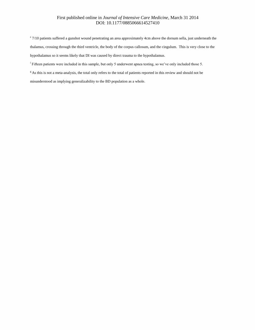
e 7/10 patients suffered a gunshot wound penetrating an area approximately 4cm above the dorsum sella, just underneath the
thalamus, crossing through the third ventricle, the body of the corpus callosum, and the cingulum. This is very close to the
hypothalamus so it seems likely that DI was caused by direct trauma to the hypothalamus.
f Fifteen patients were included in this sample, but only 5 underwent apnea testing, so we’ve only included those 5.
g As this is not a meta-analysis, the total only refers to the total of patients reported in this review and should not be
misunderstood as implying generalizability to the BD population as a whole.
First published online in Journal of Intensive Care Medicine, March 31 2014
DOI: 10.1177/0885066614527410
Table 2. Overview of hypothalamic-anterior pituitary literature review
Variables # of
studies
Findings/comments
Selected hypophysiotropic
hormones
3 LHRH det. 10/10 pts25
; CRH and GHRH det. in 5/5 and
4/541
; GHRH det. 13/23, CRH det. 12/19, LHRH det.
21/22, and one or more of GHRH, CRH, or LHRH det.
in 23/2327
Selected anterior pituitary
hormones
5-12 TSH16,27,42-51
: wnl 272/347 (78%); det. 325/386 (84%)
GH16,27,41,51,53,54
: wnl 37/60 (62%); det. 102/109 (94%)
ACTH27,41,51-53
: wnl 31/65 (48%); det. 83/114 (73%)
Anterior pituitary stimulation
tests
6 0/36 and 0/7 responded to TRH with ↑TSH54,55
0/5 responded to insulin-induced hypoglycemia or
arginine with ↑GH41
15/23 responded to 1 or more of TRH, LHRH, or
insulin27
2/7 and 4/5 responded to arginine and GRH with ↑GH27
1/1 and 1/1 responded to TRH and LHRH42
1/2 responded to insulin42
4/5 responded to TRH25
T4 (Total and free) 6-7 T416,43,44,50,73,74
: Wnl or ↑ 80/129 (62%)
fT445-50,56
: wnl or ↑ 194/299 (65%)
T3 (Total, free, and reverse) 6-9 T316,43,44,50,73,74
: wnl 27/129 (21%)
fT344-50,56,74,75
: wnl or ↑ 57/133 (17%)
rT316,45-48,50,56
: wnl or ↑ 152/169 (90%)
Thyroid hormone patterns (including TSH) most
consistent with euthyroid sick syndrome rather than
central neuroendocrine failure.
Cortisol 8 Highly variable.16,24,27,29,46,48,56,58-60
Diurnal variation apparently lost in every case.16,27,45,61
Abbreviations: LHRH = luteinizing hormone releasing hormone; CRH = corticotropin releasing hormone; GHRH = growth
hormone releasing hormone; TRH = thyrotropin releasing hormone; TSH = thyroid stimulating hormone; GH = growth hormone;
ACTH = adrenocorticotropin hormone; T4 = thyroxine; fT4 = free T4; T3 = triiodothyronine; fT3 = free T3; rT3 = reverse T3;
wnl = within normal limits; det. = detectable; ↑ = high.
First published online in Journal of Intensive Care Medicine, March 31 2014
DOI: 10.1177/0885066614527410
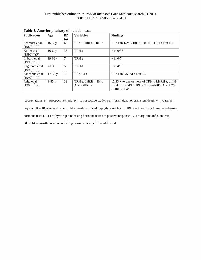
Table 3. Anterior pituitary stimulation tests
Abbreviations: P = prospective study; R = retrospective study; BD = brain death or brainstem death; y = years; d =
days; adult = 18 years and older; IH-t = insulin-induced hypoglycemia test; LHRH-t = luteinizing hormone releasing
hormone test; TRH-t = thyrotropin releasing hormone test; + = positive response; AI-t = arginine infusion test;
GHRH-t = growth hormone releasing hormone test; add’l = additional.
Publication Age BD
(n)
Variables Findings
Schrader et al.
(1980)42
(P)
16-56y 6 IH-t, LHRH-t, TRH-t IH-t + in 1/2; LHRH-t + in 1/1; TRH-t + in 1/1
Koller et al.
(1990)54
(P)
16-64y 36 TRH-t + in 0/36
Imberti et al.
(1990)55
(P)
19-62y 7 TRH-t + in 0/7
Sugimoto et al.
(1992)25
(P)
adult 5 TRH-t + in 4/5
Kinoshita et al.
(1992)41
(P)
17-50 y 10 IH-t, AI-t IH-t + in 0/5, AI-t + in 0/5
Arita et al.
(1993)27
(P)
9-85 y 39 TRH-t, LHRH-t, IH-t,
AI-t, GHRH-t
15/23 + to one or more of TRH-t, LHRH-t, or IH-
t; 2/4 + in add’l LHRH-t 7 d post-BD; AI-t + 2/7;
GHRH-t + 4/5
First published online in Journal of Intensive Care Medicine, March 31 2014
DOI: 10.1177/0885066614527410
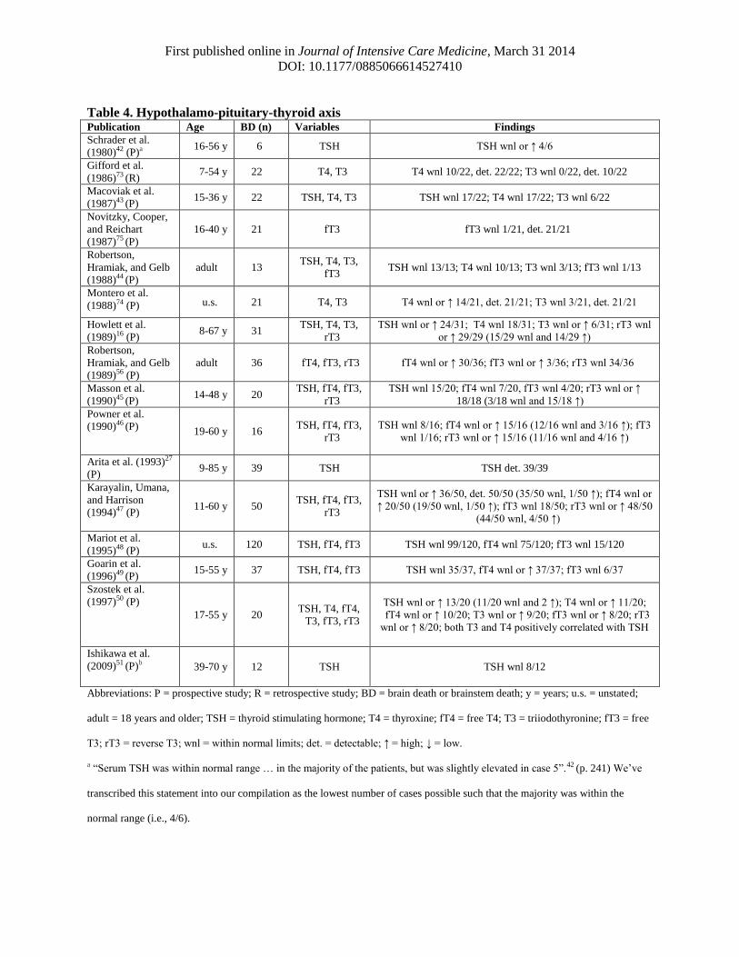
Table 4. Hypothalamo-pituitary-thyroid axis Publication Age BD (n) Variables Findings
Schrader et al.
(1980)42 (P)a 16-56 y 6 TSH TSH wnl or ↑ 4/6
Gifford et al.
(1986)73 (R) 7-54 y 22 T4, T3 T4 wnl 10/22, det. 22/22; T3 wnl 0/22, det. 10/22
Macoviak et al.
(1987)43 (P) 15-36 y 22 TSH, T4, T3 TSH wnl 17/22; T4 wnl 17/22; T3 wnl 6/22
Novitzky, Cooper,
and Reichart
(1987)75 (P)
16-40 y 21 fT3 fT3 wnl 1/21, det. 21/21
Robertson,
Hramiak, and Gelb
(1988)44 (P)
adult 13 TSH, T4, T3,
fT3 TSH wnl 13/13; T4 wnl 10/13; T3 wnl 3/13; fT3 wnl 1/13
Montero et al.
(1988)74 (P) u.s. 21 T4, T3 T4 wnl or ↑ 14/21, det. 21/21; T3 wnl 3/21, det. 21/21
Howlett et al.
(1989)16 (P) 8-67 y 31
TSH, T4, T3,
rT3
TSH wnl or ↑ 24/31; T4 wnl 18/31; T3 wnl or ↑ 6/31; rT3 wnl
or ↑ 29/29 (15/29 wnl and 14/29 ↑)
Robertson,
Hramiak, and Gelb
(1989)56 (P)
adult 36 fT4, fT3, rT3 fT4 wnl or ↑ 30/36; fT3 wnl or ↑ 3/36; rT3 wnl 34/36
Masson et al.
(1990)45 (P) 14-48 y 20
TSH, fT4, fT3,
rT3
TSH wnl 15/20; fT4 wnl 7/20, fT3 wnl 4/20; rT3 wnl or ↑
18/18 (3/18 wnl and 15/18 ↑)
Powner et al.
(1990)46 (P) 19-60 y 16 TSH, fT4, fT3,
rT3
TSH wnl 8/16; fT4 wnl or ↑ 15/16 (12/16 wnl and 3/16 ↑); fT3
wnl 1/16; rT3 wnl or ↑ 15/16 (11/16 wnl and 4/16 ↑)
Arita et al. (1993)27
(P) 9-85 y 39 TSH TSH det. 39/39
Karayalin, Umana,
and Harrison
(1994)47 (P) 11-60 y 50
TSH, fT4, fT3,
rT3
TSH wnl or ↑ 36/50, det. 50/50 (35/50 wnl, 1/50 ↑); fT4 wnl or
↑ 20/50 (19/50 wnl, 1/50 ↑); fT3 wnl 18/50; rT3 wnl or ↑ 48/50
(44/50 wnl, 4/50 ↑)
Mariot et al.
(1995)48 (P) u.s. 120 TSH, fT4, fT3 TSH wnl 99/120, fT4 wnl 75/120; fT3 wnl 15/120
Goarin et al.
(1996)49 (P) 15-55 y 37 TSH, fT4, fT3 TSH wnl 35/37, fT4 wnl or ↑ 37/37; fT3 wnl 6/37
Szostek et al.
(1997)50 (P)
17-55 y 20 TSH, T4, fT4,
T3, fT3, rT3
TSH wnl or ↑ 13/20 (11/20 wnl and 2 ↑); T4 wnl or ↑ 11/20;
fT4 wnl or ↑ 10/20; T3 wnl or ↑ 9/20; fT3 wnl or ↑ 8/20; rT3
wnl or ↑ 8/20; both T3 and T4 positively correlated with TSH
Ishikawa et al.
(2009)51 (P)b 39-70 y 12 TSH TSH wnl 8/12
Abbreviations: P = prospective study; R = retrospective study; BD = brain death or brainstem death; y = years; u.s. = unstated;
adult = 18 years and older; TSH = thyroid stimulating hormone; T4 = thyroxine; fT4 = free T4; T3 = triiodothyronine; fT3 = free
T3; rT3 = reverse T3; wnl = within normal limits; det. = detectable; ↑ = high; ↓ = low.
a “Serum TSH was within normal range … in the majority of the patients, but was slightly elevated in case 5”.42 (p. 241) We’ve
transcribed this statement into our compilation as the lowest number of cases possible such that the majority was within the
normal range (i.e., 4/6).

First published online in Journal of Intensive Care Medicine, March 31 2014
DOI: 10.1177/0885066614527410
b Hormones were measured at autopsy, 4-25 days post-BD. All patients were autopsied within 12 hours of cardiac arrest. Blood
was taken from the right cardiac chamber.
First published online in Journal of Intensive Care Medicine, March 31 2014
DOI: 10.1177/0885066614527410
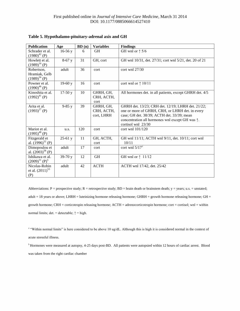
Table 5. Hypothalamo-pituitary-adrenal axis and GH
Publication Age BD (n) Variables Findings
Schrader et al.
(1980)42
(P)
16-56 y 6 GH GH wnl or ↑ 5/6
Howlett et al.
(1989)16
(P)
8-67 y 31 GH, cort GH wnl 10/31, det. 27/31; cort wnl 5/21, det. 20 of 21
Robertson,
Hramiak, Gelb
(1989)56
(P)
adult 36 cort cort wnl 27/30
Powner et al.
(1990)46
(P)
19-60 y 16 cort cort wnl or ↑ 10/11
Kinoshita et al.
(1992)41
(P)
17-50 y 10 GHRH, GH,
CRH, ACTH,
cort
All hormones det. in all patients, except GHRH det. 4/5
Arita et al.
(1993)27
(P)
9-85 y 39 GHRH, GH,
CRH, ACTH,
cort, LHRH
GHRH det. 13/23; CRH det. 12/19; LHRH det. 21/22;
one or more of GHRH, CRH, or LHRH det. in every
case; GH det. 38/39; ACTH det. 33/39; mean
concentration all hormones wnl except GH was ↑.
cortisol wnl 23/30
Mariot et al.
(1995)48
(P)
u.s. 120 cort cort wnl 101/120
Fitzgerald et
al. (1996)52
(P)
25-61 y 11 GH, ACTH,
cort
GH wnl 11/11; ACTH wnl 9/11, det. 10/11; cort wnl
10/11
Dimopoulou et
al. (2003)58
(P)
adult 17 cort cort wnl 5/17a
Ishikawa et al.
(2009)51
(P)b
39-70 y 12 GH GH wnl or ↑ 11/12
Nicolas-Robin
et al. (2011)53
(P)
adult 42 ACTH ACTH wnl 17/42, det. 25/42
Abbreviations: P = prospective study; R = retrospective study; BD = brain death or brainstem death; y = years; u.s. = unstated;
adult = 18 years or above; LHRH = luteinizing hormone releasing hormone; GHRH = growth hormone releasing hormone; GH =
growth hormone; CRH = corticotropin releasing hormone; ACTH = adrenocorticotropin hormone; cort = cortisol; wnl = within
normal limits; det. = detectable; ↑ = high.
a “Within normal limits” is here considered to be above 10 ug/dL. Although this is high it is considered normal in the context of
acute stressful illness.
b Hormones were measured at autopsy, 4-25 days post-BD. All patients were autopsied within 12 hours of cardiac arrest. Blood
was taken from the right cardiac chamber