
Pituitary microadenomas: experience with Gd-DOTA-enhanced MR imaging at 0.5 Telsa
1
Distinct Signaling Pathways Mediate Stimulation of Cell
Cycle Progression and Prevention of Apoptotic Cell Death by
Estrogen in Rat Pituitary Tumor PR1 Cells
Simona Caporali,*, # Manami Imai,*,¶ Lucia Altucci,* Massimo Cancemi,* Silvana
Caristi,*,‡ Luigi Cicatiello,* Filomena Matarese,* Roberta Penta,* Dipak K.
Sarkar,** Francesco Bresciani,* and Alessandro Weisz*,1
*Dipartimento di Patologia generale, Seconda Università degli Studi di Napoli, Napoli,
Italy; and **Department of Animal Sciences, Rutgers, The State University of New
Jersey, New Brunswick, USA
Running title: Dual estrogen regulation of cell growth
1 Corresponding author:
Dipartimento di Patologia generale Seconda Università degli Studi di Napoli Vico L. De Crecchio, 7 80138 Napoli, Italy [email protected] Tel. (0039) 081 566-5702 Fax (0039) 081 566-5702
Key Words: estrogen, nuclear receptors, cell cycle, apoptosis, pituitary tumours
Copyright 2003 by The American Society for Cell Biology.
MBC in Press, published on September 5, 2003 as 10.1091/mbc.E03-05-0303

2
Estrogens control cell growth and viability in target cells via a interplay of genomic and
extra-genomic pathways not yet elucidated. Here we show evidence that cell
proliferation and survival are differentially regulated by estrogen in rat pituitary tumour
PR1 cells. Pico to femtomolar concentrations of 17β-estradiol (E2) are sufficient to
foster PR1 cell proliferation, whereas nanomolar concentrations of the same are needed
to prevent cell death, that occurs at high rate in these cells in the absence of hormone.
Activation of endogenous (PRL) or transfected estrogen-responsive genes occur at the
same, higher concentrations of E2 required to promote cell survival, whereas
stimulation of cyclin D3 expression and DNA synthesis occur at lower E2
concentrations. Similarly, the pure anti-estrogen ICI 182,780 inhibits ERE-dependent
trans-activation and cell death more effectively than cyclin-cdk activity, G1-S transition
or DNA synthesis rate. In antiestrogen-treated and/or estrogen-deprived cells, death is
due predominantly to apoptosis. Estrogen-induced cell survival, but not E2-dependent
cell cycle progression, can be prevented by an inhibitor of c-Src kinase or by blockade
of the MEK/ERK signaling pathway. These data indicate the coexistence of two
distinguishable estrogen signaling pathways in PR1 cells, characterized by different
functions and sensitivity to hormones and anti-hormones.

3
INTRODUCTION
Estrogen regulation of target cell proliferation results from promotion of both cell
growth and survival (Altucci and Gronemeyer, 2001). Estrogens, in fact, control the
function of several genes and proteins directly involved in cell cycle regulation,
including cyclins, proto-oncogenes and negative cell cycle regulators (Altucci et al.,
1996; Dinda et al., 1997), while they are well known modulators of cell life and death
in different cell types, including uterine epithelia (Pollard et al., 1987), breast cancer
cells (Kyprianou et al., 1991) and neurons (Singer et al., 1996; Sawada et al., 2000).
The cellular mechanism(s) mediating estrogen prevention of programmed cell death
have not been clarified yet. E2 causes up-regulation of Bcl-2, the anti-apoptotic factor
which is able to suppress cytocrome-c release ensuing caspase-3 activation in neurons
(Dubal et al., 1999) and breast cancer cell lines (El Etreby et al., 1998), while the anti-
estrogen tamoxifen down-regulates Bcl-2 and mediates apoptosis via caspase-3 and
JNK-1 activation in breast carcinoma cells (Mandlekar et al., 2000), where ERα
directly regulates Bcl-2 gene transcription (Perillo et al., 2000). On the other hand,
other evidences suggest an inhibitory effect of estrogen on non-genomic apoptotic
signals. Kousteni et al. (Kousteni et al., 2001) have shown that the anti-apoptotic
effects of estrogens on bone cells are mediated by the extracellular signal-regulated
kinases (ERKs) cascade via an ER-α region that is not involved in genotropic actions of
these steroids. Moreover, it has been reported that E2 prevents UV- or taxol-induced
apoptosis in breast cancer cells by inhibiting JNK activation and stimulating ERK
activity (Razandi et al., 2000). These authors observed the same also with membrane-
impermeable E2-BSA conjugates, suggesting the possible involvement of a putative
membrane receptor in mediating these hormonal effects. The causal role of these non-
genotropic estrogen signaling pathways and their correlation with the classical, gene
trascription-mediated actions of these hormones are far from being understood. For
example, while some reports indicate a central role for ERKs and PKC-α signaling in
estrogen regulation of cell cycle progression (Castoria et al., 1999; Manole et al., 2001;
Marino et al., 2001), other authors have shown cell cycle effects of these hormones
independently from these cascades. Furthermore, a growing body of evidence suggest
4
the existence of cross-talks between estrogen receptors and other signaling pathway
effectors (Tolon et al., 2000; Coleman and Smith, 2001).
PR1 is an estrogen-responsive cell line derived from DES-treated F344 female rats
(Pastorcic et al., 1995) that shows a unique response to E2, since in these cells the
concentration of E2 required for cell proliferation are 1000-fold lower than that
required to increase expression of the hormone-responsive prolactin (PRL) gene (Chun
et al., 1998), suggesting biphasic or distinct mechanisms for estrogen control of cell
proliferation and gene activity. Furthermore, the ERK-dependent signaling is rapidly
activated in this cell line in response to estrogen and hormone-induced PRL production
is prevented by MEK inhibition (Watters et al., 2000). These observations indicate that
this cell line represents a useful model to test the possibility that multiple, independent
hormone-responsive signaling cascades may coexist in the same cellular background
and control distinct cellular functions.
We have thus investigated here the effects of estrogen deprivation and stimulation on
cell cycle progression, target gene trans-activation and cell survival in PR1 cells,
aiming at understanding the mechanism(s) that underlie promotion of tumor cell growth
and survival by estrogens. Results show that estrogens control cell growth, DNA
synthesis and cell fate via distinguishable mechanisms in this cell line.
5
MATERIALS AND METHODS
Culture Conditions
PR1 is a pituitary cell line derived from E2-treated F344 female rats (Pastorcic et al.,
1995). Cells were cultured in DMEM-F12 medium (Biowhittaker-Europe)
supplemented with 10% foetal bovine serum (FBS; Hyclone) and antibiotics (Sigma,
UK). Culture media were replaced every 2 days. Estrogen deprivation was obtained by
culturing the cells in DMEM-F12 phenol red-free (GIBCO-Life Technologies) medium
containing 10% charcoal-treated (DCC)-FBS. For treatment with 17β-estradiol, MEK
inhibitors (50µM PD98059-New England Biolabs; 30µM U0126-Cell Signaling) and/or
c-Src inhibitor (2µM PP1-Alexis Biochemicals) cells were kept in hormone-free
medium, as indicated. Culture media have been changed every day.
Cell Growth Analysis
Colorimetric assay: Cells (16000/well) were loaded in 96 well dishes. After appropriate
stimulation, cells were washed in PBS and fixed with 12.5 % glutaraldehyde for 20 min
at room temperature, before further washing with H2O and incubation with 0.05%
methylene blue for 30 min, washing and incubation with 0.33 M HCl for 18hrs.
Absorption was measured at 595 nm (Dufourny et al., 1997).
[3H]-thymidine incorporation: cells were loaded in 6-wells dishes with or without E2,
ICI 182,780 or kinase inhibitors. The rate of DNA synthesis or the cell number were
estimated by measuring [3H]-thymidine (methyl-3H TdR: 5 Ci/mmol, Amersham Italia)
incorporation in DNA during 1h or 24 hrs respectively. DNA was precipitated by TCA
5% and resolved in 0.1 M NaOH and 2% Na2CO3, samples were then neutralized with
HCl 1N and radioactivity measured by liquid scintillation counter (Wallac). For [3H]-
thymidine pulse, data were normalized for total DNA content.
Cell Death Analysis
For cytofluorimetric analysis, cells were cultured in 100 mm dishes and collected in
0,1% Sodium Citrate and 50 µg/ml propidium iodide (PI). After 18 hrs incubation, the
percentage of cells with sub- G1 DNA was evaluated for flow cytometry with a FACS
ANALYZER (FACScalibur, Becton Dickinson Inc.).
6
For Annexin V-BOBO-1 labeling, cells were plated (30000cells/well) in 24-well dishes
and, after stimulation, the medium was removed and cells were incubated in the
Annexin V solution (Hepes 10mM, NaCl 140 mM and CaCl2 and 1µg/ml Annexin V-
Alexa-568, Roche), 1µg/ml BOBO-1 (Molecular Probes). Simultaneous application of
DNA stain and Bobo-1 (Molecular Probes), for dye exclusion tests, was used and cells
were analyzed by fluorescence microscopy (Leika).
Transient Transfections
For transient transfection, triplicate 60-mm culture dishes (1X10⁶ cells/dish) were
transfected by liposome-mediated (DOTAP- Roche Mannheim, Germany) gene transfer
with ERE-tk luc (1.5 µg/transfection), a luciferase reporter gene including estrogen
responsive elements. 6 hrs after addition of the liposome-DNA mixture, media were
changed and cells further incubated for 24hrs. Cells were then harvested by washing,
scraped in lysis buffer (Promega Italia) and lysated with 3 cycles of rapid freeze-
thawing. After clearing the cell lysates by 5 minutes centrifugation at 4°C, protein
concentration was determined in the crude extracts with a colorimetric assay
(BIORAD, Italia). Luciferase activity was assayed with Luciferase assay reagent
(Promega), following manifacturer’s instructions. Results were normalized for protein
concentration or controlled using a Dual Luciferase Assay Kit (Promega).
Immunoblot Analysis
Cell extracts were prepared in NP40 buffer [50mM Tris-HCl pH 8.0, 150mM NaCl, 1%
NP40, 10µM Sodium fluoride, 0.1mM Sodium orthovanadate, 1mM phenylmethyl
sulphonyl fluoride (PMSF), 10 µg/ml aprotinin, leupeptin, pepstatin A and 20nM
okadaic acid].
Cells were washed twice in ice-cold PBS, scraped in ice-cold NP40 buffer and lysated
for 10 min. Cell lysates were cleared by 15 minutes centrifugation at 4°C, protein
content in the supernatant was assayed by Bradford Protein Assay (BIORAD). The cell
extracts were diluted 1:1 in 2X Laemmly sample buffer, followed by boiling for 5 min.
Equal amounts of protein were loaded and separated for SDS/PAGE gel. Protein smears
were transferred to cellulose nitrate filters (Schleicher and Schuell, Germany) and
blocked 30 min at 37°C in 5% non fat milk in TBST (TBS with 0.1% Tween 20).
7
Membranes were incubated with the primary antibody for 1h at RT in TBST. Primary
antibodies were the following: anti-Cyclin D3 (Transduction Laboratories) 1:1000;
anti-rat prolactin antibodies (provided by the National Hormone and Pituitary Program,
NIDKK) 1µg/ml; anti-ERK (K23; Santa Cruz Biotechnology) 100µg/ml. After
washing, a horseradish peroxidase-linked secondary antibody (Amersham-Pharmacia)
was added and target protein bands were detected using ECL (Amersham-Pharmacia).
Caspase-3 Colorimetric Assay
Caspase-3 activity was assayed with a colorimetric assay kit (Biovision) from Alexis-
biochemicals according to standard protocols suggested by the producer. Briefly, the
assay is based on spectrophotometric detection of the chromophore p-nitroanilide
(pNA) after cleavage from the labelled substrate DEVD-pNA. After the treatment, cells
were lysated with the cell lysis buffer included in the kit, incubated for 10 min on ice
and then centrifuged at 13,000 rpm for 1 min at 4°C. The supernatant was added to the
reaction buffer with dithiothreitol and DEVD-p-NA, and incubated 18h at 37°C.
Relative caspase-3 activity was measured as optical density at 405 nm. 200µg of total
proteins of each samples was used.
Cdc2 Kinase Assay
Cdc-2 kinase activity was assayed with Signatect cdc2 Protein Kinase Assay (Promega,
Italia) according with the protocol suggested by the producer. A biotinylated peptide
substrate derived from histone H1 (PKTPKKAKKL) was used as substrate. 1µg of
proteins was assayed and radio-labelled. Phosphorylated substrate was recovered from
the reaction mix with SAM2 Biotin Capture Membrane. Results are expressed as pmol/
min/ µg of proteins.
RNA analysis by Multiplexed RNase protection
Total RNA was extracted with Eurozol (Euroclone) as indicated by the producer. Rat
apoptosis template sets (rAPO-1, 45601P, Pharmingen) were labelled with 32P-uridine
triphosphate (NEN). The ribonuclease (RNAse) protection assay was performed
according to the supplier's instructions (Pharmingen, San Diego, California). Briefly,
total RNA (4µg) and 6 to 8x10⁵ of labelled probes were used for hybridization. After

8
RNAse treatments, the protected probe fragments were resolved on a 5% urea-
polyacrylamide-bis-acrylamide gel. Dried gels were placed on film (Hyperfilm-MP,
Amersham) in a cassette with an intensifying screen. Exposure time varied depending
upon application.

9
RESULTS
Characterization of the mitogenic responses of PR1 cells to estrogen
PR1 cells derive from F344 rat pituitary tumors induced by prolonged in vivo treatment
with estrogens. As reported previously (Chun et al., 1998) PR1 cells are highly
responsive to estrogen, being capable to respond to very low, sub-physiological
concentration of E2. Analysis of estrogen receptor expression in this cell line reveals
that both ER-α and ER-β subtypes can be detected (Chun et al., 1998) and our
unpublished results). The proliferative response of PR1 cells to different concentrations
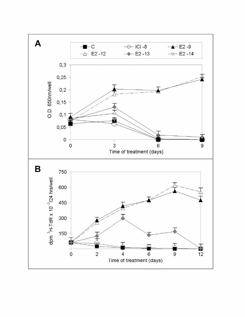
of E2 is shown in Fig. 1, as assessed by a colorimetric cell quantitation assay (Fig. 1A)
or by [3H]-thymidine incorporation into DNA (Fig. 1B). The results indicate that
concentrations of the hormone >10-12 M induce a substantial and progressive increase of
cell number, both determining an increase during up to 9 days of treatment, as detected
by either of the two quantitative methods applied. On the contrary, induction of cell
cycle proliferation by lower concentrations of hormone (10-13 to 10-14 M) is transient,
increasing slightly during the first 3-4 days of treatment and decreasing thereafter.
Interestingly, in the absence of estrogen a progressive and drastic reduction in the initial
number of cells can be observed (Fig. 1A-B), so that by day 4-6 few cells can be
detected, suggesting that massive cell death has occurred.
Effects of low concentrations of E2 on target gene transcription and cell cycle control
in PR1 cells
To determine the activity of PR1 cells ERs to their cognate hormone, we measured the
effects of different concentrations of E2 on transcription of a transiently transfected
ER-responsive reporter gene (ERE-tk-luc) and compared the results obtained with what
observed in the results reported in Fig. 1. Results show that a concentration of hormone
of 10-12M, able to induce a maximal stimulatory effect of PR1 cell proliferation
(Fig.1A-B), is able to promote only half-maximal ER-mediated trans-activation (Fig.
2A). This result was confirmed by analysis of PRL gene activation by E2, an
endogenous marker of ER activity of PR1 cell genome (Chun et al., 1998), that shows a
comparable response to the different concentrations of hormone tested (Fig. 2B).
Interestingly, 10-12M E2 promotes a limited induction of PRL expression, when

10
compared to higher concentrations such as 10-9M, while both concentrations are equally
effective in promoting cell proliferation (compare data reported in Fig.s 1 and 2A-B).
This was further verified by assessing the sensitivity of key cell cycle progression
markers to E2. As shown in the lower panel of Fig. 2B, the expression of cyclin D3, an
estrogen regulated gene product that is rate-limiting for G1 progression in this cell line
(Watters et al., 2000), increases in response to 10-12M E2 to a degree comparable to
what observed in response to higher concentrations of the hormone. Furthermore, this
same concentration of E2 is sufficient to trigger maximal increase of the DNA synthesis
rate (Fig. 2C), a marker of S-phase entry under these test conditions. These results
indicate that two distinct estrogen effector pathways show a different sensitivity to the
hormone in PR1 cells.
Biphasic response of PR1 cells to estrogen signaling blockade with the pure anti-
estrogen ICI 182,780
Given the high sensitivity of PR1 cells to estrogens, it can not be excluded that traces of
these hormones still present in the culture media containing charcoal-treated foetal
bovine serum (DCC-FBS) may influence interpretation of the experimental results.
Indeed, when analyzing the effects of hormone withdrawal on cell cycle phasing in PR1
cells by cytofluorimetry we observed consistent cell cycle arrest only when an estrogen
antagonist was added to the culture medium 24-36hrs before analysis (our unpublished
results). Furthermore, charcoal treatment of the serum might affect the growth
characteristics of PR1 cells by removal or reduction of one or more essential serum
component. For these reasons we decided to control the results described above by
evaluating the effects on PR1 cells of estrogen signaling ablation by addition of the
pure antiestrogen ICI 182,780 to standard culture medium. Under these experimental
conditions, we compared DNA synthesis rate (measured by [3H]-TdR incorporation),
cdc2 kinase activity, ERE-tk-luc trans-activation and cell proliferation in the absence
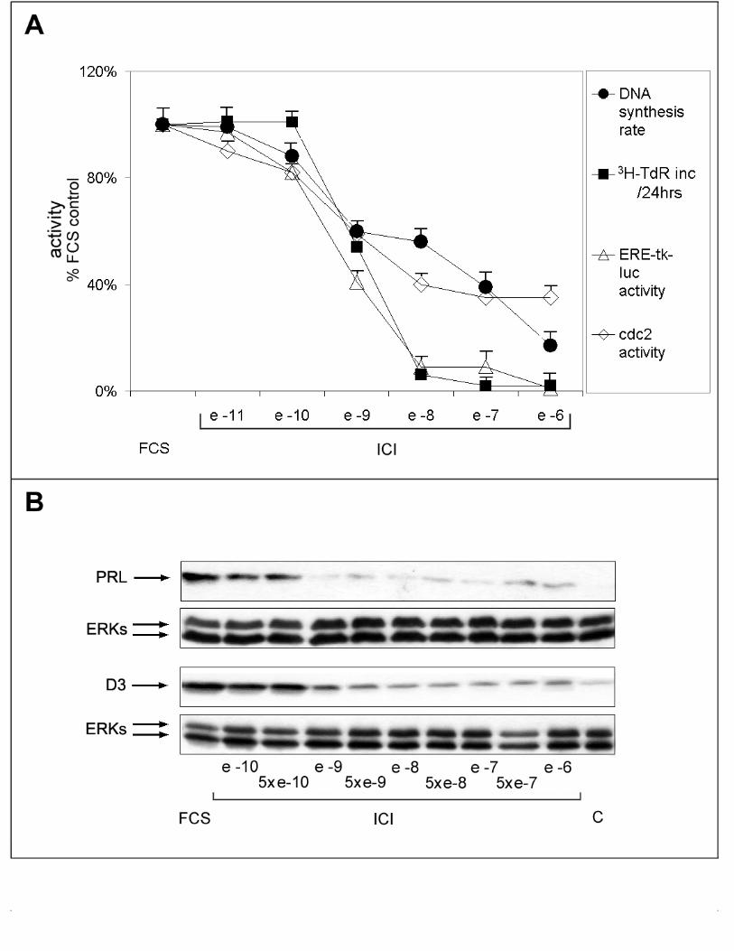
and presence of increasing concentrations of ICI 182,780. As shown in Fig. 3A, a
variable degree of responsiveness to estrogen ablation was observed for the different
effectors studied. In particular, ERE-mediated transcriptional activation and cell
proliferation were more sensible to inhibition by the antiestrogen then DNA synthesis
rate or cdc2 activity (Fig. 3A: IC50 of 10-9 and 10-7M, respectively). Furthermore, while
11
ERE-tk-luc activity and cell number decreased abruptly as the concentration of ICI
182,780 in the medium was raised above 10-10M, reaching a value close to 8-12% of the
untreated control in the presence of 10-8M antiestrogen, DNA synthesis rate and cdc-2
activity decreased more gradually in response to the antihormone, reaching a minimum
value of 20-35% in the presence of >10-7M ICI 182,780 (Fig. 3A). These measures
were independently confirmed by analysis of PRL and cyclin D3 expression changes
under the same experimental conditions. In line with the data shown in Fig. 3A, in fact,
PRL expression was completely inhibited by 10-9M ICI, a concentration that only partly
inhibited cyclin D3 expression (Fig. 3B).
These results, when combined, confirm the existence of at least two classes of estrogen
responsive effector pathways in PR1 cells, the first characterized by low sensitivity to
E2 and higher sensitivity to hormone blockade with ICI 182,780, controlling cell
number and ERE-mediated gene transcription, and the second one more sensitive to E2
and less responsive to the antihormone, controlling cell cycle progression.
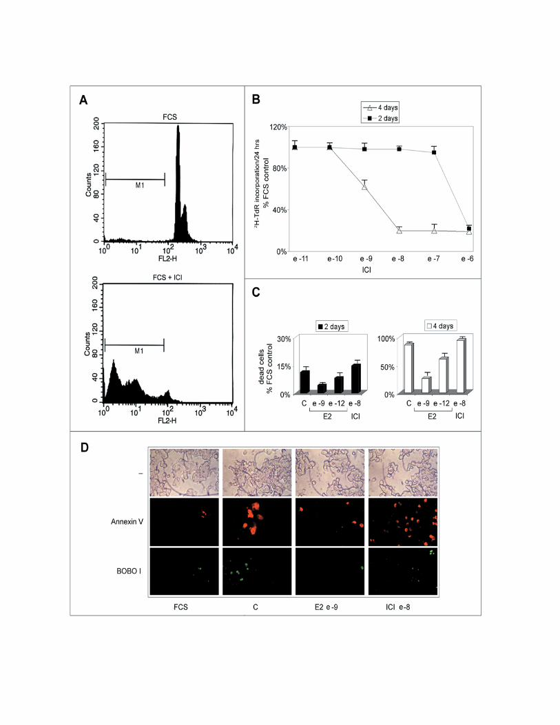
Estrogen deprivation induces apoptosis in PR1 cells that can be prevented by
stimulation with E2
During prolonged treatment of PR1 cultures with ICI 182,780, or estrogen-free media,
the number of cells decreases well below the values observed in control cultures (see
Fig.s 1 and 3A). We observed that under these conditions cells undergo drastic changes
in their morphology and assume features reminiscent of apoptosis. Cytofluorimetric
analysis of estrogen-deprived or antiestrogen-treated PR1 cell cultures shows a
progressive increase of the “pre-G1” population, representing dead cells, as shown,
following 4 days treatment with ICI 182,780, at the bottom of Fig. 4A (M1 gate). This
is confirmed by the data reported graphically in Fig. 4B, where the effects of increasing
concentrations of ICI 182,780 on the cell number is reported, to show that after 4 days
of treatment a significant reduction of this parameter (up to 85% decrease) is observed
at concentrations of antiestrogen >10-10M. This decrease appears to be due primarily to
cell loss that becomes evident starting from day 3 of treatment by a great number of
floating, dead cells (Fig. 4B and our unpublished results). Notably, this dramatic
response to estrogen signaling blockade is over imposable to what observed for ER-
mediated trans-activation (Fig. 3), in particular for what concerns the steep decrease in
12
ERE-tk-luc activity detectable at concentrations >10-10M. The same effect observed
following treatment with the antihormone could be detected upon estrogen withdrawal,
in which case it was reversed by stimulation with ≥10-9M E2 (Fig. 4C). Using
fluorescence microscopy, cell death by apoptosis was confirmed to occur in PR1 cells
following estrogen withdrawal or ICI 182,780 treatment, as shown in Fig. 4D by double
fluorescent staining of the cultures with Annexin V (red), revealing cells undergoing
apoptosis, and Bobo I (green), staining only dead cells. ICI 182,780 was found to
induce an increase of Annexin V-positive cells in 24hrs, while treatment of hormone-
deprived cells with 10-9M E2 for the same time was sufficient to prevent the appearance
of dying or death cells. The nature of the cell death induced by estrogen withdrawal
was further investigated at a molecular level by multi-probe RNase mapping, by which
the expression level of genes encoding known apoptosis regulator, including Fas, Fas-
ligand, Bcl-2, Bax, Bcl-x and caspase-1, -2 and –3, was assessed in PR1 cells before
and after treatment. Although significant differences in expression of the investigated
mRNAs were not detected (our unpublished results), caspase-3 gene was found highly
expressed in PR1 cells (Fig. 5A). Caspase-3 has been reported to play a central effector
role in multiple apoptotic pathways and may thus represent one of the end-points of the
pro-apoptotic pathway(s) inhibited by estrogen in PR1 cells and released upon estrogen
withdrawal or ICI 182,780 treatment. Following this observation, the enzymatic activity
of caspase-3 was measured with a colorimetric assay to assess activation of this pro-
apoptotic effector in PR1 cells following estrogen signaling blockade. As shown in Fig.
5B, treatment of the cells with ICI 182,780 resulted in the progressive, time-dependent
increase in caspase-3 activity, reaching a 4- to 5-fold increase after 1.5 to 4 days of
treatment. This effect of ICI 182,780 was dose-dependent, inducing a significant
increment in caspase-3 activity at concentrations ≥10-9M (our unpublished results).
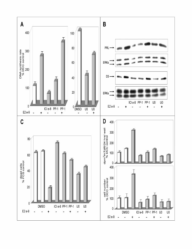
Extracellular signal-regulated (ERK) and Src kinase inhibitors do not block
estrogen-induced increase in DNA synthesis rate in PR1 cells
ERK and Src family kinases have been reported to play a pivotal role in estrogen
prevention of apoptotic cell death (Razandi et al., 2000). To gain further insights in
estrogen-dependent pathways that control cell cycle progression and cell survival in
PR1 cells, the effects of ERK and Src kinase inhibition on hormone-mediated
13
stimulation of DNA synthesis rate, inhibition of apoptotic cell death and activation of
target gene expression were investigated. In PR1 cells estrogens have been described to
stimulate ERK signaling and it has been reported that inhibition of the ERK-activating
MEK or Src enzymes prevents certain effects of estrogens, such as PRL gene activation
(Watters et al., 2000). As shown in Fig. 6A, the Src kinase inhibitor PP-1 (2µM) do not
affect basal or estrogen-induced DNA synthesis (measured as [3H]-TdR incorporation
rate), suggesting that neither of these activities is strictly dependent on Src activity. On
the other hand, the MEK-1 and –2 inhibitor U0126 shows a marked inhibitory effect on
estrogen-independent DNA synthesis but it does not prevent completely the estrogen-
dependent increase of this parameter (Fig. 6A). Comparable results were obtained when
using a different MEK inhibitor (PD98059; our unpublished results). These last results
suggest that while the activity of this MEK-ERK signaling cascade is a necessary pre-
requisite for efficient DNA synthesis in PR1 cells, its blockade reduces but do not
obliterate the ability of estrogen to enhance this cell cycle-dependent process. Cyclin
D3 accumulation in response to E2 was not prevented by MEK inhibition with U0126
(Fig. 6B), despite the fact that this compound determined a significant (50-60%)
reduction in basal expression of the cyclin in hormone-starved control cells, further
suggesting that estrogen-mediated cell cycle progression in PR1 cells is not fully
dependent on the activity of the ERK signaling cascade. Similarly, Src kinase inhibition
by PP-1 did not affect the ability of E2 to promote expression of this G1 cyclin (Fig.
6B). The ability of the hormone to stimulate PRL gene expression, instead, was fully
prevented by cell treatment with U0126, as previously reported for PD98059 (Watters
et al., 2000), but not by Src kinase blockade with PP-1 (Fig. 6B). Interestingly, MEK
and Src kinase inhibitors both efficiently prevented the positive effects of E2 on cell
survival (Fig. 6C), resulting in a marked overall reduction in estrogen dependent PR1
cell proliferation, as determined by both the standard tritiated thymidine incorporation
assay and direct cell count (upper and lower panels of Fig. 6D). U0126 alone appears to
induce a marked inhibition of apoptosis in PR1 cells, independently of estrogen (Fig.
6C), similarly to what reported previously in other cellular backgrounds (Bacus et al.,
2001).
When combined, these results support the possibility that estrogen controls survival,
apoptosis and cell cycle progression in PR1 cells via distinct signaling cascades, among

14
which the ERKs pathway is likely to play an important role in mediating the
proliferative effects of E2 not through mitogenesis but via prevention of cell loss by
apoptosis.
15
DISCUSSION
Fisher 344 is a rat strain highly sensitive to the carcinogenic effects of estrogens. In
these animals, 6-10 weeks of treatment with estrogenic compounds induces at high
frequency pituitary prolactinomas (Kaplan and De Nicola, 1976), which are often
hormone-responsive and for this reason can be exploited to investigate the molecular
and genetic bases of hormone-mediated carcinogenesis and tumor progression and of
the hormone-responsive phenotypes of cancer cells. The PR1 cell line, derived from
one such F344 lactotroph tumours, expresses both ER-α and ER-β receptor subtypes
and shows a high sensitivity to estrogen, representing a good model to analyse the
signal transduction cascades mediating estrogen effects in target cells. Indeed, very low
concentrations of E2, inducing as low as 0.1% receptor occupancy, are sufficient for
half-maximal stimulation of PR1 cell proliferation, PRL gene activation requires 1,000-
fold more hormone (Chun et al., 1998). This result suggests that either a small pool of
active ER is sufficient to promote proliferation of this cell line or, alternatively, that
hormone-responsive pathways exist here which are triggered by very low
concentrations of estrogen and are independent from the ‘classical’ ER. In the first case,
the high sensitivity of the cell replication machinery to estrogen might be explained
with the existence of cellular factors, critical for cell cycle control, interacting with ER
with higher affinity than the factors involved in PRL gene control.
The results reported here demonstrate that different signaling pathways are implicated
in the control of two key estrogen-regulated functions in PR1 cells, namely cell cycle
progression and cell survival. Indeed, we found that E2 promotes cell cycle progression
and prevents cell death and, more important, that the hormone differently regulates
these events. We observed in fact a dissociation between the mitogenic and the anti-
apoptotic effects of E2, since concentrations of E2 ≤10-12M are effective in enhancing
DNA synthesis rate and cell proliferation (Fig.s 1 and 2B), while a preventive effect
toward apoptosis requires 100-fold more E2 (Fig. 4C). Interestingly, only the latter
response occurs under conditions that clearly reflect optimal activation of the trans-
activating functions of ERs. Expression of the PRL gene, an estrogen-responsive gene
directly targeted by ERs, is minimal upon cell exposure to 10-12M E2 (Fig. 2B) and the
same applies to ERE-mediated transcription of an exogenous reporter gene (Fig. 2A).
16
Promotion of PR1 cell survival shows an over imposable sensitivity to E2, since cell
death occurring spontaneously in hormone-deprived cultures can be efficiently
prevented only by concentrations of E2 well above 10-12M. On the contrary, expression
of cyclin D3, a component of the cell cycle regulatory machinery that marks G1-phase
progression, or DNA synthesis rate, an indicator of S-phase activity, are already
significantly activated at concentration of hormone ≤10-12M (Fig.s 2B and 2C). A
possible functional explanation to these observations could reside on the fact that the
DES-driven carcinogenic process that led to clonal expansion in the tumor might have
required that primary functions, such as the capacity of cells to replicate, be supported
also by very low concentrations of circulating estrogens. On the other hand, promotion
of cell survival is more critical during tumor initiation, when clonal selection might
have been dependent upon higher concentrations of estrogenic compounds. Indeed,
tumorigenesis and tumor growth are known to result from the combined activities of
signaling pathways that control the balance between cell proliferation and arrest and
cell death and survival (Altucci and Gronemeyer, 2001).
The comparable sensitivity to E2 of ERE-mediated promoter regulation and the cell
survival phenotype might reflect also a functional link between the two cellular
processes they represent. Indeed, the products of primary, ER-responsive ‘master’
genes might be responsible for modulation of ‘secondary’ gene programs, or even a
complex circuitry of different and overlapping genetic programs, that permit PR1 cells
survival. Similar control circuitries have been postulated for estrogen control of
epithelial-to-mesenchymal transition and, possibly, the metastatic phenotype in breast
cancer (Fujita et al., 2003) or of cell cycle progression in hormone-responsive cells
(Weisz and Bresciani, 1993).
Cell cycle progression, DNA synthesis, trans-activation of target genes and prevention
of apoptosis are thus inscribed in distinct regulatory circuitries with different priorities
in these cells. This dissociation among estrogen-dependent functions of PR1 cells
becomes even more evident when the pure anti-estrogen ICI 182,780 is used to block
hormonal signaling. The concentrations of this compound that abolish ERE-mediated
transcription or promote significant cell loss are, in fact, only partly inhibitory upon
DNA synthesis rate and cyclin-dependent cdc2 activity (Fig. 3A). More important,
while the activity of the former two markers tend to drop steeply when anti-hormone
17
concentration is raised above 10-10M, that of the latter decreases gradually and less
markedly. Similarly, abrupt PRL gene inhibition occurs when ICI 182,780
concentration is raised above 5x10⁻¹⁰M, while suppression of cyclin D3 levels is once
again more gradual and resistant to the anti-hormone, occurring only in the presence of
higher concentrations of compound (Fig. 3B). These evidences fully support the idea
that estrogens and anti-estrogens both exploit different pathways to intervene on each
different vital function of PR1 cells. Apoptosis, be it induction by ICI 182,780 or
prevention by E2, is strictly dependent upon the transcriptional activity of the ERs,
while different signaling cascades molecules mediate cell cycle control.
ICI 182,780 induces apoptosis by activating the proteolitic activity of caspase-3 (Fig.
5B). Caspase-3 is a downstream apoptotic effector, reported to be activated in response
to either of at least two different pathways, one involving Bcl-2 and cytochrome c
release while the other dependent upon activation of death receptors. Both pathways
can be regulated by estrogen. In fact E2 up-regulates Bcl-2 gene expression and two
EREs have been reported to be present in the promoter region of the Bcl-2 gene (Perillo
et al., 2000). Moreover, estrogen-mediated regulation of the expression of TRAIL-
R2/Killer/DR5 gene (Wang and Jeng, 2000) and induction of TNFR1 and TRADD
gene expression by ICI 182,780 and Tamoxifen (Smolnikar et al., 2000) have been
reported. The induction of apoptosis mediated by ICI 182,780 is dependent on ER
trans-activation, most likely being controlled by a signaling molecule functionally
located downstream of ER activation. In this respect, it is worth mentioning that this
pure antiestrogen, known to block ER-α activity by increasing protein turnover and
thereby reducing its intracellular content (Dauvois et al., 1992) as well as by disrupting
nucleocytoplasmic shuttling of the receptor (Dauvois et al., 1993), can also affect
directly ER trans-acting functions by inducing co-repressor binding to the LXXLL
motif of the receptor molecule (Huang et al., 2002; Webb et al., 2003).
MEK inhibitors can interfere with estrogen signaling in PR1 and other pituitary tumor
cells (Watters et al., 2000). Interestingly, MAPK blockade by MEK inhibition has been
reported by these authors to block the ability of estrogens to regulate PRL gene
expression. Analysis of the data obtained here with protein kinase inhibitors (Fig.s 6)
strongly support the possibility that different estrogen-dependent pathways control cell
cycle progression and apoptosis/gene trans-activation. The MEK inhibitor U0126 and
18
the Src tyrosine kinase inhibitor PP-1, in fact, can prevent induction of PR1 cell
survival by E2 (Fig. 6C), while being ineffective toward hormone-dependent DNA
synthesis rate (Fig. 6A) and cyclin D3 expression (Fig. 6B), suggesting that G1-phase
progression in response to estrogen can occur in these cells also in the absence of Src-
and MEK-dependent signaling. PP1 has a minimal, if any, influence on E2-dependent
up-regulation of PRL levels, while U0126 abrogates this response completely. This
implies that Src may act in concert with estrogen in regulating PRL, and possibly other
genes, also via effector molecules disjunct from the MEK-ERK cascade. Interestingly,
both PP-1 and U0126 prevent estrogen-mediated increase in cell number, suggesting
that promotion of cell survival is dominant over induction of cell division in
determining tumor cell population control by estrogen. This implies that cell death can
occur even in the presence of residual hormonal signaling, once the level of estrogen
stimulation decreases below a critical threshold.
In conclusion, the results reported here demonstrate that estrogens can control multiple
cellular functions in target cells by means of an integrated network of signaling
pathways, involving distinct effectors molecules. These different hormone-responsive
pathways can be activated in parallel, in concert -thanks to functional cross-talks, or
independently from each other, while they can co-operate toward the same function or
exert a selective role. The knowledge that several signaling pathways are differently
regulated by estrogens suggests ways to improve our knowledge of the ‘master’ players
involved in the regulation of complex cellular functions, such as cell transformation,
proliferation, differentiation and death. At the same time, the results reported here
indicate that PR1 cells represent a unique tool for the molecular and genetic dissection
of estrogen action, to foster our understanding of the mechanisms by which estrogens
influences different biological functions in the cell.

19
ACKNOWLEDGMENTS
Research supported by Associazione Italiana per la Ricerca sul Cancro (Research
Grants 2001-2003 to A.W.), Ministero dell’Istruzione, Università e Ricerca
(Grants: PRIN 2001067229_002 to F.B., PRIN 2002067514_002 and FIRB
RBNE0157EH to A.W.), European Commission (Contracts: BMH4-CT98-3433 to
A.W., QLG1-CT-2000-01935 and QLK3-CT-2002-02029 to L.A.), Regione
Campania (Fondi per la Ricerca di Base, L. 41/99 to A.W. and L. 41/00 to L.A.)
and Seconda Università degli Studi di Napoli and Università degli Studi di Torino
(Fondi per la Ricerca di Ateneo 2001-02).
We thank W. Basile, T. Battista, J. Gorski, and A.F. Harlow (National Hormone
and Pituitary Program, NIDKK) for cell lines, reagents and technical assistance.

20
FOOTNOTES
# Present address: Istituto Dermopatico dell'Immacolata, Via dei Monti di Creta 104,
00167 Roma, Italy
¶ Present address: Department of Obstetrics and Gynecology, Kitasato University,
Kitasato 1-15-1 Sagamihara, Kanagawa, Japan
‡ Present address: Dipartimento di Patologia e Microbiologia sperimentale, Università
degli Studi di Messina, Via Consolare Valeria, 98125 Messina, Italy
21
REFERENCES
Altucci, L., Addeo, R., Cicatiello, L., Dauvois, S., Parker, M.G., Truss, M., Beato, M., Sica, V., Bresciani, F., and Weisz, A. (1996). 17beta-Estradiol induces cyclin D1 gene transcription, p36D1-p34cdk4 complex activation and p105Rb phosphorylation during mitogenic stimulation of G(1)-arrested human breast cancer cells. Oncogene 12, 2315-2324.
Altucci, L., and Gronemeyer, H. (2001). Nuclear receptors in cell life and death. Trends Endocrinol Metab 12, 460-468.
Bacus, S.S., Gudkov, A.V., Lowe, M., Lyass, L., Yung, Y., Komarov, A.P., Keyomarsi, K., Yarden, Y., and Seger, R. (2001). Taxol-induced apoptosis depends on MAP kinase pathways (ERK and p38) and is independent of p53. Oncogene 20, 147-155.
Castoria, G., Barone, M.V., Di Domenico, M., Bilancio, A., Ametrano, D., Migliaccio, A., and Auricchio, F. (1999). Non-transcriptional action of oestradiol and progestin triggers DNA synthesis. Embo J 18, 2500-2510.
Chun, T.Y., Gregg, D., Sarkar, D.K., and Gorski, J. (1998). Differential regulation by estrogens of growth and prolactin synthesis in pituitary cells suggests that only a small pool of estrogen receptors is required for growth. Proc Natl Acad Sci U S A 95, 2325-2330.
Coleman, K.M., and Smith, C.L. (2001). Intracellular signaling pathways, nongenomic actions of estrogens and ligand-indipendent activation of estrogen receptors. Frontiers Biosci. 6, 1379-1391.
Dauvois, S., Danielian, P.S., White, R., and Parker, M.G. (1992). Antiestrogen ICI 164,384 reduces cellular estrogen receptor content by increasing its turnover. Proc Natl Acad Sci U S A 9, 4037-4041.
Dauvois, S., White, R., and Parker, M.G. (1993). The antiestrogen ICI 182780 disrupts estrogen receptor nucleocytoplasmic shuttling. J Cell Sci. 104, 1377-1388.
Dinda, S., Kodali-Gali, S., Sevilla, L., Burkley, M., Hurd, C., and Moudgil, V.K. (1997). Inhibition of proliferation of T47D human breast cancer cells: alterations in progesterone receptor and p53 tumor suppressor protein. Mol Cell Biochem 175, 81-89.
Dubal, D.B., Shughrue, P.J., Wilson, M.E., Merchenthaler, I., and Wise, P.M. (1999). Estradiol modulates bcl-2 in cerebral ischemia: a potential role for estrogen receptors. J Neurosci 19, 6385-6393.
Dufourny, B., Alblas, J., van Teeffelen, H.A., van Schaik, F.M., van der Burg, B., Steenbergh, P.H., and Sussenbach, J.S. (1997). Mitogenic signaling of insulin-like growth factor I in MCF-7 human breast cancer cells requires phosphatidylinositol 3-kinase and is independent of mitogen-activated protein kinase. J Biol Chem 272, 31163-31171.
22
El Etreby, M.F., Liang, Y., Wrenn, R.W., and Schoenlein, P.V. (1998). Additive effect of mifepristone and tamoxifen on apoptotic pathways in MCF-7 human breast cancer cells. Breast Cancer Res Treat 51, 149-168.
Fujita, N., Jaye, D.L., Kajita, M., Geigerman, C., Moreno, C.S., and Wade, P.A. (2003). MTA3, a Mi-2/NuRD Complex Subunit, Regulates an Invasive Growth Pathway in Breast Cancer. Cell 113, 207-219.
Huang, H.J., Norris, J.D., and McDonnell, D.P. (2002). Identification of a negative regulatory surface within estrogen receptor alpha provides evidence in support of a role for corepressors in regulating cellular responses to agonists and antagonists. Mol Endocrinol 16, 1778-1792.
Kaplan, S.E., and De Nicola, A.F. (1976). Protein and RNA synthesis in pituitary tumors from F344 rats given implants of estrogen. J Natl Cancer Inst 56, 37-42.
Kousteni, S., Bellido, T., Plotkin, L.I., O'Brien, C.A., Bodenner, D.L., Han, L., Han, K., DiGregorio, G.B., Katzenellenbogen, J.A., Katzenellenbogen, B.S., Roberson, P.K., Weinstein, R.S., Jilka, R.L., and Manolagas, S.C. (2001). Nongenotropic, sex-nonspecific signaling through the estrogen or androgen receptors: dissociation from transcriptional activity. Cell 104, 719-730.
Kyprianou, N., English, H.F., Davidson, N.E., and Isaacs, J.T. (1991). Programmed cell death during regression of the MCF-7 human breast cancer following estrogen ablation. Cancer Res 51, 162-166.
Mandlekar, S., Yu, R., Tan, T.H., and Kong, A.N. (2000). Activation of caspase-3 and c-Jun NH2-terminal kinase-1 signaling pathways in tamoxifen-induced apoptosis of human breast cancer cells. Cancer Res 60, 5995-6000.
Manole, D., Schildknecht, B., Gosnell, B., Adams, E., and Derwahl, M. (2001). Estrogen promotes growth of human thyroid tumor cells by different molecular mechanisms. J Clin Endocrinol Metab 86, 1072-1077.
Marino, M., Di stefano, E., Caporali, S., Ceracchi, G., Pallottini, V., and Trentalance, A. (2001). beta-estradiol stimulation of DNA synthesis requires different PKC isoforms in HepG2 and MCF7 cells. J Cell Physiol 188, 170-177.
Pastorcic, M., De, A., Boyadjieva, N., Vale, W., and Sarkar, D.K. (1995). Reduction in the expression and action of transforming growth factor beta 1 on lactotropes during estrogen-induced tumorigenesis in the anterior pituitary. Cancer Res 55, 4892-4898.
Perillo, B., Sasso, A., Abbondanza, C., and Palumbo, G. (2000). 17beta-estradiol inhibits apoptosis in MCF-7 cells, inducing bcl-2 expression via two estrogen-responsive elements present in the coding sequence. Mol Cell Biol 20, 2890-2901.
Pollard, J.W., Pacey, J., Cheng, S.V., and Jordan, E.G. (1987). Estrogens and cell death in murine uterine luminal epithelium. Cell Tissue Res 249, 533-540.
23
Razandi, M., Pedram, A., and Levin, E.R. (2000). Plasma membrane estrogen receptors signal to antiapoptosis in breast cancer. Mol Endocrinol 14, 1434-1447.
Sawada, H., Ibi, M., Kihara, T., Urushitani, M., Honda, K., Nakanishi, M., Akaike, A., and Shimohama, S. (2000). Mechanisms of antiapoptotic effects of estrogens in nigral dopaminergic neurons. Faseb J 14, 1202-1214.
Singer, C.A., Rogers, K.L., Strickland, T.M., and Dorsa, D.M. (1996). Estrogen protects primary cortical neurons from glutamate toxicity. Neurosci Lett 212, 13-16.
Smolnikar, K., Loffek, S., Schulz, T., Michna, H., and Diel, P. (2000). Treatment with the pure antiestrogen faslodex (ICI 182780) induces tumor necrosis factor receptor 1 (TNFR1) expression in MCF-7 breast cancer cells. Breast Cancer Res Treat 63, 249-259.
Tolon, R.M., Castillo, A.I., A.M., J.-L., and A, A. (2000). Association with Ets-1 causes ligand- and AF2- indipendent activation of nuclear receptors. Mol Cell Biol 20, 8793-8802.
Wang, T.T., and Jeng, J. (2000). Coordinated regulation of two TRAIL-R2/KILLER/DR5 mRNA isoforms by DNA damaging agents, serum and 17beta-estradiol in human breast cancer cells. Breast Cancer Res Treat 61, 87-96.
Watters, J.J., Chun, T.Y., Kim, Y.N., Bertics, P.J., and Gorski, J. (2000). Estrogen modulation of prolactin gene expression requires an intact mitogen-activated protein kinase signal transduction pathway in cultured rat pituitary cells. Mol Endocrinol 14, 1872-1881.
Webb, P., Nguyen, P., and Kushner, P.J. (2003). Differential SERM effects on corepressor binding dictate ERalpha activity in vivo. J Biol Chem 278, 6912-6920.
Weisz, A., and Bresciani, F. (1993). Estrogen regulation of proto-oncogenes coding for nuclear proteins. Crit Rev Oncog 4, 361-388.
24
FIGURE LEGENDS
Figure 1. Effects of estrogen and the pure antiestrogen ICI 182,780 on PR1 cell
proliferation. A. PR1 cells were maintained in estrogen-free medium (C) for 4 days,
before re-plating and stimulation with the indicated concentrations of 17β-estradiol
(E2) or ICI 182,780 (ICI). At each indicated time, cells were collected and their number
was evaluated by a colorimetric assay. B. For labelled thymidine (3H-TdR)
incorporation assays, cells were incubated as described above for the last 24-hrs before
collection with radioactive thymidine and then processed as described in methods. The
data shown (±S.D.) are representative of at least three, separate experiments carried out
with six or eight replicate determinations.
Figure 2. Effects of estrogen on ERE-mediated reporter gene transcription, DNA
synthesis rate and endogenous gene expression in PR1 cells. A. Cells were maintained
in estrogen-free medium (C) for 4 days, before transfection with the ERE-tk-luc
reporter gene, followed by stimulation for 24-hrs with the indicated molar
concentrations of either 17β-estradiol (E2) or ICI 182,780 (ICI) and assay of luciferase
activity in whole-cell extracts, carried out as described in the text. B. Intracellular
prolactin (PRL) and cyclin D3 (D3) protein content was evaluated in whole cell lysates
by Western blotting analysis, following 48-hrs stimulation carried out as described
above. ERK1/2 assessment in the same blot or sample have been used for normalization
and the results are reported (ERKs). C. Pulse-labelling with labelled thymidine (3H-
TdR) for 1-hr was used to assess the effects of E2 and ICI treatment for 48-hrs on DNA
synthesis rate.
FCS: cells were maintained in medium containing 10% foetal bovine serum throughout
the experiment. The data shown (±S.D.) are representative of multiple determinations
carried out in replicate.
Figure 3. Distinct responses of PR1 cells to progressive estrogen receptor blockade
with the antiestrogen ICI 182,780. A. Effects of 48-hrs blockade of estrogen signaling
with the indicated concentrations of ICI 182,780 on DNA synthesis rate (full circles),
measured by pulse-labeling with radioactive thymidine, cell growth (full squares),
25
measured by labeling with radioactive thymidine for 24-hrs, cdc-2 cyclin-dependent
kinase activity (open lozengues), ERE-tk-luc activity (open triangle). Each point is the
mean ±S.D. of at least two determinations carried out in triplicate. B. Whole cell lysates
were prepared for evaluating by Western blotting intracellular prolactin (PRL) and
cyclin D3 (D3) protein content after 48-hrs treatment of the cells as described above.
ERK1/2 assessment in the same blot or sample have been used for normalization and
the results are reported (ERKs).
FCS: cells were maintained in medium containing 10% foetal bovine serum throughout
the experiment. The data shown (±S.D.) are representative of multiple determinations
carried out in replicate.
Figure 4. Analysis of PR1 cells viability following estrogen deprival and re-stimulation
or anti-estrogen treatment. A. Representative cytofluorimetric profile: cells growing in
medium containing 10% FCS medium were analyzed before (upper panel) or after
treatment with ICI 182,780 (10-6M; lower panel). M1 indicate the pre- G1 fraction of
dead cells. B. Kinetics of induction of cell death by ICI 182,780 treatment of PR1 cells.
C. Kinetics of induction of PR1 cell death upon culture in estrogen-free medium (C),
without or with ICI 182,780 (ICI), and reversal by stimulation with 17β-estradiol (E2).
D. Cells were maintained in estrogen-free medium (C) for 2 days, before treatment with
E2 10-9 M or ICI 10-8 M for 24-hrs and then were analyzed for Annexin V/BOBO-1
double staining by fluorescence microscopy.
FCS: cells were maintained in medium containing 10% foetal bovine serum throughout
the experiment. The data shown (±S.D.) are representative of multiple determinations
carried out in replicate.
Figure 5. Caspase-3 expression level and activity in PR1 cells following estrogen
blockade with ICI 182,780. A. Expression levels of caspase 3 mRNA in cells treated as
indicated with either 17β-estradiol (E2) or ICI 182,780 (ICI) were assessed by a
multiple RNAse protection assay. The signals corresponding to the mRNA of the house
keeping genes L32 and GAPDH are also shown for normalization. Cells were
maintained in whole medium containing 10% foetal bovine serum (FCS) or phenol red-
free medium containing charcoal-stripped serum (DCC) throughout the experiment. B.
26
Cells were treated with 10-6 M ICI 182,780 for the indicated times before evaluation of
caspase-3 enzyme activity in cell extracts by a colorimetric assay, as described in the
Methods section. Where indicated, cells were maintained in DCC medium without (C)
or with (E2) 10-9 M 17β-estradiol for 48hrs before analysis.
The data shown (±S.D.) are representative of multiple determinations carried out in
replicate.
Figure 6. Effects of MEK and c-Src inhibition on estrogen-induced stimulation
evaluated on different PR1 cells parameters: DNA synthesis rate, endogenous gene
expression, cell proliferation and prevention of cell death. PR1 cells cultured in
estrogen-free medium (C) were treated with the indicated concentration of 17β-
estradiol (E2) in the presence of 2µM c-Src kinase inhibitor PP-1 or of 30µM MEK
inhibitor U0126 (dissolved in DMSO). A. DNA synthesis rate was assessed after 48-hrs
of treatment by pulse-labeling with radioactive thymidine. B. Prolactin (PRL) and
cyclin D3 (D3) expression in the cells were evaluated by Western blotting after
normalization for ERK protein expression in the same blot or sample. C. Extent of cell
death in the cultures evaluated by cytofluorimetric analysis. D. Growth of the cultures
assessed by cell labeling with radioactive thymidine for 24hrs or by direct cell count
(upper and lower panel, respectively).
The data shown (±S.D.) are representative of multiple replicate determinations.


Copyright © 2022 FDOKUMEN