Bisphenol A Induces Both Transient and Permanent Histofunctional Alterations of the...
10
Bisphenol A Induces Both Transient and Permanent Histofunctional Alterations of the Hypothalamic- Pituitary-Gonadal Axis in Prenatally Exposed Male Rats JORGE G. RAMOS*, JORGELINA VARAYOUD*, LAURA KASS, HORACIO RODRÍGUEZ, LUCIANA COSTABEL, MO ´ NICA MUN ˜ OZ-DE-TORO, AND ENRIQUE H. LUQUE Laboratorio de Endocrinologı ´a y Tumores Hormonodependientes, School of Biochemistry and Biological Sciences, Universidad Nacional del Litoral, 3000 Santa Fe, Argentina Exposure to bisphenol A (BPA) in utero has been shown to induce alterations in the prostate of 30-d-old Wistar rats. Herein, we examine both the time course of BPA action on the rat prostate and the effects of BPA on the male hypothalamic- pituitary-gonadal axis. This was achieved by exposing rats to BPA in utero, followed by immunohistochemistry and mor- phometric analysis of prostatic tissue, evaluation of estrogen receptor- (ER) and ER mRNA expression in both the pre- optic area (POA) and medial basal hypothalamus, and deter- mination of PRL, LH, and testosterone serum levels. On d 30 (peripubertal period), the prostatic periductal stroma of BPA- exposed rats demonstrated a significantly larger layer of fi- broblasts than that of controls, whereas on d 120 (adulthood) no significant differences were observed. Moreover, BPA- exposed rats on d 15 exhibited an increase in stromal cellular proliferation compared with controls. Decreased expression of both androgen receptor in prostatic stromal cells and pros- tatic acid phosphatase in epithelial cells was observed only on d 30 in BPA-exposed males. BPA did not alter POA ER mRNA expression, whereas a 4-fold increase in POA ER mRNA ex- pression was observed on both d 30 and 120. No alterations were observed in either ER or ER expression in the medial basal hypothalamus. BPA-exposed males exhibited increased PRL levels only on d 30, whereas a transient increase in serum testosterone levels was observed on d 15. These results sup- port the hypothesis that prenatal exposure to environmental doses of BPA induces both transient and permanent age- dependent alterations in the male reproductive axis at dif- ferent levels. (Endocrinology 144: 3206 –3215, 2003) X ENOESTROGENS HAVE BEEN suggested to disrupt the internal signaling network that regulates repro- ductive development and function in animals and humans (1). In males, environmental exposure to estrogenic com- pounds has been associated with a high incidence of repro- ductive disorders such as cryptorchidism, decreased sperm count, and erectile dysfunction (2, 3). Bisphenol A (BPA) is a xenoestrogen used in the manufacture of polycarbonate plastics and epoxy resins from which a variety of products are made, including reusable milk and food storage con- tainers, baby formula bottles, the interior lacquer-coating of food cans, and dental sealants and composites. Studies have shown that incomplete polymerization of these products during manufacture and/or depolymerization due to in- creased temperatures (induced either intentionally for sterilization purposes or unintentionally during storage in warehouses) causes BPA and its derivatives to leach into foods (4 –23 g/can), beverages (7–58 g/g), and saliva (90 – 913 g/saliva collected in a 1-h period after application of dental sealant) in concentrations that are sufficient to induce the proliferation of estrogen target cells in culture (4 – 6). These data indicate that humans are exposed to BPA. Recently, concentrations of BPA during pregnancy have been reported in serum (from 0.3–18.9 ng/ml), follicular fluid (1–2 ng/ml), fetal serum (0.2–9.2 ng/ml), amniotic fluid (8.3 8.7 ng/ml), and placental tissue (1.0 –104.9 ng/g) (7, 8). These reports showed that BPA is present in serum during pregnancy as well as in fetal serum and full-term amniotic fluid, confirming passage through the placenta. In laboratory animals, prenatal exposure to BPA and aro- chlor was shown to modify anogenital distance (AGD), in- crease prostate size, decrease epididymal weight, and alter estrous cyclicity and plasma LH levels (9 –11). Using low doses of BPA, Steinmetz et al. (12) induced hyperprolactine- mia in ovariectomized Fisher 344 rats. In the same rat strain, Khurana et al. (13) showed that exposure of newborns to either BPA or octylphenol induced both delayed and pro- longed hyperprolactinemia. Moreover, they reported alter- ations in estrogen receptor- (ER) mRNA expression in the medial basal hypothalamus (MBH) of female neonates, whereas no differences in hypothalamic ER mRNA expres- sion were observed in male pups. BPA-induced hyperpro- lactinemia has been postulated to be an indirect mechanism that could increase the incidence of prostate inflammation in the adult rat (14). In a previous report (15) we demonstrated that in utero exposure to environmentally relevant levels of BPA de- creased androgen receptor (AR) expression and modified prostate periductal stromal cell phenotype at d 30 in Wistar rats. In addition, we observed a decrease in prostatic acid phosphatase (PAP) expression, suggesting alterations in prostatic functional activity. Numerous efforts have focused on elucidating the effects of estrogenization during early Abbreviations: AGD, Anogenital distance; AR, androgen receptor; BPA, bisphenol A; BrdU, bromodeoxyuridine; DMSO, dimethylsulfox- ide; ER, estrogen receptor; LHRH, LH-releasing hormone; MBH, medial basal hypothalamus; PAP, prostatic acid phosphatase; POA, preoptic area; -SMA, -smooth muscle actin; T, testosterone; Vim, vimentin. 0013-7227/03/$15.00/0 Endocrinology 144(7):3206 –3215 Printed in U.S.A. Copyright © 2003 by The Endocrine Society doi: 10.1210/en.2002-0198 3206 by on March 18, 2007 endo.endojournals.org Downloaded from
-
Upload
independent -
Category
Documents
-
view
9 -
download
0
Transcript of Bisphenol A Induces Both Transient and Permanent Histofunctional Alterations of the...

Bisphenol A Induces Both Transient and PermanentHistofunctional Alterations of the Hypothalamic-Pituitary-Gonadal Axis in Prenatally Exposed Male Rats
JORGE G. RAMOS*, JORGELINA VARAYOUD*, LAURA KASS, HORACIO RODRÍGUEZ,LUCIANA COSTABEL, MONICA MUNOZ-DE-TORO, AND ENRIQUE H. LUQUE
Laboratorio de Endocrinologıa y Tumores Hormonodependientes, School of Biochemistry and Biological Sciences,Universidad Nacional del Litoral, 3000 Santa Fe, Argentina
Exposure to bisphenol A (BPA) in utero has been shown toinduce alterations in the prostate of 30-d-old Wistar rats.Herein, we examine both the time course of BPA action on therat prostate and the effects of BPA on the male hypothalamic-pituitary-gonadal axis. This was achieved by exposing rats toBPA in utero, followed by immunohistochemistry and mor-phometric analysis of prostatic tissue, evaluation of estrogenreceptor-� (ER�) and ER� mRNA expression in both the pre-optic area (POA) and medial basal hypothalamus, and deter-mination of PRL, LH, and testosterone serum levels. On d 30(peripubertal period), the prostatic periductal stroma of BPA-exposed rats demonstrated a significantly larger layer of fi-broblasts than that of controls, whereas on d 120 (adulthood)no significant differences were observed. Moreover, BPA-exposed rats on d 15 exhibited an increase in stromal cellular
proliferation compared with controls. Decreased expressionof both androgen receptor in prostatic stromal cells and pros-tatic acid phosphatase in epithelial cells was observed only ond 30 in BPA-exposed males. BPA did not alter POA ER� mRNAexpression, whereas a 4-fold increase in POA ER� mRNA ex-pression was observed on both d 30 and 120. No alterationswere observed in either ER� or ER� expression in the medialbasal hypothalamus. BPA-exposed males exhibited increasedPRL levels only on d 30, whereas a transient increase in serumtestosterone levels was observed on d 15. These results sup-port the hypothesis that prenatal exposure to environmentaldoses of BPA induces both transient and permanent age-dependent alterations in the male reproductive axis at dif-ferent levels. (Endocrinology 144: 3206–3215, 2003)
XENOESTROGENS HAVE BEEN suggested to disruptthe internal signaling network that regulates repro-
ductive development and function in animals and humans(1). In males, environmental exposure to estrogenic com-pounds has been associated with a high incidence of repro-ductive disorders such as cryptorchidism, decreased spermcount, and erectile dysfunction (2, 3). Bisphenol A (BPA) isa xenoestrogen used in the manufacture of polycarbonateplastics and epoxy resins from which a variety of productsare made, including reusable milk and food storage con-tainers, baby formula bottles, the interior lacquer-coating offood cans, and dental sealants and composites. Studies haveshown that incomplete polymerization of these productsduring manufacture and/or depolymerization due to in-creased temperatures (induced either intentionally forsterilization purposes or unintentionally during storage inwarehouses) causes BPA and its derivatives to leach intofoods (4–23 �g/can), beverages (7–58 �g/g), and saliva (90–913 �g/saliva collected in a 1-h period after application ofdental sealant) in concentrations that are sufficient to inducethe proliferation of estrogen target cells in culture (4–6).These data indicate that humans are exposed to BPA.Recently, concentrations of BPA during pregnancy havebeen reported in serum (from 0.3–18.9 ng/ml), follicular fluid
(1–2 ng/ml), fetal serum (0.2–9.2 ng/ml), amniotic fluid(8.3 � 8.7 ng/ml), and placental tissue (1.0–104.9 ng/g) (7,8). These reports showed that BPA is present in serum duringpregnancy as well as in fetal serum and full-term amnioticfluid, confirming passage through the placenta.
In laboratory animals, prenatal exposure to BPA and aro-chlor was shown to modify anogenital distance (AGD), in-crease prostate size, decrease epididymal weight, and alterestrous cyclicity and plasma LH levels (9–11). Using lowdoses of BPA, Steinmetz et al. (12) induced hyperprolactine-mia in ovariectomized Fisher 344 rats. In the same rat strain,Khurana et al. (13) showed that exposure of newborns toeither BPA or octylphenol induced both delayed and pro-longed hyperprolactinemia. Moreover, they reported alter-ations in estrogen receptor-� (ER�) mRNA expression in themedial basal hypothalamus (MBH) of female neonates,whereas no differences in hypothalamic ER� mRNA expres-sion were observed in male pups. BPA-induced hyperpro-lactinemia has been postulated to be an indirect mechanismthat could increase the incidence of prostate inflammation inthe adult rat (14).
In a previous report (15) we demonstrated that in uteroexposure to environmentally relevant levels of BPA de-creased androgen receptor (AR) expression and modifiedprostate periductal stromal cell phenotype at d 30 in Wistarrats. In addition, we observed a decrease in prostatic acidphosphatase (PAP) expression, suggesting alterations inprostatic functional activity. Numerous efforts have focusedon elucidating the effects of estrogenization during early
Abbreviations: AGD, Anogenital distance; AR, androgen receptor;BPA, bisphenol A; BrdU, bromodeoxyuridine; DMSO, dimethylsulfox-ide; ER, estrogen receptor; LHRH, LH-releasing hormone; MBH, medialbasal hypothalamus; PAP, prostatic acid phosphatase; POA, preopticarea; �-SMA, �-smooth muscle actin; T, testosterone; Vim, vimentin.
0013-7227/03/$15.00/0 Endocrinology 144(7):3206–3215Printed in U.S.A. Copyright © 2003 by The Endocrine Society
doi: 10.1210/en.2002-0198
3206
by on March 18, 2007 endo.endojournals.orgDownloaded from
postnatal life on prostate development and function (16, 17).These studies demonstrated that several effects of neonatalestrogenization on male puberty and the male reproductivetract are transient and that the reversibility of the process isa function of the dose, the end points examined, and the ratstrain employed (18, 19). However, little is known about thetime course of the alterations observed in males prenatallyexposed to environmentally relevant doses of xenoestrogens.Taking into account the above-mentioned findings, the firstgoal of the present study was to assess the time course ofhistofunctional disruption of the rat ventral prostate inducedby prenatal BPA exposure. The second goal was to evaluatethe action of BPA on the male hypothalamic-pituitary-gonadal axis by evaluating several different end points. ER�and ER� mRNA expression in the preoptic area (POA) andMBH were scanned using an RT-PCR technique. PRL, LH,and testosterone (T) serum levels were also determined toascertain whether these parameters were differentiallyaffected.
Materials and MethodsAnimals and experimental design
Sexually mature female rats of an inbred Wistar-derived strain werebred at the Department of Human Physiology (Santa Fe, Argentina).Animals were maintained under a controlled environment (22 � 2 C;lights on from 0600–2000 h) and had free access to pellet laboratory chow(Constantino, Cordoba, Argentina) and tap water supplied from glassbottles. All rats were handled in accordance with the principles andprocedures outlined in the Guide for the Care and Use of LaboratoryAnimals issued by the U.S. National Academy of Sciences.
Proestrous females were caged overnight with males of proven fer-tility. Day 1 of pregnancy was designated as the day that sperm werefound in the vagina. Pregnant rats were placed into three experimentalgroups: dimethylsulfoxide (DMSO) vehicle-treated (control), 25 �g/kgbody weight/d BPA (25-BPA), and 250 �g/kg body weight/d BPA(250-BPA).
Timed pregnant rats were assigned to each group (n � 7–9 mothers/treatment group) and then individually housed in stainless steel cages.In our colony, delivery occurs on d 23 between 1230–1400 h. On ges-tation d 8, a miniature osmotic pump (model 1002, Alza Corp., Palo Alto,CA) was inserted sc over the spine caudal into the scapula. Osmoticpumps were filled with either 25-BPA or 250-BPA (Sigma-Aldrich Corp.,St. Louis, MO) dissolved in DMSO (99.9%, molecular biology grade,Sigma-Aldrich Corp.) or only with DMSO in control rats. BPA or itsvehicle was administered continuously from d 8 of gestation to the dayof parturition (d 23). The BPA solutions were released at a rate of 0.25�l/h. No signs of acute or chronic toxicity were observed, and nosignificant differences in weight gain between BPA-exposed and controlmothers were recorded during gestation. No differences in litter size orpup body weight were observed at birth or at weaning. Moreover, sexratios of the litters were comparable in the three groups, and AGDmeasured at birth and on postnatal d 4 did not differ among females ormales of different groups (data not shown).
After parturition, pups were weighed and sexed according to AGD,
and litters of eight pups (preferably four males and four females) wereleft with lactating mothers until sacrifice or weaning on postnatal d 22.Males from a single mother were killed on selected postnatal daysrepresentative of prepuberty (d 15), peripuberty (d 30), and adulthood(d 120). There were six to eight animals per group at each time pointevaluated. Siblings were excluded from the same experimental group.Pups were injected with bromodeoxyuridine (BrdU; Sigma-AldrichCorp.; 6 mg/100 g body weight/1.5 ml PBS, ip) 2 h before sacrifice.Animals were killed by decapitation, trunk blood was collected, andserum stored at �20 C until used for hormone assays.
Ventral prostates were microdissected, weighed, and fixed by im-mersion in 10% formalin buffer for 6 h at room temperature. Fixed tissuewas dehydrated in an ascending series of ethanol, cleared in xylene, andembedded in paraffin. Serial sections (5 �m thick) of ventral prostatewere mounted on 3-aminopropyl triethoxysilane (Sigma-AldrichCorp.)-coated slides and dried for 24 h at 37 C. For each prostate spec-imen, three sections separated at 20-�m intervals were evaluated. Tosecure uniformity between sections of each animal, a nonparametricANOVA between sections of the same specimen was performed.
From d 30 and 120 rats, brain tissue blocks containing mainly the POAor MBH were quickly microdissected under a GZ6 series dissectingmicroscope (Leica Corp., Buffalo, NY). The POA fragment was encom-passed by the anterior portion of the anterior commissure, the beginningof the ascending optical tracts, and laterally by a virtual line that isprojected from the internal capsule to the external edge of the opticaltracts. The MBH fragment was delimited by the beginning of the as-cending optical tracts and the mammillary bodies. All microdissectionswere performed using thick coronal sections so that the ventral portionof the third ventricle was always visible (20). After removal, tissuesamples were immediately frozen in liquid nitrogen and stored at �80C until used for RNA analysis.
Immunohistochemistry and morphometric analysis
The expression of several markers was evaluated by immunohisto-chemistry to characterize the cellular phenotype and biological behaviorof the prostatic tissue (15, 21). Incubation with primary antibodies wasperformed at 4 C for 14–16 h (Table 1). Antigens were stained using3,3�-diaminobenzidine tetrahydrochloride (Sigma-Aldrich Corp.), andsections were counterstained with Mayer’s hematoxylin (Biopur, Rosa-rio, Argentina). Each immunohistochemical run included both positiveand negative controls. Negative controls were incubated with nonim-mune mouse or rabbit serum (Sigma-Aldrich Corp.).
BrdU incorporation into proliferating cells was determined by im-munohistochemistry (22) and evaluated in both epithelial (basal andglandular) and stromal cell nuclei. Zonation of the prostate tissue wasperformed as previously described (15). The periductal stromal zone ofeach duct was defined as a circular area 18 �m wide around the duct(projecting from the basement membrane toward the outer layers). Thetotal periductal area was calculated using digital image analysis soft-ware (Image Pro-Plus 4.1.0.1 system, Media Cybernetics, Silver Spring,MD) for 60 ducts in each histological section using a Dplan �40 objectivelens with a reticule in the eyepiece. All immunostained epithelial andstromal nuclei within the defined regions, regardless of intensity, werescored as positive. Positive cells were expressed as the percent ratio ofthe total number of epithelial or stromal cells evaluated in the ventralprostate.
Image analysis of immunostained tissue sections was performed bycolor segmentation analysis as previously described (15). Briefly, cy-
TABLE 1. Antibodies used for immunohistochemistrya
Antibody Animal source Clone Company Dilution
BrdU Mouse 85-2C8 Novocastra (Newcastle upon Tyne, UK) 1:400Vim Mouse V9 Novocastra (UK) 1:100�-SMA Mouse �sm-1 Novocastra (UK) 1:50ER� Mouse 6F-11 Novocastra (UK) 1:60AR Rabbit Affinity BioReagents (Golden, CO) 1:120PAP Rabbit Sigma (St. Louis, MO) 1:400
a Specificity of antibodies used has been tested by the suppliers and by us using Western blot assays of tissues containing the proteins underinvestigation.
Ramos et al. • Effects of BPA on the Male Reproductive Axis Endocrinology, July 2003, 144(7):3206–3215 3207
by on March 18, 2007 endo.endojournals.orgDownloaded from

toskeletal protein expression was quantified in the periductal stromausing an automated standard sequence programmed by Auto-Pro macrolanguage. Using consecutive histological sections, the automatic scriptwas performed to measure the percentage of the reference periductalarea (relative area) occupied by vimentin (Vim) or �-smooth muscleactin (�-SMA) cells. The microscope was prepared for Koehler illumi-nation. This was achieved by recording a reference image of an emptyfield for the correction of unequal illumination (shading correction) andby calibrating the measurement system with a reference slide to deter-mine background threshold values. The reference slides contain a seriesof tissue sections stained in the absence of primary antibody. The imageresolution was set at 640 � 480 pixels, and the final screen resolution was0.103 �m/pixel.
For ER�- and AR-positive cell characterization, 2 sections were eval-uated for each prostate specimen, and 30 representative fields in eachsection were scored using a Dplan �40 objective. Positive cells wereexpressed as the percent ratio of the total number of epithelial or stromalcells measured in the examined area of the ventral prostate.
To obtain quantitative data regarding PAP expression in ductal ep-ithelial cells, 2 sections for each prostate specimen were evaluated, and30 representative fields in each section were digitalized and recordedusing a Dplan �40 objective. Using Auto-Pro macro language, a secondscript was created to measure the OD as previously described by Ramoset al. (15). All epithelial cells with positive OD values were consideredPAP-positive cells. Results were expressed as the percent ratio of thetotal number of epithelial cells measured in the examined area.
RNA analysis by RT-PCR
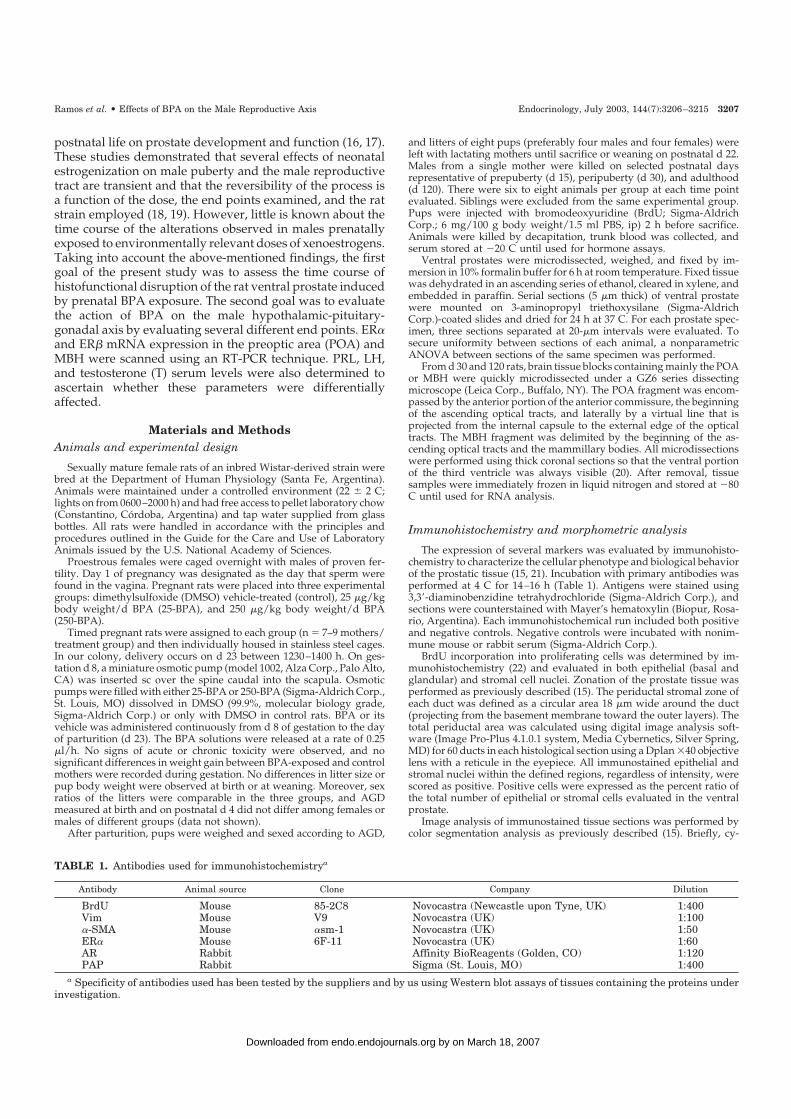
An optimized RT-PCR protocol was employed to analyze the relativeexpression levels of ER� and ER� mRNAs in the POA and MBH on d30 and 120 in rats in all experimental groups. Total RNA was isolatedusing the single step, acid guanidinium thiocyanate-phenol-chloroformextraction method (23). Equal quantities (4 �g) of total RNA were reversetranscribed into cDNA with avian myeloblastosis virus reverse tran-scriptase (12.5 U; Promega Corp., Madison, WI) using 200 pmol randomprimers (Promega Corp.). Twenty units of ribonuclease inhibitor(RNAout, Invitrogen, Buenos Aires, Argentina) and 100 nmol of a deoxyNTP mixture were added to each reaction tube in a final volume of 30�l of 1� avian myeloblastosis virus reverse transcriptase buffer. RT wasperformed at 42 C for 90 min. Reactions were terminated by heating at97 C for 5 min and cooling on ice, followed by dilution of the reversetranscribed cDNA with ribonuclease-free water to a final volume of 60�l. RNA incubated under identical conditions, but without reverse tran-scriptase, served as a negative control. Primer pairs used for amplifi-cation of the target genes are shown in Table 2. The primers were selectedbased upon a previous reference (24) according to the published cDNAsequences of the rat ER� (25), ER� (26), and L19 ribosomal protein (27).All amplifications were performed in duplicate. To perform comparativePCR, aliquots of cDNA samples equivalent to 800 ng total RNA inputwere used in each PCR amplification. Each reaction mixture contained2.5 U Taq DNA polymerase (Promega Corp.), 2.5 mm MgCl2 (PromegaCorp.), 0.2 mm of each of the four deoxy-NTPs (Promega Corp.), and 20pmol of each primer (Invitrogen) in a final volume of 50 �l of 1� PCRTaq buffer. After initial denaturation at 97 C for 5 min, the reactionmixture was subjected to successive cycles of denaturation at 96 C for1 min, annealing at 55 C for 1 min, and extension at 72 C for 2 min. Afinal extension cycle at 72 C for 15 min was included. The optimalnumber of cycles for each reaction was determined experimentally toyield linear relationships between signal intensity and cycle number(Fig. 1). The optimal number of cycles was 36 for ER� and ER� and 20for L19 ribosomal protein, using separate reactions for each target gene.
In all assays negative controls using RNA without RT and Taq poly-merase-negative tubes were performed to minimize the introduction ofpotential artifacts. All PCR products were cloned using the TA cloningkit (Invitrogen), and specificity was confirmed by DNA sequencing (datanot shown).
The generated cDNA fragments were resolved on 1.5% agarose gelscontaining ethidium bromide (Sigma-Aldrich Corp.); molecular weightswere determined by comparison with molecular weight standards (CienMarker, Biodynamics, Buenos Aires, Argentina). Agarose gel imageswere digitized using a Sony ExwaveHAM color video camera (SonyElectronics, Inc., Park Ridge, NJ) and the Image Pro-Plus 4.1.0.1 imagesystem analyzer (Media Cybernetics, Silver Spring, MD). The absoluteOD for each PCR product was obtained by densitometry. Values for ER�and ER� PCR-amplified sequences were normalized with respect to thatof the L19 ribosomal protein, allowing relative levels of the specificmRNAs to be expressed in arbitrary units.
Hormone assays
Serum levels of PRL and LH were determined by RIA using the kitprovided by the NIDDK as previously described (28, 29). Results wereexpressed in terms of rat PRL (RP3) and rat LH (RP3) reference prep-arations. Intra- and interassay coefficients of variation for PRL were 8.1%and 11.4%, respectively, whereas coefficients for LH were 5.4% and 9.8%,respectively. The lowest detectable levels were 0.039 ng/ml for PRL and0.016 ng/ml for LH. T concentrations were determined by RIA using[1,2-N-3H]T (60 Ci/mmol; NEN Life Science Products, Boston, MA) anda specific antibody (Immunotech Diagnostic, Montreal, Canada) as pre-viously described and validated by Suescun et al. (30). The sensitivityof the assay was 12 pg/ml, and the intraassay coefficients of variationwere 7.1%.
Data analysis
Statistical analyses were performed using the Kruskal-Wallis one-way ANOVA, and significance between groups was determined usingthe Dunn’s posttest. P � 0.05 was accepted as indicating a significantdifference between groups. Nonlinear correlations were determined bysigmoidal approximation (31).
ResultsPrenatal BPA exposure induces transient changes instromal cell phenotype and proliferative activity in theventral prostate
The absolute weights of the ventral prostates are presentedin Table 3. There were no significant differences in theweights of ventral prostates between BPA-treated and con-trol animals at any of the ages studied. At a histological level,
TABLE 2. Sequence of primers used in RT-PCR experiments
Primer name Sequence PCR product size (bp)
ER� sense 5�-AAT TCT GAC AAT CGA CGC CAG-3�ER� antisense 5�-GTG CTT CAA CAT TCT CCC TCC TC-3� 345ER� sense 5�-TTC CCG GCA GCA CCA GTA ACC-3�ER� antisense 5�-TCC CTC TTT GCG TTT GGA CTA-3� 262L19 sense 5�-GAA ATC GCC AAT GCC AAC TC-3�L19 antisense 5�-ACC TTC AGG TAC AGG CTG TG-3� 290
TABLE 3. Mean � SEM of absolute ventral prostate weights atnecropsy
AgeExperimental group
Control 25-BPA 250-BPA
d 15 22.46 � 5.12 23.57 � 3.53 21.79 � 4.36d 30 119.4 � 7.96 115.46 � 9.27 113.28 � 8.59d 120 474.64 � 34.4 470.30 � 36.1 462.45 � 43.9
3208 Endocrinology, July 2003, 144(7):3206–3215 Ramos et al. • Effects of BPA on the Male Reproductive Axis
by on March 18, 2007 endo.endojournals.orgDownloaded from
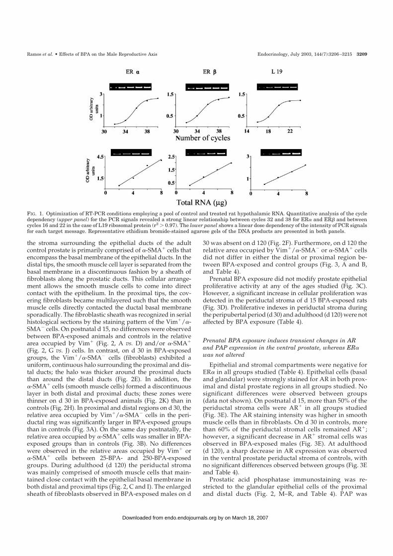
the stroma surrounding the epithelial ducts of the adultcontrol prostate is primarily comprised of �-SMA� cells thatencompass the basal membrane of the epithelial ducts. In thedistal tips, the smooth muscle cell layer is separated from thebasal membrane in a discontinuous fashion by a sheath offibroblasts along the prostatic ducts. This cellular arrange-ment allows the smooth muscle cells to come into directcontact with the epithelium. In the proximal tips, the cov-ering fibroblasts became multilayered such that the smoothmuscle cells directly contacted the ductal basal membranesporadically. The fibroblastic sheath was recognized in serialhistological sections by the staining pattern of the Vim�/�-SMA� cells. On postnatal d 15, no differences were observedbetween BPA-exposed animals and controls in the relativearea occupied by Vim� (Fig. 2, A vs. D) and/or �-SMA�
(Fig. 2, G vs. J) cells. In contrast, on d 30 in BPA-exposedgroups, the Vim�/�-SMA� cells (fibroblasts) exhibited auniform, continuous halo surrounding the proximal and dis-tal ducts; the halo was thicker around the proximal ductsthan around the distal ducts (Fig. 2E). In addition, the�-SMA� cells (smooth muscle cells) formed a discontinuouslayer in both distal and proximal ducts; these zones werethinner on d 30 in BPA-exposed animals (Fig. 2K) than incontrols (Fig. 2H). In proximal and distal regions on d 30, therelative area occupied by Vim�/�-SMA� cells in the peri-ductal ring was significantly larger in BPA-exposed groupsthan in controls (Fig. 3A). On the same day postnatally, therelative area occupied by �-SMA� cells was smaller in BPA-exposed groups than in controls (Fig. 3B). No differenceswere observed in the relative areas occupied by Vim� or�-SMA� cells between 25-BPA- and 250-BPA-exposedgroups. During adulthood (d 120) the periductal stromawas mainly comprised of smooth muscle cells that main-tained close contact with the epithelial basal membrane inboth distal and proximal tips (Fig. 2, C and I). The enlargedsheath of fibroblasts observed in BPA-exposed males on d
30 was absent on d 120 (Fig. 2F). Furthermore, on d 120 therelative area occupied by Vim�/�-SMA� or �-SMA� cellsdid not differ in either the distal or proximal region be-tween BPA-exposed and control groups (Fig. 3, A and B,and Table 4).
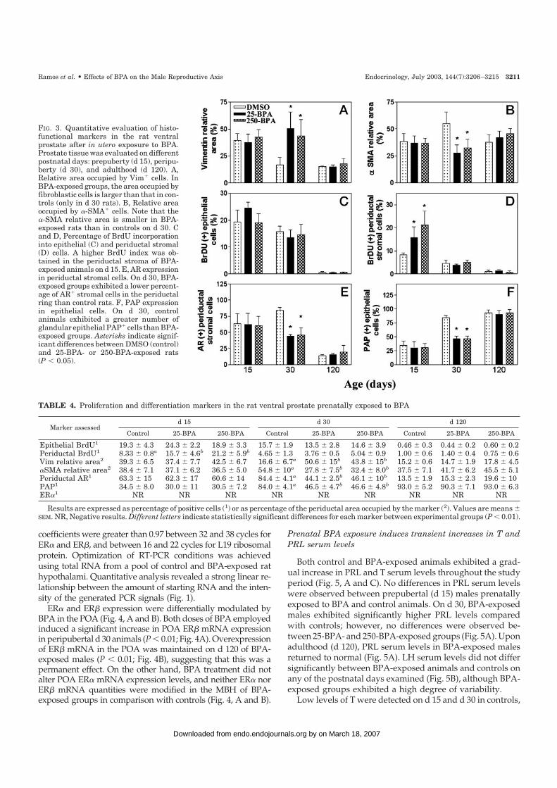
Prenatal BPA exposure did not modify prostate epithelialproliferative activity at any of the ages studied (Fig. 3C).However, a significant increase in cellular proliferation wasdetected in the periductal stroma of d 15 BPA-exposed rats(Fig. 3D). Proliferative indexes in periductal stroma duringthe peripubertal period (d 30) and adulthood (d 120) were notaffected by BPA exposure (Table 4).
Prenatal BPA exposure induces transient changes in ARand PAP expression in the ventral prostate, whereas ER�was not altered
Epithelial and stromal compartments were negative forER� in all groups studied (Table 4). Epithelial cells (basaland glandular) were strongly stained for AR in both prox-imal and distal prostate regions in all groups studied. Nosignificant differences were observed between groups(data not shown). On postnatal d 15, more than 50% of theperiductal stroma cells were AR� in all groups studied(Fig. 3E). The AR staining intensity was higher in smoothmuscle cells than in fibroblasts. On d 30 in controls, morethan 60% of the periductal stromal cells remained AR�;however, a significant decrease in AR� stromal cells wasobserved in BPA-exposed males (Fig. 3E). At adulthood(d 120), a sharp decrease in AR expression was observedin the ventral prostate periductal stroma of controls, withno significant differences observed between groups (Fig. 3Eand Table 4).
Prostatic acid phosphatase immunostaining was re-stricted to the glandular epithelial cells of the proximaland distal ducts (Fig. 2, M–R, and Table 4). PAP was
FIG. 1. Optimization of RT-PCR conditions employing a pool of control and treated rat hypothalamic RNA. Quantitative analysis of the cycledependency (upper panel) for the PCR signals revealed a strong linear relationship between cycles 32 and 38 for ER� and ER� and betweencycles 16 and 22 in the case of L19 ribosomal protein (r2 0.97). The lower panel shows a linear dose dependency of the intensity of PCR signalsfor each target message. Representative ethidium bromide-stained agarose gels of the DNA products are presented in both panels.
Ramos et al. • Effects of BPA on the Male Reproductive Axis Endocrinology, July 2003, 144(7):3206–3215 3209
by on March 18, 2007 endo.endojournals.orgDownloaded from
expressed throughout the cytoplasm; however, stainingwas concentrated in the apical region. In controls therewas a sharp increase in the percentage of PAP� cells andin the staining intensity from d 15–30 (Fig. 2, M vs. N),which was sustained on d 120 (Fig. 2O). In contrast, BPA-exposed animals did not exhibit this sharp increase on d30 (Fig. 2Q), and levels of PAP expression returned tocontrol values on d 120 (Figs. 2R and 3F).
Prenatal BPA exposure permanently up-regulates ER�mRNA expression in the male POA
The relative expression of ER� and ER� mRNAs in the POAand MBH of d 30 and 120 animals was evaluated by RT-PCR.Validation of the RT-PCR assays is summarized in Fig. 1. Foramplification in the exponential phase of PCR, different num-bers of cycles were tested for each mRNA. All linear correlation
FIG. 2. Photomicrographs showing a transient histofunctional disruption of the rat ventral prostate after in utero exposure to BPA. Immu-nohistochemical evaluation of Vim, �-SMA, and PAP was performed at prepuberty (d 15), peripuberty (d 30), and adulthood (d 120). In controlanimals, the periductal immunostaining pattern for Vim was discontinuous in distal tips (B, arrow), whereas the �-SMA layer formed acontinuous ring around the ducts (H, arrow). In BPA-exposed groups, the Vim-staining pattern (fibroblastic cells) was presented as a uniform,thick halo surrounding the ducts only on d 30 (E, arrowhead). In addition, on d 30, the �-SMA layer (smooth muscle cells) presented severaldiscontinuities and was thinner (K, arrowhead) than in controls. No differences were observed in Vim and/or �-SMA expression patterns inthe periductal stroma of d 15 and 120 rats. PAP is a cytoplasmic marker of epithelial cells (M–R). Although PAP expression was presentthroughout the entire cytoplasm, the apical region was more intensely stained than basal region (N, arrow). In control animals, there was asharp increase in the percentage of PAP� cells, and in the staining intensity from d 15–30, which was sustained on d 120. In contrast,BPA-exposed animals did not exhibit this sharp increase on d 30 (Q, arrowhead); levels of PAP expression returned to control values by d 120.Solid bar, 50 �m.
3210 Endocrinology, July 2003, 144(7):3206–3215 Ramos et al. • Effects of BPA on the Male Reproductive Axis
by on March 18, 2007 endo.endojournals.orgDownloaded from
coefficients were greater than 0.97 between 32 and 38 cycles forER� and ER�, and between 16 and 22 cycles for L19 ribosomalprotein. Optimization of RT-PCR conditions was achievedusing total RNA from a pool of control and BPA-exposed rathypothalami. Quantitative analysis revealed a strong linear re-lationship between the amount of starting RNA and the inten-sity of the generated PCR signals (Fig. 1).
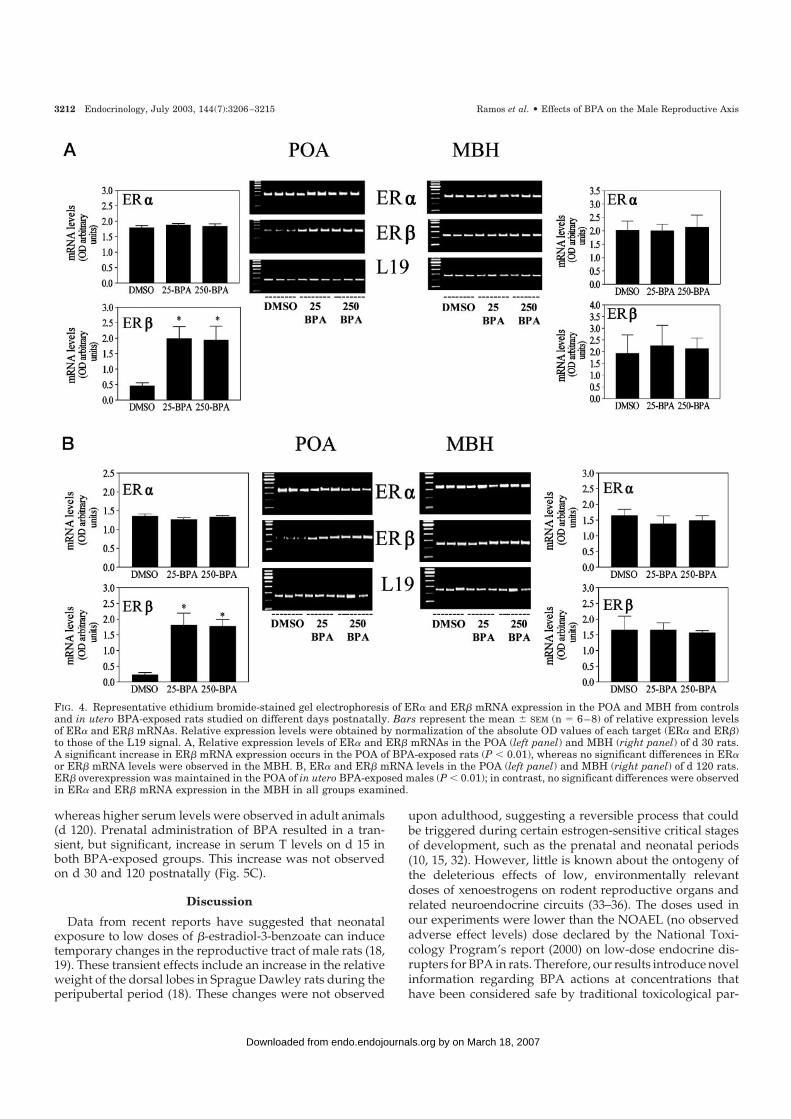
ER� and ER� expression were differentially modulated byBPA in the POA (Fig. 4, A and B). Both doses of BPA employedinduced a significant increase in POA ER� mRNA expressionin peripubertal d 30 animals (P � 0.01; Fig. 4A). Overexpressionof ER� mRNA in the POA was maintained on d 120 of BPA-exposed males (P � 0.01; Fig. 4B), suggesting that this was apermanent effect. On the other hand, BPA treatment did notalter POA ER� mRNA expression levels, and neither ER� norER� mRNA quantities were modified in the MBH of BPA-exposed groups in comparison with controls (Fig. 4, A and B).
Prenatal BPA exposure induces transient increases in T andPRL serum levels
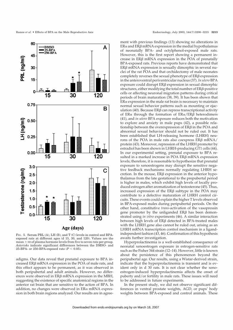
Both control and BPA-exposed animals exhibited a grad-ual increase in PRL and T serum levels throughout the studyperiod (Fig. 5, A and C). No differences in PRL serum levelswere observed between prepubertal (d 15) males prenatallyexposed to BPA and control animals. On d 30, BPA-exposedmales exhibited significantly higher PRL levels comparedwith controls; however, no differences were observed be-tween 25-BPA- and 250-BPA-exposed groups (Fig. 5A). Uponadulthood (d 120), PRL serum levels in BPA-exposed malesreturned to normal (Fig. 5A). LH serum levels did not differsignificantly between BPA-exposed animals and controls onany of the postnatal days examined (Fig. 5B), although BPA-exposed groups exhibited a high degree of variability.
Low levels of T were detected on d 15 and d 30 in controls,
FIG. 3. Quantitative evaluation of histo-functional markers in the rat ventralprostate after in utero exposure to BPA.Prostate tissue was evaluated on differentpostnatal days: prepuberty (d 15), peripu-berty (d 30), and adulthood (d 120). A,Relative area occupied by Vim� cells. InBPA-exposed groups, the area occupied byfibroblastic cells is larger than that in con-trols (only in d 30 rats). B, Relative areaoccupied by �-SMA� cells. Note that the�-SMA relative area is smaller in BPA-exposed rats than in controls on d 30. Cand D, Percentage of BrdU incorporationinto epithelial (C) and periductal stromal(D) cells. A higher BrdU index was ob-tained in the periductal stroma of BPA-exposed animals on d 15. E, AR expressionin periductal stromal cells. On d 30, BPA-exposed groups exhibited a lower percent-age of AR� stromal cells in the periductalring than control rats. F, PAP expressionin epithelial cells. On d 30, controlanimals exhibited a greater number ofglandular epithelial PAP� cells than BPA-exposed groups. Asterisks indicate signif-icant differences between DMSO (control)and 25-BPA- or 250-BPA-exposed rats(P � 0.05).
TABLE 4. Proliferation and differentiation markers in the rat ventral prostate prenatally exposed to BPA
Marker assessedd 15 d 30 d 120
Control 25-BPA 250-BPA Control 25-BPA 250-BPA Control 25-BPA 250-BPA
Epithelial BrdU1 19.3 � 4.3 24.3 � 2.2 18.9 � 3.3 15.7 � 1.9 13.5 � 2.8 14.6 � 3.9 0.46 � 0.3 0.44 � 0.2 0.60 � 0.2Periductal BrdU1 8.33 � 0.8a 15.7 � 4.6b 21.2 � 5.9b 4.65 � 1.3 3.76 � 0.5 5.04 � 0.9 1.00 � 0.6 1.40 � 0.4 0.75 � 0.6Vim relative area2 39.3 � 6.5 37.4 � 7.7 42.5 � 6.7 16.6 � 6.7a 50.6 � 15b 43.8 � 15b 15.2 � 0.6 14.7 � 1.9 17.8 � 4.5�SMA relative area2 38.4 � 7.1 37.1 � 6.2 36.5 � 5.0 54.8 � 10a 27.8 � 7.5b 32.4 � 8.0b 37.5 � 7.1 41.7 � 6.2 45.5 � 5.1Periductal AR1 63.3 � 15 62.3 � 17 60.6 � 14 84.4 � 4.1a 44.1 � 2.5b 46.1 � 10b 13.5 � 1.9 15.3 � 2.3 19.6 � 10PAP1 34.5 � 8.0 30.0 � 11 30.5 � 7.2 84.0 � 4.1a 46.5 � 4.7b 46.6 � 4.8b 93.0 � 5.2 90.3 � 7.1 93.0 � 6.3ER�1 NR NR NR NR NR NR NR NR NR
Results are expressed as percentage of positive cells (1) or as percentage of the periductal area occupied by the marker (2). Values are means �SEM. NR, Negative results. Different letters indicate statistically significant differences for each marker between experimental groups (P � 0.01).
Ramos et al. • Effects of BPA on the Male Reproductive Axis Endocrinology, July 2003, 144(7):3206–3215 3211
by on March 18, 2007 endo.endojournals.orgDownloaded from
whereas higher serum levels were observed in adult animals(d 120). Prenatal administration of BPA resulted in a tran-sient, but significant, increase in serum T levels on d 15 inboth BPA-exposed groups. This increase was not observedon d 30 and 120 postnatally (Fig. 5C).
Discussion
Data from recent reports have suggested that neonatalexposure to low doses of �-estradiol-3-benzoate can inducetemporary changes in the reproductive tract of male rats (18,19). These transient effects include an increase in the relativeweight of the dorsal lobes in Sprague Dawley rats during theperipubertal period (18). These changes were not observed
upon adulthood, suggesting a reversible process that couldbe triggered during certain estrogen-sensitive critical stagesof development, such as the prenatal and neonatal periods(10, 15, 32). However, little is known about the ontogeny ofthe deleterious effects of low, environmentally relevantdoses of xenoestrogens on rodent reproductive organs andrelated neuroendocrine circuits (33–36). The doses used inour experiments were lower than the NOAEL (no observedadverse effect levels) dose declared by the National Toxi-cology Program’s report (2000) on low-dose endocrine dis-rupters for BPA in rats. Therefore, our results introduce novelinformation regarding BPA actions at concentrations thathave been considered safe by traditional toxicological par-
FIG. 4. Representative ethidium bromide-stained gel electrophoresis of ER� and ER� mRNA expression in the POA and MBH from controlsand in utero BPA-exposed rats studied on different days postnatally. Bars represent the mean � SEM (n � 6–8) of relative expression levelsof ER� and ER� mRNAs. Relative expression levels were obtained by normalization of the absolute OD values of each target (ER� and ER�)to those of the L19 signal. A, Relative expression levels of ER� and ER� mRNAs in the POA (left panel) and MBH (right panel) of d 30 rats.A significant increase in ER� mRNA expression occurs in the POA of BPA-exposed rats (P � 0.01), whereas no significant differences in ER�or ER� mRNA levels were observed in the MBH. B, ER� and ER� mRNA levels in the POA (left panel) and MBH (right panel) of d 120 rats.ER� overexpression was maintained in the POA of in utero BPA-exposed males (P � 0.01); in contrast, no significant differences were observedin ER� and ER� mRNA expression in the MBH in all groups examined.
3212 Endocrinology, July 2003, 144(7):3206–3215 Ramos et al. • Effects of BPA on the Male Reproductive Axis
by on March 18, 2007 endo.endojournals.orgDownloaded from
adigms. Our data reveal that prenatal exposure to BPA in-creased ER� mRNA expression in the POA of male rats, andthis effect appears to be permanent, as it was observed inboth peripubertal and adult animals. However, no differ-ences were observed in ER� mRNA expression in the MBH,suggesting the existence of specific anatomical regions in theanterior rat brain that are sensitive to the action of BPA. Inaddition, no changes were observed in ER� mRNA expres-sion in both brain regions analyzed. Our results are in agree-
ment with previous findings (13) showing no alterations inER� and ER� mRNA expression in the medial hypothalamusof neonatally BPA- and octylphenol-exposed male rats.However, this is the first report showing a permanent in-crease in ER� mRNA expression in the POA of prenatallyBPA-exposed rats. Previous reports have demonstrated thatER� mRNA expression is sexually dimorphic in several nu-clei of the rat POA and that orchidectomy of male neonatescompletely reverses the sexual phenotype of ER� expressionin the anteroventral periventricular nucleus (37). In utero BPAexposure could disrupt ER� expression in sexual dimorphicstructures, either modifying the total number of ER�-positivecells or affecting neuronal migration patterns during criticalperiods of brain maturation (38, 39). It has been shown thatER� expression in the male rat brain is necessary to maintainnormal sexual behavior patterns such as mounting or ejac-ulation (40). Because ER� can repress transcriptional activityof ER� through the formation of ER�/ER� heterodimers(41), and in utero BPA exposure reduces both the motivationto explore and anxiety in male pups (42), a possible rela-tionship between the overexpression of ER� in the POA andabnormal sexual behavior should not be ruled out. It hasbeen established that LH-releasing hormone (LHRH) neu-rons of the POA in male rats also coexpress ER� mRNA/protein (43). Moreover, repression of the LHRH promoter byestradiol has been shown in LHRH-producing GT1 cells (44).In our experimental setting, prenatal exposure to BPA re-sulted in a marked increase in POA ER� mRNA expressionlevels; therefore, it is reasonable to hypothesize that prenatalexposure to xenoestrogens may disrupt the sensitive nega-tive feedback mechanisms normally regulating LHRH se-cretion. In the mouse, ER� expression in the anterior hypo-thalamus from the late gestational to the prepubertal periodis higher in males, which exhibit high levels of locally pro-duced estrogen after aromatization of testosterone (45). Thus,increased expression of the ER� subtype in the POA maycontribute to a defective maturation of LHRH control cir-cuits. These events could explain the higher T levels observedin BPA-exposed males during peripubertal periods. On theother hand, constitutive trans-activation of the vasopressingene promoter by the unliganded ER� has been demon-strated using in vitro experiments (46). A similar interactionbetween high levels of ER� detected in BPA-treated maleswith the LHRH gene also cannot be ruled out, arising as anLHRH mRNA transcription control mechanism in a ligand-independent fashion (43, 46). Confirmation of this hypothesisawaits further investigation.
Hyperprolactinemia is a well-established consequence ofneonatal xenoestrogen exposure in estrogen-sensitive ratssuch as the Fisher 344 strain (12–14). However, little is knownabout the persistence of this phenomenon beyond theperipubertal age. Our results, using a Wistar-derived strain,indicate that the hyperprolactinemia is transient and is ev-ident only in d 30 rats. It is not clear whether the xeno-estrogen-induced hyperprolactinemia affects the onset ofpuberty and/or fertility in male rats. These issues will needto be addressed in future experiments.
In the present study, we did not observe significant dif-ferences in ventral prostate weights, AGD, or pups’ bodyweights between BPA-exposed and control animals. These
FIG. 5. Serum PRL (A), LH (B), and T (C) levels in control and BPA-exposed rats at different ages (d 15, 30, and 120). Values are themean � SD of plasma hormone levels from five to seven rats per group.Asterisks indicate significant differences between the DMSO- and25-BPA- or 250-BPA-exposed rats (P � 0.05).
Ramos et al. • Effects of BPA on the Male Reproductive Axis Endocrinology, July 2003, 144(7):3206–3215 3213
by on March 18, 2007 endo.endojournals.orgDownloaded from
results are in accordance with previous studies that used asimilar range of BPA doses (47). However, we demonstratedin a previous study (15) that prenatal exposure to BPA altersthe differentiation pattern of the periductal stromal cells inthe rat ventral prostate on postnatal d 30. These results takentogether suggest that differences in organ weight-related endpoints would not have enough sensitivity to significantlyevaluate the effects of low doses of BPA. In BPA-exposedgroups, the presence of a thick layer of Vim�/�-SMA– cellsin the periductal zone contrasts with the multicellularsmooth muscle Vim–/�-SMA� layer observed in controls.Herein, we demonstrated that the presence of a thicker layerof fibroblasts in the periductal stroma on d 30 is the conse-quence of a modified proliferative status during early de-velopment (d 15). Contemporary with this disruption of cel-lular dynamics in BPA-exposed rats, increased T serumlevels were observed. A cause-effect relationship betweenhigher T serum levels and the modified proliferative indexesin the periductal stroma cannot be ruled out. Previous workhas shown that ER� expression is confined to mesenchymalcells only during early development of ventral prostate (48).In agreement with this study and confirming our previousreport (15), control groups were negative for ER� in bothepithelial and stromal compartments, and BPA did not mod-ify this pattern of expression. Decreased PAP expressionobserved in BPA-exposed groups could be mediated by ei-ther a direct effect of BPA on the columnar epithelial cells orthrough an indirect consequence of primary events occurringin the stroma (16, 17, 49). Stromal signals are believed to becritical in determining the decision of epithelial cells to un-dergo proliferation, apoptosis, or differentiation. The de-creased AR expression observed in the periductal stromalcells on d 30 may affect the androgen-signaling pathwayresulting in decreased PAP expression, although a directeffect of BPA on the epithelial cells also cannot be ruled out.During adulthood, no differences were observed in the peri-ductal stromal architecture. Prostatic acid phosphatase ex-pression in columnar epithelial cells during adulthood wasincreased in all groups analyzed, and the differences ob-served on d 30 were not present on d 120. In addition, func-tional relationships have been established between hyper-prolactinemia and prostatic tissue disorders in rats (14). Asthe histoarchitectural and functional changes that were ob-served in the ventral prostates of BPA-exposed rats are tem-porally associated with elevated serum PRL levels, furtherstudies will need to be undertaken to elucidate the possiblerelationships between these phenomena. The transient char-acteristic of the changes observed in the ventral prostates andin T and PRL serum levels could be the result of early or-ganizational effects that are associated with the actions ofexogenous or endogenous estrogens during organogenesisand development (1). Our hypothesis is that organizationaleffects induced by low doses of xenoestrogens during earlydevelopment stages could be expressed during critical pe-riods such as puberty, when the hormonal milieu exhibits ahigh degree of variation, and compensatory mechanisms(such as negative feedback systems) are not fully developed.
In summary, in utero exposure to environmentally relevantlevels of BPA increased ER� mRNA expression in the ante-rior hypothalamic structures of the pubertal male rat, and
this overexpression persisted into adulthood. In contrast,prenatal exposure to BPA transiently affects the ventral pros-tate of peripubertal rats by decreasing AR expression, alter-ing proliferative activity in the periductal stromal cells, anddecreasing PAP expression. All of these morphological andfunctional changes that were observed in the ventral prostateon d 30 were transient, as they were not observed duringadulthood. In addition, other transient effects observed wereincreases in T and PRL serum levels in prepubertal BPA-exposed rats, whereas no differences were detected duringadulthood. A close time dependence of the biological re-sponses to xenoestrogens during development together withthe differential estrogen sensitivity of the various rat strainsemployed and/or end points examined could explain at leastin part the extraordinary variability in the in vivo resultsobtained to date regarding endocrine disrupters (1, 10, 47,50, 51).
The findings reported in this study support the hypothesisthat environmental xenoestrogen exposure may be associ-ated with low seminal quality and other male reproductivedysfunctions that affect human fertility (2, 3).
Acknowledgments
We are very grateful to Dr. Hugo F. Carrer [the Argentine NationalCouncil for Science and Technology (CONICET), Cordoba, Argentina]for neuroanatomical advice, to Dr. Charles E. Powell (Nova SoutheasternUniversity, Ft. Lauderdale, FL) for critical reading of the manuscript, andto Drs. Leonardo Bussmann and Ricardo Calandra (IBYME, BuenosAires, Argentina) for valuable RIA contributions. We also thank Mr. JuanC. Villarreal and Mr. Juan Grant for technical assistance and animal careand the NIDDK for RIA reagents.
Received December 26, 2002. Accepted March 19, 2003.Address all correspondence and requests for reprints to: Enrique H.
Luque, Ph.D., Laboratorio de Endocrinologıa y Tumores Hormonode-pendientes, School of Biochemistry and Biological Sciences, Casilla deCorreo 242, 3000 Santa Fe, Argentina. E-mail: [email protected].
This work was supported by grants from the Argentine Ministry ofHealth (Carrillo-Onativia Award), the Argentine National Agency forthe Promotion of Science and Technology (PICT-99 13-7002), and theNational University of Litoral (CAID96-2001). J.G.R. and J.V. are Fellowsof the CONICET. L.C. is a Carrillo-Onativia Fellow. E.H.L. is a CareerInvestigator of the CONICET.
* J.G.R. and J.V. contributed equally to this work.
References
1. McLachlan JA 2001 Environmental signaling: what embryos and evolutionteach us about endocrine disrupting chemicals. Endocr Rev 22:319–341
2. Oliva A, Giami A, Multigner L 2002 Environmental agents and erectile dys-function: a study in a consulting population. J Androl 23:546–550
3. Toppari J, Larsen JC, Christiansen P, Giwercman A, Grandjean P, GuilletteLJ, Jegou B, Jensen TK, Jouannet P, Keiding N, Leffers H, MacLachlan JA,Meyer OM, Muller J, Rajperts-De Meyts E, Scheike T, Sharpe R, SumplerJ, Skakkebaek NE 1996 Male reproductive health and environmental xe-noestrogens. Environ Health Perspect 104:741–803
4. Brotons JA, Olea-Serrano MF, Villalobos M, Olea N 1994 Xenoestrogensreleased from lacquer coating in food cans. Environ Health Perspect 103:608–612
5. Biles JE, McNeal TP, Begley TH, Hollifield HC 1997 Determination ofBisphenol-A in reusable polycarbonate food-contact plastics and migration tofood simulating liquids. J Agric Food Chem 45:3541–3544
6. Olea N, Pulgar R, Perez P, Olea-Serrano F, Rivas A, Novillo-Fertrell A,Pedraza V, Soto AM, Sonnenschein C 1996 Estrogenicity of resin-based com-posites and sealants used in dentistry. Environ Health Perspect 104:298–305
7. Ikezuki Y, Tsutsumi O, Takai Y, Kamei Y, Taketani Y 2002 Determination ofbisphenol A concentrations in human biological fluids reveals significant earlyprenatal exposure. Hum Reprod 17:2839–2841
8. Schonfelder G, Wittfoht W, Hopp H, Talsness CE, Paul M, Chahoud I 2002
3214 Endocrinology, July 2003, 144(7):3206–3215 Ramos et al. • Effects of BPA on the Male Reproductive Axis
by on March 18, 2007 endo.endojournals.orgDownloaded from

Parent bisphenol A accumulation in the human maternal-fetal-placental unit.Environ Health Perspect 110:A703–A707
9. Welshons WV, Nagel SC, Thayer KA, Judy BM, Vom Saal FS 1999 Low-dosebioactivity of xenoestrogens in animals: fetal exposure to low doses of me-thoxychlor and other xenoestrogens increases adult prostate size in mice.Toxicol Ind Health 15:12–25
10. Gupta C 2000 Reproductive malformation of the male offspring followingmaternal exposure to estrogenic chemicals. Proc Soc Exp Biol Med 224:61–68
11. Rubin BS, Murray MK, Damassa DA, King JC, Soto AM 2001 Perinatalexposure to low doses of bisphenol-A affects body weight, patterns of estrouscyclicity and plasma LH levels. Environ Health Perspect 109:675–680
12. Steinmetz R, Brown NG, Allen DL, Bigsby RM, Ben-Jonathan N 1997 Theenvironmental estrogen bisphenol A stimulates prolactin release in vitro andin vivo. Endocrinology 138:1780–1786
13. Khurana S, Ranmal S, Ben-Jonathan N 2000 Exposure of newborn male andfemale rats to environmental estrogens: delayed and sustained hyperpro-lactinemia and alterations in estrogen receptor expression. Endocrinology141:4512–4517
14. Stoker TE, Robinette CL, Britt BH, Laws SC, Cooper R 1999 Prepubertalexposure to compounds that increase prolactin secretion in the male rat: effectson the adult prostate. Biol Reprod 61:1636–1643
15. Ramos JG, Varayoud J, Sonnenschein C, Soto AM, Munoz-de-Toro M,Luque EH 2001 Prenatal exposure to low doses of bisphenol A alters theperiductal stroma and glandular cell function in the rat ventral prostate. BiolReprod 65:1271–1277
16. Chang WY, Wilson MJ, Birch L, Prins GS 1999 Neonatal estrogen stimulatesproliferation of periductal fibroblasts and alters the extracellular matrix com-position in the rat prostate. Endocrinology 140:405–415
17. Habermann H, Ghang WY, Birch L, Mehta P, Prins GS 2001 Developmentalexposure to estrogens alters epithelial cell adhesion and gap junction proteinsin the adult rat prostate. Endocrinology 142:359–369
18. Putz O, Schwartz CB, Kim S, LeBlanc GA, Cooper RL, Prins GS 2001 Neo-natal low- and high-dose exposure to estradiol benzoate in the male rat. I.Effects on the prostate gland. Biol Reprod 65:1496–1505
19. Putz O, Schwartz CB, LeBlanc GA, Cooper RL, Prins GS 2001 Neonatal low-and high-dose exposure to estradiol benzoate in the male rat. II. Effects on theprostate gland. Biol Reprod 65:1506–1517
20. Paxinos G, Watson C 1997 The rat brain in stereotaxic coordinates. Compact3rd ed. San Diego: Academic Press
21. Munoz-de-Toro M, Maffini M, Kass L, Luque EH 1998 Proliferative activityand steroid hormone receptor status in male breast carcinoma. J SteroidBiochem Mol Biol 67:333–339
22. Kass L, Varayoud JG, Ortega HH, Munoz-de-Toro MM, Luque EH 2000Detection of bromodeoxyuridine in formalin-fixed tissue. DNA denaturationfollowing microwave or enzymatic digestion pretreatment is required. EurJ Histochem 44:185–191
23. Chomczynski P, Sacchi N 1987 Single-step method of RNA isolation by acidguanidinium thiocyanate-phenol-chloroform extraction. Anal Biochem 162:156–159
24. Tena-Sempere M, Navarro J, Pinilla L, Gonzalez L, Huhtaniemi I, AguilarE 2000 Neonatal exposure to estrogen differentially alters estrogen receptor �and � mRNA expression in rat testis during postnatal development. J Endo-crinol 165:345–357
25. Koike S, Saka M, Muramatsu M 1987 Molecular cloning and characterizationof rat estrogen receptor cDNA. Nucleic Acids Res 15:2499–2513
26. Kuiper GGJM, Enmark E, Pelto-Huikko M, Nilsso S, Gustafsson J-A 1996Cloning of a novel estrogen receptor expressed in rat prostate and ovary. ProcNatl Acad Sci USA 93:5925–5930.
27. Chang X-L, Lin A, McNally J, Pelleg D, Meyuha O, Wool Y 1987 The primarystructure of rat ribosomal protein L19. J Biol Chem 262:1111–1115
28. Lux-Lantos V, Becu-Villalobos D, Bianchi M, Rey-Roldan E, Chamson-ReigA, Pignataro O, Libertun C 2001 GABA(B) receptors in anterior pituitary cells.Mechanism of action coupled to endocrine effects. Neuroendocrinology 73:334–343
29. Luque EH, Munoz-de-Toro M, Smith PF, Neill JD 1986 Subpopulations oflactotropes detected with the reverse hemolytic plaque assay show differentialresponsiveness to dopamine. Endocrinology 118:2120–2124
30. Suescun MO, Gonzalez SI, Chiauzzi VA, Calandra RS 1985 Effects of inducedhypoprolactinemia on testicular function during gonadal maturation in the rat.J Androl 6:77–82
31. Siegel S 1956 Nonparametric statistics for the behavioral sciences. New York:McGraw-Hill
32. Timms BG, Petersen SL, Vom Saal FS 1999 Prostate gland growth duringdevelopment is stimulated in both male and female rat fetuses by intrauterineproximity to female fetuses. J Urol 161:1694–1701
33. Vom Saal FS, Cooke PS, Buchanan DL, Palanza P, Thayer KA, Nagel SC,Parmigiani S, Welshons WV 1998 A physiologically based approach to thestudy of bisphenol A and other estrogenic chemicals on the size of reproductiveorgans, daily sperm production, and behavior. Toxicol Ind Health 14:239–260
34. Colerangle JB, Roy D 1997 Profound effects of the weak environmental es-trogen-like chemical bisphenol A on the growth of the mammary gland ofNoble rats. J Steroid Biochem Mol Biol 60:153–160
35. Sheehan DM 2000 Activity of environmentally relevant low doses of endo-crine disruptors and the bisphenol A controversy: initial results confirmed.Proc Soc Exp Biol Med 224:57–60
36. Funabashi T, Kawaguchi M, Kimura F 2001 The endocrine disrupters butylbenzyl phthalate and bisphenol A increase the expression of progesteronereceptor messenger ribonucleic acid in the preoptic area of adult ovariecto-mized rats. Neuroendocrinology 74:77–81
37. Orikasa C, Kondo Y, Hayashi S, McEwen BS, Sakuma Y 2002 Sexuallydimorphic expression of estrogen receptor � in the anteroventral periven-tricular nucleus of the rat preoptic area: Implication in luteinizing hormonesurge. Proc Natl Acad Sci USA 99:3306–3311
38. Tobet SA, Henderson RG, Whiting PJ, Sieghart W 1999 Special relationshipof �-aminobutyric acid to the ventromedial nucleus of the hypothalamusduring embryonic development. J Comp Neurol 405:88–98
39. Dellovade TL, Young M, Ross EP, Henderson R, Caron K, Parker K, TobetSA 2000 Disruption of the gene encoding SF-1 alters the distribution of hy-pothalamic neuronal phenotypes. J Comp Neurol 423:579–589
40. Ogawa S, Lubahn DB, Korach KS, Pfaff DW 1997 Behavioral effects ofestrogen receptor gene disruption in male mice. Proc Natl Acad Sci USA94:1476–1481
41. Weihua Z, Saji S, Makinen S, Cheng G, Jensen EV, Warner M, GustafssonJA 2000 Estrogen receptor (ER) �, a modulator of ER � in the uterus. Proc NatlAcad Sci USA 97:5936–5941
42. Farabollini F, Porrini S, Dessi-Fulgheri F 1999 Perinatal exposure to theestrogenic pollutant bisphenol A affects behavior in male and female rats.Pharmacol Biochem Behav 64:687–694
43. Hrabovszky E, Steinhauser A, Barabas K, Shughrue PJ, Petersen SL,Merchenthaler I, Liposits Z 2000 Estrogen receptor-� immunoreactivity inluteinizing hormone-releasing hormone neurons of the rat brain. Endocrinol-ogy 142:3261–3264
44. Roy D, Angelini NL, Belsham DD 1999 Estrogen directly represses gonado-tropin-releasing hormone (GnRH) gene expression in estrogen receptor-�(ER�)- and ER�-expressing GTI-7 GnRH neurons. Endocrinology 140:5045–5053
45. Karolczak M, Beyer C 1998 Developmental sex differences in estrogen recep-tor-� mRNA expression in the mouse hypothalamus/preoptic region. Neu-roendocrinology 68:229–234
46. Shapiro RA, Xu C, Dorsa DM 2000 Differential transcriptional regulation ofrat vasopressin gene expression by estrogen receptor � and �. Endocrinology141:4056–4064
47. Tinwell H, Haseman J, Lefevre PA, Wallis N, Ashby J 2002 Normal sexualdevelopment off two strains of rats exposed in utero to low doses of bisphenolA. Toxicol Sci 68:339–348
48. Prins GS, Birch L 1997 Neonatal estrogen exposure up-regulates estrogenreceptor expression in the developing an adult rat prostate lobes. Endocri-nology 138:1801–1809
49. Grossfeld GD, Hayward SW, Tlsty TD, Cunha GR 1998 The role of stromain prostatic carcinogenesis. Endocrine-Related Cancer 5:253–270
50. Kwon S, Stedman DB, Elswick BA, Cattley RC, Welsch F 2000 Pubertaldevelopment and reproductive functions of Crl:CD BR Sprague-Dawley ratsexpose to bisphenol A during prenatal and postnatal development. Toxicol Sci55:399–406
51. Goloubkova T, Ribeiro MFM, Rodrigues LP, Cecconello AL, Spritzer PM2000 Effects of xenoestrogen bisphenol A on uterine and pituitary weight,serum prolactin levels and immunoreactive prolactin cells in ovariectomizedWistar rats. Arch Toxicol 74:92–98
Ramos et al. • Effects of BPA on the Male Reproductive Axis Endocrinology, July 2003, 144(7):3206–3215 3215
by on March 18, 2007 endo.endojournals.orgDownloaded from