Study of oxidative stress induction after exposure to bisphenol a and methylparaben in rats
11
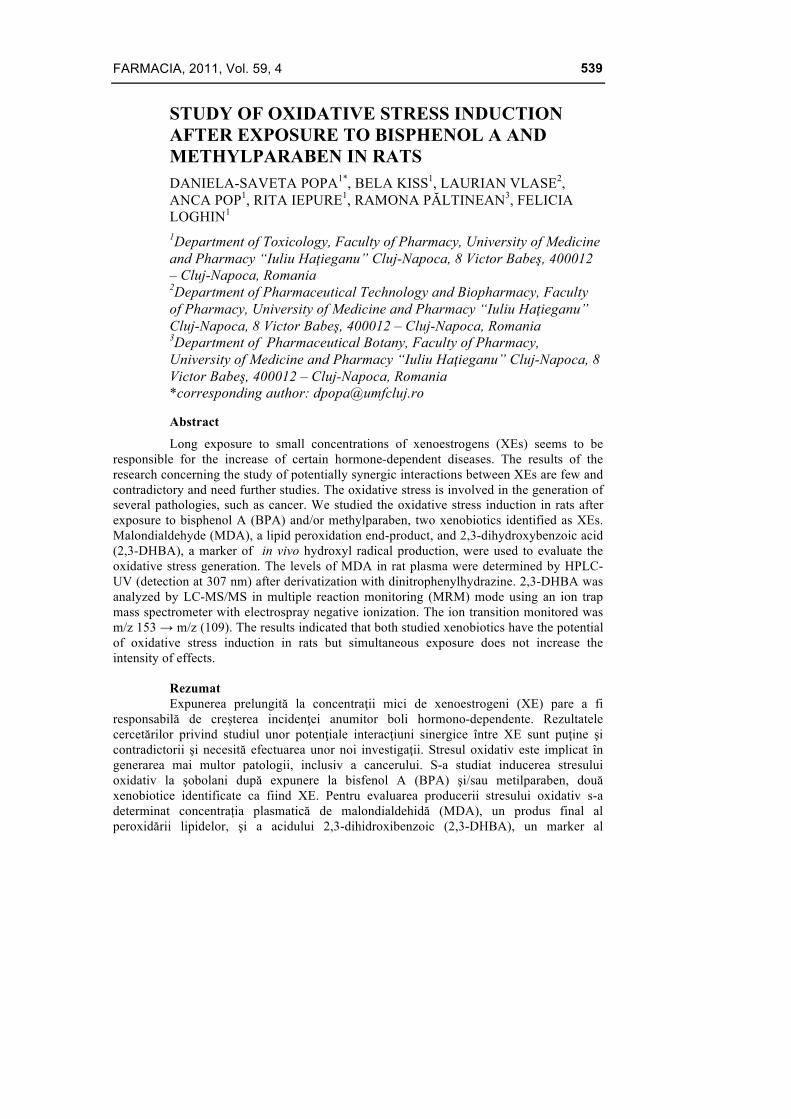
FARMACIA, 2011, Vol. 59, 4 539 STUDY OF OXIDATIVE STRESS INDUCTION AFTER EXPOSURE TO BISPHENOL A AND METHYLPARABEN IN RATS DANIELA-SAVETA POPA 1* , BELA KISS 1 , LAURIAN VLASE 2 , ANCA POP 1 , RITA IEPURE 1 , RAMONA PĂLTINEAN 3 , FELICIA LOGHIN 1 1 Department of Toxicology, Faculty of Pharmacy, University of Medicine and Pharmacy “Iuliu Haţieganu” Cluj-Napoca, 8 Victor Babeş, 400012 – Cluj-Napoca, Romania 2 Department of Pharmaceutical Technology and Biopharmacy, Faculty of Pharmacy, University of Medicine and Pharmacy “Iuliu Haţieganu” Cluj-Napoca, 8 Victor Babeş, 400012 – Cluj-Napoca, Romania 3 Department of Pharmaceutical Botany, Faculty of Pharmacy, University of Medicine and Pharmacy “Iuliu Haţieganu” Cluj-Napoca, 8 Victor Babeş, 400012 – Cluj-Napoca, Romania *corresponding author: [email protected] Abstract Long exposure to small concentrations of xenoestrogens (XEs) seems to be responsible for the increase of certain hormone-dependent diseases. The results of the research concerning the study of potentially synergic interactions between XEs are few and contradictory and need further studies. The oxidative stress is involved in the generation of several pathologies, such as cancer. We studied the oxidative stress induction in rats after exposure to bisphenol A (BPA) and/or methylparaben, two xenobiotics identified as XEs. Malondialdehyde (MDA), a lipid peroxidation end-product, and 2,3-dihydroxybenzoic acid (2,3-DHBA), a marker of in vivo hydroxyl radical production, were used to evaluate the oxidative stress generation. The levels of MDA in rat plasma were determined by HPLC- UV (detection at 307 nm) after derivatization with dinitrophenylhydrazine. 2,3-DHBA was analyzed by LC-MS/MS in multiple reaction monitoring (MRM) mode using an ion trap mass spectrometer with electrospray negative ionization. The ion transition monitored was m/z 153 → m/z (109). The results indicated that both studied xenobiotics have the potential of oxidative stress induction in rats but simultaneous exposure does not increase the intensity of effects. Rezumat Expunerea prelungită la concentraţii mici de xenoestrogeni (XE) pare a fi responsabilă de creşterea incidenţei anumitor boli hormono-dependente. Rezultatele cercetărilor privind studiul unor potenţiale interacţiuni sinergice între XE sunt puţine şi contradictorii şi necesită efectuarea unor noi investigaţii. Stresul oxidativ este implicat în generarea mai multor patologii, inclusiv a cancerului. S-a studiat inducerea stresului oxidativ la şobolani după expunere la bisfenol A (BPA) şi/sau metilparaben, două xenobiotice identificate ca fiind XE. Pentru evaluarea producerii stresului oxidativ s-a determinat concentrația plasmatică de malondialdehidă (MDA), un produs final al peroxidării lipidelor, şi a acidului 2,3-dihidroxibenzoic (2,3-DHBA), un marker al
-
Upload
independent -
Category
Documents
-
view
4 -
download
0
Transcript of Study of oxidative stress induction after exposure to bisphenol a and methylparaben in rats
FARMACIA, 2011, Vol. 59, 4
539
STUDY OF OXIDATIVE STRESS INDUCTION AFTER EXPOSURE TO BISPHENOL A AND METHYLPARABEN IN RATS DANIELA-SAVETA POPA1*, BELA KISS1, LAURIAN VLASE2, ANCA POP1, RITA IEPURE1, RAMONA PĂLTINEAN3, FELICIA LOGHIN1 1Department of Toxicology, Faculty of Pharmacy, University of Medicine and Pharmacy “Iuliu Haţieganu” Cluj-Napoca, 8 Victor Babeş, 400012 – Cluj-Napoca, Romania 2Department of Pharmaceutical Technology and Biopharmacy, Faculty of Pharmacy, University of Medicine and Pharmacy “Iuliu Haţieganu” Cluj-Napoca, 8 Victor Babeş, 400012 – Cluj-Napoca, Romania 3Department of Pharmaceutical Botany, Faculty of Pharmacy, University of Medicine and Pharmacy “Iuliu Haţieganu” Cluj-Napoca, 8 Victor Babeş, 400012 – Cluj-Napoca, Romania *corresponding author: [email protected]
Abstract
Long exposure to small concentrations of xenoestrogens (XEs) seems to be responsible for the increase of certain hormone-dependent diseases. The results of the research concerning the study of potentially synergic interactions between XEs are few and contradictory and need further studies. The oxidative stress is involved in the generation of several pathologies, such as cancer. We studied the oxidative stress induction in rats after exposure to bisphenol A (BPA) and/or methylparaben, two xenobiotics identified as XEs. Malondialdehyde (MDA), a lipid peroxidation end-product, and 2,3-dihydroxybenzoic acid (2,3-DHBA), a marker of in vivo hydroxyl radical production, were used to evaluate the oxidative stress generation. The levels of MDA in rat plasma were determined by HPLC-UV (detection at 307 nm) after derivatization with dinitrophenylhydrazine. 2,3-DHBA was analyzed by LC-MS/MS in multiple reaction monitoring (MRM) mode using an ion trap mass spectrometer with electrospray negative ionization. The ion transition monitored was m/z 153 → m/z (109). The results indicated that both studied xenobiotics have the potential of oxidative stress induction in rats but simultaneous exposure does not increase the intensity of effects.
Rezumat Expunerea prelungită la concentraţii mici de xenoestrogeni (XE) pare a fi
responsabilă de creşterea incidenţei anumitor boli hormono-dependente. Rezultatele cercetărilor privind studiul unor potenţiale interacţiuni sinergice între XE sunt puţine şi contradictorii şi necesită efectuarea unor noi investigaţii. Stresul oxidativ este implicat în generarea mai multor patologii, inclusiv a cancerului. S-a studiat inducerea stresului oxidativ la şobolani după expunere la bisfenol A (BPA) şi/sau metilparaben, două xenobiotice identificate ca fiind XE. Pentru evaluarea producerii stresului oxidativ s-a determinat concentrația plasmatică de malondialdehidă (MDA), un produs final al peroxidării lipidelor, şi a acidului 2,3-dihidroxibenzoic (2,3-DHBA), un marker al
FARMACIA, 2011, Vol. 59, 4
540
producerii de radicali hidroxil in vivo. Nivelele de MDA în plasma de şobolan s-au determinat prin HPLC-UV (detecţie la 307 nm) după derivatizare cu dinitrofenilhidrazină, iar cele de 2,3-DHBA prin LC-MS/MS în modul MRM (multiple reaction monitoring) folosind un spectrometru de masă cu trapă ionică şi ionizare negativă prin electrospray. S-a monitorizat tranziţia m/z 153 → m/z (109). Rezultatele au indicat faptul că ambele xenobiotice studiate au potenţial de a induce stres oxidativ la şobolani, dar în cazul expunerii simultane nu s-a observat o creştere a intensităţii efectelor.
Keywords: Oxidative stress, bisphenol A, methylparaben, xenoestrogens.
Introduction
Long exposure to small concentrations of xenoestrogens (XEs) seems to be responsible for the increase of certain hormone-dependent diseases (breast or prostate cancer) [9, 10, 25]. The results of the research concerning the study of potentially synergic interactions between XEs are few and contradictory and need further investigations [6, 14, 18, 30, 33].
Bisphenol A (BPA) and parabens are xenobiotics identified as xenoestrogen molecules. Bisphenol A is a monomer of polycarbonate and epoxy resins largely used in packaging industry for food and drinks. It can be released from packages, becoming a food contaminant [10, 21, 25]. Parabens are esthers of 4-hydroxybenzoic acid used as antibacterial agents in numerous foods, cosmetics and pharmaceutical products [8]. Human exposure to these substances is not negligible and must be reevaluated in the case of subjects diagnosed with breast cancer or with increased risk of developing breast cancer.
At present, it is well known that oxidative stress is involved in the pathophysiology of several diseases including cancer [24, 29]. Moreover, the oxidation of estrogens determines the formation of some metabolites with different estrogenic and genotoxic potential [3, 13].
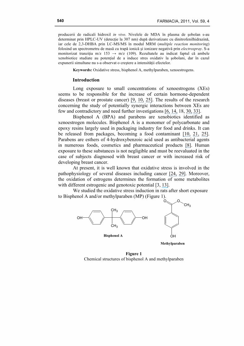
We studied the oxidative stress induction in rats after short exposure to Bisphenol A and/or methylparaben (MP) (Figure 1).
CH3
CH3
OHOH
Bisphenol A OH
O OCH3
Methylparaben
Figure 1 Chemical structures of bisphenol A and methylparaben
FARMACIA, 2011, Vol. 59, 4
541
Malondialdehyde (MDA), a lipid peroxidation end-product, and 2,3-dihydroxybenzoic acid (2,3-DHBA), a marker of in vivo hydroxyl radical production, were used to evaluate the oxidative stress generation [31]. A LC-UV assay for MDA in rat hepatic microsomes was adapted to allow the quantification of the same biomarker in rat plasma samples [16, 17]. In order to determine the in vivo generation of hydroxyl free radicals, salicylic acid was administered as their trapping agent. The plasmatic level of 2,3-dihydroxybenzoic acid, a specific metabolite of salicylic acid formed only in the presence of hydroxyl radicals, was determined [28].
The aim of this study was to determine to what extent the exposure to BPA and MP generates oxidative stress through these two mechanisms and if there is any interaction in the case of simultaneous exposure to BPA and MP.
Materials and Methods
Reagents and materials Bisphenol A and methylparaben of analytical-reagent grade (≥99%)
were purchased from Sigma-Aldrich (SUA) and Merck (Darmstadt, Germany), respectively. HPLC-grade acetonitrile, methanol, formic acid, ammonium acetate, and hexane were purchased from Merck (Darmstadt, Germany). All other chemicals were analytical reagent grade and they were obtained from Merck (sodium hydroxide, hydrochloric acid, 2,4-dinitrophenylhydrazine (DNPH), acetaldehyde, sodium salicylate). Deionised water was obtained using a Milli-Q water purification system (Millipore, Milford, MA, USA).
Animals Female Wistar rats (body weight 146±18 g) were supplied from the
Practical Skills and Experimental Medicine Centre of the University of Medicine and Pharmacy “Iuliu Haţieganu” Cluj-Napoca (Romania). The rats were kept in temperature, humidity, day/night cycle standard conditions and they had access to food and water ad libitum throughout the experiment.
Experimental protocol The experimental protocol was in compliance with the institutional
and national guidelines for laboratory animal experiments, being approved by the Ethics Committee of the University. The protocol included 3 experimental groups and a negative control group, each consisting of 10 rats. The substances were administered for 10 days in food as following: group I – a daily dose of 50 mg/kg bw BPA and 250 mg/kg bw MP; group II
FARMACIA, 2011, Vol. 59, 4
542
- a daily dose of 50 mg/kg bw BPA; group III - a daily dose of 250 mg/kg bw MP. Group IV was the negative control group and received standard food without substances.
Plasma samples Blood was collected from the retro-orbital sinus in the presence of
sodium fluoride as anticoagulant on the 10th day of the experiment. 90 min before the blood sampling sodium salicylate (58 mg/100g/mL, an equivalent dose of 500 mg salicylic acid / kg bw) was administered orally (by gastric intubation) to all animals, as in vivo hydroxyl radical trap. 30 min after the administration of sodium salicylate, 60 min before the blood sampling, the animals from group I received by gastric intubation BPA (50 mg/kg bw) and MP (250 mg/kg bw), the animals from group II only BPA (50 mg/kg bw) and the animals of group III only MP (250 mg/kg bw). Plasma separated by centrifugation (3000 g / 15 min) was stored at -20°C until the analysis.
Sample preparation In order to quantify total MDA, plasma samples (75 µL) were treated
with 1% H2SO4 (75 µL) and 6M NaOH (25 µL) in a water bath for 30 min at 60ºC. After the hydrolysis of MDA bound to proteins, a solution of acetaldehyde (100 µL) as internal standard (IS) was added, followed by a deproteinisation step with 36% HClO4 (63 µL). After vortex-mixing (10 s) and centrifugation (10 min at 10000 rpm), the supernatants (150 µL) were derivatized with 5mM DNPH in 2M HCl (20 µL) at room temperature, protected from light. The obtained hydrazone was extracted in hexane (1.2 mL) and the organic layer was evaporated in a centrifugal concentrator at 30°C. The residues were dissolved in mobile phase (150 µL) and 50 µL were injected into the HPLC system.
In order to quantify 2,3-DHBA, plasma samples (0.2 mL) were deproteinized with acetonitrile (0.6 mL). After vortex-mixing (10 s) and centrifugation (2 min at 12000 rpm), the supernatants (0.15 mL) were transferred in autosampler vials and 4 µL were injected into the HPLC system.
Instrumentation and chromatographic conditions The analysis of total MDA was performed using the method
developed and validated by the authors [17] on a 2695 Waters Alliance HPLC system (Waters, Milford, MA, USA) composed of a quaternary pump, autosampler, column heater and solvent degasser. The HPLC unit was connected to a 996 PDA (photodiode array) detector and a 2475
FARMACIA, 2011, Vol. 59, 4
543
fluorescence detector (Waters, Milford, MA, USA). Chromatographic separation was achieved on a Spherisorb ODS column 250mmx4mm, 5µm) with a Spherisorb ODS (20mmx4mm, 3µm) guard column, maintained at 25°C. The mobile phase consisted in a mixture of 1% formic acid/acetonitrile (62/38, v/v). The flow rate was 1mL/min and the absorbance of the eluent was monitored at 307nm, corresponding to the maximum in the UV spectra of the MDA hydrazone obtained after derivatization. Data acquisition and processing were performed using Empower 2 software (Waters, USA) [17].
The analysis of 2,3-DHBA was performed using the method developed and validated by the authors [28]. The HPLC system used was an 1100 series Agilent Technologies model (Darmstadt, Germany) consisting of a G1312A binary pump, an in-line G1379A degasser, a G1329A autosampler, a G1316A column thermostat and an Agilent Ion Trap Detector 1100 SL (Darmstadt, Germany). Chromatographic separation was performed on a Luna HILIC (100 x 2.0 mm, 4 µm) column (Phenomenex Inc., USA), maintained at 15ºC, under isocratic conditions, using a mixture of 50 mM ammonium acetate in water (pH 4.5)/acetonitrile as the mobile phase (94:6, v/v) with a flow rate of 0.5 mL/min. Chromatograms were processed using Quant Analysis software (Agilent Technologies). The detection was performed in multiple reaction monitoring (MRM) mode using an ion trap mass spectrometer equipped with an electrospray ionization ion source (ESI), operated in negative mode: dry gas nitrogen at 12 L/min, dry gas temperature 350ºC, nebulizer 60 psi (nitrogen), capillary 3500 V. Helium was used as collision gas and the collision potential was 1.1 V. The ion transition monitored was m/z 153 → m/z (109) for both analytes [28].
Statistical analysis The results are presented as mean values ± standard deviation and
the experimental groups were compared with the control group and between them by ANOVA analysis of variance using Origin 4.1 computer software.
Results and Discussion
a) Oxidative stress induction by lipid peroxidation A representative chromatogram obtained from a rat treated orally
with 50 mg/kg bw/day bisphenol A for 10 days (group II) and 500 mg salicylic acid /kg bw 90 min before the blood sampling is shown in Figure 2.

FARMACIA, 2011, Vol. 59, 4
544
Figure 2
Quantification of total MDA. Chromatogram of sample plasma from a rat after oral administration of BPA (50 mg/kg bw /day for 10 days) and salicylic acid (500
mg/kg bw, 90 min before the blood sampling)
The MDA levels were significantly high for all treated animal groups comparatively with the control group (p<0.05 in the case of simultaneous exposure to BPA and MP; p<0.01 in the case of exposure to BPA or MP) (Figure 3). Consequently, the exposure of rats to BPA and MP, respectively, generates oxidative stress due to lipid peroxidation. The obtained results are in accordance with the data published in the literature and can be partially explained based on the metabolization pathways of the two tested substances.
Figure 3
Total MDA levels in rat plasma after exposure to bisphenol A (BPA) and/or methylparaben (MP) (*, p<0.05; **, p<0.01)
There are numerous reports on the toxicity of BPA on liver [7, 19], central nervous system (CNS) [4, 22], reproductive system [5, 26] and kidney, mediated by reactive oxygen species (ROS) induction [15]. Thus,
FARMACIA, 2011, Vol. 59, 4
545
oral exposure of male rats to BPA (25 mg/kg bw/day) caused oxidative damage in liver by disturbing the balance between ROS and antioxidant defense systems. The activity of some hepatic enzymes (aspartate transaminase, alanine transaminase, lactate dehydrogenase) and thiobarbituric acid-reactive substances (TBARS) were increased, and glutathione level was decreased in the group treated with BPA. Hepatic necrosis and congestion were observed in livers of rats exposed to BPA [19]. BPA generates significantly high concentrations of MDA in brain and sperm of male rats treated orally (25 mg/kg bw/day) for 45 days and causes oxidative damage in the brain and testes of rats [4, 5]. The production of ROS increases in a dose-dependent manner in sperm of rats after short term exposure (1 week) [26]. The main metabolic pathway of BPA is detoxification by glucuronidation. A minor route is the oxidation by hydroxylation to a catechol followed by further transformation to an o-quinone. The catechol-o-quinone couple is capable of redox cycling with generation of reactive oxygen species (ROS) and oxidative stress [20].
Parabens can induce cytotoxicity by mitochondrial failure dependent on induction of membrane permeability transition accompanied by the mitochondrial depolarization and depletion of cellular ATP through uncoupling of oxidative phosphorylation [32]. After oral administration, MP is quickly and completely absorbed from the gastrointestinal tract. It is hydrolyzed to p-hydroxybenxoic acid by esterases and than conjugated to p-hydroxyhippuric acid, ester and ether glucuronides and an ethereal sulfate [32]. Paraben (p-hydroxybenzoic acid) induced lipid peroxidation in the liver of mice in vivo [1] and in liver and kidney homogenates in vitro [2]. In skin cells, parabens may induce oxidative stress after conversion to glutathione conjugates of hydroquinone by reacting with singlet oxygen and glutathione [27]. Lipid peroxidation was significantly increased in MP-treated human skin keratinocytes only after UVB exposure [12].
In our study the degree of increase in MDA levels was not significantly different between the animals treated with BPA or MP alone and those treated with the combination of BPA and MP. Simultaneous exposure to BPA and MP did not result in additive effect, synergism or potentiation in terms of lipid peroxidation.
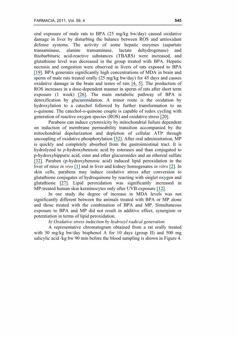
b) Oxidative stress induction by hydroxyl radical generation A representative chromatogram obtained from a rat orally treated with 50 mg/kg bw/day bisphenol A for 10 days (group II) and 500 mg salicylic acid /kg bw 90 min before the blood sampling is shown in Figure 4.
FARMACIA, 2011, Vol. 59, 4
546
Figure 4
Quantification of 2,3-DHBA. Chromatogram of sample plasma from a rat after oral administration of BPA (50 mg/kg bw/day for 10 days) and salicylic acid (500
mg/kg bw 90 min before the blood sampling); retention times: 2,3-DHBA - 1.4 min, 2,5-DHBA – 1.85 min.
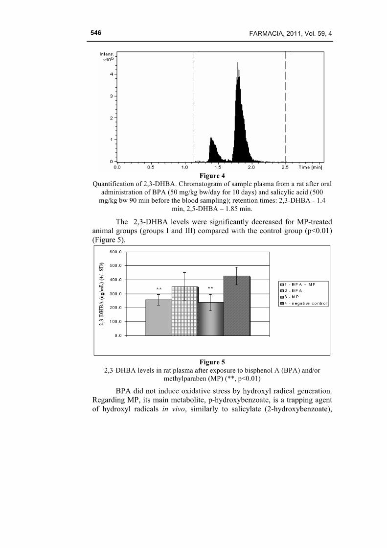
The 2,3-DHBA levels were significantly decreased for MP-treated animal groups (groups I and III) compared with the control group (p<0.01) (Figure 5).
Figure 5
2,3-DHBA levels in rat plasma after exposure to bisphenol A (BPA) and/or methylparaben (MP) (**, p<0.01)
BPA did not induce oxidative stress by hydroxyl radical generation. Regarding MP, its main metabolite, p-hydroxybenzoate, is a trapping agent of hydroxyl radicals in vivo, similarly to salicylate (2-hydroxybenzoate),
FARMACIA, 2011, Vol. 59, 4
547
forming 3,4-dihydroxybenzoate [11, 23]. This explains why the 2,3-DHBA levels decreased in animals treated with MP.
Conclusions
The exposure of rats to BPA and MP, respectively, generates oxidative stress due to lipid peroxidation. Simultaneous exposure to BPA and MP did not lead to the appearance of any type of interaction (additive, synergism or potentiation) in the generation of lipid peroxidation. The generation of hydroxyl radicals was not identified as a mechanism of oxidative stress induction by BPA. MP acts as a trapping agent for hydroxyl radicals by its metabolite, p-hydroxybenzoate, showing a certain protection against the deleterious effects of these radicals. Therefore, simultaneous exposure to BPA and MP did not have a negative impact on the cellular oxidative status.
Acknowledgements
This work was supported by CNCSIS –UEFISCSU Romania, project number PNII – IDEI 1337/2008.
References 1. Asnani VM, Verma RJ. Ameliorative effects of ginger extract on paraben-induced lipid
peroxidation in the liver of mice. Acta Pol Pharm. 2009;66(3):225-228. 2. Asnani V, Verma RJ. Antioxidative effect of rhizome of Zinziber officinale on paraben
induced lipid peroxidation: an in vitro study. Acta Pol Pharm. 2007;64(1):35-37. 3. Atkinson C, Skor HE, Dawn Fitzgibbons E, Scholes D, Chen C, Wähälä K, Schwartz SM,
Lampe JW. Urinary equol excretion in relation to 2-hydroxyestrone and 16-alpha-hydroxyestrone concentrations: an observational study of young to middle-aged women. J Steroid Biochem Mol Biol. 2003;86(1):71-77.
4. Aydoğan M, Korkmaz A, Barlas N, Kolankaya D. The effect of vitamin C on bisphenol A, nonylphenol and octylphenol induced brain damages of male rats. Toxicology. 2008;249(1):35-39.
5. Aydoğan M, Korkmaz A, Barlas N, Kolankaya D. Pro-oxidant effect of vitamin C coadministration with bisphenol A, nonylphenol, and octylphenol on the reproductive tract of male rats. Drug Chem Toxicol. 2010;33(2):193-203.
6. Benachour N, Moslemi S, Sipahutar H, Seralini GE. Cytotoxic effects and aromatase inhibition by xenobiotic endocrine disrupters alone and in combination. Toxicol Appl Pharmacol. 2007;222(2):129-140.
7. Bindhumol V, Chitra KC, Mathur PP. Bisphenol A induces reactive oxygen species generation in the liver of male rats. Toxicology. 2003;188(2-3):117-124.
8. Boberg J, Taxvig C, Christiansen S, Hass U. Possible endocrine disrupting effects of parabens and their metabolites. Reprod Toxicol. 2010;30(2):301-312.
9. Darbre PD, Charles AK.Environmental oestrogens and breast cancer: evidence for combined involvement of dietary, household and cosmetic xenoestrogens. Anticancer Res. 2010;30(3):815-827.
10. Fernandez SV, Russo J. Estrogen and xenoestrogens in breast cancer. Toxicol Pathol. 2010;38(1):110-122.
FARMACIA, 2011, Vol. 59, 4
548
11. Freinbichler W, Bianchi L, Colivicchi MA, Ballini C, Tipton KF, Linert W, Corte LD. The detection of hydroxyl radicals in vivo. J Inorg Biochem. 2008;102(5-6):1329-1333.
12. Handa O, Kokura S, Adachi S, Takagi T, Naito Y, Tanigawa T, Yoshida N, Yoshikawa T. Methylparaben potentiates UV-induced damage of skin keratinocytes. Toxicology. 2006;227(1-2):62-72.
13. Hsu JF, Chang YC, Chen TH, Lin LC, Liao PC. Evaluation of electrospray ionization and atmospheric pressure chemical ionization for simultaneous detection of estrone and its metabolites using high-performance liquid chromatography/tandem mass spectrometry. J Chromatogr B Analyt Technol Biomed Life Sci. 2007;860(1):49-56.
14. Imamura L, Kurashina K, Kawahira T, Omoteno M, Tsuda M. Additional aepression of Activity-dependent c-Fos and BDNF mRNA expression by lipophilic compounds accompanying a decrease in Ca2+ influx into neurons. Neurotoxicology. 2005;26(1):17-25.
15. Kabuto H, Hasuike S, Minagawa N, Shishibori T. Effects of bisphenol A on the metabolisms of active oxygen species in mouse tissues. Environ Res. 2003;93(1):31-35.
16. Kiss B, Popa DS, Crişan G, Bojiţă M, Loghin F. The evaluation of antioxidant potential of Veronica officinalis and Rosmarinus officinalis extracts by monitoring malondialdehide and glutathione levels in rats. Farmacia. 2009;57(4): 432-441.
17. Kiss B, Popa DP, Loghin F, Iepure R, Bojiţă M. Dozarea malondialdehidei libere şi totale prin HPLC. Farmacia. 2005;53(5):17-24.
18. Knudsen FR, Pottinger TG. Interaction of endocrine disrupting chemicals, single and in combination, with estrogen-, androgen-, and corticosteroid-binding sites in rainbow trout (Oncorhynchus mykiss). Aquatic Toxicology. 1999;44:159–170.
19. Korkmaz A, Ahbab MA, Kolankaya D, Barlas N. Influence of vitamin C on bisphenol A, nonylphenol and octylphenol induced oxidative damages in liver of male rats. Food Chem Toxicol. 2010;48(10):2865-2871.
20. Kovacic P. How safe is bisphenol A? Fundamentals of toxicity: metabolism, electron transfer and oxidative stress. Med Hypotheses. 2010;75(1):1-4.
21. LaPensee EW, Ben-Jonathan N. Novel roles of prolactin and estrogens in breast cancer: resistance to chemotherapy. Endocr Relat Cancer. 2010;17(2):R91-107.
22. Lin Y, Zeng XG, Wu DS, Wang X. Study on bisphenol A induced primary cultured mesencephalic neuronal cell injury by oxidative stress. Wei Sheng Yan Jiu. 2006;35(4):419-422.
23. Liu S, Liu M, Peterson S, Miyake M, Vallyathan V, Liu KJ. Hydroxyl radical formation is greater in striatal core than in penumbra in a rat model of ischemic stroke. J Neurosci Res. 2003;71(6):882-888.
24. López-Lázaro M. A New View of Carcinogenesis and an Alternative Approach to Cancer Therapy. Mol Med. 2010;16(3-4):144-153.
25. Maffini MV, Rubin BS, Sonnenschein C, Soto AM. Endocrine disruptors and reproductive health: the case of bisphenol-A. Mol Cell Endocrinol. 2006;254-255:179-186.
26. Minamiyama Y, Ichikawa H, Takemura S, Kusunoki H, Naito Y, Yoshikawa T. Generation of reactive oxygen species in sperms of rats as an earlier marker for evaluating the toxicity of endocrine-disrupting chemicals.Free Radic Res. 2010;44(12):1398-1406.
27. Nishizawa C, Takeshita K, Ueda J, Nakanishi I, Suzuki KT, Ozawa T. Reaction of para-hydroxybenzoic acid esters with singlet oxygen in the presence of glutathione produces glutathione conjugates of hydroquinone, potent inducers of oxidative stress. Free Radic Res. 2006;40(3):233-240.
28. Popa DS, Vlase L, Chirilă D, Loghin F. Rapid LC-MS/MS assay for the evaluation of hydroxyl radical generation and oxidative stress induction in vivo in rats. Revista Română de Medicină de Laborator, 2010; 18(4/4):37-45.
29. Roberts RA, Smith RA, Safe S, Szabo C, Tjalkens RB, Robertson FM.Toxicological and pathophysiological roles of reactive oxygen and nitrogen species. Toxicology. 2010;276(2):85-94.
30. Safe S. Clinical correlates of environmental endocrine disruptors. Trends Endocrinol
FARMACIA, 2011, Vol. 59, 4
549
Metab. 2005;16(4):139-144. 31. Sprinţeroiu M, Bălălău D, Guţu CM, Ilie M, Petraru C. Quantitative analysis of
malondialdehyde in normal human plasma using fluorescence and the standard addition method. Farmacia. 2010;58(4):509-514.
32. Soni MG, Taylor SL, Greenberg NA, Burdock GA. Evaluation of the health aspects of methyl paraben: a review of the published literature. Food Chem Toxicol. 2002;40(10):1335-1373.
33. Tollefsen KE. Interaction of estrogen mimics, singly and in combination, with plasma sex steroid-binding proteins in rainbow trout (Oncorhynchus mykiss). Aquatic Toxicology; 2002;56:215–225.
__________________________________ Manuscript received: March 9th 2011