Hematological Changes and Oxidative Stress Induction of ...
11
Abdel-Halim et al. 22 Hematological Changes and Oxidative Stress Induction of Titanium Dioxide Nanoparticles in Male Mice after Intraperitoneal Injection of Different Doses for 28 Days: Study of Organ’s Responsibility NanoWorld Journal Research Article Open Access https://doi.org/10.17756/nwj.2021-088 Khaled Y. Abdel-Halim 1* , Safaa R. Osman 1 Mohamed A. F. Abuzeid 2 and Alaa M. Khozimy 2 1 Mammalian & Aquatic Toxicology Department, Central Agricultural Pesticides Laboratory, Agricultural Research Center (ARC), 12618-Dokki, Giza, Egypt 2 Department of Plant Protection, Faculty of agriculture, Damanhur University, 22516-Damanhur, Egypt * Correspondence to: Khaled Y. Abdel-Halim Mammalian & Aquatic Toxicology Department Central Agricultural Pesticides Laboratory Agricultural Research Center (ARC) 12618-Dokki, Giza, Egypt Tel/Fax: +20 2 02 37602209 E-mail: [email protected] Received: May 29, 2021 Accepted: July 14, 2021 Published: July 15, 2021 Citation: Abdel-Halim KY, Osman SR, Abuzeid MAF, Khozimy AM. 2021. Hematological Changes and Oxidative Stress Induction of Titanium Dioxide Nanoparticles in Male Mice after Intraperitoneal Injection of Different Doses for 28 Days: Study of Organ’s Responsibility. NanoWorld J 7(1): 22-32. Copyright: © 2021 Abdel-Halim et al. is is an Open Access article distributed under the terms of the Creative Commons Attribution 4.0 International License (CC-BY) (http:// creativecommons.org/licenses/by/4.0/) which permits commercial use, including reproduction, adaptation, and distribution of the article provided the original author and source are credited. Published by United Scientific Group Abstract Titanium dioxide nanoparticles (TiO 2 NPs) are supposed to be the most widely engineered nanoparticles (NPs) that are used in the world. e study evaluated the toxicity of TiO 2 NPs (12 - 64 nm) in male mice by intraperitoneal injection at five doses of either control (0), 2.5, 5.0, 10.0, and 20.0 mg/kg of body weight for 28 d. Hematological changes and oxidative stress indicators like Catalase (CAT), Glutathione-S-transferase (GST), Glutathione peroxidase (GPx), Glutathione reductase (GR), Lipid peroxidation (LPO), Glutathione (GSH) content as well as Carbonyl protein (CP) were explored. Present results showed significant differences (P < 0.05) in all hematological measurements at the end of the study for treated groups, respect to their control. TiO 2 NPs caused dose-dependent oxidative stress in lung, liver, spleen, kidney, heart, and muscle samples of the treated animals, concerning the control group. Noticeable important increases (P < 0.05) of malondialdehyde (MDA) levels were represented in all treatments of samples as following: heart > muscle > spleen > liver > kidney > lung, with respect to their controls. Also, noticeable important increases were recorded for GPx activity and CP level. However, significant declines were noted in CAT, and GST activities, and GSH content in treated animals, concerning control. Moreover, excess of such CP, MDA, and other biomarker alterations may provide a fingerprint of potential toxic effects of TiO 2 NPs during long-term exposure scenario. us, extensive application of such NPs raises the consideration about biosafety. Keywords Titanium dioxide nanoparticles, Mice, Hematology, Oxidative stress, Intra- peritoneal injection Introduction Titanium dioxide (TiO 2 ) is considered the most abundantly produced NMs and used in food, paints and in personal care products [1-2]. It may be proper to the greatest briefly engineered nanoparticles (NPs; 1-100 nm) in the worldwide. It has broad spectrum as bio-medical ceramic and orthopaedic implants [3]. Due to these properties, TiO 2 NPs are progressively used in endoprosthesis and supports in bone tissues re-establishments [4-5]. However, nano-toxicological investigations exist critical role for the harmless and sustainable enlargement of the evolving and recognized nanomaterials (NMs) e.g. TiO 2 NPs [5]. Actually, TiO 2 NPs could likewise induce risky/cytotoxic effects. For example, it induced DNA double strand breaks in bone marrow cells following oral administration in rats [6]. Zhang et al. [7] detected that, TiO 2 NPs enhanced pro-inflammatory
-
Upload
khangminh22 -
Category
Documents
-
view
3 -
download
0
Transcript of Hematological Changes and Oxidative Stress Induction of ...
Abdel-Halim et al. 22
Hematological Changes and Oxidative Stress Induction of Titanium Dioxide Nanoparticles in Male Mice after Intraperitoneal Injection of Different Doses for 28 Days: Study of Organ’s Responsibility
NanoWorld Journal
Research Article Open Access
https://doi.org/10.17756/nwj.2021-088
Khaled Y. Abdel-Halim1*, Safaa R. Osman1 Mohamed A. F. Abuzeid2 and Alaa M. Khozimy2
1Mammalian & Aquatic Toxicology Department, Central Agricultural Pesticides Laboratory, Agricultural Research Center (ARC), 12618-Dokki, Giza, Egypt 2Department of Plant Protection, Faculty of agriculture, Damanhur University, 22516-Damanhur, Egypt
*Correspondence to:Khaled Y. Abdel-Halim Mammalian & Aquatic Toxicology Department Central Agricultural Pesticides Laboratory Agricultural Research Center (ARC) 12618-Dokki, Giza, Egypt Tel/Fax: +20 2 02 37602209 E-mail: [email protected] Received: May 29, 2021 Accepted: July 14, 2021 Published: July 15, 2021
Citation: Abdel-Halim KY, Osman SR, Abuzeid MAF, Khozimy AM. 2021. Hematological Changes and Oxidative Stress Induction of Titanium Dioxide Nanoparticles in Male Mice after Intraperitoneal Injection of Different Doses for 28 Days: Study of Organ’s Responsibility. NanoWorld J 7(1): 22-32.
Copyright: © 2021 Abdel-Halim et al. This is an Open Access article distributed under the terms of the Creative Commons Attribution 4.0 International License (CC-BY) (http://creativecommons.org/licenses/by/4.0/) which permits commercial use, including reproduction, adaptation, and distribution of the article provided the original author and source are credited.
Published by United Scientific Group
AbstractTitanium dioxide nanoparticles (TiO2NPs) are supposed to be the most widely
engineered nanoparticles (NPs) that are used in the world. The study evaluated the toxicity of TiO2NPs (12 - 64 nm) in male mice by intraperitoneal injection at five doses of either control (0), 2.5, 5.0, 10.0, and 20.0 mg/kg of body weight for 28 d. Hematological changes and oxidative stress indicators like Catalase (CAT), Glutathione-S-transferase (GST), Glutathione peroxidase (GPx), Glutathione reductase (GR), Lipid peroxidation (LPO), Glutathione (GSH) content as well as Carbonyl protein (CP) were explored. Present results showed significant differences (P < 0.05) in all hematological measurements at the end of the study for treated groups, respect to their control. TiO2NPs caused dose-dependent oxidative stress in lung, liver, spleen, kidney, heart, and muscle samples of the treated animals, concerning the control group. Noticeable important increases (P < 0.05) of malondialdehyde (MDA) levels were represented in all treatments of samples as following: heart > muscle > spleen > liver > kidney > lung, with respect to their controls. Also, noticeable important increases were recorded for GPx activity and CP level. However, significant declines were noted in CAT, and GST activities, and GSH content in treated animals, concerning control. Moreover, excess of such CP, MDA, and other biomarker alterations may provide a fingerprint of potential toxic effects of TiO2NPs during long-term exposure scenario. Thus, extensive application of such NPs raises the consideration about biosafety.
KeywordsTitanium dioxide nanoparticles, Mice, Hematology, Oxidative stress, Intra-
peritoneal injection
IntroductionTitanium dioxide (TiO2) is considered the most abundantly produced NMs
and used in food, paints and in personal care products [1-2]. It may be proper to the greatest briefly engineered nanoparticles (NPs; 1-100 nm) in the worldwide. It has broad spectrum as bio-medical ceramic and orthopaedic implants [3]. Due to these properties, TiO2NPs are progressively used in endoprosthesis and supports in bone tissues re-establishments [4-5]. However, nano-toxicological investigations exist critical role for the harmless and sustainable enlargement of the evolving and recognized nanomaterials (NMs) e.g. TiO2NPs [5]. Actually, TiO2NPs could likewise induce risky/cytotoxic effects. For example, it induced DNA double strand breaks in bone marrow cells following oral administration in rats [6]. Zhang et al. [7] detected that, TiO2NPs enhanced pro-inflammatory
NanoWorld Journal | Volume 7 Issue 1, 2021
Hematological Changes and Oxidative Stress Induction of Titanium Dioxide Nanoparticles in Male Mice after Intraperitoneal Injection of Different Doses for 28 Days: Study of Organ’s Responsibility Abdel-Halim et al.
23
particles. Induction of ROS, free radicals, oxidative stress, damage and apoptosis are common observations in a wide variety of cell types exposed to TiO2NPs in vivo and in vitro. Oxidative stress consequences in changes in the production of superoxide dismutase (SOD) or antioxidant defense enzymes. The liver is an active organ for detoxication and TiO2NPs can enter liver cell. Humans are ever more exposed to TiO2 via inhalation, dermal or oral exposure, thus keeping in view the potential health hazards of TiO2NPs on humans. Established on animal assessments, International Agency for Research on Cancer (IARC) classified TiO2 as a group 2B carcinogen (possibly carcinogenic to humans) [33]. Also, the French agency for food, environmental and occupational health and safety (ANSES) banned the use of TiO2 as a food additive (E171) due to its genotoxic potential [34]. While numerous research sites noted that, TiO2NPs can induce adverse effects including DNA damage and chromosomal damage, findings are contradictory [6, 35]. The study aimed to investingate the adverse impacts of different intraperitoneal doses of TiO2NPs for 28 d on male mice using haematological studies as well as oxidative stress enzyme activity assays.
Materials and MethodsChemical and reagents
Titanium dioxide nanoparticles (TiO2NPs) were supplied by Nano-Tech Lab., Dream Land, 6th October City, Egypt. Nanoparticles (NPs) were achieved for reliable characterization techniques. Chemicals: thiobarbituric acid (TBA) and sodium azide (NaN3) were supplied by LOBA CHEMIE PVT. Ltd, Mumbai-400005, India. Phosphate buffer, sodium phosphate monobasic; dibasic and potassium phosphate monobasic; dibasic were supplied by J.T. BAKER Chem. Co, Phillipsburg, N.J. 08865. Trichloro acetic acid (TCA; Cl3C3COOH), hydrochloric acid (HCl) and hydrogen peroxide (H2O2) were obtained from Research Lab. Fine Chem. Indust., Mumbai 400002, India. Ethylene diamine tetra acetic acid disodium salt (EDTA), ethanol (C2H5OH), 1-Chloro 2, 4-dinitrobenzene (CDNB), reduced glutathione (GSH), Tris-HCl: 2-amino-2-hydroxy methyl-propane-1, 3-diol, β-nicotinamide adenine dinucleotide reduced form (β-NADPH), oxidized glutathione (GSSG), and bovine serum albumin (BSA) were attained from Sigma Chem. Co. P.O. Box 14508 St. Louis MO 63178, USA.
Characterization of NPs
An aliquot of prepared TiO2 was achieved for visualization on Scanning Electron Microscopy (SEM) ( JOEL, JSM 5300) with high resolution at an accelerating voltage of 120 Kev. The sample was coated on a copper grid and scanned for its size and shape. X-ray Electron Dispersive Analysis (EDA) was accomplished by using X-ray Oxford detector unit (model 6647, England) equipped with SEM ( JOEL, JSM 5300) to scan the purity of the prepared NPs. Titanium ions (Ti2+) were firm in the used solutions of TiO2NPs by dynamic light scattering (DLS) (DTS Nano v 5.2; Malvern Zeta sizer Nano ZS, Malvern Instruments, UK). Nanoparticles (NPs) suspension was sonicated by using sonicator bath at ambient conditions for 20 min at 40 W.
gene expression in proosteoblast cells (MC3T3-E1). Wang et al. [8] showed that, TiO2NPs have potential toxic effects in some organs, where they caused damage to the knee joints in rabbits.
Moreover, TiO2NPs can become heightened and harm-less in different body parts after entering the body through numerous pathways, e.g. administration via the abdominal cavity or inhalation [9-10]. It can be toxic to numerous of cell, e.g. human lymphoblastoid cells and hepatoma cells [11-12]. It can induce a critical stress response in glial cells of mouse brains, resulting in neuron injury and dysfunction [13]. The persistence degree of neuron cell lines exposed to TiO2NPs significantly decreased in a typical time and dose-dependent manner [14]. Several studies demonstrated mechanisms of NPs toxicity. It may impose genotoxic effects through change the configuration of molecular composite and porousness of the cell membrane and induce oxidative stress [15-17]. In oxidative stress, reactive oxygen species (ROS), e.g. hydroxyl radicals, are generated and cause DNA oxidation as noted for 8-hydroxygaunisine (8-OHdG), resulting in mistakes and al-terations in DNA repetition [18-19]. Moreover, ROS could make inflammation and joint feed-forward relations between oxidative stress and inflammation, resulting in DNA damage and cell apoptosis [20-21]. Nevertheless, the complete regu-lar data on the toxicity of TiO2NPs remains limited. Goals of most investigators was to disclose the effect and the principal mechanism of TiO2NPs exposure on human health.
In spite of this increase in the prolonged use of engineered NMs and the profits of such use of engineered NPs to society, there is a little knowledge concerning their likely toxicological effects on human as well as environmental health and safety [22-26]. Physical and chemical properties of NMs are pre-dictable to impose significant effects on the performance and properties of macromolecules, cells and body parts [27].
Due to their particularly small size and unique physical properties, the behavior of NMs in the environment, uptake, distribution and impacts within living organisms are likely to be different when compared to their conventional forms [28]. For example, gold NPs (AuNPs) are far smaller than the diameter of a common cell and thus has an opportunity to pass in the human body during production, transportation, storage and consumption. The same properties make them to be useful for different applications, despite they are harmful and toxic to the environment and organisms [29]. Just, various metallic NPs have been displayed to cause severe effects both in vitro and in vivo [30-31]. Some NMs have been established for their toxicity to humans and other organisms either upon contact or after persistent environmental exposure [24]. Thus, there is an urgent need to fill up the gaps in our accepting and for research and regulatory activities to ensure these compounds do not pose a significant hazard to human and environmental health. This is vital to ensure the sustainability of the industry. TiO2NPs are very reactive and may be toxic due to their properties, especially larger surface area [32]. They can damage human and animal cells by increasing oxidative stress mechanism. Biosafety of this material needs to be estimated. Some reviews have suggested that the smaller-scale NPs had a greater inflammogenic effect than larger
NanoWorld Journal | Volume 7 Issue 1, 2021 24
Hematological Changes and Oxidative Stress Induction of Titanium Dioxide Nanoparticles in Male Mice after Intraperitoneal Injection of Different Doses for 28 Days: Study of Organ’s Responsibility Abdel-Halim et al.
Animal rearing
Healthy male mice (mean; 30.0 ± 3.0 g) were obtained from Institute of Public Health, Alexandria University, Egypt. They were allocated to plastic cages covered with metal grids and allowed to acclimate for 2 weeks under laboratorial conditions, before divided into experimental groups. The mice were provided with free water and food ad libitum. Damanhour University Animal Health Care (DU-AHC) committee approved the animal caring ethics (DMU-2020-0026). Guideline of animal care was surveyed according to the National Institutes of Health Guide for the care and use of laboratory animals (NIH Publications No. 8023, revised 1987).
Dosage procedures
Oxidative stress and hematological alterations in mal mice were investigated. The animals were randomly divided into four group and an additional control group, with 10 mouse/group. Suspension of TiO2NPs was intraperitoneal injected (i.p) at dosage: 2.5, 5.0, 10.0 and 20.0 mg /kg b.w once a day for 28 d. Vehicle (citrate solution) was injected into mice of the control group. The animals were observed every day, and no animal died during the study. On the 28th d, the animals were anesthetized with 2% phenobarbital (60 ml/kg, i.p) and dissected for sampling procedures. Blood samples were collected from heart by using heparinized syringe and specific blood tubes. They were subjected for complete blood count (CBC). On the other hand, liver spleen, heart, kidney and muscles were taken and stored at -20 °C until analysis.
Hematological measurements
One ml of blood from treated animals was collected in sterilized tube with 0.5 µl of EDTA as an anticoagulant. For CBC analysis, the samples were subjected on cell counter instrument (Medonic serial no.14641-Swdish) and the data were presented at software program.
Biochemical quantifications
Sample preparation
An aliquot (0.5 g, each tissue) was homogenized with cold-saline solution (1:10 w/v) and then centrifuged at 5000 rpm and 4 °C for 15 min. Supernatant was used as a source for Catalase (CAT), Glutathione-S-transferase (GST), Glu-tathione peroxidase (GPx), Glutathione reductase (GR), while homogenate was used for Lipid peroxidation (LPO) and Glu-tathione (GSH) content.
LPO
Thiobarbituric acid reactive substances (TBARS) were used as an index of LPO according to Rice-Evans et al. [36] through quantification of malondialdehyde (MDA) content. An aliquot (250 µl) of each tissue homogenate was mixed with 1 ml of 15% (w/v) trichloroacetic acid (TCA) in 25 mM HCl C.m and 2ml of 0.37% thiobarbituric acid (TBA). The mixture was boiled for 10 min, quickly cooled, and immediately centri-fuged at 5000 rpm for 5 min. The absorbance was determined at 535 nm. MDA was quantified by using an extinction coeffi-cient of 156 mM-1 and expressed as mM g-1 tissue.
GSH
Principle of the used method depends on reduction of 5, 5ˊ-dithiobis 2- nitrobenzoic acid (DTNB) with GSH to pro-duce a yellow complex which was measured at 405 nm [37]. Enzyme source (500 µl) was mixed with the same volume of 500 mM TCA, followed by centrifugation at 3000 rpm for 15 min. An aliquot (500 µl) was well mixed with 1 ml of each 100 mM PBS buffer, pH 7.4 and 1 mM DTNB. After 10 min, the absorbance was measured at 405 nm against blank. GSH concentration was expressed as nM g-1 tissue.
CAT
The enzyme activity was assayed independent on decrease of absorbance at 240 nm in association with hydrogen perox-ide (H2O2) consumption [38]. The reaction mixture consist-ed of 1 ml of 12.5 mM H2O2 (substrate), 2 ml of 66.7 mM phosphate buffer, pH 7.0 and enzyme source. The activity was expressed as U mg-1 tissue. The unit of CAT is the amount of enzyme which liberates half the peroxide oxygen from hydro-gen peroxide solution of any concentration in 100 µl at 25ºC.
GST
The activity was determined by the spectrophotometric method of Habig and Jakoby [39] by using 1-Chloro, 2-4 di-nitrobenzene (CDNB). Enzyme source was mixed with 500 µl of potassium phosphate buffer (50 mM; pH 6.5). The incu-bation was done at 25ºC for 5 min, followed by mixing with 100 µl of 0.2 M CDNB and 150 µl of 10 mM GSH. After 1 min, the change of absorbance was recorded every 30 s for 6 min at 340 nm. The enzyme activity was expressed as nM mg-1 min-1.
GPx
The enzyme activity was measured according to Flohe and Gunzler [40] by mixing phosphate buffer solution (100 mM), EDTA (50 mM), sodium azide (250 mM), H2O2 (10 mM) and enzyme in a cuvette. The change in absorbance was followed every 3 s for 40 s at 340 nm. Enzyme activity was expressed as mUGPx mg-1 protein. One unit of GPx is de-fined as the amount of enzyme necessary to oxidize 1µmol of β-NADPH per min.
GR
The activity of GR was assayed independent on the de-crease in the absorbance during β-NADPH oxidation [41]. In each cuvette, 0.1M potassium phosphate buffer, 3.4 mM EDTA, pH 7.6, 30 mM oxidized glutathione (GSSG), 0.8 mM β-NADPH and 1.0% of bovine serum albumin (BSA) were mixed by inversion. Then, 100 µl of the enzyme was add-ed. The absorbance was followed at 340 nm for approximately 5 min. Enzyme activity was expressed as U mg-1 protein. One unit will reduce 1.0 µM of GSSG per min at pH 7.6 at 25 °C.
Carbonyl protein (CP)
An aliquot of frozen tissue was mixed with 5% ice-cold sulfosalicylic acid (1:20 w/v) and then centrifuged at 13.000 rpm for 15 min. The supernatant was discarded and 0.5 ml of 2, 4-dinitrophenylhydrazine (10 mM) solution was added to the pellets. The samples were kept at room temperature for
NanoWorld Journal | Volume 7 Issue 1, 2021 25
Hematological Changes and Oxidative Stress Induction of Titanium Dioxide Nanoparticles in Male Mice after Intraperitoneal Injection of Different Doses for 28 Days: Study of Organ’s Responsibility Abdel-Halim et al.
1h with vigorous vortex every 15 min, then 0.5 ml of 20% TCA solution was added and the tubes were re-centrifuged as described above. The supernatant was again discarded and the excess of 2, 4-dinitrophenylhydrazine was removed by washing the pellet three times with 1 ml of ethanol: ethyl ac-etate (1:1 v/v), followed by hearty vortex and re-centrifuging as defined before. Finally, the pellets were liquefied in 6 M of guanidine chloride and incubated for 15 min at 37ºC. The maximum absorbance at 360-370 nm was verified and the find CP content was estimated by using the extinction coefficient of 22 mM-1 [42].
Total protein assay
Protein level was determined according to the method of Lowry et al. [43]. BSA was used as a standard.
Statistical analysis
All data presented as mean ± SE were subjected to anal-ysis of variance (ANOVA) and means were compared to sig-nificance by Student-Newman Keuls at the probability of 0.05 [44].
ResultsTiO2NPs characterization
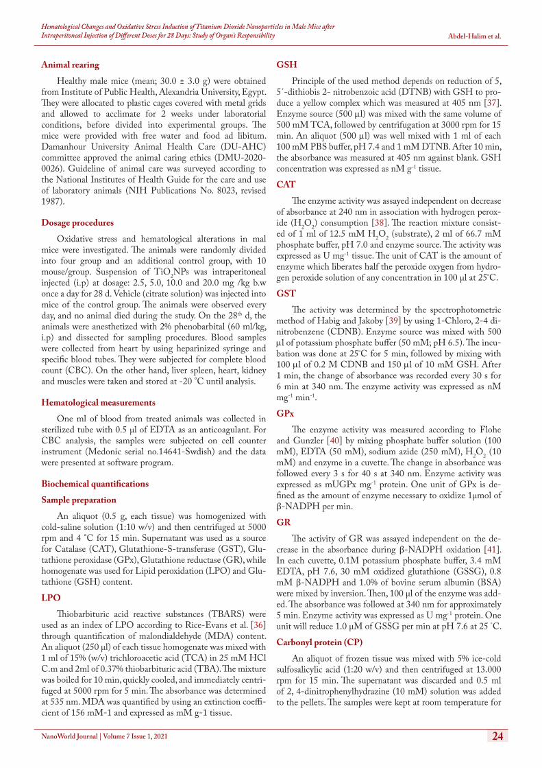
The examined NPs exhibited characteristic spherical shape with size ranged from 12 to 64 nm as illustrated in SEM im-age (Figure 1a). In addition, EDA pattern for elemental anal-ysis is plotted in Figure 1b achieving the dominance of TiO2 (100.0%) respect to total count dividing into Ti (27.06%) and O (72.94%). The average of zeta size of TiO2NPs in vehicle solution ranged from 90.0 to 105.0 nm as checked in DLS (Figure 1c).
Hematological changes
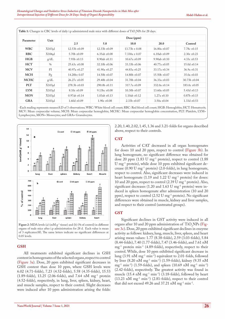
The data among CBC of treated and control animals are presented in Table 1. White blood cells (WBCs) exhibited levels in the treated animals greater than control with mean values: 12.53, 12.33, 13.73, and 16.80 × 10³µl for treatments: 2.5, 5.0, 10.0 and 20.0 ppm, respectively, respect to control (7.78 × 10³µl). In addition, TiO2NPs doses significantly increased RBCs levels with mean values: 5.70, 6.35, 7.33 and 6.18 × 10³µl for the same treatments, respect to control (2.30 × 10³µl). In the same manner, hemoglobin and hematocrit significantly increased in all treated animals, respect to control. However, mean corpuscular hemoglobin (MCH) significantly was lower than the control. Platelets (PLT) significantly increased in the treated animals at levels: 278.3, 290.0, 317.7, and 332.0 × 10³µl for the above treatments, respect to control (183.0 × 10³µl). lymphocytes (LYM), monocytes (MON) and granulocytes (GAR) increased with increasing dose, where the high dose (20 mg/kg) exhibited the highest LYM (13.60 x 10³ µl), respect to control (5.43 x 10³µl). While, the highest one of MON (1.27 x 10³µl) was induced by the same dose, respect to control (0.87 x 10³µl). The same patter was observed for GAR levels in a decreasing order.
Oxidative stress responses
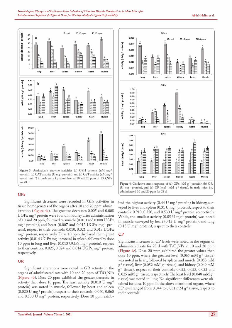
LPO
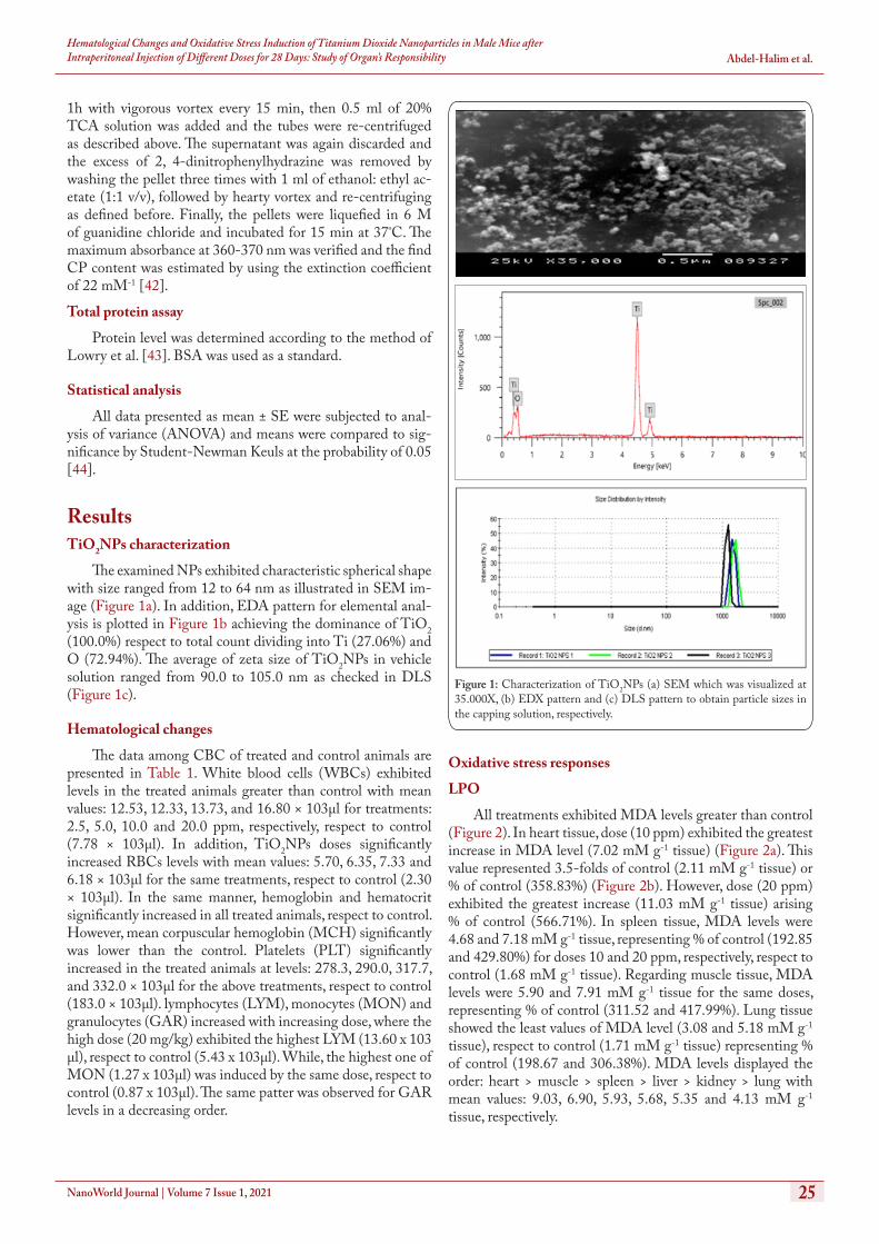
All treatments exhibited MDA levels greater than control (Figure 2). In heart tissue, dose (10 ppm) exhibited the greatest increase in MDA level (7.02 mM g-1 tissue) (Figure 2a). This value represented 3.5-folds of control (2.11 mM g-1 tissue) or % of control (358.83%) (Figure 2b). However, dose (20 ppm) exhibited the greatest increase (11.03 mM g-1 tissue) arising % of control (566.71%). In spleen tissue, MDA levels were 4.68 and 7.18 mM g-1 tissue, representing % of control (192.85 and 429.80%) for doses 10 and 20 ppm, respectively, respect to control (1.68 mM g-1 tissue). Regarding muscle tissue, MDA levels were 5.90 and 7.91 mM g-1 tissue for the same doses, representing % of control (311.52 and 417.99%). Lung tissue showed the least values of MDA level (3.08 and 5.18 mM g-1 tissue), respect to control (1.71 mM g-1 tissue) representing % of control (198.67 and 306.38%). MDA levels displayed the order: heart > muscle > spleen > liver > kidney > lung with mean values: 9.03, 6.90, 5.93, 5.68, 5.35 and 4.13 mM g-1 tissue, respectively.
Figure 1: Characterization of TiO2NPs (a) SEM which was visualized at 35.000X, (b) EDX pattern and (c) DLS pattern to obtain particle sizes in the capping solution, respectively.
NanoWorld Journal | Volume 7 Issue 1, 2021 26
Hematological Changes and Oxidative Stress Induction of Titanium Dioxide Nanoparticles in Male Mice after Intraperitoneal Injection of Different Doses for 28 Days: Study of Organ’s Responsibility Abdel-Halim et al.
GSH
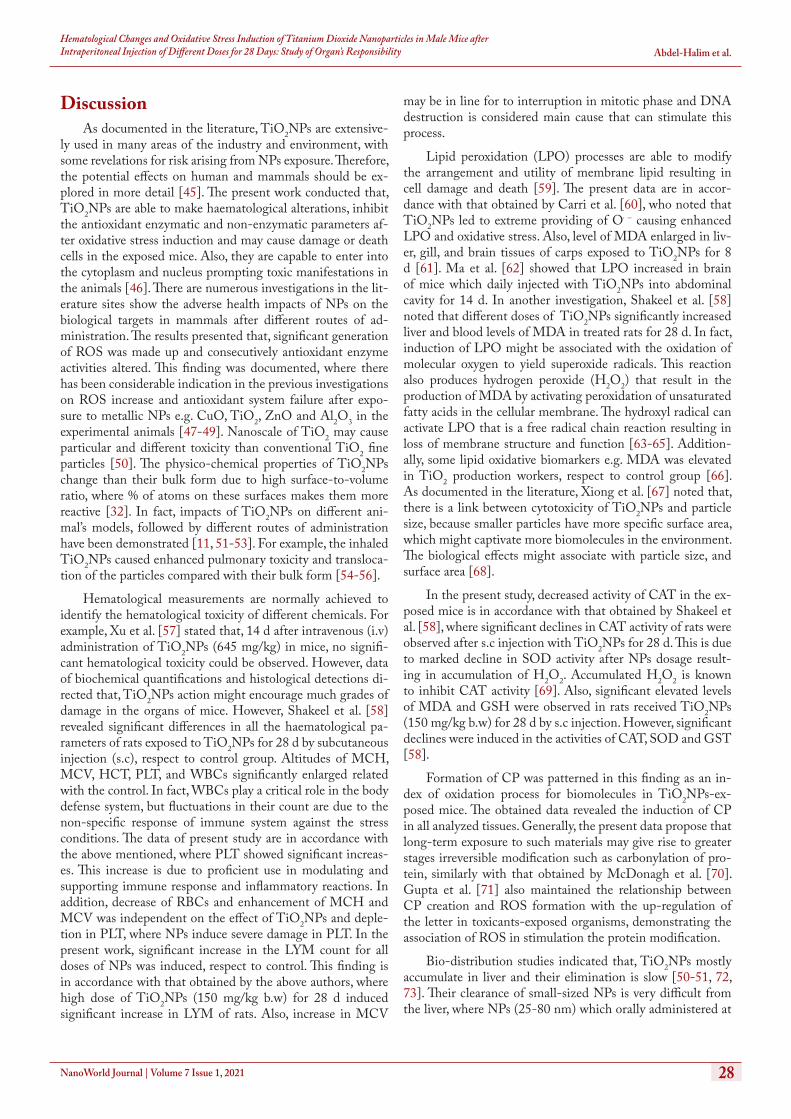
All treatments exhibited significant declines in GSH content in homogenates of the selected organs, respect to control (Figure 3a). Dose, 20 ppm exhibited significant decreases in GSH content than dose 10 ppm, where GSH levels were 6.02 (4.71-folds), 7.23 (4.52-folds), 5.58 (4.35-folds), 15.53 (1.89-folds), 13.25 (2.06-folds), and 7.64 nM mg-1 protein (4.52-folds), respectively, in lung, liver, spleen, kidney, heart, and muscle samples, respect to their control. Slight decreases were induced after 10 ppm administration arising the folds:
2.20, 1.40, 2.02, 1.45, 1.36 and 3.21-folds for organs described above, respect to their controls.
CAT
Activities of CAT decreased in all organ homogenates for doses 10 and 20 ppm, respect to control (Figure 3b). In lung homogenate, no significant difference was obtained for dose 20 ppm (1.83 U mg-1 protein), respect to control (1.88 U mg-1 protein), while dose 10 ppm exhibited significant de-crease (0.90 U mg-1 protein) (2.0-folds), in lung homogenate, respect to control. Also, significant decreases were induced in heart homogenate (1.19 and 1.22 U mg-1 protein) for doses: 10 and 20 ppm, respect to control (2.39 U mg-1 protein). Also, significant decreases (1.20 and 1.63 U mg-1 protein) were in-duced in spleen homogenate after administration (10 and 20 ppm), respect to control (2.52 U mg-1 protein). No significant differences were obtained in muscle, kidney and liver samples, and respect to their control (untreated groups).
GST
Significant declines in GST activity were induced in all organs after 10 and 20 ppm administration of TiO2NPs (Fig-ure 3c). Dose, 20 ppm exhibited significant declines in enzyme activity as follows: kidney, lung, muscle, liver, spleen, and heart arising mean values: 1.77 (8.38-folds), 2.59 (3.03-folds), 5.84 (8.44-folds), 7.40 (1.77-folds), 7.47 (3.46-folds), and 7.61 nM mg-1 protein min-1 (4.89-folds), respectively, respect to their control. While, dose 10 ppm exhibited significant decrease in lung (3.91 nM mg-1 min-1) equivalent to 2.01-folds, followed by liver (8.20 nM mg-1 min-1) (1.59-folds), kidney (9.35 nM mg-1 min-1) (1.59-folds), and spleen (10.69 nM mg-1 min-1) (2.42-folds), respectively. The greatest activity was found in muscle (15.4 nM mg-1 min-1) (3.18-folds), followed by heart (13.23 nM mg-1 min-1) (2.81-folds), respect to their control that did not exceed 49.26 and 37.21 nM mg-1 min-1.
Table 1: Changes in CBC levels of daily i.p administered male mice with different doses of TiO2NPs for 28 days.
-Each reading represents means±S.D of 3 observations; WBC: White blood cell count; RBC: Red blood cell count; HGB: Hemoglobin; HCT: Hematocrit; MCV: Mean corpuscular volume; MCH: Mean corpuscular hemoglobin; MCHC: Mean corpuscular hemoglobin concentration, PLT: Platelets, LYM= Lymphocytes, MON= Monocytes; and GRA= Granulocytes.
Parameter UnitDose (ppm)
2.5 5.0 10.0 20.0 ControlWBC X103µl 12.53b ±0.09 12.33b ±0.09 13.73b ± 0.08 16.80a ±0.07 7.78c ±0.15RBC X106µl 5.70b ±0.09 6.35ab ±0.08 7.330a ± 0.07 6.18ab ±0.09 2.30c ±0.23HGB g/dL 7.93b ±0.13 8.90ab ±0.11 10.67a ±0.09 9.90ab ±0.10 4.33c ±0.53HCT % 25.63c ±0.08 32.10b ±0.06 33.33b ±0.06 40.77a ±0.05 15.0d ±0.14MCV FI 40.97a ±0.27 41.90a ±0.27 44.83a ±0.25 33.17a ±0.34 36.9a ±0.31MCH Pg 14.20b± 0.07 14.50b ±0.07 14.80b ±0.07 15.50b ±0.07 35.0a ±0.03
MCHC g/dL 26.27c ±0.05 29.40b ±0.04 31.70b ±0.04 36.33a ±0.03 30.73b ±0.04PLT X103µl 278.3b ±0.03 290.0b ±0.11 317.7a ±0.09 332.0a ±0.14 183.0c ±0.05LYM X103µl 8.10c ±0.09 9.13bc ±0.08 10.30b ±0.07 13.60a ±0.05 5.43d ±0.13
MON X103µl 0.97ab ±0.14 1.03ab ±0.13 1.10ab ±0.12 1.27a ±0.10 0.87b ±0.15GRA X103µl 1.60d ±0.09 1.90c ±0.08 2.33b ±0.07 3.50a ±0.04 1.33d ±0.53
Figure 2: MDA levels (a) (mM g-1 tissue) and (b) (% of control) in different organs of male mice after i.p administration for 28 d. Each value is mean of 3 replicates±SE. The same letters indicate no significant differences at 0.05 levels.
% o
f con
trol
NanoWorld Journal | Volume 7 Issue 1, 2021 27
Hematological Changes and Oxidative Stress Induction of Titanium Dioxide Nanoparticles in Male Mice after Intraperitoneal Injection of Different Doses for 28 Days: Study of Organ’s Responsibility Abdel-Halim et al.
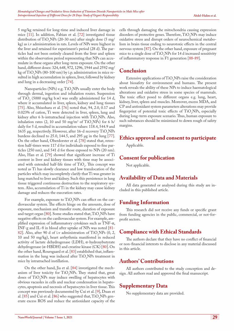
GPx
Significant decreases were recorded in GPx activities in tissue homogenates of the organs after 10 and 20 ppm admin-istration (Figure 4a). The greatest decreases 0.005 and 0.008 UGPx mg-1 protein were found in kidney after administration of 10 and 20 ppm, followed by muscle (0.010 and 0.008 UGPx mg-1 protein), and heart (0.007 and 0.012 UGPx mg-1 pro-tein), respect to their controls: 0.010, 0.021 and 0.013 UGPx mg-1 protein, respectively. Dose 10 ppm displayed the highest activity (0.014 UGPx mg-1 protein) in spleen, followed by dose 10 ppm in lung and liver (0.013 UGPx mg-1 protein), respect to their controls: 0.025, 0.024 and 0.014 UGPx mg-1 protein, respectively.
GR
Significant alterations were noted in GR activity in the organs of administered rats with 10 and 20 ppm of TiO2NPs (Figure 4b). Dose 20 ppm exhibited the greater decrease in activity than dose 10 ppm. The least activity (0.010 U mg-1 protein) was noted in muscle, followed by heart and spleen (0.020 U mg-1 protein), respect to their controls: 0.040, 0.060 and 0.530 U mg-1 protein, respectively. Dose 10 ppm exhib-
ited the highest activity (0.44 U mg-1 protein) in kidney, sur-veyed by liver and spleen (0.31 U mg-1 protein), respect to their controls: 0.910, 0.320, and 0.530 U mg-1 protein, respectively. While, the smallest activity (0.05 U mg-1 protein) was noted in muscle, surveyed by heart (0.12 U mg-1 protein), and lung (0.13 U mg-1 protein), respect to their controls.
CP
Significant increases in CP levels were noted in the organs of administered rats for 28 d with TiO2NPs at 10 and 20 ppm (Figure 4c). Dose 20 ppm exhibited the greater values than dose 10 ppm, where the greatest level (0.065 mM g-1 tissue) was noted in heart, followed by spleen and muscle (0.053 mM g-1 tissue), liver (0.052 mM g-1 tissue), and kidney (0.049 mM g-1 tissue), respect to their controls: 0.022, 0.023, 0.022 and 0.025 mM g-1 tissue, respectively. The least level (0.048 mM g-1 tissue) was noted in lung. No significant differences were ob-tained for dose 10 ppm in the above mentioned organs, where CP level ranged from 0.044 to 0.051 mM g-1 tissue, respect to their controls.
Figure 3: Antioxidant enzyme activities (a) GSH content (nM mg-1 protein), (b) CAT activity (U mg-1 protein), and (c) GST activity (nM mg-1 protein min-1) in male mice i.p administered 10 and 20 ppm of TiO2NPs for 28 d. Figure 4: Oxidative stress response of (a) GPx (nM g-1 protein), (b) GR
(U mg-1 protein), and (c) CP level (mM g-1 tissue), in male mice i.p. administered 10 and 20 ppm for 28 d.
NanoWorld Journal | Volume 7 Issue 1, 2021 28
Hematological Changes and Oxidative Stress Induction of Titanium Dioxide Nanoparticles in Male Mice after Intraperitoneal Injection of Different Doses for 28 Days: Study of Organ’s Responsibility Abdel-Halim et al.
DiscussionAs documented in the literature, TiO2NPs are extensive-
ly used in many areas of the industry and environment, with some revelations for risk arising from NPs exposure. Therefore, the potential effects on human and mammals should be ex-plored in more detail [45]. The present work conducted that, TiO2NPs are able to make haematological alterations, inhibit the antioxidant enzymatic and non-enzymatic parameters af-ter oxidative stress induction and may cause damage or death cells in the exposed mice. Also, they are capable to enter into the cytoplasm and nucleus prompting toxic manifestations in the animals [46]. There are numerous investigations in the lit-erature sites show the adverse health impacts of NPs on the biological targets in mammals after different routes of ad-ministration. The results presented that, significant generation of ROS was made up and consecutively antioxidant enzyme activities altered. This finding was documented, where there has been considerable indication in the previous investigations on ROS increase and antioxidant system failure after expo-sure to metallic NPs e.g. CuO, TiO2, ZnO and Al2O3 in the experimental animals [47-49]. Nanoscale of TiO2 may cause particular and different toxicity than conventional TiO2 fine particles [50]. The physico-chemical properties of TiO2NPs change than their bulk form due to high surface-to-volume ratio, where % of atoms on these surfaces makes them more reactive [32]. In fact, impacts of TiO2NPs on different ani-mal’s models, followed by different routes of administration have been demonstrated [11, 51-53]. For example, the inhaled TiO2NPs caused enhanced pulmonary toxicity and transloca-tion of the particles compared with their bulk form [54-56].
Hematological measurements are normally achieved to identify the hematological toxicity of different chemicals. For example, Xu et al. [57] stated that, 14 d after intravenous (i.v) administration of TiO2NPs (645 mg/kg) in mice, no signifi-cant hematological toxicity could be observed. However, data of biochemical quantifications and histological detections di-rected that, TiO2NPs action might encourage much grades of damage in the organs of mice. However, Shakeel et al. [58] revealed significant differences in all the haematological pa-rameters of rats exposed to TiO2NPs for 28 d by subcutaneous injection (s.c), respect to control group. Altitudes of MCH, MCV, HCT, PLT, and WBCs significantly enlarged related with the control. In fact, WBCs play a critical role in the body defense system, but fluctuations in their count are due to the non-specific response of immune system against the stress conditions. The data of present study are in accordance with the above mentioned, where PLT showed significant increas-es. This increase is due to proficient use in modulating and supporting immune response and inflammatory reactions. In addition, decrease of RBCs and enhancement of MCH and MCV was independent on the effect of TiO2NPs and deple-tion in PLT, where NPs induce severe damage in PLT. In the present work, significant increase in the LYM count for all doses of NPs was induced, respect to control. This finding is in accordance with that obtained by the above authors, where high dose of TiO2NPs (150 mg/kg b.w) for 28 d induced significant increase in LYM of rats. Also, increase in MCV
may be in line for to interruption in mitotic phase and DNA destruction is considered main cause that can stimulate this process.
Lipid peroxidation (LPO) processes are able to modify the arrangement and utility of membrane lipid resulting in cell damage and death [59]. The present data are in accor-dance with that obtained by Carri et al. [60], who noted that TiO2NPs led to extreme providing of O⋅ − causing enhanced LPO and oxidative stress. Also, level of MDA enlarged in liv-er, gill, and brain tissues of carps exposed to TiO2NPs for 8 d [61]. Ma et al. [62] showed that LPO increased in brain of mice which daily injected with TiO2NPs into abdominal cavity for 14 d. In another investigation, Shakeel et al. [58] noted that different doses of TiO2NPs significantly increased liver and blood levels of MDA in treated rats for 28 d. In fact, induction of LPO might be associated with the oxidation of molecular oxygen to yield superoxide radicals. This reaction also produces hydrogen peroxide (H2O2) that result in the production of MDA by activating peroxidation of unsaturated fatty acids in the cellular membrane. The hydroxyl radical can activate LPO that is a free radical chain reaction resulting in loss of membrane structure and function [63-65]. Addition-ally, some lipid oxidative biomarkers e.g. MDA was elevated in TiO2 production workers, respect to control group [66]. As documented in the literature, Xiong et al. [67] noted that, there is a link between cytotoxicity of TiO2NPs and particle size, because smaller particles have more specific surface area, which might captivate more biomolecules in the environment. The biological effects might associate with particle size, and surface area [68].
In the present study, decreased activity of CAT in the ex-posed mice is in accordance with that obtained by Shakeel et al. [58], where significant declines in CAT activity of rats were observed after s.c injection with TiO2NPs for 28 d. This is due to marked decline in SOD activity after NPs dosage result-ing in accumulation of H2O2. Accumulated H2O2 is known to inhibit CAT activity [69]. Also, significant elevated levels of MDA and GSH were observed in rats received TiO2NPs (150 mg/kg b.w) for 28 d by s.c injection. However, significant declines were induced in the activities of CAT, SOD and GST [58].
Formation of CP was patterned in this finding as an in-dex of oxidation process for biomolecules in TiO2NPs-ex-posed mice. The obtained data revealed the induction of CP in all analyzed tissues. Generally, the present data propose that long-term exposure to such materials may give rise to greater stages irreversible modification such as carbonylation of pro-tein, similarly with that obtained by McDonagh et al. [70]. Gupta et al. [71] also maintained the relationship between CP creation and ROS formation with the up-regulation of the letter in toxicants-exposed organisms, demonstrating the association of ROS in stimulation the protein modification.
Bio-distribution studies indicated that, TiO2NPs mostly accumulate in liver and their elimination is slow [50-51, 72, 73]. Their clearance of small-sized NPs is very difficult from the liver, where NPs (25-80 nm) which orally administered at
NanoWorld Journal | Volume 7 Issue 1, 2021 29
Hematological Changes and Oxidative Stress Induction of Titanium Dioxide Nanoparticles in Male Mice after Intraperitoneal Injection of Different Doses for 28 Days: Study of Organ’s Responsibility Abdel-Halim et al.
5 mg/kg retained for long-time and induced liver damage in mice [11]. In addition, Fabian et al. [72] investigated tissue distribution of TiO2NPs (20-30 nm) after single dose (5 mg/kg) as i.v administration in rats. Levels of NPs were highest in the liver and retained for experiment’s period (28 d). The par-ticles had not been entirely cleared from the liver and spleen within the observation period representing that NPs can accu-mulate in these organs after long-term exposure. On the other hand, different doses: 324, 648, 972, 1296, 1944 and 2592 mg/kg of TiO2NPs (80-100 nm) by i.p. administration in mice re-sulted in high accumulation in spleen, liver, followed by kidney and lung in a decreasing order [74].
Nanoparticles (NPs) e.g. TiO2NPs usually enter the body through dermal, ingestion and inhalation routes. Suspension of TiO2 (5000 mg/kg b.w) was orally administered for 14 d, where it accumulated in liver, spleen, kidney and lung tissues [75]. Also, Shiuohara et al. [76] noted that, 94, 2.0, 0.17 and 0.023% of cation, Ti were detected in liver, spleen, lung and kidney after 6 h-intratracheal injection with TiO2NPs. Also, inhalation rates (2, 10 and 50 mg/m3 of TiO2NPs) for 6 h/daily for 5 d, resulted in accumulation values: 118.4, 544.9 and 1635 µg, respectively. However, after 16-d recovery TiO2NPs burdens declined to 25.0, 144.5, and 295 µg in the lung [77]. On the other hand, Oberdorster et al. [78] stated that, reten-tion half-times were 117 d for individuals exposed to fine par-ticles (250 nm), and 541 d for those exposed to NPs (20 nm). Also, Han et al. [79] showed that significant increase of Ti content in liver and kidney tissues with time may be associ-ated with extended half-life time of TiO2. This concept was noted as Ti has slowly clearance and low translocation of the particles which may incompletely clarify that Ti was greater in lung matched to liver and kidney. Such this persistence in lung tissue triggered continuous destruction to the respiratory sys-tem. Also, accumulation of Ti in the kidney may cause kidney damage and reduces the execration rates.
For example, exposure to TiO2NPs can effect on the car-diovascular system. The effects hinge on the amounts, dose of exposure, mechanism and transfer route, duration of exposure and target organ [80]. Some studies stated that, TiO2NPs have negative effects on the cardiovascular system. For example, am-plified expression of inflammatory cytokines such as TNF-α, INF-g and IL-8 in blood after uptake of NPs was noted [81-82]. Also, after 90 d of i.v administration of TiO2NPs (0, 2, 10 and 50 mg/kg), heart arrhythmia manifested in reduced activity of lactate dehydrogenase (LDH), α-hydroxybutyrate dehydrogenase (α-HBDH) and creatine kinase (CK) [80]. On the other hand, Roursgaard et al. [83] established that, inflam-mation in the lung was induced after TiO2NPs treatment in mice by intratrached instillation.
On the other hand, Jia et al. [84] investigated the mech-anism of liver toxicity for TiO2NPs. They stated that, great doses of TiO2NPs may induce swelling of hepatocytes with obvious vacuoles in cells and nuclear condensation in hepato-cytes, apoptosis and necrosis of hepatocytes in liver tissue. This concept was previously documented by Cui et al. [9], Duan et al. [85] and Cui et al. [86] who suggested that, TiO2NPs gen-erate excess ROS and reduce the antioxidant capacity of the
cells through damaging the mitochondria causing expression disorders of protective genes. Therefore, TiO2NPs may induce oxidative stress and disrupt orders of neurochemical metabo-lism in brain tissue ending to neurotoxic effects in the central nervous system [87]. On the other hand, exposure of pregnant mice to a single dose of TiO2NPs for 14 d increased sensitivity of inflammatory response in F1 generation [88-89].
ConclusionExtensive applications of TiO2NPs raise the consideration
about biosafety for environment and humans. The present work reveals the ability of these NPs to induce haematological alterations and oxidative stress in some species of mammals. The toxic effect posed to different organs e.g. lung, heart, kidney, liver, spleen and muscles. Moreover, excess MDA, and CP and antioxidant system parameters alterations may provide fingerprint of potential toxic effects of TiO2NPs, especially during long-term exposure scenario. Thus, human exposure to such substances should be minimized to down rough of safety margins.
Ethics approval and consent to participateApplicable.
Consent for publicationNot applicable.
Availability of Data and MaterialsAll data generated or analysed during this study are in-
cluded in this published article.
Funding InformationThis research did not receive any funds or specific grant
from funding agencies in the public, commercial, or not-for-profit sectors.
Compliance with Ethical StandardsThe authors declare that they have no conflict of financial
or non-financial interests to disclose in any material discussed in this article.
Authors’ ContributionsAll authors contributed to the study conception and de-
sign. All authors read and approved the final manuscript.
Supplementary DataNo supplementary data are provided.
NanoWorld Journal | Volume 7 Issue 1, 2021 30
Hematological Changes and Oxidative Stress Induction of Titanium Dioxide Nanoparticles in Male Mice after Intraperitoneal Injection of Different Doses for 28 Days: Study of Organ’s Responsibility Abdel-Halim et al.
References1. Weir A, Westerhoff P, Fabricius L, Hristovski K, Von Goetz N. 2012.
Titanium dioxide nanoparticles in food and personal care products. Environ Sci Technol 46(4): 2242-2250. https://doi.org/10.1021/es204168d
2. Vance ME, Kuiken T, Vejerano EP, McGinnis SP, Hochella MF, et al. 2015. Nanotechnology in the real world: redeveloping the nanomate-rial consumer products inventory. Beilstein J Nanotechnol 6: 1769-1780. https://doi.org/10.3762/bjnano.6.181
3. Kubota S, Johkura K, Asanuma K, Okouchi Y, Ogiwara N, et al. 2004. Titanium oxide nanotubes for bone regeneration. J Ma-ter Sci Mater Med 15(9):1031-1035. https://doi.org/10.1023/B:-JMSM.0000042689.78768.77
4. Jain S, Jain AP, Jain S, Gupta ON, et al. 2003. Nanotechnology: an emerging area in the field of dentistry. J Dent Sci 20: 1-9. https://doi.org/10.1016/j.jds.2013.08.004
5. Tautzenberger A, Kovtun A, Ignatius A. 2012. Nanoparticles and their potential for application in bone. Int J Nanomed 7: 4545-4557. https://doi.org/10.2147/IJN.S34127
6. Chen Z, Wang Y, Ba T, Li Y, Pu J, et al. 2014. Genotoxic evaluation of titanium dioxide nanoparticles in vivo and in vitro. Toxicol Lett 226(3): 314-319. https://doi.org/10.1016/j.toxlet.2014.02.020
7. Zhang Y, Yu W, Jiang X, Lv K, Sun S, et al. 2011. Analysis of the cytotoxicity of differentially sized titanium dioxide nanoparticles in murine MC3T3-E1 preosteoblasts. J Mater Sci Mater Med 22(8): 1933-1945. https://doi.org/10.1007/s10856-011-4375-7
8. Wang JX, Fan YB, Gao Y, Hu QH, Wang TC. 2009. TiO2 nanopar-ticles translocation and potential toxicological effect in rats after in-tra-articular injection. Biomaterials 30(27): 4590-4600. https://doi.org/10.1016/j.biomaterials.2009.05.008
9. Cui Y, Gong X, Duan Y, Li N, Hu R, et al. 2010. Hepatocyte apop-tosis and its molecular mechanisms in mice caused by titanium di-oxide nanoparticles. J Hazard Mater 183(1-3): 874-880. https://doi.org/10.1016/j.jhazmat.2010.07.109
10. Hu R, Gong X, Duan Y, Li N, Che Y, et al. 2010. Neurotoxicologi-cal effects and the impairment of spatial recognition memory in mice caused by exposure to TiO2 nanoparticles. Biomaterials 31(31): 8043-8050. https://doi.org/10.1016/j.biomaterials.2010.07.011
11. Wang J, Zhou G, Chen C, Yu H, Wang T, et al. 2007. Acute toxici-ty and biodistribution of different sized titanium dioxide particles in mice after oral administration. Toxicol Lett 168(2): 176-185. https://doi.org/10.1016/j.toxlet.2006.12.001
12. Wang L, Mao J, Zhang GH, Tu MJ. 2010. Nano-cerium-ele-ment-doped titanium dioxide induces apoptosis of Bel 7402 human hepatoma cells in the presence of visible light. World J Gastroenterol 13(29): 4011-4014. https://doi.org/10.3748/wjg.v13.i29.4011
13. Pogue AI, Jones BM, Bhattacharjee S, Percy ME, Zhao Y, et al. 2012. Metal-sulfate induced generation of ROS in human brain cells: de-tection using an isomeric mixture of 5- and 6-Carboxy-2′,7′-Dichlo-rofluorescein Diacetate (Carboxy-DCFDA) as a cell Permeant tracer. Int J Mol Sci 13(8): 9615-9626. https://doi.org/10.3390/ijms13089615
14. Wu J, Sun J, Xue Y. 2010. Involvement of JNK and P53 activation in G2/M cell cycle arrest and apoptosis induced by titanium dioxide nanoparticles in neuron cells. Toxicol Lett 199(3): 269-276. https://doi.org/10.1016/j.toxlet.2010.09.009
15. Geiser M, Rothen-Rutishauser B, Kapp N, Schurch S, Kreyling W, et al. 2005. Ultrafine particles cross cellular membranes by nonphagocyt-ic mechanisms in lungs and in cultured cells. Environ Health Perspect 113(11):1555-1560. https://doi.org/10.1289/ehp.8006
16. Ma L, Ze Y, Liu J, Liu H, Liu C, et al. 2009. Direct evidence for interaction between nano-anatase and superoxide dismutase from rat erythrocytes. Spectrochim Acta A Mol Biomol Spectrosc 73(2): 330-335. https://doi.org/10.1016/j.saa.2009.02.041
17. Ma L, Zhao J, Wang J, Liu J, Duan Y, et al. 2009. The acute liver injury in mice caused by nano-anatase TiO2. Nanoscale Res Lett 4(11): 1275-1285. https://doi.org/10.1007/s11671-009-9393-8
18. Hirakawa K, Mori M, Yoshida M, Oikawa S, Kawanishi S. 2004. Pho-to-irradiated titanium dioxide catalyzes site specific DNA damage via generation of hydrogen peroxide. Free Radic Res 38(5): 439-447. https://doi.org/10.1080/1071576042000206487
19. Reeves JF, Davies SJ, Dodd NJ, Jha AN. 2008. Hydroxyl radicals (*OH) are associated with titanium dioxide (TiO2) nanoparticle-in-duced cytotoxicity and oxidative DNA damage in fish cells. Mutat Res 640(1-2): 113-122. https://doi.org/10.1016/j.mrfmmm.2007.12.010
20. Kang SJ, Kim BM, Lee YJ, Hong SH, Chung HW. 2009. Titanium dioxide nanoparticles induce apoptosis through the JNK/p38-caspase-8-bid pathway in phytohemagglutinin-stimulated human lympho-cytes. Biochem Biophys Res Commun 386(4): 682-687. https://doi.org/10.1016/j.bbrc.2009.06.097
21. Goncalves DM, Chiasson S, Girard D. 2010. Activation of human neutrophils by titanium dioxide (TiO2) nanoparticles. Toxicol In Vitro 24(3): 1002-1008. https://doi.org/10.1016/j.tiv.2009.12.007
22. Handy RD, Shaw BJ. 2007. Toxic effects of nanoparticles and nano-materials: Implications for public health, risk assessment and the public perception of nanotechnology. Health Risk Soc 9(2): 125-144. https://doi.org/10.1080/13698570701306807
23. Handy R, Henry T, Scown T, Johnston B, Tyler C. 2008. Manufac-tured nanoparticles: Their uptake and effects on fish—a mechanis-tic analysis. Ecotoxicology 17(5): 396-409. https://doi.org/10.1007/s10646-008-0205-1
24. Crosera M, Bovenzi M, Maina G, Adami G, Zanette C, et al. 2009. Nanoparticle dermal absorption and toxicity: a review of the litera-ture. Int Arch Occup Environ Health 82(9): 1043-1055. https://doi.org/10.1007/s00420-009-0458-x
25. Bratosin D, Fagadar-Cosma E, Gheorghe AM, Rugina A, Ardelean A, et al. 2011. In vitro toxi-and ecotoxicological assessment of porphy-rine nanomaterials by flow cytometry using nucleated erythrocytes. Carpathian Journal of Earth Environmental Sciences 6(2): 225-234.
26. Ramsden CS. 2012. The effects of manufactured nanoparticles on fish physiology, reproduction and behaviour. In: Faculty of Science. School of Biomedical and Biological Sciences, pp 255
27. Saman S, Moradhaseli S, Shokouhian A, Ghorbani M. 2013. Histo-pathological effects of ZnO nanoparticles on liver and heart tissues in Wistar rats. Adv Biores 4(2): 83-88.
28. Scown T. 2009. Uptake and effects of nanoparticles in fish. In: Biolog-ical Sciences. University of Exeter, pp 363.
29. Fazilati M. 2013. Investigation toxicity properties of zinc oxide nanoparticles on liver enzymes in male rat. Euro J Exp Biol 3(1): 97-103.
30. Medina C, Santos-Martinez M, Radomski A, Corrigan O, Radoms-ki M. 2007. Nanoparticles: pharmacological and toxicological sig-nificance. Br J Pharmacol 150(5): 552-558. https://doi.org/10.1038/sj.bjp.0707130
31. Takhar P, Mahant S. 2011. In vitro methods for nanotoxicity assess-ment: advantages and applications. Arch Appl Sci Res 3(2): 389-403.
32. Alarifi S, Ali D, Al-Doaiss AA, Ali BA, Ahmed M, et al. 2013. His-tologic and apoptotic changes induced by titanium dioxide nanoparti-cles in the livers of rats. Int J Nanomedicine 8: 3937-3943. https://doi.org/10.2147/IJN.S47174
33. IARC. 2010. Working Group on the evaluation of carcinogenic risks to humans. IARC monographs 93: carbon black, titanium dioxide, and talc. Int Agency Res Cancer
34. ANSES. 2019. Opinion of the French agency for food, environmental and occupational health & safety on the risks associated with ingestion of the food additive E171. [Accessed on August 28, 2021]
NanoWorld Journal | Volume 7 Issue 1, 2021 31
Hematological Changes and Oxidative Stress Induction of Titanium Dioxide Nanoparticles in Male Mice after Intraperitoneal Injection of Different Doses for 28 Days: Study of Organ’s Responsibility Abdel-Halim et al.
35. Shi H, Magaye R, Castranova V, Zhao J. 2013. Titanium dioxide nanoparticles: a review of current toxicological data. Part Fibre Toxicol 10: 15. https://doi.org/10.1186/1743-8977-10-15
36. Rice-Evans CA, Diplock AT, Symons NCR. 1991. Technique in Free Radical Research. Elsevier, Amsterdam.
37. Beulter E, Duron O, Kelly BM. 1963. Improved method for the deter-mination of blood glutathione. J Lab Clin Med 61: 882-888.
38. Beers JAR, Sizer RF. 1952. Spectrophotometric method for measur-ing the breakdown of hydrogen peroxide by catalase. J Biol Chem 195: 133-140.
39. Habig WH, Jakoby WB. 1981. Glutathione-S-transferase (rat and hu-man). Methods Enzymol 77: 218-231. https://doi.org/10.1016/s0076-6879(81)77029-0
40. Flohe L, Gunzler WA. 1984. Assays of glutathione peroxidase. In: Methods of Enzymology. Academic Press, New York, USA.
41. Goldberg DM, Spooner RJ. 1987. In: Bregmay, H.V. (Ed.), Methods of Enzymatic Analysis, vol. 3 Verlag Chemie, pp. 258-265.
42. Stedman E, Levine R. 2000. Protein oxidation. Ann NY Acad Sci 899(1): 191-203. https://doi.org/10.1111/j.1749-6632.2000.tb06187.x
43. Lowry OH, Rasebrough NJ, Farr AL, Randall RJ. 1951. Protein mea-surement with the folin phenol reagent. J Biol Chem 193(1): 265-275.
44. Cohort Software Inc. 1985. Costate User Manual, Version 3 cohort Tucson, Arizona, USA.
45. Baranowska-Wójcik E, Szwajgier D, Oleszczuk P, Winiarska-Miec-zan A. 2020. Effects of titanium dioxide nanoparticles exposure on human health-a review. Biol Trace Elem Res 193(1): 118-129. https://doi.org/10.1007/s12011-019-01706-6.
46. Shrivastava R, Raza S, Yadav A, Kushwaha P, Flora SJ. 2014. Effects of sub-acute exposure to TiO2, ZnO and Al2O3 nanoparticles on ox-idative stress and histological changes in mouse liver and brain. Drug Chem Toxicol 37(3): 336-347. https://doi.org/10.3109/01480545.2013.866134
47. Yu X, Zhao X, Ze Y, Wang L, Liu D, et al. 2014. Changes of se-rum parameters of TiO2 nanoparticle-induced atherosclerosis in mice. J Hazard Mater 280: 364-371. https://doi.org/10.1016/j.jhazmat.2014.08.015
48. Hu H, Guo Q, Wang C, Ma X, He H, et al. 2015. Titanium dioxide nanoparticles increase plasma glucose via reactive oxygen species-in-duced insulin resistance in mice. J Appl Toxicol 35(10): 1122-1132. https://doi.org/10.1002/jat.3150
49. Lei R, Yang B, Wu C, Liao M, Ding R, et al. 2015. Mitochondrial dys-function and oxidative damage in the liver and kidney of rats following exposure to copper nanoparticles for five consecutive days. Toxicol Res 4(2): 351-364. https://doi.org/10.1039/C4TX00156G
50. Liang G, Pu Y, Yin L, Liu R, Ye B, et al. 2009. Influence of different sizes of titanium dioxide nanoparticles on hepatic and renal functions in rats with correlation to oxidative stress. J Toxic Environ Health A 72(11-12): 740-745. https://doi.org/10.1080/15287390902841516
51. Vasantharaja D, Ramalingam V, Aadinaath Reddy G. 2015. Oral tox-ic exposure of titanium dioxide nanoparticles on serum biochemical changes in adult male wister rats. Nanomed J 2(1): 46-53.
52. Bermudez E, Mangum JB, Wong BA, Asgharian B, Hext PM, et al. 2004. Pulmonary responses of mice, rats, and hamsters to subchron-ic inhalation of ultrafine titanium dioxide particles. Toxicol Sci 77(2): 347-357. https://doi.org/10.1093/toxsci/kfh019
53. Grassian VH, O’Shaughnessy PT, Adamcakova-Dodd A, Pettibone JM, Thorne PS. 2007. Inhalation exposure study of titanium dioxide nanoparticles with a primary particle size of 2 to 5 nm. Environ Health Perspect 115(3): 397-402. https://doi.org/10.1289/ehp.9469
54. Ferin J, Oberdörster G, Penney DP. 1992. Pulmonary retention of ul-trafine and fine particles in rats. Am J Respir Cell Mol Biol 6(5): 535-542. https://doi.org/10.1165/ajrcmb/6.5.535
55. Bermudez E, Mangum JB, Asgharian B, Wong BA, Reverdy EE, et al. 2002. Long-term pulmonary responses of three laboratory rodent species to subchronic inhalation of pigmentary titanium dioxide par-ticles. Toxicol Sci 70(1): 86-97. https://doi.org/10.1093/toxsci/70.1.86
56. Oberdörster G, Sharp Z, Atudorei V, Elder A, Gelein R, et al. 2002. Extrapulmonary translocation of ultrafine carbon particles follow-ing whole-body inhalation exposure of rats. J Toxic Environ Health A 65(20): 1531-1543. https://doi.org/10.1080/00984100290071658
57. Xu J, Shi H, Ruth M, Yu H, Lazar L, et al. 2013. Acute Toxici-ty of Intravenously Administered Titanium dioxide nanoparticles in mice. PLoS ONE 8(8): e70618. https://doi.org/10.1371/journal.pone.0070618
58. Shakeel M, Jabeen F, Qureshi NA, Fakhr-e-Alam M. 2016. Toxic ef-fects of titanium dioxide nanoparticles and titanium dioxide bulk salt in the liver and blood of male Sprague-Dawley rats assessed by differ-ent assays. Biol Trace Elem Res 173: 405-426. https://doi.org/10.1007/s12011-016-0677-4
59. Behl C, Davis JB, Lesley R, Schubert D. 1994. Hydrogen peroxide mediates amyloid protein activity. Cell 77(6): 817-827. https://doi.org/10.1016/0092-8674(94)90131-7
60. Carré G, Hamon E, Ennahar S, Estner M, Lett MC, et al. 2014. TiO2 photocatalysis damages lipids and proteins in Escherichia coli. Appl Environ Microbiol 80(8): 2573-2581.s https://doi.org/10.1128/AEM.03995-13
61. Hao L, Wang Z, Xing B. 2009. Effect of sub-acute exposure to TiO2 nanoparticles on oxidative stress and histopathological changes in Juvenile Carp (Cyprinus carpio). J Environ Sci (China) 21(10): 1459-1466. https://doi.org/10.1016/s1001-0742(08)62440-7
62. Ma L, Liu J, Li N, Wang J, Duan Y, et al. 2010. Oxidative stress in the brain of mice caused by translocated nanoparticulate TiO2 deliv-ered to the abdominal cavity. Biomaterials 31(1): 99-105. https://doi.org/10.1016/j.biomaterials.2009.09.028
63. Ray DE. 1991. Pesticides derived from plants and other organisms. Handb Pestic Toxicol 2(13): 585-636.
64. Kale M, Rathore N, John S, Bhatnagar D. 1999. Lipid peroxidative damage on pyrethroid exposure and alterations in antioxidant status in rat erythrocytes: a possible involvement of reactive oxygen spe-cies. Toxicol Lett 105(3): 197-205. https://doi.org/10.1016/S0378-4274(98)00399-3
65. Sharma P, Singh R, Jan M. 2014. Dose-dependent effect of deltame-thrin in testis, liver, and kidney of Wistar rats. Toxicol Int 21(2): 131-139. https://doi.org/10.4103/0971-6580.139789
66. Pelclova D, Zdimal V, Kacer P, Zikova N, Komarc M, et al. 2017. Markers of lipid oxidative damage in the exhaled breath condensate of nano TiO2 production workers. Nanotoxicol 1(1): 52-63. https://doi.org/10.1080/17435390. 2016.1262921
67. Xiong S, George S, Yu H, Damoiseaux, R, France B, et al. 2013. Size influences the cytotoxicity of poly (lactic-co-glycolic acid) (PLGA) and titanium dioxide (TiO2) nanoparticles. Arch Toxicol 87(6):1075-1086. https://doi.org/10.1007/s00204-012- 0938-8
68. Thai SF, Wallace KA, Jones CP, Ren H, Grulke E, et al. 2016. Differ-ential genomic effects of six different TiO2 nanomaterials on human liver HepG2 cells. J Biochem Mol Toxicol 30(7): 331-341. https://doi.org/10.1002/jbt.21798
69. Latchoumycandane C, Mathur P. 2002. Induction of oxidative stress in the rat testis after short-term exposure to the organochlorine pesticide methoxychlor. Arch Toxicol 76(12): 692-698. https://doi.org/10.1007/s00204-002-0388-9
70. McDonagh B, Tyther R, Sheehan D. 2005. Carbonylation and glu-tatmonylation of proteins in the blue mussel Myrilus edulis detected by proteomic analysis and western blotting: actin as a target for oxi-dative stress. Aquat Toxicol 73(3): 315-326. https://doi.org/10.1016/j.aquatox.2005.03.020
NanoWorld Journal | Volume 7 Issue 1, 2021 32
Hematological Changes and Oxidative Stress Induction of Titanium Dioxide Nanoparticles in Male Mice after Intraperitoneal Injection of Different Doses for 28 Days: Study of Organ’s Responsibility Abdel-Halim et al.
71. Gupta S, Siddique H, Mathur N, Vishwakarma A, Mishra R, Sax-ena D, et al. 2007. Induction of hsp70, alterations in oxidative stress markers and apoptosis against dichlorovos exposure in transgentic Drosophila melanogaster: modulation by reactive oxygen species. Bio-chim Biophys Acta 1770(9): 1382-1394. https://doi.org/10.1016/j.bbagen.2007.05.010
72. Fabian E, Landsiedel R, Ma-Hock L, Wiench K, Wohlleben W, et al. 2008. Tissue distribution and toxicity of intravenously administered titanium dioxide nanoparticles in rats. Arch Toxicol 82(3): 151-157. https://doi.org/10.1007/s00204-007-0253-y
73. Sugibayashi K, Todo H, Kimura E. 2008. Safety evaluation of titanium dioxide nanoparticles by their absorption and elimination profiles. J Toxicol Sci 33(3): 293-298. https://doi.org/10.2131/jts.33.293
74. Chen J, Dong X, Zhao J, Tang G. 2009. In vivo acute toxicity of tita-nium dioxide nanoparticles to mice after intraperitioneal injection. J Appl Toxicol 29(4): 330-337. https://doi.org/10.1002/jat.1414.
75. Jiangxue W, Guoqiang Z, Chunying C, Yu H, Wang T, et al. 2006. Acute toxicity and biodistribution of different sized titanium dioxide particles in mice after oral administration. Toxicol Lett 168(2): 176-185. https://doi.org/10.1016/j.toxlet. 2006.12.001
76. Shinohara N, Danno N, Ichinose T, Sasaki T, Fukui H, et al. 2014. Tis-sue distribution and clearance of intravenously administered titanium dioxide (TiO2) nanoparticles. Nanotoxicol 8(2):132-141. https://doi.org/10.3109/17435390.2012.763001
77. Ma-Hock L, Burkhardt S, Strauss V, et al. 2009. Development of a short- term inhalation test in the rat using nano-titanium diox-ide as a model substance. Inhal Toxicol 21(2): 102-118. https://doi.org/10.1080/08958370802361057
78. Oberdorster G, Ferin J, Lehnert BE. 1994. Correlation between par-ticle size, in vivo particle persistence, and lung injury. Environ Health Perspect 102 Suppl 5(Suppl 5): 173-179. https://doi.org/10.1289/ehp.102-1567252
79. Han B, Pei Z, Shi L, Wang Q, Li C, et al. 2020. TiO2 nanoparticles caused DNA damage in lung and extra-pulmonary organs through ROS-activated FOXO3a signaling pathway after intratracheal ad-ministration in rats. Int J Nanomedicine 15: 6279-6294. https://doi.org/10.2147/IJN.S254969
80. Chen Z, Wang Y, Zhuo L, Chen S, Zhao L, et al. 2015. Effect of titanium dioxide nanoparticles on the cardiovascu-lar system af-ter oral administration. Toxicol Lett 239(2): 123-130. https://doi.org/10.1016/j.toxlet.2015.09.013
81. Trouiller B, Reliene R, Westbrook A, Solaimani P, Schiestl RH. 2009. Titanium dioxide nanoparticles induce DNA damage and genetic in-stability in vivo in mice. Cancer Res 69(22): 8784-878969. https://doi.org/10.1158/0008-5472.CAN-09-2496
82. Geui S, Zhang Z, Zheng L, Cui Y, Liu X, et al. 2011. Molecular mechanism of kidney injury of mice caused by exposure to titanium dioxide nanoparticles. J Hazard Mater 195: 365-370. https://doi.org/10.1016/j.jhazmat.2011.08.055
83. Roursgaard M, Jensen KA, Poulsen SS, Jensen NE, Poulsen LK, et al. 2011. Acute and sub-chronic airway inflammation after intratracheal instillation of quartz and titanium dioxide agglomerates in mice. Sci-entificWorldJournal 11: 801-825. https://doi.org/10.1100/tsw.2011.67
84. Jia X, Wang S, Zhou L, Sun L. 2017. The potential liver, brain, and embryo toxicity of titanium dioxide nanoparticles on mice. Nanoscale Res Lett 12(1): 478-491. https://doi.org/10.1186/s11671-017-2242-2
85. Duan Y, Liu J, Ma L, Li N, Liu H, et al. 2010. Toxicological character-istics of nanoparticulate anatase titanium dioxide in mice. Biomaterials 31(5): 894-899. https://doi.org/10.1016/j.biomaterials.2009.10.003
86. Cui Y, Liu H, Ze Y, Zengli Z, Hu Y, et al. 2012. Gene expression in liver injury caused by long-term exposure to titanium dioxide nanopar-ticles in mice. Toxicol Sci 128(1): 171-185. https://doi.org/10.1093/toxsci/kfs153
87. Calabrese V, Mancuso C, Calvani M, Rizzarelli E, Butterfield DA, et al. 2007. Nitric oxide in the central nervous system: neuroprotec-tion versus neurotoxicity. Nat Rev Neurosci 8(10): 766-775. https://doi.org/10.1038/nrn2214
88. Yamashita K, Yoshioka Y, Higashisaka K, Mimura K, Morishita Y, et al. 2011. Silica and titanium dioxide nanoparticles cause pregnan-cy complications in mice. Nat Nanotechnol 6(5): 321-328. https://doi.org/10.1038/nnano.2011.41
89. Mohammadipour A, Fazel A, Haghir H, Motejaded F, Rafatpanah H, et al. 2014. Maternal exposure to titanium dioxide nanoparticles during pregnancy; impaired memory and decreased hippocampal cell proliferation in rat offspring. Environ Toxicol Pharmacol 37(2): 617-625. https://doi.org/10.1016/j.etap.2014.01.014