Effects of maternal separation, early handling, and gonadal sex on regional metabolic capacity of...
13
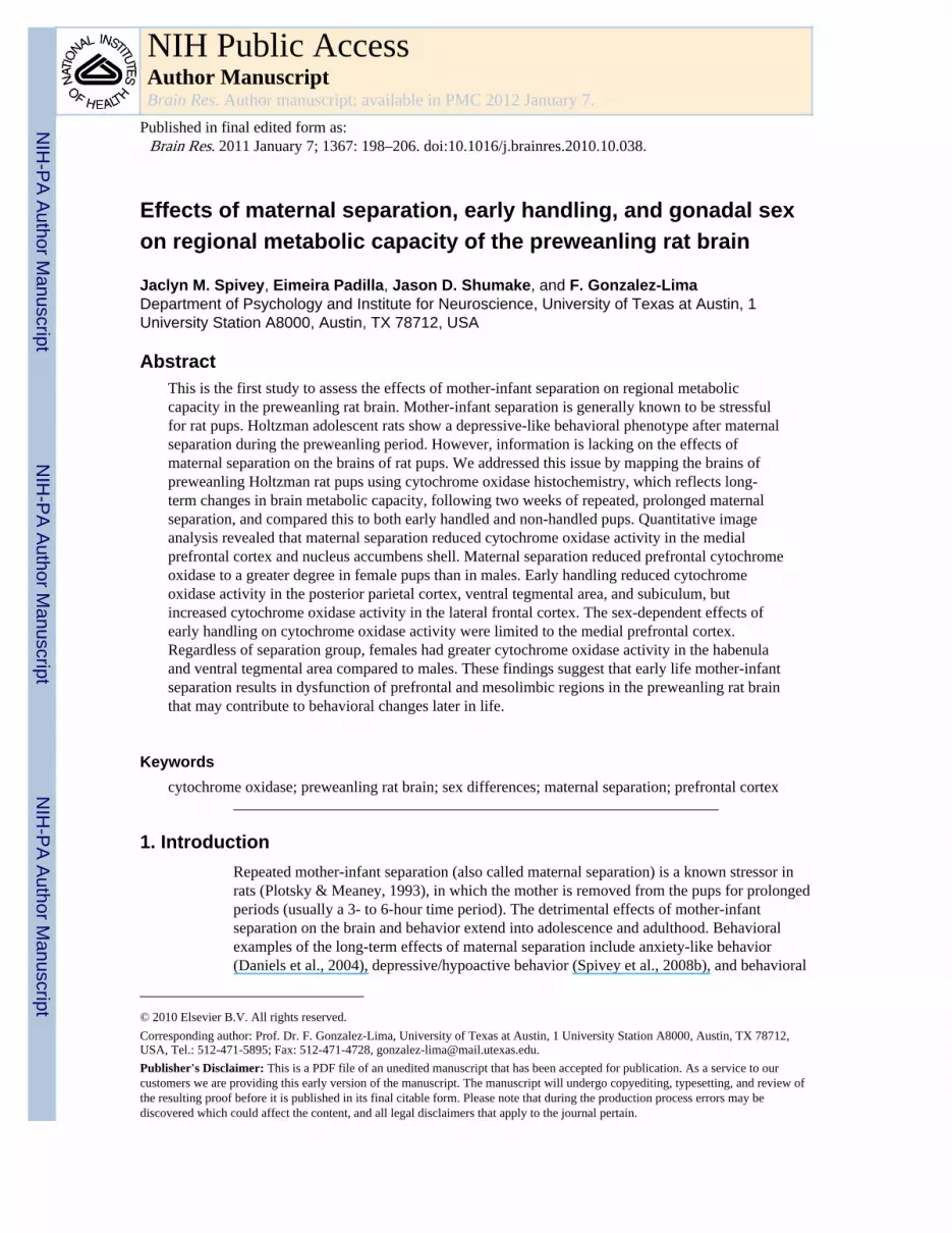
Effects of maternal separation, early handling, and gonadal sex on regional metabolic capacity of the preweanling rat brain Jaclyn M. Spivey, Eimeira Padilla, Jason D. Shumake, and F. Gonzalez-Lima Department of Psychology and Institute for Neuroscience, University of Texas at Austin, 1 University Station A8000, Austin, TX 78712, USA Abstract This is the first study to assess the effects of mother-infant separation on regional metabolic capacity in the preweanling rat brain. Mother-infant separation is generally known to be stressful for rat pups. Holtzman adolescent rats show a depressive-like behavioral phenotype after maternal separation during the preweanling period. However, information is lacking on the effects of maternal separation on the brains of rat pups. We addressed this issue by mapping the brains of preweanling Holtzman rat pups using cytochrome oxidase histochemistry, which reflects long- term changes in brain metabolic capacity, following two weeks of repeated, prolonged maternal separation, and compared this to both early handled and non-handled pups. Quantitative image analysis revealed that maternal separation reduced cytochrome oxidase activity in the medial prefrontal cortex and nucleus accumbens shell. Maternal separation reduced prefrontal cytochrome oxidase to a greater degree in female pups than in males. Early handling reduced cytochrome oxidase activity in the posterior parietal cortex, ventral tegmental area, and subiculum, but increased cytochrome oxidase activity in the lateral frontal cortex. The sex-dependent effects of early handling on cytochrome oxidase activity were limited to the medial prefrontal cortex. Regardless of separation group, females had greater cytochrome oxidase activity in the habenula and ventral tegmental area compared to males. These findings suggest that early life mother-infant separation results in dysfunction of prefrontal and mesolimbic regions in the preweanling rat brain that may contribute to behavioral changes later in life. Keywords cytochrome oxidase; preweanling rat brain; sex differences; maternal separation; prefrontal cortex 1. Introduction Repeated mother-infant separation (also called maternal separation) is a known stressor in rats (Plotsky & Meaney, 1993), in which the mother is removed from the pups for prolonged periods (usually a 3- to 6-hour time period). The detrimental effects of mother-infant separation on the brain and behavior extend into adolescence and adulthood. Behavioral examples of the long-term effects of maternal separation include anxiety-like behavior (Daniels et al., 2004), depressive/hypoactive behavior (Spivey et al., 2008b), and behavioral © 2010 Elsevier B.V. All rights reserved. Corresponding author: Prof. Dr. F. Gonzalez-Lima, University of Texas at Austin, 1 University Station A8000, Austin, TX 78712, USA, Tel.: 512-471-5895; Fax: 512-471-4728, [email protected]. Publisher's Disclaimer: This is a PDF file of an unedited manuscript that has been accepted for publication. As a service to our customers we are providing this early version of the manuscript. The manuscript will undergo copyediting, typesetting, and review of the resulting proof before it is published in its final citable form. Please note that during the production process errors may be discovered which could affect the content, and all legal disclaimers that apply to the journal pertain. NIH Public Access Author Manuscript Brain Res. Author manuscript; available in PMC 2012 January 7. Published in final edited form as: Brain Res. 2011 January 7; 1367: 198–206. doi:10.1016/j.brainres.2010.10.038. NIH-PA Author Manuscript NIH-PA Author Manuscript NIH-PA Author Manuscript
-
Upload
independent -
Category
Documents
-
view
0 -
download
0
Transcript of Effects of maternal separation, early handling, and gonadal sex on regional metabolic capacity of...
Effects of maternal separation, early handling, and gonadal sexon regional metabolic capacity of the preweanling rat brain
Jaclyn M. Spivey, Eimeira Padilla, Jason D. Shumake, and F. Gonzalez-LimaDepartment of Psychology and Institute for Neuroscience, University of Texas at Austin, 1University Station A8000, Austin, TX 78712, USA
AbstractThis is the first study to assess the effects of mother-infant separation on regional metaboliccapacity in the preweanling rat brain. Mother-infant separation is generally known to be stressfulfor rat pups. Holtzman adolescent rats show a depressive-like behavioral phenotype after maternalseparation during the preweanling period. However, information is lacking on the effects ofmaternal separation on the brains of rat pups. We addressed this issue by mapping the brains ofpreweanling Holtzman rat pups using cytochrome oxidase histochemistry, which reflects long-term changes in brain metabolic capacity, following two weeks of repeated, prolonged maternalseparation, and compared this to both early handled and non-handled pups. Quantitative imageanalysis revealed that maternal separation reduced cytochrome oxidase activity in the medialprefrontal cortex and nucleus accumbens shell. Maternal separation reduced prefrontal cytochromeoxidase to a greater degree in female pups than in males. Early handling reduced cytochromeoxidase activity in the posterior parietal cortex, ventral tegmental area, and subiculum, butincreased cytochrome oxidase activity in the lateral frontal cortex. The sex-dependent effects ofearly handling on cytochrome oxidase activity were limited to the medial prefrontal cortex.Regardless of separation group, females had greater cytochrome oxidase activity in the habenulaand ventral tegmental area compared to males. These findings suggest that early life mother-infantseparation results in dysfunction of prefrontal and mesolimbic regions in the preweanling rat brainthat may contribute to behavioral changes later in life.
Keywordscytochrome oxidase; preweanling rat brain; sex differences; maternal separation; prefrontal cortex
1. IntroductionRepeated mother-infant separation (also called maternal separation) is a known stressor inrats (Plotsky & Meaney, 1993), in which the mother is removed from the pups for prolongedperiods (usually a 3- to 6-hour time period). The detrimental effects of mother-infantseparation on the brain and behavior extend into adolescence and adulthood. Behavioralexamples of the long-term effects of maternal separation include anxiety-like behavior(Daniels et al., 2004), depressive/hypoactive behavior (Spivey et al., 2008b), and behavioral
© 2010 Elsevier B.V. All rights reserved.Corresponding author: Prof. Dr. F. Gonzalez-Lima, University of Texas at Austin, 1 University Station A8000, Austin, TX 78712,USA, Tel.: 512-471-5895; Fax: 512-471-4728, [email protected]'s Disclaimer: This is a PDF file of an unedited manuscript that has been accepted for publication. As a service to ourcustomers we are providing this early version of the manuscript. The manuscript will undergo copyediting, typesetting, and review ofthe resulting proof before it is published in its final citable form. Please note that during the production process errors may bediscovered which could affect the content, and all legal disclaimers that apply to the journal pertain.
NIH Public AccessAuthor ManuscriptBrain Res. Author manuscript; available in PMC 2012 January 7.
Published in final edited form as:Brain Res. 2011 January 7; 1367: 198–206. doi:10.1016/j.brainres.2010.10.038.
NIH
-PA Author Manuscript
NIH
-PA Author Manuscript
NIH
-PA Author Manuscript
hyperactivity and impulsivity (Daniels et al., 2009; Colorado et al., 2006; Kaneko et al.,1994; Jimenez-Vasquez et al., 2001). In particular, Spivey et al. (2008b) found thatadolescent rats belonging to the Holtzman strain appear to be particularly susceptible to thisdepressive behavioral profile following maternal separation, as measured by reduced levelsof general activity and risk-taking in the open field.
Long-term effects of maternal separation on the brain include, but are not limited to,changes to the production and binding of stress hormone receptors (Ladd et al., 2005), andalterations of dendritic morphology (Monroy et al., 2010) and neurotransmitter levels(Matthews et al., 2001) in the striatum, prefrontal cortex, and hippocampus. However, fewerstudies have addressed the more short-term effects of maternal separation on the brain orbehavior. Some have assessed the effects on hyperactive behavior (Daniels et al., 2009;Colorado et al., 2006) and response to cocaine (Marin & Planeta, 2004) in adolescence.More recent studies have shown decreases in glucocorticoid receptor mRNA andcorticotrophin releasing factor-1 mRNA in the hippocampus of 14-day old rats separated for3 hours (Litvin et al., 2010). Immediately following single 24-hour deprivations, increases inapoptosis in the hippocampus and various other brain regions are apparent (Zhang et al.,2002). Maternal separation stress also causes reductions in growth hormone and exaggeratedhypothalamic-pituitary-adrenal (HPA) axis response in preweanling rat pups (Kuhn &Schanberg, 1998). However, it appears that there is a lack of investigation into theimmediate effects of repeated prolonged maternal separation in the preweanling rat brain.
Therefore, this study aimed to assess the brain effects of 2 weeks of repeated maternalseparation in the infant rat brain. Quantitative cytochrome oxidase (CO) histochemistry canbe used as a marker for brain regional metabolic capacity (Gonzalez-Lima & Cada, 1998).CO builds up in the mitochondria of neurons as a reflection of increased demand for energyutilization (Wong-Riley et al., 1998). Specifically, CO is the rate-limiting enzyme in themitochondrial electron transport chain in which ATP is formed from the breakdown ofglucose, and therefore CO activity is tightly coupled to functional activity of a brain region(Wong-Riley et al., 1998). Energy demands over long periods of time are reflected in COactivity (Shumake & Gonzalez-Lima, 2003), and CO histochemistry is therefore anappropriate marker for studying the cumulative effects of behavioral and sex effects (Sakataet al., 2005).
We have previously demonstrated that adolescent Sprague-Dawley males and females showsex differences in brain CO activity (Spivey et al., 2008a), particularly in the prefrontal andposterior parietal cortical regions. Some neurochemical changes in the brain resulting frommaternal separation have been reported to be sex-dependent. The prefrontal cortex(Zimmerberg & Brown, 1998; Matthews et al., 2001; Jimenez-Vasquez et al., 2001),hippocampus (Matthews et al., 2001), paraventricular nucleus of the hypothalamus (Genestet al., 2004), and striatum (Sircar et al., 2001) are among the regions reported to havematernal separation-related sex differences. However, it should be noted that Holtzman ratshave shown diminished sex differences in antinociceptive responses (Terner et al., 2003)and in the effects of maternal separation on open-field behavior (Spivey et al., 2008b).
Based on the reviewed studies of brain changes produced by maternal separation (MS) andearly handling (EH) from the existing literature, in combination with our previouscharacterization of depressive behavior in adolescent Holtzman rats (Spivey et al., 2008b),we hypothesized that MS and EH would alter regional metabolic capacity in a variety ofbrain regions, including the prefrontal cortex, posterior parietal cortex, nucleus accumbensshell and mesolimbic pathway. Additionally, we hypothesized that the effects of MS and EHon regional metabolic capacity could differ between males and females during thepreweanling period.
Spivey et al. Page 2
Brain Res. Author manuscript; available in PMC 2012 January 7.
NIH
-PA Author Manuscript
NIH
-PA Author Manuscript
NIH
-PA Author Manuscript
2. ResultsTable 1 shows the mean CO values for each group, by region. Sixty pups were divided intothree separation groups at postnatal day 2 (P2): MS, maternal separation; EH, earlyhandling; and SFR, standard facility rearing. Each of these three separation groups hadmales and females, making a total of six groups. For 10 days, the MS subjects wereseparated for 6 h daily, and the EH subjects were separated for 15 min daily. The SFRsubjects were not separated. Their brains were collected at P13 and processed for COquantitative histochemistry. The number of pups was 20 per separation condition but due totissue damage in some subjects, the resulting n for each brain region in a group is providedin parentheses in Table 1. There were sixteen brain regions analyzed per subject. Eachindividual brain region was analyzed using a 2(sex) × 3 (separation group) univariateanalysis of variance (ANOVA). This was followed by individual group contrasts. With thesethree separation conditions, there could be significant differences between MS and SFRgroups, between EH and SFR groups, and between MS and EH groups. For the sake ofclarity, below we will report sequentially 1) an overview of the results, 2) the main effects ofseparation, 3) the interaction between separation and sex, 4) the main effects of sex, and 5)the litter effects.
2.1. OverviewAn overview of the results in Table 1 showed that EH resulted in the most widespreadeffects on CO activity, with increases in the lateral and medial prefrontal cortex, anddecreases in the posterior parietal cortex, subiculum, and ventral tegmental area. In contrast,effects of MS were limited to decreases in CO activity in the nucleus accumbens and medialprefrontal cortex. Interactions between gonadal sex and separation group were consistent inthe prefrontal cortical regions of MS animals, and to a lesser extent in EH animals. Maineffects of sex were limited to the habenula and ventral tegmental area.
2.2. Main effects of separation2.2.1. MS vs. SFR—MS resulted in lower CO activity compared to SFR in the deep layerof the medial prefrontal cortex (MS < SFR; F(2,42) = 15.879; p < 0.001).
2.2.2. EH vs. SFR—EH increased CO activity relative to SFR in the superficial layer ofthe lateral frontal cortex (EH > SFR; F(2,43) = 10.513; p < 0.001). EH decreased COactivity relative to SFR in the deep layers of the posterior parietal cortex (EH < SFR;F(2,42) = 3.981; p = 0.026), in the subiculum (EH < SFR; F(2,47) = 5.370; p = 0.008) andin the ventral tegmental area (EH < SFR; F(2,47) = 7.542; p = 0.001).
2.2.3. MS vs. EH—EH showed greater CO activity than MS in the lateral frontal cortex, inboth the superficial (EH > MS; F(2,43) = 10.513; p < 0.001) and deep (EH > MS; F(2,43) =9.335; p = 0.001) layers. EH also had greater CO activity than MS in the medial prefrontalcortex, in both the superficial (EH > MS; F(2,42) = 7.246; p = 0.002) and deep layers (EH >MS; F(2,42) = 15.879; p < 0.001). EH showed greater CO activity than MS in the shell ofthe nucleus accumbens (EH > MS; F(2,46) = 4.642; p = 0.015) and the caudate putamen(EH > MS; F(2,36) = 3.380; p = 0.045). Metabolic activity was reduced in EH animalsrelative to MS animals in both the subiculum (EH < MS; F(2,47) = 5.371; p = 0.008) andventral tegmental area (EH < MS; F(2,47) = 7.542; p = 0.001).
2.3. Interaction between separation effects and sexMS subjects showed the most sex-dependent separation effects in the frontal cortex. MSdecreased metabolic activity in female pups more than male pups in the medial prefrontaland lateral frontal cortex, as reflected by significant sex × separation interactions for these
Spivey et al. Page 3
Brain Res. Author manuscript; available in PMC 2012 January 7.
NIH
-PA Author Manuscript
NIH
-PA Author Manuscript
NIH
-PA Author Manuscript
regions (Fs(2,42) > 7.1, ps < 0.01). Subsequent simple effects tests revealed that in allfrontal cortex regions measured, MS females had lower CO activity than MS males (MS-F <MS-M; ps < 0.003). Conversely, in the deep layer of the medial prefrontal cortex, EHfemales had greater CO activity than EH males (EH–F > EH–M; p < 0.05). In addition,simple-effects tests showed that MS females had significantly lower CO values in all frontalcortical regions as compared to females from SFR and EH groups (MS-F < SFR-F; MS-F <EH-F; ps < 0.01), while there were no significant group differences in CO activity amongmales.
2.4. Main effects of sexTwo regions showed sex differences independent of separation condition, with femalesshowing greater metabolic activity than males in the habenula (F > M; F(1,42) = 4.636; p <0.05), and ventral tegmental area (F > M; F(1,47) = 4.193; p < 0.05).
2.5. Litter effectsSince there were only few litters used, we evaluated litter effects on CO activity using three-level hierarchical ANOVAs (this is a type of ANOVA in which comparisons are performedsequentially between separation groups, between litters/within groups, and between sexes/within litters, to identify separate influences contributing to the effects). Most regions didnot show significant between-litter differences within separation groups (see Table 1).However, significant litter effects were noted for the lateral frontal cortex deep layer(F(3,42) = 3.12; p < 0.05), habenula (F(3,41) = 5.00; p < 0.01), ventral tegmental area(F(3,46) = 12.3; p < 0.001), and subiculum (F(3,46) = 9.95; p < 0.001). The analysis ofseparation effects for these regions should be viewed with caution because high between-litter variance could have either manufactured false differences or masked true differencesrelated to separation condition.
In summary, the medial prefrontal cortex and accumbens showed separation effects notsignificantly confounded by litter, but separation effects detected in the VTA and subiculumwere confounded by litter. However, because sex was modeled within litter in thehierarchical analysis, the sex differences observed in the habenula and VTA were notconfounded by litter.
3. DiscussionIn preweanling rats, prolonged (6 h/day for 10 days) mother-infant separation (MS) showedmain effects in terms of reducing brain metabolic capacity in the deep layers of the medialprefrontal cortex, caudate putamen, and the shell of the nucleus accumbens. Early handling(EH) with brief separation (15 min/day for 10 days) had contrasting effects on brainmetabolic capacity, increasing CO activity in the lateral frontal and medial prefrontal cortexcompared to MS, but decreasing CO activity in the ventral tegmental area, subiculum, andposterior parietal cortex. In terms of sex differences, MS reduced brain metabolic capacity inthe lateral frontal and medial prefrontal cortical regions to a greater degree in female pupsthan in males. All female pups, regardless of separation group, showed greater CO activitycompared to males in the habenula and ventral tegmental area.
Our findings are consistent with existing literature which has demonstrated that maternalseparation does affect the prefrontal cortex. For instance, corticotrophin-releasing hormoneand glucocorticoid receptor mRNA levels are reduced in the prefrontal cortex of maternallyseparated rats (Ladd et al., 2005). Reductions in prefrontal cortical thickness (Spivey et al.,2009) and in dendritic processes of the prefrontal pyramidal neurons have also beendemonstrated in maternally separated rats (Monroy et al., 2010; Pascual & Zamora-Leon,
Spivey et al. Page 4
Brain Res. Author manuscript; available in PMC 2012 January 7.
NIH
-PA Author Manuscript
NIH
-PA Author Manuscript
NIH
-PA Author Manuscript
2007). In addition, irregular patterns of interneuron development in the medial prefrontalcortex of maternally separated animals have been found in another rodent species (Helmekeet al., 2008). All of these previously reported findings are consistent with reductions inmetabolic demands in the medial prefrontal cortex following prolonged repeated maternalseparation. We also showed that among SFR pups that were not separated, P13 malesappeared with lower frontal metabolic capacity than females. But after prolonged maternalseparation, a marked metabolic hypofrontality was induced in MS females but not in MSmales.
There are several behavioral implications of reductions in CO activity in the prefrontalcortex of preweanling rats. Prefrontal hypometabolism is strongly related to behavioralhyperactivity in boys with attention deficit hyperactivity disorder (ADHD) as well as malerats selectively bred as models of hyperactive behavior (Gonzalez-Lima and Sadile, 2000;Gonzalez-Lima, 2005). The medial prefrontal cortex is also implicated in depression in thehuman and animal literature (Shumake et al., 2000; Shumake and Gonzalez-Lima, 2003).Metabolic brain mapping in infant rats in particular, has shown that prefrontal corticalregions are involved in the maturation of behavioral inhibition (Nair et al., 2001a, 2001b).Perhaps hypometabolism in medial prefrontal cortex is also related to depression orbehavioral inhibition dysfunction in general, and the behavioral outcome is dependent on theorganism’s predispositions to a certain stress-related psychopathology such as depression(Padilla et al., 2009). For example, Holtzman rats (as used in the present study) show ahypoactive phenotype after our MS treatment (Spivey et al., 2008b). Maternal separationmay lead to hypoactive behavioral effects in Holtzman rats because of their greater geneticpredisposition to show learned helplessness and depressive-like responses to stress (Wielandet al., 1986; Padilla et al., 2009).
Similarly, the sex differences we found in the preweanling brain may be related to differentbiological predispositions among males and females. Brain CO activity is a well-establishedindex of metabolic capacity correlated with behavioral predispositions, especially sex effectsin rats and other species (Sakata et al., 2000, 2002a, 2002b, 2005; Spivey et al., 2008a;Crews et al., 2009). As preweanlings, Holtzman females generally have higher metaboliccapacity than males in most brain regions, including the prefrontal cortex. This supportsprevious findings in which female adolescent Holtzman rats show less risk-taking and shortmovement behavior in a novel open field compared to males, suggesting an increasedcapacity for behavioral inhibition (Spivey et al., 2008b). The MS-related sex differencesduring the preweanling period which are not present in adolescent behavior could bereflective of functional pathways still in the process of maturing (Shumake et al., 2004).
As compared to females, juvenile Sprague-Dawley male rats have lower prefrontalmetabolic capacity under baseline conditions (Spivey et al., 2008a). This baseline sexdifference is consistent with the lower prefrontal CO values observed in SFR Holtzman malepups. In addition, when preweanling Holtzman rats were subjected to MS stress the femalesshowed the strongest frontal cortex metabolic reductions relative to SFR and EH groups.There are also sex-dependent strain differences in the behavioral response to MS. Forexample, after identical MS treatments, females of the Holtzman strain show hypoactivebehavior; whereas Sprague-Dawley males show hyperactive behavior. Therefore, the effectsof identical MS protocols on prefrontal metabolism and behavior are dependent on the rat’sgenetic background and sex, with Holtzman females being more predisposed to showhypoactivity.
In MS animals, hypometabolism in the accumbens shell may be functionally related toprefrontal hypometabolism because rat accumbens shell neurons are strongly regulated byexcitatory afferents arising from the prefrontal cortex (Pennartz et al., 1994). Our findings
Spivey et al. Page 5
Brain Res. Author manuscript; available in PMC 2012 January 7.
NIH
-PA Author Manuscript
NIH
-PA Author Manuscript
NIH
-PA Author Manuscript
are also consistent with existing literature which has demonstrated that maternal separationaffects the nucleus accumbens, in particular the shell (Zimmerberg and Brown, 1998;Andersen et al., 1999; Brake et al., 2004). This MS effect was similar for males and females,and may be linked to the hypoactive phenotype exhibited by Holtzman rats after MS vs. EHtreatments (Spivey et al., 2008b). More generally, Holtzman rats show increasedvulnerability to respond to stressful treatments with depressive-like behavior, such asincreased rates of learned helplessness (Padilla et al., 2009). We interpret the reduction inMS pups as compared to EH pups as linked to the role of the nucleus accumbens shell indepressive-like behavior and antidepressant drug action (Shirayama & Chaki, 2006). Inparticular, metabolic reduction of the accumbens shell has been found to produce anhedonia,which is a key feature of human depression as well as of rats predisposed to develop learnedhelplessness (Shumake et al., 2005). This interpretation would be consistent with thedepressive-like phenotype exhibited by Holtzman rats after MS and other types of stressors(Wieland et al., 1986; Spivey et al., 2008b; Padilla et al., 2009).
The lateral frontal cortex (often regarded as part of motor cortex in rats) was moremetabolically active in EH as compared to MS animals, and this finding is likely related tothe hypoactive behavior of Holtzman MS animals (Spivey et al., 2008b). Briefly, wepreviously found in Holtzman six-week-old rats a hypoactive phenotype for MS animals, butincreases in risk-taking behaviors for EH animals. Increases in lateral frontal cortexmetabolic capacity in EH animals would likely correspond to increased propensity forambulatory activities, such as entering the exposed zone of an open field (Spivey et al.,2008b).
In conclusion, our findings are novel in that they are the first to report experience-basedchanges in regional metabolic capacity in preweanling rats as a result of early handling andmother-infant separation. The hypothesis that repeated prolonged mother-infant separationwould alter prefrontal cortex regional metabolic capacity was supported. Our findings areconsistent with existing literature that finds MS-dependent changes in prefrontal cortex andnucleus accumbens. This study is also the first to evaluate sex-dependent changes inmetabolic capacity in the brain following two weeks of mother-infant separation.
4. Experimental Procedures4.1. Subjects
Subjects were 60 Holtzman albino rat pups born to 6 timed-pregnant mothers bred in thecolony of the Animal Resources Center at the University of Texas at Austin. Pregnantmothers were singly housed and maintained on a 12h/12h light/dark photoperiod, with lightson at 06:00 h and lights off at 18:00 h, in a room with temperature maintained at 22°C and40% humidity. Food and water were available ad libitum. The day of birth was marked aspostnatal day (P) 0. On P2, litters were culled to as close to ten pups as possible, alwaysconsisting of an equal male-to-female ratio. Anogenital distance was used to determinemales from females at this age. All procedures were conducted in accordance with theguidelines of the National Institutes of Health in an animal facility accredited by theAmerican Association for the Accreditation of Laboratory Animal Care-International, andthe protocol was approved by the Institutional Animal Care and Use Committee.
4.2. Maternal separation protocolThe six litters were divided into three groups, as previously described (Colorado et al., 2006;Spivey et al., 2008a, 2008b): Maternal Separation (MS), in which separation consisted of 6hours; Early Handled (EH), in which separation consisted of 15 minutes; and StandardFacility Reared (SFR), who were not handled at all except for the culling on P2 and
Spivey et al. Page 6
Brain Res. Author manuscript; available in PMC 2012 January 7.
NIH
-PA Author Manuscript
NIH
-PA Author Manuscript
NIH
-PA Author Manuscript
biweekly cage changes by facility staff. Separation protocols for MS and EH groupsconsisted of daily separation from P2 through P6; no separation from P7 to P8, and dailyseparation from P9 through P13, as in our other studies (Colorado et al., 2006; Spivey et al.,2008a, 2008b). Maternal separation began at 0730 h and ended at 1330 h daily, while earlyhandling occurred from 1300 h to 1315 h daily. The dams were removed from the homecage, and all pups were removed and placed as a litter into a bedding-lined holding cage,and dams were returned to the home cage for the duration of the separation. Holding cageswere then placed in an incubator (30–34°C) to maintain thermoregulation for the duration ofthe separation period. Upon return to the home cage, dams were again removed from thehome cage, pups were returned to the cage, and the dams were returned to the pups.
4.3. Tissue preparationImmediately following the final separation period on P13, pups were returned as a group toa holding cage, then transferred individually to a separate room and decapitated. Brains wererapidly extracted and frozen in isopentane. The brains were stored at −40°C until sectioning.Tissue was sectioned at 40 micrometers in a −20°C cryostat. The sections were kept frozenon slides at −40°C until processing for cytochrome oxidase histochemistry. Every thirdsection was used for staining. Staining and quantification of neural tissue for cytochromeoxidase activity was used to determine the metabolic capacity of brain regions based ondensitometric histochemical analysis of the sections and of brain paste standards of knowncytochrome oxidase activity measured spectrophotometrically (Gonzalez-Lima & Cada,1998).
Brain tissue was stained using the cytochrome oxidase histochemistry procedure previouslydescribed (Gonzalez-Lima & Cada, 1998). Slides were first treated in 10% sucrosephosphate buffer (0.1M, pH 7.6) containing 0.5% glutaraldehyde (Grade II) for 5 min. Threechanges at 5 min each of 10% sucrose phosphate buffer were followed by 10 min in Trisbuffer (0.05 M, pH 7.6) containing 275 mg/l cobalt chloride, 10% sucrose, and 0.5%dimethylsulfoxide. The slides were then rinsed for 5 min in phosphate buffer and incubatedat 37°C for 60 min in 700 ml of an oxygen-saturated reaction solution containing 350 mg ofdiaminobenzidine tetrahydrochloride (DAB), 52.5 mg of cytochrome c, 35 g of sucrose, 14mg of catalase, and 1.75 ml of dimethylsulfoxide in phosphate buffer. To stop the reactionand fix the tissue, a 30 min immersion in 10% sucrose phosphate buffer with 4% formalin(v/v) was used before dehydrating with ethanol, clearing with xylene, and coverslippingwith Permount.
Anatomically matched sections from male and female subjects were stained in the samebatch to minimize the possibility of inter-batch variability during the comparison betweensubject groups. In addition, sets of homogenized tissue standards (10, 20, 40, 60, 80 micron-thick sections) were included with each batch of slides (Gonzalez-Lima & Cada, 1998).These standards were used to convert tissue optical density measures to cytochrome oxidaseactivity units via a regression equation based on their optical density andspectrophotometrically determined enzymatic activity.
Using an image-processing system (JAVA, Jandel Scientific, Corte Madera, CA), opticaldensity was sampled from regions of interest. The size of the square-shaped samplingwindow was adjusted for each region so that it was as large as possible while still allowingfor four, non-overlapping readings to be taken bilaterally. For each cortical region, opticaldensity was sampled in both superficial (I–III) and deep (IV–VI) layers across three adjacentsections and averaged. Cortical regions were analyzed separately in superficial and deeplayers because we have previously found differences between them in the cortex of six-week-old rats (Spivey et al., 2008a). These optical density values were then converted to
Spivey et al. Page 7
Brain Res. Author manuscript; available in PMC 2012 January 7.
NIH
-PA Author Manuscript
NIH
-PA Author Manuscript
NIH
-PA Author Manuscript
cytochrome oxidase activity units (micromol/min/g tissue wet weight), which weredetermined by spectrophotometry of cytochrome oxidase standards as described before.
4.4. Regions of interestThe maternal separation literature describes numerous alterations in both behavior and brainactivity. The regions were selected based on studies regarding maternal separation andchanges in brain activity. Hyperactive behavior has been reported as one long-term effect ofMS (Colorado et al., 2006; Kaneko et al., 1994) and the relationship between hyperactivebehavior and the prefrontal cortex is well documented (Sullivan & Brake, 2003).Dysfunction to the prefrontal cortex is also seen subsequent to MS (Monroy et al., 2010;Jimenez-Vasquez et al., 2001; Ladd et al., 2005). Many studies have used a myriad ofmeasures to show brain differences in MS animals, including neurotransmitter levels, stresshormone levels, drug manipulations, and receptor binding studies. Alongside findings ofbehavioral hyperactivity, studies have shown increases in catecholamine levels in thestriatum and dorsal hippocampus (Matthews et al., 2001), and changes in the nucleusaccumbens in both MS (Monroy et al., 2010; Zimmerberg & Brown, 1998) and EH animals(Brake et al., 2004). Changes to the reward sensitivity in MS animals by way of reducedresponding to sucrose solutions (Matthews, Wilkinson & Robbins, 1996) or cocaine(Matthews et al., 1999) was a rationale for considering dopamine related regions, includingthe ventral tegmental area, nucleus accumbens, striatum, and prefrontal cortex in the currentstudy.
In order to account for divergent phenotypes found by various MS protocols, we alsoconsidered other studies that have reported hypoactive behavior following MS (Spivey et al.,2008b). Such a depressive phenotype in rats is associated with changes to the habenula,ventral tegmental area, striatum, prefrontal and infralimbic cortex, and hippocampus(Shumake et al., 2005). Due to the stressful nature of MS in infancy, many reports havelisted changes to anxiety-like behaviors and defensive withdrawal in adolescence (Coloradoet al., 2006), as well as alterations in neural activity in stress-related regions such as theparaventricular nucleus of the hypothalamus and central amygdala (Liu et al., 2000; Genestet al., 2004; Ladd et al., 2005; Plotsky et al., 2005). The posterior parietal cortex was ofinterest due to its relationship to attention and motor behaviors in rodents (Kolb & Walkey,1987; Mattelli & Luppino, 2001), and is also reported to have less CO activity in males thanfemales (Spivey et al., 2008a).
Cortical regions were assessed separately in terms of superficial (I – III) and deep (IV – VI)layers, based also on previous findings in which cytochrome oxidase levels showed groupdifferences in superficial cortical layers but not deep layers (Gonzalez-Lima & Sadile,2000). All regions of interest and corresponding Bregma levels (Paxinos & Watson, 1997;Gonzalez-Lima & Cada, 1998) are depicted in Figure 1.
4.5. Statistical analysisData from this experiment were analyzed using SPSS software (version 11.5, SPSS,Chicago, IL). Differences were considered significant at the two-tailed p < 0.05 level for alltests. A 2 (sex) × 3 (separation group) univariate ANOVA was used to assess separationgroup and sex differences in each region of interest (Spivey et al., 2008a). Additional post-hoc Bonferroni-corrected simple effects tests were utilized where indicated. Litter effects onCO activity were evaluated using three-level hierarchical ANOVAs.
AcknowledgmentsWe gratefully acknowledge Michael Benvenuti and Lynn Krug for their contributions to sectioning and imaging ofthe brain tissue. Supported in part by NIH grants R01 MH076847 and T32 MH65728 directed by FGL. JMS
Spivey et al. Page 8
Brain Res. Author manuscript; available in PMC 2012 January 7.
NIH
-PA Author Manuscript
NIH
-PA Author Manuscript
NIH
-PA Author Manuscript
conducted this research in partial fulfillment of her requirements for a Ph.D. degree at the University of Texas atAustin.
ReferencesAndersen SL, Lyss PJ, Dumont NL, Teicher MH. Enduring neurochemical effects of early maternal
separation on limbic structures. Ann. N. Y. Acad. Sci. 1999; 877:756–759. [PubMed: 10415699]Brake WG, Zhang TY, Diorio J, Meaney MJ, Gratton A. Influence of early postnatal rearing
conditions on mesocorticolimbic dopamine and behavioural responses to psychostimulants andstressors in adult rats. Eur. J. Neurosci. 2004; 19:1863–1874. [PubMed: 15078560]
Colorado RA, Shumake J, Conejo NM, Gonzalez-Pardo H, Gonzalez-Lima F. Effects of maternalseparation, early handling, and standard facility rearing on orienting and impulsive behavior ofadolescent rats. Behav. Processes. 2006; 71:51–58. [PubMed: 16242858]
Crews D, Rushworth D, Gonzalez-Lima F, Ogawa S. Litter environment affects behavior and brainmetabolic activity of adult knockout mice. Front. Behav. Neurosci. 2009; 3:1–18. [PubMed:19194528]
Daniels WMU, Fairbairn LR, van Tilburg G, McEvoy CRE, Zigmond MJ, Russell VA, Stein DJ.Maternal separation alters nerve growth factor and corticosterone levels but not the DNAmethylation status of the exon 17 glucocorticoid receptor promoter region. Metab. Brain Dis. 2009;24:615–627. [PubMed: 19816761]
Daniels WMU, Pietersen CY, Carstens ME, Stein DJ. Maternal separation in rats leads to anxiety-likebehavior and a blunted ACTH response and altered neurotransmitter levels in response to asubsequent stressor. Metab. Brain Dis. 2004; 19:3–14. [PubMed: 15214501]
Genest SE, Gulemetova R, Laforest S, Drolet G, Kinkead R. Neonatal maternal separation and sex-specific plasticity of the hypoxic ventilatory response in awake rat. J. Physiol. 2004; 554:543–557.[PubMed: 14634199]
Gonzalez-Lima, F.; Cada, A. Quantitative histochemistry of cytochrome oxidase activity: Theory,methods, and regional brain vulnerability. In: Gonzalez-Lima, F., editor. Cytochrome Oxidase inNeuronal Metabolism and Alzheimer's Disease. New York: Plenum Press; 1998. p. 55-90.
Gonzalez-Lima F, Sadile AG. Network operations revealed by brain metabolic mapping in a geneticmodel of hyperactivity and attention deficit: the Naples high- and low-excitability rats. Neurosci.Biobehav. Rev. 2000; 24:157–160. [PubMed: 10654673]
Gonzalez-Lima, F. Cortical and limbic systems mediating the predisposition to attention deficit andhyperactivity. In: Larimer, MP., editor. Attention Deficit Hyperactivity Disorder Research.Hauppauge, New York: Nova Science Publishers; 2005. p. 1-18.
Helmeke C, Ovtscharoff W Jr, Poeggel G, Braun K. Imbalance of immunohistochemical characterizedinterneuron populations in the adolescent and adult rodent medial prefrontal cortex after repeatedexposure to neonatal separation stress. Neurosci. 2008; 152:18–28.
Jimenez-Vasquez PA, Mathe AA, Thomas JD, Riley EP, Ehlers CL. Early maternal separation altersneuropeptide Y concentrations in selected brain regions in adult rats. Dev. Brain Res. 2001;131:149–152. [PubMed: 11718845]
Kaneko W, Riley E, Ehlers CL. Behavioral and electrophysiological effects of early repeated maternalseparation. Depression. 1994; 2:43–53.
Kolb B, Walkey J. Behavioural and anatomical studies of the posterior parietal cortex in the rat.Behav. Brain Res. 1987; 23:127–145. [PubMed: 3566907]
Kuhn C, Schanberg S. Responses to maternal separation: mechanisms and mediators. Int. J. Dev.Neurosci. 1998; 16:261–270. [PubMed: 9785122]
Ladd CO, Thrivikraman KV, Huot RL, Plotsky PM. Differential neuroendocrine responses to chronicvariable stress in adult Long Evans rats exposed to handling-maternal separation as neonates.Psychoneuroendocrinol. 2005; 30:520–533.
Litvin Y, Tovote P, Pentkowski NS, Zeyda T, King LB, Vasconcellos AJ, Dunlap C, Spiess J,Blanchard DC, Blanchard RJ. Maternal separation modulates short-term behavioral andphysiological indices of the stress response. Horm. Behav. 2010 in press.
Spivey et al. Page 9
Brain Res. Author manuscript; available in PMC 2012 January 7.
NIH
-PA Author Manuscript
NIH
-PA Author Manuscript
NIH
-PA Author Manuscript
Liu D, Caldji C, Sharma S, Plotsky PM, Meaney MJ. Influence of neonatal rearing conditions onstress-induced adrenocorticotropin responses and norepinepherine release in the hypothalamicparaventricular nucleus. J. Neuroendocrinol. 2000; 12:5–12. [PubMed: 10692138]
Marin MT, Planeta CS. Maternal separation affects cocaine-induced locomotion and response tonovelty in adolescent, but not in adult rats. Brain Res. 2004; 1013:83–90. [PubMed: 15196970]
Matelli M, Luppino G. Parietofrontal circuits for action and space perception in the macaque monkey.Neuroimage. 2001; 14:S27–S32. [PubMed: 11373129]
Matthews K, Wilkinson LS, Robbins TW. Repeated maternal separation of preweanling rats attenuatesbehavioral responses to primary and conditioned incentives in adulthood. Physiol. Behav. 1996;59:99–107. [PubMed: 8848498]
Matthews K, Robbins TW, Everitt BJ, Caine SB. Repeated neonatal maternal separation altersintravenous cocaine self-administration in adult rats. Psychopharmacology (Berl.). 1999; 141:123–134. [PubMed: 9952036]
Matthews K, Dalley JW, Matthews C, Tsai TH, Robbins TW. Periodic maternal separation of neonatalrats produces region- and gender-specific effects on biogenic amine content in postmortem adultbrain. Synapse. 2001; 40:1–10. [PubMed: 11170216]
Monroy E, Hernandez-Torres E, Flores G. Maternal separation disrupts dendritic morphology ofneurons in prefrontal cortex, hippocampus, and nucleus accumbens in male rat offspring. J. Chem.Neuroanat. 2010; 40:93–101. [PubMed: 20553852]
Nair HP, Berndt JD, Barrett D, Gonzalez-Lima F. Metabolic mapping of brain regions associated withbehavioral extinction in preweanling rats. Brain Res. 2001a; 903:141–153. [PubMed: 11382397]
Nair HP, Berndt JD, Barrett D, Gonzalez-Lima F. Maturation of extinction behavior in infant rats:Large-scale regional interactions with medial prefrontal cortex, orbitofrontal cortex, and anteriorcingulate cortex. J. Neurosci. 2001b; 21:4400–4407. [PubMed: 11404426]
Padilla E, Barrett D, Shumake J, Gonzalez-Lima F. Strain, sex, and open-field behavior: Factorsunderlying the genetic susceptibility to helplessness. Behav. Brain Res. 2009; 201:257–264.[PubMed: 19428642]
Pascual R, Zamora-Leon SP. Effects of neonatal maternal deprivation and postweaning environmentalcomplexity on dendritic morphology of prefrontal pyramidal neurons in the rat. Acta Neurobiol.Exp. (Wars.). 2007; 67:471–479. [PubMed: 18320724]
Paxinos, G.; Watson, C. The rat brain in stereotaxic coordinates. Third ed.. San Diego, CA: AcademicPress, Inc.; 1997.
Pennartz CM, Groenewegen HJ, Lopes da Silva FH. The nucleus accumbens as a complex offunctionally distinct neuronal ensembles: an integration of behavioural, electrophysiological andanatomical data. Prog. Neurobiol. 1994; 42:719–761. [PubMed: 7938546]
Plotsky PM, Meaney MJ. Early, postnatal experience alters hypothalamic corticotropin-releasingfactor (CRF) mRNA, median eminence CRF content and stress-induced release in adult rats. BrainRes. Mol. Brain Res. 1993; 18:195–200. [PubMed: 8497182]
Plotsky PM, Thrivikraman KV, Nemeroff CB, Caldji C, Sharma S, Meaney MJ. Long-termconsequences of neonatal rearing on central corticotropin-releasing factor systems in adult maleoffspring. Neuropsychopharmacol. 2005; 30:2192–2204.
Sakata J, Coomber P, Gonzalez-Lima F, Crews D. Functional connectivity among limbic brain areas:Differential effects of incubation temperature and gonadal sex in the leopard gecko, Eublepharismacularius. Brain Behav. Evol. 2000; 55:139–151. [PubMed: 10899708]
Sakata J, Gonzalez-Lima F, Gupta A, Crews D. Repeated interactions with females elevate metaboliccapacity in the limbic system of male rats. Brain Res. 2002a; 936:27–37. [PubMed: 11988227]
Sakata JT, Gupta A, Gonzalez-Lima F, Crews D. Heterosexual housing increases the retention ofcourtship behavior following castration and elevates metabolic capacity in limbic brain nuclei inmale whiptail lizards, Cnemidophorus inornatus. Horm. Behav. 2002b; 42:263–273. [PubMed:12460586]
Sakata JT, Crews D, Gonzalez-Lima F. Behavioral correlates of differences in neural metaboliccapacity. Brain Res. Rev. 2005; 48:1–15. [PubMed: 15708625]
Shirayama Y, Chaki S. Neurochemistry of the nucleus accumbens and its relevance to depression andantidepressant action in rodents. Curr. Neuropharmacol. 2006; 4:277–291. [PubMed: 18654637]
Spivey et al. Page 10
Brain Res. Author manuscript; available in PMC 2012 January 7.
NIH
-PA Author Manuscript
NIH
-PA Author Manuscript
NIH
-PA Author Manuscript
Shumake J, Poremba A, Edwards E, Gonzalez-Lima F. Congenital helpless rats as a genetic model forcortex metabolism in depression. Neuroreport. 2000; 11:3793–3798. [PubMed: 11117493]
Shumake J, Gonzalez-Lima F. Brain systems underlying susceptibility to helplessness and depression.Behav. Cogn. Neurosci. Rev. 2003; 2:198–221. [PubMed: 15006293]
Shumake J, Conejo-Jimenez N, Gonzalez-Pardo H, Gonzalez-Lima F. Brain differences in newbornrats predisposed to helpless and depressive behavior. Brain Res. 2004; 1030:267–276. [PubMed:15571675]
Shumake J, Barrett D, Gonzalez-Lima F. Behavioral characteristics of rats predisposed to learnedhelplessness: Reduced reward sensitivity, increased novelty seeking, and persistent fear memories.Behav. Brain Res. 2005; 164:222–230. [PubMed: 16095730]
Sircar R, Mallinson K, Goldbloom LM, Kehoe P. Postnatal stress selectively upregulates striatal N-methyl--aspartate receptors in male rats. Brain Res. 2001; 904:145–148. [PubMed: 11516421]
Spivey JM, Colorado RA, Conejo NM, Gonzalez-Pardo H, Gonzalez-Lima F. Juvenile male ratsdisplay lower cortical metabolic capacity than females. Neurosci. Lett. 2008a; 440:255–259.[PubMed: 18579304]
Spivey JM, Barrett D, Padilla E, Gonzalez-Lima F. Mother-infant separation leads to hypoactivebehavior in adolescent Holtzman rats. Behav. Processes. 2008b; 79:59–65. [PubMed: 18585869]
Spivey JM, Shumake J, Colorado RA, Conejo NM, Gonzalez-Pardo H, Gonzalez-Lima F. Adolescentfemale rats are more resistant than males to the effects of early stress on prefrontal cortex andimpulsive behavior. Dev. Psychobiol. 2009; 51:277–288. [PubMed: 19125421]
Sullivan RM, Brake WG. What the rodent prefrontal cortex can teach us about attention-deficit/hyperactivity disorder: the critical role of early developmental events on prefrontal function.Behav. Brain Res. 2003; 146:43–55. [PubMed: 14643458]
Terner JM, Lomas LM, Smith ES, Barrett AC, Picker MJ. Pharmacogenetic analysis of sex differencesin opioid antinociception in rats. Pain. 2003; 106:381–391. [PubMed: 14659521]
Wieland S, Boren JL, Consroe PF, Martin A. Stock differences in the susceptibility of rats to learnedhelplessness training. Life Sci. 1986; 39:937–944. [PubMed: 3489150]
Wong-Riley, M.; Nie, F.; Hevner, R.; Liu, S. Brain cytochrome oxidase: Functional significance andbigenomic regularion in the CNS. In: Gonzalez-Lima, F., editor. Cytochrome Oxidase in NeuronalMetabolism and Alzheimer's Disease. New York: Plenum Press; 1998. p. 1-53.
Zhang LX, Levine S, Dent G, Zhan Y, Xing G, Okimoto D, Kathleen Gordon M, Post RM, Smith MA.Maternal deprivation increases cell death in the infant rat brain. Dev. Brain Res. 2002; 133:1–11.[PubMed: 11850058]
Zimmerberg B, Brown R. Prenatal experience and postnatal stress modulate the adult neurosteroid andcatecholaminergic stress responses. Int. J. Dev. Neurosci. 1998; 16:217–228. [PubMed: 9785118]
Spivey et al. Page 11
Brain Res. Author manuscript; available in PMC 2012 January 7.
NIH
-PA Author Manuscript
NIH
-PA Author Manuscript
NIH
-PA Author Manuscript
Figure 1.Photographs of CO-stained hemisections (left) and corresponding coronal brain drawings(right) illustrating the locations of each region of interest by Bregma level according to thePaxinos and Watson (1997) and Gonzalez-Lima and Cada (1998) atlases. Abbreviations arelisted in Table 1.
Spivey et al. Page 12
Brain Res. Author manuscript; available in PMC 2012 January 7.
NIH
-PA Author Manuscript
NIH
-PA Author Manuscript
NIH
-PA Author Manuscript
NIH
-PA Author Manuscript
NIH
-PA Author Manuscript
NIH
-PA Author Manuscript
Spivey et al. Page 13
Tabl
e 1
Bra
in c
ytoc
hrom
e ox
idas
e ac
tivity
(mea
n+S.
E.M
) afte
r rep
eate
d m
ater
nal s
epar
atio
n (M
S), e
arly
han
dlin
g (E
H) o
r sta
ndar
d fa
cilit
y re
arin
g (S
FR) i
npr
ewea
nlin
g ra
ts.
Reg
ion
Abb
rev.
Bre
gma
SFR
EH
MS
Effe
cts (
p =
)
Mal
e (n
)Fe
mal
e (n
)M
ale
(n)
Fem
ale
(n)
Mal
e (n
)Fe
mal
e (n
)Se
para
tiona
Sexb
Sep.
×sex
cL
itter
d
Late
ral f
ront
al c
orte
x, su
perf
icia
lLF
R-S
3.7
134±
6 (6
)14
2±7
(5)
153±
5 (1
0)15
3±5
(10)
146±
5 (9
)11
7±5
(9)
<0.0
010.
086
0.00
3 0
.17
Late
ral f
ront
al c
orte
x, d
eep
LFR
-D3.
713
7±6
(6)
145±
7 (5
)15
3±5
(10)
151±
5 (1
0)14
6±5
(9)
116±
5 (9
)<0
.001
0.07
90.
003
0.0
4
Med
ial p
refr
onta
l cor
tex,
supe
rfic
ial
MPF
C-S
3.7
125±
6 (6
)14
2±7
(5)
138±
5 (1
0)13
9±5
(10)
133±
5 (9
)10
7±5
(8)
0.00
20.
506
0.00
2 0
.06
Med
ial p
refr
onta
l cor
tex,
dee
pM
PFC
-D3.
713
6±7
(6)
139±
7 (5
)14
4±5
(10)
160±
5 (1
0)13
4±6
(9)
109±
6 (8
)<0
.001
0.67
90.
002
0.1
6
Nuc
leus
acc
umbe
ns (s
hell)
AC
B2.
712
4±7
(9)
119±
8 (6
)13
2±6
(10)
137±
6 (1
0)11
4±7
(8)
116±
7 (9
)0.
014
0.92
70.
745
0.8
7
Infr
alim
bic
corte
xIL
2.7
126±
6 (9
)11
8±7
(6)
127±
6 (1
0)13
4±6
(10)
116±
6 (8
)11
5±6
(9)
0.05
10.
951
0.49
8 0
.710
Cau
date
put
amen
CP
−1.8
143±
8 (6
)14
8±9
(5)
161±
7 (9
)14
8±7
(8)
139±
7 (8
)13
0±8
(6)
0.04
50.
419
0.50
5 0
.92
Glo
bus p
allid
usG
P−1.8
97±8
(6)
109±
8 (5
)11
7±6
(9)
105±
7 (8
)10
3±7
(8)
88±
8 (6
)0.
119
0.39
60.
200
0.5
59
Para
vent
ricul
ar n
ucle
usPV
N−1.8
118±
6 (7
)10
7±7
(5)
104±
4 (1
1)10
0±5
(8)
102±
5 (1
0)11
6±5
(8)
0.16
30.
957
0.06
6 0
.620
Cen
tral a
myg
dala
CEA
−1.8
131±
5 (7
)11
3±7
(5)
115±
4 (1
1)11
8±5
(8)
118±
5 (1
0)12
4±5
(8)
0.52
80.
471
0.07
2 0
.311
Hab
enul
aH
B−3.8
168±
8 (8
)18
3±10
(5)
148±
7 (1
1)16
8±8
(9)
162±
9 (7
)17
0±8
(8)
0.12
00.
037
0.72
9 0
.005
Post
erio
r par
ieta
l cor
tex,
supe
rfic
ial
PPA
-S−3.8
147±
7 (8
)15
5±9
(5)
138±
6 (1
1)14
2±7
(9)
145±
7 (7
)15
8±7
(11)
0.14
80.
151
0.79
0 0
.231
Post
erio
r par
ieta
l cor
tex,
dee
pPP
A-D
−3.8
126±
5 (8
)14
1±6
(5)
121±
4 (1
1)12
1±4
(9)
127±
5 (7
)13
4±5
(8)
0.02
60.
057
0.34
3 0
.076
Peria
qued
ucta
l gre
yD
MPA
G−6.3
111±
8 (8
)13
2±10
(5)
119±
7 (1
1)12
0±8
(9)
120±
8 (9
)10
5±7
(11)
0.48
00.
747
0.11
3 0
.434
Ven
tral t
egm
enta
l are
aV
TA−6.3
98±8
(8)
109±
10 (5
) 7
7±7
(11)
86±
7 (9
) 9
8±7
(9)
117±
7 (1
1)0.
001
0.04
60.
748
<0.0
01
Subi
culu
mSU
B−6.3
121±
7 (8
)13
7±9
(5)
105±
6 (1
1)11
4±7
(9)
128±
7 (9
)12
9±6
(11)
0.00
80.
138
0.58
7<0
.001
a Bol
d va
lue
indi
cate
s sig
nific
ant s
epar
atio
n di
ffer
ence
inde
pend
ent o
f sex
.
b Bol
d va
lue
indi
cate
s sig
nific
ant s
ex d
iffer
ence
inde
pend
ent o
f sep
arat
ion.
c Bol
d va
lue
indi
cate
s sig
nific
ant i
nter
actio
n be
twee
n se
para
tion
and
sex.
d Bol
d va
lue
indi
cate
s sig
nific
ant c
onfo
und
of li
tter c
ohor
t on
sepa
ratio
n ef
fect
s.
Brain Res. Author manuscript; available in PMC 2012 January 7.