Towards High Molecular Weight Poly(bisphenol A carbonate ...
Upload
khangminh22Category
view
4download
0
Citation: Fonseca, M.I.; Lorigo, M.;
Cairrao, E. Endocrine-Disrupting
Effects of Bisphenol A on the
Cardiovascular System: A Review. J.
Xenobiot. 2022, 12, 181–213. https://
doi.org/10.3390/jox12030015
Academic Editor: Aldo Viarengo
Received: 10 May 2022
Accepted: 11 July 2022
Published: 13 July 2022
Publisher’s Note: MDPI stays neutral
with regard to jurisdictional claims in
published maps and institutional affil-
iations.
Copyright: © 2022 by the authors.
Licensee MDPI, Basel, Switzerland.
This article is an open access article
distributed under the terms and
conditions of the Creative Commons
Attribution (CC BY) license (https://
creativecommons.org/licenses/by/
4.0/).
Review
Endocrine-Disrupting Effects of Bisphenol A on theCardiovascular System: A ReviewMaria Inês Fonseca 1,2,† , Margarida Lorigo 1,2,† and Elisa Cairrao 1,2,*,†
1 CICS-UBI, Health Sciences Research Centre, University of Beira Interior, 6200-506 Covilhã, Portugal;[email protected] (M.I.F.); [email protected] (M.L.)
2 FCS-UBI, Faculty of Health Sciences, University of Beira Interior, 6200-506 Covilhã, Portugal* Correspondence: [email protected]; Tel.: +351-275-329049† These authors contributed equally to this work.
Abstract: Currently, the plastic monomer and plasticizer bisphenol A (BPA) is one of the most widelyused chemicals. BPA is present in polycarbonate plastics and epoxy resins, commonly used in foodstorage and industrial or medical products. However, the use of this synthetic compound is a growingconcern, as BPA is an endocrine-disrupting compound and can bind mainly to estrogen receptors,interfering with different functions at the cardiovascular level. Several studies have investigated thedisruptive effects of BPA; however, its cardiotoxicity remains unclear. Therefore, this review’s purposeis to address the most recent studies on the implications of BPA on the cardiovascular system. Ourfindings suggest that BPA impairs cardiac excitability through intracellular mechanisms, involvingthe inhibition of the main ion channels, changes in Ca2+ handling, the induction of oxidative stress,and epigenetic modifications. Our data support that BPA exposure increases the risk of developingcardiovascular diseases (CVDs) including atherosclerosis and its risk factors such as hypertensionand diabetes. Furthermore, BPA exposure is also particularly harmful in pregnancy, promoting thedevelopment of hypertensive disorders during pregnancy. In summary, BPA exposure compromiseshuman health, promoting the development and progression of CVDs and risk factors. Further studiesare needed to clarify the human health effects of BPA-induced cardiotoxicity.
Keywords: BPA; plasticizer; endocrine disruptor; cardiotoxicity; human health
1. Introduction
According to the World Health Organization, cardiovascular diseases (CVDs) are theleading cause of death worldwide. The majority of CVDs are chronic and asymptomaticover a long time, and usually, the first symptoms only appear as the disease progresses.However, CVDs can also induce immediate sudden death, which is the main cause ofpremature mortality worldwide. It is estimated that by the year 2030, 23.6 million peoplewill die from CVDs each year. However, there is a slight downward trend in mortality andCVD incidence in north-eastern and Southern Europe [1].
Currently, the influence of environmental contaminants on humans has been proposedas a cause of CVDs [2]. Every year, millions of tons of plastic are produced worldwidewhich results in continuous daily human exposure to these environmentally toxic chem-icals [3]. Endocrine-disrupting compounds (EDCs) are defined by the North AmericanEnvironmental Protection Agency as a natural or synthetic compounds which can interferewith the actions of the endocrine system. Specifically, EDCs can mimic or antagonize the ac-tion of endogenous hormones and alter their synthesis, transport, binding, and elimination.Then, these emerging compounds can disrupt normal hormonal homeostasis, reproduc-tion, and/or behavior [4,5]. Moreover, EDCs are potential modulators of cardiovascularphysiology, from which emerges the need for the study of their cardiotoxicity [6].
Among the various EDCs, bisphenol A (BPA) (Figure 1) stands out as one of the mostwidely produced EDCs worldwide [7]. BPA, also designated as 4,4’-ispropylidenediphenol
J. Xenobiot. 2022, 12, 181–213. https://doi.org/10.3390/jox12030015 https://www.mdpi.com/journal/jox

J. Xenobiot. 2022, 12 182
by IUPAC, is a synthetic organic compound formed of two phenol groups, used in poly-carbonate plastics and epoxy resins [8]. In 1891, BPA was first synthesized by the chemistAlexender P.Dianin, and about 40 years later, some of its estrogenic effects began to bediscovered [9]. Its properties give plastics greater thermal resistance and elasticity, and forthis reason, BPA is still in use after 130 years of its discovery.
J. Xenobiot. 2022, 12, FOR PEER REVIEW 2
Among the various EDCs, bisphenol A (BPA) (Figure 1) stands out as one of the most
widely produced EDCs worldwide [7]. BPA, also designated as 4,4’‐ispropylidenediphe‐
nol by IUPAC, is a synthetic organic compound formed of two phenol groups, used in
polycarbonate plastics and epoxy resins [8]. In 1891, BPA was first synthesized by the
chemist Alexender P.Dianin, and about 40 years later, some of its estrogenic effects began
to be discovered [9]. Its properties give plastics greater thermal resistance and elasticity,
and for this reason, BPA is still in use after 130 years of its discovery.
Regarding its appearance, BPA is a solid, white, crystalline substance whose melting
point is 156 °C, with a boiling point of 220 °C (at a pressure of 5 hPa). Furthermore, BPA
has a water–octanol coefficient of log Pow = 3.32, indicating that it has good solubility in
fats, and contrariwise, low solubility in water (~200 mg/dL3 at 25 °C). The presence of
hydroxyl groups determines the good reactivity of BPA. Like other phenols, bisphenol
can be converted into ethers, esters, and salts [10]. The structure of BPA is similar to that
of 17β‐estradiol, and for that reason, this EDC binds to estrogenic receptors such as ERα,
ERβ, ERγ, G‐protein‐coupled estrogen receptor (GPR30), and peroxisome proliferator‐ac‐
tivated receptor gamma (PPAR‐γ) [11]. Although the mechanisms of action are not yet
fully understood, BPA has been shown to induce insulin resistance, adipogenesis, pancre‐
atic β‐cell dysfunction, inflammation, and oxidative stress [12].
Figure 1. BPA chemical structure, drawn in ChemDraw®®.
Therefore, given its ubiquity and endocrine‐disrupting (estrogenic) properties, daily
exposure to BPA has become a major public health concern, and it is even considered that
the cardiovascular system is highly susceptible to the disruptive effects of BPA [13]. In‐
deed, several studies have associated BPA exposure with an increased risk of developing
CVDs via different intracellular mechanisms (as will be described in this review). Thus,
our aim is to address the disrupting effects of BPA in the human cardiovascular system,
reviewing the current literature based on epidemiological data and experimental studies
in humans and animals, with a focus on the underlying molecular mechanisms.
2. Approach to the Review
Recent studies regarding the cardiovascular effects of BPA on animal and human
models will be presented in this review. A literature review was carried out for epidemi‐
ological and experimental data on the cardiovascular system and supported by in vitro
studies. A PubMed search on articles published between the years 2011 and 2022 was car‐
ried out. The database search was performed using a combination of terms relating to
bisphenol A (“bisphenol A”, ”BPA”, “endocrine disruptor compound”, and “plastic con‐
taminants”), to the cardiovascular system (“cardiovascular system”, “arteries”, “vascu‐
lar”, “smooth muscle”, “vascular smooth muscle”, “smooth muscle cells”, “endothelium”,
and “heart”), and to cardiovascular outcomes (“cardiovascular diseases”, “hypertension”,
“endothelial dysfunction”, ”atherosclerosis”, ”myocardial infarction”, “heart failure”,
“heart rate variability”, “blood pressure”, and “peripheral vascular disease”). In addition
to these terms, we also included in the search relevant citations of the articles used. From
all the articles retrieved, duplicates, unrelated, and inaccessible papers were excluded.
This review was performed following a weight‐of‐evidence approach, and the results of
the most important studies and those with greater relevance for this paper are described
below.
Figure 1. BPA chemical structure, drawn in ChemDraw®®.
Regarding its appearance, BPA is a solid, white, crystalline substance whose meltingpoint is 156 ◦C, with a boiling point of 220 ◦C (at a pressure of 5 hPa). Furthermore, BPAhas a water–octanol coefficient of log Pow = 3.32, indicating that it has good solubility infats, and contrariwise, low solubility in water (~200 mg/dL3 at 25 ◦C). The presence ofhydroxyl groups determines the good reactivity of BPA. Like other phenols, bisphenol canbe converted into ethers, esters, and salts [10]. The structure of BPA is similar to that of17β-estradiol, and for that reason, this EDC binds to estrogenic receptors such as ERα, ERβ,ERγ, G-protein-coupled estrogen receptor (GPR30), and peroxisome proliferator-activatedreceptor gamma (PPAR-γ) [11]. Although the mechanisms of action are not yet fullyunderstood, BPA has been shown to induce insulin resistance, adipogenesis, pancreaticβ-cell dysfunction, inflammation, and oxidative stress [12].
Therefore, given its ubiquity and endocrine-disrupting (estrogenic) properties, dailyexposure to BPA has become a major public health concern, and it is even considered thatthe cardiovascular system is highly susceptible to the disruptive effects of BPA [13]. Indeed,several studies have associated BPA exposure with an increased risk of developing CVDsvia different intracellular mechanisms (as will be described in this review). Thus, our aimis to address the disrupting effects of BPA in the human cardiovascular system, reviewingthe current literature based on epidemiological data and experimental studies in humansand animals, with a focus on the underlying molecular mechanisms.
2. Approach to the Review
Recent studies regarding the cardiovascular effects of BPA on animal and human mod-els will be presented in this review. A literature review was carried out for epidemiologicaland experimental data on the cardiovascular system and supported by in vitro studies. APubMed search on articles published between the years 2011 and 2022 was carried out.The database search was performed using a combination of terms relating to bisphenol A(“bisphenol A”, ”BPA”, “endocrine disruptor compound”, and “plastic contaminants”),to the cardiovascular system (“cardiovascular system”, “arteries”, “vascular”, “smoothmuscle”, “vascular smooth muscle”, “smooth muscle cells”, “endothelium”, and “heart”),and to cardiovascular outcomes (“cardiovascular diseases”, “hypertension”, “endothelialdysfunction”, ”atherosclerosis”, ”myocardial infarction”, “heart failure”, “heart rate vari-ability”, “blood pressure”, and “peripheral vascular disease”). In addition to these terms,we also included in the search relevant citations of the articles used. From all the articlesretrieved, duplicates, unrelated, and inaccessible papers were excluded. This review wasperformed following a weight-of-evidence approach, and the results of the most importantstudies and those with greater relevance for this paper are described below.
3. Exposure to BPA
Globally, the use of BPA has progressively increased, reaching more than 10 milliontonnes per year [7,10]. BPA is present in 95% of products requiring epoxy resins and

J. Xenobiot. 2022, 12 183
polycarbonates, such as food containers, bottles, toys, dental products, CDs, DVDs, andwater pipes [14]. The use of BPA in consumables and medical products makes its exposurecontinuous, having been detected, for example, in urine in over 90% of the United States(US) population [15]. In addition, BPA has also been identified in other biological sam-ples, such as maternal blood (0.3 to 18.9 ng/mL) [16–19], maternal urine (31.9 µg/L) [19],amniotic liquid (median = 0.26 ng/mL) [17], placental tissue (median = 12.7 ng/g) [16],umbilical cord blood (0.2 to 9.2 ng/mL) [16,18,20], breast milk (0.61 to 0.7 µg/L) [19,21], andhuman colostrum (3.41 ng/mL) [22]. However, in biomonitoring studies, urinary samplesof BPA are often used. The reason is that BPA is a non-persistent chemical, so its chemicalconcentration is higher in these samples, compared to human plasma or serum [6,23].Nevertheless, the degree of exposure to BPA is quite variable depending on socioeconomicfactors, lifestyle, medical status, and exposure pathways [6]. With regard to this, oral expo-sure is considered the most prevalent, with BPA levels associated with dietary choices [6,24].On the other hand, cutaneous absorption and/or inhalation may also be associated with ahigher level of exposure to unconjugated or biologically active BPA, which may persist forlonger periods (~5.4 h) compared to ingested, subject to first-pass metabolism [23].
BPA, similarly to other EDCs, interacts with receptors activated by estrogens, andro-gens, thyroid hormones, and peroxisome proliferator, and acts as an agonist or antagonistvia a receptor-dependent signaling pathway; this is attributed to its chemical structure.However, its chemical structure may be an advantage, as demonstrated for binding to ER,in which BPA does not achieve proper accommodation in the confines of the hormone-binding site (it only induces a displacement of α-helices forming the ligand-binding domain(LBD)) [11]. Moreover, Tan et al. demonstrated that EDCs share three levels of key frag-ments: primary and secondary fragments (responsible for the receptors binding, whichdiscriminate active and inactive compounds), and tertiary fragments that determine theiractivity type (agonist, antagonist, or agonist–antagonist (A-Anta)). This determinationis achieved via the interaction of EDCs with the functional lobes, directly affecting theAF-2 surface, which is responsible for coregulator recruitment. In the case of BPA, thisEDC contained primary fragments of oxygen-containing aromatics and secondary ones(bisphenol group) [25]. The coexistence of primary and secondary fragments is responsi-ble for activating BPA (active compound). Activation of the estrogen receptor (ER) andandrogen receptor (AR) is achieved via interactions of the secondary fragment of stabilizedBPA conformations in the LBD by forming hydrogen bonds with R394 amino acid andvia van der Waals interactions with N705 amino acid, respectively. The comprehensionof secondary fragments forming interacting networks with LBD amino acids is the basisfor the activity of BPA. Ligand fragments of BPA interact with LBD and cause changes inthe conformation of the AF-2 surface, recruiting two cofactors and, thus, determining itstertiary fragment (A-Anta activity).
Similar to natural hormones, some of the experimental studies with BPA suggest anon-monotonic response, highlighting that risk assessment is required with exposures from‘lower’ to ‘higher’ doses, given the characteristic U-shaped response also observed by otherEDCs [26]. Not surprisingly, this property can complicate BPA toxicity risk assessment, asthis EDC can interact with hormone receptors in specific cell types and/or have multiplebiological endpoints with linear dose–response that collectively produce a non-monotonicdose–response relationship [27].
Over the past few years, there has been growing concern regarding the adverse effectsof BPA exposure on human health. These adverse effects have led countries such asDenmark and Belgium to restrict the use of BPA in food packaging for children between theages of 0 and 3. Sweden has also limited the use of BPA in varnishes and food packagingcoatings for children in the same age group. In addition, Austria has restricted the use ofBPA in pacifiers and bottles since October 2010 [28].
Therefore, given the characteristics of BPA as an EDC and its continued exposureto humans, in the following sections, the endocrine-disrupting effects of BPA on the

J. Xenobiot. 2022, 12 184
cardiovascular system will be described for animal models (Section 4) and human models(Section 5) (please see below).
4. Effects of BPA on Animal Models
Many studies have linked BPA exposure to adverse effects on health, mainly in repro-ductive organs; neural, immune, and metabolic systems; and cancer [29,30]. Nevertheless,recent evidence further revealed the relationship between BPA exposure and the inci-dence of cardiovascular disease, myocardial infarction, hypertension, and altered cardiacelectrophysiology [27,31].
Recently, Lind et al. established several key characteristics of cardiovascular toxicantsin research: (1) drug discovery, (2) environmental health hazard assessment, (3) researchbiomarkers (for epidemiological studies and clinical trials), and (4) clinical practice. Thus,the author defined several techniques using in vitro, in vivo, and ex vivo models that arecurrently used to classify a substance as cardiotoxic. In this review, we will try to addressthe effects of BPA in these various aspects [32].
4.1. In Vitro Studies
BPA exposure has been extensively studied in rodents, fish, and canine animals.In vitro studies can offer a quicker and more flexible approach to health effects and are alsoan indispensable tool for studying mechanistic pathways.
As previously mentioned, ion channels play key roles in the excitability of cardiac cells(the sinoatrial node, atria, atrioventricular node, Purkinje fibers, and ventricles cells), andin the regulation of vascular smooth muscle and in endothelial cells. Thus, any alterationin the activity or structures of ion channels in these cells may induce cardiovascularpathologies [33,34].
Regarding the effect of BPA on ion channel activity, Asano et al. was the first to performa study on human and canine coronary SMC, and showed that BPA (10 µmol/L) activateslarge conductance Maxi-K channels (BKCa) in a non-genomic pathway. BKCa activationdepend on the two main subunits of this channel (α-subunit and β1 subunit). The α-subunitalone was sufficient for the response of these channels to BPA; however, in the presence ofthe regulatory subunit (β1 subunit), the response to BPA was improved. Thus, the authorsconcluded that the rapid effect of BPA was similar to that observed for estradiol, andsuggested that the effect of BPA could provoke a vasodilatory effect on coronary arteriesthrough the opening of Maxi-K channels [35]. A few years later, in 2014, Rottgen et al.demonstrated, in a genomic study, that BPA (100 µmol/L) activates the BK channels throughan increase in α- and β1-subunits expression, corroborating the work of Asano et al. Indeed,it was concluded that BPA activates BK channels via an extracellular binding site and via anintracellular binding site that depends on the presence of the β1 subunit [36]. Both studies,Rottgen et al. and Asano et al., suggested that BPA induces a vasorelaxant effect. Morerecently, in 2017, in rat aorta, the BPA vasorelaxant effect was proven, and the authors alsoshowed, via patch clamp in A7r5 cells—a vascular smooth muscle cell line obtained fromembryonic rat aorta—that BPA induces an inhibition in the voltage-dependent calcium(Ca2+) influx currents via a non-genomic pathway. Moreover, the authors also demonstratedthat these Ca2+ currents were due to the L-type Ca2+ channels [37].
Concerning the BPA effect in the ionic channels from cardiomyocytes, Deutschmannet al. showed—in rat GH3 cells, mouse dorsal root ganglion neurons or cardiac myocytes,and recombinant human R-type Ca2+ channels expressed in human embryonic kidney(HEK) 293 cells—that BPA (1–100 µmol/L) can rapidly and reversibly inhibit Ca2+ currentsthrough native L-, N-, P/Q-, T-type Ca2+ channels [38]. The following year, Michaela et al.corroborated this effect of BPA (1–100 µmol/L) on T-type Ca2+ channels, these channelsbeing similar to those expressed in nodal and conduction cardiac cells [39]. The inhibitionof L-type Ca2+ channels was also shown by Liang et al. [40] in ventricular cells, and byHyun et al. in human-induced pluripotent stem-cell-derived cardiomyocytes (hiPSC-CMs).Moreover, this author also observed that BPA (1–100 µmol/L) inhibited Nav1.5 and hERG

J. Xenobiot. 2022, 12 185
channel activity [41]. More recently, the effect of BPA was compared with BPA substitutes,BPF and BPS; the inhibitory effect on the voltage-gated sodium channel (Nav1.5), L-typevoltage-gated Ca2+ channel (Cav1.2), and the rapidly activating delayed rectifier potassiumchannel (hERG) was greater for BPA [42]. Moreover, we can mention that the effect ofBPA on sodium (Na+) and potassium (K+) channels has been analyzed previously by otherauthors [41–44].
The regulation of the cardiovascular system, as mentioned above, does not dependexclusively on the modulation (inhibition or activation) of the ion channels. The phospho-rylation of key regulatory proteins is also preponderant in cell signaling pathways thatcontrol the concentration of intracellular and extracellular ions, of which Ca2+ is the mostimportant. Thus, it has also been shown by some studies that BPA can act to modify thesepathways [27].
The exposure of rat cardiomyocytes to BPA and 17β-oestradiol (E2) shows that thesecompounds rapidly promoted arrhythmogenesis in cardiac myocytes, and those actionswere mediated by the alteration of myocyte Ca2+ cycling. The effects of each compound oncontractility were female-specific, that is, the contractility of male cardiomyocytes was notaffected by either BPA or E2 [45–47]. Thus, Yan et al. demonstrated, for the first time, thatacute BPA exposure (10−9 M) increased the duration of sustained ventricular arrhythmiasin isolated female rat ventricular cardiomyocytes [45]. These arrhythmias were mediatedthrough rapid Erα- and Erβ-dependent signaling mechanisms through a rapid modulationof Ca2+ handling, particularly via an increase in Ca2+ leakage from the sarcoplasmicreticulum. The previously mentioned effects were abolished when samples were pre-treatedwith an ER antagonist [45]. In the same sense, Belcher et al. showed that low nanomolarconcentrations of BPA (0.001–1 nmol/L) and estrogen (17β-estradiol or E2) could sex-specifically alter estrogen-signaling in cultured adult rodent cardiomyocytes [46]. Two yearslater Yan et al. showed that acute BPA exposure, alone or combined with 17β-estradiol (E2),induces a double-edged effect in female rat hearts with ischemia–reperfusion (IR) injury.The authors showed that BPA (1 nmol/L) confers a protective effect against infarction,and impairs ventricular arrhythmia after IR injury [47]. In the same year, 2013, Gao et al.performed a study to elucidate the signaling mechanisms underlying the rapid impact ofBPA on myocyte Ca2+ handling and arrhythmogenesis in female rat ventricular myocytes.The study shows that BPA (1 nmol/L) activates two parallel signaling pathways, thecAMP/PKA pathway, and the PLC/IP3/Ca2+/CAMKII pathway, which selectively impacttwo key Ca2+ handling proteins, ryanodine receptors and phospholamban (PLB) [48].
In 2019, Pinto et al., using cell lines, showed that BPA, bisphenol AF (BPAF) andbisphenol C (BPC) were agonists with different potencies for the three zebrafish estrogenreceptors [49].
On the other hand, Ramadan et al. used neonatal cardiomyocytes to analyze the effectof BPA exposure [50]. These neonatal populations are more vulnerable to EDCs, so thestudy of these more vulnerable populations is of utmost importance, to be able to assess theeffects of these compounds [51]. The authors exposed the cardiomyocytes to a wide rangeof BPAs that mimic environmental, clinical, and supraphysiological levels, and showeda reduction in spontaneous beating rate, an increase in heart rate variability (HRV), areduction in the Ca2+ transient amplitudes, and prolongation of the Ca2+ transient upstrokeand the duration time [50]. BPA exposure (1–100 µmol/L) also reduces the Ca2+ transientrise time and decreases the Ca2+ transient amplitude of hiPSC-CM in a dose-dependentmanner [41].
Additionally, using mouse embryonic stem cells (ESCs) line R1, derived from 129 mousestrains that are differentiated in cardiomyocytes, Zhou et al. showed, in 2020, that individualand combined exposure to 10 ng/mL of BPA and 100 ng/mL of perfluorooctane sulfonate(PFOS) during embryonic stem cell differentiation could enlarge cardiomyocyte size, increasecollagen expression, and damage mitochondria. In summary, combined exposure to PFOSand BPA could lead to adverse effects on heart development, and the interaction betweenPFOS and BPA may affect the rat fetal heart [52].

J. Xenobiot. 2022, 12 186
In EC, exposure to BPA (0–10 µmol/L) for 24 h increased the necroptosis/apoptosisratio, the expression of Rat Receptor Interacting Protein 3 (RIP3), and CamKII activation.Moreover, the application of necrostatin-1, an inhibitor of necroptosis, improved BPA-induced cardiac dysfunction and prevented the inflammatory and hemorrhagic responsein mice. In conclusion, these authors demonstrated that BPA activates the RIP 3-CamKIInecroptotic pathway, leading to endothelial cell death. This mechanism may also beinvolved in heart failure, as the endothelial barrier loses its function; this leads to theweakening of the vascular wall of the coronary arteries in a hypertensive condition, causingventricular hemorrhages and cardiac and pulmonary congestion [53].
In summary, it seems clear that BPA exposure induces electrical changes in cardiacmuscle cells and vascular SMC, with L-type Ca2+ channels and voltage-dependent K+
channels being the most affected by BPA (Table 1). Thus, we can say that BPA inhibits Ca2+
channels and activates K+ channels, can act as a negative inotropic agent in cardiomyocytes,and may also have a negative chronotopic effect on both SMC and cardiomyocytes. Fur-thermore, there was also a clear association between exposure to BPA and alterations inCa2+ handling, and in some parameters that promote oxidative stress, such as NO. Thus,and although much remains to be revealed about the mechanistic effects of BPA on thecardiovascular system, the association between the increase in cardiovascular pathologies,such as arrhythmias, and exposure to BPA seems clear.
Table 1. Summaries of the disruptive effects of BPA in the animal in vitro studies 1.
Drugs Concentration Animals/Organs/Cells Results References
BPAand
Penitrem
10 µmol/L1 µmol/L
Canine coronarysmooth muscle cells
AD 293 cells
n Activated an external current in smooth musclecells previously inhibited by penitrem
n Increased Maxi-K activity[35]
BPA and/or17β-
estradiol(E2)-1 nmol/L
Ventricular myocytesand Sprague Dawleyadult mice heart andERβ knockout mice
(Erβ−/−)
n Rapid induced arrhythmogenic effectin females
n Pronounced when combined with estradioln Ventricular arrhythmiasn Rapidly altered myocyte Ca2+ handlingn Increased sarcoplasmic reticulum leakn Ryanodine inhibition of SR Ca2+ leak
suppressed estrogen-inducedtriggered activities.
[45]
BPA and/or E2 0.001–1 nmol/L
Rat Sprague Dawleymyocytes and femaleknockout Erβ mice.
n Concentration–response curve for stimulatoryeffects (contractility and arrhythmogenic) ofBPA and E2 in female myocytes wasinverted-U-shaped
n Rapid arrhythmogenic effects
[46]
BPA 1–100 µmol/L
HEK293 cellstransfected withHuman CardiacSodium Channel
n BPA induced a dose-dependent tonic block ofthe human Nav1.5 sodium channel [43]
BPAor
BPA and E21 nmol/L Adult Sprague Dawley
rats’ hearts
n Increase in the duration of sustainedventricular arrhythmias
n Increased ventricular fibrillation durationn Pro-arrhythmic effects of estrogens abolished
by MPP combined with PHTPPn Reduced infarction size
[47]
BPA 1 nmol/L Female ratventricular myocytes
n BPA rapidly activated two parallel signalingpathways, the cAMP/PKA pathway, and thePLC/IP3/Ca2+/CAMKII pathway.
[48]

J. Xenobiot. 2022, 12 187
Table 1. Cont.
Drugs Concentration Animals/Organs/Cells Results References
BPA 1–100 µmol/L Mouse cardiacmyocytes
n BPA interacted with calcium channels bybinding to an external site outside thepore-forming region
[38]
BPAmembrane-impermeantBPA-mono-
sulfate (BPA-MS)
100 µmol/L AD 293 cells expressingα or α + β1 subunits n Increased BK channel activity [36]
BPA 1–100 µmol/LHEK 293 cells
transfected withCaV3.1-CaV3.3
n BPA inhibited T-type calcium channelsn Low (nanomolar) concentrations inhibited only
a minor part of channelsn Micromolar concentrations blocked the channel
in both open and inactivated states.
[39]
BPA 0.1 nmol/L−1–1µmol/L
Female ratventricular myocytes n Inverted-U-shaped dose–response [40]
BPA 0.001–100µmol/L
Neonatal ratcardiomyocytes
n Reduced Ca2+ transient amplituden Prolonged Ca2+ transient release time
[54]
BPA 0.001–100µmol/L
A7R5 cells fromrat aorta n Inhibition of L-type calcium channels [37]
BPA 100 µmol/L Neonatal ratcardiomyocytes
n Reduced the spontaneous beating rate andincreased beat rate variability.
n Diminished calcium transient amplitudes,prolonged calcium transient upstroke andduration time.
[50]
BPA 1–100 µmol/L Zebrafish larvaeZebrafish cell lines
n BPA, BPAF, and BPC were agonists withdifferent potencies for the three zebrafishestrogen receptors
[49]
BPA and/orPFOS
25 µmol/L for14 days Rat cardiomyocytes
n Increased level of total collagen anddynamin-associated protein 1 mRNA
n Decrease in mitochondrial length and ATP level[52]
BPA 0–10 µmol/LBPA for 24 h
Murine aortic ECs(MAECs) and
H9c2 cells.
n Increased the expression of RIP 3n Increased expression of inflammatory cytokines [53]
BPA 1–100 µmol/L hiPSC-CM
n BPA exposure inhibited Ca2+ transients andcardiac contraction
n BPA exposure affected Cav1.2, Nav1.5, andhERG channel activity.
[41]
BPABisphenol SBisphenol F
0.0–100 µmol/L hiPSC-CMn BPA was the most potent inhibitor of the
sodium channel, L-type Ca2+ channel, andhERG channel current
[42]
1 Legend: BPA—bisphenol A; Ca2+—calcium; hiPSC-CMs—human-induced pluripotent stem-cell-derived car-diomyocytes; PFOS—perfluorooctane sulfonate.
4.2. Ex Vivo Studies
A few ex vivo studies have shown exposure to BPA in the cardiovascular system.The first ex vivo study was performed in rat atria by Pant et al. in 2011 [55]. The au-thors analyzed the direct action of BPA (0.1–100 µmol/L) on rat atria and demonstratedthat BPA decreases the contractility of beating atria and decreases the rate and force ofatrial contractions due to the activation of the NO–guanylyl cyclase pathway [55]. ThePosnack group shows that exposure to higher concentrations of BPA (0.1–100 µmol/L)could decrease the rate and force of contractility and cardiac conduction velocity in thehearts of female rats [30] and, to a lesser extent, in the male heart [54]. More specifically, in2014, it was first demonstrated, in whole hearts of adult female rats, that exposure to BPA

J. Xenobiot. 2022, 12 188
adversely affected cardiac electrical conduction in a concentration-dependent manner. Inthis way, the authors showed that BPA exposure, after acute exposure (≤15 min), results inatrioventricular conduction delay, confirmed by the longer PR segment times and by thedecrease in epicardial conduction. Exposure to the highest concentration of BPA resultedin longer QRS breaks and softened heartbeats, resulting in a complete blockage of theheart [30]. In 2015, the Posnack group observed, in female hearts, that during sinus rhythm,BPA exposure decreased left ventricular pressure and contractility activity (inotropic effect)in a dose-dependent manner. In male hearts, BPA exposure also modulated contractileperformance, but to a lesser extent. Moreover, the study also showed that BPA exposure(0.001–100 µmol/L) modulated Ca2+ handling by reducing diastolic and systolic Ca2+ [54].Therefore, the authors concluded that if these results were transposed to live experiments(human exposure), individuals, after chronic exposure, would be expected to show cardiacconduction abnormalities (i.e., bundle branch block, bradycardia, or arrhythmia).
In 2018, Feiteiro et al. performed a study in an organ bath to observe the vasorelaxanteffect of BPA in rat aorta rings devoid of endothelium with noradrenaline (1 µmol/L)and potassium chloride (60 mmol/L). Afterward, cumulative concentrations of BPA(0.001–100 µmol/L) were administered to aortic rings, and it was observed that BPA in-duces rapid and concentration-dependent relaxation. In summary, the authors suggestedthat BPA inhibits the L-type Ca2+ channels, resulting in the relaxation of vascular smoothmuscle. This non-genomic effect is similar to that observed for estradiol and other sexhormones in the same samples [37]. Thus, the authors proved that BPA also modulatesthe regulation of the vascular smooth muscle, which is essential for vasoreactivity, andmay be involved in some cardiovascular diseases such as hypertension and coronaryartery disease.
More recently, in 2021, Filice et al. performed ex vivo experiments on excised goldfishhearts. This research group showed that BPA affects the fish heart by inducing time- andconcentration-dependent damage. First, they observed that the spontaneous heart rate wasunaffected by BPA at 10 µmol/L, but for 25 µmol/L, a significant decrease was verified;this suggests a concentration-dependent chronotropic effect. Moreover, this previousresponse was also time-dependent, since the effect on animals exposed for 10 days wasmore pronounced than on those who were exposed for 4 days. In the hearts of goldfishtreated with 10 µmol/L BPA, those exposed to 25 µmol/L required a higher preloadpressure to achieve the physiological baseline cardiac output, leading the authors to suspecta detrimental effect of the BPA on basal performance [56].
In summary, the ex vivo experiments seem to corroborate the in vitro experiments,especially regarding the association between exposure to BPA and the development ofcardiac arrhythmias and hypertension.
4.3. In Vivo Studies
Only a few in vivo mammalian studies have been performed regarding the effect ofthis EDC on the cardiovascular system. These studies were conducted on rodent and fishmodels. To simplify the understanding of the studies, we will first address all the studieson rats, chronologically and then those on fish, also chronologically.
The first study performed in vivo was in 2013, where Patel et al. demonstrated, inmice, that long-term exposure to BPA causes increased protein expression of DNMT3a(DNA methyltransferase 3a), and changes in patterns of protein expression and cardiacstructure and function, as well as BP [57]. The authors also found a reduction in kidneyweight, which might suggest that early exposure to BPA may impair kidney developmentin male mice. Moreover, the authors also identified differences between male and femalerats after BPA exposure, such as concentric remodulation in males, and increased diastolicBP in all females. Some proteins that are essential for Ca2+ homeostasis, such as sodiumCa2+ exchanger-1, PLB, phospho-PLB, and calsequestrin 2, were changed in terms ofquantification. Alterations in their expression support increased Ca2+ mobility in malesand reduced Ca2+ mobility in females, becoming evidence of cardiac function changes.

J. Xenobiot. 2022, 12 189
Finally, DNMT3a expression was increased in all BPA males and females who were given0.5 µg/kg/day of BPA, and reduced in those females who received 200 µg/kg/day. Thesechanges are suggestive of BPA-directed alterations detected in reproductive tissues and arealso targeted in heart tissues [57].
In the next year, Saura et al. used CD1 mice orally administered BPA in their drinkingwater (4 nmol/L to 400 µmol/L). The authors showed that BPA induces high BP throughAng-II-mediated CaMKII-α uncoupling of eNOS and impairs carotid relaxation in mice.These data suggested that Ang-II-induced activation of CaMKII-α can play a key role in theendothelium dysfunction induced by BPA [58]. In the same year, Kim et al. investigatedwhether BPA (50 µg/kg body weight/day—12 weeks) would induce atherosclerosis, and;for this, the authors used an animal model of atherosclerosis, apolipoprotein E knockout(ApoE−/−) mice. The data suggested that an increase in non-HDL cholesterol levels maybe a major contributing factor to BPA-induced atherosclerosis. The study also showed thatexpression of TNF-α and IL-6 in the aorta increased, but the serum levels of those inflam-matory cytokines, which are the main inductor for atherosclerosis, were not changed [59].
In 2015, BP was studied by Belcher et al. and they showed that exposure to BPAresulted in decreased systolic and mean atrial pressures (MAP) in both male and femalemice. Moreover, males exposed to BPA above 5 µg/kg/d presented a significant decreasein systolic BP and MAP, and in female rats, a significant decrease was only observed fromthe highest BPA exposure group, 300 µg/kg/d. Furthermore, in the same study, changes inthe composition of the extracellular matrix of collagen were observed; these consisted of anaccumulation of collagen in the heart, which may induce cardiac remodeling and abnormalfibrosis. The authors also performed transcriptome analysis and showed that BPA exposurecan induce sex-specific alterations in gene expression, which indicated dysregulation of thecollagen extracellular matrix and altered lipid metabolism of the rat heart. Thus, this studyallowed us to conclude that BPA presents negative effects at the cardiac level, especially inresponse to cardiac ischemia [60].
In addition to the effects of BPA on Ca2+ handling, this compound may also haveeffects on some parameters of oxidative stress, such as NO. With regard to this, in 2015, theresearch group of Aboul-Ezz et al. studied the effect of BPA exposure, in rats that received adaily oral administration of BPA (25 mg/kg for 6 weeks and 10 mg/kg for 6 and 10 weeks),on the NO levels of male albino rats. The adverse effects of BPA on rat hearts were mainlydue to the production of reactive oxygen species. In addition, it is postulated that thedecreased level of NO and reduction in antioxidant defenses of the heart may result invasoconstriction, which may lead to a decreased blood supply to the cardiac tissue and,ultimately, to a state of myocardial ischemia [61].
In the same year, 2015, Patel et al. performed a study to analyze whether BPA exposure(25 ng/mL−5 µg BPA/kg BW/day) is involved in cardiovascular remodeling. For this,C57bl/6n male mice were chronically exposed to BPA and myocardial infarction wasinduced in the mice. The data showed that chronic BPA exposure reduces remodeling aftermyocardial infarction by increasing monocyte and macrophage inflammation and reducingmyofibroblast repair function [62].
Concerning the effect of BPA on hypertrophy in the NCTR Sprague Dawley rat, the firststudy to analyze the BPA effect in vivo was performed by Gear et al. in 2017 [63]. Rodentprogressive cardiomyopathy is a common background lesion of undetermined etiology, andeven though it occurs in both genders, it affects mainly males. This injury is suspected toarise from a localized microvascular dysfunction, and the resulting lesions phenotypicallyprogress from minor to extensive focal mononuclear cell infiltration, myocyte degeneration,and fibrosis [64]. The research work of Gear et al. observed the cardiomyopathy-likelesions in the sections used for the characterization of left ventricular wall thickness andfibrosis. In this study on female rats treated with BPA or EE at 21 days of age (PND21),cardiomyopathy incidence was increased compared to control females, and a significantincrease in severity was found for BPA and EE groups which received 2.5, 250, or 25,000 µgof these compounds. When the researchers observed the PND90 at 6 months, 100% of

J. Xenobiot. 2022, 12 190
control-sample males and females had cardiomyopathy from both the stop dose and thecontinuous dose. Remarkably, the greatest morphometric effects observed were due to thefact that the duration of the treatment caused changes in body weight and the accumulationof cardiac collagen. Exposure to BPA caused an increase in the incidence and severityof progressive cardiomyopathy in female rats at 21 days, and increased the severity ofcardiomyopathy in both sexes at 90 days [63].
In the same year, Klint et al. used juvenile female Fischer 344 rats to study BPAexposure (5, 50, and 500 µg BPA/kg bodyweight/day) in the cardiovascular functionmarkers and fructose in in vivo cardiac tissues. The markers analyzed were vascularendothelial growth factor (VEGF), eNOS, and angiotensin I-converting enzyme (ACE1),which are known as estrogen-responsive genes in cardiovascular cells and tissues. VEGFexerts its function by targeting VEGFR2 (VEGF receptor 2), in EC. The oral low-dose BPAexposure of rats from pre-adolescence to adulthood up-regulated the expression of genesthat control angiogenesis (Vegf and Vegfr2), a gene related to vasoconstriction (Ace1), anda gene related to endothelial dysfunction (eNos) [65].
In 2018, Sui et al. performed a study to analyze the effect of perinatal BPA expo-sure on atherosclerosis development in offspring. To this end, they developed a mousemodel—PXR-humanized apolipoprotein E-deficient (huPXR•ApoE−/−)—to study BPA’satherogenic effect. The data showed that perinatal BPA exposure (50 mg/kg) impairedatherosclerosis in adult male huPXR•ApoE−/− offspring but had no effects on their con-trol newborn rats. The BPA perinatal exposure did not modify the plasma lipid levels;nevertheless, it increased aortic and atherosclerotic lesional CD36 expression, potentiallythrough pregnane X receptor-dependent epigenetic regulation [66].
In 2019, Bruno et al. demonstrated that exposure to high doses of BPA increases therisk of female mice suffering from myocarditis, an inflammatory heart disease, with aninverted-U dose–effect curve. The data also showed that exposure to BPA significantlyincreased inflammatory mediators such as CD4+ T cells, IFNγ, IL-17A, TLR4, caspase-1,and IL-1β in the heart. The increase in cardiac fibrosis compared to the controls was alsoshown. To analyze the effect of BPA in cardiac remodeling, the effect of BPA in the mastcells was also analyzed, and an increase in the number and degranulation of these cellswas observed, mainly in the pericardium. In summary, it was demonstrated that BPAexposure (0.5, 5, and 50 µg BPA/kg body weight) increases the risk of viral pericarditis, aninflammation of the pericardial layers, due to mast cell degranulation [67].
More recently, Reventun et al., in 2020, exposed mice to a more prolonged exposure;they were orally exposed to 4 × 10⁻⁵ mol/L of BPA in their drinking water for 16 weeks.The authors observed that BPA induces an increased heart rate, prolonged PQ interval, PRsegment, and impaired cardiac contractility. Moreover, and as expected, BPA increasedsystolic and diastolic BP after 4 weeks, which was further elevated at 16 weeks [53]. In thesame year, Zhou et al. demonstrated the effect of the exposure of pregnant rats to BPA,PFOS, or their combination for 19 days on fetal hearts. The authors observed that BPAalone, and in combination with PFOS, induced an increase in septal thickness in the ven-tricular tissue, and also increased myocardial collagen content. Additionally, these in vivoexperiments’ results were confirmed by in vitro experiments, and the thickening of theventricular septum may be related to the effects of this mixture exposure on mitochondrialmetabolism [52].
The zebrafish (Danio rerio) model has been used on studies to assess cardiovasculardevelopment and function [68–70]. Indeed, although the zebrafish heart is composed ofa single ventricle, the cardiac electrophysiologic system and characteristics are similar tothat of a four-chambered vertebrate [70]. Furthermore, zebrafish have a high homologyof thyroid hormone signaling with mammals, enabling extrapolation to human healtheffects and the extensive accumulated knowledge on their thyroid signaling pathways [71].Moreover, the model is also very important to study environmental pollutants, mainlyEDCs, since these models have three ERs (zfERα, zfERβ2, and zfERβ1) that are 50%homologous in their amino-acid sequence identity to the human ER [72]. Therefore, by

J. Xenobiot. 2022, 12 191
studying the implications of exposure to EDCs on the cardiovascular system of this model,we can extrapolate the observed adverse outcomes to human health.
In a study carried out on zebrafish embryos, Cypher et al. analyzed the cardiovascularresponse during early development and discovered that it was altered by the presence ofBPA and hypoxia (0.25, 1 and 5 mg/L and 1.0 mg O2/L). The results showed that all thecardiovascular parameters analyzed, except for venous diameter, reduced more duringexposure to BPA and hypoxia together than to BPA and hypoxia alone. This joint effect wassynergistically superior, and there was an interaction between both parameters. Thus, theauthors demonstrated, for the first time, that BPA exposure modifies the cardiovascularsystem during hypoxia more so than during normoxia [73].
In the same year, 2015, in a study performed on adult zebrafish, Lombo et al. assessedthe potential effects of BPA in paternal exposure on offspring development. Adult zebrafishmales were exposed to BPA during spermatogenesis and reproduced with control females.The results demonstrated that exposure to BPA increases the rate of heart failure in theprogeny to F2 and decreases the gene expression of cardiac development in F1 embryos.Furthermore, it was also observed that after exposure to 2000 µg/L BPA, an increasedpercentage of cardiac malformations occurred in future generations (F1 and F2), such ascardiac edema and incorrect looping, and showed disorganized heart walls [74]. Thus, thiswork allowed us to conclude, for the first time, that exposure to adult males may promoteadverse cardiovascular effects in the following two generations.
Three years later, in 2018, Cypher et al. developed the study discussed above, andobserved that co-exposure to BPA (0.001–100 µg/L) and hypoxia could interfere withcardiovascular function, and vascular parameters were the most affected, particularly atlower BPA concentrations. BPA and oxygen concentration interacted to affect vascularparameters, particularly arterial red-blood-cell velocity [75]. In the same year, anotherstudy investigated the estrogenic responses and estrogen receptor signaling mechanisms bywhich BPA and its metabolite 4-Methyl-2,4-bis(p-hydroxyphenyl)pent-1-ene (MBP) act. Thedata showed that MBP in zebrafish has an effect several orders of magnitude greater thanthe effect observed for BPA. Furthermore, it was also demonstrated that the developmentof atrioventricular valves and bulbus arteriosus are major constituents in the heart, bothfor BPA and its metabolite. Estrogenic signal transduction for both bisphenols is mediatedvia an estrogen receptor 1-dependent pathway [76]. The following year, in 2019, the sameresearch group demonstrated that exposure to BPA (100 and 1000 µg/L) and MBP (2.5 and25 µg/L) activates the Estrogen Response Element (ERE) that acts primarily on heart valves.The integrity of the heart valves after exposure was compromised, with extra-cellularmatrix collagen deficiency being the main reason for this, resulting in a modification ofvascular function (reduced ventricular beat rate and blood flow) [77].
On the other hand, Pinto et al. demonstrated the estrogenic activity of BPA, BPAF, andBPC in zebrafish, and showed zfERα selectivity via the activation of the GFP reporter inthe heart valves of zebrafish larvae. The authors also concluded that BPAF and BPC have abigger affinity for zebrafish receptors than that observed by BPA [49].
Later, the same research group, Lombó and Herráez, observed that exposure to BPAduring the early stages of development seriously affects the development of the heart. Theexposure of BPA (2000 and 4000 µg/L) on embryos led to changes in cardiac phenotype;induced overexpression of hand2, a crucial factor for cardiomyocyte differentiation; in-creased the ER expression (esr2b); promoted an overexpression of histone acetyltransferase(kat6a); and also caused an increase in histone acetylation, estrogenic and epigenetic mech-anisms; the latter are closely related, might act in synergy, and could be responsible for theupregulation observed in the transcription factor hand2, crucial for cardiac formation [78].In the last year, in 2021, the same authors investigated the underlying molecular mecha-nisms of paternal exposure to BPA (100 and 2000 µg/L) to induce long-term effects on F1cardiogenesis. The results showed that male exposure induces an increase in sperm histoneacetylation (which is inherited by the F1) and, thus, alters the chromatin structure of thegenes essential for heart development and those of the HAT in charge of maintaining the

J. Xenobiot. 2022, 12 192
profile. Furthermore, when F1 embryos obtained from BPA-exposed males were treatedwith EGCG, histone acetylation levels were restored (which prevent ER overexpressionand transcription factors) and, thus, reduced the likelihood of heart disease [79].
More recently, in 2022, Ji et al. studied the developmental vascular toxicity of BPA(0.25–12 mg L−1) and three predominant substitutes (BPF, BPS, and BPAF) in zebrafishembryos. They demonstrated that all drugs induce adverse effects on early vasculardevelopment. Moreover, BPAF showed the highest vascular toxicity, followed by BPF andBPA, while BPS exhibited the weakest toxicity [80].
Concerning the experiments with other fishes, a study on the role of BPA on tissuestress was performed by Filice et al. in the goldfish heart, by analyzing stress and pro-apoptotic markers, such as HSPs, Bax, and Cytochrome c, in cardiac extracts. The resultsshowed that despite the unchanged expression of Hsp70 and Hsp90 in the presence of BPA10 µmol/L, it significantly decreased in animals exposed to 25 µmol/L for the same period.However, they revealed the hypothesis that apoptosis is activated at low concentrationsof the EDC; this is explained by the increased levels of the pro-apoptotic markers Baxand Cytochrome c which were detected after exposure at 10 µmol/L of BPA, but not at25 µmol/L. This same study showed unchanged lipid peroxidation levels, increased OMP(oxidatively modified proteins) levels, and increased SOD (superoxide dismutase) activityin goldfish hearts exposed to 10 µmol/L, which suggests that in the presence of low BPAconcentrations, increased SOD activity may contribute to counteracting lipid peroxidation.On the other hand, at high BPA concentrations, not only may lipid peroxidation and OMPdecrease, but so may the activity of the antioxidant enzyme [56]. In 2022, Schönemannet al. showed that 7 days of exposure to 10 µg/L of the BPA metabolite, MBP, altered theliver proteome of male Cyprinodon variegatus fish. Furthermore, MBP enhanced ribosomalactivity, protein synthesis, and transport, with upregulation of 91% of the ribosome-relatedproteins, and 12 proteins whose expression is regulated by ERE. Moreover, the acidicprotein (WAP) was the protein most affected by MBP exposure, indicating that WAP maybe a good new biomarker for xenoestrogens [81].
In conclusion, in vivo studies corroborate in vitro and ex vivo studies, and demon-strate that BPA and its metabolites can cause alterations in the cardiovascular system,leading to various pathologies such as higher BP, arrhythmias, atherosclerosis, endothe-lial dysfunction, cardiac ischemia, myocardial infarction, cardiomyopathy, myocarditis,congenital heart defects or cardiac anomalies, which may develop up to the second gen-eration (F2). Further studies are, therefore, required to determine the exact mechanismsby which this bisphenol induces adverse cardiovascular effects. Summaries of the dis-ruptive effects of BPA in the animal in vitro, ex vivo, and in vivo studies can be seen inTables 1–3, respectively.
Table 2. Summaries of the disruptive effects of BPA in animal ex vivo studies 1.
Drugs Concentration Animals/Organs/Cells Results References
BPA 0.1–100 µmol/L Adult albino rats ofCharles Foster strain
n Depressed the contractility of spontaneouslybeating atria
n Decreased the rate and force of atrialcontractions simultaneously.
[55]
BPA 0.1–100 µmol/L Sprague Dawley ratadult hearts
n Prolonged PR segmentsn Decreased epicardial conduction velocityn Prolonged action potential durationn Delayed atrioventricular conductionn Prolonged QRS intervalsn Dropped ventricular beats
[30]

J. Xenobiot. 2022, 12 193
Table 2. Cont.
Drugs Concentration Animals/Organs/Cells Results References
BPA 0.001–100 µmol/L Sprague Dawley rathearts
n Decreased left ventricular developed pressureand inotropy in a dose-dependent manner
n Reduced contractile performancen Altered Ca2+ handling in the heart and
neonatal cardiomyocytes
[54]
BPA 0.001–100 µmol/L Male Wistar aorta rats n Rapid and concentration-dependent relaxationof rat aorta [37]
BPA 10 µmol/L and25 µmol/L
Goldfish (C. auratus)adults hearts
n Impaired Frank–Starling responsen Structural myocardium changesn Increased cardio-somatic indicesn Altered oxidative staten Negative chronotropic effect
[56]
1 Legend: BPA—bisphenol A; Ca2+—calcium.
Table 3. Summaries of the disruptive effects of BPA in the animal in vivo studies 1.
Drugs Concentration Animals/Organs/Cells Results References
BPA 0.5, 5.0 and200 µg/kg day Rats
n Altered cardiac structure/function andblood pressure
n Increased body weight, BMI, and body surface arean ERCA2a, NCX1, and CASQ2 expression was
altered sex-specifically
[57]
BPA 50 µg/kg bodyweight/day–12 weeks
ApoE−/−
male mice
n Increase in non-HDL cholesteroln Increased HDL cholesteroln Increased the expression of TNF-α and IL-6
[59]
BPA 4 nmol/L–400 µmol/L Mice CD1
n BPA induced high blood pressure and impairedcarotid relaxation in mice
n BPA regulated blood pressure by inducingAngII/CaMKII-α uncoupling of eNOS
[58]
BPAorEE
0.15–5000 µg/kg/day CD1 mice
n Decreased systolic blood pressuren Dimorphic sexual changes in extracellular
matrix compositionn Altered autonomic tone
[60]
BPA 25 mg/kg10 mg/kg
Adult male Wistaralbino rats
n Increase in malondialdehyden Decrease in catalase activityn Significant decrease in reduced glutathione and
acetylcholinesterase activity.n Decrease in nitric oxide leveln Increase in body weight
[61]
BPA 25 ng/mL–5 µgBPA/kg BW/day C57bl/6n mice
n Collagen and αSMA expression werereduced by 50%
n Reduced cardiac remodeling after an experimentalmyocardial infarction
[62]
BPA 100 and2000 µg/L Zebra fish
n Increased rate of heart failures of progeny up to F2n Decreased gene expression of cardiac development
in F1 embryosn cardiac edema, incorrect looping, and showed
disorganized heart walls in F1 and F2
[74]
BPAand/orhipóxia
0.25, 1 and5 mg/L
1.0 mg O2/LZebra fish embryos n Induced severe bradycardia
n Reduced cardiac output[73]

J. Xenobiot. 2022, 12 194
Table 3. Cont.
Drugs Concentration Animals/Organs/Cells Results References
BPAorEE
BPA (2.5–25,000 µg/kg day)
EE (0.05 or0.5 µg/kg/day)
PND21, PND90,PND180 Sprague
Dawley rat
n Heart weight gainn Increased fibrosisn Increased incidence and severity of
progressive cardiomyopathyn Myocardial degeneration was observed in both
males and females at PND21 and PND90
[63]
BPA5, 50, and 500 µg
BPA/kgbodyweight/day
Juvenile femaleFischer 344 rats
n Increased mRNA expression of Vegf, Vegfr2, eNos,and Ace1 in rat heart
[65]
BPA 50 mg/kgAdult
PXR-HumanizedMice
n hPXR-mediated epigenetic regulation of aortic fattyacid transporter CD36 expression in the aorta
n Increased atherosclerosis[66]
BPA 0.1 and 1.0 mg/L Zebrafish embryosn Induced GFP fluorescence expression
in heart valvesn ERE activation via estrogen receptor 1
[76]
BPAand/orhypoxia
0.001–100 µg/L Zebrafish larvae n Decreased red blood cell velocity and outerdiameter of the caudal vein
[75]
BPA0.5, 5, 50 µg
BPA/kg bodyweight
BALB/c Micen Increased viral myocarditis and pericarditisn Increased CD4+ T cells, IFNγ, IL-17A, TLR4,
caspase-1, and IL-1β in the heart[67]
BPAand/orEGCG
2000 and4000 µg/L BPA
50 and100 µmol/L
EGCG
Zebrafish embryosn Impaired cardiogenesisn Altered gene expression of cardiomyocyte
differentiation and histone acetylation[78]
BPAor
metaboliteMBP
100 and1000 µg/L
2.5 and 25 µg/L
Embryo—larvalzebrafish
n Ultrastructural changes in atrioventricularvalve sections
n Altered gene expression responsible for thedevelopment and function of the cardiac valve.
n Narrowing and lack of collagen in theextracellular matrix
[77]
BPA 1–100 µmol/L Zebrafish larvae n Activation of GFP expression in heart(zfERα-dependent) at lower concentrations.
[49]
BPA
Orally exposed to4 × 10−5 mol/L
of BPA indrinking water
for 4, 8, and16 weeks
Wild-type CD1 mice
n Increased heart raten Prolonged PQ interval and PR segmentn Cardiac contractility impairedn Decreased ejection fractionn Diastolic and systolic interventricular septum
thickness (IVSd) were increasedn Increased systolic and diastolic blood pressure
[53]
BPA 2 and100 µg/L BPA Pregnant rats
n Increased septal thickness in the ventricular tissuen Increased myocardial collagen content [52]
BPAEGCG
100 and2000 µg/L BPA
50 µmol/L EGCGZebrafish male
n Induced an increase in sperm histone acetylationn Modified the chromatin structure of crucial genes
for heart development[79]
BPA BPA(0.25–12 mg L−1) Zebrafish embryos
n Stopped intersegmental vessel (ISV) growthn Delayed common cardinal vein (CCV) remodelingn Decreased subintestinal vessels (SIVs)
[80]
BPA 10 µmol/L and25 µmol/L
Goldfish (C. auratus)adult hearts
n Impaired Frank–Starling response.n Structural myocardium changesn Increased cardio-somatic indicesn Altered oxidative state
[56]

J. Xenobiot. 2022, 12 195
Table 3. Cont.
Drugs Concentration Animals/Organs/Cells Results References
BPA andmetabolite
MBP
7 d exposure to10 µg/L of BPA
and MBP
Male Cyprinodonvariegatus fish
n Induced proteome alterations typical ofestrogenic EDC
n Increased acidic protein (WAP)[81]
1 Legend: BPA—bisphenol A; EE—17α- ethinylestradiol; EGCG—epigallocatechin gallate; MBP—4-Methyl-2,4-bis(p-hydroxyphenyl)pent-1-ene.
5. Effects of BPA on Humans
Several studies have shown that there is an increased risk of premature CVDs posi-tively associated with environmental factors, such as exposure to EDCs [31]. Environmentalexposure to BPA appears to be harmful to human health; however, there are still few studiesassessing the toxicity of this EDC in humans. This poses some concern because BPA has apotentially disruptive effect, but also because exposure to this compound is ubiquitous [82].Additionally, BPA’s ability to cause adverse effects on humans is well documented inepidemiological studies, supporting a positive association between higher exposure toBPA and an increased risk of CVDs or risk factors for them. Therefore, this section willaddress the main disrupting effects of BPA described in humans through in vitro studies(namely on ion channels and electrophysiology, Ca2+ handling, vascular endothelium, andpregnancy exposome) and the epidemiological evidence supporting a relationship betweenBPA and CVDs (including risk factors and pregnancy exposure) (please see below).
5.1. In Vitro Studies5.1.1. Effects of BPA on Ion Channels and Electrophysiology
Chemical exposure to BPA has been associated with changes in cardiac excitability,notably through changes in HRV and/or electrical conduction [27]. As recently reviewedby Cooper and Posnack [27] and Ramadan et al. [6] several studies have shown that BPAhas an inhibitory effect on individual ion channels, with an important role in the regulationof cardiac action potentials.
As heart toxicity may derive from modified cardiac electrophysiology, O’Reilly et al., in2012, investigated the interaction between BPA and the human Nav1.5 channel (the predom-inant voltage-gated Na+ channel subtype expressed in the human heart and responsible forthe action potential upstroke). The electrophysiology results in the HEK-transfected cell lineshowed that BPA (1–100 µmol/L) blocks the channel (Kd = 25.4 ± 1.3 µmol/L). Dockingpredictions suggested that BPA-induced blockage involves the local anesthetic receptorand may enter the closed-state pore via membrane-located side fenestrations, possiblyvia a cavity delimited by F1760 and contiguous with the DIII–IV pore fenestration [43].In cardiac tissue, this effect on Na+ channel currents will reduce the rate of depolariza-tion and slow cardiac conduction velocity [6]. According to these authors, Prudencioet al. also recently reported, in 2021, that BPA has a half-maximal inhibitory concentration(IC50) of 55.3 µmol/L and 23.6 µmol/L BPA for fast/peak and late Na+ channel currents,respectively, using the same cell type [42]. In the same year, Ae Hyun et al. also demon-strated that BPA (1–100 µmol/L) significantly inhibited sodium current (INa) channels(IC50 = 56.5 µmol/L) [41]. Using both optical and microelectrode array methodologies,the authors suggested that BPA exposure altered cardiac function in human-inducedpluripotent stem-cell-derived cardiomyocytes (hiPSC-CMs), as a slowing of the actionpotential upstroke (1–100 µmol/L BPA) and a reduction in the action potential amplitude(30–100 µmol/L BPA) were observed. These alterations may be due to the inhibition of INachannels [41]. As recently reviewed by Horváth et al., the late sodium currents’ (INa, late)inhibitors are potential antiarrhythmic agents. Increased late INa seems to play an impor-tant pathophysiological role in cardiac diseases, including rhythm disorders. Regardingthe BPA-induced blockade, as the late INa channels are active during the plateau action

J. Xenobiot. 2022, 12 196
potential phase, it is expected that this EDC shortens the repolarization time and, in turn,reduces the ICa channels [44].
In this sense, some studies have also shown that BPA can inhibit ICa. In 2013,Deutschmann et al. reported that BPA (1–100 µmol/L) acts as a potent blocker of voltage-activated Ca2+ channels. Briefly, the authors determined the mechanisms of blockage andthe structural elements of BPA essential for its action, and verified that BPA rapidly and re-versibly inhibited the recombinant human R-type Ca2+ channels expressed in HEK 293 cells.BPA binding to the channel occured in the extracellular part (outside the pore formationregion), not involving intracellular signaling pathways. Moreover, this binding was voltageindependent and did not affect channel gating, indicating that binding occurs with thechannel in its resting state [38]. On the other hand, as T-type Ca2+ channels are importantregulatory elements in the cardiovascular system, in the same year, Michaela et al. alsoperformed electrophysiological studies to evaluate the effects of BPA on these channels inHEK 293 cells. The authors observed a concentration-dependent inhibition of T-type Ca2+
channels; for nanomolar concentrations of BPA, they observed inhibition of the channels(order of efficiency: CaV3.2 ≥ CaV3.1 > CaV3.3) without affecting the voltage dependenceand kinetics of channel gating. However, BPA at micromolar concentrations accelerated thecurrent-decay kinetics, shifted the voltage dependence of steady-state inactivation to morenegative values, and inhibited the current amplitudes. Thus, the authors suggested thatBPA (1–100 µmol/L) appears to act as a modifier of channel gating and directly plugs thepores of the conductive channel at high concentrations. The concentration range in whichBPA-induced inhibition was observed corresponds to concentrations detected in humanfluids; therefore, it may be relevant for the evaluation of the effects of this EDC on cardiovas-cular health, not least because T-type Ca2+ channels are expressed in nodal and conductioncells [39]. Regarding L-type Ca2+ channels, in 2021, Ae Hyun et al. also demonstrated thatBPA dose-dependently inhibited ICa channels in hiPSC-CMs (IC50 = 6.9 µmol/L). Theseresults seem to suggest that BPA can lead to cardiac dysfunction and cardiac risk factors(e.g., arrhythmias) [41]. In this context, it is expected that the effects of BPA on cardiacphysiology are mediated by a Ca2+-dependent mechanism, given the importance that thesechannels have both for cardiac excitability and contractility. Moreover, due to the inhibitoryeffects that BPA exerts on L-type Ca2+ channels, and given the role of these channels inthe plateau phase of action potentials, it is expected that BPA can also reduce the durationof the action potential. Concordantly, Ae Hyun et al. also revealed that acute exposure toBPA can shorten the optic action potential (10–100 µmol/L) [41]. Moreover, BPA can alsoexert effects on other ion channels; for example, Asano et al. reported that 100 µmo/L ofBPA increases the large-conductance Ca2+/voltage-sensitive K+ channel (Maxi-K) currentin human coronary artery SMC [35].
In summary, and taken together, the literature agrees that BPA has a dose-dependentmonotonic effect on voltage-gated Na+ and Ca2+ channels [27,83], impairing cardiac elec-trophysiology (please see Table 4). Scientific evidence agrees that BPA has effects on Ca2+
currents, directly impairing cardiac automaticity and electrical conduction. However,further studies are needed to clarify whether these effects are sex-specific and what thedose–response relationships are with the application of BPA alone or in combination withother bisphenols. Furthermore, it is still important to clarify whether the effects induced byBPA are direct or involve other intracellular signaling pathways, as well as to unveil whatrole this EDC plays in cardiac development [6].
5.1.2. Effects of BPA on Ca2+ Handling
Some research on rodent models has demonstrated the effects that BPA exposure hason intracellular Ca2+ handling, contractility, and relaxation, as previously described inSection 4. Most studies associate these effects with the inhibition of ionic currents and/orphosphorylation of key regulatory proteins induced by BPA [27], not least because BPAcan inhibit Ca2+ ion influx through interaction with voltage-gated Ca2+ channels. Thus,

J. Xenobiot. 2022, 12 197
and since cardiomyocyte contractility is proportional to the magnitude of this slow inwardcurrent, BPA may act as a negative inotropic agent [27,84].
In humans, BPA exposure has also been associated with alterations in both Ca2+ re-lease and/or sequestration back to the sarcoplasmic reticulum [85]. With regard to this,Ae Hyun et al. demonstrated that BPA dose-dependently inhibited ICa channels, Ca2+
transients, and contraction in hiPSC-CMs. The results showed that acute exposure to BPA(1–100 µmol/L) in a dose-dependent manner slows the Ca2+ transient rise time and de-creases the Ca2+ transient amplitude of hiPSC-CMs [41]. In another perspective, Cheng et al.analyzed how BPA at low doses of BPA (equivalents to human internal exposure levels)could induce cardiac hypertrophy via the calcineurin (CnAβ)-dynamin-related protein1 (DRP1) signaling pathway by disrupting Ca2+ homeostasis. Using human embryonicstem-cell-derived cardiomyocytes (XX and XY karyotypes), the authors discovered that BPA(8 ng/mL) significantly elevated hypertrophic-related mRNA expression levels, enhancedcellular area, and reduced ATP supplementation, evidencing a hypertrophic cardiomyocytephenotype in vitro. Additionally, BPA-induced excessive fission was promoted via CnAβ-mediated dephosphorylation of DRP1. At the molecular level, the increase in cytosolic Ca2+
levels due to low doses of BPA could discriminate between karyotyped-derived cardiomy-ocytes. Thus, because the results are more prominent in XX-karyotyped cells, these resultsare suggestive that there exists a potential BPA-induced sex-specific hypertrophic risk interms of abnormal mitochondrial fission and ATP production through the impairment ofCnAβ-DRP1 signaling [86].
In summary, to our knowledge, these were the only two studies in human cells thatevaluated the effects of BPA on Ca2+ handling (please see Table 4), and there are also noin vivo studies that address the effects of BPA on cardiac contractility. Nevertheless, theliterature is in agreement with studies in rodent models, suggesting that human exposure toBPA also has disruptive effects on intracellular Ca2+ handling. Further studies are neededto clarify the role of this EDC on cardiac contractility at this point.
5.1.3. Effects on Vascular Endothelium
Several investigations have suggested that urinary BPA levels are associated with thepathogenesis of age-related pathologies (where CVDs is included). Vascular endotheliumdysfunction may be implicated in CVDs [87], including atherosclerosis. The literaturedescribes that there exists an association between elevated exposure to BPA and CVDs;however, little is known about the effects of BPA on the human endothelium. With regardto this, Andersson and Brittebo (2012) were the first authors to investigate the effects of BPA(0.1 nmol/L–1 µmol/L) on selected biomarkers of endothelial dysfunction, inflammation,and angiogenesis in human umbilical vein endothelial cells (HUVECs). The authors demon-strated that BPA (≤1 µmol/L) increased the mRNA expression of the proangiogenic genes(VEGFR-2, VEGF-A, eNOS, and Cx43) and increased NO production in HUVECs. More-over, the results also showed that BPA increased the expression of phosphorylated eNOSand endothelial tube formation in the cells. Thus, the authors suggested that exposure toBPA has direct proangiogenic effects on human primary EC in vitro and, more importantly,human endothelium may be an important target for BPA [88]. Ribeiro-Varandas et al.also demonstrated that BPA at plasma concentrations (0.5–10 ng/mL) induces aneugeniceffects on EC. The authors found alterations in micronucleus formation and cell divisionprocesses (leading to mitotic abnormalities), as well as positive regulation of the genesof encoded proteins associated with chromosomal segregation [89]. In the following year(2014), the same authors also reported that BPA (10 ng/mL and 1 µg/mL) impairs tran-scription and decreases viability in aging vascular EC. The authors suggested that BPA isassociated with the etiology of age-related human pathologies, such as atherosclerosis, byinterfering with senescence in primary vascular EC [90]. Kim et al. also demonstrated thatBPA (0.1–10 nmol/L) appears to be involved in accelerating atherosclerosis but found nochanges in HUVEC proliferation or migration [59].

J. Xenobiot. 2022, 12 198
Taken together, these findings suggest that BPA exposure may induce endothelialdysfunction, promoting the development of age-related diseases such as atherosclerosis(please see Table 4). Further studies are needed to clarify the adverse effects adjacent to BPAexposure and the mechanisms of toxicity by which the vascular endothelium is impaired.In this way, better targeting in clinical practice will be achieved.
5.1.4. Effects in Pregnancy Exposome
In recent years, there has been increased interest in the endocrine-disrupting effectsof BPA on human pregnancy and fetal development. As recently reviewed by Lorigo andCairrao, several studies have reported the presence of BPA in various biological matricesimportant in pregnancy (including maternal blood and urine, amniotic liquid, placenta, um-bilical cord blood, breast milk, and human colostrum) [51]. Usually, detected concentrationsof BPA are higher in urine and, therefore, urine is preferentially the most-used biologicalsample (not least because BPA is a non-persistent compound) [6,82]. Nevertheless, differentanalytical techniques have been performed to detect BPA in human samples [7].
Worryingly, concerning pregnancy, studies have shown that BPA can not only pen-etrate, but also accumulate, in the human placenta, with studies even demonstratinghigher levels of this EDC in the placenta than in maternal plasma [6]. Consequently, thismaternal–fetal transfer may be one of the causes of BPA-induced cardiovascular disordersin adulthood [51].
Indeed, BPA is one of the most-studied EDCs, resulting in epigenetic disruption,including changes in DNA methylation, acetylation, genomic imprinting, and modificationsin the expression of microRNAs and non-coding RNAs [51]. These changes are particularlyimportant in those who are pregnant, a susceptible populational group, as the fetus hasa high rate of DNA synthesis. In 2014, Nahar et al. investigated the hypothesis that inutero exposure to BPA influences the expression and epigenetic regulation of phase I and IIxenobiotic-metabolizing enzymes (XME) genes during development. The authors found anassociation of higher levels of BPA (35.4–56.1 ng/g) with significantly reduced expressionof XME genes, and with increased site-specific methylation at COMT and increased averagemethylation at SULT2A1 promoters [91]. Later, in 2018, Montrose et al. also investigatedwhether maternal exposure to BPA (0.57 and 0.78 ng/mL) in the first trimester of pregnancyis associated with infant cord blood DNA methylation. The authors found decreases inthe methylation of imprinted (H19, IGF2) and unprinted (PPARA, ESR1) genes and ofrepetitive element LINE-1 (long interspersed nuclear element-1 or L1), with increasing BPAconcentrations [92]. Overall, it seems that BPA accumulation in the maternal placenta isresponsible for global methylation, which may lead to fetal growth retardation [93,94].
In addition to BPA-induced epigenetic changes, studies are reporting a direct effect onpregnancy physiology. In 2010, Mørck et al. conducted studies in the BeWo trophoblastcell line, placental explant cultures, placental perfusions, and skin diffusion models, allof human origin, to assess the effects of BPA exposure during pregnancy. The authorsdemonstrated that there is BPA cytotoxicity in these cells (EC50 = 100–125 µmol/L). BPAexposure (1 nmol/L) significantly increased β-hCG secretion and caspase-3 expression inplacental explants. A rapid transfer of this EDC through the term placentae and the BeWocell monolayer was also observed, as well as transdermal transport. Thus, these results seemto indicate that placental transfer of BPA (1 nmol/L) to the fetus occurs, with potentiallyadverse effects on both placental and fetal development [95]. Concordantly, and using thesame cells, Ponniah et al. also demonstrated, in 2015, that BPA exposure (0–9.0 µmol/L)may have implications on placental trophoblasts during development, as this EDC inducedtrophoblast cell death under conditions of cellular stress [96]. Additionally, in the sameyear, Spagnoletti et al. evaluated the effect of BPA (1 × 10⁻¹⁵ to 1 × 10⁻⁷ mol/L) on themain physiological processes which characterize the extravillous trophoblast in humantrophoblast cells HTR-8/SVneo. The results showed that BPA acts on these cells, alteringkey physiological processes in placenta development. Although the exact mechanismby which BPA acts on human trophoblasts needs further clarification, this study showed

J. Xenobiot. 2022, 12 199
that there are reductions in the processes of cell migration and invasion as well as adifferentiation of HTR-8/SVneo towards polyploidy via the process of endoreduplicationinduced by BPA [97]. More recently, in 2018, Basak et al. also demonstrated that lowconcentrations of BPA (1 nmol/L) can not only affect cellular growth and developmentand angiogenic activities, but also, concordantly with previous studies, induce alterationsin DNA methylation of the stress response and down-regulation of angiogenic growthfactors. These alterations were observed using HTR8/SVneo cells, during the first trimesterof pregnancy [98].
Therefore, the mentioned studies are concordant in their statements that exposureto BPA plays a determinant role in pregnancy physiology. Additionally, exposure toBPA has also been associated with adverse placental outcomes v multiple mechanisms ofaction (MOAs) [99–101]. Consequently, BPA exposure may also induce several fetal andobstetric outcomes. Risk of increased pregnancy loss, longer pregnancies or preterm birth,metabolic dysfunction, altered somatometric parameters, and newborn weight are some ofthe demonstrated outcomes (please see reviews [100,102]).
In summary, all the studies agree that BPA exposure plays a determining role inpregnancy, and consequently, may increase susceptibility to CVDs in later life (please seeTable 4). In addition to BPA-induced epigenetic changes, this EDC also directly affects thephysiology of pregnancy, which, taken together, highlight the need for future studies toassess the risk of BPA-induced reproductive toxicity.
Table 4. Summaries of the disruptive effects of BPA in in vitro studies using human cell lines 1.
Topic StudiedMechanism Concentration Type of Cells Observed Effects References
Ion
chan
nels
and
elec
trop
hysi
olog
y
Nav1.5 channels 1–100 µmol/L HEK-transfectedcell line
n BPA blockage of the channel (Kd = 25.4 ± 1.3 µmol/L);n BPA-induced blockage involved the local anesthetic
receptor and may have entered the closed-state pore viamembrane-located side fenestrations.
[43]
Nav1.5 channels 0.0–100µmol/L
HEK-transfectedcell line
n BPA had a half-maximal inhibitory concentration (IC50)of 55.3 µmol/L and 23.6 µmol/L BPA for fast/peak andlate Na+ channel currents
[42]
Nav1.5 channels 1–100 µmol/L hiPSC-CMs
n BPA significantly inhibited Na+ current channels(IC50 = 56.5 µmol/L)
n BPA slowed the action potential upstroke(1–100 µmol/L)
n BPA reduced the action potential amplitude.
[41]
Recombinanthuman R-typeCa2+ channels
1–100 µmol/L HEK 293 cells
n BPA included rapid and reversible inhibition ofthe channels.
n BPA binding occured with the channel in its resting state,and in the extracellular part not involving intracellularsignaling pathways.
[38]
T-type Ca2+
channels1–100 µmol/L HEK 293 cells
n BPA appeared to act as a modifier of channel gating anddirectly plugged the pores of the conductive channel athigh concentrations.
[39]
L-type Ca2+
channels Cav1.21–100 µmol/L hiPSC-CMs n BPA dose-dependently inhibited Ca2+ current channels
(IC50 = 6.9 µmol/L).[41]
Maxi-K channels 100 µmol/L HCASMC n BPA increased Maxi-K currents [35]
Ca2+
hand
ling
Ca2+ currentchannels, Ca2+
transients andcontraction
1–100 µmol/L hiPSC-CMsn BPA in a dose-dependent manner slowed the Ca2+
transient rise time and decreased the Ca2+
transient amplitude[41]
Cardiachypertrophy bydisrupting Ca2+
homeostasis
8 ng/mL
Humanembryonic stem-
cell-derivedcardiomyocytes
n BPA induced sex-specific hypertrophic risk in terms ofabnormal mitochondrial fission and ATP production byimpairing CnAβ-DRP1 signaling
[86]
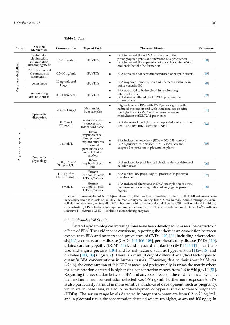
J. Xenobiot. 2022, 12 200
Table 4. Cont.
Topic StudiedMechanism Concentration Type of Cells Observed Effects References
Vas
cula
ren
doth
eliu
m
Endothelialdysfunction,
inflammation,and angiogenesis
0.1–1 µmol/L HUVECs
n BPA increased the mRNA expression of theproangiogenic genes and increased NO production
n BPA increased the expression of phosphorylated eNOSand endothelial tube formation
[88]
Cell division andchromosomalsegregation
0.5–10 ng/mL HUVECs n BPA at plasma concentrations induced aneugenic effects [89]
Senescence 10 ng/mL and1 µg/mL HUVECs n BPA impaired transcription and decreased viability in
aging vascular EC[90]
Acceleratingatherosclerosis 0.1–10 nmol/L HUVECs
n BPA appeared to be involved in acceleratingatherosclerosis
n BPA does not altered the HUVEC proliferationor migration
[59]
Preg
nanc
yex
poso
me
Epigeneticdisruption
35.4–56.1 ng/g Human fetalliver samples
n Higher levels of BPA with XME genes significantlyreduced expression and with increased site-specificmethylation at COMT and increased averagemethylation at SULT2A1 promoters
[91]
0.57 and0.78 ng/mL
Maternal urinesamples and
Infant cord bloodn BPA decreased methylation of imprinted and unprinted
genes and repetitive element LINE-1[92]
Pregnancyphysiology
1 nmol/L
BeWotrophoblast cellline, placental
explant cultures,placental
perfusions, andskin diffusion
models
n BPA induced cytotoxicity (EC50 = 100–125 µmol/L).n BPA significantly increased β-hCG secretion and
caspase-3 expression in placental explants.[95]
0, 0.09, 0.9, and9.0 µmol/L
BeWotrophoblast cell
linen BPA induced trophoblast cell death under conditions of
cellular stress[96]
1 × 10⁻¹⁵ to1 × 10−7 mol/L
Humantrophoblast cellsHTR-8/SVneo
n BPA altered key physiological processes in placentadevelopment
[97]
1 nmol/LHuman
trophoblast cellsHTR-8/SVneo
n BPA induceed alterations in DNA methylation of stressresponse and down-regulation of angiogenic growthfactors
[98]
1 Legend: BPA—bisphenol A; CnAβ—calcineurin; DRP1—dynamin-related protein 1; HCASMC—human coro-nary artery smooth muscle cells; HEK—human embryonic kidney; hiPSC-CMs–human-induced pluripotent stem-cell-derived cardiomyocytes; HUVECs—human umbilical vein endothelial cells; IC50—half-maximal inhibitoryconcentration; LINE-1—long interspersed nuclear element-1 or L1; Maxi-K—large conductance Ca2+/voltage-sensitive K+ channel; XME—xenobiotic-metabolizing enzymes.
5.2. Epidemiological Studies
Several epidemiological investigations have been developed to assess the cardiotoxiceffects of BPA. The evidence is consistent, reporting that there is an association betweenexposure to BPA and an increased prevalence of CVDs [103,104] including atherosclero-sis [105], coronary artery disease (CAD) [104,106–109], peripheral artery disease (PAD) [110],dilated cardiomyopathy (DCM) [109], and myocardial infarction (MI) [104,111]; heart fail-ure; and angina pectoris [104] and its risk factors, such as hypertension [112–115] anddiabetes [103,108] (Figure 2). There is a multiplicity of different analytical techniques toquantify BPA concentrations in human tissues. However, due to their short half-lives(<24 h), the concentration of this EDC is measured preferentially in urine, the matrix wherethe concentration detected is higher (the concentration ranges from 1.6 to 946 µg/L) [51].Regarding the association between BPA and adverse effects on the cardiovascular system,the maximum mean concentration detected was 4.66 ng/mL. Furthermore, exposure to BPAis also particularly harmful in more sensitive windows of development, such as pregnancy,which are, in these cases, related to the development of hypertensive disorders of pregnancy(HDPs). The serum range levels detected in pregnant women are from 0.2 to 20 ng/mL.and in placental tissue the concentration detected was much higher, at around 100 ng/g. In

J. Xenobiot. 2022, 12 201
urine samples, the concentration ranged from 0.87 to 31.9 µg/L [51,116] for the pregnantwomen, and 1.65 ng/mL when there was an association between urinary BPA concentra-tions and CVDs (Table 5). With regard to this, epidemiological studies reporting the effectsof BPA exposure and its association with CVDs and risk factors, as well as the implicationsof exposure during pregnancy, will be discussed in detail in the next two sections.
J. Xenobiot. 2022, 12, FOR PEER REVIEW 21
Figure 2. Summary representation of the endocrine‐disrupting effects of bisphenol A on the cardi‐
ovascular system.
5.2.1. Cardiovascular Diseases and Risk Factors
Several epidemiological studies have been carried out, mostly from a National
Health and Nutrition Examination Survey (NHANES) dataset. The assessment of cardio‐
vascular parameters was performed using urine samples from the participants for whom
BPA concentrations were analyzed [13]. The first study was conducted in 2008 by Lang et
al., who analyzed data from NHANES from 2003 to 2004 and showed that there is an
association between CVDs and elevated urinary BPA levels (OR = 1.39), after adjusting for
the confounding factors age and sex. The authors also found a relationship between higher
BPA concentrations and diabetes (OR = 1.39) [103]. On the other hand, a relationship be‐
tween BPA exposure and PAD and/or CAD was also assessed and found. With regard to
this, Melzer et al., in 2010, also analyzed BPA concentrations with heart diseases by ana‐
lyzing NHANES 2003–2006 data, separately between 2003/2004 and 2005/2006, and
pooled. The results of urinary BPA concentrations were smaller for the 2005/2006 data
(mean 1.79 ng/mL) than those from 2003/3004 (mean 2.49 ng/mL). After adjustment for
confounding factors, the authors found that there was an association between higher BPA
levels (2003/3004 data) with CAD (OR = 1.33), but the same was not observed with diabe‐
tes. Contrarily, when data were pooled, an association between higher BPA concentra‐
tions and these diseases was found (OR = 1.42 for CAD and OR = 1.24 for diabetes) [108].
In summary, the authors verified that higher levels of urinary BPA were cross‐sectionally
associated with heart disease in NHANES 2003–2004 and NHANES 2005–2006, independ‐
ent of traditional risk factors. Thus, and due to BPA levels having previously been associ‐
ated with CAD (which is an atherosclerotic disease), Lind et al. also evaluated, in 2011, a
possible relationship between exposure to BPA and atherosclerosis in a cross‐sectional
study. The results showed that elevated levels of BPA were related to the echogenicity of
the plaques, suggesting a role for BPA in atherosclerosis [105]. One year later, Melzer et
al. continued their previous studies, using data for 10.8 years from the European Prospec‐
tive Investigation of Cancer—Norfolk, UK. The authors showed that urinary BPA concen‐
trations were low (median value, 1.3 ng/mL) and that an increased BPA concentration was
associated with incident CAD, after adjustment for the confounding factors age, sex, and
urinary creatinine (OR = 1.13) [106]; similar trends were obtained to the previous study by
the same authors [108]. Additionally, in the same year, but in a different study, Melzer et
al. also studied the possible association between BPA exposure with grades of severity of
CAD on angiography [107] in 591 patients participating in The Metabonomics and Ge‐
nomics in Coronary Artery Disease study in Cambridgeshire, UK. The authors found that
there was a higher urinary concentration of BPA in patients with severe CAD compared
to patients with normal coronary arteries (OR = 1.43; 95% CI = 1.03–1.98, p = 0.033).
Figure 2. Summary representation of the endocrine-disrupting effects of bisphenol A on the cardio-vascular system.
5.2.1. Cardiovascular Diseases and Risk Factors
Several epidemiological studies have been carried out, mostly from a National Healthand Nutrition Examination Survey (NHANES) dataset. The assessment of cardiovascu-lar parameters was performed using urine samples from the participants for whom BPAconcentrations were analyzed [13]. The first study was conducted in 2008 by Lang et al.,who analyzed data from NHANES from 2003 to 2004 and showed that there is an associ-ation between CVDs and elevated urinary BPA levels (OR = 1.39), after adjusting for theconfounding factors age and sex. The authors also found a relationship between higherBPA concentrations and diabetes (OR = 1.39) [103]. On the other hand, a relationshipbetween BPA exposure and PAD and/or CAD was also assessed and found. With regardto this, Melzer et al., in 2010, also analyzed BPA concentrations with heart diseases byanalyzing NHANES 2003–2006 data, separately between 2003/2004 and 2005/2006, andpooled. The results of urinary BPA concentrations were smaller for the 2005/2006 data(mean 1.79 ng/mL) than those from 2003/3004 (mean 2.49 ng/mL). After adjustment forconfounding factors, the authors found that there was an association between higher BPAlevels (2003/3004 data) with CAD (OR = 1.33), but the same was not observed with diabetes.Contrarily, when data were pooled, an association between higher BPA concentrationsand these diseases was found (OR = 1.42 for CAD and OR = 1.24 for diabetes) [108]. Insummary, the authors verified that higher levels of urinary BPA were cross-sectionally asso-ciated with heart disease in NHANES 2003–2004 and NHANES 2005–2006, independent oftraditional risk factors. Thus, and due to BPA levels having previously been associated withCAD (which is an atherosclerotic disease), Lind et al. also evaluated, in 2011, a possiblerelationship between exposure to BPA and atherosclerosis in a cross-sectional study. Theresults showed that elevated levels of BPA were related to the echogenicity of the plaques,suggesting a role for BPA in atherosclerosis [105]. One year later, Melzer et al. continuedtheir previous studies, using data for 10.8 years from the European Prospective Investiga-tion of Cancer—Norfolk, UK. The authors showed that urinary BPA concentrations werelow (median value, 1.3 ng/mL) and that an increased BPA concentration was associatedwith incident CAD, after adjustment for the confounding factors age, sex, and urinarycreatinine (OR = 1.13) [106]; similar trends were obtained to the previous study by the sameauthors [108]. Additionally, in the same year, but in a different study, Melzer et al. also

J. Xenobiot. 2022, 12 202
studied the possible association between BPA exposure with grades of severity of CADon angiography [107] in 591 patients participating in The Metabonomics and Genomics inCoronary Artery Disease study in Cambridgeshire, UK. The authors found that there was ahigher urinary concentration of BPA in patients with severe CAD compared to patients withnormal coronary arteries (OR = 1.43; 95% CI = 1.03–1.98, p = 0.033). Furthermore, for thecase of patients with intermediate disease, significant differences were also almost obtained(OR = 1.69; 95% CI = 0.98–2.94, p = 0.061). Thus, it can be concluded that BPA exposurewas associated with the severity of angiography-defined coronary artery stenosis and maycontribute to a future risk of developing CAD in the adult population [107]. Taken together,these findings highlight the need for further studies to accurately estimate the prospectiveexposure–response curve and, thus, establish the underlying mechanisms [106,107].
Additionally, in 2012, another study by Shankar et al. examined the relationshipbetween urinary BPA levels and peripheral arterial disease (PAD) (which is a subclinicalmeasure of atherosclerotic vascular disease and a strong independent risk factor for CVDsand mortality) in a nationally representative sample of U.S. adults [110]. This study, whichwas conducted with participants from NHANES 2003–2004, also showed that there is apositive association between increased levels of BPA with DBP, independent of traditionalCVD risk factors (OR = 2.69; 95% CI = 1.02–7.09; p = 0.01) [110]. Although they cannotprovide definitive conclusions, these results suggest that environmental exposure to BPAmay promote the development of PAD [110]. Additionally, in 2015, Xiong et al. performeda case-control study evaluating the concentrations of BPA in patients with DCM, anddemonstrated higher levels of BPA in DCM patients compared with the healthy group(6.9 ± 2.7 ng/mL vs. 3.8 ± 1.9 ng/mL, p < 0.001), suggesting that BPA exposure may pro-mote the development of this pathology [109]. On the other hand, if a positive associationbetween BPA exposure and CAD has been shown, this has not been evaluated in patientswith type 2 diabetes (T2D). With regard to this, in 2019, Hu et al. evaluated this possiblerelationship through two nested case-control studies in two independent European cohorts(SURDIAGENE and ESTHER). The authors demonstrated the exposure to BPA in the twostudies (ESTHER cohort: 31% and SURDIAGENE cohort: 38%). The meta-analysis of theresults showed that in booth cohorts, there exists a positive association between MI andthe detection of BPA in urine (OR = 1.97; 95% CI = 1.05–3.70, p = 0.04) [111]. More recently,in 2020, Cai et al. evaluated the relationship between urinary BPA levels and CVDs in theU.S. adult population using data from NHANES 2003–2014. With this study, the authorsdemonstrated a positive association with heart failure, CAD, angina pectoris, MI, andCVDs, which was more evident in males [104].
On the other hand, hypertension is a risk factor for CVDs and has also been associatedwith high levels of BPA. Shankar and Teppala (2012) defined hypertension as “bloodpressure-reducing medication use and/or blood pressures >140/90 mm of Hg” [112]. Withregard to this, these authors analyzed the relationship between urinary levels of BPA andhypertension in a Multi-ethnic Sample of U.S. Adults. The study was also conducted withparticipants from NHANES 2003–2004. In agreement with the previous studies mentioned,the results demonstrated a positive association between elevated urinary BPA levels andhypertension, independent of traditional risk factors (OR = 1.50; 95% CI = 1.12–2.00,p = 0.007) [112]. Moreover, Bae et al., in 2012, investigated the associations of BPA exposurewith HRV and blood pressure (BP) in elderly citizens in Seoul, and demonstrated thaturinary BPA was associated negatively with HRV and positively with BP. Furthermore, theauthors also found an association with hypertension (OR = 1.27; CI 95% = 0.85–1.88 [113].Later, in 2015, the same authors also analyzed a possible association between exposureto BPA (from consumption of canned beverages) and BP and HRV. The main findingswere that consuming canned beverages (and the consequent increase in BPA exposure)is related to BP increase, but differences in HRV were not found [114]. In the same year,Aekplakorn et al. also determined the association of serum BPA with hypertension in theThai Population. The authors demonstrated that there is an association between serumBPA levels with hypertension in women (OR = 2.16; 95% CI = 1.31–3.56) [115].

J. Xenobiot. 2022, 12 203
From the studies mentioned above, it is clear that the NHANES dataset has been widelyused to draw conclusions about BPA exposure and adverse cardiovascular health outcomes.However, in 2012, La Kind et al. questioned whether this approach is the most appropriate,especially for a chemical with a short physiological half-life such as BPA, and chronicdiseases with multifactorial etiologies. With regard to this, analyzing four datasets (2003/04;2005/06; 2007/08 and 2009/10) and selecting a priori consistent methods, the authors triedto clarify the association between urinary BPA concentrations and diabetes, CAD, and/orMI. After adjustment for confounding factors, the results showed no association betweenurinary BPA levels and heart disease and diabetes, thus, not supporting previous studieswherein different methods and definitions were used. Thus, the authors suggested thatusing the NHANES data to study the association of BPA with CVDs is not appropriate,and may lead to inconsistent results due to this discrepancy between methodologies.Further epidemiological studies should be conducted to toxicologically evaluate the roleof BPA in the association with CVD development [117], as the experimental design of theepidemiological studies developed so far is constraining our clear comprehension of thistopic [118]. In addition, Olsén et al. also assessed the associations between circulatinglevels of BPA and coronary risk in an elderly population [119] using the Framingham riskscore to predict CVD risk. Contrary to previous studies, but corroborating the studies of LaKind et al. [117,118], the authors found no strong associations between BPA and FRS in thepopulation, thus suggesting that BPA exposure is not a risk factor for CVDs [119].
In summary, the set of epidemiological studies presented suggests that BPA exposureseems to be related to the development of CVDs or risk factors for CVDs, such as hyper-tension. Most studies are conducted using NHANES data, evidencing that this causalrelationship is consistent with higher urinary BPA levels. The causal relationship withdiabetes has also been established. However, due to the discrepancy in methodologiesperformed (including exclusion criteria, definitions, and scoring systems) caution is neededin their comparative analysis.
5.2.2. Pregnancy Exposure
HDPs are one of the leading causes of maternofetal mortality and morbidity. Exposureto EDCs is suspected to increase BP, but few studies have investigated the impact ofthese chemicals on pregnant women [120]. Due to BPA being suspected to cross theplacenta during pregnancy, possibly affecting children’s health, Bae et al. also analyzedmaternal urinary concentrations of BPA during midterm pregnancy (around 20 weeks)and in the BP of children at age 4. The authors demonstrated that there exists a positiveassociation between the diastolic (and not systolic) BP of the children with a maternalurinary concentration of BPA above a threshold level (4.5 µg/g creatinine) [121]. Moreover,Warembourg et al. also found that BPA exposure was associated with a significant decreasein systolic and/or diastolic BP during pregnancy (most frequently observed during thesecond trimester) [120]. Recently, in 2020, Sol et al. also evaluated the association betweenmaternal BPA urine concentrations during pregnancy and BP in childhood in a population-based prospective cohort study. The authors demonstrated that this association is sex-dependent, as evidenced by higher BP in boys after fetal BPA exposure. However, theseresults highlight the need for further studies to unravel the underlying mechanisms andlong-term outcomes [122].
According to the recently reviewed studies, BPAs (as well as phthalates) are theclass of EDCs that appear to mostly harm the fetoplacental vasculature during pregnancyand have been associated with the development of pregnancy complications such aspreeclampsia (PE) [51]. However, these specific exposure–response relationships are not yetfully established [123]. In 2014, Leclerc et al. were the first authors to demonstrate that therewas an association between PE and BPA accumulation in the placenta. The authors foundthat placentas from women with PE had a higher accumulation of this EDC compared toplacentas from normotensive women [124]. In the following year, Fergunson et al. alsoobserved that there is an association between urinary BPA concentrations and angiogenic

J. Xenobiot. 2022, 12 204
biomarkers during pregnancy, which may be indicative of this pathology through placentaldisruption development and/or function during gestation [125]. Concordantly, Spagnolettiet al. also demonstrated that BPA acts on trophoblasts and alters physiological processesinvolved in normal placental development [97], and Cantonwine et al. also reported thatearly pregnancy (~10 weeks gestation) appears to be a window of increased susceptibilityfor the development of this HDP associated with BPA exposure [126]. Furthermore, in 2017,Philips et al., in their findings, suggested that there is probably a greater association ofbisphenols in early pregnancy with a risk of PE than with gestational hypertension (as nosignificant changes in BP were found) [127]. The same authors also recently described thatearly exposure to BPA during pregnancy appears to be related to changes in cardiometabolichealth later in life [128].
Additionally, the postpartum period may be a vulnerable life stage for a woman’scardiometabolic health [129]. According to the recent review by Lorigo and Cairrao, consid-ering the postpartum period is equally as important as considering the pregnancy periodwhen talking about endocrine disruption. The associations between elevated levels ofEDCs in general, including BPA, and vascular changes in children or adults are not yetdemonstrated; they require further study, because pregnancy and postpartum are bothsusceptible windows of endocrine exposure. Thus, further studies must be developed toclarify these mechanisms and, thus, understand whether the risk of CVDs is temporary orpermanent in later life [51]. With regard to this, in 2020, Perng et al. analyzed the effectof BPA during pregnancy on weight from delivery through 1 year postpartum among199 women in Mexico City. Through their prospective cohort study, the authors demon-strated that prenatal exposure to BPA is inversely associated with weight at delivery, butthere exists a slower rate of weight loss through the first postpartum year [129]. However,to our knowledge, these authors were the only ones to evaluate the effects of BPA in thepostpartum period.
Therefore, taken together, these studies indicate that there is an environmental contri-bution by EDCs in the development of HDPs; therefore, urgent action should be taken todecrease exposure to these emerging compounds to reduce the mortality and morbidityassociated with these conditions.
Table 5. Summaries of the disruptive effects of BPA in human epidemiological studies 1.
Topic Studied Population Gender Concentration Observed Effects References
Car
diov
ascu
lar
dise
ases
NHANES from2003–2004
1455 adults aged 18through 74 years
694 men761 women
4.53 ng/mL(urinary)
4.66 ng/mL(urinary)
n Association between CVDs and elevated urinaryBPA levels
n Association between higher BPA concentrationsand diabetes
[103]
NHANES 2003–2006data, separately
between 2003/2004and 2005/2006 and
pooledn = 1455 (2003/04)
and n = 1493(2005/06) adults aged
18–74 years
694 men and761 women in
2003/04720 men and
773 women in2005/06
2.49 ng/mL(urinary)
1.79 ng/mL(urinary)
n Urinary BPA concentrations were smaller for the2005/2006 data than those from 2003/3004
n Association between higher BPA levels (2003/3004data) and CAD, but not with diabetes.
n Data pooled showed an association betweenhigher BPA concentrations and CAD and diabetes
[108]
Population-basedProspective
Investigation of theVasculature in
Uppsala Seniorsstudy (1016 subjects
all aged 70)
510 women and506 men
3.76 ng/mL(serum)
n Elevated levels of BPA were related to theechogenicity of the plaques, suggesting a role forBPA in atherosclerosis
[105]
758 incident CADcases and
861 controls followedfor 10.8 years from
the EuropeanProspective
Investigation ofCancer—Norfolk, UK
534 men and327 women incontrol group501 men and
257 women inCAD group
Control vs. CADgroup
(1.24 ng/mL vs.1.35 ng/mL)
(urinary)
n Urinary BPA concentrations were lown Increased BPA concentration was associated with
incident CAD[106]

J. Xenobiot. 2022, 12 205
Table 5. Cont.
Topic Studied Population Gender Concentration Observed Effects References
591 patientsparticipating in TheMetabonomics and
Genomics inCoronary ArteryDisease study in
Cambridgeshire, UK
120 controls(62 women and
58 men)385 patients
(301 men and84 women)
86 Intermediatediseases (43 menand 43 women)
Control vs. CADvs. intermediate(2.13 vs. 3.82 vs.
3.31 ng/mL)(urinary)
n Association between BPA exposure with grades ofseverity of CAD on angiography
n Higher urinary concentration of BPA in patientswith severe CAD compared to patients withnormal coronary arteries
[107]
745 participants inthe NHANES
2003–2004
361 women and392 men
2.30 ng/mL(urinary)
n A positive association between increased levels ofBPA with DBP
[110]
88 DCM patients and88 age-and
gender-matchedhealthy controls
59 men and29 women with
DCM55 men and33 women
Control
DCM vs. controlgroup (6.9 ± 2.7
vs. 3.8 ± 1.9ng/mL) (serum)
n Higher levels of BPA in DCM patients comparedwith the healthy group
[109]
NHANES 2003–2014(n = 9139, aged≥20 years)
4467 men and4672 women -
n Positive association with heart failure, CAD,angina pectoris, MI, and CVDs, which was moreevident in males
[104]
Hyp
erte
nsio
n
1380 subjects fromNHANES 2003–2004
700 women and680 men(580 with
hypertension)
1.5–4.0 ng/mL(urinary)
n A positive association between elevated urinaryBPA levels and hypertension
[112]
560 noninstitutionalizedelderly citizens from
August 2008 toAugust 2010 in Seoulfrom Korean Elderly
EnvironmentalPanel Study
521 participantswere included(138 men and383 women)
(men) and1.3 (women)µg/g of
creatinine(urinary)
n Urinary BPA was associated negatively with HRVand positively with BP
n BPA was associated with hypertension[113]
60 noninstitutionalizedelderly participants,
who were aged≥60 years betweenFebruary 2014 and
March 2014
60 participants,56 were women
and 4 male
1.13 ± 1.76 µg/L(urinary)
n Association between exposure to BPA with BPincreased, but differences in HRV were not found
[114]
A subsample of 2558randomly selected
from the ThaiNational Health
Examination SurveyIV, 2009
1275 men and1283 women
0.35 ng/mL(men) and
0.33 ng/mL(women)(serum)
n Association between serum BPA levels withhypertension in women
[115]
Preg
nanc
yex
posu
re
645 children at theage of 4 who wereborn from womenwho participated,
midterm during theirpregnancy, in a birth
cohort study fromAugust 2008 to
July 2011
486 mother–childpairs were
included in thepresent analysis
Maternal urinary:0.9 µg/L(urinary)
n A positive association between diastolic (and notsystolic) BP of the children with the maternalurinary concentration of BPA
[121]
152 female volunteerparticipants in theHuman Early-LifeExposome project
152 pregnantwomen
3.1 µg/gcreatinine(urinary)
n BPA exposure was associated with a significantdecrease in systolic and/or diastolic BP
[120]
1064 mother-childpairs/childhood at a
mean age of9.7 years old
1064mother-child
pais
6.0 nmol/L(boys) and7.2 nmol/L
(girls)(urinary)
n Higher BP in boys after fetal BPA exposure [122]
58 pregnancies,including
35 normotensive and23 preeclamptic
women
35 normotensivepregnant women
and 23preeclamptic
pregnant women
Control vs. PE(3.00 vs.
2.80 ng/mL–maternal serum;
2.17 vs. 2.23ng/mL–fetal
serum; 3.00 vs.9.40–placentalhomogenate)
n Placentas from women with PE had a higheraccumulation of this EDC compared to placentasfrom normotensive women
[124]

J. Xenobiot. 2022, 12 206
Table 5. Cont.
Topic Studied Population Gender Concentration Observed Effects References
A nested case-controlpopulation consistingof 130 mothers whodelivered preterm
and 352 whodelivered term froma prospective birth
cohort
130 women and352 controls
7.08% change(adjusted with
soluble fms-liketyrosine kinase-1)
(urinary)
n Association between urinary BPA concentrationsand angiogenic biomarkers during pregnancy
[125]
A nested case-controlstudy of preterm
birth was performedin 2011 from women
enrolled in aprospective birthcohort study at
Women’s Hospitalsin Brigham and inBoston (included
50 cases of PE)
482 women (50with PE)
Cases vs. control(1.56 vs.
1.38 ng/mL)(urinary)
n Early pregnancy (~10 weeks gestation) was awindow of increased susceptibility for thedevelopment of this HDP associated withBPA exposure
[126]
1233 womenexcluding thosewithout any BP
measurement or withpre-existing
hypertension
1233 women 1.65 ng/mL(urinary)
n Association of bisphenols in early pregnancy witha risk of PE
[127]
1 year postpartumamong 199 women in
Mexico City199 women 1.18 ng/mL
(urinary)
n Prenatal exposure to BPA was inversely associatedwith weight at delivery, but there exists a slowerrate of weight loss through the firstpostpartum year
[129]
1 Legend: BP—blood pressure; BPA—bisphenol A; CAD—coronary artery disease; CVDs—cardiovascular dis-eases; DCM—dilated cardiomyopathy; EDC—endocrine-disrupting compound; HRV—heart rate variability;MI—myocardial infarction; NHANES—National Health and Nutrition Examination Survey; PE—preeclampsia.
6. Conclusions
The analysis of several studies of BPA-induced cardiotoxicity led us to concludethat exposure to this EDC is associated with an increased prevalence of CVDs. It hasbecome clear that BPA, even acting at lower doses as an EDC, can cause adverse effectson cardiovascular health, especially at more susceptible stages of development, suchas pregnancy. BPA exposure impairs not only cardiac electrophysiology, but also Ca2+
signaling, leading to arrhythmias and changes in contractile functions (contraction andrelaxation) (Figure 3). However, the concentrations used in most in vitro studies arerelatively high. Therefore, the choice of concentrations to be used in the in vitro, ex-vivo, and in vivo studies is one of the greatest challenges of toxicology, and this choiceshould always be dependent on the concentrations detected in epidemiological studies (aconcentration range from 1.6 to 946 µg/L) [51] and environmental studies (effluent rangingfrom non-detect to 370 µg/L, soil ranging from <0.01 to 1000 µg/kg, and sediments rangingfrom 3400 to 20,136 µg/kg DW [130]. The use of higher concentrations (20–200 times higherthan therapeutic Cmax) in in vitro studies is consensual. These concentrations are alsodesignated using pharmacological concentrations and cause more accurate results thanlower concentrations [131]. The ranges of these pharmacological concentrations are thosethat are required to cause effects at the cellular level, and are normally bigger than thoserequired to cause harmful effects in vivo. This interrelationship is called the in vitro–in vivoscaling factor. However, this factor depends mainly on the mechanism of action of thesubstance, the metabolization route, and the mechanism of toxicity. Thus, only these highconcentrations may be predictive of the effects induced by these substances [131,132]. Insummary, the use of a high concentration range is crucial to determine the occurrence oftoxic effects of substances, such as for BPA.

J. Xenobiot. 2022, 12 207J. Xenobiot. 2022, 12, FOR PEER REVIEW 27
Figure 3. Summary representation of Bisphenol A (BPA) exposure pathways and main mechanisms
involved in BPA‐induced cardiotoxicity.
Another very important parameter that remains under great controversy is the daily
amount considered safe for human exposure, which is also referred to as the acceptable
daily intake (ADI) by the Food and Drug Administration (FDA), and as the reference dose
by the Environmental Protection Agency (EPA). This dose is currently at 50 μg/kg/day
[133] and assumes that even if a daily exposure of this amount occurs, no adverse effects
occur. However, recent studies have shown that the lowest observed adverse effect level
(LOAEL), which serves as the reference ADI, is much lower than currently accepted
(20,000‐fold lower). These studies have led several members of the Endocrine Society to
notify the FDA and EPA that there are several incorrect assumptions regarding the calcu‐
lation of AID for BPA: (1) the responses to BPA are likely to be monotonic, which, in real‐
ity, is not the case for EDC; (2) the threshold at which there are no effects, which is also
very controversial and questioned, as it is known that it can develop changes in the next
generations; (3) a similar response for both sexes, which is also not true for many re‐
sponses to BPA; and (4) only toxicological guideline studies are valid [133].
Thus, it seems clear that the human current daily exposure is not safe and that the
problems arising from the massive use of this EDC continue to be a reality. The contro‐
versial studies can be justified through the different experimental approaches that are ap‐
plied, as well as changes in the levels of doses, duration of exposure, or even the experi‐
mental model used. With regard to this, more studies are needed to further clarify the
mechanism of BPA toxicity in the cardiovascular system. We emphasize that it is essential
to use a wide range of concentrations to adequately address the dose–response relation‐
ship of BPA.
Furthermore, as humans are widely exposed to multiple bisphenols, this underscores
the need for and importance of studying co‐exposure patterns and/or the ‘exposome’, al‐
ways considering multiple exposures and, thus, the best way to address the adverse ef‐
fects of this EDC on cardiovascular health. The development of new BPA analogues, as
well as the study of the safety of some analogues currently already developed, are a key
point in the evolution of research on these compounds. Environmental and human expo‐
sure to these compounds should be free of health risks, especially in the most vulnerable
groups such as children and pregnant women. Finally, it is also important that public
health approaches are taken to clarify which are the best practices for reducing exposure
to BPA (e.g., recommending the use of glass containers for food storage, reducing the use
of canned products and plastic containers, etc.), thus improving human lifestyles and,
consequently, decreasing the risk of BPA‐induced cardiotoxicity.
Author Contributions: Conceptualization, E.C.; writing—original draft preparation, M.I.F., M.L.
and E.C.; writing—review and editing, M.L. and E.C.; supervision, M.L. and E.C.; funding acquisi‐
tion, E.C. All authors have read and agreed to the published version of the manuscript.
Figure 3. Summary representation of Bisphenol A (BPA) exposure pathways and main mechanismsinvolved in BPA-induced cardiotoxicity.
Another very important parameter that remains under great controversy is the dailyamount considered safe for human exposure, which is also referred to as the acceptable dailyintake (ADI) by the Food and Drug Administration (FDA), and as the reference dose bythe Environmental Protection Agency (EPA). This dose is currently at 50 µg/kg/day [133]and assumes that even if a daily exposure of this amount occurs, no adverse effects occur.However, recent studies have shown that the lowest observed adverse effect level (LOAEL),which serves as the reference ADI, is much lower than currently accepted (20,000-foldlower). These studies have led several members of the Endocrine Society to notify the FDAand EPA that there are several incorrect assumptions regarding the calculation of AID forBPA: (1) the responses to BPA are likely to be monotonic, which, in reality, is not the casefor EDC; (2) the threshold at which there are no effects, which is also very controversial andquestioned, as it is known that it can develop changes in the next generations; (3) a similarresponse for both sexes, which is also not true for many responses to BPA; and (4) onlytoxicological guideline studies are valid [133].
Thus, it seems clear that the human current daily exposure is not safe and that theproblems arising from the massive use of this EDC continue to be a reality. The controversialstudies can be justified through the different experimental approaches that are applied,as well as changes in the levels of doses, duration of exposure, or even the experimentalmodel used. With regard to this, more studies are needed to further clarify the mechanismof BPA toxicity in the cardiovascular system. We emphasize that it is essential to use a widerange of concentrations to adequately address the dose–response relationship of BPA.
Furthermore, as humans are widely exposed to multiple bisphenols, this underscoresthe need for and importance of studying co-exposure patterns and/or the ‘exposome’,always considering multiple exposures and, thus, the best way to address the adverseeffects of this EDC on cardiovascular health. The development of new BPA analogues, aswell as the study of the safety of some analogues currently already developed, are a keypoint in the evolution of research on these compounds. Environmental and human exposureto these compounds should be free of health risks, especially in the most vulnerable groupssuch as children and pregnant women. Finally, it is also important that public healthapproaches are taken to clarify which are the best practices for reducing exposure to BPA(e.g., recommending the use of glass containers for food storage, reducing the use of cannedproducts and plastic containers, etc.), thus improving human lifestyles and, consequently,decreasing the risk of BPA-induced cardiotoxicity.
Author Contributions: Conceptualization, E.C.; writing—original draft preparation, M.I.F., M.L. andE.C.; writing—review and editing, M.L. and E.C.; supervision, M.L. and E.C.; funding acquisition,E.C. All authors have read and agreed to the published version of the manuscript.
Funding: M.L. acknowledges the Ph.D. fellowship from FCT (Reference: 2020.06616.BD). This workwas developed within the scope of the CICS-UBI projects UIDB/00709/2020 and UIDP/00709/2020,financed by national funds through the Portuguese Foundation for Science and Technology/MCTES.

J. Xenobiot. 2022, 12 208
Institutional Review Board Statement: Not applicable.
Informed Consent Statement: Not applicable.
Data Availability Statement: Not applicable.
Conflicts of Interest: The authors declare no conflict of interest.
Abbreviations
Ang-II angiotensin IIBP blood pressureBPA bisphenol ABPAF bisphenol AFBPC bisphenol CCa2+ calciumCAD coronary artery diseaseCnAβ calcineurinCVDs cardiovascular diseasesDCM dilated cardiomyopathyDRP1 dynamin-related protein 1EC endothelial cellsEDC endocrine-disrupting compound;EDHF endothelium-derived hyperpolarizing factorER estrogen receptorsET-1 endothelin 1GPR30 G-protein-coupled estrogen receptorHDPs hypertensive disorders of pregnancyHEK human embryonic kidneyhiPSC-CMs human-induced pluripotent stem-cell-derived cardiomyocytesHRV heart rate variabilityHUVECs human umbilical vein endothelial cellsIC50 half-maximal inhibitory concentrationiPSCs induced pluripotent stem cellsIR ischemia–reperfusionK+ potassiumMAP mean atrial pressuresMBP 4-Methyl-2,4-bis(p-hydroxyphenyl)pent-1-eneMI myocardial infarctionMOA mechanisms of actionNa+ sodiumNHANES National Health and Nutrition Examination SurveyNO nitric oxidePAD peripheral artery diseasePE preeclampsiaPFOS perfluorooctane sulfonate;PGI-2 prostacyclinPLB phospholambanPPAR-γ peroxisome proliferator-activated receptor gammaRIP3 Rat Receptor-Interacting Protein 3SMC smooth muscle cellsTxA2 thromboxaneU.S. United StatesXME xenobiotic-metabolizing enzymes
References1. Francula-Zaninovic, S.; Nola, I.A. Management of Measurable Variable Cardiovascular Disease’ Risk Factors. Curr. Cardiol. Rev.
2018, 14, 153–163. [CrossRef] [PubMed]2. Bhatnagar, A. Environmental Determinants of Cardiovascular Disease. Circ. Res. 2017, 121, 162–180. [CrossRef] [PubMed]

J. Xenobiot. 2022, 12 209
3. Leslie, H.A.; van Velzen, M.J.M.; Brandsma, S.H.; Vethaak, A.D.; Garcia-Vallejo, J.J.; Lamoree, M.H. Discovery and quantificationof plastic particle pollution in human blood. Environ. Int. 2022, 163, 107199. [CrossRef] [PubMed]
4. Diamanti-Kandarakis, E.; Bourguignon, J.P.; Giudice, L.C.; Hauser, R.; Prins, G.S.; Soto, A.M.; Zoeller, R.T.; Gore, A.C. Endocrine-disrupting chemicals: An Endocrine Society scientific statement. Endocr. Rev. 2009, 30, 293–342. [CrossRef] [PubMed]
5. Kabir, E.R.; Rahman, M.S.; Rahman, I. A review on endocrine disruptors and their possible impacts on human health. Environ.Toxicol. Pharmacol. 2015, 40, 241–258. [CrossRef]
6. Ramadan, M.; Cooper, B.; Posnack, N.G. Bisphenols and phthalates: Plastic chemical exposures can contribute to adversecardiovascular health outcomes. Birth Defects Res. 2020, 112, 1362–1385. [CrossRef]
7. Vandenberg, L.N.; Hauser, R.; Marcus, M.; Olea, N.; Welshons, W.V. Human exposure to bisphenol A (BPA). Reprod. Toxicol. 2007,24, 139–177. [CrossRef]
8. Campanale, C.; Massarelli, C.; Savino, I.; Locaputo, V.; Uricchio, V.F. A Detailed Review Study on Potential Effects of Microplasticsand Additives of Concern on Human Health. Int. J. Environ. Res. Public Health 2020, 17, 1212. [CrossRef]
9. Dodds, E.C.; Lawson, W. Synthetic strogenic Agents without the Phenanthrene Nucleus. Nature 1936, 137, 996. [CrossRef]10. Michałowicz, J. Bisphenol A—Sources, toxicity and biotransformation. Environ. Toxicol. Pharmacol. 2014, 37, 738–758. [CrossRef]11. Acconcia, F.; Pallottini, V.; Marino, M. Molecular Mechanisms of Action of BPA. Dose Response 2015, 13, 1559325815610582.
[CrossRef]12. Banerjee, O.; Singh, S.; Saha, I.; Pal, S.; Banerjee, M.; Kundu, S.; Syamal, A.K.; Maji, B.K.; Mukherjee, S. Molecular dissection of
cellular response of pancreatic islet cells to Bisphenol-A (BPA): A comprehensive review. Biochem. Pharmacol. 2022, 201, 115068.[CrossRef] [PubMed]
13. Gao, X.; Wang, H.-S. Impact of Bisphenol A on the Cardiovascular System—Epidemiological and Experimental Evidence andMolecular Mechanisms. Int. J. Environ. Res. Public Health 2014, 11, 8399–8413. [CrossRef] [PubMed]
14. Rancière, F.; Lyons, J.G.; Loh, V.H.; Botton, J.; Galloway, T.; Wang, T.; Shaw, J.E.; Magliano, D.J. Bisphenol A and the risk ofcardiometabolic disorders: A systematic review with meta-analysis of the epidemiological evidence. Environ. Health A Glob.Access Sci. Source 2015, 14, 46. [CrossRef]
15. Vandenberg Laura, N.; Chahoud, I.; Heindel Jerrold, J.; Padmanabhan, V.; Paumgartten Francisco, J.R.; Schoenfelder, G. Urinary,Circulating, and Tissue Biomonitoring Studies Indicate Widespread Exposure to Bisphenol A. Environ. Health Perspect. 2010,118, 1055–1070. [CrossRef] [PubMed]
16. Schönfelder, G.; Wittfoht, W.; Hopp, H.; Talsness, C.E.; Paul, M.; Chahoud, I. Parent bisphenol A accumulation in the humanmaternal-fetal-placental unit. Environ. Health Perspect. 2002, 110, A703–A707. [CrossRef] [PubMed]
17. Yamada, H.; Furuta, I.; Kato, E.H.; Kataoka, S.; Usuki, Y.; Kobashi, G.; Sata, F.; Kishi, R.; Fujimoto, S. Maternal serum and amnioticfluid bisphenol A concentrations in the early second trimester. Reprod. Toxicol. 2002, 16, 735–739. [CrossRef]
18. Kuroda, N.; Kinoshita, Y.; Sun, Y.; Wada, M.; Kishikawa, N.; Nakashima, K.; Makino, T.; Nakazawa, H. Measurement of bisphenolA levels in human blood serum and ascitic fluid by HPLC using a fluorescent labeling reagent. J. Pharm. Biomed. Anal. 2003,30, 1743–1749. [CrossRef]
19. Hines, E.P.; Mendola, P.; von Ehrenstein, O.S.; Ye, X.; Calafat, A.M.; Fenton, S.E. Concentrations of environmental phenols andparabens in milk, urine and serum of lactating North Carolina women. Reprod. Toxicol. 2015, 54, 120–128. [CrossRef]
20. Tan, B.L.; Ali Mohd, M. Analysis of selected pesticides and alkylphenols in human cord blood by gas chromatograph-massspectrometer. Talanta 2003, 61, 385–391. [CrossRef]
21. Sun, Y.; Irie, M.; Kishikawa, N.; Wada, M.; Kuroda, N.; Nakashima, K. Determination of bisphenol A in human breast milk byHPLC with column-switching and fluorescence detection. Biomed. Chromatogr. 2004, 18, 501–507. [CrossRef] [PubMed]
22. Kuruto-Niwa, R.; Tateoka, Y.; Usuki, Y.; Nozawa, R. Measurement of bisphenol A concentrations in human colostrum. Chemosphere2007, 66, 1160–1164. [CrossRef]
23. Völkel, W.; Colnot, T.; Csanády, G.A.; Filser, J.G.; Dekant, W. Metabolism and Kinetics of Bisphenol A in Humans at Low DosesFollowing Oral Administration. Chem. Res. Toxicol. 2002, 15, 1281–1287. [CrossRef] [PubMed]
24. Cimmino, I.; Fiory, F.; Perruolo, G.; Miele, C.; Beguinot, F.; Formisano, P.; Oriente, F. Potential Mechanisms of Bisphenol A (BPA)Contributing to Human Disease. Int. J. Mol. Sci. 2020, 21, 5761. [CrossRef] [PubMed]
25. Tan, H.; Wang, X.; Hong, H.; Benfenati, E.; Giesy, J.P.; Gini, G.C.; Kusko, R.; Zhang, X.; Yu, H.; Shi, W. Structures of Endocrine-Disrupting Chemicals Determine Binding to and Activation of the Estrogen Receptor α and Androgen Receptor. Environ. Sci.Technol. 2020, 54, 11424–11433. [CrossRef]
26. Vandenberg, L.N. Non-Monotonic Dose Responses in Studies of Endocrine Disrupting Chemicals: Bisphenol a as a Case Study.Dose-Response 2014, 12, 259–276. [CrossRef]
27. Cooper, B.L.; Posnack, N.G. Characteristics of Bisphenol Cardiotoxicity: Impaired Excitability, Contractility, and Relaxation.Cardiovasc. Toxicol. 2022, 22, 273–280. [CrossRef]
28. Boudalia, S.; Oudir, M. Bisphenol-A: Legislation in Industrials Countries and in Algeria. Res. J. Environ. Toxicol. 2016, 10, 189–192.[CrossRef]
29. Xing, J.; Zhang, S.; Zhang, M.; Hou, J. A critical review of presence, removal and potential impacts of endocrine disruptorsbisphenol A. Comp. Biochem. Physiol. C Toxicol. Pharmacol. 2022, 254, 109275. [CrossRef]
30. Posnack, N.G.; Jaimes, R., 3rd; Asfour, H.; Swift, L.M.; Wengrowski, A.M.; Sarvazyan, N.; Kay, M.W. Bisphenol A exposure andcardiac electrical conduction in excised rat hearts. Environ. Health Perspect. 2014, 122, 384–390. [CrossRef]

J. Xenobiot. 2022, 12 210
31. Zhang, Y.-F.; Shan, C.; Wang, Y.; Qian, L.-L.; Jia, D.-D.; Zhang, Y.-F.; Hao, X.-D.; Xu, H.-M. Cardiovascular toxicity and mechanismof bisphenol A and emerging risk of bisphenol S. Sci. Total Environ. 2020, 723, 137952. [CrossRef] [PubMed]
32. Lind, L.; Araujo, J.A.; Barchowsky, A.; Belcher, S.; Berridge, B.R.; Chiamvimonvat, N.; Chiu, W.A.; Cogliano, V.J.; Elmore, S.;Farraj, A.K.; et al. Key Characteristics of Cardiovascular Toxicants. Environ. Health Perspect. 2021, 129, 95001. [CrossRef]
33. Cumberland, M.J.; Riebel, L.L.; Roy, A.; O’Shea, C.; Holmes, A.P.; Denning, C.; Kirchhof, P.; Rodriguez, B.; Gehmlich, K. BasicResearch Approaches to Evaluate Cardiac Arrhythmia in Heart Failure and Beyond. Front. Physiol. 2022, 13, 806366. [CrossRef][PubMed]
34. Verkerk, A.O.; Marchal, G.A.; Zegers, J.G.; Kawasaki, M.; Driessen, A.H.G.; Remme, C.A.; de Groot, J.R.; Wilders, R. Patch-ClampRecordings of Action Potentials From Human Atrial Myocytes: Optimization Through Dynamic Clamp. Front. Pharmacol. 2021,12, 649414. [CrossRef]
35. Asano, S.; Tune, J.D.; Dick, G.M. Bisphenol A activates Maxi-K (K(Ca)1.1) channels in coronary smooth muscle. Br. J. Pharmacol.2010, 160, 160–170. [CrossRef] [PubMed]
36. Rottgen, T.S.; Fancher, I.S.; Asano, S.; Widlanski, T.S.; Dick, G.M. Bisphenol A activates BK channels through effects on α and β1subunits. Channels 2014, 8, 249–257. [CrossRef] [PubMed]
37. Feiteiro, J.; Mariana, M.; Glória, S.; Cairrao, E. Inhibition of L-type calcium channels by Bisphenol A in rat aorta smooth muscle.J. Toxicol. Sci. 2018, 43, 579–586. [CrossRef]
38. Deutschmann, A.; Hans, M.; Meyer, R.; Häberlein, H.; Swandulla, D. Bisphenol A Inhibits Voltage-Activated Ca2+ Channels inVitro: Mechanisms and Structural Requirements. Mol. Pharmacol. 2013, 83, 501. [CrossRef] [PubMed]
39. Michaela, P.; Mária, K.; Silvia, H.; L’ubica, L. Bisphenol A differently inhibits CaV3.1, CaV3.2 and CaV3.3 calcium channels.Naunyn-Schmiedeberg’s Arch. Pharmacol. 2014, 387, 153–163. [CrossRef]
40. Liang, Q.; Gao, X.; Chen, Y.; Hong, K.; Wang, H.-S. Cellular Mechanism of the Nonmonotonic Dose Response of Bisphenol A inRat Cardiac Myocytes. Environ. Health Perspect. 2014, 122, 601–608. [CrossRef]
41. Hyun, S.-A.; Lee, C.Y.; Ko, M.Y.; Chon, S.-H.; Kim, Y.-J.; Seo, J.-W.; Kim, K.K.; Ka, M. Cardiac toxicity from bisphenol A exposure inhuman-induced pluripotent stem cell-derived cardiomyocytes. Toxicol. Appl. Pharmacol. 2021, 428, 115696. [CrossRef] [PubMed]
42. Prudencio, T.M.; Swift, L.M.; Guerrelli, D.; Cooper, B.; Reilly, M.; Ciccarelli, N.; Sheng, J.; Jaimes, R., III; Posnack, N.G. Bisphenol Sand Bisphenol F Are Less Disruptive to Cardiac Electrophysiology, as Compared with Bisphenol A. Toxicol. Sci. 2021, 183, 214–226.[CrossRef] [PubMed]
43. O’Reilly, A.O.; Eberhardt, E.; Weidner, C.; Alzheimer, C.; Wallace, B.A.; Lampert, A. Bisphenol A Binds to the Local AnestheticReceptor Site to Block the Human Cardiac Sodium Channel. PLoS ONE 2012, 7, e41667. [CrossRef] [PubMed]
44. Horváth, B.; Hézso, T.; Kiss, D.; Kistamás, K.; Magyar, J.; Nánási, P.P.; Bányász, T. Late Sodium Current Inhibitors as PotentialAntiarrhythmic Agents. Front. Pharmacol. 2020, 11, 413. [CrossRef]
45. Yan, S.; Chen, Y.; Dong, M.; Song, W.; Belcher, S.M.; Wang, H.-S. Bisphenol A and 17β-Estradiol Promote Arrhythmia in theFemale Heart via Alteration of Calcium Handling. PLoS ONE 2011, 6, e25455. [CrossRef]
46. Belcher, S.M.; Chen, Y.; Yan, S.; Wang, H.-S. Rapid Estrogen Receptor-Mediated Mechanisms Determine the Sexually DimorphicSensitivity of Ventricular Myocytes to 17β-Estradiol and the Environmental Endocrine Disruptor Bisphenol A. Endocrinology 2012,153, 712–720. [CrossRef]
47. Yan, S.; Song, W.; Chen, Y.; Hong, K.; Rubinstein, J.; Wang, H.S. Low-dose bisphenol A and estrogen increase ventriculararrhythmias following ischemia-reperfusion in female rat hearts. Food Chem. Toxicol. Int. J. Publ. Br. Ind. Biol. Res. Assoc. 2013,56, 75–80. [CrossRef]
48. Gao, X.; Liang, Q.; Chen, Y.; Wang, H.-S. Molecular Mechanisms Underlying the Rapid Arrhythmogenic Action of Bisphenol A inFemale Rat Hearts. Endocrinology 2013, 154, 4607–4617. [CrossRef]
49. Pinto, C.; Hao, R.; Grimaldi, M.; Thrikawala, S.; Boulahtouf, A.; Aït-Aïssa, S.; Brion, F.; Gustafsson, J.; Balaguer, P.; Bondesson, M.Differential activity of BPA, BPAF and BPC on zebrafish estrogen receptors in vitro and in vivo. Toxicol. Appl. Pharmacol. 2019,380, 114709. [CrossRef]
50. Ramadan, M.; Sherman, M.; Jaimes, R.; Chaluvadi, A.; Swift, L.; Posnack, N.G. Disruption of neonatal cardiomyocyte physiologyfollowing exposure to bisphenola. Sci. Rep. 2018, 8, 7356. [CrossRef]
51. Lorigo, M.; Cairrao, E. Fetoplacental vasculature as a model to study human cardiovascular endocrine disruption. Mol. AspectsMed. 2021, 87, 101054. [CrossRef] [PubMed]
52. Zhou, R.; Cheng, W.; Feng, Y.; Wang, W.; Liang, F.; Luo, F.; Yang, S.; Wang, Y. Combined effects of BPA and PFOS on fetal cardiacdevelopment: In vitro and in vivo experiments. Environ. Toxicol. Pharmacol. 2020, 80, 103434. [CrossRef] [PubMed]
53. Reventun, P.; Sanchez-Esteban, S.; Cook, A.; Cuadrado, I.; Roza, C.; Moreno-Gomez-Toledano, R.; Muñoz, C.; Zaragoza, C.;Bosch, R.J.; Saura, M. Bisphenol A induces coronary endothelial cell necroptosis by activating RIP3/CamKII dependent pathway.Sci. Rep. 2020, 10, 4190. [CrossRef] [PubMed]
54. Posnack, N.G.; Brooks, D.; Chandra, A.; Jaimes, R.; Sarvazyan, N.; Kay, M. Physiological response of cardiac tissue to bisphenol a:Alterations in ventricular pressure and contractility. Am. J. Physiol. Heart Circ. Physiol. 2015, 309, H267–H275. [CrossRef]
55. Pant, J.; Ranjan, P.; Deshpande, S.B. Bisphenol A decreases atrial contractility involving NO-dependent G-cyclase signalingpathway. J. Appl. Toxicol. 2011, 31, 698–702. [CrossRef]
56. Filice, M.; Leo, S.; Mazza, R.; Amelio, D.; Garofalo, F.; Imbrogno, S.; Cerra, M.C.; Gattuso, A. The heart of the adult goldfishCarassius auratus as a target of Bisphenol A: A multifaceted analysis. Environ. Pollut. 2021, 269, 116177. [CrossRef]

J. Xenobiot. 2022, 12 211
57. Patel, B.B.; Raad, M.; Sebag, I.A.; Chalifour, L.E. Lifelong Exposure to Bisphenol A Alters Cardiac Structure/Function, ProteinExpression, and DNA Methylation in Adult Mice. Toxicol. Sci. 2013, 133, 174–185. [CrossRef]
58. Saura, M.; Marquez, S.; Reventun, P.; Olea-Herrero, N.; Arenas, M.I.; Moreno-Gómez-Toledano, R.; Gómez-Parrizas, M.;Muñóz-Moreno, C.; González-Santander, M.; Zaragoza, C.; et al. Oral administration of bisphenol A induces high blood pressurethrough angiotensin II/CaMKII-dependent uncoupling of eNOS. FASEB J. 2014, 28, 4719–4728. [CrossRef]
59. Kim, M.J.; Moon, M.K.; Kang, G.H.; Lee, K.J.; Choi, S.H.; Lim, S.; Oh, B.-C.; Park, D.J.; Park, K.S.; Jang, H.C.; et al. ChronicExposure to Bisphenol A can Accelerate Atherosclerosis in High-Fat-Fed Apolipoprotein E Knockout Mice. Cardiovasc. Toxicol.2014, 14, 120–128. [CrossRef]
60. Belcher, S.M.; Gear, R.B.; Kendig, E.L. Bisphenol A alters autonomic tone and extracellular matrix structure and inducessex-specific effects on cardiovascular function in male and female CD-1 mice. Endocrinology 2015, 156, 882–895. [CrossRef]
61. Aboul Ezz, H.S.; Khadrawy, Y.A.; Mourad, I.M. The effect of bisphenol A on some oxidative stress parameters and acetyl-cholinesterase activity in the heart of male albino rats. Cytotechnology 2015, 67, 145–155. [CrossRef] [PubMed]
62. Patel, B.B.; Kasneci, A.; Bolt, A.M.; Di Lalla, V.; Di Iorio, M.R.; Raad, M.; Mann, K.K.; Chalifour, L.E. Chronic Exposure toBisphenol A Reduces Successful Cardiac Remodeling After an Experimental Myocardial Infarction in Male C57bl/6n Mice.Toxicol. Sci. 2015, 146, 101–115. [CrossRef] [PubMed]
63. Gear, R.; Kendziorski, J.A.; Belcher, S.M. Effects of bisphenol A on incidence and severity of cardiac lesions in the NCTR-Sprague-Dawley rat: A CLARITY-BPA study. Toxicol. Lett. 2017, 275, 123–135. [CrossRef]
64. Hailey, J.R.; Maleeff, B.E.; Thomas, H.C.; Pearse, G.; Klapwijk, J.C.; Cristofori, P.G.; Berridge, B.; Kimbrough, C.L.; Parker, G.A.;Morton, D.; et al. A Diagnostic Approach for Rodent Progressive Cardiomyopathy and Like Lesions in Toxicology Studies up to28 Days in the Sprague Dawley Rat (Part 1 of 2). Toxicol. Pathol. 2017, 45, 1043–1054. [CrossRef]
65. Klint, H.; Lejonklou, M.H.; Karimullina, E.; Rönn, M.; Lind, L.; Lind, P.M.; Brittebo, E. Low-dose exposure to bisphenol A incombination with fructose increases expression of genes regulating angiogenesis and vascular tone in juvenile Fischer 344 ratcardiac tissue. Upsala J. Med. Sci. 2017, 122, 20–27. [CrossRef] [PubMed]
66. Sui, Y.; Park, S.-H.; Wang, F.; Zhou, C. Perinatal Bisphenol A Exposure Increases Atherosclerosis in Adult Male PXR-HumanizedMice. Endocrinology 2018, 159, 1595–1608. [CrossRef] [PubMed]
67. Bruno, K.A.; Mathews, J.E.; Yang, A.L.; Frisancho, J.A.; Scott, A.J.; Greyner, H.D.; Molina, F.A.; Greenaway, M.S.; Cooper, G.M.;Bucek, A.; et al. BPA Alters Estrogen Receptor Expression in the Heart After Viral Infection Activating Cardiac Mast Cells and TCells Leading to Perimyocarditis and Fibrosis. Front. Endocrinol. 2019, 10, 598. [CrossRef] [PubMed]
68. Freeman, J.L.; Weber, G.J.; Peterson, S.M.; Nie, L.H. Embryonic ionizing radiation exposure results in expression alterations ofgenes associated with cardiovascular and neurological development, function, and disease and modified cardiovascular functionin zebrafish. Front. Genet. 2014, 5, 268. [CrossRef]
69. Vargas, R.; Vásquez, I.C. Cardiac and somatic parameters in zebrafish: Tools for the evaluation of cardiovascular function. FishPhysiol. Biochem. 2016, 42, 569–577. [CrossRef]
70. Sedmera, D.; Reckova, M.; deAlmeida, A.; Sedmerova, M.; Biermann, M.; Volejnik, J.; Sarre, A.; Raddatz, E.; McCarthy, R.A.;Gourdie, R.G.; et al. Functional and morphological evidence for a ventricular conduction system in zebrafish and Xenopus hearts.Am. J. Physiol. Heart Circ. Physiol. 2003, 284, H1152–H1160. [CrossRef]
71. Murk, A.J.; Rijntjes, E.; Blaauboer, B.J.; Clewell, R.; Crofton, K.M.; Dingemans, M.M.; Furlow, J.D.; Kavlock, R.; Köhrle, J.; Opitz, R.;et al. Mechanism-based testing strategy using in vitro approaches for identification of thyroid hormone disrupting chemicals.Toxicol. In Vitro Int. J. Publ. Assoc. BIBRA 2013, 27, 1320–1346. [CrossRef] [PubMed]
72. Menuet, A.; Pellegrini, E.; Anglade, I.; Blaise, O.; Laudet, V.; Kah, O.; Pakdel, F. Molecular Characterization of Three EstrogenReceptor Forms in Zebrafish: Binding Characteristics, Transactivation Properties, and Tissue Distributions1. Biol. Reprod. 2002,66, 1881–1892. [CrossRef] [PubMed]
73. Cypher, A.D.; Ickes, J.R.; Bagatto, B. Bisphenol A alters the cardiovascular response to hypoxia in Danio rerio embryos. Comp.Biochem. Physiol. C Toxicol. Pharmacol. 2015, 174–175, 39–45. [CrossRef] [PubMed]
74. Lombó, M.; Fernández-Díez, C.; González-Rojo, S.; Navarro, C.; Robles, V.; Herráez, M.P. Transgenerational inheritance of heartdisorders caused by paternal bisphenol A exposure. Environ. Pollut. 2015, 206, 667–678. [CrossRef]
75. Cypher, A.D.; Fetterman, B.; Bagatto, B. Vascular parameters continue to decrease post-exposure with simultaneous, but notindividual exposure to BPA and hypoxia in zebrafish larvae. Comp. Biochem. Physiol. C Toxicol. Pharmacol. 2018, 206–207, 11–16.[CrossRef]
76. Moreman, J.; Takesono, A.; Trznadel, M.; Winter, M.J.; Perry, A.; Wood, M.E.; Rogers, N.J.; Kudoh, T.; Tyler, C.R. EstrogenicMechanisms and Cardiac Responses Following Early Life Exposure to Bisphenol A (BPA) and Its Metabolite 4-Methyl-2,4-bis(p-hydroxyphenyl)pent-1-ene (MBP) in Zebrafish. Environ. Sci. Technol. 2018, 52, 6656–6665. [CrossRef]
77. Brown, A.R.; Green, J.M.; Moreman, J.; Gunnarsson, L.M.; Mourabit, S.; Ball, J.; Winter, M.J.; Trznadel, M.; Correia, A.; Hacker,C.; et al. Cardiovascular Effects and Molecular Mechanisms of Bisphenol A and Its Metabolite MBP in Zebrafish. Environ. Sci.Technol. 2019, 53, 463–474. [CrossRef]
78. Lombó, M.; González-Rojo, S.; Fernández-Díez, C.; Herráez, M.P. Cardiogenesis impairment promoted by bisphenol A exposureis successfully counteracted by epigallocatechin gallate. Environ. Pollut. 2019, 246, 1008–1019. [CrossRef]
79. Lombó, M.; Herráez, M.P. Paternal Inheritance of Bisphenol A Cardiotoxic Effects: The Implications of Sperm Epigenome. Int. J.Mol. Sci. 2021, 22, 2125. [CrossRef]

J. Xenobiot. 2022, 12 212
80. Ji, G.; Gu, J.; Guo, M.; Zhou, L.; Wang, Z.; Shi, L.; Gu, A. A systematic comparison of the developmental vascular toxicity ofbisphenol A and its alternatives in vivo and in vitro. Chemosphere 2022, 291, 132936. [CrossRef]
81. Schönemann, A.M.; Moreno Abril, S.I.; Diz, A.P.; Beiras, R. The bisphenol A metabolite MBP causes proteome alterations inmale Cyprinodon variegatus fish characteristic of estrogenic endocrine disruption. Environ. Pollut. 2022, 300, 118936. [CrossRef][PubMed]
82. Rochester, J.R. Bisphenol A and human health: A review of the literature. Reprod. Toxicol. 2013, 42, 132–155. [CrossRef] [PubMed]83. Soriano, S.; Ripoll, C.; Alonso-Magdalena, P.; Fuentes, E.; Quesada, I.; Nadal, A.; Martinez-Pinna, J. Effects of Bisphenol A on ion
channels: Experimental evidence and molecular mechanisms. Steroids 2016, 111, 12–20. [CrossRef] [PubMed]84. Katz, A.M.; Lorell, B.H. Regulation of Cardiac Contraction and Relaxation. Circulation 2000, 102, Iv-69–Iv-74. [CrossRef]85. Edwards, J.N.; Blatter, L.A. Cardiac alternans and intracellular calcium cycling. Clin. Exp. Pharmacol. Physiol. 2014, 41, 524–532.
[CrossRef] [PubMed]86. Cheng, W.; Yang, S.; Li, X.; Liang, F.; Zhou, R.; Wang, H.; Feng, Y.; Wang, Y. Low doses of BPA induced abnormal mitochondrial
fission and hypertrophy in human embryonic stem cell-derived cardiomyocytes via the calcineurin-DRP1 signaling pathway: Acomparison between XX and XY cardiomyocytes. Toxicol. Appl. Pharmacol. 2020, 388, 114850. [CrossRef] [PubMed]
87. Mangana, C.; Lorigo, M.; Cairrao, E. Implications of Endothelial Cell-Mediated Dysfunctions in Vasomotor Tone Regulation.Biologics 2021, 1, 231–251. [CrossRef]
88. Andersson, H.; Brittebo, E. Proangiogenic effects of environmentally relevant levels of bisphenol A in human primary endothelialcells. Arch. Toxicol. 2012, 86, 465–474. [CrossRef]
89. Ribeiro-Varandas, E.; Viegas, W.; Sofia Pereira, H.; Delgado, M. Bisphenol A at concentrations found in human serum inducesaneugenic effects in endothelial cells. Mutat. Res. 2013, 751, 27–33. [CrossRef]
90. Ribeiro-Varandas, E.; Pereira, H.S.; Monteiro, S.; Neves, E.; Brito, L.; Ferreira, R.B.; Viegas, W.; Delgado, M. Bisphenol A DisruptsTranscription and Decreases Viability in Aging Vascular Endothelial Cells. Int. J. Mol. Sci. 2014, 15, 15791–15805. [CrossRef]
91. Nahar, M.S.; Kim, J.H.; Sartor, M.A.; Dolinoy, D.C. Bisphenol A-associated alterations in the expression and epigenetic regulationof genes encoding xenobiotic metabolizing enzymes in human fetal liver. Environ. Mol. Mutagen. 2014, 55, 184–195. [CrossRef]
92. Montrose, L.; Padmanabhan, V.; Goodrich, J.M.; Domino, S.E.; Treadwell, M.C.; Meeker, J.D.; Watkins, D.J.; Dolinoy, D.C.Maternal levels of endocrine disrupting chemicals in the first trimester of pregnancy are associated with infant cord blood DNAmethylation. Epigenetics 2018, 13, 301–309. [CrossRef] [PubMed]
93. Troisi, J.; Mikelson, C.; Richards, S.; Symes, S.; Adair, D.; Zullo, F.; Guida, M. Placental concentrations of bisphenol A and birthweight from births in the Southeastern U.S. Placenta 2014, 35, 947–952. [CrossRef] [PubMed]
94. Nahar, M.S.; Liao, C.; Kannan, K.; Harris, C.; Dolinoy, D.C. In utero bisphenol A concentration, metabolism, and global DNAmethylation across matched placenta, kidney, and liver in the human fetus. Chemosphere 2015, 124, 54–60. [CrossRef] [PubMed]
95. Mørck, T.J.; Sorda, G.; Bechi, N.; Rasmussen, B.S.; Nielsen, J.B.; Ietta, F.; Rytting, E.; Mathiesen, L.; Paulesu, L.; Knudsen, L.E.Placental transport and in vitro effects of Bisphenol A. Reprod. Toxicol. 2010, 30, 131–137. [CrossRef]
96. Ponniah, M.; Billett, E.E.; De Girolamo, L.A. Bisphenol A increases BeWo trophoblast survival in stress-induced paradigmsthrough regulation of oxidative stress and apoptosis. Chem. Res. Toxicol. 2015, 28, 1693–1703. [CrossRef]
97. Spagnoletti, A.; Paulesu, L.; Mannelli, C.; Ermini, L.; Romagnoli, R.; Cintorino, M.; Ietta, F. Low concentrations of Bisphenol Aand para-Nonylphenol affect extravillous pathway of human trophoblast cells. Mol. Cell Endocrinol. 2015, 412, 56–64. [CrossRef]
98. Basak, S.; Srinivas, V.; Duttaroy, A.K. Bisphenol-A impairs cellular function and alters DNA methylation of stress pathway genesin first trimester trophoblast cells. Reprod. Toxicol. 2018, 82, 72–79. [CrossRef]
99. Vrooman, L.A.; Xin, F.; Bartolomei, M.S. Morphologic and molecular changes in the placenta: What we can learn from environ-mental exposures. Fertil. Steril. 2016, 106, 930–940. [CrossRef]
100. Strakovsky, R.S.; Schantz, S.L. Impacts of bisphenol A (BPA) and phthalate exposures on epigenetic outcomes in the humanplacenta. Environ. Epigenet. 2018, 4, dvy022. [CrossRef]
101. Strakovsky, R.S.; Schantz, S.L. Using Experimental Models to Assess Effects of Bisphenol A (BPA) and Phthalates on the Placenta:Challenges and Perspectives. Toxicol. Sci. Off. J. Soc. Toxicol. 2018, 166, 250–268. [CrossRef] [PubMed]
102. Basak, S.; Das, M.K.; Duttaroy, A.K. Plastics derived endocrine-disrupting compounds and their effects on early development.Birth Defects Res. 2020, 112, 1308–1325. [CrossRef] [PubMed]
103. Lang, I.A.; Galloway, T.S.; Scarlett, A.; Henley, W.E.; Depledge, M.; Wallace, R.B.; Melzer, D. Association of Urinary BisphenolA Concentration with Medical Disorders and Laboratory Abnormalities in Adults. JAMA 2008, 300, 1303–1310. [CrossRef][PubMed]
104. Cai, S.; Rao, X.; Ye, J.; Ling, Y.; Mi, S.; Chen, H.; Fan, C.; Li, Y. Relationship between urinary bisphenol a levels and cardiovasculardiseases in the U.S. adult population, 2003–2014. Ecotoxicol. Environ. Saf. 2020, 192, 110300. [CrossRef]
105. Lind, P.M.; Lind, L. Circulating levels of bisphenol A and phthalates are related to carotid atherosclerosis in the elderly.Atherosclerosis 2011, 218, 207–213. [CrossRef]
106. Melzer, D.; Osborne, N.J.; Henley, W.E.; Cipelli, R.; Young, A.; Money, C.; McCormack, P.; Luben, R.; Khaw, K.-T.; Wareham, N.J.;et al. Urinary Bisphenol A Concentration and Risk of Future Coronary Artery Disease in Apparently Healthy Men and Women.Circulation 2012, 125, 1482–1490. [CrossRef] [PubMed]
107. Melzer, D.; Gates, P.; Osborn, N.J.; Henley, W.E.; Cipelli, R.; Young, A.; Money, C.; McCormack, P.; Schofield, P.; Mosedale, D.; et al.Urinary Bisphenol A Concentration and Angiography-Defined Coronary Artery Stenosis. PLoS ONE 2012, 7, e43378. [CrossRef]

J. Xenobiot. 2022, 12 213
108. Melzer, D.; Rice, N.E.; Lewis, C.; Henley, W.E.; Galloway, T.S. Association of Urinary Bisphenol A Concentration with HeartDisease: Evidence from NHANES 2003/06. PLoS ONE 2010, 5, e8673. [CrossRef]
109. Xiong, Q.; Liu, X.; Shen, Y.; Yu, P.; Chen, S.; Hu, J.; Yu, J.; Li, J.; Wang, H.-S.; Cheng, X.; et al. Elevated serum Bisphenol A level inpatients with dilated cardiomyopathy. Int. J. Environ. Res. Public Health 2015, 12, 5329–5337. [CrossRef]
110. Shankar, A.; Teppala, S.; Sabanayagam, C. Bisphenol A and Peripheral Arterial Disease: Results from the NHANES. Environ.Health Perspect. 2012, 120, 1297–1300. [CrossRef]
111. Hu, C.; Schöttker, B.; Venisse, N.; Limousi, F.; Saulnier, P.J.; Albouy-Llaty, M.; Dupuis, A.; Brenner, H.; Migeot, V.; Hadjadj, S.Bisphenol A, Chlorinated Derivatives of Bisphenol A and Occurrence of Myocardial Infarction in Patients with Type 2 Diabetes:Nested Case-Control Studies in Two European Cohorts. Environ. Sci. Technol. 2019, 53, 9876–9883. [CrossRef] [PubMed]
112. Shankar, A.; Teppala, S. Urinary Bisphenol A and Hypertension in a Multiethnic Sample of US Adults. J. Environ. Public Health2012, 2012, 481641. [CrossRef] [PubMed]
113. Bae, S.; Kim, J.H.; Lim, Y.-H.; Park, H.Y.; Hong, Y.-C. Associations of Bisphenol A Exposure with Heart Rate Variability and BloodPressure. Hypertension 2012, 60, 786–793. [CrossRef] [PubMed]
114. Bae, S.; Hong, Y.C. Exposure to bisphenol A from drinking canned beverages increases blood pressure: Randomized crossovertrial. Hypertension 2015, 65, 313–319. [CrossRef]
115. Aekplakorn, W.; Chailurkit, L.-O.; Ongphiphadhanakul, B. Association of Serum Bisphenol A with Hypertension in ThaiPopulation. Int. J. Hypertens. 2015, 2015, 594189. [CrossRef]
116. Mehlsen, A.; Høllund, L.; Boye, H.; Frederiksen, H.; Andersson, A.-M.; Bruun, S.; Husby, S.; Jensen, T.K.; Timmermann, C.A.G.Pregnancy exposure to bisphenol A and duration of breastfeeding. Environ. Res. 2022, 206, 112471. [CrossRef]
117. LaKind, J.S.; Goodman, M.; Naiman, D.Q. Use of NHANES Data to Link Chemical Exposures to Chronic Diseases: A CautionaryTale. PLoS ONE 2012, 7, e51086. [CrossRef]
118. LaKind, J.S.; Goodman, M.; Mattison, D.R. Bisphenol A and indicators of obesity, glucose metabolism/type 2 diabetes andcardiovascular disease: A systematic review of epidemiologic research. Crit. Rev. Toxicol. 2014, 44, 121–150. [CrossRef]
119. Olsén, L.; Lind, L.; Lind, P.M. Associations between circulating levels of bisphenol A and phthalate metabolites and coronary riskin the elderly. Ecotoxicol. Environ. Saf. 2012, 80, 179–183. [CrossRef]
120. Warembourg, C.; Basagaña, X.; Seminati, C.; de Bont, J.; Granum, B.; Lyon-Caen, S.; Manzano-Salgado, C.B.; Pin, I.; Sakhi, A.K.;Siroux, V.; et al. Exposure to phthalate metabolites, phenols and organophosphate pesticide metabolites and blood pressureduring pregnancy. Int. J. Hyg. Environ. Health 2019, 222, 446–454. [CrossRef]
121. Bae, S.; Lim, Y.H.; Lee, Y.A.; Shin, C.H.; Oh, S.Y.; Hong, Y.C. Maternal Urinary Bisphenol A Concentration During MidtermPregnancy and Children’s Blood Pressure at Age 4. Hypertension 2017, 69, 367–374. [CrossRef] [PubMed]
122. Sol, C.M.; Santos, S.; Asimakopoulos, A.G.; Martinez-Moral, M.-P.; Duijts, L.; Kannan, K.; Trasande, L.; Jaddoe, V.W.V. Associationsof maternal phthalate and bisphenol urine concentrations during pregnancy with childhood blood pressure in a population-basedprospective cohort study. Environ. Int. 2020, 138, 105677. [CrossRef] [PubMed]
123. Varshavsky, J.; Smith, A.; Wang, A.; Hom, E.; Izano, M.; Huang, H.; Padula, A.; Woodruff, T.J. Heightened susceptibility: A reviewof how pregnancy and chemical exposures influence maternal health. Reprod. Toxicol. 2020, 92, 14–56. [CrossRef] [PubMed]
124. Leclerc, F.; Dubois, M.F.; Aris, A. Maternal, placental and fetal exposure to bisphenol A in women with and without preeclampsia.Hypertens. Pregnancy 2014, 33, 341–348. [CrossRef] [PubMed]
125. Ferguson, K.K.; McElrath, T.F.; Cantonwine, D.E.; Mukherjee, B.; Meeker, J.D. Phthalate metabolites and bisphenol-A in associationwith circulating angiogenic biomarkers across pregnancy. Placenta 2015, 36, 699–703. [CrossRef]
126. Cantonwine David, E.; Meeker John, D.; Ferguson Kelly, K.; Mukherjee, B.; Hauser, R.; McElrath Thomas, F. Urinary Concentra-tions of Bisphenol A and Phthalate Metabolites Measured during Pregnancy and Risk of Preeclampsia. Environ. Health Perspect.2016, 124, 1651–1655. [CrossRef]
127. Philips, E.M.; Trasande, L.; Kahn, L.G.; Gaillard, R.; Steegers, E.A.P.; Jaddoe, V.W.V. Early pregnancy bisphenol and phthalatemetabolite levels, maternal hemodynamics and gestational hypertensive disorders. Hum. Reprod. 2019, 34, 365–373. [CrossRef]
128. Philips, E.M.; Jaddoe, V.W.V.; Trasande, L. Effects of early exposure to phthalates and bisphenols on cardiometabolic outcomes inpregnancy and childhood. Reprod. Toxicol. 2017, 68, 105–118. [CrossRef]
129. Perng, W.; Kasper, N.M.; Watkins, D.J.; Sanchez, B.N.; Meeker, J.D.; Cantoral, A.; Solano-González, M.; Tellez-Rojo, M.M.;Peterson, K. Exposure to Endocrine-Disrupting Chemicals During Pregnancy Is Associated with Weight Change Through 1 YearPostpartum Among Women in the Early-Life Exposure in Mexico to Environmental Toxicants Project. J. Women’s Health 2020,29, 1419–1426. [CrossRef]
130. Corrales, J.; Kristofco, L.A.; Steele, W.B.; Yates, B.S.; Breed, C.S.; Williams, E.S.; Brooks, B.W. Global Assessment of Bisphenol Ain the Environment: Review and Analysis of Its Occurrence and Bioaccumulation. Dose-Response 2015, 13, 1559325815598308.[CrossRef]
131. Hengstler, J.G.; Sjögren, A.-K.; Zink, D.; Hornberg, J.J. In Vitro prediction of organ toxicity: The challenges of scaling andsecondary mechanisms of toxicity. Arch. Toxicol. 2020, 94, 353–356. [CrossRef] [PubMed]
132. Albrecht, W. Which concentrations are optimal for in vitro testing? EXCLI J 2020, 19, 1172–1173. [CrossRef] [PubMed]133. Vom Saal, F.S.; Vandenberg, L.N. Update on the Health Effects of Bisphenol A: Overwhelming Evidence of Harm. Endocrinology
2021, 162, bqaa171. [CrossRef] [PubMed]
Copyright © 2022 FDOKUMEN