Endocrine and Reproductive Physiology
352
-
Upload
khangminh22 -
Category
Documents
-
view
0 -
download
0
Transcript of Endocrine and Reproductive Physiology


Endocrineand ReproductivePhysiology

n n n n n n n n n n n n n n n
Look for these other volumes in the Mosby Physiology Monograph Series titles:
BLAUSTEIN et al: Cellular Physiology and Neurophysiology
CLOUTIER: Respiratory Physiology
HUDNALL: Hematology: A Pathophysiologic Approach
JOHNSON: Gastrointestinal Physiology
KOEPPEN & STANTON: Renal Physiology
LEVY & PAPPANO: Cardiovascular Physiology

n n n n n n n n n n n n
Endocrineand ReproductivePhysiology
F O U R T H E D I T I O N
Edited by
BRUCE A. WHITE, PhDProfessor
Department of Cell Biology
University of Connecticut Health Center
Farmington, Connecticut
SUSAN P. PORTERFIELD, PhDProfessor of Physiology, Emeritus, and
Associate Dean for Curriculum, Emeritus,
Medical College of Georgia
Augusta, Georgia
1600 John F. Kennedy Blvd.Ste 1800Philadelphia, PA 19103-2899
ENDOCRINE AND REPRODUCTIVE PHYSIOLOGY ISBN: 978-0-323-08704-9
Copyright # 2013 by Mosby, an imprint of Elsevier Inc.Copyright # 2007, 2000, 1997 by Mosby, Inc., an affiliate of Elsevier Inc.
No part of this publication may be reproduced or transmitted in any form or by any means, electronic or mechanical, includingphotocopying, recording, or any information storage and retrieval system, without permission in writing from the publisher.Details on how to seek permission, further information about the Publisher’s permissions policies and our arrangements withorganizations such as the Copyright Clearance Center and the Copyright Licensing Agency, can be found at our website: www.elsevier.com/permissions.
This book and the individual contributions contained in it are protected under copyright by the Publisher (other than as may benoted herein).
Notices
Knowledge and best practice in this field are constantly changing. As new research and experience broaden ourunderstanding, changes in research methods, professional practices, or medical treatment may become necessary.
Practitioners and researchers must always rely on their own experience and knowledge in evaluating and usingany information, methods, compounds, or experiments described herein. In using such information or methods theyshould be mindful of their own safety and the safety of others, including parties for whom they have a professionalresponsibility.
With respect to any drug or pharmaceutical products identified, readers are advised to check the most currentinformation provided (i) on procedures featured or (ii) by the manufacturer of each product to be administered, toverify the recommended dose or formula, the method and duration of administration, and contraindications. It is theresponsibility of practitioners, relying on their own experience and knowledge of their patients, to make diagnoses, todetermine dosages and the best treatment for each individual patient, and to take all appropriate safety precautions.
To the fullest extent of the law, neither the Publisher nor the authors, contributors, or editors, assume any liabilityfor any injury and/or damage to persons or property as a matter of products liability, negligence or otherwise, or from anyuse or operation of any methods, products, instructions, or ideas contained in the material herein.
Library of Congress Cataloging-in-Publication DataWhite, Bruce Alan.Endocrine and reproductive physiology / Bruce A. White, Susan P.
Porterfield. – 4th ed.p. ; cm. – (Mosby physiology monograph series)
Rev. ed. of: Endocrine physiology / Susan P. Porterfield, Bruce A.White. 3rd. ed. c2007.Authors’ names reversed on previous edition.Includes bibliographical references and index.ISBN 978-0-323-08704-9 (pbk.)I. Porterfield, Susan P. II. Porterfield, Susan P. Endocrine
physiology. III. Title. IV. Series: Mosby physiology monograph series.[DNLM: 1. Endocrine Glands–physiology. 2. Reproductive
Physiological Phenomena. WK 102]
612.4–dc232012033781
Senior Content Strategist: Elyse O’GradyContent Development Manager: Marybeth ThielPublishing Services Manager: Gayle MayProduction Manager: Hemamalini RajendrababuSenior Project Manager: Antony PrinceDesign Direction: Steve Stave
Printed in the United States of America
Last digit is the print number: 9 8 7 6 5 4 3 2 1

n n n n n n n n n n n n n n n
P R E FAC E
This 4th edition, Endocrine and Reproductive Physiol-
ogy, has been updated and, to some extent, reorga-
nized. The most substantive change is Chapter 3. In
fact, Chapter 3 grew to an untenable length for this
monograph. Nevertheless, the worldwide type 2 diabe-
tes epidemic emphasizes the need for comprehensive
understanding of the role of hormones in regulating
energy metabolism. To retain background informa-
tion, we placed a significant amount of Chapter 3 ma-
terial online in Student Consult. We think it provides
an adequate background for the student to understand
the important points of hormonal regulation of energy
metabolism.
Also in this 4th edition, Key Words and Concepts
has been moved to Student Consult, along with Ab-
breviations and Symbols, and Suggested Readings.
The student is encouraged to define the key words,
stating their importance, function, and interactive
molecules, using the text as reference when necessary.
This edition has been reorganized in that the life
history of the reproductive systems has been allocated
its own chapter. This brings together embryonic/fetal
development of the male and female reproductive sys-
tems, the changes that occur at puberty in boys and
girls, and the decline of reproductive function with
age (especially in women).
I wish to thank my two colleagues at UConn Health
Center, Drs. John Harrison and Lisa Mehlmann, who
wrote significant parts of Chapters 4 and 11, respec-
tively. I also want to thank Rebecca Persky (UConn
School of Medicine, Class of 2014), who read several
chapters and whose comments/suggestions led to sig-
nificant improvement of those chapters.
I also want to thank Elyse O’Grady and Barbara
Cicalese at Elsevier for their patience and assistance
in developing the 4th Edition.
Bruce A. White
v

Intentionally left as blank

n n n n n n n n n n n n n n n
CON T EN TS
C H A P T E R 1INTRODUCTION TO THEENDOCRINE SYSTEM. . . . . . 1
Objectives 1
Chemical Nature of Hormones 3
Transport of Hormones in the Circulation 9
Cellular Responses to Hormones 9
Summary 23
Self-study Problems 25
Keywords and Concepts 25.e1
C H A P T E R 2ENDOCRINE FUNCTION OF THEGASTROINTESTINAL TRACT. . 27
Objectives 27
Enteroendocrine Hormone Families and TheirReceptors 29
Gastrin and the Regulation of GastricFunction 30
Enteroendocrine Regulation of the ExocrinePancreas and Gallbladder 35
Insulinotropic Actions of GastrointestinalPeptides (Incretin Action) 38
Enterotropic Actions of GastrointestinalHormones 39
Summary 41
Self-study Problems 42
Keywords and Concepts 42.e1
C H A P T E R 3ENERGY METABOLISM . . . . 43
Key Pathways Involved in EnergyMetabolism 43.e1
Objectives 43
Overview of Energy Metabolism 43
General Pathways Involved in EnergyMetabolism 45
Key Hormones Involved in MetabolicHomeostasis 46
Metabolic Homeostasis: The IntegratedOutcome of Hormonal and Substrate/Product Regulation of MetabolicPathways 51
Liver 63
Skeletal Muscle 65
Adipose Tissue-Derived Hormones andAdipokines 66
Appetite Control and Obesity 67
Diabetes Mellitus 70
Summary 73
Self-study Problems 75
Keywords and Concepts 75.e1
C H A P T E R 4CALCIUM AND PHOSPHATEHOMEOSTASIS. . . . . . . . 77
Objectives 77
vii

Calcium and Phosphorus are ImportantDietary Elements that Play Many CrucialRoles in Cellular Physiology 77
Physiologic Regulation of Calciumand Phosphate: Parathyroid Hormone and1,25-Dihydroxyvitamin D 78
Small Intestine, Bone, and Kidney DetermineCa2þ and Pi Levels 83
Pathologic Disorders of Calcium andPhosphate Balance 92
Summary 97
Self-study Problems 98
Keywords and Concepts 98.e1
C H A P T E R 5HYPOTHALAMUS-PITUITARYCOMPLEX. . . . . . . . . . . 99
Objectives 99
Embryology and Anatomy 99
Neurohypophysis 101
Adenohypophysis 108
Summary 127
Self-study Problems 128
Keywords and Concepts 128.e1
C H A P T E R 6THE THYROID GLAND. . . . 129
Objectives 129
Anatomy and Histology of the ThyroidGland 129
Production of Thyroid Hormones 130
Transport and Metabolism of ThyroidHormones 135
Summary 145
Self-study Problems 146
Keywords and Concepts 146.e1
C H A P T E R 7THE ADRENAL GLAND. . . . 147
Objectives 147
Anatomy 147
Adrenal Medulla 150
Adrenal Cortex 154
Zona Glomerulosa 166
Pathologic Conditions Involving the AdrenalCortex 172
Summary 175
Self-study Problems 176
Keywords and Concepts 176.e1
C H A P T E R 8LIFE CYCLE OF THE MALE ANDFEMALE REPRODUCTIVESYSTEMS. . . . . . . . . . 177
Objectives 177
General Components of a ReproductiveSystem 177
Overview of Meiosis 178
Basic Anatomy of the ReproductiveSystems 180
Sexual Development in Utero 181
Puberty 187
Menopause and Andropause 190
Summary 191
Self-study Problems 193
Keywords and Concepts 193.e1
viii CONTENTS
C H A P T E R 9THE MALE REPRODUCTIVESYSTEM . . . . . . . . . . 195
Objectives 195
Histophysiology of the Testis 195
Transport, Actions, and Metabolism ofAndrogens 201
Hypothalamus-Pituitary-TestisAxis 205
Male Reproductive Tract 207
Disorders Involving the Male ReproductiveSystem 210
Summary 212
Self-study Problems 213
Keywords and Concepts 213.e1
C H A P T E R 10THE FEMALE REPRODUCTIVESYSTEM . . . . . . . . . . 215
Objectives 215
Anatomy and Histology of theOvary 215
Growth, Development, and Functionof the Ovarian Follicle 217
The Human MenstrualCycle 226
Female Reproductive Tract 228
Biology of Estradiol andProgesterone 234
Ovarian Pathophysiology 236
Summary 237
Self-study Problems 238
Keywords and Concepts 238.e1
C H A P T E R 11FERTILIZATION, PREGNANCY,AND LACTATION . . . . . . 239
Objectives 239
Fertilization, Early Embryogenesis,Implantation, and Placentation 239
Placental Transport 255
The Fetal Endocrine System 255
Maternal Endocrine Changes DuringPregnancy 255
Maternal Physiologic Changes DuringPregnancy 257
Parturition 258
Mammogenesis and Lactation 259
Contraception 261
In Vitro Fertilization 262
Summary 262
Self-study Problems 264
Keywords and Concepts 264.e1
APPENDIX A: ANSWERS TO SELF-STUDYPROBLEMS. . . . . . . . . . . 265
APPENDIX B: COMPREHENSIVEMULTIPLE-CHOICE EXAMINATION. . . 273
APPENDIX C: HORMONE RANGES. . . 281
APPENDIX D: ABBREVIATIONS ANDSYMBOLS. . . . . . . . . . . . 285
INDEX. . . . . . . . . . . . . 289
ixCONTENTS

Intentionally left as blank

n n n n n n n n n n n n n n n
1 INTRODUCTION TO THEENDOCRINE SYSTEM
O B J E C T I V E S
1. Identify the chemical nature of the major hormones.
2. Describe how the chemical nature influences hormone
synthesis, storage, secretion, transport, clearance,
mechanism of action, and appropriate route of exoge-
nous hormone administration.
3. Explain the significance of hormone binding to plasma
proteins.
4. Describe the major signal transduction pathways, and
their mechanism for termination, for different classes
of hormones and provide a specific example of each.
E ndocrine glands secrete chemical messengers,
called hormones (Table 1-1), into the extracellular
fluid. Secreted hormones gain access to the circulation,
often via fenestrated capillaries, and regulate target
organs throughout the body. The endocrine system is
composed of the pituitary gland, the thyroid gland,
parathyroid glands, and adrenal glands (Fig. 1-1).
The endocrine system also includes the ovary and tes-
tis, which carry out a gametogenic function that is ab-
solutely dependent on their endogenous endocrine
function. In addition to dedicated endocrine glands,
endocrine cells reside as a minor component (in terms
of mass) in other organs, either as groups of cells (the
islets of Langerhans in the pancreas) or as individual
cells spread throughout several glands, including the
gastrointestinal (GI) tract, kidney, heart, adipose
tissue, and liver. In addition there are several types of
hypothalamic neuroendocrine neurons that pro-
duce hormones. The placenta serves as a transitory ex-
change organ, but also functions as an important
endocrine structure of pregnancy.
The endocrine system also encompasses a range of
specific enzymes, either cell associated or circulating,
that perform the function of peripheral conversion
of hormonal precursors (see Table 1-1). For example,
angiotensinogen from the liver is converted in the cir-
culation to angiotensin I by the renal-derived enzyme
renin, followed by conversion to the active hormone
angiotensin II by the transmembrane ectoenzyme an-
giotensin I–converting enzyme (ACE) that is enriched
in the endothelia of the lungs (see Chapter 7). Another
example of peripheral conversion of a precursor to an
active hormone involves the two sequential hydroxyl-
ations of vitamin D in hepatocytes and renal tubular
cells.
Numerous extracellular messengers, including pros-
taglandins, growth factors, neurotransmitters, and cyto-
kines, also regulate cellular function. However, these
messengers act predominantly within the context of a
microenvironment in an autocrine or paracrine manner,
and thus are discussed only to a limited extent where
needed.
1

TABLE 1-1
Hormones and Their Sites of Production
Hormones Synthesized and Secreted by Dedicated Endocrine Glands
Pituitary Gland
Growth hormone (GH)
Prolactin
Adrenocorticotropic hormone (ACTH)
Thyroid-stimulating hormone (TSH)
Follicle-stimulating hormone (FSH)
Luteinizing hormone (LH)
Thyroid Gland
Tetraiodothyronine (T4; thyroxine)
Triiodothyronine (T3)
Calcitonin
Parathyroid Glands
Parathyroid hormone (PTH)
Islets of Langerhans (Endocrine Pancreas)
Insulin
Glucagon
Somatostatin
Adrenal Gland
Epinephrine
Norepinephrine
Cortisol
Aldosterone
Dehydroepiandrosterone sulfate (DHEAS)
Hormones Synthesized by Gonads
Ovaries
Estradiol-17b
Progesterone
Inhibin
Testes
Testosterone
Antimullerian hormone (AMH)
Inhibin
Hormones Synthesized in Organs with a Primary Function
Other Than Endocrine
Brain (Hypothalamus)
Antidiuretic hormone (ADH; vasopressin)
Oxytocin
Corticotropin-releasing hormone (CRH)
Thyrotropin-releasing hormone
Gonadotropin-releasing hormone (GnRH)
Growth hormone–releasing hormone (GHRH)
Somatostatin
Dopamine
Brain (Pineal Gland)
Melatonin
Heart
Atrial natriuretic peptide (ANP)
Kidney
Erythropoietin
Adipose Tissue
Leptin
Adiponectin
Stomach
Gastrin
Somatostatin
Ghrelin
Intestines
Secretin
Cholecystokinin
Glucagon-like peptide-1 (GLP-1)
Glucagon-like peptide-2 (GLP-2)
Glucose-dependent insulinotropic peptide (GIP; gastrin inhibitory
peptide)
Motilin
Liver
Insulin-like growth factor-1 (IGF-I)
Hormones Produced to a Significant Degree by Peripheral
Conversion
Lungs
Angiotensin II
Kidney
1a,25-dihydroxyvitamin D
Adipose, Mammary Glands, Other Organs
Estradiol-17b
Liver, Sebaceous Gland, Other Organs
Testosterone
Genital Skin, Prostate, Other Organs
5-Dihydrotestosterone (DHT)
Many Organs
T3
2 ENDOCRINE AND REPRODUCTIVE PHYSIOLOGY

To function, hormones must bind to specific recep-
tors expressed by specific target cell typeswithin target
organs. Hormones are also referred to as ligands, in the
context of ligand receptor binding, and as agonists, in
that their binding to the receptor is transduced into a
cellular response. Receptor antagonists typically bind
to a receptor and lock it in an inactive state, unable
to induce a cellular response. Loss or inactivation of a
receptor leads to hormonal resistance. Constitutive
activation of a receptor leads to unregulated, hormone-
independent activation of cellular processes.
The widespread delivery of hormones in the blood
makes the endocrine system ideal for the functional
coordination of multiple organs and cell types in the
following contexts:
1. Allowing normal development and growth of the
organism
2. Maintaining internal homeostasis
3. Regulating the onset of reproductive maturity at
puberty and the function of the reproductive
system in the adult
In the adult, endocrine organs produce and secrete
their hormones in response to feedback control sys-
tems that are tuned to set-points, or set ranges, of
the levels of circulating hormones. These set-points
are genetically determined but may be altered by
age, circadian rhythms (24-hour cycles or diurnal
rhythms), seasonal cycles, the environment, stress, in-
flammation, and other influences.
The material in this chapter covers generalizations
common to all hormones or to specific groups of hor-
mones. The chemical nature of the hormones and their
mechanisms of action are discussed. This presentation
provides thegeneralized informationnecessary tocatego-
rizethehormonesandtomakepredictionsaboutthemost
likely characteristics of a given hormone. Some of the
exceptions to these generalizations are discussed later.
CHEMICALNATUREOFHORMONES
Hormones are classified biochemically as proteins/
peptides, catecholamines, steroid hormones, and
iodothyronines. The chemical nature of a hormone
determines the following:
1. How it is synthesized, stored, and released
2. How it is carried in the blood
3. Its biologic half-life (t1/2) and mode of clearance
4. Its cellular mechanism of action
Proteins/Peptides
The protein and peptide hormones can be grouped
into structurally related molecules that are encoded
by gene families (Box 1-1). Protein/peptide hormones
gain their specificity from their primary amino acid
Adrenal glands
Thyroid gland
Parathyroid glands
Pituitary gland
Hypothalamus
Ovaries
Testes
Pancreas
FIGURE 1-1 n Major glands of the
endocrine system. (From Koeppen BM,
Stanton BA, editors: Berne and Levy
Physiology, 6th ed., Philadelphia, 2010,
Mosby.)
3INTRODUCTION TO THE ENDOCRINE SYSTEM

sequence, which confers specific higher-order struc-
tures, and from posttranslational modifications, such
as glycosylation.
Protein/peptide hormones are synthesized on
the polyribosome as larger preprohormones or pre-
hormones (remove). The nascent peptides have at
their N terminus a group of 15 to 30 amino acids called
the signal peptide, which directs the growing poly-
peptide through the endoplasmic reticular membrane
into the cisternae. The signal peptide is enzymatically
removed, and the protein is then transported from the
cisternae to the Golgi apparatus, where it is packaged
into amembrane-bound secretory vesicle that buds off
into the cytoplasm. Posttranslational modification oc-
curs in the endoplasmic reticulum, Golgi apparatus,
and secretory vesicle.
The original gene transcript is called either a prehor-
mone or a preprohormone (Fig. 1-2). Removing the
signal peptide produces either a hormone or a prohor-
mone. Aprohormone is a polypeptide that requires fur-
ther cleavage before the mature hormone is produced.
Often this final cleavage occurs while the prohormone
is within the Golgi apparatus or the secretory vesicle.
Sometimes prohormones contain the sequence of
multiple hormones. For example, the protein, pro-
opiomelanocortin (POMC), contains the amino acid
sequences of adrenocorticotropic hormone (ACTH)
and a-melanocyte-stimulating hormone (aMSH).
However, the pituitary corticotrope produces ACTH
only, whereas keratinocytes and specific hypotha-
lamic neurons produce aMSH, but not ACTH. The
ability of cells to process the same prohormone in-
to different peptides is due to cell type expression
of prohormone (also called proprotein) convertases,
resulting in cell-specific processing of the prohormone.
Protein/peptide hormones are stored in the gland
as membrane-bound secretory vesicles and are re-
leased by exocytosis through the regulated secretory
pathway. Thismeans that hormones are not continually
n n n n n n n n n n
BOX 1 - 1
CHARACTERISTICS OF PROTEIN/PEPTIDE HORMONES
n Synthesized as prehormones or preprohormones
n Stored in membrane-bound secretory vesicles
(sometimes called secretory granules)
n Regulated at the level of secretion (regulated exocy-
tosis) and synthesis
n Often circulate in blood unbound
n Usually administered by injection
n Hydrophilic and signal through transmembrane
receptors
Pre-growth hormone
Growth hormone
Signal peptidase
Prepro-opiomelanocortin
Signal peptidase
Pro-opiomelanocortin
ACTH
& &
Pituitary specificprohormone convertases
A
B
FIGURE 1-2 n Prehormone and preprohor-
mone processing.
4 ENDOCRINE AND REPRODUCTIVE PHYSIOLOGY

secreted, but rather that they are secreted in response
to a stimulus, through a mechanism of stimulus-
secretion coupling. Exocytosis involves the coupling
of transmembrane Snare proteins that reside in the
secretory vesicular membrane (V-Snares) and in the
cell membrane (target or T-Snares). Regulated
exocytosis is induced by an elevation of intracellular
Ca2þ along with activation of other components
(e.g., small G proteins), which interact with Snares
and Snare-associated proteins (e.g., a Ca2þ-bindingprotein called synaptotagmin). This ultimately leads to
the fusion of the secretory vesicular membrane with
the cell membrane and exocytosis of the vesicular
contents.
Protein/peptide hormones are soluble in aqueous
solvents and, with the notable exceptions of the
insulin-like growth factors (IGFs) and growth hor-
mone (GH), circulate in the blood predominantly
in an unbound form; therefore, they tend to have
short biologic half-lives (t1/2). Protein hormones
are removed by endocytosis and lysosomal turnover
of hormone receptor complexes (see later). Many
protein hormones are small enough to appear in
the urine in a physiologically active form. For exam-
ple, follicle-stimulating hormone (FSH) and luteiniz-
ing hormone (LH) are present in urine. Pregnancy
tests using human urine are based on the presence
of the placental LH-like hormone, human chorionic
gonadotropin (hCG).
Proteins/peptides are readily digested if adminis-
tered orally. Hence, they must be administered by
injection or, in the case of small peptides, through
a mucous membrane (sublingually or intranasally).
Because proteins/peptides do not cross cell mem-
branes readily, they signal through transmembrane
receptors.
Catecholamines
Catecholamines are synthesized by the adrenal
medulla and neurons and include norepinephrine,
epinephrine, and dopamine (Fig. 1-3; Box 1-2).
The primary hormonal product of the adrenal me-
dulla is epinephrine, and to a lesser extent, norepi-
nephrine. Epinephrine is produced by enzymatic
modifications of the amino acid tyrosine. Epineph-
rine and other catecholamines are ultimately stored
in secretory vesicles that are part of the regulated
secretory pathway. Epinephrine is hydrophilic and
circulates either unbound or loosely bound to albu-
min. Epinephrine and norepinephrine are similar to
protein/peptide hormones in that they signal through
membrane receptors, called adrenergic receptors.
Catecholamines have short biologic half-lives (a few
minutes) and are inactivated by intracellular enzymes.
Inactivated forms diffuse out of cells and are excreted
in the urine.
Norepinephrine
OH
CHCH2NH2HOHO
Epinephrine
OH
CHCH2NHCH3HOHO
NH2
Tyrosine
CH2CHCOOH
HO
FIGURE 1-3 n Structure of the catecholamines,
norepinephrine and epinephrine, and their precursor,
tyrosine.
n n n n n n n n n n
BOX 1 - 2
CHARACTERISTICS OF CATECHOLAMINES
n Derived from enzymatic modification of tyrosine
n Stored in membrane-bound secretory vesicles
n Regulated at the level of secretion (regulated
exocytosis) and through the regulation of the
enzymatic pathway required for their synthesis
n Transported in blood free or only loosely associated
with proteins
n Often administered as an aerosol puff for opening
bronchioles, and several specific analogs (agonists
and antagonists) can be taken orally
n Hydrophilic and signal through transmembrane
G-protein-coupled receptors calledadrenergic receptors
5INTRODUCTION TO THE ENDOCRINE SYSTEM

Steroid Hormones
Steroid hormones aremade by the adrenal cortex, ova-
ries, testes, and placenta (Box 1-3). Steroid hormones
from these glands fall into five categories: progestins,
mineralocorticoids, glucocorticoids, androgens, and
estrogens (Table 1-2). Progestins and the corticoids
are 21-carbon steroids, whereas androgens are 19-
carbon steroids and estrogens are 18-carbon steroids.
Steroid hormones also include the active metabolite of
vitamin D, which is a secosteroid (see Chapter 4).
Steroid hormones are synthesized by a series of
enzymatic modifications of cholesterol (Fig. 1-4). The
enzymatic modifications of cholesterol are of three
general types: hydroxylations, dehydrogenations/hydro-
genations, and breakage of carbon-carbon bonds. The
purposeof thesemodifications is toproduceacholesterol
derivative that is sufficiently unique to be recognized by
a specific receptor. Thus, progestins bind to the proges-
terone receptor (PR), mineralocorticoids bind to the
mineralocorticoid receptor (MR), glucocorticoids
bind to the glucocorticoid receptor (GR), androgens
bind to the androgen receptor (AR), estrogens bind to
the estrogen receptor (ER), and the active vitamin D
metabolite binds to the vitamin D receptor (VDR).
The complexity of steroid hormone action is in-
creased by the expression of multiple forms of each
receptor. Additionally, there is some degree of nonspe-
cificity between steroid hormones and the receptors
they bind to. For example, glucocorticoids bind to
the MR with high affinity, and progestins, glucocorti-
coids, and androgens can all interact with the PR, GR,
and AR to some degree. An appreciation of this “cross-
talk” is important to the physician who is prescribing
synthetic steroids. For example, medroxyprogesterone
acetate (a synthetic progesterone given for hormone
replacement therapy in postmenopausal women)
binds well to the AR as well as the PR. As discussed
subsequently, steroid hormones are lipophilic and pass
through cell membranes easily. Accordingly, classic
steroid hormone receptors are localized intracellularly
and act by regulating gene expression. More recently,
membrane and juxtamembrane receptors have been
discovered that mediate rapid, nongenomic actions
of steroid hormones.
Steroidogenic cell types are defined as cells that
can convert cholesterol to pregnenolone, which is
the first reaction common to all steroidogenic pathways.
Steroidogenic cells have some capacity for cholesterol
synthesis but often obtain cholesterol from circulating
cholesterol-rich lipoproteins (low-density lipopro-
teins and high-density lipoproteins; see Chapter 3).
Pregnenolone is then further modified by six or fewer
enzymatic reactions. Because of their hydrophobic
nature, steroid hormones and precursors can leave the
steroidogenic cell easily and so are not stored. Thus,
steroidogenesis is regulated at the level of uptake,
n n n n n n n n n n
BOX 1 - 3
CHARACTERISTICS OF STEROIDHORMONES
n Derived from enzymatic modification of cholesterol
n Cannot be stored in secretory vesicles because of
lipophilic nature
n Regulated at the level of the enzymatic pathway
required for their synthesis
n Transported in the blood bound to transport
proteins (binding globulins)
n Signal through intracellular receptors (nuclear
hormone receptor family)
n Can be administered orally
TABLE 1-2
Steroid Hormones
FAMILYNO. OFCARBONS SPECIFIC HORMONE
PRIMARY SITEOF SYNTHESIS PRIMARY RECEPTOR
Progestin 21 Progesterone Ovary placenta Progesterone receptor (PR)
Glucocorticoid 21 Cortisol, corticosterone Adrenal cortex Glucocorticoid receptor (GR)
Mineralocorticoid 21 Aldosterone, 11-Deoxycorticosterone Adrenal cortex Mineralocorticoid receptor (MR)
Androgen 19 Testosterone, Dihydrotestosterone Testis Androgen receptor (AR)
Estrogen 18 Estradiol-17b, Estriol Ovary placenta Estrogen receptor (ER)
6 ENDOCRINE AND REPRODUCTIVE PHYSIOLOGY

storage, and mobilization of cholesterol and at the level
of steroidogenic enzyme gene expression and activity.
Steroids are not regulated at the level of secretion of
the preformed hormone. A clinical implication of this
mode of secretion is that high levels of steroid hor-
mone precursors are easily released into the blood
when a downstream steroidogenic enzyme within a
given pathway is inactive or absent (Fig. 1-5). In
comparing the ultrastructure of a protein hormone–
producing cell to thatof a steroidogeniccell, proteinhor-
mone–producing cells store the product in secretory
granules and have extensive rough endoplasmic reticu-
lum. In contrast, steroidogenic cells store precursor
(cholesterol esters) in the form of lipid droplets, but
do not store product. Steroidogenic enzymes are
localized to smooth endoplasmic reticulum membrane
and within mitochondria, and these two organelles are
numerous in steroidogenic cells.
An important feature of steroidogenesis is that
steroid hormones often undergo further modifications
(apart from those involved in deactivation and excre-
tion) after their release from the original steroidogenic
cell. This is referred to as peripheral conversion. For
example, estrogen synthesis by the ovary and placenta
requires at least two cell types to complete the pathway
of cholesterol to estrogen (see Chapters 10 and 11). This
means that one cell secretes a precursor, and a second
cell converts the precursor to estrogen. There is also
considerable peripheral conversion of active steroid
hormones. For example, the testis secretes sparingly lit-
tle estrogen. However, adipose, muscle, and other tis-
sues express the enzyme for converting testosterone
(a potent androgen) to estradiol-17b. Peripheral con-version of steroids plays an important role in several
endocrine disorders (e.g., see Fig. 1-5).
Steroid hormones are hydrophobic, and a signifi-
cant fraction circulates in the blood bound to transport
proteins (see later). These include albumin, but also the
specific transport proteins, sex hormone–binding
globulin (SHBG) and corticosteroid-binding globu-
lin (CBG) (see later). Excretion of hormones typically
involves inactivating modifications followed by glucu-
ronide or sulfate conjugation in the liver. These modi-
fications increase the water solubility of the steroid and
decrease its affinity for transport proteins, allowing
the inactivated steroid hormone to be excreted by the
kidney. Steroid compounds are absorbed fairly readily
in the gastrointestinal tract and therefore often may be
administered orally.
Thyroid Hormones
Thyroid hormones are classified as iodothyronines
(Fig. 1-6) that are made by the coupling of iodinated
tyrosine residues through an ether linkage (Box 1-4;
see Chapter 6). Their specificity is determined by
A
HO
HO
HO
Cholesterol
O
O
C
CH3
CH2OH
O
OO
Progesterone
HO
Estradiol
HOTestosterone
O
CHC
Aldosterone
HO
CH2OH
O
OH
O
CCortisol
A
B
19
18
21
2022 23
2426
27
25
1
4
29
6
10
121314
1617
1511
3 5 7
8
B
C D
FIGURE 1-4 n Cholesterol and steroid hormone derivatives.
(From Koeppen BM, Stanton BA, editors: Berne and Levy
Physiology, 6th ed., Philadelphia, 2010, Mosby.)
7INTRODUCTION TO THE ENDOCRINE SYSTEM

the thyronine structure, but also by exactly where
the thyronine is iodinated. Normally, the predom-
inant iodothyronine released by the thyroid is T4
(3,5,30,5,-tetraiodothyronine, also called thyroxine),
which acts as a circulating precursor of the active form,
T3 (3,5,30-triiodothyronine). Thus, peripheral con-version through specific 50-deiodination plays an
important role in thyroid function (see Chapter 6).
Thyroid hormones cross cell membranes by both
diffusion and transport systems. They are stored extra-
cellularly in the thyroid as an integral part of the gly-
coprotein molecule thyroglobulin (see Chapter 6).
Thyroid hormones are sparingly soluble in blood
and are transported in blood bound to thyroid
hormone–binding globulin (TBG). T4 and T3 have
long half-lives of 7 days and 24 hours, respectively. Thy-
roid hormones are similar to steroid hormones in that
the thyroid hormone receptor (TR) is intracellular
Cholesterol
Testosterone
Androstenedione
17-HSD type 3
Cholesterol
Androstenedione
17-HSD type 3
Normal testis Null mutation of 17-HSD type 3(testis-specific enzyme)
Predominant secretedproduct of testis
Predominant secretedproduct of testis
Male pseudohermaphroditism(XY, sterile, female phenotype, hyperplastic testes)
Peripheral conversionto androgens &
estrogens
FIGURE 1-5 n Example of the
effect of an enzyme defect on
steroid hormone precursors in
blood.
O
3′
CH2CHCOOH
NH2
CH2CHCOOH
NH2
HO
Thyroxine (T4)3,5,3′,5′-Tetraiodothyronine
3,5,3′-Triiodothyronine (T3)
HO
I
I
5′
O
3
5
I
I
I I
I
FIGURE 1-6 n Structure of thyroid hormones, which are
iodinated thyronines.
n n n n n n n n n n
BOX 1 - 4
CHARACTERISTICS OFTHYROID HORMONES
n Derived from the iodination of thyronines
n Lipophilic, but stored in thyroid follicle by covalent
attachment to thyroglobulin
n Regulated at the level of synthesis, iodination, and
secretion
n Transported in blood tightly bound to proteins
n Signal through intracellular receptors (nuclear
hormone receptor family)
n Can be administered orally
8 ENDOCRINE AND REPRODUCTIVE PHYSIOLOGY

and acts as a transcription factor. In fact, the TR
belongs to the same gene family that includes steroid
hormone receptors and vitamin D receptors. Thyroid
hormones canbe administeredorally and sufficient hor-
mone is absorbed intact to make this an effective mode
of therapy.
TRANSPORT OF HORMONES INTHE CIRCULATION
A significant amount of steroid and thyroid hormones
is transported in the blood bound to plasma proteins
that are produced in a regulated manner by the liver.
Protein and polypeptide hormones are generally
transported free in the blood. There exists an equilib-
rium among the concentrations of bound hormone
(HP), free hormone (H), and plasma transport pro-
tein (P); if free hormone levels drop, hormone will
be released from the transport proteins. This relation-
ship may be expressed as follows:
H½ � � P½ � ¼ HP½ � or K ¼ H½ � � P½ �= HP½ �where K ¼ the dissociation constant.
The free hormone is the biologically active form
for target organ action, feedback control, and clear-
ance by uptake and metabolism. Consequently, in
evaluating hormonal status, one must sometimes de-
termine free hormone levels rather than total hormone
levels alone. This is particularly important because
hormone transport proteins themselves are regulated
by altered endocrine and disease states.
Protein binding serves several purposes. It prolongs
the circulating t1/2 of the hormone. The bound hor-
mone represents a “reservoir” of hormone and as such
can serve to buffer acute changes in hormone secre-
tion. In addition, steroid and thyroid hormones are
lipophilic and hydrophobic. Binding to transport pro-
teins prevents these hormones from simply partition-
ing into the cells near their secretion and allows them
to be transported throughout the circulation.
CELLULAR RESPONSES TOHORMONES
Hormones regulate essentially every major aspect of
cellular function in every organ system. Hormones
control the growth of cells, ultimately determining
their size and competency for cell division. Hormones
regulate the differentiation of cells through genetic
and epigenetic changes and their ability to survive
or undergo programmed cell death. Hormones influ-
ence cellular metabolism, ionic composition, and
transmembrane potential. Hormones orchestrate sev-
eral complex cytoskeletal-associated events, including
cell shape, migration, division, exocytosis, recycling/
endocytosis, and cell-cell and cell-matrix adhesion.
Hormones regulate the expression and function of
cytosolic and membrane proteins, and a specific
hormone may determine the level of its own receptor,
or the receptors for other hormones.
Although hormones can exert coordinated, pleiotro-
pic control on multiple aspects of cell function, any
given hormone does not regulate every function in ev-
ery cell type. Rather, a single hormone controls a subset
of cellular functions in only the cell types that express
receptors for that hormone (i.e., the target cell). Thus,
selective receptor expression determines which cells will
respond to a given hormone. Moreover, the differenti-
ated epigenetic state of a specific cell will determine how
it will respond to a hormone. Thus, the specificity of
hormonal responses resides in the structure of the
hormone itself, the receptor for the hormone, and
the cell type in which the receptor is expressed. Serum
hormone concentrations are extremely low (10�11
to 10�9 M). Therefore, a receptor must have a high
affinity, as well as specificity, for its cognate hormone.
Hormone receptors fall into two general classes:
transmembrane receptors and intracellular recep-
tors that belong to the nuclear hormone receptor
family.
Transmembrane Receptors
Most hormones are proteins, peptides, or catechol-
amines that cannot pass through the cell membrane.
Thus, these hormones must interact with transmem-
brane protein receptors. Transmembrane receptors
are proteins that contain three domains (proceeding
from outside to inside the cell): (1) an extracellular
domain that harbors a high-affinity binding site for
a specific hormone; (2) one to seven hydrophobic,
transmembrane domains that span the cell membrane;
and (3) a cytosolic domain that is linked to signaling
proteins.
9INTRODUCTION TO THE ENDOCRINE SYSTEM

Hormone binding to a transmembrane receptor
induces a conformational shift in all three domains
of the receptor protein. This hormone receptor
binding–induced conformational change is referred to
as a signal. The signal is transduced into the activation
of one or more intracellular signaling molecules.
Signaling molecules then act on effector proteins,
which, in turn, modify specific cellular functions. The
combination of hormone receptor binding (signal),
activation of signaling molecules (transduction), and
the regulation of one or more effector proteins is re-
ferred to as a signal transduction pathway (also called
simply a signaling pathway), and the final integrated
outcome is referred to as the cellular response.
Signaling pathways linked to transmembrane
receptors are usually characterized by the following:
A. Receptor binding followed by a conformational
shift that extends to the cytosolic domain. The con-
formational shift may result in one or more of the
following:
1. Activation of a guanine exchange function of a
receptor (see later).
2. Homodimerization and/or heterodimerization
of receptors to other receptors or co-receptors
within the membrane.
3. Recruitment and activation of signaling pro-
teins by the cytosolic domain.
B. Multiple, hierarchal steps in which downstream
effector proteins are dependent on and driven by
upstream receptors and signaling molecules and ef-
fectorproteins.Thismeansthat lossor inactivationof
oneormore componentswithin thepathway leads to
hormonal resistance, whereas constitutive activa-
tion or overexpression of components can provoke
a cellular response in a hormone-independent,
unregulated manner.
C. Amplification of the initial hormone receptor
binding–induced signal, usually by inclusion of
an enzymatic step within a signaling pathway.
Amplification can be so great that maximal re-
sponse to a hormone is achieved upon hormone
binding to a fraction of available receptors.
D. Activation of multiple divergent or convergent
pathways from one hormone receptor–binding
event. For example, binding of insulin to its recep-
tor activates three separate signaling pathways.
E. Antagonism by constitutive and regulated nega-
tive feedback reactions. This means that a signal is
dampened or terminated by opposing pathways.
Gain of function of opposing pathways can result
in hormonal resistance.
Signaling pathways use several common modes
of informational transfer (i.e., intracellular mes-
sengers and signaling events). These include the
following:
1. Conformational shifts. Many signaling compo-
nents are proteins and have the ability to toggle
between two (ormore) conformational states that
alter their activity, stability, or intracellular loca-
tion. As discussed previously, signaling begins
with hormone receptor binding that induces a
conformational change in the receptor (Fig.
1-7). The other modes of informational transfer
discussed later either regulate or are regulated
by conformational shifts in transmembrane re-
ceptors and in downstream signaling proteins.
2. Covalent phosphorylation of proteins and
lipids (Fig. 1-8). Enzymes that phosphorylate
proteinsor lipids are calledkinases,whereas those
Extracellular domain
Transmembrane domain
cytosolic domain
HormoneFIGURE 1-7 n Example of hormone-
induced conformational change in
transmembrane receptor. This often
promotes dimerization of receptors as
well as conformational changes in the
cytosolic domain that unmasks a spe-
cific activity (e.g., guanine nucleotide
exchange factor activity, tyrosine kinase
activity).
10 ENDOCRINE AND REPRODUCTIVE PHYSIOLOGY
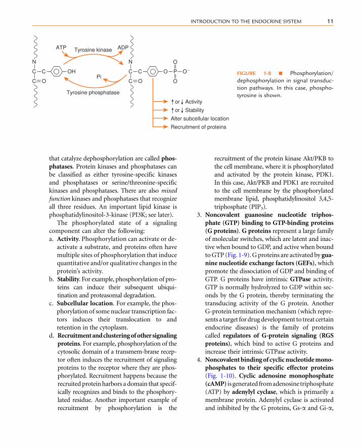
that catalyze dephosphorylation are called phos-
phatases. Protein kinases and phosphatases can
be classified as either tyrosine-specific kinases
and phosphatases or serine/threonine-specific
kinases and phosphatases. There are also mixed
function kinases and phosphatases that recognize
all three residues. An important lipid kinase is
phosphatidylinositol-3-kinase (PI3K; see later).
The phosphorylated state of a signaling
component can alter the following:
a. Activity. Phosphorylation can activate or de-
activate a substrate, and proteins often have
multiple sites of phosphorylation that induce
quantitative and/or qualitative changes in the
protein’s activity.
b. Stability. For example, phosphorylation of pro-
teins can induce their subsequent ubiqui-
tination and proteasomal degradation.
c. Subcellular location. For example, the phos-
phorylation of some nuclear transcription fac-
tors induces their translocation to and
retention in the cytoplasm.
d. Recruitmentandclusteringofothersignaling
proteins. For example, phosphorylation of the
cytosolic domain of a transmem-brane recep-
tor often induces the recruitment of signaling
proteins to the receptor where they are phos-
phorylated. Recruitment happens because the
recruited protein harbors a domain that specif-
ically recognizes and binds to the phosphory-
lated residue. Another important example of
recruitment by phosphorylation is the
recruitment of the protein kinase Akt/PKB to
the cell membrane, where it is phosphorylated
and activated by the protein kinase, PDK1.
In this case, Akt/PKB and PDK1 are recruited
to the cell membrane by the phosphorylated
membrane lipid, phosphatidylinositol 3,4,5-
triphosphate (PIP3).
3. Noncovalent guanosine nucleotide triphos-
phate (GTP) binding to GTP-binding proteins
(G proteins). G proteins represent a large family
of molecular switches, which are latent and inac-
tive when bound to GDP, and active when bound
toGTP (Fig. 1-9).G proteins are activated by gua-
nine nucleotide exchange factors (GEFs), which
promote the dissociation of GDP and binding of
GTP. G proteins have intrinsic GTPase activity.
GTP is normally hydrolyzed to GDP within sec-
onds by the G protein, thereby terminating the
transducing activity of the G protein. Another
G-protein termination mechanism (which repre-
sents a target fordrugdevelopment to treat certain
endocrine diseases) is the family of proteins
called regulators of G-protein signaling (RGS
proteins), which bind to active G proteins and
increase their intrinsic GTPase activity.
4. Noncovalentbindingofcyclicnucleotidemono-
phosphates to their specific effector proteins
(Fig. 1-10). Cyclic adenosine monophosphate
(cAMP) is generated fromadenosine triphosphate
(ATP) by adenylyl cyclase, which is primarily a
membrane protein. Adenylyl cyclase is activated
and inhibited by the G proteins, Gs-a and Gi-a,
Tyrosine kinase
Tyrosine phosphatase
PiOH
ATP ADP
CC
N
C O
C O O�
O
O
PC
N
C O
Recruitment of proteins
Alter subcellular location
or Activity
or Stability
FIGURE 1-8 n Phosphorylation/
dephosphorylation in signal transduc-
tion pathways. In this case, phospho-
tyrosine is shown.
11INTRODUCTION TO THE ENDOCRINE SYSTEM

respectively (see later). There are three general in-
tracellular effectors of cyclic AMP (cAMP):
a. cAMP binds to the regulatory subunit of
protein kinase A (PKA; also called cAMP-
dependent protein kinase). Inactive PKA is
a heterotetramer composed of two catalytic
subunits and two regulatory subunits. cAMP
binding causes the regulatory subunits to
dissociate from the catalytic subunits, thereby
generating two molecules of active catalytic
PKA subunits (PKAc). PKAc phosphorylates
numerous proteins on serine and threonine
residues. Substrates of PKAc include
numerous cytosolic proteins as well as
transcription factors, most notably cAMP-
responsive element–binding protein (CREB
protein).
b. A second effector of cAMP is Epac (exchange
protein activated by cAMP), which has two
isoforms. Epac proteins act as GEFs (see ear-
lier) for small G proteins (called Raps). Raps
in turn control a wide array of cell functions,
including formation of cell-cell junctional
complexes and cell-matrix adhesion, Ca2þ
release from intracellular stores (especially
in cardiac muscle) and in the augmentation
PKA
cAMP
R
R R
C C E
C C
RAP RAP
Cellularresponse
AC
Protein phosphorylation(membrane, cytosolic,
& nuclear proteins)
Activation ofeffector proteins
Ionic current(e.g., K�)
ATP cAMP
AMP
PDE
GTPGDP
CNGHCN
FIGURE 1-10 n Cyclic AMP/PKA in signal
transduction pathways. AC, adenylyl cyclase;
PDE, phosphodiesterase; R & C, regulatory and
catalytic subunits, respectively, of protein kinase A
(PKA); E, EPAC (exchange protein activated by
cAMP); CNG, cyclic nucleotide–gated channel;
HCN, hyperpolarization-induced cyclic nucleotide–
modulated channel.
Intrinsic GTPaseRGS protein
GEF
EffectorproteinG protein GTP
(active)
G protein GDP(inactive)
FIGURE 1-9nGproteins in signal transduction
pathways. GEF, guanine nucleotide exchange
factor; RGS, regulator of G-protein signaling.
12 ENDOCRINE AND REPRODUCTIVE PHYSIOLOGY
of glucose-dependent insulin secretion by
glucagon-like peptide-1 in pancreatic islet bcells (see Chapter 3).
c. cAMP (and cyclic guanosine monophosphate
[cGMP], discussed later) also binds directly to
and regulates ion channels. These are of two
types: cyclic nucleotide gated (CNG) chan-
nels and hyperpolarization-activated cyclic
nucleotide modulated (HCN) channels. For
example, norepinephrine, which acts through
a Gs-coupled receptor, increases heart rate in
part through increasing a depolarizing inward
Kþ and Naþ current via an HCN at the sino-
atrial node.
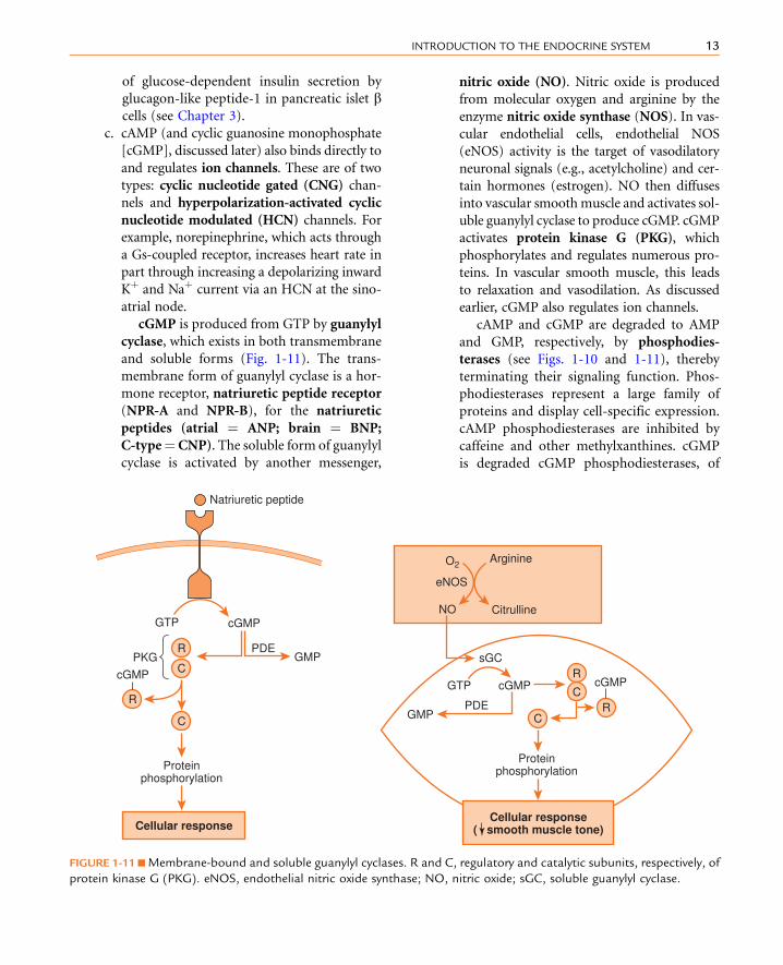
cGMP is produced from GTP by guanylyl
cyclase, which exists in both transmembrane
and soluble forms (Fig. 1-11). The trans-
membrane form of guanylyl cyclase is a hor-
mone receptor, natriuretic peptide receptor
(NPR-A and NPR-B), for the natriuretic
peptides (atrial ¼ ANP; brain ¼ BNP;
C-type¼CNP). The soluble form of guanylyl
cyclase is activated by another messenger,
nitric oxide (NO). Nitric oxide is produced
from molecular oxygen and arginine by the
enzyme nitric oxide synthase (NOS). In vas-
cular endothelial cells, endothelial NOS
(eNOS) activity is the target of vasodilatory
neuronal signals (e.g., acetylcholine) and cer-
tain hormones (estrogen). NO then diffuses
into vascular smoothmuscle and activates sol-
uble guanylyl cyclase to produce cGMP. cGMP
activates protein kinase G (PKG), which
phosphorylates and regulates numerous pro-
teins. In vascular smooth muscle, this leads
to relaxation and vasodilation. As discussed
earlier, cGMP also regulates ion channels.
cAMP and cGMP are degraded to AMP
and GMP, respectively, by phosphodies-
terases (see Figs. 1-10 and 1-11), thereby
terminating their signaling function. Phos-
phodiesterases represent a large family of
proteins and display cell-specific expression.
cAMP phosphodiesterases are inhibited by
caffeine and other methylxanthines. cGMP
is degraded cGMP phosphodiesterases, of
C
Proteinphosphorylation
C C
C
R
R
cGMP
cGMP
Natriuretic peptide
GTP
R
R
Proteinphosphorylation
PDEGMP
GTP cGMP cGMP
Cellular response( smooth muscle tone)Cellular response
PKG GMP sGCPDE
ArginineO2
NO
eNOS
Citrulline
FIGURE 1-11 nMembrane-bound and soluble guanylyl cyclases. R and C, regulatory and catalytic subunits, respectively, of
protein kinase G (PKG). eNOS, endothelial nitric oxide synthase; NO, nitric oxide; sGC, soluble guanylyl cyclase.
13INTRODUCTION TO THE ENDOCRINE SYSTEM

which one isoform is inhibited by sildenafil
(Viagra). In some contexts, cAMP and cGMP
can modulate each other (a phenomenon
called cross-talk) through the regulation of
phosphodiesterases. For example, oocyte
arrest is maintained by high levels of cAMP.
The LH surge decreases cGMP in surround-
ing follicle cells by decreasing the local
production of a natriuretic peptide. This
results in lowered oocyte cyclic GMP. Because
cGMP inhibits the oocyte cAMP-specific
phosphodiesterase, lowered cGMP leads to
decreased cAMP, thereby allowing the
oocyte to complete the first meiotic division
(see Chapter 10).
5. Generation of lipid informational molecules,
which act as intracellular messengers. These
include diacylglycerol (DAG) and inositol 1,4,5-
triphosphate (IP3), which are cleaved from
phosphatidylinositol 4,5-bisphosphate (PIP2) by
membrane-bound phospholipase C (PLC). DAG
activates certain isoforms of protein kinase C
(Fig. 1-12). IP3 binds to the IP3 receptor, which
is a large complex forming a Ca2þ channel, on
the endoplasmic reticulum membrane, and pro-
motes Ca2þ efflux (see later) from the endoplas-
mic reticulum into the cytoplasm. Some isoforms
of DAG-activated PKC are also Ca2þ dependent,
so the actions of IP3 converge on and reinforce
those of DAG. The DAG signal is terminated by
lipases, whereas IP3 is rapidly inactivated by
dephosphorylation.
6. Noncovalent Ca2þ binding (see Fig. 1-12). Cyto-
solic levels of Ca2þ are maintained at very low levels
(i.e., 10�7 to 10–8 M), by either active transport of
Ca2þ out of the cell, or into intracellular compart-
ments (e.g., endoplasmic reticulum). As discussed
earlier, IP3 binding to the IP3 receptor increases the
flow of Ca2þ into the cytoplasm from the endoplas-
mic reticulum. Ca2þ can also enter the cytoplasm
through the regulated opening of Ca2þ channels
in the cell membrane. This leads to an increase in
Ca2þ binding directly to numerous specific effector
proteins, which leads to a change in their activities.
Additionally, Ca2þ regulates several effector proteins
indirectly, through binding to the messenger protein,
calmodulin. Several of the Ca2þ/calmodulin targets
are enzymes, which amplify the initial signal of in-
creased cytosolic Ca2þ. The Ca2þ-dependent mes-
sage is terminated by the lowering of cytosolic
Ca2þ by cell membrane and endoplasmic reticular
Ca2þ ATPases (i.e., Ca2þ pumps).
Transmembrane Receptors UsingG Proteins
The largest family of hormone receptors is the
G-protein-coupled receptor (GPCR) family. These
receptors span the cell membrane seven times and
are referred to as 7-helix transmembrane receptors.
The G proteins that directly interact with GPCRs are
termed heterotrimeric G proteins and are composed
of an a subunit (Ga), and a b/g subunit dimer
(Gb/g). The Ga subunit binds GTP and functions as
I
IP3
Cellular response
Ca2�� CaMCa2�� CBP
Ca2�
Ca2�
SER
IP3R�
PKCPLCC C
COH C C DAG
Effectorproteins
ProteinphosphorylationP1P2FIGURE 1-12 n IP3 (inositol
1,4,5-triphosphate) and DAG
(diacylglycerol) in signaling path-
ways. PLC, phospholipase C;
PIP2, phosphatidylinositol 4,5-
bisphosphate; IP3R,IP3 receptor;
SER smooth endoplasmic re-
ticulum; CaM, calmodulin;
CBP, calcium-binding proteins.
14 ENDOCRINE AND REPRODUCTIVE PHYSIOLOGY
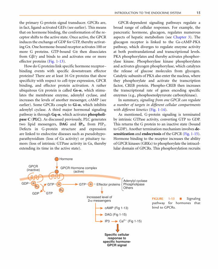
the primary G-protein signal transducer. GPCRs are,
in fact, ligand-activated GEFs (see earlier). This means
that on hormone binding, the conformation of the re-
ceptor shifts to the active state. Once active, the GPCR
induces the exchange of GDP for GTP, thereby activat-
ing Ga. One hormone-bound receptor activates 100 or
more G proteins. GTP-bound Ga then dissociates
from Gb/g and binds to and activates one or more
effector proteins (Fig. 1-13).
How do G proteins link specific hormone receptor–
binding events with specific downstream effector
proteins? There are at least 16 Ga proteins that show
specificity with respect to cell-type expression, GPCR
binding, and effector protein activation. A rather
ubiquitous Ga protein is called Gs-a, which stimu-
lates the membrane enzyme, adenylyl cyclase, and
increases the levels of another messenger, cAMP (see
earlier). Some GPCRs couple to Gi-a, which inhibits
adenylyl cyclase. A third major hormonal signaling
pathway is through Gq-a, which activates phospholi-
pase C (PLC). As discussed previously, PLC generates
two lipid messengers, DAG and IP3, from PIP2.
Defects in G-protein structure and expression
are linked to endocrine diseases such as pseudohypo-
parathyroidism (loss of Gs activity) or pituitary tu-
mors (loss of intrinsic GTPase activity in Gs, thereby
extending its time in the active state).
GPCR-dependent signaling pathways regulate a
broad range of cellular responses. For example, the
pancreatic hormone, glucagon, regulates numerous
aspects of hepatic metabolism (see Chapter 3). The
glucagon receptor is linked to the Gs-cAMP-PKA
pathway, which diverges to regulate enzyme activity
at both posttranslational and transcriptional levels.
PKA phosphorylates and thereby activates phosphor-
ylase kinase. Phosphorylase kinase phosphorylates
and activates glycogen phosphorylase, which catalyzes
the release of glucose molecules from glycogen.
Catalytic subunits of PKA also enter the nucleus, where
they phosphorylate and activate the transcription
factor, CREB protein. Phospho-CREB then increases
the transcriptional rate of genes encoding specific
enzymes (e.g., phosphoenolpyruvate carboxykinase).
In summary, signaling from one GPCR can regulate
a number of targets in different cellular compartments
with different kinetics (Fig. 1-14).
As mentioned, G-protein signaling is terminated
by intrinsic GTPase activity, converting GTP to GDP.
This returns the G protein to an inactive state (bound
to GDP). Another terminationmechanism involves de-
sensitization and endocytosis of the GPCR (Fig. 1-15).
Hormone binding to the receptor increases the ability
of GPCR kinases (GRKs) to phosphorylate the intracel-
lular domain of GPCRs. This phosphorylation recruits
cAMP (Fig 1-13)
Adenylyl cyclasePhospholipaseOthers
Specific cellularresponse to
specific hormone-GPCR signal
DAG (Fig 1-15)
IP3 Ca2� (Fig 1-15)
Increased level of2nd messengers
Effector proteins
Hormone
GPCR�Hormone complex(active)
GPCR(inactive)
GTP �GDP � GTP
GTPGDP�/�
�/��
FIGURE 1-13 n Signaling
pathway for hormones that
bind to GPCRs.
15INTRODUCTION TO THE ENDOCRINE SYSTEM

proteins called b-arrestins. GRK-induced phosphory-
lation and b-arrestin binding inactivate the receptor,
and b-arrestin couples the receptor to clathrin-
mediated endocytotic machinery. Some GPCRs are
dephosphorylated and rapidly recycled back to the cell
membrane (without hormone), whereas others are de-
graded in lysosomes. GRK/b-arrestin-dependent inac-tivation and endocytosis is an important mechanism
for hormonal desensitization of a cell after exposure
to excessive hormone. Hormone receptor endocytosis
(also called receptor-mediated endocytosis) is also an
important mechanism for clearing protein and peptide
hormones from the blood.
Receptor Tyrosine Kinases
Receptor tyrosine kinases (RTKs) can be classified into
two groups: the first acting as receptors for several
growth factors (e.g., epidermal growth factor, platelet-
derived growth factor), and the second group for insu-
lin and insulin-like growth factors (IGFs). The former
group of RTKs comprises transmembrane glycopro-
teins with an intracellular domain containing intrinsic
tyrosine kinase activity. Growth factor binding induces
dimerization of the RTKwithin the cell membrane, fol-
lowed by transphosphorylation of tyrosine residues,
generating phosphotyrosine (pY). The phosphotyro-
sines function to recruit proteins. One recruited protein
is phospholipase C, which is then activated by phos-
phorylation and generates the messengers DAG and
IP3 from PIP2 (see earlier). A second critically impor-
tant protein that is recruited to pY residues is the
adapter protein, Grb2, which is complexed with a
GEF named SOS. Recruitment of SOS to themembrane
allows it to activate a small, membrane-bound mono-
meric G protein called Ras. Ras then binds to its effector
protein, Raf. Raf is a serine-specific kinase that phos-
phorylates and activates the dual-function kinase,
MEK. MEK then phosphorylates and activates a
Glucagon
Glucagon receptor
GTP • Gs
Adenylyl cyclase
cAMP
Nucleus Cytoplasm
PKA Catalyticsubunit
Hepatic glucose output
Phosphorylase kinase
Glycogenolysis
CREB
Gluconeogenic enzymegene transcription
Gluconeogenesis
Phosphorylase
FIGURE 1-14 n Coordinated regulation of cytoplasmic and
nuclear events by PKA to produce a general cellular
response.
GPCR Hormone • GPCR
GDP • Gαβγ
β-arrestin binding
Inactivation of GPCRand endocytosis of
hormone-GPCR–β arrestin complex
GRK
RecyclingGTP • Gα
Pi
Phosphatase
Digestion bylysosomal enzymes
Hormone • GPCR • P
FIGURE 1-15 n GPCR inactivation and endocytosis to
lysosomes (desensitization) and/or recycling back to the
cellmembrane in a dephosphorylated form (resensitization).
16 ENDOCRINE AND REPRODUCTIVE PHYSIOLOGY

mitogen-activated protein kinase (MAP kinase, also
called ERK). Activated MAP kinases then enter the nu-
cleus and phosphorylate and activate several transcrip-
tion factors. This signaling pathway is referred to as the
MAP kinase cascade, and it transduces and amplifies a
growth factor–RTK signal into a cellular response in-
volving a change in the expression of genes encoding
proteins involved in proliferation and survival.
The insulin receptor (IR) differs from growth factor
RTKs in several respects. First, the latent IR is already
dimerized by Cys-Cys bonds, and insulin binding in-
duces a conformational change that leads to transphos-
phorylation of the cytoplasmic domains (Fig. 1-16).
A major recruited protein to pY residues is the insulin
receptor substrate (IRS), which is then phosphorylated
on tyrosine residues by the IR. The pY residues on IRS
recruit the Grb-2/SOS complex, thereby activating
growth responses to insulin through the MAP kinase
pathway (see Fig. 1-16). The pY residues on the IRS
also recruit the lipid kinase, PI3K, activating and
concentrating the kinase near its substrate, PIP2, in the
cellmembrane.As discussed earlier, this ultimately leads
to activation of Akt/PKB, which is required for the
metabolic responses to insulin (Fig. 1-17). The IR also
activates a pathway involving the small G protein, TC-
10 (see Fig. 1-17). The smallG-protein-dependent path-
way and the Akt/PKB pathway are both required for the
actions of insulin on glucose uptake (see Chapter 3).
RTKs are down regulated by ligand-induced endo-
cytosis. Additionally, the signaling pathways from
RTKs, including IR and IRS, are inhibited by serine/
threonine phosphorylation, tyrosine dephosphoryla-
tion, and the suppressor of cytokine signaling proteins
(see next section).
Receptors Associated with CytoplasmicTyrosine Kinases
Another class of membrane receptor falls into the cyto-
kine receptor family and includes receptors for growth
hormone, prolactin, erythropoietin, and leptin. These
pY
IR
IRS
pYpY
GrbSoS
Ras GDP GTP
GDP
Mek Mek�P
MAPK MAPK�P
Transfer to nucleus
Phosphorylation of transcription factors
Change in gene expression
Cellular response(Primarily mitogenic actions
of insulin)
Insulin
Ras
Raf
FIGURE 1-16 n Signaling from
the insulin receptor (a receptor
tyrosine kinase) through the
MAPK pathway. pY, phosphory-
lated tyrosine residue in protein.
17INTRODUCTION TO THE ENDOCRINE SYSTEM

receptors, which exist as dimers, do not have intrinsic
proteinkinaseactivity. Instead, thecytoplasmicdomains
are stably associated with members of the JAK kinase
family (Fig. 1-18). Hormone binding induces a con-
formational change, bringing the two JAKs associated
with the dimerized receptor closer together and causing
their transphosphorylation and activation. JAKs then
phosphorylate tyrosine residues on the cytoplasmic do-
mains of the receptor. The pY residues recruit latent
transcription factors called STAT (signal transducers
and activators of transcription) proteins. STATs be-
come phosphorylated by JAKs, which causes them to
dissociate from the receptor, dimerize, and translocate
into the nucleus, where they regulate gene expression.
A negative feedback loop has been identified for
JAK/STAT signaling. STATs stimulate expression of
one or more suppressors of cytokine signaling
(SOCS) proteins. SOCS proteins compete with STATS
for binding to the pY residues on cytokine receptors
(Fig. 1-19). This terminates the signaling pathway at
the step of STAT activation. Recent studies show that
a SOCS protein is induced by insulin signaling. SOCS
3 protein plays a role in terminating the signal from
the IR, but also in reducing insulin sensitivity in
hyperinsulinemic patients.
Receptor Serine/Threonine KinaseReceptors
One group of transmembrane receptors are bound
and activated by members of the transforming
growth factor (TGF)-b family, which includes the
hormones antimullerian hormone and inhibin. Un-
bound receptors exist as dissociated heterodimers,
called RI and RII (Fig. 1-20). Hormone binding to
RII induces dimerization of RII with RI, and RII acti-
vates RI by phosphorylation. RI then activates latent
transcription factors called Smads. Activated Smads
heterodimerize with a Co-Smad, enter the nucleus,
and regulate specific gene expression.
Membrane Guanylyl Cyclase Receptors
As discussed previously, the membrane-bound forms
of guanylyl cyclase constitute a family of a receptors for
natriuretic peptides (see Fig. 1-11). The hormonal role
PP P
Active Akt/PKBAlso recruitment ofactivation of PKC isoforms
Activation ofsmall G protein TCIO
GLUT 4(in vesicle)
Akt/PKB
PDK
R C
P13K
pY
IRS
pY
Insulin
pY
IR
Protein phosphorylation
Increased uptakeof glucose
Glucose
Insertion ofGLUT4 into
cell membrane
Cellular response(primarily metabolic actions
of insulin)
P
P1P2 P1P3
FIGURE 1-17 n Signaling from
the insulin receptor through the
phosphatidylinositol-3-kinase
(PI3K)/Akt/PKB pathway. R and
C; regulatory and catalytic sub-
units, respectively, of PI3K. PIP2,
phosphatidylinositol 4,5-bispho-
sphate; PIP3, phosphatidylino-
sitol 3,4,5 trisphosphate. PKC,
protein kinase C; pY, phosphory-
lated tyrosine residue in protein.
18 ENDOCRINE AND REPRODUCTIVE PHYSIOLOGY

of atrial natriuretic peptide (ANP) will be discussed
in Chapter 7.
Signaling from Intracellular Receptors
Steroid hormones, thyroid hormones, and 1,25-
dihydroxyvitamin D act primarily through intracellu-
lar receptors. These receptors are structurally similar
and are members of the nuclear hormone receptor
superfamily that includes receptors for steroid hor-
mones, thyroid hormone, lipid-soluble vitamins,
peroxisomeproliferator–activated receptors (PPARs),
and othermetabolic receptors (liver X receptor, farnesyl
X receptor).
Nuclear hormone receptors act as transcriptional
regulators. This means that the signal of hormone re-
ceptor binding is transduced ultimately into a change
in the transcriptional rate of a subset of the genes that
are expressed within a differentiated cell type. One re-
ceptor binds to a specific DNA sequence, called a hor-
mone response element, often close to the promoter
of one gene, and influences the rate of transcription of
that gene in a hormone-dependent manner (see later).
However, multiple hormone receptor–binding events
Hormone/cytokine
Hormone/cytokine receptor
Cytoplasm
STAT
pY pY
pY
STAT dimer
STAT regulation ofgene expression
pY
STAT
JAK JAK
Nucleus
FIGURE 1-18 n Signaling from cytokine receptor family.
Cytokine receptor
Cellular responses
Recruitment bypY residues
Recruitment viapY residues
SOCS inhibitsrecruitment
↑ SOCS expression
Insulin receptor
Cellular responses
FIGURE 1-19 n Role of suppressor of cytokine signaling
SOCS protein in terminating signals from cytokine family
and insulin receptors.
Cytoplasm
SMAD
Active SMAD
Co-SMAD
Co-SMAD
P
P
P
TGF-β–relatedhormones
RII/RI dimer
RII
NucleusRegulation of
specific gene expression
FIGURE 1-20 n Signaling from TGF-b-related hormones.
19INTRODUCTION TO THE ENDOCRINE SYSTEM
are collectively transduced into the regulation of
several genes. Moreover, regulation by one hormone
usually includes activation and repression of the tran-
scription of many genes in a given cell type. Note that
we have already discussed examples of signaling to
transcription factors by transmembrane receptors.
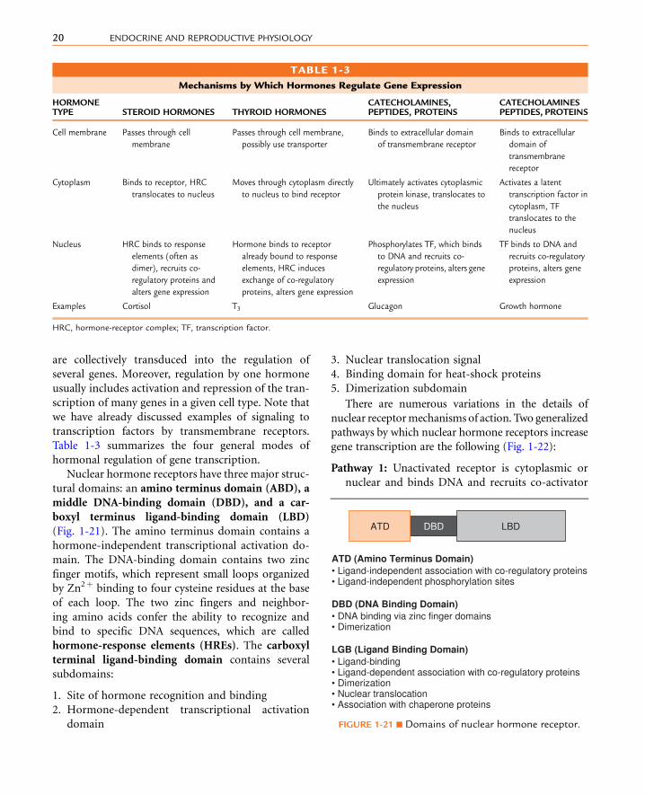
Table 1-3 summarizes the four general modes of
hormonal regulation of gene transcription.
Nuclear hormone receptors have three major struc-
tural domains: an amino terminus domain (ABD), a
middle DNA-binding domain (DBD), and a car-
boxyl terminus ligand-binding domain (LBD)
(Fig. 1-21). The amino terminus domain contains a
hormone-independent transcriptional activation do-
main. The DNA-binding domain contains two zinc
finger motifs, which represent small loops organized
by Zn2þ binding to four cysteine residues at the base
of each loop. The two zinc fingers and neighbor-
ing amino acids confer the ability to recognize and
bind to specific DNA sequences, which are called
hormone-response elements (HREs). The carboxyl
terminal ligand-binding domain contains several
subdomains:
1. Site of hormone recognition and binding
2. Hormone-dependent transcriptional activation
domain
3. Nuclear translocation signal
4. Binding domain for heat-shock proteins
5. Dimerization subdomain
There are numerous variations in the details of
nuclear receptormechanisms of action. Two generalized
pathways by which nuclear hormone receptors increase
gene transcription are the following (Fig. 1-22):
Pathway 1: Unactivated receptor is cytoplasmic or
nuclear and binds DNA and recruits co-activator
TABLE 1-3
Mechanisms by Which Hormones Regulate Gene Expression
HORMONETYPE STEROID HORMONES THYROID HORMONES
CATECHOLAMINES,PEPTIDES, PROTEINS
CATECHOLAMINESPEPTIDES, PROTEINS
Cell membrane Passes through cell
membrane
Passes through cell membrane,
possibly use transporter
Binds to extracellular domain
of transmembrane receptor
Binds to extracellular
domain of
transmembrane
receptor
Cytoplasm Binds to receptor, HRC
translocates to nucleus
Moves through cytoplasm directly
to nucleus to bind receptor
Ultimately activates cytoplasmic
protein kinase, translocates to
the nucleus
Activates a latent
transcription factor in
cytoplasm, TF
translocates to the
nucleus
Nucleus HRC binds to response
elements (often as
dimer), recruits co-
regulatory proteins and
alters gene expression
Hormone binds to receptor
already bound to response
elements, HRC induces
exchange of co-regulatory
proteins, alters gene expression
Phosphorylates TF, which binds
to DNA and recruits co-
regulatory proteins, alters gene
expression
TF binds to DNA and
recruits co-regulatory
proteins, alters gene
expression
Examples Cortisol T3 Glucagon Growth hormone
HRC, hormone-receptor complex; TF, transcription factor.
DBD LBD
ATD (Amino Terminus Domain)• Ligand-independent association with co-regulatory proteins• Ligand-independent phosphorylation sites
DBD (DNA Binding Domain)• DNA binding via zinc finger domains• Dimerization
LGB (Ligand Binding Domain)• Ligand-binding• Ligand-dependent association with co-regulatory proteins• Dimerization• Nuclear translocation• Association with chaperone proteins
ATD
FIGURE 1-21 n Domains of nuclear hormone receptor.
20 ENDOCRINE AND REPRODUCTIVE PHYSIOLOGY

Pathway 1 (Steroid hormones)
(–) Hormone
GTFs
HRE Gene
Basal transcription
Recruitment of co-activators
Recruitment and activation ofgeneral transcription factor
(+) Hormone
GTFsHR
Co-act
HR
HREChromatinstructure
Gene
Stimulated transcription
Recruitment of co-activators
Recruitment of activation ofgeneral transcription factors
(+) Hormone
GTFs
Chromatinstructure
Gene
Stimulated transcription
Dissociation of co-repressors(+) Hormone
GTFs
Gene
Basal transcription
Pathway 2 (Thyroid hormones, vitamin D, PPARs)
(–) Hormone
Blocking general transcription factor
RXR
Co-repress
HR
HREChromatinstructure
Gene
Repressed transcription
RXR HR
HRE
RXR
Co-act
HR
HRE
FIGURE 1-22 n Two general mechanisms
by which nuclear receptor and hormone
complexes increase gene transcription.
HRE, hormone response element; co-
repress, co-repressor proteins; GTFs,
general transcription factors; HR,
hormone receptor; RXR, retinoid X
receptor; Co-act, co-activator proteins.
21INTRODUCTION TO THE ENDOCRINE SYSTEM
proteins on hormone binding. This mode is ob-
served for the ER, PR, GR, MR, and AR (i.e., steroid
hormone receptors). In the absence of hormone,
some of these receptors are held in the cytoplasm
through an interaction with chaperone proteins
(so-called heat-shock proteins because their levels
increase in response to elevated temperatures and
other stresses). Chaperone proteins maintain the
stability of the nuclear receptor in an inactive
configuration. Hormone binding induces a confor-
mational change in the receptor, causing its disso-
ciation from heat-shock proteins. This exposes the
nuclear localization signal and dimerization do-
mains, so receptors dimerize and enter the nucleus.
Once in the nucleus, these receptors bind to their
respective HREs. The HREs for the PR, GR, MR,
and AR are inverted repeats with the recognition
sequence, AGAACANNNTGTTCT. Specificity is
conferred by neighboring base sequences and
possibly by receptor interaction with other tran-
scriptional factors in the context of a specific
gene promoter. The ER usually binds to an in-
verted repeat with the recognition sequence,
AGGTCANNNTGACCT. The specific HREs are
also referred to as an estrogen-response element
(ERE), progesterone-response element (PRE),
glucocorticoid-response element (GRE), min-
eralocorticoid-response element (MRE), and
androgen-response element (ARE). Once bound
to their respective HREs, these receptors recruit
other proteins, called co-regulatory proteins, which
are either co-activators or co-repressors. Co-
activators act to recruit other components of the
transcriptional machinery and probably activate
some of these components. Co-activators also pos-
sess intrinsic histone acetyltransferase (HAT) activ-
ity, which acetylates histones in the region of the
promoter. Histone acetylation relaxes chromatin
coiling, making that region more accessible to
transcriptional machinery. Although the mechanis-
tic details are beyond the scope of this chapter, the
student should appreciate that steroid receptors
can also repress gene transcription through recruit-
ment of co-repressors that possess histone dea-
cetylase (HDAC) activity and that transcriptional
activation and repression pathways are induced con-
comitantly in the same cell. HDAC inhibitors are be-
ing studied in the context of treating some cancers
because they restart the expression of silenced tumor
suppressor genes.
Pathway 2: Receptor is always in nucleus and ex-
changes co-repressors with co-activators on hor-
mone binding. This pathway is used by the
thyroid hormone receptors (THRs), vitamin D
receptors, PPARs, and retinoic acid receptors.
For example, the THR is bound, usually as a hetero-
dimer, with the retinoic acid X receptor (RXR). In
the absence of thyroid hormone, the THR/RXR re-
cruits co-repressors. As stated earlier, co-repressors
recruit proteins with histone deacetylase (HDAC)
activity. In contrast to histone acetylation, histone
deacetylation allows tighter coiling of chromatin,
which makes promoters in that region less accessible
to the transcriptional machinery. Thus, THR/RXR
heterodimers are bound to thyroid hormone re-
sponse elements (TREs) in the absence of hormone
and maintain the expression of neighboring genes at
a “repressed” level. Thyroid hormone (and other li-
gands of this class) readily move into the nucleus
and bind to their receptors. Thyroid hormone bind-
ing induces dissociation of co-repressor proteins,
thereby increasing gene expression to a basal level.
The hormone receptor complex subsequently re-
cruits co-activator proteins, which further increase
transcriptional activity to the “stimulated” level.
Termination of steroid hormone receptor signaling
is poorly understood but appears to involve phos-
phorylation, ubiquitination, and proteasomal degra-
dation. Circulating steroid and thyroid hormones
are cleared as described previously.
In summary, hormones signal to cells through
membrane or intracellular receptors. Membrane re-
ceptors have rapid effects on cellular processes (e.g.,
enzyme activity, cytoskeletal arrangement) that are in-
dependent of new protein synthesis. Membrane recep-
tors can also rapidly regulate gene expression through
either mobile kinases (e.g., PKA, MAPKs) or mobile
transcription factors (e.g., STATs, Smads). Steroid hor-
mones have slower, longer-term effects that involve
chromatin remodeling and changes in gene expres-
sion. Increasing evidence points to rapid, nongenomic
effects of steroid hormones as well, but these pathways
are still being elucidated.
The presence of a functional receptor is an absolute
requirement for hormone action, and loss of a
22 ENDOCRINE AND REPRODUCTIVE PHYSIOLOGY

receptor produces essentially the same symptoms as
loss of hormone. In addition to the receptor, there
are fairly complex pathways involving numerous in-
tracellular messengers and effector proteins. Accord-
ingly, endocrine diseases can arise from abnormal
expression or activity of any of these signal transduc-
tion pathway components.
Overview of the Termination Signals
Most of what has been discussed in this chapter de-
scribes the stimulatory arm of signal transduction.
As noted earlier, all signal transduction of hormonal
signals must have termination mechanisms to avoid
sustained and uncontrolled stimulation of target cells.
Part of this stems from the cessation of the original
stimulus for increasing a hormone’s level, and mech-
anisms to clear the hormone (i.e., removal of signal).
However, there exist a wide array of intracellular
mechanisms that terminate the signaling pathway
within the target cells. Some of these are listed in
Table 1-4. Note that overactivity of terminating mech-
anisms can lead to hormonal resistance.
S U M M A R Y
1. The endocrine system is composed of:
n Dedicated hormone-producing glands (pitui-
tary, thyroid, parathyroid, and adrenal)
n Testes and ovaries, whose intrinsic endocrine
function is absolutely necessary for gametogenesis
n Hypothalamic neuroendocrine neurons
n Scattered endocrine cells that exist as clusters of
endocrine-only cells (islets of Langerhans) or as
cells within organs that are have a nonendocrine
primary function (pancreas, GI tract, kidney)
2. Endocrine signaling involves the secretion of a
chemical messenger, called a hormone, that circu-
lates in the blood and reaches an equilibrium with
the extracellular fluid. Hormones alter many func-
tions of their target cells, tissues, and organs
through specific, high-affinity interactions with
their receptors.
3. Protein/peptide hormones:
n Are produced on ribosomes, become inserted
into the cisternae of the endoplasmic reticulum,
transit the Golgi apparatus, and finally are stored
in membrane-bound secretory vesicles. The
release of these vesicles represents a regulated
mode of exocytosis. Each hormone is first made
as a prehormone, containing a signal peptide
that guides the elongating polypeptide into the
cisternae of the endoplasmic reticulum.
n Arefrequentlysynthesizedaspreprohormones.Af-
ter removal of the signal peptide, the prohormone
is processed by prohormone convertases.
n Typically do not cross cell membranes and act
through transmembrane receptors (see later).
n Mostly circulate as free hormones, and are ex-
creted in the urine or cleared by receptor-
mediated endocytosis and lysosomal degradation.
4. Catecholamine hormones:
n Include the hormones, epinephrine (Epi) and
norepinephrine (Norepi). Epi and Norepi are
derivatives of tyrosine, which is enzymatically
modified by several reactions. Ultimately, Epi
and Norepi are stored in a secretory vesicle
and are released in through regulated exocytosis.
n Act through transmembrane GPCRs receptors
called adrenergic receptors.
TABLE 1-4
Some Modes of Signal Transduction Termination
MECHANISM OF SIGNALTRANSDUCTIONTERMINATION EXAMPLE
Receptor-mediated endocytosis linked
to lysosomal degradation
Many transmembrane
receptors
Phosphorylation/dephosphorylation
of receptor or “downstream”
components of signaling pathway
Serine phosphorylation of
insulin receptor and
insulin receptor substrate
by other signaling
pathways
Ubiquitination/proteasomal
degradation
Steroid hormone receptors
Binding of an inhibitory regulatory
factor
Regulatory subunit of PKA
Intrinsic terminating enzymatic activity GTPase activity of G proteins
23INTRODUCTION TO THE ENDOCRINE SYSTEM

5. Steroid hormones:
n Include cortisol (glucocorticoid), aldosterone
(mineralocorticoid), testosterone, and dihydro-
testosterone (androgens), estradiol (estrogen),
progesterone (progestin), and 1,25 dihydroxy-
vitamin D3 (secosteroid).
n Are derivatives of cholesterol, which is modified
by a series of cell-specific enzymatic reactions.
n Are lipophilic and cross membranes readily.
Thus, steroid hormones cannot be stored in se-
cretory vesicles. Steroid production is regulated
at the level of synthesis. Several steroid hor-
mones are produced to a significant extent by
peripheral conversion of precursors.
n Circulate bound to transport proteins. Steroid
hormones are cleared by enzymatic modifica-
tions that increase their solubility in blood and
decrease their affinity for transport proteins.
Steroid hormones and their inactive metabolites
are excreted in the urine.
n Act through intracellular receptors, which are
members of the nuclear hormone receptor fam-
ily. Most steroid hormone receptors reside in the
cytoplasm and are translocated to the nucleus af-
ter ligand (hormone) binding. Each steroid hor-
mone regulates the expression of numerous
genes in their target cells.
6. Thyroid hormones are:
n Iodinated derivatives of thyronine. The term
thyroid hormone typically refers to 3,5,30,50-tetraiodothyronine (T4 or thyroxine) and 3,5,30-triiodothyronine (T3). T4 is an inactive precursor
of T3, which is produced by 50-deiodination of T4.
n Synthesized and released by the thyroid epithe-
lium (see Chapter 6 for more detail)
n Circulate tightly bound to transport proteins
n Lipophilic and cross cell membranes. T3 binds to
one of several isoforms of thyroid hormone re-
ceptors (THRs), which form heterodimers with
retinoid X receptor (RXR) and reside bound to
their response elements in the nucleus in the
absence of hormone. Hormone binding induces
an exchange in the co-regulatory proteins that
interact with the THRs.
7. Protein, peptide, and catecholamine hormones
signal through transmembrane receptors and use
several common forms of informational transfer:
n Conformational change
n Binding by activated G proteins
n Binding by Ca2þ or Ca2þ-calmodulin. IP3 is a
major lipid messenger that increases cytosolic
Ca2þ levels through binding to the IP3 receptor.
n Phosphorylation and dephosphorylation, using
kinasesandphosphatases, respectively.Thephos-
phorylation state of a protein affects activity,
stability, subcellular localization, and recruit-
ment binding of other proteins. Note that phos-
phorylated lipids such as PIP3 also play a role
in signaling.
8. Transmembrane receptor families:
n G-protein-coupled receptors (GPCRs) act as
guanine nucleotide exchange factors (GEFs)
to activate the Ga subunit of the heterotrimeric
a/b/g G-protein complex. Depending on the
type of Ga subunit that is activated, this will
increase cAMP levels, decrease cAMP levels,
or increase protein kinase C activity and
Ca2þ levels. All catecholamine receptors (ad-
renergic receptors) are GPCRs. GPCRs are in-
ternalized by a receptor-mediated endocytosis
that involves GRK and b-arrestin. Endocytosisresults in the lysosomal clearance of the
hormone. The receptor may be digested in the
lysosome or may be recycled to the cell
membrane.
n The insulin receptor is a tyrosine kinase recep-
tor that activates the Akt/PKB pathway, the
G-protein TC10-related pathway, and theMAPK
pathway. The insulin receptor uses the scaffolding
protein insulin receptor substrate (IRS; four
isoforms) as part of its signaling to these three
pathways.
n Some protein hormones (e.g., growth hormone,
prolactin) bind to transmembrane receptors
that belong to the cytokine receptor family.
This are constitutively dimerized receptors that
are bound by janus kinases (JAKs). Hormone
binding interacts with both extracellular do-
mains and induces JAK-JAK cross-phosphory-
lation, followed by recruitment and binding
of STAT proteins. Phosphorylation of STATs
activates them and induces their translocation
to the nucleus, where they act as transcription
factors.
24 ENDOCRINE AND REPRODUCTIVE PHYSIOLOGY
n Hormones that are related to transforming
growth factor-b (TGF-b), such as antimullerian
hormone, signal through a co-receptor (recep-
tor I and receptor II) complex that ultimately
signals to the nucleus through activated Smad
proteins.
n Atrial natriuretic peptide (and related peptides)
bind to a transmembrane receptor that contains a
guanylyl cyclase domain within the cytosolic do-
main. These receptors signal by increasing cGMP,
which activates protein kinase G (PKG) and
cyclic nucleotide-gated channels. cGMP also
regulates selective phosphodiesterases.
n Steroid hormones bind to members of the
nuclear hormone transcription factor family.
Steroid hormone receptors usually reside in
the cytoplasm. Hormone binding induces
nuclear translocation, dimerization, and DNA
binding. Steroid hormone receptor complexes
regulate many genes in a target cell.
9. Thyroid hormone (T3) receptors (THRs) are
related to steroid hormone receptor, but they con-
stitutively remain in the nucleus bound to thyroid
hormone response DNA elements. T3 binding
typically induces an exchange of co-regulatory
proteins and altered gene expression.
S E L F - S T U D Y P R O B L E M S
1. How do protein hormones differ from steroid
hormones in terms of their storage within an en-
docrine cell?
2. How does binding to serum transport proteins in-fluence hormone metabolism and hormone
action?
3. How would a large increase in the GTPase activity
of Gs-a affect signaling through GPCRs linked to
Gs-a?
4. What role does the IRS protein play in transduc-ing insulin receptor signaling into a growth re-
sponse? a metabolic response?
5. Name an example of a transmembrane receptor–
associated transcription factor that translocates
to the nucleus.
6. Explain the mechanism of receptor-mediated en-
docytosis of a hormone that binds to a GPCR.
7. What is the importance of the GEF activity of aGPCR to its ability to signal?
8. ExplainhowPLCgenerates twosecondmessengers.
K E Y W O R D S A N D C O N C E P T S
n 7-Helix transmembrane receptorsn Adenylyl cyclasen Adrenal cortex
For full list of keywords and concepts see Student
Consult
SUGGESTED READ INGS
Huang CC, Tesmer JJ: Recognition in the face of diversity: Interac-
tions of heterotrimeric G proteins and G protein-coupled
receptor (GPCR) kinases with activated GPCRs, J Biol Chem
286:7715–7721, 2011.
Jean-Alphonse F, Hanyaloglu AC: Regulation of GPCR signal
networks via membrane trafficking, Mol Cell Endocrinol
331:205–214, 2011.
Rose RA, Giles WR: Natriuretic peptide C receptor signalling in the
heart and vasculature, J Physiol 586:353–366, 2008.
25INTRODUCTION TO THE ENDOCRINE SYSTEM

Intentionally left as blank

K E Y W O R D S A N D C O N C E P T S
n Agonistn Androgenn Androgen receptorn Androgen response element (ARE)n Antagonistn b-Arrestinsn Ca2þ
n Ca2þ ATPasesn Ca2þ channelsn Calmodulinn cAMP phosphodiesterasen cAMP response element–binding protein (CREB)n Catecholaminen Cellular responsen cGMP phosphodiesterasen Circadian (diurnal) rhythmsn Co-activator proteinsn Co-repressorsn Corticosteroid-binding globulinn Covalent phosphorylation of proteinsand lipids
n Cyclic AMPn Cyclic GMPn Cyclic nucleotide monophosphatesn Cycloperhydrophenanthrene ringn Cytokine receptor familyn Diacylglycerol (DAG)n Docking proteinn Effector proteinsn Eicosanoidsn Endocrine glandn Endocrine systemn Epinephrinen Estrogenn Estrogen receptorn Estrogen response element (ERE)n Exocrine glandn Exocytosisn G-protein exchange factor (GEF)n Gan Gi-an Glucocorticoidn Glucocorticoid receptorn Glucocorticoid response element (GRE)
n Glucuronide conjugationn GPCR kinase (GRK)n G-protein-coupled receptor (GPCR)n Gq-an Grb2/SOSn Gs-an GTP-binding proteins (G proteins)n Guanylyl cyclasen Gb/gn Heterotrimeric G proteinsn High-affinity receptorn Histone acetyltransferase (HAT)n Histone deacetylase (HDAC)n Hormonal desensitizationn Hormonal resistancen Hormonen Hormone response elements (HREs)n Inositol 1,4,5-triphosphate (IP3)n Insulin receptor (IR)n Insulin receptor substrate (IRS)n Intracellular messengersn Intrinsic GTPase activityn Iodothyroninen JAK kinase familyn Leukotrienesn Ligandn Ligand-activated GEFn Ligand-induced endocytosisn MEKn Mineralocorticoidn Mineralocorticoid receptorn Mineralocorticoid response element (MRE)n Mitogen-activated protein kinase (MAPK)n Mixed-function kinases and phosphatasesn Nitric oxide (NO)n Norepinephrinen Nuclear receptor superfamilyn Ovaryn Peripheral conversionn Phosphatidylinositol 3,4,5-triphosphate (PIP3)n Phosphatidylinositol-3-kinase (PI3K)n Phospholipase Cn Phosphotyrosine (pY)n PKA catalytic subunitn PKA regulatory subunitn Placenta
25.e1INTRODUCTION TO THE ENDOCRINE SYSTEM

n Prehormonen Preprohormonen Progesterone receptorn Progesterone response element (PRE)n Progestinn Prohormone convertasen Prostacyclinn Prostaglandinsn Protein kinase A (PKA)n Protein kinase B (PKB/Akt)n Protein kinase G (PKG)n Protein/peptide hormonen Rafn Rasn Receptorn Receptor serine/threonine kinasesn Receptor tyrosine kinases (RTKs)n Regulated secretory pathwayn Regulators of G-protein signaling (RGS proteins)n Second messenger hypothesisn Serine/threonine-specific kinases andphosphatases
n Set-pointn Sex hormone–binding globulinn Signal peptidase
n Signal peptiden Signal recognition complexn Signal transduction pathwayn Smadsn STATn Steroid hormonen Steroidogenic cellsn Stimulus-secretion couplingn Sulfate conjugationn Suppressors of cytokine signaling (SOCS)proteins
n Target celln Target organn Testisn Thromboxanesn Thyroid hormone receptorn Thyroid hormone–binding globulinn Thyroid hormone–response element (TRE)n Transforming growth factor (TGF)-b familyn Transport proteinsn Tyrosine kinases and phosphatasesn Ultradian rhythmsn Vitamin Dn Vitamin D receptorn Vitamin D response element (VRE)
25.e2 ENDOCRINE AND REPRODUCTIVE PHYSIOLOGY

n n n n n n n n n n n n n n n
2 ENDOCRINE FUNCTION OF THEGASTROINTESTINAL TRACT
O B J E C T I V E S
1. Understand the role of well-established GI hormones
associated with the following four major aspects of
GI physiology:
n The regulation of gastric acid secretion and gastric
motility
n The regulation of secretion from the exocrine pancreas
and the gallbladder and their associated ducts
n The stimulation of GI tract growth (an enterotropic
action)
n The enhancement of nutrient-induced insulin secre-
tion by the endocrine pancreas (incretin action)
Note: A fifth general function of GI hormones, the effect on appetite, is discussed in the context of energy homeostasis in Chapter 3.
We begin our discussion of endocrine
physiology with the hormonal function and regulation
of the gastrointestinal (GI) tract. The discovery of se-
cretin in 1902 by Bayliss and Starling represented the
first characterization of a hormone as a blood-borne
chemical messenger, released at one site and acting
at multiple other sites. Indeed, the epithelial layer of
the mucosa of the GI tract harbors numerous enter-
oendocrine cell types, which collectively represent
the largest endocrine cell mass in the body.
The diffuse enteroendocrine system is perhaps the
most basic example of endocrine tissue in that it is
composed of unicellular glands situated within a sim-
ple epithelium. Most enteroendocrine cells, called
open cells, extend from the basal lamina of this epithe-
lium to the apical surface (Fig. 2-1), although there are
also closed enteroendocrine cells, which do not ex-
tend to the luminal surface. The apical membranes
of open enteroendocrine cells express either receptors
or transporters that allow the cell to sample the
contents of the lumen. Luminal contents, called secre-
togogues, stimulate specific enteroendocrine cell types
to secrete their hormones. This sampling or nutrient
tasting is independent of osmotic and mechanical
forces. The secretogogue mechanisms involved are
poorly understood, but some appear to require the ab-
sorption of the nutrient. There is also evidence for the
luminal secretion of paracrine peptide factors from
the surrounding absorptive epithelial cells that stimu-
late hormonal release from enteroendocrine cells. As
part of their response to luminal contents, specific
enteroendocrine cell types display distinct localiza-
tions along the GI tract (Table 2-1). We will see that
these localizations are central to the regulation and
function of each cell type.
In the simplest model of enteroendocrine cell func-
tion, a hormone is released from the basolateral mem-
brane in response to the presence of a secretogogue at
27

the luminal side of the cell. The secreted hormone dif-
fuses into blood vessels in the underlying lamina pro-
pria, thereby gaining access to the general circulation.
Circulating GI hormones regulate GI tract functions
by binding to specific receptors at one or more sites
within the GI tract and its extramural glands. In the
classic model, the secretion of the hormone by an enter-
oendocrine cell is subsequently terminated when the
luminal concentration of its secretogogue diminishes,
thereby terminating the secretion of the hormone.
This simple model of the enteroendocrine system
does not fully account for the integration with other
systemic responses to a meal. Both open and closed
enteroendocrine cells are regulated by the enteric ner-
vous system (ENS) and paracrine factors secreted by
neighboring epithelial cells (intrinsic regulators of
1. Specific luminal secretogogues(e.g., H+, amino acids, fatty acids)
stimulate “open” enteroendocrine cell at apical surface
Epithelium of mucosathat lines the lumen
of the GI tract
3. Hormone enterssplanchnic blood vessels
in lamina propria
2. “Open” and “closed”cells secrete hormonefrom basolateral side
Arterial supply Venous return(to the hepatic portal vein)
“Closed” type cell• Does not reach lumen• Must be regulated by neuronal, paracrine, or endocrine inputs
Also regulated byneuronal, paracrine,and endocrine inputs
Open Closed
FIGURE 2-1 n Closed and open
enteroendocrine cells. Entero-
endocrine cells sit within the
simple epithelium of the GI
tract. “Open” cells extend from
the basal lamina to the lumen.
“Closed” cells do not reach
the lumen. Both cells secrete
hormones that enter capillaries
in the lamina propria beneath
the epithelium.
TABLE 2-1
Distribution of Enteroendocrine Cells Along the GI Tract
STOMACH DUODENUM JEJUNUM ILEUM COLON
G cell (gastrin) x (x)
S cell (secretin) x x
I cell (CCK) x x (x)
K cell (GIP) x x
L cell (GLP-1) x x
L cell (GLP-2) x x
M cell (motilin) x x
Ghrelin-secreting cell x (x) (x) (x) (x)
x, Primary location of concentration; (x), less concentrated.
28 ENDOCRINE AND REPRODUCTIVE PHYSIOLOGY

enteroendocrine cell function). Additionally, there are
extrinsic regulators of enteroendocrine cells, most
notably the autonomic nervous system and endocrine
glands that reside outside of the GI tract. Conversely, GI
hormones can have local (i.e., paracrine) actions on the
afferent nerves of autonomic or enteric reflexes, so
the response to a GI hormone can be mediated by a
neurotransmitter. Thus, GI tract function is orches-
trated through a complex interplay of neural and endo-
crine responses and actions. It is not surprising,
therefore, that GI function is often perturbed in pa-
tients with psychiatric disorders (e.g., depression) and
endocrine disorders (e.g., hyperthyroidism).
The hormones secreted by the enteroendocrine sys-
tem function to maintain the health of the GI tract and
its extramural glands and provide an integrated re-
sponse to the acquisition of nutrients. This integrated
response to GI hormones is due, in part, to their ability
to regulate multiple functions of the GI tract.
ENTEROENDOCRINE HORMONEFAMILIES AND THEIR RECEPTORS
All established GI hormones are peptides and bind to
G-protein-coupled receptors (GPCRs; see Chapter 1)
located on the plasma membrane of target cells. GI
hormones, as well as their cognate GPCRs, can be or-
ganized into gene families based on structural homol-
ogies. In this chapter, we discuss members of three
enteroendocrine hormone families: gastrin, secre-
tin, and motilin (Table 2-2).
The gastrin family includes gastrin and cholecys-
tokinin (CCK), which share a common stretch of 5
amino acids at the C-terminus. Gastrin binds with
high affinity to the CCK-2 receptor (previously called
the CCK-B/gastrin receptor). CCK binds with high
affinity to the CCK-1 receptor.
The secretin family includes the hormones secretin,
glucagon, and glucagon-like peptides (including
GLP-1 and GLP-2) and gastric inhibitory polypeptide
TABLE 2-2
Enteroendocrine Hormone Families and Their Receptors
HORMONE FAMILY MEMBERS OF FAMILYRECEPTOR AND PRIMARYSIGNALING PATHWAY
PRIMARY DISTRIBUTION OF THE RECEPTOR(RELATED TO GI FUNCTION)
Gastrin Gastrin (G cell) CCK2 receptor
Gq - " in Ca2þ and PKC
Gastric ECL cell and parietal cell
CCK (I cell) CCK1 receptor
Gq - " in Ca2þ and PKC
Gallbladder muscularis and sphincter of
hepatopancreatic ampulla
Pancreatic acinar cells
Pancreatic ducts
Vagal afferents and enteric neurons
Stomach muscularis and pyloric sphincter
Gastric D cells
Secretin Secretin (S cell) Secretin receptor
Gs - " in cAMP
Pancreatic ducts and biliary ducts
Pancreatic acinar cells
G cells and pancreatic cells
GLP-1 (L cell) GLP-1 receptor
Gs - " in cAMP
b Cells of pancreatic islets
GLP-2 (L cell) GLP-2 receptor
Gs - " in cAMP
GI tract, especially small intestine
GIP (K cell) GIP receptor
Gs - " in cAMP
b Cells of pancreatic islets
Gastric mucosa and muscularis
Motilin Motilin (M cell) Motilin receptor
Gq - " in Ca2þ and PKC
(also binds erythromycin)
Stomach and small intestines, especially in smooth
muscle cells and enteric neurons
Ghrelin (P/D1 cell) GHS receptor type 1a (GHS-RIa)
Gq - " in Ca2þ and PKC
Pituitary and hypothalamus
29ENDOCRINE FUNCTION OF THE GASTROINTESTINAL TRACT
(GIP; more recently referred to as glucose-dependent
insulinotropic peptide—see later). This family also
includes the neurocrine factor, vasoactive intestinal
peptide (VIP). The corresponding GPCRs for each
member of the secretin family of peptides are also struc-
turally related. These receptors are all primarily coupled
toGs signaling pathways that increase intracellular cy-
clic adenosine monophosphate (cAMP) in target cells.
Themotilin family includes the hormonesmotilin
and ghrelin. Ghrelin was originally identified as a
growth hormone secretogogue (GHS) but is most
abundant in the fundus of the stomach. The receptors
for motilin and ghrelin are GPCRs that are linked to
Ga-q/phospholipase/IP3 pathways, which, in turn,
stimulate protein kinase C- and Ca2þ-dependent sig-naling pathways (see Chapter 1).
Many GI peptides are also expressed by tissues
outside of the GI tract. Pathophysiologically, GI pep-
tides can be secreted in an uncontrolled manner from
tumors. Other physiologic sites of production include
other endocrine glands (e.g., the pituitary gland) and
reproductive structures. Several peptides are produced
by the central (CNS) and peripheral (PNS) nervous
systems, where they are used as neurotransmitters or
neuromodulatory factors. For example, cholecystoki-
nin (CCK) is expressed in the neocortical region of the
CNS and the genitourinary-associated nerves of the
PNS. As for its role in the CNS, CCK has been linked
to anxiety and panic disorders. This also means that
receptors for these peptides also reside within the
CNS, the PNS, and probably other non-neural tissues.
Thus, a pharmacologic agent (agonist or antagonist)
related to a specific GI peptide can potentially have
a wide range of effects, depending on its stability
and whether it can cross the blood-brain barrier.
The possibility also exists that extra-GI sites of synthe-
sis can “spill over” into the general circulation and
affect GI function.
GASTRIN AND THE REGULATIONOF GASTRIC FUNCTION
The stomach acts as a food reservoir. People eat dis-
continuously and typically eat more at one sitting than
their GI tract can process immediately. Thus, the
stomach holds the ingested food and gradually releases
partially digested food (chyme) into the first part of
the small intestine, the duodenum. The layers of the
stomach wall carry out two basic functions: secretion
and contraction/relaxation.
Overview of Regulation of GastricSecretion and Motility
The innermost layer of the stomach wall, the gastric
mucosa, contains glandular and surface mucus-
producing epithelia and can be divided into proximal
and distal segments. Two of the proximal portions of
the stomach (fundus and body) contain the main gas-
tric mucosal glands (Fig. 2-2). Within these glands, the
parietal cells secrete HCl, which is important for hy-
drolysis of macromolecules, activation of proenzymes,
and the sterilization of ingested food. Parietal cells
also secrete intrinsic factor, which is a glycoprotein
required for the efficient absorption of vitamin B12.
The glands of the fundus and body also contain the
chief cells, which secrete digestive enzymes (e.g., pep-
sinogen, gastric lipase). A third cell type, the mucous
cell, is found in the neck of the gastric glands and on
the surface throughout the stomach. Mucous cells se-
crete mucigens, which buffer and protect the lining of
the stomach, particularly in the vicinity of the main
gastric glands. Because gastric enzyme andmucus pro-
duction is primarily under nervous control, with little
endocrine input, we focus here on gastric acid secre-
tion and motility.
The distal part of the gastric mucosa, the pyloric
antrum, has an important enteroendocrine function.
This part of the stomach contains two types of “open”
enteroendocrine cells. The G cells secrete gastrin, a
hormone, and the D cells secrete somatostatin, a
paracrine factor. These two peptides act antagonisti-
cally in a negative feedback loop to regulate gastric
blood flow, cell growth, secretion, and motility (see
later). D cells are also found within the fundus and
body region, where they directly inhibit parietal cell
secretion.
An outer layer of the stomach wall, the muscularis
externa, is composed of smooth muscle. The relaxa-
tion of this muscle allows distention and storage,
and its contractions ultimately move the partially
digested food (chyme) into the duodenum. There
are two gateways into and out of the stomach. These
are the lower esophageal sphincter (LES) and the
30 ENDOCRINE AND REPRODUCTIVE PHYSIOLOGY

pyloric sphincter, respectively. The LES allows swal-
lowed food particles to enter the stomach and protects
the esophagus from the reflux of acidic chyme. The
pyloric sphincter operates in conjunction with the
muscularis externa to allow only small particles of
digested chyme to escape the stomach and enter
the duodenum. The pyloric sphincter also prevents
backflow of chyme into the stomach.
In general, regulation of gastric function involves
the stimulation of secretion and motility as needed
(i.e., in the presence of food), and the inhibition of
gastric secretion and motility as acidic chyme reduces
the pH of the stomach, or as chyme moves into the
small intestine and colon. In this way, the stomach
avoids excessive acid secretion in the absence of buff-
ering foodstuffs. Further, the portion of the GI tract
below the stomach protects itself from exposure to ex-
cessive amounts of acid, which is both damaging to the
intestinal lining and inhibitory to the activity of intes-
tinal enzymes. Additionally, the small intestine, in
which the majority of digestion and absorption oc-
curs, controls the flow rate of food into and through
the small intestines in order to optimize digestion
and absorption of nutrients, salts, and water. The in-
ability to properly regulate acid secretion and its flow
into the intestine usually gives rise to duodenal ulcers,
although patients with a gastrin-producing tumor
(Zollinger-Ellison syndrome) can present with ulcer-
ation of the esophagus, stomach, and duodenum.
The general model of gastric control in response to a
meal can be organized into three phases. The cephalic
phase, which accounts for about 20% of the response to
a meal during the digestive period, is activated by the
actual or imagined smell and sight of food, or by the
presence of food in the mouth. The cephalic phase is
associated with increased gastric secretion but de-
creased motility, in anticipation of the need to store
and start digesting food. The gastric phase, which ac-
counts for about 10% of the postprandial response, is
activated by the presence of food in, and mechanical
distention of, the stomach. During the gastric phase, se-
cretion is strongly stimulated, and this is accompanied
by an increase in peristaltic contractions and gastric
emptying. The third phase is the intestinal phase, dur-
ing which an acidic mixture of partially digested food
(chyme) moves in a regulated manner through the py-
loric sphincter into the small intestine and ultimately
into the colon. The processes of enzymatic digestion
and absorption that occur during the digestive phase
account for 70% of the digestive period. Themovement
of food into the lower GI tract generally moderates both
gastric secretion and emptying.
Fundus of stomachLower esophageal
sphincter
Pyloric sphincter
Duodenum
Body ofstomach
Antrum of stomach
Antrum of stomach• Site of gastrin secretion from G cells
Main gastric glands• Site of acid secretion and intrinsic factor secretion from parietal cells• Site of enzyme secretion from chief cells
FIGURE 2-2 n Anatomy of the stomach.
31ENDOCRINE FUNCTION OF THE GASTROINTESTINAL TRACT

Gastrin and the Stimulation ofGastric Function
Gastric HCl secretion from parietal cells is stimulated
by three pathways:
n Paracrine stimulation by histamine, which is
secreted by neighboring enterochromaffin-like
(ECL) cells
n Enteric nervous system and vagal parasympa-
thetic nervous system stimulation via gastrin-
releasing peptide (GRP) and acetylcholine
n Direct and indirect hormonal stimulation by the
peptide hormone gastrin
Gastrin is produced by the G cells of the stomach
antrum and proximal duodenum. In humans, the
term gastrin refers to a 17-amino acid peptide that
has modifications at both termini (G-17). In fact,
the production of G-17 is an excellent example of
how a peptide-encoding gene gives rise to multiple,
larger precursors, which are also secreted into the
blood. G-17 is the product of sequential posttransla-
tional processing of preprogastrin, which can be gen-
erally characterized in three phases (Fig. 2-3). In the
first phase, sulfation and proteolysis generate a mix-
ture of gastrin precursors, called progastrins. The
second phase involves proteolysis within secretory
granules that generates C-termini peptides. Processing
of these intermediates also includes the cyclization of
the glutaminyl to a pyroglutamyl residue. The third
stage involves the amidation of the C-terminus to pro-
duce amidated gastrins. The primary secreted bioac-
tive product of human G cells is G-17 (i.e., 17
amino acids). The pyroglutamyl residue at the amino
terminus and the amidation of the C-terminus protect
G-17 from digestion by circulating aminopeptidases
and carboxypeptidases. G-17 binds with high affinity
to the CCK2 receptor and is responsible for all of the
gastrin effects on the stomach. The last four amino
acids assign gastrin-like biologic activity to G-17. A
synthetic, clinically used form of gastrin, pentagas-
trin, contains the last four amino acids, plus an ala-
nine at the amino terminus that confers increased
stability.
During the cephalic phase, gastric HCl secretion is
stimulated by vagal (parasympathetic) inputs. Pre-
ganglionic vagal efferents activate enteric neurons
that directly stimulate the parietal cells and stimulate
the release of histamine from ECL cells (Fig. 2-4).
These actions are mediated by acetylcholine, which
binds to the muscarinic receptor. Vagal stimulation
of gastrin is mediated by the neurocrine factor, GRP,
released from enteric neurons.
Progastrins
Preprogastrin
Gastrin intermediates
Bioactive 17-aminoacid gastrin (G-17)
• Sulfation of tyrosyl residue near C-terminus• Endoproteolysis into several fragments
• Further endoproteolysis• Cyclization of glutaminyl residue at N-terminus to a pyroglutamyl residue
• Further endoproteolysis of larger fragments into 17-amino acid C-terminal peptide, which is primary product of G cells in humans• Amidation of C-terminus
FIGURE 2-3 n Processing of
preprogastrin.
32 ENDOCRINE AND REPRODUCTIVE PHYSIOLOGY

During the gastric phase, gastrin secretion from G
cells is primarily stimulated by the presence of pep-
tides and amino acids in the lumen of the antrum
(Fig. 2-5). Gastrin secretion can also be stimulated
by stomach distention as detected by mechanosen-
sors during the gastric phase, acting through local
neuronal pathways, and through a vagovagal reflex.
Circulating gastrin levels increase by several-fold
within 30 to 60 minutes after ingestion of a meal.
The primary action of gastrin is the stimulation of
HCl secretion by the parietal cells of the gastric glands
within the fundus and body of the stomach. To accom-
plish this, gastrin must enter and circulate through the
general circulation and then exit capillaries and ven-
ules within the lamina propria of the gastric mucosa
in the body and fundus (i.e., upstream of where gastrin
is released within the stomach).
Gastrin evokes HCl secretion primarily through
binding to the CCK2 receptor on ECL cells. ECL cells,
which reside in the lamina propria of the gastric mu-
cosa, produce histamine in response to gastrin (see
Fig. 2-5). Gastrin binding to the Gq-coupled CCK2 re-
ceptor on ECL cells increases intracellular Ca2þ, which
leads to exocytosis of histamine-containing secretory
vesicles. Gastrin also increases histamine synthesis
and storage by increasing the expression of histidine
decarboxylase, which generates histamine from histi-
dine, and type 2 vesicular monoamine transporter
(VMAT-2), which transports and concentrates hista-
mine into the secretory vesicles. Thus, gastrin coordi-
nates both the secretion and synthesis of histamine in
ECL cells. Histamine, in turn, stimulates HCl secretion
in a paracrine manner by binding to the H2 receptor
on nearby epithelial parietal cells. Gastrin also has a
direct, although less important, effect on parietal cells.
During the intestinal phase of a meal, the decrease
in gastric contents relieves the stimulation of G cells by
amino acids and peptides, and by distention-induced
vagovagal pathways. The decrease in gastric contents
also reduces the buffering capacity of the gastric lu-
men. Thus, during the intestinal phase and the inter-
digestive period, the acidity of the stomach decreases.
When the pH falls below 3, acid stimulates the D cells
to secrete the paracrine peptide, somatostatin. So-
matostatin acts through its receptors (SS-R) to inhibit
gastrin secretion from neighboring G cells (Fig. 2-6).
Parietalcell
H�
To heart
ACh GRP G cell
ECLcell
EntericPreganglionic
vagalE
ACh ACh
HIST
GAST
EntericPreganglionic
vagalE
AChEnteric
Lamina propria Epithelium Gastriclumen
Preganglionicvagal
E
Gastrin in hepatic portal vein
ACh
From heart Gastrin in gastric veins
FIGURE 2-4 n Regulation of
gastric HCl secretion during the
cephalic phase of a meal. The
thought, sight, or smell of food,
or the presence of food in the
mouth, stimulates acid secretion
through the vagal preganglionic
parasympathetic nerves, which
stimulate the release of acetylcho-
line (ACh) from postganglionic
enteric nerves. Enteric nerve fibers
secreting ACh stimulate parietal
cells directly and through the
release of histamine from entero-
chromaffin-like (ECL) cells. Gas-
trin is also stimulated by enteric
neuronal fibers that release gastrin-
releasing peptide (GRP). As a hor-
mone, gastrin levels increase in the
general circulation. Gastrin stimu-
lates gastric HCl secretion by bind-
ing to CCK2 receptors on ECL
cells (and, to a lesser extent, on
parietal cells).
33ENDOCRINE FUNCTION OF THE GASTROINTESTINAL TRACT

Parietalcell
H�
Chyme
AAsPeptides
To heart
ACh GRP G cell
ECLcell
EntericPreganglionic
vagalE
ACh ACh
HIST
GAST
EntericPreganglionic
vagal
Afferent ofvagovagal response
E
AChEnteric
Lamina propria Epithelium Gastriclumen
Preganglionicvagal
E
Gastrin in hepatic portal vein
ACh
From heart Gastrin in gastric veins
Mechanosensors
FIGURE 2-5 n Regulation of
gastrin secretion during the
gastric phase of a meal. Luminal
amino acids and peptides
strongly stimulate G cells in
the antrum to secrete gastrin.
Gastrin secretion and HCl secre-
tion are also stimulated by stom-
ach distention through local and
autonomic (vagovagal) reflexes.
Parietalcell
D cell
G cell
SS
Enterogastrones fromintestines in gastric
veinsH�
ECLcell
Lamina propria Epithelium Gastriclumen
FIGURE 2-6 n Regulation of gas-
trin secretion during the intestinal
phase of a meal. The exit of food
(chyme) from the stomach lumen
reduces buffering of HCl. A low
pH stimulates D cells to release
the paracrine factor, somato-
statin (SS), which inhibits gastrin
secretion from neighboring G
cells. The exact nature of physio-
logic enterogastrones in humans
is not well established. Candi-
dates include secretin and gastric-
inhibitory peptide (gip) from the
small intestine, and peptide yy
from the ileum and colon.
34 ENDOCRINE AND REPRODUCTIVE PHYSIOLOGY

Gastrin release and gastric emptying are also
inhibited during the intestinal phase by the release
of hormones and neural signals from the small
intestine and colon in response to acidity, hyperto-
nicity, distention, and specific molecules (e.g., fatty
acids). These hormones are collectively referred to
as enterogastrones. The identity of the physiologic
enterogastrones in humans that inhibit gastric acid
secretion remains uncertain but includes candidates
such as secretin and GIP from the duodenum
and jejunum and peptide YY and GLP-1 from the
distal ileum and colon. CCK is a well-established
inhibitor of gastric motility and emptying. CCK
is released from the duodenum and jejunum in
response to the presence of luminal fatty acids (see
Fig. 2-6).
ENTEROENDOCRINE REGULATIONOF THE EXOCRINE PANCREASAND GALLBLADDER
The exocrine pancreas is an extramural gland that
empties its secretory products through a main ex-
cretory duct into the GI tract at the duodenum
(Fig. 2-7). The acini of the exocrine pancreas produce
enzymes necessary to digest macromolecules in the
small intestine. Pancreatic enzymes have optimal ac-
tivities at a neutral pH. Accordingly, the cells that line
the pancreatic ducts secrete a bicarbonate-rich fluid,
which serves to neutralize acidic chyme in the duode-
num. The gallbladder is also an extramural organ. It
receives bile that is secreted by the liver. Bile is both
stored and concentrated in the gallbladder. Bile is
Bile duct
Accessory pancreatic duct
Main pancreatic ductMinor duodenal papilla
Major duodenal papilla
Hepatopancreatic ampulla
FIGURE 2-7 n Anatomy of the common bile duct, pancreas, pancreatic duct, and duodenum. The gallbladder (not shown)
stores and concentrates bile from the liver. Contraction of the gallbladder and relaxation of the sphincter of Oddi
(surrounds the hepatopancreatic ampulla) allows bile to flow down the common bile duct into the duodenum.
Pancreatic enzymes and bicarbonate reach the duodenum via larger and larger ducts that eventually form the main
pancreatic duct. This duct joins the common bile duct just before it reaches the duodenum, to form the
hepatopancreatic ampulla. Inset shows a higher magnification of the exocrine pancreas. The termini of the secretory
units are the pancreatic acini, which secrete enzymes. The ductal epithelium secretes a bicarbonate-rich fluid. Note
that the ductal epithelium of the common bile duct also secretes a bicarbonate-rich fluid. (ª Elsevier. Drake et al:
Gray’s Anatomy for Students, www.studentconsult.com.)
35ENDOCRINE FUNCTION OF THE GASTROINTESTINAL TRACT

released into small intestine through the common bile
duct, which usually joins themain pancreatic duct to
form the hepatopancreatic ampulla just before open-
ing into the duodenum (see Fig. 2-7). A major func-
tion of bile is the emulsification of triglycerides
to increase their accessibility to pancreatic lipase.
In order to perform this function, aggregates (called
micelles) of bile acids and other lipids are required.
Micelle formation requires neutral or slightly
alkaline conditions. Accordingly, the epithelial cells
of the common bile duct secrete a bicarbonate-rich
fluid.
Pancreatic and gallbladder functions are primarily
regulated by the autonomic nervous system during
the interdigestive period (pancreatic secretion occurs
in phase with the migrating myoelectric complex
[MMC] in humans), and during the cephalic and gas-
tric phases of the digestive period. However, during
the intestinal phase, when these glands are most ac-
tive, they are predominantly under endocrine control
by two GI hormones, secretin and CCK. Secretin pri-
marily regulates ductal secretion of a bicarbonate-rich
fluid from both pancreatic and bile ducts. CCK pri-
marily stimulates enzyme secretion from pancreatic
acinar cells and gallbladder contraction. This dual reg-
ulation allows for fine-tuning of the qualitative nature
of the product (e.g., in terms of the percentage of
bicarbonate and protein in pancreatic juice) that is
finally secreted into the duodenum.
The classic model for secretin and CCK action on
the pancreas is that the appearance of acid, long-chain
fatty acids, and glycine-containing dipeptides and
tripeptides in the duodenum stimulates the open
enteroendocrine cells to secrete the two hormones.
Secretin and CCK then circulate in the blood and bind
to their specific receptors on either ductal or acinar
cells, respectively (Fig. 2-8).
However, there is evidence that secretin has
permissive effects on CCK actions, and vice versa.
Moreover, it is also clear that the autonomic and en-
teric nervous systems have a permissive effect on the
secretin and CCK actions. The neurotransmitter,
ACh, and a secretin-related enteric neurocrine pep-
tide, VIP, stimulate pancreatic ductal and acinar cells
and synergize with secretin and CCK. Patients who
have a VIPoma (i.e., a tumor producing high levels
of VIP) suffer from pancreatic diarrhea because of a
constant high level of pancreatic secretion into the gut.
Fatty acids,dipeptides,tripeptides
Duodenallumen
H+S cellSecretin
CCK
CCK1R
Secretin-R
Bicarbonate-rich fluidPancreatic enzymes
I cell
Duodenal hormones reach the exocrinepancreas via general circulation
Pancreatic juicereaches the duodenumvia the pancreatic ductand hepatopancreatic
ampulla
FIGURE 2-8 n Hormonal regula-
tion of pancreatic secretion by
secretin and CCK.
36 ENDOCRINE AND REPRODUCTIVE PHYSIOLOGY

Secretin
Secretin is produced by S cells in the duodenum and
jejunum. Similar to gastrin, secretin is produced by
posttranslational processing of a larger preprosecretin
molecule. Most secretin is a carboxyl amidated
27-amino acid peptide.
The primary stimulus for secretin release is a
decrease in duodenal pH. The threshold pH value for
secretin release is 4.5. Circulating secretin levels increase
rapidly (approximately 10 minutes) after acidified
chyme passes through the pyloric sphincter into the du-
odenum. The exact mechanism by which Hþ induces
secretin release from S cells is unclear. There is evidence
for a direct action ofHþ on S cells as well as evidence for
indirect actions through enteric neurons and through a
phospholipase A2–like secretin-releasing factor.
The primary short-term action of secretin is the
stimulation of the secretion of a bicarbonate-rich
fluid from the pancreatic and biliary ducts during
the intestinal phase of the digestive period (see
Fig. 2-8). Secretin acts through the secretin receptor,
which is linked to cAMP-dependent pathways. Signal-
ing from the secretin receptor opens apical Cl� chan-
nels (cystic fibrosis transmembrane conductance
regulator or CFTR) thereby increases the flow of
transport Cl� (and, through paracellular osmotic
drag, water) into the lumen. Cl� is then exchanged
for HCO3�. Upregulation of this process by secretin
can occur through the opening of preexisting CFTR
transporters in the apical membrane and through the
exocytotic insertion of transporter-containing vesicles
into themembrane. The importance of the CFTR chan-
nel to pancreatic function underlies the dysfunction of
pancreatic secretion observed in patients with cystic
fibrosis.
Secretin also binds to its receptor on the pancreatic
acinar cells. Although secretin has a minimal effect on
acinar cells by itself, secretin synergizes with the hor-
mone CCK to further enhance pancreatic enzyme
secretion over that achieved by CCK alone. Secretin
may also function as an enterogastrone by inhibiting
stomach acid secretion.
Cholecystokinin
CCK is a 33-amino acid peptide produced by the I cells
of the duodenum and jejunum. CCK is structurally
similar to gastrin, with the 5 amino acids at the carboxyl
terminus identical to both hormones. CCK is also sul-
fated on a tyrosine that is the seventh amino acid from
the carboxy terminus. CCK binds primarily to the
CCK1 receptor (formerly called the CCKA receptor),
whereas gastrin preferentially binds to the CCK2 recep-
tor. Both hormones can weakly interact with the other’s
receptor, and desulfation of CCK increases its affinity
for the CCK2 receptor. The CCK1 receptor is linked
to protein kinase C–dependent and Ca2þ-dependentpathways.
The primary stimulus for CCK secretion is the pres-
ence of long-chain fatty acids or monoglycerides in
the small intestine (see Fig. 2-8). CCK secretion is also
induced by glycine-containing dipeptides and tripep-
tides. The mechanism by which any of these act to
stimulate CCK release is obscure, although there is
some evidence for a postabsorptive effect of lipids after
their assembly into chylomicrons. There is also evi-
dence for a CCK-releasing peptide (CCK-RP) that is
released luminally from enterocytes and stimulates
CCK release through binding to a CCK-RP receptor
on the apical membrane of I cells. Like secretin,
CCK primarily regulates pancreatic and biliary
function. In the pancreas, CCK stimulates enzyme
secretion from the acinar cells (see Fig. 2-8). The
CCK1 receptor increases intracellular DAG and Ca2þ,which results in the exocytosis of enzyme-containing
secretion granules. CCK also has a permissive effect
on the ability of secretin to stimulate bicarbonate
secretion.
CCK is a strong stimulator of gallbladder con-
traction, and CCK deficiency disorders have been
linked to impairment of gallbladder contraction
and cholelithiasis (gallstones). CCK induces gall-
bladder contraction both directly and indirectly
through activation of vagal afferent neurons. CCK
also stimulates bile secretion into the duodenum
through promoting relaxation of the sphincter of
the hepatopancreatic ampulla (sphincter of Oddi).
This latter action on hepatobiliary function is likely
due to the CCK-dependent release of inhibitory
neurotransmitters, such as nitric oxide, from enteric
neurons. As mentioned, CCK also inhibits gastric
emptying, which reduces duodenal acidity and
allows emulsification, digestion, and absorption of
lipids.
37ENDOCRINE FUNCTION OF THE GASTROINTESTINAL TRACT

Motilin and Stimulation of Gastric andSmall Intestinal Contractions During theInterdigestive Period
Motilin is a 22-amino acid peptide produced from a
114-amino acid prepromotilin and secreted by the
M cells of the small intestine. Motilin secretion is
inhibited by the presence of food or acid in the small
intestine and is stimulated by alkalinization of the
small intestine.
Circulating motilin levels peak every 1 to 2 hours
in fasting individuals, in phase with the MMC. The
MMC is a set of organized contractions that move
aborally from the stomach to the ileum and clean
the stomach and small intestines of indigestible
particles. The MMC may also prevent the colonic
bacteria from migrating into the small intestine.
Motilin may function to either initiate or integrate
the MMC.
The motilin receptor is a GPCR that activates the
phospholipase C signaling pathway. The motilin recep-
tor also binds and is activated by the macrolide antibi-
otic, erythromycin (see Table 2-2). Erythromycin and
other motilin receptor agonists are used in the treat-
ment of delayed gastric emptying (gastroparesis),
which is common in patients with diabetes mellitus
and in some postsurgical patients.
INSULINOTROPIC ACTIONS OFGASTROINTESTINAL PEPTIDES(INCRETIN ACTION)
Elevated circulating levels of nutrients, particularly
blood glucose, are strong stimuli of insulin secretion
from the pancreatic b cells (see Chapter 3). The pos-
sibility that GI hormones also regulate the secretion
of insulin was revealed by observations that oral ad-
ministration of glucose caused a greater rise in insulin
than did glucose administered by an intravenous
route. This enteroinsular response gave rise to the
concept of incretins. In this model, an enteroendo-
crine cell type senses nutrients in the GI tract and
releases a hormone (an incretin),which, in turn, prepares
the pancreatic b cells for the impending rise in blood nu-
trients (primarily blood glucose). There are two incretins
in humans, gastric inhibitory peptide (GIP; also re-
ferred to as glucose-dependent insulinotropic peptide),
and glucagon-like peptide-1 (GLP-1). These peptides
(or analogs thereof) are currently being investigated
for the treatment of type 2 diabetes mellitus (see
Chapter 3). An important feature of incretins is that their
ability to increase insulin secretion is strongly dependent
on glucose levels. Thismeans that incretin analogs pose a
low risk for inducing severe hypoglycemia (low blood
sugar) because once blood glucose falls, the effect of
incretins is terminated.
In general, GIP and GLP-1 act through Gs-coupled
receptors on b cells, which increase cAMP. This acts in
a permissive or synergistic manner with the main glu-
cose/adenosine triphosphate (ATP)-dependent path-
way that leads to an increased intracellular Ca2þ and
the release of insulin. For example, cAMP-EPAC sig-
naling (see Chapter 1) may promote the docking
and regulated exocytosis of secretory vesicles in b cells.
Incretins also enhance the synthesis of insulin and
of proteins that sensitize the b cells to glucose levels,
such as the glucose transporter, GLUT-2, and
hexokinase.
Gastric Inhibitory Peptide/Glucose-Dependent Insulinotropic Peptide
GIP is a 42-amino acid peptide secreted by the K cells
of the small intestine and is a member of the secretin
gene family. The primary stimulus for GIP release is
the presence of long-chain fatty acids, triglycerides,
glucose, and amino acids in the lumen of the small
intestine.
GIP was first discovered as an enterogastrone in an-
imal models, in which it inhibited gastric acid secre-
tion and intestinal motility. However, physiologic
levels of GIP have only a modest effect on stomach
function in humans. In contrast, GIP has an important
physiologic role as an incretin. GIP knockout mice dis-
play a reduced ability to maintain normal blood glu-
cose levels after an oral glucose load (impaired glucose
tolerance).
In rare cases, the GIP receptor is inappropriately
expressed on cells of the zona fasciculata of the adrenal
cortex (see Chapter 7). These patients display enlarged
adrenals and food-induced hypercortisolism. In these
patients, food in the small intestine stimulates the re-
lease of GIP, which then stimulates cortisol production
by the adrenal cortex (see Chapter 7).
38 ENDOCRINE AND REPRODUCTIVE PHYSIOLOGY

Glucagon-like Peptide-1
The glucagon gene is an example of a gene that en-
codes a large precursor protein (preproglucagon),
which is proteolytically processed to form active and
inactive peptides (Fig. 2-9). Furthermore, the prohor-
mone convertases that digest preproglucagon display
cell-specific expression, so different products are re-
leased from different cell types. In the a cells of the
endocrine pancreas, the active product is glucagon
(see Chapter 3). In contrast, intestinal L cells express
preproglucagon but secrete GLP-1 and GLP-2 as bio-
logically active peptides. GLP-1 is stimulated by the
presence of free fatty acids and glucose in the lumen
of the ileum and colon. GLP-1 secretion is also in-
creased by neuronal pathways stimulated by free fatty
acids and glucose in the upper small intestine. GLP-1 is
co-secreted with the other glucagon-derived peptide,
GLP-2, and peptide YY (which is not structurally re-
lated to glucagon). The tropic effect of GLP-2 is dis-
cussed later.
Like GIP, GLP-1 acts as an incretin. GLP-1 knock-
out mice have impaired glucose tolerance. GLP-1,
along with peptide YY, also appears to be a component
of the ileal brake, in which free fatty acids and carbo-
hydrates in the ileum inhibit gastric emptying through
increased secretion of GLP-1 and peptide YY. This en-
terogastrone action of GLP-1 further enhances the
ability of the organism to control excessive blood glu-
cose excursions. A problem with the therapeutic use
of native GLP-1 is the fact that it is rapidly degraded.
The use of more stable analogs, called exendins, and
inhibitors of enzymatic degradation are currently
under investigation for enhancing pancreatic b-cellfunction in type 2 diabetic patients.
ENTEROTROPIC ACTIONS OFGASTROINTESTINAL HORMONES
An important characteristic of many hormones is their
ability to promote the growth of their target tissues.
This tropic effect helps to maintain the health and in-
tegrity of the target tissues and optimizes the ability of
target tissues to perform their differentiated functions.
In addition to the actions of GI hormones on the
maintenance of healthy GI structure and physiology,
the tropic actions of GI hormones are of current
clinical interest for several reasons, including the
following:
n The promotion of hypertrophy and hyperplasia of
GI tissues, which sometimes progress to cancer, by
the excessive secretion of a GI hormone (usually
from a tumor)
n The ability of the GI tract to adapt to a diseased por-
tion of the tract, and/or corrective surgery that in-
volves resection or bypass of a GI segment
n The ability to grow new pieces of GI tissue in vitro
(i.e., tissue-engineered neointestine) from pluripo-
tential or stem cells, which can be used for replace-
ment of diseased or resected portions
n The ability to promote pancreatic islet growth and
neogenesis in diabetic patients
SP GRPP GLUC GLP-1 GLP-2
Preproglucagon
GRPP GLUC GLP-1 GLP-2
Inactive Active Inactive
Products frompancreatic A cell
Products from intestinal L cell GLP-1 GLP-2
“Glicentin”-inactive
GRPP GLUC
Active Active
FIGURE 2-9 n Cell-specific processing of
preproglucagon.
39ENDOCRINE FUNCTION OF THE GASTROINTESTINAL TRACT

Gastrin
In addition to its well-established role in the regula-
tion of gastric acid secretion, gastrin exerts several
other effects on the stomach and GI tract. The second
most important action of gastrin is its developmental
and trophic effect on the gastric mucosa. Gastrin
knockout mice display poorly differentiated gastric
mucosa, with a reduced number of ECL and parietal
cells. In contrast, patients suffering from Zollinger-
Ellison syndrome (see earlier) exhibit hypertrophy
and hyperplasia of the gastric mucosa, as well as en-
larged submucosal rugal folds. Overgrowth is particu-
larly true for the ECL cell population. Although ECL
cell proliferation can progress to carcinoid tumor
formation, this is rare and usually requires other
abnormalities. As discussed earlier, progastrin and
glycine-extended gastrin (G-Gly) appear to promote
the proliferation of colonic mucosa.
Gastric acid, through its effects on D cells and
somatostatin release, inhibits the growth of G cells.
Thus, long-term inhibition of gastric acid production
(e.g., with pharmacologic proton pump inhibitors or
H2 receptor blockers) can lead to an overgrowth of
antral G cells.
Secretin and Cholecystokinin
CCK has a direct effect on pancreatic acinar cells
that promotes their maintenance and growth.
Secretin inhibits pancreatic ductal cell growth
through binding to the secretin receptor. In contrast,
the secretin-related neurotransmitter, VIP, stimu-
lates ductal growth through the VIP receptor (called
VPAC1 receptor). In some ductal pancreatic adeno-
carcinomas, the secretin receptor is defective, but
the VPAC1 receptor is intact. Thus, loss of secretin
receptor function may shift the cell toward net
proliferation.
Glucagon-like Peptide-1
One of the most exciting and promising aspects of
enterotropic actions of GI hormones is the tropic
effect that GLP-1 has on pancreatic islet develop-
ment and growth, particularly with respect to the
b cells. GLP-1 has been shown to induce differentiation
of human islet stem cells into b cells in vitro. In mice
and rats, GLP-1 and exendin-4 have protected against
surgically and chemically induced diabetes, increased
b-cell mass and neogenesis, and inhibited b-cell apop-tosis. Further, GLP-1 receptor knockout mice do not
display exendin-4-induced regeneration of islets after
partial pancreatectomy. Thus, GLP-1 or analogs may
become valuable reagents in the treatment of diabetic
patients whose b-cell mass has been compromised.
Glucagon-like Peptide-2
GLP-2 is co-secreted with GLP-1 by the intestinal L
cells. Unlike GLP-1, GLP-2 does not have an insulino-
tropic action. GLP-2 binds to its own receptor (the
GLP-2 receptor) and has potent trophic effects on
the intestines. In fact, evidence of this effect was first
discovered in a patient who presented with a massive
overgrowth of the small intestine. The patient was also
found to have a tumor in the kidney that was produc-
ing large amounts of glucagon-related peptides.GLP-2
has been used to prevent mucosal atrophy in patients
receiving total parenteral nutrition, and it promotes
intestinal growth and adaptation in patients un-
dergoing resection of bowel. GLP-2 also has positive
effects on hexose transport and may enhance other
absorptive functions of intestinal villi.
40 ENDOCRINE AND REPRODUCTIVE PHYSIOLOGY

S U M M A R Y
1. Gastrointestinal (GI) hormones are produced by
enteroendocrine cells. GI hormones are peptides
or proteins and bind to G-protein-coupled recep-
tors on their target cells. GI hormones are pro-
duced by specific cell types that reside in specific
regions of the GI tract. The secretion of GI hor-
mones is stimulated primarily by luminal secreto-
gogues and by neuronal (enteric and autonomic)
and paracrine signals.
2. Gastrin plays a major role in the stimulation of gas-
tric acid secretion. Gastrin is secreted by G cells in
the stomach antrum in response to amino acids and
peptides in the antral lumen and in response to neu-
ronal stimulation. The primary secreted form of gas-
trin by the stomach is the 17-amino acid G-17 form.
G-17 has a cyclized glutaminyl residue at its N-
terminus and an amidated glycine at its C-terminus,
which increase thebiologichalf-lifeof secretedgastrin.
Gastrinbinds to theCCK2receptor andacts primarily
by stimulating enterochromaffin-like cells (ECL cells)
to secrete histamine. Histamine then stimulates the
parietal cells of the stomach to secrete HCl.
3. The major enteroendocrine cells of the duodenum
and jejunum are the S cells and I cells, which secrete
secretin and cholecystokinin (CCK), respectively.
Secretin is released primarily during the intestinal
phase of a meal in response to increased acidity in
the duodenum. Secretin promotes the secretion of
a bicarbonate-rich fluid from the bile duct and pan-
creatic ducts, which empty into the duodenum.
CCK promotes the contraction of the gallbladder
and relaxation of the sphincter of the hepatopan-
creatic ampulla, thus promoting the emptying of
bile into the duodenum. CCK also stimulates
enzyme secretion from pancreatic acinar cells.
4. Motilin is secretedby theMcellsof the small intestine
during the interdigestive phase (i.e., in between
meals), inphasewith themigratingmyoelectriccom-
plex.Motilinpromotes emptyingof the stomach and
small intestines. The motilin receptor is activated by
erythromycin,whichcanbeused to treatdelayedgas-
tric emptying (gastroparesis).
5. GI hormones called incretins are secreted in
response to luminal nutrients (especially glucose)
and increase the ability of blood glucose to stimulate
insulin secretion from the pancreatic islets of Lan-
gerhans. Incretins include gastric inhibitory peptide
(GIP), which has been named more recently for its
incretin effect as glucose-dependent insulinotropic
peptide. GIP is secreted from the K cells of the small
intestine. Another important incretin is glucagon-
like peptide-1 (GLP-1), which is secreted by the in-
testinal L cells. Because of their ability to sensitize
insulin-producing b cells to glucose, incretins are
being tested for the treatment of type 2 diabetesmel-
litus (T2DM; see Chapter 3).
6. GI hormones also have important trophic effects.
Gastrin stimulates the growth of the gastric
mucosa, especially the ECL cells and submucosa.
Secretin and CCK promote the growth of exocrine
pancreas tissue. GLP-1 promotes b-cell prolifera-tion, which may prove an important function of
GLP-1 in the treatment of T2DM. GLP-2, which
is related to, but is a separate hormone from,
GLP-1, promotes GI mucosal growth and is used
to treat patients at risk for GI mucosal atrophy.
7. Zollinger-Ellinger syndrome is caused by a gastrin-
producing tumor. Patients have ulcerations of the
esophagus, stomach, and duodenum and overgrowth
of the stomach mucosa and rugal submucosal folds.
41ENDOCRINE FUNCTION OF THE GASTROINTESTINAL TRACT
S E L F - S T U D Y P R O B L E M S
1. What are the three phases of the digestive period?
Which one has the greatest release of gastrin?Why?2. When administered during the interdigestive
period, what are the predicted effects on gastrin
secretion of the following experimental agents?
a. A somatostatin antagonist
b. A mix of amino acids in the antral lumen
c. Increased acidity in the antral lumen
d. A muscarinic agonist
e. Gastrin-releasing peptide
3. What is the relation between gastric emptying andgastrin secretion from duodenal S and I cells?
4. What are the effects of CCK on the following?
a. Pancreatic bicarbonate secretion
b. Pancreatic enzyme secretion
c. Biliary bicarbonate secretion
d. Contraction of the gallbladder muscularis
e. Contraction of the sphincter of Oddi
5. What is the relation betweenGLP-1 and glucagon?6. Define incretin. Name two incretins.
7. What enterotropic effect is observed in patients
with Zollinger-Ellison syndrome?
8. Why does erythromycin promote gastric emptying?
K E Y W O R D S A N D C O N C E P T S
n Acetylcholinen Amidated gastrinsn Autonomic nervous system
For full list of keywords and concepts see Student
Consult
SUGGESTED READ INGS
Fourmy D, Gigoux V, Reubi JC: Gastrin in gastrointestinal diseases,
Gastroenterology 141:814–818, e811–e813, 2011.
Ishii H, Sato Y, Takei M, et al: Glucose-incretin interaction revisited,
Endocr J 58:519–525, 2011.
Poitras P, Peeters TL: Motilin, Curr Opin Endocrinol Diabetes Obes
15:54–57, 2008.
Rehfeld JF: Incretin physiology beyond glucagon-like peptide 1 and
glucose-dependent insulinotropic polypeptide: Cholecystokinin
and gastrin peptides, Acta Physiol 201:405–411, 2011.
42 ENDOCRINE AND REPRODUCTIVE PHYSIOLOGY

K E Y W O R D S A N D C O N C E P T S
n CCKn CCK1 receptorn Cephalic phasen Chief cellsn Cholecystokinin (CCK)n Chymen Duodenumn Endocrine glandsn Enteric nervous systemn Enteroendocrine celln Enterogastronesn Enterochromaffin-like (ECL) cellsn Enterotropic actionn Erythromycinn Exendinsn Exocrine pancreasn Extrinsic regulatorsn Food-induced hypercortisolismn Fundus and bodyn G-17n Gallbladdern Gastric phasen Gastrinn Gastrin-releasing peptide (GRP)n Gastroparesisn Ghrelinn Glucose-dependent/insulinotropic peptiden G-protein-coupled receptorsn Growth hormone secretogogue
n HCln Hormonen I cellsn Impaired glucose tolerancen Incretinsn Incretion actionn Intestinal phasen Intrinsic factorn Intrinsic regulatorsn Migrating myoelectric complex (MMC)n Motilinn Mucigensn Oxyntic cellsn Paracrinen Parietal cellsn Pentagastrinn Peptide YYn Preprogastrinn Pyloric antrumn S cellsn Secretinn Secretin-releasing factorn Secretogoguesn Somatostatinn Stomachn Vagal parasympathetic nervous systemn Vagovagal reflexn Vasoactive intestinal peptiden Vitamin B12
n Zollinger-Ellison syndrome
42.e1ENDOCRINE FUNCTION OF THE GASTROINTESTINAL TRACT

n n n n n n n n n n n n n n n
3ENERGY METABOLISM
O B J E C T I V E S
1. Provide an overview of energy metabolism with empha-
sis on maintaining blood glucose within the normal
range.
2. Introduce the primary hormones involved in the regula-
tion of energy metabolic homeostasis.
3. Cover the hormonal regulation of specific enzymatic
pathways.
4. Discuss the role of adipose tissue as an endocrine organ.
5. Discuss imbalances in energy metabolism and their
consequences in type 1 and type 2 diabetes mellitus.
Note: See Key Pathways Involved in Energy Metabolism on Student Consult.
OVERVIEW OF ENERGYMETABOLISM
Cells continually performwork to grow, proliferate, and
migrate; tomaintain their structural integrity and inter-
nal environment; to respond to stimuli; and to perform
their differentiated functions (Fig. 3-1). The resting
metabolic rate of humans constitutes about 60% to
70% of total potential energy expenditure. Cells derive
their energy to perform this work from the universal en-
ergy carrier, adenosine triphosphate (ATP). The enzy-
matic hydrolysis of the terminal phosphate group
(thereby generating adenosine diphosphate, or ADP)
releases a significant amount of energy that is coupled
to and drives many other energetically unfavorable re-
actions. Cells need a continual supply of ATP. This is
achieved by oxidizing carbon-based fuels (also referred
to as nutrients or energy substrates). The primary fuels
are monosaccharides; free fatty acids (FFAs; also
called nonesterified fatty acids); amino acids (AAs),
and ketone bodies.
The regulation of energy metabolism serves to
maintain adequate levels of intracellular ATP in all cell
types (especially the brain) at all times, while keeping
intracellular and circulating energy substrates within
normal ranges. Thus, energy metabolism needs to be
viewed from both the single cell and the organismal
points of view, and with the appreciation that the
details of energy metabolism and its regulation vary
among different cell types.
Nutrient Partitioning
During a time of caloric excess (i.e., during the diges-
tive period shortly after a meal, referred to as the fed
state), a fraction of nutrients are used for ATP produc-
tion, and excess fuel is partitioned into various storage
depots (see Fig. 3-1). Insulin is the primary hormone
that orchestrates fuel use and storage during the fed
state, thereby preventing blood glucose and lipids
from exceeding certain thresholds. Insulin also pro-
motes protein synthesis.
43

SUPPLEMENT TO CHAPTER 3:OVERVIEW OF KEY PATHWAYSINVOLVED IN ENERGYMETABOLISM
Use, storage, and de novo synthesis of fuels are per-
formed by metabolic pathways, that is, a series of en-
zymatic reactions. Various cell membrane and
intracellular transporters are closely integrated into
the enzymatic pathways to ensure adequate flow of
substrates and products. Metabolic pathways display
the following characteristics:
1. Cell specificity. Some enzymatic pathways and
transporters exist in all cells, but not all cells can
use all types of nutrients.
2. Subcellular localization. Enzymes and transporters
have specific subcellular locationsassociatedwithdif-
ferent intracellular compartments or organelles.
3. A mix of reversible and irreversible reactions. The
term reversible means that one enzyme catalyzes
the forward and reverse reactions, depending
on the relative amounts of substrate and product.
The term irreversible means that, given the physi-
ologic concentrations of substrate and product, one
enzyme catalyzes the forward reaction, while a sec-
ond enzyme catalyzes the reverse reaction. Irrevers-
ible reactions are important targets of hormonal
control.
4. Multiple layers of regulation. Many enzymes (espe-
cially those that catalyze irreversible reactions) are
controlled by allosteric regulators, including imme-
diate or distant substrates and products. Intracellu-
lar sensors of nutrients and energy status (e.g.,
mTORC1 and AMPK) help to orchestrate anabolic
and catabolic pathways within the cell. Hormones
and neurotransmitters, which are themselves
regulated by nutrient status in the blood, impose
additional regulation on the expression, localiza-
tion, and/or activity of metabolic enzymes at the
transcriptional, posttranscriptional, and posttrans-
lational levels.
ATP Synthesis by Glycolysis
ATP is generated from the oxidation of carbohydrates
through the process of glycolysis, or the splitting (ly-
sis) of glucose. For a cell to initiate glycolysis, it must
facilitate transport of glucose into the cytoplasm
and phosphorylate glucose to glucose-6-phosphate.
Circulating glucose is transported across the cell
membrane by bidirectional facilitative glucose trans-
porters, called GLUTs. The phosphorylation of
glucose to glucose-6-phosphate is catalyzed by hexo-
kinases. The hexokinase found in hepatocytes and
pancreatic b cells has a low affinity for glucose (i.e.,
it transports glucose only when glucose is available
at elevated concentrations) and is designated glucoki-
nase. In these two cell types, expression of glucokinase
is paired with the expression of a low-affinity,
high-capacity glucose transporter, GLUT2.
Glycolysis is an oxidative, catabolic process that can
be used by all cell types to generate ATP. Glycolysis has
two general stages:
1. The energy investment phase (Fig. S3-1; left panel).
This involves the consumption of two ATPs to gen-
erate a 6-carbon structure with 2 phosphorylated
carbons (fructose-1,6-bisphosphate).
2. The energy capture phase (see Fig. S3-1; right panel).
This involves the splitting of the 6-carbon interme-
diate into two 3-carbon intermediates, each with 1
phosphate, followed by an oxidation and reduction
step that uses inorganic phosphate (Pi) and NADþ
to phosphorylate a second carbon. This generates a
total of 4 Pi groups per molecule of glucose, each of
which is used to convert ADP to ATP.
Glycolysis yields a net of 2 ATP molecules per glu-
cose oxidized. It also consumes NADþ by reducing it
to NADH and Hþ. NADþ must be continually replen-
ished from NADH for glycolysis to continue. During
anaerobic glycolysis, the conversion of pyruvate to
lactate in the cytoplasm regenerates NADþ from
NADH (see Fig. S3-1; gray arrows). Anaerobic glycol-
ysis does not require oxygen or mitochondria and
therefore can be used by mitochondria-poor cells
(erythrocytes, white fast-twitch skeletal muscle fibers,
lens cells of the eye) to generate ATP. It is also used by
certain rapidly proliferating cancer cells, which may
find themselves in a hypoxic environment, and which
use TCA cycle intermediates for macromolecular (es-
pecially phospholipid) synthesis as opposed to oxida-
tive phosphorylation.
In aerobic glycolysis (see Fig. S3-1; orange arrows),
the final product of glycolysis, pyruvate, is transported
43.e1ENERGY METABOLISM

into mitochondria and oxidatively decarboxylated to
acetyl CoA. The 2 moles of NADH formed during gly-
colysis will also be transported into the inner mito-
chondrial matrix through shuttles and will be
oxidized back to NADþ by oxidative phosphorylation
(see later). This process will yield 3 more moles of
ATP/NADH, or 6 moles of ATP per 2 NADH mole-
cules generated from 1 molecule of glucose. Thus, aer-
obic glycolysis potentially yields 8 moles of ATP per
mole of glucose.
The glycolytic pathway includes mostly revers-
ible, bidirectional reactions. However, there are
two irreversible reactions that occur during the
energy investment phase, each at the point of ATP
use, and one irreversible reaction during the energy
capture phase at a point of ATP production (see
Fig. S3-1; white arrows). For these reactions to be
reversed, a second enzyme is required for each step.
In the liver, these three irreversible reactions of gly-
colysis are key targets of allosteric and hormonal
regulation. Therefore, we return to these reactions
in more detail later.
ATP Synthesis by OxidativePhosphorylation
In most cell types, the product of glycolysis, pyruvate
(3 carbons), enters the mitochondria and is converted
into the acetyl coenzyme A (acetyl CoA; 2 carbons) by
oxidative decarboxylation, generating 1 mole of CO2
per mole of acetyl CoA (Fig. S3-2). Acetyl CoA enters
the TCA cycle by condensation with oxaloacetate (4
carbons) to form citrate (6 carbons). During the cy-
cling of citrate back to oxaloacetate, the two carbons
of acetyl CoA are lost as CO2, and 3 moles of NADH
and 1 mole of FADH2 are generated through the reduc-
tion of NADþ and FAD, respectively. NADH and
FADH2 are oxidized by the process of oxidative phos-
phorylation, using the electron transport chain and
ATP synthase complex, yielding more than 20 moles
NAD+ regenerated
2 NADH entermitochondria
2 NET ATPformed
2 ATPformed
6 ATP formedNAD+ regenerated
2 Pi incorporated2 NAD+ used,
2 NADH formed1 ATPinvested
1 ATPinvested
Glucose
Glucose-6-P
PyruvateLactate & H+
Fructose-6-P
Fructose-1,6-BIS-P
Fructose-1,6-bis-P
Glyceraldehyde-3-P
3-Phosphoglycerate
Phosphoenolpyruvate
1,3-BIS-phosphoglycerate
Dihydroxyacetone-P
Cytoplasm Cytoplasm
FIGURE S3-1 n The two phases of
glycolysis: the left panel shows
investment of ATP, and the right
panel shows capture of ATP.
Open arrows indicate hormonally
regulated irreversible reactions. In
the right panel, the gray arrow
indicates anaerobic glycolysis,
and the orange arrow indicates
aerobic glycolysis.
43.e2 ENDOCRINE AND REPRODUCTIVE PHYSIOLOGY

of ATP per mole of glucose (see Fig. S3-2). Thus, the
TCA cycle and oxidative phosphorylation, which are
in close proximity to each other in the mitochondria,
are very efficient methods of generating ATP from glu-
cose. However, this requires molecular oxygen (O2) and
generates CO2. This is why humans need to breathe air
and exhale (i.e., perform respiration), and why oxida-
tive phosphorylation can proceed only as fast as the re-
spiratory and cardiovascular systems can deliver O2 to,
and remove CO2 from, tissues. Therefore, even tissues
with mitochondria rely on anaerobic glycolysis for
some energy needs (which, by the way, still needs
adequate vascular supply to deliver glucose and remove
lactate and Hþ). As discussed subsequently, the
process of oxidative phosphorylation is also a major
contributor to the generation of reactive oxygen spe-
cies (ROS), which impose oxidative stress that is harm-
ful to cells.
An important feature of oxidative phosphorylation
is the existence of some variability in the efficiency of
coupling proton flow to ATP synthesis. For example,
certain hormonal and energy states promote the ex-
pression of uncoupling proteins (UCPs) that allow
protons to circumvent the ATP synthase as they flow
from the outer to the innermitochondrial matrix. This
results in the release of energy as heat, instead of incor-
poration into the high-energy phosphate bonds in
ATP, and induces an overall decrease in the efficiency
of oxidative phosphorylation.
Glycolytic and TCA Cycle IntermediatesAre Also Used for Biosynthetic PathwaysUnrelated to ATP Synthesis
It should be emphasized that numerous pathways con-
verge on the TCA cycle, either removing carbons from
(cataplerosis) or introducing carbons into (anaplero-
sis) the cycle. For example, citrate can be shuttled to
the cytoplasm and be used for de novo lipid biosynthe-
sis (cataplerosis), whereas glutamine and glutamate
can be converted to a-ketoglutarate that enters the
TCA cycle (anaplerosis). Another example is the exit
of malate for the production of glucose (gluconeogen-
esis; see later) from pyruvate in the liver. Accordingly,
the TCA cycle is not a closed pathway, but one that is in
dynamic equilibrium with other biosynthetic and
metabolic requirements of the cell.
Thesameis true forglycolysisasglucose-6-phosphate
is shuttled into several pathways other than glycolysis
(e.g., pentose phosphate pathway), and other glycolytic
intermediates can exit the glycolytic pathway (e.g.,
dihydroxyacetone phosphate for glycerol production;
3-phosphoglycerate for serine biosynthesis). Thus, the
exit of carbons from ATP-producing pathways has to
bebalancedwith thebiosyntheticneedsof cells, theirnu-
trient status, and their AMP/ATP ratio. mTORC1 and
AMPK contribute significantly to the ability of the cells
to balance the myriad fates of carbons with the need
for energy.
Making ATP from Glucose
As discussed earlier, glucose enters glycolysis and gen-
erates a net of 2 ATPs per glucose. If the cell contains
mitochondria, the pyruvate generated by glycolysis
can enter the TCA cycle and produce significantly
more ATPs through oxidative phosphorylation. The
accessibility of glucose for glycolytic metabolism de-
pends on GLUT transporters and the hexokinases. As
shown in Table S3-1, most GLUT isoforms inmost cells
are not regulated by hormones. However, skeletal mus-
cle and adipose tissue rely heavily on GLUT4 and
Oxidativephosphorylation
11 ATP/acetyl CoA22 ATP/glucose
Pyruvate(3 carbons)
Acetyl CoA(2 carbons)
Oxaloacetate(4 carbons)
3 NADH+1 FADH2
Citrate(6 carbons)
CO2
2 CO2
Mitochondria
TCA cycle
FIGURE S3-2 n Overview of the TCA cycle and oxidative
phosphorylation.
43.e3ENERGY METABOLISM

GLUT12 transporters but only express these trans-
porters at the cell membrane during the fed state in
response to insulin. Insulin acts primarily by inducing
the exocytosis and membrane fusion of intracellular
vesicles (called GLUT storage vesicles) containing
GLUT4 and GLUT12 transporters within their
membranes.
Making ATP from FFAs
A clear advantage of the TCA cycle and mitochondrial
respiration is the ability of cells to use energy sub-
strates other than glucose that can form 2-carbon ace-
tyl CoA molecules. The most densely caloric fuel is
FFAs, of which long-chain fatty acids are most com-
monly stored as triglyceride. Long-chain fatty acids
(12 to 22 carbons; referred to simply as FFAs herein)
are released by lipolysis from triglyceride storage de-
pots that reside primarily in adipose tissue (see later).
FFAs circulate almost entirely bound to albumin
(Fig. S3-3) and enter cells either passively (owing to
their lipophilic nature) or through a process of facili-
tated diffusion involving FFA transport proteins (e.g.,
CD36, also called fatty acid translocase, or FAT). In the
cytoplasm, FFAs are bound to fatty acid–binding pro-
teins and become activated by conversion to fatty acyl
CoA (FACoA). For long-chain FACoAs to be oxidized
for ATP production, they need to be transported to the
inner mitochondrial matrix by the carnitine-
palmitoyl transferase-I (CPTI; outer mitochondrial
membrane [OMM]) and CPTII (inner mitochon-
drial membrane [IMM]) transport system (see
Fig. S3-3). As discussed later, CPTI is tightly regulated
by an intermediate (malonyl CoA) in fatty acid
FFA-albumin
�-oxidation
TCA cycleox/phos
Acetyl CoA
FA-CoA FA-CoA
ATP
Blood Cytoplasm
Thiokinase
Thiokinase
FFA
FFA-FABP
FFA
FFA FFA
CD36
CPT-I
OMM
CPT-II
IMM Inner mito matrix
FIGURE S3-3 n ATP
synthesis from FFAs.
TABLE S3-1
Abridged List of Glucose Transporters
TRANSPORTERS DISTRIBUTION CHARACTERISTICS
GLUT1 Ubiquitous High affinity; transports basal
levels of glucose
GLUT2 Pancreatic b cell Low affinity; important during
fasting-to-fed transition
GLUT3 Ubiquitous High affinity; primary GLUT in
neuronal tissue
GLUT4 Skeletal muscle
and adipose
tissue
Dependent on insulin signaling
for translocation to cell
membrane from
intracytoplasmic site
GLUT5 Small intestine
and
spermatozoa
Fructose transporter
43.e4 ENDOCRINE AND REPRODUCTIVE PHYSIOLOGY

synthesis. FACoA is catabolized by the repetitive, cycli-
cal process ofb-oxidation, during which 2 carbons areremoved per cycle as acetyl CoA and enter the TCA cy-
cle. More than 120 moles of ATP are generated from
the oxidation of 1 mole of palmitoyl CoA (palmitate
is a 16-carbon long-chain saturated fatty acid). One
molecule of triglyceride with three palmitate acyl
chains stores the potential of more than 350 molecules
of ATP. This fact underscores the challenge for over-
weight or obese patients with insulin resistance or
T2DM to improve insulin sensitivity and reduce car-
diovascular risk by losing weight.
Making ATP from Amino Acids
The presence of mitochondrial respiration and the
TCA cycle also allows cells to use AAs for energy.
AAs enter the circulation during the digestion of a
meal. AAs are also released from functional proteins
(e.g., skeletal muscle a-actin) during fasting. There
are multiple selective AA transporters in cell mem-
branes. AAs can be oxidized for energy only after
removal of their amino group. This occurs through
transamination (transfer of their amino group to an-
other molecule) or oxidative deamination. The use of
amino acids for energy requires the conversion of toxic
ammonia (NH3) to urea by the urea cycle in the liver
and excretion of urea by the kidneys.Most cells transfer
the amino group of amino acids to glutamic acid
(Glu), thereby generating glutamine (Gln), which is
sent to the liver for detoxification through the urea cy-
cle (Fig. S3-4). Skeletal muscle also uses pyruvate as a
receptor of amino groups, thereby generating alanine.
Alanine is converted back to pyruvate at the liver
(see Fig. S3-4). The flowof carbons from glucose to py-
ruvate to alanine in muscle, followed by alanine to py-
ruvate to glucose in liver, is referred to as the alanine-
glucose cycle. The carbon skeletons (ketoacids) of de-
aminated AAs converge on the TCA cycle by conversion
tomultiple intermediates. In the liver, some AAs can be
used for the production of glucose during gluconeogen-
esis (see later), some AAs can be used for production of
ketone bodies (KBs; see later), and some can be used for
either gluconeogenesis or ketogenesis.
Liver Skeletal muscleMost cells
Gln
ATP
Urea
ATP
AAs
Ketoacids
Glucose
Gln AAs
Glu �-KG
PYR Ala
Glu �-KG
Ala PYR
NH3NH3
NH3
Glu
Alanine-Glucose cycle
Ketoacids
FIGURE S3-4 n Making ATP from
amino acids.
43.e5ENERGY METABOLISM

Making ATP from Ketone Bodies
Acetoacetate and 3-hydroxybutyrate represent the
energetically useful KBs (acetone is a third KB, but
does not contribute to ATP synthesis). KBs do not ex-
ist in significant levels in the diet. Rather, KBs repre-
sent a fourth class of fuels that are synthesized de
novo from acetyl CoA (primarily from FFAs and
ketogenic AAs) in the liver and exported into the
bloodstream for other organs to use. Extrahepatic
tissues convert KBs back to acetyl CoA using enzymes
not expressed in hepatocytes (Fig. S3-5). KBs are suf-
ficiently hydrophilic to not require albumin or lipo-
proteins for transport in the blood and can be
readily used by all extrahepatic cells with mitochon-
dria. Even the brain can use KBs at the high levels
reached during an extended fast or starvation for up
to 50% of its ATP production. However, KBs are acids,
and the unchecked production of KBs in untreated
T1DM leads to the potentially fatal condition of
ketoacidosis.
Storage Forms of Energy
In general, a significant fraction of nutrients are
stored during the fed state. The enzymes involved in
both synthesis of energy storage molecules during
the fed state and their catabolism and release of energy
substrates during the fasting state are key targets of
hormonal regulation of energy metabolism.
Glycogen Glucose can be stored as glycogen, which is
a large, branched polymer of glucose molecules
(Fig. S3-6). For glycogen synthesis, glucose-6-
phosphate must be converted to glucose-1-phosphate
by the enzyme, phosphoglucomutase. Glucose-1-
phosphate is then added to glycogen chains by two
repetitive reactions. Two metabolically important
depots of glycogen reside in the liver and in skeletal
muscle. Glycogen particles are not enclosed by mem-
branes but are associated with numerous proteins, in-
cluding glycogen synthase and branching enzyme
(i.e., enzymes that build glycogen polymers) and
glycogen phosphorylase and debranching enzyme
(i.e., enzymes that break down glycogen polymers
and release glucose-1-phosphate) and interact with
intracellular membranes.
During the fasting period, individual glucose-1-
phosphate moieties can be cleaved from glycogen
and metabolized back to glucose-6-phosphate.
In the liver, glucose-6-phosphate is transported into
the smooth endoplasmic reticulum (sER) and con-
verted to free glucose by glucose-6-phosphatase
(see Fig. S3-6). Glucose exits the sER through a poorly
defined transporter and exits the hepatocyte through
bidirectional GLUT2 transporters and probably other
glucose transporters (e.g., GLUT1, GLUT9). Thus,
liver glycogen can directly contribute to blood glucose
levels. Skeletal muscle does not express glucose-
6-phosphatase, so glycogenolysis is linked to intra-
myocellular glycolysis in response to increased muscle
contraction. Lactate produced by anaerobic glycolysis
Metabolism of ketone bodies
CH3 – CH – CH2 – C – O–
CH3 – C – CH2 – C – O–
OH
acetyl CoA
CH3 – C – CH2 – C – S – CoA
=
O
=
O
–=
O
=
O
�-hydroxybutyratedehydrogenase
succinyl CoA
acetoacetate
hydroxybutyrate
succinate
NADH
NAD+
succinyl-CoA:acetoacetate;CoA transferase
CoA
TCAcycle
2 �
thiolase
=
O
acetoacetyl CoA
FIGURE S3-5nMetabolismofketonebodies. (#Elsevier. From
Baynes JW, Dominiczak MH: Medical Biochemistry, 2nd ed.,
Philadelphia, 2005, Mosby. See http://www.studentconsult.com.)
43.e6 ENDOCRINE AND REPRODUCTIVE PHYSIOLOGY

in skeletal muscle can be converted to glucose in the
liver by gluconeogenesis (see later). In this manner,
muscle glycogen potentially contributes indirectly to
blood glucose levels.
Triglyceride Triglyceride (TG) represents the storage
form of fatty acids (Fig. S3-7). Each molecule of TG
contains three long-chain fatty acids (saturated or un-
saturated) esterified to each of the 3 carbons of glycerol
(which must be in the form of glycerol-3-phosphate to
initiate this sequence). TG can be synthesized from
FFAs and glycerol-3-phosphate in multiple tissues,
but only adipose tissue has evolved as a relatively safe
storage depot for TG. Significant TG accumulation in
nonadipose tissue and organs, such as skeletal muscle
and liver (called ectopic lipid), can seriously compro-
mise their physiologic functions and render them resis-
tant to insulin.
During fasting and exercise, FFAs and glycerol are
released from the adipocyte TG store through the ac-
tion of three lipases. Adipocyte triglyceride lipase
(ATGL) and hormone-sensitive lipase (HSL) digest
TG to diacylglyceride (DAG) and 2-monoacylglyceride
(MAG). MAGs are catabolized to a fatty acyl chain and
glycerol by MAG lipase. As discussed later, catechol-
amines (epinephrine and norepinephrine) constitute
the primary signal for intracellular lipolysis through
their activation of HSL activity and the accessibility
of HSL to the TG droplet–cytoplasm interface. Thus,
in general, the hormonal milieu associated with the
fed state favors a net flux of FFAs into TG, whereas
the fasting and exercise states favor a net release of
FFAs and glycerol from adipose tissue.
Dietary Triglyceride As discussed earlier, FFAs are a
rich source of energy and can be extracted from the
blood andmetabolized through oxidation bymany cell
types. During the digestion of a mixedmeal (fed state),
the uptake and metabolism of glucose by liver, skeletal
muscle, and adipose tissue serve to prevent prolonged
elevated levels of circulating glucose. Excessively high
levels of FFAsduring this timewould competewith glu-
cose for metabolic pathways. The inability to metabo-
lize glucose would lead to impaired glucose uptake
(glucose intolerance), longer and higher excursions
of glucose levels after a meal, and reactive hyperinsu-
linemia, which would stress pancreatic b cells (see
later).
Glycogen
o
o
H
H H
H
H
HH
o
o
o
oH
H
H
H
H
H
o
o
o
oH
o
o
o
o
o
o
o
oo
o
o
Glucose
GlucoseG-6-P
Glut2
Glucose-1-P
Glucose-6-P
sER
Glycogenolysis
Glucokinase
Hepatocyte Sinusoid
G-6-phosphatase
Glycogensynthesis
FIGURE S3-6n Storageof glucose
as glycogen (black arrows) and
production of glucose from gly-
cogen (orange arrows).
43.e7ENERGY METABOLISM

The competition between glucose and FFAs during
the fed state is avoided by the fact that FFAs that are
absorbed by the intestine do not enter the blood as
FFAs. Instead, absorbed FFAs are reesterified with
2-monoglycerol to form TGs within the intestinal
enterocytes. Because TGs are highly hydrophobic,
they must be incorporated into lipoprotein particles
called chylomicrons before entering the circulation
(Fig. S3-8). Chylomicrons have a hydrophobic core
of TG and cholesterol esters and an amphipathic
shell of phospholipids, free cholesterol, and several as-
sociated proteins (called apoproteins). The primary
apoprotein of nascent chylomicrons is called ApoB48.
Chylomicrons first enter abdominal lymphatic
vessels, which empty into the thoracic duct, which
in turn empties into the peripheral circulation at the
bifurcation of the left subclavian and left jugular veins.
This means that chylomicrons, unlike glucose, bypass
the hepatic portal vein and the liver during their first
pass through the circulation. Within the circulation,
chylomicrons receive additional apoproteins that are
transferred from circulating high-density lipoprotein
(HDL) particles. In particular, ApoE and ApoCII are
acquired by chylomicrons in this way (see Fig. S3-8).
Note that one of the main functions of HDL is to
provide a circulating pool of apoproteins for other
lipoprotein particles.
During the fed state, insulin induces the expres-
sion of lipoprotein lipase (LPL) specifically within
adipocytes. LPL is secreted by the adipocytes,
binds to heparan sulfate proteoglycans, undergoes
transcellular transport across the capillary endothe-
lial cells, and adheres to the external side of the api-
cal (luminal) membrane of the endothelia in
association with a scaffolding protein (called
GPIHBP1). LPL binds to chylomicrons and digests
their TG core. The apoprotein, ApoCII, is an abso-
lutely required cofactor for LPL. FFAs that are re-
leased by this extracellular lipolysis are transported
out of the capillary bed and into the adipocytes by
several fatty acid–binding proteins and transporters
(Box S3-1; see Fig. S3-8).
For FFAs to be reesterified into TGwithin adipocytes,
there needs to be a supply of glycerol-3-phosphate (see
Fig. S3-8). During the fed state, adipocytes also actively
take up glucose through insulin-dependent GLUT4
transporters, metabolize it by glycolysis to dihydro-
xyacetone phosphate (DHAP), and then convert it to
Glycolysis
Esterification
� Oxidation
LipolysisTCAcycle
OXphos
GlucoseGlucoseGlucose-6-P
Pyruvate
Glycerol Glycerol
Gycerol-3-P
TGACCoA
FACoA FFA
FFA FFA
Glut4
ATP
Adipocyte ECF
FFA fromchylomicrons
FIGURE S3-7 n Reesterification
and storage of FFAs as TG in
adipocytes (black arrows) and
lipolysis of TG to release glycerol
and FFAs (orange arrows).
43.e8 ENDOCRINE AND REPRODUCTIVE PHYSIOLOGY

glycerol-3-phosphate by glycerol-3-phosphate dehydro-
genase. This provides the needed substrate for reesterifi-
cation of FFAs and their intracellular storage asTG.TG is
stored as lipid droplets within the cytoplasm. Again,
because of the extreme hydrophobicity of TGs, these
intracytoplasmic droplets are coated and stabilized by
several proteins, including the perilipins.
Endogenous Triglyceride What happens to the chylo-
microns after they leave the capillary beds of the adipose
tissue? Most chylomicrons are not completely divested
of their TG content during their pass through adipose
tissue, and these partially digested, TG-depleted chylo-
microns are called chylomicron remnants. Chylomicron
remnants represents the overflow fraction of dietary
TG, and their abundance is determined, in part, by
the amount of dietary TG ingested and the relative sen-
sitivity (or resistance) of adipose tissue to insulin, which
determines the levels of lipoprotein lipase.
Chylomicron remnants are atherogenic and need
to be effectively cleared from the circulation. This
is performed by the liver through the process of
receptor-mediated endocytosis, using the low-density
lipoprotein (LDL) receptor and the LDL receptor–
related protein-1 (LRP1). Chylomicron remnants
bind to heparan sulfate proteoglycans (HSPG) in the
liver extracellular space (called the space of Disse), which
increase clearance by increasing the affinity of remnants
to LRP1 and by binding toHSPG receptors,which also
n n n n n n n n n n
BOX S3 - 1
TYPE 1 HYPERLIPOPROTEINEMIA
Several human mutations in proteins that are involved
in chylomicron catabolism lead to a condition called
type 1 hyperlipoproteinemia or chylomicronemia(excessive levels of chylomicrons in the blood). These
mutations affect several gene products involved in
the ability of LPL to digest chylomicrons. These include
mutations in lipoprotein lipase, ApoCII, lipase matu-
ration factor (allows proper folding assembly of LPL
in the endoplasmic reticulum), and GPIHBP1 (tethers
LPL on the apical side of capillaries). Chylomicronemia
presents in childhood with extreme abdominal paincaused by acute pancreatitis. Retinal blood vesselsappear milky (lipemia retinalis). The treatment for
chylomicronemia involves rigid dietary fat restriction.
Glucose
Glycolysis
Glucokinase
ApoB100
Glucose-6-P
Pyruvate
Pyruvate
ACCoA
Citrate Citrate ACCoA MAL CoA
FC and CEs
FACoA
PLs
TGs
VLDL
ATP
Malate
OA
Malate
OA
(-)
CPT-II CPT-I
FIGURE S3-8 n Conversion of
glucose into lipid in the liver.
In the face of excess nutrients
and high levels of ATP and
NADH, the TCA slows down,
and citrate accumulates and
leaves the mitochondria.
Cytosolic citrate is converted
to fatty acyl CoA (FACoA)
through the process of de novo
lipogenesis. Malonyl CoA levels
block the flow of FACoAs into
the mitochondria through the
carnitine-palmitoyl transferase-
I transporter. This effectively
blocks the use of newly
synthesized FACoAs for
oxidation and ATP production
and for ketogenesis. During the
fast state, lipids are assembled
with ApoB100 into VLDL and
exported (orange arrows).
43.e9ENERGY METABOLISM

internalize the remnants through receptor-mediated en-
docytosis.LDLreceptorandLRP1recognize theapopro-
tein,ApoE,which is transferred tonascent chylomicrons
from HDL particles (see Fig. 3-8). Endocytosed chylo-
micron remnants are completely digested by lysosomal
lipases and proteases, thereby releasing FFAs, glycerol,
cholesterol, and phospholipids, along with amino acids
from the apoproteins, into the hepatocyte cytoplasm.
Depending on the fed or fast state and the relative abun-
dance of nutrients to the liver, the released FFAs from
chylomicron remnants can be used for the following:
1. ATP production through b-oxidation2. KB synthesis
3. TG synthesis through reesterification with glycerol-
3-phosphate to form TG (Fig. S3-9)
Unlike adipocytes, the liver expresses the enzyme,
glycerol kinase, which directly converts glycerol to glyc-
erol-3-phosphate. If nutrients and ATP are abundant,
the TG, cholesterol esters, free cholesterol, and phos-
pholipids will be reassembled into lipoprotein particles
called very-low-density lipoproteins (VLDLs).
Another important source of intrahepatic fatty acyl
CoAs and TG formation is monosaccharides. These
include glucose, galactose (which is converted to glu-
cose), and fructose. Monosaccharides are most abun-
dant during the fed state, and insulin and other factors
(see later) promote their conversion to fatty acyl CoA
by de novo lipid synthesis (Fig. S3-10). This involves
the generation of pyruvate through glycolysis, fol-
lowed by the entry of pyruvate into the mitochondria
and decarboxylation to acetyl CoA. Acetyl CoA enters
the TCA cycle by condensing with oxaloacetate to form
citrate. The abundance of carbons during the fed state
leads to an inhibition of subsequent steps within the
TCA cycle, allowing citrate to accumulate and exit
the mitochondria through a transport system. Cyto-
plasmic citrate is metabolized to cytoplasmic acetyl
CoA, malonyl CoA, and finally long-chain fatty acyl
CoA. Malonyl CoA also inhibits the CPT1 trans-
porter, thereby avoiding the futile cycle of fatty acyl
CoA synthesis coupled to b-oxidation of fatty acyl CoA.Other sources of intrahepatic TG include circulat-
ing FFA-albumin complexes and receptor-mediated
endocytosis or lipolysis by hepatic lipase (HL) of
intermediate-density lipoprotein (IDL) particles (see
Fig. S3-9). These two sources are more likely to
provide fatty acyl CoA chains during fasting or exer-
cise, at a time when they are more likely to be used
for ATP production and KB synthesis than for TG
and VLDL synthesis.
The primary apoprotein associated with VLDL is
ApoB100 (see Figs. S3-9 and S3-10). In a manner sim-
ilar to chylomicrons, nascent VLDL particles accept
other apoproteins, including ApoE and ApoCII, from
circulating HDL (see Fig. S3-9). The formation and se-
cretion of VLDL by hepatocytes occurs primarily dur-
ing the fasting and exercise states. During this
metabolic state, LPL activity is low in adipose tissue.
Conversely, LPL activity in the capillary beds of skel-
etal muscle is not dependent on insulin and is en-
hanced by physical activity probably through an
AMPK-dependent mechanism. Thus, VLDL provides
high-energy substrates (i.e., FFAs) to working skeletal
muscle between meals. Excess VLDL can also eventu-
ally return TG to adipose tissue.
Low-Density Lipoprotein What happens to partially di-
gested VLDL? Most VLDL particles lose a significant
amount of TG, giving rise to VLDL remnants, also
called IDL particles (see Fig. S3-9). Like chylomicron
remnants, some IDL binds to LDLR or LRP1 and is
endocytosed by the liver. A major difference between
chylomicron remnants and IDL is that a significant
fraction of IDL escapes endocytosis and is further pro-
cessed by the ectoenzyme, hepatic lipase, into LDLpar-
ticles (see Fig. S3-9). LDL particles have lost most of
their TG and are rich in cholesterol, primarily choles-
terol esters. Like chylomicrons, VLDL particles receive
cholesterol esters from HDL particles by the action of
cholesterol ester transfer protein (CETP),which is as-
sociatedwithHDL (see Fig. S3-9). This further increases
the cholesterol in LDL (as the descendant of VLDL).
Even though cholesterol is never metabolized for
ATP, the fact that VLDL particles represent a means
to remove excess FFAs from the liver ties circulating
LDL to energy balance. Essentially all cells synthesize
cholesterol from cytoplasmic acetyl CoA. Most choles-
terol is synthesized by hepatocytes, and the liver rep-
resents the largest pool of intracellular cholesterol in
the body. LDL particles serve to deliver supplemental
cholesterol to peripheral cells, especially proliferating
cells, in which cholesterol is a required component
of the cell membrane. LDL particles also provide
43.e10 ENDOCRINE AND REPRODUCTIVE PHYSIOLOGY

FFAs
FACoA
FACoA
FACoA
Glycl3P
Glycyl3P
Glycl
Glycl
FFAs FFAs
Lipids
Lipids
CM
CR
CR
AQP9
LDLR,LRP1,HSPG
Coat proteins(perilipins, ATGL, etc.)
ALB
CM
B48
B100 VLDL
CII, E
HDL
HDL
C
B
A
F
D
E
LPL
2-MG
2-MG
TG
Dietary TG
TG
TG
TG
TG
TG
TG
TG
T
T
T
CM
FATP
FATP
CE
CETP
FIGURE S3-9 nDelivery of dietary TG to adipocytes through chylomicrons (CM) and the overflow of chylomicron remnants
(CR) to the liver. A, Dietary TGs are enzymatically digested to FFAs and 2-monoglycerides (2-MG). B, Absorbed FFAs are
“activated” by conversion to fatty acyl CoAs (FACoA) and reesterified with 2-MG to reform TG. TG, ApoB48 (B48), and
other lipids (phospholipids, free cholesterol, cholesterol esters) are assembled into CMs and secreted at the basolateral side
of the enterocytes. CMs enter lymphatics and gain access to the peripheral circulation. C, CMs obtain additional
apoproteins, including ApoCII (CII) and ApoE (E) from HDL. CMs also receive cholesterol esters (CE) from HDL as
mediated by the cholesterol ester transfer protein (CETP). In the presence of large, TG-rich CMs, some TGs (T) are
exchanged for the CE, so that HDLs accept some TGs. D, The TGs within CM are digested by lipoprotein lipase (LPL)
in the capillary beds of adipocytes. FFAs may diffuse or are transported by fatty acid transport proteins (FATP) across
both endothelial cell membranes and the adipocyte cell membrane and enter the adipocyte. Glycerol is taken up by
several cell types, including hepatocytes, through the glycerol transporter, aquaporin-9 (AQP9). Note that hepatocytes
express glycerol kinase, which converted glycerol to glycerol-3-phosphate. E, FFAs are converted to fatty acid CoAs
within the adipocyte and esterified with glycerol-3-phosphate to form TG. Because of their extreme hydrophobicity, TG
droplets within the adipocyte cytoplasm are associated with amphipathic coat proteins, including perilipins
and adipose TG lipase (ATGL). F, Partially digested CMs are called chylomicron remnants (CRs). They are effectively
removed from the circulation by the liver by receptor-mediated endocytosis involving the LDL receptor (LDLR), LDLR-
related protein-1 (LRP1), and heparan sulfate proteoglycan (HSPG) receptors. CRs are completely digested within
lysosomes, and the released FFAs are converted to FACoAs and are reesterified with glycerol-3-phosphate to form TGs.
TGs, ApoB100 (B100), and other lipids will be assembled into VLDLs and exported.
43.e11ENERGY METABOLISM

supplemental cholesterol to cells of the adrenal cortex
(see Chapter 7), testis (see Chapter 9), ovary (see
Chapter 10), and placenta (see Chapter 11) for the
synthesis of steroid hormones. Cholesterol is also used
in keratinocytes for the production of vitamin D3 (see
Chapter 4). Cholesterol is delivered to the liver by
LDL, where it is used for the synthesis of bile acids
or secreted as free cholesterol within bile, and to the
liver and intestine for the assembly of VLDL and chy-
lomicrons, respectively. A small amount of cholesterol
(about 500 mg/day) is excreted from the liver through
the gastrointestinal tract, and the conjugated metabo-
lites of steroid hormones and vitamin D–related mol-
ecules are primarily excreted by the kidney (about
25 mg/day).
Cholesterol imposes negative feedback on choles-
terol synthesis and uptake, thereby helping to keep
intracellular cholesterol within a normal range
(Fig. S3-11). This negative feedback involves the
binding of sterols to a protein complex that ultimately
VLDL
ATP
Liver
KBs
FFAs
FACoAATP
FFAs
CE
FACoA
Cholesterolfor membranes orsteroidogenesis
Cholesterolfor bile salts, bile,VLDL assembly,
excretion
Skeletal andcardiacmuscle
Manycell types
CII, E
HLLDL
ALB
ApoE
HDL
HDL
T TT
T
T
T
T
T
VLDL VLDL
LPL
IDL
FATP
FATP
LDLR,LRP1
LDLR
TIDLLDLR IDL
CETP
FIGURE S3-10 n Fates of VLDL. VLDL receives several apoproteins from circulating HDL, including ApoCII (CII) and ApoE
(E). Cholesterol ester transfer protein (CETP) transfers cholesterol esters (CE) from HDL to VLDL. Normally, very little TG
(T) is exchanged for the CE, but this increases in the face of large, very TG-laden VLDL. VLDL TG is used by skeletal and
cardiac muscle, which produces lipoprotein lipase (LPL) in an insulin-independent manner. Partial digestion of VLDL
generates intermediate-density lipoproteins (IDLs; essentially VLDL remnants). About 50% of IDLs are cleared from the
blood by the liver by receptor-mediated endocytosis. The remaining IDLs undergo further digestion of TG by hepatic
lipase (HL). This generates cholesterol-rich, TG-poor low-density lipoprotein (LDL) particles. LDL particles are
endocytosed by the LDL receptor (LDLR) and provide cholesterol for dividing cells and for steroidogenic cells. LDL is
cleared through LDLR-mediated endocytosis at the liver. Note that LDL particles lose ApoE and retain only ApoB100,
which is recognized by the LDLR only.
43.e12 ENDOCRINE AND REPRODUCTIVE PHYSIOLOGY

inhibits the nuclear localization of a transcription fac-
tor, sterol-regulatory element–binding protein-2
(SREBP2). SREBP2 promotes the transcription of
cholesterol synthetic enzymes, especially the rate-
limiting enzyme, HMG CoA reductase. SREBP2 also
promotes the transcription of the gene encoding the
LDL receptor.
LDL particles lose ApoE, retaining only ApoB100
for receptor binding (see Fig. S3-10). This means that
LDL particles can only undergo receptor-mediated
endocytosis through binding to the LDL receptor;
they are not recognized by LRP1. Thus, mutations
in the LDL receptor (or associated proteins involved
in endocytosis of this receptor) cause severe hyper-
cholesterolemia owing to the failure of tissues (pri-
marily the liver) to remove LDL from the blood.
The ensuing severe hypercholesterolemia leads to car-
diovascular disease and death.
High-Density Lipoprotein The last category of lipo-
proteins that needs to be discussed briefly is HDL
(Fig. S3-12). HDL particles begin as ApoA1, secreted
by the intestine and liver, which quickly acquires
lecithin (phosphatidylcholine). This discoidal form
of HDL accepts excess cholesterol from peripheral
cells, especially macrophages that have engulfed
dead cells or oxidized lipoproteins. The efflux of cho-
lesterol is carried out by the ABCA1 and ABCG1
transporters. As cholesterol loads onto HDL, it is im-
mediately esterified by the HDL-associated enzyme,
lecithin-cholesterol acyltransferase (LCAT), which
is produced by the liver, binds to nascent HDL, and
is activated by ApoA1. The hydrophobic cholesterol
esters cluster to the center of the growing HDL particle.
As mentioned earlier, cholesterol esters can be trans-
ferred to the TG-rich lipoproteins, chylomicrons, and
VLDL (see Figs. S3-8 and S3-10), a process mediated
by the protein CETP. In the presence of large, highly
TG-rich VLDL, transfer of cholesterol esters involves
the reciprocal transfer of TG from VLDL to HDL.
HDL can deliver supplemental cholesterol to peripher-
al cells (e.g., steroidogenic cells), but most HDLs return
cholesterol to the liver (see Fig. S3-12). HDL unloads
cholesterol esters onbinding to theHDLreceptor, called
scavenger receptor class B1 (SR-B1). Lipid-richHDLpar-
ticles (called HDL2) can also be digested by hepatic
lipase, regenerating small HDL particles called HDL3.
This entire process is referred to as reverse cholesterol
transport, which is especially important in removing
cholesterol from macrophages within the intima of
blood vessels.
In summary, FFAs are a rich source of energy and
are stored as TG, primarily within adipocytes. TG
from the diet is carried as TG to the adipocytes by
chylomicrons. Overflow of this pathway, as repre-
sented by chylomicron remnants, transfers residual
TG, as well as some cholesterol esters, free choles-
terol, and phospholipids, to the liver. The liver can-
not safely store significant amounts of TG and
assembles TG into VLDL particles. VLDL particles
are secreted by the liver primarily during fasting
and exercise and supply skeletal and cardiac muscle
(and some other tissues) with FFAs or return the
TGs to adipose tissue. The metabolism of VLDL gen-
erates the cholesterol-rich LDL. HDL particles are
generated by the liver and small intestine, and accept
HMGCoA CholesterolMevalonateAcCoA
LDLR
SREBP2
(–)
LDL receptor gene
HMGCoAreductase
HMGCoA reductase gene
FIGURE S3-11 n Regulation of
cellular cholesterol levels by negative
feedback on SREBP2.
43.e13ENERGY METABOLISM

free cholesterol from peripheral cells, esterify it, and
transfer free cholesterol and cholesterol esters back to
the liver for excretion.
Protein Unlike glucose stored as glycogen or TG stored
in depot fat, proteins performmany dynamic functions
other than storage of energy. Nevertheless, proteins are
hydrolyzed when needed to produce amino acids. Pro-
teins are catabolized primarily through the proteo-
some. Protein breakdown occurs during autophagy, a
process by which cells catabolize their intracellularmac-
romolecules and organelles to provide a carbon source
for energy. Not surprisingly, mTORC1 inhibits and
AMPK promotes protein breakdown and autophagy.
Gluconeogenesis: Making Glucose fromGlycerol, Lactate, and Amino Acids
The breakdown of glycogen is a transient way by which
the liver can contribute directly to blood glucose levels
during a short fast (i.e., 8 to 12 hours). The liver, and to
a lesser extent the kidney, can also produce glucose for a
much longer period by converting circulating energy
substrates such as lactate, glycerol, and some amino
acids into glucose through gluconeogenesis. This is
accomplished by the carboxylation of pyruvate to oxa-
loacetate by pyruvate carboxylase as opposed to the
oxidative decarboxylation of pyruvate by the pyruvate
dehydrogenase complex (Fig. S3-13). Oxaloacetate
exits the mitochondria as malate, which is then reoxi-
dized to oxaloacetate. Oxaloacetate is then converted
to phosphoenolpyruvate (PEP) by the enzyme, PEP
carboxykinase (PEPCK). As discussed later, PEP is es-
sentially processed through a reverse glycolysis back to
glucose-6-phosphate. As discussed earlier, glucose-6-
phosphate is transported into the smooth endoplasmic
reticulum and is dephosphorylated by glucose-6-phos-
phatase. Glucose exits the mitochondria and is released
into the blood through the bidirectional GLUT2
transporter.
Importantly, acetyl CoA cannot be used to make
net glucose. This means that FFAs, KBs, and certain
amino acids cannot directly contribute to blood glucose
TG
T
CM, VLDL
CE
FCFC
HDL
HDL3 HDL2
LCATLCAT
HL
ABCA1 ABCG1
FC, CE
ApoA1
Smallint
Peripheralcells
Macrophages
ApoA1
Liver
SR-B1
CECETP
CE
FIGURE S3-12 n HDL formation and reverse cholesterol transport. ApoA1 is secreted by the liver and small intestine and
rapidly acquires lecithin (phosphatidylcholine). HDLs receive excess cholesterol from peripheral cells, especially
macrophages, and rapidly convert cholesterol to cholesterol esters (CE) through the HDL-associated protein, lecithin-
cholesterol acyltransferase (LCAT). Some CE will be transferred to TG-rich lipoprotein particles through CETP. HDL
particles grow from HDL3 to HDL2 as more CE is loaded. HDL2 unloads free cholesterol (FC) and CEs at the liver
through interaction with scavenger receptor B1 (SR-B1). HDL particles can also be digested by hepatic lipase (HL).
43.e14 ENDOCRINE AND REPRODUCTIVE PHYSIOLOGY

levels. However, the use of FFAs by tissues spares blood
glucose (i.e., it has a glucose-sparing effect) for use by
the brain and cells without mitochondria. Additionally,
KBs eventually reach levels sufficient to be used by the
brain, thereby reducing glucose use by the brain.
Summary of Key Metabolic Pathways
ATP is the primary source of energy in all cells. The
body can make ATP from carbohydrates (glucose, ga-
lactose, and fructose), FFAs, amino acids, and KBs.
However, the brain is exclusively dependent on glucose,
except after days of fasting, when it can use KBs.
Humans can store excess calories as glycogen, TGs,
and protein during a meal and release stored energy
substrates as needed during a fast or for physical activ-
ity. The liver releases glucose into the blood, which is
called hepatic glucose production. Hepatic glucose pro-
duction involves glycogenolysis in the short term and
gluconeogenesis in the long term, using lactate, glyc-
erol, and some amino acids for the de novo synthesis
of glucose. Additionally, in times of an extended fast,
the liver can convert energy substrates to KBs for use
by other organs (especially the brain). The enzymatic
pathways that coordinate the partitioning of energy
stores during a meal, and their use between meals,
are regulated by both nutrient status and by key hor-
mones. Before discussing how hormones regulate these
pathways, we first need to learn about the hormones
themselves; return to Chapter 3.
G-6-phosphatase
PEPCK
Glycerol
ATP
BGGlucoseGlucose-6-P
PEP
Pyruvate
AcCoA
PC PDH
Fructose-1,6-bisphosphatase
OXPHOS
TCAcycle
MalateMalate
Oxaloacetate
Lactatealanine
Glucogenicamino acids
Oxaloacetate
Glut2
Hepatocyte Sinusoid
FFAsCD36
FIGURE S3-13 n Gluconeogenesis in the liver. FFAs become the source of ATP during the fast state. The large amounts of
acetyl CoA (AcCoA) produced inhibit pyruvate dehydrogenase (PDH) and activate pyruvate carboxylase (PC), driving
carbons toward oxaloacetate that enters the cytoplasm through the malate shuttle. Oxaloacetate is then converted to
glucose through the actions of gluconeogenic enzymes, pyruvate carboxykinase (PEPCK), fructose-1,6-bisphosphatase,
and glucose-6-phosphatase (G-6-phosphatase). Carbons for gluconeogenesis come from lactate, alanine, and other
glucogenic amino acids and glycerol (black boxes). Note that acetyl CoA by itself cannot yield a net increase in glucose.
43.e15ENERGY METABOLISM

During a time of caloric deficit (i.e., interdigestive
period between meals, sleeping, and fasting, referred
to as the fasting state; or during physical work or ex-
ercise, referred to as exercise), the stored depots of fuel
are mobilized and burned. Also, during the fasting
state, the liver synthesizes two fuels (glucose and ke-
tone bodies) from various carbon sources and exports
these fuels for use by extrahepatic tissues (see Fig. 3-1).
Glucagon and catecholamines represent the primary
hormones that induce the mobilization of energy
stores and new synthesis of glucose and ketone bodies
during the fasting state, thereby preventing blood glu-
cose from declining to dangerously low levels (see
later). Glucagon and catecholamines also promote
proteolysis and the release of amino acids.
Several other hormones, including cortisol and
growth hormone, also play important roles in themo-
bilization of energy stores, the control of circulating
glucose and lipids, and the balance between protein
synthesis and degradation; these are discussed in
Chapters 5 and 7.
Importantly, all cells must balance energy needs
with other uses of carbon, especially the synthesis of
macromolecules (lipids, nucleic acids, proteins) and
the assembly of organelles. There are several master
regulatory nutrient and energy sensors that monitor
intracellular nutrient levels and the relative amount of
ATP to balance anabolic pathways with catabolic ones.
Two of these regulatory factors aremammalian target
of rapamycin complex-1 (mTORC1) and adenosine
monophosphate (AMP)-activated kinase (AMPK;
also a multimeric complex). In the presence of insulin
and high levels of intracellular nutrients (amino
acids, glucose) that signal an abundance of carbons,
mTORC1 promotes energy-consuming anabolic path-
ways, such as ribosomal RNA production and ribo-
some assembly, protein synthesis, and lipogenesis
(Fig. 3-2). Also, activation of mTORC1 inhibits the
consumption of intracellular macromolecules and
organelles for energy and survival, a process termed
autophagy. In contrast, a drain on energy supply
associated with an elevation of AMP levels, or an
indication of high energy use such as elevated intra-
muscular Ca2þ levels and or reduced O2, activates
AMPK (see Fig. 3-2). Hormones associated with a
fasting state (ghrelin, adiponectin; discussed later)
also activate AMPK. Active AMPK inhibits anabolic
pathways (in part by inhibiting mTORC1 activity)
Diet
Glucose
Glucose
Liver
GlycogenLiver, Sk muscle
FFAsOxidation
Insulin
Work and heat60-70% Resting metabolic rate
25-30% Movement, digestion/absorption
ATPConstant amount
Universal (all cells)No storage depot
Heat
60%40%
TGsAdipose tissue
AAs
KBs
ProteinsSk muscle
GlucagonCatecholamines
FIGURE 3-1 n Energy sources
that can be used for ATP
synthesis. FFAs, free fatty acids;
AAs, amino acids; KBs, ketone
bodies. Note that KBs do
not exist in the diet, but are
made by the liver. Insulin drives
storage, whereas glucagon and
catecholamines drive use for ATP.
44 ENDOCRINE AND REPRODUCTIVE PHYSIOLOGY

and promotes energy-generating catabolic pathways,
including glycolysis and b-oxidation of FFAs (see later).Although other important energy-sensing factors exist
in cells, mTORC1 and AMPK have emerged as two
centrally important factors involved in cellular energy
balance, and the interaction of these two complexes
with hormonal signaling is discussed later.
In thinking about the regulation of intracellular en-
ergy metabolism in different metabolic states (fed vs.
fast), one should always keep in mind that the brain
normally relies exclusively on glucose for energy. As
an obligate glucose user, the function of the brain
is critically dependent on circulating levels of blood
glucose, much as it is dependent on a continuous sup-
ply of oxygen. A fall in blood glucose levels below
60 mg/100 mL (i.e., acute hypoglycemia) first leads
to a response by the autonomic nervous system
(sweating, nausea, heart racing). Further decline in
blood glucose causes neuroglycopenia, which is
associated with impaired central nervous system
functions, including the loss of vision, cognition,
andmuscle coordination, as well as lethargy and weak-
ness. Severe hypoglycemia can ultimately lead to
coma, convulsions, and death. Thus, a major role of
the fasting-related hormones (glucagon, catechol-
amines) is to maintain blood glucose levels above
60 mg/100 mL.
Conversely, it is important that fasting blood glu-
cose levels remain below 100 mg/100 mL. Chronic el-
evation of glucose, due to glucose intolerance or,
worse, diabetes mellitus (DM), imposes a broad range
of stresses on cells that ultimately are manifested by
the compromised function or failure of specific
organs. As discussed later, DM also causes serious
metabolic and osmotic derangements as well as
cardiovascular complications. Thus, the fed-related
hormone, insulin, maintains blood glucose levels be-
low the upper normal limit (i.e., below 100 mg/dL
for an overnight fasting glucose).
Circulating levels of lipids, notably FFAs, triglycer-
ides (TGs), and cholesterol, are closely linked to glu-
cose metabolism and need to be kept below specific
thresholds. As a fed-related hormone, insulin also
plays an important role in the maintenance of blood
lipids below these thresholds. Abnormally high blood
lipid levels can lead to the accumulation of TG and
other lipids in nonadipose tissue organs (called
ectopic lipid; especially in liver and skeletal muscle).
This ectopic TG and lipid compromise the ability of
insulin to regulate glucose and lipid levels (termed in-
sulin resistance) and increases the risk for type 2 diabe-
tes (T2DM) and cardiovascular disease.
GENERAL PATHWAYS INVOLVEDIN ENERGY METABOLISM
Adiscussion of the metabolic pathways involved in en-
ergy metabolism is beyond the scope of this book. An
overview of the pathways involved in ATP production,
fuel storage and use, the production of new fuels
Mitogenicgrowth factors
Anabolicpathways
Catabolicpathways
mTORC1 AMPK
Ghrelin
ADP
ATP
High O2
Nutrients Insulin Exercise
AMP
Low O2
Adiponectin
FIGURE 3-2 n mTORC1 and AMPK act as
nutrient and energy sensors and regulate
metabolism in conjunction with hormones.
45ENERGY METABOLISM

(glucose and ketone bodies) by the liver, and the pro-
duction and metabolism of lipoprotein particles is
provided in the Supplement to Chapter 3 on the Stu-
dent Consult site. For a more in-depth discussion of
these pathways, the student is encouraged to consult
a biochemistry text (e.g., Baynes JW, Dominiczak
MH: Medical Biochemistry, 2nd ed., Philadelphia,
2005, Mosby.)
KEY HORMONES INVOLVEDIN METABOLIC HOMEOSTASIS
Endocrine Pancreas
The islets of Langerhans constitute the endocrine
portion of the pancreas (Fig. 3-3). About 1 million is-
lets, making up about 1% to 2% of total pancreatic
mass, are spread throughout the pancreas. The islets
are composed of several cell types, each producing a
different hormone. In islets situated in the body, tail,
and anterior portion of the head of the pancreas (all of
which have a common embryologic origin), the most
abundant cell type is the b cell. The b cells make up
about three fourths of these cells of the islets and pro-
duce the hormone insulin. The a cellsmake up about
10% of these islets and secrete the hormone glucagon.
The third major cell type of the islets within these re-
gions is the d (D) cells, whichmake up about 5% of the
cells and produce the peptide somatostatin (gastric
somatostatin was discussed in Chapter 2, as an inhib-
itor of gastrin secretion). A fourth cell type, the F cell,
represents about 80% of the cells in the islets situated
within the posterior portion of the head of the pan-
creas (including the uncinate process) and secretes
the peptide pancreatic polypeptide. Because the phys-
iologic function of pancreatic polypeptide in humans
remains obscure, it is not further discussed here.
Blood flow through the islets is somewhat autono-
mous from the blood flow to the surrounding exocrine
tissue. Insulin secreted from the inner b cells reaches
the outer a cells. Consequently, the first cells affected
by circulating insulin are the a cells, in which insulin
inhibits glucagon secretion.
Insulin
Insulin is the primary anabolic hormone that is re-
sponsible for maintaining the upper limit of blood
glucose and FFA levels. Insulin achieves this by the fol-
lowing mechanisms:
1. Promoting glucose uptake and use by skeletal mus-
cle and adipose tissue
2. Increasing glycogen storage in liver and skeletal
muscle
3. Suppressing glucose output by the liver
4. Promoting TG synthesis and storage in the liver
and adipose tissue
5. Promoting the clearance of chylomicrons from the
blood
6. Suppressing lipolysis of adipose TG stores
Insulin also promotes protein synthesis from
amino acids and inhibits protein degradation in
peripheral tissues. Finally, insulin regulates metabolic
homeostasis through effects on satiety.
Insulin Structure, Synthesis, and Secretion Insulin is a
protein hormone that belongs to a gene family that
also includes insulin-like growth factors I and II
(IGF-I, IGF-II), relaxin, and several insulin-like pep-
tides. Organized, functional islets appear in the human
pancreas at the beginning of the third trimester of ges-
tation. Insulin gene expression and islet cell biogenesis
E
I
FIGURE 3-3n Islet of Langerhans (I) surrounded by exocrine
pancreatic tissue (E), but separated by a connective tissue
capsule (C).
46 ENDOCRINE AND REPRODUCTIVE PHYSIOLOGY
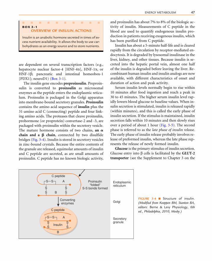
are dependent on several transcription factors (e.g.,
hepatocyte nuclear factor-4 [HNF-4a], HNF-1a, orHNF-1b; pancreatic and intestinal homeobox-1
[PDX1]; neuroD1) (Box 3-1).
The insulin gene encodes preproinsulin. Preproin-
sulin is converted to proinsulin as microsomal
enzymes as the peptide enters the endoplasmic reticu-
lum. Proinsulin is packaged in the Golgi apparatus
into membrane-bound secretory granules. Proinsulin
contains the amino acid sequence of insulin plus the
31-amino acid C (connecting) peptide and four link-
ing amino acids. The proteases that cleave proinsulin,
prohormone (or proprotein) convertase-2 and -3, are
packaged with proinsulin within the secretory vesicle.
The mature hormone consists of two chains, an achain and a b chain, connected by two disulfide
bridges (Fig. 3-4). Insulin is stored in secretory vesicles
in zinc-bound crystals. Because the entire contents of
the granule are released, equimolar amounts of insulin
and C peptide are secreted, as are small amounts of
proinsulin. C peptide has no known biologic activity,
and proinsulin has about 7% to 8% of the biologic ac-
tivity of insulin. Measurements of C peptide in the
blood are used to quantify endogenous insulin pro-
duction in patients receiving exogenous insulin, which
has been purified from C peptide.
Insulin has about a 5-minute half-life and is cleared
rapidly from the circulation by receptor-mediated en-
docytosis. It is degraded by lysosomal insulinase in the
liver, kidney, and other tissues. Because insulin is se-
creted into the hepatic portal vein, almost one half
of the insulin is degraded before leaving the liver. Re-
combinant human insulin and insulin analogs are now
available, with different characteristics of onset and
duration of action and peak activity.
Serum insulin levels normally begin to rise within
10 minutes after food ingestion and reach a peak in
30 to 45 minutes. The higher serum insulin level rap-
idly lowers blood glucose to baseline values. When in-
sulin secretion is stimulated, insulin is released rapidly
(within minutes), and this is called the early phase of
insulin secretion. If the stimulus is maintained, insulin
secretion falls within 10 minutes and then slowly rises
over a period of about 1 hour (Fig. 3-5). The second
phase is referred to as the late phase of insulin release.
The early phase of insulin release probably involves re-
lease of preformed insulin, whereas the late phase rep-
resents the release of newly formed insulin.
Glucose is the primary stimulus of insulin secretion.
Glucose entry into b cells is facilitated by the GLUT-2
transporter (see the Supplement to Chapter 3 on the
n n n n n n n n n n
BOX 3 - 1
OVERVIEW OF INSULIN ACTIONS
Insulin is an anabolic hormone secreted in times of ex-
cess nutrient availability. It allows the body to use car-
bohydrates as an energy source and to store nutrients.
C peptide
Convertingenzymes
Endoplasmicreticulum
Secretorygranule
Golgi
C peptide
Insulin
A
B
B
SS
SS
SS
SS
SS
ASS
Proinsulin“folded”
S-S bonds formed
FIGURE 3-4 n Structure of insulin.
(Modified from Koeppen BM, Stanton BA,
editors: Berne & Levy Physiology, 6th
ed., Philadelphia, 2010, Mosby.)
47ENERGY METABOLISM

Student Consult site for a list of GLUT isoforms).
Once glucose enters the b cell, it is phosphorylated
to glucose-6-phosphate by the low-affinity hexoki-
nase, glucokinase. Glucokinase is referred to as the
glucose sensor of the b cell because the rate of glucose
entry is correlated to the rate of glucose phosphoryla-
tion, which, in turn, is directly related to insulin secre-
tion. Heterozygous mutations in glucokinase are one
defect that leads to inadequate insulin release in pa-
tients with maturity-onset diabetes of youth (MODY).
Glucose-6-phosphate is metabolized by b cells, increas-
ing the intracellular ATP/ADP ratio and closing an
ATP-sensitive Kþ channel (Fig. 3-6). This results in
depolarization of the b-cell membrane, which opens
voltage-gated Ca2þ channels. Increased intracellular
Ca2þ levels activate exocytosis of secretory vesicles.
CLINICAL BOX 3-1
Heterozygous mutations in any one of the islet tran-
scription factors, as well as glucokinase (see later), re-
sult in progressively inadequate production of insulin.
This leads to MODY, which typically manifests before
25 years of age.
CLINICAL BOX 3-2
The ATP-sensitive Kþ channel is a protein complex
that contains an ATP-binding subunit called SUR1.This subunit is also activated by sulfonylurea and
meglitinide drugs, which are used as oral agents to
treat hyperglycemia in patients with partially impaired
b-cell function. Rare mutations in the ATP-sensitiveKþ channel that keep it in the open conformation,
thereby blocking glucose-induced insulin secretion,
result in early-onset diabetes.
Glucose is the primary stimulus for insulin secre-
tion. When serum glucose levels rise, insulin secretion
is stimulated; when the levels fall, insulin secretion de-
creases to baseline. Certain amino acids (leucine) and
vagal (parasympathetic) cholinergic innervation
(i.e., in response to a meal) also stimulate insulin
through increasing intracellular Ca2þ levels (see
Fig. 3-6). Long-chain FFAs also increase insulin
secretion, although to a lesser extent than glucose
and amino acids. FFAs may act through a G-protein-
coupled receptor (GPR40) on the b-cell membrane
or as a nutrient that increases ATP through b-oxidation (see Fig. 3-6).
As discussed in Chapter 2, nutrient-dependent
stimulation of insulin release is enhanced by the incre-
tin hormones glucagon-like peptide-1 (GLP1) and
gastric inhibitory peptide (GIP) and possibly other
gastrointestinal (GI) hormones (e.g., cholecystokinin
[CCK], secretin, and gastrin). These act primarily by
raising intracellular cyclic AMP (cAMP), which
amplifies the intracellular Ca2þ effects of glucose (see
Fig. 3-6). Intracellular cAMP acts both through phos-
phokinase A (PKA)-dependent and EPAC (exchange
protein activated by cAMP)–dependent pathways
0 10 20 30 40
Minutes
Plasma insulin
Glucose infusion
Plasma glucose
FIGURE 3-5 n Biphasic response of insulin secretion to
glucose infusion. (Modified from Koeppen BM, Stanton BA,
editors: Berne & Levy Physiology, 6th ed., Philadelphia, 2010,
Mosby.)
48 ENDOCRINE AND REPRODUCTIVE PHYSIOLOGY

(see Chapter 1) in b cells. Incretin hormones do not
increase insulin secretion in the absence of glucose.
Insulin secretion is inhibited by a2-adrenergic
receptors, which are activated by epinephrine (from
the adrenal medulla) and norepinephrine (from post-
ganglionic sympathetic fibers). The a2-adrenergicreceptors are coupled to a Gi-containing trimeric
G-protein complex that inhibits adenylyl cyclase
and decreases cAMP levels (see Fig. 3-6). Adrenergic
inhibition of insulin serves to protect against hypogly-
cemia, especially during exercise. Although somato-
statin from D cells inhibits both insulin and
glucagon, its physiologic role in pancreatic islet func-
tion is unclear.
Insulin Receptor The insulin receptor is a member of
the receptor tyrosine kinase (RTK) family (see
Chapter 1), which includes receptors for several other
growth factors, such as IGFs, platelet-derived growth
factor (PDGF), and epidermal growth factor (EGF).
The insulin receptor is expressed on the cell membrane
as a homodimer composed of a/b monomers
(Fig. 3-7). The a/b monomer is synthesized as one
protein, which is then proteolytically cleaved, with
the two fragments connected by a disulfide bond.
The two a/bmonomers are also held together by a di-
sulfide bond between the a subunits. The a subunits
are external to the cell membrane and contain the hor-
mone-binding site. The b subunits span the mem-
brane and contain tyrosine kinase on the cytosolic
surface.
Insulin binding to the insulin receptor induces the
b subunits to cross-phosphorylate each other on three
tyrosine residues. These phosphotyrosine residues
recruit three classes of adaptor proteins: the insulin-
receptor substrates (IRS1-4), Shc protein, and APS
protein. The IRS proteins are phosphorylated by
the tyrosine kinase activity of the insulin receptor.
The phosphotyrosine residues on IRS recruit PI3
kinase to the membrane, where it is phosphorylated
and activated by the insulin receptor. PI3 kinase con-
verts phosphoinositide-4,5-bisphosphate (PIP2) to
GlucokinaseATP
PLC
Oxidation
cAMPAC
Potentiation
GRPAD
FFAs
FFAs
Insulinsecretion
Insulin-richsecretorygranules
Glucose Glucose
Amino acids
GLUT-2
G-6-P
ATP-sensitiveK� channel
Voltage-sensitiveCa2� channel
Ca2�Ca2�
(i)IP3
Gi
Gs GLP-1 R
GLP-1 (incretin)
Epinephrinenorepinephrine
Gq
AChMR
PKA
K�(i)
K�
Sur subunit
Sulfonylurea drugs
Depolarization
� cell
�2 adrenergic receptor
FIGURE 3-6 n Regulation of insulin secretion from b cells by nutrients (glucose, amino acids, FFAs) and the hormones/
neurotransmitters, glucagon-like peptide-1 (GLP1), epinephrine, norepinephrine, and acetylcholine (ACh).
49ENERGY METABOLISM
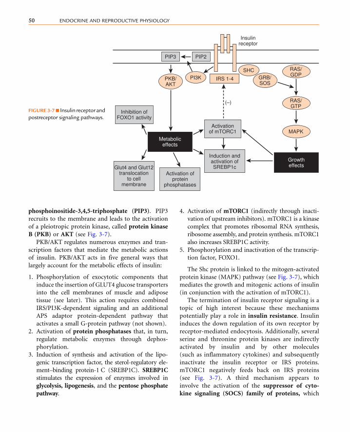
phosphoinositide-3,4,5-triphosphate (PIP3). PIP3
recruits to the membrane and leads to the activation
of a pleiotropic protein kinase, called protein kinase
B (PKB) or AKT (see Fig. 3-7).
PKB/AKT regulates numerous enzymes and tran-
scription factors that mediate the metabolic actions
of insulin. PKB/AKT acts in five general ways that
largely account for the metabolic effects of insulin:
1. Phosphorylation of exocytotic components that
induce the insertion of GLUT4 glucose transporters
into the cell membranes of muscle and adipose
tissue (see later). This action requires combined
IRS/PI3K-dependent signaling and an additional
APS adaptor protein-dependent pathway that
activates a small G-protein pathway (not shown).
2. Activation of protein phosphatases that, in turn,
regulate metabolic enzymes through dephos-
phorylation.
3. Induction of synthesis and activation of the lipo-
genic transcription factor, the sterol-regulatory ele-
ment–binding protein-1 C (SREBP1C). SREBP1C
stimulates the expression of enzymes involved in
glycolysis, lipogenesis, and the pentose phosphate
pathway.
4. Activation of mTORC1 (indirectly through inacti-
vation of upstream inhibitors). mTORC1 is a kinase
complex that promotes ribosomal RNA synthesis,
ribosome assembly, and protein synthesis. mTORC1
also increases SREBP1C activity.
5. Phosphorylation and inactivation of the transcrip-
tion factor, FOXO1.
The Shc protein is linked to the mitogen-activated
protein kinase (MAPK) pathway (see Fig. 3-7), which
mediates the growth and mitogenic actions of insulin
(in conjunction with the activation of mTORC1).
The termination of insulin receptor signaling is a
topic of high interest because these mechanisms
potentially play a role in insulin resistance. Insulin
induces the down regulation of its own receptor by
receptor-mediated endocytosis. Additionally, several
serine and threonine protein kinases are indirectly
activated by insulin and by other molecules
(such as inflammatory cytokines) and subsequently
inactivate the insulin receptor or IRS proteins.
mTORC1 negatively feeds back on IRS proteins
(see Fig. 3-7). A third mechanism appears to
involve the activation of the suppressor of cyto-
kine signaling (SOCS) family of proteins, which
IRS 1-4
(–)
PIP2
Insulinreceptor
PIP3
Metaboliceffects
Growtheffects
Inhibition ofFOXO1 activity
Activationof mTORC1
Glut4 and Glut12translocation
to cellmembrane
Activation ofprotein
phosphatases
Induction andactivation ofSREBP1c
MAPK
RAS/GDP
RAS/GTP
PKB/AKT
GRB/SOS
PI3KSHC
FIGURE 3-7n Insulin receptorand
postreceptor signaling pathways.
50 ENDOCRINE AND REPRODUCTIVE PHYSIOLOGY

reduces activity or levels of the insulin receptor and
IRS proteins.
Glucagon
Glucagon is an important counter-regulatory hor-
mone that increases blood glucose levels through its ef-
fects on liver glucose output. Glucagon promotes the
production of glucose through elevated glycogenolysis
and gluconeogenesis and through decreased glycolysis
and glycogen synthesis. Glucagon also inhibits hepatic
FFA synthesis from glucose. Glucagon also maintains
blood glucose indirectly through stimulation of keto-
genesis, which provides an alternative energy source
that leads to glucose sparing in many tissues.
Glucagon Structure, Synthesis, and Secretion As dis-
cussed in Chapter 2, glucagon is a member of the se-
cretin gene family. Preproglucagon is proteolytically
cleaved in the pancreatic islet a cell in a cell-specific
manner to produce the 29-amino acid glucagon (refer
to Fig. 2-10 in Chapter 2). Glucagon is highly con-
served among mammals.
Like insulin, glucagon circulates in an unbound form
and has a short half-life (about 6minutes). The predom-
inant site of glucagon degradation is the liver, which de-
grades asmuch as 80%of the circulating glucagon in one
pass. Because glucagon (either from the pancreas or
the gut) enters the hepatic portal vein and is carried to
the liver before reaching the systemic circulation, a large
portionof the hormonenever reaches the systemic circu-
lation. The liver is the primary target organ of glucagon,
with lesser effects on adipose tissue.
As discussed later, glucagon opposes the actions
of insulin. Thus, several factors that stimulate in-
sulin inhibit glucagon. Indeed, it is the insulin-to-
glucagon ratio that determines the net flow of hepatic
metabolic pathways. A major stimulus for glucagon
secretion is a drop in blood glucose, which is primar-
ily an indirect effect of the removal of inhibition by
insulin (Fig. 3-8). Circulating catecholamines, which
inhibit insulin secretion through a2-adrenergicreceptors, stimulate glucagon secretion through b2-adrenergic receptors (see Fig. 3-8). Serum amino
acids also stimulate glucagon secretion. This means
that a protein meal will increase postprandial levels
of glucagon along with insulin, thereby protecting
against hypoglycemia. In contrast, a carbohydrate-
only meal stimulates insulin secretion and inhibits
glucagon secretion.
Glucagon Receptor The glucagon receptor is a 7-trans-
membrane receptor primarily linked to Gs-containing
heterotrimeric G-protein complex (see Chapter 1).
Consequently, glucagon increases intracellular cAMP
levels in the liver. The increase in cAMP initiates the
cascade of metabolic changes associated with enzyme
phosphorylation. As discussed earlier, activation of
opposing protein phosphatases is one of the general
pathways of insulin signaling.
Epinephrine and Norepinephrine
The other major counter-regulatory factors are the
catecholamines epinephrine and norepinephrine.
Epinephrine and norepinephrine are secreted by the
adrenal medulla (see Chapter 7), whereas only nor-
epinephrine is released from postganglionic sympa-
thetic nerve endings. The direct metabolic actions
of catecholamines are mediated primarily by b2-
and b3-adrenergic receptors located on muscle,
adipose, and liver. Like the glucagon receptor,
b-adrenergic receptors are linked to a Gs signaling
pathway that increases intracellular cAMP. Epineph-
rine also promotes glycogenolysis and gluconeogene-
sis through the a1 adrenergic receptor, which is
coupled to the Gq/IP3/DAG signaling pathway.
Catecholamines are released from sympathetic nerve
endings and the adrenal medulla in response to de-
creased glucose concentrations, stress or alarm, and ex-
ercise. Decreased glucose levels (i.e., hypoglycemia) are
primarily sensed by hypothalamic neurons, which ini-
tiate a sympathetic response to release catecholamines.
Catecholamines circulate in the blood as free hor-
mones, and both circulating and tissue catecholamines
are rapidly enzymatically inactivated (see Chapter 7).
METABOLIC HOMEOSTASIS: THEINTEGRATED OUTCOME OFHORMONAL AND SUBSTRATE/PRODUCT REGULATION OFMETABOLIC PATHWAYS
The hormonal regulation of themajor metabolic path-
ways, with emphasis on key regulated enzymes and
transporters, is presented in this section.Three organs
51ENERGY METABOLISM

play predominant roles in energy use and storage: the
liver, skeletal muscle, and adipose tissue. A fourth or-
gan, the hypothalamus, through its own metabolic
pathways and in response to hormones and nutrients,
also plays a key role in the acquisition, use, and
storage of fuels. We will also introduce additional
hormones that originate from the GI tract and
adipose tissue and modulate energy metabolism. As
stated earlier, an overview of key metabolic pathways
and lipoprotein metabolism is available on Student
Consult (see the Supplement to Chapter 3 on the
Student Consult site).
Energy Metabolism During the Fed State
During the fed state, insulin drives the storage of
excess calories and the new synthesis of cellular mac-
romolecules and organelles.
Hepatic Metabolism of Nutrients in the Fed StateAnatomic Considerations The liver is anatomically
and functionally situated between the GI tract
and the heart. The main veins that drain the GI tract
of its ingested nutrients all converge to form the
hepatic portal vein. A portal vein is one that ends
in a capillary bed before reaching the heart. In the
Inhibit secretion
Inhibit secretion
Inhibit secretion
Decrease level
Increase level
Stimulate secretion
Stimulate secretion
Bloodglucose
• Increase glucose uptake and utilization (muscle and adipose)
• Decrease hepatic glucose production• Increase hepatic conversion of glucose to glycogen and lipids
• Inhibit hepatic ketogenesis• Inhibit HSL and decrease release of FFAs from adipose
• Increase hepatic glucose production: Glycogenolysis Gluconeogenesis
• Decrease hepatic conversion of glucose to glycogen or lipids
• Decrease uptake by adipose and muscle• Increase hepatic ketogenesis• Increase release of gluconeogenic substrates from muscle and adipose
• Increase HSL and release of FFAs from adipose
Glucagon
SympathoadrenalactivityCatecholamines
β Cell Insulin
α Cell
FIGURE 3-8 n Inter-
action of insulin, glu-
cagon, and catecho-
lamines in the regula-
tion of each other and
of blood glucose (the
primary secretogogue
for insulin). (Modified
from Koeppen BM, Stan-
ton BA, editors: Berne
& Levy Physiology, 6th
ed., Philadelphia, 2010,
Mosby.)
52 ENDOCRINE AND REPRODUCTIVE PHYSIOLOGY

case of the hepatic portal vein, it receives the
venous drainage of the GI tract and then ends at
the hepatic sinusoids (a sinusoid is a discontinuous
capillary). This means that the liver is the first
organ to receive the ingested carbohydrates and
amino acids before they reach the general circula-
tion (TGs bypass the liver at first because they enter
lymphatics as chylomicron particles). The liver is
also the first organ to respond to hormones and
cytokines from the pancreas and intra-abdominal
adipose tissue.
The Big Picture Glucose enters the liver and is
stored as glycogen (glycogen synthesis); the normal
maximal amount of glycogen in the liver is about 80 g
(Fig. 3-9). Glucose is then diverted into glycolysis,
generating pyruvate. Pyruvate enters the tricarbox-
ylic acid (TCA) cycle to produce reducing equiva-
lents (NADH, FADH2) for use in ATP synthesis
through oxidative phosphorylation, but excess
ATP and NADH will allow the diversion of citrate
into the cytoplasm, where it is converted to malonyl
coenzyme A (CoA) and ultimately fatty acyl CoA (de
novo lipogenesis). Citrate can also provide carbons
for cholesterol synthesis (not shown). Some glucose
also enters the pentose phosphate pathway (PPP),
thereby generating NADPH, which is required for
lipogenesis and defense against oxidative damage.
Fatty acyl CoAs are modified (e.g., desaturated)
and ultimately esterified to glycerol-3-P to produce
TG. However, the export of TG and other lipids as
very-low-density lipoproteins (VLDLs) is sup-
pressed during the fed state. The breakdown of glyco-
gen (glycogenolysis) to glucose and the production
of glucose from 3-carbon glucogenic molecules (glu-
coneogenesis) are both suppressed by insulin,
thereby reducing hepatic glucose production (see
Fig. 3-9). Fatty acyl oxidation and ketogenesis are
also suppressed (see Fig. 3-9).
Glycogenolysis
Glycolysis
G-6-Phosphatase
PPP
PDHPC
Glucokinase
� oxidation
LipogenesisATP
BG
NAPDH
FC and CEs
KBs KBs
TGs VLDL VLDL
PLs
CR
Glucose
Pyruvate
AcCoA
MAL CoA FACoA
Glucose-6-P
Glycogen
Glycerol
Glycogensynthesis
Gluco-neogenesis
TCAcycle
OXPHOS
LactatealanineOxalo-
acetate
Aminoacids
GLUT2
LDLLRP1HSPG
SinusoidHepatocyte
FIGURE 3-9 nOverview of metabolic pathways in the liver during the fed stage (black arrows) and the fast stage (orange arrows).
53ENERGY METABOLISM

Amino acids are assembled into proteins through
activation of mTORC1 by both amino acids and
insulin. Amino acids are also used for the synthesis
of other macromolecules (e.g., heme, nucleotides).
Amino acids, in particular glutamine and glutamic
acid, also provide carbons (as a-ketoglutarate)to the TCA cycle (anaplerosis) as a replacement
for carbons exiting the TCA cycle as citrate
(cataplerosis).
As the fed state progresses, chylomicron remnants
enter the hepatocyte through receptor-mediated en-
docytosis, releasing TG to be packaged into VLDL
later (see Fig. 3-9). Circulating FFAs (as FFA-albumin
complexes) are diminished during the fed state (see
adipose tissue below), so the uptake of FFAs by the
liver is minimal.
All of these metabolic pathways are orchestrated by
insulin. As discussed earlier, a major insulin-activated
transcription factor is SREBP1C. The genes and en-
zymes stimulated by SREBP1C are listed in Table 3-1.
Hormonal Regulation of Key Reactions inthe Liver During the Fed State
See the Supplement to Chapter 3 on the Student Con-
sult site for additional figures.
Intracellular Transport and Trapping ofMonosaccharides Ingested monosaccharides (glucose,
fructose, and galactose) reach the liver sinusoids and
flow down their concentration gradient into hepato-
cytes through a bidirectional, facilitative transporter,
called GLUT-2 (Fig. 3-10). GLUT2 is a low-affinity,
high-capacity facilitative transporter well suited to
TABLE 3-1
Hepatic Genes Stimulated by SREBP1C
GENE ENZYME PATHWAY
GCKR Glucokinase (hexokinase-4) Glycolysis
PKLR Pyruvate kinase (liver and red
blood cells)
Glycolysis
ACLY Adenosine triphosphate and
citrate lyase
De novo lipogenesis
ACACA Acetyl coenzyme A (CoA)
carboxylase-1(acetyl CoA
carboxylase-a)
De novo lipogenesis
ACACB Acetyl CoA carboxylase-2
(acetyl CoA carboxylase-b)De novo lipogenesis
FASN Fatty acid synthase De novo lipogenesis
G6PD Glucose-6-phosphate
dehydrogenase
Pentose phosphate pathway
(hexose monophosphate
shunt)
SREBP1c
SREBP1c
GK GKGKRP
PKB/AKT
Glucose
Glucose-6-P
Fructose-6-P
FOXO1
FOXO1
Sinusoid
Cytoplasm
Nucleus
GK geneexpression
G-6-P’ASEgene exp
G-6-P’ASE
GLUT2
FIGURE 3-10 n Regulation of
glucokinase (GK) and glucose-6-
phosphatase (G-6-P’ASE) during
fed (black arrows) and fast (orange
arrows) stages.
54 ENDOCRINE AND REPRODUCTIVE PHYSIOLOGY

moving high amounts of absorbed glucose during and
soon after a meal. GLUT-2 expression or localization
at the cell membrane is not regulated.
Intrahepatic glucose (galactose is efficiently con-
verted to glucose) is trapped by phosphorylation to
glucose-6-phosphate by the enzyme, glucokinase
(GK). As discussed previously for the pancreatic bcells, glucokinase has a low affinity and high Vmax.
Thus, glucokinase, which is a member of the hexoki-
nase family of enzymes, is paired with GLUT-2 in the
b cell and hepatocyte. The glucokinase reaction is
irreversible and is opposed by the enzyme glucose-
6-phosphatase (see Fig. 3-10).
Hepatic glucokinase is tightly regulated by metab-
olites and by insulin (see Fig. 3-10). In the absence of
glucose, glucokinase is sequestered in the nucleus by
binding to glucokinase regulatory protein (GKRP).
Increased glucose promotes the dissociation of
glucokinase from its regulatory protein and transloca-
tion into the cytoplasm, where it converts glucose
to glucose-6-phosphate. The downstream product,
fructose-6-phosphate, inhibits glucokinase by pro-
moting its sequestration.
Insulin, acting through the PKB/AKT signaling
pathway, stimulates the new synthesis of glucokinase
through activation of SREBP1C. Insulin drives the nu-
clear localization of SREBP1C, which in turn drives
glucokinase gene expression (see Fig. 3-10). Insulin
also inhibits expression of glucose-6-phosphatase
through the inactivation of FOXO1. Nuclear FOXO1
stimulates glucose-6-phosphatase gene expression.
PKB/AKT phosphorylates FOXO1, thereby sequester-
ing the transcription factor in the cytoplasm (see
Fig. 3-10).
Storage of Glucose as Glycogen Insulin increases theac-
tivity of glycogen synthase in several ways (Fig. 3-11).
Insulin indirectly increases glycogen synthase through
increased expression of glucokinase because high levels
of glucose-6-phosphate allosterically increase glycogen
synthase activity.
Insulin also increases the activity of protein
phosphatase-1 (see Fig. 3-11), which dephosphorylates
and thereby activates glycogen synthase. Insulin also
inactivates glycogen synthase kinase-3, which, in
turn, promotes the accumulation of dephosphorylated
GK
GlucoseGlucose-6-P
Glucose-1-PPKA
ProteinP’TASE
ProteinP’TASE
Insulin
GSK3�
Glycogensynthase
Glycogensynthase-P
Glycogen(80 g max)
(–)
Glucoseglucose-6-P
ATP
Glycogenphosphorylase A-P
Glycogenphosphorylase B
GlucagonCatecholamines
Phosphorylasekinase B
Phosphorylasekinase A-P
FIGURE 3-11 n Regulation
of glycogen synthesis (black
arrows, fed) and glycogenolysis
(orange arrows, fast).
55ENERGY METABOLISM

(active) glycogen synthase. Insulin may also prevent the
futile cycle of glycogen synthesis and glycogenolysis
through inhibition of glycogen phosphorylase (see
Fig. 3-11). Insulin-activated protein phosphatase-1 de-
phosphorylates and inactivates glycogen phosphory-
lase as well as phosphorylase kinase. Glycogen
phosphorylase is also inactivated by glucose, glucose-
6-phosphate, and ATP.
Glycolysis Glucose is also metabolized through the
glycolytic pathway. Once glycogen stores are filled,
glycogen synthesis ceases, further increasing the flux
of glucose through glycolysis. There are two hor-
monally regulated cytoplasmic enzymes within gly-
colysis (after glucokinase): phosphofructokinase-1
(PFK1) and liver-specific pyruvate kinase (PK-L).
Phosphofructokinase-1 ThePFK1 reaction converts
fructose-6-phosphate to fructose-1,6-bisphosphate
(Fig. 3-12).This reaction is irreversible, requiring a sep-
arate enzyme, fructose-1,6-bisphosphatase, to catalyze
the reverse reaction. The PFK1 reaction is one of the
most tightly regulated reactions in metabolism, and
it is also one that does not involve SREBP1C. During
the fed state, PFK1 activity is moderated by high levels
of ATP and cytoplasmic citrate (as discussed earlier,
citrate moves from the mitochondria to the cytoplasm
at times when the TCA cycle is slowed down by abun-
dant ATP andNADH during the fed state). In contrast,
AMP allosterically activates PFK-1.
Another major allosteric activator of PFK1 is the
glycolytic side product, fructose-2,6-bisphosphate
(see Fig. 3-12). Fructose-2,6-bisphosphate is produced
from fructose-6-phosphate by the phosphofructose
kinase-2 (PFK2) function of the bifunctional enzyme,
PFK2/fructose-2,6-bisphosphatase. The PFK2 func-
tion of the enzyme is active when the enzyme is
dephosphorylated by an insulin-activated protein
phosphatase-1 (seeFig. 3-12).The fructose-2,6-bispho-
sphatase function is active when the enzyme is
phosphorylated by a glucagon- and catecholamine-
activated protein kinase A (see Fig. 3-12). Through the
generation of fructose-2,6-bisphosphate, insulin par-
tially offsets the inhibitory actions of ATP and cytoplas-
mic citrate on PFK1. This allows glucose to continue
through the glycolytic pathway during the fed state.
The opposing reaction that converts fructose-1,6-
bisphosphate to fructose-6-phosphate is catalyzed by
thegluconeogenic enzyme, fructose-1,6-bisphosphatase.
Fructose-2,6-bisphosphate is an allosteric inhibitor of
fructose-1,6-bisphosphatase (see Fig. 3-12).
Pyruvate Kinase The pyruvate kinase reaction is
also irreversible and converts phosphoenol pyruvate
to pyruvate (Fig. 3-13). This reaction is coupled to
ATP synthesis and accounts for the net two ATPs pro-
duced by anaerobic glycolysis. Pyruvate kinase is allo-
sterically activated by the upstream intermediate,
fructose-1,6-bisphosphate (see previous section). In
this way, insulin activates pyruvate kinase indirectly
Fructose-1,6-bisP
Fructose-6-PFructose-2,6-bisP
PKAProtein
phosphatase
PFK2
PFK1 (–)
Insulin Glucagoncatecholamines
F-2,6-bisP’TASE
F-1,6-bisP’TASE
FIGURE 3-12 n Hormonal regulation of
phosphofructose kinase-1 (PFK1) and fructose-
1,6-bisphosphatase (F-1,6-bisP’TASE) by levels
of fructose-2,6-bisphosphate. The dual activity
enzyme, PFK2/F-2,6-bisP’TASE, acts as the
kinase when dephosphorylated and as a
phosphatase when phosphorylated. Dashed
lines indicate allosteric inhibition.
56 ENDOCRINE AND REPRODUCTIVE PHYSIOLOGY

through a feed-forward mechanism that is initiated by
the insulin-induced production of fructose-2,6-
bisphosphate (see Fig. 3-13). Pyruvate kinase is also
regulated by phosphorylation. Again, insulin activates
pyruvate kinase through insulin-dependent dephos-
phorylation by protein phosphatase. Insulin also has
a longer-term effect on pyruvate kinase gene transcrip-
tion through the PKB/AKT-SREBP1C pathway (see
Fig. 3-13).
Entry of Pyruvate into the Tricarboxylic Acid CycleGlycolysis generates pyruvate, which can be con-
verted to lactate and Hþ by lactate dehydrogenase.
This replenishes NADþ levels required for glycolysis
to continue, and lactate dehydrogenase is activated
by a low NADþ/NADH ratio. In hepatocytes with ac-
tive mitochondria, NADþ is continually regenerated
from NADH through the electron transport chain.
This promotes the entry of pyruvate into the mito-
chondria, where it enters the TCA cycle. This requires
the conversion of pyruvate into acetyl CoA, as cata-
lyzed by the pyruvate dehydrogenase (PDH) com-
plex. PDH kinase is associated with the PDH
complex, and phosphorylation of the PDH complex
inhibits activity. PDH kinase is activated by ATP, ace-
tyl CoA, and NADH. In addition, the PDH complex
itself is inhibited by NADH and acetyl CoA. However,
in the face of high glycolytic flux during the fed state,
abundant pyruvate acts as a potent inhibitor of PDH
kinase, thereby activating PDHand promoting the con-
version of pyruvate to acetyl CoA (Fig. 3-14). In
addition, insulin stimulates PDH phosphatase, which
dephosphorylates and activates PDH.
Tricarboxylic Acid Cycle Acetyl CoA (2 carbons) is con-
densed with oxaloacetate (4 carbons) to form citrate, as
catalyzed by citrate synthase. In the absence of abundant
energy, citrate is cycled through seven reactions back to
oxaloacetate, generating 3 NADH and 1 FADH2, which
will generate 11 ATPs through processing by the electron
transport chain and oxidative phosphorylation.
In the fed state, however, ATP and NADH are
abundant and allosterically inhibit two TCA reactions
after citrate is formed. The slowing of the TCA cycle
allows citrate to accumulate and ultimately flow
through citrate transporters into the cytoplasm. Cyto-
plasmic citrate is the first intermediate in de novo
lipogenesis (see Fig. 3-14).
De Novo Lipogenesis De novo lipogenesis normally
occurs only in the liver, adipose tissue, and mam-
mary glands. De novo synthesis of fatty acyl CoAs
involves three hormonally regulated enzymes: ATP-
citrate lyase (ACLY), acetyl CoA carboxylase (ACC1
and ACC2), and fatty acid synthase (FASN). Cyto-
plasmic citrate is converted to acetyl CoA, and acetyl
CoA can be used for fatty acyl CoA synthesis (see
Fig. 3-14) or cholesterol synthesis (not shown).
Pyruvate
Fructose-1,6-bisP
PKA
Pyruvate kinasegene expression
Pyruvatekinase-P
Proteinphosphatase
Phosphoenolpyruvate
Pyruvatekinase
SREBP1c
PKB/AKTInsulin
Glucagoncatecholamines
FIGURE 3-13 n Regulation of pyruvate
kinase during the fed (black arrows) and
fast (orange arrows) states. Fructose-1,6-
bisphosphate is an allosteric activator
of pyruvate kinase.
57ENERGY METABOLISM
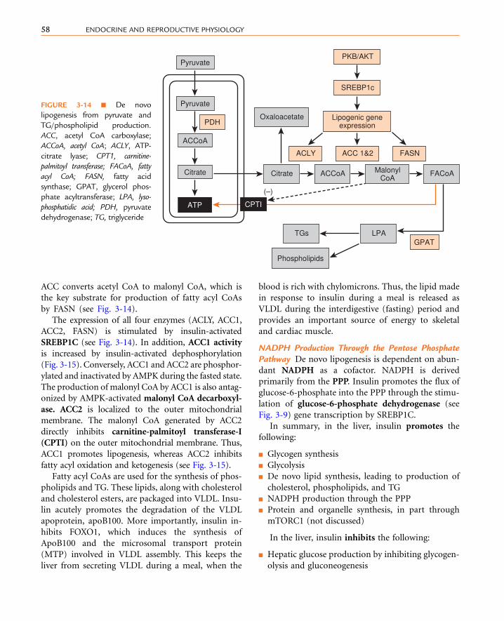
ACC converts acetyl CoA to malonyl CoA, which is
the key substrate for production of fatty acyl CoAs
by FASN (see Fig. 3-14).
The expression of all four enzymes (ACLY, ACC1,
ACC2, FASN) is stimulated by insulin-activated
SREBP1C (see Fig. 3-14). In addition, ACC1 activity
is increased by insulin-activated dephosphorylation
(Fig. 3-15). Conversely, ACC1 and ACC2 are phosphor-
ylated and inactivated by AMPK during the fasted state.
The production of malonyl CoA by ACC1 is also antag-
onized by AMPK-activated malonyl CoA decarboxyl-
ase. ACC2 is localized to the outer mitochondrial
membrane. The malonyl CoA generated by ACC2
directly inhibits carnitine-palmitoyl transferase-I
(CPTI) on the outer mitochondrial membrane. Thus,
ACC1 promotes lipogenesis, whereas ACC2 inhibits
fatty acyl oxidation and ketogenesis (see Fig. 3-15).
Fatty acyl CoAs are used for the synthesis of phos-
pholipids and TG. These lipids, along with cholesterol
and cholesterol esters, are packaged into VLDL. Insu-
lin acutely promotes the degradation of the VLDL
apoprotein, apoB100. More importantly, insulin in-
hibits FOXO1, which induces the synthesis of
ApoB100 and the microsomal transport protein
(MTP) involved in VLDL assembly. This keeps the
liver from secreting VLDL during a meal, when the
blood is rich with chylomicrons. Thus, the lipid made
in response to insulin during a meal is released as
VLDL during the interdigestive (fasting) period and
provides an important source of energy to skeletal
and cardiac muscle.
NADPH Production Through the Pentose PhosphatePathway De novo lipogenesis is dependent on abun-
dant NADPH as a cofactor. NADPH is derived
primarily from the PPP. Insulin promotes the flux of
glucose-6-phosphate into the PPP through the stimu-
lation of glucose-6-phosphate dehydrogenase (see
Fig. 3-9) gene transcription by SREBP1C.
In summary, in the liver, insulin promotes the
following:
n Glycogen synthesis
n Glycolysis
n De novo lipid synthesis, leading to production of
cholesterol, phospholipids, and TG
n NADPH production through the PPP
n Protein and organelle synthesis, in part through
mTORC1 (not discussed)
In the liver, insulin inhibits the following:
n Hepatic glucose production by inhibiting glycogen-
olysis and gluconeogenesis
Citrate
ACCoA
Pyruvate
Pyruvate
ATP
Lipogenic geneexpression
Phospholipids
SREBP1c
ACC 1&2
MalonylCoA
FACoAACCoA
TGs LPA
Citrate
(–)
Oxaloacetate
ACLY
PDH
GPAT
FASN
PKB/AKT
CPTI
FIGURE 3-14 n De novo
lipogenesis from pyruvate and
TG/phospholipid production.
ACC, acetyl CoA carboxylase;
ACCoA, acetyl CoA; ACLY, ATP-
citrate lyase; CPT1, carnitine-
palmitoyl transferase; FACoA, fatty
acyl CoA; FASN, fatty acid
synthase; GPAT, glycerol phos-
phate acyltransferase; LPA, lyso-
phosphatidic acid; PDH, pyruvate
dehydrogenase; TG, triglyceride
58 ENDOCRINE AND REPRODUCTIVE PHYSIOLOGY
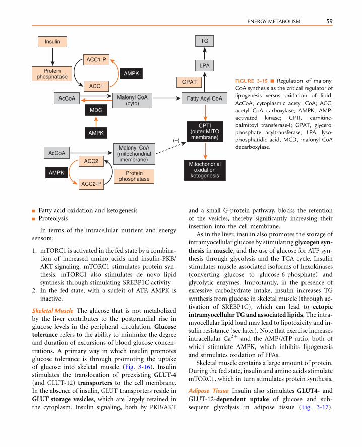
n Fatty acid oxidation and ketogenesis
n Proteolysis
In terms of the intracellular nutrient and energy
sensors:
1. mTORC1 is activated in the fed state by a combina-
tion of increased amino acids and insulin-PKB/
AKT signaling. mTORC1 stimulates protein syn-
thesis. mTORC1 also stimulates de novo lipid
synthesis through stimulating SREBP1C activity.
2. In the fed state, with a surfeit of ATP, AMPK is
inactive.
Skeletal Muscle The glucose that is not metabolized
by the liver contributes to the postprandial rise in
glucose levels in the peripheral circulation. Glucose
tolerance refers to the ability to minimize the degree
and duration of excursions of blood glucose concen-
trations. A primary way in which insulin promotes
glucose tolerance is through promoting the uptake
of glucose into skeletal muscle (Fig. 3-16). Insulin
stimulates the translocation of preexisting GLUT-4
(and GLUT-12) transporters to the cell membrane.
In the absence of insulin, GLUT transporters reside in
GLUT storage vesicles, which are largely retained in
the cytoplasm. Insulin signaling, both by PKB/AKT
and a small G-protein pathway, blocks the retention
of the vesicles, thereby significantly increasing their
insertion into the cell membrane.
As in the liver, insulin also promotes the storage of
intramyocellular glucose by stimulating glycogen syn-
thesis in muscle, and the use of glucose for ATP syn-
thesis through glycolysis and the TCA cycle. Insulin
stimulates muscle-associated isoforms of hexokinases
(converting glucose to glucose-6-phosphate) and
glycolytic enzymes. Importantly, in the presence of
excessive carbohydrate intake, insulin increases TG
synthesis from glucose in skeletal muscle (through ac-
tivation of SREBP1C), which can lead to ectopic
intramyocellular TG and associated lipids. The intra-
myocellular lipid load may lead to lipotoxicity and in-
sulin resistance (see later). Note that exercise increases
intracellular Ca2þ and the AMP/ATP ratio, both of
which stimulate AMPK, which inhibits lipogenesis
and stimulates oxidation of FFAs.
Skeletal muscle contains a large amount of protein.
During the fed state, insulin and amino acids stimulate
mTORC1, which in turn stimulates protein synthesis.
Adipose Tissue Insulin also stimulates GLUT4- and
GLUT-12-dependent uptake of glucose and sub-
sequent glycolysis in adipose tissue (Fig. 3-17).
AMPK
MDC
AMPK
AMPK
Proteinphosphatase
Proteinphosphatase
AcCoA
AcCoA
Fatty Acyl CoA
(–)
LPA
TG
Malonyl CoA(cyto)
Insulin
ACC1-P
ACC1
ACC2
ACC2-P
GPAT
CPTI(outer MITOmembrane)
Mitochondrialoxidation
ketogenesis
Malonyl CoA(mitochondrial
membrane)
FIGURE 3-15 n Regulation of malonyl
CoA synthesis as the critical regulator of
lipogenesis versus oxidation of lipid.
AcCoA, cytoplasmic acetyl CoA; ACC,
acetyl CoA carboxylase; AMPK, AMP-
activated kinase; CPTI, carnitine-
palmitoyl transferase-I; GPAT, glycerol
phosphate acyltransferase; LPA, lyso-
phosphatidic acid; MCD, malonyl CoA
decarboxylase.
59ENERGY METABOLISM

Adipose tissue uses glycolysis for energy needs, but
also for the generation of glycerol-3-phosphate,
which is required for the reesterification of FFAs into
TGs (see Fig. 3-17).
Dietary FFAs reach the adipocytes in the form of
TGs within chylomicrons (refer to the Supplement
to Chapter 3 on the Student Consult site for a
description of lipoproteins and their functions). The
TGs within the chylomicrons are digested by the extra-
cellular lipase lipoprotein lipase. Insulin stimulates
the expression of lipoprotein lipase within adipocytes
and its exocytosis and migration to the apical side of
endothelia of adipose capillary beds (Fig. 3-18). This
action of insulin thereby promotes the release of FFAs
from chylomicrons within adipose tissue. Insulin also
stimulates translocation of fatty acid transport pro-
teins (e.g., CD36) into the cell membrane. Fatty acid
transport proteins facilitate the movement of FFAs
into the adipocyte and the activation of FFAs by their
conversion to fatty acyl CoAs. Insulin stimulates
glycolysis in adipocytes, which generates the glycerol-
3-phosphate required for reesterification of FFAs
into TGs (see Fig. 3-17). Insulin stimulates protein
phosphatases, which, in turn, dephosphorylate and
inactivate hormone-sensitive lipase (HSL) and dephos-
phorylate and stabilize TG droplet coat proteins
(e.g., perilipins) (see Fig. 3-18).
Insulin signaling also activates a nuclear receptor,
called peroxisome proliferator-activated receptor-g(PPARg). PPARg promotes the differentiation of pre-
adipocytes into adipocytes. Expansion of the mature
adipocyte population can alleviate insulin resistance
by expanding the cellular compartment where TG can
be stored safely.
As in liver and skeletal muscle, insulin and amino
acids promote the protein synthesis through activa-
tion of mTORC1.
Overview of EnergyMetabolismDuring theFasting State
Several hours after a meal, nutrient levels fall, leading
to lower levels of insulin secretion. Consequently, the
actions of insulin on hepatic, muscle, and adipose
tissue are attenuated. The decrease in insulin
also relieves inhibition of glucagon secretion (see
Glycolysis
PDH
TCA cycle
OX/PHOS
GlucoseGlucoseGlucose-6-P
Pyruvate
Glycogen
AC CoA
GLUTstoragevesicle
Insulinsignaling
Glycogensynthesis
Insulinsignaling GLUT4
ECF
ATP
Myocyte
GLUT4
GLUT4
GLUT4
FIGURE 3-16 n Regulation of
glucose uptake and use in skeletal
muscle during the fed stage.
60 ENDOCRINE AND REPRODUCTIVE PHYSIOLOGY
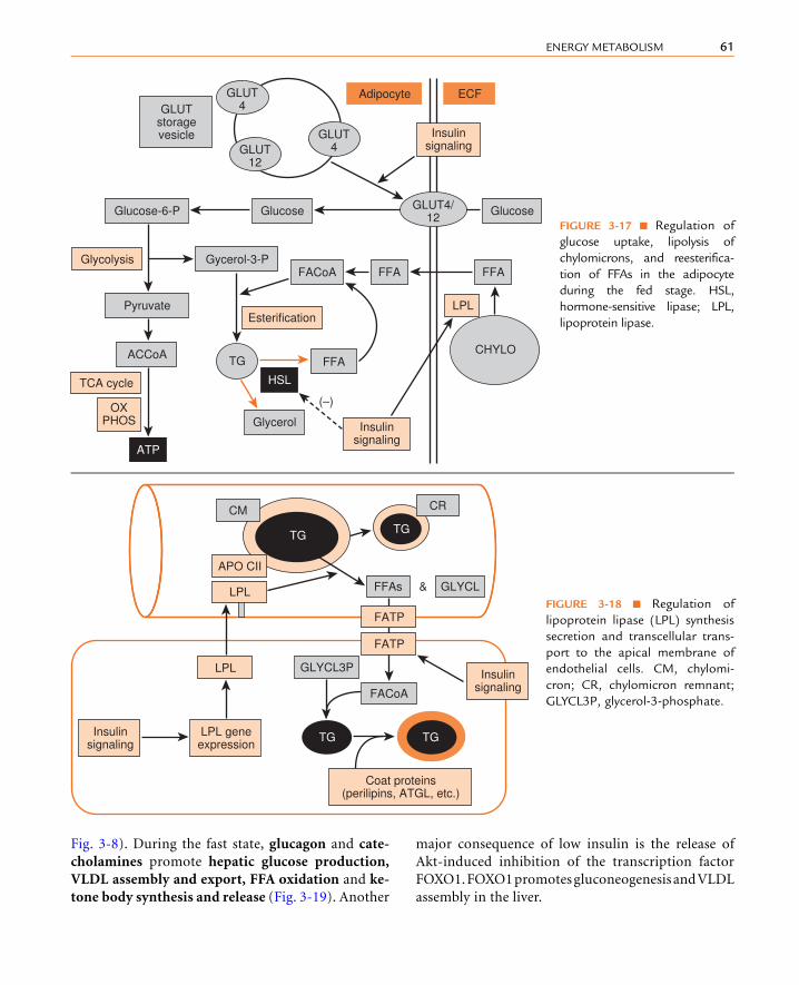
Fig. 3-8). During the fast state, glucagon and cate-
cholamines promote hepatic glucose production,
VLDL assembly and export, FFA oxidation and ke-
tone body synthesis and release (Fig. 3-19). Another
major consequence of low insulin is the release of
Akt-induced inhibition of the transcription factor
FOXO1.FOXO1promotesgluconeogenesis andVLDL
assembly in the liver.
LPLEsterification
Glycolysis
TCA cycle
Glucose
FFAFFA
ATP
HSL
FACoA
FFA
(–)
CHYLO
Glucose
Glycerol
Gycerol-3-P
TG
Glucose-6-P
Pyruvate
ACCoA
GLUTstoragevesicle Insulin
signaling
Insulinsignaling
OXPHOS
ECFAdipocyteGLUT4
GLUT12
GLUT4
GLUT4/12
FIGURE 3-17 n Regulation of
glucose uptake, lipolysis of
chylomicrons, and reesterifica-
tion of FFAs in the adipocyte
during the fed stage. HSL,
hormone-sensitive lipase; LPL,
lipoprotein lipase.
TG TG
TGTG
Insulinsignaling
Insulinsignaling
LPL geneexpression
Coat proteins(perilipins, ATGL, etc.)
LPL
LPL
APO CII
CM CR
FFAs GLYCL
GLYCL3P
FACoA
&
FATP
FATP
FIGURE 3-18 n Regulation of
lipoprotein lipase (LPL) synthesis
secretion and transcellular trans-
port to the apical membrane of
endothelial cells. CM, chylomi-
cron; CR, chylomicron remnant;
GLYCL3P, glycerol-3-phosphate.
61ENERGY METABOLISM

Muscle fibers import minimal amounts of glucose
because of basal levels of GLUT4 in their plasma mem-
brane. Muscle fibers switch from using glucose to using
abundant FFAs, as well as ketone bodies in a longer
fast, for ATP synthesis (Fig. 3-20). Skeletal muscle also
releases lactate and amino acids. During exercise or in
response to a fight-or-flight situation, muscle mobilizes
its glycogen stores for ATP production (see Fig. 3-20).
The adipocytes have low GLUT-4 levels at their
cell membrane and consequently cannot generate
glycerol-3-phosphate for TG synthesis. The low insu-
lin and high catecholamine levels promote lipolysis of
TG and the release of FFAs into the blood (Fig. 3-21).
Adipocytes will also switch to FFAs and ketone bodies
for ATP synthesis.
Glycogen
Glucose
Gluconeogenesis
Used for ATP
KBs
VLDL
FFAs
TG, other lipidsApoB 100
To muscle, heart,adipose tissue
To muscle, heart,brain, etc.
From adipose tissue
Amino acids from musclelactate from RBCs, muscle
glycerol from adipose
To brain,obligate glucose users
exercising muscle
Hepatocytefast
FIGURE 3-19 n Overview of hepatocyte
function during the fasting stage.
Glycogen
Glucose
Protein
Lactate
Used for ATP
Exercise, alarm
Used for ATP KBs
FFAs
Amino acids(esp. alanine)
To liver forgluconeogenesis
From adipose tissue as FFA,from liver as VLDL
From liver
To liver forgluconeogenesis and
ketogenesis
Used for ATPin exercising
muscle
Muscle fiberfast
FIGURE 3-20 n Overview of muscle
fiber function during the fasting stage.
62 ENDOCRINE AND REPRODUCTIVE PHYSIOLOGY

LIVER
Hormonal Regulation of Key Reactions inthe Liver During the Fasting State
See Figure 3-19 for an overview.
Hepatic Glucose ProductionGlycogenolysis Glucagon and catecholamines, act-
ing through Gs-coupled receptors (glucagon receptor
and b2-adrenergic receptor, respectively) stimulate
PKA, leading to the phosphorylation and activation
of phosphorylase kinase and glycogen phosphorylase
(Fig. 3-11). Additionally, the loss of insulin signaling
prevents the dephosphorylation and activation of
glycogen synthase. Collectively, these actions lead to
the increase in hepatic glucose-6-phosphate levels.
For glucose-6-phosphate to leave the liver through
the GLUT-2 transporter (and contribute to hepatic
glucose production), it needs to be dephosphorylated
to glucose. This reaction is catalyzed by glucose-6-
phosphatase within the smooth endoplasmic reticu-
lum (see Fig. 3-10). In the absence of insulin-PKB/
AKT signaling, FOXO1 remains in the nucleus and
stimulates glucose-6-phosphatase gene expression
(see Fig. 3-22).
Glycogenolysis supports hepatic glucose produc-
tion for about 12-16 hours at the beginning of a fast.
Gluconeogenesis Gluconeogenic enzymes include
pyruvate carboxylase, phosphoenolpyruvate carbox-
ykinase (PEPCK), and fructose-1,6-bisphosphatase,
as well as glucose-6-phosphatase (discussed earlier).
All of these reactions are irreversible (i.e., opposed by
a separate glycolytic enzyme). Gluconeogenesis can
continue for days and weeks, as long as glucogenic
substrates are delivered to the liver.
PYRUVATE CARBOXYLASE During the fast state, he-
patic pyruvate is primarily generated from lactate al-
anine, and glycerol as opposed to glucose (Fig. 3-22).
Furthermore, little pyruvate is decarboxylated to
acetyl CoA by the PDH complex. Rather, pyruvate is
converted to the 4-carbon oxaloacetate by pyruvate
carboxylase (PC). This switch occurs primarily in re-
sponse to an increase in FFA oxidation. As discussed
later, adipose tissue releases FFAs during the fast state,
and FFAs are used by the liver, skeletal muscle, and ad-
ipose tissue for ATP production during the fast state.
FFAs are readily oxidized in the liver. Loss of insulin
signaling decreases malonyl CoA levels, removing
the inhibition on CPTI and allowing FFAs to enter
the mitochondria. As FFAs undergo b-oxidation, themassive amounts of acetyl CoA released inhibit the
PDH complex, both directly and indirectly through
activation of PDH kinase and allosterically activate
pyruvate carboxylase. PDH is further inhibited by
the low activity of PDH phosphatase in the absence
of insulin. The oxaloacetate produced exits the mito-
chondria through the malate shuttle (see Fig. 3-22).
PEPCK Cytoplasmic oxaloacetate is converted to
phosphoenolpyruvate (PEP) by PEPCK (see Fig. 3-22).
PEPCK expression is increased by glucagon- and epi-
nephrine-PKA-CREB signaling (see Chapter 1) and by
FOXO1, which remains active in the nucleus in the
absence of insulin. Importantly, phosphoenolpy-
ruvate is not efficiently converted back to pyruvate
(a futile cycle) because glucagon and epinephrine
phosphorylate and inactivate pyruvate kinase.
FRUCTOSE-1,6-BISPHOSPHATASE In the absence of in-
sulin signaling and increased glucagon/PKA signaling,
levels of fructose-2,6-bisphosphate decrease. This leads
toa reduction inPFK1activity, but a releaseof inhibition
FFAsUsed for ATP
Used for ATP KBs
TGlipolysis
To muscle, heart for ATPTo liver for ATP and KBs
From liver
Adipocytefast
FIGURE 3-21 n Overview of adipocyte
function during the fasting stage.
63ENERGY METABOLISM

(i.e., stimulation) of fructose-2,6-bisphosphatase (see
Fig. 3-12). As this enzyme becomes activated, its sub-
strate, fructose-2,6-bisphosphate, reaches an equilib-
rium at a lower level. This further inhibits pyruvate
kinase, which is allosterically activated by fructose-
1,6-bisphosphate (see Fig. 3-13).
Switch to Use of FFAs for ATP and Ketogenesis As
discussed earlier, adipose tissue releases FFAs during
the fast state. These will be used by the hepatocytes
for ATP production through b-oxidation (see
Fig. 3-22). In the face of low insulin, malonyl CoA is
not produced by acetyl CoA carboxylase-2, thereby
allowing fatty acyl CoAs to enter the mitochondria
through the CPT transporters. Intrahepatic fatty
acids and fatty acid derivatives act as ligands and
activate a member of the nuclear receptor family
(see Chapter 1), called PPARa. Activated PPARa stim-
ulates the expression of CPTI and the enzymes in-
volved in b-oxidation, thereby promoting fatty acid
oxidation. PPARa is also activated by glucagon- and
catecholamine-PKA signaling and by AMPK.
The oxidation of fatty acyl CoAs requires their con-
version to acetyl CoA, and the continuation of fatty
acyl oxidation requires the regeneration of free CoA.
This is accomplished in the liver by the formation of
ketone bodies, acetoacetate (which degrades sponta-
neously to acetone), and b-hydroxybutyrate. The
liver does not possess the enzymes for ketone body ca-
tabolism. Consequently, ketone bodies are released
into the blood (see Fig. 3-22) and, because of their hy-
drophilic nature, easily circulate unbound to carrier
Glycerol
Glucose Glucose
Glucagon/epinephrinesignaling
KBsKBs
FFAsFACoA
Pyruvate
Pyruvate
(–)(+)
Malate Malate
OA
AcCoA
PEP
Oxaloacetate
CREB
PEPCK
FATP
PKA
PKA
PK-PPK
PC PDH
FOXO1
Glucogenicamino acids
Lactatealanine
PEPCKgene
expression
Sinusoid
Hepatocyte
ATP
CPTICPTII
TCA
FIGURE 3-22 n Regulation of the gluconeogenic enzymes, pyruvate carboxylase (PC) and phosphoenolpyruvate
carboxykinase (PEPCK) during the fast stage. CREB, cyclic AMP response element binding protein; CPT, carnitine-
palmitoyl transferase (CPTI is in outer mitochondrial membrane; CPTII is in inner mitochondrial membrane; FATP,
fatty acid transport protein; KBs, ketone bodies (acetoacetate and hydroxybutyrate); OA, oxaloacetate; PDH, pyruvate
dehydrogenase; PK, pyruvate kinase; PKA protein kinase A.
64 ENDOCRINE AND REPRODUCTIVE PHYSIOLOGY

proteins or particles. Ketone bodies can be used by all
cells with mitochondria and are used by the brain dur-
ing an extended fast or starvation.
Metformin is first-line therapy to treat patients
with T2DM. Metformin acts primarily at the liver,
which effectively transports it into the hepatocyte
cytoplasm. Metformin appears to target AMPK
indirectly and promotes fatty acid oxidation, while
suppressing the anabolic pathways, gluconeogenesis,
and de novo lipogenesis.
SKELETAL MUSCLE
In the skeletal muscle, a high catecholamine-to-insulin
ratio promotes increased proteolysis and decreased
protein synthesis. This results in the release of gluco-
neogenic and ketogenic amino acids for use by the liver.
Skeletal muscle is very capable of using FFAs for en-
ergy. FFAs enter myocytes and generate high levels of
intramitochondrial acetyl CoA (see Fig. 3-23). As in
the liver, pyruvate dehydrogenase is inhibited by
the relatively abundant acetyl CoA generated by
b-oxidation. Thus, more pyruvate is converted to
lactate (there is no pyruvate carboxylase or gluconeo-
genesis in muscle), and the released lactate is used by
the liver for gluconeogenesis. Skeletal muscle also
expresses lipoprotein lipase (LPL) in an insulin-
independent manner, and skeletal muscle LPL in-
creases during the fast state and exercise. This allows
skeletal muscle to import FFAs from two sources: cir-
culating FFA-albumin complexes and VLDL.
As in the liver, PKA and fatty acids activate PPARa,which drives the process of lipid oxidation. The switch
from glucose to FFAs as a source of energy, along with
low levels of GLUT4 in the cell membrane (due to low
insulin), allows the skeletal muscle to spare glucose
for the brain and obligate glucose users.
During exercise, muscle glycogen is mobilized for
ATP production (see Fig. 3-23). Glycogenolysis in
muscle is largely driven by intracellular Ca2þ, whichactivates phosphorylase kinase as a Ca2þ-calmodulin
complex. In response to acute stress or alarm, norepi-
nephrine is released as a fight-or-flight reaction.
Norepinephrine signals through the a1-adrenergic
Lactate
Protein
Aminoacids
Aminoacids
Lactate
KBs
ATP
Musclefiber
Phosphorylasekinase Ca2�
FACoA
AcCoA
Pyruvate
Pyruvate
Glucose-6-P
Glycogen
FATP
�2AR
P’LASE A-PExercise
P’LASE B
Alarm
FFAsfrom adipose
tissue and VLDL
Norepinephrine(sympatheticinnervation)
TCAcycle
ECF
CPTII
Glycolysis
CPTI
PDH
AMP
(–)
FIGURE 3-23nRegulation
ofmusclemetabolismdur-
ing fast stage and during
exercise of a fight-or-flight
alarm response. a2AR,a2-adrenergic receptor.
65ENERGY METABOLISM

receptor that is coupled to a Gq/phospholipase C sig-
naling pathway. This ultimately causes a rapid release
of intracellular Ca2þ (see Chapter 1). AMP is also an
allosteric activator of muscle-specific glycogen phos-
phorylase. During exercise in a fasted individual, the
depletion of ATP also leads to activation of AMPK,
which also activates PPARa and promotes lipid oxi-
dation. However, AMPK also increases GLUT4 trans-
porters at the cell membrane, thereby increasing
glucose uptake, and activates glycolysis. Thus, exercise
has the potential of causing hypoglycemia.
Adipose Tissue
In adipose tissue, catecholamines (and to a much
lesser extent, glucagon) stimulate phosphorylation
of HSL and perilipin proteins that surround and sta-
bilize fat droplets (Fig. 3-24). Phosphorylated peri-
lipins dissociate from the triglyceride-cytoplasm
interface and allow access to adipose triglyceride li-
pase (ATGL) and hormone-sensitive lipase, which
is activated by phosphorylation. Complete deesterifi-
cation of TGs results in the production of FFAs and
glycerol. FFAs circulate in the blood as FFA-albumin
complexes, and, as discussed previously, become a
very important source of energy substrate in muscle
and liver. This use of FFAs, especially by skeletal mus-
cle, plays an essential glucose-sparing role.
The adipocyte uses FFAs (and ketone bodies) for
ATP synthesis because it has very little access to
carbohydrates in the fast state. The ability of adipo-
cytes to import glucose is very low because only basal
levels of insulin-dependent GLUT-4 transporters are
localized at the cell membrane, and the adipocyte
does not store glycogen. The low abundance of
glucose also results in very low levels of glycerol-3-
phosphate. This results in minimal reesterification
of released FFAs back to acylglycerides, avoiding a
futile cycle.
In summary, both skeletal muscle and adipose tis-
sue contribute to circulating blood glucose through
the release of gluconeogenic substrates (lactate, amino
acids, glycerol) and indirectly through the release of
FFAs, which allow skeletal muscle and other tissues
to consume less glucose (glucose-sparing). Finally,
release of FFAs and ketogenic amino acids supports
ketogenesis by the liver.
ADIPOSE TISSUE–DERIVEDHORMONES AND ADIPOKINES
White adipose tissue (WAT) contributes to the regula-
tion of energy metabolism in the adult through the
production of hormones and adipokines. Adipose tis-
sue is composed of several cell types. The TG-storing
Insulinsignaling
Catecholaminesignaling
Proteinphosphatase
BloodGlycerol
FFAs
ATP
TG Droplet
PDEcAMP
PKA
MGL
HSL
HSL-P
AMP
ATGL
Coatproteins
FIGURE 3-24 n Regulation of
lipolysis in the adipocyte during
the fast stage. ATGL, adipocyte
triglyceride lipase; HSL, hormone-
sensitive lipase; MGL, mono-
glyceride lipase; PDE, cyclic AMP
phosphodiesterase; PKA, protein
kinase A.
66 ENDOCRINE AND REPRODUCTIVE PHYSIOLOGY

cell is called the adipocyte. These cells develop during
gestation in humans from preadipocytes. This process
of adipocyte differentiation, which may continue
throughout life, is promoted by several transcription
factors. One of these factors is SREBP1C, which regu-
lates genes involved in FFA and TG synthesis. SREBP-
1 C is activated by lipids as well as insulin and several
growth factors and cytokines. Another important
transcription factor in adipose tissue is PPARg. PPARgis a member of the nuclear receptor superfamily and
the natural ligands for PPARg are FFAs and their de-
rivatives. Activated PPARg promotes expression of
genes involved in TG storage. Thus, an increase in food
consumption leads to SREBP1C and PPARg activa-
tion, which increases preadipocyte differentiation into
small adipocytes, and an up regulation of enzymes
within these cells to allow storage of the excess fat.
The thiazolidinediones (TZDs) are pharmacologic ac-
tivators of PPARg that are used to treat insulin resis-
tance and T2DM because they increase the adipose
storage depot, but some forms of TZDs have several
serious side effects and contraindications.
In addition to adipocytes, about 50% of WAT is
composed of nonadipocyte cells, including resident
connective tissue cells (e.g., fibroblasts, macrophages)
and a connective tissue matrix, cells associated with
blood vessels, and cells associated with inflammatory
and immune responses. WATalso receives a rich auto-
nomic innervation. Several cell types contribute to the
integrated endocrine function of WAT.
WAT is divided into subcutaneous and intra-
abdominal (visceral) depots. Intra-abdominal WAT
refers primarily to omental and mesenteric fat and is
the smaller of the two depots. These depots receive
different blood supplies that are drained in a funda-
mentally different way, in that the venous return from
the intra-abdominal fat leads into the hepatic portal
system. Thus, intra-abdominally derived FFAs are
mostly cleared by the liver, whereas subcutaneous fat
is the primary site for providing FFAs tomuscle during
exercise or fasting. The regulation of intra-abdominal
and subcutaneous adipose tissue also differs. These de-
pots are innervated by distinct sets of neurons within
autonomic nuclei in the spinal cord and brainstem
and are influenced differently by sex steroids. Men
tend to gain fat in the intra-abdominal depot (android
[apple-shaped] adiposity), whereas women tend to
gain fat in the subcutaneous depot, particularly in
the thighs and buttocks (gynecoid [pear-shaped]
adiposity). Finally, these two depots display differ-
ences in hormone production and enzyme activities
(Table 3-2).
Visceral adipose tissue secretes less adiponectin,
which has beneficial effects on insulin resistance
through activation of PPARa and AMPK. Adiponectin
also has a strong beneficial action on cardiovascular
vascular tissue, including exerting anti-inflammatory
effects, opposing oxidative stress, and increasing
cell survival in response to disease or stress (see
Table 3-2). Visceral tissue also secretes less leptin. Lep-
tin has an important role in liporegulation in periph-
eral tissues. Leptin protects peripheral tissues (i.e., the
liver, skeletal muscle, cardiac muscle, b cells) from
the accumulation of too much lipid, directing storage
of excess caloric intake into adipose tissue. This ac-
tion of leptin, while opposing the lipogenic actions
of insulin, contributes significantly to the mainte-
nance of insulin sensitivity (as defined by insulin-
dependent glucose uptake) in peripheral tissues.
Leptin also acts as a signal that the body has sufficient
energy stores to allow for reproduction and to en-
hance erythropoiesis, lymphopoiesis, and myelopoi-
esis. For example, in women suffering from anorexia
nervosa, leptin levels are extremely low, resulting in
low ovarian steroids, amenorrhea (lack of menstrual
bleeding), anemia due to low red blood cell
production, and immune dysfunction, which pro-
motes lipid oxidation and glucose uptake and use
in nonadipose tissue.
Visceral adipose tissue secretes more inflamma-
tory cytokines than subcutaneous adipose tissue,
including tumor necrosis factor-a (TNF-a) and in-
terleukin-6 (IL-6). These cytokines oppose insulin
signaling at the receptor and post-receptor levels.
APPETITE CONTROL AND OBESITY
The amount of energy stored by an individual is
determined by calorie intake and calories expended
as energy/day. In many individuals, input and
output are in balance, so the weight of that individual
remains relatively constant. However, the abundance
of inexpensive high-fat, high-carbohydrate food,
along with more sedentary lifestyles, is contributing
67ENERGY METABOLISM

to a pandemic of obesity and the pathologic sequelae
of obesity, including T2DM and cardiovascular
disease.
The preponderance of stored energy consists of fat,
and individuals vary greatly in the amounts and per-
centages of body weight that are accounted for by ad-
ipose tissue. About 25% of the variance in total
body fat appears to be accounted for by genetic factors.
A genetic influence on fat mass is supported by the
following:
n Tendency to correlate better with that of their
biologic parents than with that of their adoptive
parents
n Greater similarity of adipose stores in identical
(monozygotic) twins, whether reared together or
apart, than in fraternal (dizygotic) twins
n Greater correlation between the gains in body weight
and in abdominal fat in identical twins than in frater-
nal twins when they are fed a caloric excess
n Discovery of several genes that cause obesity
In addition, the gestational environment has a pro-
found effect on the body mass of the adult. The effect
of maternal diet on the weight and body composition
of offspring is called fetal programming. Low
birthweight correlates with increased risk for obesity,
cardiovascular disease, and diabetes. These findings
suggest that the efficiency of fetal metabolism has
plasticity and can be altered by the environment in
utero. The development of a thrifty metabolism would
be advantageous to an individual born to a mother
who received poor nutrition and into a life that
meant chronic undernourishment. However, a thrifty
TABLE 3-2
Adipose Tissue– and Stomach-Derived Hormones Involved in Metabolism, Appetite, andInsulin Resistance and Sensitivity
HORMONE/CYTOKINE
CELL OFORIGIN SC VS. IA
STIMULUS FORSECRETION PRIMARY TARGET ACTIONS
Leptin Adipocyte SC > IA Increased adiposity Hypothalamus Decreases appetite
Increases energy expenditure in
adipose and nonadipose tissue
Improves insulin sensitivity
Allows for reproductive maturity
Adiponectin Adipocyte SC > IA Weight loss
(including surgical)
Muscle
Liver
Blood vessels
and heart
Macrophages
Activates oxidation of free fatty acids
Anti-inflammatory/antioxidant
Improves insulin sensitivity
Improves health of cardiovascular
tissues and protects against cell
death
Tumor
necrosis
factor-a
Adipocyte
White adipose
tissue
Macrophage
IA > SC Engorgement of
adipocytes
Liver
Muscle
Adipocytes
Other organs
Reduces adipocyte mass
Proinflammatory
Opposes insulin signaling
Increases insulin resistance
Atherogenic
Interleukin-6 White adipose
tissue
Macrophage
IA > SC Other inflammatory
cytokines
Liver
Muscle
Adipocytes
Other organs
Opposes insulin signaling
Increases insulin resistance
Increases acute-phase protein
production by liver
Proinflammatory systemically
Ghrelin P/D1 cells of
stomach
e cells of islets
Not
applicable
Empty stomach Pituitary gland
Hypothalamus
Increases growth hormone secretion
(Opposes glucoregulatory and
liporegulatory actions of insulin)
Increases insulin resistance
Increases appetite
68 ENDOCRINE AND REPRODUCTIVE PHYSIOLOGY

metabolism increases the risk for obesity in the face of
the caloric excess often confronting today’s individuals.
Body Mass Index
Ameasure of adiposity is the body mass index (BMI).
The BMI of an individual is calculated as follows:
BMI ¼ weight kgð Þ=height m2� �
The BMI of healthy lean individuals ranges from 20
to 25. A BMI greater than 25 indicates that the individ-
ual is overweight, whereas a BMI over 30 indicates obe-
sity. The condition of being overweight or obese is a risk
factor for multiple pathologies, including insulin resis-
tance, dyslipidemia, diabetes, cardiovascular disease,
and hypertension.
Clearly, an excess of abdominal fat poses a greater
risk factor for the pathologies mentioned previously.
Thus, another indicator of body composition is circum-
ference of the waist (measured in inches around the nar-
rowest point between ribs and hips when viewed from
the front after exhaling) divided by the circumference
of the hips (measured at the point where the buttocks
are largest when viewed from the side). This waist-to-
hip ratio may be a better indicator than BMI of body
fat, especially as it relates to the risk for developing dis-
eases. A waist-to-hip ratio of greater than 0.95 in men,
or 0.85 in women, is linked to a significantly higher risk
for developing diabetes and cardiovascular disease.
Hypothalamic Neurons and AppetiteControl
The arcuate (ARC) nucleus (a nucleus is a collection of
neuronal cell bodies within the central nervous system)
in the hypothalamus is the key regulator of fuel sensing
and food intake. One group of ARCneurons synthesizes
pro-opiomelanocortin (POMC) and cocaine and am-
phetamine-regulated transcript (CART). As discussed
more inChapter 5, POMCisproteolytically processed in
a cell specific manner: in the ARC POMC/CART neu-
rons, POMC is processed to form a-MSH. POMC/
CART neurons project to second-order neurons located
in several areas of the brain, including the ventromedial
hypothalamus, the lateral hypothalamus, and the brain-
stem. a-MSH binds to the melanocortin receptors,
MC3R andMC4R, on second-order neurons, which in-
nervate other areas of the central nervous system and
orchestrate the cessation of eating (anorexigenic effect)
and an increase in energy output. These circuits also co-
ordinate autonomic nervous system activity, with di-
verse endocrine actions on thyroid gland function,
reproduction, and growth. CART has a similar anorex-
igenic action. A second group of neurons expresses the
peptides, NPY and agouti-related peptide (AgRP).
AgRP competes for a-MSH at MC4R receptors and in-
hibits their activation.NPYacts atNPY receptors and is a
potent orexigen. Thus, the NPY/AgRP neurons increase
eating (orexigenic effect) and diminish energy use.
Leptin represses signaling from the NPY/AgRP and
stimulates the production of POMC-derived a-MSH
and the production of CART, both of which inhibit
food intake. Thus, leptin acts as a satiety signal that
decreases food consumption and increases energy
expenditure (Fig. 3-25).
To maintain overall energy homeostasis, the system
must also balance specific nutrient intake and expen-
diture, for example, CHO intake with CHO oxidation.
This may account for some specificity in neuropeptide
Increasedadipose mass
Leptin
�MSH
MC4R
AgRP NPY
(–)
(–)
(+)
(+)
NPY/AgRP
POMC/CART
Reducedorexigenic
action of NPY
Appetite
Energy use
FIGURE 3-25 n Regulation of appetite by the action of
leptin on two groups of neurons in the arcuate nucleus
of the hypothalamus. See text for explanation. Thin
arrows denote inhibited action.
69ENERGY METABOLISM

and neurotransmitter responses to meals. Serotonin
produces satiety after glucose ingestion. Gastrointesti-
nal hormones, such as CCK and GLP-1, produce sati-
ety by humoral effects, but their local production in
the brain may participate in nutrient and caloric reg-
ulation. Insulin is also an important regulator of ap-
petite. The recently discovered hormone ghrelin (see
Table 3-2) is an acylated peptide with potent orexi-
genic activity that arises in endocrine cells in the mu-
cosa of the stomach. Plasma levels of ghrelin rise in
humans in the 1 to 2 hours that precede their normal
meals. The plasma levels of ghrelin fall drastically to
minimal values about 1 hour after eating. Ghrelin
appears to stimulate food intake by reacting with its
receptor in hypothalamic neurons that express NPY.
DIABETES MELLITUS
DM is a disease in which insulin levels and responsive-
ness of tissues to insulin are insufficient to maintain
normal levels of plasma glucose. Although the diagno-
sis of DM is based primarily on plasma glucose, DM
also causes dyslipidemia (high TG-rich lipoproteins,
low high-density lipoprotein [HDL]). Normal fasting
(i.e., no caloric intake for at least 8 hours) plasma glu-
cose levels should be below 100 mg/dL. A patient is
considered to have impaired glucose control if the fast-
ing plasma glucose is between 110 and 126 mg/dL, and
the diagnosis of DM is made if the fasting plasma glu-
cose exceeds 126 mg/dL on two successive days. An-
other approach to the diagnosis of diabetes is the oral
glucose tolerance test. After overnight fasting, the pa-
tient is given a bolus amount of glucose (usually 75 g)
orally, and blood glucose levels are measured at
2 hours. The glucose is administered orally rather
than intravenously (IV) because the insulin response
to an oral glucose load is faster and greater than the
response to an IV load (i.e., the incretin effect; see
Chapter 2). A 2-hour plasma glucose greater than
200 mg/dL on 2 consecutive days is sufficient to make
the diagnosis of DM. The diagnosis of diabetes is also
indicated if the patient presents with symptoms asso-
ciated with diabetes (see later) and has a nonfasting
plasma glucose value of greater than 200 mg/dL.
T1DM and T2DM represent the two major forms
of DM. T1DM accounts for about 10% of newly
diagnosed cases. T1DMusually, but not always, occurs
in the early teenage years. T1DM involves an autoim-
mune-mediated destruction of the islet b cells. T1DM
represents an absolute deficit in insulin. Untreated
T1DM results in runaway catabolic and starvation-
associated metabolism, in which FFAs released from
adipose tissue flood the liver and are converted into
ketone bodies, while hepatic glucose production is in-
creased and glucose uptake by muscle is minimal. The
rampant ketogenesis leads to diabetic ketoacidosis,
which is a form of metabolic acidosis. Uncorrected
ketoacidosis can lead to cardiovascular collapse
and coma. Diabetic ketoacidosis also results in the
loss of Kþ from the intracellular compartment and
ultimately from the body through increased renal ex-
cretion. Thus, Kþ replacement needs to be one com-
ponent of the treatment of diabetic ketoacidosis.
Hyperglycemia causes osmotic diuresis and dehydra-
tion but ultimately can lead to systemic hyperosmo-
lality and neurologic dysfunction and coma. T1DM
is usually not associated with obesity. Instead, muscle
wasting and dehydration promote weight loss, mus-
cle pain, and weakness. Patients experience frequent
urination matched with excessive thirst and frequent
drinking (polydipsia). Patients also experience hun-
ger due to imbalances in the hypothalamic signaling,
leading to frequent eating (polyphagia). Patients
with T1DM require insulin replacement therapy.
T2DM is by far the more common form, account-
ing for 90% of diagnosed cases. However, T2DM is
usually a progressive and insidious disease that re-
mains undiagnosed in a significant percentage of pa-
tients for several years. T2DM is often associated
with visceral obesity and lack of exercise; indeed,
obesity-related T2DM is reaching epidemic propor-
tions worldwide. Usually, there are multiple causes
for the development of T2DM in a given individual
that are associated with defects in the ability of target
organs to respond to insulin (i.e., insulin resistance),
along with some degree of b-cell damage and defi-
ciency. Insulin sensitivity can be compromised at the
level of the insulin receptor (IR) or, more commonly,
at the level of postreceptor signaling. T2DM appears
to be the consequence of insulin resistance, followed
by reactive hyperinsulinemia, but ultimately by rela-
tive hypoinsulinemia (i.e., inadequate release of
70 ENDOCRINE AND REPRODUCTIVE PHYSIOLOGY

insulin to compensate for the end-organ resistance).
Although insulin resistance specifically refers to an in-
ability of insulin to maintain blood glucose levels below
normal upper limits, the underlying causes of insulin
resistance differ among patients. Three major underly-
ing causes of obesity-induced insulin resistance are as
follows:
1. A decreased ability of insulin to increase GLUT4-
mediated uptake of glucose, especially by skeletal
muscle. This function, which is specifically a part
of the glucometabolic regulation by insulin, may
be due to the excessive accumulation of TG in the
muscle of obese individuals. Excessive caloric
intake induces hyperinsulinemia. Initially, this
leads to excessive glucose uptake into skeletal
muscle. Just as in the liver (see Fig. 3-13), exces-
sive calories in the form of glucose promote lipo-
genesis and, through the generation of malonyl
CoA, repression of fatty acyl CoA oxidation.
By-products of fatty acid and TG synthesis, such
as diacylglycerol and ceramide, may accumulate
and stimulate signaling pathways (e.g., protein
kinase C–dependent pathways) that antagonize
signaling from the insulin receptor or IRS pro-
teins. Thus, insulin resistance in the skeletal mus-
cle of obese individuals may be due to
lipotoxicity. High caloric intake is also associ-
ated with abundant circulating amino acids,
which stimulate mTORC1. mTORC1 negatively
feeds back on the insulin receptor and IRS
proteins.
CLINICAL BOX 3-3
Exercise and weight loss are effective treatments for
obesity-related insulin resistance and T2DM. The ben-
eficial effects from exercise are due, in part, to the ac-
tivation of AMPK. AMPK activates PPARg and lipid
oxidation and inhibits de novo lipogenesis. The oral
hypoglycemic agent, metformin, is a front-line therapy
for T2DM and appears to act, in part, through activa-
tion of AMPK. Metformin is readily transported by
hepatocytes and inhibits hepatic glucose produc-
tion and increases lipid oxidation. Metformin also un-
couples oxidative phosphorylation, allowing more
calories to be lost as heat. Fibrate drugs target PPARa,which stimulates FFA oxidation. Fibrates are used to
lower circulating lipids in T2DM patients with
dyslipidemia.
2. A decreased ability of insulin to repress hepatic glucose
production. The liver makes glucose by glycogenoly-
sis in the short term and by gluconeogenesis in the
long term. The ability of insulin to repress key
hepatic enzymes in both of these pathways is
attenuated in insulin-resistant individuals. Insulin
resistance in the liver may also be due to lipotoxicity
in obese individuals (e.g., fatty liver or hepatic stea-
tosis). The degree of insulin resistance is correlated
to degree of visceral (e.g., abdominal) obesity. Note
that secreted products of visceral adipose tissue enter
the hepatic portal system, conveying these products
directly to hepatocytes. Visceral adipose tissue is
likely to affect insulin signaling at the liver in several
ways, in addition to the effects of lipotoxicity. For ex-
ample, visceral adipose tissue releases the cytokine,
TNF-a, which has been shown to antagonize insulin
signaling pathways. Also, TG in visceral adipose tis-
sue has a high rate of turnover (possibly owing to a
rich sympathetic innervation) so that the liver is ex-
posed to high levels of FFAs, which further exacer-
bate hepatic lipotoxicity.
3. An inability of insulin to repress hormone-sensitive
lipase or increase LPL in adipose tissue. High HSL
and low LPL are major factors in the dyslipidemia
associated with insulin resistance and diabetes.
The dyslipidemia is characterized as hypertrigly-
ceridemia and large TG-rich VLDL particles pro-
duced by the liver. Because of their high TG
content, large VLDLs give rise to small, dense
low-density lipoprotein (LDL) particles, which
are very atherogenic, and low levels of HDL parti-
cles, which normally play a protective role against
vascular disease. Insulin resistance in adipose tis-
sue is likely due to the production of anti-insulin
local factors, such as TNF-a and other inflamma-
tory cytokines. Note that reduction of LPL in ad-
ipose tissue results in the import of more TG by
the liver upon endocytosis of TG-rich chylomi-
cron remnants.
71ENERGY METABOLISM
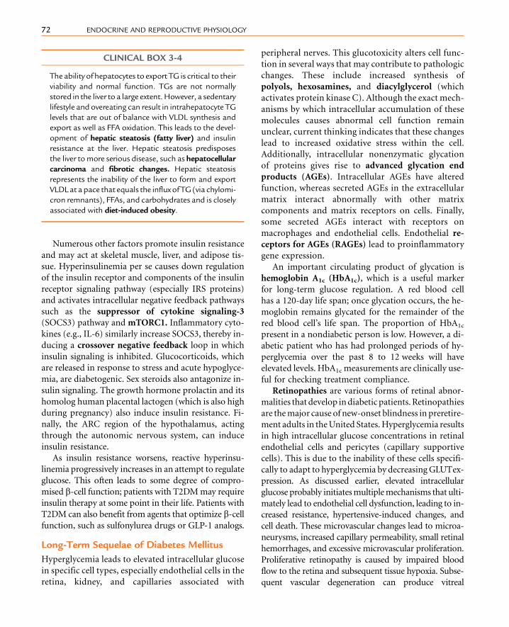
CLINICAL BOX 3-4
The ability of hepatocytes to export TG is critical to their
viability and normal function. TGs are not normally
stored in the liver to a large extent.However, a sedentary
lifestyle and overeating can result in intrahepatocyte TG
levels that are out of balance with VLDL synthesis and
export as well as FFA oxidation. This leads to the devel-
opment of hepatic steatosis (fatty liver) and insulin
resistance at the liver. Hepatic steatosis predisposes
the liver tomore serious disease, such as hepatocellularcarcinoma and fibrotic changes. Hepatic steatosis
represents the inability of the liver to form and export
VLDLat a pace that equals the influx of TG (via chylomi-
cron remnants), FFAs, and carbohydrates and is closely
associated with diet-induced obesity.
Numerous other factors promote insulin resistance
and may act at skeletal muscle, liver, and adipose tis-
sue. Hyperinsulinemia per se causes down regulation
of the insulin receptor and components of the insulin
receptor signaling pathway (especially IRS proteins)
and activates intracellular negative feedback pathways
such as the suppressor of cytokine signaling-3
(SOCS3) pathway and mTORC1. Inflammatory cyto-
kines (e.g., IL-6) similarly increase SOCS3, thereby in-
ducing a crossover negative feedback loop in which
insulin signaling is inhibited. Glucocorticoids, which
are released in response to stress and acute hypoglyce-
mia, are diabetogenic. Sex steroids also antagonize in-
sulin signaling. The growth hormone prolactin and its
homolog human placental lactogen (which is also high
during pregnancy) also induce insulin resistance. Fi-
nally, the ARC region of the hypothalamus, acting
through the autonomic nervous system, can induce
insulin resistance.
As insulin resistance worsens, reactive hyperinsu-
linemia progressively increases in an attempt to regulate
glucose. This often leads to some degree of compro-
mised b-cell function; patients with T2DMmay require
insulin therapy at some point in their life. Patients with
T2DM can also benefit from agents that optimize b-cellfunction, such as sulfonylurea drugs or GLP-1 analogs.
Long-Term Sequelae of Diabetes Mellitus
Hyperglycemia leads to elevated intracellular glucose
in specific cell types, especially endothelial cells in the
retina, kidney, and capillaries associated with
peripheral nerves. This glucotoxicity alters cell func-
tion in several ways that may contribute to pathologic
changes. These include increased synthesis of
polyols, hexosamines, and diacylglycerol (which
activates protein kinase C). Although the exact mech-
anisms by which intracellular accumulation of these
molecules causes abnormal cell function remain
unclear, current thinking indicates that these changes
lead to increased oxidative stress within the cell.
Additionally, intracellular nonenzymatic glycation
of proteins gives rise to advanced glycation end
products (AGEs). Intracellular AGEs have altered
function, whereas secreted AGEs in the extracellular
matrix interact abnormally with other matrix
components and matrix receptors on cells. Finally,
some secreted AGEs interact with receptors on
macrophages and endothelial cells. Endothelial re-
ceptors for AGEs (RAGEs) lead to proinflammatory
gene expression.
An important circulating product of glycation is
hemoglobin A1c (HbA1c), which is a useful marker
for long-term glucose regulation. A red blood cell
has a 120-day life span; once glycation occurs, the he-
moglobin remains glycated for the remainder of the
red blood cell’s life span. The proportion of HbA1c
present in a nondiabetic person is low. However, a di-
abetic patient who has had prolonged periods of hy-
perglycemia over the past 8 to 12 weeks will have
elevated levels. HbA1c measurements are clinically use-
ful for checking treatment compliance.
Retinopathies are various forms of retinal abnor-
malities that develop indiabetic patients.Retinopathies
are themajor cause of new-onset blindness in preretire-
ment adults in theUnited States.Hyperglycemia results
in high intracellular glucose concentrations in retinal
endothelial cells and pericytes (capillary supportive
cells). This is due to the inability of these cells specifi-
cally to adapt to hyperglycemia by decreasing GLUTex-
pression. As discussed earlier, elevated intracellular
glucose probably initiatesmultiplemechanisms that ulti-
mately lead to endothelial cell dysfunction, leading to in-
creased resistance, hypertensive-induced changes, and
cell death. These microvascular changes lead to microa-
neurysms, increased capillary permeability, small retinal
hemorrhages, and excessive microvascular proliferation.
Proliferative retinopathy is caused by impaired blood
flow to the retina and subsequent tissue hypoxia. Subse-
quent vascular degeneration can produce vitreal
72 ENDOCRINE AND REPRODUCTIVE PHYSIOLOGY

hemorrhage, retinal detachment, and neovascular glau-
coma, all of which can lead to severe visual loss. As blood
glucose and therefore blood osmolarity rise, the volume
of the lens changes, distorting vision. Diabetic patients
commonly have cataracts, and sorbitol and glycosylated
protein accumulation have been proposed as mecha-
nisms for inducing cataract formation.
Peripheralnervedamage(neuropathy)canoccurasa
resultofmetabolic, oxidative,or immune-relateddamage
to neurons or Schwann cells. Additionally, the microvas-
culature of peripheral nerves undergoes changes similar
to those seen in retinopathies andmay represent an event
that is concurrent with, or causal to, peripheral neuropa-
thy. Schwann cells (supportive cells involved in myelina-
tion) are among those shown to accumulate sorbitol as a
result of hyperglycemia. Diabetic patients can exhibit
sensory loss, paresthesias, and even pain as a result of
the neurologic damage. Neuropathies of the autonomic
nerves also develop in diabetic patients, which can lead
to numerous symptoms in multiple organ systems, in-
cluding erectile dysfunction, postural hypotension, and
heat intolerance. The sensory loss is more apparent in
the extremities, particularly the lower portions of the legs
and feet. This poses particular problems because, as dia-
beticpatients lose cutaneous sensation in the feet, theybe-
comeunaware of poorly fitting shoes and aremore prone
to injuries. Poor peripheral circulation aggravates this
problem. Because diabetic patients have impairedwound
healing, foot ulcerations can become a serious threat.
Diabetes is a common cause of impairment of renal
function (nephropathy) and is the greatest cause of
end-stage renal disease in North America. Clinical
or overt diabetic nephropathy is characterized by the
loss of greater than 300 mg of albumin in the urine
over a 24-hour period (microalbuminuria) and pro-
gressive decline of renal function. Nephropathies de-
velop from microvascular changes that occur in the
glomerular capillaries. The glomerular capillary base-
ment membrane thickens, resulting in thicker walls
and narrower lumina (glomerulosclerosis) and expan-
sion of the supportive mesangial cells. Podocytes de-
tach and undergo apoptosis. Poor renal filtration
also leads to activation of the renin-angiotensin
system (see Chapter 7), inducing hypertension.
Atherosclerosis develops in diabetic patients at an
accelerated rate (macroangiopathies). Diabetic patients
are more likely to have coronary artery disease and
myocardial infarction than are nondiabetic individuals.
Macrovascular disease is also associated with necrosis of
lower extremities and the need for amputation. Many
diabetic patients with coronary artery disease have the
additional risk factors ofhypertension,abdominalobe-
sity, insulin resistance, and dyslipidemia. This cluster
of factors has been identified as the metabolic
syndrome (also called syndrome X, insulin resistance
syndrome, and cardiovascular dysmetabolic syndrome).
Some of the consequences of visceral obesity, insulin
resistance, and dyslipidemia were discussed earlier.
S U M M A R Y
1. Cells make ATP to meet their energy needs. ATP is
made by glycolysis and by the TCA cycle coupled
to oxidative phosphorylation.
2. Cells express intracellular sensors of nutrients and
energy. Two of these are mTORC1, which senses
amino acids and growth factor/insulin signaling
and promotes anabolic pathways; and AMPK,
which senses a low level of energy (AMP/ATP ra-
tio) and inhibits anabolic pathways, while driving
catabolic pathways.
3. Cells can oxidize carbohydrate (primarily in the
form of glucose), amino acids, and FFAs to make
ATP. Additionally, the liver makes ketone bodies
(as well as glucose) for other tissues to oxidize
for energy in times of fasting.
4. Some cell types are limited in what energy sub-
strates they can oxidize for energy. The brain is
normally exclusively dependent on glucose for
energy. Thus, blood glucose must be maintained
above 60 mg/dL for normal autonomic and
central nervous system function. Conversely,
inappropriately high levels of glucose (i.e., fasting
glucose above 100 mg/dL) promote glucotoxicity
in specific cell types, leading to the long-term
complications of diabetes.
5. The endocrine pancreas produces the hormones
insulin, glucagon, somatostatin, gastrin, ghrelin,
and pancreatic polypeptide.
6. Insulin is produced from the b cells and is an ana-
bolic hormone that is secreted in times of excess
73ENERGY METABOLISM
nutrient availability. It allows the body touse carbo-
hydrates as energy sources and store nutrients.
7. Major stimuli for insulin secretion include in-
creased serum glucose and certain amino acids.
Cholinergic (muscarinic) receptor activation also
increases insulin secretion, whereas a2-adrenergicreceptors inhibit insulin secretion. The GI tract
releases incretin hormones that stimulate pan-
creatic insulin secretion. GLP-1 and GIP are
particularly potent in augmenting glucose-
dependent stimulation of insulin secretion.
8. Insulin binds to the insulin receptor, which is
linked to multiple pathways that mediate meta-
bolic and growth effects of insulin.
9. During the digestive period, insulin acts on the liver
to promote conversion of glucose to glucose-6-
phosphate. Insulin also increases glycogenesis, gly-
colysis, and fatty acid synthesis in the liver. Insulin
inhibits hepatic glucose production (glycogenolysis
and gluconeogenesis) and ketogenesis. Insulin reg-
ulates hepatic metabolism by both regulating gene
expression and posttranslational dephosphoryla-
tion events.
10. Insulin increases GLUT-4-mediated glucose
uptake in muscle and adipose tissue. Insulin
increases glycogenesis, glycolysis, and, in the pres-
ence of caloric excess, lipogenesis in muscle and
adipose tissue. Insulin increasesmuscle amino acid
uptake and protein synthesis. Insulin also increases
fatty acid esterification and lipoprotein lipase ac-
tivity and decreases HSL activity in the adipocyte.
11. Glucagon is a catabolic hormone. Its secretion in-
creases during periods of food deprivation, and it
acts to mobilize nutrient reserves. It also mobi-
lizes glycogen, fat, and protein.
12. Glucagonis released inresponse todecreasedserum
glucose (and therefore insulin) and increased se-
rum amino acid levels and b-adrenergic signaling.13. Glucagon binds to the glucagon receptor, which is
linked to Gs/PKA-dependent pathways.
14. The primary target organ for glucagon is the liver.
Glucagon increases liver glucose output by increas-
ing glycogenolysis and gluconeogenesis. It also in-
creases oxidation of fatty acids and ketogenesis.
15. Glucagon regulates hepatic metabolism by both
regulating gene expression, and through posttrans-
lational PKA-dependent phosphorylation events.
16. Themajor counter-regulatory factors inmuscle and
adipose is the adrenal hormone epinephrine, and
the sympathetic neurotransmitter norepinephrine.
These two factors act throughb2- andb3-adrenergicreceptors to increase cAMP levels. Epinephrine and
norepinephrine increase glycogenolysis and fatty
acyl oxidation in muscle and increase hormone-
sensitive lipase in adipose tissue.
17. It is increasingly well established that adipose tis-
sue has an endocrine function, especially in terms
of energy homeostasis. Hormones produced by
adipose tissue include leptin, adiponectin, IL-6,
and TNF-a.18. The ARC of the hypothalamus is the central reg-
ulator of appetite and energy use. The POMC/
CART neurons secrete a-MSH, which acts on
second-order neurons to promote satiety and
increase energy expenditure. NPY/AgRP neurons
act to promote eating and reduce energy expendi-
ture. Several hormones act on these neurons to
control nutrient and energy balance, including
leptin, insulin, CCK, and ghrelin.
19. The more common forms of DM are classified as
type 1 (T1DM) and type 2 (T2DM).T1DM is char-
acterizedby thedestructionofpancreaticb cellsandrequires exogenous insulin for treatment. T2DM
can be due to numerous factors but usually is char-
acterized as insulin resistance coupled to some de-
gree of b-cell deficiency. Patients with T2DM may
require exogenous insulin at some point to main-
tain blood glucose levels.
20. Obesity-associated T2DM is currently at epidemic
proportions worldwide. Obesity-associated T2DM
is characterized by insulin resistance due to
lipotoxicity, hyperinsulinemia, and inflammatory cy-
tokinesproducedbyadipose tissue.T2DMisoftenas-
sociatedwithobesity, insulinresistance,hypertension,
and coronary artery disease. This constellation of risk
factors is referred to asmetabolic syndrome.21. Major symptoms of DM include hyperglycemia,
polyuria, polydipsia, polyphagia, muscle wast-
ing, electrolyte depletion, and ketoacidosis (in
T1DM).
22. Long-term complications of poorly controlled
diabetes are due to excess intracellular glucose
(glucotoxicity) in specific cells, especially in
the retina, kidney, and peripheral nerves. This leads
to retinopathies, nephropathies, and neuropathies.
23. Diabetes also increases the risk for cardiovascular
disease and loss of adequate blood flow in the
lower extremities.
74 ENDOCRINE AND REPRODUCTIVE PHYSIOLOGY

CLINICAL BOX 3-5
Statin drugs are used to treat individuals with hyper-
cholesterolemia due to excessive LDL cholesterol. Sta-
tins target 3-hydroxy-3-methylglutaryl (HMG) CoA
reductase, so one effect of these drugs is to simply
lower cholesterol synthesis. Additionally, lowering
cholesterol synthesis in the liver leads to an activation
of SREBP2 and an increase in LDL receptor expres-
sion. This allows the liver to increase the clearance
of LDL cholesterol form the blood.
Why is LDL cholesterol a risk factor for cardiovascu-
lar disease? LDL particles are relatively small andmake
their way into the lamina propria of the tunica intima
(the layer just below the endothelial lining) of blood
vessels at regions of endothelial damage and death
due to hypertension, cigarette smoke, or other fac-
tors. Oxidation of LDL components leads to engulf-
ment by macrophages that ultimately become
engorged with cholesterol. At this point, macro-
phages are called foam cells. Foam cells become partic-
ipants in a series of events that lead to the
development of an atherosclerotic plaque. Plaques
are dangerous because they can become unstable
and rupture. Once this happens, the blood is exposed
to prothrombic molecules. This induces the formation
of blood clots (thrombi) that can occlude the arterial
lumen. In the coronary arteries, a thrombus poten-
tially leads to myocardial infarction, and in arteries
in the brain, a thrombus causes a stroke.
S E L F - S T U D Y P R O B L E M S
1. During the fed state, how does the role of glycol-
ysis in the liver differ from the role of glycolysis in
adipose tissue?
2. How does the function of glycogen differ in the
liver versus the skeletal muscle?
3. Normally, the brain is dependent on glucose.
What other energy substrate is used by the brainduring a prolonged fast? What is the origin of
this substrate?
4. What is the relation between mitochondrial cit-
rate levels and lipogenesis?
5. What two enzymes in adipocytes are dysregu-
lated in DM that contribute to high levels of cir-
culating TGs?
6. Why does loss of the LDL receptor give rise tohigh blood cholesterol?
7. What futile cycle do high levels of malonyl CoA
prevent?
8. Howwould amutant glucokinase with decreased
transport activity affect insulin secretion?
9. How does insulin regulate the following hepatic
enzymes: glucokinase, fructose-1,6-bisphospha-
tase, pyruvate kinase, acetyl CoA carboxylase,PEPCK? Be specific.
10. What is the basis for ketoacidosis in patients
with poorly managed type 1 diabetes mellitus?
11. How is obesity related to insulin resistance?
K E Y W O R D S A N D C O N C E P T S
n Acetoacetaten Acetyl CoA carboxylasen Adenosine triphosphate (ATP)
For full list of keywords and concepts see Student
Consult
SUGGESTED READ INGS
Asher G, Schibler U: Crosstalk between components of circadian
and metabolic cycles in mammals, Cell Metab 13:125–137,
2011.
Baker RG, Hayden MS, Ghosh S: NF-kappaB, inflammation, and
metabolic disease, Cell Metab 13:11–22, 2011.
Benito M: Tissue specificity on insulin action and resistance: Past to
recent mechanisms, Acta Physiol 201:297–312, 2011.
Jitrapakdee S: Transcription factors and coactivators controlling nu-
trient and hormonal regulation of hepatic gluconeogenesis, Int J
Biochem Cell Biol 44:33–45, 2010.
Kim MS, Wang Y, Rodrigues B: Lipoprotein lipase mediated fatty
acid delivery and its impact in diabetic cardiomyopathy, Biochim
Biophys Acta 1821:800–808, 2012.
Lin HV, Accili D: Hormonal regulation of hepatic glucose produc-
tion in health and disease, Cell Metab 14:9–19, 2011.
Mitra S, Goyal T, Mehta JL: Oxidized LDL, LOX-1 and atheroscle-
rosis, Cardiovasc Drugs Ther 25:419–429, 2011.
Rowland AF, Fazakerley DJ, James DE: Mapping insulin/GLUT4 cir-
cuitry, Traffic 12:672–681, 2011.
75ENERGY METABOLISM

K E Y W O R D S A N D C O N C E P T S
n Adipocyten Adipocytokinen Adiponectinn AdipoR1n AdipoR2n Adipose tissuen Adrenal hormone epinephrinen Albuminurian Amino acid (AA)n AMP kinasen Android (apple-shaped) adiposityn ApoB48n ApoCIIn ApoEn ApoB100n Apoprotein (Apo)n APS proteinn Atherogenicn ATP-citrate lyasen ATP-sensitive Kþ channeln b-hydroxybutyraten b-oxidationn Brainn Brown adipose tissue (BAT)n Cachexian Cachexinn Carnitine palmitoyl-transferase (CPTI and CPTII)n Cholesterol ester transfer protein (ETP)n Chylomicronn Chylomicron remnantsn Dawn phenomenonn Diacylglyceroln Diet-induced obesityn Early phase of insulin releasen Electron transport chainn Endothelial cell dysfunctionn Energy metabolismn Epinephrinen Fatty acid synthase (FAS) complexn Fatty livern Fibraten Free fatty acid (FFA)n Fructose-1,6-bisphosphaten Fructose-2,6-bisphosphate
n Fructose-6-phosphatasen Fructose-bisphosphate-2n Glucagonn Glucokinasen Glucometabolicn Glucosen Glucose sensorn Glucose tolerancen Glucose-6-phosphatasen Glucose-6-phosphate dehydrogenasen Glucotoxicityn Glycerol kinasen Glycogenn Glycogen phosphorylasen Glycogen synthasen Glycogen synthase kinase-3n Glycolysisn Gynecoid (pear-shaped) adiposityn Hemoglobin A1c (HbA1c)n Hepatic lipasen Hepatic steatosisn Hexosaminesn High-density lipoprotein (HDL)n Hormone-sensitive lipasen Hypoglycemian Insulinn Insulin resistancen Insulin receptor substrate (IRS)n Intermediate-density lipoprotein (IDL)n Ketone bodiesn Late phase of insulin releasen Leptinn Leptin receptor (LR); long form (LRb)n Leptin resistancen Lipoprotein lipase (LPL)n Lipoprotein particlen Liporegulationn Lipotoxicityn Livern Low-density lipoprotein (LDL)n Metforminn Nephropathyn Neuropathyn Nonenzymatic glycationn Nonesterified fatty acidsn Non-insulin-dependent diabetes mellitus
75.e1 ENDOCRINE AND REPRODUCTIVE PHYSIOLOGY

n Norepinephrinen Oxidative phosphorylationn Oxidized LDLn Palmitoyl CoA desaturasen Pancreatic isletn Pancreatic polypeptiden Pentose phosphate shuntn PEP carboxykinase (PEPCK)n Peroxisome proliferation–activated receptor-g(PPARg)
n Phosphoenolpyruvate carboxylase (PEPCK)n Phosphofructokinase-1 (PFK1)n Phosphofructokinase-2 (PFK2)n Phosphorylase kinasen Polydipsian Polyolsn Polyphagian Polyurian Proinsulin (connecting) peptiden Protein kinase B (PKB)–dependentpathway
n Protein phosphatase-1n Pyruvate carboxylasen Pyruvate dehydrogenase (PDH)n Pyruvate kinase
n Reactive oxygen species (ROS)n Scavenger receptorsn Shc proteinn Skeletal musclen Somogyi effectn Sterol-regulatory element–binding protein-1C(SREBP1C)
n Sulfonylurea drugsn Suppressor of cytokine signaling (SOCS)n SURn Sympathetic neurotransmitternorepinephrine
n Thiazolidinedione (TZD)n Thiophorasen Transaminationn Tricarboxylic acid (TCA) cyclen Triglyceride (TG)n Tumor necrosis factor-a (TNF-a)n Type 1 diabetes mellitus (T1DM)n Type 2 diabetes mellitus (T2DM)n Urea cyclen Very-low-density lipoprotein (VLDL)n VLDL remnantsn Voltage-gated Ca2þ channelsn White adipose tissue (WAT)
75.e2ENERGY METABOLISM

Intentionally left as blank

n n n n n n n n n n n n n n n
4 CALCIUM AND PHOSPHATEHOMEOSTASIS
O B J E C T I V E S
1. Describe the structure and synthesis of PTH, the regula-
tion of PTH secretion, and the nature of the PTH receptor.
2. Describethestructureandsynthesisof1,25-dihydroxyvita-
minD, the regulationof 1,25-dihydroxyvitaminDproduc-
tion, and the receptor for 1,25-dihydroxyvitamin D.
3. Discuss the roles of the GI tract, bone, and kidneys in
Ca2þ/Pi homeostasis.
4. Discuss the actions of calcitonin, PTHrP, FGF23, and go-
nadal and steroid hormones on Ca2þ/Pi metabolism.
5. Discuss the pathophysiology associated with imbal-
ances in PTH and 1,25-dihydroxyvitamin D.
Calcium (Ca2þ) and phosphate (Pi) are es-
sential to human life, playing important structural
roles in hard tissues (i.e., bones and teeth) and
important regulatory roles in metabolic and sig-
naling pathways. The two primary sources of
circulating Ca2þ and Pi are the diet and the skeleton
(Fig. 4-1).
Two hormones, 1,25-dihydroxyvitamin D (also
called calcitriol) and parathyroid hormone (PTH),
regulate intestinal absorption of Ca2þ and Pi and
the release of Ca2þ and Pi into the circulation after
bone resorption. The primary processes for removal
of Ca2þ and Pi from the blood are renal excretion
and bone formation (see Fig. 4-1), and 1,25-
dihydroxyvitamin D and PTH regulate these processes
as well. Other hormones and paracrine growth
factors also have clinical relevance to Ca2þ and Pi
homeostasis.
CALCIUM AND PHOSPHORUS AREIMPORTANT DIETARY ELEMENTSTHAT PLAY MANY CRUCIAL ROLESIN CELLULAR PHYSIOLOGY
Calcium is an essential dietary element. In addition to
getting calcium from the diet, humans contain a vast
store (i.e.,>1 kg) of calcium in their bones, which can
be called on to maintain normal circulating levels of
Dietary calciumand phosphate
GI tract BonePlasma calcium (10 mg/dL)Plasma phosphate (4 mg/dL)
FecesKidneys
Urine
FIGURE 4-1 n Daily Ca2þ and Pi fluxes.
77

calcium in times of dietary restriction and during the
increased demands of pregnancy and nursing. Circu-
lating calcium exists in three forms (Table 4-1): free
ionized calcium (Ca2þ), protein-bound calcium,
and calcium complexed with anions (e.g., phosphates,
bicarbonate, citrate). The ionized form represents
about 50% of circulating calcium, and because this
form is so critical to many cellular functions, Ca2þ
levels in both extracellular and intracellular compart-
ments are tightlycontrolled (seeChapter 1 fordiscussion
of Ca2þ-dependent signaling pathways). Circulating
Ca2þ is under direct hormonal control and is normally
maintained in a relatively narrow range. Either too little
of Ca2þ (hypocalcemia; total serum Ca2þ <8.5 mg/dL
[2.1 mM]) or too much Ca2þ (hypercalcemia; total se-
rumCa2þ>10.5 mg/dL [2.6 mM]) in thebloodcan lead
to a broad range of pathophysiologic changes, including
neuromuscular dysfunction, central nervous system dys-
function, renal insufficiency, calcification of soft tissue,
and skeletal pathologies.
Phosphorus is also an essential dietary element and
is stored in large quantities in bone complexed with
calcium. In the blood, most phosphorus exists in the
ionized form of phosphoric acid, which is called
inorganic phosphate (Pi). Most circulating Pi is in
the free ionized form, but some Pi (<20%) circulates
as a protein-bound form or complexed with cations
(see Table 4-1). Phosphorus also exists as pyrophos-
phate (two Pi groups in a covalent linkage). Unlike
Ca2þ, phosphate is incorporated covalently as single
or multiple phosphate groups into many molecules,
and consequently, soft tissues contain about 10-fold
more phosphate than Ca2þ. This means that signifi-
cant tissue damage (e.g., crush injury with massive
muscle cell death) can result in hyperphosphatemia,
which can then complex with Ca2þ to cause acute hy-
pocalcemia. Phosphate represents a key intracellular
component. Indeed, it is the high-energy phosphate
bonds of adenosine triphosphate (ATP) that maintain
life. Phosphorylation and dephosphorylation of pro-
teins, lipids, secondmessengers, and cofactors represent
key regulatory steps in numerous metabolic and
signaling pathways, and phosphate also serves as the
backbone for nucleic acids.
PHYSIOLOGIC REGULATION OFCALCIUM AND PHOSPHATE:PARATHYROID HORMONE AND1,25-DIHYDROXYVITAMIN D
PTH and 1,25-dihydroxyvitamin D represent the two
physiologically important hormones that are dedi-
cated to the maintenance of normal blood Ca2þ and
Pi levels in humans. As such, they are referred to as a
calciotropic hormones. The structure, synthesis, and
secretion of these two hormones and their receptors
will be discussed here. In the following section, the
detailed actions of PTH and 1,25-dihydroxyvitamin
D on the three key sites of Ca2þ/Pi homeostasis
(i.e., gut, bone, and kidney) will be discussed.
Parathyroid Hormone
PTH is a key hormone that protects against a hypocal-
cemic challenge. The primary targets of PTH are bone
and kidneys. PTH also functions in a positive feed-
forward loop by stimulating 1,25-dihydroxyvitamin
D production.
Parathyroid Glands The parathyroid glands develop
from the endodermal lining of the third and fourth
branchial pouches. They usually develop into four
loosely organized glands: two superior and two infe-
rior parathyroid glands. The embryonic anlage of
the parathyroids become associated with the caudal
migration of the thyroglossal duct, so the parathyroid
glands usually become situated on the dorsal side
of the right and left lobes of the thyroid gland
(Fig. 4-2). The exact positions of the parathyroid
glands are variable, and more than 10% of humans
harbor a fifth parathyroid gland. The predominant
parenchymal cell type in the parathyroid gland is the
TABLE 4-1
Forms of Ca2þ and Pi in Plasma
IONSIZE(MG/DL)
IONIZED(%)
PROTEINBOUND(%)
COMPLEXED(%)
Ca2þ* 10 50 45 5
Pi 4 84 10 6
*Ca2þ is bound (i.e., complexed) to various anions in the plasma,
including HCO �3 , citrate, Pi, and SO �
4 . Pi is complexed to various
cations, including Naþ and Kþ.From Koeppen BM, Stanton BA: Renal Physiology, 4th ed., Philadelphia,
2007, Mosby.
78 ENDOCRINE AND REPRODUCTIVE PHYSIOLOGY

principal (also called chief) cell. These cells are the
primary endocrine cell of the gland. With age, a larger
mitochondria-rich, eosinophilic cell type, the oxyphil
cell, appears. Although the oxyphil cell is not normally
important to PTH secretion, PTH-overproducing
tumors (i.e., primary hyperparathyroidism) can be
derived from both principal and oxyphil cells.
Structure, Synthesis, and Secretion of ParathyroidHormone Secreted PTH is an 84-amino acid polypep-
tide. PTH is synthesized as a preproPTH, which is
proteolytically processed to proPTH at the endoplas-
mic reticulum, and then to PTH in the Golgi and
secretory vesicles. Unlike proinsulin, all intracellular
proPTH is normally converted to PTH before secre-
tion. PTH has a short half-life (<5 minutes) because
it is proteolytically cleaved into biologically inactive
N-terminal and C-terminal fragments that are
excreted by the kidney.
The primary signal that stimulates PTH secretion is
low circulating Ca2þ levels (Fig. 4-3). The extracellular
Ca2þ concentration is sensed by the parathyroid prin-
cipal cells through a Ca2þ-sensing receptor (CaSR).
The CaSR is a member of the seven-transmembrane
G-protein-coupled receptor superfamily, which forms
disulfide-linked dimers in the membrane of chief cells
of the parathyroid glands. The CaSR is also expressed
in calcitonin-producing C cells, renal tubules, and sev-
eral other tissues. In the parathyroid gland, increasing
amounts of extracellular Ca2þ bind to the CaSR and
activate incompletely understood downstream signal-
ing pathways that repress PTH secretion. Thus, basal
PTH secretion (i.e., PTH secretion in the absence of
Superior parathyroid gland
Thyrocervical trunk
Right recurrent laryngeal nerve
Inferior parathyroid gland
Left recurrent laryngeal nerve
Left subclavian artery
Inferior thyroid artery
Superior thyroid artery
FIGURE 4-2 nAnatomic position of the parathyroid glands.
(Redrawn from Drake RL, Vogl W, Mitchell AWM: Gray’s
Anatomy for Students, Philadelphia, 2005, Elsevier.)
Exocytosis
1,25-Vitamin D
PTH gene
(–)
(+)
(–)
(–)
PTH
PTH
pro-PTH
Parathyroid
cell membrane
Ca2+CaSR
CaSR
Pre-CaSR
Prepro-PTH
PTH mRNA
CaSR mRNA
CaSR gene
Nuc
leus
Gq+Gi
Downstreamsignalingpathway
FIGURE 4-3 n Regulation of
PTH gene expression and PTH
secretion. The primary regulator of
PTH is extracellular Ca2þ, which
is sensed by the Ca2þ-sensingreceptor (CaSR). The CaSR is a G-
protein-coupled receptor (GPCR)
linked to Gq and Gi, but the
downstream signaling that inhibits
PTH secretion and PTH gene
expression is poorly understood.
1,25-Dihydroxyvitamin D inhibits
PTH gene expression directly
and indirectly by stimulating CaSR
gene expression.
79CALCIUM AND PHOSPHATE HOMEOSTASIS

CaSR signaling) is high but is inhibited by high extra-
cellular Ca2þ-CaSR binding and signaling.
Although the CaSR binds to extracellular Ca2þ
with relatively low affinity, the CaSR is extremely sen-
sitive to changes in extracellular Ca2þ. A 0.1-mM drop
in blood Ca2þ produces an increase in circulating
PTH levels from basal (5% of maximum) to maximal
levels (Fig. 4-4). Thus, the CaSR regulates PTH out-
put in response to subtle fluctuations in Ca2þ on a
minute-to-minute basis. It should be noted that the
CaSR is also stimulated by high levels of magnesium,
so hypermagnesemia also inhibits PTH secretion.
CLINICAL BOX 4-1
Patients with familial benign hypocalciuric hypercal-cemia (FBHH) or neonatal severe hyperparathyroid-ism are heterozygous or homozygous, respectively, for
inactivating mutations of the CaSR. In these patients,
the CaSR fails to appropriately inhibit PTH secretion
in response to high levels of blood calcium. The CaSR
also plays a direct role in Ca2þ reabsorption at the
kidney. The hypocalciuria (i.e., inappropriately low
Ca2þ excretion in the face of high circulating Ca2þ
levels) in patients with FBHH is due to the lowered
ability of the CaSR to monitor blood calcium and re-
spond by increasing urinary Ca2þ excretion.
PTH production is also regulated at the level of
gene transcription (see Fig. 4-3). The preproPTH
gene is repressed by a calcium-response element
within the promoter of this gene. Thus, the signaling
pathway that is activated by Ca2þ binding to the
CaSR ultimately leads to repression of prepro-
PTH gene expression and PTH synthesis. The prepro-
PTH gene is also repressed by 1,25-dihydroxyvitamin
D (acting through vitamin D–responsive elements, dis-
cussed later). The ability of 1,25-dihydroxyvitamin D
to hold PTH gene expression in check is reinforced by
the coordinated up-regulation of CaSR gene expression
by positive vitamin D–responsive elements in the pro-
moter of the CaSR gene (see Fig. 4-3).
Parathyroid Hormone Receptor The PTH receptor is a
seven-transmembrane, G-protein-linked membrane
receptor. Because this receptor also binds PTHrP
(see later), it is usually referred to as the PTH/PTHrP
receptor. The PTH/PTHrP receptor is primarily
coupled to a Gas signaling pathway that leads to
increased cyclic adenosine monophosphate (cAMP),
although it also is coupled to Gaq/11-phospholipaseC–dependent pathways. The PTH/PTHrP receptor is
expressed on osteoblasts in bone, and in the proximal
and distal tubules of the kidney, as the receptor for the
systemic actions of PTH. However, the PTH/PTHrP
receptor is also expressed in many developing struc-
tures in which PTHrP has an important paracrine
function.
Vitamin D
Vitamin D is actually a prohormone that must
undergo two successive hydroxylations to become the
active form, 1,25-dihydroxyvitamin D (Fig. 4-5). Vita-
min D plays a critical role in Ca2þ absorption, and to a
lesser extent Pi absorption, by the small intestine. Vita-
min D also regulates aspects of bone remodeling and
renal reabsorption of Ca2þ and Pi.
Structure, Synthesis, and Transport of Active VitaminD Metabolites Vitamin D3 (D3; also called cholecal-
ciferol) is synthesized by ultraviolet light (UV B) con-
version of 7-dehydrocholesterol in the more basal
layers of the skin (Fig. 4-6). UV radiation opens up
100
50
5
1.00 1.20
Free Ca2+ in blood (mM)
1.10 1.30 1.40
Ser
um P
TH
(%
of m
axim
um)
FIGURE 4-4 n Ca2þ/PTH secretion dose-response curve.
80 ENDOCRINE AND REPRODUCTIVE PHYSIOLOGY

the B ring of cholesterol, generating pre–vitamin D3,
which then undergoes a temperature-dependent
isomerization into D3. Vitamin D3 is therefore referred
to as a secosteroid, which is a class of steroids in which
one of the cholesterol rings is opened. Vitamin D2
(D2, also called ergocalciferol and also a secosteroid)
is the form produced in plants. Vitamins D3 and to a
lesser extent D2 are absorbed from the diet and
are equally effective after conversion into active
hydroxylated forms.
B
C D1
1211 13
14
2 103
45
9
6
8
171615
18
2221
20 23
24 26
25
27
7
5
1211 13
14
43
21
9
6
8
17
1615
18
2221
20 2324
26
25
27
7
10
19
HO
HOA
Skin
7-Dehydrocholesterol
Light
A
Liver
Cholecalciferol(Vitamin D3)
CH2
OH
Kidney
25-Hydroxycholecalciferol(25-OHD3)
HO
CH2
1,25-(OH)2D3
HO
CH2
OH
OH
CH2
24,25-(OH)2D3
OH
HO
OH
FIGURE 4-5n Biosynthesis of 1,25-
dihydroxyvitamin D dihydroxycho-
lecalciferol.
81CALCIUM AND PHOSPHATE HOMEOSTASIS

CLINICAL BOX 4-2
The balance between UV-dependent, endogenously
synthesized vitamin D3 and the absorption of dietary
forms of vitamin D becomes important in certain sit-
uations. Individuals with higher epidermal melanin
content and those who live in higher latitudes convert
less 7-dehydrocholesterol into vitamin D3 and thus
are more dependent on dietary sources of vitamin
D. Dairy products are enriched in vitamin D3, but
not all individuals tolerate or enjoy dairy products.
Institutionalized, sedentary elderly patients who stay
indoors and avoid dairy products are particularly at
risk for developing vitamin D deficiency.
D3 is transported in the blood from the skin to the
liver. Dietary D3 and D2 reach the liver directly
through transport in the portal circulation and indi-
rectly through chylomicrons (see Fig. 4-6). In the
liver, D2 and D3 are hydroxylated at the 25 carbon
position to yield 25-hydroxyvitamin D (at this junc-
ture, no distinction will be made between D3 and D2
metabolites because they are equipotent). Hepatic
vitamin D 25-hydroxylase is expressed at a relatively
constant and high level, so circulating levels of
25-hydroxyvitamin D largely reflect the amount of
precursor available for 25-hydroxylation. Because
the hydroxyl group at the 25 carbon position repre-
sents the second hydroxyl group on the molecule,
Enterocyte
Vit D-chylomicron Vit D
Dietaryvitamin D3andvitamin D2
LymphaticVit D-DBPand
Vit D-chylomicron
Vitamin D3
7-Dehydrocholesterol
Basal keratinocyte
UV-B
25(OH)-Vit D–DBP
25-Hdroxyvitamin D
Vit D
25-Hydroxylase
Cavalblood
Hepatocyte
Peripheral blood
Vit D-DBP
Portal blood
25(OH)-Vit D–DBP
Renal tubule
Glomerular filtrate
1,25-Dihydroxyvitamin D
25-Hydroxyvitamin D
1,24,25-Trihydroxyvitamin D
24,25-Dihydroxyvitamin D
Megalin-mediated
endocytosis
1α-Hydroxylase
24-Hydroxylase
24-Hydroxylase
1,25-Dihydroxyvitamin D Biological effects atgut, bone, kidney, etc.
FIGURE 4-6 n Vitamin Dmetabolism.
Vitamin D can be synthesized in the
skin keratinocyte or absorbed from
the gastrointestinal tract enterocyte.
Vitamin D is transported to the liver
hepatocyte, where it is hydroxylated
at the 25 carbon. 25-Hydroxyvitamin
D is carried in the blood by vitamin
D–binding protein (DBP) to the renal
proximal tubules, where it is hydroxyl-
ated at either the 1aposition (activat-ing) or the 24 position (inactivating).
Note that 25-hydroxyvitamin D that
exits into the glomerular filtrate can
be recaptured by the apical mem-
brane protein, megalin, which binds
and internalizes DBP.
82 ENDOCRINE AND REPRODUCTIVE PHYSIOLOGY

25-hydroxyvitamin D is also referred to as calcifediol
(see Fig. 4-6).
25-Hydroxyvitamin D is further hydroxylated
in the mitochondria of the proximal tubules of the
kidney at either the 1a carbon or 24 carbon position
(see Figs. 4-5 and 4-6). The 1a-hydroxylase (also calledCYP1a in humans) generates 1,25-dihydroxyvitamin
D (also called calcitriol), which is the most active
form of vitamin D. Hydroxylation at the 24 position,
generating 24,25-dihydroxyvitamin D and 1,24,25-
trihydroxyvitamin D, represents an inactivation
pathway.
Vitamin D and its metabolites circulate in the
blood primarily bound to vitamin D–binding protein
(DBP). DBP is a serum glycoprotein of about 60 kDa
that is related to the albumin gene family and is syn-
thesized by the liver. DBP binds more than 85% of
25-hydroxyvitamin D and 1,25-dihydroxyvitamin D.
As a result of binding to other proteins, only 0.4%
of the active metabolite, 1,25-dihydroxyvitamin D,
circulates as free steroid. DBPallows for themovement
of the highly lipophilic molecules within the aqueous
environment of the blood. Studies using experimental
mouse genetics indicate that DBP provides a reservoir
of vitamin D metabolites that protects against vitamin
D deficiency. The bound fractions of vitamin D me-
tabolites have a circulating half-life of several hours.
DBP may assist in the reuptake of the fraction
of 25-hydroxyvitamin D that passes through the
glomerular filter. DBP binds to 25-hydroxyvitamin
D with high affinity, so a significant amount of 25-
hydroxyvitamin D that enters the glomerular lumen
is complexed to DBP. The apical membranes of the
proximal tubule cells express the protein, megalin,
which is a member of the low-density lipoprotein
(LDL) receptor superfamily. Megalin functions to re-
capture a broad range of proteins from the glomerular
filtrate. Megalin binds DBP, some of which is com-
plexed with 25-hydroxyvitamin D, and internalizes
the complex through receptor-mediated endocytosis
(see Fig. 4-6).
The renal 1a-hydroxylase represents a key target
of regulation of vitamin D action (Fig. 4-7). First,
there exists a product feedback loop, in which
1,25-dihydroxyvitamin D inhibits 1a -hydroxylase ex-
pression and stimulates 24-hydroxylase expression.
Ca2þ is also an important regulator of the renal 1a-hydroxylase. Low circulating levels of Ca2þ indirectly
stimulate renal 1a-hydroxylase expression through
increased PTH levels, whereas elevated Ca2þ inhibits
1a-hydroxylase activity directly through the CaSR
in the proximal tubule. A low-phosphate diet also
stimulates renal 1a-hydroxylase activity in a PTH-
independent manner. Some of the effect of a low-
phosphate diet on renal 1a-hydroxylase activity is
dependent on a functional pituitary gland, which
may respond to hypophosphatemia by increased
growth hormone secretion (see Chapter 5).
Vitamin D Receptor 1,25-Dihydroxyvitamin D exerts
its actions primarily through binding to the nuclear
vitamin D receptor (VDR). The VDR is a 50-kDa
protein and is a member of the nuclear hormone re-
ceptor superfamily, which also includes steroid and
thyroid hormone receptors and metabolic receptors
such as the PPARs (see Chapter 1). The VDR is a tran-
scription factor and binds to DNA sequences (vitamin
D–responsive elements) as a heterodimer with the
retinoid X receptor (RXR). Thus, a primary action
of 1,25-dihydroxyvitamin D is to regulate gene expres-
sion in its target tissues, including the small intestine,
bone, kidneys, and parathyroid gland.
SMALL INTESTINE, BONE, ANDKIDNEY DETERMINE CA21
AND PI LEVELS
The general effects of PTH and 1,25-dihydroxyvitamin
D on Ca2þ and Pi levels at the small intestine, bone,
kidneys, and parathyroid glands is summarized in
Table 4-2.
Handling of Ca21 and Pi by theSmall Intestine
(Mosby Physiology Monograph Series cross reference:
Chapter 12 in Gastrointestinal Physiology, Sixth Ed.,
LR Johnson)
Dietary levels of calcium can vary, but in general,
North Americans consume about 1.5 kg of calcium
per day. Of this, about 200 g is absorbed by the proxi-
mal small intestine. Importantly, fractional absorption
of calcium is stimulated by 1,25-dihydroxyvitamin D,
so absorption can be made more efficient in the face
of declining dietary calcium.
83CALCIUM AND PHOSPHATE HOMEOSTASIS

Ca2þ is absorbed from the duodenum and jejunum
both by a Ca2þ- and hormone-regulated, active-
transport transcellular route and by a passive, bulk-flow
paracellular route. Little is known about whether
the paracellular route is regulated. However, significant
progress has been made in our understanding of the
transcellular route (Fig. 4-8). Ca2þ enters the transcel-
lular route by gaining access to the intestinal enterocytes
through the apical membrane. The movement of Ca2þ
from the lumen of the gastrointestinal (GI) tract into
the enterocyte, which is favored both by concentration
and electrochemical gradients, is facilitated by apical
epithelial calcium channels, called TrpV5 and TrpV6.
Once inside, Ca2þ ions bind to abundant cytoplasmic
proteins called calbindin-D, specifically calbindin-D9K,
in the human intestine. Calbindin-D9K serves to main-
tain the low cytoplasmic free Ca2þ concentrations,
thus preserving the favorable lumen-to-enterocyte
1α-Hydroxylase
24-Hydroxylase
CYP1α gene
CYP24 gene
(+)(–) (–)
(+)
Nucleus
25(OH)-Vit D
25(OH)-Vit D
1,25(OH)2-Vit D
1,25(OH)2-Vit D
25,25(OH)2-Vit D
cAMP/PKAsignalingpathway
Ca2+
signalingpathway?
GsGq/Gi
25(OH)-Vit D–DBP
Megalin-mediated
endocytosis
Epi
thel
ial c
ell o
f pro
xim
al tu
bule
Lumen of proximal tubule
CaSR
Ca2+ PTH
Renal capillary
FIGURE 4-7 n Regulation of the
renal CYP1a gene expression
by Ca2þ and hormones.
84 ENDOCRINE AND REPRODUCTIVE PHYSIOLOGY

concentration gradient during a meal. Calbindin-D9K
may also play a role in apical-to-basolateral shuttling
of Ca2þ. Ca2þ is actively transported across the baso-
lateral membrane, against an electrochemical and con-
centration gradient, by the plasma membrane
calcium ATPase (PMCA). The sodium-calcium
exchanger (NCX) may also contribute to the active
transport of Ca2þ out of the enterocytes. 1,25-
Dihydroxyvitamin D stimulates the expression of all
of the components (i.e., TrpV channels, calbindin-
D9K, and PMCA) involved in Ca2þ uptake by the small
intestine. PTH affects Ca2þ absorption at the gut in-
directly by stimulating renal 1a-hydroxylase activity.The fraction of phosphate absorbed by the jejunum
remains relatively constant at about 70% and is under
minor hormonal control by 1,25-dihydroxyvitamin D.
The limiting process in transcellular Pi absorption is
transport across the apical brush border, which is car-
ried out by an isoform (NPT2b) of the sodium-Pi
cotransporter, NPT2.
TABLE 4-2
Actions of Parathyroid Hormone and 1,25-Dihydroxyvitamin D on Ca2þ/Pi Homeostasis
SMALL INTESTINE BONE KIDNEYPARATHYROIDGLAND
PTH No direct action Promotes osteoblastic
growth and survival
Stimulates 1a-hydroxylase
activity
No direct action
Regulates M-CSF,
RANKL, and OPG
production by osteoblast
Stimulates Ca2þ
reabsorption by distal
nephron by increasing
Chronic high levels
promote net Ca2þ and
Pi release from bone
Inhibits Pi reabsorption by
proximal nephron (represses
NPT2a expression)
1,25-Dihydroxyvitamin D Increases Ca2þ
absorption by
increasing TrpV
channels, calbindin-D,
and PMCA expression
Sensitizes osteoblasts
to PTH
Minimal actions on
Ca2þ reabsorption
Directly inhibits PTH
gene expression
Marginally increases Pi
absorption
Regulates osteoid
production and
calcification
Promotes Pi reabsorption by
proximal nephron (stimulates
NPT2a expression)
Directly stimulates
CaSR gene
expression
Calbindin9K
Ca2+-Calbindin9k
Ca2+ Ca2+ Ca2+
PMCATrpV
5 and 6
Luminal side Serosal side
FIGURE 4-8n Intestinal absorption of Ca2þ through the transcellular route. Ca2þ enters through the Ca2þ channel, TrpV 5
or TrpV 6, in the luminal membrane of the enterocyte. Ca2þ is then shuttled from the apical side of the cell to the basal side
by the carrier protein, calbindin-D9k. Ca2þ is then actively transported out of the basolateral side by the plasma membrane
Ca2þ ATPase (PMCA), and calbindin-D9K recycles. 1,25-Dihydroxyvitamin D increases the expression of all these proteins
in the gastrointestinal tract.
85CALCIUM AND PHOSPHATE HOMEOSTASIS

Handling of Ca21 and Pi by Bone
Bone represents amassive and dynamic extracellular de-
posit of proteins and minerals (mainly Ca2þ and Pi).
Once maximal bone mass has been achieved in the
adult, the skeleton is constantly remodeled through
the concerted activities of the resident bone cell types.
The processes of bone accretion and bone resorption
are in balance in a healthy, physically active, and appro-
priately nourished individual. Of the about 1 kg of
calcium immobilized in bone, about 500 mg of Ca2þ
(i.e., 0.05% of skeletal calcium) is mobilized from and
deposited into bone each day. However, the process of
bone remodeling can be modulated to provide a net
gain or loss of Ca2þ and Pi to the blood and is respon-
sive to physical activity (or lack thereof), diet, age, and
hormonal regulation. Because the integrity of bone is
absolutely dependent on Ca2þ and Pi, chronic dysregu-
lation ofCa2þ andPi levels, or of the hormones that reg-
ulate Ca2þ and Pi, leads to pathologic changes in bone.
Histophysiology of Adult Bone
The biogenesis, growth, and remodeling of bone is a
complex process and will not be fully explained here.
The key features required to understand the role of
adult bone in the hormonal regulation of calcium-
phosphate metabolism are discussed next.
Most of the bone (about 75%) is compact, cortical
bone that makes up the outer surfaces of long and flat
bones (Fig. 4-9). The inner core of bones is composed
of interconnecting spicules whose orientation be-
comes organized by stress forces. This bone is called
cancellous (or trabecular) bone, and although it
makes up only 25% of total bone mass, its surface area
is several-fold greater than that of cortical bone. The
greater surface area means that trabecular bone is
much more accessible to bone cells and thus more
dynamic in its turnover.
In the adult, bone remodeling involves the destruc-
tion of preformed bone, with the release of Ca2þ, Pi,and hydrolyzed fragments of the proteinaceous matrix
(called osteoid) into the blood; and new synthesis of
osteoid at the site of resorption, with subsequent cal-
cification of the osteoid, primarily with Ca2þ and Pi,
from the blood. Bone remodeling occurs continually
in about 2 million discrete sites involving subpopula-
tions of bone cells, called basic multicellular units.
The cells involved in bone remodeling fall into two
major classes: cells that promote the formation of bone
(osteoblasts) and cells that promote the resorption of
bone (osteoclasts). However, it should be emphasized
that bone remodeling is a highly integrated process,
and osteoblasts also play a primary role in the initia-
tion and regulation of bone resorption (Fig. 4-10).
Haversiancanals
Periosteum
Outercircumferentiallamellae
Volkmann’scanal
Innercircumferentiallamellae
Trabecularbone
Bloodvessels
Interstitial bonelamellae
Haversiansystem(osteon)
FIGURE 4-9nDiagramofa typical long
bone shaft showing compact cortical
bone around the perimeter and
cancellous trabecular bone in the
center. (From Stevens A, Lowe J: Human
Histology, 3rd ed., Philadelphia, 2005,
Mosby.)
86 ENDOCRINE AND REPRODUCTIVE PHYSIOLOGY

Osteoblasts develop from mesodermally derived stro-
mal cells that have the potential to differentiate into
muscle, adipose, cartilage, and bone (i.e., osteoblasts)
cells. Several paracrine and endocrine factors modu-
late the osteoblast differentiation program, which is
dependent on the expression of bone-specific tran-
scription factors. For example, the transcription factor
Runx2 is essential for osteoblast differentiation and
is mutated in patients with cleidocranial dysplasia,
which is a congenital syndrome characterized by
multiple defects in bone formation.
Osteoblasts express factors that induce osteo-
clast differentiation from cells of the monocyte-
macrophage lineage and fully activate osteoclast
function (see Fig. 4-10). Osteoblasts release mono-
cyte colony-stimulating factor (M-CSF), which is
a secreted cytokine that binds to its receptor,
c-Fms, on osteoclast precursor cells. M-CSF induces
the earliest differentiating processes that lead to oste-
oclast precursors. M-CSF also acts in concert with
another factor, RANKL (named for receptor activa-
tor ofNF-kB ligand), to promote osteoclastogenesis.
RANKL can exist as a 40- to 45-kDa protein on
the cell membrane of osteoblasts and as a soluble
31-kDa form. RANKL binds to its receptor, RANK,
on osteoclast precursor membranes. RANK is
structurally related to the receptor for tumor ne-
crosis factor-a (TNF-a) and signals through NF-
kB-related pathways to induce osteoclastogenesis.
This process involves the clustering and fusion of
several osteoclast precursors, giving rise to a fused,
polykaryonic osteoclast. The perimeter of the oste-
oclast membrane facing the bone adheres tightly to
the bone, essentially sealing off the area of osteo-
clast-bone contact (see Fig. 4-10). The osteoclast
cell membrane facing the bone transforms into a
ruffled border, from which enzymes (e.g., cathep-
sin K) and HCl are secreted. The acidic enzyme-
rich microenvironment proceeds to dissolve the
calcified crystals and ultimately hydrolyzes type I
collagen and other osteoid components. After about
2 weeks, osteoclasts receive a different signal from
neighboring osteoblasts. This signal is osteoprote-
gerin (OPG), which acts as a soluble decoy receptor
for RANKL (see Fig. 4-10). Consequently, the pro-
osteoclastic signal from osteoblasts is terminated.
Osteoblast Preosteoclasts
Monocyte/macrophagelineage preosteoclast
RANKL
OPG
RANKL
Stromal cell
Release of Ca2+
and Pi into blood
Differentiation
RANK
Fusion and activation
M-CSF
C-FMS
Polykaryonicosteoclast
Hydrolytic enzymesH+
RANK
Resorption cavity in bone
FIGURE 4-10 n Osteoblast regula-
tion of osteoclast differentiation
and function.
87CALCIUM AND PHOSPHATE HOMEOSTASIS

During a reversal phase, osteoclasts are then
recruited to adjacent sites of bone, which extends the
resorption cavity, also called the cutting cone, further
into the bone. Alternatively, some osteoclasts may un-
dergo apoptosis. Adjacent osteoblasts migrate into the
resorbed area (now vacated by osteoclasts) and begin
to lay down osteoid. Some of the components in oste-
oid (e.g., osteocalcin and alkaline phosphatase) pro-
mote its calcification. This process removes Ca2þ
and Pi from the blood and deposits them first as
calcium phosphate crystals. Later, bicarbonate and hy-
droxide ions are incorporated into the calcium phos-
phate to form hydroxyapatite crystals. The bone is
laid down in organized layers, called lamellae, starting
from the perimeter of the resorption cavity and pro-
gressing inward. In the fully repaired region, multiple
concentric lamellae surround a central haversian canal
or groove housing a nutritive capillary (see Fig. 4-9).
This area of bone accretion is also called the closing
cone. As the osteoblasts become surrounded by and
entrapped within bone, they become osteocytes that
sit within small spaces, called haversian lacunae. Os-
teocytes remain interconnected through cell processes
that run within canaliculi and form communicating
junctions with adjacent cell processes. The new con-
centric layers of bone, along with the interconnected
osteocytes and the central canal, are referred to collec-
tively as an osteon. The exact function of osteocytes is
presently unclear, although evidence exists for a role of
osteocytes in the sensing of mechanical stress in bones.
CLINICAL BOX 4-3
The importance of the RANK/RANKL/osteoprote-
gerin system is made evident by mutations in the
human genes for RANK and osteoprotegerin that
are associated with bone deformities. Loss of RANKL
in mice causes osteopetrosis (i.e., excessive bone den-sity) because of the loss of osteoclasts. Conversely,
loss of osteoprotegerin causes osteoporosis (reducedbone density) because of a high number of overly
active osteoclasts. Furthermore, our current under-
standing of bone regulation is basedonhowhormones,
cytokines, and other factors alter the balance between
RANKL and osteoprotegerin and on how they regulate
the differentiation, survival, and apoptosis of osteo-
blasts versus osteoclasts.
As a calciotropic hormone, PTH is a primary endo-
crine regulator of bone remodeling in adults. The
PTH/PTHrP receptor is expressed on osteoblasts,
but not on osteoclasts. Therefore, PTH directly stim-
ulates osteoblastic activity and indirectly stimulates
osteoclastic activity through osteoblast-derived para-
crine factors (i.e., M-CSF, RANKL). Sustained elevated
levels of PTH shift the balance to a relative increase in
osteoclast activity, thereby increasing bone turnover
and reducing bone density. In contrast, intermittent
administration of low doses of PTH promotes osteo-
blast survival and bone anabolic functions, increases
bone density, and reduces the risk for fracture in
humans. This is mediated, at least in part, by decreased
production of sclerostin (SOST) by osteocytes. SOST
is an inhibitor of bone formation that suppresses os-
teoblast differentiation by inhibiting Wnt signaling
in osteoblast progenitor cells. Loss-of-function muta-
tions of the SOST gene in humans cause sclerosteosis,
a disease characterized by excessive bone mass.
CLINICAL BOX 4-4
Regulation of bone remodeling by PTH requires nor-
mal levels of 1,25-dihydroxyvitamin D. In vitamin
D–deficient individuals, the Ca2þ-PTH secretion curve
is shifted to the right. Thus, normal Ca2þ levels are
less effective in suppressing PTH secretion, and ele-
vated PTH levels and increased bone turnover result.
The vitamin D receptor is expressed in osteoblasts,
and normal 1,25-dihydroxyvitamin D levels are also
required for coordination of osteoid production with
its calcification. In vitamin D–deficient individuals,
osteoid is not properly calcified, and the bone is weak.
In children, this leads to rickets, in which growth of
long bones is abnormal, and the weakened bones lead
to bowing of extremities and collapse of the rib cage
(see later). In adults, vitamin D deficiency leads to
osteomalacia, which is characterized by poorly
calcified osteoid associated with pain, increased risk
for fracture, and vertebral collapse (see later).
Handling of Ca21 and Pi by the Kidneys
(Mosby Physiology Monograph Series cross reference:
Chapter 9 in Renal Physiology, Third Ed., BM Koep-
pen and BA Stanton)
88 ENDOCRINE AND REPRODUCTIVE PHYSIOLOGY

The kidneys filter a large amount of Ca2þ (about
10 g) each day, but most of the filtered Ca2þ is reab-
sorbed by the nephron. Renal excretion typically
accounts for the loss of about 200 mg of Ca2þ per day,
which is counterbalanced by net intestinal absorption
of about 200 mg/day. In the proximal tubule, most
of theCa2þ is reabsorbed by a passive, paracellular path-
way. As in the duodenum, transcellular Ca2þ transport
also exists and involves the constitutive expression of
apical epithelial calcium channels (Trp-V5 and Trp-
V6), intracellular Ca2þ-binding proteins (calbindins),
and active Ca2þ extrusion (by PMCA and NCX) at
the basolateral membrane. Ca2þ reabsorption in the
thick ascending limb (TAL) of the loop of Henle uses
both paracellular and transcellular transport mecha-
nisms. Paracellular transport in the TAL is driven by
a lumen-positive electrical gradient established by the
Na-K-2Cl transporter in the luminal membrane follow-
ingKþ leakage back into the lumen. TheCaSR is located
in the basolateral membrane of TAL cells, and its activa-
tion by high serum calcium inhibits the Na-K-2Cl sym-
porter and reduces paracellular Ca2þ transport (Fig. 4-
11). Clinically, inhibition of this transporter by loop di-
uretics such as furosemide has been used to treat hyper-
calcemia. Transcellular Ca2þ transport in the cortical
portionof theTALand thedistal convoluted tubuleoc-
curs by an active transport process that is stimulated by
PTH (see Fig. 4-11). Inhibition of the thiazide-sensitive
Naþ-Cl� symporter in the luminal membrane of distal
tubule cells enhances Ca2þ reabsorption. Thiazide di-
uretics are therefore used to prevent renal calciumwast-
ing in idiopathic hypercalciuria.
As discussed earlier, intestinal absorption of
phosphate is largely proportional to the amount of
phosphate in the diet and is only slightly regulated
by 1,25-dihydroxyvitamin D. This leaves the kidney
with an important role in the regulation of circulating
phosphate levels. Phosphate is mostly reabsorbed by
the proximal convoluted tubule through a hormonally
regulated transcellular route. As in the small intestine,
phosphate enters the apical surface of the proximal
tubules in a rate-limiting manner through a
sodium-phosphate cotransporter (NPT). In contrast
to the NPT isoform expressed in the intestine
(NPT2b), the kidney expresses an additional isoform,
NPT2a, which is under strong hormonal regulation.
PTH down regulates NPT2a expression on the apical
membranes of proximal renal tubule cells, thereby in-
creasing phosphate excretion (see Fig. 4-11). In con-
trast, 1,25-dihydroxyvitamin D increases NPT2 gene
expression in the proximal tubules.
Integrated Physiologic Regulationof Ca21/Pi Metabolism: Response ofPTH and 1,25-Dihydroxyvitamin Dto a Hypocalcemic Challenge
The integrated response of PTH and 1,25-
dihydroxyvitamin D to a hypocalcemic challenge is
shown in Figure 4-12. Low blood Ca2þ, as detectedby the CaSR on the parathyroid chief cells, stimulates
PTH secretion. In the kidney, PTH rapidly increases
Ca2þ levels by increasing fractional reabsorption of
Ca2þ in the distal renal tubules. The renal effects of
PTH on Ca2þ reabsorption are reinforced by Ca2þ
levels as sensed by the CaSR and, to a lesser extent,
by 1,25-dihydroxyvitamin D. PTH also inhibits the ac-
tivity of the sodium-dependent phosphate transporter
(NPT2), thereby increasing Pi excretion. The relative
loss of phosphate serves to increase free, ionized
Ca2þ in the blood. At the bone, PTH stimulates oste-
oblasts to secrete RANKL, which, in turn, rapidly
increases osteoclast activity, leading to increased bone
resorption and the release of Ca2þ and Pi into the
blood.
In a slower phase of the response to hypocalcemia,
PTH and low Ca2þ directly stimulate 1a-hydroxylase(CYP1a) expression in theproximal renal tubule, thereby
increasing 1,25-dihydroxyvitamin D levels. In the small
intestine, 1,25-dihydroxyvitamin D supports adequate
Ca2þ levels in the long termby stimulating Ca2þ absorp-
tion. These effects occur over hours and days and in-
volve increasing the expression of TrpV5 and TrpV6
calcium channels, calbindin-D9k, and PMCA. 1,25-
Dihydroxyvitamin D also stimulates osteoblast release
of RANKL, thereby amplifying the effect of PTH.
1,25-Dihydroxyvitamin D and the CaSR play key roles
in negative feedback. Thus, elevated PTH stimulates 1,25-
dihydroxyvitaminDproduction,which then inhibits PTH
gene expression directly, and indirectly by up regulating
the CaSR. 1,25-Dihydroxyvitamin D also represses renal
1a-hydroxylase activity while increasing 24-hydroxylase
activity. As blood Ca2þ levels rise back to normal levels,
they shut off PTH secretion and 1a-hydroxylase.
89CALCIUM AND PHOSPHATE HOMEOSTASIS

Hormonal Regulation of Calcium andPhosphate: Pharmacologic Regulators
Calcitonin The primary actions of calcitonin are on
bone and kidney. Calcitonin lowers serum calcium
and phosphate levels, primarily by inhibiting bone
resorption, in many species of animals. However,
although human calcitonin can lower serum calcium
and phosphate levels in humans, it takes high doses
to show this effect. There are no definitive complica-
tions from calcitonin deficiency or excess in humans.
For this reason, it is unlikely that calcitonin has an im-
portant physiologic role in humans. Medical interest
in calcitonin stems from the fact that potent forms
of calcitonin can be used therapeutically in the treat-
ment of bone disorders. Calcitonin is also a useful his-
tochemical marker of medullary thyroid cancer.
Ca2� Transport Along the Nephron
DT
�9%
TAL
CCD
PT
~60% UF
MCD
LumenLumen
Furosemide
TRPV5
Thiazide
Calbindin
Gs PTH
PTHR1
PMCA
NCX1
Claudin 16
CaSRCa2�
Ca2�
Ca2�
Ca2�
Ca2�
3Na�
Na�
Ca2�
Na�
K� K�
K�
2Cl�
Cl�
Paracellular: TAL
Transcellular: DCT (Cortical TAL)Blood
Blood
20%
70%
1%
1%
FIGURE 4-11 nHandling of Ca2þ by the distal nephron and proximal nephron (see text). Diagram of the nephron and the
transport pattern of Ca2þ along the nephron. (From Koeppen BM, Stanton BA: Renal Physiology, 3rd ed., St. Louis, 2001, Mosby.
Details of the Ca2þ reabsorption by the TAL and DCT were drawn by Dr. John Harrison, University of Connecticut Health Center,
Farmington, Conn.)
90 ENDOCRINE AND REPRODUCTIVE PHYSIOLOGY

Parafollicular C Cells The cells that produce calcito-
nin are called the parafollicular C cells. These cells
are derived from the ultimobranchial bodies and
become incorporated and interspersed among the
thyroid follicles as the thyroglossal duct migrates
caudally. Parafollicular C cells do not invade the thy-
roid epithelium and thus are not in contact with the
follicular colloid.
Structure, Synthesis, and Secretion of CalcitoninCalcitonin is a 32-amino acid polypeptide. Because
there is minimal species variation, calcitonins from
other species are biologically active in humans. In fact,
salmon calcitonin is about 20 times more potent in
humans than human calcitonin. Normal serum calci-
tonin levels are about 10 to 50 pg/mL, and its half-life
in circulation is less than 1 hour. Because the primary
site of inactivation is the kidney, serum calcitonin
levels are often elevated in renal failure. Alternative
splicing of the calcitonin gene in other tissues can pro-
duce calcitonin gene-related peptide (CGRP), which
is a potent vasodilator and positive cardiac inotrope.
The secretion of calcitonin is primarily regulated by
the same CaSR that regulates PTH secretion. However,
elevated extracellular Ca2þ levels stimulate the synthesis
and secretion of calcitonin.
Calcitonin Receptor The calcitonin receptor is closely
related to the secretin and PTH/PTHrP receptors. It is
a seven-transmembrane Gas-coupled receptor that
acts primarily through cAMP-dependent signaling
pathways. In contrast to the PTH/PTHrP receptor,
the calcitonin receptor is expressed in osteoclasts.
Calcitonin acts rapidly and directly on osteoclasts to
suppress bone resorption. Paget disease is character-
ized by excessive bone turnover that is driven by large,
bizarre osteoclasts (see later). Because these osteoclasts
retain the calcitonin receptor, active forms of calcito-
nin can be used to suppress aberrant osteoclastic activ-
ity in patients with this disease.
The calcitonin receptor is also expressed in the
nephron, where calcitonin inhibits phosphate and cal-
cium reabsorption.
Hormonal Regulation of Calcium andPhosphate: Regulators Overexpressedby Cancers
Parathyroid Hormone–Related Peptide Parathyroid
hormone–related peptide (PTHrP) is a peptide para-
crine factor that shows limited structural similarity to
PTH but nevertheless binds to and signals through
the PTH/PTHrP receptor. PTHrP is expressed in several
HypocalcemicchallengeLow blood
Ca2+
↑ BloodCa2+
↑ PTH ↑ CYP1α ↑ 1,25(OH)2-Vit D
↑ Boneturnover
↑ Ca2+
reabsorptionand
↑ Pi excretionby kidneys
↑ Dietary Ca2+
absorption bysmall intestine
Rapid responses(mins–hrs)
Slower responses(hr–days)
Negative feedback
Negative feedback
FIGURE 4-12n Integratedresponse
to a hypocalcemic challenge.
91CALCIUM AND PHOSPHATE HOMEOSTASIS

developing tissues, including the growth plate of bones
and the mammary glands. PTHrP is not regulated by
circulating calcium and normally does not play a role
in Ca2þ/Pi homeostasis in the adult. However, certain
neoplasias can secrete high levels of PTHrP, which then
produces symptoms of hyperparathyroidism (see later).
CLINICAL BOX 4-5
Fibroblast growth factor-23 (FGF23) is an approxi-
mately 30-kDapeptide that is normally expressed by os-
teocytes. It acts on proximal tubule cells of the kidney to
inhibit Pi reabsorption and promote phosphate excre-
tion. FGF23 is inactivated by a protease that cleaves
FGF23 into N-terminal and C-terminal peptides. One
protease involved in FGF23 processing, although it is
not a direct substrate, is PHEX (for phosphate-
regulating gene with homologies to endopeptidases
on the X chromosome). PHEX is mutated in X-linkedhypophosphatemia, characterized by renal phosphate
wasting, rickets, osteomalacia, and inappropriate low-
normal levels of 1,25-dihydroxyvitamin D.
Current evidence indicates that when PHEX is
mutated, FGF23 levels increase and inhibit both
phosphate reabsorption and 1a-hydroxylase in the prox-imal renal tubules. Increased expression of FGF23 has
been linked to autosomal recessive hypophosphatemic
rickets and tumor-induced rickets/osteomalacia.
Regulation of Ca21/Pi Metabolism byImmune and Inflammatory Cells
It is interesting to note that the RANKL/RANK/
osteoprotegerin signaling system is similar to the TNF re-
ceptor/NF-kB signaling pathways used in cells involved
in the immune system and in inflammation. This link
is further stressed by the fact that activated T cells express
high levels of RANKL in response to stimulation by the
cytokines, TNF-a, and several interleukins. Thus, inflam-
matory bone diseases (e.g., rheumatoid arthritis) are as-
sociated with increased RANKL-to-osteoprotegerin
ratios in the vicinity of the inflammatory site, with sub-
sequent erosions of bone and osteoporosis.
RANKL is also overproduced by cells associated
with several malignant bone diseases (e.g., multiple
myeloma, skeletal metastatic breast cancer). As noted
earlier, some malignant cells also overexpress PTHrP,
which induces RANKL expression in neighboring
osteoblasts. Thus, several malignancies are associated
with bone damage and hypercalcemia.
The 1a-hydroxylase enzyme is expressed by mono-
cytes and peripheral macrophages. In the autoimmune
disease of sarcoidosis, overactive macrophages pro-
duce high levels of 1,25-dihydroxyvitamin D, resulting
in hypercalcemia.
Regulation of Ca21/Pi Metabolism byGonadal and Adrenal Steroid Hormones
Gonadal and adrenal steroid hormones have pro-
found effects on calcium and phosphate metabolism
and skeletal health. Estradiol-17b (E2; see Chapter 10)
hasaboneanabolicandcalciotropiceffect at several sites.
E2 stimulates intestinal calcium absorption and renal
tubular calcium reabsorption. E2 is also one of the most
potent regulators of osteoblast and osteoclast function.
Estrogen promotes survival of osteoblasts and apoptosis
of osteoclasts, thereby favoring bone formation over
resorption. In postmenopausal women, estrogen defi-
ciency results in an initial phase of rapid bone loss that
lasts about 5 years, followed by a second phase of slower
bone loss. During the second phase, the individual is
chronically challengedwith hypocalcemia because of in-
efficient calcium absorption and renal calciumwasting.
This can result in secondary hyperparathyroidism,
which further exacerbates bone loss. Exercise, high levels
of dietary calcium with supplemental vitamin D, and
hormonal replacement therapy can prevent postmeno-
pausal osteoporosis.Androgens alsohavebone anabolic
and calciotropic effects, although some of these effects
are due to the peripheral conversion of testosterone to
E2 (see Chapter 9).
In contrast to gonadal steroids, the adrenal gluco-
corticoids (e.g., cortisol) promote bone resorption
and renal calcium wasting and inhibit intestinal cal-
cium absorption. Patients treated with high levels of
a glucocorticoid (e.g., as an anti-inflammatory and
immunosuppressive drug) can develop glucocorti-
coid-induced osteoporosis.
PATHOLOGIC DISORDERS OFCALCIUM AND PHOSPHATEBALANCE
Hyperparathyroidism (Primary)
Primary hyperparathyroidism is caused by excessive
production of PTH by the parathyroid glands. It is fre-
quently caused by a single adenoma confined to one of
92 ENDOCRINE AND REPRODUCTIVE PHYSIOLOGY
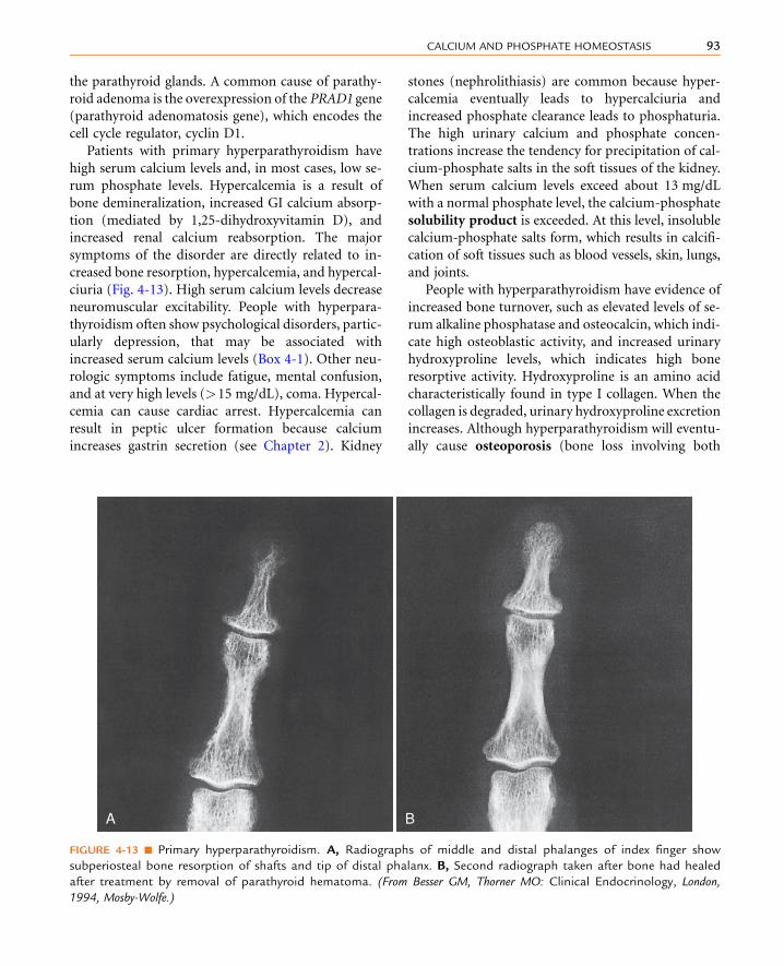
the parathyroid glands. A common cause of parathy-
roid adenoma is the overexpression of the PRAD1 gene
(parathyroid adenomatosis gene), which encodes the
cell cycle regulator, cyclin D1.
Patients with primary hyperparathyroidism have
high serum calcium levels and, in most cases, low se-
rum phosphate levels. Hypercalcemia is a result of
bone demineralization, increased GI calcium absorp-
tion (mediated by 1,25-dihydroxyvitamin D), and
increased renal calcium reabsorption. The major
symptoms of the disorder are directly related to in-
creased bone resorption, hypercalcemia, and hypercal-
ciuria (Fig. 4-13). High serum calcium levels decrease
neuromuscular excitability. People with hyperpara-
thyroidism often show psychological disorders, partic-
ularly depression, that may be associated with
increased serum calcium levels (Box 4-1). Other neu-
rologic symptoms include fatigue, mental confusion,
and at very high levels (>15 mg/dL), coma. Hypercal-
cemia can cause cardiac arrest. Hypercalcemia can
result in peptic ulcer formation because calcium
increases gastrin secretion (see Chapter 2). Kidney
stones (nephrolithiasis) are common because hyper-
calcemia eventually leads to hypercalciuria and
increased phosphate clearance leads to phosphaturia.
The high urinary calcium and phosphate concen-
trations increase the tendency for precipitation of cal-
cium-phosphate salts in the soft tissues of the kidney.
When serum calcium levels exceed about 13 mg/dL
with a normal phosphate level, the calcium-phosphate
solubility product is exceeded. At this level, insoluble
calcium-phosphate salts form, which results in calcifi-
cation of soft tissues such as blood vessels, skin, lungs,
and joints.
People with hyperparathyroidism have evidence of
increased bone turnover, such as elevated levels of se-
rum alkaline phosphatase and osteocalcin, which indi-
cate high osteoblastic activity, and increased urinary
hydroxyproline levels, which indicates high bone
resorptive activity. Hydroxyproline is an amino acid
characteristically found in type I collagen. When the
collagen is degraded, urinary hydroxyproline excretion
increases. Although hyperparathyroidism will eventu-
ally cause osteoporosis (bone loss involving both
BA
FIGURE 4-13 n Primary hyperparathyroidism. A, Radiographs of middle and distal phalanges of index finger show
subperiosteal bone resorption of shafts and tip of distal phalanx. B, Second radiograph taken after bone had healed
after treatment by removal of parathyroid hematoma. (From Besser GM, Thorner MO: Clinical Endocrinology, London,
1994, Mosby-Wolfe.)
93CALCIUM AND PHOSPHATE HOMEOSTASIS

osteoid and mineral), it is not necessarily the present-
ing symptom. However, bone demineralization is
apparent. These individuals frequently exhibit hyper-
chloremic acidosis. Some people with hyperparathy-
roidism have the bone disorder osteitis fibrosa
cystica, which is characterized by bone pain, multiple
bone cysts, a tendency for pathologic fractures of long
bones, and histologic abnormalities of the bone.
Pseudohypoparathyroidism
Pseudohypoparathyroidism is a rare familial disorder
characterized by tissue resistance to PTH. In many in-
stances, the problem is thought to originate with the
PTH receptor. Often there is a decrease in levels of
the guanine nucleotide–binding protein, Gs. Individ-
uals with pseudohypoparathyroidism demonstrate in-
creased PTH secretion and low serum calcium levels,
sometimes associated with congenital defects of the
skeleton, including shortened metacarpal and meta-
tarsal bones.
Hypoparathyroidism
Hypoparathyroidism is associated with low serum
calcium levels and high serum phosphate levels.
The hypocalcemia results from both a PTH and a
1,25-dihydroxyvitamin D deficiency. Consequently,
there is a decrease in bone calcium mobilization by
both osteoclastic resorption and osteocytic osteolysis.
Because 1,25-dihydroxyvitamin D is deficient, GI
absorption of calcium is impaired. The PTH defi-
ciency decreases renal calcium reabsorption, thereby
decreasing fractional calcium reabsorption. Although
fractional calcium reabsorption decreases, the urinary
calcium level is generally low. Alkalosis occurs be-
cause bicarbonate excretion decreases; this further
lowers the free calcium level in serum. Although
the serum calcium level is low, bone demineralization
is usually not a problem because of the high serum
phosphate level. Hypocalcemia increases neuromus-
cular excitability, increasing the possibility of tetany
and even convulsions. Hypocalcemia alters cardiac
function. It can produce a first-degree heart block.
The low serum calcium level decreases myocardial
contractility.
The most prominent symptom of hypoparathy-
roidism is increased neuromuscular excitability
(Box 4-2). Low serum calcium concentrations decrease
the neuromuscular threshold. This can be manifested
as repetitive responses to a single stimulus and as spon-
taneous neuromuscular discharge. The increased neu-
romuscular excitability can result in tingling in the
fingers or toes (paresthesia), muscle cramps, or even
tetany. Laryngeal spasms can be fatal. Sometimes the
serum calcium level is not low enough to produce overt
tetany, but latent tetany can be demonstrated by inflat-
ing a blood pressure cuff on the arm to a pressure
greater than systolic pressure for 2 minutes. The resul-
tant oxygen deficiency precipitates overt tetany as
demonstrated by carpal-pedal spasms. This is called
Trousseau sign (Fig. 4-14A). Another test is to tap
the facial nerve, which evokes facial muscle spasms
(Chvostek sign).
Treatment of hypoparathyroidism is difficult
because of the lack of readily available effective hu-
man PTH. The disorder is frequently treated with a
n n n n n n n n n n
BOX 4 - 1
SYMPTOMS OF HYPERPARATHYROIDISM
n Kidney stones
n Osteoporosis
n Gastrointestinal disturbances, peptic ulcers, nau-
sea, constipation
n Muscle weakness, decreased muscle tone
n Depression, lethargy, fatigue, mental confusion
n Polyuria
n High serum phosphate concentration; low serum
calcium concentration
n n n n n n n n n n
BOX 4 - 2
SYMPTOMS OF HYPOPARATHYROIDISM
n Tetany, convulsions, paresthesias, muscle cramps
n Decreased myocardial contractility
n First-degree heart block
n Central nervous system problems, including irrita-
bility and psychosis
n Intestinal malabsorption
n Low serum calcium concentration; high serum
phosphate concentration
94 ENDOCRINE AND REPRODUCTIVE PHYSIOLOGY

high-calcium diet, vitamin D, and occasionally thia-
zide diuretics to decrease renal calcium clearance.
Thiazide diuretics increase calcium reabsorption in
the thick ascending limb of the loop of Henle. Acute
hypocalcemia can be treated with intravascular cal-
cium gluconate infusion.
Hypomagnesemia resulting from either severe
malabsorption or chronic alcoholism can cause hy-
poparathyroidism. Hypomagnesemia impairs the se-
cretion of PTH and decreases the biologic response
to PTH.
Vitamin D Deficiency
Vitamin D deficiency produces hypocalcemia and hy-
pomagnesemia and decreases GI absorption of cal-
cium and phosphate. The drop in the serum
calcium level stimulates PTH secretion, which stimu-
lates renal phosphate clearance, thereby aggravating
the serum phosphate loss. Because the level of the
calcium-phosphate product in serum, and hence in
body fluids, is low, bone mineralization is impaired,
and demineralization is increased. This leads to oste-
omalacia in adults or rickets in children. The second-
ary elevation in PTH can produce osteoporosis.
Rickets and osteomalacia are disorders in which bone
mineralization is defective. Osteoid is formed, but
it does not mineralize adequately. If the calcium-
phosphate product level or the pH in bone fluid bath-
ing the osteoid is low, demineralization rather than
mineralization is favored. Rickets is caused by a vita-
min D deficiency before skeletal maturation; it in-
volves problems in not only the bone but also the
cartilage of the growth plate (Fig. 4-14B and C). Oste-
omalacia is the term used when inadequate bone min-
eralization occurs after skeletal growth is complete and
the epiphyses have closed.
Paget Disease
Paget disease results in bone deformities. It is char-
acterized by an increase in bone resorption followed
by an increase in bone formation. The new bone is
generally abnormal and often irregular. Serum alka-
line phosphatase and osteocalcin levels are in-
creased, as are those of urinary hydroxyproline.
Pain, bone deformation, and bone weakness can
occur (Fig. 4-14D).
D
B C
A
FIGURE 4-14 n A, Position of hand in hypocalcemic tetany.
B, Radiograph of left hand of 9-year-old boy with rickets
caused by malnutrition. He would eat only potato chips. All
the bony structures are osteopenic. Note widening of space
between provisional zone of calcification and epiphysis
of left radius. C, After 2 months of force feedings, rickets
has subsided. Note decreased width of space between
provisional zone of calcification and epiphysis of radius and
increased bone calcification. D, Radiograph of skull of
patient with Paget disease. Thickness of skull is increased,
and sclerotic changes are seen scattered throughout skull,
consistent with healing phase of Paget disease. (A from Hall
R, Evered DC: Color Atlas of Endocrinology, 2nd ed., London,
1990, Mosby-Wolfe. B to D, Courtesy of Dr. C. Joe.)
95CALCIUM AND PHOSPHATE HOMEOSTASIS

Bone Problems of Renal Failure (RenalOsteodystrophy)
Approximately 0.9 g, or more than 50% of dietary
phosphate, is normally lost in the urine in a day. Con-
sequently, the kidney serves as the major excretory
route for phosphate. As renal function, and hence
phosphate clearance, decreases, the serum phosphate
concentration rises. The increase in serum phosphate
concentration will lower serum calcium levels by
exceeding the solubility product and hence increasing
calcium-phosphate precipitation. A drop in the serum
calcium level is an effective stimulus for PTH, and
serum PTH levels also rise (Fig. 4-15). In addition,
vitamin D activation by 1a-hydroxylase occurs in
the renal proximal tubules. In kidney failure,
vitamin D activation is impaired, which decreases
GI absorption of calcium and phosphate. This results
in a further drop in the serum calcium level and aggra-
vates the preexisting problem with excess PTH secre-
tion. The result is to stimulate bone resorption and
demineralization. As bone demineralization occurs,
it aggravates the hyperphosphatemia because the renal
mechanisms of counteracting the hyperphosphatemia
are now defective.
Figure 4-16 shows the effect of renal impairment on
phosphate, calcium, vitamin D, and PTH.
Increased phosphate retention
Increased serum phosphate
Decreased serum calcium
Increased PTH secretion
Increased bone resorption
Decreased vitamin D activation
Decreased GI calcium and phosphate absorption
Renal destruction
FIGURE 4-16 n The physiologic basis
of bone loss in renal failure. GI,
gastrointestinal; PTH, parathyroid
hormone.
Ser
um P
TH
(μE
g/m
L)0
Serum phosphate (mg/dL)
5000
3000
1000
600
400
200
3 4 5 6 7 8 9 10 11 12
FIGURE 4-15 n Relationship between serum parathormone
(PTH) level and serum phosphate level in patients
with renal failure. (Redrawn from Bordier PF, Marie PF,
Arnaud CD: Evolution of renal osteodystrophy: Correlation of
bone histomorphometry and serum mineral and immunoreactive
parathyroid hormone values before and after treatment with
calcium carbonate or 25-hydroxycholecalciferol. Kidney Int 7
[Suppl 2]:102, 1975.).
96 ENDOCRINE AND REPRODUCTIVE PHYSIOLOGY

S U M M A R Y
1. Serum calcium levels are a function of the rate at
which calcium enters from GI absorption and
bone demineralization and leaves through bone
mineralization, GI excretion, and renal excretion.
Serum calcium levels are normally maintained
within a narrow range.
2. Serumphosphate levels aredeterminedby the rateat
whichphosphate enters fromGIabsorption, soft tis-
sue efflux, and bone demineralization and leaves
throughGIexcretion, soft tissue influx, boneminer-
alization, and renal excretion. Serum phosphate
levelsnormally fluctuateovera relativelywide range.
3. The major physiologic hormones regulating
serum calcium and phosphate levels are parathy-
roid hormone (PTH) and 1,25-dihydroxyvitamin
D (calcitriol). Calcitonin is an important phar-
macologic regulator of calcium and phosphate.
4. PTH is an 84-amino acid peptide, whose ex-
pression is regulated by serum Ca2þ through
the Ca2þ-sensing receptor (CaSR) and 1,25-
dihydroxyvitamin D. PTH is made by the princi-
pal cells of the parathyroid glands. PTH binds to
the PTH/PTHrP receptor, which is primarily
coupled to a Gs/cAMP/PKA pathway.
5. Vitamin D can be synthesized from 7-
dehydrocholesterol in skin in the presence of
ultraviolet light. It is hydroxylated to 25-
hydroxycholecalciferol (calcifediol) in the liver
and activated by renal 1a-hydroxylase to 1,25-
dihydroxycholecalciferol (calcitriol). PTH stimu-
lates the renal 1a-hydroxylase activity, whereas
1,25-dihydroxyvitamin D negatively feeds back
on the enzyme. 1,25-Dihydroxyvitamin D binds
to a nuclear vitamin D receptor (VDR), which
regulates specific gene expression.
6. 1,25-Dihydroxyvitamin D strongly promotes in-
testinal Ca2þ absorption and weakly increases
Pi absorption. PTH has little if any direct effect
at the intestine.
7. Osteoblasts are bone-forming cells that are of
mesenchymal origin. They synthesize bone ma-
trix and produce an environment that promotes
bone mineralization. Osteocytes are osteoblasts
that have become entrapped in bone. Osteoclasts
are large, multinucleate cells derived from hema-
topoietic stem cells. Mature, activated osteoclasts
secrete enzymes and acid that resorb bone.
8. The flux of Ca2þ and Pi into and out of bone is de-
termined by the relative activities of osteoblasts
versus osteoclasts, which exist as basic multicellu-
lar units at about 2million sites within bone. Bone
resorption is initiated byosteoblasts, which recruit
and activate monocyte-macrophage-lineage cells
to become mature polykaryonic osteoclasts. This
occurs through the expression of M-CSF and
RANKL by osteoblasts, which bind to their recep-
tors, C-FMS and RANK, respectively, on osteo-
clasts. Bone repair is initiated by the release of
osteoprotegerin (OPG) from osteoblasts, which
acts as a soluble decoy for RANKL. This causes
osteoclasts to stop bone resorption and either
move to a new resorption site or undergo apop-
tosis. Osteoblasts then secrete osteoid, which
undergoes calcification. As osteoblasts secrete
osteoid in a lamellar, outside-in, configuration,
they reform osteons.
9. The PTH/PTHrP receptor is expressed on the
osteoblast, not the osteoclast. PTH promotes
osteoblast differentiation, proliferation, and sur-
vival, and intermittent administration of PTH
promotes bone formation. PTH also increases
M-CSF and RANKL expression by osteoblasts,
and a chronic high level of PTH shifts the balance
in favor of bone resorption.
10. 1,25-Dihydroxyvitamin D acts through the VDR
on osteoblasts to increase osteoblast differentia-
tion, promote secretion of normal osteoid com-
ponents, and sensitize osteoblasts to PTH.
11. PTH increases the fractional reabsorption of cal-
cium at the distal nephron. However, because the
filtered load of calcium increases, renal excretion
of calcium typically increases after PTH adminis-
tration. PTH decreases renal Pi and bicarbonate
reabsorption. 1,25-Dihydroxyvitamin D increases
both Ca2þ and Pi reabsorption by the kidney.
12. Calcitonin acts on osteoclasts to inhibit their
bone-resorptive function and on the kidney to in-
hibit Ca2þ reabsorption. Although active analogs
97CALCIUM AND PHOSPHATE HOMEOSTASIS

of calcitonin are useful pharmacologic agents for
the treatment of imbalances in Ca2þ homeostasis
(e.g., Paget disease), endogenous human calcito-
nin does not play an important role in Ca2þ/Pihomeostasis.
13. Other factors that regulate Ca2þ and/or Pi levels,
either physiologically or pathophysiologically, in-
clude PTHrP, FGF23, gonadal steroids, and adre-
nal steroids.
14. Patients with hyperparathyroidism typically have
hypercalcemia, hypophosphatemia, hyperchlo-
remia, and acidosis. They are prone to kidney
stones because of hypercalciuria and hyperphos-
phaturia.
15. Patients with hypoparathyroidism typically have
hypocalcemia, hyperphosphatemia, hypochlore-
mia, and alkalosis. They may show symptoms
of increased neuromuscular excitability such as
paresthesias, muscle cramps, and tetany.
16. Children with a vitamin D deficiency are prone to
develop rickets, whereas adults with a vitamin D
deficiency develop osteomalacia. The vitamin
D deficiency results in decreased GI absorption
of calcium, phosphate, and magnesium.
S E L F - S T U D Y P R O B L E M S
1. How would the loss of 1,25-dihydroxyvitamin D
directly and indirectly alter PTH secretion?
2. What is the relationship between osteoblasts andbone resorption?
3. Why does unregulated overproduction of PTHrP
(e.g., produced by a tumor) cause hypercalcemia?
4. What would be the effect of overproduction
of osteoprotegerin on bone density?5. How does vitamin D deficiency affect Pi
levels? Why?
6. What is the physiologic basis for polyuria and
nocturia in hyperparathyroidism?
7. Why are the urinary calcium level and fractional
reabsorption of calcium typically low in
hypoparathyroidism?
K E Y W O R D S A N D C O N C E P T S
n 1,25-Dihydroxyvitamin Dn Adrenal corticoids (cortisol)n Autosomal-recessive hypophosphatemicrickets
For full list of keywords and concepts see Student
Consult
SUGGESTED READ INGS
Brannon PM, PiccianoMF: Vitamin D in pregnancy and lactation in
humans, Annu Rev Nutr 31:89–115, 2011.
Chopra S, Cherian D, Jacob JJ: The thyroid hormone, parathyroid
hormone and vitamin D associated hypertension, Indian J Endo-
crinol Metab 15(Suppl 4):S354–S360, 2011.
Kohli SS, Kohli VS: Role of RANKL-RANK/osteoprotegerin molec-
ular complex in bone remodeling and its immunopathologic im-
plications, Indian J Endocrinol Metab 15:175–181, 2011.
Krishnan AV, Feldman D: Mechanisms of the anti-cancer and anti-
inflammatory actions of vitamin D, Annu Rev Pharmacol Toxicol
51:311–336, 2011.
McGreevy C, Williams D: New insights about vitamin D and cardio-
vascular disease: A narrative review, Ann Intern Med 155:820–826,
2011.
Sikjaer T, Rejnmark L, Mosekilde L: PTH treatment in hypoparathy-
roidism, Curr Drug Saf 6:89–99, 2011.
98 ENDOCRINE AND REPRODUCTIVE PHYSIOLOGY

K E Y W O R D S A N D C O N C E P T S
n Basic multicellular unitsn Ca2þ-sensing receptor (CaSR)n Calcitonin gene–related peptide (CGRP)n Calbindin-D9K
n Calbindin-Dsn Calciotropic hormonesn Calcitrioln Calcium channelsn Calcium response elementn Cancellous (trabecular) bonen Carpal-pedal spasmsn Cholecalciferoln Chvostek signn Cleidocranial dysplasian Compact cortical bonen Estradiol-17bn Familial benign hypocalciuric hypercalcemia(FBHH)
n Fibroblast growth factor-23 (FGF23)n Gonadal/adrenal steroid hormonesn Hypercalcemian Hyperchloremic acidosisn Hyperphosphatemian Hypocalcemian Inorganic phosphate (Pi)n Monocyte colony-stimulating factor
n Neonatal severe hyperparathyroidismn Nuclear vitamin D receptor (VDR)n Osteitis fibrosa cystican Osteoblastsn Osteoclastsn Osteoidn Osteoporosisn Oxyphil celln Paget diseasen Parafollicular C cellsn Parathyroid glandsn Parathyroid hormone (PTH)n Parathyroid hormone–related peptide (PTHrP)n Plasma membrane calcium ATPase (PMCA)n PreproPTHn Principal (chief) celln ProPTHn PTH/PTHrP receptorn Sodium-calcium exchanger (NCX)n Sodium-phosphate cotransporter (NPT2)n Solubility productn Trousseau signn TrpV5n TrpV6n Tumor-induced rickets/osteomalacian Vitamin D–responsive elementsn Vitamin D–binding protein (DBP)n X-linked hypophosphatemia
98.e1CALCIUM AND PHOSPHATE HOMEOSTASIS

n n n n n n n n n n n n n n n
5 HYPOTHALAMUS-PITUITARYCOMPLEX
O B J E C T I V E S
1. Discuss the embryology and anatomy of the pituitary
gland.
2. Review the function of the neurohypophysis (posterior
pituitary), including the synthesis, regulation, and func-
tion of two neurohormones: antidiuretic hormone
(ADH; also called vasopressin) and oxytocin.
3. Describe the neurovascular connection between the hypo-
thalamus and the adenohypophysis (anterior pituitary).
4. Develop the concept of an endocrine axis.
5. Describe the cytology of the adenohypophysis, along
with the structure and function of the six hormones pro-
duced by the adenohypophysis.
6. Discuss the significant direct effects of growth hormone
and prolactin on nonendocrine organs.
7. Present some forms of pituitary pathophysiology.
The pituitary gland (also called the hypoph-
ysis) is a small (about 0.5 g in weight) yet complex en-
docrine structure at the base of the forebrain (Fig. 5-1A
and B). It is composed of an epithelial component,
called the adenohypophysis or anterior pituitary,
and a neural structure, called the neurohypophysis.
The caudal end of the neurohypophysis is called the
pars nervosa, or posterior pituitary. The anterior pitu-
itary contains five cell types that secrete six hormones.
The neurohypophysis acts as a site of release of multiple
neurohormones. All endocrine functions of the pitui-
tary gland are regulated by the hypothalamus and by
negative and positive feedback loops.
EMBRYOLOGY AND ANATOMY
Microscopic examination of the pituitary reveals two
distinct types of tissues: epithelial and neural
(Fig. 5-1C and D). This dual nature of the gland is best
understood by reviewing its development. During
development, a caudal extension of the primitive fore-
brain (i.e., the diencephalon) grows toward the roof of
the primitive oral cavity (Fig. 5-2). This neural down-
growth, called the infundibulum, secretes factors that
induce the epithelium of the roof of the oral cavity to
extend cranially toward the base of the developing
brain. This extension of the oral ectoderm is called
Rathke pouch. As Rathke pouch moves upward, the
following events occur:
1. Rathke pouch loses its contact with the oral cavity,
and by doing so, becomes a ductless endocrine
structure. Remnants of Rathke pouch may persist
and can give rise to craniopharyngiomas.
2. Rathke pouch comes into direct contact with the
infundibulum. The cells on the posterior side of
the pouch lumen facing the infundibulum give rise
to the pars intermedia in the fetus. These cells de-
generate in the adult human pituitary. The cells on
the anterior side of the pouch lumen facing away
99

from the infundibulum expand considerably and
give rise to the pars distalis. The pars distalis makes
up almost all of the adenohypophysis in the adult
and is also referred to as the anterior pituitary.
A third division of Rathke pouch develops into
the pars tuberalis and is composed of a thin layer
of cells that wrap around the infundibular stalk at
the superior end of the anterior pituitary. To sum-
marize, the adenohypophysis (i.e., the epithelial
portion of the pituitary, or anterior pituitary) de-
velops from epithelial cells (the oral ectoderm)
and is composed of the pars distalis, a thin layer
called the pars tuberalis, and the pars intermedia,
which is lost in adult humans.
3. The infundibular process expands at its lower end
to give rise to a structure called the pars nervosa.
The pars nervosa is also called the posterior lobe
of the pituitary (or simply, the posterior pitui-
tary). At the superior end of the infundibulum, a
funnel-shaped swelling develops called themedian
eminence. The rest of the infundibular process,
which extends from the medium eminence down
to the pars nervosa, is called the infundibulum.
To summarize, the neurohypophysis develops from
a downgrowth of neural tissue at the base of the
diencephalon (corresponding to the hypothalamus
in the adult) and gives rise to the pars nervosa, the
infundibulum, and the median eminence. The
B D
CA
Hypothalamus
Neuro-hypophyseal
stalk
Cerebellum
Pons
Spinalcord
Pituitarygland
A
B
B
Cp
A
A
B
H
H
FIGURE 5-1 n A, Magnetic resonance image of the head shows the proximity of the hypothalamus and pituitary gland
and their connection by a neurohypophyseal (pituitary) stalk. B, Pituitary gland is located in the sella turcica (arrow).
C, Histology of pars distalis. D, Histology of pars nervosa. A, acidophil; B, basophil; Cp, chromophobe; H, Herring
bodies. (A, Courtesy of Dr. Steven Weiner. From Berne RM, Levy MN, Koeppen BM, et al: Physiology, 5th ed., St. Louis, 2004,
Mosby, 2004. B, Courtesy of Dr. C. Joe. C and D, From Young B, Lowe JS, Stevens A, et al: Wheater’s Functional Histology,
5th ed., Edinburgh, 2006, Churchill Livingstone, 2006.)
100 ENDOCRINE AND REPRODUCTIVE PHYSIOLOGY

infundibulum and the pars tuberalis make up the
pituitary stalk.
CLINICAL BOX 5-1
With development, the pituitary gland becomes
encased in the sphenoid bone in a structure called
the sella turcica (see Fig. 5-1). The pituitary stalk
emerges superiorly out of the sella turcica in the vicinity
of the optic nerves and optic chiasm.Generally, cancers
of the pituitary have only one way to expand, which
is superiorly into the brain and against the optic nerves.Thus, any increase in the size of the pituitary is often
associated with dizziness or vision problems. The sellaturcica is sealed off from the brain by a membrane
called the diaphragma sellae. Defective development
of the diaphragma sellae can allow cerebrospinal fluid
to enter the sellar cavity and encroach on developing pi-
tuitary tissue. This can give rise to empty sella syndrome,which represents a reduction of pituitary tissue (but not
always pituitary function) within the sella turcica.
NEUROHYPOPHYSIS
The pars nervosa is a neurovascular structure that is
the site of release of neurohormones adjacent to a rich
bed of fenestrated capillaries. The peptide hormones
that are released are antidiuretic hormone (ADH)
(also called vasopressin) and oxytocin. The cell bod-
ies of the neurons that project to the pars nervosa are
located in the supraoptic nuclei (SON) and paraven-
tricular nuclei (PVN) of the hypothalamus (in this
context, a nucleus refers to a collection of neuronal cell
bodies residing within the central nervous system
[CNS]—a ganglion is a collection of neuronal cell bod-
ies residing outside the CNS). The cell bodies of these
neurons are described asmagnocellular (i.e., large cell
bodies) and are equipped with enough biosynthetic
capacity to produce a short-lived peptide hormone
that is released into and diluted by the peripheral cir-
culation. The magnocellular neurons project axons
down the infundibular stalk as the hypothalamohypo-
physeal tracts. These axons terminate in the pars ner-
vosa (Fig. 5-3). In addition to axonal processes and
termini from the SON and PVN, there are glial-like
supportive cells called pituicytes. As is typical of endo-
crine organs, the posterior pituitary is extensively vas-
cularized, and the capillaries are fenestrated, thereby
facilitating diffusion of hormones into the vasculature.
Synthesis of Antidiuretic Hormone(Vasopressin) and Oxytocin
ADH and oxytocin are nonapeptides (9 amino acids)
and are similar in structure, differing in only 2 amino
acids (Fig. 5-4). They have limited overlapping
Pars nervosa(posterior lobe)
Infundibulum
Median eminence
Pars tuberalis
Pars distalis(anterior lobe)
Pars intermedia(lost in adult)
Fluid-filled spaces(remnants of lumen of
Rathke’s pouch)
Hypothalamus
III
Pituitarystalk
Ade
nohy
poph
ysis
Neurohypophysis
Oralcavity
III
Oralcavity
Rathke’spouch
III
Infundibulum
Diencephalon
FIGURE 5-2 n Deve-
lopment of the
pituitary gland from
neural and epithelial
sources.
101HYPOTHALAMUS-PITUITARY COMPLEX

activity. ADH (vasopressin) and oxytocin are synthe-
sized as preprohormones. Each prohormone harbors
the structure of oxytocin or ADH, each of which is
composed of 9 amino acids and a co-secreted peptide
called either neurophysin-II (associated with ADH)
or neurophysin-I (associated with oxytocin). These
preprohormones are called preprovasophysin and
prepro-oxyphysin. The N-signal peptide is cleaved
as the peptide is transported into the endoplasmic re-
ticulum. The prohormone is packaged in the endo-
plasmic reticulum and Golgi in a membrane-bound
secretory granule in the cell bodies within the SON
and PVN (Fig. 5-5). The secretory granules are con-
veyed intra-axonally through a “fast” (i.e., mm/hour)
adenosine triphosphate (ATP)-dependent transport
mechanism down the infundibular stalk to the axonal
termini in the pars nervosa. During transit of the se-
cretory granule, the prohormones are proteolytically
cleaved, producing equimolar amounts of hormone
and neurophysin. Secretory granules containing fully
processed peptides are stored in the axonal termini.
Axonal swellings due to the storage of secretory gran-
ules can be observed by light microscopy with certain
stains and are termed Herring bodies (see Fig. 5-1D).
ADHand oxytocin are released at the pars nervosa in
response to stimuli that are primarily detected at the cell
bodies and their dendrites in the SON and PVN of the
hypothalamus. The stimuli are primarily in the form of
neurotransmitters released from hypothalamic inter-
neurons. On sufficient stimulus, the neurons will depo-
larize and propagate an action potential down the axon.
Opticchiasma
Mamillarybody
Hypothalamo-hypophyseal
tract
Paraventricular nucleus
Supraoptic nucleusThird
ventricle
Anteriorlobe
Inferiorhypophyseal artery
FIGURE 5-3 n Axonal projections from the
paraventricular nuclei (PVN) and sup-
raoptic nuclei (SON) to the pars nervosa.
cys-tyr-phe-gln-asn-cys-pro-arg-gly-NH2
cys-tyr-ile-gln-asn-cys-pro-leu-gly-NH2
Oxytocin
Antidiuretic hormone (ADH)
FIGURE 5-4 n Structure of antidiuretic hormone and
oxytocin.
102 ENDOCRINE AND REPRODUCTIVE PHYSIOLOGY

At the axonal termini, the action potential increases in-
tracellular Ca2þ and results in a stimulus-secretion re-
sponse, with the exocytosis of ADH or oxytocin, along
with neurophysins, into the extracellular fluid (ECF) of
the pars nervosa (see Fig. 5-5). Both hormones and
neurophysins gain access to the peripheral circulation,
and both can be measured in the blood.
CLINICAL BOX 5-2
There is no known biologic function for circulating
neurophysins. However, several mutations in familialdiabetes insipidus (in which ADH production is defi-
cient) have been mapped to mutations within the
neurophysin structure, suggesting that the sequence
and structure of the neurophysin portion are impor-
tant for the correct processing of the prohormone.
Because posterior pituitary hormones are synthe-
sized in the hypothalamus rather than the pituitary,
hypophysectomy (pituitary removal) does not neces-
sarily permanently disrupt synthesis and secretion of
these hormones. Immediately after hypophysectomy,
secretion of the hormones decreases. However, over
a period ofweeks, the severed proximal end of the tract
will show histologic modification, and pituicytes will
form around the neuron terminals. Secretory vacuoles
are visible, and secretionof hormone resumes fromthis
proximal end. Secretion of hormone can actually po-
tentially return to normal levels. In contrast, a lesion
higher up on the pituitary stalk can lead to loss of
the neuronal cell bodies in the PVN and SON.
Antidiuretic Hormone
Actions of ADH (Mosby PhysiologyMonograph Series
cross reference: Chapter 9 in Renal Physiology, Third
Ed., BM Koeppen and BA Stanton)
• Synthesis of prohormone• Packaging of prohormone with prohormone convertases into secretory granule
• Intra-axonal transport• Cleavage of prohormone
• Storage of secretory vesicles• Exocytosis of secretory vesicles near capillary bed• Release of hormone and neurophysin
Magnocellularneurons inPVN and SON
Hypothalamo-hypophyseal tract
Pars nervosa
FIGURE 5-5 n Intra-axonal trans-
port and processing of preprova-
sophysin and prepro-oxyphysin.
103HYPOTHALAMUS-PITUITARY COMPLEX

The primary functions of ADH in humans are
maintenance of normal osmolality of body fluids
and maintenance of normal blood volume. The pri-
mary target cells of the ADH are the cells lining the dis-
tal renal tubule and the principal cells of the collecting
ducts in the kidney. ADH binds to the vasopressin-2
(V2) receptor on the basal side of renal cells (Fig. 5-6).
The V2 receptor is a G-protein-coupled receptor
(GPCR) linked to the Gs-cAMP-PKA pathway. Signal-
ing from the V2 receptor induces the insertion of ves-
icles containing the water channel protein, called
aquaporin-2, into the apical membrane of the princi-
pal cells, thereby increasing water permeability of this
membrane. ADH also increases the gene expression
and new synthesis of aquaporin-2. As the basolateral
side of the target cells constitutively expresses aqua-
porin-3 and -4, the ADH-induced increase in apical
membrane aquaporin-2 enhances the transepithelial
flow of water from the lumen toward the renal inter-
stitium. Therefore, in the presence of ADH, urine flow
decreases (antidiuresis), and urine osmolality ap-
proaches that of the medullary epithelium (about
1200 mOsm/kg). In the absence of ADH, urine flow
increases (diuresis), and urine osmolality decreases.
ADH increases mesangial cell contraction, which
lowers the filtration coefficient of the glomerular
membrane and therefore decreases the glomerular fil-
tration rate. This action will further decrease the vol-
ume of urine flow. ADH inhibits renin release, a
response that could be beneficial in compensation
for an increase in ECF osmolality.
As part of its role in the defense against the cardio-
vascular consequences of severe volume depletion,
ADH levels increase to supraphysiologic levels (i.e., in-
crease by greater than 100-fold) during vasodilatory
shock. At these levels, ADH binds to the V1 receptor
on vascular smooth muscle. The V1 receptor is coupled
to a Gq-phospholipase C-intracellular Ca2þ signaling
pathway, which increases vascular smooth muscle con-
traction. Thus, the vasopressive actions of ADH become
important during early states of vasodilatory shock.
Regulation of ADH Secretion ADH is released in re-
sponse to increased ECF osmolality or decreased blood
volume and pressure. Osmoreceptive neurons, proba-
bly in the hypothalamus or circumventricular organs,
innervate the magnocellular neurons of the PVN and
SON. These osmoreceptive neurons respond to changes
Insertion of aquaporin 2into apical membrane
Collecting duct
H2O H2O
Aquaporin 2
PKA
Aquaporins 3 + 4 inbasolateral membrane
Movement of H2Oalong osmotic gradient
ADH
LumenV2 receptor
FIGURE 5-6nMechanismofantidiuretic
hormone action on the kidney to pro-
mote water retention (i.e., antidiuresis).
104 ENDOCRINE AND REPRODUCTIVE PHYSIOLOGY

in ECF osmolality by shrinkage or swelling. Thus, in-
creased osmolarity indirectly stimulates the magnocel-
lular cells and action potential frequency increases in
the neuronal axons constituting the hypothalamohy-
pophyseal tract, with a resultant increase in posterior
pituitary ADH release. Because the actual stimulus is
cellular dehydration, the response to the hyperosmolal-
ity depends on the nature of the solutes. Solutes such
as sodium, sucrose, and mannitol that do not readily
enter the osmoreceptor cells are effective stimulators,
whereas urea, to which the cells are more permeable,
has about one third the potencyof sodium.These effects
may be demonstrated with the following relationship:
" ECF osmolality !" ADH !" Renal water reabsorption
!# ECF osmolality
The regulatory system is sensitive to serum osmo-
lality changes in the range of 280 to 295 mOsm/kg
(Fig. 5-7). Within this range, a rise in as little as 1%
in serum osmolality will stimulate a measurable in-
crease in ADH secretion.
ADH release can also be stimulated by a drop in effec-
tive blood volume. The receptors for this stimulus are
the cardiovascular volume receptors, including low-
pressure receptors in the atria of the heart, great veins,
and pulmonary vasculature and high-pressure receptors
in the aortic arch and carotid sinus baroreceptors
(Fig. 5-8). Although all of these volume receptors are
0
270
4
8
12
280 290 300 310
Plasma osmolality(mOsm/kg)
Pla
sma
AD
H (
ng/L
)
Thirst
FIGURE 5-7 n Relation between plasma osmolality and
plasma antidiuretic hormone. (Redrawn from Wilson JD,
Foster DW, Kronenberg HM, et al, editors: Williams’ Textbook
of Endocrinology, 9th ed., Philadelphia, 1998, WB Saunders.)
ParaventricularneuronsOsmoreceptors
Supraopticneurons
Opticchiasm
Anteriorlobe
Posteriorlobe
ADH
Pituitary
Baroreceptorinput
Cerebellum
Vagus andglossopharyngealnerves
Vasomotor center(medulla oblongata)
FIGURE 5-8 n Anatomy of the
hypothalamus and pituitary
gland (midsagittal section) depi-
cting the pathways for anti-
diuretic hormone secretion. The
closed box illustrates an expanded
view of the hypo-thalamus and
pituitary gland. (From Koeppen
BM, Stanton BA: Renal Physio-
logy, 3rd ed., St. Louis, 2001,
Mosby.)
105HYPOTHALAMUS-PITUITARY COMPLEX

capable of regulating ADH secretion, the predominant
regulator appears to be the atrial volume receptors. The
sensitivity of the system to volume change is low at small
volumechanges.However, volumechangedoes becomea
significant stimulus when circulating blood volume de-
creases 8% to 10% or more (Fig. 5-9). This becomes
the only mechanism of ADH stimulation during hemor-
rhage. A decrease in effective blood volume increases the
sensitivity of ADH secretion to an increase in ECF
osmolality.
Relationship Between Osmotic and Volume Stimuli Vas-cular volume influences the sensitivity of the system to
osmotic stimuli. At lower vascular volumes, the system
becomesmore sensitive to a rise in serum osmolality. In
turn, as vascular volume increases, the sensitivity of
ADH release to osmotic stimuli decreases.
Other Factors Altering ADH Secretion Several drugs,
including barbiturates, nicotine, and opiates, increase
ADH secretion. Alcohol is an effective suppressor of
ADH secretion. For this reason, consumption of alco-
holic beverages can lead to dehydration rather than
volume expansion. Nausea increases ADH secretion,
affording a protective effect against imminent volume
loss due to vomiting. The hormones atrial natriuretic
peptide (ANP) and cortisol (see Chapter 7) inhibit
ADH secretion.
Regulation of Thirst The regulation of thirst and drin-
king behavior is an important component of body
fluid balance regulation. Thirst is regulated by many
of the same factors that regulate ADH secretion. In-
creased serum osmolality, decreased vascular volume,
and ADH secretion are effective stimuli for thirst. The
osmoreceptors regulating thirst involve medial hy-
pothalamic regions that approximate theosmoreceptors
regulating ADH secretion. Angiotensin II is also
thought to play a major role in the regulation of thirst.
There are many components to the regulation of drink-
ing, which include, in humans, chemical factors, social
factors, and pharyngeal and gastrointestinal factors.
Degradation ADH is predominantly destroyed by pro-
teolysis in the kidney and liver. The circulating half-life
of ADH is about 15 to 20 minutes.
CLINICAL BOX 5-3
A deficiency in ADH production results in diabetes
insipidus (DI). People with DI are unable to concen-
trate urine normally and, therefore, excrete a large vol-
ume of urine. These individuals can have urinary flow
rates as high as 25 L/day. Thirst increases as a result
of the dehydration caused by the high urinary flow.
Diabetes insipidus differs from osmotic diuresis in
that in the former, the urinary osmolality (or specific
gravity) is much lower than plasma, whereas in the lat-
ter, the urinary osmolality approaches that of plasma.
Neurogenic (Pituitary-Hypothalamic) DiabetesInsipidusNeurogenic DI is due tomutations in the preprovaso-physin gene or to destruction of either the hypothal-
amus (e.g., by hypothalamic tumors) or the posterior
pituitary (e.g., by metastatic disease). Thus, excessive
water is lost in the urine. People with neurogenic
diabetes insipidus have a high urine volume and a low
urinary osmolality (Table 5-1), accompanied by a high
plasma osmolality with inappropriately lowADH levels.
If fluids are withheld, these patients continue to pro-
duce an excessive urinary volume and a dilute urine.
Note that the receptor for ADH (vasopressin-2, or V2,
0
0
50
Blood volume depletion (%)
Pla
sma
AD
H (
pg/m
L)
5
10
15
20
25
30
35
40
45
5 10 15 20
FIGURE 5-9 n Relation between blood volume and plasma
antidiuretic hormone (ADH). (Redrawn from Greenspan FS,
Strewler GJ: Basic and Clinical Endocrinology, 5th ed.,
Norwalk, CT, 1997, Appleton & Lange.)
106 ENDOCRINE AND REPRODUCTIVE PHYSIOLOGY

receptor) is intact and will respond to exogenous ADH
administration. Thus, ADH treatment will decrease
urinary volume and increase urinary osmolality and will
decrease plasma osmolality.
Nephrogenic Diabetes InsipidusIndividuals with nephrogenic DI have normal ADH
production but lack a normal renal ADH response.
The two primary defects in congenital nephrogenic
DI are mutations in the V2 receptor and aqua-
porin-2 (see Fig. 5-6). Acquired nephrogenic DI canoccur from disruption of renal architecture with wash-
out of the medullary gradient or by certain drugs (e.g.,
lithium) that impair the signaling pathway from the
V2 receptor. Blood ADH levels are normal or elevated
in patients with nephrogenic DI, and administration
of exogenous ADH analogs does not decrease the
urinary flow rate.
Psychogenic Diabetes InsipidusThose with psychogenic DI are compulsive water
drinkers. If water is withheld, the ADH secretion in-
creases and urinary flow decreases, whereas osmolal-
ity increases. Individuals with this disorder respond to
treatment with ADH.
Syndrome of Inappropriate Secretion ofAntidiuretic HormoneMany disorders can produce inappropriately high
ADH concentrations relative to plasma osmolality.
Some neoplasms produce ADH and release it into
plasma. This is particularly common with pulmonary
carcinomas, but it can occur in other types of tumors,
including nonmalignant tumors. In addition, there are
many other causes of the syndrome of inappropriate
secretion of antidiuretic hormone (SIADH). Pulmo-
nary tuberculosis is often associated with SIADH, as
are trauma, anesthesia, and pain. In SIADH, falling
serum osmolality does not inhibit ADH secretion be-
cause control of ADH secretion is no longer linked to
the normal regulatory mechanisms.
In an individual with SIADH who consumes a
normal amount of water, water is retained because
of the inappropriately high ADH levels. The resultant
increase in blood volume and hence blood pressure
increases renal glomerular filtration and therefore
increases the loss of sodium in the urine. The hyper-volemia stimulates release of atrial natriuretic peptide(ANP), which promotes renal sodium loss. The per-
son consequently becomes hyponatremic (low blood
sodium) and has a low serum osmolality. The urine
osmolality is inappropriately high (the free water
clearance decreases). If water is restricted in an
individual with this condition, serum sodium and
osmolality will return to normal.
Oxytocin
The nonapeptide oxytocin is structurally similar to
ADH, and there is some overlap in biologic activity.
Although the major actions of oxytocin are on uterine
motility and milk release, many other biologic actions
have been proposed.
Oxytocin andUterineMotility Oxytocin stimulates con-
traction of the uterine myometrium. The magnitude
of the oxytocin action depends on the phase of
the menstrual cycle. Estrogens increase the uterine
response to oxytocin, and progestins decrease the re-
sponse. Although uterine responsiveness to oxytocin
increases around the time of parturition, oxytocin is
not thought to be a factor initiating labor. Oxytocin
secretion does not increase until after labor has
begun. Once labor begins, the stretching of the
TABLE 5-1
Analysis of Various Types of Diabetes Insipidus
NEUROGENIC NEPHROGENIC PSYCHOGENIC
Plasma osmolality " "6¼ #Urine osmolality # # #Plasma ADH Low Normal to high Low
Urine osmolality after mild water deprivation No change No change "Plasma ADH after water deprivation No change "6¼ "Urine osmolality after administration of ADH "6¼ No change "
ADH, antidiuretic hormone.
107HYPOTHALAMUS-PITUITARY COMPLEX

vagina and cervix stimulates oxytocin release, which
facilitates labor. This is referred to as a neuroendo-
crine reflex, which in this case has a positive feed-
back nature. Whereas negative feedback loops confer
stability, positive feedback loops confer instability—
that is, “something has to give.” In the case of labor,
increasing labor contractions stimulate the cervix
and vagina, stimulating more oxytocin, increasing
labor contractions, and so on. The pregnancy be-
comes unstable and is terminated by the delivery
of the baby.
Sexual intercourse can stimulate oxytocin release in
both men and women. Although the exact role of oxy-
tocin in men is not entirely understood, the increased
release of oxytocin during intercourse in women may
aid in sperm transport in the female reproductive tract
by stimulating uterine motility.
Oxytocin and Milk Letdown The role of in the mam-
mary glands during nursing is discussed in Chapter 11.
Degradation Like ADH, oxytocin circulates unbound.
It has a relatively short half-life of 3 to 5 minutes. Its
degradation occurs primarily in the liver and kidney.
However, it can also be degraded in other tissues, in-
cluding the mammary glands and uterus.
Pathologic Conditions Involving Oxytocin No known
pathologic problems are associated with excess levels
of oxytocin. Although a deficiency of oxytocin does
not cause major problems, it can prolong labor and
produce lactational difficulties as a result of poor milk
ejection in some women. Oxytocin release is inhibited
by several forms of stress.
ADENOHYPOPHYSIS
Because the pars distalis makes up most of the adeno-
hypophysis in the adult human, the terms adenohy-
pophysis, pars distalis, and anterior pituitary are
often used synonymously. The anterior pituitary is
composed of five endocrine cell types that produce
six hormones (Table 5-2). Because of the tinctorial
characteristics of the cell types, the corticotropes,
thyrotropes, and gonadotropes are referred to as
pituitary basophils, whereas the somatotropes and
lactotropes are referred to as pituitary acidophils
(see Fig. 5-1C). All but one of these hormones are
part of an endocrine axis.
Endocrine Axes
Amajor part of the endocrine system is organized into
endocrine axes (Fig. 5-10), which contain three levels
of hormonal output: The highest level of hormonal
output is actually neurohormonal, and is made up
of several hypothalamic nuclei, collectively referred
to as the hypophysiotropic region of the hypothala-
mus, that regulate the adenohypophysis. These nuclei
are distinguished from the magnocellular neurons of
the PVN and SON that project to the pars nervosa
in that they have small parvicellular neuronal
cell bodies that project axons to the median emi-
nence. Parvicellular neurons release neurohormones
called releasing hormones at the median eminence
(Fig. 5-11). The median eminence is like the pars ner-
vosa in that it represents another neurovascular organ.
Releasing hormones secreted from axonal endings at
the median eminence enter a primary plexus of fenes-
trated capillaries. Hypothalamic-releasing hormones
are then conveyed from the median eminence to a
second capillary plexus located in the pars distalis
by the hypothalamohypophyseal portal vessels (a
“portal” vessel is defined as a vessel that begins and
ends in capillaries without going through the heart).
With one exception (see later) all releasing hormones
are short-lived peptides (see Table 5-2) and reach sig-
nificant levels only in the private portal system be-
tween the hypothalamus and the pituitary gland. At
the secondary capillary plexus, the releasing hormones
diffuse out of the vasculature and bind to their specific
receptors on specific cell types within the anterior
pituitary.
CLINICAL BOX 5-4
The neurovascular link (i.e., the pituitary stalk) be-
tween the hypothalamus and pituitary is somewhat
fragile and can be disrupted by physical trauma, sur-
gery, or hypothalamic disease. Damage to the stalk
and subsequent functional isolation of the anterior pi-
tuitary result in the decline of all anterior pituitary
tropic hormones except prolactin (see later).
The cells of the anterior pituitary make up the sec-
ond, intermediate level of an endocrine axis. The ante-
rior pituitary secretes protein hormones that are
referred to as tropic hormones—adrenocorticotropic
108 ENDOCRINE AND REPRODUCTIVE PHYSIOLOGY

TABLE 5-2
Endocrine Cell Types of the Adenohypophysis
CELL TYPE CORTICOTROPE THYROTROPE GONADOTROPE SOMATOTROPE LACTOTROPE
Primary
hypothalamic
regulation
Corticotropin-releasing
hormone (CRH) (41-aa
peptide) stimulatory
Thyrotropin-releasing
hormone (TRH)
(tripeptide)
stimulatory
Gonadotropin-releasing hormone
(GnRH) (decapeptide) stimulatory
Growth hormone-releasing
hormone (GHRH)
(44-aa peptide) stimulatory
and somatostatin
(tetradecapeptide) inhibitory
Dopamine
(catecholamine)
inhibitory
Prolactin (PRL)
releasing factor
(stimulatory)
Tropic hormone
secreted
Adrenocorticotropic
hormone (ACTH) (39-aa
peptide)
Thyroid-stimulating
hormone (TSH)
(glycoprotein
hormone)
Follicle-stimulating hormone and
luteinizing hormone (FSH LH) and
(glycoprotein hormone)
Growth hormone (GH)
(ca. 22-kDa protein)
Prolactin (ca. 23-kDa
protein)
Receptor MC2R (Gs-linked GPCR) TSH receptor (Gs-linked
GPCR)
FSH and LH receptors
(Gs-linked GPCRs)
GH receptor (JAK/STAT-linked
cytokine receptor)
PRL receptor (JAK/
STAT-linked
cytokine receptor)
Target endocrine
gland
Zona fasciculata and zona
reticularis of the adrenal
cortex
Thyroid epithelium Ovary (theca and granulosa*)
Testis (Leydig and Sertoli)
Liver (but also direct actions—
especially in terms of
metabolic effects)
No endocrine target
organ—not part of
an endocrine axis
Peripheral hormone
involved in
negative feedback
Cortisol Triiodothyronine (T3) Estrogen,{ progesterone,
testosterone, inhibin{IGF-1 None
*Both follicular and luteinized thecal and granulosa cells.
{Estrogen can also have a positive feedback in women.
{Inhibin selectively inhibits FSH release from the gonadotrope. IGF-1, insulin-like growth factor-1.
109
HYPOTHALAMUS-PITUITARYCOMPLEX

hormone (ACTH), thyroid-stimulating hormone
(TSH), follicle-stimulating hormone (FSH), luteiniz-
ing hormone (LH), growth hormone (GH), and pro-
lactin (PRL) (see Table 5-2). With a few exceptions,
tropic hormones bind their receptors on peripheral
endocrine glands. Because of this arrangement, pitui-
tary tropic hormones generally do not directly regulate
physiologic responses.
The third level of an endocrine axis involves the
peripheral endocrine organs, which include the
thyroid gland, the adrenal cortex, the ovary, the testis,
and the liver. These peripheral endocrine glands are
stimulated by pituitary tropic hormones to secrete
thyroid hormone, cortisol, estrogen,progesterone, tes-
tosterone, and insulin-like growth factor-1 (IGF-1).
Thus, we refer to the following endocrine axes:
hypothalamus-pituitary-adrenal axis, hypothalamus-
pituitary-thyroid axis, hypothalamus-pituitary-ovary
axis, hypothalamus-pituitary-testis axis, and hypothala-
mus-pituitary-liver axis. These axes, through the periph-
eral hormones they regulate, have a broad range of effects
on growth, metabolism, homeostasis, and reproduction,
asdiscussed inChapters6,7,9, and10.Theendocrineaxes
have the following important features:
1. The activity of a specific axis is normally main-
tained at a set-point (which in truth is a normal
range of activity). The set-point is determined pri-
marily by the integration of hypothalamic stimula-
tion and peripheral hormone negative feedback.
Importantly, the negative feedback is not exerted
primarily by the physiologic responses regulated
by a specific endocrine axis, but rather from the pe-
ripheral hormone acting on the pituitary and hypo-
thalamus (see Fig. 5-10). Thus, if the level of a
peripheral hormone drops, the secretion of hypo-
thalamic-releasing hormones and pituitary tropic
hormones will increase. As the level of peripheral
hormone rises, the hypothalamus and pituitary will
decrease secretion owing to negative feedback.
Although some nonendocrine physiologic parame-
ters (e.g., acute hypoglycemia) can regulate some
endocrine axes, the axes function semiautono-
mously with respect to the physiologic changes they
Hypophysiotropicparvicellular
neurons
Hypothalamus(3o endocrine
disorder)
Anterior pituitary(2o endocrine
disorder)
Peripheralendocrine gland
(1o endocrinedisorder)
Releasing hormone
Endocrine cell typesin pars distalis
Tropic hormone
Adrenal,thyroid,gonads,
liver
Peripheral hormone
Physiological responses
Via hypothalamohypophysealportal vessels
Some feedback
Longfeedback
loop
(�)
(�)FIGURE 5-10 n Configuration of an endo-
crine axis.
110 ENDOCRINE AND REPRODUCTIVE PHYSIOLOGY

produce. This configuration means that a periph-
eral hormone (e.g., thyroid hormone) can evolve
to regulate multiple organ systems, without those
organ systems exerting competing negative feed-
back regulation on the hormone. Clinically, this
partial autonomy means that multiple aspects of
a patient’s physiology are at the mercy of whatever
derangements exist within a specific axis.
2. Hypothalamic hypophysiotropic neurons often se-
crete in a pulsatile manner and are entrained to
daily and seasonal rhythms through CNS inputs.
Additionally, hypothalamic nuclei receive various
neuronal inputs from higher and lower levels of
the brain. These can be short term (e.g., various
stresses/infections) or long term (e.g., onset of
reproductive function at puberty). Thus, the
inclusion of the hypothalamus in an endocrine axis
allows for the integration of a considerable amount
of information in determining or changing the set-
point of that axis.
3. The loss of a peripheral hormone (e.g., thyroid
hormone) may be due to a defect at the level of
the peripheral endocrine gland (e.g., thyroid),
the pituitary gland, or the hypothalamus, which
are referred to as primary, secondary, and ter-
tiary endocrine disorders, respectively (see
Fig. 5-10). A thorough understanding of the feed-
back relationships within an axis allows the phy-
sician to determine where the defect lies. Primary
endocrine deficiencies tend to be the most severe
because they often involve complete absence of
the peripheral hormone. Disorders can also be
due to excessive secretion at the primary, second-
ary, or tertiary level of an axis. This is usually due
to a hormone-producing tumor (e.g., Cushing
disease is due to an ACTH-producing pituitary
tumor).
CLINICAL BOX 5-5
Clinically, the inclusion of the hypothalamus within
an endocrine axis means that a broad range of com-
plex, neurogenic states can alter pituitary function.
Psychosocial dwarfism is a striking example of this,
in which children who are abused or under intense
emotional stress have lower growth rates as a result
of decreased growth hormone secretion by the pitui-
tary gland.
Endocrine Function of theAnterior Pituitary
The anterior pituitary comprises the following endo-
crine cell types: corticotropes, thyrotropes, gonado-
tropes, somatotropes, and lactotropes. Each cell
type is discussed subsequently in the context of hor-
monal production and action, hypothalamic regula-
tion, and feedback regulation.
Corticotropes
Corticotropes stimulate (i.e., are tropic to) the adre-
nal cortex, as part of the hypothalamus-pituitary-
adrenal (HPA) axis. Corticotropes produce the
Hypothalamic nuclei producingreleasing hormones
Anterior pituitaryhormones
Opticchiasma Mammillary
body
Anteriorlobe
Posteriorlobe
FIGURE 5-11 n Hypophyseotropic hormones (also called
hypothalamic releasing hormones) are secreted into
hypophyseal portal circulation and then transported to
anterior pituitary. (Modified from Aron DC, Findling JW,
Tyrrell JB, et al: Hypothalamus and pituitary. In Greenspan FS,
Strewler GJ, editors: Basic and Clinical Endocrinology, 5th
ed., Norwalk, CT, 1997, Appleton & Lange.)
111HYPOTHALAMUS-PITUITARY COMPLEX

hormone, adrenocorticotropic hormone (ACTH;
corticotropin), which stimulates two zones of the
adrenal cortex (see Chapter 7).
ACTH is a 39-amino acid peptide that is
synthesized as part of a larger prohormone, pro-
opiomelanocortin (POMC). Corticotropes are also
referred to as POMC cells (note that POMC neurons
were discussed in relation to appetite control in
Chapter 3; these neurons release a�melanocyte-
stimula-g hormone [a-MSH] as their neurotransmit-
ter). POMC harbors the peptide sequence for ACTH,
a- and b-MSH, endorphins (endogenous opioids),
and enkephalins (Fig. 5-12). However, the human cor-
ticotrope expresses only the prohormone convertases
capable of producing ACTH as the sole active hor-
mone secreted from these cells in humans. The other
fragments that are cleaved out of POMC and secreted
by the corticotropes are the N-terminal fragment and
b-lipotropic hormone (b-LPH). Neither of these latter
two fragments appears to play a physiologic role in
humans.
ACTH circulates as an unbound hormone and has a
short half-life of about 10 minutes. ACTH binds to the
melanocortin-2 receptor (MC2R) on cells in the
adrenal cortex (Fig. 5-13). MC2R is a GPCR coupled
to the Gs-cAMP-PKA signaling pathway. ACTH acutely
increases cortisol and adrenal androgen production but
also increases expression of steroidogenic enzyme genes
and, in the long-term, promotes growth and survival of
two zones of the adrenal cortex (see Chapter 7).
CLINICAL BOX 5-6
At supraphysiologic levels (e.g., Addison disease with
loss of negative feedback by cortisol), ACTH causes
darkening of light-colored skin. Normally, keratino-
cytes express the POMC gene, but secrete a-MSH
instead of ACTH. Keratinocytes secrete a-MSH in re-
sponse to ultraviolet light, and a-MSH acts as a para-
crine factor on neighboring melanocytes to darken the
skin. a-MSH binds to the MC1R on melanocytes.
However, at high levels, ACTH can also cross-react
with the MC1R receptor on skin melanocytes (see
Fig. 5-13). Thus, darkening of skin is a clinical sign
of excessive ACTH levels, especially in the presence
of low cortisol).
ACTH is under stimulatory control by the hypo-
thalamus. A subset of parvicellular hypothalamic
neurons expresses the peptide, procorticotropin-
releasing hormone (pro-CRH). Pro-CRH is pro-
cessed to an amidated 41-amino acid peptide, CRH.
CRH binds to the CRH receptor, CRH-R1, on cortico-
tropes. CRH-R1 is a GPCR linked to a Gs-cAMP-PKA
signaling pathway. CRH acutely stimulates ACTH se-
cretion and increases transcription of the POMC gene.
The parvicellular neurons that express CRH also
coexpress ADH. ADH binds to V3 receptors on cor-
ticotropes. The V3 receptor is GPCR linked to
a Gq–phospholipase C signaling pathway. ADH
potentiates the action of CRH on corticotropes.
Signalpeptide N-terminal peptide ACTH β-LPH
Pro-opiomelanocortin
ACTH (1-39) β-LPH (1-91)
β-Endorphin
β-MSH
γ-LPHCLIPα-MSH
Met-enk
N-
(61-91)
(41-58)
(1-58)(18-39)(1-13)
(61-65)
FIGURE 5-12 n Original gene
transcript of pro-opiomelano-
cortin encodes the amino acid
sequences of multiple bioactive
compounds. Note that adreno-
corticotropic hormone (ACTH)
is the only bioactive peptide
released by the human cortico-
trope. aMSH, a-melanocyte-
stimulating hormone; bLPH,b-lipotrophic hormone; bMSH,
b-melanocyte-stimulating hor-
mone; CLIP, corticotropin-like
intermediate peptide; gLPH, g-lipotrophic hormone; Met-enk,
metenkephalin.
112 ENDOCRINE AND REPRODUCTIVE PHYSIOLOGY

ACTH secretion shows a pronounced diurnal pat-
tern, with a peak in early morning and a valley in late
afternoon (Fig. 5-14). In addition, secretion of CRH,
and hence secretion of ACTH, is pulsatile. There are
multiple regulators of the HPA axis, and many of them
are mediated through the CNS (Fig. 5-15). Many types
of stress, both neurogenic (e.g., fear) and systemic
(e.g., infection), stimulate ACTH secretion. The stress
effects are mediated through CRH and vasopressin
and the CNS. The response to many forms of severe
stress can persist despite negative feedback from high
cortisol levels. This means that the hypothalamus has
the ability to reset the set-point of the HPA axis in re-
sponse to stress. Severe, chronic depression can cause
such a resetting of the HPA axis due to hypersecretion
of CRH and is, in fact, a factor in the development of
tertiary hypercortisolism (i.e., excess cortisol produc-
tion due to hypothalamic dysfunction). Because corti-
sol has profound effects on the immune system (see
Chapter 7), the HPA axis and the immune system are
PKA
POMC expressionACTH secretion
CRH
CRH-R1
Pituitarycorticotrope
• Loss of feedback
Release of CRHat median eminence
ACTH(physiological
levels)
ACTH(supraphysiological
levels)
PKA
Steroidogenesiscell growth
Cortisol
Diverse physiologicaleffects
In particular Blood glucose Inflammatory response
High-affinitybinding toMC2R
Adrenalcortex
Melanocytesin skinPKA
Melanin synthesisand dispersal
Skin darkening
POMCexpression
�-MSH
Low-affinitybinding toMC1R
Keratinocytesin skin
UV light
Phy
siol
ogic
al, h
igh
affin
ityCortisol (�)
Cortisol (�)CRHneurons
FIGURE 5-13 n Normal levels of
adrenocorticotropic hormone (ACTH)
act on the MC2R to increase cortisol.
Supraphysiologic levels of ACTH (due
to loss of cortisol) act on both the
MC2R and the MC1R on melano-
cytes, causing skin darkening.
113HYPOTHALAMUS-PITUITARY COMPLEX

closely coupled, and cytokines—particularly interleu-
kin-1 (IL-1), IL-2, and IL-6—stimulate the HPA axis.
Cortisol exerts a negative feedback on the pituitary,
where it suppresses POMC gene expression and ACTH
secretion, and on the hypothalamus, where it de-
creases pro-CRH gene expression and CRH release.
As mentioned earlier, ACTH has a long-term effect
on the growth and survival of adrenocortical cells.
This means that long-term administration of exoge-
nous corticosteroids will cause the adrenal cortex to
atrophy because of the negative feedback of the exog-
enous hormone on ACTH secretion. In such a patient,
termination of exogenous corticosteroid therapy must
be gradual in order to allow the adrenal cortex to
regain its normal functional capacity.
Thyrotropes
Thyrotropes regulate thyroid function by secreting
the hormone TSH (also called thyrotropin) as part
of the hypothalamus-pituitary-thyroid axis. TSH
is one of three pituitary glycoprotein hormones
along with FSH and LH (see later). TSH is a heterodi-
mer composed of an a-subunit, called the
a-glycoprotein subunit (a -GSU), and a b-subunit(b -TSH) (Fig. 5-16). The a -GSU is common to
TSH, FSH, and LH, whereas the b-subunit is specificto the hormone (i.e., b -TSH, b -FSH, and b -LH are
all unique).
Glycosylation (in particular, terminal sialylation)
of the subunits increases their stability in the circula-
tion. The half-lives of TSH, FSH, and LH (and an
LH-like placental glycoprotein hormone, human
chorionic gonadotropin [hCG]) are relatively long,
ranging from tens of minutes to several hours.
Physical stress(e.g., surgery)
Emotional stress(e.g., fear)
Metabolic stress(e.g., acute hypoglycemia)
Infection andinflammation
(via cytokines)
(–)
(–)
(–)CRH
ACTH
Cortisol
FIGURE 5-15 n Hypothalamic-pituitary-adrenal axis
illustrating factors regulating secretion of corticotropin-
releasing hormone (CRH). ACTH, adrenocorticotropic
hormone.
Ser
um A
CT
H le
vel
Awake Sleep
FIGURE 5-14 n Diurnal pattern for serum adreno-
corticotropic hormone (ACTH).
Plusα-glycoprotein
subunit(α-GSU)
TSH TSH receptorβ-TSH subunit
FSH FSH receptorβ-FSH subunit
LH LH receptorβ-LH subunit
hCG* LH receptorβ-hCG subunit*
*hCG is human chorionic gonadotropin. hCG is madeby the placenta, and binds to the LH receptor.
FIGURE 5-16 n Pituitary glycopro-
tein hormones. Human chorionic
gonadotropin (hCG) is made by
the placenta and binds to the
luteinizing hormone (LH) recep-
tor. FSH, follicle-stimulating hor-
mone; TSH, thyroid-stimulating
hormone.
114 ENDOCRINE AND REPRODUCTIVE PHYSIOLOGY

Glycosylation also serves to increase the affinity and
specificity of the hormones for their receptors.
TSH binds to the TSH receptor on thyroid epithe-
lial cells (Fig. 5-17). The TSH receptor is a GPCR
linked to a Gs-cAMP-PKA signal transduction path-
way. As discussed in Chapter 6, the production of thy-
roid hormones is a complex, multistep process. TSH
stimulates essentially every aspect of thyroid function.
TSH also has a strong tropic effect, stimulating hyper-
trophy, hyperplasia, and survival of thyroid epithelial
cells. Indeed, in geographic regions of low iodide avail-
ability (iodide is required for thyroid hormone synthe-
sis), TSH levels become elevated because of reduced
negative feedback. Elevated TSH levels can produce
noticeable growth of the thyroid, causing a bulge in
the neck, called a goiter.
The pituitary thyrotrope is stimulated by the
thyrotropin-releasing hormone (TRH). TRH is
produced by a subset of parvicellular hypothalamic
neurons. TRH is a tripeptide, with cyclization of a glu-
tamine at its N-terminus (pyroGlu), and an amidated
C-terminus (similar to the structure of gastrin ter-
mini; see Chapter 2). TRH is synthesized as a larger
prohormone, which contains six copies of TRH within
its sequence. TRH binds to the TRH receptor on the
thyrotropes (see Fig. 5-19). The TRH receptor is a
GPCR linked to a Gq–phospholipase C signaling path-
way. TRH neurons are regulated by numerous CNS-
mediated stimuli. TRH is released according to a diur-
nal rhythm (highest during overnight hours, lowest
around dinnertime). TRH is regulated by various
stresses, but unlike with CRH, stresses inhibit TRH se-
cretion. These include physical and mental stress, star-
vation, and infection. The active form of thyroid
hormone, triiodothyronine (T3), negatively feeds back
on both pituitary thyrotropes and TRH-producing
neurons. T3 represses both b -TSH expression and
the sensitivity of thyrotropes to TRH. T3 also inhibits
TRH production and secretion.
Gonadotropes
The gonadotrope is a dual hormone producer in that
the same cell secretes FSH and LH. FSH and LH are
also referred to as gonadotropins and have the same
nomenclature in men and women, while being named
for their actions in women. The gonadotrope regulates
the function of gonads in both sexes. As such, the
gonadotrope plays an integral role in the hypothala-
mus-pituitary-testis axis and the hypothalamus-
pituitary-ovary axis (Fig. 5-18).
As discussed earlier, FSH and LH are pituitary
glycoprotein hormones composed of a common
a -GSU heterodimerized with a unique b -FSH or
Ca2� and PKC
TSH synthesisTSH secretion
TRH
TRH receptor
Pituitarythyrotrope
Stress• Infection• Starvation
TemperatureMetabolic state
TSH
PKA
• All aspects of thyroid hormone synthesis and secretion• Cell growth
T4, T3
Diverse physiologicaleffects
TSH receptor
Thyroidepithelialcell
T3 (�)
T3 (�)TRH neuron
Diurnalrhythm
CNSinputs
FIGURE 5-17 n Hypothalamus-pituitary-thyroid axis.
PKA, protein kinase A; PKC, protein kinase C; TRH,
thyrotropin-releasing hormone; TSH thyroid-stimulating
hormone; T4, tetraiodothyronine; T3, triiodothyronine
(active form of thyroid hormone).
115HYPOTHALAMUS-PITUITARY COMPLEX

b -LH subunit. Importantly, FSH and LH are segre-
gated to a large degree into different secretory granules
and are not co-secreted in equimolar amounts (in con-
trast to ADH and neurophysin, for example). This al-
lows for the modulation of the ratio of FSH/LH
secretion by the gonadotropes. FSH and LH bind to
their respective receptors, which are both GPCRs pri-
marily coupled to Gs-cAMP-PKA signaling pathways.
The actions of FSH and LH on gonadal function are
complex, especially in women, and are discussed in de-
tail in Chapters 9 and 10. In general, gonadotropins
promote testosterone production in men and estrogen
and progesterone secretion in women. FSH also in-
creases the secretion of a TGF-b-related protein hor-
mone, called inhibin, in both sexes.
FSH and LH secretion are regulated by one hypo-
thalamic releasing hormone, gonadotropin-releasing
hormone (GnRH). GnRH is a 10-amino acid peptide
produced by a subset of parvicellular hypothalamic
GnRH neurons. GnRH is produced as a larger prohor-
mone, and as part of its processing to a decapeptide, is
modified with a cyclized glutamine (pyroGlu) at its
amino terminus and has an amidated carboxy
terminus.
Ca2+, PKC, other pathways?
LH synthesis and secretion
FSH synthesis and secretion
GnRH
GnRH receptor
Pituitarygonadotrope
SlowGnRHpulses
FastGnRHpulses
PubertyOpioidsStressProlactin
Pulsatile releaseof GnRH at median eminence
LH
PKA
SteroidogenesisGametogenesis
Inhibin
Sex steroids
Diverse physiologicaleffects
LH receptor
FSH
FSH receptor
Gonadalcell types
(–)
(+) GnRH neurons
Sex steroids
(–)
(+)Sex
steroids
CNSinputs
(–) Inhibin(selectivenegativefeedbackon FSH)
FIGURE 5-18 n Hypothalamus-pituitary-
gonadal axis. FSH, follicle-stimulating hor-
mone; GnRH, gonadotropin-releasing
hormone; LH, luteinizing hormone.
116 ENDOCRINE AND REPRODUCTIVE PHYSIOLOGY

CLINICAL BOX 5-7
During embryonic development, the GnRH neurons
migrate to themediobasal hypothalamus from the na-
sal placode. Patients with Kallmann syndrome have
tertiary hypogonadotropic hypogonadism, often as-
sociated with loss of sense of smell (anosmia). Thisis due to a mutation in the KAL gene, which results
in the failure of the GnRH neuronal precursors to
properly migrate to the hypothalamus from the region
of the nasal placode and to establish a neurovascular
link to the pars distalis.
GnRH binds to the GnRH receptor, which is a
GPCR coupled primarily to a Gq–phospholipase C
signaling pathway. GnRH is released in a pulsatile
manner, and both the pulsatile secretion and the fre-
quency of the pulses have important effects on the
gonadotrope. Continuous infusion of GnRH down-
regulates the GnRH receptor, resulting in a decrease
in FSH and LH secretion. In contrast, pulsatile secre-
tion does not desensitize the gonadotrope to GnRH,
and FSH and LH secretion are normal. At a frequency
of 1 pulse per hour, GnRH preferentially increases LH
secretion (Fig. 5-19). At a slower frequency of 1 pulse
per 3 hours, GnRH preferentially increases FSH secre-
tion. The mechanism by which the frequency of GnRH
secretion determines the ratio of FSH to LH levels in
the blood is poorly understood but may involve
multiple signaling pathways linked to the GnRH re-
ceptor, leading to differential synthesis or glycosyla-
tion, or both, of FSH versus LH.
GnRH secretion is inhibited by numerous drugs
(e.g., opioids, selective serotonin reuptake inhibitors)
and by intense exercise and mental stress (depression).
GnRH secretion is also regulated by the program of
puberty. During infancy and childhood, GnRH is un-
der inhibitory signaling from the CNS. At puberty, this
inhibition is lost, and GnRH levels increase. The hor-
mone leptin, which acts as a gauge of adipose mass and
thus energy supplies, plays a permissive role in
puberty.
CLINICAL BOX 5-8
Anorexia nervosa is an eating disorder in which pa-
tients (usually early adolescent girls) develop an ex-
treme resistance to eating and a distorted body
image in which they perceive themselves as fat even
in the face of emaciation. A diagnostic criterion for an-
orexia nervosa in postmenarchal girls is amenorrhea, as
defined by the absence of three consecutive menstrual
cycles. The amenorrhea is due to loss of GnRH secre-
tion in response to an extreme energy deficit.
Gonadotropins increase sex steroid synthesis. In
men, testosterone and estrogen negatively feed back
at the level of the pituitary and the hypothalamus.
40
30
20
10
50
0
400
300
200
100
500
020 20 25 30 35 4015 1510 105 50
Days
LH (
ng/m
L)
FS
H (
ng/m
L)
1 GnRH pulse/hr 1 GnRH pulse/hr1 GnRHpulse/3 hr
FIGURE 5-19 n Frequency
encoded regulation of follicle-
stimulating hormone (FSH) and
luteinizing hormone (LH). A
higher frequency of GnRH pulses
preferentially stimulates LH secre-
tion, whereas a slower frequency
preferentially stimulates FSH
secretion. (FromLarsen PR, Kronen-
berg HM, Melmed S, et al, editors:
Williams Textbookof Endocrinol-
ogy, 10th ed., Philadelphia, 2003,
Saunders, 2003; and Koeppen BM,
Stanton BA, editors: Berne & Levy
Physiology, 6th ed., Philadelphia,
2010, Mosby, p 719.)
117HYPOTHALAMUS-PITUITARY COMPLEX
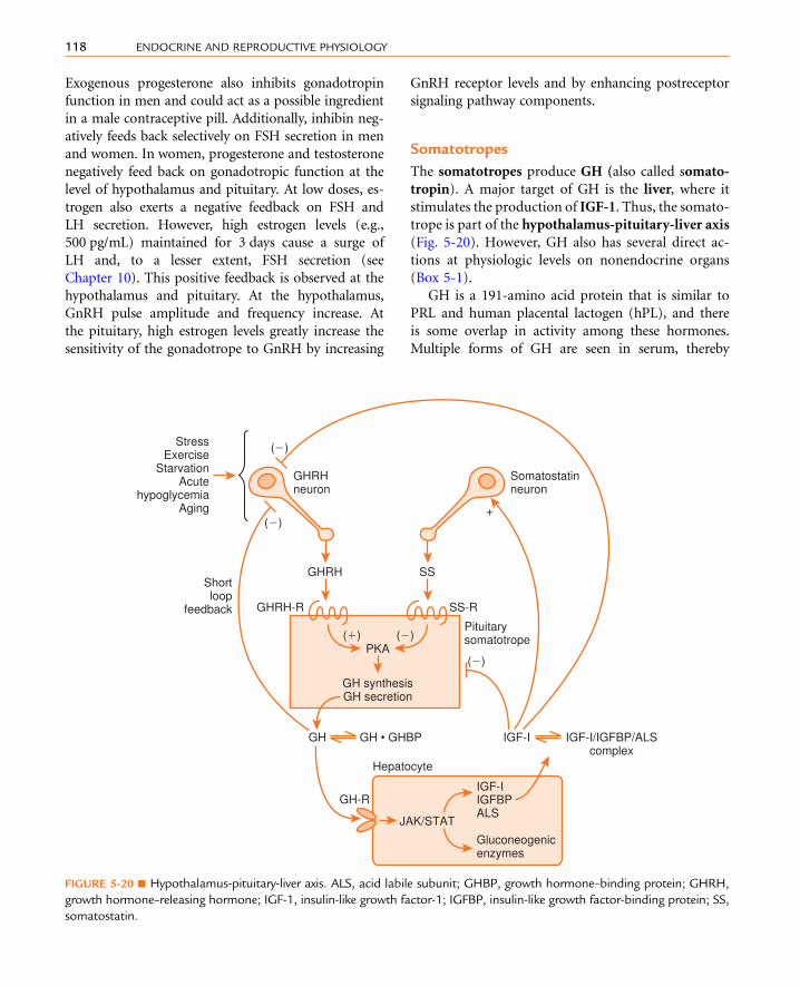
Exogenous progesterone also inhibits gonadotropin
function in men and could act as a possible ingredient
in a male contraceptive pill. Additionally, inhibin neg-
atively feeds back selectively on FSH secretion in men
and women. In women, progesterone and testosterone
negatively feed back on gonadotropic function at the
level of hypothalamus and pituitary. At low doses, es-
trogen also exerts a negative feedback on FSH and
LH secretion. However, high estrogen levels (e.g.,
500 pg/mL) maintained for 3 days cause a surge of
LH and, to a lesser extent, FSH secretion (see
Chapter 10). This positive feedback is observed at the
hypothalamus and pituitary. At the hypothalamus,
GnRH pulse amplitude and frequency increase. At
the pituitary, high estrogen levels greatly increase the
sensitivity of the gonadotrope to GnRH by increasing
GnRH receptor levels and by enhancing postreceptor
signaling pathway components.
Somatotropes
The somatotropes produce GH (also called somato-
tropin). A major target of GH is the liver, where it
stimulates the production of IGF-1. Thus, the somato-
trope is part of the hypothalamus-pituitary-liver axis
(Fig. 5-20). However, GH also has several direct ac-
tions at physiologic levels on nonendocrine organs
(Box 5-1).
GH is a 191-amino acid protein that is similar to
PRL and human placental lactogen (hPL), and there
is some overlap in activity among these hormones.
Multiple forms of GH are seen in serum, thereby
SSGHRH
+
PKA
GH synthesisGH secretion
GH GH • GHBP IGF-I IGF-I/IGFBP/ALScomplex
GHRH-R SS-R
Pituitarysomatotrope
Somatostatinneuron
GHRHneuron
StressExercise
StarvationAcute
hypoglycemiaAging
Shortloop
feedback
(�)
(�)(�)
(�)
(�)
JAK/STAT
Gluconeogenicenzymes
IGF-IIGFBPALS
GH-R
Hepatocyte
FIGURE 5-20 n Hypothalamus-pituitary-liver axis. ALS, acid labile subunit; GHBP, growth hormone–binding protein; GHRH,
growth hormone–releasing hormone; IGF-1, insulin-like growth factor-1; IGFBP, insulin-like growth factor-binding protein; SS,
somatostatin.
118 ENDOCRINE AND REPRODUCTIVE PHYSIOLOGY

constituting a “family of hormones,” with the 191-
amino acid (22-kDa) form representing about 75%
of the circulating GH. The GH receptor is a member
of the cytokine-GH-PRL-erythropoietin receptor fam-
ily and, as such, is linked to the JAK-STAT signaling
pathway. Human GH can also act as an agonist for
the PRL receptor.
About 50% of the 22-kDa form of GH in serum is
bound to the N-terminal portion (the extracellular do-
main) of the GH receptor, called GH-binding protein
(GHBP). Laron dwarfism is characterized by the ab-
sence of or decrease in normal GH receptors and
GHBP and by normal GH secretion. GHBP reduces re-
nal clearance and thus increases the biologic half-life
of GH. The circulating half-life for GH is only about
20 minutes. The liver and kidney are major sites of
hormone degradation.
GH secretion is under dual control by the hypo-
thalamus (see Fig. 5-20). The hypothalamus pre-
dominantly stimulates GH secretion through the
peptide,growthhormone–releasinghormone(GHRH).
GHRH is a member of the vasoactive intestinal
peptide (VIP)-secretin-glucagon family and is
processed into a 44-amino acid peptide with an
amidatedC-terminus froma larger prohormone.GHRH
binds to the GHRH receptor, which is coupled to a
Gs-cAMP-PKA signaling pathway. GHRH enhances
GH secretion and GH gene expression. The hypothala-
mus inhibits pituitary GH synthesis and release through
the peptide somatostatin. Somatostatin is a cyclic
tetradecapeptide that is found in many locations in the
body (see Chapter 2). Somatostatin in the anterior
pituitary inhibits GH and TSH release. Somatostatin
binds to the somatostatin receptor, which lowers
cyclic adenosine monophosphate (cAMP) through a
Gi-linked signaling pathway.
The primary negative feedback on the somatotrope
is exerted by IGF-1. GH stimulates IGF-1 production
by the liver, and IGF-I then inhibits GH synthesis and
secretion at the pituitary and hypothalamus by a clas-
sic long feedback loop. In addition, GH exerts negative
feedback on GHRH release through a short feedback
loop. GH also increases somatostatin release.
GH secretion, like that of ACTH, shows prominent
diurnal rhythms, with peak secretion occurring in the
early morning just before awakening (see Fig. 5-20).
Its secretion is stimulated during deep, slow-wave sleep
(stages III and IV). GH secretion is lowest during the
day. This rhythm is entrained to sleep-wake patterns
rather than light-dark patterns, so a phase shift occurs
in people whowork night shifts. As is typical of anterior
pituitary hormones, GH secretion is pulsatile. The
levels of GH in serum vary widely (0 to 30 ng/mL, with
most values usually falling between 0 and 3). Because of
this marked variation, serum GH values are of minimal
clinical value unless the sampling time is known. Fre-
quently, rather than measuring GH, the clinician mea-
sures IGF-1 because its secretion is regulated by GH,
and IGF-1 has a relatively long circulating half-life that
buffers pulsatile and diurnal changes in secretion.
GH secretion is also regulated by several different
physiologic states (see Fig. 5-20). GH is classified as
one of the stress hormones and is increased by neuro-
genic and physical stress. As discussed later, GH pro-
motes lipolysis, increases protein synthesis, and
antagonizes the ability of insulin to reduce blood glu-
cose. It is not surprising, therefore, that hypoglycemia
is a stimulus for GH secretion, and GH is classified as a
hyperglycemic hormone. Although its secretion is not
regulated by minor variations in serum glucose levels,
its release is stimulated by falling glucose levels or by
n n n n n n n n n n
BOX 5 - 1
METABOLIC ACTIONS OF GROWTHHORMONE
CARBOHYDRATES
Increase blood glucose
Decrease peripheral insulin sensitivity
Increase hepatic output of glucose
Administration results in increased serum insulin
levels
PROTEINS
Increase tissue amino acid uptake
Increase incorporation into proteins
Decrease urea production
Produce positive nitrogen balance
LIPIDS
Promotes lipolysis
Can be ketogenic after long-term administration,
particularly if insulin is deficient
INSULIN-LIKE GROWTH FACTOR
Stimulates IGF production
Stimulates growth
Is mitogenic
119HYPOTHALAMUS-PITUITARY COMPLEX
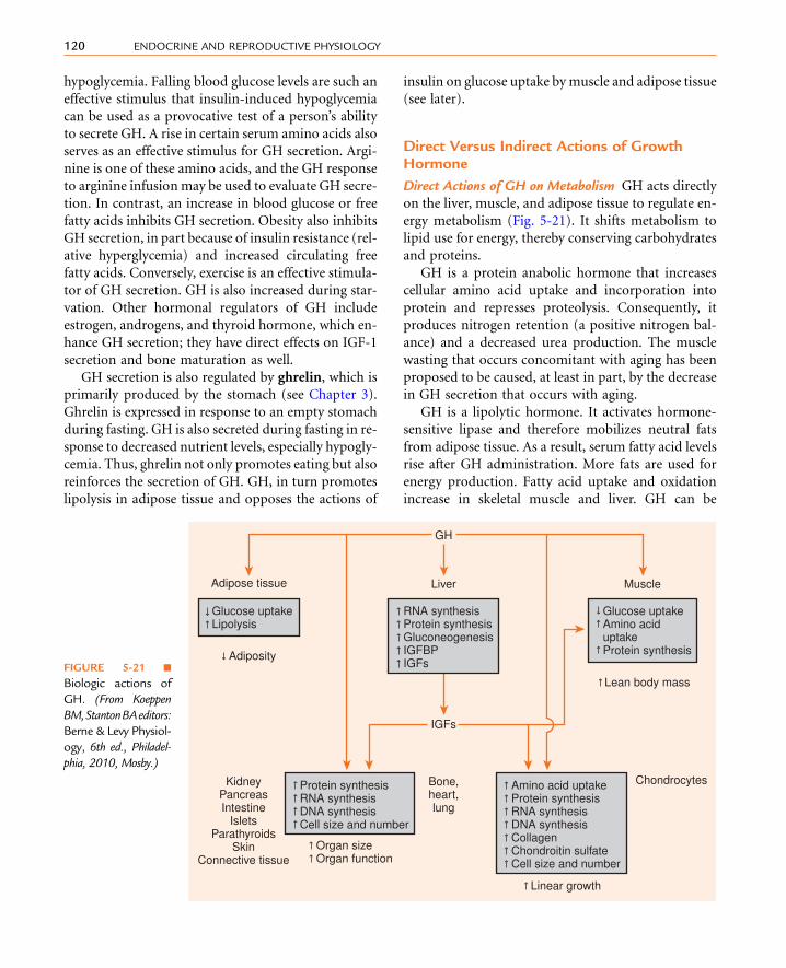
hypoglycemia. Falling blood glucose levels are such an
effective stimulus that insulin-induced hypoglycemia
can be used as a provocative test of a person’s ability
to secrete GH. A rise in certain serum amino acids also
serves as an effective stimulus for GH secretion. Argi-
nine is one of these amino acids, and the GH response
to arginine infusionmay be used to evaluate GH secre-
tion. In contrast, an increase in blood glucose or free
fatty acids inhibits GH secretion. Obesity also inhibits
GH secretion, in part because of insulin resistance (rel-
ative hyperglycemia) and increased circulating free
fatty acids. Conversely, exercise is an effective stimula-
tor of GH secretion. GH is also increased during star-
vation. Other hormonal regulators of GH include
estrogen, androgens, and thyroid hormone, which en-
hance GH secretion; they have direct effects on IGF-1
secretion and bone maturation as well.
GH secretion is also regulated by ghrelin, which is
primarily produced by the stomach (see Chapter 3).
Ghrelin is expressed in response to an empty stomach
during fasting. GH is also secreted during fasting in re-
sponse to decreased nutrient levels, especially hypogly-
cemia. Thus, ghrelin not only promotes eating but also
reinforces the secretion of GH. GH, in turn promotes
lipolysis in adipose tissue and opposes the actions of
insulin on glucose uptake bymuscle and adipose tissue
(see later).
Direct Versus Indirect Actions of GrowthHormone
Direct Actions of GH on Metabolism GH acts directly
on the liver, muscle, and adipose tissue to regulate en-
ergy metabolism (Fig. 5-21). It shifts metabolism to
lipid use for energy, thereby conserving carbohydrates
and proteins.
GH is a protein anabolic hormone that increases
cellular amino acid uptake and incorporation into
protein and represses proteolysis. Consequently, it
produces nitrogen retention (a positive nitrogen bal-
ance) and a decreased urea production. The muscle
wasting that occurs concomitant with aging has been
proposed to be caused, at least in part, by the decrease
in GH secretion that occurs with aging.
GH is a lipolytic hormone. It activates hormone-
sensitive lipase and therefore mobilizes neutral fats
from adipose tissue. As a result, serum fatty acid levels
rise after GH administration. More fats are used for
energy production. Fatty acid uptake and oxidation
increase in skeletal muscle and liver. GH can be
Adipose tissue
GH
Liver Muscle
Lean body mass
Adiposity
Glucose uptake Lipolysis
RNA synthesis Protein synthesis Gluconeogenesis IGFBP IGFs
Glucose uptake Amino acid uptake Protein synthesis
IGFs
KidneyPancreasIntestine
IsletsParathyroids
SkinConnective tissue
Bone,heart,lung
Protein synthesis RNA synthesis DNA synthesis Cell size and number
Amino acid uptake Protein synthesis RNA synthesis DNA synthesis Collagen Chondroitin sulfate Cell size and number
Linear growth
Chondrocytes
Organ size Organ function
FIGURE 5-21 n
Biologic actions of
GH. (From Koeppen
BM,StantonBAeditors:
Berne & Levy Physiol-
ogy, 6th ed., Philadel-
phia, 2010, Mosby.)
120 ENDOCRINE AND REPRODUCTIVE PHYSIOLOGY
ketogenic as a result of the increase in fatty acid oxida-
tion (the ketogenic effect of GH is not seen when in-
sulin levels are normal). If insulin is given along with
GH, the lipolytic effects of GH are abolished.
GH alters carbohydrate metabolism. Many of its
actions may be secondary to the increase in fat mobi-
lization and oxidation; an increase in serum free fatty
acids inhibits glucose uptake and utilization in skeletal
muscle and adipose tissue. After GH administration,
blood glucose rises. The hyperglycemic effects of GH
are mild and slower than those of glucagon and epi-
nephrine. The increase in blood glucose results, in
part, from decreased glucose uptake and use in skeletal
muscle and adipose tissue. Liver glucose output
increases, and this is probably not a result of glyco-
genolysis. In fact, glycogen levels can rise after GH
administration. However, the increase in fatty acid
oxidation and, hence, the rise in liver acetyl coenzyme
A (acetyl CoA) stimulate gluconeogenesis, followed
by increased glucose production from substrates such
as lactate and glycerol.
GH antagonizes the action of insulin at the post-
receptor level in skeletal muscle and adipose tissue
(but not liver). Hypophysectomy (removal of the pitu-
itary gland) can improve diabetic management
because GH, like cortisol, decreases insulin sensitivity.
Because GH produces insulin insensitivity, it is consid-
ered a diabetogenic hormone. When secreted in ex-
cess, GH can cause insulin resistance and ultimately
type 2 diabetes. Normal levels of GH are required
for normal pancreatic function and insulin secretion.
In the absence of GH, insulin secretion declines.
Indirect Effects of GH on Growth GH promotes the
growth of bones and visceral organs. GHadministration
increases skeletal and visceral growth; children without
GH show growth stunting or dwarfism. If given in vivo,
GH results in increased cartilage growth, long-bone
length, and periosteal growth. Most of these are medi-
ated by a group of hormones called IGFs (see Fig. 5-21).
Insulin-like Growth Factors IGFs aremultifunctional
hormones that regulate cellular proliferation, differen-
tiation, and cellular metabolism. These protein hor-
mones resemble insulin in structure and function.
The two hormones in this family, IGF-1 and IGF-2,
are produced in many tissues and have autocrine,
paracrine, and endocrine actions. IGF-1 is the major
form produced in most adult tissues, and IGF-2 is
the major form produced in the fetus. Both
compounds are structurally similar to proinsulin, with
IGF-1 having 42% structural homology with proinsu-
lin. IGFs and insulin cross-react with each other’s re-
ceptors, and IGFs in high concentration mimic the
metabolic actions of insulin. Both IGF-1 and IGF-2
act through type 1 IGF receptors. However, IGF-2 also
binds to the type 2 IGF–mannose-6-phosphate re-
ceptor. This receptor does not resemble the insulin re-
ceptor and does not have intrinsic tyrosine kinase.
Binding to these receptors probably facilitates inter-
nalization and degradation of the growth factor. IGFs
stimulate glucose and amino acid uptake and protein
and DNA synthesis. They were initially called somato-
medins because they mediate GH (somatotropin) ac-
tion on cartilage and bone growth. IGFs have many
other actions, and GH is not the only regulator of
IGF formation. Initially, IGFs were thought to be pro-
duced in the liver in response to a GH stimulus. It is
now known that IGFs are produced in many tissues
and that their many actions are autocrine or paracrine.
The liver is probably the predominant source of circu-
lation IGFs (see Fig. 5-20).
Essentially all circulating IGFs are transported in se-
rum bound to insulin-like growth factor–binding
proteins (IGFBPs). IGFBPs bind to IGFs and then as-
sociate with another protein, called acid labile subunit
(ALS). GH stimulates the hepatic production of IGF-1,
IGFBPs, and ALS (see Fig. 5-20). The IGFBP–ALS–
IGF-1 complex mediates transport and bioavailability
of the IGF-1. Although IGFBPs generally inhibit IGF ac-
tion, they greatly increased the biologic half-life of IGFs
(up to 12 hours). IGFBP proteases degrade IGFBP and
probably play a role locally in generating free (i.e.,
active) IGFs. This is of interest in the context of IGF-
responsive cancers (e.g., prostate cancer), which may
overexpress one or more IGFBP proteases.
Although GH is an effective stimulator for IGF pro-
duction, the correlation between GH and IGF-1 is
greater than the correlation between GH and IGF-2.
During puberty, when GH levels increase, IGF-1 levels
increase in parallel.
IGFs have profound effects on bone and cartilage
(see Fig. 5-21). They stimulate the growth of bones,
cartilage, and soft tissue and regulate essentially all as-
pects of the metabolism of the cartilage-forming cells,
called chondrocytes. IGFs aremitogenic. Although ap-
positional growth of long bones continues after
121HYPOTHALAMUS-PITUITARY COMPLEX

closure of the epiphyses, growth in length ceases. IGFs
stimulate osteoblast replication and collagen and bone
matrix synthesis. Serum IGF levels correlate well with
growth in children.
Interaction of Role of Growth Hormone,Insulin-like Growth Factor, and Insulin inDifferent Metabolic States
When ample supplies of nutrients are available, the
high serum amino acid levels stimulate GH and insu-
lin secretion, and the high serum glucose levels stim-
ulate insulin secretion. The high serum GH, insulin,
and nutrient supplies stimulate IGF production, and
these conditions are appropriate for growth.
However, if the diet is high in calories but low in
amino acids, the conditions change. Whereas the high
carbohydrate availability results in high insulin avail-
ability, the low serum amino acid levels inhibit GH
and IGF production. These conditions allow dietary
carbohydrates and fats to be used, but conditions
are unfavorable for tissue growth.
During fasting, when nutrient availability decreases,
serum GH levels rise, and serum insulin levels fall
(because of hypoglycemia). Importantly, GH cannot
stimulate IGF production in the absence of insulin.
This means that during starvation, with very low levels
of insulin, IGF secretion is effectively inhibited, and the
conditions are not favorable for growth. Under these
circumstances, the decrease in IGF-1 reduces a negative
feedback onGH. The consequent rise in GH secretion is
beneficial because it promotes fat mobilization while
minimizing tissue protein loss. In the absence of insu-
lin, peripheral tissue glucose use decreases, thereby
conserving glucose for essential tissues such as brain.
Pathologic Conditions InvolvingGrowth Hormone
GH is necessary for growth before adulthood. Defi-
ciencies can produce dwarfism, and excesses can pro-
duce gigantism. Normal growth requires not only
normal levels of GH but also normal levels of thyroid
hormones, sex steroids, and insulin.
Dwarfism If a GH deficiency occurs before puberty,
growth is severely impaired (Fig. 5-22). Individuals
with this condition are relatively well proportioned
and have normal intelligence. If the anterior pituitary
deficiency is limited to GH, they can have a normal
life span. They are sometimes pudgy because they
lose GH-induced lipolysis. If they have panhypopi-
tuitary dwarfism (all anterior pituitary hormones
are deficient) so that gonadotropins are deficient,
they may not mature sexually and remain infertile.
People with dwarfism show few metabolic abnormal-
ities other than a tendency toward hypoglycemia,
insulinopenia, and increased insulin sensitivity.
There are multiple potential sites of impairment.
GH secretion may be reduced, GH-stimulated IGF
production may decrease, or IGF action may be
FIGURE 5-22 n A 17-year-old boy with growth hormone
deficiency associated with hypopituitarism. The patient
has short stature for his age (note scale bar) and
underdeveloped genitalia. (From Besser GM, Thorner MO:
Clinical Endocrinology, London, 1994, Mosby-Wolfe.)
122 ENDOCRINE AND REPRODUCTIVE PHYSIOLOGY

deficient. Laron dwarfs are GH resistant because of a
genetic defect in the expression of the GH receptor so
that response to GH is impaired. Hence, although the
serum GH levels are normal to high, they do not pro-
duce IGFs in response to GH. Treating patients
afflicted by Laron dwarfism with GH will not correct
the growth deficiency.
The African pigmy represents another example of
abnormal growth. Individuals with this condition
have normal serum GH levels, but they do not exhibit
the normal rise in IGF that occurs at puberty. How-
ever, the IGF-2 levels are normal. Unlike the Laron
dwarfs, they do not totally lack the IGF response
to GH.
Growth Hormone Deficiency in Adults GH deficiency
in adults is only currently becoming recognized as a
pathologic syndrome. If the GH deficiency occurs after
the epiphyses close, growth is not impaired. A GH de-
ficiency is one of many possible causes of hypogly-
cemia. Recent studies have shown that extended
deficiencies of GH lead to body composition changes.
The percentage of the body weight that is fat increases,
whereas the percentage that is protein decreases. In ad-
dition, muscle weakness and early exhaustion are
symptoms of GH deficiency.
Because the muscle loss that occurs with aging may
result from an age-related decline in GH production,
GH is being used experimentally in elderly people to de-
lay the physical decline associated with aging. The effi-
cacy of this treatment in humans has not been
established.
Growth Hormone Excess Before Puberty Excessive GH
production before puberty results in gigantism. Indi-
viduals with this condition can reach heights greater
than 8 feet. The GH excess results in an increase in
body weight as well as height. Many complications
are associated with gigantism. These individuals
frequently have glucose intolerance and hyperinsuli-
nemia. Overt clinical diabetes mellitus can develop,
but ketoacidosis is rare. They have cardiovascular
problems, including cardiac hypertrophy (all viscera
increase in size); they are more susceptible to infec-
tions than normal; and they rarely live past their
20s. Hypersecretion of GH generally results from pitu-
itary tumors; tumor growth eventually compresses
other components of the anterior pituitary, decreasing
secretion of other anterior pituitary hormones.
Figure 5-23 shows RobertWadlow, the Alton Giant.
At 1 year, he weighed 62 pounds. His adult size was
8 feet, 11 inches and 475 pounds. Note the long
CBA
FIGURE 5-23 n A notable example of growth hormone excess was Robert Wadlow, later known as the “Alton Giant.”
Although he weighed only 9 pounds at birth, he grew rapidly, and by 6 months of age, he weighed 30 pounds. At
1 year of age, he weighed 62 pounds. Growth continued throughout his life. Shortly before his death at age 22 years
from cellulitis of the feet, he was 8 feet, 11 inches tall and weighed 475 pounds. (A and B, from Fadner F: Biography of
Robert Wadlow, Boston, MA, 1944, Bruce Humphreys. C, Courtesy Dr. C. M. Charles and Dr. C. M. MacBryde.)
123HYPOTHALAMUS-PITUITARY COMPLEX

extremities. The androgen deficiency secondary to the
gonadotropin deficiency caused delayed puberty,
resulting in late closure of the epiphyses and contrib-
uting to long bone growth.
Acromegaly Excessive GH secretion after the epiphy-
ses close results in appositional bone growth, but not
a further lengthening of long bones. Cartilage and
membranous bones continue to grow, and gross de-
formities can result. In addition, soft tissue growth
increases, and the abdomen protrudes as a result of
visceral enlargement. Brain weight increases, with a re-
sultant decrease in ventricular size. There is an increase
in the growth of the nose, ears, and mandible, with the
mandibular enlargement producing prognathism and
widely spaced teeth (Fig. 5-24). The calvarium
thickens, and the frontal sinuses enlarge, resulting
in protrusion of the frontal ridge of the orbit of the
eye. The characteristic enlargement of the hands
and feet is the basis for the name acromegaly (acro,
end or extremity; megaly, enlargement). The exces-
sive bone and cartilage growth can produce carpal
tunnel syndrome and joint problems. The voice
deepens because of laryngeal growth. Acromegaly
usually results from a functional tumor of the soma-
totropes. Because it is generally slow in onset,
patients typically do not seek medical help for 13 to
14 years.Unfortunately, by that time, they typically have
permanent physical deformities, including coarsening
of facial features (see Fig. 5-24). People with gigantism
eventually exhibit acromegaly if the condition is not
corrected before puberty. A person with untreated
acromegaly has a shortened life expectancy.
Extended treatment of adults with GH results in
changes in body composition, with the percentage of
body protein increasing and the percentage of body
fat decreasing.
Lactotropes
The lactotrope produces the hormone, PRL, which is
a 199-amino acid single-chain protein. The lactotrope
differs from the other endocrine cell types of the
adenohypophysis in two major ways:
n The lactotrope is not part of an endocrine axis. This
means that PRL acts directly on nonendocrine cells
to induce physiologic changes.
n The production and secretion of PRL is predomi-
nantly under inhibitory control by the hypothalamus
through the neurotransmitter, dopamine. Thus, dis-
ruption of the pituitary stalk and the hypothala-
mohypophyseal portal vessels (e.g., due to surgery
orphysical trauma) results inan increase inPRL levels,
but a decrease in ACTH, TSH, FSH, LH, and GH.
PRL circulates unbound to serum proteins
and thus has a relatively short half-life of about 20
minutes. As is typical of protein hormones, there
is heterogeneity of circulating forms of PRL, and
the 199-amino acid form represents only 60% to
FIGURE 5-24 n Left, Severe acromegaly.Middle, Radiograph of a normal skull. Right, Radiograph of skull of a woman with
acromegaly that demonstrates effects of acromegaly on morphologic features of skull. The sella turcica is enlarged as a
result of growth of pituitary adenoma. The skull is thicker than normal, and protrusion of frontal ridge with
enlargement of frontal sinuses is evident. (Left, From Clinico-pathologic conference: acromegaly, diabetes, hypermetabolism,
proteinuria and heart failure. Am J Med 20:133, 1956; Middle and Right, Courtesy of Dr. C. Joe.)
124 ENDOCRINE AND REPRODUCTIVE PHYSIOLOGY

80% of the PRL measured by radioimmunoassays.
Normal basal serum concentrations are similar in
men and women.
PRL release is normally under tonic inhibition by
the hypothalamus. This is exerted by dopaminergic
tracts that secrete dopamine at the median eminence.
Dopamine binds to the D2 receptor, which is linked
to a Gi signaling pathway. There is also evidence for
the existence of a prolactin-releasing factor (PRF).
The exact nature of this compound is not known, al-
though many factors, including TRH and hormones
in the glucagon family (secretin, glucagon, VIP, and
gastroinhibitory peptide [GIP]), can stimulate PRL
release.
The regulation of PRL secretion during pregnancy
and lactation is discussed in Chapter 11.
Drugs that interfere with dopamine synthesis or
action increase PRL secretion. Many commonly
prescribed antihypertensive drugs and tricyclic antide-
pressant drugs are dopamine inhibitors. Bromocrip-
tine is a dopamine agonist that can be used to inhibit
PRL secretion. Somatostatin, TSH, and GH also inhibit
PRL secretion.
The PRL receptor belongs to the cytokine-GH-
PRL-erythropoietin receptor superfamily. Therefore,
PRL acts through a JAK-STAT signaling pathway
(see Chapter 1). At least 85 different actions have been
proposed for PRL; which actions are seen often de-
pends on the dose of hormone used and the species
studied. In humans, the predominant physiologic role
of PRL is the regulation of essentially every aspect
of postnatal breast development and function, as
discussed in Chapter 11.
CLINICAL BOX 5-9
PRL-secreting tumors (prolactinomas) account for
about 70% of all anterior pituitary tumors. Further-
more, many drugs interfere with dopamine produc-
tion or action and hence increase PRL release. For
these reasons, hyperprolactinemia is a common
disorder in humans. Hyperprolactinemia in women
is associated with oligomenorrhea or amenorrheaand infertility. GnRH release, the gonadotrope
response to GnRH, and the ovarian response to LH
all decrease. In the early stages of the pathologic con-
dition, PRL suppresses follicular maturation, leading
to an inadequate corpus luteum and a short luteal
phase. As the hyperprolactinemia persists, the preovu-
latory estrogen peak is lost, thereby lengthening the
cycle and leading to oligomenorrhea and anovulatory
cycles. Hyperprolactinemia can produce infertility in
men. Although breast enlargement in men can occur,
true gynecomastia (inappropriate growth of breasts)
and galactorrhea (inappropriate flow of milk) are
rare.
The primary symptoms causing men and postmen-
opausal women with PRL-secreting tumors to
seek medical attention may be those resulting from
compression by the pituitary mass. These patients
may experience severe headaches or visual distur-bances that can include bitemporal hemianopia (de-
fect in vision in the temporal half of the field of vision
in both eyes). Both men and women may complain of
decreased libido and signs of hypogonadism.
Dopamine agonists not only inhibit PRL produc-
tion but also repress lactotrope growth and prolifera-
tion. Thus, dopamine agonists can be used as an
antitumor drug in the case of dopamine-responsive
prolactinomas.
Hypopituitarism
Panhypopituitarism There are many causes of hypopi-
tuitarism, which can involve either hypothalamic or
pituitary problems. The deficiencies can be variable
for the different anterior pituitary hormones. The
symptoms of hypopituitarism are slow in onset and
are reflected in deficiencies in the target organs of the
anterior pituitary. Hypogonadism, hypothyroidism,
hypoadrenalism, and growth impairment (in chil-
dren) may be present. People with panhypopituitarism
tend to have sallow complexions because of the ACTH
deficiency, and they become particularly sensitive to
the actions of insulin because of the decreased secretion
of the insulin antagonists GH and cortisol. They are
prone to develop hypoglycemia, particularly when
stressed. Hypogonadism is manifested by amenorrhea
inwomen, impotence inmen, and loss of libido in both
men and women. Some of the clinical manifestations
of hypothyroidism are cold, dry skin, constipation,
hoarseness, and bradycardia. The myxedema (nonpit-
ting edema) associated with severe hypothyroidism
is rare. Adrenal insufficiency caused by the ACTH
deficiency can result in weakness, mild postural hypo-
tension, hypoglycemia, and a loss of pubic and axillary
125HYPOTHALAMUS-PITUITARY COMPLEX
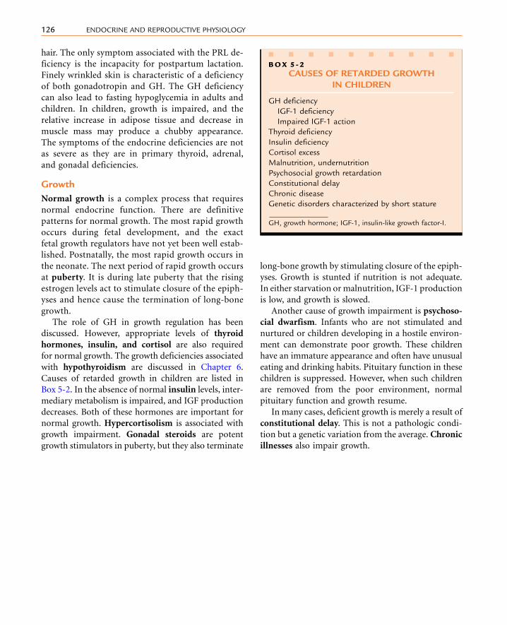
hair. The only symptom associated with the PRL de-
ficiency is the incapacity for postpartum lactation.
Finely wrinkled skin is characteristic of a deficiency
of both gonadotropin and GH. The GH deficiency
can also lead to fasting hypoglycemia in adults and
children. In children, growth is impaired, and the
relative increase in adipose tissue and decrease in
muscle mass may produce a chubby appearance.
The symptoms of the endocrine deficiencies are not
as severe as they are in primary thyroid, adrenal,
and gonadal deficiencies.
Growth
Normal growth is a complex process that requires
normal endocrine function. There are definitive
patterns for normal growth. The most rapid growth
occurs during fetal development, and the exact
fetal growth regulators have not yet been well estab-
lished. Postnatally, the most rapid growth occurs in
the neonate. The next period of rapid growth occurs
at puberty. It is during late puberty that the rising
estrogen levels act to stimulate closure of the epiph-
yses and hence cause the termination of long-bone
growth.
The role of GH in growth regulation has been
discussed. However, appropriate levels of thyroid
hormones, insulin, and cortisol are also required
for normal growth. The growth deficiencies associated
with hypothyroidism are discussed in Chapter 6.
Causes of retarded growth in children are listed in
Box 5-2. In the absence of normal insulin levels, inter-
mediary metabolism is impaired, and IGF production
decreases. Both of these hormones are important for
normal growth. Hypercortisolism is associated with
growth impairment. Gonadal steroids are potent
growth stimulators in puberty, but they also terminate
long-bone growth by stimulating closure of the epiph-
yses. Growth is stunted if nutrition is not adequate.
In either starvation or malnutrition, IGF-1 production
is low, and growth is slowed.
Another cause of growth impairment is psychoso-
cial dwarfism. Infants who are not stimulated and
nurtured or children developing in a hostile environ-
ment can demonstrate poor growth. These children
have an immature appearance and often have unusual
eating and drinking habits. Pituitary function in these
children is suppressed. However, when such children
are removed from the poor environment, normal
pituitary function and growth resume.
In many cases, deficient growth is merely a result of
constitutional delay. This is not a pathologic condi-
tion but a genetic variation from the average. Chronic
illnesses also impair growth.
n n n n n n n n n n
BOX 5 - 2
CAUSES OF RETARDED GROWTHIN CHILDREN
GH deficiency
IGF-1 deficiency
Impaired IGF-1 action
Thyroid deficiency
Insulin deficiency
Cortisol excess
Malnutrition, undernutrition
Psychosocial growth retardation
Constitutional delay
Chronic disease
Genetic disorders characterized by short stature
GH, growth hormone; IGF-1, insulin-like growth factor-I.
126 ENDOCRINE AND REPRODUCTIVE PHYSIOLOGY

S U M M A R Y
1. The pituitary gland (also called the hypophysis)
is derived from a neural structure (the infundib-
ulum) and an epithelial structure (Rathke
pouch). The infundibulum develops into the
neurohypophysis, which includes the median
eminence, infundibular stalk, and pars nervosa
(also called the posterior pituitary). Rathke
pouch develops into the adenohypophysis (also
called anterior pituitary), which includes the
pars distalis, pars tuberalis, and pars intermedia
(the pars intermedia is lost in the adult human).
The pituitary gland sits in a pocket of the sphe-
noid bone, called the sella turcica, at the base of
the forebrain.
2. Magnocellular hypothalamic neurons in the para-
ventricular and supraoptic nuclei project axons
down the infundibular stalk and terminate in
the pars nervosa. The pars nervosa is a neurovas-
cular organ, wherein neurohormones are released
and diffuse into the vasculature.
3. Two neurohormones, antidiuretic hormone
(ADH; also called vasopressin) and oxytocin,
are synthesized in the hypothalamus in the
magnocellular neuronal cell bodies. ADH and
oxytocin are transported intra-axonally down
the hypothalamohypophyseal tracts to the pars
nervosa. Stimuli perceived by the cell bodies
and dendrites in the hypothalamus control the
release of ADH and oxytocin at the pars
nervosa.
4. The primary action of ADH is to promote water
reuptake at the distal nephron and collecting
duct. ADH also has vasopressive actions, which
are important during vasodilatory shock.
5. Diabetes insipidus (DI) is a disease in which either
there is deficient ADH (central DI) or deficient re-
sponse to ADH at the kidney (nephrogenic DI). DI
isassociatedwith increasedurine flow,dehydration,
and increased thirst. The syndrome of inappropri-
ate ADH secretion (SIADH) is characterized by
high ADH levels, which increase volume and blood
pressure, and a low serum osmolarity.
6. Oxytocin acts on the breast to cause milk letdown
during nursing and on the uterus to cause muscu-
lar contractions during labor.
7. The adenohypophysis secretes several tropic
hormones, which are part of the endocrine axes.
An endocrine axis includes the hypothalamus,
the pituitary, and a peripheral endocrine gland.
The set-point of an axis is largely controlled
by the negative feedback of the peripheral hor-
mone on the pituitary and hypothalamus.
8. The adenohypophysis contains five endocrine cell
types: corticotropes, thyrotropes, gonadotropes,
somatotropes, and lactotropes. Corticotropes
secrete ACTH, thyrotropes secrete TSH, gonado-
tropes secrete FSH and LH, somatotropes secrete
GH, and lactotropes secrete PRL.
9. The predominant control exerted by the hypo-
thalamus on the anterior pituitary is mediated
by releasing hormones. These small peptides
are carried through the hypophyseal portal
system to the anterior pituitary, where they
control synthesis and release of the pituitary
hormones ACTH, TSH, FSH, LH, and GH.
PRL is under predominantly inhibitory control
by the hypothalamus through the catecholamine
dopamine.
10. GH stimulates growth primarily through the reg-
ulation of the growth-promoting hormones IGF-1
and IGF-2. GH also has metabolic actions. It raises
blood glucose by decreasing peripheral tissue
utilization. It is protein anabolic and lipolytic.
11. The predominant action of PRL in humans is the
initiation and maintenance of lactation.
12. Normal growth is a complex process that requires
normal endocrine function. Consequently, gro-
wth deficiencies are associated with many
endocrine disorders in children.
127HYPOTHALAMUS-PITUITARY COMPLEX

S E L F - S T U D Y P R O B L E M S
1. Explain the origins of the neurohypophysis and
adenohypophysis.2. Howisaportionof theneurohypophysis critical to
the normal function of the anterior pituitary?
3. Which is ADH secretion more sensitive to:
changes in volume or changes in osmolality?
4. Describe the production of ADH in terms of syn-
thesis, processing, and secretion. Where does
each of these occur?
5. How does SIADH result in hyponatremia?6. Name two proteins that are mutated in congen-
ital nephrogenic diabetes insipidus.
7. Cushing disease is hypercortisolism due to hypo-
thalamic-independent excessiveACTHproduction
by corticotropes. Is this a primary, secondary, ortertiary endocrine disorder?
8. McCune-Albright syndrome is a genetically mo-
saic condition in which some cells express a Gs
protein with an activating mutation. Explain
why this can lead to excessive growth of somato-
tropes and high GH levels.
9. Why is GH referred to as a diabetogenic
hormone?10. How does stress (in any form) affect the secre-
tion of the following: CRH, TRH, GnRH, and
GHRH?
11. How is skin darkening related to primary
hypocortisolism?
12. What is the effect of hypoglycemia on GH and
IGF-1 secretion?
K E Y W O R D S A N D C O N C E P T S
n Acid labile subunit (ALS)n Acidophilsn Acromegaly
For full list of keywords and concepts see Student
Consult
SUGGESTED READ INGS
Appel JG, Bergsneider M, Vinters H, et al: Acromegaly due to an ec-
topic pituitary adenoma in the clivus: Case report and review of
literature, Pituitary Sep 30. Epub ahead of print.
Birzniece V, Nelson AE, Ho KK: Growth hormone and physical per-
formance, Trends Endocrinol Metab 22:171–178, 2011.
Cariboni A, Maggi R, Parnavelas JG: From nose to fertility: The long
migratory journey of gonadotropin-releasing hormone neurons,
Trends Neurosci 30:638–644, 2007.
Clayton PE, Banerjee I, Murray PG, et al: Growth hormone, the
insulin-like growth factor axis, insulin and cancer risk, Nat
Rev Endocrinol 7:11–24, 2011.
Fideleff HL, Boquete HR, SuarezMG, et al: Prolactinoma in children
and adolescents, Horm Res 72:197–205, 2009.
Pendleton C, Adams H, Salvatori R, et al: On the shoulders of giants:
Harvey Cushing’s experience with acromegaly and gigantism at
the Johns Hopkins Hospital, 1896–1912, Pituitary 14:53–60,
2011.
Raff H: Cushing’s syndrome: Diagnosis and surveillance using sali-
vary cortisol, Pituitary 15:64–70, 2012.
128 ENDOCRINE AND REPRODUCTIVE PHYSIOLOGY

K E Y W O R D S A N D C O N C E P T S
n Adenohypophysisn Adrenal cortexn Adrenocorticotropic hormone (ACTH)n African pigmyn a-Glycoprotein subunit (a-GSU)n Anterior pituitaryn Antidiuresisn Antidiuretic hormonen Aquaporin-2n Atrial natriuretic peptide (ANP)n Basophilsn Bitemporal hemianopian Corticotropen Corticotropin-releasing hormone (CRH)n Cortisoln Craniopharyngioman Diabetes insipidusn Diabetogenic hormonen Diaphragma sellaen Dopaminen Dwarfismn Empty sella syndromen Endocrine axesn Follicle-stimulating hormone (FSH)n GH-binding protein (GHBP)n Ghrelinn Gigantismn Gonadotropen Gonadotropin-releasing hormone (GnRH)n Growth hormone (GH)n Growth hormone–releasing hormone(GHRH)
n Herring bodiesn Human chorionic gonadotropin (hCG)n Human placental lactogen (hPL)n Hyperglycemic hormonen Hypophysectomyn Hypophysiotropic region of the hypothalamusn Hypophysisn Hypothalamohypophyseal portal vesselsn Hypothalamohypophyseal tractn Hypothalamus-pituitary-adrenal (HPA) axisn Hypothalamus-pituitary-thyroid axisn IGF-binding protein (IGFBP)n IGFBP proteases
n IGF-2n Infundibulumn Insulin-like growth factor-1 (IGF-1)n KAL genen Kallmann syndromen Lactational amenorrhean Lactotropen Laron dwarfismn Lipolytic hormonen Livern Luteinizing hormone (LH)n Magnocellular neuronsn Median eminencen Melanocortin-2 receptor (MC2R)n Milk ejection (letdown)n Neuroendocrine reflexn Neurohypophysisn Neurophysin-In Neurophysin-IIn Ovaryn Oxytocinn Panhypopituitarismn Panhypopituitary dwarfismn Paraventricular nucleus (PVN)n Pars distalisn Pars intermedian Pars nervosan Pars tuberalisn Parvicellular neuronsn Peripheral endocrine organsn Pituicyten Pituitary apoplexyn Pituitary glandn Pituitary glycoprotein hormonen Positive feedbackn Posterior lobe of the pituitaryn Prepro-oxyphysinn Preprovasophysinn Primary endocrine disordern Progesteronen Prolactin (PRL)n Prolactin-releasing factor (PRF)n Pro-opiomelanocortin (POMC)n Protein anabolic hormonen Psychosocial dwarfismn Psychosocial Rathke pouchn Releasing hormones
128.e1HYPOTHALAMUS-PITUITARY COMPLEX

n Secondary endocrine disordern Sella turcican Set-pointn Sheehan syndromen Somatomedinn Somatostatinn Somatotropen Stress hormonesn Supraoptic nucleus (SON)n Syndrome of inappropriate secretion ofantidiuretic hormone (SIADH)
n Tertiary endocrine disorder
n Tertiary hypercortisolismn Tertiary hypogonadotropic hypogonadismn Testisn Testosteronen Thyroid glandn Thyroid hormonen Thyroid-stimulating hormone (TSH)n Thyrotropen Thyrotropinn Thyrotropin-releasing hormone (TRH)n Tropic hormonesn Vasopressin-2 (V2) receptor
128.e2 ENDOCRINE AND REPRODUCTIVE PHYSIOLOGY

n n n n n n n n n n n n n n n
6THE THYROID GLAND
O B J E C T I V E S
1. Explain the mechanism of synthesis of the thyroid
hormones.
2. Describe the regulation of thyroid function and the ac-
tions of thyroid hormones.
3. Compare and contrast the functions of thyroxine and
triiodothyronine.
4. Draw the regulatory feedback loop for the regulation of
thyroid function.
5. Understand the etiology, major symptoms, and patho-
physiology of the symptoms for Graves disease, Hashi-
moto thyroiditis, sporadic congenital hypothyroidism,
and cretinism.
The thyroid gland produces the prohor-
mone, tetraiodothyronine (T4), and the active hor-
mone, triiodothyronine (T3). The synthesis of T4
and T3 requires iodine, which can be a limiting factor
in some parts of the world. Much of T3 is also made by
peripheral conversion of T4 to T3. T3 acts primarily
through a nuclear receptor that regulates gene expres-
sion. T3 is critical for normal brain development and
has broad effects on metabolism and cardiovascular
function in the adult.
ANATOMY AND HISTOLOGY OFTHE THYROID GLAND
The thyroid gland is composed of right and left
lobes that sit anterolaterally to the trachea (Fig. 6-1).
Normally, the lobes of the thyroid gland are connected
by a midventral isthmus. The functional unit of the
thyroid gland is the thyroid follicle, a spherical struc-
ture, about 200 to 300 mm in diameter, which is sur-
rounded by a single layer of thyroid epithelial cells
(Fig. 6-2). The epithelium sits on a basal lamina, which
is the outermost structure of the follicle and is sur-
rounded by a rich capillary supply. The apical side
of the follicular epithelium faces the lumen of the fol-
licle. The follicular lumen itself is filled with colloid
composed of thyroglobulin, which is secreted and
iodinated by the thyroid epithelial cells. The size of
the epithelial cells and the amount of colloid are
dynamic features that change with the activity of
the gland. The thyroid gland contains another type
of cell in addition to follicular cells. Scattered within
the gland are parafollicular cells, called C cells.
These cells are the source of the polypeptide hormone,
calcitonin, whose function is unclear in humans.
129

CLINICAL BOX 6-1
Although parafollicular C cells and calcitonin may be of
minimal importance in normal humans, C cells can give
rise tomedullary thyroidcarcinoma.Medullary cancer is
the most aggressive form of thyroid cancer, and spread
to lungs, liver, bone, or other organs drastically reduces
survival. Although most medullary cancer is sporadic,
about one fifth of cases are familial. The familial forms
of medullary thyroid cancer are due to activating muta-
tionsof theRETprotooncogene. RET isa tyrosine kinasereceptor that interactswith co-receptors and is activated
byglial-derivedneurotrophic factorandrelatedproteins.
Familial medullary thyroid cancer can occur indepen-
dently of other endocrine glands or as part of amultipleendocrine neoplasia (MEN), which in this case also
involvesadrenalmedulla chromaffincells (pheochromo-
cytoma), parathyroid glands, and/or neuromas. These
cancers remain differentiated enough to secrete calcito-nin, and assay of calcitonin in the blood is useful in
assessing treatment and for follow-up examinations
for recurrence.Medullary thyroid cancer is treatedby to-
tal thyroidectomyand removal of regional lymphnodes.
Effective chemotherapy regimens that target the tyro-
sine kinase receptor, c-ret, have also been developed.
PRODUCTION OF THYROIDHORMONES
The secretory products of the thyroid gland are
iodothyronines (Fig. 6-3), a class of hormones resulting
from the coupling of two iodinated tyrosine molecules.
About 90% of the thyroid output is 3,5,30,50-tetraio-dothyronine (thyroxine, or T4). T4 is a prohormone.
About 10% is 3,5,30-triiodothyronine (T3), which is
the active form of thyroid hormone. Less than 1% of
thyroid output is 3,30,50-triiodothyronine (reverse T3,
or rT3), which is inactive. Normally, these three hor-
mones are secreted in the same proportions as they are
stored in the gland.
Because the primary product of the thyroid gland is
T4, yet the active form of thyroid hormone is T3, the
thyroid axis relies heavily on peripheral conversion
through the action of thyronine-specific deiodinases
(see Fig. 6-3). Most conversion of T4 to T3 by type 1
deiodinase occurs in tissues with high blood flows
and rapid exchanges with plasma, such as liver, kid-
neys, and skeletal muscle. This process supplies circu-
lating T3 for uptake by other tissues in which local T3
generation is too low to provide sufficient thyroid
Hyoid bone
Thyroid cartilage
Pyramidal lobe
Thyroid gland
Trachea
A
B
FIGURE 6-1 nA, Anatomy of the thyroid gland. B, Image of pertechnetate uptake by a normal thyroid gland. (Modified from
Drake RL, Vogl W, Mitchell AWM: Gray’s Anatomy for Students, Philadelphia, 2005, Churchill Livingstone.)
130 ENDOCRINE AND REPRODUCTIVE PHYSIOLOGY

hormone. Type 1 deiodinase is also expressed in the
thyroid (again, where T4 is abundant) and has a rela-
tively low affinity (i.e., a Km of 1 mM) for T4. The
levels of type 1 deiodinase are paradoxically increased
in hyperthyroidism, which contributes to elevated
circulating T3 levels in this disease.
The brain maintains constant intracellular levels of
T3 by a high-affinity deiodinase called type 2 deiodi-
nase, which is expressed in glial cells in the central
nervous system (CNS). Type 2 deiodinase has a Km of
1 nM and maintains intracellular concentrations of T3
even when free T4 falls to low levels. Type 2 deiodinase
is also present in the thyrotropes of the pituitary. In the
pituitary, type 2 deiodinase acts as a “thyroid axis
sensor” thatmediates the ability of circulating T4 to feed
back on TSH secretion (see later). The expression of
type 2 deiodinase is increased during hypothyroidism,
which helps maintain constant T3 levels in the brain.
FIGURE 6-2 n Nor-
mal rat thyroid.
Single layer of cuboi-
dal epithelial cells
(follicular cells; FC)
surround colloid (C).
Parafollicular cells (P)
produce calcitonin
(see Chapter 4).
Prohormone
I I
I I
3,5,3�5� -Tetraiodothyronine (thyroxine, or T4)
NH2
CH2CHCOOHHO O
Outer ring deiodination(activation)
Deiodinases types 1 and 2
Active
I I
I
3,5,3� -Triiodothyronine (T3)
NH2
CH2CHCOOHHO O
Inner ring deiodination(inactivation)
Deiodinase type 3
Inactive
I I
I
3,3�5� -Triiodothyronine (reverse T3)
NH2
CH2CHCOOHHO O
FIGURE 6-3 n Structure
of iodothyronines, T4,
T3, and reverse T3.
131THE THYROID GLAND

There also exists an “inactivating” deiodinase, called
type 3 deiodinase. Type 3 deiodinase is a high-affinity,
inner-ring deiodinase that converts T4 to the inactive
rT3. Type 3 deiodinase is increased during hyperthy-
roidism, which helps to blunt the overproduction
of T4. All forms of iodothyronines are further deiodi-
nated, eventually to noniodinated thyronine.
Iodide Balance
Because of the unique role of iodide (I2; the water-
soluble, ionized form of iodine, or I2) in thyroid
physiology, a description of thyroid hormone synthesis
requires some understanding of iodide turnover
(Fig. 6-4). An average of 400 mg of iodide per person
is ingested daily in the United States, compared with
a minimum daily requirement of 150 mg for adults,
90 to 120 mg for children, and 200 mg for pregnant
women. In the steady state, virtually the same amount,
400 mg, is excreted in the urine. Iodide is actively con-
centrated in the thyroid gland, salivary glands, gastric
glands, lacrimal glands,mammary glands, and choroid
plexus. About 70 to 80 mg of iodide is taken up daily bythe thyroid gland from a circulating pool that contains
about 250 to 750 mg of iodide. The total iodide contentof the thyroid gland averages 7500 mg, virtually all of
which is in the form of iodothyronines. In the steady
state, 70 to 80 mg of iodide, or about 1% of the total,
is released from the gland daily. Of this amount,
75% is secreted as thyroid hormone, and the remainder
is secreted as free iodide. The large ratio (100:1) of
iodide stored in the form of hormone to the amount
turned over daily protects the individual from the
effects of iodide deficiency for about 2 months. Iodide
is also conserved by a marked reduction in the renal
excretion of iodide as the concentration in serum falls.
Overview of Thyroid Hormone Synthesis
To understand thyroid hormone synthesis and secre-
tion, one must appreciate the directionality of each
process as it relates to the polarized thyroid epithelial
cell (Fig. 6-5). Synthesis of thyroid hormone requires
two precursors, thyroglobulin and iodide. Iodide is
transported across cells from the basal (vascular) side
to the apical (follicular luminal) side of the thyroid
epithelium. Amino acids are assembled by translation
into thyroglobulin, which is then secreted from the
apical membrane into the follicular lumen. Thus, syn-
thesis involves a basal-to-apical movement of precur-
sors into the follicular lumen (see Fig. 6-5; black
arrows). Actual synthesis of iodothyronines occurs
enzymatically in the follicular lumen close to the apical
membrane of the epithelial cells (see later). Secretion
involves fluid phase endocytosis of iodinated thyro-
globulin and apical-to-basal movement of the endo-
cytic vesicles and their fusion with lysosomes.
Stool10 �g HI
Urine390 �g I
Tissues
Thyroid
Extracellularfluid
10 �g HI
60 �g HI
80 �g I
400 �g I
Diet
320 �g I
20 �g I
50 �g I
FIGURE 6-4 n Iodine distribution and turnover in humans.
132 ENDOCRINE AND REPRODUCTIVE PHYSIOLOGY

Thyroglobulin is then enzymatically degraded, which
results in the release of thyroid hormones from the
thyroglobulin peptide backbone. Finally, thyroid hor-
mones move across the basolateral membrane, proba-
bly through a specific transporter, and ultimately into
the blood. Thus, secretion involves an apical-to-basal
movement (see Fig. 6-5; orange arrows). There are also
scavenger pathways within the epithelial cell that reuse
iodine and amino acids after enzymatic digestion of
thyroglobulin (see Fig. 6-5; open arrows).
Synthesis of Iodothyronines Within aThyroglobulin Backbone
Iodide is transported into the gland against chemical
and electrical gradients by an Na1-I2 symporter
(NIS) located in the basolateral membrane of thyroid
epithelial cells. Normally, a thyroid-to-plasma free io-
dide ratio of 30 is maintained. This so-called iodide
trap is highly expressed in the thyroid gland, but
NIS is also expressed at lower levels in the placenta, sal-
ivary glands, and actively lactating breast. One iodide
ion is transported uphill against an iodide gradient,
whereas two sodium ions move down the electro-
chemical gradient from the extracellular fluid into
the thyroid cell. The energy source for this secondary
active transporter is provided by a Naþ, Kþ-ATPase inthe plasma membrane. Expression of the NIS gene is
inhibited by iodide and stimulated by thyroid-
stimulating hormone (TSH; see below). Numerous
inflammatory cytokines also suppress NIS gene ex-
pression. A reduction in dietary iodide intake depletes
the circulating iodide pool and greatly enhances the
Lysosomes
Proteases
Deiodinase
Microtubules,microfilaments
Colloid inendosomes
Aminoacids
Pseudopods
Pendrin
Apical membrane
Basal membrane
Thyroglobulin
Endoplasmic reticulum
DIT
DIT
MIT
MIT
TG
T4T3
T4T3
T4T3
I–
I–
I–
I–
I–
DITMITThyroid
peroxidase
TG
TG +
T4T3
DITMIT
TG
T4T3
DITMIT
TG
TG
Folliclelumen
Cytoplasm
TG
T4T3
Golgi
Vesicles
NIS
FIGURE 6-5 n Synthesis
(black arrows) and secre-
tion (orange arrows) of
thyroid hormones by
the thyroid epithelial
cell. Open arrows denote
pathways involved in
the conservation of io-
dine and amino acids.
133THE THYROID GLAND

activity of the iodide trap. When dietary iodide intake
is low, the rates of thyroid uptake of iodide can reach
80% to 90%.
The steps in thyroid hormone synthesis are shown
in Figure 6-6. After entering the gland, iodide rapidly
moves to the apical plasma membrane of the ep-
ithelial cells. From there, iodide is transported into
the lumen of the follicles by a sodium-independent
iodide-chloride transporter, named pendrin.
CLINICAL BOX 6-2
Pendred syndrome refers to condition due to an auto-
somal recessive mutation in the pendrin gene (referred
to as PDS or SLC26A4). Because iodide is not efficiently
transported into the follicular lumen, patients develop
hypothyroidism. Some patients exhibit enlarged thy-
roid glands called goiters. This form of hypothyroidism
can be treated with replacement thyroxine. Unfortu-
nately, pendrin is also expressed in the inner ear
and is required for normal structural development of
the inner ear. Thus, patients with Pendred syndrome
experience hearing loss in infancy or early childhood.
Once in the follicular lumen, iodide (I�) is imme-
diately oxidized and incorporated into tyrosine resi-
dues within the primary structure of thyroglobulin.
Thyroglobulin is continually synthesized and exocy-
tosed into the follicular lumen and is iodinated to
form either monoiodotyrosine (MIT) or diiodotyro-
sine (DIT) (see Fig. 6-6). After iodination, two DIT
molecules are coupled to form T4, or one MIT and
one DIT molecule are coupled to form T3. The cou-
pling also occurs between iodinated tyrosines that re-
main part of the primary structure of thyroglobulin.
This entire sequence of reactions is catalyzed by thy-
roid peroxidase (TPO), which is an enzyme complex
that spans the apical membrane. The immediate oxi-
dant (electron acceptor) for the reaction is hydrogen
peroxide (H2O2). The mechanism whereby H2O2 is
generated in the thyroid gland involves NADPH
oxidase that is also localized to the apical membrane.
When iodide availability is restricted, the formation
of T3 is favored. This response provides more active
hormone per molecule of organified iodide. The
proportion of T3 is also increased when the gland is
hyperstimulated by TSH or other activators.
I I
I I
3,5,3�5�-Tetraiodothyronine (thyroxine, or T4)
NH2
CH2CHCOOHHO O
I I
I
3,5,3�-Triiodothyronine (T3)
NH2
CH2CHCOOHHO O
I
MIT
NH2
CH2CHCOOHHO
I
I
DIT
NH2
CH2CHCOOH +HO
I
I
DIT DIT
NH2
CH2CHCOOH +HO
I
INH2
CH2CHCOOHHO
I
I
Diiodotyrosine (DIT)
or
NH2
CH2CHCOOHHO
I
Monoiodotyrosine (MIT)
NH2
CH2CHCOOHHO
Tyrosine
NH2
CH2CHCOOHI2 + HO
2I− + H2O2 I2
FIGURE 6-6 n Reactions involved in the generation of iodide, MIT, DIT, T3, and T4.
134 ENDOCRINE AND REPRODUCTIVE PHYSIOLOGY

Secretion of Thyroid Hormones
After thyroglobulin has been iodinated, it is stored in the
lumen of the follicle as colloid (see Fig. 6-2). Release
of the T4 and T3 into the bloodstream requires endocy-
tosis and lysosomal degradation of thyroglobulin (see
Fig. 6-5; orange arrows). Enzymatically released T4 and
T3 then leave thebasal sideof the cell andenter theblood.
The MIT and DIT molecules, which also are re-
leased during proteolysis of thyroglobulin, are rapidly
deiodinated within the follicular cell by the enzyme,
intrathyroidal deiodinase (see Fig. 6-5; open arrows).
This deiodinase is specific forMITandDITand cannot
use T4 and T3 as substrates. The iodide is then recycled
into T4 and T3 synthesis. Amino acids from the diges-
tion of thyroglobulin reenter the intrathyroidal amino
acid pool and can be reused for protein synthesis. Only
minor amounts of intact thyroglobulin leave the follic-
ular cell under normal circumstances.
CLINICAL BOX 6-3
Because of its ability to trap and incorporate iodine
into thyroglobulin (called organification), the activityof the thyroid canbeassessedby radioactive iodineup-take (RAIU). For this, a tracer dose of 123I is adminis-
tered, and the RAIU is measured by placing a gamma
detector on the neck after 4 to 6 hours and after
24 hours. In the United States, where the diet is rela-
tively rich in iodine, theRAIU is about15%after 6hours
and 25% after 24 hours (Fig. 6-7). Abnormally high
RAIU (>60%) after 24 hours indicates hyperthyroid-
ism. Abnormally low RAIU (<5%) after 24 hours
indicates hypothyroidism. In individuals with extreme
chronic stimulation of the thyroid (Graves disease–
associated thyrotoxicosis), iodide is trapped, organified,
and released as hormone very rapidly. In these cases of
elevated turnover, the 6-hour RAIU will be very high,
but the24-hourRAIUwill be lower (seeFig.6-8).Anum-
ber of anions, such as thiocyanate (CNS�), perchlorate(HClO4
�), and pertechnetate (TcO4�), are inhibitors
of iodide transport through the NIS. If iodide cannot
be rapidly incorporated into tyrosine (organification
defect) after its uptake by the cell, administration of
one of these anions will, by blocking further iodide
uptake, causea rapid releaseof the iodide fromthegland
(see Fig. 6-8). This release occurs as a result of the high
thyroid-to-plasma concentration gradient of iodide.
The thyroid can be imaged using the iodine isotopes123Ior 131I, or the iodinemimic,pertechnetate (99mTc),
followed by imaging with a rectilinear scanner or
gammacamera. Imagingcandisplay the size andshape
of the thyroid (see Fig. 6-1B), as well as heterogeneities
of active versus inactive tissuewithin the thyroid gland.
Such heterogeneities are often due to the development
of thyroid nodules, which are regions of enlarged fol-
licles with evidence of regressive changes indicating cy-
cles of stimulation and involution. Particular hot
nodules (i.e., nodules that display a high RAIU) on
imaging, are usually not cancerous but may lead to
thyrotoxicosis (hyperthyroidism; see later). Cold nod-
ules are 10 times more likely to be cancerous than hot
nodules. Such nodules can be sampled for pathology
analysis by fine-needle aspiration biopsy.
The thyroid can also be imaged by ultrasonogra-phy, which is superior in resolution to RAIU imaging.
Ultrasonography is used to guide the physician during
fine-needle aspiration biopsy of a nodule. Highest
resolution of the thyroid is achieved with magneticresonance imaging.
TRANSPORT AND METABOLISM OFTHYROID HORMONES
Secreted T4 and T3 circulate in the bloodstream almost
entirely bound to proteins. Normally, only about
0.04% of total plasma T4 and 0.4% of total plasma
T3 exist in the free state (Table 6-1). Free T3 is biolog-
ically active and mediates thyroid hormone effects on
peripheral tissues as well as in negative feedback on the
pituitary and hypothalamic (see later). The major
binding protein is thyroxine-binding globulin
(TBG). TBG is synthesized in the liver and binds
one molecule of T4 or T3.
About 70% of circulating T4 and T3 is bound to
TBG; 10% to 15% is bound to another specific
thyroid-binding protein, called transthyretin (TTR).
Albumin binds 15% to 20%, and 3% is bound to
lipoproteins. Ordinarily, only alterations in TBG con-
centration significantly affect total plasma T4 and T3
levels. Two important biologic functions have been as-
cribed to TBG. First, it maintains a large circulating
reservoir of T4, which buffers any acute changes in
thyroid gland function. Second, the binding of plasma
T4 and T3 to proteins prevents the loss of these rela-
tively small hormone molecules in the urine and
thereby helps conserve iodide. TTR, in particular,
provides thyroid hormones to the CNS.
135THE THYROID GLAND

CLINICAL BOX 6-4
There are several transporters that mediate thyroid
hormone transport across cell membranes Thyroidhormone transporters include sodium taurocholate
co-transporting polypeptide (NCTP), organic anion
transporting polypeptide (OATP), L-type amino acid
transporter (LAT), and the monocarboxylate trans-
porter (MCT). These transporters show specificity
with respect to T4 versus T3 binding and cell-specific
expression. MCT8 is required for neuronal uptake of
thyroid hormones. Mutations in MCT8 are linked to
severe psychomotor retardation (Allan-Herndon-
Dudley syndrome) that cannot be treated with exoge-
nous T3 or T4.
Regulation of Thyroid Function
The most important regulator of thyroid gland func-
tion and growth is the hypothalamic-pituitary-
thyroid axis (see Chapter 5). TSH stimulates every
aspect of thyroid function. TSH has immediate, inter-
mediate, and long-term actions on the thyroid epithe-
lium. Immediate actions of TSH involve induction of
pseudopod extension, endocytosis of colloid, and for-
mation of colloid droplets in the cytoplasm, which
represent thyroglobulin within endocytic vesicles.
Shortly thereafter, iodide uptake and TPO activity
increase. Concurrently, TSH also stimulates glucose
entry into the hexose monophosphate shunt pathway,
which generates the NADPH that is needed for the
TABLE 6-1
Average Thyroid Hormone Turnover
T4 T3 rT3
Daily production (mg) 90 35 35
From thyroid (%) 100 25 5
From T4 (%) — 75 95
Extracellular pool (mg) 850 40 40
Plasma concentration
Total (mg/dL) 8.0 0.12 0.04
Free (ng/dL) 2.0 0.28 0.20
Half-life (days) 7 1 0.8
Metabolic clearance (L/day) 1 26 77
Fractional turnover per day (%) 10 75 90
100
75
50
25
123 I
Upt
ake
(% o
f dos
e)
Perchlorate
Organification defect
Hours after administration of 123I
Hypothyroidism
Normal
Extreme stimulationof thyroid gland(high turnover)
Hyperthyroidism
6 12 18 24
FIGURE 6-7 n Thyroid gland iodothyronine
uptake curves for normal, hypothyroid,
hyperthyroid, and organification defective
states.
136 ENDOCRINE AND REPRODUCTIVE PHYSIOLOGY

peroxidase reaction. TSH also stimulates the proteol-
ysis of thyroglobulin and the release of T4 and T3 from
the gland. Intermediate effects of TSH on the thyroid
gland occur after a delay of hours to days and involve
protein synthesis and the expression of numerous
genes, including those encoding NIS, thyroglobulin,
and TPO. Sustained TSH stimulation leads to the
long-term effects of hypertrophy and hyperplasia of
the follicular cells. Capillaries proliferate, and thyroid
blood flow increases. These actions, which underlie
the growth-promoting effects of TSH on the gland,
are supported by the local production of growth
factors. A noticeably enlarged thyroid gland is called
a goiter (Fig. 6-8).
CLINICAL BOX 6-5
Goiter can develop in response to multiple imbalances
and disease within the hypothalamus-pituitary-thyroid
axis, coexisting with hypothyroidism, euthyroidism
(normal), and hyperthyroidism. These imbalances
include the following:
Primary Hypothyroidismn Lack of adequate iodine in the diet (nontoxic
goiter, endemic goiter)
n Benign nodules or mutation of growth-related
gene (nontoxic goiter)
n Sporadic hypothyroidism of unknown etiology
(nontoxic goiter)
n Chronic thyroiditis (Hashimoto disease;
autoimmune-induced deficiency in thyroid
function)
Hyperthyroidismn Excessive stimulation of the TSH receptor by an
autoantibody (Graves disease)
n ExcessivesecretionofTSHfromaTSH-producing
tumor (i.e., secondary hyperthyroidism)
n Thyroid hormone–producing (toxic) adenoma
(nodular) or toxic multinodular goiter
n An inactivating mutation in the TRb-2 (see later)
The regulation of thyroid hormone secretion by
TSH is under exquisite negative feedback control. Cir-
culating thyroid hormones act on the pituitary gland
to decrease TSH secretion, primarily by repressing
TSH-b subunit gene expression. The pituitary gland
expresses the high-affinity type 2 deiodinase. Thus,
small changes in free T4 in the blood result in signifi-
cant changes in intracellular T3 in the pituitary thyro-
trope. Because the diurnal variation of TSH secretion
is small, thyroid hormone secretion and plasma
concentrations are relatively constant. Only small
nocturnal increases in secretion of TSH and release
of T4 occur. Thyroid hormones also feed back on
the hypothalamic TRH-secreting neurons. In these
neurons, T3 inhibits the expression of the prepro-
TRH gene.
Another important regulator of thyroid gland
function is iodide itself, which has a biphasic action.
At relatively low levels of iodide intake, the rate of
Colloid in endocytic vesicles
Colloid in lumen ofthyroid follicle
AA
BB
FIGURE 6-8 n The thyroid gland is located in the anterior
neck, where it is easily visualized and palpated when it is
enlarged (goiter).
137THE THYROID GLAND
thyroid hormone synthesis is directly related to iodide
availability. However, if the intake of iodide exceeds
2 mg/day, the intraglandular concentration of iodide
reaches a level that suppresses NADPHoxidase activity
and the NIS and TPO genes, and thereby the mecha-
nism of hormone biosynthesis. This autoregulatory
phenomenon is known as the Wolff-Chaikoff effect.
As the intrathyroidal iodide level subsequently falls,
NIS and TPO genes are derepressed, and the produc-
tion of thyroid hormone returns to normal. In unusual
instances, the inhibition of hormone synthesis by io-
dide can be great enough to induce thyroid hormone
deficiency. The temporary reduction in hormone syn-
thesis by excess iodide can also be used therapeutically
in hyperthyroidism.
Thyroid hormones increase oxygen use, energy
expenditure, andheat production. Therefore, it is logical
to expect that the availability of active thyroid hormone
correlates with changes in the body’s caloric and
thermal status. In fact, ingestion of excess calories, par-
ticularly in the form of carbohydrate, increases the
production and plasma concentration of T3 as well as
the individual’s metabolic rate, whereas prolonged fast-
ing leads to corresponding decreases. Because most T3
arises from circulating T4 (see Table 6-1), peripheral
mechanisms are important in mediating these changes.
However, starvation also gradually lowers T4 levels in
humans.
CLINICAL BOX 6-6
Graves disease represents the most common form of
hyperthyroidism; it occurs most frequently between
the ages of 20 and 50 years and is 10 times more com-
mon in women than in men. Graves disease is an
autoimmune disorder in which autoantibodies are
produced against the TSH receptor. The nature of
specific autoantibodies depends on the epitope that
they are directed against. The most critical type is
called the thyroid-stimulating immunoglobulin
(TSI). The hyperthyroidism is often accompanied by
a diffuse goiter due to hyperplasia and hypertrophy
of the gland. The follicular epithelial cells become tall
columnar cells, and the colloid shows a scalloped
periphery indicative of rapid turnover.
The primary clinical state found in Graves disease is
thyrotoxicosis—the state of excessive thyroid hormone
in the blood and tissues. The patientwith thyrotoxicosis
presents one of the most striking pictures in clinical
medicine. The large increase inmetabolic rate is accom-
panied by the highly characteristic symptom of weight
loss despite an increased intake of food. The increased
heat production causes discomfort in warm environ-
ments, excessive sweating, anda greater intakeofwater.
The increase in adrenergic activity is manifested by a
rapid heart rate, hyperkinesis, tremor, nervousness,
and a wide-eyed stare. Weakness is caused by a loss
of muscle mass as well as by an impairment of muscle
function. Other symptoms include a labile emotional
state, breathlessness during exercise, and difficulty in
swallowing or breathing due to compression of the
esophagus or trachea by the enlarged thyroid gland
(goiter). Themost common cardiovascular sign is sinus
tachycardia. There is an increased cardiac output asso-
ciated with widened pulse pressure due to a positive
inotropic effect coupled with a decrease in vascular re-
sistance. Major clinical signs in Graves disease are ex-
ophthalmos (abnormal protrusion of the eyeball;
Fig. 6-9) and periorbital edema due to recognition by
the anti-TSH receptor antibodies of a similar epitope
within the orbital cells (probably fibroblasts).
Graves disease is diagnosed by an elevated serum
free and total T4 or T3 level (i.e., thyrotoxicosis) and
the clinical signs of diffuse goiter and ophthalmopathy.
In most cases, the thyroid uptake of iodine or pertech-
netate is excessive and diffuse. Serum TSH levels are
low, because the hypothalamus and the pituitary gland
are inhibited by the high levels of T4 and T3. Assaying
TSH levels, and for the presence of circulating TSI, will
distinguish Graves disease (a primary endocrine disor-
der) from a rare adenoma of the pituitary thyrotrophs
(a secondary endocrine disease). The latter etiology
generates elevated TSH levels unaccompanied by TSI.
Treatment of Graves disease is usually removal of
the thyroid tissue, followed by lifelong replacement
therapy with T4. Thyroid tissue can be ablated by
either the radiation effects of 131I or by surgery. Surgi-
cal removal of the gland rarely but potentially precip-
itates a massive release of hormone, causing a thyroid
storm, which causes death in 30% of patients due
primarily to cardiac failure and arrhythmia. An alter-
native to removal of thyroid tissue is administration
of antithyroid drugs that inhibit TPO activity.
Mechanism of Thyroid Hormone Action
Free T4 and T3 enter cells by a carrier-mediated, energy-
dependent process. The transport of T4 is rate limiting
for the intracellular production of T3. Within the cell,
most, if not all, of the T4 is converted to T3 (or rT3).
138 ENDOCRINE AND REPRODUCTIVE PHYSIOLOGY

Many of the T3 actions are mediated through its bind-
ing one of members of the thyroid hormone receptor
(TR) family. The TR family belongs to the nuclear hor-
mone receptor superfamily of transcription factors. TRs
bind to a specific DNA sequence, termed a thyroid-
response element (TRE), usually as a heterodimer with
retinoid X receptor (RXR). As discussed in Chapter 1,
gene activation by T3 involves (1) the unliganded TR/
RXR bound to a TRE and recruiting co-repressor pro-
tein that deacetylate DNA in the vicinity of the regu-
lated gene; (2) binding of T3 and the dissociation of
co-repressor proteins; and (3) recruitment of coactiva-
tor proteins that, in part, acetylate DNA and activate the
gene in question (see Fig. 1-24 in Chapter 1). However,
T3 also represses gene expression, indicating that other
mechanisms exist, probably in a cell type–specific and
gene-specific manner.
In humans, there are two TR genes, THRA and
THRB, located on chromosomes 17 and 3, respec-
tively, that encode the classic nuclear thyroid hormone
receptors. THRA encodes TRa, which is alternatively
spliced to form two main isoforms. TRa1 is a bona
fide TR, whereas the other isoform does not bind
T3. THRB encodes TRb1 and TRb2, both of which
are high-affinity receptors for T3. The tissue distribu-
tion of TRa1 and TRb1 is widespread. TRa1 is espe-
cially expressed in cardiac and skeletal muscle, and
TRa1 is the dominant TR that transduces thyroid
hormone actions on the heart. By contrast, TRb1 is
expressed more in the brain, liver, and kidney. TRb2expression is restricted to the pituitary and critical
areas of the hypothalamus, as well as the cochlea
and retina. T3-bound TRb2 is responsible for inhibit-ing the expression of the prepro-TRH gene in the para-
ventricular neurons of the hypothalamus and of the
b-subunit TSH gene in pituitary thyrotropes. Thus,
negative feedback effects of thyroid hormone on both
TRH and TSH secretion are largely mediated by TRb2.T3 also down regulates TRb2 gene expression in the
pituitary gland.
CLINICAL BOX 6-7
An understanding of TR subtypes and tissue expres-
sion is of more than academic interest because
inactivating mutant genes have been found increas-
ingly to be causes of clinical syndromes manifested
by resistance to thyroid hormone (RTH) syndrome.The most common mutations occur in the pituitary-
hypothalamus-specific TRb2 subtype. In these patients,there is incomplete negative thyroid hormone feedback
at the hypothalamic-pituitary level. Thus, T4 levels
are elevated, but TSH is not suppressed. When the
resistance is purely at the hypothalamic-pituitary level,
the patient may exhibit signs of hyperthyroidism
due to excess effects of high thyroid hormone levels
on peripheral tissue, particularly on the heart
through TRa1. These individuals have clinical signs
such as goiter, short stature, decreased weight, tachy-
cardia, hearing loss, monochromatic vision, and
decreased IQ.
Physiologic Effects of Thyroid Hormone
Thyroid hormone acts on essentially all cells and tis-
sues, and imbalances in thyroid function represent
one of the most common endocrine diseases. Thyroid
hormone has many direct actions, but it also acts in
more subtle ways to optimize the actions of several
other hormones and neurotransmitters.
Cardiovascular Effects
Perhaps the most clinically important actions of thy-
roid hormone are those on cardiovascular physiology.
T3 increases cardiac output, ensuring sufficient oxygen
delivery to the tissues (Fig. 6-10). The resting heart
rate and the stroke volume are increased. The speed
and force of myocardial contractions are enhanced
FIGURE 6-9 n Severe exophthalmos of Graves disease. Note
lid retraction, periorbital edema, and proptosis. (From Hall
R, Evered DC: Color atlas of endocrinology, 2nd ed., London,
1990, Mosby-Wolfe.)
139THE THYROID GLAND

(positive chronotropic and inotropic effects, respec-
tively), and the diastolic relaxation time is shortened
(positive lusitropic effect). Systolic blood pressure is
modestly augmented, and diastolic blood pressure is
decreased. The resultant widened pulse pressure re-
flects the combined effects of the increased stroke vol-
ume and the reduction in total peripheral vascular
resistance that result from blood vessel dilation in skin,
muscle, and heart. These effects in turn are partly sec-
ondary to the increase in tissue production of heat and
metabolites that thyroid hormone induces (see later).
In addition, however, thyroid hormone decreases sys-
temic vascular resistance by dilating resistance arteri-
oles in the peripheral circulation. Total blood volume
is increased by activating the renin-angiotensin-
aldosterone axis and thereby increasing renal tubular
sodium reabsorption (see Chapter 7).
The cardiac inotropic effects of T3 are indirect,
through enhanced responsiveness to catecholamines
(see Chapter 7), and direct (Fig. 6-11). Myocardial
calcium uptake is increased, which enhances contrac-
tile force. Thyroid hormone inhibits expression of the
Na-Ca antiporter, thereby increasing intramyocellu-
lar Ca2þ concentrations. T3 increases the velocity and
strength of myocardial contraction. T3 promotes the
expression of the faster and stronger a-isoform and
represses the slower, weaker b-isoform of cardiac
myosin heavy chain. T3 also increases the ryanodine
Ca21 channels in the sarcoplasmic reticulum, pro-
moting Ca2þ release from the sarcoplasmic reticulum
during systole. The calcium adenosine triphospha-
tase (ATPase) of the sarcolemmal reticulum
(SERCA) is increased by T3, which facilitates seques-
tration of calcium during diastole and shortens the
relaxation time.
CLINICAL BOX 6-8
Thyroid hormone levels in the normal range are
necessary for optimal cardiac performance.
Hypothyroidism in humans reduces stroke volume,
left ventricular ejection fraction, cardiac output, and
the efficiency of cardiac function. The latter defect
is shown by the fact that the stroke work index [(stroke
volume/left ventricular mass) � peak systolic blood
pressure] is decreased even more than is myocardial
oxidative metabolism. The rise in systemic vascular re-
sistance may contribute to this cardiac debility. On
the other hand, hyperthyroidism increases cardiac
output and reduces peripheral resistance, generating
a widened pulse pressure. T3 increases UCP2 and
UCP3 in cardiac muscle, which uncouples ATP pro-
duction from oxygen use during the b-oxidation of freefatty acids. This can cause high-output cardiac failure.
When aging individuals develop hyperthyroidism, the
cardiac effects of thyroid hormone may include rapid
atrial arrhythmias, flutter, and fibrillation.
Heat production and CO2 in tissues
Peripheral vascular resistance
Diastolic blood pressure
Reflex adrenergic stimulation
Cardiac muscle Myosin heavy chain �/� ratio Na�,K�-ATPase Sarcoplasma Ca-ATPase �-Adrenergic signaling G-protein stimulatory/ inhibitory ratio Ventricular contractility and function Peripheral vascular resistance
Cardiac rate and output
Blood volume
Direct and indirect
Indirect Direct
FIGURE 6-10 n Mechanisms by which thyroid
hormone increases cardiac output. The indirect
mechanisms are probably quantitatively more
important.
140 ENDOCRINE AND REPRODUCTIVE PHYSIOLOGY

Effects on Basal Metabolic Rate
Thyroid hormones increase the basal rate of oxygen
consumption and heat production (e.g., basal meta-
bolic rate). As mentioned earlier, thyroid hormone in-
creases the expression of mitochondrial uncoupling
proteins (UCPs). This action is demonstrated in all
tissues except the brain, gonads, and spleen. Glucose
and fatty acid uptake and oxidation are increased
overall, as are lactate-glucose recycling and fatty
acid–triglyceride recycling. Thyroid hormone does
not augment diet-induced oxygen use, and it may
not change the efficiency of energy use during exercise.
A B
C
FIGURE 6-11 n A, A normal 6-
year-old child (left) and a
congenitally hypothyroid 17-
year-old child (right) from the
same village in an area of
endemic cretinism. Note espe-
cially the short stature, obesity,
malformed legs, and dull expres-
sion of the mentally retarded
hypothyroid child. Other features
are a prominent abdomen, a flat
and broad nose, a hypoplastic
mandible, dry and scaly skin,
delayed puberty, and muscle
weakness. Hand radiographs of
a 13-year-old normal child (B)and a 13-year-old hypothyroid
child (C). Note that the hypo-
thyroid child has a marked delay
in development of the small
bones of the hands, in growth
centers at either end of the
fingers, and in the growth center
of the distal end of the radius.
(A, From Delange FM: Endemic
cretinism. In Braverman LE, Utiger
RD, editors: Werner and Ingbar’s
the Thyroid, 7th ed., Philadelphia,
1996, Lippincott-Raven. B, From
Tanner JM, Whitehouse RH,
Marshall WA, et al: Assessment of
skeletal maturity and prediction
of adult height (TW2 method),
New York, 1975, Academic Press. C,
From Andersen HJ: Nongoitrous
hypothyroidism. In Gardner LI,
editor: Endocrine and Genetic
Diseases of Childhood and
Adolescence, Philadelphia, 1975,
Saunders.)
141THE THYROID GLAND

Thermogenesis must also increase concomitantly
with oxygen use (see earlier). Thus, changes in body
temperature parallel fluctuations in thyroid hor-
mone availability. The potential increase in body
temperature, however, is moderated by a compensa-
tory increase in heat loss through appropriate
thyroid hormone-mediated increases in blood flow,
sweating, and ventilation. Hyperthyroidism is ac-
companied by heat intolerance, whereas hypothy-
roidism is accompanied by cold intolerance.
Increased oxygen use ultimately depends on an in-
creased supply of substrates for oxidation. T3 aug-
ments glucose absorption from the gastrointestinal
tract and increases glucose turnover (glucose uptake,
oxidation, and synthesis). In adipose tissue, thyroid
hormone enhances lipolysis by increasing the number
of b-adrenergic receptors (see later). Thyroid
hormone also enhances clearance of chylomicrons.
Thus, lipid turnover (free fatty acid release from
adipose tissue and oxidation) is augmented in
hyperthyroidism.
Protein turnover (release of muscle amino acids,
protein degradation, and to a lesser extent, protein
synthesis and urea formation) is also increased by
thyroid hormones. T3 potentiates the respective
stimulatory effects of epinephrine, norepinephrine,
glucagon, cortisol, and growth hormone on gluconeo-
genesis, lipolysis, ketogenesis, and proteolysis of the
labile protein pool. The overall metabolic effect of thy-
roid hormone has been aptly described as accelerating
the response to starvation.
T3 regulates lipoprotein metabolism and choles-
terol synthesis and clearance. Hypothyroidism is asso-
ciated with an increase in TG-rich lipoproteins and
low-density lipoprotein and a decrease in high-density
lipoproteins.
The metabolic clearance of adrenal and gonadal
steroid hormones, some B vitamins, and some adminis-
tered drugs is also increased by thyroid hormone.
Respiratory Effects
T3 stimulates oxygen use and also enhances
oxygen supply. Appropriately, T3 increases the resting
respiratory rate,minute ventilation, and the ventila-
tory responses to hypercapnia and hypoxia. These
actions maintain a normal arterial PO2 when O2 use
is increased, and a normal PCO2 when CO2 production
is increased. T3 promotes erythropoietin production,
hemoglobin synthesis, and absorption of folate
and vitamin B12 from the gastrointestinal tract. Thus,
hypothyroidism is accompanied by different anemias.
Hypothyroidism in women is also associated with
loss of iron due to excessive uterine bleeding (menor-
rhagia; see later), which further contributes to the
anemic state.
Skeletal Muscle Effects
Normal function of skeletal muscles also requires op-
timal amounts of thyroid hormone. This requirement
may also be related to the regulation of energy produc-
tion and storage. In hyperthyroidism, glycolysis and
glycogenolysis are increased, and glycogen and crea-
tine phosphate are reduced. The inability of muscle
to take up and phosphorylate creatine leads to in-
creased urinary excretion of creatine. Muscle pain
and weakness can occur in both hypothyroidism
and hyperthyroidism.
Effects on the Autonomic Nervous Systemand Catecholamine Action
There is synergism between catecholamines and thy-
roid hormones. Thyroid hormones are synergistic
with catecholamines in increasing metabolic rate,
heat production, heart rate, motor activity, and
CNS excitation. T3 may enhance sympathetic
nervous system activity by increasing the number
of b-adrenergic receptors in heart muscle and by
increasing the generation of intracellular second
messengers, such as cyclic adenosine monophos-
phate (cAMP).
Effects on Growth and Maturation
Another major effect of thyroid hormone is to pro-
mote growth and maturation. A small but crucial
amount of thyroid hormone crosses the placenta,
and the fetal thyroid axis becomes functional at mid-
gestation. Thyroid hormone is extremely important
for normal neurologic development and for proper
bone formation in the fetus. Insufficient fetal thyroid
142 ENDOCRINE AND REPRODUCTIVE PHYSIOLOGY

hormone causes cretinism in the infant, characterized
by irreversible mental retardation and short stature.
Effects on Bone, Hard Tissues, and Dermis
Thyroid hormone stimulates endochondral ossifica-
tion, linear growth of bone, and maturation of the
epiphyseal bone centers. T3 enhances the maturation
and activity of chondrocytes in the cartilage growth
plate, in part by increasing local growth factor produc-
tion and action. Although thyroid hormone is not re-
quired for linear growth until after birth, it is essential
for normal maturation of growth centers in the bones
of the developing fetus. T3 also stimulates adult bone
remodeling.
The progression of tooth development and erup-
tion depends on thyroid hormone, as does the normal
cycle of growth and maturation of the epidermis, its
hair follicles, and nails. The normal degradative pro-
cesses in these structural and integumentary tissues
are also stimulated by thyroid hormone. Thus, either
too much or too little thyroid hormone can lead to
hair loss and abnormal nail formation.
Thyroid hormone alters the structure of subcutane-
ous tissue by inhibiting the synthesis, and increasing
the degradation, of mucopolysaccharides (glycosami-
noglycans) and fibronectin in the extracellular connec-
tive tissue. In hypothyroidism, the skin is thickened,
cool, and dry, and the face becomes puffy because of
the accumulationof subcutaneous glycosaminoglycans
and other matrix molecules (myxedema).
Effects on the Nervous System
Thyroid hormone regulates the timing and pace of
development of the CNS. Thyroid hormone deficiency
in utero and in early infancy decreases growth of the
cerebral and cerebellar cortex, proliferation of axons,
and branching of dendrites, synaptogenesis, myelini-
zation, and cell migration. Irreversible brain damage
results when thyroid hormone deficiency is not recog-
nized and treated promptly after birth. The structural
defects described earlier are paralleled by biochemical
abnormalities. Decreased thyroid hormone levels re-
duce cell size, RNA and protein content, tubulin-
and microtubule-associated protein, protein and lipid
content of myelin, local production of critical growth
factors, and the rates of protein synthesis.
Thyroid hormone also enhances wakefulness, alert-
ness, responsiveness to various stimuli, auditory sense,
awareness of hunger, memory, and learning capacity.
Normal emotional tone also depends on proper
thyroid hormone availability. Furthermore, the speed
and amplitude of peripheral nerve reflexes are in-
creased by thyroid hormone, as is the motility of the
gastrointestinal tract.
CLINICAL BOX 6-9
Hypothyroidism in the fetus or early childhood leads
to cretinism. Affected individuals present with severe
mental retardation, short stature with incomplete
skeletal development (see Fig. 6-11), coarse facial fea-
tures, and a protruding tongue. The most common
cause of hypothyroidism in children is iodide defi-
ciency. Iodide is not plentiful in the environment,
and deficiency of iodide is a major cause of hypo-
thyroidism in certain mountainous regions of
South America, Africa, and Asia. This tragic form of
endemic cretinism can be easily prevented by public
health programs that add iodide to table salt or that
provide yearly injections of a slowly absorbed iodide
preparation. Congenital defects are a less common
cause of neonatal and childhood hypothyroidism. In
most cases, the thyroid gland simply does not develop
(thyroid gland dysgenesis). Less frequent causes of
childhood hypothyroidism are mutations in genes
involved in thyroid hormone production (e.g., genes
forNIS, TPO, thyroglobulin, andpendrin) andblocking
antibodies to the TSH receptor (see later). The severity
of neurologic and skeletal defects is closely linked to the
time of diagnosis and replacement treatment with thy-
roid hormone (T4), with early treatment resulting in a
normal IQ with subtle neurologic deficits. Hypothyroid
babies usually appear normal at birth because ofmater-
nal thyroid hormones. However, in geographic areas of
endemic iodide deficiency, even the mother may be
somewhat hypothyroid and unable to make up for
the fetal defects. Alternatively, maternal hypothyroid-
ism can cause mild mental retardation in euthyroid fe-
tuses.Neonatal screening (T4 or TSH levels) has played
a major role in the prevention of severe cretinism. If hy-
pothyroidism at birth remains untreated for only 2 to
4 weeks, the central nervous systemwill notmaturenor-
mally in the first year of life. Developmental milestones,
such as sitting, standing, and walking, will be late, and
severe irreversible mental retardation can result.
143THE THYROID GLAND

Effects on Reproductive Organs andEndocrine Glands
In both women and men, thyroid hormone plays an
important, permissive role in the regulation of repro-
ductive function. The normal ovarian cycle of follicu-
lar development, maturation, and ovulation; the
homologous testicular process of spermatogenesis;
and the maintenance of the healthy pregnant state
are all disrupted by significant deviations of thyroid
hormone levels from the normal range. In part, these
deleterious effects may be caused by alterations in the
metabolism or availability of steroid hormones. For
example, thyroid hormone stimulates hepatic synthe-
sis and release of sex steroid–binding globulin.
Thyroid hormone also has significant effects on other
parts of the endocrine system. Pituitary production of
growth hormone is increased by thyroid hormone,
whereas that of prolactin is decreased. Adrenocortical
secretion of cortisol (see Chapter 7), as well as the met-
abolic clearance of this hormone, is stimulated, but
plasma free cortisol levels remain normal. The ratio
of estrogens to androgens (see Chapter 9) is increased
in men (in whom breast enlargement may occur with
hyperthyroidism). Decreases in both parathyroid -
hormone and in 1,25-(OH)2-vitamin D production
are compensatory consequences of the effects of thy-
roid hormone on bone resorption (see Chapter 4).
Kidney size, renal plasma flow, glomerular filtration
rate, and transport rates for a number of substances are
also increased by thyroid hormone.
CLINICAL BOX 6-10
Hypothyroidism in adults who do not have iodide
deficiency most often results from idiopathic atrophy
of the gland, which is thought to be preceded by a
chronic autoimmune inflammatory reaction. In this
form of lymphocytic (Hashimoto) thyroiditis, the an-tibodies that areproducedmayblockhormone synthe-
sis or thyroid gland growth, or they may have cytotoxic
properties. Other causes of hypothyroidism include
iatrogenic causes (e.g., radiochemical damage or
surgical removal for treatment of hyperthyroidism),
nodular goiters, andpituitaryorhypothalamicdisease.
The clinical picture of hypothyroidism in adults is
in many respects the exact opposite of that seen in
hyperthyroidism. The lower-than-normal metabolic
rate leads to weight gain without an appreciable
increase in caloric intake. The decreased thermogen-
esis lowers body temperature and causes intolerance
to cold, decreased sweating, and dry skin. Adrenergic
activity is decreased, and therefore bradycardia may
occur. Movement, speech, and thought are all
slowed, and lethargy, sleepiness, and a lowering of
the upper eyelids (ptosis) occur. An accumulation
of mucopolysaccharides—extracellular matrix—in the
tissues also causes an accumulation of fluid. This
nonpitting myxedema produces puffy features
(Fig. 6-12); an enlarged tongue; hoarseness; joint
stiffness; effusions in the pleural, pericardial, and
peritoneal spaces; and pressure on peripheral and
cranial nerves, entrapped by excess ground sub-
stance, with consequent thyroid dysfunction. Consti-
pation, loss of hair, menstrual dysfunction, and
anemia are other signs. In adults lacking thyroid hor-
mone, positron emission tomography demonstrates
a generalized reduction in cerebral blood flow and
glucose metabolism. This abnormality may explain
the psychomotor retardation and depressed affect
of hypothyroid individuals.
Replacement therapy with T4 is curative in adults.
T3 is not needed because it will be generated intra-
cellularly from the administered T4. Furthermore,
giving T3 raises plasma T3 to unphysiologic levels.
FIGURE 6-12 n Adult hypothyroidism. Note puffy face,
puffy eyes, frowzy hair, and dull, apathetic appearance.
(From Hall R, Evered DC: Color Atlas of Endocrinology,
2nd ed., London, 1990, Mosby-Wolfe.)
144 ENDOCRINE AND REPRODUCTIVE PHYSIOLOGY

S U M M A R Y
1. The thyroid gland is situated in the ventral neck,
composed of right and left lobes anterolateral to
the trachea and connected by an isthmus.
2. The thyroid gland is the source of tetraiodothy-
ronine (thyroxine, T4) and triiodothyronine (T3).
3. The basic endocrine unit in the gland is a follicle
that consists of a single spherical layer of epithelial
cells surrounding a central lumen that contains
colloid or stored hormone.
4. Iodide is taken up into thyroid cells by a sodium
iodide symporter in the basolateral plasma
membrane.
5. T4 and T3 are synthesized from tyrosine and io-
dide by the enzyme complex, thyroid peroxidase.
Tyrosine is incorporated in peptide linkages
within the protein thyroglobulin. After iodin-
ation, two iodotyrosine molecules are coupled
to yield the iodothyronines.
6. Secretion of stored T4 and T3 requires retrieval of
thyroglobulin from the follicle lumen by endocy-
tosis. To support hormone synthesis, iodide is
conserved by recycling the iodotyrosine mole-
cules that escape coupling within thyroglobulin.
7. More than 99.5% of the T4 and T3 circulates
bound to the following proteins: thyroid-binding
globulin (TBG), transthyretin, and albumin. Only
the free fractions of T4 and T3 are biologically
active.
8. T4 functions as a prohormone whose disposition
is regulated by three types of deiodinases. Mono-
deiodination of the outer ring yields 75% of the
daily production of T3, which is the principal ac-
tive hormone. Alternatively, monodeiodination
of the inner ring yields reverse T3, which is biolog-
ically inactive. Proportioning of T4 between T3
and reverse T3 regulates the availability of active
thyroid hormone.
9. Thyrotropin (TSH) acts on the thyroid gland
through its plasma membrane receptor and
cAMP to stimulate all steps in the production
of T4 and T3. These steps include iodide uptake,
iodination and coupling, and retrieval from
thyroglobulin. TSH also stimulates glucose oxida-
tion, protein synthesis, and growth of the epithe-
lial cells.
10. TSH is increased by hypothalamic TRH. T3 neg-
atively feeds back on TSH and, to a lesser extent,
TRH.
11. T3 binds to thyroid hormone receptor (TR) sub-
types that exist linked to thyroid regulatory ele-
ments (TREs) in target DNA molecules. As a
result, induction or repression of gene expression
increases or decreases a large number of enzymes,
as well as structural and functional proteins.
12. Thyroid hormone increases and is a major regu-
lator of the basal metabolic rate. Additional im-
portant actions of thyroid hormone are to
increase heart rate, cardiac output, and ventila-
tion and to decrease peripheral resistance. The
corresponding increase in heat production leads
to increased sweating. Substrate mobilization
and disposal of metabolic products are enhanced.
As part of normal cardiopulmonary function, T3
is required for erythrocyte production and
function.
13. T3 is absolutely required for normal development
and function of the CNS. In the absence of
the hormone, brain development is retarded,
and cretinism results. In the adult, T3 optimizes
normal brain function. Hypothyroidism and hy-
perthyroidism can cause erratic behavior and
depression.
14. T3 also regulates skeletal development and is
crucial to normal growth. In hypothyroidism,
growth is retarded and the bones fail to mature.
In adults, T3 increases the rates of bone resorption
and degradation of skin and hair. T3 is required
for normal muscle function and normal integrity
of the skin, nails, and hair.
15. T3 regulates several organs within the endocrine
system. T3 is required for normal reproductive
function, including fertility, normal menstrual
cycling and blood loss, ovulation, spermatogene-
sis, and erectile function.
145THE THYROID GLAND

S E L F - S T U D Y P R O B L E M S
1. What is a likely cause of goiter associated with
low T4 and T3 levels? What is a likely cause of goi-ter associated with elevated T4 and T3 levels?
2. Explain how a thyroid hormone receptor muta-
tion can result in a deficiency in cardiac function
without any change in TSH.
3. What is the relationship of the NIS to pendrin?How would inactivating mutations in each pro-
tein alter radioiodide uptake tests?
4. Why do serum T4 levels approximately double in
pregnancy? Are pregnant women hyperthyroid?
5. How does T3 affect cardiac function?
K E Y W O R D S A N D C O N C E P T S
n Basal metabolic rate (BMR)n Bruitn Colloid
For full list of keywords and concepts see Student
Consult
SUGGESTED READ INGS
Kopp P, Pesce L, Solis SJ: Pendred syndrome and iodide transport in
the thyroid, Trends Endocrinol Metab 19:260–268, 2008.
Liu YY, Brent GA: Thyroid hormone crosstalk with nuclear receptor
signaling in metabolic regulation, Trends Endocrinol Metab
21:166–173, 2010.
Michalek K, Morshed SA, Latif R, et al: TSH receptor autoanti-
bodies, Autoimmun Rev 9:113–116, 2009.
Patel J, Landers K, Li H, et al: Delivery of maternal thyroid hormones
to the fetus, Trends Endocrinol Metab 22:164–170, 2011.
146 ENDOCRINE AND REPRODUCTIVE PHYSIOLOGY

K E Y W O R D S A N D C O N C E P T S
n Couplingn Diiodotyrosine (DIT)n Endemic cretinismn Euthyroid (or hyperthyroid)n Exophthalmosn Extrathyroidal poolsn Follicular cellsn Glycosaminoglycan (GAG)n Goitern Goitrogensn Graves diseasen Hashimoto thyroiditisn Hypothyroidn Iodiden Iodide trapn Iodothyroninesn Iodotyrosinesn Lid retractionn Monoiodotyrosine (MIT)n Myxedeman Myxedema madnessn Organificationn Phagosome (endosome)
n Pretibial myxedeman Radioactive iodide uptake (RAIU)n Reverse T3 (rT3)n Sporadic congenital hypothyroidismn Subacute thyroiditisn T/S [I�]n T2, T1, T0n T3-aminen T4-aminen Tetracn Thiourea (propylthiouracil [PTU])n Thyroglobulin (TG)n Thyroid deiodinase (D1, D2, D3)n Thyroid peroxidase (TPO)n Thyroid-responsive element (TSab)n Thyrotropin, thyroid-stimulatinghormone (TSH)
n Thyrotropin-releasing hormone (TRH)n Thyroxine (T4)n Thyroxine-binding globulin (TBG)n Transthyretin (TTR) (thyroxine-bindingprealbumin)
n Triacn Triiodothyronine (T3)n Wolff-Chaikoff effect
146.e1THE THYROID GLAND

n n n n n n n n n n n n n n n
7THE ADRENAL GLAND
O B J E C T I V E S
1. Discuss the anatomy of the adrenal gland, including the
vascular supply and cortical zonation.
2. Discuss the synthesis and regulated release of catechol-
amines in the chromaffin cell.
3. Explain the action of catecholamines on different ad-
renergic receptors and the integrated effects of cate-
cholamines during exercise.
4. Outline the differences between the steroidogenic path-
ways in each zone of the adrenal cortex.
5. Describe the physiologic actions of cortisol, aldoste-
rone, DHEAS, and other adrenal androgens.
6. Describe the regulation of the zona fasciculata and
zona reticularis by the pituitary.
7. Describe the regulation of the zona glomerulosa by the
renin–angiotensin II system.
8. Describe the pathophysiology of adrenal hormone ex-
cess and underproduction.
In the adult, the adrenal glands emerge as fairly
complex endocrine structures (Box 7-1) that produce
two structurally distinct classes of hormones: steroids
and catecholamines. The catecholamine hormone,
epinephrine, acts as a rapid responder to stresses such
as hypoglycemia and exercise to regulate multiple pa-
rameters of physiology, including energy metabolism
and cardiac output. Stress is also a major secretogogue
of the longer-acting steroid hormone, cortisol, which
regulates glucose use, immune and inflammatory ho-
meostasis, and numerous other processes. The adrenal
glands also regulate salt and volume homeostasis
through the steroid hormone, aldosterone. The adrenal
gland secretes a large amount of the androgen precur-
sor, dehydroepiandrosterone sulfate (DHEAS), which
plays a major role in fetoplacental estrogen synthesis
and as a substrate for peripheral androgen synthesis
in women.
ANATOMY
The adrenal glands are bilateral structures located im-
mediately superior to the kidneys (ad, towards; renal,
kidney) (Fig. 7-1A). In humans, they are also referred
n n n n n n n n n n
BOX 7 - 1
OVERVIEW
The adrenal gland is a hybrid gland consisting of a
cortex and a medulla. The hormones of the adrenal
gland are important regulators ofmetabolismand serve
an important role in adaptation to stress. The hormone
aldosterone is critical to normal salt balance and hence
water balance. Because of the anti-inflammatory and
immunosuppressive actions of adrenal corticosteroids,
synthetic analogs are widely used in the treatment of
disorders ranging from skin rashes to arthritis.
147

V
B
Zonaglomerulosa
Corticalarteriole
Medullary arteriole Capsular artery
Capsule
Medullary vein
Subcapsularplexus
Sinusoidalvessels
Deepplexus
Medullaryplexus
Zonareticularis
Zonafasciculata
Medulla
A
Right suprarenalgland
Inferior phrenicartery
Superior suprarenalarteries
Leftsuprarenalgland
Left kidneyRight kidney
Inferiorsuprarenal artery
Inferiorvena cava
Middlesuprarenal
artery
Abdominalaorta
G
F
R
M
C
C
M
C
FIGURE 7-1 n A, Anatomy of human
adrenal glands. Adrenals sit on supe-
rior poles of kidneys and thus are also
referred to as suprarenal glands. Adre-
nal glands receive a rich arterial supply
from the inferior, middle, and superior
suprarenal arteries. In contrast, adre-
nals are drained by a single suprarenal
vein. B, Blood flow through the adre-
nal gland. Capsular arteries give rise
to sinusoidal vessels that carry blood
centripetally through the cortex to
the medulla. C, Left, Low magnifica-
tion of adrenal histology. Right, histo-
logic zonation of adrenal gland
(C, cortex; G, zona glomerulosa;
F, zona fasciculata; M, medulla;
R, zona reticularis; V, central vein).
(A, From Drake RL, Vogl W, Mitchell
AWM: Gray’s Anatomy for Students,
Philadelphia, 2005, Churchill Livingstone.
B, From Stevens A, Lowe J: Human His-
tology, 3rd ed., Philadelphia, 2005,
Mosby. C, From Young B, Lowe JS, Stevens
A, et al:Wheater’s Functional Histology,
Philadelphia, 2006, Churchill Livingstone.)
148 ENDOCRINE AND REPRODUCTIVE PHYSIOLOGY

to as the suprarenal glands because they sit on the su-
perior pole of each kidney. The adrenal glands are sim-
ilar to the pituitary, in that they are derived from both
neuronal tissue and epithelial (or epithelial-like) tis-
sue. The outer portion of the adrenal gland, called
the adrenal cortex, develops from mesodermal cells
in the vicinity of the superior pole of the developing
kidney. These cells form cords of epithelioid endocrine
cells. The cells of the cortex develop into steroidogenic
cells (see Chapter 1) and producemineralocorticoids,
glucocorticoids, and adrenal androgens (Fig. 7-2; see
Fig. 7-1C).
Soonafter the cortex forms, neural crest–derivedcells
that are associatedwith the sympathetic ganglia—called
chromaffin cells because they stain with chromium
stains—migrate into the cortical cells and become en-
capsulated by them. Thus, the chromaffin cells establish
the innerportionof the adrenal gland,which is called the
adrenalmedulla (see Fig. 7-1C). The chromaffin cells of
theadrenalmedullahave thepotentialofdeveloping into
postganglionic sympathetic neurons. They are inner-
vated by cholinergic preganglionic sympathetic neurons
and can synthesize the catecholamine neurotransmitter,
norepinephrine, from tyrosine. However, the cells of
the adrenalmedulla are exposed tohigh local concentra-
tions of cortisol from the cortex. Cortisol inhibits
neuronal differentiation of the medullary cells so that
they fail to form dendrites and axons. Additionally,
cortisol induces the expression of an additional enzyme,
phenylethanolamine-N-methyl transferase (PNMT),
in the catecholaminebiosyntheticpathway.This enzyme
adds a methyl group to norepinephrine, producing
the catecholamine hormone, epinephrine, which is
the primary hormonal product of the adrenal medulla
(see Fig. 7-2).
The high local concentration of cortisol in the me-
dulla is maintained by the vascular configuration
within the adrenal gland. The outer connective tissue
capsule of the adrenal gland is penetrated by a rich ar-
terial supply coming from three main arterial branches
(i.e., the inferior, middle, and superior suprarenal ar-
teries; see Fig. 7-1A). These give rise to the following
two types of blood vessels that carry blood from the
cortex to the medulla (see Fig. 7-1B):
1. Relatively few medullary arterioles that provide
high oxygen and nutrient blood directly to the
medullary chromaffin cells
2. Relatively numerous cortical sinusoids, into which
cortical cells secrete steroid hormones (including
cortisol)
Both vessel types fuse to give rise to the medullary
plexus of vessels that ultimately drain into a single su-
prarenal vein. Thus, secretions of the adrenal cortex
Zona glomerulosa aldosterone Zona fasciculata cortisolZona reticularis weak androgens
↑↑
↑
Cortex80%–90%
Medulla10%–20%
Chromaffin cells–epinephrine 80% norepinephrine 20%
Steroidhormone
Catecholamines
FIGURE 7-2 n Zonation and corres-
ponding endocrine function of the
adrenal gland.
149THE ADRENAL GLAND

percolate through the chromaffin cells, bathing them
in high concentrations of cortisol before leaving the
gland and entering the inferior vena cava.
ADRENAL MEDULLA
Together, the two adrenalmedullae weigh about 1 g. As
described, the adrenal medulla is similar to a sympa-
thetic ganglion without postganglionic processes.
Instead of being secreted near a target organ and
acting as neurotransmitters, adrenomedullary cate-
cholamines are secreted into the blood and act as hor-
mones. About 80% of the cells of the adrenal medulla
secrete epinephrine, and the remaining 20% secrete
norepinephrine. Although circulating epinephrine is
derived entirely from the adrenal medulla, only about
30% of the circulating norepinephrine comes from
the medulla. The remaining 70% is released from
postganglionic sympathetic nerve terminals and dif-
fuses into the vascular system. Because the adrenal
medulla is not the sole sourceof catecholamineproduc-
tion, this tissue is not essential for life.
Synthesis of Epinephrine
The enzymatic steps in the synthesis of epinephrine are
shown in Figure 7-3. Synthesis begins with the sodium-
linked transport of the amino acid, tyrosine, into the
chromaffin cell cytoplasm (see Fig. 7-3) and the
subsequent hydroxylation of tyrosine by the rate-
limiting enzyme, tyrosine hydroxylase, to produce
dihydroxyphenylalanine (DOPA). DOPA is converted
to dopamine by the cytoplasmic enzyme, aromatic
amino acid decarboxylase, and is then transported into
the secretory vesicle (also called the chromaffin gran-
ule). Within the granule, dopamine is converted to
norepinephrine by the enzyme, dopamine b-hydroxy-lase. This is an efficient reaction, so essentially all of
chromaffin granule dopamine is converted to norepi-
nephrine. In most adrenomedullary cells, essentially
all of the norepinephrine diffuses out of the chromaffin
granule by facilitated transport and ismethylatedby the
cytoplasmic enzyme, PNMT, to form epinephrine.
Epinephrine is then transported back into the granule
by vesicular monoamine transporters (VMATs).
Multiple molecules of epinephrine, and to a lesser
extent norepinephrine, are stored in the chromaffin
granule complexed with adenosine triphosphate
(ATP), Ca2þ, and proteins called chromogranins.
These multimolecular complexes are thought to de-
crease the osmotic burden of storing individual mol-
ecules of epinephrine within chromaffin granules.
Dopamine
Norepinephrine
Epinephrine
Tyrosine
Dihydroxyphenylalanine(DOPA)
Phenylethanolamine-N-methyl transferase (PNMT)
OH
Dopamine �-hydroxylase
CHCH2NH2HOHO
Cortisol
OH
CHCH2NHCH3HOHO
NH2
CH2CHCOOH
HO
Tyrosine hydroxylaseSympathetic
stimulation
Sympatheticstimulation
NH2
CH2CHCOOH
HO
Amino acid decarboxylase
HO
CH2CH2NH2
HO
HO
FIGURE 7-3 n Enzymatic steps and sites of regulation in the
synthesis of catecholamines.
150 ENDOCRINE AND REPRODUCTIVE PHYSIOLOGY

Chromogranins play a role in the biogenesis of secre-
tory vesicles and the organization of components
within the vesicles. Circulating chromogranins can
be used as a marker of sympathetic paraganglion-
derived tumors (paragangliomas). Chromaffin cells
also synthesize several secretory peptides, including
adrenomedullin and enkephalins, which can have lo-
cal, subtle effects on sympathetic input and adrenome-
dullary response.
Secretion of epinephrine and norepinephrine from
the adrenal medulla is regulated primarily by descend-
ing sympathetic signals in response to various forms
of stress, including exercise, hypoglycemia, and sur-
gery. The primary autonomic centers that initiate
sympathetic responses reside in the hypothalamus
and brainstem, and they receive inputs from the cere-
bral cortex, the limbic system, and other regions of the
hypothalamus and brainstem.
The chemical signal for catecholamine secretion
from the adrenal medulla is acetylcholine (ACh),
which is secreted from preganglionic sympathetic
neurons and binds to nicotinic receptors on chromaf-
fin cells. Nicotinic receptors are G-protein-coupled
receptors (GPCRs) that are coupled to a Gs-cAMP-
PKA pathway. ACh increases the activity of the rate-
limiting enzyme, tyrosine hydroxylase, in chromaffin
cells (see Fig. 7-3). ACh also increases the activity of
dopamine b-hydroxylase and stimulates exocytosis
of the chromaffin granules. Synthesis of epinephrine
and norepinephrine is closely coupled to secretion
so that the levels of intracellular catecholamines do
not change significantly, even in the face of changing
sympathetic activity. As discussed earlier, cortisol
regulates epinephrine production by maintaining
adequate expression of the PNMT gene in chromaffin
cells (see Fig. 7-3).
Mechanism of Action of Catecholamines
Catecholamines act through membrane GPCRs (see
Chapter 1). The individual types of adrenergic recep-
tors were first classified based on their pharmacology,
and this classification scheme has been supported by
genetics and molecular cloning. Adrenergic receptors
are generally classified as a- and b-adrenergic recep-
tors, and these are further divided into a1 and a2receptors and b1, b2, and b3 receptors (Table 7-1).
These receptors can be characterized according to
the following:
1. The relative potency of endogenous and pharmaco-
logic agonists and antagonists. The a receptors and
b3 receptors respond better to norepinephrine thanepinephrine. The b1 receptor responds equally to
the two catecholamines, whereas epinephrine is
more potent than norepinephrine for the b2 recep-tor. A large number of synthetic selective and non-
selective adrenergic agonists and antagonists now
exist.
2. Downstream signaling pathways. Table 7-1 shows
the primary pathways that are coupled to the
different adrenergic receptors. This is an oversim-
plification because differences in signaling path-
ways for a given receptor have been linked to
duration of agonist exposure and cell type.
3. Location and relative density of receptors. Impor-
tantly, different receptor types predominate in
different tissues. For example, although both aand b receptors are expressed by islet b cells, the
TABLE 7-1
Catecholamine Receptors
RECEPTOR TYPEPRIMARY MECHANISMOF ACTION
EXAMPLES OF TISSUEDISTRIBUTION AGONIST POTENCY
a1 "IP3, DAG Vascular smooth muscle Epinephrine � norepinephrine
a2 #cAMP Pancreatic b cells Epinephrine � norepinephrine
b1 "cAMP Heart Epinephrine ¼ norepinephrine
b2 "cAMP Liver Epinephrine >> norepinephrine
b3 "cAMP Adipose Norepinephrine >> epinephrine
cAMP, cyclic adenosine monophosphate; DAG, diacylglycerol; IP3, inositol triphosphate.
151THE ADRENAL GLAND

predominant response to a sympathetic discharge
is mediated by a2 receptors.
Physiologic Actions of AdrenomedullaryCatecholamines
Because the adrenal medulla is directly innervated by
the autonomic nervous system, adrenomedullary re-
sponses are very rapid. Because of the involvement
of several centers in the central nervous system
(CNS), most notably the cerebral cortex, adrenome-
dullary responses can precede onset of the actual stress
(i.e., they can be anticipated). For example, a sprinter
at the starting line can experience an adrenomedullary
response in anticipation of the starter’s gun and of the
intense exertion of sprinting. In many cases, the adre-
nomedullary output, which is primarily epinephrine,
is coordinated with sympathetic nervous activity as
determined by the release of norepinephrine from
postganglionic sympathetic neurons. However, some
stimuli (e.g., hypoglycemia) evoke a stronger adreno-
medullary response than a sympathetic nervous re-
sponse, and vice versa.
Many organs and tissues are affected by a sym-
pathoadrenal response. An informative example of
the major physiologic roles of catecholamines is the
sympathoadrenal response to exercise. Exercise is sim-
ilar to the fight-or-flight response, but without the
subjective element of fear. Exercise increases circulat-
ing levels of both norepinephrine and epinephrine.
The overall goal of the sympathoadrenal system
during exercise is to meet the increased energy
demands of skeletal and cardiac muscle while main-
taining sufficient oxygen and glucose supply to the
brain. The response to exercise includes three of the
followingmajor physiologic actions of norepinephrine
and epinephrine (Fig. 7-4):
1. Increased blood flow to the muscles is achieved by
the integrated actions of norepinephrine and epi-
nephrine on the heart, veins and lymphatics, and
the nonmuscular (e.g., splanchnic) and muscular
arteriolar beds. Norepinephrine and epinephrine
act on b1 receptors at the heart to increase the rate
(chronotropy) and strength (inotropy) of contrac-
tions and facilitate ventricular relaxation during di-
astole (lusitropy). Catecholamines also induce
vasoconstriction through a-adrenergic receptors
of high-capacity vessels (veins and lymphatics),
thereby increasing venous return to the heart. All
these effects increase cardiac output. Catechol-
amines shunt blood away from the gastrointestinal
(GI) tract through vasoconstriction of splanchnic
arterioles (a receptors) and increase blood flow
to skeletal muscle by inducing vasodilation of mus-
cle arteriolar beds through b2 receptors.2. Epinephrine promotes glycogenolysis in muscle
through b2 receptors. Exercising muscle can also
use free fatty acids (FFAs), and epinephrine and nor-
epinephrine act through b2 and b3 receptors, respec-tively, to promote lipolysis in adipose tissue. The
actions just described increase circulating levels of
lactate and glycerol, which can be used by the liver
as gluconeogenic substrates to increase glucose.
Epinephrine does, in fact, increase blood glucose
by increasing hepatic glycogenolysis and gluconeo-
genesis through b2 receptors. The promotion of
lipolysis in adipose tissue is also coordinated
with an epinephrine-induced increase in hepatic
ketogenesis. Finally, the effects of catecholamines
on metabolism are reinforced by the fact that they
stimulate glucagon secretion (b2 receptors) and
inhibit insulin secretion (a2 receptors). Efficient
production of ATP during normal exercise (i.e., a
1-hour workout) also requires efficient exchange of
gaseswith an adequate supply of oxygen to exercising
muscle. Epinephrine promotes this by relaxation of
bronchiolar smooth muscle through b2 receptors.3. Catecholamines decrease energy demand by vis-
ceral smooth muscle. In general, a sympatho-
adrenal response decreases overall motility of the
smooth muscle in the GI tract and urinary tract,
thereby conserving energy where it is not needed.
Metabolism of Catecholamines
There are two primary enzymes involved in the de-
gradation of catecholamines: monoamine oxidase
(MAO) and catechol-O-methyltransferase (COMT).
Although MAO is the predominant enzyme in neuro-
nal mitochondria, both enzymes are found in many
non-neuronal tissues, including liver and kidney.
The neurotransmitter norepinephrine is degraded by
MAO and COMT after uptake of the compound into
the presynaptic terminal. This mechanism is also
152 ENDOCRINE AND REPRODUCTIVE PHYSIOLOGY

involved in the catabolism of circulating adrenal
catecholamines. However, the predominant fate of
adrenal catecholamines is methylation by COMT in
non-neuronal tissues such as the liver and kidney.
The metabolism of catecholamines is shown in
Figure 7-5. Urinary vanillylmandelic acid (VMA)
and metanephrine are sometimes used clinically to
assess the level of catecholamine production in a
patient. Much of the urinary VMA and metanephrine
is derived from neuronal, rather than adrenal,
catecholamines.
CLINICAL BOX 7-1
A pheochromocytoma is a tumor of chromaffin cells
(also called pheochromocytes) of the adrenal me-
dulla that produces excessive quantities of epineph-
rine and norepinephrine. Paragangliomas are
derived from nonadrenal sympathetic ganglia and
secrete only norepinephrine. Although pheochromo-
cytomas are not common tumors, they are the most
common source of hyperadrenal medullary function
and are often used as an example to demonstrate
the functions of the adrenal medulla. For unknown
Heart(β1)
(+) inotropic(+) chronotropic(+) lusitropic
Bronchiole(β2)
Dilation
Hormonalreinforcement
Veins and lymphatics(α)
Vasoconstriction
Skeletal muscle arterioles(β2)
Vasodilation
Splanchnic arterioles(α)
Vasoconstriction
Nutrient supply to muscle andadequate supply of oxygen and glucose to brain
Exchange ofO2 and CO2
GI and urinary tracts(β2)
Motility
Bloodglucagon/insulin ratio
a Cells (β2)
Glucagon secretion
Adipose (β2, β3)
LipolysisGlucose uptake
Energy usage
Liver (β2)
GlycogenolysisGluconeogenesisKetogenesis
Skeletal muscle(β2)
Glycogenolysis Glucose uptake
b Cells(α)
Insulin secretion
Blood glucoseBlood ketonesBlood FFAs
Blood lactateBlood glycerol
Venous return
Blood flowto GI tract
Blood flow toskeletal muscle
Cardiac output
FIGURE 7-4n Some of the individual actions of catecholamines that contribute to the integrated sympathoadrenal response
to exercise.
153THE ADRENAL GLAND

reasons, the symptoms of excessive catecholamine
secretion (Box 7-2) are often sporadic rather than
continuous. The symptoms include hypertension,
headaches (from hypertension), sweating, anxiety,
palpitations, and chest pain. In addition, patients
with this disorder may experience orthostatic hypo-
tension (despite the tendency for hypertension). This
occurs because hypersecretion of catecholamines can
decrease the postsynaptic response to norepinephrine
as a result of down regulation of the receptors. Con-
sequently, the baroreceptor response to the blood
shifts that occur on standing is blunted.
ADRENAL CORTEX
The cortex of the adult human adrenal shows distinct
zonation with respect to histologic appearance,
steroidogenesis, and regulation. The adrenal cortex
is made up of three zones: the outer zona glomerulosa,
the middle zona fasciculata, and the inner zona reticu-
laris (see Fig. 7-2). Each zone expresses a distinct
complement of steroidogenic enzymes, resulting in
the production of a different steroid hormone as the
major endocrine product for each zone as summarized
in Figure 7-5. Recall from Chapter 1 that steroid
ZONA GLOMERULOSA
Cholesterol
Pregnenolone
Progesterone Progesterone
17(OH)-Progesterone
11-Deoxycortisol11-Deoxycorticosterone(DOC)
Corticosterone
18(OH)-Corticosterone
Aldosterone
Cortisol
DHEA
DHEAS
17(OH)-Pregnenolone
Pregnenolone Pregnenolone
Cholesterol Cholesterol
ZONA FASCICULATA ZONA RETICULARIS
Side-chaincleavage
(CYP11A1)
Conversion of �5to �4 steroid(3�-HSD)
�5 to �4 steroid(3�-HSD)
21-hydroxylase(CYP21B2)
11-hydroxylase(CYP11B2)
18-hydroxylase(CYP11B2)
18-oxidase(CYP11B2)
17, 20-lyase(CYP17)
11-hydroxylase(CYP11B1)
17-hydroxylase(CYP17)
Sulfo-transferase(SULT2A1)
DHEA
Androstenedione
Very low activityin this zone
FIGURE 7-5n Summary of the steroidogenic pathways for each of the three zones of the adrenal cortex. The major products
of each zone are shown in orange boxes. Zone-specific enzymes are in gray boxes.
154 ENDOCRINE AND REPRODUCTIVE PHYSIOLOGY

hormones are derived from cholesterol, which is en-
zymatically modified in a cell type–specific manner.
This means that the steroidogenic endocrine cells
are characterized by the steroidogenic enzymes they
express, as well as their final hormonal product. As-
sociated with the production of a different steroid
hormone, each zone has unique aspects concerning
its regulation and the configuration of the feedback
loop. An understanding of the steroidogenic path-
ways for each steroid hormone and steroidogenic cell
type is required to understand the consequences of
specific mutations in genes encoding steroidogenic
enzymes and in states of dysregulation of specific ste-
roidogenic pathways.
Zona Fasciculata
The Zona Fasciculata Makes Cortisol The largest andmost actively steroidogenic zone is themiddle zona fas-
ciculata (see Figs. 7-1B and 7-2). The zona fasciculata
produces the glucocorticoid hormone, cortisol. This
zone is composed of straight cords of large cells. These
cells havea foamycytoplasmbecause they are filledwith
lipid droplets that represent stored cholesterol esters.
Although the cellsmake some cholesterol de novo from
acetate, they are very efficient at capturing cholesterol
from the blood circulating in the form of low-density
lipoprotein particles (delivery by high-density lipo-
protein [HDL] isminimal in humans). Free cholesterol
is then esterified by the enzyme acyl CoA cholesterol
transferase (ACAT) and stored in lipid droplets
(Fig. 7-6). The stored cholesterol is continually turned
back into free cholesterol by hormone sensitive lipase
(HSL), a process that is increased by adrenocorticotro-
pic hormone (ACTH; see later).
Free cholesterol is modified by five reactions within
a steroidogenic pathway to form cortisol. However,
cholesterol is stored in the cytoplasm, and the first
enzyme of the pathway, CYP11A1, is located on the
inner mitochondrial membrane (see Fig. 7-6). Thus,
the rate-limiting reaction in steroidogenesis is the
transfer of cholesterol from the outer mitochondrial
membrane to the inner mitochondrial membrane
and its conversion to pregnenolone (P5). Although
several proteins appear to be involved, one protein,
called steroidogenic acute regulatory protein (StAR
protein), is indispensable in theprocess of transporting
cholesterol to the inner mitochondrial membrane (see
Fig. 7-6). StAR is associated with the outer mitochon-
drial membrane and phosphorylation by ACTH-
Gs-cAMP-PKA signaling increases its activity.
CLINICAL BOX 7-2
Endocytosed LDL particles are enzymatically digested
by lysosomal enzymes. Free cholesterol, but not cho-
lesterol esters, is transported out of the lysosome and
enters the cellular cholesterol pool. Cholesterol esters
are cleaved by lysosomal acid lipase (LAL) encoded by
the LIPA gene. Mutations in the LIPA gene cause
cholesterol ester storage disease and the more severe
variant,Wolman disease. Wolman disease affects nu-
merous organs and is ultimately fatal. With respect to
the adrenal cortex,Wolman disease causes adrenal in-sufficiency due to the inability of cells to use LDL cho-
lesterol for steroidogenesis. This underscores the
importance of LDL cholesterol for steroidogenesis.
The Niemann-Pick disease C transporters (NPC1andNCP2) are required for transport of free cholesterol
outof the lysosomeafter receptor-mediatedendocytosis
of theLDLreceptor.NPCdisease iscausedbyamutation
in either the NPC1 gene or, at a much lower frequency,
theNPC2 gene. NPC disease leads to progressive neuro-
degeneration and death within the first decade of life.
StAR protein is encoded by the StarD1 gene. Inac-
tivating mutations in StarD1 cause cells of the adrenal
cortex and gonads to become excessively laden with
lipid (“lipoid”) because cholesterol cannot be
accessed by CYP11A1 within the mitochondria and
used for hormone synthesis. Loss of cortisol increases
ACTH, causing adrenal hypertrophy. Thus, mutations
in StarD1 lead to lipoid congenital adrenal hyperpla-
sia. Elevated ACTH also increases cholesterol synthe-
sis and transport of cholesterol into the cell cytoplasm
through LDL receptor–mediated endocytosis, worsen-
ing the engorgement of the cell with lipid. Affected
individuals make a small amount of cortisol, aldoste-rone, or gonadal steroid hormones as a result of
StAR-independent transport. Aldosterone insuffi-
ciency represents the most serious deficit because it
leads to salt wasting, reduced blood volume, and
hypotension (especially orthostatic). Hypoaldoster-
onism also causes hyperkalemia and metabolic acido-
sis (see later). Hypocortisolism is especially a serious
threat in the face of infection, trauma, surgery, or
extended fasting (see later).
155THE ADRENAL GLAND

The pathway by which cortisol is synthesized
involves three enzymes that are not specific to the
adrenal and two enzymes that are specifically adreno-
cortical in their expression. Four of these enzymes
belong to the cytochrome P-450 mono-oxidase gene
family and thus are referred to as CYPs. The fifth
enzyme is 3b-hydroxysteroid dehydrogenase type 2
(3b-HSD2).
The steroidogenic pathway from cholesterol to
cortisol is as follows (refer to Fig. 1-4 in Chapter 1
LDLR
Receptor-mediated endocytosis
LDL
LAL
CE FC
FC CE
NPC
ACAT
CE FC AcCoA
FC P5 P4 P4
P5
ACAT
HSL
StAROMM
IMM CYP11A1 3�HSD II
StarD proteins
LYSOSOME
SER
CYTOPLASM
MITOCHONDRION
Fatdropletof CE
3�HSD II
FIGURE 7-6 n Events involved in the first reaction (conversion of cholesterol to pregnenolone [P5]) in the steroidogenic
pathway in zona fasciculata cells. Cholesterol is made de novo from acetyl CoA (AcCoA) to a limited extent (not
shown), and a significant amount of cholesterol is imported from low-density lipoprotein particles (LDL) through
receptor-mediated endocytosis of the LDL receptor (LDLR). Within endolysosomes, cholesterol esters (CE) released
from LDL particles are converted to free cholesterol (FC). Free cholesterol is transported out of the lysosome by
Niemann-Pick C1 (NPC1) and NPC2 proteins. Free cholesterol (FC) is converted to the storage form of cholesterol
esters (CEs) by the enzyme, acyl CoA cholesterol acyltransferase (ACAT). CEs coalesce to form lipid droplets in the
cytoplasm. FC is mobilized for steroidogenesis by hormone-sensitive lipase (HSL) and transported to the outer
mitochondrial membrane by one or more cytoplasmic carrier proteins of the StarD gene family. FC must then be
transported from the outer mitochondrial membrane (OMM) to the inside of the inner mitochondrial membrane
(IMM) where CYP11A1 (also called P-450 side-chain cleavage enzyme) is localized. The critical protein that carries out
this transport is steroidogenic acute regulatory (StAR) protein. The second reaction that converts P5 to progesterone
(P4) can occur in the mitochondria or at the cytoplasmic surface of the smooth endoplasmic reticulum (SER) by 3b-hydroxysteroid dehydrogenase type II (3bHSDII).
156 ENDOCRINE AND REPRODUCTIVE PHYSIOLOGY

for structure of cholesterol and numbering of
cholesterol carbons):
n Reaction 1. The side chain of cholesterol (carbons 22
to 27) is removed by CYP11A1 (also called P-450
side-chain cleavage) to generate a C21 steroid inter-
mediate, pregnenolone (Fig. 7-7). Generating a C21
intermediate is a key step because cortisol (as well as
aldosterone and progesterone) is a 21-carbon steroid.
n Reactions 2a/b and3a/b. Thenext two enzymes com-
pete with each other for pregnenolone, so they will be
presented as reactions 2a and 2b. The products of re-
actions 2a and 2b are then modified by the reciprocal
enzymes in reactions 3a and 3b, to generate the final
product, 17-hydroxyprogesterone (see Fig. 7-7).
n Reaction 2a. Pregnenolone is a substrate for the en-
zyme, 3b-HSD2, which converts the hydroxyl
group on the 3 carbon to a ketone and moves the
double bond from the 5-6 (D5) position to the
4-5 (D4) position. All active steroid hormones must
be converted to D4 structures. This reaction
converts pregnenolone (also called P5, because it
is a D5 steroid) to progesterone (also called P4, be-
cause it is a D4 steroid).
n Reaction 3a. Progesterone is then hydroxy-
lated to 17-hydroxyprogesterone by CYP17. 17-
Hydroxylation is an indispensable step for the for-
mation of cortisol (and sex steroids).Wewill see that
the presence or absence of CYP17plays an important
role in defining the nature of steroidogenic tissue.
n Reaction 2b. Pregnenolone can also be hydroxylated
by CYP17 to 17-hydroypregnenolone (this is called
the D5 pathway).
n Reaction 3b. 17-Hydroxypregnenolone can then be
converted to 17-hydroxyprogesterone by 3b-HSD.
Note that CYP17 has two separate activities: a 17-
hydroxylase function, and a 17,20-lyase function.
This latter function removes the 20 and 21 carbons,
reducing the steroid to a 19-carbon precursor of
active androgens. The zona fasciculata does not
express cofactors that promote the 17,20-lyase
activity of CYP17 and therefore does not produce
HO
CH3
CH3CH3
CH3
CH3
CH3
CH3
CH3
CH3
CH3
CH3
CH3
CH3
CH3
CH3
CH3
CH3
C C C
C C
6-carbon“side-chain”
Cholesterol(27 carbons)
CPY11A1
HO
C
12
34
510
14
17
16
15
1312
11
9
19
18
21
20
8
76
Pregnenolone(P5)
Reaction2a
(17-hydroxylasefunction)
3�-HSD
Progesterone(P4)
O
C O
17(OH)-Progesterone
O
C OOH
Reaction3a
CYP17
Reaction3b
3�-HSD
OH
17(OH)-Pregnenolone
HO
C OO Reaction2b
CYP17
(17-hydroxylasefunction)
�5pathway
�4pathway
FIGURE 7-7 n Reaction 1, catalyzed by CYP11A1, in making cortisol. Reactions 2a/b and reactions 3a/b, involving CYP17
(17-hydroxylase function) and 3b-hydroxysteroid dehydrogenase (3b-HSD), in making cortisol.
157THE ADRENAL GLAND

significant amounts of androgen precursors. In-
stead, 17-hydroxyprogesterone is efficiently fun-
neled into the cortisol-specific pathway, which
involves two subsequent hydroxylations by adreno-
cortical-specific enzymes.
n Reactions 4 and 5. 17-Hydroxyprogesterone is hy-
droxylated on the 21 carbon by CYP21, producing
11-deoxycortisol. 11-Deoxycortisol is then efficiently
hydroxylated on the 11 carbon by CYP11B1, pro-
ducing cortisol (Fig. 7-8). Note that progesterone
17(OH)-Progesterone
O
CH3
CH3
C O
CH3
OH
Progesterone
11-Deoxycortisol
Cortisol
O
CH3
CH3
C O
CH3
11-Deoxycorticosterone(DOC)
O
CH3
CH3
C O
CH2OH
Corticosterone
O
CH3
CH3
C O
CH2OH
OH
O
CH3
CH3
C O
CH2OH
Reaction4
Reaction5
Reaction 3aCYP17
11 11
21 21
CYP21B
CYP11B1
CYP21B
CYP11B1
O
HOCH3
CH3
C O
CH2OH
OHHO
FIGURE 7-8nReactions 4 and 5, involving CYP21 and CYP11B1, that carry out the last two steps of the synthesis of cortisol.
Also shown is the minor pathway leading to the synthesis of corticosterone in the zona fasciculata.
158 ENDOCRINE AND REPRODUCTIVE PHYSIOLOGY

(theproductof reaction2a) canalso enter thispathway
of 21- and 11-hydroxylations, producing deoxycorti-
costerone (DOC) and corticosterone, respectively
(see Fig. 7-5). However, CYP17 activity is robust in
the human zona fasciculata, so DOC and corticoste-
rone are normally minor products.
Transport and Metabolism of Cortisol Cortisol is
transported in blood predominantly bound to pro-
teins. These are primarily corticosteroid-binding
globulin (CBG) (also called transcortin), which binds
about 90%, and albumin, which binds 5% to 7%, of
the circulating hormone. As stated for thyroid hor-
mones in Chapter 6, it is the unbound (free) form
of the hormone that exerts biologic effects within tar-
get cells. It is also the free form of cortisol that feeds
back on the pituitary and hypothalamus. Thus,
changes in CBG levels are usually counteracted by
changes in the hypothalamus-pituitary-adrenal axis.
The liver is the predominant site of steroid inactiva-
tion, which occurs through several enzymatic steps. The
liver also conjugates 95% of active and inactive steroids
with glucuronide or sulfate so that they can be excreted
more readily by the kidney (see Chapter 1). The circulat-
ing half-life of cortisol is about 70 minutes.
Cortisol is reversibly inactivated by conversion to
cortisone. This is catalyzed by the enzyme, 11b-hydroxysteroid dehydrogenase type 2 (11b-HSD2).
Inactivation of cortisol protects the mineralocorticoid
receptor (MR) in aldosterone-responsive cells (e.g.,
distal convoluted tubule cells of the kidney) because
cortisol binds to the MR with high affinity. The inac-
tivation of cortisol by 11b-HSD2 is reversible in that
another enzyme, 11b-HSD1, converts cortisone back
to cortisol. This activation of cortisol occurs in tissues
expressing the glucocorticoid receptor (GR), includ-
ing liver, adipose, skin, and CNS.
Mechanism of Action of Cortisol Cortisol acts primar-
ily through the GR, which regulates gene transcription
(see Fig. 1-24 in Chapter 1). In the absence of hor-
mone, the GR resides in the cytoplasm in a stable com-
plex with several molecular chaperones, including
heat-shock protein 90 and cyclophilins. Cortisol-GR
binding promotes dissociation of the chaperone pro-
teins, followed by the following:
1. Rapid translocation of the cortisol-GR complex
into the nucleus.
2. Dimerization and binding to the glucocorticoid-
response elements (GREs) near the basal pro-
moters of cortisol-regulated genes.
3. Recruitment of coactivator or co-repressor pro-
teins, followed by covalent modification of chro-
matin (e.g., histone acetylation for activation;
histone deacetylation for inactivation).
4. A change (increase or decrease) in the assembly of
the general transcription factors, leading to changes
in the transcription rate of the targeted genes.
5. Phosphorylation, followed by nuclear export and/or
degradationof theGR, thereby terminating the signal.
Physiologic Actions of Cortisol Cortisol has a broad
range of actions on several organ systems (see
Box 7-2). Several of the actions of cortisol were put
forth as an integrated response to stress by Hans Selye
in the 1930s, and cortisol is often characterized as a
stress hormone. In general, cortisol maintains blood
glucose, CNS function, and cardiovascular function
during fasting, and it increases blood glucose during
stress at the expense of muscle protein. Cortisol
protects the body against the self-injurious effects
of unbridled inflammatory and immune responses.
n n n n n n n n n n
BOX 7 - 2
BIOLOGIC ACTIONS OF CORTISOL
Metabolic
Hyperglycemic
Glycogenic
Gluconeogenic
Lipolytic
Protein catabolic
Insulin antagonist in muscle and adipose tissue
Inhibits bone formation, stimulates bone resorption
Necessary for vascular response to catecholamines
Anti-inflammatory
Suppresses immune system
Inhibits antidiuretic hormone secretion and action
Stimulates gastric acid secretion
Necessary for integrity and function of gastrointestinal
tract
Stimulates red blood cell production
Alters mood and behavior
Permissive for calorigenic, lipolytic effects of
catecholamine
159THE ADRENAL GLAND

Cortisol also partitions energy to cope with stress by
inhibiting reproductive function. As stated subse-
quently, cortisol has several other effects on bone, skin,
connective tissue, the GI tract, and the developing fe-
tus that are independent of its stress-related functions.
Metabolic Actions As the term glucocorticoid
implies, cortisol is a steroid hormone from the adrenal
cortex that regulates blood glucose. Cortisol increases
blood glucose by stimulating gluconeogenesis. Cortisol
enhances the gene expression of the hepatic gluconeo-
genic enzymes, phosphoenolpyruvate carboxykinase
(PEPCK) and glucose-6-phosphatase, through direct
actions and by increasing responsiveness to glucagon
and catecholamines. Cortisol also decreases GLUT4-
mediated glucose uptake in skeletal muscle and adipose
tissue.During a fast (low insulin-to-glucagon ratio), cor-
tisol promotes glucose sparing by potentiating the effects
of catecholamines on lipolysis, thereby making FFAs
available as an energy source and glycerol available for
gluconeogenesis. Cortisol inhibits protein synthesis
and significantly increases proteolysis, especially in skel-
etal muscle, thereby providing a rich source of carbon
for hepatic gluconeogenesis. Excessive cortisol causes
muscle wasting.
During the fed state, when the insulin-to-glucagon
ratio is high, cortisol synergizes with insulin in pro-
moting hepatic glycogen synthesis. This ensures that
glycogen stores are replete at a time of stress or fasting.
Cortisol also synergizes with insulin to promote pre-
adipocyte differentiation into adipocytes, an effect
that is dependent on PPARg. Again, this ensures stor-age of excess calories during the fed state that can be
mobilized at a time of stress or fasting. The effect on
adipose tissue is region specific: cortisol specifically in-
creases abdominal and interscapular adipose tissue.
Cardiovascular Actions Cortisol reinforces the en-
hancement of the delivery of blood glucose to the
brain by its positive effects on the cardiovascular sys-
tem. Cortisol is permissive on the actions of catechol-
amines and thereby increases cardiac output and
blood pressure. Cortisol stimulates erythropoietin
synthesis and, hence, increases red blood cell produc-
tion. Anemia occurs when cortisol is deficient and
polycythemia occurs when cortisol levels are excessive.
Anti-inflammatory and Immunosuppressive Actions
Inflammation and immune responses are often part
of a response to stress. However, inflammation and
immune responses have the potential of doing signifi-
cant harm, even to the extent of causing death, to the
organism they are designed to protect if they are not
held in homeostatic balance. As a stress hormone, cor-
tisol plays an important role in maintaining immune
homeostasis.Cortisol, alongwith epinephrine andnor-
epinephrine, represses the production of proinflam-
matory cytokines and stimulate the production of
anti-inflammatory cytokines.
The inflammatory response to injury consists of lo-
cal dilation of capillaries and increased capillary per-
meability with a resultant local edema and
accumulation of white blood cells. These steps are me-
diated by prostaglandins, thromboxanes, and leuko-
trienes. Cortisol inhibits phospholipase A2, a key
enzyme in prostaglandin, leukotriene, and thrombox-
ane synthesis. Cortisol also stabilizes lysosomal mem-
branes, thereby decreasing the release of the
proteolytic enzymes that augment local swelling. In re-
sponse to injury, leukocytes normally migrate to the
site of injury and leave the vascular system. These
changes are inhibited by cortisol, as is the phagocytic
activity of the neutrophils, although bone marrow re-
lease of neutrophils is stimulated. Cortisol decreases
the number of circulating eosinophils. The prolifera-
tion of connective tissue fibroblasts involved in in-
flammation is also inhibited. This latter response is
important in the formation of barriers to the spread
of certain infectious agents. Analogs of glucocorticoid
are frequently used pharmacologically because of their
anti-inflammatory properties. When cortisol levels are
high, many of the body’s defense mechanisms against
infection are inhibited. For this reason, glucocorticoid
therapy is contraindicated as the sole medication for
the treatment of infections.
Cortisol inhibits the immune response and, for this
reason, glucocorticoid analogs have beenused as immu-
nosuppressants inorgan transplantations.Highcortisol
levels decrease the numberof circulatingT lymphocytes
(particularly helper T lymphocytes) and decrease their
ability to migrate to the site of antigenic stimulation.
Glucocorticoids promote atrophy of the thymus and
other lymphoid tissue. Although corticosteroids inhibit
cell-mediated immunity, antibodyproductionbyB lym-
phocytes does not appear to be impaired.
Figure 7-9 contrasts the normal role of cortisol in re-
sponse to a stress and the effects of chronically elevated
160 ENDOCRINE AND REPRODUCTIVE PHYSIOLOGY

cortisol due to a pathologic condition. There are im-
portant differences in the overall metabolic effects of
cortisolbetween these twostates,particularlywith respect
to lipid metabolism. During stress, cortisol synergizes
with catecholamines andglucagon topromote a lipolytic
and gluconeogenic metabolic response, while synergiz-
ing with catecholamines to promote an appropriate
cardiovascular response. During chronically elevated
levels of cortisol owing to a pathologic overproduction,
cortisol synergizes with insulin in the context of elevated
levels of glucose (because of an increased appetite) and
hyperinsulinemia (because of elevated glucose and
increased glucose intolerance) to promote truncal (ab-
dominal, visceral) and interscapular adiposity.
Stress duringinterdigestive period
(e.g., elderly person slips on iceand dislocates hip)
Liver
GlycogenolysisGluconeogenesis
CNS
Appetite
Liver Hepatic glycogen
synthesis
Cortisol
Skeletal muscle ProteolysisGLUT-4–mediatedglucose uptake
Insulin/glucagonratio
Adipose tissue
LipolysisTriglyceride synthesisPreadipocyte-to-adipocytedifferentiationGLUT-4–mediatedglucose uptake
Adipose tissue
LipolysisLipogenesisGLUT-4–mediatedglucose uptake
Skeletal muscle
ProteolysisProtein synthesisGlycogenolysisGLUT-4–mediatedglucose uptake
Epinephrine and norepinephrinefrom sympathoadrenal output
Metabolic responses to stress ensure sufficient energy to meet increased demand on body and will maintain adequate blood glucose levels necessary for conscious, deliberate action by the individual. The metabolic responses are supported by cardiovascular ones, first by those induced by the sympathoadrenal output (see Figure 7-4) but also by cortisol, which optimizes adrenergic receptor function. Cortisol will also contribute to providing energy for the incipient inflammatory and immune response to this stress, but will also protect the individual from the potential damage of unregulated inflammation.
Metabolic responses to chronically elevated cortisol in patients with Cushing disease who are otherwise healthy tend to promote localized obesity (abdominal, neck, and face), with muscle wasting and weakness, especially at the extremities. Note that the elevated cortisol in this case usually occurs in the context of an elevated insulin/glucagon ratio. Elevated cortisol stimulates appetite, which increases the insulin/glucagon ratio. High levels of cortisol antagonize insulin’s effect on GLUT-4–mediated glucose uptake into skeletal muscle and adipose tissue, so that glucose intolerance is frequently a symptom in patients with Cushing disease. This further contributes to hyperglycemia and hyperinsulinemia. Both cortisol and insulin promote the differentiation of preadipocytes into adipocytes, and lipogenesis.
Chronically elevated levels ofcortisol in otherwise well-fed person
(e.g., Cushing’s disease)
CortisolInsulin/glucagon
ratio
Epinephrine and norepinephrinefrom sympathoadrenal output
FIGURE 7-9 n Metabolic actions of cortisol (integrated with catecholamines and glucagon) in response to stress (upper
panel) and contrasted to actions of chronically elevated cortisol (integrated with insulin) in an otherwise healthy
individual (lower panel).
161THE ADRENAL GLAND

Action on Reproductive Systems Reproduction ex-
acts a considerable energy cost on the organism. In
humans, reproductive behavior and function are
dampened in response to stress. Cortisol decreases
the function of the reproductive axis at the hypotha-
lamic, pituitary, and gonadal levels.
Actions on Bone Glucocorticoids increase bone re-
sorption. They havemultiple actions that alter boneme-
tabolism. Glucocorticoids decrease intestinal calcium
absorption and decrease renal calcium reabsorption.
Bothmechanisms lower serum calcium concentrations.
As the serum calcium level drops, the secretion of para-
thyroid hormone (PTH) increases, and PTH mobilizes
calcium from bone both by stimulating resorption of
bone. In addition to this action, glucocorticoids
directly inhibit osteoblast bone-forming functions (see
Chapter 4). Although glucocorticoids are useful for
treating the inflammation associated with arthritis,
excessive use will result in bone loss (osteoporosis).
Actions on Connective Tissue Cortisol inhibits
fibroblast proliferation and collagen formation. In
the presence of excessive amounts of cortisol, the skin
thins and is more readily damaged. The connective
tissue support of capillaries is impaired, and capillary
injury (bruising) is increased.
Actions on Kidney Cortisol inhibits antidiuretic
hormone (ADH) secretion and action, so it is an
ADH antagonist. In the absence of cortisol, the action
ofADH is potentiated,making it difficult to increase the
free-water clearance in response to a water load and in-
creasing the likelihood of water intoxication. As dis-
cussed earlier, cortisol binds to the mineralocorticoid
receptor with high affinity, but this action is normally
blocked by the inactivation of cortisol to cortisone by
the enzyme 11b-HSD2.However, themineralocorticoid
activity (i.e., Naþ and water retention, Kþ and Hþ ex-
cretion) of cortisol depends on the relative amount of
cortisol (or synthetic glucocorticoids) and the activity
of 11b-HSD2. Certain agents (such as compounds in
black licorice) inhibit 11b-HSD2 and thereby increase
the mineralocorticoid activity of cortisol. Cortisol in-
creases the glomerular filtration rate by increasing
cardiac output and acting directly on the kidney.
Actions on Muscle Cortisol actions on muscle are
complex. When cortisol levels are excessive, muscle
weakness and pain are common symptoms. The
weakness has multiple origins. In part it is a result
of the excessive proteolysis (muscle wasting) that cor-
tisol produces. High cortisol levels can result in hypo-
kalemia (through the mineralocorticoid actions),
which can produce muscle weakness because it hyper-
polarizes and stabilizes the muscle cell membrane,
thereby making stimulation more difficult.
Gastrointestinal Actions Cortisol exerts a trophic
effect on the GI mucosa. In the absence of cortisol,
GI motility decreases, GI mucosa degenerates, and
GI acid and enzyme production decrease. Because
cortisol stimulates appetite, hypercortisolism is
frequently associated with weight gain. The cortisol-
mediated stimulation of gastric acid and pepsin
secretion increases the risk for ulcer development.
Psychological Actions The normal range of daily
cortisol levels maintains optimal psychological func-
tion in humans. Psychiatric disturbances are associ-
ated with either excessive or deficient levels of
corticosteroids. Excessive corticosteroids can initially
produce a feeling of well-being, but continued exces-
sive exposure eventually leads to emotional lability
and depression. Frank psychosis can occur with either
excess or deficient hormone. Cortisol has been shown
to increase the tendency for insomnia and decrease
rapid eye movement (REM) sleep. People who are
deficient in corticosteroids tend to be depressed,
apathetic, and irritable.
Actions of Cortisol During Fetal Development
Cortisol is required for normal development of the
CNS, retina, skin, GI tract, and lungs. The best-studied
system is the lungs, in which cortisol induces differen-
tiation and maturation of type 2 alveolar cells. These
cells produce surfactant during late gestation that re-
duces surface tension in the lungs and thus allows for
the onset of breathing at birth.
Regulation of Cortisol Production
Cortisol production by the zona fasciculata is regu-
lated by the hypothalamus-pituitary-adrenal axis in-
volving CRH, ACTH, and cortisol (see Chapter 5).
The hypothalamus and pituitary stimulate cortisol
production and cortisol negatively feeds back on the
hypothalamus and pituitary to maintain its set-point.
A subset of the hypophysiotropic parvicellular neu-
rons secrete corticotropin-releasing hormone (CRH),
which binds to the Gs-coupled CRH receptor on
162 ENDOCRINE AND REPRODUCTIVE PHYSIOLOGY

pro-opiomelanocortin (POMC) cells (also called cor-
ticotropes) in the pars distalis (see Chapter 5). Both
neurogenic (e.g., fear) and systemic (e.g., hypoglyce-
mia, hemorrhage, cytokines) forms of stress stimulate
CRH release (Box 7-3). CRH is also under strong
diurnal rhythmic regulation emerging from the
suprachiasmatic nucleus so that cortisol levels surge
during early predawn and morning hours and then
continually decline throughout the day and evening
(refer to Fig. 5-15 in Chapter 5, which shows diurnal
variation for ACTH). CRH acutely ACTH release and
chronically increases POMC gene expression and cor-
ticotrope hypertrophy and proliferation. Some parvi-
cellular neurons coexpress CRH and vasopressin (also
called ADH). Vasopressin that reaches the anterior
pituitary binds to the Gq-coupled vasopressin-3
receptor (V3 receptor) on corticotropes and potenti-
ates the actions of CRH.
ACTH binds to the melanocortin-2 receptor
(MC2R) located on cells in the zona fasciculata. The
MC2R is coupled primarily to a Gs-cAMP-PKA sig-
naling pathway. The effects of ACTH can be subdi-
vided into three phases:
1. Acute effects of ACTH occur withinminutes. Cho-
lesterol is rapidly mobilized from lipid droplets by
posttranslational activation of hormone-sensitive
lipase and transported to the outer mitochondrial
membrane. ACTH both rapidly increases StAR
protein gene expression and activates StAR protein
through PKA-dependent phosphorylation. Collec-
tively, these acute actions of ACTH increase preg-
nenolone levels.
2. Chronic effects of ACTH occur over a period of
several hours. These involve increasing the tran-
scription of the genes encoding the steroidogenic
enzymes and their coenzymes. ACTH also increases
the expression of the LDL receptor.
3. Trophic actions of ACTH on the zona fasciculata
and reticularis occur over a period of weeks and
months. This last effect is exemplified by atrophy of
the zona fasciculata in patients receiving therapeutic
(i.e., supraphysiologic) levels of glucocorticoid ana-
logs for at least 3 weeks. Under these conditions, the
exogenous corticosteroids completely repress CRH
and ACTH production, resulting in the atrophy of
the zona fasciculata and decline in endogenous cor-
tisol production (Fig. 7-10). At the end of therapy,
such patients need to be graduallyweaned fromexog-
enous glucocorticoids to allow the hypothalamus-
pituitary-adrenal axis to reestablish itself and the zona
fasciculata to enlarge and produce adequate amounts
of cortisol.
Cortisol inhibits both POMC gene expression
at the corticotropes and pro-CRH gene expression at
thehypothalamus.However, intense stress canoverride
the negative feedback effects of cortisol at the hypothal-
amus, thereby resetting the set-point at a higher level.
Zona Reticularis
The innermost zone, the zona reticularis, begins to ap-
pear after birth at about age 5 years. Adrenal andro-
gens, especially DHEAS, the main product of the
zona reticularis, become detectable in the circulation
at about 6 years of age. This onset of adrenal androgen
production is called adrenarche and contributes to ap-
pearance of axillary and pubic hair at about age 8 years.
DHEAS levels continue to increase, peak during the
mid-20s, and then progressively decline with age.
The Zona Reticularis Makes Adrenal Androgens The
zona reticularis differs from the zona fasciculata in
several important ways with respect to steroidogenic
enzyme activity. First, 3b-HSD is expressed at a much
lower level in the zona reticularis than in the zona fas-
ciculata, so the D5 pathway predominates in the zona
reticularis.
Second, the zona reticularis expresses cofactors or
conditions that enhance the 17,20-lyase function of
CYP17, thereby generating the 19-carbonandrogenpre-
cursor molecule, dehydroepiandrosterone (DHEA),
from 17-hydroxypregnenolone. Additionally, the zona
n n n n n n n n n n
BOX 7 - 3
STIMULI FORCORTICOTROPIN-RELEASINGHORMONE SECRETION
n Diurnal input from suprachiasmatic nucleus
n Proinflammatory cytokines/infection
n Hypoglycemia
n Hemorrhage
n Neurogenic stress (e.g., fear)
n Physical stress (e.g., surgery)
163THE ADRENAL GLAND

reticularis expressesDHEA-sulfotransferase (SULT2A1
gene), which convertsDHEA intoDHEAS (Fig. 7-11). A
limited amount of the D4 androgen, androstenedione,is also made in the zona reticularis. Small amounts of
potent androgens (e.g., testosterone) or 18-carbon
estrogens are normally produced by the human adrenal
cortex (see Fig. 7-5 for summary).
Metabolism and Fate: DHEAS and DHEA DHEAS
can be converted back to DHEA by peripheral sulfa-
tases. Importantly, several peripheral tissues (e.g., hair
follicle, breast) express steroidogenic enzymes that can
convert DHEA and androstenedione to the potent
androgens, testosterone and dihydrotestosterone
(see Chapter 9), or to the potent estrogen, estradiol
(see Chapter 10). This peripheral conversion of
DHEA, DHEAS, and androstenedione is the basis
for masculinization of a female fetus or an adult
woman by excessive adrenal androgen production.
DHEA binds to albumin and other transport glob-
ulins with low affinity and so is excreted efficiently by
the kidney. The half-life of DHEA is 15 to 30 minutes.
In contrast, DHEAS binds to albumin with very high
affinity and has a half-life of 7 to 10 hours.
Physiologic Actions of Adrenal Androgens In men,
the peripheral conversion of adrenal androgens to
active androgens is much lower than testicular pro-
duction of active androgens. However, in women,
the adrenal gland contributes to about 50% of circu-
lating active androgens, which are required for axillary
and pubic hair growth as well as libido. Under condi-
tions of adrenal androgen excess (adrenal tumor,
Cushing syndrome, congenital adrenal hyperplasia),
HypothalamusHypothalamus
QUIESCENT HPA AXIS IN PATIENTUNDERGOING LONG-TERM (>3 WK)
TREATMENT WITH GLUCOCORTICOIDNORMAL HPA AXIS
Pituitary
Atrophiedz. fasciculata
Exogenous corticosteroid(supraphysiologic levels)
Biologic actionsand
therapeutic actions(e.g., anti-inflammatoryor immunosuppressive)
Pituitary
ACTH
Cortisol
Biologic actions
Z. fasciculata
CRH
(–)
(–) (–)
(–)
FIGURE 7-10 n Comparison of normal
hypothalamus-pituitary-adrenal (HPA) axis
to quiescent HPA in an individual re-
ceiving exogenous glucocorticoid therapy.
The latter causes the zona fasciculata to
atrophy after 3 weeks, requiring a careful
withdrawal regimen that allows rebuilding
of the adrenal tissue before total cessation
of exogenous corticosteroid administration.
164 ENDOCRINE AND REPRODUCTIVE PHYSIOLOGY

Pregnenolone
O
HO
CH3
CH3
C
CH3
Androstenedione
O
CH3
CH3
O
17(OH)-Pregnenolone
OH
HO
CH3
CH3
C O
CH3
HO
CH3
CH3
O
DHEA
3β-HSD
OS
CH3
CH3
O
O
O
–ODHEAS
CYP17
CYP17
SULT2A1
FIGURE 7-11 n Steroidogenic
pathways in the zona reticularis.
The first common reaction in
the pathway, conversion of
cholesterol to pregnenolone by
CYP11A1, is not shown. The
expression of 3b-hydroxysteroiddehydrogenase (3b-HSD) is rela-tively low in the zona reticularis,
so androstenedione is a minor
product compared with DHEA
andDHEAS. The zona reticularis
also makes a small amount of
testosterone and estrogens (not
shown).
165THE ADRENAL GLAND

masculinization of women can occur. This involves
masculinization of external genitalia (e.g., enlarged
clitoris) in utero and excessive facial and body hair
(called hirsutism) and acne in adult women. Excessive
adrenal androgens also appear to play a role in ovarian
dysovulation (i.e., polycystic ovary syndrome). The
ability of the breast to convert adrenal androgens into
the active estrogen, estradiol, is a complicating factor
in estrogen receptor–positive forms of breast cancer.
Apart from providing androgen precursors, it is not
clear what other roles, if any, the zona reticularis plays
in the adult human. DHEAS is the most abundant cir-
culating hormone in young adults. DHEAS increases
steadily until it peaks in the mid-20s and then steadily
declines thereafter. Thus, there has been considerable
interest in the possible role of DHEAS in the aging
process. However, the function of this abundant ste-
roid in young adults and the potential impact of its
gradual disappearance on aging are still poorly under-
stood. It should be noted that the age-related decline
in DHEA and DHEAS has been associated with the
popular use of these steroids as dietary supplements,
even though there are few (if any) studies to support
the efficacy or safety of this practice.
Regulation of Zona Reticularis Function ACTH pro-
motes hormone production by the zona reticularis.
Both DHEA and androstenedione display the same
diurnal rhythm as cortisol (DHEAS does not because
of its long circulating half-life). The zona reticularis
shows the same atrophic changes as the zona fasciculata
under conditions of little or no ACTH. However, other
factorsmust regulate adrenal androgen function. Adre-
narche occurs in the faceof constantACTHandcortisol
levels, and the rise and decline of DHEAS is not associ-
atedwith a similar pattern of ACTHor cortisol produc-
tion.However, the other factors, whether extra-adrenal
or intra-adrenal, remain unknown.
A crucial clinical aspect of the regulation of the zona
reticularis is that, although ACTH stimulates produc-
tion of adrenal androgens, neither adrenal androgens
nor their more potent metabolites (e.g., testosterone,
dihydrotestosterone, estradiol-17b) negatively feed
back on ACTH or CRH (Fig. 7-12). This means that
an enzymatic defect associated with the synthesis of
cortisol (e.g., CYP21B deficiency) is associated with a
dramatic increase in both ACTH (no negative feedback
from cortisol) and adrenal androgens (because of the
elevated ACTH). It is this “loophole” in the hypothal-
amus-pituitary-adrenal axis that gives rise to congeni-
tal adrenal hyperplasia (discussed further later).
ZONA GLOMERULOSA
The thin, outermost zone is called the zona glomeru-
losa. This zone produces the mineralocorticoid,
aldosterone, which regulates salt and volume homeo-
stasis. The zona glomerulosa is only secondarily influ-
enced by ACTH. Rather, it is regulated primarily by the
renin-angiotensin system, extracellular Kþ, and
atrial natriuretic peptide (ANP).
The Zona Glomerulosa Makes Aldosterone
An important feature in the steroidogenic capacity of
the zona glomerulosa is that it does not express
CYP17. Therefore, zona glomerulosa cells never make
cortisol—nor do they make adrenal androgens in any
form. Pregnenolone is converted to progesterone and
Hypothalamusand
pituitary
Negativefeedback
No feedbackon CRHor ACTH
Cortisol Androgens
Biologiceffects
Biologiceffects
ACTH
MC2R
(–)
Zonafasciculata
++
MC2R
+
Zonareticularis
+
FIGURE 7-12 n The loophole in the hypothalamus-pituitary-
adrenal axis. ACTH stimulates the production of both
cortisol and adrenal androgens but only cortisol
negatively feeds back on ACTH and CRH. Thus, if
cortisol production is blocked (i.e., CYP11B1 deficiency),
ACTH levels increase, along with adrenal androgens.
166 ENDOCRINE AND REPRODUCTIVE PHYSIOLOGY

deoxycorticosterone (DOC) by 3b-HSD and CYP21,
respectively (Fig. 7-13).
A completely unique feature of the zona glomeru-
losa among the steroidogenic glands is the expression
of CYP11B2. The CYP11B2 gene lies close to CYP11B1
gene which encodes the enzyme that catalyzes the
11-hydroxylation of 11-deoxycortisol in the zona fasci-
culata to form cortisol (see Fig. 7-5) on the same chro-
mosome in humans.However,CYP11B2has a different
promoter that is regulated by different signaling path-
ways. The enzyme itself, called aldosterone synthase,
catalyzes the last three reactions fromDOC to aldoste-
rone within the zona glomerulosa. These reactions are
11-hydroxylation of DOC to form corticosterone,
18-hydroxylation to form 18-hydroxycorticosterone,
and 18-oxidation to form aldosterone (see Fig. 7-13).
Transport andMetabolism of Aldosterone Aldosterone
binds to transport proteins (albumin, corticosteroid-
binding protein) with low affinity and therefore has
a short biologic half-life of about 20 minutes. Almost
all of aldosterone is inactivated by the liver in one pass,
conjugated to a glucuronide group, and excreted by
the kidney.
Mechanism of Aldosterone Action Aldosterone acts
much like cortisol (and other steroid hormones) in
that its primary mechanism of action is through bind-
ing to a specific intracellular receptor, the MR. After
dissociation of chaperone proteins, nuclear transloca-
tion, dimerization, and binding tomineralocorticoid-
response element, the aldosterone-MR complex
regulates the expression of specific genes.
Cortisol binds equallywell to theMRandactivates the
same genes as does aldosterone. However, as discussed
earlier, thesecells alsoexpress11b-HSD2,whichconverts
cortisol to the inactive steroid, cortisone (Fig. 7-14).
Cortisone can be converted back to cortisol by
11b-HSD1, which is expressed in several glucocorti-
coid-responsive tissues, including the liver and skin.
Physiologic Actions of Aldosterone*Actions on Kidney The primary action of aldosterone
is to increase the reabsorption of Na1, followed by
H2O, by the distal nephron. About 95% of Naþ reab-
sorption in the nephron occurs before the distal
nephron, independently of aldosterone regulation.
However, the amount of Naþ reabsorbed by the
distal nephron can be regulated by a few percentages
to match changes in dietary Naþ intake. Naþ uptake
at the distal nephron is accompanied by Cl� and
H2O. As emphasized in the Mosby Renal Physiology
monograph, “a 2% change in the fractional excretion
of Naþ would produce more than a 3 liter change in
the volume of the extracellular fluid.” Salt wasting
and dehydration occur in patients with aldosterone
insufficiency.
Aldosterone increases Naþ reabsorption at the dis-
tal nephron (the latter portion of the distal convoluted
tubule and the cortical collecting duct) primarily by
increasing the expression of the a-subunit of the
epithelial Na1 channel (ENaC). Aldosterone also in-
creases the stability of ENaC in the apical (luminal)
membrane (Fig. 7-15). This action of aldosterone is
mediated by the aldosterone-inducible serine/threo-
nine kinase, SGK1. SGK1 gene expression is rapidly
and profoundly increased by aldosterone. SGK1 pre-
vents the ability of a protein, called Nedd 4-2, from
targeting ENaC for degradation.
CLINICAL BOX 7-3
The importance of ENaC in the actions of aldosterone
is made apparent by forms of aldosterone resistance(type 1 pseudohypoaldosteronism; PHA1). PHA1 is
characterized by symptoms related to lack of aldoste-
rone (salt wasting, dehydration, hyperkalemia, hypo-
tension, with very high levels of renin, angiotensin,
and aldosterone; see later). Some cases of PHA1 are
due to inactivating mutations in one of the subunits
of the ENaC. In the presence of these mutations, aldo-
sterone cannot efficiently increase Naþ reabsorption.
In contrast to PHA1, Liddle syndrome is character-
ized by hypertension, hypokalemia, and low renin and
aldosterone levels. In these patients, ENaC subunits
have mutations that prevent Nedd 4-2 from interact-
ing with them and targeting them for degradation.
Therefore, in Liddle syndrome, the ENaCs reside in
the apical membrane much longer and transport
more Naþ independently of aldosterone.
Aldosterone also promotes Naþ reabsorption by in-
creasing the activity of the basolateral Naþ/Kþ ATPase
in the distal nephron, although the hormone does not
acutely increase gene expression of this transporter.*For more information on this subject, see Koeppen BM, Stanton
BA: Renal Physiology, 3rd ed., St. Louis, 2001, Mosby.
167THE ADRENAL GLAND

OH
Pregnenolone
HO
CH3
CH3
C O
CH3
Progesterone
O
CH3
CH3
C O
CH3
11-Deoxycorticosterone(DOC)
O
CH3
CH3
C O
CH2
Corticosterone
O
CH3
CH3
C O
CH2OH
18(OH)-Corticosterone
O
CH3
CH3
C O
CH2OH
HO
Aldosterone
O
CH3
CHC O
CH2OH
HO
O
3β-HSD
CYP21B
CYP11B2
CYP11B2
CYP11B2
HO
HO
FIGURE 7-13 n Steroidogenic
pathways in the zona glomeru-
losa. The first common reaction
in the pathway, conversion of
cholesterol to pregnenolone by
CYP11A1, is not shown. Note
that the last three reactions are
catalyzed by CYP11B2.
168 ENDOCRINE AND REPRODUCTIVE PHYSIOLOGY

Aldosterone also stimulates K1 and H1 excretion.
Aldosterone increases gene expression of the renal outer
medullary K1 (ROMK) channel and the density of this
channel in the apical membrane of the distal nephron
(see Fig. 7-15). The excretion of Kþ is linked to the reab-
sorption of Naþ, in that the ENaC and the Naþ, Kþ-ATPase establish the electrochemical conditions for
apical secretion of Kþ. In this sense, SGK1 indirectly
promotes Kþ secretion. Additionally, SGK1 increases
ROMK channel insertion into the apical membrane
and increases its transporting activity. The importance
of aldosteroneonKþandHþhomeostasis is emphasized
by the findings that hyperaldosteronism leads to hypo-
kalemia andmetabolic alkalosis.
Continuous aldosterone administration results in
aldosterone escape in 2 to 3 days. Initially, sodium
retention and volume expansion are promoted, but
the volume expansion does not continue indefinitely.
As extracellular volume and therefore vascular volume
increases, the glomerular filtration rate increases.
This increases the rate of sodium delivery to the
nephron and therefore the rate of renal sodium excre-
tion, which limits the ability of aldosterone to con-
tinue expanding extracellular volume. The increase
Aldosterone
Aldosterone
MR-chaperonecomplex
MR-aldosterone complex
• Dimerization• Nuclear transport• Binding to MREs• Recruitment of co-activator proteins and GTFs• Altered transcription of aldosterone-regulated genes
• Dimerization• Nuclear transport• Binding to GREs• Recruitment of co-activator proteins and GTFs• Altered transcription of cortisol-regulated genes
GR-chaperonecomplex
GR-cortisol complex
Mineralocorticoid-specificbiologic response
Glucocorticoid-specificbiologic response
Mineralocorticoid target cell Glucocorticoid target cell
Dissociatedchaperones
Dissociatedchaperones
Cortisol
Cortisol
Cortisol
Cortisone
Cortisone
Cortisone
Blood
11β-HSD211β-HSD1
FIGURE 7-14 n The mineralocorticoid receptor (MR) is protected from activation by cortisol by the enzyme, 11b-hydroyxysteroid dehydrogenase type 2 (11b-HSD2), which converts cortisol to inactive cortisone. Cortisone can be
converted back to cortisol in glucocorticoid target cells by the enzyme, 11b-HSD1. Cortisol binds to the glucocorticoid
receptor (GR) in cortisol target cell.
169THE ADRENAL GLAND

in vascular volume will stimulate the release of ANP,
which promotes renal Naþ excretion. However, escape
from the effects of aldosterone on potassium and
hydrogen ion excretion does not occur, and potassium
depletion and metabolic alkalosis can persist.
Actions on Other Epithelia The colon is an important
extrarenal site in terms of aldosterone regulation of
salt and water homeostasis. As in the distal nephron,
aldosterone increases sodium and water reabsorption
and increases Kþ excretion in the colon. Aldosterone
has similar effects on epithelia of salivary glands,
sweat glands, and gastric glands.
Actions on Heart Muscle Clinical studies in humans
have revealed a deleterious effect of aldosterone on
cardiovascular function independent of its effects on
renal sodium and water reabsorption. Aldosterone
has a proinflammatory, profibrotic effect on the
cardiovascular system and promotes left ventricular
hypertrophy and remodeling. This effect of aldoste-
rone is associated with increased morbidity and
mortality in patients with essential hypertension.
Regulation of Aldosterone Secretion Given that Naþ
reabsorption and water uptake represent major ac-
tions of aldosterone, it would make sense that Naþ
levels and volume would feed back on aldosterone
production (Box 7-4). This occurs through the renin-
angiotensin system (RAS). In the kidney, the vascular
smooth muscle cells of the afferent arteriole adjacent
to the glomerulus, called juxtaglomerular (JG) cells,
are specialized to secrete a proteolytic enzyme called
(+)Net K+
excretion
Net Na++1reabsorption
Na+
3Na+
2K+
SGK-1
ROMK
ROMK
ATP
ADP,Pi
↑ ROMK gene expression↑ SGK-1 gene expression↑ ENaC gene expression
Nedd 4-2–mediatedENaC degradation
ENaC
ENaC
Collectingduct tubule
(+)
(+)
(–)
MRAldosterone
Extracellularfluid
K+
SGK-1
FIGURE 7-15 n Effects of aldosterone on collecting duct. Aldosterone increases gene expression of the kinase SGK1, the
epithelial sodium channel ENaC, and the renal outer medullary potassium channel ROMK. SGK1 kinase activity
reinforces aldosterone actions by increasing ENaC insertion into the membrane and inhibiting Nedd 4-2–dependent
degradation of ENaC. SGK1 also increases the activity of ROMK and the basolateral sodium-potassium ATPase.
170 ENDOCRINE AND REPRODUCTIVE PHYSIOLOGY

renin. JG cells release renin in response to a decrease
in blood pressure in the afferent arteriole—as
detected by baroreceptors in the wall of the afferent
arteriole (Fig. 7-16). JG cells also release renin in
response to decreased systemic blood pressure—as
detected by baroreceptors. Decreased systemic blood
pressure also leads to activation of sympathetic fibers
that directly innervate JG cells through b1-adrenergic
receptors. In addition to stimulation by decreased
bloodpressure, decreaseddeliveryofNaþ to specializedcells of the ascending loop of Henle, collectively called
themacula densa, causes these cells to signal to the JG
cells to release renin.
Once secreted, renin acts on circulating angioten-
sinogen (renin substrate) to produce the decapeptide,
angiotensin I. Angiotensin I is converted to angioten-
sin II (8 amino acids) by angiotensin-converting en-
zyme (ACE) in the lungs (see Fig. 7-16). Angiotensin
II binds to the Gq-coupled angiotensin I receptor on
zona glomerulosa cells and most vascular smooth
muscle cells. Angiotensin II is a potent stimulus for
aldosterone production. Angiotensin II increases StAR
and CYP11B2 (aldosterone synthase) expression. As
the name suggests, angiotensin II is also a potent va-
soconstrictor and plays a direct role in compensation
for vascular volume depletion.
n n n n n n n n n n
BOX 7 - 4
REGULATION OF ALDOSTERONEPRODUCTION BY ZONA GLOMERULOSA
CELLS
STIMULATORS
Angiotensin II
Extracellular Kþ
Acute elevated adrenocorticotropic hormone (ACTH)
INHIBITORS
Atrial natriuretic peptide (ANP)
Chronic elevated ACTH
Hypothalamicosmoreceptors
HypothalamicPVN & SON
neurons
Volume/baroreceptors
BaroreceptiveJG cells
Zonaglomerulosa
Distalnephron
Correction
Volume contractionADH Angio�gen
ALDO
Angio I
Angio II
Renin
ACE
SNS
Thirst
H2Ouptake
Increased renalNa� & H2Oreabsorption
FIGURE 7-16 n Integrated response to volumecontraction. Volumechangeand increasedosmolaritywill activate specific receptors
that lead to the release of ADHand an increase in thirst. ADHpromoteswater reabsorption by the distal nephron (see Chapter 5).
Volume contractionwill also activate the juxtaglomerular cells directly through decreased blood pressure in the afferent arteriole of
the glomerulus and through activation of the sympathetic nervous system (SNS). Renin converts angiotensinogen (Angio’gen) to
angiotensin I (Angio I). Angiotensin I–converting enzyme (ACE) in the lung then generates angiotensin II (Angio II). Angiotensin II
directly stimulatesaldosterone(ALDO)productionatthezonaglomerulosa.Aldosteroneactsonthedistalnephronto increaseNaþ
reabsorption, which results in an increase in water reabsorption due to osmotic drag.
171THE ADRENAL GLAND

Two other regulators of aldosterone production are
extracellular K1 and ANP. Rising serum K1 levels de-
polarize the glomerulosa cell membrane, thereby stim-
ulating voltage-sensitive calcium channels to open.
The resultant calcium influx stimulates aldosterone
production. In contrast to angiotensin II and extracel-
lular Kþ, ANP is a signal of too much aldosterone
causing an expanded extracellular fluid volume and
increased blood pressure. ANP is secreted by the heart
and acts directly on zona glomerulosa cells to inhibit
aldosterone production. Note that ANP also inhibits
aldosterone indirectly by inhibiting renin release and
plays an important role in the aldosterone escape re-
sponse (see earlier).
PATHOLOGIC CONDITIONSINVOLVING THE ADRENAL CORTEX
Adrenocortical Insufficiency (AddisonDisease)
Addison disease is a primary adrenal insufficiency in
which the levels of both mineralocorticoids and gluco-
corticoids are usually extremely low. In North America
and Europe, the most prevalent cause of Addison dis-
ease is autoimmune destruction of the adrenal cortex.
Because of the cortisol deficiency, ACTH secretion in-
creases. Elevated levels of ACTH can compete for the
MC1R in melanocytes, causing an increase in skin pig-
mentation, particularly in skin creases, scars, and
gums (Fig. 7-17; also see Chapter 5). The loss of the
mineralocorticoids results incontractionof extracellular
volume, producing circulatory hypovolemia and
therefore a drop in blood pressure. Because the loss
of cortisol decreases the vasopressive response to cat-
echolamines, peripheral resistance drops, adding to
the tendency toward hypotension. Hypotension
predisposes people to circulatory shock. These people
are also prone to have hypoglycemia when stressed or
fasting. The hyperglycemic actions of other hormones,
such as glucagon, epinephrine, and growth hormone,
generally will prevent hypoglycemia at other times.
Although volume depletion occurs because of the loss
of mineralocorticoids, water intoxication can develop
if a water load is given. The loss of cortisol impairs
the ability to increase free-water clearance in response
to a water load and hence rid the body of the excess
water. Patients with Addison disease exhibit hyperka-
lemic acidosis. Because cortisol is important for
muscle function, muscle weakness occurs in cortisol
deficiency. The loss of cortisol results in anemia,
decreasedGImotility and secretion, anddecreased iron
and vitamin B12 absorption. The appetite decreases
because of the cortisol deficiency, and this decreased
appetite, coupled with the GI dysfunction, will predis-
pose these persons to weight loss. These patients often
show disturbances inmood and behavior and aremore
susceptible to depression (Box 7-5). Treatment involves
replacement therapy with glucocorticoid and miner-
alocorticoid analogs.
Adrenocortical Excess
Cushing Syndrome Hypercortisolism is termed Cush-
ing syndrome. Pharmacologic use of exogenous corti-
costeroids is now the most common cause of Cushing
syndrome. The next most prevalent cause is ACTH-
secreting tumors (pituitary or extra-pituitary). The
form of Cushing syndrome caused by a functional pi-
tuitary adenoma is called Cushing disease. A fourth
cause is primary hypercortisolism resulting from a
functional adrenal tumor. If the disorder is primary
or a result of corticosteroid treatment, ACTH secre-
tion will be suppressed, and increased skin pigmenta-
tion will not occur. However, if the hypersecretion of
the adrenal is a result of an ACTH-secreting nonpitui-
tary tumor, ACTH levels sometimes become high
enough to increase skin pigmentation even in the
presence of hypercortisolism.
Increased cortisol secretion causes a tendency to
gain weight, with a characteristic abdominal and
FIGURE 7-17 n Woman on the right has Addison disease.
Note her increased pigmentation relative to her healthy
twin sister on the left. (From Hall R, Evered DC: Color
Atlas of Endocrinology, 2nd ed., London, 1990, Mosby-Wolfe.)
172 ENDOCRINE AND REPRODUCTIVE PHYSIOLOGY

interscapular (buffalo hump) fat distribution
(Table 7-2). The face appears round (moon face),
and the cheeks may be reddened (plethora), in part
because of the polycythemia and in part due to thin-
ning of the skin (Fig. 7-18A). The limbs will be thin as
a result of skeletal muscle wasting (from increased
proteolysis), and muscle weakness will be evident
(from muscle proteolysis and hypokalemia). Proximal
muscle weakness is apparent, so the patient may have
difficulty with stair climbing or rising from a sitting
position. The abdominal fat accumulation, coupled
with atrophy of the abdominal muscles and thinning
of the skin, will produce a large, protruding abdomen.
Purple abdominal striae are seen as a result of the
damage to the skin by the prolonged proteolysis, in-
creased intra-abdominal fat, and loss of abdominal
muscle tone (see Fig. 7-18B).
Capillary fragility is seen as a result of damage to the
connective tissue supporting the capillaries. Patients are
likely to show signs of osteoporosis and poor wound
healing. They have metabolic disturbances that include
glucose intolerance, hyperglycemia, and insulin resis-
tance. Prolongedhypercortisolism can lead tomanifesta-
tions ofdiabetesmellitus.However, the lipolytic effect of
cortisol by itself is so minor that if high insulin levels are
present, lipogenesis, rather than lipolysis, predominates.
Insulin probably plays an important role in the
increased adipose tissuemass typically seenwith hyper-
cortisolism. Cortisol interacts with insulin to promote
the differentiation of preadipocytes into adipocytes.
For reasons not fully understood, hypercortisolism is
associated with a peculiar pattern of fat deposition,
which is called centripetal fat distribution because
the adipose tissue is concentrated in the trunk, whereas
n n n n n n n n n n
BOX 7 - 5
MANIFESTATIONS OF PRIMARYADRENOCORTICAL INSUFFICIENCY
CORTISOL DEFICIENCY
Gastrointestinal disturbances
Anorexia
Nausea
Vomiting
Diarrhea
Abdominal pain
Weight loss
Mental disturbances
Apathy
Psychosis
Confusion
Metabolic disturbances
Hypoglycemia, especially under stress or fasting
Impaired gluconeogenesis
Increased insulin sensitivity
Cardiovascular/renal disorders
Impaired free-water clearance
Impaired pressor response to catecholamines
Hypotension
Pituitary
Increased adrenocorticotropic hormone secretion
Hyperpigmentation
ALDOSTERONE DEFICIENCY
Inability to conserve sodium
Decreased extracellular fluid volume
Decreased blood volume
Weight loss
Decreased cardiac output
Increased renin production
Hypotension
Shock
Impaired renal secretion of potassium and
hydrogen
Hyperkalemia
Metabolic acidosis
TABLE 7-2
Clinical Manifestations of Hypercortisolism
SYMPTOM METABOLIC RESULTS
Weight gain Centripetal fat distribution,
increased appetite
Protein wasting Thin skin, abdominal striae
Capillary fragility (ecchymoses)
Muscle wasting, muscle
weakness
Osteoporosis
Poor wound healing
Growth retardation
Carbohydrate intolerance Impaired glucose use, hyperglycemia
Insulin resistance
Mineralocorticoid effects
of cortisol
Hypertension, hypokalemia
Immunologic suppression Increased susceptibility to infections
Other manifestations Hirsutism, oligomenorrhea,
polycythemia, personality changes
173THE ADRENAL GLAND

wasting is seen in the arms and legs. Adipose tissue
tends to accumulate in the abdomen. Visceral adipose
tissue expresses a high level of 11b-HSD1, thereby effi-
ciently converting cortisone to cortisol and increasing
differentiation of preadipocytes to adipocytes. How-
ever, other mechanisms are likely to contribute. Also,
hypercortisolism increases the size of interclavicular
fat pads, producing the buffalo hump characteristic
of this endocrine imbalance (see Table 7-2).
Because there are many hyperglycemic hormones, a
cortisol deficiency is not likely to produce hypoglycemia
unless fasting occurs or the person is stressed. However,
cortisol is essential for proper mobilization of proteins
for glucose production. Changes in the serum after cor-
tisol administration include increased blood urea nitro-
gen; decreased serum alanine (because it is used in
gluconeogenesis); the increased branched-chain amino
acids leucine, isoleucine, and valine; and increased se-
rum fatty acid levels. The change in branched-chain
amino acid levels is indicative of decreased muscle pro-
tein synthesis and increased proteolysis, whereas the in-
crease in fatty acids reflects adipose tissue lipolysis.
Becauseof the suppressionof the immunesystemcaused
by the glucocorticoids, patients are more susceptible to
infection.Mineralocorticoid activities of the glucocorti-
coids and the possible elevation of aldosterone secretion
produce salt retention and subsequent water retention,
resulting in hypertension.
Because ACTH also regulates the zona reticularis,
Cushing disease is associated with excessive adrenal
androgen secretion. In women, this can produce
hirsutism, male pattern baldness, and clitoral
enlargement (adrenogenital syndrome).
Conn Syndrome Primary hyperaldosteronism is
called Conn syndrome. It frequently occurs as a result
of aldosterone-secreting tumors. Excessive mineralo-
corticoid secretion results in potassium depletion, so-
dium retention, muscle weakness, hypertension, and
hypokalemicalkalosis.Althoughextracellular fluidvol-
ume increases, edema is not common because of hyper-
volemia-induced ANP release that results in natriuresis.
Congenital Adrenal Hyperplasia Any enzyme blockage
that decreases cortisol synthesis will increase ACTH
secretion and produce adrenal hyperplasia. The most
common form of congenital adrenal hyperplasia
occurs as a result of a deficiency of the enzyme
21-hydroxylase (CYP21). These individuals cannot
produce normal quantities of cortisol, deoxycortisol,
DOC, corticosterone, or aldosterone. Because of im-
paired cortisol production, ACTH will be elevated.
High ACTH will drive adrenal androgen production
by the zona reticularis. As shown in Figure 7-12, an-
drogens do not feed back on ACTH or CRH and thus
stay elevated. A female fetus will be masculinized.
Conversely, conversion of adrenal androgens to estro-
gen can cause feminization (e.g., breast development,
called gynecomastia) in males.
Because patients with Addison disease are unable to
produce the mineralocorticoids, aldosterone, DOC,
A B
FIGURE 7-18 n A, Cushing syn-
drome with typical moon face
and reddish cheeks. B, Truncalobesity and abdominal striae.
(From Wilson JD, Foster DW:
Williams’ Textbook of Endocri-
nology, 8th ed., Philadelphia,
1992, Saunders.)
174 ENDOCRINE AND REPRODUCTIVE PHYSIOLOGY

and corticosterone, they have difficulty retaining salt
and maintaining extracellular volume. Consequently,
they are likely to be hypotensive. If the blockage is at
the next step, 11b-hydroxylase (CYP11B1), DOC will
be formed, and the levels of DOC will accumulate. Be-
cause DOC has significant mineralocorticoid activity
and the levels become high, these individuals tend to
retain salt andwater at an excessive rate andbecomehy-
pertensive. If there is a deficiency of CYP17, neither
cortisol nor sex hormones are produced. The inability
of the gonads to produce normal androgen levels dur-
ing fetal development can result in a female phenotype
for both males and females. A complete deficiency of
3b-HSD2 is embryonic lethal. An incomplete defi-
ciency results in the inability to produce adequate
quantities of mineralocorticoids, glucocorticoids, an-
drogens, and estrogens. The adrenal produces large
quantities of theweak androgenDHEA,which can lead
tomasculinization of females because of the expression
of 3b�HSD1 in some nonendocrine tissues.
S U M M A R Y
1. The adrenal gland is composed of a cortex, which
is of mesodermal origin, and a medulla, which is
of neuroectodermal origin. The cortex produces
steroid hormones (cortisol, aldosterone, and in-
active adrenal androgens), and the medulla pro-
duces catecholamines (epinephrine and, to a
lesser extent, norepinephrine).
2. Regulated enzymes in medullary catecholamine
synthesis are tyrosine hydroxylase and b-dopamine
hydroxylase, which are induced by sympathetic stimu-
lation, and phenylethanolamine N-methyltransferase,
which is induced by cortisol.
3. Catecholamines increase serum glucose and fatty
acid levels. They stimulate gluconeogenesis, glyco-
genolysis, and lipolysis. Catecholamines increase
cardiac output and bronchiolar dilation but have
selective effects on blood flow to different organs.
4. A pheochromocytoma is a tumor of chromaffin
tissue from the adrenal medulla that produces ex-
cessive quantities of catecholamines. Symptoms
of pheochromocytoma are often sporadic and
include hypertension, headaches, sweating, anxi-
ety, palpi-tations, chest pain, and orthostatic
hypotension.
5. The adrenal cortex displays clear structural and
functional zonation: the zona glomerulosa pro-
duces the mineralocorticoid, aldosterone; the
zona fasciculata produces the glucocorticoid, cor-
tisol; and the zona reticularis produces the andro-
gen precursors, DHEAS, DHEA, and, to a much
lesser extent, androstenedione.
6. Cortisol acts by binding to the glucocorticoid re-
ceptor. During stress, cortisol increases blood glu-
cose by increasing gluconeogenic gene expression
in the liver and breaking down muscle protein to
supply gluconeogenic precursors. Cortisol also
decreases glucose uptake by muscle and adipose
tissue and has permissive actions on glucagon
and catecholamines. Cortisol has multiple effects
on other tissue. From a pharmacologic point
of view, the immunosuppressive and anti-
inflammatory effects are the most important.
7. Cortisol is regulated by the CRH-ACTH-cortisol
axis. Cortisol negatively feeds back at the hypo-
thalamus on CRH-producing neurons and on
the pituitary corticotropes. CRH is regulated by
several forms of stress, including proinflamma-
tory cytokines, hypoglycemia, neurogenic stress,
and hemorrhage, and by diurnal inputs.
8. Adrenal androgens—DHEA, DHEAS, and andro-
stenedione—are androgen precursors. They can be
converted to active androgens peripherally and pro-
vide about 50% of circulating androgens in women.
In adultmen, the role of adrenal androgens, if any, re-
mainobscure. Inwomen,adrenalandrogenspromote
pubic and axillary hair growth and libido. Excessive
adrenal androgens in women can lead to various
degrees of virilization and ovarian dysfunction.
9. The zona glomerulosa of the adrenal cortex is the
site of aldosterone production. Aldosterone is the
strongest naturally occurring mineralocorticoid in
humans. Aldosterone promotes Naþ and water up-
take by the distal nephron, while promoting renal
Kþ and Hþ excretion. Aldosterone promotes Naþ
and water uptake in the colon and salivary glands.
Aldosterone has a proinflammatory, profibrotic
effect on the cardiovascular system and causes left
ventricular hypertrophy and remodeling.
10. Major actions of angiotensin II on the adrenal cor-
tex are increased growth and vascularity of the zona
175THE ADRENAL GLAND
glomerulosa, increasedStARandCYP11B2enzyme
activity, and increased aldosterone synthesis.
11. Major stimuli for aldosteroneproduction are a rise
in angiotensin II and a rise in serum potassium
concentration. Themajor inhibitory signal isANP.
12. Addison disease is adrenocortical insufficiency.
Common symptoms include hypotension, hyper-
pigmentation, muscle weakness, anorexia, hypo-
glycemia, and hyperkalemic acidosis.
13. Cushing syndrome results fromhypercortisolemia.
If the basis of the disorder is increased pituitary ad-
renocorticotropin secretion, the disorder is called
Cushing disease. Common symptoms of Cushing
syndrome include centripetal fat distribution,mus-
cle wasting, proximal muscle weakness, thin skin
with abdominal striae, capillary fragility, insulin
resistance, and polycythemia.
14. Congenital adrenal hyperplasia results froma con-
genital enzyme deficiency that blocks production
of cortisol. The enzyme blockage results in ele-
vated ACTH secretion, which stimulates adrenal
cortical growth and secretion of precursors
produced before the block. A 21-hydroxylase
(CYP21B) deficiency is the most common form.
S E L F - S T U D Y P R O B L E M S
1. How does epinephrine influence metabolic path-
ways in the liver? Adipose tissue?
2. Explain how catecholamines can cause vasocon-
striction in some blood vessels, while causing va-
sodilation in others.3. Why does the adrenal cortex undergo atrophy in
response to prolonged administration of syn-
thetic glucocorticoids?
4. Why does masculinization of women (adrenogen-
ital syndrome) often occur in patients withCushing disease?
5. Explain the interaction of ENaC and SGK1 in the
actions of aldosterone.
6. Explain the differences between the causes of
orthostatic hypotension in patients with
pheochromocytoma and those with Addison
disease.
K E Y W O R D S A N D C O N C E P T S
n 3b-hydroxylase dehydrogenase (3b-HSD)n 3b-hydroxysteroid dehydrogenase (3b-HSD)n 11-deoxycortisol
For full list of keywords and concepts see Student
Consult
SUGGESTED READ INGS
Ellison DH, Brooks VL: Renal nerves, WNK4, glucocorticoids, and
salt transport, Cell Metab 13:619–620, 2011.
Fourkiotis VG, Hanslik G, Hanusch F, et al: Aldosterone and the
kidney, Horm Metab Res 44:194–201, 2012.
Heinlen JE, Buethe DD, Culkin DJ, et al: Multiple endocrine neopla-
sia 2a presenting with pheochromocytoma and pituitary macro-
adenoma, ISRN Oncol 2011:732452, 2011.
Lemke U, Krones-Herzig A, Berriel Diaz M, et al: The glucocorticoid
receptor controls hepatic dyslipidemia through Hes1, Cell Metab
8:212–223, 2008.
Rauch A, Seitz S, Baschant U, et al: Glucocorticoids suppress bone
formation by attenuating osteoblast differentiation via the mo-
nomeric glucocorticoid receptor, Cell Metab 11:517–531, 2010.
Shimizu N, Yoshikawa N, Ito N, et al: Crosstalk between glucocor-
ticoid receptor and nutritional sensor mTOR in skeletal muscle,
Cell Metab 13:170–182, 2011.
Simoes E, Silva AC, Flynn JT: The renin-angiotensin-aldosterone
system in 2011: Role in hypertension and chronic kidney disease,
Pediatr Nephrol Sep 23. Epub ahead of print.
Takahashi H, Yoshika M, Komiyama Y, et al: The central mechanism
underlying hypertension: A review of the roles of sodium ions,
epithelial sodium channels, the renin-angiotensin-aldosterone
system, oxidative stress and endogenous digitalis in the brain,
Hypertens Res 34:1147–1160, 2011.
176 ENDOCRINE AND REPRODUCTIVE PHYSIOLOGY

K E Y W O R D S A N D C O N C E P T S
n 11b-HSD1n 11b-Hydroxylase (CYP11B1)n 11b-Hydroxysteroid dehydrogenase type 2(11b-HSD2)
n 17,20-Lyase functionn 17-Hydroxylase functionn 17-Hydroxyprogesteronen 18-Hydroxycorticosteronen 21-Hydroxylase (CYP21)n a- and b-Adrenergic receptorsn D4 pathwayn D5 pathwayn Acetylcholine (ACh)n Addison diseasen Adrenal androgensn Adrenal cortexn Adrenal glandsn Adrenal medullan Adrenarchen Adrenocortical insufficiencyn Adrenocorticotropic hormone (ACTH)n Adrenogenital syndromen Aldosteronen Aldosterone escapen Aldosterone synthasen Aldosterone-inducible serine/threonine kinase,SGK-1
n Androstenedionen Anemian Angiotensin In Angiotensin IIn Angiotensin-converting enzyme (ACE)n Angiotensinogen (renin substrate)n Anti-inflammatory and immunosuppressiveactions
n Anti-inflammatory propertiesn Aromatic amino acid decarboxylasen Angiotensin II receptorn Atrial natriuretic peptide (ANP)n Bone resorptionn Buffalo humpn Cardiac outputn Catechol-O-methyltransferase (COMT)n Cholesterol ester hydroxylase
n Chromaffin cellsn Chromaffin granulen Chromograninsn Coactivator proteinsn Congenital adrenal hyperplasian Conn syndromen Corticosteroid-binding globulin (CBG;transcortin)
n Corticosteronen Corticotropin-releasing hormone (CRH)n Cortisoln Cortisonen CRH receptorn Cushing diseasen Cushing syndromen CYP11A1n CYP11B1n CYP11B2n CYP21n Cytochrome P-450 mono-oxidase gene family(CYPs)
n Dehydroepiandrosterone sulfate (DHEAS)n Deoxycorticosterone (DOC)n Deoxycortisoln DHEA-sulfotransferase (SULT2A1 gene)n Dihydroxyphenylalanine (DOPA)n Dopaminen Epinephrinen Epithelial Naþ channel (ENaC)n Erythropoietinn Extracellular Kþ
n Fight-or-flight responsen Glucocorticoidn Glucocorticoid receptor (GR)n Glucocorticoid-response elements (GREs)n Glucose intolerancen GLUT4-mediated glucose uptaken Glycolysisn Hepatic gluconeogenic enzymesn Hepatic glycogenolysis and gluconeogenesisn Hepatic ketogenesisn Hirsutismn Hyperinsulinemian Hypokalemia and metabolic alkalosisn Hypothalamus-pituitary-adrenal axisn Immune homeostasisn Immunosuppressants
176.e1THE ADRENAL GLAND

n Increased appetiten Inflammation and immune responsesn Juxtaglomerular cellsn LDL receptorn Liddle syndromen Lipogenesisn Lipolysisn Macula densan Masculinization of womenn Melanocortin-2 receptor (MC2R)n Metanephrinen Mineralocorticoidn Mineralocorticoid receptor (MR)n Mineralocorticoid-response elementn Molecular chaperonesn Monoamine oxidase (MAO)n Norepinephrinen Phenylethanolamine-N-methyl transferase (PNMT)n Pheochromocytoman Phospholipase A2
n Preganglionic sympathetic neuronsn Pregnenolone (P5)n Progesterone (P4)n Proinflammatory, profibrotic effect (ofaldosterone)
n Pro-opiomelanocortin (POMC) cells(corticotropes)
n Proteolysisn Renal outer medullary Kþ (ROMK) channeln Reninn Renin-angiotensin system (RAS)n Salt wasting and dehydrationn Scavenger receptor-BI (SR-BI; the HDL receptor)n Side chain of cholesteroln Steroidogenic acute regulatory protein (StARprotein)
n Sulfatasesn Suprarenal glandsn Truncal (abdominal, visceral) adiposityn Type 1 pseudohypoaldosteronism; PHA1n Tyrosinen Tyrosine hydroxylasen Vanillylmandelic acid (VMA)n Vasopressin (ADH)n Zona fasciculatan Zona glomerulosan Zona reticularis
176.e2 ENDOCRINE AND REPRODUCTIVE PHYSIOLOGY

n n n n n n n n n n n n n n n
8 LIFE CYCLE OF THE MALEAND FEMALE REPRODUCTIVESYSTEMS
O B J E C T I V E S
1. Present an overview of meiosis.
2. Describe the general anatomic components of the male
and female reproductive systems.
3. Describe the development of the male and female
reproductive systems in utero.
4. Describe the regulation of puberty.
5. Describe the changes in adolescent boys and girls that
occur during puberty (Tanner stages).
6. Discuss the causes and physiologic changes that occur
during the female menopause.
7. Discuss the decline of androgens in men (andropause).
Many of the functional deficiencies and
clinical syndromes associated with reproduction are
linked to the following in some way (as a cause or
as a consequence):
n Embryonic development of the male and female
systems
n Onset of reproductivematurity and activity atpuberty
n Age-related changes that lead to a decline in repro-
ductive function with aging (menopause and
andropause)
Thus, before discussing the male and female repro-
ductive systems in detail, it is useful to familiarize one-
self with the general components of each system and
their changes in development and function during life.
GENERAL COMPONENTS OFA REPRODUCTIVE SYSTEM
The two anatomic components of the reproductive
system are the gonads and the reproductive tracts.
The gonads (testes in men, ovaries in women)
perform an endocrine function. The major hormonal
product of the testes is testosterone. The major hor-
monal products of the ovaries are estrogen before
ovulation and progesterone plus estrogen after ovu-
lation. Like the thyroid and adrenal glands, adult go-
nadal endocrine function is regulated within a
hypothalamus-pituitary-gonadal axis. The female
reproductive axis is remarkable in that it involves both
negative and positive feedback.
The gonads are distinct from other endocrine
glands in that they also perform an exocrine
(gametogenic) function. The testes produce sperm es-
sentially in a continuous manner, whereas the ovaries
produce eggs in a discontinuous manner at a rate of
about one egg per month.
The reproductive tracts (also referred to as internal
and external genitalia) serve to transport gametes
(sperm and eggs) and, in women, allow for fertiliza-
tion, implantation, gestation, and labor. Because clin-
ically unassisted fertilization in humans involves
internal insemination of the female tract by the male,
177

the male tract includes an intromittent organ, the
penis, and the female tract includes the copulatory or-
gan (and birth canal), the vagina. The external portions
of the tracts, called the external genitalia, receive inner-
vation that is associated with sexual excitement and
orgasm, thereby reinforcing human sex drive. The
mammary glands are an adjunct to the female repro-
ductive system, providing nourishment and immune
protection to the newborn and infant. Normal gameto-
genesis in the gonads, the development and physiology
of the male and female reproductive tracts, and
postpubertal development of the breasts are absolutely
dependent on the endocrine function of the gonads.
OVERVIEW OF MEIOSIS
Sexual reproduction in humans requires the produc-
tion of specialized haploid cells (called gametes)
through the process of meiosis (Fig. 8-1). The male
gametes are called sperm and are produced in the tes-
tes. The female gametes are called eggs (or ova) and
are produced in the ovaries. Sexual reproduction
PremeioticS phase
Meioticprophase
Meiosis I
Meiosis I
A a
B b
Two pairs ofhomologous
chromosomes
Centromere Paired sistercentromeres
A a
B
b
A
A
a
a
B
B
b
b
A
aBb
The events of meiosis
Meiotic prophase (entry into meiosis)
Leptotene stage Zygotene stageBouquet Pachytene stage Diplotene stage Diakinesis stage
Chiasmata
Meiosis I
Metaphase I Anaphase I
Interphase (no S phase)
A
a
b
B
A
a
b
B
Meiosis II
Prophase II Metaphase II Anaphase II Haploid gametes
Recombinationnodules
Chiasmata
Meiosis II
FIGURE 8-1nThe events of meiosis. (Adapted from Pollard TD, EarnshawWC:Cell Biology, 2nd ed., Philadelphia, 2008, Saunders.)
178 ENDOCRINE AND REPRODUCTIVE PHYSIOLOGY

has the advantage of generating new genotypes within
the progeny, thereby increasing the genetic diversity
of a species. Genetic diversity in humans is advanta-
geous primarily because it dilutes out the dosage
and effects of deleterious mutations, although it also
has adaptive value. The diversification of genetic ma-
terial occurs during sexual reproduction at three
levels:
1. In humans, the 23 chromosomes from the father
are complemented by 23 homologous chromo-
somes from the mother after fertilization. This gen-
erates 46,XX or 46,XY individuals with DNA from
two unrelated individuals (barring consanguinity
or inbreeding).
2. Meiosis promotes the random recombination of
chromosomes. During production of gametes,
the 23 pairs of chromosomal homologs undergo
independent assortment during meiosis so that
the possible combinations of chromosomes in a
haploid gamete equals 223, or 8,400,000 genetically
distinct gametes.
3. Before entry into meiosis, chromosomes are repli-
cated, generating sister chromatids that are closely
adhered to each other along their length. During the
first meiotic prophase, homologous chromosomes
(now as sister chromatids) pair, forming a bivalent.
Before or during pairing, double-stranded breaks
are induced in a chromatid, followed by the com-
plex process of crossing-over between homologous
chromosomes. This process occurs at two or three
sites in each chromosome and introduces genetic
recombination, thereby further scrambling the
DNA that will reside in a gamete.
As shown in Figure 8-1, meiosis consists of two
phases. During the premeiotic prophase, chromosomes
are replicated and consist of sister chromatids that are
tightly adhered to each other at the centromeres and
along their entire length. Prophase I involves the com-
plex process of crossing-over, followed by alignment
and disjunction (separation) of the homologous chro-
mosomes. Because of crossing-over, points of chromo-
somal adhesion, called chiasmata, occur. Chiasmata
resist separation of homologous chromosomes and
thereby help to guide the alignment of chromosomes
along the metaphase plate. Chiasmata are disassembled
during anaphase I, allowing the disjunction (separa-
tion) of homologous chromosomes.
A kinetochore is a large protein complex associated
with the centromere of a chromosome and connects
to microtubule spindles that pull the chromatids to
opposite poles. Importantly, in meiosis I, the kineto-
chores of the sister chromatids rotate and fuse to each
other. This results in the movement of sister chroma-
tids to the same pole at anaphase I. Consequently,
homologous chromosomes are separated during
meiosis I, but chromatids are not (see Fig. 8-1). After
meiosis I, cells enter a brief interphase, but do not rep-
licate DNA (i.e., no S phase). During this time, sister
chromatids become separated, except at their centro-
meres. Meiosis II proceeds much more rapidly and
involves the separation of chromatids. This involves
the loss of cohesion between sister centromeres at
anaphase II so that kinetochores can become attached
to spindles on opposite poles. The final product is
haploid gametes.
As discussed later, there are striking sex-specific
differences in the details of meiotic induction and pro-
gression, as well as in gamete maturation, interaction
with somatic nurse cells, and release.
CLINICAL BOX 8-1
Although meiosis and sexual reproduction confer
concrete advantages to humans, correct disjunction
or separation of chromosomes must occur at both
phases of meiosis to maintain the integrity of the
genotype (i.e., maintenance of euploidy). Failure of
correct separation in either meiosis I or meiosis II is
called nondisjunction and results in the addition of
genetic material at one pole and the deletion of
genetic material at the opposite pole. Nondisjunction
causes aneuploidy (one or more chromosomes are
not diploid) or polyploidy (all chromosomes are not
diploid). It is likely that the resulting gametes, if they
participate in fertilization and embryogenesis, will
result in spontaneous abortion. The mistake of non-
disjunction occurs with alarming frequency, in that
50% of conceptions are estimated to result in sponta-
neous abortion, of which more than 60% are caused
by aneuploidy. Overall, spontaneous abortion acts as
a safeguard against mistakes in chromosome number.
In rare cases, aneuploid individuals survive. For exam-
ple, gain of an extra chromosome 21 (trisomy 21) iscompatible with life and results in Down syndrome.
These individuals suffer mental retardation and a
decreased life expectancy.
179LIFE CYCLE OF THE MALE AND FEMALE REPRODUCTIVE SYSTEMS
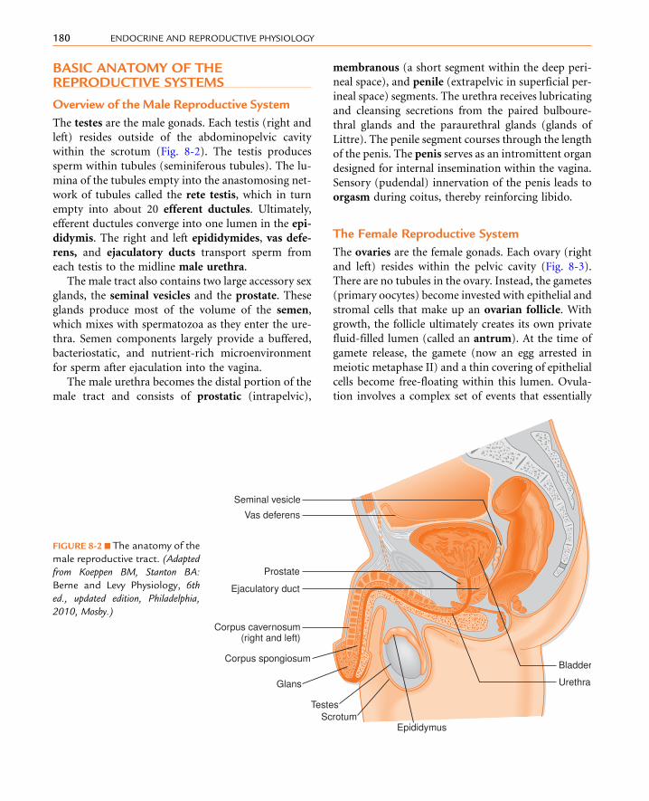
BASIC ANATOMY OF THEREPRODUCTIVE SYSTEMS
Overview of the Male Reproductive System
The testes are the male gonads. Each testis (right and
left) resides outside of the abdominopelvic cavity
within the scrotum (Fig. 8-2). The testis produces
sperm within tubules (seminiferous tubules). The lu-
mina of the tubules empty into the anastomosing net-
work of tubules called the rete testis, which in turn
empty into about 20 efferent ductules. Ultimately,
efferent ductules converge into one lumen in the epi-
didymis. The right and left epididymides, vas defe-
rens, and ejaculatory ducts transport sperm from
each testis to the midline male urethra.
The male tract also contains two large accessory sex
glands, the seminal vesicles and the prostate. These
glands produce most of the volume of the semen,
which mixes with spermatozoa as they enter the ure-
thra. Semen components largely provide a buffered,
bacteriostatic, and nutrient-rich microenvironment
for sperm after ejaculation into the vagina.
The male urethra becomes the distal portion of the
male tract and consists of prostatic (intrapelvic),
membranous (a short segment within the deep peri-
neal space), and penile (extrapelvic in superficial per-
ineal space) segments. The urethra receives lubricating
and cleansing secretions from the paired bulboure-
thral glands and the paraurethral glands (glands of
Littre). The penile segment courses through the length
of the penis. The penis serves as an intromittent organ
designed for internal insemination within the vagina.
Sensory (pudendal) innervation of the penis leads to
orgasm during coitus, thereby reinforcing libido.
The Female Reproductive System
The ovaries are the female gonads. Each ovary (right
and left) resides within the pelvic cavity (Fig. 8-3).
There are no tubules in the ovary. Instead, the gametes
(primary oocytes) become invested with epithelial and
stromal cells that make up an ovarian follicle. With
growth, the follicle ultimately creates its own private
fluid-filled lumen (called an antrum). At the time of
gamete release, the gamete (now an egg arrested in
meiotic metaphase II) and a thin covering of epithelial
cells become free-floating within this lumen. Ovula-
tion involves a complex set of events that essentially
Bladder
Urethra
Prostate
Ejaculatory duct
Vas deferens
Seminal vesicle
Corpus cavernosum(right and left)
Corpus spongiosum
Glans
TestesScrotum
Epididymus
FIGURE 8-2 n The anatomy of the
male reproductive tract. (Adapted
from Koeppen BM, Stanton BA:
Berne and Levy Physiology, 6th
ed., updated edition, Philadelphia,
2010, Mosby.)
180 ENDOCRINE AND REPRODUCTIVE PHYSIOLOGY

erode the follicular and ovarian walls at the point
where the follicle is pushing against the ovarian sur-
face, followed by the release of the egg.
Unlike the male tract, the egg is released into the pel-
vic cavity and has to be captured by the proximal seg-
ment of the female tract called the oviduct (see
Fig. 8-3). The oviduct has an opening at its proximal,
free end (the infundibulum), through which the cap-
tured egg is transported. There is usually only one egg
ovulated, either from the right or left side, depending
on which side had the largest follicle at the beginning
of the menstrual cycle. The oviduct transports the egg
toward themidline uterus and allows for themovement
of sperm from the uterus laterally toward the egg. Fer-
tilization and early development (5 to 6 days) normally
occur in the oviduct. The early embryo (blastocyst)
eventually moves into the uterine lumen and implants
into the uterinemucosa. The growing fetus is supported
in part by the elastic and fibrous inferior end of the
uterus called the cervix. At term, the newborn is ex-
pelled from the uterus through the cervix and vagina.
The vagina acts as both the copulatory organ and
the birth canal. The female external genitalia surround
the superficial opening of the vagina (called introitus).
The labia majora are homologous to an unclosed
scrotum. Vestibular bulbs (deep to the labia minora)
and the clitoris represent structures homologous to
the erectile tissue of the penis. However, unlike the pe-
nis, erection of these structures is not required for fer-
tility. Sensory (pudendal) innervation of these
structures and the vaginal wall may lead to orgasm
during coitus, thereby providing reinforcement to
libido.
SEXUAL DEVELOPMENT IN UTERO
The genetic sex of a fetus depends on the nature of
the sex chromosomes contributed by the egg and the
sperm. Normally, there are 46 chromosomes, consist-
ing of 22 pairs of autosomes and one pair of sex chro-
mosomes. The sex chromosomes are called X and Y
chromosomes; 46,XX is the normal karyotype for the
female, and 46,XY is the normal karyotype for the
male. Genetic sex determines gonadal sex. The go-
nads then either produce hormones (if male) or no
hormones (if female). The hormonal environment
Fundus of uterusIsthmusAmpulla
Infundibulumwith fimbriae
Uterine (fallopian) tube
Ovary
Body (corpus)of uterus
Internalgenitalia
Externalgenitalia
Cervix of uterus
Vagina
Clitoris
Vaginalintroitus
Openingof female
urethra
Monspubis
Labiamajora
Labiaminora
FIGURE 8-3 n The anatomy of the
female reproductive tract. (Adapted
from Koeppen BM, Stanton BA:Berne
andLevyPhysiology,6thed.,updated
edition, Philadelphia, 2010, Mosby.)
181LIFE CYCLE OF THE MALE AND FEMALE REPRODUCTIVE SYSTEMS

determines the sex of the reproductive tract and
external genitalia.
Male Development
During the first 6 weeks of development, mesodermal
cells within the genital ridge develop into a bipotential
gonadal primordium (Table 8-1). During this period,
primordial germ cells migrate from the yolk sac
endoderm into the gonadal tissue. By 6 weeks of
gestation, the gonads contain germ cells, supporting
cells, and stromal cells that will become androgen-
producing steroidogenic cells in both sexes.
The short arm of the Y chromosome contains the
SRY gene (sex-determining region on the Y chro-
mosome) that encodes the transcription factor,
SRY. SRY, along with other transcription factors,
plays a major role in the induction of differentiation
of the bipotential gonad into a testis between weeks 6
and 7 of gestation (Fig. 8-4). With testicular develop-
ment, the supportive epithelial cells differentiate into
Sertoli cells and form the seminiferous tubules.
Sertoli cells perform two critical functions in early
embryogenesis: (1) they surround male germ cells
(called spermatogonia) and produce an enzyme
(CYP26B1; also produced by Leydig cells) that
degrades locally produced retinoic acid, thereby
preventing meiotic progression of spermatogonia;
and (2) they express antimullerian hormone
(AMH, also referred to as mullerian-inhibitory sub-
stance), which causes the degeneration of the muller-
ian ducts (see later).
Hormones mediate phenotypic gender expression.
The fetus originally develops with bipotential internal
and external genitalia (Figs. 8-5 and 8-6). Internally,
there are two wolffian (also called mesonephric)
TABLE 8-1
Time Frame for Fetal Development of MaleReproductive System
GESTATIONALAGE (WK)
DEVELOPMENTAL FEATURE
6-8 Differentiation of testes
8 Retention of wolffian ducts
Regression of mullerian ducts
9-12 Development of male-type external genitalia
SRY &other TFs
Bipotentialgonad
Spermato-gonia
Sertolicell
Leydigcell
Urethralepithelium
DHT
Wolffian ductdevelopment
Müllerian ductregression
Epididymis
VASdeferens
Ejaculatoryduct
Seminalvesicle
Prostategland
Penisscrotum
DHT
Primordia ofexternalgenitalia
Testes
46XY
Quiescence
AMH
T
Peripheral5� reductase 2
FIGURE 8-4nEmbryonic development
of the male reproductive system.
AMH, antimullerian hormone; DHT,
dihydrotestosterone; T, testosterone;
TFs, transcription factors.
182 ENDOCRINE AND REPRODUCTIVE PHYSIOLOGY

ducts, which have the potential for differentiating into
the nonurethral segment of the male tract (epididy-
mis, vas deferens, ejaculatory duct, and seminal
vesicles), and two mullerian (also called parameso-
nephric) ducts, which have the potential for differen-
tiating into most of the female reproductive tract (the
oviducts, uterus, cervix, and proximal third of the
vagina). Whether male or female reproductive tracts
develop depends on the presence or absence of two
hormones produced by the fetal testis-testosterone
and AMH (Box 8-1).
The persistence and differentiation of the wolffian
ducts into the proximal nonurethral male tract re-
quires testosterone (see Fig. 8-4). By week 8, testoster-
one is produced by stromal cells that have
differentiated into Leydig cells. Androgen production
is first promoted by the placental, luteinizing hormone
(LH)-like hormone, called human chorionic gonado-
tropin (hCG), and later by fetal pituitary LH. At this
time, testosterone acts more like a paracrine factor
than a true hormone, in that testosterone from each
testis promotes the differentiation of the ipsilateral
tract (Fig. 8-7).
The degeneration of the mullerian ducts requires
production of AMH by the Sertoli cells of the testis
(see Fig. 8-4). AMH is a glycoprotein homodimer be-
longing to the transforming growth factor-b (TGF-b)gene family. Accordingly, AMH binds to a co-receptor
with serine-threonine kinase activity. AMH binds spe-
cifically to the AMH receptor type II (AMHR-II),
which binds to and activates a type-I receptor. As de-
scribed in Chapter 1, this activates a Smad-dependent
signaling pathway.
CLINICAL BOX 8-2
Persistent mullerian duct syndrome (PMDS) occursin 46,XY males. The persistence of mullerian deriva-
tives (oviducts, uterus, cervix, vaginal tissue) fre-
quently disrupts the descent of one or both testes,
resulting in unilateral or bilateral cryptorchidism.
Correction of cryptorchidism is performed by surgery.
Mutations in both the AMH gene and AMH receptor II
(AMHR-II) have been identified in families with
PMDS. Most cases involve homozygous mutations
that occur with a high frequency in certain Mediterra-
nean and North African populations that have a high
incidence of consanguinity.
The differentiation of the prostate gland and male
external genitalia occurs between week 9 and week
11 (Box 8-2; see Figs. 8-5 and 8-6). This requires the
expression of an enzyme, 5a-reductase-2, within the
primordia of these structures (urogenital sinus, genital
tubercle urethral folds, and labioscrotal swellings). The
5a-reductase-2 catalyzes the peripheral conversion of
testosterone to dihydrotestosterone (DHT). Testoster-
one and DHT bind to the same androgen receptor, but
DHT is required in these tissues. The cell-specific ac-
tions of testosterone and DHT are not completely un-
derstood but may depend on cell-specific expression
of co-regulatory proteins, the chromatin context of
cell-specific androgen-regulated genes, or cell-specific
differences in the stability of the hormone-receptor
complex. In any case, conversion of testosterone to
DHT is absolutely required for the normal develop-
ment of the prostate and external genitalia.
Vasdeferens
Indifferent stage
GonadMüllerian duct
Wolffian duct
Urogenital sinus
Ovary
Fallopiantube
EpididymisBladder
Seminalvesicle
Prostate gland
Cowper’s glandVagina
Female Male
Uterus
Testis
FIGURE 8-5 n Differentiation of internal genitalia and
primordial ducts. (Redrawn from George FW, Wilson JD:
Embryology of the genital tract. In Walsh PC, Retik AB, Stamey
TA, Vaughan ED, editors: Campbell’s Urology, 6th ed.,
Philadelphia, 1992, Saunders.)
183LIFE CYCLE OF THE MALE AND FEMALE REPRODUCTIVE SYSTEMS

Genital fold
Genital swelling Indifferent
stages
Genital tubercle
Glans
Fusedurogenitalfolds
Urethralgroove
Scrotum
Prepuce
Body ofpenis
Scrotalraphe
Male Female
Anus
Clitoris
Urethralorifice
Hymen
Labiaminora
Labiamajora
Glans
Urethralgroove
Anus
FIGURE 8-6 n Regulation of development
of male and female external genitalia. In
the presence of dihydrotestosterone (DHT)
between 9 and 12 weeks of gestation,
male external genitalia develop from the
genital tubercle, genital fold, genital
swelling, and urogenital sinus. In the
absence of DHT, female external genitalia
develop.
n n n n n n n n n n n n n n n n n n n n n
BOX 8 - 1
REGULATION OF DEVELOPMENT OF INTERNAL GENITALIA
n The wolffian ducts, when stimulated with testosterone, become the epididymis, vas deferens, seminal vesicles,
and ejaculatory ducts.
n The mullerian ducts, in the absence of Sertoli cell mullerian-inhibiting substance, become the fallopian tubes,
uterus, cervix, and upper one third of the vagina.
184 ENDOCRINE AND REPRODUCTIVE PHYSIOLOGY

CLINICAL BOX 8-3
A 5a-reductase-2 deficiency results in decreased DHT
formation. Affected persons typically have a well-
developed internal male tract (except for the prostate
gland),but incompletelymasculinizedexternal genitalia
(i.e.,ambiguousgenitalia)withmicrophallusandhypo-
spadias. The testes are either inguinal or labial. Severely
affected patients can be mistaken for females at birth.
The testosterone production is normal, and at puberty,
when testosterone production greatly increases, some
masculinization of the external genitalia may occur.
Also, 5a-reductase-1 activity in the skin appears at pu-
berty, which may generate enough DHT to induce the
pubertal changes.These include increasedmusclemass,
deepeningof the voice, anddescent of the testis into the
labioscrotal folds.Patientswhoarediagnosed in infancy
are assigned amale sex and treatedwith androgen ther-
apy to induce growth of the penis and surgical repair of
hypospadias. Patients who are diagnosed after puberty
may develop a male or female sexual identity and may
require some formof corrective surgery. It is not uncom-
mon for patients to switch from a female sexual identity
to a male one after puberty.
Female Development
In a normal 46,XX embryo, the absence of SRY allows
for the expression of other transcription factors that
induce the differentiation of the gonad into an ovary
(Fig. 8-8). However, full ovarian differentiation does
not occur until the end of the first trimester of
Undifferentiated
Wolffianducts
Müllerianducts
Normal male Normal femaleor castrated maleA B C
D E
F
Testosterone-treated female
Unilateralorchidectomized
male
Androgen-resistantmale (androgen
insensitivitysyndrome)
FIGURE 8-7 n Regulation
of development of inter-
nal genitalia. A, Both wolf-
fian and mullerian ducts
are originally present in
both male and female fe-
tuses (undifferentiated). B,If functional testes are
present, wolffian ducts de-
velop, and mullerian ducts
regress. C, If no testes are
present, mullerian ducts de-
velop, and wolffian ducts
are lost. D, If a female fetus
is exposed to testosterone,
both ductile systems can
remain. E, If a testis is re-
moved unilaterally (orchi-
dectomized), the mullerian
duct will develop, and the
wolffian duct will regress
on one side. F, A male with
functional testes but andro-
gen insensitivity will show
regression of both ductile
systems.
n n n n n n n n n n
BOX 8 - 2
REGULATION OF DEVELOPMENT OFEXTERNAL GENITALIA
n In the presence of dihydrotestosterone, the genital
tubercle, genital fold, genital swelling, and uro-
genital sinus become the penis, scrotum, and
prostate.
n In the absence of dihydrotestosterone, the genital tuber-
cle, genital fold, genital swelling, and urogenital
sinus become the labia majora, labia minora, clito-
ris, and lower two thirds of the vagina.
185LIFE CYCLE OF THE MALE AND FEMALE REPRODUCTIVE SYSTEMS

pregnancy (Box 8-3). The germ cells differentiate into
oogonia and divide mitotically. Starting at about
11 weeks of gestation, all oogonia enter meiosis and
become primary oocytes arrested in first meiotic
prophase. Oocytes become surrounded by a single layer
of supportive cells called granulosa cells and their basal
lamina. These structures are called primordial follicles
and appear in the second trimester. Although some
follicular development (see Chapter 10) occurs during
pregnancy, the ovary remains hormonally quiescent
during gestation.
The absence of Sertoli cell–derived AMH allows for
the persistence and development of the mullerian duct
into oviducts, uterus, cervix, and the inner one third of
the vagina (see Figs. 8-5 and 8-8; see Box 8-1). In the
absence of a testis and testosterone, the wolffian duct
degenerates. If high testosterone levels are present in
a female fetus because of a congenital adrenal hy-
perplasia (see Chapter 7), or because of a maternal
endocrine disorder, both sets of ducts can be retained
(see Fig. 8-7).
In the absence of androgen exposure, the external
genitalia develop into the labia majora, labia minora,
clitoris, vestibular bulbs and glands, and outer two
thirds of the vagina (see Figs. 8-6 and 8-8; see
Box 8-2).
n n n n n n n n n n
BOX 8 - 3
TIME FRAME FOR FETAL DEVELOPMENTOF THE FEMALE REPRODUCTIVE SYSTEM
7-12 WEEKS
n Meiosis of oogonia and arrest at first meiotic
prophase
n Ovarian organogenesis
n Formation of primordial follicles
11-12 WEEKS
n Oviducts, uterus, cervix, external genitalia, and
vagina
20-25 WEEKS
n Primary follicles in ovary
Wolffian ductregression
Müllerian ductdevelopment
Primordialfollicle
Granulosacell
Bipotentialgonad
Ovary OogoniaPrimaryoocyte
Primordia ofexternalgenitalia
No DHT
No AMH
Notestosterone
No SRY,expression of female TFs
THECA
Primary &early antral
follicle
Oviduct
Uterus
Cervix
Clitoris
Vest bulbs & GL
Inner vagina
Outer vagina
Labia majora
Labia minora
46XX
FIGURE 8-8 n Embryonicdevelop-
ment of the female reproductive
system. AMH, antimullerian hor-
mone;DHT,dihydrotestosterone;
TFs, transcription factors; Vest
bulbs & GL, vestibular bulbs and
gland.
186 ENDOCRINE AND REPRODUCTIVE PHYSIOLOGY

CLINICAL BOX 8-4
Kallmann syndrome type 1 is a tertiary (i.e., hypotha-lamic) endocrine disorder and a form of isolated hypo-gonatropic hypogonadism. This genetic disorder is
often associated with anosmia (absence of sense of
smell) or hyposmia (poor sense of smell). It is due to
loss of a functional protein, called anosmin, that is
encoded by the X-linked KAL1 gene. The syndrome is
caused by the inability of all or some of theGnRH neu-rons to properly migrate to the mediobasal hypothala-
mus from the nasal placode (see Chapter 5). Anosmin
is similar to adhesion molecules, and so may play a
guiding role in GnRH migration. Males are more fre-
quently affected than females. Men affected with this
disorder have undescended testes (cryptorchism).Although there is normal embryonic differentiation of
the wolffian duct–derived structures, penis develop-
ment is deficient, and microphallus often develops.
These effects probably result from the fact that early
fetal development of the internal genitalia is controlled
by testicular androgens that are initially regulated by
placental hCG (see Chapter 11), rather than fetal LH.
Adult growth and function of wolffian duct–derived
structures and prostate gland are impaired. The inabil-
ity of the fetus to secrete normal quantities of LHhas an
impact on testicular function later in development,
when androgens regulate growth of the external genita-
lia. The severity of the impairment of LH secretion is var-
iable, as is the severity of the reproductive problems
associated with the disorder. Puberty is delayed.
PUBERTY
Regulation of Timing of Puberty
The reproductive axis is driven by gonadotropin-
releasing hormone (GnRH) neurons in the medioba-
sal hypothalamus that secrete GnRH in a pulsatile
pattern. GnRH, in turn, stimulates production of pi-
tuitary LH and follicle-stimulating hormone (FSH)
(see Chapter 5). The GnRH neurons display activity
during gestation, which decreases at parturition.
GnRH neurons display a significant increase in activity
in the first 2 years of infancy (“mini-puberty of in-
fancy”), followed by about a decade of very low activ-
ity of the reproductive axis (Fig. 8-9). The resurgence
in activity of the reproductive system during adoles-
cence is called puberty and induces dramatic pheno-
typic and behavioral changes. Unfortunately, physical
changes precede maturation of the personality and
emotional stability.
The timing of puberty is variable among countries
and among ethnic populations within countries. In
the United States, the average age of puberty in girls
has declined over the past half-century, probably
owing to enhanced health and nutrition, and more
recently possibly because of increased rates of child-
hood obesity (although this has not been firmly estab-
lished). Black girls typically reach pubertal milestones
about 6 months earlier than white girls. The clinical
significance of the timing of puberty relates to
Fetus Infancy Childhood Puberty Reproductiveyears
Menopause
1st 2nd 3rdtrimesters
10
70
100
Birth 6 m o 10–14 yr 50 yr
Childhood Puberty Reproductiveyears
Menopause
Day NightLH secretion patterns
FSHPla
sma
gona
dotr
opin
s (m
IU/m
l)
FIGURE 8-9nRelative serum luteinizing hor-
mone (LH) and follicle-stimulating hor-
mone (FSH) levels across the life span in
women. (Redrawn from Braunwald E, et al:
Harrison’s Principles of Internal Medicine,
4th ed., New York, 1987, McGraw-Hill.)
187LIFE CYCLE OF THE MALE AND FEMALE REPRODUCTIVE SYSTEMS

underlying disorders or disease that induces the onset
of puberty at an abnormally young age (called preco-
cious puberty) or at an abnormally older age (delayed
puberty) or not at all. The average age of breast devel-
opment (first sign of puberty) in North American girls
is about 10 years old. The average age of testicular en-
largement (first sign of puberty) in North American
boys is about 11.5 years old. The threshold for earliest
onset of normal versus precocious puberty is 8 years in
girls and 9 years in boys. These ages may be too high as
the age of puberty declines.
After birth, the reproductive axes of both sexes show
significant activity for 1 to 2 years (see Fig. 8-9). This is
followed by the development of mechanisms that im-
poses a significant (but not absolute) suppression of
GnRH neuronal activity. The exact nature of the in-
hibitory signals that result in the “nadir of childhood”
reproductive activity remains poorly understood. The
fact that some tumors that cause swelling or tissue
destruction in the vicinity of the posterior hypothala-
mus can induce gonadotropin-dependent precocious
puberty has led some to propose the existence of an
inhibitory center within the central nervous system
(CNS). There is also some evidence for an exquisite
sensitivity of the hypothalamus and pituitary to neg-
ative feedback by steroid hormones (i.e., a low set-
point, or gonadostat). This is indicated by the findings
that primary agonadal children typically have higher
gonadotropin levels than normal children (although
gonadotropin levels are low in both cases because of
CNS inhibition). At puberty, these inhibitory mecha-
nisms are presumably terminated.
In addition to the release from CNS inhibition,
there is also strong evidence for the activation of stim-
ulatory centers in the CNS that induce pulsatile GnRH
production at puberty. Recently, much attention has
been given to the role of the neurotransmitter kisspep-
tin (Kiss1) as an inducer of GnRH pulsatile release at
puberty. Kisspeptin is encoded by the KISS1 gene.
Kiss1 is a 54-amino acid peptide and is expressed by
neurons in the hypothalamus. Kiss1 acts through a
G-protein-coupled receptor called Kiss1R (previously
called GPR54), which is expressed by GnRH neurons.
Kiss1 levels increase in the hypothalamus at puberty,
and intracerebral infusion of Kiss1 in female monkeys
induced precocious puberty. Null mutations in Kiss1R
have been described in individuals with compromised
pubertal changes, whereas gain-of-function mutations
in human Kiss1 and Kiss1R cause gonadotropin-
dependent precocious puberty.
During the peripubertal period, pulsatile sleep-
associated surges in GnRH begin to occur (see
Fig. 8-9). Gonadotrope sensitivity to GnRH increases,
resulting in an increase in LH secretion. At puberty, the
frequency and amplitude of the GnRH pulses increase.
This change increases LH and FSH secretion through-
out the day and ultimately gonadal steroid hormone
production. The maturation of the positive feedback
by estrogen on GnRH and gonadotropin secretion in
women occurs late in puberty (see Fig. 8-9; spikes dur-
ing reproductive years). The first menstrual period
(menarche) occurs 2.6 years after the onset of puberty.
Physiologic Changes Associatedwith Puberty
The developmental milestones associated with pu-
berty in British boys and girls were objectively de-
scribed by Marshall and Tanner and are referred to
as Tanner stages. The progression of secondary sex
characteristics was characterized as five stages, ending
in adult as stage 5. For boys, these changes include
testicular size, penis size (Table 8-2), and pubic hair.
TABLE 8-2
Tanner Pubertal Stages in Male
STAGE GENITALS PUBIC HAIR
1 Preadolescent Preadolescent; no pubic hair
2 Scrotum and testes enlarge;
change in scrotal skin
texture
Sparse, long, downy pubic
hair, chiefly at base of penis
3 Growth of penis in length
and further growth of
testes and scrotum
Hair darker and coarser
4 Growth of penis in length
and breadth, darkening
of scrotal skin
Adult-type hair, but area
covered is less than that in
adult
5 Adult-sized genitalia Adult hair texture and
quantity; hair is distributed
in diamond-shaped
escutcheon with hair
extending up linea alba
Data from Marshall WA, Tanner JM: Variations in the pattern of
pubertal changes in boys. Arch Dis Child 45:13, 1970.
188 ENDOCRINE AND REPRODUCTIVE PHYSIOLOGY
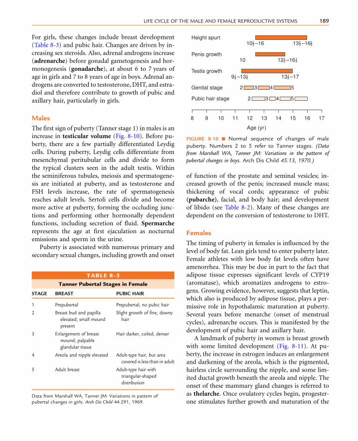
For girls, these changes include breast development
(Table 8-3) and pubic hair. Changes are driven by in-
creasing sex steroids. Also, adrenal androgens increase
(adrenarche) before gonadal gametogenesis and hor-
monogenesis (gonadarche), at about 6 to 7 years of
age in girls and 7 to 8 years of age in boys. Adrenal an-
drogens are converted to testosterone, DHT, and estra-
diol and therefore contribute to growth of pubic and
axillary hair, particularly in girls.
Males
The first sign of puberty (Tanner stage 1) in males is an
increase in testicular volume (Fig. 8-10). Before pu-
berty, there are a few partially differentiated Leydig
cells. During puberty, Leydig cells differentiate from
mesenchymal peritubular cells and divide to form
the typical clusters seen in the adult testis. Within
the seminiferous tubules, meiosis and spermatogene-
sis are initiated at puberty, and as testosterone and
FSH levels increase, the rate of spermatogenesis
reaches adult levels. Sertoli cells divide and become
more active at puberty, forming the occluding junc-
tions and performing other hormonally dependent
functions, including secretion of fluid. Spermarche
represents the age at first ejaculation as nocturnal
emissions and sperm in the urine.
Puberty is associated with numerous primary and
secondary sexual changes, including growth and onset
of function of the prostate and seminal vesicles; in-
creased growth of the penis; increased muscle mass;
thickening of vocal cords; appearance of pubic
(pubarche), facial, and body hair; and development
of libido (see Table 8-2). Many of these changes are
dependent on the conversion of testosterone to DHT.
Females
The timing of puberty in females is influenced by the
level of body fat. Lean girls tend to enter puberty later.
Female athletes with low body fat levels often have
amenorrhea. This may be due in part to the fact that
adipose tissue expresses significant levels of CYP19
(aromatase), which aromatizes androgens to estro-
gens. Growing evidence, however, suggests that leptin,
which also is produced by adipose tissue, plays a per-
missive role in hypothalamic maturation at puberty.
Several years before menarche (onset of menstrual
cycles), adrenarche occurs. This is manifested by the
development of pubic hair and axillary hair.
A landmark of puberty in women is breast growth
with some limited development (Fig. 8-11). At pu-
berty, the increase in estrogen induces an enlargement
and darkening of the areola, which is the pigmented,
hairless circle surrounding the nipple, and some lim-
ited ductal growth beneath the areola and nipple. The
onset of these mammary gland changes is referred to
as thelarche. Once ovulatory cycles begin, progester-
one stimulates further growth and maturation of the
17
Age (yr)
Pubic hair stage
1615141312111098
2 3 4 5
Genital stage 2 3 4 5
Testis growth9 �� –13 �� 13 �� –17
Penis growth10 12 �� –16 ��
Height spurt10 �� –16 13 �� –16 ��
FIGURE 8-10 n Normal sequence of changes of male
puberty. Numbers 2 to 5 refer to Tanner stages. (Data
from Marshall WA, Tanner JM: Variations in the pattern of
pubertal changes in boys. Arch Dis Child 45:13, 1970.)
TABLE 8-3
Tanner Pubertal Stages in Female
STAGE BREAST PUBIC HAIR
1 Prepubertal Prepubertal; no pubic hair
2 Breast bud and papilla
elevated; small mound
present
Slight growth of fine, downy
hair
3 Enlargement of breast
mound; palpable
glandular tissue
Hair darker, coiled, denser
4 Areola and nipple elevated Adult-type hair, but area
covered is less than in adult
5 Adult breast Adult-type hair with
triangular-shaped
distribution
Data from Marshall WA, Tanner JM: Variations in pattern of
pubertal changes in girls. Arch Dis Child 44:291, 1969.
189LIFE CYCLE OF THE MALE AND FEMALE REPRODUCTIVE SYSTEMS
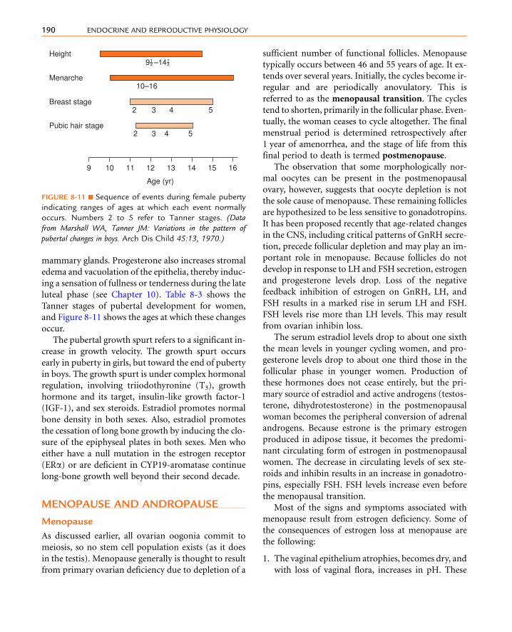
mammary glands. Progesterone also increases stromal
edema and vacuolation of the epithelia, thereby induc-
ing a sensation of fullness or tenderness during the late
luteal phase (see Chapter 10). Table 8-3 shows the
Tanner stages of pubertal development for women,
and Figure 8-11 shows the ages at which these changes
occur.
The pubertal growth spurt refers to a significant in-
crease in growth velocity. The growth spurt occurs
early in puberty in girls, but toward the end of puberty
in boys. The growth spurt is under complex hormonal
regulation, involving triiodothyronine (T3), growth
hormone and its target, insulin-like growth factor-1
(IGF-1), and sex steroids. Estradiol promotes normal
bone density in both sexes. Also, estradiol promotes
the cessation of long bone growth by inducing the clo-
sure of the epiphyseal plates in both sexes. Men who
either have a null mutation in the estrogen receptor
(ERa) or are deficient in CYP19-aromatase continue
long-bone growth well beyond their second decade.
MENOPAUSE AND ANDROPAUSE
Menopause
As discussed earlier, all ovarian oogonia commit to
meiosis, so no stem cell population exists (as it does
in the testis). Menopause generally is thought to result
from primary ovarian deficiency due to depletion of a
sufficient number of functional follicles. Menopause
typically occurs between 46 and 55 years of age. It ex-
tends over several years. Initially, the cycles become ir-
regular and are periodically anovulatory. This is
referred to as the menopausal transition. The cycles
tend to shorten, primarily in the follicular phase. Even-
tually, the woman ceases to cycle altogether. The final
menstrual period is determined retrospectively after
1 year of amenorrhea, and the stage of life from this
final period to death is termed postmenopause.
The observation that some morphologically nor-
mal oocytes can be present in the postmenopausal
ovary, however, suggests that oocyte depletion is not
the sole cause of menopause. These remaining follicles
are hypothesized to be less sensitive to gonadotropins.
It has been proposed recently that age-related changes
in the CNS, including critical patterns of GnRH secre-
tion, precede follicular depletion and may play an im-
portant role in menopause. Because follicles do not
develop in response to LH and FSH secretion, estrogen
and progesterone levels drop. Loss of the negative
feedback inhibition of estrogen on GnRH, LH, and
FSH results in a marked rise in serum LH and FSH.
FSH levels rise more than LH levels. This may result
from ovarian inhibin loss.
The serum estradiol levels drop to about one sixth
the mean levels in younger cycling women, and pro-
gesterone levels drop to about one third those in the
follicular phase in younger women. Production of
these hormones does not cease entirely, but the pri-
mary source of estradiol and active androgens (testos-
terone, dihydrotestosterone) in the postmenopausal
woman becomes the peripheral conversion of adrenal
androgens. Because estrone is the primary estrogen
produced in adipose tissue, it becomes the predomi-
nant circulating form of estrogen in postmenopausal
women. The decrease in circulating levels of sex ste-
roids and inhibin results in an increase in gonadotro-
pins, especially FSH. FSH levels increase even before
the menopausal transition.
Most of the signs and symptoms associated with
menopause result from estrogen deficiency. Some of
the consequences of estrogen loss at menopause are
the following:
1. The vaginal epithelium atrophies, becomes dry, and
with loss of vaginal flora, increases in pH. These
Age (yr)
161514131211109
Pubic hair stage2 3 4 5
Breast stage2
Menarche10–16
Height
3 4 5
9 �� –14 ��
FIGURE 8-11 n Sequence of events during female puberty
indicating ranges of ages at which each event normally
occurs. Numbers 2 to 5 refer to Tanner stages. (Data
from Marshall WA, Tanner JM: Variations in the pattern of
pubertal changes in boys. Arch Dis Child 45:13, 1970.)
190 ENDOCRINE AND REPRODUCTIVE PHYSIOLOGY

changes often result in increased incidence of vag-
inal and urinary tract infections, as well as pain
during intercourse.
2. Bone loss is accelerated, potentially leading to osteo-
porosis. The effect of menopause on osteoporosis is
staggering: greater than 1 million fractures per year
occur in the United States due to menopausal osteo-
porosis. A significant fraction of these fractures are
hip fractures, which greatly increase morbidity and
mortality and increase the loss of independence.
3. Hot flushes result from periodic increases in core
temperature, which produce peripheral vasodila-
tion, sweating, and a feeling of malaise. Hot flushes
are also correlated with sleep disturbances, which
can significantly affect a woman’s daily productiv-
ity and quality of life. Hot flushes are correlated
with LH pulses, and current thinking proposes that
a CNS mechanism leads to both events. Hot flushes
typically subside within 1 to 5 years of the onset of
menopausal symptoms.
4. The incidence of cardiovascular disease increases
markedly after menopause. This is due, in part,
to the reversal of the beneficial effect of estrogen
on high-density lipoprotein levels. However, life-
style plays an important role, and menopause can
affect lifestyle—if a woman is losing sleep because
of hot flushes, she is less likely to exercise during
the day. There are other psychosocial effects of
menopause, including fatigue and depression.
Most of the menopausal sequelae respond well to
estrogen replacement therapy (ERT), or estrogen
and progesterone replacement therapy (hormone re-
placement therapy, or HRT). Results from the
Women’s Health Initiative and other studies, however,
have provided conflicting findings on the safety of es-
trogens and progestins (which were not in the form of
bioidentical estradiol-17b and progesterone) for HRT
in postmenopausal women, emphasizing the need for
physicians to fully consider each woman’s individual
medical and family history before deciding on the
course of therapy for the alleviation of postmeno-
pausal symptoms. In general, use of the lowest effec-
tive dose and for the shortest effective duration is
standard. New analysis from the Women’s Health Ini-
tiative indicates that ERT given before the age of
60 years (i.e., short-term ERT) was not associated with
an increased risk for thromboembolic events, stroke,
or breast cancer.
Andropause
There is no distinct andropause in men. As men age,
however, gonadal sensitivity to LH decreases and
androgen production drops. As this occurs, serum
LH and FSH levels rise. Although sperm production
typically begins to decline after age 50 years, many
men can maintain reproductive function and sper-
matogenesis throughout life.
S U M M A R Y
1. The male and female reproductive systems are
composed of a gonad (testis and ovary) and a
reproductive tract.
2. The gonads perform an endocrine function,
which is regulated within a hypothalamic-
pituitary-gonad axis.
3. The gonads perform an exocrine function by the
production of gametes. This function is abso-
lutely dependent on gonadal hormones.
4. Meiosis is central to sexual reproduction. Meiosis
generates haploid gametes through two divisions,
meiosis I and meiosis II, with no DNA synthesis
in between. The major advantage of sexual
reproduction is the recombination of genetic ma-
terial. This occurs by independent segregation of
chromosomes, combining the haploid genome of
eggs with the haploid genome of sperm, and
through crossing-over.
5. The process of crossing-over involves synapsis
of homologous chromosomes (each with
two sister chromatids) to form a bivalent, fol-
lowed by a complex process that exchanges
DNA sequence information between homolo-
gous strands. Chiasmata form at these sites,
which help organize chromosomes at the
metaphase plate.
191LIFE CYCLE OF THE MALE AND FEMALE REPRODUCTIVE SYSTEMS

6. Chromosomes must undergo accurate disjunc-
tion (separation) in both divisions. Defects in
disjunction lead to nondisjunction, resulting in
aneuploidy or polyploidy.
7. The male reproductive system includes the testis,
in which the seminiferous tubules generate sper-
matozoa. These are guided out of the testis by
the rete testis and efferent ductules. Sperm then
enter the epididymis, vas deferens, ejaculatory
duct, and male urethra. Sperm receive seminal
fluid (semen) from the seminal vesicles and the
prostate. The tract ends in the penis, which
is an intromittent organ designed for internal
semination.
8. The female reproductive system includes the ova-
ries, oviducts, uterus, cervix, vagina, and external
genitalia. The female tract does not join with the
female urethra.
9. The gamete within the ovary is a primary oocyte
arrested in prophase of meiosis I. Just before ovu-
lation, the oocyte completes meiosis I and
becomes arrested at metaphase of meiosis II
(now called an egg).
10. Release of an egg involves a complex process that
involves the focal breakdown of the follicular and
ovarian walls.
11. Early embryos have a bipotential gonad, into
which primordial germ cells have migrated from
the yolk sac. Genetic sex (46,XYor 46,XX) deter-
mines gonadal sex. Gonadal sex will create a
hormonal environment that determines the de-
velopment of the reproductive tract and external
genitalia.
12. SRY is a major factor involved in male develop-
ment (formation of a testis) at 6 to 7 weeks of
gestation.
13. Formation of a testis involves the organization of
seminiferous tubules lined by spermatogonia and
Sertoli cells. Leydig cells develop in the peritubu-
lar compartment.
14. Meiosis is blocked in the male by the production
of CYP26B1, which breaks down and inactivates
retinoic acid.
15. Sertoli cells secrete AMH, thereby inducing the
regression of the female mullerian tract.
16. Leydig cells, under stimulation by hCG and, later,
fetal LH, produce testosterone. Testosterone
promotes the differentiation of the wolffian tract
into the epididymis, vas deferens, ejaculatory
duct, and seminal vesicle.
17. Testosterone also induces descent of the testis into
the scrotum.
18. Testosterone is peripherally converted to DHT by
5a-reductase-2. DHT induces the differentiation
of the urogenital sinus into the proximal male
urethra and the prostate gland. DHT induces
the primordia of the external genitalia to form
the penis and scrotum.
19. A deficiency of 5a-reductase-2 may lead to female
or ambiguous external genitalia in a 46,XY fetus.
The female phenotype changes to a male pheno-
type at puberty, requiring reassessment of sexual
identity, followed by appropriate surgery and
hormonal replacement.
20. Lack of SRY in a 46,XX female embryo allows fe-
male-specific transcription factors to drive ovar-
ian differentiation. This is completed much later
than testicular differentiation.
21. Oogonia develop from primordial germ cells and
divide mitotically to generate about 1 million
oogonia per ovary. All oogonia enter meiosis
and become oocytes arrested at the prophase
of meiosis I. Oocytes become surrounded by ep-
ithelial cells and their basal lamina, generating a
structure called the primordial follicle.
22. Absence of AMH allows the female muller-
ian duct to develop into the oviducts, uterus,
cervix, and the inner one third of the vagina. Ab-
sence of testosterone causes the male wolffian
duct to regress. Absence of DHTallows the exter-
nal genitalia to form female structures (labia
majora, labia minora, clitoris, vestibular bulbs
and glands) and the outer two thirds of the
vagina.
23. The human reproductive axis involves hypotha-
lamic pulsatile GnRH neurons and pituitary
gonadotropes that secrete LH and FSH. After sig-
nificant activity in early infancy, the reproductive
axis diminishes to very low activity for about a
decade. This may be due to both active inhibition
from the CNS and exquisite sensitivity to negative
feedback.
24. Puberty involves a significant activation of the re-
productive axes and the reproductive system. This
192 ENDOCRINE AND REPRODUCTIVE PHYSIOLOGY

probably involves cessation of CNS inhibition,
along with stimulation by other CNS centers.
25. Kisspeptin (Kiss1) is produced in the hypothala-
mus and binds to its receptor, Kiss1R, on GnRH
neurons. Null mutation in Kiss1R cause defi-
cient GnRH-dependent pubertal changes. Gain-
of-function mutations in both Kiss1 and Kiss1R
have been linked to precocious puberty.
26. The progression of puberty is semi-quantified by
the five Tanner stages. In boys, these involve
assessment of testicular size (volume), penile
growth, and pubic hair growth. In girls, Tanner
staging involves assessment of breast develop-
ment and pubic hair growth.
27. The pubertal growth spurt begins in early puberty
in girls and in late puberty in boys.This involves an
increase in the growthvelocity,which is dependent
on increased growth hormone, IGF-1, T3, and
sex steroids.
28. Menopause involves the great diminution of
ovarian steroid and inhibin output, probably
owing to insufficient number of gonadotropin-
responsive follicles. LH and FSH levels increase.
29. Menopause may be accompanied by thinning of
the vagina, vasomotor instability (hot flushes),
sleep disturbances, personality changes, osteopo-
rosis, and an increased risk for cardiovascular
disease.
30. Men do not experience an abrupt cessation in the
reproductive axis. Some men are fertile at ad-
vanced ages. Some men experience a decrease in
androgen production (an incomplete andro-
pause), along with symptoms such as low libido
and erectile dysfunction.
S E L F - S T U D Y P R O B L E M S
1. How does sexual reproduction increase genetic
diversity?
2. Explain how a 46,XX embryo would develop in the
presence of an SRY-autosome translocation (i.e.,
gain of SRY).
3. Explain the gonadal, internal tract, and external
genitalia in a 46,XY individual with a null muta-tion in the AMH receptor.
4. Compare the adult male and adult female repro-
ductive systems in terms of onset and progression
of meiosis.
5. Explain why wolffian duct derivatives develop in
embryos with Kallmann syndrome type 1.6. How do gain-of-function mutations in Kiss1R
affect the timing of puberty?
7. Why do women become infertile at menopause,
whereas men may remain fertile into their ninth
decade?
8. List the consequences of menopause.
K E Y W O R D S A N D C O N C E P T S
n 46,XXn 46,XYn 5a-reductase-1
For full list of keywords and concepts see Student
Consult
SUGGESTED READ INGS
Griswold SL, Behringer RR: Fetal Leydig cell origin and develop-
ment, Sex Dev 3:1–15, 2009.
Teles MG, Silveira LF, Tusset C, et al: New genetic factors implicated
in human GnRH-dependent precocious puberty: The role of
kisspeptin system, Mol Cell Endocrinol 346:84–90, 2011.
193LIFE CYCLE OF THE MALE AND FEMALE REPRODUCTIVE SYSTEMS

K E Y W O R D S A N D C O N C E P T S
n 5a-reductase-2n 5a-reductase-2 deficiencyn Adrenarchen Alignmentn Ambiguous genitalian AMH receptor type II (AMHR-II)n Anaphase In Anaphase IIn Andropausen Aneuploidyn Antimullerian hormone (AMH)n Antrumn Bipotential gonadal primordiumn Bivalentn Centromeren Cervixn Chiasmatan Chromatidsn Clitorisn Congenital adrenal hyperplasian Consanguinityn Crossing-overn Cryptorchidismn Cryptorchismn Delayed pubertyn Dihydrotestosterone (DHT)n Disjunctionn Down syndromen Efferent ductulesn Eggsn Ejaculatory ductsn Embryonic developmentn Epididymidesn Epididymisn Estrogenn Euploidyn Gametesn Gametogenicn Genetic diversityn Genetic recombinationn Genetic sexn GnRH neuronsn Gonadal sexn Gonadarche
n Gonadotropin-dependent precocious pubertyn Gonadsn Granulosa cellsn Haploid cellsn Human chorionic gonadotropin (hCG)n Hypogonatropic hypogonadismn Hypothalamus-pituitary-gonadal axisn Independent assortmentn Kallmann syndrome type 1n Kinetochoren KISS1 genen Kiss1Rn Kisspeptin (Kiss1)n Labia majoran Labia minoran Leydig cellsn Luteinizing hormone (LH)n Male external genitalian Male urethran Mammary glandsn Meiosis In Meiosis IIn Membranousn Menopausal transitionn Menopausen Mesonephric ductsn Microtubule spindlesn Mullerian ductsn Mullerian-inhibitory substancen Nondisjunctionn Oogonian Orgasmn Ovarian folliclen Ovariesn Oviductn Pairn Paramesonephric ductsn Penilen Penisn Peripubertal periodn Persistent mullerian duct syndrome (PMDS)n Phenotypic gendern Placental hCGn Polyploidyn Postmenopausen Precocious puberty
193.e1LIFE CYCLE OF THE MALE AND FEMALE REPRODUCTIVE SYSTEMS

n Primordial folliclesn Primordial germ cellsn Progesterone plus estrogenn Prophase In Prostaten Prostate glandn Prostaticn Pubarchen Pubertyn Reproductive tractsn Rete testisn Retinoic acidn Semenn Seminal vesiclesn Seminiferous tubulesn Sertoli cellsn Sexual reproductionn Sister chromatids
n Spermn Spermarchen Spermatogonian SRY genen Tanner stagesn Tertiaryn Testesn Testicular volumen Testisn Testosteronen Thelarchen Trisomy 21n Uterusn Vaginan Vas deferensn Vestibular bulbsn Wolffian ducts
193.e2 ENDOCRINE AND REPRODUCTIVE PHYSIOLOGY

Intentionally left as blank

n n n n n n n n n n n n n n n
9 THE MALE REPRODUCTIVESYSTEM
O B J E C T I V E S
1. Describe the organization of the male gonad, the testis,
and the process of spermatogenesis, and discuss how
this process is supported by Sertoli cells.
2. Describe the steroidogenic pathway of Leydig cells that
produces testosterone, the peripheral conversion of tes-
tosterone to estradiol-17b or dihydrotestosterone, and
the actions of these steroids in men.
3. Discuss the regulation of testicular function by the
hypothalamic-pituitary-testicular axis.
4. Describe the role of the proximal male reproductive
tract, especially the epididymis, in the further develop-
ment of sperm.
5. Discuss the more distal segments of the male reproduc-
tive tract, including the accessory sex glands, in the con-
text of emission and ejaculation.
6. Describe the neurovascular events in the penis that are
involved in erection.
7. Discuss the following pathologic conditions of the
male reproductive system: Klinefelter syndrome and
androgen insensitivity that is coupled to testicular
feminization.
In men, the reproductive system has evolved for
continuous, lifelong gametogenesis, coupled to occa-
sional internal insemination with a high density of
sperm (greater than 60�106/mL in 3 to 5 mL of
semen). This means that in adult men, the basic roles
of gonadal hormones are as follows:
n Support of gametogenesis (spermatogenesis)
n Maintenance of the male reproductive tract and
production of semen
n Maintenance of secondary sex characteristics and
libido. There is no overall cyclicity of this activity
in men.
HISTOPHYSIOLOGY OF THE TESTIS
Amajor difference between the testes and the ovaries is
that the testes reside in the scrotum outside of the
abdominopelvic cavity and are connected to the intra-
pelvic male tract by the spermatic cord (see Fig. 8-2 in
Chapter 8). Various mechanisms, including the cre-
masteric reflex, a venous countercurrent exchanger
in the spermatic cord (pampiniform plexus), folding
of scrotal skin, and sweat glands within the scrotal
skin, cooperate to maintain a testicular temperature
at about 35� C, which is crucial for sperm develop-
ment. Failure of the testes to descend through the
195

inguinal canal into the scrotum during development
results in depressed spermatogenesis.
The human testis is covered by a connective tissue
capsule and is divided into about 300 lobulesby fibrous
septa. Within each lobule are two to four loops of
seminiferous tubules. Each loop empties into an
anastomosing network of tubules called the rete testis.
The rete testis is continuous with small ducts, the
efferent ductules that lead the sperm out of the testis
into the head of the epididymis on the superior pole
of the testis (Fig. 9-1). Once in the epididymis, the
sperm pass from the head, to the body, to the tail of
the epididymis and then to the vas (ductus) deferens.
Spermatozoa are stored in the tail of the epididymis
and the vas deferens for severalmonths as viable sperm.
The presence of the seminiferous tubules in the
lobules of the testis creates two compartments within
each lobule: an intratubular compartment, which is
composed of the seminiferous epithelium of the sem-
iniferous tubule; and a peritubular compartment,
which is composed of neurovascular elements, con-
nective tissue cells, immune cells, and the interstitial
cells of Leydig, whose main function is to produce
testosterone.
The Intratubular Compartment
The seminiferous tubule is lined by a complex seminif-
erous epithelium (Fig. 9-2) composed of two cell types:
n Sperm cells in various stages of spermatogenesis
n The Sertoli cell, which is a nurse cell in intimate
contact with all sperm cells and which regulates
many aspects of spermatogenesis
Developing Sperm Cells The entire developmental
process by which spermatogonia give rise to sperma-
tozoa is called spermatogenesis. Spermatogenesis be-
gins at puberty and involves the processes of mitosis
and meiosis (see Fig. 9-2). Stem spermatogonia (also
called prespermatogonia) reside at the basal level of
the seminiferous epithelium. Stem spermatogonia
divide mitotically to generate daughter spermatogonia
(spermatocytogenesis). These mitotic divisions are
initially asymmetrical, in that one daughter cell
remains a stem spermatogonium (thereby undergoing
self-renewal throughout life), whereas the second
daughter cell will divide several times to amplify its
population. After several mitotic divisions, the
daughter spermatogonia complete S phase (DNA rep-
lication) and commit to meiotic division. Of note,
these amplifying divisions are accompanied by incom-
plete cytokinesis, so all spermatogonia daughter cells
and subsequent sperm cells (at different stages) remain
interconnected by a cytoplasmic bridge. This configu-
ration contributes to the synchrony of development of
a clonal population of sperm cells.
Spermatogonia migrate apically away from the basal
lamina as they enter the first meiotic prophase (see
Fig. 9-1). At this time, they are called primary sper-
matocytes. During the first meiotic prophase, the
hallmark processes of sexual reproduction involving
synapsis, crossing-over, formation of chiasmata, and
the first disjunction occur (see Chapter 8). Completion
of the first meiotic division gives rise to secondary
spermatocytes, which quickly (within 20 minutes)
complete the second meiotic division.
The initial products of meiosis are haploid sperma-
tids, which reside apically within the seminiferous ep-
ithelium, close to the lumen of the seminiferous tubule
(see Fig. 9-2). Spermatids are small, round cells with a
round nucleus. Spermatids undergo a remarkable
metamorphosis called spermiogenesis that results in
a spermatozoon (Fig. 9-3). The spermatozoon con-
tains the following parts:
1. A head. The head consists of twomajor components:
a. A condensed and streamlined nucleus. The chro-
matin of the nucleus is highly heterochromatic
(i.e., condensed), and the nucleosomal histones
are replaced by protamines. The DNA of a sper-
matozoan is transcriptionally silent.
b. An acrosomal vesicle. The acrosomal vesicle
contains hydrolytic enzymes transported to it
from the Golgi. These enzymes will play an im-
portant role in fertilization and the prevention
of polyspermy (see Chapter 11). The acrosomal
vesicle attaches to the forward pole of the nu-
cleus and descends along the side of the nucleus
so that it partially covers the nucleus.
2. The neck. This contains two centrioles (proximal
and distal). The proximal centriole attaches to
the nucleus, and the distal centriole will generate
a “9þ2” configuration of microtubules that is
called the axoneme.
3. The tail (also called the flagellum). The tail has a
continuous axonemal core but is composed of
196 ENDOCRINE AND REPRODUCTIVE PHYSIOLOGY

B
T T
T
PTC
PTC
C
BL
L
C
Ad
St
Z/P
AdSt
Z/P
Sz
DST
RC
LC
A
FIGURE 9-1 n A, Low-magnification drawing of testicular lobules containing coils of seminiferous tubules. B, Highermagnification of histologic organization of a section from the testicular lobule (as drawn in A), showing several
seminiferous tubules (T), which collectively make up the intratubular compartment, and the peritubular compartment
(PTC). C, Higher magnification of histologic organization of two seminiferous tubules, showing Sertoli cells (arrows),
spermatogonia (Ad and B), primary spermatocytes (Z/P), spermatids (St), and spermatozoa (Sz). L, lumen of tubule.
Note that the association of sperm cells differs in two adjacent tubules as a result of the difference in their stage of the
spermatogenic cycle. D, Higher magnification of histologic organization of the peritubular compartment (between
dashed lines) showing a cluster of Leydig cells (LC). (A, From Porterfield SP: Endocrine Physiology, 2nd ed., St. Louis, 2001,
Mosby. B to D, From Stevens A, Lowe J: Human Histology, 3rd ed., Philadelphia, 2005, Mosby.)
197THE MALE REPRODUCTIVE SYSTEM

structurally distinct regions called the middle piece,
principal piece, and end piece. The middle piece is
the thickest and contains a collar of mitochondria
that will deliver adenosine triphosphate (ATP)
for flagellar beating and motility. The outer cir-
cumference of the middle piece contains dense
fibers. The principal piece and the end piece
lack the mitochondrial sheath, and the end
piece lacks the outer dense fibers.
The process of spermatogenesis takes about
72 days. A cohort of adjacent spermatogonia enters
the process every 16 days, so the process is staggered
along the length of a seminiferous tubule. Conse-
quently, spermatogonia do not enter the process of
Cell types
Spermatogonium(type B)
DNA replication
First meioticdivision inprogress
Second meioticdivision inprogress
Immaturehaploidgametes
Haploidgametes
Primaryspermatocyte
Two secondaryspermatocytes
Secondary meioticdivision completed
4 Spermatids
Spermiogenesis
4 Spermatozoa
Frist meoticdivision completed
Meioticevents
Sertoli cell
Blood-testisbarrier
FIGURE 9-2 n Seminiferous
epithelium. Spermatogonia un-
dergo mitosis (spermatocyto-
genesis) to produce both a
reservoir of spermatogonia and
maturing spermatogonia that
differentiate into primary sper-
matocytes. These spermatocytes
remain joined by cytoplasmic
bridges (not shown). Primary
spermatocytes undergo the com-
plex process of the first meiotic
division to become secondary
spermatocytes, and then a rapid
second meiotic division (reduc-
tion-division) to become haploid
spermatids. Spermatids mature
into spermatozoa by the process
of spermiogenesis. Sertoli cells
extend from the basal to the api-
cal sides of the epithelium and
create the blood-testis barrier
between spermatogonia and
primary spermatocytes. (From
Koeppen B, Stanton B: Berne and
Levy Physiology, updated 6th ed.,
Philadelphia, 2010, Mosby.)
198 ENDOCRINE AND REPRODUCTIVE PHYSIOLOGY

spermatogenesis at the same time along the entire
length of the tubule, or in synchrony with every other
tubule (there are about 500 seminiferous tubules per
testis). Because the seminiferous tubules within one
testis total about 400 meters in length, spermatozoa
are continually being generated at many sites within
the testis at any given time. Histologic examination
of the seminiferous tubules reveals that there are
specific associations, called spermatogenic stages, of
sperm cells at any one point in time. In humans, there
are six different stages that progress and repeat as a
cycle at one point within the seminiferous tubule. This
is referred to as the spermatogenic cycle. The stages
are staggered spatially along the length of the seminif-
erous tubule, before repeating themselves. This spatial
configuration of cycles is called a spermatogenic wave.
The final release of sperm, called spermiation, is
an active process involving dissolution of adhesion be-
tween Sertoli cells and spermatozoa. It is important to
note that testicular spermatozoa after spermiation are
not fully mature. Testicular spermatozoa are barely
motile, and leave the seminiferous tubule passively
within fluid produced by the Sertoli cells.
The Sertoli Cell The Sertoli cell represents the true ep-ithelial cell of the seminiferous epithelium and extends
from the basal lamina to the lumen (Box 9-1; see
Fig. 9-3). Sertoli cells surround sperm cells, providing
structural support within the epithelium, and form
adherens-type junctions and gap junctions with all
stages of sperm cells (see Fig. 9-3). Through the forma-
tion and breakdown of these junctions, Sertoli cells
guide sperm cells toward the lumen as they advance
to later stages in spermatogenesis. Accordingly, major
secretory products of Sertoli cells include proteases
and protease inhibitors. Spermiation requires the final
breakdown of Sertoli cell–sperm cell junctions.
Another important structural feature of Sertoli cells
is the formation of tight junctions between adjacent
Sertoli cells (see Fig. 9-3). These occluding junctions
divide the seminiferous epithelium into a basal
Principal piece End piece
Nucleus
Mature spermatozoon
Head Neck Middle piece
Acrosomalvesicle
Golgi Centrioles Axoneme
AcrosomeResidual body
Mitochondria
Spermatid 1 2 3 4 5 6
Tail
FIGURE 9-3 n Spermiogenesis and structure of a spermatozoon. The residual body is phagocytized by Sertoli cells. (From
Young B, Lowe J, Stevens A, et al: Wheater’s Functional Histology, 5th ed., Edinburgh, 2007, Churchill Livingstone.)
199THE MALE REPRODUCTIVE SYSTEM

compartment, containing the spermatogonia and
early-stage primary spermatocytes, and an adluminal
compartment, containing later-stage primary sper-
matocytes and all subsequent stages of sperm cells.
As early primary spermatocytes move apically from
the basal compartment to the adluminal one, the tight
junctions need to be disassembled and reassembled.
These tight junctions form the physical basis for the
blood-testis barrier, which creates a specialized, im-
munologically safe microenvironment for developing
sperm. By blocking paracellular diffusion, the tight
junctions restrict movement of substances between
the blood and the developing germ cells through a
trans–Sertoli cell transport pathway and in this man-
ner allow the Sertoli cell to control nutrient availability
to germ cells. Accordingly, Sertoli cells also have the
responsibility for providing nutrients to this environ-
ment, such as transferrin, iron, and lactate. For exam-
ple, spermatogonia and released spermatozoa use
fructose and glucose for energy. However, sperm
undergoing meiosis cannot efficiently use glucose as
an energy source. Sertoli cells acquire glucose by the
GLUT1 transporter, metabolize it to lactate, and trans-
fer it to developing sperm, which express a sperm-
specific lactate transporter. This process is dependent
on hormonal stimulation (follicle-stimulating hor-
mone [FSH] and testosterone; see later) but also ap-
pears to be optimized by local sperm cell–generated
paracrine factors.
Thus, healthy Sertoli cell function is essential for
sperm cell viability and development. In this respect,
it should be noted that spermatogenesis is absolutely
dependent on testosterone produced by peritubular
Leydig cells (see later), yet it is the Sertoli cells that ex-
press the androgen receptor, not the developing
sperm cells. Similarly, the pituitary hormone FSH is
also required for maximal sperm production, and
again, it is the Sertoli cell that expresses the FSH recep-
tor, not the developing sperm. Thus, these hormones
support spermatogenesis indirectly through stimula-
tion of Sertoli cell function.
Sertoli cells have multiple additional functions.
Sertoli cells express the enzyme CYP19 (also called
aromatase), which converts Leydig cell–derived tes-
tosterone to the potent estrogen, estradiol-17b (see
later). This local production of estrogen may enhance
spermatogenesis in humans. Sertoli cells also produce
androgen-binding protein (ABP). ABP is encoded by
the same gene as for sex hormone–binding globulin
(SHBG; see later) but has different carbohydrate
groups and is specifically expressed intratesticularly.
ABP maintains a high androgen level within the
adluminal compartment, the lumina of the seminifer-
ous tubules, and the proximal part of the male repro-
ductive tract. Sertoli cells also produce a large amount
of fluid. This fluid provides an appropriate bathing
medium for the sperm and assists in moving the
immotile spermatozoa from the seminiferous tubule
into the epididymis. Sertoli cells perform an impor-
tant phagocytic function. This allows Sertoli cells to
engulf residual bodies, which represent cytoplasm
that is shed by spermatozoa during spermiogenesis,
as well as dead sperm cells.
Finally, the Sertoli cell has an important endocrine
role. During development, Sertoli cells produce anti-
mullerian hormone (AMH), also called mullerian-
inhibiting substance (MIS), which induces regression
n n n n n n n n n n
BOX 9 - 1
FUNCTIONS OF SERTOLI CELLS
SUPPORTIVE (“NURSING”)
n Maintaining, breaking, and re-forming multiple
junctions with developing sperm
n Maintaining blood-testis barrier
n Phagocytosis
n Transfer of nutrients and other substances from
blood to developing sperm cells
n Expression of paracrine factors and receptors for
sperm-derived paracrine factors
EXOCRINE
n Production of fluid to move immobile sperm out of
testis toward epididymis
n Production of androgen-binding protein
n Determination of release of spermatozoa (spermia-
tion) from seminiferous tubule
ENDOCRINE
n Expression of androgen receptor and follicle-
stimulating hormone receptor
n Production of mullerian-inhibiting substance, also
called antimullerian hormone
n Aromatization of testosterone to estradiol-17b(this has local effect, not strictly endocrine)
200 ENDOCRINE AND REPRODUCTIVE PHYSIOLOGY

of the embryonic mullerian duct that is programmed
to give rise to the female reproductive tract (see
Chapter 8). The Sertoli cells also produce the hormone
inhibin. Inhibin is a heterodimer protein hormone
related to the transforming growth factor-b (TGF-b)family. FSH stimulates inhibin production, which
then exerts negative feedback on gonadotropes to
inhibit FSH production. Thus, inhibin keeps FSH
levels within a specific range (see later).
The Peritubular Compartment
The peritubular compartment (see Fig. 9-1) contains
the primary endocrine cell of the testis, the Leydig
cell. This compartment also contains common cell
types of loose connective tissue and an extremely rich
peritubular capillary network that must provide nutri-
ents to the seminiferous tubules (by way of Sertoli
cells) while conveying testosterone away from the
testes to the peripheral circulation.
The Leydig Cell Leydig cells are steroidogenic stromal
cells. These cells synthesize cholesterol de novo, as well
as acquiring it through low-density lipoprotein (LDL)
receptors and, to a lesser extent, high-density lipopro-
tein (HDL) receptors (the HDL receptor is also called
scavenger receptor-BI [SR-BI]), and store cholesterol
as cholesterol esters, as described for adrenocortical
cells (see Chapter 7). Free cholesterol is generated by
a cholesterol hormone-sensitive lipase (HSL) and
transferred to the outer mitochondrial membrane,
and then to the inner mitochondrial membrane in
a steroidogenic acute regulatory protein (StAR)-
dependent manner (refer to Fig. 7-7 in Chapter 7).
As in all steroidogenic cells, cholesterol is converted
to pregnenolone by CYP11A1. Pregnenolone is then
processed to progesterone, 17a-hydroxyprogesterone,and androstenedione by 3b-hydroxysteroid dehydro-genase type 2 (3b-HSD2) and CYP17 (Fig. 9-4).
Recall from Chapter 7 that CYP17 is a bifunctional
enzyme, with a 17-hydroxylase activity and a 17,
20-lyase activity. CYP17 displays a robust level of both
activities in the Leydig cell. In this respect, the Leydig
cell is similar to the zona reticularis cell, except that it
expresses a higher level of 3b-HSD, so that the D4pathway is ultimately favored. Another major differ-
ence is that the Leydig cell expresses a Leydig cell–
specific isoform of 17b-hydroxysteroid dehydrogenase
(17b-HSD3), which converts androstenedione to
testosterone (see Fig. 9-4). Mutation of this specific
gene in men results in a form of disorders of sexual
development (DSD; see later).
TRANSPORT, ACTIONS, ANDMETABOLISM OF ANDROGENS
Intratesticular Androgen
The testosterone produced by Leydig cells has several
metabolic fates and multiple actions (Box 9-2;
Table 9-1). Because of the proximity of Leydig cells to
the seminiferous tubules, significant amounts of tes-
tosterone diffuse into the seminiferous tubules and be-
come concentratedwithin the adluminal compartment
by ABP (see Fig. 9-4). Testosterone within the seminif-
erous tubules is maintained at a significantly higher
level than the circulating testosterone level. This con-
centration of intratubular testosterone is absolutely
required for normal spermatogenesis. As noted, Sertoli
cells express the enzymeCYP19 (aromatase),which con-
verts a small amount of testosterone into the highly po-
tent estrogen estradiol-17b. Human sperm cells express
at least one isoform of the estrogen receptor (ER), and
there is some evidence fromCYP19-aromatase-deficient
men that this locally produced estrogen optimizes sper-
matogenesis in humans.
Peripheral Conversion to Estrogen
In several tissues (especially adipose tissue), testoster-
one is converted to estrogen (Fig. 9-5) by the enzyme
CYP19 (also called aromatase). This peripheral con-
version is the primary source of estrogen production
in men.
CLINICAL BOX 9-1
Studies in men with CYP19-aromatase deficiency
have shown that inability to produce estrogen results
in tall stature, owing to lack of epiphyseal closure in
long bones, and osteoporosis. Thus, peripheral estro-
gen plays an important role in bone maturation and
biology inmen. These studies also implicated estrogen
in promoting insulin sensitivity, improving lipoprotein
profiles (i.e., increasing HDL, decreasing triglycerides
and LDL), and exerting negative feedback on pituitary
gonadotropins.
201THE MALE REPRODUCTIVE SYSTEM

Pregnenolone
HO
CH3
CH3
C O
CH3
Progesterone
O
CH3
CH3
C O
CH3
3β-HSD
17(OH)-progesterone
O
CH3
CH3
C O
CH3
CYP17(17-hydroxylase)
OH
Androstenedione
O
CH3
CH3
O
Testosterone (T)
O
CH3
CH3
OH
Estradiol-17β
HO
CH3
OH
Type III17β-HSD
T + ABP T • ABP(predominantly as T)
Intoseminiferous tubule
SHBG + T SHBG • T
Into peripheralcirculation
Dihydrotestosterone (DHT)
O
CH3
CH3
OH
H
CYP19(adiposetissue)
5α-Reductase(genital skin,
prostate)
Leydig cell
FIGURE 9-4 n Steroidogenic pathway in Leydig cells leading to testosterone production (conversion of cholesterol to
pregnenolone is not shown). Testosterone (T) diffuses both into the neighboring seminiferous tubules and into the
peritubular capillary network to be carried into the peripheral circulation. In the lumina of seminiferous tubules, T is
concentrated by binding to androgen-binding protein (ABP). T is carried in the peripheral circulation by sex hormone–
binding globulin (SHBG) and albumin. The Leydig cell makes limited amounts of DHT and estradiol-17b, but
considerably more of these two steroids is made by peripheral conversion.

Peripheral Conversion toDihydrotestosterone
Testosterone can also be converted into a potent,
nonaromatizable androgen, 5a-dihydrotestosterone(DHT), by the enzyme 5a-reductase (see Fig. 9-5).
There are two isoforms of 5a-reductase: type 1 and
type 2. Major sites of 5a-reductase-2 expression are
the male urogenital tract, genital skin, hair follicles,
and liver. 5a-Reductase-2 generates DHT, which is re-
quired for masculinization of the external genitalia
and development of the prostate gland in utero, and
in many of the changes associated with puberty (see
Chapter 8), including growth and activity of the pros-
tate gland, growth of the penis, darkening and folding
of the scrotum, growth of pubic and axillary hair,
facial and body hair, and increased muscle mass (see
Fig. 9-5). Onset of 5a-reductase-1 expression occurs
at puberty. This isozyme is expressed primarily in
the skin and contributes to sebaceous gland activity
and acne associated with puberty.
CLINICAL BOX 9-2
Because DHT has strong growth-promoting (i.e., tro-
phic) effects on its target organs, the development of
selective 5a-reductase-2 inhibitors has benefited the
treatment of prostatic hypertrophy and prostatic
cancer.
Peripheral Testosterone Actions
Individuals with 5a-reductase-2 deficiency are born
with ambiguous or feminized external genitalia, thereby
demonstrating the need for conversion of testosterone
to DHT for an effect on some androgen-responsive tis-
sues. However, testosterone can act as itself in several
cell types (see Fig. 9-5). As mentioned previously, tes-
tosterone regulates Sertoli cell function. Testosterone
induces the development of the male reproductive tract
from the mesonephric duct (see Chapter 8) in the ab-
sence of 5a-reductase. Testosterone has several meta-
bolic effects, including increasing very-low-density
lipoprotein (VLDL) and LDL while decreasing HDL,
promoting the deposition of abdominal adipose tissue,
increasing red blood cell production, promoting bone
growth and health, and having a protein anabolic effect
onmuscle. Testosterone is sufficient tomaintain erectile
function and libido.
Mechanism of Androgen Action
Testosterone and DHTact through the same androgen
receptor (AR). As described for other steroid hormone
receptors (see Chapter 1), the AR resides in the cyto-
plasm bound to chaperone proteins in the absence of
ligand. Testosterone-AR binding or DHT-AR binding
causes dissociation of chaperone proteins, followed
by nuclear translocation of the androgen-AR com-
plex, dimerization, binding to an androgen-response
element (ARE), and recruitment of co-regulatory
proteins to the vicinity of a specific gene’s promoter.
n n n n n n n n n n
BOX 9 - 2
ACTIONS OF ANDROGENS
n Regulation of differentiation of male internal and
external genitalia in fetus
n Stimulation of growth, development, and function
of male internal and external genitalia
n Stimulation of sexual hair development
n Stimulation of sebaceous gland secretion
n Stimulation of erythropoietin synthesis
n Control of protein anabolic effects
n Stimulation of bone growth
n Closure of epiphyses as estrogen
n Initiation and maintenance of spermatogenesis
n Stimulation of androgen-binding protein synthesis
(synergizes with follicle-stimulating hormone)
n Maintenance of secretions of sex glands
n Regulation of behavioral effects, including libido
TABLE 9-1
Approximate Hormone Production Rates inAdult Man
Testosterone 5 mg/day
Estradiol 10-15 mg/day
Dihydrotestosterone 50-100 mg/day
203THE MALE REPRODUCTIVE SYSTEM

It remains unclear how testosterone and DHT differ in
their ability to activate the AR in the context of differ-
ent cell types.
Transport and Metabolism of Androgens
As testosterone enters the peripheral circulation, it
quickly reaches equilibrium with serum proteins.
About 60% of circulating testosterone is bound to
SHBG, 38% is bound to albumin, and about 2%
remains as free hormone (see Fig. 9-5). Testosterone
and its metabolites are excreted primarily in the urine.
About 50% of excreted androgens are found as uri-
nary 17-ketosteroids, with most of the remainder
being conjugated androgens or diol or triol deriva-
tives. Only about 30% of the 17-ketosteroids in urine
are from the testis; the rest are produced from adrenal
androgens. Androgens are conjugated with glucuro-
nate or sulfate in the liver, and these conjugated
steroids are excreted in the urine.
T
Penis
Liver
Red bloodcells
Musclemass
Abdominalvisceral fat
Skeleton
↑ VLDL↑ LDL↓ HDL
Seminalvesicles
Spermproduction
DHT
DHT
Pubertaldevelopment
Prostate
Beardgrowth
Sebumformation
PenisScrotumUrethraProstate
Intrauterinedifferentiation
EpididymisVas deferensSeminalvesicles
Feedbacksuppressionof gonado-tropinsecretion
Imprintmale patternof gonado-tropins, sexdrive, behavior
Larynx(male voice)
DHT
E2
DHT
E2
E2
E2
DHT
DHT
FIGURE 9-5 n Actions of testosterone, dihydrotestosterone (DHT), and estradiol in men. (From Koeppen B, Stanton B: Berne
and Levy Physiology, updated 6th ed., Philadelphia, 2010, Mosby.)
204 ENDOCRINE AND REPRODUCTIVE PHYSIOLOGY

Androgen analogs are administered orally, sublin-
gually, by intramuscular injection, by transdermal
patch, and by subdermal slow-release pellets.
HYPOTHALAMUS-PITUITARY-TESTIS AXIS
The testis is regulated by an endocrine axis involving
parvicellular hypothalamic gonadotropin-releasing
hormone (GnRH)-secreting neurons and pituitary
gonadotropes that produce both luteinizing hormone
(LH) and FSH (Fig. 9-6). Recall from Chapter 5 that
LH and FSH are pituitary glycoprotein hormones.
They are heterodimers, composed of a common
a-subunit—the a-glycoprotein subunit (aGSU)—and
a specific b-subunit (either LH-b or FSH-b).
Regulation of Leydig Cell Function
The Leydig cell expresses the LH receptor. LH acts on
Leydig cells much like adrenocorticotropic hormone
(ACTH) does on zona fasciculata cells (see Chapter 7).
The LH receptor is coupled to a Gs-cyclic adenosine
monophosphate cAMP-PKA signaling pathway (see
Chapter 1). Rapid effects include hydrolysis of choles-
terol esters and new expression of StAR. Less acute
effects include an increase in steroidogenic enzymegene
expressionand in the expressionof theLDLreceptor and
SR-BI. Over the long-term, LH promotes Leydig cell
growth and proliferation.
Testosterone has a negative feedback effect on LH
production by the pituitary gonadotrope as testoster-
one, DHT, and estradiol-17b (see Fig. 9-7). All three
steroid hormones inhibit the expression of LH-b,the GnRH receptor, and to a lesser extent, FSH-b.These steroids also inhibit the release of GnRH by
the hypothalamic neurons.
Regulation of Sertoli Cell Function
Although testosterone, DHT, and estrogen exert neg-
ative feedback on both LH and FSH, they selectively
inhibit LH more effectively than FSH. From a histor-
ical standpoint, this finding raised the possibility that
a Sertoli cell–derived factor might feed back on FSH
production. The Sertoli cell is stimulated by both tes-
tosterone and FSH. The FSH receptor also is coupled
primarily to a Gs-cAMP-PKA pathway. In addition
to stimulating the synthesis of proteins involved in
the nurse cell aspect of Sertoli cell function (e.g.,
ABP), FSH stimulates the synthesis of the dimeric
protein inhibin. Inhibin has a common a-subunit,coupled with either a bA-subunit, called inhibin A,
or a bB-subunit, called inhibin B. Only inhibin B is
expressed in men. Inhibin B expression is stimulated
by FSH, and inhibin B exerts a negative feedback
on the gonadotrope to selectively inhibit FSH
production.
(�)
(�) (�)
�
�
CNS
HypothalamusGnRH
Behavioraleffects
Anterior pituitaryFSHLH
Inhibin
Sertoli cellLeydig cell Androgens
EstrogensAndrogens
Spermatogenesis
Androgen-bindingprotein
Estrogens
Peripheral aromatization
FIGURE 9-6 n Summary of regulation of testicular function.
Gonadotropin-releasing hormone (GnRH) stimulates
secretion of the anterior pituitary hormones luteinizing
hormone (LH) and follicle-stimulating hormone (FSH).
These hormones act on Leydig and Sertoli cells to
stimulate production of hormones, androgen-binding
protein, and sperm. Androgens (testosterone [T] and
dihydrotestosterone [DHT]), estradiol, and inhibin
control the production of LH and FSH through negative
feedback. CNS, central nervous system.
205THE MALE REPRODUCTIVE SYSTEM

Normal set-point determinedby relatively low levels of
circulating T, notintratesticular T Hypothalamus
andpituitary
LH
LH
Leydigcell
Highintratesticular
levels of T
Sertoli cell ST
T concentration inperipheral circulation isdiluted to about 100-fold
less than T level insemiferous tubule
BloodTestis
(–)
(+)
(+)
T + ABP ABP • T
Spermatogenesis
T
Exogenous androgens entercirculation and exertexcessive negative
feedback on hypothalamusand pituitary Hypothalamus
andpituitary
LH at lowerthan normal levels
Leydigcell
Loweredintratesticular
levels of T
Sertoli cellST
Increased circulatingandrogen concentration
from exogenousadministration
Blood
Testis
(–)
(+)
(+)
T + ABP ABP • T
Insufficientspermatogenesis
Androgen
Exogenousandrogen
FIGURE 9-7 n The difference in
intratesticular testosteroneversus
circulating testosterone concen-
trations and its importance in
thehypothalamus-pituitary-testis
axis.Upperpanel, Feedback loop in
a normal adult man. Lower panel,
Administration of testosterone
(or an androgenic analog) in-
creases circulating testosterone
(androgen) levels, which in turn
increase negative feedback on re-
leaseofluteinizinghormone(LH).
Decreased LH levels diminish Ley-
dig cell activity and intratesticular
production of androgen. Low-
ered intratesticular testosterone
levels result in reducedspermpro-
duction and can cause infertility.
Note that the inhibin feedback
loop has been omitted from this
diagram. (From Koeppen B, Stanton
B: Berne and Levy Physiology,
updated 6th ed., Philadelphia,
2010, Mosby.)
206 ENDOCRINE AND REPRODUCTIVE PHYSIOLOGY

CLINICAL BOX 9-3
There exists an important loophole in the male repro-
ductive axis, which is based on the fact the intrates-ticular levels of testosterone need to be greater than
100-fold higher than circulating levels of the hormone
to maintain normal rates of spermatogenesis, but it is
the circulating levels of testosterone that provide the
negative feedback to the pituitary and hypothalamus.
This means that exogenous administration of testos-
terone can raise circulating levels sufficient to inhibit
LH but not sufficient to concentrate in the testis at
the required concentration for normal spermatogene-
sis. The decreased LH levels, however, will diminish
intratesticular production of testosterone by Leydig
cells, which will result in reduced levels of spermato-
genesis (see Fig. 9-8). This loophole is currently being
investigated as a possible strategy for developing a
male oral contraceptive. It also is the basis for sterility
in some cases of steroid abuse in men.
MALE REPRODUCTIVE TRACT
Once spermatozoa emerge from the efferent ductules,
they leave the gonad and enter the extratesticular por-
tion of the male reproductive tract (Fig. 9-8; see also
Fig. 8-2 in Chapter 8). The segments of the tract are
as follows: the epididymis (head, body, and tail),
the vas deferens, the ejaculatory duct, the prostatic
urethra, the membranous urethra, and the penile
urethra. Unlike in the female tract:
n There is a continuous lumen from the seminiferous
tubule to the end of the male tract (i.e., the tip of the
penile urethra).
n The male tract connects to the distal urinary tract
(i.e., male urethra).
In addition to conveying sperm, the primary func-
tions of the male reproductive tract are as follows:
1. Sperm spend about a month in the epididymis,
where they undergo further maturation. The epi-
thelium of the epididymis is actively secretory
and adds numerous proteins and glycolipids to
the seminal fluid. Spermatozoa that enter the head
of the epididymis are weakly motile but are
strongly unidirectionally motile by the time they
exit the tail. Spermatozoa also may undergo the
process of decapacitation, which involves stabiliza-
tion of their cell membranes to prevent spermato-
zoa from undergoing the acrosomal reaction
before contact with an egg (see Chapter 11). Sperm
become capacitated by the female reproductive
tract within the oviduct (see Chapter 10). The func-
tion of the epididymis is dependent on luminal
testosterone-ABP complexes that come from the
seminiferous tubules and on testosterone from
the blood. Of note, the epididymal epithelium is
extremely tight, so a blood-epididymis barrier
exists.
2. Sperm are stored in the tail of the epididymis and
vas deferens. Sperm can be stored for several
months without loss of viability. The primary func-
tion of the vas deferens, besides providing a storage
site, is to propel sperm during sexual intercourse
into the male urethra. The vas deferens has a very
thick muscularis that is richly innervated by sym-
pathetic nerves. Normally in response to repeated
tactile stimulation of the penis during coitus, the
muscularis of the vas deferens receives bursts of
sympathetic stimulation, causing peristaltic con-
tractions. The emptying of the contents of the
vas deferens into the prostatic urethra is called
emission. Emission immediately precedes ejacula-
tion, which is the propulsion of semen out of the
male urethra.
3. During emission, contraction of the vas deferens
coincides with contraction of the muscular coats
of the two accessory sex glands: the seminal vesi-
cles (right and left) and the prostate gland (which
surrounds the prostatic urethra). At this point,
sperm become mixed with all the components of
semen. The seminal vesicles secrete about 60% of
the volume. These glands are the primary source
of fructose, a critical nutrient for sperm. Seminal
vesicles also secrete semenogelins, which induce co-
agulation of semen immediately after ejaculation.
The alkaline secretions of the prostate, which make
up about 30% of the volume, are high in citrate,
zinc, spermine, and acid phosphatase. Prostate-
specific antigen (PSA) is a serine protease that liq-
uefies coagulated semen after a few minutes. PSA
can be detected in the blood under conditions of
prostatic infection, benign prostatic hypertrophy,
and prostatic carcinoma and is currently used as
207THE MALE REPRODUCTIVE SYSTEM

one indicator of prostatic health. The predominant
buffers in semen are phosphate and bicarbonate.
The bulbourethral glands (also called Cowper
glands) empty into the penile urethra in response
to sexual excitement before emission and ejacula-
tion. Paraurethral glands (glands of Littre) simi-
larly secrete along the length of the male urethra.
The mucous bulbourethral and paraurethral secre-
tions lubricate, cleanse, and buffer the urethra. Av-
erage sperm counts are reported to be from 60 to
100 million/mL semen. Men with sperm counts be-
low 20 million/mL, less than 50% motile sperm, or
less than 60% normally formed sperm usually are
infertile.
4. As noted, emission and ejaculation occur during
coitus in response to a reflex arc that involves sen-
sory stimulation from the penis (through the
pudendal nerve) followed by sympathetic motor
stimulation to the smooth muscle of the male tract
and somatic motor stimulation (through the
pudendal nerve) to the musculature associated
with the base of the penis. However, for sexual
intercourse to occur in the first place, the male
partner has to achieve and maintain an erection
of the penis. The penis has evolved as an intromit-
tent organ designed to separate the walls of the
vagina, pass through the potential space of the vag-
inal lumen, and deposit semen at the deep end of
the vaginal lumen near the cervix. This process of
internal insemination can be performed only if
the penis is stiffened from the process of erection.
Erection is a neurovascular event (Fig. 9-9). The
penis is composed of three erectile bodies: two cor-
pora cavernosa and one corpus spongiosum. The pe-
nile urethra runs through the corpus spongiosum (and
is also called the spongy urethra). These three bodies
are composed of erectile tissue—an anastomosing
network of potential cavernous vascular spaces lined
with continuous endothelia within a loose connective
tissue support. During the flaccid state, blood flow to
the cavernous spaces is minimal (see Fig. 9-9A), be-
cause of vasoconstriction of vasculature that shunts
blood flow away from the cavernous spaces. In
Epididymalduct
Vasdeferens
Ampulla
Seminalvesicle
Bladder
Epididymis
Rete testisSkin
Dartos muscle
Prostate
Prostatic urethra
Membranous urethra
Penile urethra
Corpora cavernosa
Corpusspongiosum
Glans penis
Seminiferoustubules
Testis
Terminalsections of
seminiferoustubules
Straight tubules
Ejaculatory duct
Bulbourethralgland
Efferentductulesin head ofepididymis
Tunica vaginalis
Tunica albuginea
Vasdeferensemergingfrom tail ofepididymis
FIGURE 9-8 n Diagram of male
reproductive tract, including the
intratesticular portion of the tract.
(Redrawn from Stevens A, Lowe J:
HumanHistology, 3rd ed., Philadelphia,
2005,Mosby.)
208 ENDOCRINE AND REPRODUCTIVE PHYSIOLOGY

A
Dorsal artery
Dorsal arteries
Deep dorsal vein
ProstateCavernous nerve(autonomic)
Erect
Flaccid
Deep dorsal vein
Tunica albuginea
Sinusoidal spaces
Sinusoidalspaces
Corpora cavernosa
Cavernous artery
Circumflexartery
Circumflexvein
Helicine arteries
Trabecular smoothmuscle
Subtunical venularplexus
Dorsal nerve(somatic)
Dorsal nerves(somatic)
Cavernous nerve (nitrergic)
Vascularsmooth
muscles ofhelicinearteries
GTP cGMP GMPGuanylyl cyclase
Type 5phosphodiesterase
Sexual stimulation(tactile, visual,
auditory, psychic)
Type 5 PDE inhibitors(e.g., Viagra)NO
Relaxation
Increased blood flow intosinusoidal cavernous spaces
Collapse of venous returnfrom cavernous spaces
Erection
Contraction of musclesaround base of penisB
FIGURE 9-9 n A, Arrangement of
the vasculature and cavernous
tissue within the penis. During
the flaccid state, blood flow into
the cavernous spaces is limited
by contraction of the helicine
arteries. B, Outline of neurovas-
cular events leading to penile
erection. (From Bhasun S, et al.
In Larsen P, Kronenberg H, Melmed
S, et al, editors:Williams Textbook
of Endocrinology, 10th ed.,
Philadelphia, 2003, Saunders.)
209THE MALE REPRODUCTIVE SYSTEM

response to sexual arousal, parasympathetic nerves in-
nervating the vascular smooth muscle of the helicine
arteries that supply blood to the cavernous spaces
release nitric oxide (NO). NO activates guanylyl
cyclase, increasing cyclic guanosine monophosphate
(cGMP), which decreases intracellular Ca2þ and
causes relaxation of the vascular smooth muscle (see
Fig. 9-9B). The vasodilation allows blood to flow into
the spaces, causing engorgement and erection (see
Fig. 9-9. The veins in the penis course to the circum-
ference of the penis before emptying into the deep dor-
sal vein. During erection, the engorged tissue presses
the veins against a noncompliant outer fascia, thereby
reducing venous drainage. Finally, somatic stimula-
tion increases contraction of muscles at the base of
the penis, further promoting erection.
CLINICAL BOX 9-4
Inability to achieve or maintain an erection is termed
erectile dysfunction (ED) and is one cause of infertil-ity.Multiple factors can lead toED, such as insufficient
androgen production; neurovascular damage (e.g.,
from diabetes mellitus, spinal cord injury); structural
damage to the penis, perineum, or pelvis; psychogenic
factors (e.g., depression, performance anxiety); pre-
scribedmedications; and recreational drugs, including
alcohol and tobacco. A major development in the
treatment of some forms of ED is availability of selec-
tive cGMP phosphodiesterase inhibitors, which assist
in the maintenance of an erection.
DISORDERS INVOLVING THE MALEREPRODUCTIVE SYSTEM
Klinefelter Syndrome (XXY SeminiferousTubule Dysgenesis)
Men with an extra X chromosome have the genetic
disorder called Klinefelter syndrome (also called
seminiferous tubular dysgenesis). Although there
are multiple permutations of the disorder, the most
common form results in a 47,XXY karyotype. Affected
persons are phenotypically male because of the pres-
ence of the Y chromosome, and they appear normal
at birth. At puberty, increased levels of gonadotropins
fail to induce normal testicular growth and spermato-
genesis. Instead, the testis becomes fibrotic and hyali-
nized and remains small and firm. The seminiferous
tubules are largely destroyed, resulting in infertility.
However, some patches of tubules may exist, allowing
for extraction of sperm to be used in intracytoplasmic
sperm injection (ICSI) into an egg as part of an
assisted reproductive procedure. Androgen produc-
tion is usually low (but this is highly variable among
patients), whereas the levels of gonadotropins are ele-
vated, thereby indicating primary hypogonadism.
A small penis and lack of body hair are two signs of re-
duced androgen production (Fig. 9-10). An elevated es-
tradiol-to-testosterone ratio can lead to moderate
feminization, including the potential for limited
FIGURE 9-10 n Klinefelter syndrome in a young man.
Limited gynecomastia is present, and body shape is
somewhat feminine. (From Besser GM, Thorner MO:
Clinical Endocrinology, 2nd ed., London, 1994, Mosby-Wolfe.)
210 ENDOCRINE AND REPRODUCTIVE PHYSIOLOGY

gynecomastia (inappropriate development of breasts).
Klinefelter syndrome is associated with a compromised
intellectual development, behavioral problems, alter-
ations in bone growth and density, and several other
comorbidities. Androgen replacement to induce virili-
zation is the most common treatment.
Androgen Insensitivity Syndrome Androgen insensitiv-
ity syndrome(AIS) results fromahereditarydefectof the
X chromosomegene controllingARexpression. Because
the defect can range frompartial to complete inability of
the AR to respond to androgens, the degree of feminiza-
tion of AIS is variable. Because the karyotype is 46,XY,
the gonad develops into the testis, which produces tes-
tosterone andMIS inutero. Themesonephric (wolffian)
duct does not develop intomale structures, however, be-
cause androgen action is deficient, and MIS causes the
mullerian duct to regress. Consequently, there are no
functional internal genitalia.
The external genitalia typically develop as female,
and the phenotype is female (Fig. 9-11). In severe
AIS, the affected person has labia, a clitoris, and a
short, blind vagina. Pubic and axillary hair is absent
or sparse because the development of sexual hair is an-
drogen dependent. Menstruation does not occur (see
Chapter 10), and serum androgen levels are high or
normal. When androgen production rises at puberty,
estradiol production increases, both from the testes
and from peripheral aromatization of androgens.
LH levels remain elevated throughout adulthood be-
cause testosterone and DHT cannot exert negative
feedback on the pituitary and hypothalamus because
of a defective AR. The increase leads to dividing, hyper-
trophic Leydig cells that produce enhanced amounts
of androstenedione, testosterone, and estradiol-17b.The androgens are peripherally converted to estro-
gens, which feminize the individual in a manner
unopposed by androgenic actions. The phenotype
that is derived from hyperstimulated Leydig cells
secreting steroids that are converted into estrogens
and lead to feminization is called testicular feminiza-
tion. The overall condition generally is referred to
as male pseudohermaphroditism. The designator
“male” is appropriate because the genotype of the
affected person is XY. The term pseudohermaphrodit-
ism refers to the presence of an incomplete mix of ex-
ternal genitalia, which are feminine in this case, and
internal genitalia, which is a male gonad in this case.
This terminology is being replaced in favor of the less
embarrassing (to the patient) and pejorative term dis-
order of sexual development (DSD)/androgen in-
sensitivity syndrome.
Note that the testes typically remain in the abdomen
because androgen action is required for testicular
descent. Because of gonadotropic hyperstimulation,
the gonads represent a probable site for cancerous
growth and are surgically removed as a precaution.
FIGURE 9-11nA 46, XY patient with complete androgen in-
sensitivity and female phenotype. Full breast development
and female body form (e.g., widened pelvis) constitute
evidence of testicular feminization. (From Quigley CA, De
Bellis A, Marschke KB, et al: Androgen receptor defects: Historical,
clinical, and molecular perspectives. Endocr Rev 16:271, 1995.)
211THE MALE REPRODUCTIVE SYSTEM

S U M M A R Y
1. Seminiferous tubules (the intratubular compart-
ment) contain Sertoli cells and developing sperm.
Sertoli cells have a supportive function, providing
the proper microenvironment for sperm develop-
ment. Tight junctions between adjacent Sertoli
cells create the blood-testis barrier. Sertoli cells also
have an important exocrine function, producing
fluid and androgen-binding protein. In addition,
Sertoli cells have an endocrine function, producing
antimullerian hormone and inhibin. Sertoli cells
express the androgen receptor and the FSH
receptor.
2. Spermatogenesis involves mitosis and meiosis. The
final product is haploid spermatozoa. Normal
spermatogenesis is dependent on FSH and high
intratesticular levels of testosterone. However,
sperm cells do not express the androgen receptor
or the FSH receptor and are completely dependent
on Sertoli cells for their development.
3. Leydig cells reside in the peritubular compartment.
Leydig cells express the LH receptor and produce
testosterone, as well as small amounts of DHT
and estradiol-17b.4. Testosterone can be converted peripherally to DHT
(e.g., in the prostate gland) or estradiol-17b (e.g.,
in adipose tissue). Testosterone and DHT regulate
secondary sex characteristics and are required for
the normal development, growth, and function
of the male reproductive tract. Estrogen is required
for normal bone mineralization and epiphyseal
plate closure in men, and for modulation of
lipoprotein profile (lowered VLDL and LDL,
increased HDL).
5. The endocrine function is regulated within a
hypothalamus-pituitary-testis axis. GnRH is pro-
duced by hypothalamic neurons and stimulates
LH and FSH production by pituitary gonadotropes.
Circulating levels of testosterone, and to some
extent DHTand estradiol-17b, exert a negative feed-back at both the pituitary and the hypothalamus.
6. The male reproductive tract includes the epididy-
mis, the vas deferens, the ejaculatory duct, and
the male urethra. The male tract also includes ac-
cessory sex glands, the seminal vesicles, and the
prostate gland. The secretions of these glands pro-
duce most of the volume of semen. Semen serves to
provide bulk to sperm, maintain an alkaline envi-
ronment for sperm, provide nutrients to sperm,
prevent sperm capacitation, and inhibit spermmo-
tility in the male reproductive tract. Emission and
ejaculation are achieved through primarily sym-
pathetic stimulation of the muscularis of the male
tract and somatic stimulation of pelvic muscles.
7. The male tract also includes a copulatory organ,
the penis. Erection of the penis is required for
internal insemination of the female tract. Erection
of the penis is a neurovascular process, involving
parasympathetic stimulation of erectile tissue arte-
rioles leading to vasodilation and engorgement of
the cavernous spaces. Multiple factors can lead to
erectile dysfunction.
8. Klinefelter syndrome (gonadal dysgenesis) results
when men have an extra X chromosome. Fibrotic
changes in the testis destroy most of the seminifer-
ous tubules.
9. Androgen insensitivity syndrome results from a
hereditary defect in the gene controlling androgen
receptor expression. As a result of diminished
feedback, LH levels are elevated, as are testosterone
levels. More testis-derived testosterone is converted
to estrogens, resulting in a female phenotype
(enhanced breast development, female pelvis).
This process is called testicular feminization. The
overall condition (high estrogen levels in the
absence of androgen effects) gives rise to male
pseudohermaphroditism.
212 ENDOCRINE AND REPRODUCTIVE PHYSIOLOGY

S E L F - S T U D Y P R O B L E M S
1. What is the relationship of Sertoli cells to the
basal and adluminal compartments of the semi-niferous tubules?
2. Name two endocrine products of Sertoli cells and
their function.
3. Describe the structure of a spermatozoon. What is
theprocess fromspermatidtospermatozooncalled?
4. Explain how the congenital loss of 17b-hydroxysteroid dehydrogenase (type 3) would
affect the following: spermatogenesis, externalgenitalia, breast development.
5. How does abuse of androgens cause low sperm
count?
6. Name one event that occurs in developing sperm
cells during the following: spermatocytogenesis,
spermiogenesis, passage through the epididymis,
emission.
7. What is the role of the seminal vesicles and pros-tate. What is PSA?
8. How does cGMP control erection?
K E Y W O R D S A N D C O N C E P T S
n 3b-hydroxysteroid dehydrogenase type 2 (3b-HSD2)n 5a-Dihydrotestosterone (DHT)n 5a-Reductase
For full list of keywords and concepts see Student
Consult
SUGGESTED READ INGS
Hu GX, Lian QQ, Ge RS, et al: Phthalate-induced testicular dysgen-
esis syndrome: Leydig cell influence, Trends Endocrinol Metab
20:139–145, 2009.
Lee YS, Cheng AW, Ahmed SF, et al: Genital anomalies in Klinefel-
ter’s syndrome, Horm Res 68:150–155, 2007.
Miner MM: Erectile dysfunction: A harbinger or consequence. Does
its detection lead to a window of curability? J Androl 32:125–134,
2011.
213THE MALE REPRODUCTIVE SYSTEM

Intentionally left as blank

K E Y W O R D S A N D C O N C E P T S
n 5a-Reductase-2n 17b-HSD3n Acrosomal vesiclen Adluminal compartmentn Androgen insensitivity syndromen Androgen receptor (AR)n Androgen-binding protein (ABP)n Antimullerian hormone (AMH)n Aromatasen Axonemen Basal compartmentn Blood-epididymis barriern Blood-testis barriern Bulbourethral glandsn Cavernous vascular spacesn Centriolesn Chiasmatan Circulating levels of testosteronen Conjugatedn Corpora cavernosan Corpus spongiosumn Crossing-overn CYP11A1n CYP17n CYP19n CYP19-aromatase deficiencyn Decapacitationn Disjunctionn Disorder of sexual development (DSD)n Efferent ductulesn Ejaculationn Ejaculatory ductn Emissionn Epididymisn Erectile dysfunction (ED)n Erectile tissuen Erectionn Estradiol-17bn Estrogenn Flagellumn Follicle-stimulating hormone (FSH)n Fructosen FSH receptorn Gonadotropin-releasing hormone (GnRH)
n Gynecomastian Headn Hormone-sensitive lipase (HSL)n Incomplete cytokinesisn Infertilityn Inhibinn Interstitial cells of Leydign Intratesticular levels of testosteronen Intratubular compartmentn Klinefelter syndromen Less acute effectsn Leydig celln LH receptorn Lobulesn Long-termn Luminal testosterone-ABP complexesn Luteinizing hormone (LH)n Male pseudohermaphroditismn Membranous urethran Mullerian-inhibiting substance (MIS)n Neckn Nitric oxide (NO)n Nucleusn Paraurethral glandsn Penile urethran Peritubular compartmentn Phagocytic functionn Prespermatogonian Primary spermatocytesn Prostate glandn Prostate-specific antigen (PSA)n Prostatic urethran Protaminesn Pubertyn Pudendal nerven Rapid effectsn Residual bodiesn Rete testisn Scavenger receptor-BI (SR-BI)n Scrotumn Secondary spermatocytesn Seminal vesiclesn Seminiferous epitheliumn Seminiferous tubular dysgenesisn Seminiferous tubulesn Sertoli cell
213.e1THE MALE REPRODUCTIVE SYSTEM

n Sex hormone–binding globulin (SHBG)n Somatic motor stimulationn Sperm cellsn Spermatidsn Spermatocytogenesisn Spermatogenesisn Spermatogenic cyclen Spermatogenic stagesn Spermatogenic waven Spermiationn Spermiogenesis
n Spongy urethran Stem spermatogonian Steroidogenic acute regulatory protein (StAR)n Strongly unidirectionally motilen Sympathetic motor stimulationn Synapsisn Tailn Testicular feminizationn Testosteronen Urinary 17-ketosteroidsn Vas (ductus) deferens
213.e2 ENDOCRINE AND REPRODUCTIVE PHYSIOLOGY

n n n n n n n n n n n n n n n
10 THE FEMALE REPRODUCTIVESYSTEM
O B J E C T I V E S
1. Describe the anatomy and histology of the ovary and
the development of the ovarian follicle.
2. Describe the steroidogenic pathways in the ovarian fol-
licle and the functions of the ovarian steroids, estradiol-
17b and progesterone.
3. Discuss the hypothalamus-pituitary-ovarian axis in the
context of the monthly menstrual cycle.
4. Discuss the changes in the physiology of the female
reproductive tract throughout the menstrual cycle.
5. Describe the anatomy and function of the female exter-
nal genitalia during the female sexual response.
6. Discuss pathophysiologic conditions of the female re-
productive system, including Turner syndrome and
polycystic ovarian syndrome.
The physiology of pregnancy and the devel-
opment and functions of the placenta and mammary
glands are discussed in Chapter 11.
The female reproductive system is composed of the
gonads, called ovaries, and the female reproductive
tract. The mammary glands (breasts) are also part of
the female reproductive system. Like the male gonads,
theovariesperforman endocrine functionandagame-
togenic function. The endocrine function is regulated
within a hypothalamic-pituitary-ovarian axis, and
ovarian hormones are absolutely necessary for the
health and normal function of the female tract. The
female reproductive system differs from the male sys-
tem in several important general aspects (Box 10-1).
ANATOMY AND HISTOLOGY OFTHE OVARY
The ovary is located within a fold of peritoneum called
the broad ligament, usually close to the lateralwall of the
pelvic cavity (Fig. 10-1). The ovary extends into
the peritoneal cavity, and ovulated eggs briefly reside
within the peritoneal cavity before they are captured
by the oviducts. Nerves and blood vessels enter and exit
the ovary at both its lateral and medial poles.
Theovary canbe roughlydivided intoanouter cortex
and an inner medulla (Fig. 10-2). The neurovascular
elements run into the medulla of the ovary. The cortex
of the ovary is composed of a densely cellular stroma.
Within this stroma reside the ovarian follicles, which
contain a primary oocyte surrounded by follicle cells
(see later). The cortex is covered by a connective tissue
capsule, called the tunica albuginea, anda layer of simple
epithelium, called ovarian surface epithelial cells.
There are no ducts emerging from the ovary to convey
its gametes to the reproductive tract. Thus, the process
of ovulation involves an inflammatory event that
erodes the wall of the ovary and follicle. After ovulation,
the ovarian surface epithelial cells rapidly divide to re-
pair the wall. It is this highly mitogenic population of
cells, the ovarian surface epithelial cells, which gives rise
to more than 80% of cases of ovarian cancer.
215

n n n n n n n n n n n n n n n n n n n n n
BOX 10 - 1
MAJOR DIFFERENCES BETWEEN MALE AND FEMALE REPRODUCTIVE SYSTEMS
MALE REPRODUCTIVE SYSTEM FEMALE REPRODUCTIVE SYSTEM
Gonads (testes) reside outside of abdominal
cavity, in scrotum
Gonads (ovaries) reside within abdominal cavity
Gonad is continuous with reproductive tract Gonad is not continuous with reproductive tract
Release of gametes (sperm) from gonads is
continuous
Release of gamete (egg) from gonads occurs once per month
Gametic reserve is replenished throughout
life
Gametic reserve is finite and exhausted by menopause
Testosterone exerts negative feedback on
secretion of pituitary LH and FSH
Estrogen exerts both negative and positive feedback on secretion of
pituitary LH and FSH
Male tract serves only male gamete transport
and maturation and delivery
Female tract serves male and female gamete transport and matura-
tion, fertilization, placentation, and gestation
Activity of male tract does not show
rhythm
Activity of female tract is based on the monthly menstrual cycle, or on
the length of a pregnancy (normally about 9 months)
Testosterone is always the primary gonadal
steroid
Estrogen is the primary gonadal steroid in the first half of the monthly
cycle, and progesterone in the second half
The male reproductive system does not
prepare for newborn
The female reproductive system prepares for newborn with breast de-
velopment and milk production and is involved in breastfeeding
of the newborn
Reproductive system Gastrointestinal system
Uterine tube
RectumOvary
Uterus
Vagina
Bladder
Urethra
Urinarysystem Anal
canal
Analaperture
FIGURE 10-1 n Anatomy of the female pelvis and
midsagittal section. (From Drake RL, Vogl W, Mitchell
AWM: Gray’s Anatomy for Students, Philadelphia, 2005,
Churchill Livingstone.)
C
C
C
H
M
FOL
FOL
FIGURE 10-2nHistologic features of the ovary. Micrograph
of the ovary shows the hilum (H), medulla (M), and cortex
(C). Follicular (FOL) formation and maturation occur in
the cortex and are responsible for the cystic spaces seen
here. (Modified from Stevens A, Lowe J: Human Histology,
3rd ed., Philadelphia, 2005, Mosby.)
216 ENDOCRINE AND REPRODUCTIVE PHYSIOLOGY

GROWTH, DEVELOPMENT, ANDFUNCTION OF THE OVARIANFOLLICLE
The ovarian follicle is the functional unit of the ovary,
performing both gametogenic and endocrine func-
tions. A histologic section of the ovary from a pre-
menopausal cycling woman contains follicular
structures at many different points of their develop-
ment. The life history of a follicle can be divided into
the following stages:
1. Resting primordial follicle
2. Growing preantral (primary and secondary) follicle
3. Growing antral (tertiary) follicle
4. Dominant (preovulatory, graafian) follicle
5. Dominant follicle within the periovulatory period
6. Corpus luteum (of menstruation or of pregnancy)
7. Atretic follicles
In this section, follicular biology is discussed in terms
of the following:
n Growth and structure of the follicle
n State of the gamete
n Endocrine function of the follicle cells
Resting Primordial Follicle
Growth and Structure Resting primordial follicles rep-
resent the earliest and simplest follicular structure in
the ovary. Primordial follicles appear duringmidgesta-
tion through the interaction of gametes and somatic
cells. The approximately 7 million oogonia enter the
process of meiosis, thereby becoming primary oocytes
(see Chapter 8). The primary oocytes arrest in pro-
phase of meiosis I.
During this time, the primary oocytes become sur-
rounded by a simple epithelium of somatic follicle
cells, thereby creating primordial follicles (Fig. 10-3).
The follicle cells (also called pregranulosa cells) estab-
lish gap junctions with each other and the oocyte. The
follicle cells themselves represent a true avascular epi-
thelium, surrounded by a basal lamina. As in Sertoli
cell–sperm interactions, the granulosa cells remain in-
timately attached to the oocyte throughout its devel-
opment. Granulosa cells provide nutrients such as
amino acids, nucleic acids, and pyruvate to support
oocyte maturation.
Primordial follicles represent the ovarian reserve
of follicles (Fig. 10-4). This is reduced from a starting
number of about 7million to less than 300,000 follicles
at reproductive maturity. Of these, a woman will ovu-
late about 450 between menarche (first menstrual
cycle) and menopause (cessation of menstrual cycles).
At menopause, less than 1000 primordial follicles are
left in the ovary. Primordial follicles are lost primarily
from death due to follicular atresia. A small subset of
primordial follicles, however, will enter follicular
growth in waves. Because the ovarian follicular reserve
represents a fixed, finite number, the rate at which
Primaryoocyte
Germinalvesicle withnucleolus
Pregranulosacells
Basallamina
Primordialfollicle
Zonapellucida
Cuboidalgranulosacells
Primaryfollicle
Thecal cell
Stratifiedgranulosacells
Secondaryfollicle
FIGURE 10-3 n Early follicular development from a
primordial follicle to a secondary, preantral follicle.
217THE FEMALE REPRODUCTIVE SYSTEM

resting primordial follicles die or begin to develop will
determine the reproductive life span of a woman.
CLINICAL BOX 10-1
Determination of the age at which a woman will reach
menopause has a strong genetic component but also
is influenced by environmental factors. For example,
cigarette smoking significantly depletes the ovarian re-
serve. An overly rapid rate of atresia or development
also depletes the reserve, giving rise to prematureovarian failure, defined as enteringmenopause beforethe age of 40 years. Premature ovarian failure can also
be caused by severe infections or tumors of the pelvis,
by chemotherapy and radiation, and by endocrine
factors that disrupt the hypothalamus-pituitary-
ovarian axis.
The rate at which resting primordial follicles enter
the growth process appears to be independent of pitu-
itary gonadotropins. There is evidence in mice that
follicle cells stimulate oocyte growth through para-
crine factors. Reciprocal regulation of granulosa cell
growth by the oocyte also probably occurs. Additional
evidence indicates that factors from growing follicles
provide restraint on the development of too many
primordial follicles. One such factor appears to be
antimullerian hormone (AMH). AMH-knockoutmice
deplete their ovarian reservemore rapidly than dowild-
type mice, as a result of a high rate of follicular devel-
opment. In summary, whether a resting follicle enters
the early growth phase is dependent primarily on
intraovarian paracrine factors that are produced by
both the follicle cells and oocytes.
The Gamete As mentioned previously, the gamete in
primordial follicles is derived from oogonia that have
entered the first meiotic division and are now called
primary oocytes. These primary oocytes progress
through most of prophase of the first meiotic division
(termed prophase I) over a 2-week period and then
arrest in the diplotene stage. This stage is characterized
by decondensation of chromatin, which supports
transcription needed for oocyte maturation. Meiotic
arrest at this stage, which may last up to 50 years,
appears to be due to “maturational incompetence,”
or the lack of necessary cell cycle proteins to support
the completion of meiosis. The nucleus of the oocyte,
called the germinal vesicle, remains intact at this stage.
Endocrine Function Although primordial follicles
release paracrine factors, they do not produce ovarian
steroid hormones.
Growing Preantral Follicles
Growth and Structure The first stage of follicular
growth involves the preantral follicle, which refers
to development that occurs before the formation of
a fluid-filled antral cavity. One of the first visible signs
of follicle growth is the appearance of cuboidal gran-
ulosa cells. At this point, the follicle is referred to as a
primary follicle (see Fig. 10-3). As granulosa cells
proliferate, they form a multilayered (i.e., stratified)
epithelium around the oocyte. At this stage, the follicle
is referred to as a secondary follicle (see Fig. 10-3).
Once a secondary follicle acquires three to six layers
of granulosa cells, it secretes paracrine factors that
induce nearby stromal cells to differentiate into epi-
thelioid thecal cells. Thecal cells form a flattened layer
of cells around the follicle. Once a thecal layer forms,
the follicle is referred to as a mature preantral follicle
(see Fig. 10-3). In humans, it takes several months for
a primary follicle to reach the mature preantral stage.
Follicular development is associated with an inward
movement of the follicle from the outer cortex to the
inner cortex, closer to the vasculature of the ovarian
medulla. Follicles release angiogenic factors that
Ovarian reserve<300,00 primordial
follicles at menarche
Atresia>270,000 primordial follicles
Growth<30,000 primordial
follicles
Ovulation<500 dominant follicles
FIGURE 10-4 n Fate of the
ovarian reserve in women.
218 ENDOCRINE AND REPRODUCTIVE PHYSIOLOGY
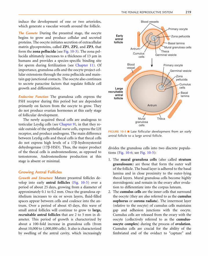
induce the development of one or two arterioles,
which generate a vascular wreath around the follicle.
The Gamete During the preantral stage, the oocyte
begins to grow and produce cellular and secreted
proteins. The oocyte initiates secretion of extracellular
matrix glycoproteins, called ZP1, ZP2, and ZP3, that
form the zona pellucida (see Fig. 10-3). The zona pel-
lucida ultimately increases to a thickness of 13 mm in
humans and provides a species-specific binding site
for sperm during fertilization (see Chapter 11). Of
importance, granulosa cells and the oocyte project cel-
lular extensions through the zona pellucida and main-
tain gap junctional contacts. The oocyte also continues
to secrete paracrine factors that regulate follicle cell
growth and differentiation.
Endocrine Function The granulosa cells express the
FSH receptor during this period but are dependent
primarily on factors from the oocyte to grow. They
do not produce ovarian hormones at this early stage
of follicular development.
The newly acquired thecal cells are analogous to
testicular Leydig cells (see Chapter 9), in that they re-
side outside of the epithelial nurse cells, express the LH
receptor, and produce androgens. The main difference
between Leydig cells and thecal cells is that thecal cells
do not express high levels of a 17b-hydroxysteroiddehydrogenase (17b-HSD). Thus, the major product
of the thecal cells is androstenedione, as opposed to
testosterone. Androstenedione production at this
stage is absent or minimal.
Growing Antral Follicles
Growth and Structure Mature preantral follicles de-
velop into early antral follicles (Fig. 10-5) over a
period of about 25 days, growing from a diameter of
approximately 0.1 to 0.2 mm. Once the granulosa ep-
ithelium increases to six or seven layers, fluid-filled
spaces appear between cells and coalesce into the an-
trum. Over a period of about 45 days, this wave of
small antral follicles will continue to grow to large,
recruitable antral follicles that are 2 to 5 mm in di-
ameter. This period of growth is characterized by
about a 100-fold increase in granulosa cells (from
about 10,000 to 1,000,000 cells). It also is characterized
by swelling of the antral cavity, which increasingly
divides the granulosa cells into two discrete popula-
tions (Fig. 10-6; see Fig. 10-5):
1. The mural granulosa cells (also called stratum
granulosum) are those that form the outer wall
of the follicle. The basal layer is adhered to the basal
lamina and in close proximity to the outer-lying
thecal layers. Mural granulosa cells become highly
steroidogenic and remain in the ovary after ovula-
tion to differentiate into the corpus luteum.
2. The cumulus cells are the inner cells that surround
the oocyte (they are also referred to as the cumulus
oophorus or corona radiata). The innermost layer
(relative to the oocyte) of cumulus cells maintains
gap and adhesion junctions with the oocyte.
Cumulus cells are released from the ovary with the
oocyte (collectively referred to as the cumulus-
oocyte complex) during the process of ovulation.
Cumulus cells are crucial for the ability of the
fimbriated end of the oviduct to “capture” and
Germinal vesicleCumuluscells
Antrum
Earlyantral
follicle
Largerecruitable
antralfollicle
Zona pellucida
Primary oocyte
Mural granulosa cellsBasal lamina
Theca
Blood vessels
Germinal vesicle
Cumuluscells
Antrum
Zonapellucida
Primary oocyte
Muralgranulosa
cells
Basallamina
Bloodvessel
Theca
FIGURE 10-5 n Late follicular development from an early
antral follicle to a large antral follicle.
219THE FEMALE REPRODUCTIVE SYSTEM

move the oocyte by a ciliary transport mechanism
along the length of the oviduct to the site of
fertilization.
Early antral follicles are dependent on pituitary
FSH for normal growth. Large antral follicles become
highly dependent on pituitary FSH for their growth
and sustained viability. As discussed later (under
Dominant Follicle), 2- to 5-mm follicles are recruited
to enter a rapid growth phase by a transient increase in
follicle-stimulating hormone (FSH) that occurs to-
ward the end of a previous menstrual cycle.
The Gamete Theoocytegrowsrapidly in theearly stages
of antral follicles; growth then slows in larger follicles.
During the antral stage, the oocyte synthesizes sufficient
amounts of cell cycle components, such as cyclin-
dependent kinase-1 (CDK1) and cyclin B, and the
oocyte becomes competent to complete meiosis I at
ovulation. Thus, in preantral follicles, the oocyte fails
to complete meiosis I because of a dearth of specific
meiosis-associated proteins (i.e., they are incompetent
to complete meiosis I). Larger antral follicles, however,
gain meiotic competence but still maintain meiotic
arrest until the midcycle luteinizing hormone (LH)
surge. Meiotic arrest is achieved by the maintenance of
elevated cyclic adenosine monophosphate (cAMP)
levels in the mature oocyte. The oocyte expresses a
constitutively active (i.e., active without a ligand) G-
protein-coupled receptor, called GPR3, that maintains
high cAMP levels (Figs. 10-7 and 10-8). Through a
cAMP-PKA phosphorylation cascade, the cyclin B–
cyclin-dependent kinase, CDK1, complex (also called
maturation-promoting factor,orMPF) iskept inactive.
As discussed in Chapter 1, intracellular cAMP levels are
determined by the activity of adenylyl cyclase, which
generates cAMP, and by the activity of phosphodiester-
ases (PDEs), which metabolize cAMP to AMP. Cyclic
guanosine monophosphate (cGMP) contributes to
the maintenance elevated cAMP levels in competent
oocytes by inhibiting the oocyte-specific PDE, PDE3A.
Cyclic GMP is made in the cumulus cells and mural
granulosa cells and is transferred to the oocyte through
gap junctions (see Fig. 10-8).
A
ZP
MG
BV
T
CR
O
FIGURE 10-6 n Histologic features of ovarian graafian
follicle. Ovum (O) is surrounded by zona pellucida (ZP).
As a result of shrinkage artifacts, the zona pellucida
appears larger than normal. Cumulus cells (C) are
indicated, as is the large antrum (A). BV, blood vessels
in outer thecal stroma; MG, mural glomerulosa cell; T,
thecal cells.
Oogonium
Primary oocytearrested at prophase I
Primary oocytearrested at prophase I
Secondary oocytearrested at metaphase II
Haploid ovum
Meiosis begins, but levelsof proteins required forcompletion of meiosis aretoo low—oocyte arrests atprophase I.
As oocyte grows, it synthe-sizes enough proteins(e.g., CDK1, cyclin B) tocomplete meiosis (i.e.,meiotic competence), buthigh cAMP levels gener-ated by GPR3 activelymaintain arrest.
A few hours before ovu-lation, the oocytecompletes meiosis I andextrudes the 1st polarbody. It has synthesizedenough MAPK pathwayproteins to arrest at meta-phase II.
The secondary oocytecompletes meiosis at fertil-ization and extrudes 2ndpolar body.
FIGURE 10-7 n Phases of oocyte development.
220 ENDOCRINE AND REPRODUCTIVE PHYSIOLOGY

Endocrine Function Thecal cells of large antral follicles
produce significant amounts of androstenedione and,
to a much lesser extent, testosterone (Fig. 10-9). This
is due to high expression of CYP17 with both 17-
hydroxylase and 17,20-lyase activities (Fig. 10-10A).
Androgens are converted to estradiol-17b by themu-
ral granulosa cells (see Fig. 10-9). At this stage, FSH
stimulates proliferation of granulosa cells and induces
the expression of CYP19-aromatase (Fig. 10-10B) re-
quired for estrogen synthesis. Additionally, the mural
granulosa cells of the large antral follicles produce in-
creasing amounts of inhibin B during the early follic-
ular phase. Low levels of estrogen and inhibin exert a
negative feedback effect on FSH secretion, thereby
contributing to the selection of the follicle with the
most FSH-responsive cells.
cGMP cGMP cGMPcAMP
AMP
cGMP PDE3A
(�) AC
GsZP
GPR3
FIGURE 10-8 n Mechanisms involved in meiotic arrest of a
meiotically competent primary oocyte. The smaller cells
represent cumulus, stalk, and mural granulosa cells—all
connected by gap junctions and all contributing to
elevated cyclic guanosine monophosphate in the oocyte.
AC, adenylyl cyclase; PDE 3A, phosphodiesterase 3A; ZP,
zona pellucida.
LH LH
Androstenedione
CholesterolCholesterol
Pregnenolone
Progesterone
StAR proteinCYP11A1
3β-HSD
Acetate
LDLR- and HDLR-mediated uptake
Androstenedione
Estradiol-17β
LH receptorLH receptor
Stimulation ofsteroidogenesis
LDLR- and HDLR-mediated uptake
Stimulation ofsteroidogenesis
FIGURE 10-9 n Two-cell model of ovarian steroidogenesis. 3b-HSD, 3b-hydroxysteroid dehydrogenase; 17b-HSD, 17b-hydroxysteroid dehydrogenase; FSH, follicle-stimulating hormone; HDLR, high-density lipoprotein receptor; LDLR,
low-density lipoprotein receptor; LH, luteinizing hormone; StAR protein, steroidogenic acute regulatory protein.
221THE FEMALE REPRODUCTIVE SYSTEM

Dominant Follicle
Growth and Structure Attheendofapreviousmenstrual
cycle, a crop of large (2- to 5-mm) antral follicles (see
Fig. 10-4) is recruited to begin rapid, gonadotropin-
dependent development. The total number of recruited
follicles in both ovaries can be as high as 20 in a younger
woman (<33 years of age), but rapidly declines at older
ages. The number of recruited follicles is reduced to the
ovulatory quota (which is one in humans) by the process
of selection. As FSH levels decline, the rapidly growing
follicles progressively undergo atresia, until one follicle is
left. Generally, the largest follicle with the most FSH
receptors of the recruited crop becomes the dominant
follicle. Selection occurs during the early follicular phase.
By midcycle, the dominant follicle becomes a large pre-
ovulatory follicle that is 20 mm in diameter and
contains about 50 million granulosa cells by the midcycle
gonadotropin surge.
The Gamete The oocyte is competent to completemeio-
sis I but remains arrested in the dominant follicle through
the mechanisms described earlier. Growth of the oocyte
continues, but at a slower rate—thehumanoocyte reaches
a diameter of about 140 mm by ovulation. The stalk by
which cumulus cells are attached to the mural granulosa
cells becomes increasingly attenuated.
Endocrine Function The newly selected follicle emerges
for the first time during its development as a significant
steroidogenic gland. Ovarian steroidogenesis requires
both theca and granulosa cells (see Fig. 10-9). As dis-
cussed earlier, thecal cells express LH receptors and
produce androgens. Basal levels of LH stimulate the
expression of steroidogenic enzymes, as well as the
LDL receptor in the theca. Thecal cells show robust
expression of CYP11A1 (also called P-450 cholesterol
side-chain cleavage), 3b-hydroxysteroid dehydroge-
nase (3b-HSD), andCYP17, with both 17-hydroxylase
activity and 17,20-lyase activity. Androgens (primarily
androstenedione but also some testosterone) released
from the theca can diffuse into the mural granulosa
cellsorcanenter thevasculature surrounding the follicle.
The mural granulosa cells of the selected follicle
have a high number of FSH receptors and are very
sensitive to FSH signaling. FSH strongly up-regulates
CYP19 (aromatase) gene expression and activity (see
Fig. 10-9). CYP19 converts androstenedione to the
weak estrogen, estrone, and converts testosterone to
the potent estrogen, estradiol-17b. Granulosa cells
express activating isoforms of 17b-HSD, which ulti-
mately drives steroidogenesis toward the production
of estradiol-17b (see later). FSH also induces the
expression of inhibin B during the follicular phase.
Of importance, FSH also induces the expression of
LH receptors in the mural granulosa cells during the
second half of the follicular phase (see Fig. 10-9).
Thus, mural granulosa cells become responsive to both
gonadotropins, allowing these cells to maintain high
levels of CYP19 in the face of declining FSH levels.
Acquisition of LH receptors also ensures that mural
granulosa cells will respond to the LH surge (see later).
The Dominant Follicle During thePeriovulatory Period
The periovulatory period can be defined as the time
from the onset of the LH surge to the expulsion of the
cumulus-oocyte complex out of the ovary (i.e., ovula-
tion). This process lasts 32 to 36 hours in women.
Starting at the same time, and superimposed on the
process of ovulation, is a change in the steroidogenic
function of the theca and mural granulosa cells. This
process is called luteinization and culminates in the
formation of a corpus luteum that is capable of pro-
ducing large amounts of progesterone, along with es-
trogen, within a few days after ovulation.
Growth and Structure The LH surge induces dramatic
structural changes in the dominant follicle that involve
its rupture, ovulation of the cumulus-oocyte complex,
Rupturedantrum
Luteinizing mural granulosawithout basal lamina
Zonapellucida
1st polarbody
2� oocyteafter GVDB
Peritonealcavity
Surface epithelium
Expandedcumulus
Tunica albugineaLuteinizing theca
Bloodvessels
andthecal
cellsinvading
granulosa
FIGURE 10-10 n Ovulation.
222 ENDOCRINE AND REPRODUCTIVE PHYSIOLOGY

and the biogenesis of a new structure called the corpus
luteum from the remaining thecal cells and mural
granulosa cells. Major structural changes occur during
this transition (see Fig. 10-10):
1. Before ovulation, the large preovulatory follicle
presses against the ovarian surface, generating a
poorly vascularized bulge of the ovarian wall called
the stigma. The LH surge induces the release of in-
flammatory cytokines and hydrolytic enzymes from
the theca andgranulosa cells. These secreted compo-
nents lead to the breakdown the follicle wall, tunica
albuginea, and surface epithelium in the vicinity of
the stigma. At the end of this process, the antral cav-
ity becomes continuous with the peritoneal cavity.
2. The stalk-like attachment of the cumulus cells to the
mural granulosa cells detaches, and the cumulus-
oocyte complex becomes free-floating within the
antral cavity. As an indirect response to the LH surge
(i.e., in response to LH-dependent paracrine fac-
tors), the oocyte releases the transforming growth
factor-b (TGF-b)-related factor, GDF9. GDF9 (see
earlier) stimulates the cumulus cells to secrete hya-
luronic acid and other extracellular matrix compo-
nents. These secreted components enlarge the entire
cumulus-oocyte complex, a process called cumulus
expansion. This enlarged cumulus-oocyte complex
is more easily captured and transported by the
oviduct. The expanded cumulus also makes the
cumulus-oocyte complex easier for spermatozoa
to find. The cumulus-oocyte complex is released
through the ruptured stigma in a slow, gentle pro-
cess, indicating that the follicular fluid in the antrum
is not under increased pressure. The specific forces
that lead to expulsion of the cumulus-oocyte com-
plex are unknown.
3. The basal lamina of the mural granulosa cells is
enzymatically degraded, and blood vessels and
outer-lying theca can push into the granulosa cells.
Granulosa cells secrete angiogenic factors, such as
vascular endothelial growth factor (VEGF),
angiopoietin-2, and basic fibroblast growth fac-
tor (bFGF), which significantly increase the blood
supply to the new corpus luteum.
The Gamete Before ovulation, the primary oocyte is
competent to complete meiosis but is arrested in pro-
phase I as a result of high cAMP levels (see Fig. 10-8).
The LH surge induces the oocyte to progress tometa-
phase of meiosis II. The oocyte subsequently arrests at
metaphase II until fertilization. LH receptors are only
present on the mural granulosa cells. The LH surge in-
duces a series of events that eventually lead to a de-
crease in cGMP and closure of gap junctions (except
for those between the oocyte and immediately adja-
cent cumulus cells) in mural granulosa and cumulus
cells. As a result, cGMP levels fall in the oocyte, allow-
ing PDE3A to degrade cAMP.
The decrease in cAMP and protein kinase A (PKA)
activity ultimately leads to activation of MPF, com-
posed of CDK1 and cyclin B. Cyclin B synthesis is el-
evated during the periovulatory period, thereby
increasing CDK1 activity. CDK1 activity is further en-
hanced by dephosphorylation, which is an indirect re-
sult of decreased PKA activity. The fully active MPF
drives nuclear events that complete meiosis I with
the extrusion of the first polar body. The secondary
oocyte (called an egg) then arrests in metaphase of
meiosis II. This is achieved by an increase in an activ-
ity called cytostatic factor (CSF). It is now known
that CSF is composed of the kinase, c-Mos, its target
mitogen-activated kinase kinase (MAPKK), also
called MEK1 (see Chapter 1), and MAPK. Thus, ele-
vation of the MAPK signaling pathway is required
for arrest at metaphase II, and fertilization leads to
the rapid degradation of MAPK. It should be empha-
sized that our understanding of normal oocyte matu-
ration has had a major impact on the ability to treat
infertile couples through the process of in vitro fertil-
ization (IVF). Normal oocyte biology dictates the type
of hormonal treatment, the timing of egg retrieval, and
the meiotic stage of eggs used for fertilization.
Endocrine Function Both thecal and mural granulosa
cells express LH receptors at the time of the LH surge.
The LH surge induces terminal differentiation (called
luteinization) of the granulosa cells—a process that
will continue for several days after ovulation. During
the periovulatory period, the LH surge induces the
following shifts in the steroidogenic activity of the
mural granulosa cells (Fig. 10-11):
1. It transiently inhibits CYP19 expression and, con-
sequently, estrogen production. The rapid decline
in estrogen helps to turn off the positive feedback
on LH secretion.
223THE FEMALE REPRODUCTIVE SYSTEM

2. By inducing the breakdown of the basal lamina, the
LH surge causes the direct vascularization of the
granulosa cells. This makes LDL and HDL choles-
terol accessible to these cells for steroidogenesis.
The LH surge also increases the expression of the
LDL receptor and HDL receptor (SR-BI) in gran-
ulosa cells.
3. The LH surge increases the expression of StAR pro-
tein, CYP11A1 (side-chain cleavage enzyme), and
3b-HSD. Because CYP17 activity, especially the
17,20-lyase function, is largely absent in granulosa
cells, these cells begin to secrete progesterone, and
progesterone levels will gradually increase over the
next week.
The Corpus Luteum
Growth and Structure After ovulation, the remnant of
the antral cavity fills with blood from damaged blood
vessels in the vicinity of the stigma. This gives rise to a
corpus hemorrhagicumwith clotted blood within the
former antral lumen (Fig. 10-12). Within a few days,
red blood cells and debris are removed by macro-
phages, and fibroblasts fill in the antral cavity with a
hyaline-like extracellular matrix. In the mature corpus
luteum, the granulosa cells, now called granulosa lu-
tein cells, enlarge and become filled with lipid (choles-
terol esters). The enlarged granulosa lutein cells
collapse into and partially fill in the old antral cavity.
Proliferation of these cells is very limited. The theca,
along with blood vessels, mast cells, macrophages, leu-
kocytes, and other resident connective tissue cells, in-
filtrates the granulosa layer at multiple sites.
The human corpus luteum is programmed to live
for 14�2 days (corpus luteum of menstruation), un-
less rescued by the LH-like hormone, human chorionic
gonadotropin (hCG), that originates from an implant-
ing embryo (hCG is the protein that is detected in
pregnancy tests; see Chapter 11). If rescued, the cor-
pus luteum of pregnancy will remain viable for as
long as the pregnancy (usually about 9 months).
The mechanism by which the corpus luteum of men-
struation regresses in 14 days is not fully understood,
although the timing is very consistent. In response to
LH LH
Androstenedione
Thecal lutein cell
Granulosalutein cell
CholesterolCholesterol
Pregnenolone
Progesterone
StAR proteinCYP11A1
3�-HSD
Acetate
LDLR- and HDLR-mediated uptake
Androstenedione
Estradiol-17�
LH receptorLH receptor
Stimulation ofsteroidogenesis
LDLR- and HDLR-mediated uptake
Stimulation ofsteroidogenesis
FIGURE 10-11 n Two-cell model
of ovarian steroidogenesis during
the luteal phase. 3b-HSD, 3b-hydroxysteroid dehydrogenase;
HDLR, high-density lipoprotein
receptor; LDLR, low-density
lipoprotein receptor; LH, lutei-
nizing hormone; StAR protein,
steroidogenic acute regulatory
protein. Note the absence of a
basal lamina.
224 ENDOCRINE AND REPRODUCTIVE PHYSIOLOGY

paracrine factors and, perhaps, declining progesterone
production, the corpus luteum becomes progressively
unresponsive to pituitary LH and needs the extra
amount of hCG to remain viable. The corpus luteum
ultimately is turned into a scar-like body called the
corpus albicans (see Fig. 10-12), which sinks into
the medulla of the ovary and is slowly absorbed.
The Gamete The LH surge induces two parallel events,
ovulation and luteinization. If ovulation occurs nor-
mally, the corpus luteum is devoid of a gamete.
Endocrine Function Progesterone production by the
corpus luteum (see Fig. 10-11) increases steadily from
the onset of the LH surge and peaks during the mid-
luteal phase. The main purpose of this timing is to
transform the uterine lining into an adhesive and sup-
portive structure for implantation and early preg-
nancy. As discussed in Chapter 11, the midluteal
phase is synchronized with early embryogenesis, so
the uterus is optimally primed when a blastocyst
tumbles into the uterus around day 22 of the men-
strual cycle. Estrogen production transiently decreases
in response to the LH surge but then rebounds and
also peaks at midluteal phase.
Luteal hormonal output is absolutely dependent on
basal LH levels (see Fig. 10-11). In fact, progesterone
output is closely correlated with the pulsatile pattern
of LH release in women. Both FSH and LH are reduced
to basal levels during the luteal phase by the negative
feedback from progesterone and estrogen. Also, gran-
ulosa lutein cells secrete inhibin A, which further
represses FSH secretion. The elevated estrogen levels
at midluteal phase may be responsible for the decrease
in the sensitivity of the corpus luteum to LH, so
progesterone and estrogen levels decline during the
second half of the luteal phase unless an increase in
circulating LH-like activity (i.e., in the form of
hCG) compensates for the decreased sensitivity to LH.
CLINICAL BOX 10-2
The corpus luteummust generate large amounts of pro-
gesterone for an adequate number of days in order to
support implantation and early pregnancy. Thus, thedurationof the lifeof the corpus luteum(14 days) is very
regular, and a shortened luteal phase typically leads to
infertility. The quality of the corpus luteum is largely de-
pendent on the size and health of the dominant follicle
from which the corpus luteum developed. Dominant
follicle development, in turn, is dependent on normal
hypothalamic and pituitary stimulation during the
follicular phase. Numerous factors that perturb hypo-
thalamic and pituitary output during the follicular
phase, including heavy exercise, starvation, high pro-lactin levels, and abnormal thyroid function, can lead
to luteal phase deficiency (LPD) and infertility.
The corpus luteum of other mammalian species
also produces an insulin-like hormone called relaxin.
The human corpus luteum produces very low levels of
relaxin, however, and the physiologic role of circulat-
ing relaxin in humans has not been established.
Follicular Atresia
Follicular atresia refers to the demise of an ovarian fol-
licle and represents by far the predominant process
in the ovary. During atresia, the granulosa cells and
GL
GL
BC
CHNB
BC
CA
FIGURE 10-12 n Histologic features of the corpus
hemorrhagicum. Micrograph of an ovary containing two
corpora hemorrhagicum (CH). These arise from the co-
ovulation of two dominant follicles, creating the potential
for nonidentical twins. Each corpus luteum shows a central
blood clot (BC) surrounded by a thick layer of lipid-rich
granulosa lutein cells (GL). Degenerating corpora lutea are
called corpora albicans (CA), which look like scar tissue
and are eventually degraded. (Modified from Stevens A, Lowe
J: Human Histology, 3rd ed., Philadelphia, 2005, Mosby.)
225THE FEMALE REPRODUCTIVE SYSTEM

oocytes undergo apoptosis. The thecal cells typically
persist and repopulate the cellular stroma of the ovary.
The thecal cells retain LH receptors and the ability to
produce androgens and collectively are referred to as
the interstitial gland of the ovary. Follicles can un-
dergo atresia at any time during development.
Follicular Development and the MonthlyMenstrual Cycle
The first half of the monthly menstrual cycle is referred
to as the follicular phase of the ovary and is character-
ized by the recruitment and growth of 15 to 20 large,
antral follicles (2 to 5 mm in diameter), selection of
one of these follicles as the dominant follicle, and growth
of the dominant follicle until ovulation. The dominant
folliclemust contain a fully developed,meiotically com-
petent oocyte and somatic follicle cells that secrete high
levels of estrogen. It takes several months for a primor-
dial follicle to reach the size of a large antral follicle that
can be recruited (Fig. 10-13). Therefore, it should be
noted that much of follicular development occurs inde-
pendently of the monthly menstrual cycle. The second
half of the monthly menstrual cycle is referred to as
the luteal phase of the ovary and is dominated by the
hormonal secretionsof the corpus luteum.Nevertheless,
small follicles continue to develop within the ovarian
stroma during the luteal phase.
THE HUMAN MENSTRUAL CYCLE
As stated earlier, late follicular development and luteal
function are absolutely dependent on normal hypo-
thalamic and pituitary function. As in the male, hypo-
thalamic neurons secrete gonadotropin-releasing
hormone (GnRH) in a pulsatile manner. GnRH, in
turn, stimulates LH and FSH production by pituitary
gonadotropes. A high frequency of GnRH pulses (one
pulse every 60 to 90 minutes) selectively promotes LH
production, whereas a slow frequency (one pulse every
120 minutes) selectively promotes FSH production.
A major difference between the male and the female
reproductive axes is themidcycle gonadotropin surge
in females, which is dependent on a high level of
estrogen over a specific duration coming from the
dominant follicle.
A highly dynamic conversation occurs among the
ovary, pituitary, and hypothalamus, which orches-
trates the events of the menstrual cycle (Fig. 10-14).
This section outlines the main points of events involv-
ing the ovary and pituitary gonadotrope that regulate
the menstrual cycle, with an overview of hypothalamic
involvement. In the next section, the effects of the
hormonal changes on the female reproductive tract,
especially the uterus, are discussed.
The following outline of events, numbered as
depicted in Figure 10-14, begins with the ovary at
Primordial follicle (0.03-0.04 mm)
Primary follicle (0.04-0.06 mm)
Secondary preantral follicle (0.06-2 mm)
Small antral follicle (0.2-2 mm)
Large, recruitable antral follicle (2-5 mm)
Dominant follicle at ovulation (15-20 mm)
Initiation and gonadotropin-independent growth phase
Follicles enter and undergo early growththat is regulated by paracrine signalingbetween oocyte and follicle cells, andbetween growing and resting follicles—not dependent on pituitary gland.
Basal growth phase
Follicles are dependent ongonadtropins—but have lowsensitivity and are not influencedby fluctuations associated withthe menstrual cycle.
Rapid growth phaseFollicles are extremely sensitive toincrease in FSH at end ofmenstrual cycle and extremelydependent on FSH to avoid atresia.
Initiation
> 3 Months
25 Days
45 Days
14 Days
FIGURE 10-13 n Timing and
phases of follicular growth.
226 ENDOCRINE AND REPRODUCTIVE PHYSIOLOGY

the end of the luteal phase of a previous, nonfertile
cycle:
Ovary—event 1: In the absence of fertilization and
implantation, the corpus luteum regresses and dies
(a phenomenon called luteolysis). This leads to a
drastic decline in the levels of progesterone, estro-
gen, and inhibin A by day 24 of the menstrual cycle.
Pituitary gonadotrope—event 2: The gonadotrope
perceives the end of luteal function as a release from
negative feedback. This permits a rise in FSH that
occurs about 2 days before the onset of menstrua-
tion. The basis for the selective increase in FSH is
incompletely understood but may result from the
slow frequency of GnRH pulses during the luteal
phase, which is due to high progesterone levels.
Ovary—event 3: The rise in FSH levels recruits a crop
of large (2- to 5-mm) antral follicles to begin rapid,
highly gonadotropin-dependent growth. These fol-
licles produce low levels of estrogen and inhibin B.
Pituitary gonadotrope—event 4: The gonadotrope
responds to the slowly rising levels of estrogen
and inhibin B by decreasing FSH secretion. Loss
of high levels of progesterone and estrogen causes
an increase in the frequency of GnRH pulses,
thereby selectively increasing LH synthesis and se-
cretion by the gonadotrope. Thus, the LH/FSH ra-
tio slowly increases throughout the follicular phase.
Ovary—event 5: The ovary’s response to declining
FSH levels is follicular atresia of all the recruited
follicles, except for one dominant follicle. Thus,
the process of selection is driven by an extreme de-
pendency of follicles on FSH in the face of declining
FSH secretion. Usually, only the largest follicle with
the most FSH receptors and best blood supply (i.e.,
most angiogenic) survives. This follicle produces
increasing amounts of estradiol-17b and inhibin
B during the second half of the follicular phase.
FSH also induces the expression of LH receptors
FSH
LH
Pituitary
OvaryProgesterone
4. E and inhibin negativelyfeed back on FSH.
Estradiol 17�
2. Pituitary responds to falling E and P by
increasing FSH secretion.
6. High E has positivefeedback on gonadotropes—LH (and some FSH) surges.
1. Corpus luteum dies, E and P
levels fall.
5. Declining FSH levels progressively cause atresia of
all but 1 follicle—leading to selection of dominant follicle, which produces
high levels of E.
9. The corpus luteum progressively becomes less
sensitive to basal LH—dies if levels of LH-like activity
(i.e., hCG) do not increase.
7. LH surge induces meiotic maturation,ovulation, and luteinization. The corpus luteum produces high P, along with E
and inhibin.
3. FSH recruits a cohort of large antralfollicles to enter rapid growth phase.
Follicles secrete low amounts of E and inhibin.
8. High P, E, and inhibin negatively feed back on LH and FSH, returning them to
basal levels.
14 280
FIGURE 10-14 n The human
menstrual cycle—a conversation
between the ovary and the pitu-
itary, with the hypothalamus
as a facilitator. See text for
comments on the involvement
of the hypothalamus. E, estro-
gen; FSH, follicle-stimulating
hormone; hCG, human chori-
onic gonadotropin; LH, luteiniz-
ing hormone; P, progesterone.
227THE FEMALE REPRODUCTIVE SYSTEM

in the mural granulosa cells of the dominant
follicle.
Pituitary gonadotrope—event 6: Once the dominant
follicle causes the circulating estrogen levels to ex-
ceed200 pg/mL for about 50hours inwomen, estro-
gen exerts a positive feedback on the gonadotrope,
producing the midcycle LH surge. This is enhanced
by the small amount of progesterone starting to be
made at midcycle. The exact mechanism of the pos-
itive feedback is unknown,but it occurs largely at the
level of the pituitary. GnRH receptors and the sen-
sitivity to GnRH signaling increase dramatically in
the gonadotropes. The hypothalamus contributes to
the gonadotropin surge by increasing the frequency
of GnRH pulses. There is some evidence that other
neurons in the hypothalamus (e.g., kisspeptin
neurons; see Chapter 8) respond to high levels of
estrogen by increasing the frequency and amount
of GnRH released.
Ovary—event 7: The LH surge drives three general
events in the ovary:
1. The primary oocyte completes meiosis I and
arrests at metaphase of meiosis II. This is associ-
ated with germinal vesicle breakdown (GVBD0;
the germinal vesicle refers to the nucleus of the
oocyte), which is the dissolution of the nuclear
membrane and interphase nuclear structure.
GVBD occurs about 30 hours after the onset of
the LH surge.
2. The wall of the follicle and of the ovary at the
stigma is broken down, and the free-floating
cumulus-oocyte complex is extruded from the
ovary (i.e., ovulation). This occurs about 32 to
36 hours after the onset of the LH surge.
3. The mural granulosa cells and theca cells are
restructured to form the corpus luteum. This in-
volves direct vascularization of the granulosa cells
and their differentiation into progesterone- and
estrogen-producing cells. Note that estrogen pro-
duction transiently drops for about 2 days after
the onset of LH production, which may termi-
nate the positive feedback. The granulosa cells
also secrete inhibin A. The process of luteiniza-
tion continues for several days after the onset
of the LH surge. The small amount of progester-
one secreted during the periovulatory period
contributes to the magnitude of the LH surge.
Pituitary gonadotrope—event 8: Rising levels of pro-
gesterone, estrogen, and inhibin A by the mature
corpus luteum negatively feed back on the pituitary
gonadotrope. Even though estrogen levels exceed
the 200 pg/mL threshold for positive feedback,
the high progesterone levels block any positive
feedback. Consequently, both FSH and LH levels
decline to basal levels.
Ovary—event 9: Basal levels of LH (but not FSH) are
absolutely required for normal corpus luteum
function. The corpus luteum becomes progressively
insensitive to LH signaling, however, and dies un-
less LH-like activity (i.e., hCG from an implanted
embryo) increases. In a nonfertile cycle, the corpus
luteum of menstruation will regress in 14 days, and
progesterone and estrogen levels will start to de-
cline by about 10 days.
Pituitary gonadotrope—event 10: Removal of nega-
tive feedback causes an increase in FSH at the
end of the cycle, and the entire process begins again.
From this sequence of events, it is evident that the
ovary is the primary clock for the menstrual cycle.
The timing of the two main pituitary-based events—
the transient rise in FSH that recruits large antral folli-
cles and the LH surge that induces ovulation—is deter-
mined by two respective ovarian events:
n Highly regular life span of a corpus luteum and its
demise after 14 days
n Growth of the dominant follicle to a point at which
it can maintain a sustained high production of
estrogen that induces a switch to positive feedback
at the pituitary
The hypothalamic release of GnRH changes over
the cycle. The frequency of GnRH pulses increases
during the second half of the follicular phase and
decreases during the luteal phase.
FEMALE REPRODUCTIVE TRACT
The female reproductive tract does not connect di-
rectly to the ovaries (Fig. 10-15). The internal portion
of the tract consists of right and left oviducts and the
following midline structures: uterus, cervix, and va-
gina. The external opening of the vagina is surrounded
by the external genitalia.
228 ENDOCRINE AND REPRODUCTIVE PHYSIOLOGY

The Oviduct
Structure and Function The oviducts (also called the
uterine tubes or fallopian tubes) are muscular tubes
that are opened at both ends. The end of the oviduct
close to the surface of each ovary has finger-like pro-
jections, called fimbriae. The opposite end pierces the
wall of the uterus and opens into the uterine lumen.
The oviducts can de divided into four sections
(Fig. 10-16). Going from ovary to uterus, these sec-
tions are named as follows:
1. Infundibulum, which includes the fimbriae
2. Ampulla, which has a relatively wide lumen and
extensive folding of the mucosa
3. Isthmus, which has a relatively narrow lumen and
less mucosal folding
4. Intramural or uterine segment, which extends
through the uterine wall at the superior corners
(horns) of the uterus
The wall of the oviduct is composed of a mucosa
called the endosalpinx, a two-layered muscularis
called themyosalpinx, and outer-lying connective tis-
sue, the perisalpinx, that contains numerous blood
vessels (Fig. 10-17). The endosalpinx is lined by a sim-
ple epitheliummade up of two cell types: ciliated cells
and secretory cells. The cilia aremost numerous at the
ovarian end (infundibulum and ampulla) and beat to-
ward the uterus. The cilia on the fimbriae are the sole
mechanism for transport of the ovulated cumulus-
oocyte complex into the oviduct. Once the complex
passes through the ostium of the oviduct and enters
the ampulla, it is moved by both cilia and peristaltic
contractions of the myosalpinx.
Fundus ofuterus
Uterine (fallopian)tube
Ovary
Body (corpus)of uterus
Internalgenitalia
Externalgenitalia
Cervix of uterus
Vagina
Clitoris
Vaginalintroitus
Openingof female
urethra
Mons pubis
Labiamajora
Labiaminora
FIGURE 10-15 n The internal and external genitalia of the
female reproductive tract. (Modified from Drake RL, Vogl
W, Mitchell AWM: Gray’s Anatomy for Students,
Philadelphia, 2005, Churchill Livingstone.)
Intramural segmentIsthmusAmpulla
Perisalpinx
EndosalpinxMyosalpinx
Ovary
Infundibulum
Fimbriae
C
B
A
FIGURE 10-16 n A, Structures of the oviduct. B, Cross
section of the oviduct at the ampulla, with a large lumen
filled with mucosal (endosalpinx) folds. C, Cross section
of the oviduct at the isthmus, showing a much smaller
lumen but a thicker muscularis (myosalpinx). (Modified
from Stevens A, Lowe J: Human Histology, 3rd ed.,
Philadelphia, 2005, Mosby.)
229THE FEMALE REPRODUCTIVE SYSTEM

As shown in Figure 10-17, the ovarian end of the
oviduct (infundibulum and ampulla) has a wide lu-
men partially filled with a highly folded myosalpinx.
This allows the cumulus-oocyte complex to be trans-
ported while in intimate contact with ciliated mucosal
cells. The uterine end of the oviduct (isthmus and in-
tramural segment) has a narrow lumen and a relatively
thicker muscularis. This allows for slow transport of
an early embryo to the uterus primarily by peristaltic
waves of the muscularis.
Themain functions of the oviducts are the following:
1. Capture of the cumulus-oocyte complex at ovula-
tion and transport of the cumulus-oocyte complex
toamidwaypoint (theampullary-isthmus junction),
where fertilization takes place. Oviductal secretions
coat and infuse the cumulus-oocyte complex and
may be required for viability and fertilizability.
2. Providing a site for sperm storage. Women who
ovulate up to 5 days after sexual intercourse can be-
come pregnant. Sperm remain viable by adhering
to the epithelial cells lining the isthmus. The secre-
tions of the oviduct also induce capacitation and
hyperactivity of sperm (see Chapter 11).
3. Providing nutritional support to the preimplanta-
tion embryo by the oviductal secretions. Also, the
timing of the movement of the embryo into the
uterus is critical because the human uterus has
an implantation window of about 3 days. The ovi-
duct needs to harbor the early embryo until it
reaches the blastocyst stage (5 days after fertiliza-
tion); then it allows the embryo to move into the
uterine cavity (see Chapter 11).
The secretory cells produce a protein-rich mucus
that is conveyed along the oviduct to the uterus by
the cilia. This ciliary-mucus escalator maintains a
healthy epithelium, moves the cumulus-oocyte com-
plex toward the uterus, and may provide directional
cues for swimming sperm. The movement of the
cumulus-oocyte complex slows at the ampullary-
isthmus junction, where fertilization normally takes
place. This appears to be due in part to a thick mucus
that is produced by the human isthmus and to an
increased tone of the muscularis of the isthmus. The
composition of oviductal secretions is complex and in-
cludes growth factors, enzymes, and oviduct-specific
glycoproteins. Note that IVF has shown that the secre-
tions of the oviduct are not absolutely necessary for
fertility by in vitro techniques. However, normal ovi-
ductal function is absolutely required for both fertiliza-
tion and implantation from in vivo insemination, and
to minimize the risk for ectopic implantation (i.e.,
implantation outside of the uterus). In fact, the most
common site of ectopic implantation is the oviduct.
CLINICAL BOX 10-3
Primary ciliary dyskinesia (also called immotile ciliasyndrome or Kartagener syndrome) is a highly het-
erogeneous inherited disease caused by the absence
or defect of one of the many components of the
ciliary-flagellar axoneme. The mutation can cause
no beating (ciliary immotility), abnormal beating
UTERINE LUMEN
Epithelium
Uterinegland
Uterine artery
Spiralartery
Straightartery
Radial branch
Arcuate
Capillaries
Venous lakes
Functionalzone
Basalzone
Myometrium
Endometrium
FIGURE 10-17 n Structure of the uterine endometrium.
(Modified from Strauss J III, Coutifaris C: The endometrium and
myometrium: Regulation and dysfunction. In Yen SSC, Jaffe RB,
Barbieri RL, editors: Reproductive Endocrinology, 4th ed.,
Philadelphia, 1999, Saunders, pp 191-217.)
230 ENDOCRINE AND REPRODUCTIVE PHYSIOLOGY

(ciliary dyskinesia), or loss of cilia (ciliary aplasia). The
disease is primarily characterized by infections of the
upper respiratory tract, nasal tract, and middle ear.
The importance of ciliary transport in the oviduct is
indicated by the finding that about 50% of women
with primary ciliary dyskinesia are infertile or
subfertile.
Hormonal Regulation During the Menstrual Cycle In
general, estrogen secreted during the follicular phase
increases endosalpinx epithelial cell size and height.
Estrogen increases blood flow to the lamina propria
of the oviducts, increases the production of oviduct-
specific glycoproteins (whose functions are poorly un-
derstood), and increases ciliogenesis throughout the
oviduct. Estrogen promotes the secretion of a thick
mucus in the isthmus and increases tone of the mus-
cularis of the isthmus, thereby keeping the cumulus-
oocyte complex at the ampullary-isthmus junction
for fertilization. High progesterone, along with estro-
gen, during the early to midluteal phase decreases ep-
ithelial cell size and function. Progesterone promotes
deciliation. Progesterone also decreases the secretion
of thick mucus and relaxes the tone in the isthmus.
The Uterus
Structure and Function Theuterus is a single organ that
sits in themidline of the pelvic cavity between the blad-
der and the rectum. The mucosa of the uterus is called
the endometrium, the three-layered, thick muscularis
is called themyometrium, and the outer connective tis-
sue and serosa are called the perimetrium. The parts of
the uterus are the fundus, which is that portion that
rises superiorly from the entrance of the oviducts; the
body, whichmakes upmost of the uterus; the isthmus,
a short narrowedpart of thebody at its inferior end; and
the cervix, which extends into the vagina (see Figs. 10-1
and 10-15). Because the cervical mucosa is distinct
from the rest of the uterus and does not undergo the
process of menstruation, it is discussed separately.
The established functions of the uterus are related
to supporting a pregnancy (see Chapter 11). Themain
functions of the uterus are as follows:
1. Provide a suitable site for attachment and implan-
tation of the blastocyst, including a thick, nutrient-
rich stroma
2. Limit the invasiveness of the implanting embryo so
that it stays in the endometrium and does not reach
the myometrium
3. Provide a maternal side of the mature placental ar-
chitecture. This includes the basal plate, to which
the fetal side attaches, and large, intervillous spaces
that become filled with maternal blood after the
first trimester
4. Grow and expand with the growing fetus so that the
fetus develops within an aqueous, largely nonadhe-
sive environment
5. Provide strong muscular contractions to expel the
fetus and placenta at term
An understanding of the function of the uterus and
hormonally induced uterine changes during nonfertile
menstrual cycles requires a basic knowledge of the fine
structure of the endometrium and of the relationship
of the uterine blood supply to the endometrium
(Fig. 10-17). The luminal surface of the endometrium
is covered by a simple cuboidal-columnar epithelium.
The epithelium is continuous with mucosal glands
(called uterine glands) that extend deep into the en-
dometrium. The mucosa is vascularized by spiral ar-
teries, which are branches of the uterine artery that
runs through the myometrium. The terminal arteri-
oles of the spiral arteries project to a position just be-
neath the surface epithelium. These arterioles give rise
to a subepithelial plexus of capillaries and venules,
which have ballooned, thin-walled segments called ve-
nous lakes or lacunae. The lamina propria (i.e., the
connective tissue and stroma of the mucosa support-
ing the epithelium) itself is densely cellular. The stro-
mal cells of the lamina propria play important roles
both during pregnancy and menstruation.
About two thirds of the luminal side of the endome-
trium is lost during menstruation and is called the
functional zone (also called the stratum functionalis)
(see Fig. 10-17). The basal one third of endometrium
that remains aftermenstruation is called thebasal zone
(also called the stratumbasale). Thebasal zone is fedby
straight arteries that are separate fromthe spiral arteries
and contains all of the cell types of the endometrium
(i.e., epithelial cells from the remaining tips of glands,
stromal cells, and endothelial cells).
Hormonal Regulation of the Uterine EndometriumDuring the Menstrual Cycle Phases the of uterine cycle
are controlled by ovarian estrogen and progesterone.
231THE FEMALE REPRODUCTIVE SYSTEM

Thus, phases of the endometrial cycle correspond to
phases of the ovarian cycle.
The Proliferative Phase At the endofmenses (days3
to 5), the functional layer of the uterine endometrium
has been shed, and the basal layer is undergoing reepi-
thelialization (Fig. 10-18). In the ovary, the follicular
phase is under way. By day 5 of the ovarian cycle, FSH
has recruited a cohort of 2- to 5-mm large antral follicles
that begin producing low but increasing levels of estra-
diol.Once thedominant follicle is selectedatmidfollicu-
lar phase, estradiol production increases dramatically
(see Fig. 10-14). The estrogen produced by the follicular
phase of the ovary drives the proliferative phase of the
uterine endometrium. Estrogen induces all cell types in
the basal layer to proliferate. In fact, the definition of an
estrogenic compound has historically been one that is
uterotropic. It is not clear whether estrogen stimulates
the growth and differentiation of pluripotential stem
cells or stimulates the growth of cells that are already
defined as endothelial, epithelial, and stromal. Estrogen
increases cell proliferation directly through estrogen
receptor-a (ERa) and indirectly through the produc-
tion of growth factors, such as insulin-like growth
factor-1 (IGF-1). Estrogen also induces the expression
of progesterone receptors, thereby priming the uterine
endometrium so that it can respond to progesterone
during the luteal phase of the ovary.
During the proliferative phase, the functional layer
of the endometrium is rebuilt, and the endometrium
increases from about 0.5 to 5 mm in thickness. Mitotic
figures are found throughout the tissue. The uterine
glands display a straight or coiled shape with narrow
lumina (see Fig. 10-18).
The Secretory Phase By ovulation, the thickness of
stratum functionalis has been reestablished under the
proliferative actions of estradiol (see Fig. 10-18). After
ovulation, the corpus luteum produces high levels of
progesterone, along with estradiol. The luteal phase
of the ovary switches the proliferative phase of the uter-
ine endometrium to the secretory phase. In general,
progesterone inhibits further endometrial growth and
induces the differentiation of epithelial and stromal
cells. Progesterone induces the uterine glands to secrete
a nutrient-rich product, which will support implanting
blastocyst viability. As the secretory phase proceeds, the
mucosal uterine glands become corkscrewed and saccu-
lated. Progesterone also induces changes in the adhe-
sivity of the surface epithelium, thereby generating
the window of receptivity for implantation (see
Chapter 11). Progesterone also promotes the differenti-
ation of the stromal cells into predecidual cells, which
must be prepared to form the decidua of pregnancy, or
toorchestratemenstruation in the absence of pregnancy.
Of importance, progesterone opposes the prolifer-
ative actions of estradiol. Progesterone down regulates
the estrogen receptor. Progesterone also induces
inactivating isoforms of 17b-HSD, thereby convert-
ing the active estradiol into the inactive estrone. Pro-
gesterone also up regulates the expression of a steroid
sulfotransferase that sulfates and inactivates estrogen.
GE
F
B
Ovulation
Late Menstrual
Ovary � Follicular (E2)
Uterus � Menstrual & Proliferative
Ovary � Luteal (P4 & E2)
Uterus � Secretory
Late Proliferative Early Secretory Late Secretory
Day 3 – 5 Day 10 – 13 Day 15 – 18 Day 22 – 25
FIGURE 10-18 n Changes in
endometrial glands at different
phases of the uterine cycle, and
the corresponding phases of
the ovarian cycle. (Modified partial
images from Young B, Lowe J, Stevens
A, et al: Wheater’s Functional
Histology, 5th ed., Edin-burgh,
2006, Churchill Livingstone.)
232 ENDOCRINE AND REPRODUCTIVE PHYSIOLOGY

This opposition of the mitogenic actions of estradiol
by progesterone is extremely important in protecting
the uterine endometrium from estrogen-induced uter-
ine cancer. By contrast, the administration of unop-
posed estrogen significantly increases the risk for
uterine cancer in women.
The Menstrual Phase In a nonfertile cycle, death of
the corpus luteum leads to a sudden withdrawal of
progesterone and estrogen, which leads to changes
in the uterine endometrium that result in the loss of
the lamina functionalis. Menstruation normally lasts
3 to 5 days (called a period), and the volume of blood
loss ranges from 25 to 35 mL. Menstruation coincides
with the early follicular phase of the ovary.
The breakdown of the stratum functionalis is due
to the up regulation of hydrolytic enzymes, calledma-
trix metalloproteases, which destroy the extracellular
matrix and basal lamina of the endometrium. These
enzymes are produced by the three resident cell types
of the endometrium: the epithelial cell, the stromal
cell, and the endothelial cell. Matrix metalloproteases
also are produced by leukocytes, which infiltrate into
the endometrium just before menstruation. The other
major component that leads to menstruation is the
production of prostaglandins. The inducible enzyme
required for prostaglandin synthesis, cyclooxygenase-
2 (COX-2), is increased in endothelial cells on pro-
gesterone withdrawal. This increases production of
inflammatory prostaglandins, especially PGF2a, which,
in turn, promotes contraction of the smooth muscle
cells of the myometrium and the vascular smooth mus-
cle cells of the spiral arteries. Intermittent spiral artery
contraction and dilation cause hypoxic necrosis, fol-
lowed by reperfusion injury of weakened tissue. The
degree of tissue loss and the onset of tissue repair appear
to be dependent on increasing estrogen levels during
the early follicular phase.
CLINICAL BOX 10-4
Disorders of menstruation are relatively common and
include menorrhagia (loss of >80 mL of blood) and
dysmenorrhea (painful periods). The existence of few,irregular periods, called oligomenorrhea, and the
absence of periods, called amenorrhea, often are
due to dysfunction or cessation of the hypothala-
mus-pituitary-ovarian axis, as opposed to local pelvic
pathophysiology.
Hormonal Regulation of the Myometrium The smooth
muscle cells of themyometrium also are responsive to
changes in steroid hormones. Peristaltic contractions
of the myometrium favor movement of luminal con-
tents from the cervix to the fundus at ovulation, and
these contractions may play a role in rapid bulk trans-
port of ejaculated sperm from the cervix to the ovi-
ducts. During menstruation, contractions propagate
from the fundus to the cervix, thereby promoting ex-
pulsion of sloughed functional zone. The size and
number of smooth muscle cells are determined by es-
trogen and progesterone. Healthy, cycling women
maintain a robust myometrium, whereas the myome-
trium progressively thins in postmenopausal women.
The Cervix
Structure and Function The cervix represents the infe-
rior extension of the uterus that projects into the va-
gina (see Figs. 10-1 and 10-15). It has a mucosa that
lines the endocervical canal, which has a highly elastic
lamina propria, and a muscularis that is continuous
with the myometrium. The cervix acts as a gateway
to the upper female tract; at midcycle, the endocervical
canal facilitates sperm viability and entry. During the
luteal phase, changes in the endocervical canal serve to
impede the passage of sperm and microbes, thereby
minimizing the chance of superimplantation of a sec-
ond embryo, as well as inhibiting ascending infections
into the placenta, fetal membranes, and fetus. The cer-
vix physically supports the weight of the growing fetus.
At term, cervical softening and dilation allow passage
of the newborn and placenta from the uterus into the
vagina.
Hormonal Regulation of Cervical Mucus During theMenstrual Cycle The endocervical canal is lined by a
simple columnar epithelial gland that secretes cervical
mucus in a hormonally responsive manner. Estrogen
stimulates production of a copious quantity of thin, wa-
tery, slightly alkalinemucus that is an ideal environment
for sperm. The macromolecules within this mucus are
thought to facilitate sperm movement. Progesterone
stimulates production of a scant, viscous, slightly acidic
mucus that is hostile to sperm and does not “fern.” This
thick mucus forms a barrier within the endocervical ca-
nal during the secretory phase of the endometrium and
during pregnancy, when the placenta produces high
amounts of progesterone (see Chapter 11).
233THE FEMALE REPRODUCTIVE SYSTEM

The Vagina
Structure and Function The vagina represents one of
the copulatory structures in women and acts as the
birth canal (see Fig. 10-16). Its mucosa is lined by a
nonkeratinized, stratified squamous epithelium. The
mucosa has a thick lamina propria enriched with elas-
tic fibers and is well vascularized. There are no glands
in the vagina, so lubrication during intercourse comes
from the following:
n Cervical mucus (especially during midcycle)
n A transudate (i.e., ultrafiltrate) from the blood ves-
sels of the lamina propria
n From the vestibular glands (see later)
The mucosa is surrounded by a relatively thin (i.e.,
relative to the uterus and cervix) two-layeredmuscularis
and anouter connective tissue. The vaginalwall is inner-
vated by branches of the pudendal nerve,which contrib-
ute to sexual pleasure and orgasm during intercourse.
Hormonal Regulation During the Menstrual Cycle The
superficial cells of the vaginal epithelium are continu-
ally desquamating and the nature of these cells is influ-
enced by the hormonal environment. Estrogen
stimulates proliferation of the vaginal epithelium
and increases their glycogen content. Estradiol also in-
duces minimal keratinization of the apical layers. Pro-
gesterone increases the desquamation of the epithelial
cells. The glycogen is metabolized to lactic acid by
commensal lactobacilli, thereby maintaining an acidic
environment. This relative acidity inhibits infections
by noncommensal bacteria and fungi.
The External Genitalia
Structure and Function The female external genitals are
surrounded by the labia majora (homologs of the
scrotum) laterally and the mons pubis anteriorly (see
Fig. 10-16). The vulva collectively refers to an area that
includes the labia majora and the mons pubis, plus the
labiaminora, the clitoris, the vestibule of the vagina, the
vestibular bulbs (glands), and the external urethral or-
ifice (Fig. 10-19). The vulva also is referred to as the pu-
dendum by clinicians. The structures of the vulva serve
the functions of sexual arousal and orgasm, directing
the flow of urine and providing a partial cover of the
opening of the vagina, thereby inhibiting the entry of
pathogens.
The clitoris is thehomologof thepenis. The clitoris is
composed of erectile tissue that undergoes the process of
erection in essentially the same manner as the penis.
Unlike the penis, clitoral tissue is completely separate
from the urethra. Thus, the only function of the clitoris
is involved with sexual arousal and climax at orgasm.
Hormonal Regulation During the Menstrual Cycle The
structures of the vulva do not show marked changes
during the menstrual cycle. The health and function
of these structures, however, are dependent on hor-
monal support. The external genitalia and vagina ap-
pear to be responsive to androgens (testosterone and
dihydrotestosterone), as well as estrogen. Androgens
also act on the central nervous system (CNS) to in-
crease libido in women.
BIOLOGY OF ESTRADIOL ANDPROGESTERONE
Mechanisms of Estrogen and ProgesteroneHormone Action
Estrogen and progesterone are steroid hormones;
accordingly, their cognate receptors belong to the nu-
clear hormone receptor superfamily (see Chapter 1).
Frenulum
Urethralopening
Vestibule(between
labia minora)
Hymen
Vaginalopening
Fourchette
Prepuceof clitoris
Glans clitoris
Lateral fold
Medial fold
Opening ofduct ofparaurethralgland
Labiumminus
Opening of duct ofgreater vestibular gland
FIGURE 10-19 n External genitalia in women. (From Drake
RL, Vogl W, Mitchell AWM: Gray’s Anatomy for Students,
Philadelphia, 2005, Churchill Livingstone.)
234 ENDOCRINE AND REPRODUCTIVE PHYSIOLOGY

Biologic Effects of Estrogen andProgesterone
Although the levels of estradiol and progesterone fluc-
tuate during the menstrual cycle, estrogen and proges-
terone levels are always higher in women than in men.
Estradiol and progesterone have multiple effects that
can be categorized according to whether they are di-
rectly related to the reproductive system. As discussed
previously, both steroid hormones have profound ef-
fects on the ovary, oviduct, uterus, cervix, vagina, and
external genitalia, and on the hypothalamus and pitu-
itary. Estrogen and progesterone also have important
effects on the following nonreproductive tissues:
Bone: Estrogen is required for closure of the epiphy-
seal plates of long bones in both sexes. Estradiol has
a bone anabolic effect and a calciotropic effect at
several sites. E2 stimulates intestinal calcium ab-
sorption and renal tubular calcium reabsorption.
E2 also is one of the most potent regulators of os-
teoblast and osteoclast function. Estrogen pro-
motes survival of osteoblasts and apoptosis of
osteoclasts, thereby favoring bone formation over
resorption. Loss of estradiol at menopause is fre-
quently associated with osteoporosis.
Liver: The overall effect of estradiol on the liver is to
improve circulating lipoprotein profiles. Estrogen
increases expression of the LDL receptor, thereby
increasing clearance of cholesterol-rich LDL parti-
cles by the liver. Estrogen also increases circulating
levels of high-density lipoprotein (HDL) levels.
Estrogen regulates hepatic production of several
transport proteins, including cortisol-binding pro-
tein, thyroid hormone–binding protein, and sex
hormone–binding protein.
Cardiovascular organs: Premenopausal women have
significantly lower cardiovascular disease than
men or postmenopausal women. Estrogen pro-
motes vasodilation through increased production
of nitric oxide, which relaxes vascular smooth mus-
cle and inhibits platelet activation. Estrogen pro-
motes angiogenesis in target tissues.
Integument: Overall, estrogen and progesteronemain-
tain a healthy, smooth skin with normal epidermal
and dermal thickness. Estrogen stimulates prolifer-
ation and inhibits apoptosis of keratinocytes. In
the dermis, estrogen and progesterone increases
collagen synthesis and inhibits (along with proges-
terone) the breakdown of collagen by suppressing
matrix metalloproteases. Estrogen increases glycos-
aminoglycan production and deposition in the der-
mis. Estrogen also promotes wound healing.
CNS: In general, estrogen is neuroprotective—that is,
it inhibits neuronal cell death in response to hypoxia
or other insults. The positive effects on angiogenesis
may account for some of the beneficial and stimu-
lant-like actions of estrogen on the CNS. Currently,
the proposed benefits of estrogen for the onset and
severity of Parkinson disease and Alzheimer disease
are controversial. Progesterone acts on the hypo-
thalamus to increase the set-point for thermoregu-
lation, thereby elevating body temperature by
approximately 0.5� F. This is the basis for using bodytemperature measurements to determine whether
ovulation has occurred. Progesterone generally acts
as a depressant on the CNS. Loss of progesterone on
demise of the corpus luteum of menstruation is the
basis for premenstrual syndrome (PMS) and the
severe variant premenstrual dysphoria disorder
(PMDD) experienced by some women.
Adipose tissue : Estrogen decreases adipose tissue by
decreasing lipoprotein lipase activity and increas-
ing hormone-sensitive lipase (i.e., it has a lipolytic
effect). Loss of estrogen results in an accumulation
of adipose tissue, especially in the abdomen.
The actions of estrogen and progesterone on the
maternal physiology and breast development and
function are discussed in Chapter 11.
Transport and Metabolism of OvarianSteroids
Steroid hormones are sparingly soluble in blood and
are carried primarily associated with plasma proteins.
About 60% of the estrogen is transported bound to sex
hormone–binding globulin (SHBG), 20% is bound
to albumin, and 20% is in the free form.
Progesterone binds primarily to cortisol-binding
globulin (CBG) (i.e., transcortin) and albumin.Because
it has a relatively low binding affinity for these proteins,
its circulating half-life (t1/2) is about 5 minutes.
Although the ovary is the primary site of estrogen
production, it is important to understand that periph-
eral aromatization of androgens to estrogens can
235THE FEMALE REPRODUCTIVE SYSTEM

generate locally high levels of estradiol in specific tis-
sues. For example, the fact that CYP19 (aromatase) is
expressed in the adipose tissue of the breast is the basis
for the use of aromatase inhibitors in the treatment of
estrogen-dependent breast cancer.
Estrogens and progestins are degraded in the liver
to inactive metabolites, conjugated with sulfate or glu-
curonide, and excreted in the urine. Major metabolites
of estradiol include estrone, estriol, and catecholestro-
gens (2-hydroxyestrone and 2-methoxyestrone). The
major metabolite of progesterone is pregnanediol,
which is conjugated with glucuronide and excreted
in the urine.
OVARIAN PATHOPHYSIOLOGY
Turner Syndrome
Turner syndrome, or gonadal dysgenesis, is the most
common cause of congenital hypogonadism. In about
50%of cases, it results from the complete absenceof the
secondX chromosome, so the karyotype of the affected
person is 45,XO. The germ cells do not develop, and
each gonad consists of a connective tissue-filled streak.
Themajor clinical characteristics include short stature,
a characteristic webbed neck, low-set ears, a shield-
shaped chest, short fourth metacarpals, and sexual in-
fantilism resulting from gonadal dysgenesis (Fig. 10-
20). Internal and external genitalia typically are female.
Polycystic Ovarian Syndrome
Chronically anovulatory women with high circulating
androgen, estrogen, and LH levels often have the dis-
order called polycystic ovarian syndrome (PCOS).
This syndrome may be caused by any of a broad array
of underlying problems, and PCOS accounts for 75%
of anovulatory infertility. Currently, the diagnosis of
PCOS requires two of the following three conditions:
amenorrhea, evidence of excessive androgen secre-
tion (i.e., acne, hirsutism), and polycystic ovaries,
as usually detected by sonogram (Fig. 10-21). The
FIGURE 10-20 n Female with Turner syndrome. Note
the characteristically broad, webbed neck. Stature is
reduced, and sexual secondary characteristics are poorly
developed. (From Goodman RM, Gorlin RJ: Atlas of the
Face in Genetic Disorders, 2nd ed., St. Louis, 1977, Mosby.)
FIGURE 10-21 n Sonogram of polycystic ovary. Cysts (arrows)
are due to large antral follicles in the cortex that failed to
ovulate. (Courtesy of Dr. AndreaDiLuigi, Department ofObstetrics
and Gynecology, University of Connecticut Health Center,
Farmington, Conn.)
236 ENDOCRINE AND REPRODUCTIVE PHYSIOLOGY

ovarian cysts represent large antral follicles that have
failed to ovulate and luteinize. The continuous go-
nadotropin secretion leads to ovarian enlargement,
and the ovaries typically show a thickened capsule
and numerous follicles, many of which are undergoing
atresia. FSH levels are low, which inhibits granulosa
cell function, and the high intrafollicular androgen
level inhibits follicular maturation. A significant pro-
portion of the circulating estrogen, present in high
levels, is estrone formed from peripheral aromatiza-
tion of androstenedione. These high androgen levels
can produce hirsutism and acne. Hirsutism is the ab-
normal formation of coarse sexual hair in regions
atypical for a woman, such as the face, back, and chest.
The exact cause of PCOS is not well understood,
but the primary defect appears to be inappropriate
signals between the hypothalamic-pituitary axis and
the ovary. A significant subset of patients with PCOS
are overweight or obese and have insulin resistance
and hyperinsulinemia. Insulin promotes ovarian
androgen production, and hyperinsulinism may
account for increased androgen production. Reduc-
tion of insulin levels (such as by weight loss, exercise,
or metformin administration) ameliorates the hy-
perandrogenism and PCOS in these patients. Alterna-
tively, an inadequate response of the follicle to
FSH may be due to impaired IGF-1 or insulin
signaling.
S U M M A R Y
1. The female reproductive system includes the
ovary, oviducts, uterus, cervix, vagina, and exter-
nal genitalia, along with the pituitary gonado-
tropes and the hypothalamic GnRH neurons.
The mammary glands (breasts) also can be con-
sidered a part of the female reproductive system.
2. The ovarian phases of the human menstrual cycle
are the follicular phase, the periovulatory period,
and the luteal phase.
3. The ovarian follicle contains a primary oocyte
arrested in meiotic prophase and variable layers
of granulosa and thecal cells. Preantral and early
antral follicular growth is gonadotropin indepen-
dent. Intermediate antral follicular growth is de-
pendent on a basal level of FSH, but not affected
by fluctuations in FSH associated with the men-
strual cycle. Large antral follicular development
is exquisitely dependent on fluctuations of FSH.
Follicles can degenerate at any phase during the
process of atresia.
4. The dominant follicle is selected based on its size,
number of FSH receptors, aromatase activity, and
blood supply. The dominant follicle is the endo-
crine structure of the follicular phase. The thecal
cells express the LH receptor, and LH stimulates
the production of androgens (primarily andro-
stenedione). The granulosa cells express the
FSH receptor, and FSH promotes the aromatiza-
tion of androgens to estrogens (primarily
estradiol). FSH also induces the expression of
the LH receptor in granulosa cells of the domi-
nant follicle.
5. The dominant follicle signals that it is ready to
ovulate by its estrogen production. High sus-
tained levels of estrogen induce the midcycle
LH surge through a positive feedbackmechanism.
This is due, in part, to a marked increase in the
sensitivity of the pituitary gonadotropes to GnRH
pulses.
6. The periovulatory period involves the meiotic
maturation of the primary oocyte to a secondary
oocyte (egg) arrested at metaphase II. Ovulation
involves the rupture of the follicular wall at the
stigma, release of the cumulus-oocyte complex,
and differentiation of the remaining follicular
cells into a corpus luteum.
7. The luteal phase is characterized by high proges-
terone production. The corpus luteum is pro-
grammed to die in 14 days, unless rescued
by hCG.
8. Progesterone opposes the estradiol-dependent
proliferation of the endometrial functional zone.
9. The oviduct functions to capture the cumulus-
oocyte complex, transport and nurture both male
and female gametes, promote fertilization and
early embryonic development, and determine
the timing of the movement of the blastocyst into
the uterine cavity.
237THE FEMALE REPRODUCTIVE SYSTEM

10. The mucosa of the uterus is called the endome-
trium.The function of the endometrium is to allow
implantation and placentation. Estrogen produced
during themid- to late follicular phase of the ovary
drives the proliferative phase of the uterus, during
which the endometrium grows in thickness. The
progesterone produced by the luteal phase of the
ovary drives the secretory phase of the uterus. Loss
of progesterone after the death of the corpus
luteum causes the endometrium to break down.
This represents the menstrual phase of the uterus.
11. The cyclic changes in ovarian steroids also alter cer-
vical mucus and vaginal epithelium. The external
genitalia are responsive to estrogen and androgens.
12. Estrogen and progesterone regulate numerous pro-
cesses directly associated with reproduction. How-
ever, these steroids also regulate nonreproductive
aspects of physiology, including bone growth and
health, cardiovascular function, hepatic function,
andothers.Estrogenandprogesteroneactprimarily
through interaction with classic estrogen and pro-
gesterone receptors, which belong to the family of
nuclear hormone receptors. Estrogen and proges-
terone alsohave rapid,membrane-initiated actions.
13. Turner syndrome (gonadal dysgenesis) is the
most common cause of congenital hypogonad-
ism. It typically results from the absence of the
second X chromosome, so the karyotype of the
affected person is 45,XO.
14. Polycystic ovarian syndrome produces chronic
anovulation. Circulating androgen, estrogen,
and LH levels typically are high.
S E L F - S T U D Y P R O B L E M S
1. During treatment for in vitro fertilization, thepatient
receives a daily injection of FSH for 8 to 10 days, fol-
lowed by one injection of hCG. Cumulus-oocyte
complexesare retrieved35hoursafter thehCGinjec-
tion from ovarian follicles just before they ovulate.Because oocytes are retrieved before ovulation,
and patients are treated with progesterone after re-
trieval, what is the purpose of the hCG injection?
2. When and in what cells is the LH receptor
expressed during the menstrual cycle?
3. Describe the major ovarian processes that occur
during the periovulatory period.
4. What is meant by the “two-cell model” of ovariansteroidogenesis?
5. Name three effects of estrogen on reproductive tis-
sue and three effects on nonreproductive tissue.
6. What would be the outcome of a truncated luteal
phase with early death of the corpus luteum on
the uterine endometrium?
7. What is the ovarian reserve?
8. What is the response of the pituitary gonadotropesto the death of a corpus luteum of menstruation?
9. What is the response of the ovary to declining FSH
levels during the early follicular phase?
K E Y W O R D S A N D C O N C E P T S
n 3b-Hydroxysteroid dehydrogenase (3b-HSD)n 17b-Hydroxysteroid dehydrogenase (17b-HSD)n 17-Hydroxylase activity
For full list of keywords and concepts see Student
Consult
SUGGESTED READ INGS
Arevalo MA, Santos-Galindo M, Lagunas N, et al: Selective estrogen
receptor modulators as brain therapeutic agents, J Mol Endocrinol
46:R1–R9, 2011.
Conti M, Hsieh M, Musa Zamah A, et al: Novel signaling mecha-
nisms in the ovary during oocyte maturation and ovulation,
Mol Cell Endocrinol 356:65–73, 2012.
Mehlmann LM: Stops and starts in mammalian oocytes: Recent
advances in understanding the regulation of meiotic arrest
and oocyte maturation, Reproduction 130:791–799, 2005.
Pasquali R, Stener-Victorin E, Yildiz BO, et al: PCOS Forum:
Research in polycystic ovary syndrome today and tomorrow,
Clin Endocrinol 74:424–433, 2011.
238 ENDOCRINE AND REPRODUCTIVE PHYSIOLOGY

K E Y W O R D S A N D C O N C E P T S
n 17,20-Lyase activityn 45,XOn Abnormal thyroid functionn Amenorrhean Ampullan Androgensn Androstenedionen Angiogenic factorsn Angiopoietin-2n Anovulatory infertilityn Antimullerian hormone (AMH)n Antral folliclesn Aromatase inhibitorsn Atresian Basal laminan Basal levels of LHn Basal zone (stratum basale)n Basic fibroblast growth factor (bFGF)n Birth canaln Bodyn Bone morphogenetic proten-15 (BMP15)n Capacitationn Capture of the cumulus-oocyte complexn Cervical mucusn Cervixn Ciliated cellsn c-kitn Clitorisn c-Mosn Copulatory structuresn Corona radiatan Corpus albicansn Corpus hemorrhagicumn Corpus luteumn Corpus luteum of menstruationn Corpus luteum of pregnancyn Cortisol-binding globulin (CBG)n Cuboidal granulose cellsn Cumulus cellsn Cumulus expansionn Cumulus oophorusn Cumulus-oocyte complexn Cyclic adenosine monophosphate (cAMP)n Cyclic guanosine monophosphate (cGMP)
n Cyclin Bn Cyclin-dependent kinase-1 (CDK1)n Cyclooxygenase-2 (COX-2)n CYP11A1 (P-450 cholesterol side-chaincleavage)
n CYP11A1 (side-chain cleavage enzyme)n CYP17n CYP19-Aromatasen Cytostatic factor (CSF)n Deciduan Dermal thicknessn Dominant folliclen Dysmenorrhean Ectocervixn Endocervical canaln Endocervixn Endocrinen Endometriumn Endothelial celln Epidermaln Epithelial celln Estradioln Estradiol-17bn Estrogenn Estrogen receptor-a (ERa)n Estrogen receptor antagonistn Estronen Excessive androgen secretionn Ferningn Fertilizationn Fimbriaen First polar bodyn Follicle-stimulating hormone (FSH)n Follicular atresian Follicular phase of the ovaryn FSH receptorsn Functional zone (stratum functionalis)n Fundusn Gametogenicn Gap junctionsn GDF9n Germinal vesicle breakdown (GVBD)n Glycosaminoglycan productionn GnRH receptorsn Gonadotropin-releasing hormone (GnRH)n GPR3
238.e1THE FEMALE REPRODUCTIVE SYSTEM

n GPR30n Granulosa lutein cellsn Growth differentiation factor-9 (GDF9)n HDL cholesteroln HDL receptor (SR-BI)n Heavy exercisen High-density lipoprotein (HDL)n High progesterone levelsn High prolactin levelsn Hormone-sensitive lipasen Hyperactivity of spermn Hypoxic necrosisn Implantationn Inactivating isoforms of 17b-HSDn Infertilityn Infundibulumn Inhibin An Inhibin Bn Interstitial gland of the ovaryn Intramural or uterine segmentn Isthmusn Keratinocytesn Kit ligandn Labian Labia majoran Lacunaen Large, recruitable antral folliclesn LDL cholesteroln LDL receptorn Leiomyomasn Leukocytesn LH receptorsn LH surgen LH/FSH ration Lipoproteinn Lipoprotein lipase activityn Livern Low-density lipoprotein (LDL)n Luteal phasen Luteal phase deficiency (LPD)n Luteal phase of the ovaryn Luteinizationn Luteinizing hormone (LH)n Luteolysisn Matrix metalloproteasesn Maturation-promoting factor (MPF)
n Meiotic arrestn Meiotic competencen Menopausen Menorrhagian Menstruationn Metaphase IIn Midcycle gonadotropin surgen Mitogen-activated kinase kinase (MAPKK, MEK1,MAPK)
n Mons pubisn Mural granulose cellsn Mural granulose cells (stratum granulosum)n Myometriumn Myosalpinxn Neuroprotectiven Nitric oxiden Oligomenorrhean Orgasmn Ovarian folliclesn Ovarian reserven Ovarian surface epithelial cellsn Ovariesn Ovary gonadotrope—event 7n Ovary—event 1n Ovary—event 3n Ovary—event 5n Ovary—event 9n Oviducts (uterine or fallopian tubes)n Ovulationn Ovulatory quotan PDE3An Perimetriumn Periodn Periovulatory periodn Perisalpinxn PGF2an Phosphodiesterase (PDE)n Pituitary FSHn Pituitary gonadotrope—event 1n Pituitary gonadotrope—event 2n Pituitary gonadotrope—event 4n Pituitary gonadotrope—event 6n Polycystic ovarian syndrome (PCOS)n Polycystic ovariesn Positive feedbackn Preantral
238.e2 ENDOCRINE AND REPRODUCTIVE PHYSIOLOGY

n Predecidual cellsn Pregnancyn Pregranulosa cellsn Premature ovarian failuren Premenstrual dysphoria disorder (PMDD)n Premenstrual syndrome (PMS)n Primary ciliary dyskinesia (immotile ciliasyndrome, Kartagener syndrome)
n Primary folliclen Primary oocytesn Primingn Progesteronen Progesterone receptorsn Proliferative phasen Prophase In Prostaglandinsn Pudendumn Pulsatilen Raloxifenen Relaxinn Secondary folliclen Secondary oocyte (egg)n Secretory cellsn Secretory phasen Selectionn Selective estrogen receptor modulator (SERM)n Sensitivity to GnRH signalingn Set-point for thermoregulationn Sex hormone–binding globulin (SHBG)
n Sexual arousaln Slow frequency of GnRH pulsesn Sperm storagen Spinnbarkeitn Spiral arteriesn StAR proteinn Starvationn Stromal celln Sulfotransferasen Tamoxifenn Testosteronen Thecal cellsn Transportn Transudaten Turner syndrome (gonadal dysgenesis)n Uterine fibroidsn Uterine glandsn Uterotropicn Vascular endothelial growth factor (VEGF)n Vasodilationn Ventilatory responsen Vestibular glandsn Vulvan Window of receptivityn Wound healingn Zona pellucida (ZP)n ZP1n ZP2n ZP3
238.e3THE FEMALE REPRODUCTIVE SYSTEM

n n n n n n n n n n n n n n n
11 FERTILIZATION, PREGNANCY,AND LACTATION
O B J E C T I V E S
1. Describe the synchronization among fertilization, early
embryonic events, and the human menstrual cycle.
2. Describe the events involved in fertilization.
3. Explain how implantation and placentation occur.
4. Discuss the endocrine and transport functions of the
placenta.
5. Describe the development of the fetal endocrine system.
6. Discuss maternal endocrine changes during pregnancy.
7. Discuss the current models for the initiation and pro-
gression of labor (parturition) in humans.
8. Describe the development and regulation of the mam-
mary glands.
9. Discuss the endocrine basis for contraception, the
“morning after” pill, and the abortion pill.
Human reproduction involves internal in-
semination, internal fertilization, and internal gesta-
tion, all within the female tract. Internal gestation
also involves the development of a transient organ,
the placenta. The placenta is remarkable in that it is
composed of tissues from two organisms:
n An extraembryonic membrane (called the chorion)
of the fetus
n Endometrial tissue (called decidua) of the mother
From an endocrine point of view, pregnancy repre-
sents a state in which three separate endocrine
systems—maternal, fetal, and placental—interact to
promote adequate growth and nutrition of the fetus,
the timing of parturition, and preparation of the
maternal mammary glands to support extrauterine
life of the fetus.
FERTILIZATION, EARLYEMBRYOGENESIS, IMPLANTATION,AND PLACENTATION
Synchronization with Maternal Ovarianand Reproductive Tract Function
Fertilization, early embryogenesis, implantation,
and early gestation all are synchronized with the hu-
man menstrual cycle (Fig. 11-1). Just before ovula-
tion, the ovary is in the late follicular stage and
produces high levels of estrogen. Estrogen promotes
growth of the uterine endometrium and induces
expression of the progesterone receptor. Estrogen
ultimately induces the luteinizing hormone (LH)
surge, which in turn induces meiotic maturation
of the oocyte and ovulation of the cumulus-oocyte
complex.
239

The events between fertilization and implantation
take about 6 days to complete, and implantation occurs
at about day 21 of the menstrual cycle. At this time, the
ovary is in themidluteal phase, secreting large amounts
ofprogesterone.Progesterone stimulates secretion from
uterine glands, which provide nutrients to the embryo.
This is referred toashistotrophicnutritionand isan im-
portant mode of maternal-to-fetal transfer of nutrients
for about the first trimester of pregnancy, after which
it is replaced by hemotrophic nutrition. Progesterone
inhibits myometrial contraction and prevents the
release of paracrine factors that lead to menstruation.
Progesterone induces the window of receptivity in
the uterine endometrium, which exists from about day
20 to 24 of the menstrual cycle. This receptive phase is
associated with increased adhesivity of the endome-
trial epithelium and involves the formation of cellular
extensions, called pinopodes, on the apical surface of
endometrial epithelia, along with increased expression
of adhesive proteins (e.g., integrins, cadherins) and
decreased expression of antiadhesive proteins (e.g.,
mucins) in the apical cell membrane.
Thus, during the time it takes a fertilized egg to
implant in the uterus, the uterine endometrium is at
d7 Late follicular phase
Dominant follicle–high E2 output
Ovary andpituitary
Uterineendometrium
Egg andembryo
Peak E2 levelsOnset of LH surge
d14
d13d15
Ovulation
Early luteal phase d21 d28Late luteal phase
Rescue of CL by hCGCorpus luteum formulationIncreasing levels of P4 and E2
Late secretory phaseEarly secretory phase
P4 induces secretion from E2-primed glands and opposition of E2 mitogenic effects
Late proliferative phase
Growth of functional zoneInduction of P4 receptors
d24d20
Receptive periodDecreased antiadhesion protein expression
Increased adhesion protein expression
d20d17
2-cellembryo
Fertilization—pronuclear stage
d16
d15d13
d22 d23d26
Completion of interstitial implantation
Blastocyst inuterus
Blastocyst hatches from zona pellucida
and implantation begins on days22-23 (6-7 days
after fertilization)
1st polar body and arrest at metaphase II35 h after onset of LH surge
Resumption of meiosis I
Arrested primary oocyte
FIGURE 11-1n Synchronization of the humanmenstrual cycle with fertilization and early embryogenesis. CL, corpus luteum;
E, estrogen; E2, estrone; hCG, human chorionic gonadotropin; LH, luteinizing hormone; P4, progesterone.
240 ENDOCRINE AND REPRODUCTIVE PHYSIOLOGY

its full thickness, actively secretory, and capable of
tightly adhering to the implanting embryo. It also
should be noted that the uterine endometrium is well
vascularized at the time of implantation. Spiral arter-
ies extend to the basal lamina of the surface epithelium
(see Fig. 10-19 in Chapter 10) and give rise to rich cap-
illary beds and postcapillary venous lakes (also called
lacunae). Apart from its nutrient supply to all cells of
the endometrium, the extensive blood supply immedi-
ately adjacent to the surface epithelium plays a critical
role in capturing embryonic human chorionic gonad-
otropin (hCG) and transporting hCG to the ovary,
where it rescues the corpus luteum. The rich endome-
trial blood supply also is important for efficient
delivery of progesterone to the endometrium.
Fertilization
Fertilization accomplishes both the recombination of
genetic material to form a new, genetically distinct or-
ganism and the initiation of events that begin embry-
onic development. There are several steps that must
occur for successful fertilization to be achieved. The
sperm must find its way to the egg, and the sperm
and the egg must contact, recognize one another,
and fuse. After sperm-egg fusion, an intracellular sig-
naling cascade occurs within the egg that has two ma-
jor consequences. First, it allows the egg to regulate
sperm entry such that only one sperm can fuse with
the egg. This prevents polyspermy, which is lethal.
Second, it “wakes up” the metabolically quiescent
egg so that it can resume meiosis and begin embryonic
development. This process is called egg activation.
Spermatozoa present in the male ejaculate enter the
vagina near the cervix and must reach the ampulla of
the oviduct where fertilization occurs.
Sperm transport is largely dependent on the female
reproductive tract and, while the sperm are still in the
uterus, is independent of swimming. Large numbers
of sperm in the ejaculate generally are required for suc-
cessful fertilization of the egg by one sperm—of the
300 million sperm typically ejaculated, only about
200 reach the oviduct. Clinically, males with fewer
than 20 million sperm per milliliter of ejaculate are
considered to be infertile.
The female reproductive tract is an important
regulator of sperm transport. Toward the end of the
follicular phase of the menstrual cycle, before ovula-
tion, estrogen levels are high. Estrogen causes the cer-
vix to produce a watery mucus, often called “egg
white cervical mucus” because of its consistency. This
mucus forms channels to aid the passage of sperm
through the cervix, and only motile sperm can pass
through this barrier. Estrogen also causes contrac-
tions of the myometrium to help propel sperm
upward toward the oviduct (i.e., cervical-to-fundal
contractions).
Sperm must undergo a process called capacitation
in the female reproductive tract before they are able to
fertilize the egg. Sperm capacitation is an incompletely
understood, transient event that occurs largely in the
oviduct and modifies the spermatozoan in several
ways so that it becomes capable of fertilizing the
egg. These changes include the following:
1. An altered membrane fluidity due to the removal of
cholesterol from the sperm membrane
2. The removal of proteins and carbohydrates from
the membrane that may otherwise block sites that
bind to the egg
3. A change in membrane potential that may permit
Ca2þ to enter the sperm and thereby facilitate
the acrosome reaction (see later)
4. Phosphorylation of numerous proteins
Incapacitated sperm bind actively to the epithelial
cells of the oviductal isthmus and become unbound
when they are capacitated. This binding slows the
capacitation process, extends the sperm life span, pre-
vents too many sperm from reaching the egg, and
increases the probability that sperm will be in the ovi-
duct when the egg is ovulated. Sperm can therefore
survive in the female reproductive tract for several
days.Hyperactivation is another phenomenon associ-
ated with sperm capacitation. Hyperactivation in-
volves a change in flagellar motion from a wavelike
to a whiplike motion. This type of flagellar movement
is necessary for the sperm to detach from the oviductal
epithelium, is well suited to swimming through the
thick oviductal fluid, and helps propel the sperm
through the outer layers of the egg to reach the egg’s
plasma membrane.
Capacitated sperm reach the egg, surrounded by its
expanded cumulus cells, in the ampulla of the ovi-
duct. Fertilization involves the penetration of the
241FERTILIZATION, PREGNANCY, AND LACTATION

egg by the entire sperm. To do this, the sperm must
breach three barriers (Fig. 11-2):
n Expanded cumulus
n Zona pellucida
n Plasma membrane of the egg (called the oolemma)
The cumulus cell matrix is composed predomi-
nantly of hyaluronic acid, and the sperm are able to
digest through this layer with a membrane-bound hy-
aluronidase called PH-20. The next obstacle the sperm
encounters is the zona pellucida, an extracellular coat
made up of three glycoproteins called ZP1, ZP2, and
ZP3. The sperm contains one or more ZP3 receptors.
Sperm binding to ZP3 triggers the acrosome reaction,
in which the inner sperm plasmamembrane fuses with
the outer acrosomal membrane to release the contents
of the acrosomal vesicle (see Fig. 11-2). The enzymes
released from the acrosomal vesicle then digest the
zona pellucida. After the acrosome reaction, the sperm
loses ZP3 receptors, and it undergoes a secondary
binding instead to ZP2. The sperm is thus held in place
as the enzymes from the acrosome digest a hole in the
zona pellucida and the still-swimming sperm can go
through to reach the egg plasma membrane. The mo-
lecular mechanisms involved in the interactions of the
sperm and egg plasma membranes are not completely
understood. Sperm-egg fusion is likely to involve
tetraspanin proteins in the egg that bind to a sperm
protein called Izumo (see Fig. 11-2).
The entire sperm enters the egg during fusion. The
flagellum and mitochondria disintegrate, so most of
the mitochondrial DNA in cells is maternally derived.
Once the sperm is inside the egg, protamines associated
with the tightly condensed spermDNA are uncoiled by
the highly reducing egg cytoplasm, causing deconden-
sation of the sperm DNA. Amembrane called the pro-
nucleus forms around the sperm DNA as the newly
activated egg completes the second meiotic division.
The egg is a metabolically quiescent cell that is “wo-
ken up” as a result of sperm-egg fusion, in a process
called egg activation. All of the events associated with
egg activation depend on intracellular release of
Ca2+ in the egg, which occurs soon after sperm-egg
fusion. Ca2þ release is stimulated by the production
of inositol 1,4,5-triphosphate (IP3) in response to
the sperm phospholipase C enzyme, PLCz. IP3 bindsto its receptor on the endoplasmic reticulum and
opens Ca2þ channels (see Chapter 1).
One of the earliest Ca2þ-dependent events that oc-curs at fertilization of mammalian eggs is the
Zonapellucida
Corticalgranules
Expanded cumulus withhyaluronic acid–rich matrix
Acrosomalvesicle
Zona pellucida1st polar
body
Zonapellucida
ZP2 receptor
ZP3 receptor
Egg plasmamembrane
PLCζ
ZP3
ZP3
ZP2
ZP2ZP2
ZP2
Ca2+
Ca2+
Ca2+
PIP2 IP3
DAG
↑Ca2+
↑Ca2+
ZP2f
ZP2
PH-202a
2b
2c
2d
3
4
2
1
6
53,4
FIGURE 11-2 n Events of fertiliza-
tion: (1) penetration of ex-
panded cumulus; (2) acrosome
reaction; (3) fusion of sperm
membrane with egg membrane
and (4) egg activation; (5) exo-cytosis of cortical granules and
block to polyspermy. DAG,
diacylglycerol; IP3, inositol 1,4,5-
triphosphate; PIP3, phosphati-
dylinositol 4,5-biphosphate;
PLCx, phospholipase Cx; ZP2,
ZP3, zona pellucida glycopro-
teins 2 and 3; ZP2f. (Courtesy of
Dr. Lisa Mehlmann, Department of
Cell Biology, University of Connecti-
cut Health Center, Farmington,
Conn.)
242 ENDOCRINE AND REPRODUCTIVE PHYSIOLOGY

prevention of polyspermy. Enzyme-filled vesicles,
called cortical granules, reside in the outermost, or
cortical, region of the unfertilized egg. These vesicles
translocate to the plasma membrane and release hy-
drolytic enzymes through exocytosis. These enzymes
modify ZP2, generating ZPf, which blocks binding
of any acrosome-reacted sperm. Thus, only one sperm
usually enters the egg. Occasionally, more than one
sperm does enter the egg. This results in a triploid cell,
which is unable to develop further.
Ca2þ release also stimulates the egg to reenter the
cell cycle, complete meiosis, and recruit maternal
messenger RNA (mRNA) after fertilization. The
unfertilized egg is held in meiotic arrest at metaphase
of meiosis II by the cell cycle regulatory protein com-
plex, maturation-promoting factor (MPF), as well as
cytostatic factor (CSF), which contains components
of the mitogen-activating protein kinase (MAPK)
pathway. Ca2þ-calmodulin-dependent pathways in-
activate both MPF and CSF so that the metaphase II
chromosomes decondense, the anaphase-promoting
complex becomes active, and the egg can form a pro-
nucleus. The unfertilized egg is transcriptionally inac-
tive, and Ca2þ release at fertilization also is needed for
the recruitment of stored maternal mRNAs for trans-
lation into maternally derived proteins needed for
early embryonic development.
The activated egg completes the second meiotic
division as the sperm DNA decondenses and a pronu-
cleus forms around it (Fig. 11-3). Once the egg has
completed meiosis, a pronucleus forms around the fe-
male chromosomes aswell. A centrosome, contributed
by the sperm, becomes a microtubule-organizing
center fromwhichmicrotubules extend until they con-
tact the female pronucleus. Themale and female DNAs
replicate as the two pronuclei are pulled together. Once
the pronuclei contact each other, the nuclear mem-
branes break down, the chromosomes align on a
common metaphase plate, and the first embryonic
cleavage occurs.
Early Embryogenesis and Implantation
Fertilization typically occurs onday 15 or 16 of themen-
strual cycle, and implantation occurs about 6 days later.
Thus, the first week of embryogenesis occurs within the
luminaof theoviduct anduterus (Fig. 11-4). Formost of
this time, the embryo remains encapsulated by the zona
pellucida. The first two cleavages take about 2 days, and
the embryo reaches a 16-cellmorulaby 3 days. Theouter
cells of the morula become tightly adhesive with each
other and begin transporting fluid into the embryonic
mass. During days 4 and 5, the transport of fluid gener-
ates a cavity, called theblastocyst cavity (blastocoele), and
the embryo is now called a blastocyst (Fig. 11-5). The
blastocyst is composed of two subpopulations of cells:
n The eccentric inner cell mass
n An outer, epithelial-like layer of trophoblasts. The
region of trophoblast layer immediately adjacent
Female pronucleus
Male pronucleus
Male centriolewith asters
• Migration of pronuclei• Replication of DNA
• Breakdown of both pronuclei• Chromosomes align along metaphase plate
1st cleavage
2nd polar body
1st polar body
FIGURE 11-3 n Pronuclear formation and first embryonic
cleavage.
243FERTILIZATION, PREGNANCY, AND LACTATION

to the inner cell mass is referred to as the embryonic
pole, and it is this region that attaches to the uterine
endometrium at implantation.
The embryo resides within the oviduct during the
first 3 days and then enters the uterus. By 5 to 6 days
of development, the trophoblasts of the blastocyst
secrete proteases that digest the outer-lying zona
pellucida. At this point, corresponding to about day
22 of the menstrual cycle, the hatched blastocyst
(Fig. 11-6) is able to adhere to and implant into the
receptive uterine endometrium.
At the time of attachment and implantation,
the trophoblasts differentiate into two cell types: an
inner layer of cytotrophoblasts and an outer layer of
multinuclear and multicellular syncytiotrophoblasts
(Fig. 11-7). The cytotrophoblasts initially provide a
feeder layer of continuously dividing cells. Syncytio-
trophoblasts perform three general types of functions:
adhesive, invasive, and endocrine. Syncytiotropho-
blasts express adhesive surface proteins (i.e., cadherins
and integrins) that bind to uterine surface epithelia,
and as the embryo implants, these bind to components
of the uterine extracellular matrix. In humans, the em-
bryo completely burrows into the superficial layer of
the endometrium (see Fig. 11-7). This mode of im-
plantation, called interstitial implantation, is the
most invasive among placental mammals. Interstitial
implantation involves adhesion-supported invasion
and migration of syncytiotrophoblasts into the endo-
metrium, along with the breakdown of extracellular
matrix by the secretion of matrix metalloproteases
and other hydrolytic enzymes.
The endocrine function begins with the onset of
implantation, when syncytiotrophoblasts begin se-
creting the LH-like protein hCG (see later), which
Posteriorwallof theuterus
Morula Eight-cellstage
Blastocysts
Four-cellstage
Two-cellstage
Zygote
OocyteOvulation
Ovary
Fertilization
Fimbria
Endometrium
Myometrium
FIGURE 11-4 n Ferti-
lization and human
development during the
first week.
Inner cell mass
Blastocyst cavity
Trophoblast
Zona pellucida
Zona pellucida
Blastocyst cavity
A
B
C
FIGURE 11-5n Early cleavage stages in embryos. A,Morula.
B, Early blastocyst with zona pellucida intact. C, Late
blastocyst shows inner cell mass and blastocyst cavity.
244 ENDOCRINE AND REPRODUCTIVE PHYSIOLOGY

maintains the viability of the corpus luteum (corpus
luteum of pregnancy) and, thus, maintains proges-
terone secretion. Syncytiotrophoblasts become highly
steroidogenic by 10 weeks and make progesterone at
sufficient levels to maintain pregnancy independently
of the corpus luteum. Syncytiotrophoblasts produce
several other hormones, as well as enzymes that mod-
ify hormones (see later).
As implantation and placentation progress, syncy-
tiotrophoblasts take on the important functions of
phagocytosis (during histotrophic nutrition) and bi-
directional placental transfer of gases, nutrients, and
wastes. Exchange across the syncytiotrophoblasts in-
volves diffusion (e.g., gases), facilitated transport
(e.g., GLUT1-mediated transfer of glucose), active
transport (e.g., amino acids by specific transporters),
and pinocytosis-transcytosis (e.g., of iron-transferrin
complexes).
There also is a maternal response to implantation,
called decidualization, which involves the transforma-
tion of the endometrial stroma into enlarged and
glycogen-filled decidual cells (the endometrium is
now referred to as the decidua). The decidua forms an
epithelial-like sheet with adhesive junctions that
inhibits migration of the implanting embryo. The de-
cidua also secretes factors, such as tissue inhibitors of
metalloproteases (TIMPs), which moderate the activity
of syncytiotrophoblastic-derived hydrolytic enzymes in
the endometrial matrix. Consequently, decidualization
allows for regulated invasion during implantation.
DC
BA
FIGURE 11-6 n Clea-
vage stages of human
eggs fertilized in vitro.
A, Two cells 39 hours
after fertilization. Polar
body is at right of
boundary between the
two cells. B, Four cells
42 hours after fertiliza-
tion. C, Eight cells 49hours after fertilization.
D,Hatching blastocyst123hoursafterfertiliza-
tion. InA toC,numer-
ous spermatozoa can
beseenclingingtozona
pellucida. (From Veeck
LL:Atlasof theHuman
Oocyte and Early Con-
ceptus, vol. 2, Baltimore,
1991,Williams&Wilkins.)
245FERTILIZATION, PREGNANCY, AND LACTATION

CLINICAL BOX 11-1
Normally, the implanting embryo and placenta do not
extend to and involve the myometrium. Placental
accreta is the destruction of the endometrium and ad-
herenceof theplacenta to themyometrium,a condition
associatedwithpotentially life-threateningpostpartum
hemorrhage. It is important to note that the decidual
response occurs only in the uterus. Thus, the highly in-
vasive nature of the human embryo poses considerable
risk to the mother in the case of ectopic implantations.
The most common site of ectopic implantation is the
oviduct (giving rise to a tubal pregnancy), but implan-
tations also rarely occur in the ovary and cervix and
within the abdominal cavity.
Structure of the Mature Placenta
The progression of placental development is compli-
cated, and the reader is referred to embryology texts
for a more complete discussion than the one presented
here. It is useful to consider placental development
with a focus first on the entire gravid uterus and then
on the fine structure of the mature placenta.
Initially, the growing syncytiotrophoblasts extend
evenly from the embryo into the outer-lying decidua.
At about 9 days, spaces appear within the syncytiotro-
phoblast layer, called lacunae. These spaces become
filled with the secretions of endometrial glands, mater-
nal blood from degraded vessels, and the remnants of
enzymatically digested matrix, referred to as the
Blastocystcavity
Inner cellmass
Decidua
Trophoblast
Blastocystcavity
Cytotrophoblast
Syncytiotrophoblast
Blastocystcavity
Amnioticcavity
Cytotrophoblast
Yolk sac
Mesoderm
Extracoelomiccavity
Syncytiotrophoblast
Lacunae
Deciduabasalis
Amnion
Maternal vein
Amnioticcavity
A B
C D
FIGURE 11-7 n Steps in implantation. A, Early implantation of blastocyst. B, Formation of cytotrophoblast and
syncytiotrophoblast. C, Formation of amniotic cavity. D, Formation of lacunae with maternal venous penetration.
246 ENDOCRINE AND REPRODUCTIVE PHYSIOLOGY

embryotroph, which provides for histotrophic nutri-
tion (Fig. 11-8).
By the end of the second week of development, the
columns of syncytiotrophoblasts with a core of cyto-
trophoblasts are distinguishable as primary villi
(Fig. 11-9). By this time, a new extraembryonic layer,
called extraembryonic mesoderm, becomes associ-
ated with the cytotrophoblast and syncytiotrophoblast
layers. The three layers are now referred to as the cho-
rionic membrane. After primary villi gain a mesoder-
mal core, they are referred to as secondary villi. The
extraembryonic mesoderm provides a connection,
called the connecting stalk, between the chorion
and the embryo. It is within this mesoderm that the
fetal (umbilical) circulation develops. Once villi
contain umbilical blood vessels, they are referred to
as tertiary villi (see Fig. 11-9). Chorionic villi repre-
sent the functional unit of the placenta and, through
extensive branching, greatly increase the surface area
for maternal-fetal exchange.
Althoughvilli develop fromthe entire spherical cho-
rionic membrane, they quickly degenerate around
most of the chorion, forming a smooth chorion, or
the chorion laeve (Fig. 11-10). In the regionof the orig-
inal embryonic pole, however, the chorion develops
into a highly branching villous chorion, called the cho-
rion frondosum (see Fig. 11-10). The chorion frondo-
sum represents the fetal side of the mature placenta.
The uterine decidua immediately apposed to the
chorion frondosum is called the decidua basalis and
forms the maternal side of the mature placenta (see
Fig. 11-10). The decidua that is subjacent to the
chorion laeve is called the decidua capsularis. With
time, the decidua capsularis fuses with the decidua
parietalis, which is the part of the uterine endome-
trium that is not directly associated with the chorionic
membrane (see Fig. 11-10). This means that the orig-
inal uterine lumen is obliterated. The decidua capsu-
laris ultimately degenerates.
Another extraembryonic membrane, called the am-
nion, grows and surrounds the developing fetus. The
amnion becomes a fluid-filled sac, allowing for a non-
adhesive environment in which the fetus can develop.
By the beginning of the third trimester, the amnion
fuses with the chorion, forming the amniochorionic
membrane, which in turn fuses with the decidua
parietalis (see Fig. 11-10). With the disappearance of
Maternal blood
Primary chorionic villus
Chorionic sac
Chorionic cavity
Embryo
Syncytiotrophoblast
Trophoblasticlacunar network
Extraembryonicsomatic mesoderm
FIGURE 11-8 n Histotrophic nutrition (arrows) as the
endometrial glands and spiral arteries are invaded and
eroded by advancing syncytiotrophoblasts (at 14 days).
(Modified from Moore KL, Persaud TVN: The Developing
Human: Clinically Oriented Embryology, Philadelphia,
2003, Saunders.)
1° Villus 2° Villus
STB
FBVsCTB
Mesenchyme
3° Villus
FIGURE 11-9 n Development of primary (1�), secondary(2�), and tertiary (3�) villi. CTB, cytotrophoblast; FBVs,
fetal blood vessels; STB, syncytiotrophoblast.
247FERTILIZATION, PREGNANCY, AND LACTATION
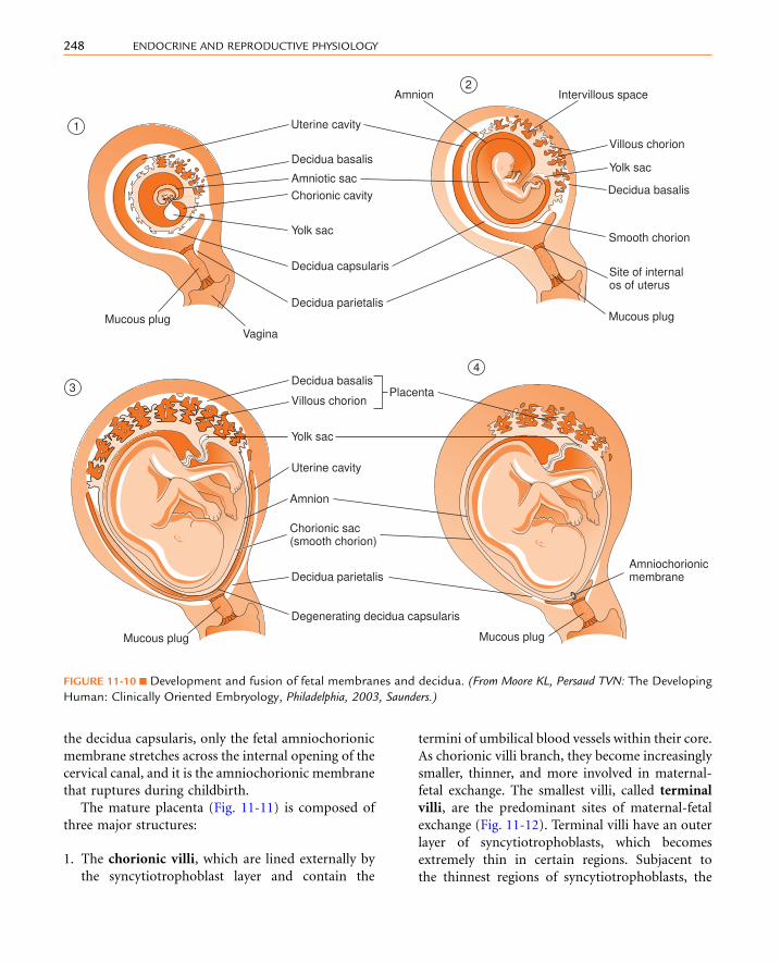
the decidua capsularis, only the fetal amniochorionic
membrane stretches across the internal opening of the
cervical canal, and it is the amniochorionic membrane
that ruptures during childbirth.
The mature placenta (Fig. 11-11) is composed of
three major structures:
1. The chorionic villi, which are lined externally by
the syncytiotrophoblast layer and contain the
termini of umbilical blood vessels within their core.
As chorionic villi branch, they become increasingly
smaller, thinner, and more involved in maternal-
fetal exchange. The smallest villi, called terminal
villi, are the predominant sites of maternal-fetal
exchange (Fig. 11-12). Terminal villi have an outer
layer of syncytiotrophoblasts, which becomes
extremely thin in certain regions. Subjacent to
the thinnest regions of syncytiotrophoblasts, the
Uterine cavity
Amnion
Uterine cavity
Amnion
Intervillous space
Villous chorion
Yolk sac
Villous chorion
Decidua basalis
Smooth chorion
Chorionic sac(smooth chorion)
Site of internal os of uterus
Mucous plugMucous plug
1
3
4
2
Vagina
Decidua basalis
Amniotic sac
Decidua basalisPlacenta
Decidua capsularis
Decidua parietalis
Decidua parietalis Amniochorionic membrane
Degenerating decidua capsularis
Chorionic cavity
Yolk sac
Yolk sac
Mucous plug Mucous plug
FIGURE 11-10 n Development and fusion of fetal membranes and decidua. (From Moore KL, Persaud TVN: The Developing
Human: Clinically Oriented Embryology, Philadelphia, 2003, Saunders.)
248 ENDOCRINE AND REPRODUCTIVE PHYSIOLOGY

cytotrophoblasts disappear, and an umbilical capil-
lary presses against the syncytiotrophoblast layer.
Thus, nutrients from the maternal blood that
bathes the terminal villi (see intervillous space in
next entry) have to cross only a single, flat layer
of syncytiotrophoblast, the fused basal lamina of
the syncytiotrophoblast and capillary endothelium,
and a flattened umbilical endothelial cell. This
barrier between maternal blood and the umbilical
circulation is called the placental membrane (see
Fig. 11-12) and is also called a vasculosyncytial
membrane. It represents the thinnest barrier
to maternal-fetal exchange among placental (i.e.,
eutherian) mammals.
2. The intervillous space, into which maternal blood
flows from the open ends of spiral arteries (see
Fig. 11-11). This blood bathes the chorionic villi
and returns to maternal circulation through endo-
metrial veins. Because the maternal side of the pla-
centa is represented by maternal blood within the
intervillous space, and the fetal side is represented
by the chorionic vasculosyncytial membrane, hu-
man placentation is referred to as hemochorial
placentation.
3. The decidua basalis. Some villi, called anchoring
villi, extend through the intervillous space and an-
chor onto the decidua (see Fig. 11-11). Columns of
cytotrophoblasts migrate out of the end of the an-
choring villi and spread across the decidua basalis.
These extravillous cytotrophoblasts form an ad-
hesive layer, called the cytotrophoblastic shell, that
anchors the chorion frondosum to the decidua
basalis (see Fig. 11-11). Spiral arteries extend
through the decidua basalis and open into the
intervillous space through breaks in the cytotro-
phoblastic shell. During the first trimester, extravil-
lous cytotrophoblasts migrate into the spiral
arteries and plug them up. Thus, embryonic and
early fetal development is supported primarily
by histotrophic nutrition within a hypoxic
Amniochorionicmembrane
Placental septum
Decidua basalis
MyometriumEndometrial
veinsEndometrial
arteries
Maternal circulation
Cytotrophoblastic shell
Anchoring villus
Chorionicplate
Fetalcirculation
Umbilical vein(O2-rich blood)
Umbilical arteries(O2-poor blood)
Intervillousspace
Branchvilli
Main stemvillus
Stump ofmain stem villus
Amnion
Smoothchorion
Deciduaparietalis
FIGURE 11-11 n Structure of the
mature hemochorial placenta.
Villi have been removed in some
segments to show flow of mater-
nalblood. (FromMooreKL,Persaud
TVN: The Developing Human:
Clinically Oriented Embryology,
Philadelphia, 2003, Saunders.)
249FERTILIZATION, PREGNANCY, AND LACTATION

environment. During this time, the cytotropho-
blasts that have invaded the spiral arteries replace
the tunica media (i.e., the vascular smooth muscle
layer) and tunica intima (i.e., the endothelia and
their lamina propria), thereby converting the arter-
ies into low-resistance, high-capacitance vessels.
At the beginning of the second trimester, coinci-
dent with entry of the fetus into a rapid growth
phase, the converted spiral arteries become un-
plugged, and hemotrophic nutrition predomi-
nates until parturition.
CLINICAL BOX 11-2
Preeclampsia, which is a form of hypertension and
proteinuria of pregnancy, is often accompanied by a
shallow invasion of the placenta and an inability of
the cytotrophoblasts to convert the spiral arteries.
This condition leads to intrauterine growth restric-tion (IUGR) of the fetus, increasing the risk for perina-
tal mortality.
Endocrine Function of the Placenta
The syncytiotrophoblasts of the placenta produce
several steroid and protein hormones. The general
functions of these hormones in pregnancy include
the following:
n Maintaining the pregnant state of the uterus
n Stimulating lobuloalveolar growth and function of
maternal breasts
n Adapting aspects of maternal metabolism and phys-
iology to support a growing fetus
n Regulating aspects of fetal development
n Regulating the timing and progressionof parturition
Human Chorionic Gonadotropin
The first hormone produced by the syncytiotropho-
blasts is hCG. This hormone is structurally related
to the pituitary glycoprotein hormones (see
Chapter 5). As such, hCG is composed of a common
a-glycoprotein subunit (aGSU) and a hormone-
specific b-subunit, b-hCG. Antibodies used to detect
hCG (as in laboratory assays and over-the-counter
pregnancy tests) are designed to specifically detect
the b-subunit. Human chorionic gonadotropin is
most similar to LH and binds with high affinity to
the LH receptor. The b-subunit of hCG is longer than
that of LH and contains more sites for glycosylation,
which greatly increases the half-life of hCG to up to
30 hours. The stability of hCG allows it to rapidly ac-
cumulate in the maternal circulation, so hCG is de-
tectable within maternal serum within 24 hours of
implantation. Serum hCG levels double every 2 days
for the first 6 weeks until they peak at about 10 weeks.
Serum hCG then declines to a constant level at about
50% of the peak value (Fig. 11-13).
Endothelium offetal capillary
Connective tissue core of villus
Hofbauer cells
Persistingcytotrophoblast cells
Fibrinoidmaterial
Syncytiotrophoblast
Placentalmembrane
CytotrophoblastFetal(umbilical)capillariesSyncytiotrophoblast
Nuclear aggregation or syncytial knot
Placentalmembrane
FIGURE 11-12 n Cross section of early (upper) and mature
(lower) terminal villi. In the mature terminal villus, the
cytotrophoblast layer becomes discontinuous, the fetal
vessels assume an eccentric position subjacent to the
syncytiotrophoblast layer, and the syncytiotrophoblast
becomes very thinned out except for nuclear aggregations
(also called syncytial knots). (From Moore KL, Persaud TVN:
The Developing Human: Clinically Oriented Embryology,
Philadelphia, 2003, Saunders.)
250 ENDOCRINE AND REPRODUCTIVE PHYSIOLOGY

The primary action of hCG is to stimulate LH re-
ceptors on the corpus luteum. This prevents luteolysis
and maintains a high level of luteal-derived progester-
one production during the first 10 weeks, after which
the syncytiotrophoblasts take over progesterone pro-
duction. The rapid increase in hCG is responsible
for the nausea of morning sickness associated with
early pregnancy. Human chorionic gonadotropin
binds weakly to the thyrotropin (thyroid-stimulating
hormone [TSH]) receptor, so early pregnancy can be
associated with a transient gestational hyperthy-
roidism. A small amount (i.e., 1% to 10%) of hCG
enters into the fetal circulation. The hCG stimulates
fetal Leydig cells to produce testosterone before
the fetal gonadotropic axis is fully mature. hCG may
also stimulate the fetal adrenal cortex during the
first trimester.
CLINICAL BOX 11-3
Human chorionic gonadotropin is produced in high
levels by cancers derived from trophoblastic cells,
such as molar disease and choriocarcinoma. Thus,hCG levels can be used as a measure of the efficacy
of chemotherapy.
10
100
0 10 20 30 40
Weeks of gestation
20
40
60
80
Hum
an c
horio
nic
gona
dotr
opin
(m
IU/m
l)
10
0 10 20 30 40
Weeks of gestation
2
4
6
8
Hum
an p
lace
ntal
lact
ogen
(�
g/m
l)
A
B C
120
100
ng/m
L
80
60
20
0 10 20 30 40
Weeks of gestation
Progesterone
20
10
ng/m
L
0 10 20 30 40
Weeks of gestation
Estradiol
Estrone
Estriol
FIGURE 11-13 n A,Maternal serum human chorionic gonadotropin levels during pregnancy. B, Human placental lactogen
levels during pregnancy. C, Progesterone and estrogen. (C, From Koeppen B, Stanton B: Berne and Levy Physiology, updated 6th
ed., Philadelphia, 2010, Mosby.)
251FERTILIZATION, PREGNANCY, AND LACTATION

Progesterone
The syncytiotrophoblasts express high levels of
CYP11A1 (side-chain cleavage enzyme) and a pla-
centa-specific 3b-hydroxysteroid dehydrogenase type1 (3b-HSD1) but do not express CYP17 (Fig. 11-14).
Syncytiotrophoblasts also express the receptors (e.g.,
low-density lipoprotein [LDL] receptor) that import
cholesterol from the maternal blood. Consequently,
the placenta produces a high amount of progesterone,
which is absolutely required to maintain a quiescent
myometrium and a pregnant uterus. Progesterone
production by the placenta is largely unregulated—the
placenta produces as much progesterone as the supply
of cholesterol and the levels of CYP11A1 and 3b-HSD
will allow.Ofnote, placental steroidogenesisdiffers from
that in the adrenal cortex, ovaries, and testis, in that cho-
lesterol is transported into the placental mitochondria
by a mechanism that is independent of the labile
steroidogenic acute regulatory (StAR) protein. Thus,
this first step in steroidogenesis is not a regulated,
rate-limiting step in theplacentaas it is inother steroido-
genic glands. Thismeans that a fetuswithan inactivating
mutation in StAR proteinwill develop lipoid congenital
adrenal hyperplasia (see Chapter 7) and hypogonadism
but will have normal progesterone levels produced by
their placenta. It also should be noted that progesterone
production by the placenta does not require fetal tissue.
Consequently, progesterone levels are largely inde-
pendent of fetal health status and cannot be used as a
measure of fetal well-being. Maternal progesterone
levels continue to increase throughout pregnancy (see
Fig. 11-13).
Progesterone is released primarily into the mater-
nal circulation and is required for implantation and
the maintenance of pregnancy. Progesterone also
has several effects on maternal physiology and in-
duces breast growth and differentiation (discussed
later). The switch from corpus luteum–derived pro-
gesterone to placenta-derived progesterone (referred
to as the luteal-placental shift) is complete at about
the eighth week of pregnancy. Progesterone and
pregnenolone are used by the definitive zone of the
fetal cortex (see later) to make cortisol late in
pregnancy.
Cholesterol VLDL
Effects in maternalcompartment
LDL
Receptor-mediatedendocytosis
Maternal liver
Cholesterol
Pregnenolone
Progesterone
CortisolCortisol
Maternal adrenal cortex
Cortisone
Protection of fetaladrenal axis frommaternal cortisol(and vice versa)
CYP17 BLOCK
Syncytiotrophoblast
CYP11A1
StAR proteinnot required
3�-HSDPregnenolone
Progesterone
17(OH)-progesterone
Cortisol
3�-HSD
11�-HSD type 2
Fetal pituitary
ACTH
Cholesterol
MC2R
CYP11A1StAR protein
CYP17(17-hydroxylase)
CYP21
CYP11B111-Deoxycortisol
Effects in fetal compartment(3rd trimester)
Definitivezone of
fetaladrenalcortex
FIGURE 11-14 n Progesterone
biosynthesis by the syncytio-
trophoblast. ACTH, adreno-
corticotropic hormone; 3b-HSD, 3b-hydroxysteroid dehy-
drogenase; 11b-HSD, 11b-hydroxysteroid dehydrogenase;
LDL, low-density lipoprotein;
MC2R, melanocortin-2 recep-
tor (ACTH receptor); StAR
protein, steroidogenic acute
regulatory protein; VLDL, very-
low-density lipoprotein.
252 ENDOCRINE AND REPRODUCTIVE PHYSIOLOGY

Estrogen
Estrogens are produced by the syncytiotrophoblasts.
Syncytiotrophoblasts lack CYP17 and are dependent
on another cell type to provide 19-carbon androgens
for aromatization. The ancillary, androgen-producing
cell resides in the fetal adrenal cortex.
The cells of the fetal adrenal cortex emerge at week
5, and by 8 weeks there exists a distinct cortex and me-
dulla. Initially, the cortex is composed of one zone, the
fetal zone (Fig. 11-15). The fetal zone becomes sur-
rounded by cells that make up the definitive cortex,
which will differentiate into the adult zona glomeru-
losa and zona fasciculata. The fetal zone constitutes
as much as 80% of the bulk of the large fetal adrenal
gland. The fetal zone recedes after birth (see Fig. 11-15).
The zona reticularis develops after 1 to 3 years, but
does not secrete hormones until adrenarche at 6 to
8 years of age.
The fetal zone does not express 3b-HSD and re-
leases the sulfated form of the inactive androgen,
dehydroepiandrosterone sulfate (DHEAS), through-
out most of gestation (Fig. 11-16). The production of
DHEAS from the fetal adrenal is absolutely dependent
on fetal adrenocorticotropic hormone (ACTH) from
the fetal pituitary by the end of the first trimester.
The DHEAS released from the fetal zone has two
fates (see Fig. 11-16). First, DHEAS can go directly
to the syncytiotrophoblast, where it is desulfated by
a placental steroid sulfatase and used as a 19-carbon
substrate for the synthesis of estradiol-17b and es-
trone. The second fate of DHEAS is 16-hydroxylation
in the fetal liver by the enzyme CYP3A7. 16-
Hydroxyl-DHEAS is then converted by the syncytio-
trophoblasts to the major estrogen of pregnancy,
called estriol (see Fig. 11-16).
Because estrogen production is dependent on a
healthy fetus, estriol levels can be used to assess fetal
well-being. The fetoplacental unit represents the
collective term for the tissues that make estrogen.
Estrogens increase uteroplacental blood flow, enhance
LDL receptor expression in syncytiotrophoblasts, and
induce several components (e.g., prostaglandins, oxy-
tocin receptors) involved in parturition. Estrogens
increase the growth and development of the mam-
mary glands directly, and also indirectly through
the stimulation of maternal pituitary prolactin
production (see later). Estrogens are not necessary
for a normal pregnancy but are required for labor
and parturition.
Human Placental Lactogen
Human placental lactogen (hPL), also called human
chorionic somatomammotropin (hCS), is a 191-
amino acid protein hormone produced in the syncy-
tiotrophoblast that is structurally similar to growth
hormone (GH) and prolactin (PRL). It can be detected
within the syncytiotrophoblast by 10 days after con-
ception and in maternal serum by 3 weeks of gestation
Fetal zone
1T
DHEAS
Fetal zone
Definitivecortex
2T
DHEAS
Fetal zone
Z FASC
Z GLOM
3T
DHEASCortisol
Aldosterone
Fetal zone
Z FASC
Z GLOM
2 months
CortisolAldosteroneLow DHEAS
Z RETIC
Z FASC
Z GLOM
1-3 yr
CortisolAldosterone
FIGURE 11-15n The zones of the
fetal adrenal cortex.
253FERTILIZATION, PREGNANCY, AND LACTATION

(see Fig. 11-13). Maternal serum levels rise progres-
sively throughout the remainder of the pregnancy.
As much as 1 g/day of hPL can be secreted late in
gestation!
Like GH, hPL is protein-anabolic and lipolytic. Its
antagonistic action to insulin contributes to the diabe-
togenicity of pregnancy. Glucose is a major energy
substrate for the fetus, and hPL increases glucose
availability by inhibiting maternal glucose uptake
(Fig. 11-17). The lipolytic actions help the mother
to shift to the use of free fatty acids for energy. Despite
its very high levels in the maternal blood, hPL is prob-
ably not essential for normal pregnancy.
Other Placental Hormones
The placenta is a source of many other hormones. The
placenta produces parathyroid hormone–related
protein (PTHrP), which increases placental calcium
transport (see later). The role of placental corticotro-
pin-releasing hormone (CRH) in the induction of la-
bor is discussed later.
Effects in maternalcompartment
Protects motherfrom virilization
Placentalestrogens
PregnenoloneACTH
Fetal pituitary
DHEA-S
Testosterone
Estradiol-17�
Estriol
16(OH)-Testosterone
16(OH)-Androstendione
16(OH)-DHEA
16(OH)-DHEA-S
DHEA
Androstenedione
Estrone
CYP17 BLOCK
Syncytiotrophoblast
MC2R
Receptor-mediatedendocytosis
Pregnenolone
Pregnenolone-S
17(OH)-Pregnenolone-S
DHEA-S
DHEA-S
16(OH)-DHEA-S
Cholesterol VLDL
17�-HSD
17�-HSD
17�-HSD
3�-HSD BLOCK
Fetal zone of fetal adrenal
Fetal liver
CYP3A7
3�-HSD
Steroid sulfatase
Steroid sulfatase
3�-HSD
CYP19 CYP19
CYP19
CholesterolCYP11A1
StAR protein
CYP17
Sulfotransferase
CYP17
FIGURE 11-16 n Estrogen biosynthesis by the fetoplacental unit. ACTH, adrenocorticotropic hormone; DHEA,
dehydroepiandrostenedione; DHEAS, dehydroepiandrostenedione sulfate; 3b-HSD, 3b-hydroxysteroid dehydrogenase;
17b-HSD, 17b-hydroxysteroid dehydrogenase; MC2R, melanocortin-2 receptor (ACTH receptor); 16(OH)-, 16-hydroxy-;
StAR protein, steroidogenic acute regulatory protein; VLDL, very-low-density lipoprotein.
254 ENDOCRINE AND REPRODUCTIVE PHYSIOLOGY
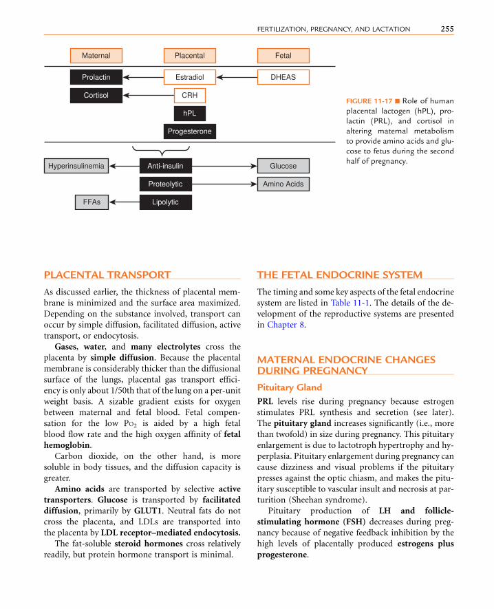
PLACENTAL TRANSPORT
As discussed earlier, the thickness of placental mem-
brane is minimized and the surface area maximized.
Depending on the substance involved, transport can
occur by simple diffusion, facilitated diffusion, active
transport, or endocytosis.
Gases, water, and many electrolytes cross the
placenta by simple diffusion. Because the placental
membrane is considerably thicker than the diffusional
surface of the lungs, placental gas transport effici-
ency is only about 1/50th that of the lung on a per-unit
weight basis. A sizable gradient exists for oxygen
between maternal and fetal blood. Fetal compen-
sation for the low PO2 is aided by a high fetal
blood flow rate and the high oxygen affinity of fetal
hemoglobin.
Carbon dioxide, on the other hand, is more
soluble in body tissues, and the diffusion capacity is
greater.
Amino acids are transported by selective active
transporters. Glucose is transported by facilitated
diffusion, primarily by GLUT1. Neutral fats do not
cross the placenta, and LDLs are transported into
the placenta by LDL receptor–mediated endocytosis.
The fat-soluble steroid hormones cross relatively
readily, but protein hormone transport is minimal.
THE FETAL ENDOCRINE SYSTEM
The timing and some key aspects of the fetal endocrine
system are listed in Table 11-1. The details of the de-
velopment of the reproductive systems are presented
in Chapter 8.
MATERNAL ENDOCRINE CHANGESDURING PREGNANCY
Pituitary Gland
PRL levels rise during pregnancy because estrogen
stimulates PRL synthesis and secretion (see later).
The pituitary gland increases significantly (i.e., more
than twofold) in size during pregnancy. This pituitary
enlargement is due to lactotroph hypertrophy and hy-
perplasia. Pituitary enlargement during pregnancy can
cause dizziness and visual problems if the pituitary
presses against the optic chiasm, and makes the pitu-
itary susceptible to vascular insult and necrosis at par-
turition (Sheehan syndrome).
Pituitary production of LH and follicle-
stimulating hormone (FSH) decreases during preg-
nancy because of negative feedback inhibition by the
high levels of placentally produced estrogens plus
progesterone.
Maternal Placental Fetal
Prolactin Estradiol
Cortisol CRH
hPL
Progesterone
Anti-insulin
Proteolytic
Glucose
Amino Acids
Lipolytic
Hyperinsulinemia
FFAs
DHEAS
FIGURE 11-17 n Role of human
placental lactogen (hPL), pro-
lactin (PRL), and cortisol in
altering maternal metabolism
to provide amino acids and glu-
cose to fetus during the second
half of pregnancy.
255FERTILIZATION, PREGNANCY, AND LACTATION
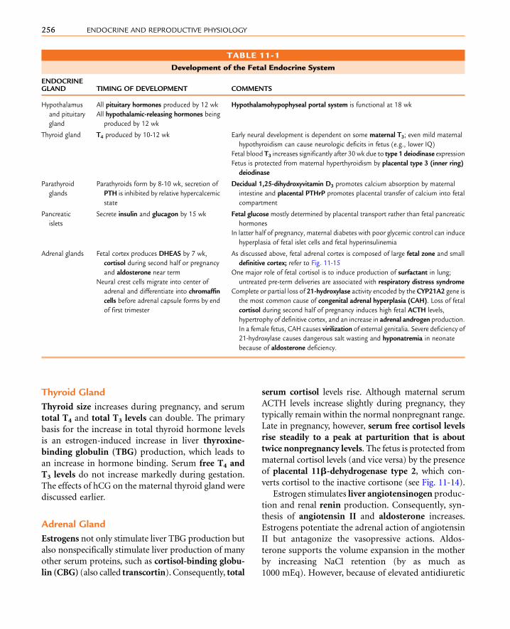
Thyroid Gland
Thyroid size increases during pregnancy, and serum
total T4 and total T3 levels can double. The primary
basis for the increase in total thyroid hormone levels
is an estrogen-induced increase in liver thyroxine-
binding globulin (TBG) production, which leads to
an increase in hormone binding. Serum free T4 and
T3 levels do not increase markedly during gestation.
The effects of hCG on the maternal thyroid gland were
discussed earlier.
Adrenal Gland
Estrogens not only stimulate liver TBG production but
also nonspecifically stimulate liver production of many
other serum proteins, such as cortisol-binding globu-
lin (CBG) (also called transcortin). Consequently, total
serum cortisol levels rise. Although maternal serum
ACTH levels increase slightly during pregnancy, they
typically remain within the normal nonpregnant range.
Late in pregnancy, however, serum free cortisol levels
rise steadily to a peak at parturition that is about
twice nonpregnancy levels. The fetus is protected from
maternal cortisol levels (and vice versa) by the presence
of placental 11b-dehydrogenase type 2, which con-
verts cortisol to the inactive cortisone (see Fig. 11-14).
Estrogen stimulates liver angiotensinogen produc-
tion and renal renin production. Consequently, syn-
thesis of angiotensin II and aldosterone increases.
Estrogens potentiate the adrenal action of angiotensin
II but antagonize the vasopressive actions. Aldos-
terone supports the volume expansion in the mother
by increasing NaCl retention (by as much as
1000 mEq). However, because of elevated antidiuretic
TABLE 11-1
Development of the Fetal Endocrine System
ENDOCRINEGLAND TIMING OF DEVELOPMENT COMMENTS
Hypothalamus
and pituitary
gland
All pituitary hormones produced by 12 wk
All hypothalamic-releasing hormones being
produced by 12 wk
Hypothalamohypophyseal portal system is functional at 18 wk
Thyroid gland T4 produced by 10-12 wk Early neural development is dependent on some maternal T3; even mild maternal
hypothyroidism can cause neurologic deficits in fetus (e.g., lower IQ)
Fetal blood T3 increases significantly after 30 wk due to type 1 deiodinase expression
Fetus is protected from maternal hyperthyroidism by placental type 3 (inner ring)
deiodinase
Parathyroid
glands
Parathyroids form by 8-10 wk, secretion of
PTH is inhibited by relative hypercalcemic
state
Decidual 1,25-dihydroxyvitamin D3 promotes calcium absorption by maternal
intestine and placental PTHrP promotes placental transfer of calcium into fetal
compartment
Pancreatic
islets
Secrete insulin and glucagon by 15 wk Fetal glucose mostly determined by placental transport rather than fetal pancreatic
hormones
In latter half of pregnancy, maternal diabetes with poor glycemic control can induce
hyperplasia of fetal islet cells and fetal hyperinsulinemia
Adrenal glands Fetal cortex produces DHEAS by 7 wk,
cortisol during second half or pregnancy
and aldosterone near term
Neural crest cells migrate into center of
adrenal and differentiate into chromaffin
cells before adrenal capsule forms by end
of first trimester
As discussed above, fetal adrenal cortex is composed of large fetal zone and small
definitive cortex; refer to Fig. 11-15
One major role of fetal cortisol is to induce production of surfactant in lung;
untreated pre-term deliveries are associated with respiratory distress syndrome
Complete or partial loss of 21-hydroxylase activity encoded by the CYP21A2 gene is
the most common cause of congenital adrenal hyperplasia (CAH). Loss of fetal
cortisol during second half of pregnancy induces high fetal ACTH levels,
hypertrophy of definitive cortex, and an increase in adrenal androgen production.
In a female fetus, CAH causes virilization of external genitalia. Severe deficiency of
21-hydroxylase causes dangerous salt wasting and hyponatremia in neonate
because of aldosterone deficiency.
256 ENDOCRINE AND REPRODUCTIVE PHYSIOLOGY

hormone (ADH) and decreased threshold for thirst,
the osmolality of the blood falls slightly.
MATERNAL PHYSIOLOGICCHANGES DURING PREGNANCY
Physiologic changes occur in the pregnant woman
both as a consequence of the size of the developing fe-
tus and as a result of the endocrine and cardiovascular
changes associated with the pregnancy (Box 11-1).
Cardiovascular Changes
Pregnancy is associated with an increase in cardiac
output coupled to a reduction in peripheral resistance
and an expansion of total body and plasma volume.
The cardiac output during pregnancy increases to
about 40% more than preconception levels, with most
of this increase occurring by 8 weeks of gestation. Car-
diac output is due to both positive chronotropic and
inotropic effects of pregnancy. These may be due to
increased sympathetic tone and an increased sensitive
to catecholamines.
Peripheral resistance declines in response to sev-
eral factors. As discussed previously, the spiral arteries
are converted to low-resistance, highly compliant ves-
sels, allowing the uteroplacental blood flow to increase
by an order of magnitude from the end of the first tri-
mester to term (about 60 to 600 mL per minute).
Overall, vascular resistance decreases throughout the
maternal circulatory system, and diastolic pressure
tends to drop. Blood pressure typically does not rise
until late in pregnancy.
Total body volume increases by the retention of up
to 8 L of water, which is partitioned among the mater-
nal and growing fetal circulatory systems, uteropla-
cental circulation, and an enlarging amniotic sac.
Blood volume increases by up to 50% during preg-
nancy to accommodate an increasing uteroplacental
circulation. Bloating or mild edema is common in
pregnancy.
CLINICAL BOX 11-4
Because the growing uterus exerts pressure on the
veins of the legs where these veins enter the abdomen,
venous pressure in the lower extremities rises on
standing, and edema and venous damage can occur.
Lying in the supine position can result in compression
of the inferior vena cava by the uterus, followed byma-
ternal hypotension.
Respiratory Changes
As pregnancy proceeds, the functional residual ca-
pacity (volume of air in the lungs at the end of a quiet
expiration) and the residual volume (volume remain-
ing at the end of a maximal expiration) decrease, and
respiratory rate remains unchanged. Minute volume
increases and tidal volume increases, so PCO2
decreases.
There are three major causes of the respiratory
changes associated with pregnancy. The bulk of the
growing fetus and uterus increases intra-abdominal
pressure and forces the diaphragm upward. The high
metabolic rate of the growing fetus increases maternal
oxygen consumption and carbon dioxide production.
In addition, progesterone acts on the central nervous
system to lower the set-point for regulation of res-
piration by carbon dioxide, thereby increasing
ventilation.
n n n n n n n n n n
BOX 11 - 1
PHYSIOLOGIC CHANGES IN PREGNANCY
CARDIOVASCULAR CHANGES
n Vascular volume
n Peripheral resistance
n Stroke volume
n Heart rate
n Contractility
n Cardiac output
RESPIRATORY CHANGES
n Minute volume
n Tidal volume
n PCO2n Functional residual capacity
n Inspiratory reserve volume
RENAL CHANGES
n Antidiuretic hormone, renin, angiotensin II, aldo-
sterone secretion
n Respiratory alkalosis
257FERTILIZATION, PREGNANCY, AND LACTATION

Renal Changes
The glomerular filtration rate increases about 60%
over nonpregnant levels, causing the filtered load to
increase, which can lead to glucosuria and aminoac-
iduria in pregnancy.
Maternal serum levels of the mineralocorticoid
deoxycorticosterone (DOC) increase during preg-
nancy. This increase in circulating DOC levels results
not from increased adrenal secretion but from renal
conversion of placental progesterone to DOC.
Increased levels of DOC and aldosterone (see earlier)
stimulate renal salt and water retention.
Gastrointestinal Changes
Heartburn, or reflux of acidic gastric secretions into the
esophagus, occurs for multiple reasons. The increased
intra-abdominal pressure increases intragastric pres-
sure, which increases the likelihood of reflux into the
esophagus. Progesterone also decreases lower esopha-
geal sphincter tone, thereby increasing reflux tendency.
Diabetogenicity of Pregnancy
Pregnancy represents an insulin-resistant, hyper-
insulinemic state (see Fig. 11-17). During the last half
of pregnancy, maternal energy metabolism shifts from
an anabolic state in which nutrients are stored to a cat-
abolic state, sometimes described as accelerated star-
vation, in which maternal energy metabolism shifts
toward fat utilization with glucose sparing. The pe-
ripheral responsiveness to insulin decreases and pan-
creatic insulin secretion increases. b-Cell hyperplasiaoccurs in pregnancy. Although this usually does not
lead to any clinical condition, pregnancy aggravates
existing diabetes mellitus, and diabetes mellitus can
develop for the first time in pregnancy. If the diabetes
resolves spontaneously with delivery, the condition is
referred to as gestational diabetes. Hormones contrib-
uting to the diabetogenicity of pregnancy are hPL,
prolactin, cortisol, and progesterone (see Fig. 11-17).
PARTURITION
Human pregnancy lasts an average of 40 weeks from
the beginning of the last menstrual period (gestational
age). This corresponds to an average fetal age of
38 weeks. Parturition is the process whereby uterine
contractions (labor) lead to childbirth. Labor consists
of three stages:
1. Strong uterine contractions that force the fetus
against the cervix, with dilation and thinning of
the cervix (several hours)
2. Delivery of the fetus (<1 hour)
3. Delivery of the placenta, along with contractions
of the myometrium, which serve to halt bleeding
(<10 minutes).
Parturition control in humans is complex, and the
exact mechanisms underlying parturition control are
not well understood. In many species, such as sheep,
the timing of parturition is controlled by fetus-derived
signals, and fetal regulation is at least a factor inhumans.
Placental Corticotropin-ReleasingHormone and the Fetal Adrenal Axis
The placenta produces CRH, which is identical to the
41-amino acid peptide produced by the hypothala-
mus. Placental CRH production and maternal serum
CRH levels increase rapidly during late pregnancy
and labor. Moreover, circulating CRH is either in
the form of free CRH, which is bioactive, or com-
plexed to a CRH-binding protein. Maternal levels of
CRH-binding protein plummet during late preg-
nancy and labor, and free CRH levels increase. Placen-
tal CRH also accumulates in the fetal circulation and
stimulates fetal ACTH secretion. ACTH stimulates
both fetal adrenal cortisol production and feto-
placental estrogen production. In contrast to the
inhibitory effect of cortisol on hypothalamic CRH
production, cortisol stimulates placental CRH pro-
duction. This establishes a self-amplifying positive
feedback loop. CRH itself promotesmyometrial con-
tractions through sensitizing the uterus to prosta-
glandins and oxytocin. Estrogens also directly and
indirectly stimulate myometrial contractility. This
model correlates with the onset of parturition with
cortisol-inducedmaturation of fetal systems, includ-
ing the lungs and gastrointestinal system.
Estrogen
Estrogen is required for labor. It increases receptors
and sensitivity of the myometrium to oxytocin and
increases prostaglandin production.
258 ENDOCRINE AND REPRODUCTIVE PHYSIOLOGY

CLINICAL BOX 11-5
In male fetuses with X-linked steroid sulfatase defi-ciency, the fetoplacental unit cannot make estrogens.
This results in maternal estrogen levels that are an
order of magnitude less than those in normal preg-
nancies. These babies typically are delivered by cesar-
ean section because the absence of estrogen results in
a quiescent myometrium and a pregnancy that goes
several weeks beyond the due date. Nevertheless,
the pregnancy proceeds normally, and the newborn
is normal except for the phenotype associated with
sulfatase deficiency (ichthyosis, or scaly skin).
Oxytocin
Oxytocin stimulates powerful uterine contractions
(Fig. 11-18). It is released in response to stretch of
the cervix, and it stimulates uterine contractions,
thereby facilitating delivery. Oxytocin is also used to
induce parturition, and uterine sensitivity to oxytocin
increases before parturition.
Prostaglandins
Prostaglandins and other cytokines increase uterine
motility, and levels of these compounds increase
during parturition, thereby facilitating delivery. The
prostaglandins PGF2a and PGE2 increase uterine
motility. Large doses of these compounds have been
used to induce labor.
Uterine Size
Uterine size is thought to be a factor regulating par-
turition because stretch of smooth muscle, including
the uterus, increases muscle contraction. In addition,
uterine stretch stimulates uterine prostaglandin pro-
duction. Multiple births generally occur prematurely.
MAMMOGENESIS AND LACTATION
Structure of the Mammary Gland
The mammary gland is composed of about 20 lobes,
each with an excretory lactiferous duct that opens at
the nipple (Fig. 11-19). Lobes, in turn, are composed
of several lobules, which contain secretory structures
called alveoli, and the terminal portions of the ducts.
The epithelia of the alveoli and ducts is composed
of apical luminal ductal or alveolar cells and a
Stretch of cervix
Oxytocin release
Estrogen
Oxytocin receptors
Oxytocin response
Uterine
FIGURE 11-18 n Role of oxytocin in parturition.
Lactiferous sinus
Lactiferous duct
Segmental duct
Nipple
Nipple adenoma
Papillomas
Traumatic fat necrosis
HyperplasiaMost carcinomas
FibroadenomaCysts
Paget’s disease
Lesions
Anatomic structures
Adipose tissue
Lobule
Terminal duct
Ductulesor acini
Terminal ductlobular unit
FIGURE 11-19nAnatomy of the breast and major lesions at
each site within the breast. (From Cotran RS, Kumar V,
Robbins SL: Pathologic Basis of Disease, 5th ed.,
Philadelphia, 1994, Saunders.)
259FERTILIZATION, PREGNANCY, AND LACTATION
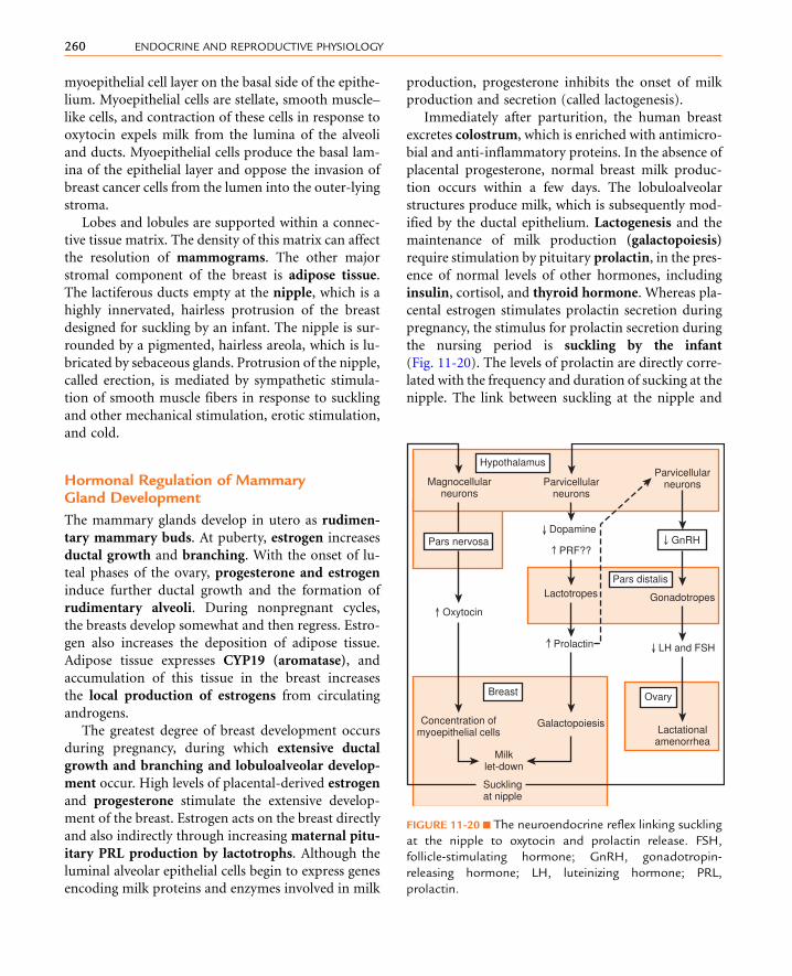
myoepithelial cell layer on the basal side of the epithe-
lium. Myoepithelial cells are stellate, smooth muscle–
like cells, and contraction of these cells in response to
oxytocin expels milk from the lumina of the alveoli
and ducts. Myoepithelial cells produce the basal lam-
ina of the epithelial layer and oppose the invasion of
breast cancer cells from the lumen into the outer-lying
stroma.
Lobes and lobules are supported within a connec-
tive tissue matrix. The density of this matrix can affect
the resolution of mammograms. The other major
stromal component of the breast is adipose tissue.
The lactiferous ducts empty at the nipple, which is a
highly innervated, hairless protrusion of the breast
designed for suckling by an infant. The nipple is sur-
rounded by a pigmented, hairless areola, which is lu-
bricated by sebaceous glands. Protrusion of the nipple,
called erection, is mediated by sympathetic stimula-
tion of smooth muscle fibers in response to suckling
and other mechanical stimulation, erotic stimulation,
and cold.
Hormonal Regulation of MammaryGland Development
The mammary glands develop in utero as rudimen-
tary mammary buds. At puberty, estrogen increases
ductal growth and branching. With the onset of lu-
teal phases of the ovary, progesterone and estrogen
induce further ductal growth and the formation of
rudimentary alveoli. During nonpregnant cycles,
the breasts develop somewhat and then regress. Estro-
gen also increases the deposition of adipose tissue.
Adipose tissue expresses CYP19 (aromatase), and
accumulation of this tissue in the breast increases
the local production of estrogens from circulating
androgens.
The greatest degree of breast development occurs
during pregnancy, during which extensive ductal
growth and branching and lobuloalveolar develop-
ment occur. High levels of placental-derived estrogen
and progesterone stimulate the extensive develop-
ment of the breast. Estrogen acts on the breast directly
and also indirectly through increasing maternal pitu-
itary PRL production by lactotrophs. Although the
luminal alveolar epithelial cells begin to express genes
encoding milk proteins and enzymes involved in milk
production, progesterone inhibits the onset of milk
production and secretion (called lactogenesis).
Immediately after parturition, the human breast
excretes colostrum, which is enriched with antimicro-
bial and anti-inflammatory proteins. In the absence of
placental progesterone, normal breast milk produc-
tion occurs within a few days. The lobuloalveolar
structures produce milk, which is subsequently mod-
ified by the ductal epithelium. Lactogenesis and the
maintenance of milk production (galactopoiesis)
require stimulation by pituitary prolactin, in the pres-
ence of normal levels of other hormones, including
insulin, cortisol, and thyroid hormone. Whereas pla-
cental estrogen stimulates prolactin secretion during
pregnancy, the stimulus for prolactin secretion during
the nursing period is suckling by the infant
(Fig. 11-20). The levels of prolactin are directly corre-
lated with the frequency and duration of sucking at the
nipple. The link between suckling at the nipple and
Magnocellularneurons
Parvicellularneurons
Parvicellularneurons
GnRH
Gonadotropes
LH and FSH
Lactationalamenorrhea
Pars nervosa
Oxytocin
Concentration ofmyoepithelial cells
Breast
Hypothalamus
Milklet-down
Sucklingat nipple
Galactopoiesis
Prolactin
Lactotropes
PRF??
Dopamine
Pars distalis
Ovary
FIGURE 11-20 n The neuroendocrine reflex linking suckling
at the nipple to oxytocin and prolactin release. FSH,
follicle-stimulating hormone; GnRH, gonadotropin-
releasing hormone; LH, luteinizing hormone; PRL,
prolactin.
260 ENDOCRINE AND REPRODUCTIVE PHYSIOLOGY

prolactin secretion involves a neuroendocrine reflex,
in which dopamine (the prolactin-release inhibitory
factor; see Chapter 5) secretion at the median
eminence is inhibited. It also is possible that suckling
increases the secretion of unidentified prolactin-
releasing hormones.
Prolactin also inhibits GnRH release; conse-
quently, nursing can be associated with lactational
amenorrhea (see Fig. 11-20). This effect of prolactin
has been called nature’s contraceptive and may play a
role in spacing out pregnancies. Only very regular
nursing over a 24-hour period, however, is sufficient
to allow for a prolactin-induced anovulatory state in
the mother. Thus, lactational amenorrhea is not an ef-
fective or reliable form of birth control for most
women.
CLINICAL BOX 11-6
The inhibition of GnRH by high levels of prolactin is
important clinically. The prolactinoma is the most
common form of hormone-secreting pituitary tumor,
and hyperprolactinemia is a significant cause of infer-
tility in both sexes. Hyperprolactinemia can also be as-
sociated with galactorrhea, or the inappropriate flow
of breast milk, in men and women.
Suckling at the nipple also stimulates the release of
oxytocin from the posterior pituitary (see Chapter 5)
through a neuroendocrine reflex (see Fig. 11-20).Oxy-
tocin receptors on the myoepithelial cells cause con-
tractions that ultimately induce milk letdown, or the
expulsion of milk from alveolar and ductal lumina.
Oxytocin release and milk letdown can be induced
by psychogenic stimuli, such as the sound of a baby
crying on a television program or thinking about
the baby. Such psychogenic stimuli do not affect pro-
lactin release.
CLINICAL BOX 11-7
The breast epithelium represents a hormonally re-
sponsive, highly mitogenic population of cells. Inva-sive breast cancer (IBC) arises from breast
epithelium (primarily from the epithelial cells lining
small ducts). IBC represents a very common cancer
in women in the United States and is a major cause
of death among women older than 45 years. Themost
commonly diagnosed form of IBC is called luminal A,in which most cells express the a isoform of the estro-
gen receptor ERa. These tumors are highly dependent
on estrogen and regress or fail to recur in response
to antiestrogen treatment. This treatment involves
the use of a selective estrogen receptor modulator
(SERM) such as tamoxifen or aromatase (CYP19)inhibitor. Treatment of an early (not metastasized)
form of luminal A IBC usually involves surgical resec-
tion of the cancer (lumpectomy, mastectomy),
followed by several weeks of radiation therapy,
followed by antiestrogen therapy for several years.
The overall survival rate for early, lymph node–
negative, ERa-positive luminal A IBC is well over
90% in North America.
CONTRACEPTION
Behavioral and Mechanical Approaches
There are multiple methods of contraception. These
methods include the age-old rhythm method, which
relies on abstinence from sexual intercourse during
fertile periods around the time of ovulation. (The fer-
tile period is considered to be the period extending
from 3 to 4 days before the time of ovulation until
3 to 4 days afterward.) A second method is with-
drawal before ejaculation, coitus interruptus. Both
of these methods have higher failure rates (20% to
30%) than the barrier methods (2% to 12%), intra-
uterine devices (IUDs) (<2%), and oral contracep-
tives (<1%).
Barriers such as condoms or diaphragms are more
effective as contraceptives when used with spermi-
cidal jellies.
Among the various methods of contraception,
IUDs are the most effective, except for oral contracep-
tives. These devices are thought to prevent implanta-
tion by producing a local inflammatory response in
the endometrium. Some forms of IUDs contain cop-
per, zinc, or progestins, which inhibit sperm transport
or viability in the female reproductive tract.
Female (tubule ligation) and male (vasectomy)
sterilization are also effective options, especially for
couples who have children and do not wish for fur-
ther procreation in the face of continued sexual
activity.
261FERTILIZATION, PREGNANCY, AND LACTATION

Oral Contraceptives
Oral contraceptives have been marketed in the United
States since the early 1960s. The doses of steroids used
today are many-fold lower than those used in the ini-
tial preparations. Properly used, oral contraceptives
have a low failure rate.
Many forms of oral contraceptives are marketed to-
day. The trend over the years has been to decrease the
dosage of steroids used because the side effects are dose
dependent. All oral steroidal contraceptives contain a
progestin analog, and some also contain an estrogen
analog. The numerous formulations, applications
(e.g., pills, patches), and configurations of the timing
and duration available are beyond the scope of this
chapter.
Oral contraceptives work through multiple mecha-
nisms. Most block the LH surge that triggers ovula-
tion. However, some pills, such as the progestin-only
minipill, block fertility by changing the nature of cer-
vical mucus, by altering endometrial development,
and by regulating fallopian tube motility. Because
these contraceptives suppress FSH, they impair early
follicular development.
Side effects include bloating, breast tenderness,
and unscheduled bleeding (breakthrough bleeding).
Estrogen-progesterone combination pills appear to in-
crease the risk for venous thrombosis. The effect of
combination pills on breast cancer is minimal, and
they decrease the incidence of ovarian and uterine can-
cer. However, cervical cancer is linked to the use of oral
contraceptives.
Hormonal Treatment for EmergencyContraception and Abortion
Emergency contraception involves hormonal treat-
ment designed to inhibit or delay ovulation, inhibit cor-
pus luteum function, and disrupt the function of the
oviducts and uterus. Candidates for emergency con-
traception include women who have been sexually
assaulted or who experienced a failure of a barrier
method (e.g., ruptured condom). The currently pre-
ferred medication is levonorgestrel (Plan B), which
is a synthetic progestin-only pill. The efficacy of the pill
is inversely correlated with the time it is taken after in-
tercourse. The exact mechanism of action is not known.
Treatment has no effect if implantation has occurred.
Medical (hormonal) termination of pregnancy
(abortion) can be achieved until up to 49 days of ges-
tation by administration of mifepristone (RU-486).
Mifepristone is a progesterone receptor antagonist
(i.e., an antiprogestin), which induces collapse of
the pregnant endometrium. Mifepristone is followed
48 hours later by ingestion or vaginal insertion of a
synthetic prostaglandin E (e.g., misoprostol), which
induces myometrial contractions.
IN VITRO FERTILIZATION
The development of in vitro fertilization (IVF) and
other assisted pregnancy procedures was made possi-
ble through our increased understanding of the basic
science of reproduction. A general description of IVF
is available online at Student Consult.
S U M M A R Y
1. The events of fertilization, early embryogenesis,
and implantation are synchronized with the hor-
monal changes of the human menstrual cycle,
ultimately ensuring a receptive uterus at the time
of implantation.
2. Spermatozoa bind to the epithelium of the ovi-
ductal isthmus, which secretes factors that ca-
pacitate the sperm. Hyperactivation allows the
sperm to detach and swim to the cumulus-oocyte
complex in the ampulla. Fertilization involves
the penetration by a spermatozoon of the ex-
panded cumulus as mediated by a membrane
hyaluronidase, penetration of the zona pellucida
as mediated by the acrosome reaction, and fusion
with the oocyte membrane as mediated by spe-
cific membrane fusion proteins. The egg is acti-
vated, completes the second meiotic division,
and releases cortical granule enzymes that prevent
polyspermy. The female and male pronuclei are
drawn together, line up on a metaphase plate,
and undergo the first cleavage.
3. The embryo undergoes cleavage and formation of
a morula within the oviduct. The embryo enters
the uterus on day 3, forms a blastocyst, degrades
262 ENDOCRINE AND REPRODUCTIVE PHYSIOLOGY

the zona pellucida (hatches), and implants on
day 6 or 7.
4. The trophoblast layer differentiates into the cyto-
trophoblastic layer and an outer syncytiotropho-
blastic layer. The syncytiotrophoblasts secrete
invasive enzymes, express adhesion molecules,
produce protein and steroid hormones, and ulti-
mately become the primary cell involved in
maternal-fetal exchange. With the addition of an
extraembryonic mesoderm, the trophoblast layers
become the chorion. The fetal (umbilical) blood
vessels develop within the mesenchyme of the ex-
traembryonic mesoderm. The chorion ultimately
gives rise to protrusions, the chorionic villi, which
constitute the functional unit of the placenta.
5. The uterine endometrium decidualizes in re-
sponse to implantation. The decidua impose re-
straint on the invading embryo.
6. The intervillous spaces become filled with mater-
nal blood from eroded spiral arteries. This gives
rise to a hemochorial placenta. Spiral arteries
are plugged up by extravillous cytotrophoblasts
for the first trimester so that the embryo/early fe-
tus develops in a relatively hypoxic environment
and receives histotrophic nutrition. The cyto-
trophoblasts convert the spiral arteries to low-
resistance, high-capacitance vessels that supply
blood to the placenta during the second and third
trimesters.
7. The first hormone produced by the placenta is
hCG. This hormone is structurally similar to
LH but has a longer half-life. Its function is to res-
cue the corpus luteum, which is needed to
produce progesterone for the first 10 weeks.
Human chorionic gonadotropin also stimulates
male fetal gonadal production of testosterone
and the early fetal adrenal.
8. Progesterone production is taken over by the pla-
centa. Syncytiotrophoblasts use maternal choles-
terol to make progesterone. Because no fetal
tissues are involved in progesterone synthesis,
progesterone levels are not a measure of fetal
health. Progesterone is required for pregnancy.
Progesterone maintains a quiescent uterus and
affects several aspects of maternal physiology.
Progesterone is used by the fetal adrenal cortex
for cortisol synthesis.
9. The fetal adrenal cortex is different from the
adult. The large inner zone is called the fetal zone.
The fetal zone produces DHEAS, which is then
converted by the syncytiotrophoblasts to estradiol
and estrone. DHEAS also is converted to 16-
hydroxy-DHEAS by the fetal liver, and this steroid
is further converted to estriol by the syncytiotro-
phoblasts. This multiorgan pathway for estrogen
synthesis is referred to as the fetoplacental unit.
Maternal serum estriol levels can be used as an in-
dicator of fetal health. Estrogen is not required for
pregnancy. The primary function of estrogen is to
prepare the uterus for parturition. Both estrogen
and progesterone play important roles in mam-
mary gland development during pregnancy.
10. The outer definitive zone begins to make cortisol
at midgestation and to make aldosterone close to
term. Cortisol production increases toward late
pregnancy, playing a role in fetal lung surfactant
synthesis, fetal gastrointestinal tract maturation,
and other aspects of late fetal development.
11. Human placental lactogen (hPL) is structurally
and functionally similar to both GH and PRL.
It is an insulin antagonist and is lipolytic.
12. Glucose crosses the placenta by GLUT1 carrier-
mediated facilitated diffusion. Amino acid trans-
port is by carrier-mediated secondary active
transport, and LDL transport is by receptor-
mediated endocytosis. Gas transport is by simple
diffusion.
13. Cardiovascular changes in pregnancy include
increased vascular volume; decreased peripheral
resistance; and increased heart rate, cardiac con-
tractility, and cardiac output.
14. Respiratory changes in pregnancy include in-
creasedminute volume and increased tidal volume.
15. During pregnancy, ADH, renin, angiotensin II,
and aldosterone secretion all increase. These
changes produce sodium and water retention.
16. The exact mechanism underlying initiation of
parturition in humans has not been defined. Pos-
sible stimuli include increases in placental CRH
production, fetal ACTH and cortisol production,
uterine size, oxytocin receptor concentration, and
uterine prostaglandin production. Parturition re-
quires estrogen, which stimulates prostaglandin
synthesis and oxytocin receptor expression.
263FERTILIZATION, PREGNANCY, AND LACTATION

17. Mammary glands are lobuloalveolar structures.
Estrogen and progesterone promote ductal and
alveolar growth, whereas progesterone and prolac-
tin stimulate alveolar development. The extensive
mammary development in pregnancy is driven
by PRL, estradiol, and progesterone. Milk produc-
tion in pregnancy is blocked by progesterone.
18. After parturition, sucking at the nipple is required
for prolactin and oxytocin secretion. Prolactin
maintains milk production (galactopoiesis), and
oxytocin causes the myoepithelial cells to con-
tract. Prolactin inhibits GnRH secretion—the ba-
sis for lactational amenorrhea and for both male
and female infertility due to a prolactinoma with
associated hyperprolactinemia.
19. Breast cancer is often a hormonally responsive
cancer in the earlier stages, so antiestrogens and
aromatase inhibitors are effective as adjuvant
therapy.
20. Elucidation of the basic science of reproductive
endocrinology has led to the development of oral
contraceptives, emergency contraceptives, medi-
cal abortion pills, and in vitro fertilization
procedures.
S E L F - S T U D Y P R O B L E M S
1. What is meant by egg activation?2. Describe the initial events of implantation.
3. How does placental progesterone synthesis differ
from placental estriol synthesis?
4. What is the role of estrogen in pregnancy and
parturition?
5. What is the basis for hyperthyroidism during the
first trimester of some pregnancies? Why is ittransient?
6. How does progesterone affect maternal res-
piration?
7. What is the relationship between infertility and
PRL in a physiologic situation? In a pathologic
condition?
K E Y W O R D S A N D C O N C E P T S
n 1,25-Dihydroxyvitamin Dn 3b-Hydroxysteroid dehydrogenase (3b-HSD)n a-Chain of hCG
For full list of keywords and concepts see Student
Consult
SUGGESTED READ INGS
Axlund SD, Sartorius CA: Progesterone regulation of stem and pro-
genitor cells in normal andmalignant breast,Mol Cell Endocrinol
257:71–79, 2012.
Lee HJ, Ormandy CJ: Interplay between progesterone and prolactin
in mammary development and implications for breast cancer,
Mol Cell Endocrinol 357:101–107, 2012.
Taylor HS, Manson JE: Update in hormone therapy use in meno-
pause, J Clin Endocrinol Metab 96:255–264, 2011.
Wetendorf M, Demayo FJ: The progesterone receptor regulates im-
plantation, decidualization, and glandular development via a
complex paracrine signaling network, Mol Cell Endocrinol
357:108–118, 2012.
264 ENDOCRINE AND REPRODUCTIVE PHYSIOLOGY

K E Y W O R D S A N D C O N C E P T S
n a-Glycoprotein subunit (aGSU)n Accelerated starvationn Accreten Acrosomal vesiclen Acrosome reactionn ADH secretionn Adhesiven Adhesive proteinsn Adipose tissuen Aldosteronen Alveolin Amino acidsn Aminoacidurian Amniochorionic membranen Amnionn Ampulla of the oviductn Angiotensin IIn Anterior pituitary trophic hormonesn Antidiuretic hormone (ADH)n Aromatase (CYP19) inhibitorsn b-Chainn b-hCGn b-Cell hyperplasian Bidirectional placental transfern Blastocystn Blood volumen Branchingn Capacitationn Cardiac outputn Carrier-mediated facilitated diffusionn Carrier-mediated secondary active transportn Centrosomen Cervical mucusn Choriocarcinoman Chorionn Chorion frondosumn Chorion laeven Chorionic membranen Chorionic villin Coitus interruptusn Colostrumn Complete meiosisn Condomn Congenitally hypothyroid fetus
n Connecting stalkn Corpus luteumn Corpus luteum of pregnancyn Cortical granulesn Corticosteroidsn Corticotropin-releasing hormone (CRH)n Cortisol-binding globulin (CBG) (transcortin)n Cortisol-induced maturation of fetal systemsn CRH-binding proteinn Cumulus cellsn CYP11A1 (side-chain cleavage enzyme)n CYP19 (aromatase)n Cytostatic factor (CSF)n Cytotrophoblastic shelln Cytotrophoblastsn Deciduan Decidua basalisn Decidua capsularisn Decidua parietalisn Decidual cellsn Decidualizationn Decondensationn Definitive cortexn Dehydroepiandrosterone sulfate (DHEAS)n Delivery of the fetusn Delivery of the placentan DHEASn Diabetogenicity of pregnancyn Diaphragmn Dilation and thinning of the cervixn Dopamine (prolactin-release inhibitory factor)n Ductal growthn Ductn Early embryogenesisn Early gestationn Ectopic implantationn Eggn Egg activationn Electrolytesn Embryon Embryonic polen Embryotrophn Emergency contraceptionn Endocrinen Endometrial developmentn Endometrial stroma
264.e1FERTILIZATION, PREGNANCY, AND LACTATION
n Endometrial veinsn ERan Estradiol-17bn Estrioln Estrogenn Estrogen plus progesteronen Estronen Expanded cumulusn Expansion of total body and plasma volumen Extensive ductal growthn Extraembryonic mesodermn Extravillous cytotrophoblastsn Fallopian tube motilityn Fertilizationn Fetal ACTH secretionn Fetal adrenal cortexn Fetal adrenal cortisoln Fetal adrenocorticotropic hormone (ACTH)n Fetal anterior pituitaryn Fetal cortisoln Fetal hemoglobinn Fetal Leydig cellsn Fetal livern Fetal lungsn Fetal neurologic developmentn Fetal PRLn Fetal thyroid glandn Fetal zonen Fetoplacental estrogenn Fetoplacental unitn First embryonic cleavagen Follicle-stimulating hormone (FSH)n Free CRHn Free T3 levelsn Free T4 levelsn Functional residual capacityn Galactopoiesisn Gasesn Gastric emptying raten Gestational diabetesn GH levelsn Glucosen Glucosurian GnRH agonist or antagonistn GnRH releasen Growth hormone variant (GH-V)
n Hatched blastocystn hCGn Hemochorial placentationn Hemotrophic nutritionn High dose of FSHn Histotrophic nutritionn Human chorionic gonadotropin (hCG)n Human placental lactogen (hPL)n Hyaluronic acidn Hyperactivationn Hypophyseal portal systemn Hypothalamic-releasing or -inhibiting hormonesn Hypothalamusn Hypoxic environmentn Ichthyosis (scaly skin)n Implantationn In vitro fertilization (IVF)n Increase in cardiac outputn Infertilityn Inner cell massn Inositol 1,4,5-triphosphate (IP3)n Insulinn Insulin glucagonn Insulin-resistant staten Interstitial implantationn Infertilen Intervillous spacen Intracellular release of Ca2þ in the eggn Intracytoplasmic sperm injection (ICSI)n Intrauterine device (IUD)n Intrauterine growth restriction (IUGR)n Invasiven Invasive breast cancer (IBC)n Laborn Lactational amenorrhean Lactogenesisn Lacunaen Late follicular stagen LDL receptor–mediated endocytosisn Levonorgestrel (Plan B)n Liver angiotensinogenn Lobuloalveolar developmentn Lower esophageal sphincter tonen Low-resistance, high-capacitance vesselsn Luteal-placental shiftn Luteinizing hormone (LH)
264.e2 ENDOCRINE AND REPRODUCTIVE PHYSIOLOGY

n Luteinizing hormone (LH) surgen Mammary glandsn Mammogramsn Maternal hyperthyroidismn Maternal hypothyroidismn Maternal mRNAn Maternal pituitary prolactinn Maturation-promoting factor (MPF)n Medical (hormonal) termination of pregnancy(abortion)
n Meiotic maturationn Metaphase of meiosis IIn Microtubule-organizing centern Midluteal phasen Mifepristone (RU-486)n Milk letdownn Minute volume increasesn Mitogen-activating protein kinase (MAPK)pathway
n Molar diseasen Morning sicknessn Morulan Multiple birthsn Myoepithelial cellsn Myometrial contractilityn Myometrial contractionsn Myometriumn Neuroendocrine reflexn Nipplen Oral contraceptivesn Ovarian hyperstimulation syndrome (OHSS)n Oviductn Oviductal isthmusn Ovulationn Oxytocinn Oxytocin receptorsn Parathyroidn Parathyroid hormone–related protein (PTHrP)n Parturitionn Peripheral resistancen PH-20n Phagocytosisn Pinopodesn Pituitary glandn Pituitary lactotrophsn Placental 11b-dehydrogenase type 2
n Placental corticotrophin-releasing hormone (CRH)n Placental membranen Placental PTHrPn Placental type 3 (inner ring) deiodinasen Placenta-specific 3b-hydroxysteroiddehydrogenase type 1 (3b-HSD1)
n Plasma membrane of the egg (oolemma)n Polyspermyn Positive feedbackn Posterior pituitaryn Postpartum hemorrhagen Preeclampsian Primary villin Progesteronen Progesterone receptor antagonist (antiprogestin)n Prolactin (PRL)n Pronucleusn Prostaglandinsn Prostaglandins PGF2a and PGE2n Reduction in peripheral resistancen Residual volumen Respiratory distress syndrome (RDS)n Reverse T3 (rT3)n Rhythm methodn Rudimentary alveolin Rudimentary mammary budsn Second meiotic divisionn Secondary villin Selective estrogen receptor modulator (SERMs)n Serum-free cortisol levelsn Side effectsn Simple diffusionn Spermn Sperm-egg fusionn Spermicidal jelliesn Spiral arteriesn Steroid hormonesn Steroidogenic acute regulatory (StAR) proteinn Stored maternal mRNAn Sucklingn Surfactantn Syncytiotrophoblastsn Tamoxifenn Terminal villin Tertiary villin Thirst
264.e3FERTILIZATION, PREGNANCY, AND LACTATION

n Thyroid hormonesn Thyroid sizen Thyroxine (T4)n Thyroxine-binding globulin (TBG)n Tidal volume increasesn Total body volumen Total serum cortisoln Total T3 levelsn Total T4 levelsn Transient gestational hyperthyroidismn Triiodothyronine (T3)n Triploid celln Trophoblastic cellsn Trophoblastsn TSH receptorsn Tubal pregnancyn Tubule ligation (female sterilization)n Tunica intiman Tunica media
n Type 3 deiodinasen Type 1 monodeiodinasen Umbilical (fetal circulation)n Uterine contractionsn Uterine sizen Vaginal tentingn Vaginal transudaten Vasculosyncytial membranen Vasectomy (male sterilization)n Venous thrombosisn Watern Watery mucusn Window of receptivityn X-linked steroid sulfatase deficiencyn Zona pellucidan ZP1n ZP2n ZP3n ZP3 receptors
264.e4 ENDOCRINE AND REPRODUCTIVE PHYSIOLOGY

n n n n n n n n n n n n n n n
A P P E N D I X AAnswers to Self-Study Problems
CHAPTER 1
1. Protein hormones are stored within secretory
vesicles and are secreted in response to a stimulus.
Steroid hormones freely diffuse out of cells. Their
synthesis, as opposed to secretion, is regulated by
stimuli.
2. Hormone-binding proteins in the serum generally
increase the circulating half-life of a hormone. It is
the free fraction (i.e., unbound) that is considered
to be active.
3. Increasing the GTPase activity of Gs would result in
a more rapid inactivation of Gs, thereby decreasing
adenylate cyclase activity and cAMP levels. Thus,
the cell would demonstrate resistance to hormones
that act through Gs-coupled GPCRs.
4. The IRS protein is recruited to and phosphorylated
by the insulin receptor. The phosphotyrosines
within the IRS protein recruit and activate the
Ras-Raf-MAPK pathway, which transduces insulin
receptor binding into a growth response. Other
phosphotyrosines on IRS protein activate the PI3
kinase–PKB pathway, which is linked primarily
to metabolic actions of insulin (including GLUT4
translocation to the membrane).
5. Cytokines and TGF-b-related hormones (e.g., in-
hibin) signal by phosphorylating a transcription
factor (cytokines use STATs; TGF-b-related hor-
mones use SMADs). The transcription factors then
translocate to the nucleus (as dimers) and activate
the expression of specific genes.
6. Hormone binding to a GPCR induces a conforma-
tional change in the receptor,whichmakes the recep-
tor a substrate for serine/threonine GPCR kinases
(GRKs). b-Arrestins then bind to the phosphory-
lated residue and link the receptor to clathrin-coated
pits and clathrin-mediated endocytosis. Receptors
within endosomes may recycle back to the mem-
brane, or may be destroyed by lysosomal enzymes
if the endosome fuses with a lysosome.
7. Guanosine nucleotide exchange factors (GEFs) al-
low a small G protein to exchange a bound GDP
(inactive state) for GTP (active state). GPCRs are
essentially ligand-activated GEFs for the trimeric
G-protein complexes.Without this activity, GPCRs
could not activate the a subunits of the trimeric G
protein complex.
8. Phospholipase C (PLC) cleaves the membrane
phospholipid, phosphatidylinositol, into diacylgly-
cerol (DAG) and inositol-3-phosphate (IP3). DAG
activates certain isoforms of protein kinase C,
whereas IP3 binds to its receptor in the endo-
plasmic reticulum to induce release of Ca2þ into
the cytoplasm.
CHAPTER 2
1. Cephalic, gastric, and intestinal. The greatest release
of gastrin occurs during the gastric phase. This
occurs because (1) the presence of amino acids in
the stomach directly stimulates gastrin release from
G cells; (2) the presence of food promotes gastrin
release through pressor receptors in the stomach
wall; (3) the presenceof foodbuffers acidity, thereby
decreasing inhibitory somatostatin release; and
(4) there is minimal inhibitory signaling from the
small intestine and colon.
2. (a) Stimulation. (b) Stimulation. (c) Inhibition.
(d) Stimulation. (e) Stimulation.
3. S cells secrete secretin, I cells secrete CCK. CCK
inhibits gastric motility. Secretin may act as an
enterogastrone to inhibit gastrin secretion but
has a minimal effect on gastric emptying.
265

4. (a) Some stimulation. (b) Strong stimulation.
(c) No effect. (d) Increase. (e) Decrease.
5. Both glucagon-like peptide-1 (GLP-1) and gluca-
gon are encoded within the same gene that encodes
the preproglucagon prohormone. Glucagon is
secreted by a cells of the endocrine pancreas and
binds to the glucagon receptor. GLP-1 is released
by intestinal L cells and acts through the GLP-1
receptor.
6. An incretin is a hormone (peptide) that is released
in response to food (especially glucose) in the
intestine and enhances the ability of glucose to
promote insulin release from the pancreatic b cells.
Two incretins are GIP and GLP-1.
7. Hypertrophy and hyperplasia of the gastric mucosa
and rugae (submucosal folds). Enterochromaffin-
like (ECL) cells show the greatest degree of
proliferation.
8. Erythromycin binds to and activates the motilin
receptor.
CHAPTER 3
1. In the fed state, glycolysis in the liver leads to de
novo fatty acid and triglyceride synthesis. In the
adipose tissue, glycolysis generates glycerol-
3-phosphate, which is used to reesterify fatty
acids (from the digestion of chylomicrons by
LPL) into triglycerides. Glycolysis is also used to
generate ATP.
2. Glycogen is a storage form of glucose. In the
liver, glycogenolysis contributes directly to blood
glucose because the liver can dephosphorylate
glucose-6-phosphate to glucose. In the muscle,
glycogen is used during exercise to provide ATP
by glycolysis. Skeletal muscle cannot dephos-
phorylate glucose-6-phosphate and thus cannot
directly contribute to blood glucose levels.
3. Ketone bodies. The liver produces ketone bodies
from free fatty acids.
4. Accumulation of mitochondrial citrate (in times
of plentiful ATP) can be transferred to the cyto-
plasm, where it can generate cytoplasmic acetyl
CoA used for lipogenesis.
5. Lipoprotein lipase (insulin-dependent activation)
and hormone-sensitive lipase (insulin-dependent
inhibition).
6. LDL particles lose apoprotein E and bind to LDL
receptors through apoprotein B100. LDL recep-
tors remove these high cholesterol particles from
the blood by receptor-mediated endocytosis. The
liver plays the major role in LDL receptor–
mediated removal of LDL particles, although ste-
roidogenic cells and proliferating cells (i.e., cells
that need cholesterol) also take up LDL particles
by the LDL receptor. Loss of LDL receptor results
in a high LDL concentration in the blood and,
therefore, high cholesterol content in the blood.
7. Malonyl CoA inhibits the carnitine-palmitoyl
transferase-I transporter. This prevents the futile
cycle of synthesizing fatty acids only to have them
transported into the mitochondria for b-oxidation.8. Decreased glucokinase activity would inhibit gly-
colysis and therefore ATP production in b cells.
Lower ATP would result in less insulin secreted.
Heterozygous null mutations of glucokinase
cause one from of MODY.
9. a. Glucokinse—insulin increases gene expression
through SREBP-1C;
b. Fructose-1,6-bisphosphatase—insulin represses
gene expression through inactivation of FOXO1
and inhibits enzyme activity by increasing
the level of fructose-2,6-bisphosphate, which
is an allosteric inhibitor of fructose-1,
6-bisphosphatase.
c. Pyruvate kinase—insulin both increasesPK gene
expression through SREBBP-1C and increases
PK activity through protein phosphatase-
mediated dephosphorylation.
d. AcetylCoAcarboxylase—insulin increasesACC1
and ACC2 gene expression through SREBP-1C,
and increases activity by phosphatase-mediated
dephosphorylation.
e. PEPCK—insulin inhibits PEPCK gene expres-
sion, at least in part by inactivation of FOXO1.
10. In the presence of a low I/G ratio, more fatty acids
are released from adipose tissue. These fatty acids
are transported to the liver where they are metab-
olized. b-Oxidation increases. The low I/G ratio
inhibits glycolysis and lipogenesis. Thus, malonyl
CoA levels remain low. Malonyl CoA is an
inhibitor of carnitine-palmitoyl transferase-I: as
malonyl CoA levels drop, carnitine-palmitoyl
transferase-I activity increases. Mitochondria
266 ENDOCRINE AND REPRODUCTIVE PHYSIOLOGY

contain the enzymes for b-oxidation and ketogene-sis. The elevated acetyl CoA production from
b-oxidation, alongwith the decreasedTCAcycle ac-
tivity caused byNADdepletion, results in increased
ketone body production. T1DM also results in an
increased hepatic glucose production; enhanced
gluconeogenic flux involves an enhanced efflux of
oxaloacetate (as malate) from the mitochondria,
further making acetyl CoA available for ketogenesis
(as opposed to citrate production).
11. Obesity is associated with the accumulation of
ectopic TGs in skeletal muscle and liver. By-
products of TG synthesis and turnover (especially
diacylglycerol and ceramide) activate signaling
pathways (serine/threonine kinases) that phos-
phorylate and desensitize the insulin receptor
and insulin-receptor substrate.
CHAPTER 4
1. 1,25-Dihydroxyvitamin D directly represses PTH
gene expression. 1,25-Dihydroxyvitamin D also in-
creases gene expression of the Ca2þ-sensing receptor,which represses PTH in response to elevated serum
calcium. Therefore, loss of 1,25-dihydroxyvitamin
D would lead to an increase of PTH secretion. PTH
would also increase in response to lowered Ca2þ,primarily owing to less absorption by the GI tract.
2. Osteoclasts perform the bone resorption phase of
bone remodeling. Osteoblasts promote the differ-
entiation of monocyte-macrophage lineage cells
into osteoclast precursors (through secretion of
M-CSF) and the maturation of osteoclast precur-
sors into actively resorbing osteoclasts (through
membrane expression and secretion of RANKL).
Note that PTH/PTHrP receptors are expressed by
osteoblasts, not osteoclasts.
3. PTH-related peptide (PTHrP) binds to the same re-
ceptor as PTH (the PTH/PTHrP receptor). PTHrP
normally acts as a paracrine factor. However,
high levels of PTHrP can be produced by neo-
plasms, thereby causing hypercalcemia (as in
hyperparathyroidism).
4. Osteoprotegerin acts as a decoy inhibitor of
RANKL, thereby inhibiting osteoclast-mediated
bone resorption. Overexpression of osteoprotegerin
would cause overly dense bone (osteopetrosis).
5. Vitamin D (i.e., 1,25-dihydroxyvitamin D) elevates
Pi levels, promoting the proper calcification of
osteoid. Pi absorption by the GI tract occurs largely
unregulated. However, Pi reabsorption by the
proximal tubule in the kidney is enhanced by
vitamin D. This is because the sodium-phosphate
exchanger expressed in the kidney is NPT2a, which
is under strong hormonal regulation.
6. Hyperparathyroidism increases renal excretion of
calcium and phosphate, and the presence of the
additional osmotically active electrolytes decreases
renal water reabsorption.
7. The urinary calcium level is low because the low
serum calcium levels result in a decreased filtered
load for calcium even though fractional reabsor-
ption of calcium is low in the absence of PTH.
CHAPTER 5
1. The neurohypophysis (posterior pituitary [pars
nervosa], infundibular stalk, andmedian eminence)
is derived from the infundibular downgrowth of
the diencephalon. The adenohypophysis (anterior
pituitary[parsdistalispluspars tuberalis]) isderived
from Rathke pouch, a cranial outgrowth of the
oral ectoderm.
2. The median eminence is where releasing hor-
mones are released and enter the hypothalamohy-
pophyseal portal vessels, which run down the
infundibular stalk.
3. Osmolality.
4. ADH is synthesized in the hypothalamus, specif-
ically in the cell bodies or magnocellular neurons
of the SON and PVN. ADH is synthesized as pre-
provasophysin, which is proteolytically processed
during intra-axonal transport down the stalk.
ADH is released from the axonal termini at the
pars nervosa.
5. ADH secretion is no longer regulated according
to normal servomechanisms. The unregulated,
inappropriately high ADH levels lead to excess
volume and decrease osmolality. The increased
volume stimulates ANP, promoting sodium loss.
The decreased osmolality further contributes to
hyponatremia.
6. Aquaporin-2 and the vasopressin-2 receptor.
7. Secondary.
267ANSWERS TO SELF-STUDY PROBLEMS

8. The GHRH receptor is coupled to a Gs-cAMP-
PKA assay and increases GH gene expression
and somatotrope proliferation.
9. GH is a weak counter-regulatory hormone and
opposes insulin-dependent glucose uptake.
10. Cortisol and GH are both “stress hormones” that
maintain blood glucose during stress. As
expected, stress increases CRH and GHRH.
TRH is inhibited by cortisol and thus is decreased
by stress—this would decrease metabolic de-
mands during stress. Similarly, the reproductive
system imposes significant metabolic demands,
and its activity is decreased during stress. Thus,
GnRH is decreased by stress.
11. ACTH binds to the MC1R on melanocytes with
low affinity. However, primary hypocortisolism
leads to high ACTH levels, sufficient to activate
the MC1R.
12. Acute hypoglycemia increases GHRH and GH
secretion. GH then increases IGF-1 production,
especially at the liver.
CHAPTER 6
1. Iodine deficiency with impaired thyroid hormone
synthesis is the most common cause of goiter in
parts of the world that do not have access to iodized
salt, seafood, kelp, dairy products, or other sources
of iodine. Graves disease causes unregulated
thyroid hormone production (hyperthyroidism)
and thyroid hyperplasia and hypertrophy that de-
velops into an enlarged thyroid gland (i.e., goiter).
2. The thyroid hormone receptor (TR) is in fact a
gene family encoding TRa1, TRb1, and TRb2.TRa1 is expressed primarily in cardiac and skeletal
muscle. An inactivating mutation in TRa1 would
result in decreased cardiac output and cardiac
hypofunction. However, the TRb2 isoform is
expressed in the pituitary thyrotropes and hypo-
thalamic TRH neurons. If this isoform is not
mutated, the feedback remains intact.
3. NIS transports iodide across the basal membrane
into the thyroid epithelial cell, whereas pendrin
transports iodide across the apical membrane,
out of the thyroid epithelial cell, and into the fol-
licular lumen. An inactivating mutation in NIS
would result in a very low curve for radioiodide
uptake. An inactivating mutation in pendrin
would result in a radioiodide uptake that is initially
normal, but drops owing to leak of radioiodide
out of the thyroid gland. This is essentially the
same radioiodide uptake curve observed in an
organification defect.
4. The increased estrogen production resulting from
the pregnancy increases liver TBG production. As
TBG levels increase, serum hormone binding
increases. To maintain normal serum free T4 levels,
TSH increases T4 production until a new equilib-
rium is established in which free hormone levels
are close to normal and total levels (bound plus
free) are high. Normal pregnant women have sig-
nificant thyroidal changes during pregnancy, but
they are not considered to be hyperthyroid.
5. T3 increases cardiac output, resting heart rate, and
stroke volume. The speed and force of myocardial
contractions are enhanced (positive chronotropic
and inotropic effects, respectively), and the diastolic
relaxation time is shortened (positive lusitropic
effect). T3 induces a widened pulse pressure due to
the combined effects of the increased stroke volume
and the reduction in total peripheral vascular resis-
tance that results from blood vessel dilation in skin,
muscle, and heart. These effects in turn are partly
secondary to the increase in tissue production of
heat and metabolites that T3 induces. T3 also de-
creases systemic vascular resistance by dilating
resistance arterioles in the peripheral circulation.
CHAPTER 7
1. Epinephrine acts as a counter-regulatory hormone
at the liver—it stimulates glycogenolysis, gluco-
neogenesis, and ketogenesis. At the adipocyte, epi-
nephrine has a strong lipolytic action, through the
activation of hormone-sensitive lipase (HSL).
2. Catecholamines act through binding to adrenergic
receptors. The b2-adrenergic receptor is coupled to
the Gs-cAMP-PKA pathway, which promotes vas-
cular smooth muscle relaxation (through phos-
phorylation of myosin light-chain kinase) and,
thus, vasodilation. Other vessels have a high den-
sity of a1-adrenergic receptors, which are coupled
to a Gq-PLC-IP3-Ca2þ signaling pathway that
promotes vasoconstriction.
268 ENDOCRINE AND REPRODUCTIVE PHYSIOLOGY

3. Synthetic glucocorticoids inhibit CRH and ACTH
production. Low ACTH levels lead to reduced
production of endogenous glucocorticoids and
adrenal androgens, but also to atrophy of the zona
fasciculata and zona reticularis.
4. Excessive ACTH will drive adrenal androgen syn-
thesis in the zona reticularis. The high levels of
weak androgens lead to higher levels of testoster-
one and DHT being produced peripherally in such
cells as hair follicle cells, causing hirsutism.
5. Aldosterone increases the synthesis of ENaC
(a-subunit). Aldosterone also increases SGK1 gene
expression. SGK1 prevents the ability of a protein,
called Nedd 4–2, from targeting ENaC for deg-
radation. Thus, aldosterone promotes Naþ reab-
sorption by increasing the synthesis and stability of
ENaC in the apical membrane of the distal tubule.
6. A pheochromocytoma produces chronic high
levels of catecholamines, which down regulate all
adrenergic receptors. In Addison disease, very
low levels of aldosterone deplete the intravascular
volume, reducing blood pressure. Low cortisol will
decrease angiotensinogen production by the liver
and decreases adrenergic receptor expression
(especially a1) and signaling in blood vessels.
CHAPTER 8
1. (1) Pairing of chromosomes from genetically
unrelated individuals. (2) Independent assortment
of chromosomes during production of haploid
gametes. (3) Crossing-over during prophase of
meiosis I.
2. SRY will drive the testicular differentiation of the
bipotential gonad. The testis will secrete testoster-
one and AMH, thereby causing the regression of
the mullerian ducts and development of the wolf-
fian ducts. Differentiation of the urogenital sinus
and external genitalia will also be male. Thus, the
individual will develop as if they were 46,XY. Psy-
chosexual differentiation is most likely to be male.
3. This individual will have normal male develop-
ment of the testis, internal tract, and external
genitalia. However, the inability to respond to
AMH will cause “persistent mullerian duct syn-
drome,” in which the mullerian-derived structures
will fail to regress. These structures can physically
disrupt testicular descent of one or both testes.
Treatment involves surgical removal of mullerian
derivatives and correction of an undescended
testis.
4. In the testis, meiosis is inhibited in early develop-
ment by breakdown of retinoic acid. After puberty,
meiosis is a continuous process from self-renewing
stem cells (spermatogonia) that progress through
all steps within about 70 days. In the ovary, all
oogonia commit to meiosis, thereby generating a
finite number of primary oocytes. Primary oocytes
arrest at prophase of meiosis I. Meiosis is contin-
ued in response to the LH surge just before
ovulation. The secondary oocyte (egg) arrests at
metaphase of meiosis II. Meiosis is only completed
on fertilization.
5. Kallmann syndrome type 1 is a form of tertiary
hypogonadism resulting in low GnRH, LH, FSH,
and testosterone (in males). Early production of
testosterone is needed for initial development
of wolffian structures. Placental hCG, as opposed
to fetal pituitary LH, stimulates early testicular
production of testosterone.
6. Gain of function mutations in the Kiss1R induce
gonadotropin-dependent precocious puberty.
7. During the development of the ovary, retinoic acid
induces all oogonia to commit to meiosis, and
these primary oocytes become surrounded by pre-
follicle cells. As a result of follicular atresia, and to a
much less extent ovulation, follicles are reduced in
number during gestation and throughout infancy,
childhood, adolescence, and adulthood. There is
no self-renewal. At menopause, there are too few
functional follicles to enter the menstrual cycle.
In men, the spermatogonia remain meiotically qui-
escent owing to the presence of CYP26B1, which
degrades retinoic acid. By the time spermatogonia
enter meiosis at puberty, their microenvironment
regulates their divisions, and they self-renew as well
as differentiate into primary spermatogonia by the
process of asymmetrical division.
8. Menopause is associated with the loss of estradiol
and progesterone production by the ovary. This
leads to vasomotor instability (hot flushes), de-
creased bone density (which may progress to os-
teoporosis), and genital atrophy and vaginal
dryness. Postmenopausal women also lose the
269ANSWERS TO SELF-STUDY PROBLEMS

beneficial effects of estrogen on lipoprotein profile
(i.e., high HDL, low LDL) and develop a greater
risk for cardiovascular disease.
CHAPTER 9
1. The Sertoli cells form occluding junctions just api-
cal to the spermatogonia—it is these junctions be-
tween adjacent Sertoli cells that create the basal and
adluminal compartments of the seminiferous
epithelium.
2. Sertoli cells produce AMH, which cause the regres-
sion of the mullerian ducts, and inhibin, which
feeds back negatively on FSH production by the
pituitary gland.
3. Spermatozoa consist of a head with a condensed
and streamlined nucleus and an acrosomal vesicle,
and a neck with two centrioles (proximal and
distal). The proximal centriole attaches to the nu-
cleus and the distal centriole will generate a “9þ 2”
configuration of microtubules that is called
the axoneme. The tail (also called the flagellum),
with a middle piece containing a collar of
mitochondria, is the principal piece and the end
piece. Spermiogenesis.
4. 17b-HSD3 is a testis-specific enzyme that catalyzes
the conversion of androstenedione to testosterone.
Without testosterone production, spermatogenesis
would not occur, and the external genitalia would
differentiate into female structures. Loss of testicu-
lar testosterone production would result in high
levels of LH and high levels of circulating andro-
stenedione. Androstenedione will be peripherally
converted to estrone and estradiol, as well as to tes-
tosterone and DHT. Usually, conversion to estra-
diol (directly or through estrone) is greater than
conversion to testosterone and DHT, resulting in
significant breast development.
5. Normal spermatogenesis is absolutely dependent
on LH-driven intratesticular production of
testosterone, which leads to extremely high levels
of testosterone within the seminiferous tubules.
Exogenous androgens will increase blood tes-
tosterone levels enough to inhibit LH, which will
actually result in decreased intratesticular levels
of testosterone.
6. (1) Mitosis of spermatogonia. (2) Loss of most
cytoplasm. (3) Incapacitation. (4) Mixing of sperm
with secretions from seminal vesicles and prostate.
7. The seminal vesicles and the prostate gland pro-
duce most of the volume of semen. This contains
buffer (citrate), antimicrobial agent (high Znþ),fructose, etc. Also, semen contains semenogelins
that in lower vertebrates create a fibrin-like plug
in the vagina. PSA is a prostate-specific serine-
protease that eventually breaks down the plug.
In humans, these proteins represent evolutionary
remnants. However, PSA can enter the blood when
the prostate is infected or damaged by a growing
prostatic tumor. PSA levels, especially rapid
changes in these levels, are used to assess prostate
gland health.
8. cGMP increases Ca2þ efflux from the vascular
smooth muscle cells of the helicine arteries that
empty into the cavernous spaces of the penis or
clitoris. Decreased intracellular Ca2þ promotes
vasodilation, allowing blood to enter the cavernous
spaces.
CHAPTER 10
1. To induce meiotic maturation of the primary
oocyte to an egg (secondary oocyte at metaphase
II). Only this cell can be fertilized.
2. The LH receptor is always expressed on thecal cells.
It is not expressed in granulosa cells until in a large
preovulatory follicle. The LH receptor is expressed
on luteinized theca and granulosa cells. LH expres-
sion is induced by FSH during the late follicular
phase of the ovary.
3. The LH surge induces the following during the
periovulatory period: (1) meiotic maturation of
the oocyte; (2) cumulus expansion and breakdown
of contact between cumulus and mural granulosa
cells; (3) secretion of hydrolytic enzymes that erode
the follicular and ovarianwall, and the basal lamina
of the mural granulosa, allowing for direct vascu-
larization; and (4) luteinization of the follicle cells,
leading to the onset of progesterone secretion.
4. The theca cells convert cholesterol to androstenedi-
one. However, theca express little 17b-HSD and
essentially no CYP19 aromatase and, thus, cannot
270 ENDOCRINE AND REPRODUCTIVE PHYSIOLOGY

produce estradiol. The androstenedione must en-
ter the granulosa cells, which convert it to estradiol.
5. For example, estrogen has a negative and positive
feedback on pituitary gonadotropes, stimulates
growth of the uterine endometrium, stimulates
ductal growth in the breasts, stimulates ciliogenesis
in the oviduct, and stimulates secretion of a thin,
watery mucus by the cervix. Nonreproductive ac-
tions of estrogen include bone mineralization,
growth and epiphysial plate closure, increased
HDL and decreased VLDL and LDL production,
increased vasodilation in general, maintenance of
healthy skin, and increased lipolysis.
6. The endometrium would not fully develop secre-
tory activity, would not fully express surface pro-
teins involved in implantation, and would
undergo early menses.
7. The ovarian reserve indicates the number of pri-
mordial follicles in the ovary at any given time.
8. There is a selective rebound in FSH secretion.
9. Selection of recruited follicles.
CHAPTER 11
1. The process by which the sperm induces Ca2þ
waves in the egg. This induces the cortical reaction,
completion of meiosis, and synthesis of proteins
involved in early embryogenesis.
2. The trophoblast differentiates of into cytotropho-
blasts and syncytiotrophoblasts. Syncytiotrophoblasts
express adhesion molecules and hydrolytic enzymes
that allow for invasion, and hCG .
3. Placental progesterone is completed solely by the
syncytiotrophoblast and is independent of fetal
viability. Estriol requires the fetal hypothalamus,
pituitary, adrenal, and liver, as well as the syncy-
tiotrophoblast of the placenta. Thus, fetal and
placental health impacts estriol synthesis.
4. Estrogen is not needed for a normal pregnancy, ex-
cept for the development of the breasts for nursing
and for a sufficiently responsive myometrium for
labor to occur. This latter action involves up-
regulation of oxytocin receptors and increased
prostaglandin synthesis.
5. hCG increases rapidly during the first trimester
and cross-reacts with the TSH receptor on the ma-
ternal thyroid. hCG production declines to a lower
steady level after the first trimester, thereby termi-
nating the hyperthyroidism.
6. Respiratory changes in response to progesterone
include increased minute volume and increased
tidal volume.
7. Prolactin inhibits GnRH and thus promotes in-
fertility. During very regular nursing, high prolac-
tin levels cause lactational amenorrhea, which
inhibits pregnancy while a newborn is being
nursed. The most common type of pituitary
tumor is the prolactinoma. This is associated
with pathologically elevated prolactin, amenor-
rhea, and infertility.
271ANSWERS TO SELF-STUDY PROBLEMS

Intentionally left as blank

n n n n n n n n n n n n n n n
A P P E N D I X BComprehensive Multiple-Choice Examination
1. Which receptor resides in the cytoplasm in the
absence of hormone?
a. Insulin receptor
b. Thyroid hormone receptor
c. Prolactin receptor
d. Glucocorticoid receptor
2. One general mode of an intracellular signaling
pathway involves:
a. Exocytosis of matrix molecules
b. Protein synthesis of a hormone receptor
c. Covalent phosphorylation of proteins or
lipids
d. Replication of DNA
3. G-protein-coupled receptors (GPCRs) function as:
a. Ligand-activated tyrosine kinases
b. Ligand-activated GEFs
c. Ligand-activated phosphatases
d. Ligand-activated phospholipases
4. GPCRs can be down regulated by:
a. Ligand-induced endocytosis
b. Transphosphorylation on tyrosine residues
c. Dimerization within the cell membrane
d. Activation of the Gs subunit
5. The transcription factors called STATs are acti-
vated by which class of receptor?
a. Receptor tyrosine kinase
b. Steroid hormone receptor
c. GPCR
d. Cytokine receptor
6. Which of the following is true concerning
coactivator proteins?
a. They relax chromatin coiling
b. They reside in the cytoplasm
c. They repress gene transcription
d. They directly bind to steroid hormones
7. During the gastric phase, gastrin secretion is stim-
ulated primarily by:
a. Histamine
b. Long-chain fatty acids
c. Somatostatin
d. Peptides
8. Erythromycin can be used to treat delayed gastric
emptying through acting as an agonist for:
a. Somatostatin
b. Motilin
c. CCK
d. GLP-1
9. Zollinger-Ellison syndrome (gastrin-secreting tu-
mor) causes the overgrowth of which cell type?
a. S cell
b. M cell
c. Pancreatic acinar cell
d. ECL cell
10. The term incretin is used to describe a hormone
that:
a. Sensitizes G cells to stomach distention
b. Sensitizes intestinal K cells to long-chain fatty
acids
c. Sensitizes pancreatic b cells to glucose
d. Sensitizes intestinal L cells to glucose
11. A25-year-old patient was found to have abnormal
overnight fasting blood values during a yearly
physical exam. Genetic testing revealed the pres-
ence of a partial loss-of-function mutation in
GLUT2 gene. The finding from the initial blood
work that ultimately pointed to GLUT2 was:
a. Elevated liver enzymes
b. Low cortisol
c. Elevated LDL
d. Elevated glucose
273

12. During acute hypoglycemia, insulin secretion is
inhibited by low glucose and:
a. Cholinergic signaling through the muscarinic
receptor
b. Catecholamine signalling through the a2-adrenergic receptor
c. Glucagon signaling through the glucagon
receptor
d. GLP-1 signaling through the GLP-1 receptor
13. The insulin receptor regulates metabolism pri-
marily through a signaling pathway involving:
a. Phosphoinositide-3,4,5-triphosphate
b. Cyclic AMP
c. Ca2þ
d. Ras
14. A primary and direct stimulus for glucagon secre-
tion during the FASTING state is:
a. Low blood sugar
b. Cholinergic innervation
c. Low insulin
d. Low blood free fatty acids
15. Insulin suppresses glucose-6-phosphatase
through:
a. Activation of SREBP-1
b. Activation of protein phosphatase-1
c. Inhibition of FOXO1
d. Activation of cAMP-PKA
16. During the fed state, insulin activates which
enzyme/transporter?
a. Adipose tissue lipoprotein lipase
b. Hepatic pyruvate carboxylase
c. Muscle glycogen phosphorylase
d. Hepatic GLUT2
17. Quantitatively, insulin maintains glucose toler-
ance through:
a. Suppression of hepatic glycogenolysis
b. Increased GLUT4-mediated glucose uptake in
muscle
c. Suppression of adipocyte hormone-sensitive
lipase
d. Increased hepatic glycolysis
18. A homozygous inactivating mutation in the CaSR
results in:
a. Hypercalciuria
b. Low levels of 1,25-dihydroxyvitamin D3
c. Elevated PTH levels
d. Hypophosphatemia
19. Intermittent treatment with PTH improves bone
density through:
a. Stimulation of RANKL by osteoblasts
b. Induction of cell death of osteoclasts
c. Inhibiting secretion of SOST by osteocytes
d. Decreasing osteoprotegerin production by
osteoblasts
20. The sodium-phosphate cotransporter (NPT) iso-
form 2a is expressed in the proximal renal tubules.
NPT2a is regulated in the following manner:
a. Increased by urine Pi
b. Increased by calciuria
c. Decreased by PTH
d. Decreased by 1,25-dihydroxyvitamin D
21. The primary action of 1,25-dihydroxyvitamin D
to increase serum Ca2þ is to:
a. Stimulate of osteoblast RANKL production
b. Increase intestinal Ca2þ absorption
c. Increase renal phosphate excretion
d. Stimulate PTH production
22. The expression of renal 1a-hydroxylase is directlystimulated by:
a. FGF23
b. CaSR
c. PTH
d. High serum Ca2þ
23. Damage to the pituitary stalk may result in an
increase in:
a. FSH
b. GH
c. ACTH
d. PRL
24. The pituitary cell that produces two hormones is
the:
a. Thyrotrope
b. Gonadotrope
c. Corticotrope
d. Somatotrope
25. Provasophysin in synthesized in magnocellular
cells within the:
a. Anterior pituitary
b. Posterior pituitary
c. Hypothalamus
d. Infundibular stalk
26. During a fast, GH secretion is stimulated by:
a. Insulin
b. Glucagon
274 ENDOCRINE AND REPRODUCTIVE PHYSIOLOGY

c. Ghrelin
d. Somatostatin
27. Tertiary hypercortisolism may result from:
a. Interleukin stimulation of CRH
b. A functional adrenocortical tumor
c. A functional pituitary corticotrope tumor
d. Genetic amplification of the POMC gene
28. A gain-of-function mutation in the type 3 deiodi-
nase may cause:
a. Elevated blood T3 levels
b. Suppressed T4 synthesis
c. Elevated iodine uptake curve
d. Suppressed TSH levels
29. With respect to cardiovascular function, hypo-
thyroidism causes:
a. Increased inotropy and increased peripheral
resistance
b. Increased inotropy and decreased
peripheral resistance
c. Decreased inotropy and increased peripheral
resistance
d. Decreased inotropy and decreased
peripheral resistance
30. Iodide is transported into the colloid by:
a. Sodium/iodide symporter (NIS)
b. Thyroid-specific iodotyrosine deiodinase
c. Thyroid peroxidase
d. Pendrin
31. How would acute chemical inhibition of thyroid
peroxidase affect the iodine uptake curve?
a. Decreased initial uptake, thenplateau over 24 hr
b. Normal initial uptake, with loss of iodine
within 24 hr
c. Increased initial uptake, with loss of iodine
within 24 hr
d. Increased initial uptake, then plateau over
24 hr
32. Maternal blood thyroid hormone levels in late
pregnancy can be characterized as:
a. Elevated total T4, normal free T4
b. Elevated total T4, elevated T4
c. Normal total T4, normal T4
d. Normal total T4, elevated T4
33. In an individual with iodine deficiency–induced
goiter and signs of cold intolerance and a de-
creased heart rate, one would expect to find the
following circulating hormone levels:
a. High TSH and high T3
b. High TSH and low T3
c. Low TSH and high T3
d. Low TSH and low T3
34. In an individual with Graves disease–induced
goiter and signs of heat intolerance and an
elevated heart rate, one would expect to find
the following circulating hormone levels:
a. High TSH and high T3
b. High TSH and low T3
c. Low TSH and high T3
d. Low TSH and low T3
35. A thyroid hormone receptor not bound to T3 is
located:
a. In the cytoplasm
b. Within the plasma membrane
c. In the nucleus
d. Within the endoplasmic reticulummembrane
36. During exercise, epinephrine and norepinephrine
act to:
a. Increase muscle glycogenolysis
b. Increase hepatic glycolysis
c. Decrease adipocyte lipolysis
d. Decrease hepatic ketogenesis
37. A clinical sign or symptom of adrenocortical
insufficiency (Addison disease) is:
a. Puffy, flushed face (“moon face”)
b. Skin darkening
c. Increased skin bruisability
d. Hyperglycemia
38. A congenital null mutation of CYP11B1 would
result in:
a. Sodium wasting
b. Hyperglycemia
c. Masculinization of a female fetus
d. Atrophy of adrenal cortex
39. One treatment for hypertension is the use of
aldosterone receptor antagonists (e.g., spirono-
lactone). A side effect of this treatment can be:
a. Cardiac hypertrophy
b. Hyperkalemia
c. Metabolic alkalosis
d. Edema
40. Glucocorticoid analogs are used at high levels to
suppress inflammation. A side effect of this treat-
ment can be:
a. Adrenocortical hypertrophy
275COMPREHENSIVE MULTIPLE-CHOICE EXAMINATION

b. Hypoglycemia
c. Skin darkening
d. Osteoporosis
41. Cortisol is prevented from interacting with the
mineralocorticoid receptor (MR) in the distal
nephron through the action of:
a. Cortisol-binding protein
b. 11b-hydroxysteroid dehydrogenase type 2
c. 17b-hydroxysteroid dehydrogenase type 1
d. Serum and glucocorticoid-regulated kinase
(SGK)
42. Aldosterone resistance (type 1 pseudohypoal-
dosteronism) can be due to an inactivating
mutation in:
a. Nedd 4–2
b. Epithelial sodium channel (ENaC)
c. 11b-hydroxysteroid dehydrogenase type 2
d. Aquaporin-2
43. The basis for the generation of millions of genet-
ically distinct gametes in a gonad is called:
a. Independent assortment
b. Genetic recombination
c. Disjunction
d. Euploidy
44. Phenotypic gender is directly regulated by:
a. XX or XY sex chromosomes
b. Reproductive tract differentiation
c. Sex steroids
d. Gonadal differentiation
45. The primary source of endogenous estradiol in a
postmenopausal woman is the peripheral conver-
sion of androgens made by:
a. The remaining few follicles
b. The ovarian stroma
c. The adrenal cortex
d. The adipose tissue
46. Congenital deficiency of 5a-reductase type 2 may
lead to the following in a male:
a. Ovarian differentiation of the gonad
b. Poorly developed seminal vesicle
c. Persistence of mullerian duct derivatives
d. Poorly developed prostate gland
47. A mutation that renders the FSH receptor consti-
tutively active would result in:
a. Elevated blood testosterone levels
b. Low blood LH levels
c. Elevated blood inhibin levels
d. Low androgen-binding protein (APB) in the
seminiferous tubule
48. Administration of an exogenous androgen can re-
sult in:
a. Elevated sperm production
b. Elevated blood LH levels
c. Elevated hematocrit (polycythemia)
d. Elevated proteolysis in muscle
49. The Sertoli cell performs the following functions,
except:
a. Regulates sperm development up to full
motility
b. Expresses androgen-binding protein
c. Maintains blood-testis barrier
d. Expresses receptors for both FSH and
testosterone
50. Congenital deficiency of 17b-hydroxysteroid de-
hydrogenase (HSD) type 3 in a 46,XY individual
would result in the following:
a. Lack of pubic hair in the adult
b. Breast development at puberty
c. Precocious penile and testicular growth at
puberty
d. Development of oviducts and uterus
51. DHT binds to the:
a. Estrogen receptor
b. DHT receptor
c. Androgen receptor
d. Prostate-specific antigen (PSA)
52. Up regulation of penile cyclic GMP phospho-
diesterase would result in the following within
the vascular smooth muscle in the helicine
arteries:
a. Increased tone
b. Increased nitric oxide levels
c. Decreased intracellular Ca2þ
d. Increased cyclic GMP
53. Emission refers to sperm moving into the:
a. Vas deferens
b. Spongy urethra
c. Epididymis
d. Prostatic urethra
54. Testosterone has the following effect on the liver:
a. Up regulates LDL receptor
b. Increases VLDL production
c. Increases ApoA1 expression
d. Activates AMP kinase (AMPK)
276 ENDOCRINE AND REPRODUCTIVE PHYSIOLOGY

55. In the late follicular phase, the LH receptor is
expressed on:
a. Theca cells only
b. Granulosa cells only
c. Theca and granulosa cells
d. Theca, granulosa cells, and the oocyte
56. During the secretory phase of the uterus, proges-
terone induces:
a. Inactivation of estradiol to estrone
b. Proliferation of predecidual cells
c. Myometrial contractions
d. Release of matrix metalloproteases from
stroma
57. The ovarian reserve is a collective term for:
a. The number of preovulatory follicles
b. The number of primordial follicles
c. The ovarian stroma
d. The number of oogonia
58. Duringher annual exam, apatient tells her gynecol-
ogist that she has noticed increased facial hair and
acne. Ultrasound imaging of the ovaries reveals
the presence of multiple “cysts,” and the diagnosis
of polycystic ovary syndrome (PCOS) is made.
The patient’s BMI is 32, indicating obesity. In this
individual, the probable root cause of her PCOS is:
a. Elevated estradiol production
b. Hyperinsulinemia
c. Peripheral conversion of estrone to androgens
d. Elevated circulating FSH
59. In the two-cell model of ovarian steroidogenesis,
thecal cells primarily produce:
a. Estradiol
b. Progesterone
c. Testosterone
d. Androstenedione
60. A key factor in the selection of a dominant follicle
is high expression of which receptor?
a. FSH receptor
b. Androgen receptor
c. LH receptor
d. Progesterone receptor
61. During the periovulatory period, the following
process occurs:
a. Meiotic completion and formation of the
second polar body
b. Up regulation of CYP19-aromatase in
granulosa cells
c. Cumulus expansion
d. Selection of a dominant follicle
62. The steroidogenic pathway of the mature corpus
luteum is characterized by very low expression of:
a. CYP19-aromatase
b. CYP17 (17-hydroxylase)
c. 3b-Hydroxysteroid dehydrogenase (3b-HSD)
d. 17b-Hydroxysteroid dehydrogenase
(17b-HSD; activating isoform)
63. The inability of the cumulus-oocyte complex to
enter the female reproductive tract may be caused
by an infection or inflammation of:
a. The isthmus of the oviduct
b. The ampulla of the oviduct
c. The intramural portion of the oviduct
d. The infundibulum of the oviduct
64. The priming of the endometrium by estrogen
refers to:
a. Development of vascular lacunae
b. Induction of progesterone receptors
c. Rapid proliferation of all cell types
d. Expression of pinopods by the surface
epithelial cells
65. The primary signal for the LH surge is:
a. Increased frequency of GnRH pulses by
hypothalamic GnRH neurons
b. Decreased inhibin B production from both
ovaries
c. Persistently high blood levels of estradiol from
the dominant follicle
d. Rapid down regulation of the GnRH
receptors on the gonadotropes
66. Sperm undergo the process of capacitation as they:
a. Are stored in the tail of the epididymis
b. Become adhered to the oviduct
c. As they pass through the cervical mucus
d. In the vagina just after ejaculation
67. An infertile couple undergo in vitro fertilization.
Although the husband ejaculates several hundred
million sperm, with greater than 50% normal
phenotype, they fail to fertilize hatched oocytes
from the wife’s ovaries. This observation suggests
a failure of:
a. Surface hyaluronidase (PH-20) expression
b. Binding to ZP3
c. Sperm-egg fusion
d. Sperm hyperactivation
277COMPREHENSIVE MULTIPLE-CHOICE EXAMINATION

68. The “window of receptivity” refers to:
a. The desire on the part of the woman to have
sex
b. The adherent endometrium at midluteal
phase
c. The duration of a viable cumulus-oocyte
complex in the oviduct
d. The time of increased thickness of cervical
mucus
69. The primary function of the endometrial lacunae
and associated vessels is to:
a. Capture hCG from the implanting embryo
b. Provide nutrition and oxygen to the
implanting embryo
c. Deliver ovarian hormones to the implanting
embryo
d. Induce the differentiation of syncytiotropho-
blasts around the implanting embryo
70. In humans, the placental barrier in the mature
placenta includes the following cell types:
a. Syncytiotrophoblast, fetal endothelial cell
b. Maternal endothelial cell, syncytiotrophoblast,
fetal endothelial cell
c. Syncytiotrophoblast, cytotrophoblast, fetal
endothelial cell
d. Cytotrophoblast, fetal endothelial cell
71. In a fetus carrying a null mutation in the gene
encoding StAR protein, the following would be
observed during the third trimester:
a. Maternal progesterone levels would be very
low
b. Maternal estriol levels would be very low
c. Amniotic ACTH levels would be very low
d. Maternal cortisol levels would be very low
72. In a fetus carrying a null mutation in the liver
CYP3A7 gene encoding 16-hydroxylase, maternal
blood would show an absence of:
a. Progesterone
b. Estradiol
c. Estriol
d. Estrone
73. Transient gestational hyperthyroidism is
due to:
a. Deficiency in the placental type 1 deiodinase
b. Induction of hypothalamic TRH by elevated
levels of progesterone
c. Cross-reaction of hCG with the TSH
receptor
d. Estradiol-induced hypertrophy and
hyperplasia of thyroid epithelial cells
74. Reversible vision problems may be experienced
by some women during late pregnancy. This is
due to an enlarged pituitary gland that presses
on the optic nerves. Pituitary enlargement is
caused by:
a. Estrogen-induction of lactotrope size and
number
b. Progesterone-induced edema within the sella
turcica
c. GnRH-induced growth of gonadotropes
d. hCG-induced growth of thyrotropes
75. One factor that assists the mother in increasing
her volume (for the umbilical circulation,
growing fetus, and enlarging amniotic sac) is:
a. Progesterone inhibition of antinatriuretic
factor (ANF)
b. hCG inhibition of ADH
c. Estrogen induction of liver angiotensinogen
d. Increased threshold for thirst
76. Lactational amenorrhea is similar to clinical
amenorrhea and infertility due to:
a. Elevated ovarian androgens
b. Hyperprolactinemia
c. An oxytocin-producing tumor
d. Cessation of GnRH neuronal pulsatility
77. Tamoxifen is used to treat invasive breast cancer
(after surgery and radiation). The mechanism
of tamoxifen’s action is:
a. Inhibition of CYP19-aromatase
b. Competitive inhibition of the progesterone
receptor
c. Increased inactivation of circulating estradiol
d. Competitive inhibition of the estrogen
receptor
78. Maternal cortisol levels increase during preg-
nancy and contribute to:
a. Increased prolactin production by the
pituitary gland
b. Increased maternal tidal volume
c. Increased maternal insulin levels
d. Increased production of cortisol-binding
protein by the liver
278 ENDOCRINE AND REPRODUCTIVE PHYSIOLOGY

79. Fetal cortisol rises significantly just before term.
This is due to positive feedback between cortisol
and placental:
a. CRH
b. Progesterone
c. Estrogen
d. hPL
80. Extravillous cytotrophoblasts perform the follow-
ing function during the first trimester:
a. Inductionofdecidual cells to formthebasalplate
b. Phagocytosis of dead cells
c. Conversion of spiral arteries
d. Formation of a layer of the amniodecidual
membrane
ANSWERS TO COMPREHENSIVE MULTIPLE-CHOICE EXAM
1. d 17. b 33. b 49. a 65. c
2. c 18. c 34. c 50. b 66. b
3. b 19. c 35. c 51. c 67. c
4. a 20. c 36. a 52. a 68. b
5. d 21. b 37. b 53. d 69. a
6. a 22. c 38. c 54. b 70. a
7. d 23. d 39. b 55. c 71. b
8. b 24. b 40. d 56. a 72. c
9. d 25. c 41. b 57. b 73. c
10. c 26. c 42. b 58. b 74. a
11. d 27. a 43. a 59. d 75. c
12. b 28. c 44. c 60. a 76. b
13. a 29. c 45. c 61. c 77. d
14. c 30. d 46. d 62. b 78. c
15. c 31. b 47. c 63. d 79. a
16. a 32. a 48. c 64. b 80. c
279COMPREHENSIVE MULTIPLE-CHOICE EXAMINATION

Intentionally left as blank

n n n n n n n n n n n n n n n
A P P E N D I X CHormone Ranges
HORMONE NORMAL RANGE, CONVENTIONAL UNITS
Adrenal steroids, plasma
Aldosterone, supine, saline suppression <8.5 ng/dL
Aldosterone, upright, normal diet 5-20 ng/dL
Cortisol
8 AM 5-25 mg/dL
4 PM 3-12 mg/dL
Overnight dexamethasone suppression <5 mg/dL
Dehydroepiandrosterone (DHEA) 2-9 mg/dL
DHEAS 50-250 mg/dL
Adrenal steroids, urine
Aldosterone 5-19 mg/day
Cortisol, free 20-100 mg/day
17-Hydroxycorticosteroids 2-10 mg/day
17-Ketosteroids
Men 7-25 mg/day
Women 4-16 mg/day
Angiotensin II, plasma 10-60 pg/mL
Antidiuretic hormone (ADH; vasopressin)
Random fluid intake 1-3 pg/mL
Dehydration 18-24 hr 4-14 pg/mL
Calciferols (Vitamin D3), plasma
1,25-dihydroxyvitamin D3 15-60 pg/mL
25-hydroxyvitamin D3 8-40 ng/mL
Calcitonin, plasma
Normal <19 pg/mL
Medullary thyroid cancer >100 pg/mL
Calcium, ionized serum 4-5.6 mg/dL
Catecholamines, urine
Free catecholamines <100 mg/day
Epinephrine <50 mg/day
Metanephrine <1.3 ng/day
Norepinephrine 15-89 mg/day
Vanillylmandelic acid (VMA) <8 mg/day
Continued
281
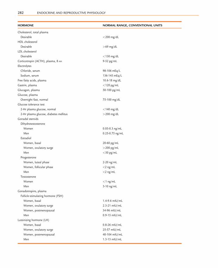
HORMONE NORMAL RANGE, CONVENTIONAL UNITS
Cholesterol, total plasma
Desirable <200 mg/dL
HDL cholesterol
Desirable >69 mg/dL
LDL cholesterol
Desirable <130 mg/dL
Corticotropin (ACTH), plasma, 8 AM 9-52 pg/mL
Electrolytes
Chloride, serum 98-106 mEq/L
Sodium, serum 136-145 mEq/L
Free fatty acids, plasma 10.6-18 mg/dL
Gastrin, plasma <120 pg/mL
Glucagon, plasma 50-100 pg/mL
Glucose, plasma
Overnight fast, normal 75-100 mg/dL
Glucose tolerance test
2-Hr plasma glucose, normal <140 mg/dL
2-Hr plasma glucose, diabetes mellitus >200 mg/dL
Gonadal steroids
Dihydrotestosterone
Women 0.05-0.3 ng/mL
Men 0.25-0.75 ng/mL
Estradiol
Women, basal 20-60 pg/mL
Women, ovulatory surge >200 pg/mL
Men <50 pg/mL
Progesterone
Women, luteal phase 2-20 ng/mL
Women, follicular phase <2 ng/mL
Men <2 ng/mL
Testosterone
Women <1 ng/mL
Men 3-10 ng/mL
Gonadotropins, plasma
Follicle-stimulating hormone (FSH)
Women, basal 1.4-9.6 mIU/mL
Women, ovulatory surge 2.3-21 mIU/mL
Women, postmenopausal 34-96 mIU/mL
Men 0.9-15 mIU/mL
Luteinizing hormone (LH)
Women, basal 0.8-26 mIU/mL
Women, ovulatory surge 25-57 mIU/mL
Women, postmenopausal 40-104 mIU/mL
Men 1.3-13 mIU/mL
282 ENDOCRINE AND REPRODUCTIVE PHYSIOLOGY

HORMONE NORMAL RANGE, CONVENTIONAL UNITS
Growth hormone, plasma
After 100 g glucose orally <2 mg/mL
After insulin-induced hypoglycemia >9 ng/mL
Human chorionic gonadotropin (hCG)
Men and nonpregnant women <3 mIU/mL
Insulin, plasma, fasting 5-20 mU/ml
Insulin-like growth factor (IGF-1) 0.35-2.2 U/mL
Ketone bodies
Acetoacetate <1 mg/dL
b-Hydroxybutyrate <3 mg/dL
Lactate, plasma 5-20 mg/dL
Magnesium, serum 1.8-3.0 mg/dL
Osmolality, plasma 285-295 mOsmol/L
Oxytocin, plasma
Random 1.25-5 ng/mL
Women, ovulatory surge 5-10 ng/mL
Parathyroid hormone, serum (intact PTH) 10-65 pg/mL
Phosphorus, inorganic, serum 3.0-4.5 mg/dL
Prolactin, serum
Nonpregnant women and men 2-15 ng/mL
Pyruvate, plasma 0.3-0.9 mg/dL
Renin activity, plasma, normal sodium intake
Standing 9.3 � 4.3 ng/mL/hr
Supine 3.2 � 1 ng/mL/hr
Thyroid function tests
Free thyroxine estimate 0.7-2.0 ng/dL
Radioactive iodine uptake, 24 hr 5%-30%
Resin T3 uptake, serum 25%-35%
Reverse T3 (rT3), serum 10-40 ng/dL
Thyrotropin (TSH), serum 0.5-5 mU/mL
Thyroxine (T4), serum 5-12 mg/dL
Triiodothyronine (T3), serum 70-190 ng/dL
Triglycerides, plasma <160 mg/dL
283HORMONE RANGES

Intentionally left as blank

n n n n n n n n n n n n n n n
A P P E N D I X DAbbreviations and Symbols
aGSU alpha glycoprotein subunit
b-hCG hormone-specific b-subunit ofhCG
b-TSH b-Thyroid-stimulating hormone
3b-HSD 3b-Hydroxysteroid dehydrogenase
11b-HSD2 11b-Hydroxysteroid dehydrogenase
17b-HSD 17b-HydroxysteroiddehydrogenaseAA amino acid
ABP androgen-binding protein
ACE angiotensin-converting enzyme
Ach acetylcholine
ACTH adrenocorticotropic hormone
(corticotropin)
ADH antidiuretic hormone
AIS androgen insensitivity syndrome
ALS acid-labile subunit
AMH antimullerian hormone
ANP atrial natriuretic peptide
APO apoprotein
AR androgen receptor
ARE androgen-response element
ATP adenosine triphosphate
BAT brown adipose tissue
bFGF basic fibroblast growth factor
BMP-15 bone morphogenetic protein-15
BMR basal metabolic rate
Ca2þ calcium phosphate
CaMKII Ca2þ-calmodulin-dependent
protein kinase-II
cAMP cyclic adenosine monophosphate
CaSR Ca2þ-sensing receptor
CBG corticosteroid-binding globulin
(also transcortin)
CCK cholecystokinin
CDK1 cyclin-dependent kinase-1
CETP cholesterol ester transfer protein
cGMP cyclic guanosine monophosphate
CGRP calcitonin gene–related peptide
CNS central nervous system
COMT catechol-O-methyltransferase
COX-2 cyclooxygenase-2
CPT-1/CPT-2 carnitine-palmitoyl transferase
CREB protein cAMP-response element–binding
protein
CRH corticotropin-releasing hormone
CSF cytostatic factor
CYP11b 11b-HydroxylaseCYP21b 21b-HydroxylaseCYP cytochrome P-450 mono-oxidase
gene
DAG diacylglycerol
DBP vitamin D–binding protein
DHEAS dehydroepiandrosterone sulfate
DHT dihydrotestosterone
DI diabetes insipidus
DIT diiodotyrosine
DM diabetes mellitus
DNA deoxyribonucleic acid
DOC deoxycorticosterone
DOPA dihydroxyphenylalanine
ECL enterochromaffin-like (cell)
ED erectile dysfunction
EGF epidermal growth factor
EnaC epithelial Naþ channel
ENS enteric nervous system
ER estrogen receptor
ERE estrogen-response element
FAS fatty acid synthase (complex)
FBHH familial benign hypocalciuric
hypercalcemia
FDA Food and Drug Administration
FFA free fatty acid
285

FGF-23 fibroblast growth factor-23
FSH follicle-stimulating hormone
Ga Ga-subunitGb/g Gb-subunit dimer
G-6-P glucose-6-phosphate
GAG glycosaminoglycan
GDF-9 growth differentiation factor-9
GEF guanine nucleotide exchange factor
GFR glomerular filtration rate
GH growth hormone
GHRH growth hormone–releasing
hormone
GHS growth hormone secretogogue
GH-V growth hormone variant
GI gastrointestinal
GIP gastroinhibitory peptide
GLUT glucose transporter
GnRH gonadotropin-releasing hormone
GPCR G-protein-coupled receptor
GPCR3 G-protein-coupled receptor-3
GR glucocorticoid receptor
GRE glucocorticoid-response element
GRK/RTK GPCR kinases
GRP gastrin-releasing peptide
GTP guanosine nucleotide triphosphate
GVBD germinal vesicle breakdown
HAD histone diacetylase
HAT histone acetyltransferase
HbA1c hemoglobin A1c
hCG human chorionic gonadotropin
HCO3� bicarbonate ion
hCS human chorionic
somatomammotropin
HDL high-density lipoprotein
HPA hypothalamus-pituitary-adrenal
hPL human placental lactogen
HRE hormone-response element
HSD hydroxysteroid dehydrogenase
HSL hormone-sensitive lipase
ICSI intracytoplasmic sperm injection
IDL intermediate-density lipoprotein
(particles)
IGF insulin-like growth factor
IGF-1 insulin-like growth factor-1
IGFBP insulin-like growth factor–binding
proteins
IP3 inositol 1,4,5-triphosphate
IR insulin receptor
IRS insulin receptor substrate
IUD intrauterine device
IVF in vitro fertilization
LDL low-density lipoprotein
LH luteinizing hormone
LPD luteal phase deficiency
LPL lipoprotein lipase
MAO monoamine oxidase
MAP mitogen-activated protein kinase
(also ERK)
MAPK mitogen-activated kinase
MC2R melanocortin-2 receptor
MCsF monocyte colony-stimulating
factor
MIT monoiodotyrosine
MMC migrating myoelectric complex
MODY mature onset of diabetes of the
young
MPF maturation-promoting factor
MR mineralocorticoid receptor
MRE mineralocorticoid-response
element
MIS mullerian-inhibiting substance
MPF maturation-promoting factor
mRNA messenger RNA
NCX sodium-calcium exchanger
NO nitric oxide
OHSS ovarian hyperstimulation
syndrome
OPG osteoprotegerin
OxPhos oxidative phosphorylation
PCO2 partial pressure of carbon dioxide
PCOS polycystic ovary syndrome
PEPCK\t PEP carboxykinase (phosphoenol-
pyruvate carboxykinase)
PFK1 phosphofructokinase-1
PGE2 prostaglandin E2PGF2a prostaglandin F2aPHEX phosphate-regulating gene with
homologies to endopeptidases on
the X chromosome
Pi phosphate
PI3K phosphatidylinositol-3-kinase
PIP3 phosphatidylinositol 3,4,5-
triphosphate
PKA protein kinase A
286 ENDOCRINE AND REPRODUCTIVE PHYSIOLOGY

PKB protein kinase B
PKG protein kinase G
PLCz phospholipase CzPMCA plasma membrane calcium ATPase
PMS premenstrual syndrome
PNMT phenylethanolamine-N-methyl
transferase
PO2 partial pressure of oxygen
POMC pro-opiomelanocortin
PPARg peroxisome proliferator-activated
receptor-gPR progesterone receptor
PRE progesterone-response element
PRF prolactin-releasing factor
PRL prolactin
PSA prostate-specific antigen
PTH parathyroid hormone
PTHrP parathyroid hormone–related
peptide
PTU propylthiouracil
PVH paraventricular hypothalamus
PVN paraventricular nuclei
RAIU radioactive iodide uptake
RANKL receptor activator of NF-KB ligand
RAS renin-angiotensin system
RDS respiratory distress syndrome
RGS regulators of G-protein signaling
RIA radioimmunoassay
RNA ribonucleic acid
ROMK channel renal outer medullary Kþ channel
ROS reactive oxygen species
rT3 reverse T3
RTK receptor tyrosine kinase
SERM selective estrogen receptor
modulator
SHBG sex hormone–binding globulin
SIADH syndrome of inappropriate secre-
tion of antidiuretic hormone
SOCS suppressor of cytokine signaling
SON supraoptic nuclei
SRBEP-1C sterol regulatory-binding element
protein-1C
SRY sex-determining region Y
StAR protein steroidogenic acute regulatory
protein
STAT signal transducers and activator of
transcription
SUR ATP-binding subunit
t1/2 half-life
T1DM type 1 diabetes mellitus
T2DM type 2 diabetes mellitus
T3 triiodothyronine
T4 thyroxine
TBG thyroxine-binding globulin
TCA tricarboxylic acid (cycle)
TGF transforming growth factor
TGF-b transforming growth factor-bTG thyroglobulin
TGs triglycerides
TNF-a tumor necrosis factor-aTPO thyroid peroxidase
TR thyroid hormone receptor
TRE thyroid hormone–response element
TRH thyrotropin-releasing hormone
(also thyroid-releasing hormone)
T/S thyroid/serum (ratio)
T/S[I] thyroid/serum (measured with
radioactive iodide)
TSA thyroid-stimulating antibody
TSAb thyroid-stimulating antibody
(abnormal)
TSH thyroid-stimulating hormone (also
called thyrotropin)
TTR thyroxine-binding prealbumin
TZD thiazolidinedione
VDR vitamin D receptor
VEGF vascular endothelial growth factor
VLDL very-low-density lipoprotein
VMA vanillylmandelic acid
WAT white adipose tissue
287ABBREVIATIONS AND SYMBOLS

Intentionally left as blank

n n n n n n n n n n n n n n n
I N D E X
Note: Page numbers followed by b indicate boxes, f indicate figures, and t indicate tables. Page numbers with ‘e’ indicate the material isavailable online only.
AAbortion, 262ACE. See Angiotensin I-converting enzymeAcetyl CoA, 57–58Acetylcholine, 32, 151Acromegaly, 124, 124fAcrosome reaction, 241ACTH. See Adrenocorticotropic hormoneAddison disease, 172, 172f, 174–176Adenohypophysis, 99, 108–128, 109tAdenosine monophosphate (AMP), 11,
44–45Adenosine monophosphate (AMP)-
activated kinase (AMPK), 44–45,45f, 58, 65–66, 71
Adenosine triphosphate (ATP), 45–46, 64–65actions of, 56, 57delivery of, 196–198energy metabolism and, 43, 44fsynthesis of, 56–57, 62
Adenylyl cyclase, 11ADH. See Antidiuretic hormoneAdhesive proteins, 240Adipocytes, 60, 62, 63fAdiponectin, 67Adipose tissue, 66, 68t, 260endocrine cells in, 1in energy use and storage, 51–52estrogen and, 235
Adipose tissue-derived hormones andadipokines, 66–67
Adrenal androgens, 147–149, 163–164Adrenal cortex, 6, 112, 154–166, 168fdevelopment of, 147–149in endocrine axis, 110–112pathologic conditions with, 172–176
Adrenal glands, 1, 147–176anatomy of, 147–150, 148fduring pregnancy, 256–257
Adrenal glucocorticoids, 92Adrenal medulla, 5, 51, 149, 150–154Adrenal steroid hormones, 92Adrenergic receptors, 5, 49, 51, 65–66Adrenocortical excess, 172–176Adrenocortical insufficiency, 172, 173b
Adrenocorticotropic hormone (ACTH)adrenal glands and, 4, 162–163, 166,
166f, 253in hypothalamus-pituitary complex,
108–110, 111–112, 112f,113–114, 114f
Adrenomedullary catecholamines, 152Advanced glycation end products, 72Afferent nerves, 28–29African pigmy, 123Agonists, 3Albumin, 135–136, 159Aldosterone, 166, 170factions of, 167–170metabolism of, 167secretion of, 166–172, 171b, 256–257
Alton giant, 123–124, 123fAlveoli, 259–260Amenorrhea, 117, 125, 236–237Amino acids, 43, 48, 54, 62, 71, 255Aminoaciduria, 258Amniochorionic membrane, 247–248Amnion, 247–248AMP. See Adenosine monophosphateAMPK. See Adenosine monophosphate
(AMP)-activated kinaseAmpulla, 229of oviduct, 241
Anatomyof adrenal glands, 147–150, 148fof hypothalamus-pituitary complex,
99–101, 100f, 101f, 105fof ovaries, 215–216, 216fof reproductive system, 180–181of stomach, 30–31, 31fof thyroid glands, 129–130, 130f, 131f
Anchoring villi, 249Androgens, 6actions of, 201–205, 203b, 222adrenal, 147–149, 163–164intratesticular, 201metabolism of, 201–205transport of, 201–205
Androgen insensitivity syndrome,211–213, 211f
Androgen receptor, 6, 200, 203–204Androgen-binding protein, 200Androgen-response element (ARE), 20Andropause, 190–193Androstenedione, 221, 221fAngiotensin I, 1Angiotensin I-converting enzyme
(ACE), 1, 171Angiotensin II, 1, 256–257Anorexia nervosa, 117bAnterior pituitary, 99, 108, 111Antidiuretic hormone (ADH), 101,
103–107, 162blood volume and, 105–106, 106fdisorders with, 107bmechanism of, 104fsecretion regulation of, 104–106synthesis of, 101–103, 102f
Anti-inflammatory actions, 160–161Anti-mullerian hormone, 200–201, 218Antral follicles, 219–221, 219fAppetite control
hypothalamic neurons and, 69–70, 69fobesity and, 67–70
APS protein, 49–50Aquaporin-2, 104Arcuate nucleus, 69ARE. See Androgen-response elementAromatase inhibitors, 235–236b-Arrestins, 15–16Atherosclerosis, 73–75ATP. See Adenosine triphosphateATP-sensitive Kþ channel, 47–48, 48b, 49fAtrial natriuretic peptide, 107, 166Autonomic nervous system, 28–29, 45, 142Autophagy, 44–45
BBasal metabolic rate, 141–142Bidirectional placental transfer, 245Bile, 35–36, 35fBile ducts, 35–36, 35fBiliary function, 37Birth canal, 234Blastocyst, 243–244, 245f
289

Blood volume, ADH and, 105–106, 106fBody mass index (BMI), 69Bone, 143Ca2þ and, 83–92, 86fcortisol and, 162estrogen and, 235Pi and, 83–92, 86frenal failure problems with, 96–98, 96fvitamin D deficiency and, 88b
Brain, 45Bulbourethral glands, 207
CCa2þ. See CalciumCa2þ-sensing receptor, 79–80, 79fCalbindin-D, 84–85Calcitonin, 90, 91Calcitonin gene-related peptide, 91Calcitonin receptor, 91Calcium (Ca2þ), 77, 77f, 78t, 85tbone and, 83–92, 86fin cellular physiology, 77–78in egg, 242handling of, 90fhormonal regulation of, 90–92kidney and, 83–92pathologic disorders of, 92–98in physiologic regulation of, 78–83, 89small intestine and, 83–92, 85f
Calcium homeostasis, 77–98Calcium-response element, 80cAMP. See Cyclic AMPCancer, hormonal regulation and, 91–92Capacitation, 241Cardiac output, 257Cardiovascular changes, during
pregnancy, 257Cardiovascular systemcortisol and, 160estrogen and, 235thyroid hormones and, 139–140, 140f, 141f
Cardiovascular volume receptors, 105–106Carnitine-palmitoyl transferase-1, 58Carpal-pedal spasms, 94CART. See Cocaine and amphetamine-
regulated transcriptCatecholamines, 5, 44, 52f, 60–61, 66, 147actions of, 51, 142, 151–152, 151tadrenomedullary, 152characteristics of, 5bmetabolism of, 152–154, 154fsecretion of, 151vasopressive response to, 172
Catechol-O-methyltransferase (COMT),152–154
CBG. See Corticosteroid-binding globulinCCK. See CholecystokininCells. See specific cellsCellular response, 1, 3–9Centromeres, 179Centrosome, 243Cephalic phase, 31
Cervix, 181, 233cGMP. See Cyclic guanosine
monophosphateChemical nature, of hormones, 3–9Chief cells, 30Cholecystokinin (CCK), 29, 30, 35, 36, 36f,
37, 40, 41Cholecystokinin (CCK)-1 receptor, 29, 29tCholecystokinin (CCK)-2 receptor, 29, 29t, 33Cholelithiasis, 37Cholesterol, 6–7, 7f, 154–155, 155b
steroidogenic pathway of, 156–159,157f, 158f
Cholesterol-rich lipoproteins, 6–7Chorion, 247Chorion frondosum, 247, 249Chorion laeve, 247Chorionic membrane, 247Chorionic villi, 247, 248Chromaffin cells, 149Chromogranins, 150–151Chylomicron remnants, 54Citrate, 57–58Cleidocranial dysplasia, 86–87Clitoris, 181, 234Closed enteroendocrine cells, 27, 28–29Cocaine and amphetamine-regulated
transcript (CART), 69Colostrum, 260–261Common bile duct, 35–36COMT. See Catechol-O-methyltransferaseCongenital adrenal hyperplasia, 163–164,
174–176, 186Conn syndrome, 174Connective tissue, cortisol and, 162Contraception, 261–262Corpus hemorrhagicum, 224, 225fCorpus luteum, 222, 224–225, 251–252Cortical granules, 242–243Corticosteroid-binding globulin (CBG), 7,
159, 256Corticotropes, 111–114Corticotropin-releasing hormone (CRH),
162–163, 163b, 258Cortisol, 44, 110–111, 114, 149, 155–159
actions ofbiologic, 159bcardiovascular, 160gastrointestinal, 162mechanism of, 159metabolic, 160–161, 161fphysiologic, 159–162psychological, 162
bone and, 162connective tissue and, 162during fetal development, 162kidney and, 162levels of, 256muscle and, 162production regulation of, 162–163reproductive system and, 162transport and metabolism of, 159
Cortisol-binding globulin, 235Cortisone, 159Co-Smad, 18CREB protein, 15CRH. SeeCorticotropin-releasing hormoneCuboidal granulosa cells, 218Cumulus cells, 219, 241–242Cushing syndrome, 172–174, 174fCyclic AMP (cAMP), 11, 12, 12f, 13–14,
48–49, 142, 220Cyclic guanosine monophosphate (cGMP),
13–14, 13f, 220Cyclindependent kinase-1, 220CYP11A1, 201, 222, 224, 252CYP17, 201CYP19, 201–203, 201b, 221, 260Cystic fibrosis, 37Cytochrome P-450 mono-oxidase, 156Cytokines, 113–114, 160Cytokine receptor family, 17–18Cytokine signaling proteins.
See Suppressors of cytokinesignaling proteins
Cytoplasmic tyrosine kinases, 17–18Cytotrophoblastic shell, 249Cytotrophoblasts, 244
DDAG. See DiacylglycerolDBP. See Vitamin D-binding proteinDe novo lipogenesis, 57–58Decapacitation, 207Decidua basalis, 247, 249, 249f, 250fDecidua capsularis, 247Decidua parietalis, 247Decidual cells, 245–246Decidualization, 245–246Decondensation, 242Degradation, 106–107Dehydroepiandrosterone (DHEA),
163–164, 222Dehydroepiandrosterone sulfate (DHEAS),
163–164, 165f, 166, 253Deiodinases, 130–131, 132, 135Deoxycorticosterone (DOC), 158, 158f,
166–167, 174–176, 258Desensitization, 15–16DHEA. See DehydroepiandrosteroneDHEAS. See Dehydroepiandrosterone
sulfateDiabetes
gestational, 258of young, mature-onset, 48b
Diabetes insipidus, 106familial, 103nephrogenic, 107, 107tneurogenic, 106–107, 107tpsychogenic, 107, 107t
Diabetes mellitus, 45, 70–75, 173long-term sequelae of, 72–75T1DM, 70T2DM, 70–72
290 INDEX

Diabetic ketoacidosis, 70Diabetogenic hormone, 121Diabetogenicity, of pregnancy, 254, 258Diacylglycerol (DAG), 14, 15Diaphragma sellae, 101Diffuse enteroendocrine system, 27Dihydrotestosterone, 183–185, 203, 204f5-a Dihydrotestosterone, 2031,25-Dihydroxyvitamin D, 77, 78–83,
81f, 89Diiodotyrosine, 134, 134fDisorder of sexual development/androgen
insensitivity syndrome, 211Distention, of stomach, 33DOC. See DeoxycorticosteroneDominant follicle, 222–224, 227Dopamine, 5, 124, 260–261Ductal pancreatic carcinomas, 40Ducts, 259–260Duodenal pH, 37Duodenum, 35–36, 35fDwarfism, 111, 119, 122–123, 122f, 126
EECL cells. See Enterochromaffin-like cellsEffector proteins, 10Efferent ductules, 180, 196, 207Egg, 178–179, 241activation of, 241, 242
Ejaculatory ducts, 180, 207Embryo, 247Embryogenesis, early, 239–254, 244fEmbryology, of hypothalamus-pituitary
complex, 99–101, 100f, 101fEmbryonic development, 177, 182fEmbryonic pole, 243Embryotroph, 246–247Empty sella syndrome, 101ENaC. See Epithelial Naþ channelEndocervical canal, 233Endocrine axis, 108–111, 110fEndocrine cells, 1Endocrine disorders, 111Endocrine functionof GI tract, 27–42implantation and, 244–245of ovarian follicles, 215, 218, 219, 221,
222, 223–224, 225Endocrine glands, 2t, 3f, 144–146adrenal, 1parathyroid, 1pituitary, 1thyroid, 1
Endocrine pancreas, 46Endocrine systemenzymes in, 1, 10, 11ffetal, 239, 255glands in, 1, 2t, 3fintroduction to, 1–26maternal, 239ovaries, 1, 6placental, 239
summary of, 23btestis, 1, 6
Endocytosis, 15–16Endometrial stroma, 245–246Endometrial veins, 249Endometrium, 231Energy, storage forms of, 51–52Energy metabolism, 43–76adipose tissue-derived hormones and
adipokines, 66–67appetite control and obesity, 67–70ATP and, 43, 44fdiabetes mellitus, 45during fast state, 60–62during fed state, 52–54general pathways involved in, 45–46metabolic homeostasis
hormonal regulation of, 51–62key hormones with, 46–51
overview of, 43–45skeletal muscle, 65–66
Enteric nervous system (ENS), 28–29, 32Enteric neurons, 32Enterochromaffin-like (ECL) cells, 32,
33, 33fEnteroendocrine cells, 41extrinsic regulators of, 28–29function of, 27–28types of, 27, 28f, 28t
closed, 27, 28–29open, 27, 28–29
Enteroendocrine hormone families andreceptors, 29–30. See also Gastrin;Secretin
motilin, 29, 29t, 30, 38, 41Enteroendocrine regulation, 35–38Enterogastrones, 35, 37Enterotropic actions, of GI hormones,
39–42Enzymes, 1, 10, 11fEpididymis, 180, 196, 207Epinephrine, 5, 5f, 49, 51, 147, 149, 149f,
150–151, 150factions of, 153fsecretion of, 151
Epithelial calcium channels, 84–85Epithelial Naþ channel (ENaC), 167, 167bERE. See Estrogen-response elementErectile dysfunction, 210Erection, 208–210Ergocalciferol, 80–82Erythromycin, 38Estradiol, biology of, 234–236Estradiol-17b, 221, 221f, 222, 232–233, 253Estrogen, 6, 110–111, 177adipose tissue and, 235biological effects of, 235bone and, 235cardiovascular system and, 235high level of, 239liver and, 235mechanisms of, 234
peripheral conversion to, 201–202during pregnancy, 258–259production of, 253, 254f, 256, 260
Estrogen receptor, 6, 232Estrogen-response element (ERE), 20Exendins, 39Exocrine pancreas, 35–38Exocytosis, 4–5External genitalia. See GenitaliaExtraembryonic mesoderm, 247Extravillous cytotrophoblasts, 249Extrinsic regulators, of enteroendocrine
cells, 28–29
FFamilial benign hypocalciuric
hypercalcemia (FBHH), 80Familial diabetes insipidus, 103Fast state, 60–62, 63–64Fatty acids. See Free fatty acids; Long-chain
fatty acidsFatty acid transport proteins, 60FBHH. See Familial benign hypocalciuric
hypercalcemiaFed state, 53f
energy metabolism in, 52–54hepatic metabolism, of nutrients during,
52–54Feedback control systems, 3Female. See also Genitalia; Sexual
development; Tanner pubertal stagereproductive system of, 177–194, 181f,
215–238, 216breproductive tract of, 228–234,
229f, 241cervix, 233external genitalia, 181, 181f, 184f,
234, 234foviduct, 229–231, 229futerus, 231–233
Fertilization, 239–264, 242f, 244fFetal adrenal axis, 258Fetal adrenal cortex, 253, 253fFetal development, cortisol during, 162Fetal endocrine system, 239, 255, 256tFetal hemoglobin, 255Fetal programming, 68–69Fetoplacental unit, 255FFAs. See Free fatty acidsFibroblast growth factor-23, 92bFimbriae, 229First embryonic cleavage, 243, 243fFollicles. See specific folliclesFollicle lumen, 133f, 134Follicle-stimulating hormone (FSH), 5,
187, 200, 220, 221in hypothalamus-pituitary complex,
108–110, 114–118, 114f, 116f, 117fproduction of, 255
Follicle-stimulating hormonereceptors, 222Follicular atresia, 217–218, 225–226, 227Follicular phase, 226, 226f
291INDEX

Food-induced hypercortisolism, 38,115–117, 116f
Free fatty acids (FFAs), 43, 60–61, 61f, 62,62f, 64–65, 66
Free hormone, 9Fructose-1,6-bisphosphate, 63–64Fructose-2,6-bisphosphate, 56, 63–64FSH. See Follicle-stimulating hormoneFunctional residual capacity, 257
GG proteins, 11heterotrimeric, 14–15transmembrane receptors’ use of, 9,
14–16Gallbladder, 35–38contraction of, 37
Gamete, 218, 219, 222, 223, 225Gametogenic function, of ovarian
follicles, 215Ga-q/phospholipase/IP3 pathways, 30Gastric contraction stimulation, 38Gastric functionregulation of, 30–35stimulation of, 32–35
Gastric inhibitory peptide (GIP), 38, 48–49Gastric inhibitory polypeptide, 29–30Gastric mucosa, 30Gastric phase, 31, 34fGastric secretion and motility regulation,
30–31during cephalic phase, 31during gastric phase, 31, 34fduring intestinal phase, 31, 33, 34f, 35,
36, 37Gastrin, 27, 29, 29t, 32, 40, 41gastric function regulation and, 30–35gastric function stimulation and, 32–35
Gastrin-producing tumor, 31Gastrin-releasing peptide (GRP), 32Gastrointestinal changes, during
pregnancy, 258Gastrointestinal (GI) hormones, 39–42Gastrointestinal (GI) peptides, 38–39Gastrointestinal (GI) tract, 51–52cortisol and, 162endocrine cells in, 1endocrine function of, 27–42
GEFs. See Guanine nucleotide exchangefactors
Gene expression, hormonal regulationof, 20t
Genetic diversity, 178–179Genitalia, 181–182, 183–185, 183f, 184fexternal, 164–166, 177–178
female, 181, 181f, 184f, 234, 234fmale, 182–185, 182f, 183f, 184f, 185b
internalfemale, 181f, 183f, 185fmale, 182–183, 183f, 184b, 185f
Germinal vesicle breakdown, 228Gestation, early, 239
Gestational diabetes, 258GH. See Growth hormoneGH-binding protein, 119Ghrelin, 30, 69–70, 120GHRH. See Growth hormone-releasing
hormoneGI. See GastrointestinalGI peptides, 30Gi-a, 15Gigantism, 122GIP. See Gastric inhibitory peptideGlands. See specific glandsGlucagon, 29–30, 44, 46, 51, 52f, 60–61Glucagon receptor, 51Glucagon-like peptides, 29–30Glucagon-like peptide-1, 39, 40, 48–49Glucagon-like peptide-2, 40–42Glucocorticoids, 6, 92, 147–149, 160, 162Glucocorticoid receptor, 6Glucocorticoid-response elements, 159Glucokinase, 43.e1, 47–48, 55Glucokinase regulatory protein, 55Gluconeogenesis, 63–64, 160Glucose, 47–48, 53, 55–56, 60f.
See also Hepatic glucoseproduction; Obligate glucose user
intolerance of, 45, 123, 173intrahepatic, 55tolerance of, 59transportation of, 255
Glucose-6-phosphate, 46, 47–48, 54f, 55,63, 160
Glucose-dependent insulinotropicpeptide, 38
Glucosuria, 258GLUT1, 255GLUT-2 transporter, 47–48, 54–55, 54fGLUT-4 transporter, 59–60, 62, 65, 71, 160Glycerol-3-phosphate, 59–60Glycogen, 53, 55–56, 62Glycogen phosphorylase, 55–56, 64f, 65–66Glycogen synthase, 55–56Glycogenolysis, 63, 65–66, 152Glycolysis, 53, 56–57Glycosaminoglycan, 235Glycosylation, 114–115GnRH. See Gonadotropin-releasing
hormoneGoiter, 136–137, 137fGonadal steroid hormones, 92, 126, 155Gonadotropes, 111, 115–118Gonadotropin-releasing hormone
(GnRH), 105, 116–117, 116f, 117f,187, 205, 205f, 226, 228, 261
Gonads, 177G-protein-coupled receptor (GPCR),
14–16, 15f, 16f, 29, 30, 38, 48, 151Gq-a, 15Graves’ disease, 138, 139fGrowth, 126–128
maturation and, 142–143retarded, 126b
Growth hormone (GH), 5, 44, 108–110actions of, 120–122, 120f
direct, 120–121indirect, 121
adult deficiency of, 123excess before puberty of, 123–124, 123finteraction of, 122metabolic actions of, 119bpathologic conditions of, 122–124secretion of, 119somatotropes and, 118–120
Growth hormone receptor, 118–119Growth hormone-releasing hormone
(GHRH), 119GRP. See Gastrin-releasing peptideGs signaling pathways, 29–30Gs-a, 15GTP. See Noncovalent guanosine
nucleotide triphosphateGuanine nucleotide exchange factors
(GEFs), 11, 14–15Guanylyl cyclase, 1Gynecomastia, 125, 210–211
HHaploid cells, 178–179Haversian canal, 88Haversian lacunae, 88hCG. See Human chorionic gonadotropinHCL secretion, 30, 32, 33, 33fHDAC. See Histone deacetylaseHDL. See High-density lipoproteinHeart muscle, 170Hemochorial placentation, 249Hemoglobin A1C, 72Hemotrophic nutrition, 249Hepatic glucose production, 60–61, 63–64,
71Hepatic metabolism, of nutrients, 52–54Hepatic portal vein, 52–53Hepatic steatosis, 72, 72bHepatopancreatic ampulla, 35–36, 37Herring bodies, 100f, 101–102Heterotrimeric G proteins, 14–15High-density lipoprotein (HDL), 155, 224Histamine, 32, 33Histone deacetylase (HDAC), 20Histotrophic nutrition, 240, 246–247, 247fHormonal development, of mammary
glands, 260–261Hormonal precursors, peripheral
conversion of, 1, 2t, 7, 8fHormonal regulation
of Ca2þ, 90–92cancer and, 91–92of gene expression, 20tduring menstrual cycle, 231–233, 234of metabolic homeostasis, 51–62of Pi, 90–92
Hormonal resistance, 3, 10Hormones, 1, 27. See also specific hormones
adult male production of, 203t
292 INDEX

binding of, 1cellular responses to, 3–9
membrane guanylyl cyclase receptors,18–19
receptor serine/threonine kinasereceptors, 18
receptors, cytoplasmic tyrosinekinases with, 17–18
RTKs, 16–17, 18fsignaling from intracellular receptors,19–23
transmembrane receptors, 5, 9–14transmembrane receptors using Gproteins, 9, 14–16
chemical nature of, 3–9catecholamines, 5, 5bproteins/peptides, 3–5, 4bsteroid hormones, 6–7, 6b, 6tthyroid hormones, 7–9, 8b, 8f
circulatory transport of, 9delivery of, 3receptors and, 3releasing, 108secretion of, 3, 27–28, 29tropic, 108–110
Hormone response element (HREs), 19–20Hormone-sensitive lipase, 201, 235HPA axis. See Hypothalamus-pituitary
adrenal axisHREs. See Hormone response elementHuman chorionic gonadotropin (hCG),
114–115, 240–241, 250–251, 251fHuman menstrual cycle, 226–228,
227f, 240fHuman placental lactogen, 253–254, 255fHyaluronic acid, 242Hydroxyapatite crystals, 8825-Hydroxyvitamin D, 82–83Hyperactivation, 241Hypercortisolism, 38, 113–114, 126,
172, 173tHyperglycemic hormone, 119–120Hyperinsulinemia, 123, 124Hyperkalemic acidosis, 172Hyperlipoproteinemia, 43.e9Hyperparathyroidism, 92–94, 93f, 94bHyperphosphatemia, 78Hyperprolactinemia, 125Hyperthyroidism, 138, 140, 142Hypervolemia, 107Hypoadrenalism, 125–126Hypocalcemic challenge, 89, 91fHypocalciuria, 80Hypoglycemia, 45, 65–66, 123Hypogonadism, 125–126Hyponatremic, 107Hypoparathyroidism, 94–95Hypophysiotropic neurons, 108, 111, 111fHypopituitarism, 125–126Hypothalamic neuroendocrine neurons, 1Hypothalamic neurons, appetite control
and, 69–70, 69f
Hypothalamohypophyseal portalvessels, 108
Hypothalamohypophyseal tracts, 101Hypothalamus, 51–52, 111bHypothalamus-pituitary adrenal (HPA)
axis, 111–112, 113–114, 114f, 115,116f, 164f
Hypothalamus-pituitary complex, 99–128adenohypophysis, 99, 108–128embryology and anatomy of, 99–101,
100f, 101f, 105fneurohypophysis, 99, 101–108
Hypothalamus-pituitary-gonadal axis, 177Hypothalamus-pituitary-liver axis,
118, 118fHypothalamus-pituitary-testis axis,
205–207, 206fHypothalamus-pituitary-thyroid axis,
136–137Hypothyroidism, 125–126, 134, 140, 142,
143, 144, 144fHypoxic environment, 249
IIGFBPs. See Insulin-like growth factor-
binding proteinsIGFs. See Insulin-like growth factorsIleal brake, 39Immunosuppressive actions, 160–161Implantation, 239–254, 246fIn vitro fertilization, 262–264Incomplete cytokinesis, 196Incretions, 38, 41, 48–49Infertility, 125Infundibulum, 99–101, 229Inhibin, 200–201, 205Inhibin A, 225Inhibin B, 221, 222Inorganic phosphate, 78Inositol 1,4,5-triphosphate (IP3), 14, 242Insulin, 43, 46–51, 47b, 47f, 52f, 55actions of, 55–57, 58, 71, 71binteraction of, 122, 162promoting of, 58, 59–60resistance to, 50–51, 70–72, 71bsecretion of, 46–49, 47b, 48f, 49f,
260–261Insulin receptor, 49–51, 50fInsulin receptor substrates, 49–50Insulin-like growth factor-binding proteins
(IGFBPs), 121Insulin-like growth factors (IGFs), 5,
110–111, 118, 118f, 119, 121–122Insulinotropic actions, of GI peptides,
38–39Insulinotropic peptide, glucose-
dependent, 38Insulin-resistant,hyperinsulinemic state, 258Integument system, 235Interstitial cells of Leydig. See Leydig cellsInterstitial implantation, 244Intervillous space, 249
Intestinal absorption, 77Intestinal phase, 31, 33, 34f, 35, 36, 37Intestine. See Small intestineIntracellular Ca2þ, 4–5, 48, 65–66Intracellular receptors, signaling from,
19–23Intracellular signaling molecule, 10Intrahepatic glucose, 55Intramural segment, 229Intratesticular androgen, 201Intratubular compartment, 196–201Intrinsic factor, 30Iodide balance, 132, 132fIodide trap, 133–134Iodinated thyroglobulin, 132–133Iodothyronines, 7–9, 8f, 130, 131f, 132–134Ion channels, 13IP3. See Inositol 1,4,5-triphosphateIslets of Langerhans, 1, 46, 46fIsthmus, 229, 231Izumo, 242
JJAK kinase family, 17–18, 19f
KKallmann syndrome, 117, 187bKetogenesis, 53, 64–65, 152Ketone bodies synthesis and release, 60–61,
62, 62f, 65fKey hormones, with metabolic
homeostasis, 46–51Kidney, 167–170
Ca2þ and, 83–92cortisol and, 162endocrine cells in, 1Pi and, 83–92
Kinetochore, 179Klinefelter syndrome, 210–213, 210f
LLabia majora, 181, 234Lactase dehydrogenase, 57Lactation, 239–264Lactational amenorrhea, 261Lactogenesis, 260–261Lactotropes, 111, 124–125Laron dwarfism, 119, 122–123Late follicular stage, 239LDL. See Low-density lipoproteinLeukotrienes, 160Leydig cells, 196, 201, 202f, 205, 251–252LH. See Luteinizing hormoneLigands, 3, 64Lipid informational molecules, 14, 14fLipid-soluble vitamins, 19Lipolysis, 66f, 152Lipoproteins
cholesterol-rich, 6–7VLDLs, 53, 53f, 54, 58, 60–61
Lipoprotein base, 60Lipoprotein lipase, 60, 61f, 65, 235
293INDEX

Liver, 159bile secreted by, 35–36endocrine cells in, 1in energy use and storage, 51–52estrogen and progesterone in, 235,
256–257in fast state, 63–64glycogen in, 53insulin in, 58
Lobules, 196Long-chain fatty acids, 37, 48Low-density lipoprotein (LDL), 155, 224Lower esophageal sphincter tone, 258Luteal phase, 226Luteinization, 222Luteinizing hormone (LH)production of, 108–110, 114–117, 114f,
116f, 117f, 205, 255surge of, 117–118, 220, 222–224, 225, 239in urine, 5
MMagnocellular, 101Male. See also Genitalia; Sexual
development; Tanner pubertal stagehormone production of, 203tpseudohermaphroditism of, 211reproductive system of, 177–194,
195–214, 216breproductive tract of, 207–210, 208furethra of, 180
Malonyl CoA decarboxylase, 58, 59fMammalian target of rapamycin complex-1
(mTORC1), 44–45, 45f, 50–51, 54,58, 59, 60, 71
Mammary glands, 177–178, 253hormonal development of, 260–261structure of, 259–260, 259f
Mammogenesis, lactation and, 259–261Mammograms, 260MAO. See Monoamine oxidaseMAP. See Mitogen-activated proteinMAPK. See Mitogen-activated protein
kinaseMasculinization, 164–166Maternal endocrine changes, during
pregnancy, 255–257Maternal messenger RNA, 243Maternal ovarian reproductive tract
function, synchronization with,239–241
Maternal physiologic changes, duringpregnancy, 257–258, 257b
Maternal pituitary prolactin, 253, 260Matrix metalloproteases, 233Maturation-promoting factor, 243Mature-onset diabetes, of young, 48bMechanosensors, 33Median eminence, 108Megalin, 83Meiosis, 178–179, 178f, 223, 243Meiotic arrest, 220, 221f
Meiotic competence, 220Meiotic maturation, 239Melanocortin-2 receptor, 112, 163a-Melanocyte-stimulating hormone
(aMSH), 4Membranes. See specific membranesMembrane guanylyl cyclase receptors,
18–19Membrane-bound phospholipase C
(PLC), 14Membranous urethra, 207Menopause, 190–193Menstrual cycle
hormonal regulation during,231–233, 234
human, 226–228, 227f, 240fphases of, 232, 232f
Menstruation, 233Metabolic actions, of GH, 119bMetabolic homeostasis
hormonal regulation of, 51–62key hormones with, 46–51
Metabolism. See also Energy metabolismof aldosterone, 167of androgens, 201–205of catecholamines, 152–154, 154fof cortisol, 159of nutrients, hepatic, 52–54of thyroid hormones, 135–146, 136tof vitamin D, 82f
Metformin, 65Microtubule-organizing center, 243Midluteal phase, 240Migrating myoelectric complex (MMC),
36, 38Milk letdown, 108, 261Mineralocorticoids, 6, 147–149, 166Mineralocorticoid receptor, 6, 169fMineralocorticoid-response element
(MRE), 20, 167Mitogen-activated protein (MAP) kinase,
16–17, 17f, 223Mitogen-activated protein kinase (MAPK)
pathway, 50, 243MMC. See Migrating myoelectric complexMonoamine oxidase (MAO), 152–154Monocyte colony-stimulating factor, 87Monoglycerides, 37Monoiodotyrosine, 134, 134fMonosaccharides, 43, 54–55Mons pubis, 234Morning sickness, 251–252Motilin, 29, 29t, 30, 38, 41MRE. See Mineralocorticoid-response
elementmTORC1. See Mammalian target of
rapamycin complex-1Mucous cells, 30Mullerian-inhibiting substance,
200–201Mural granulosa cells, 219, 221Muscarinic receptor, 32
Muscle. See also Skeletal musclecortisol and, 162heart, 170
Myometrium, 231, 233, 241
NNADH, 53, 57NADPH, 53, 58–59, 137–138Naþ-I¯ symporter, 133–134Neimann-Pick disease C transporters, 155Neoplasms, 107Nephrogenic diabetes insipidus, 107, 107tNervous system, 143Neuroendocrine reflex, 107–108, 260–261Neurogenic diabetes insipidus,
106–107, 107tNeuroglycopenia, 45Neurohypophysis, 99, 101–108Neurophysin-I, 101–102Neurophysin-II, 101–102Nicotinic receptors, 151Nipple, 260Nitric oxide, 13, 208–210Noncovalent Ca2þ binding, 14Noncovalent guanosine nucleotide
triphosphate (GTP), 11, 12f, 14–15Norepinephrine, 5, 5f, 49, 51, 65–66,
149, 150secretion of, 151
Nuclear hormone receptors, 9, 19–22,20f, 21f
Nuclear vitamin D receptor, 83Nutrient partitioning, 43–45
OObesity, 67–70Obesity-induced insulin resistance, 71, 71bObligate glucose user, 45Oocytes, 218, 220fOpen enteroendocrine cells, 27, 28–29Oral contraceptives, 262Orgasm, 181, 234Osmolality, 104–106, 105fOsmoreceptive neurons, 104–105Osmotic diuresis, 106Osmotic stimuli, 106Osteoblasts, 86–87, 87fOsteoclasts, 86–87, 88Osteocytes, 88Osteopetrosis, 88Osteoporosis, 88, 93–94, 173Osteoprotegerin, 87Ovarian follicles, 180–181, 215
endocrine function of, 215, 218, 219,221, 222, 223–224, 225
gametogenic function of, 215growth, development, function of,
217–226Ovarian pathophysiology, 236–238Ovarian reserve, 217–218Ovarian steroidogenesis, 221f, 224fOvarian steroids, 235–236
294 INDEX

Ovarian surface epithelial cells, 215Ovaries, 1, 6, 108, 180–181anatomy and histology of, 215–216, 216fphases of, 227, 228
follicular, 226, 226fluteal, 226
Oviduct, 181, 229–231, 229f, 241Oviductal isthmus, 241Ovulation, 222f, 239Oxidative phosphorylation, 53Oxyphil cell, 78–79Oxytocin, 101–103, 107–108milk letdown and, 108pathologic conditions involving, 108during pregnancy, 259, 259f, 261uterine motility and, 107–108
PP13 kinase, 49–50Paget disease, 95, 95fPancreasendocrine, 46exocrine, 35–38
Pancreatic acinar cells, 40Pancreatic carcinomas, ductal, 40Pancreatic ducts, 35–36Pancreatic function, 37Pancreatic islet, 51Panhypopituitarism, 125–126Paracrine peptide factors, 27Paracrine stimulation, 32Parafollicular C cells, 91, 129–130Parathyroid glands, 1, 78–79, 79fParathyroid hormone (PTH)actions of, 78–83, 85t, 88intestinal absorption regulated by, 77response of, 89secretion of, 79–80, 79f, 80f
Parathyroid hormone receptor, 80Parathyroidhormone-relatedpeptide, 91–92Parathyroid hormone-related protein, 254Paraurethral glands, 207Paraventricular nuclei, 101, 102fParietal cells, 30, 32Pars distalis, 99, 108Pars intermedia, 99Pars nervosa, 100, 101Pars tuberalis, 99Parturition, 258–259, 259fParvicellular neuronal cell bodies, 108Pathways. See specific pathwaysPDH. See Pyruvate dehydrogenasePendrin, 134Penile urethra, 207Penis, 180, 208, 209fPentagastrin, 32Pentose phosphate pathway, 53, 58–59PEPCK. See Phosphoenolpyruvate
carboxykinasePeptides. See specific peptidesPerimetrium, 231Periovulatory period, 222–224
Peripheral conversion, of hormonalprecursors, 1, 2t, 7, 8f
Peripheral endocrine organs, 110–111Peripheral nerve damage, 73Peripheral resistance, 257Peripheral testosterone actions, 203Peripubertal period, 188Peritubular compartment, 201Peroxisome proliferator-activated receptor-g
(PPARg), 60Peroxisome proliferator-activated receptors
(PPARs), 19, 22, 60PFK1. See Phosphofructokinase-1PFK2. See Phosphofructokinase-2PH-20, 242Phagocytosis, 245Phenylethanolamine-N-methyl transferase
(PNMT), 149, 150, 150fPhosphatases, 10, 11fPhosphate (Pi), 77, 77f, 78t, 83–85, 85tbone and, 83–92, 86fin cellular physiology, 77–78hormonal regulation of, 90–92inorganic, 78kidney and, 83–92pathologic disorders of, 92–98physiologic regulation of, 78–83, 89small intestine and, 83–92, 85f
Phosphate homeostasis, 77–98Phosphodiesterases, 12f, 13–14, 13f, 220Phosphoenolpyruvate carboxykinase
(PEPCK), 63, 64f, 160Phosphofructokinase-1 (PFK1), 56, 56fPhosphofructokinase-2 (PFK2), 56Phosphoinositide-3,4,5-bisphosphate
(PIP3), 49–50Phosphoinositide-4,5-bisphosphate
(PIP2), 49–50Phosphorylase kinase, 55–56, 64fPhysiologic actionsof cortisol, 159–162of thyroid hormones, 139
Physiologic regulation, of Pi, 78–83, 89Pi. See PhosphatePinopodes, 240PIP2. See Phosphoinositide-4,5-
bisphosphatePIP3. See Phosphoinositide-3,4,5-
bisphosphatePituicytes, 101Pituitary acidophils, 108Pituitary basophils, 108Pituitary glands, 1, 99–101, 101f, 105f, 255Pituitary glycoprotein hormones, 114, 114fPituitary gonadotrope, 227, 228Pituitary stalk, 100damage to, 108b
PKA. See Protein kinase APKB. See Protein kinase BPKG. See Protein kinase GPlacenta, 1, 6mature, 246–250, 249f
Placental corticotropin-releasing hormone,254, 258
Placental hormones, 254Placental membrane, 248Placental transport, 255Placentation, 239–254Plasma membrane calcium ATPase, 84–85Plasma osmolality, 105fPLC. See Membrane-bound phospholipasePNMT. See Phenylethanolamine-N-methyl
transferasePolarized thyroid epithelial cell, 132–133Polycystic ovarian syndrome, 236–238, 236fPolyspermy, 241POMC. See Pro-opiomelanocortinPPARg. See Peroxisome proliferator-
activated receptor-gPPARa, 64, 65–66PPARs. See Peroxisome
proliferator-activated receptorsPRE. See Progesterone-response elementPreadipocytes, 60Preantral follicle, 218–219Preganglionic vagal efferents, 32Pregnancy, 231, 239–264
adrenal glands during, 256–257changes duringcardiovascular, 257gastrointestinal, 258maternal endocrine, 255–257maternal physiologic, 257–258, 257brenal, 258respiratory, 257
diabetogenicity of, 254, 258estrogen during, 258–259oxytocin during, 259, 259f, 261thyroid glands during, 256, 260–261
Pregnenolone, 6–7, 155–156Prehormones, 3–4, 4fPremenstrual syndrome, 235Preprogastrin, 32, 32fPreproglucagon, 39, 39fPreprohormones, 3–4, 4fPreproinsulin, 47Prepromotilin, 38Prepro-oxyphysin, 101–102, 103fPreprosecretin, 37Preprovasophysin, 101–102, 103f, 106–107Primary follicle, 218Primary oocytes, 218Primary spermatocytes, 196Primary villi, 247, 247fPrimordial follicle, 217–218, 217fPrincipal cell, 78–79PRL, 124–125, 255Procorticotropin-releasing hormone, 112Progastrins, 32, 32fProgesterone
biology of, 234–236central nervous system and, 257secretion of, 110–111, 177, 221f, 222,
228, 235, 240, 244–245, 252, 252f
295INDEX

Progesterone receptor, 6, 232Progesterone-response element (PRE), 20Progestins, 6Prohormone convertases, 4, 112Proinsulin, 47Prolactin, 108–110, 260–261Prolactinomas, 125bProliferative phase, 232Pronucleus, 242Pro-opiomelanocortin (POMC), 4, 69, 112,
112f, 162–163Prostaglandins, 160, 233, 259Prostate, 180, 183–185, 203Prostate-specific antigen (PSA), 207Prostatic urethra, 207Proteins. See specific proteinsProtein kinases, 10Protein kinase A (PKA), 12, 15, 16f,
63–64, 65Protein kinase B (PKB), 49–50, 50f, 55Protein kinase C- and Ca2þ -dependent
signaling pathways, 30Protein kinase G (PKG), 13Protein phosphatases, 50, 55–56PSA. See Prostate-specific antigenPseudohypoparathyroidism, 94Psychogenic diabetes insipidus, 107, 107tPsychosocial dwarfism, 111, 126PTH. See Parathyroid hormonePuberty, 177, 187–190. See also Tanner
pubertal stageGH and, 123–124, 123f
Pudendum, 234Pyloric antrum, 30Pyruvate, 56–57, 57fPyruvate carboxylase, 63Pyruvate dehydrogenase (PDH), 57, 63Pyruvate kinase, 56–57, 57f, 63–64
RRANKL, 87, 88b, 89Rathke pouch, 99–101, 101fReceptors. See also specific receptorsbinding of, 10constitutive activation of, 3with cytoplasmic tyrosine kinases, 17–18high affinity of, 9hormones and, 3specificity of, 9
Receptor antagonists, 3Receptor serine/threonine kinase
receptors, 18Receptor tyrosine kinases (RTKs), 16–17,
18f, 495a-Reductase, 203Regulated secretory pathway, 4–5Releasing hormones, 108, 111fRenal changes, during pregnancy, 258Renal failure, bone problems of, 96–98, 96fReproductive organs, thyroid hormones
and, 144–146Reproductive system
andropause, 190–193basic anatomy of, 180–181cortisol and, 162female, 177–194, 181f, 215–238, 216bgeneral components of, 177–178gonads, 177reproductive tracts, 177–178
male, 177–194, 195–214meiosis, 178–179, 178fmenopause, 190–193puberty, 187–190sexual development in utero,
181–187, 183fReproductive tracts, 177–178.
See also Femalemale, 207–210, 208f
Residual volume, 257Respiratory changes, during pregnancy, 257Respiratory system, 142Resting primordial follicles. See Primordial
follicleRetarded growth, 126bRete testis, 196Retinopathies, 71RTKs. See Receptor tyrosine kinases
SScavenger receptor, 201Sclerostin, 88Scrotum, 195–196Secondary follicle, 218Secondary spermatocytes, 196Secondary villi, 247Secosteroid, 80–82Secretary phase, 232–233Secretin, 29–30, 29t, 35, 36, 36f, 37
CCK and, 40short-term action of, 37
Secretin-related enteric neurocrine peptide,VIP, 36, 40
Secretogogues, 27–28Secretory vesicles, 4–5Sella turcica, 101Semen, 180Seminal vesicles, 180, 207Seminiferous epithelium, 196, 198fSeminiferous tubules, 196, 197fSertoli cells, 199–201, 200b, 205–207Set-points, 3Sex hormone-binding globulin (SHBG), 7,
204, 235Sexual arousal, 234Sexual development
disorder of, 211in utero, 181–187, 183ffemale, 185–187, 186b, 186fmale, 182–185, 182f, 182t
SHBG. See Sex hormone-binding globulinShc protein, 49–50SIADH. See Syndrome of inappropriate
secretion of antidiuretic hormonesSignal peptide, 4
Signal transducers and activators oftranscription (STAT), 17–18
Signal transduction pathway, 10–14,10f, 12f
Signaling, from intracellular receptors,19–23
Skeletal muscle, 51–52, 59, 60f, 62,65–66, 142
Smads, 18Small intestine
Ca2þ and, 83–92, 85fcontractions of, 38Pi and, 83–92, 85f
SOCS proteins. See Suppressors of cytokinesignaling proteins
Sodium-calcium exchanger, 84–85Sodium-phosphate cotransporter, 89Sodium-Pi cotransporter, 85Somatostatin, 27, 33, 119Somatotropes, 111, 118–120Sperm, 177, 178–179, 264
developing cells of, 196–199transport of, 241
Spermatids, 196–198Spermatocytes, 196Spermatogenesis, 196, 198–199, 199fSpermatozoan, 196–198, 199fSperm-egg fusion, 242Spermiation, 199Spermiogenesis, 196–198Spiral arteries, 231, 233, 240–241, 249SREBP1C. See Sterol-regulatory element-
binding protein-1 CStAR. See Steroidogenic acute regulatory
proteinSTAT. See Signal transducers and activators
of transcriptionStem spermatogonia, 196Steroid hormones, 6–7, 6b, 6t, 19, 22, 255
adrenal, 92gonadal, 92, 119, 126precursors of, 6–7
Steroidogenesis, 7, 221f, 224fSteroidogenic acute regulatory protein
(StAR), 155–156, 156f, 201,224, 252
Steroidogenic cell types, 6–7Steroidogenic pathway, of cholesterol,
156–159, 157f, 158fSteroids, 147Sterol-regulatory element-binding
protein-1 C (SREBP1C), 50, 54,54t, 55, 56, 58
Stimulus-secretion coupling, 4–5Stomach
anatomy of, 30–31, 31fdistention of, 33function of, 30
Stress hormone, 159–160Suckling, 260–261, 260fSuppressors of cytokine signaling (SOCS)
proteins, 18, 19f, 50–51, 72
296 INDEX

Supraoptic nuclei, 101, 102fSuprarenal glands, 147–149Synapsis, 196Syncytiotrophoblasts, 244, 246f, 252, 253Syndrome of inappropriate secretion of
antidiuretichormones (SIADH), 107
TT1DM. See Diabetes mellitusT2DM. See Diabetes mellitusT3. See TriiodothyronineT4. See ThyroxineTanner pubertal stage, 188–189in female, 189–190, 189t, 190fin male, 188t, 189, 189f
Target cell types, 1Target organs, 1, 3TBG. See Thyroid hormone-binding
globulinTerminal villi, 248, 250fTermination signals, 23–25, 23tTertiary hypogonadism, 117bTertiary villi, 247Testes, 1, 6, 110–111, 180, 180f
histophysiology of, 195–201Testicular feminization, 211Testosterone, 110–111, 177, 196, 204f, 207b,
222, 251–252Tetraspanin proteins, 242TGF-b. See Transforming growth factor
familyThecal cells, 218, 221, 222Thermogenesis, 142Thirst regulation, 106THRA, 139THRB, 139Thromboxanes, 160THRs. See Thyroid hormone receptorThyroglobulin, 132–133, 134Thyroid follicle, 129–130Thyroid functionassessment of, 135b, 136fregulation of, 136–138
Thyroid glands, 1, 110–111, 129–146, 137fanatomy and histology of, 129–130,
130f, 131fduring pregnancy, 256, 260–261
Thyroid hormones, 19, 110–111action mechanism of, 138–139chemical nature of, 7–9, 8b, 8feffects of
on autonomic nervous system, 142on basal metabolic rate, 141–142
on bone, hard tissues, and dermis, 143cardiovascular, 139–140, 140f, 141fon catecholamine action, 142on growth and maturation,
142–143on nervous system, 143physiologic, 139on reproductive organs and endocrine
glands, 144–146respiratory, 142on skeletal muscle, 142
levels of, 140production of, 130–135secretion of, 135transport and metabolism of,
135–146, 136tThyroid hormone receptor (THRs), 7–9,
22, 138–139Thyroid hormone syndrome, 139Thyroid hormone synthesis, 132–133, 133fThyroid hormone transporters, 136Thyroid hormone-binding globulin
(TBG), 7–9Thyroid peroxidase, 134Thyroid response elements (TREs), 22Thyroid-stimulating hormone (TSH),
108–110, 114, 114f, 115, 115ffunction of, 136–137, 137fregulation of, 137
Thyrotropes, 111, 114–115Thyrotropin-releasing hormone, 115Thyroxine (T4), 7–9, 8fThyroxine-binding globulin, 135, 256Transduced signal, 1Transforming growth factor (TGF)-b
family, 18, 19fTransient gestational hyperthyroidism,
251–252Transmembrane receptors, 5, 9–14G proteins and, 9, 14–16
Transthyretin, 135–136TREs. See Thyroid response elementsTricarboxylic acid, 53, 57Triglycerides, 35–36Triiodothyronine (T3), 7–9, 8f, 129Tropic hormones, 108–110Trousseau sign, 94, 95fTSH. See Thyroid-stimulating hormoneTumor necrosis factor-a, 87Tunica intima, 249Tunica media, 249Turner syndrome, 236, 236fTyrosine, 5, 5f, 150
UUrethra, 180, 207Urinary 17-ketosteroids, 204Uterine lumen, 230fUterine motility, 107–108Uterine size, 259Uterus, 231–233
VVagal parasympathetic nervous system,
32, 48Vagina, 181Vagovagal reflex, 33Vas deferens, 180, 196, 207Vasculosyncytial membrane, 248Vasoactive intestinal peptide, 29–30Vasodilatory shock, 104Vasopressin, 101–103Vasopressin-2 receptor, 104Very-low-density lipoproteins (VLDLs),
53, 53f, 54, 58, 60–61Vestibular bulbs, 181Villi, 247, 247f, 248, 249, 250fVIP. See Secretin-related enteric neurocrine
peptide, VIPVitamin D, 6, 80–83
deficiency of, 82b, 84f, 88b, 95metabolism of, 82f
Vitamin D receptor, 6, 22, 83Vitamin D-binding protein
(DBP), 83Vitamins, lipid-soluble, 19VLDLs. See Very-low-density lipoproteinsVoltage-gated Ca2þ channels, 47–48Vulva, 234
WWAT. See White adipose tissueWatery mucus, 241White adipose tissue (WAT), 66–67“Window of receptivity,” 240Wolff-Chaikoff effect, 137–138
ZZona fasciculata, 154f, 155–162Zona glomerulosa, 166–172, 168fZona reticularis, 163–166Zone pellucida, 219, 220f, 243–244ZP1, 242ZP2, 242ZP3, 242
297INDEX

Intentionally left as blank

Intentionally left as blank

Intentionally left as blank

HORMONE NORMAL RANGE, CONVENTIONAL UNITS
Adrenal steroids, plasma
Aldosterone, supine, saline suppression <8.5 ng/dL
Aldosterone, upright, normal diet 5-20 ng/dL
Cortisol
8 AM 5-25 mg/dL
4 PM 3-12 mg/dL
Overnight dexamethasone suppression <5 mg/dL
Dehydroepiandrosterone (DHEA) 2-9 mg/dL
DHEAS 50-250 mg/dL
Adrenal steroids, urine
Aldosterone 5-19 mg/day
Cortisol, free 20-100 mg/day
17-Hydroxycorticosteroids 2-10 mg/day
17-Ketosteroids
Men 7-25 mg/day
Women 4-16 mg/day
Angiotensin II, plasma 10-60 pg/mL
Antidiuretic hormone (ADH; vasopressin)
Random fluid intake 1-3 pg/mL
Dehydration 18-24 hr 4-14 pg/mL
Calciferols (Vitamin D3), plasma
1,25-dihydroxyvitamin D3 15-60 pg/mL
25-hydroxyvitamin D3 8-40 ng/mL
Calcitonin, plasma
Normal <19 pg/mL
Medullary thyroid cancer >100 pg/mL
Calcium, ionized serum 4-5.6 mg/dL
Catecholamines, urine
Free catecholamines <100 mg/day
Epinephrine <50 mg/day
Metanephrine <1.3 ng/day
Norepinephrine 15-89 mg/day
Vanillylmandelic acid (VMA) <8 mg/day
Cholesterol, total plasma
Desirable <200 mg/dL
HDL cholesterol
Desirable >69 mg/dL
LDL cholesterol
Desirable <130 mg/dL
Corticotropin (ACTH), plasma, 8 AM 9-52 pg/mL
Electrolytes
Chloride, serum 98-106 mEq/L
Sodium, serum 136-145 mEq/L
Continued

HORMONE NORMAL RANGE, CONVENTIONAL UNITS
Free fatty acids, plasma 10.6-18 mg/dL
Gastrin, plasma <120 pg/mL
Glucagon, plasma 50-100 pg/mL
Glucose, plasma
Overnight fast, normal 75-100 mg/dL
Glucose tolerance test
2-Hr plasma glucose, normal <140 mg/dL
2-Hr plasma glucose, diabetes mellitus >200 mg/dL
Gonadal steroids
Dihydrotestosterone
Women 0.05-0.3 ng/mL
Men 0.25-0.75 ng/mL
Estradiol
Women, basal 20-60 pg/mL
Women, ovulatory surge >200 pg/mL
Men <50 pg/mL
Progesterone
Women, luteal phase 2-20 ng/mL
Women, follicular phase <2 ng/mL
Men <2 ng/mL
Testosterone
Women <1 ng/mL
Men 3-10 ng/mL
Gonadotropins, plasma
Follicle-stimulating hormone (FSH)
Women, basal 1.4-9.6 mIU/mL
Women, ovulatory surge 2.3-21 mIU/mL
Women, postmenopausal 34-96 mIU/mL
Men 0.9-15 mIU/mL
Luteinizing hormone (LH)
Women, basal 0.8-26 mIU/mL
Women, ovulatory surge 25-57 mIU/mL
Women, postmenopausal 40-104 mIU/mL
Men 1.3-13 mIU/mL
Growth hormone, plasma
After 100 g glucose orally <2 mg/mL
After insulin-induced hypoglycemia >9 ng/mL
Human chorionic gonadotropin (hCG)
Men and nonpregnant women <3 mIU/mL
Insulin, plasma, fasting 5-20 mU/ml
Insulin-like growth factor (IGF-1) 0.35-2.2 U/mL
Ketone bodies
Acetoacetate <1 mg/dL
b-Hydroxybutyrate <3 mg/dL

HORMONE NORMAL RANGE, CONVENTIONAL UNITS
Lactate, plasma 5-20 mg/dL
Magnesium, serum 1.8-3.0 mg/dL
Osmolality, plasma 285-295 mOsmol/L
Oxytocin, plasma
Random 1.25-5 ng/mL
Women, ovulatory surge 5-10 ng/mL
Parathyroid hormone, serum (intact PTH) 10-65 pg/mL
Phosphorus, inorganic, serum 3.0-4.5 mg/dL
Prolactin, serum
Nonpregnant women and men 2-15 ng/mL
Pyruvate, plasma 0.3-0.9 mg/dL
Renin activity, plasma, normal sodium intake
Standing 9.3 � 4.3 ng/mL/hr
Supine 3.2 � 1 ng/mL/hr
Thyroid function tests
Free thyroxine estimate 0.7-2.0 ng/dL
Radioactive iodine uptake, 24 hr 5%-30%
Resin T3 uptake, serum 25%-35%
Reverse T3 (rT3), serum 10-40 ng/dL
Thyrotropin (TSH), serum 0.5-5 mU/mL
Thyroxine (T4), serum 5-12 mg/dL
Triiodothyronine (T3), serum 70-190 ng/dL
Triglycerides, plasma <160 mg/dL