Characterization of Hsp70 gene in Chironomus riparius: Expression in response to endocrine...
9
Characterization of Hsp70 gene in Chironomus riparius: Expression in response to endocrine disrupting pollutants as a marker of ecotoxicological stress Mónica Morales 1 , Rosario Planelló 1 , Pedro Martínez-Paz, Oscar Herrero, Estrella Cortés, José Luis Martínez-Guitarte, Gloria Morcillo ⁎ Grupo de Biología y Toxicología Ambiental, Facultad de Ciencias, Universidad Nacional de Educación a Distancia, UNED, Senda del Rey 9, 28040 Madrid, Spain abstract article info Article history: Received 27 August 2010 Received in revised form 8 October 2010 Accepted 10 October 2010 Available online 18 October 2010 Keywords: Hsp70 Hsc70 Cadmium Butyl benzyl phthalate (BBP) Diethylhexyl phthalate (DEHP) Bisphenol A (BPA) Pentachlorophenol (PCP) 4-Nonylphenol (NP) Tributyltin oxide (TBTO) Ethinylestradiol (EE) We characterized the Hsp70 cDNA in Chironomus riparius and evaluated its expression profile under different environmental stressors. It is highly conserved, at both DNA and protein levels, displaying many of the hallmarks of Hsps and sharing 80–96% of overall amino acid identities with homologous sequences from other diptera. The changes are mainly concentrated in the C-terminal domain of the protein. Phylogenetic analysis was consistent with the known classification of insects. The Hsp70 gene was located by in situ hybridization in region III-3A at the third polytene chromosome, a locus activated upon heat shock as shown by RNA pol II binding. As C. riparius is widely used in aquatic ecotoxicology testing, we studied Hsp70 gene induction in fourth instar aquatic larvae submitted to heat shock and selected environmental pollutants classified as potential endocrine disruptors. RT-PCR analysis showed that Hsp70 mRNA levels increased significantly (p b 0.05) after short-term acute exposures to a temperature shift (HS), cadmium chloride (Cd), butyl benzyl phthalate (BBP), diethylhexyl phthalate (DEHP), bisphenol A (BPA), 4-nonylphenol (NP) and ethinylestradiol (EE). However, neither pentachlorophenol (PCP) nor tributyltin (TBTO) treatments were able to activate the Hsp70 gene. The cognate form, Hsc70, was also analysed and, unlike Hsp70, was not altered by any of the different treatments assayed. Moreover, at the times tested, there was no significant mortality of the larvae. The rapid upregulation of the Hsp70 gene suggests that it is sensitive and selective for different environmental pollutants, and could be used as an early molecular endpoint in ecotoxicological studies. © 2010 Elsevier Inc. All rights reserved. 1. Introduction Several thousand anthropogenic chemicals are continuously released into the natural environment, and all organisms are challenged by events that cause acute or chronic stress. Gene–environment interactions play a critical role in these processes. Environmental toxicants can trigger biological effects at the organism level only after initiating biochemical and cellular events. The cellular response to stress is characterized by the activation of genes involved in cell survival to counteract the physiological disturbance induced by physical or chemical agents. Cells activate a set of genes, called heat-shock genes as they were discovered by temperature insults, which mediate protective responses to temperature, radiation and environmental contaminants (Morimoto, 1998). It is widely accepted that the family of heat-shock proteins counteracts cellular stress and its associated damage (Feder and Hoffman, 1999; Nolen and Morimoto, 2002). Hsps are suitable as an early warning bioindicator of environmental hazard, because of their sensitivity to even minor changes in cellular homeostasis and their conservation along the evolutionary scale. Currently, their potential use for predicting the toxicity of chemicals is being actively investigated (De Pomerai, 1996; Gupta et al., 2010). Attention is also now being focused on modulating the expression of this group of proteins for the treatment of a wide variety of human diseases (Powers and Workman, 2007). Among Hsps, the Hsp70 family represents one of the most highly conserved proteins identified to date, and has constitutive as well as regulated members in all the organisms examined (Mayer and Bukau, 2005). Hsp70 is one of the most abundantly induced proteins under a variety of stress conditions, while Hsc70 members are constitutively expressed under normal growth conditions. Most experimental work on the Hsp70 family has aimed to clarify the molecular mechanism of the chaperon system, with considerable recent progress in under- standing the family's diverse functions in cells related to signalling pathways and protein homeostasis (for a review see Young, 2010). In addition, it is worth noting the recent advances in the molecular description of Hsp70 genes in a variety of species, as well as their evaluation in response to environmental stressors and toxicants (Dang et al., 2010; Karouna-Renier and Rao, 2009; Rhee et al., 2009; Ming et al., 2010; Simoncelli et al., 2010; Sinha et al., 2010; Su et al., Comparative Biochemistry and Physiology, Part C 153 (2011) 150–158 Abbreviations: aa, amino acids; BBP, butyl benzyl phthalate; bp, base pairs; BPA, bisphenol A; DEHP, diethylhexyl phthalate; EE, ethinylestradiol; NP, 4-nonylphenol; PCP, pentachlorophenol; RACE, rapid amplification of cDNA ends; TBTO, tributyltin oxide. ⁎ Corresponding author. Tel.: + 34 913987328; fax: + 34 913987628. E-mail address: [email protected] (G. Morcillo). 1 These authors contributed equally to this paper. 1532-0456/$ – see front matter © 2010 Elsevier Inc. All rights reserved. doi:10.1016/j.cbpc.2010.10.003 Contents lists available at ScienceDirect Comparative Biochemistry and Physiology, Part C journal homepage: www.elsevier.com/locate/cbpc
-
Upload
independent -
Category
Documents
-
view
0 -
download
0
Transcript of Characterization of Hsp70 gene in Chironomus riparius: Expression in response to endocrine...
Comparative Biochemistry and Physiology, Part C 153 (2011) 150–158
Contents lists available at ScienceDirect
Comparative Biochemistry and Physiology, Part C
j ourna l homepage: www.e lsev ie r.com/ locate /cbpc
Characterization of Hsp70 gene in Chironomus riparius: Expression in response toendocrine disrupting pollutants as a marker of ecotoxicological stress
Mónica Morales 1, Rosario Planelló 1, Pedro Martínez-Paz, Oscar Herrero, Estrella Cortés,José Luis Martínez-Guitarte, Gloria Morcillo ⁎Grupo de Biología y Toxicología Ambiental, Facultad de Ciencias, Universidad Nacional de Educación a Distancia, UNED, Senda del Rey 9, 28040 Madrid, Spain
Abbreviations: aa, amino acids; BBP, butyl benzylbisphenol A; DEHP, diethylhexyl phthalate; EE, ethinylestpentachlorophenol; RACE, rapid amplification of cDNA end⁎ Corresponding author. Tel.: +34 913987328; fax: +
E-mail address: [email protected] (G. Morcillo1 These authors contributed equally to this paper.
1532-0456/$ – see front matter © 2010 Elsevier Inc. Aldoi:10.1016/j.cbpc.2010.10.003
a b s t r a c t
a r t i c l e i n f oArticle history:Received 27 August 2010Received in revised form 8 October 2010Accepted 10 October 2010Available online 18 October 2010
Keywords:Hsp70Hsc70CadmiumButyl benzyl phthalate (BBP)Diethylhexyl phthalate (DEHP)Bisphenol A (BPA)Pentachlorophenol (PCP)4-Nonylphenol (NP)Tributyltin oxide (TBTO)Ethinylestradiol (EE)
We characterized the Hsp70 cDNA in Chironomus riparius and evaluated its expression profile under differentenvironmental stressors. It is highly conserved, at both DNA and protein levels, displaying many of thehallmarks of Hsps and sharing 80–96% of overall amino acid identities with homologous sequences from otherdiptera. The changes are mainly concentrated in the C-terminal domain of the protein. Phylogenetic analysiswas consistent with the known classification of insects. The Hsp70 gene was located by in situ hybridization inregion III-3A at the third polytene chromosome, a locus activated upon heat shock as shown by RNA pol IIbinding. As C. riparius is widely used in aquatic ecotoxicology testing, we studied Hsp70 gene induction infourth instar aquatic larvae submitted to heat shock and selected environmental pollutants classified aspotential endocrine disruptors. RT-PCR analysis showed that Hsp70 mRNA levels increased significantly(pb0.05) after short-term acute exposures to a temperature shift (HS), cadmium chloride (Cd), butyl benzylphthalate (BBP), diethylhexyl phthalate (DEHP), bisphenol A (BPA), 4-nonylphenol (NP) and ethinylestradiol(EE). However, neither pentachlorophenol (PCP) nor tributyltin (TBTO) treatments were able to activate theHsp70 gene. The cognate form, Hsc70, was also analysed and, unlike Hsp70, was not altered by any of thedifferent treatments assayed. Moreover, at the times tested, there was no significant mortality of the larvae.The rapid upregulation of the Hsp70 gene suggests that it is sensitive and selective for different environmentalpollutants, and could be used as an early molecular endpoint in ecotoxicological studies.
phthalate; bp, base pairs; BPA,radiol; NP, 4-nonylphenol; PCP,s; TBTO, tributyltin oxide.34 913987628.
).
l rights reserved.
© 2010 Elsevier Inc. All rights reserved.
1. Introduction
Several thousand anthropogenic chemicals are continuouslyreleased into the natural environment, and all organisms are challengedby events that cause acute or chronic stress. Gene–environmentinteractions play a critical role in these processes. Environmentaltoxicants can trigger biological effects at the organism level only afterinitiating biochemical and cellular events. The cellular response to stressis characterized by the activation of genes involved in cell survival tocounteract the physiological disturbance induced by physical orchemical agents. Cells activate a set of genes, called heat-shock genesas they were discovered by temperature insults, which mediateprotective responses to temperature, radiation and environmentalcontaminants (Morimoto, 1998). It is widely accepted that the family ofheat-shock proteins counteracts cellular stress and its associateddamage (Feder and Hoffman, 1999; Nolen and Morimoto, 2002). Hsps
are suitable as an early warning bioindicator of environmental hazard,because of their sensitivity to even minor changes in cellularhomeostasis and their conservation along the evolutionary scale.Currently, their potential use for predicting the toxicity of chemicals isbeing actively investigated (De Pomerai, 1996; Gupta et al., 2010).Attention is alsonowbeing focusedonmodulating theexpressionof thisgroup of proteins for the treatment of a wide variety of human diseases(Powers and Workman, 2007).
Among Hsps, the Hsp70 family represents one of the most highlyconserved proteins identified to date, and has constitutive as well asregulated members in all the organisms examined (Mayer and Bukau,2005). Hsp70 is one of the most abundantly induced proteins under avariety of stress conditions, while Hsc70 members are constitutivelyexpressed under normal growth conditions. Most experimental workon the Hsp70 family has aimed to clarify the molecular mechanism ofthe chaperon system, with considerable recent progress in under-standing the family's diverse functions in cells related to signallingpathways and protein homeostasis (for a review see Young, 2010). Inaddition, it is worth noting the recent advances in the moleculardescription of Hsp70 genes in a variety of species, as well as theirevaluation in response to environmental stressors and toxicants(Dang et al., 2010; Karouna-Renier and Rao, 2009; Rhee et al., 2009;Ming et al., 2010; Simoncelli et al., 2010; Sinha et al., 2010; Su et al.,

151M. Morales et al. / Comparative Biochemistry and Physiology, Part C 153 (2011) 150–158
2010; Waagner et al., 2010; Zhanng and Denlinger, 2010). It is nowclear that compounds, including pesticides, metals and a variety oforganic chemicals, are able to induce the production of some Hsps.However, depending upon the type of inducer there are variations inthe pattern, magnitude, kinetics and duration of Hsp expression,which are still not clearly understood. Our knowledge of the un-derlying mechanisms governing the activation of Hsp70 genes is stillfar from complete. This environmental perspective adds further in-terest for studying the relevance of the Hsp70 gene as a toxicologicalendpoint after exposure to different environmental pollutants (Guptaet al., 2010).
Invertebrates, especially arthropods such as insects and crusta-ceans, constitute the vast majority of animal species on earth. Yet,they have receivedmuch less attention than vertebrates regarding thepotential toxicity of most man-made chemicals. It is well known thataquatic insects are sensitive bioreporters of xenobiotic contamination,as exposure occurs during critical developmental stages, suchas embryogenesis, larval development and pupation. The midgeChironomus riparius is an EPA- and OECD-approved test organismwidely used in environmental toxicology (EPA, 1996; OCDE 2001).Chironomid larvae are employed in aquatic toxicity studies because oftheir ecological relevance in freshwater ecosystems and theirassociation with benthic sediments, where the accumulation ofmany pollutants takes place. Survival tests and changes in develop-mental parameters are used in most studies to evaluate toxicityresponses. Larval mouthpart deformities also function as indicators ofanthropogenic stress (Martinez et al., 2003). Moreover, the giantpolytene chromosomes from the salivary gland cells are a particularlysuitable material for analysing the genotoxic effects of thesecompounds (Michailova et al., 2006). Although such studies haveprovided valuable data, novel molecular endpoints should also beused, in combination with classical reproductive endpoint and life-cycle testing, to increase our understanding of the mechanisms andmodes of action of xenobiotics. The genes coding Hsps have recentlyacquired great relevance, and Hsp70 has been sequenced andevaluated as a biomarker of exposure to metals and insecticides insome species of chironomids, such as Chironomus yoshimatsui andChironomus dilutus (Yoshimi et al., 2002; Karouna-Renier and Rao,2009). The aim of the present study was to characterize the Hsp70gene in C. riparius and to investigate transcriptional regulation of thisgene under control and different stressful conditions, includingtemperature shifts and exposures to metals, insecticides and dif-ferent organic chemicals classified as potential endocrine disruptingcompounds (EU-Strategy for Endocrine Disruptors/Environment-Endocrine Disruptors Website). Developmental and reproductiveimpairments have been clearly demonstrated for these chemicals ina number of species, but relatively little is known about the subtleeffects at the molecular level. Moreover, gene expression profilescould be a powerful new endpoint for ecotoxicological studies (Snellet al., 2003).
2. Material and methods
2.1. Animals and treatments
The experimental animals were fourth instar larvae from the midgeC. riparius. They were obtained from laboratory cultures; larvae wereoriginally collected from natural populations in a non-polluted area ofValencia (Spain), and reared under standard laboratory conditionsaccording to toxicity testing guidelines (US-EPA, 1996; OECD, 2001).Larvae were grown from egg masses in an aqueous culture medium(0.5 mM CaCl2, 1 mM NaCl, 1 mM MgSO4, 0.1 mM NaHCO3, 0.025 mMKH2PO4, and 0.01 mM FeCl3) supplemented with nettle leaves, com-mercial fish food, and cellulose tissue in polyethylene tanks (500 mL).Cultures were maintained under constant aeration at 20 °C and understandard light–dark periods (16L:8D). For experimental treatments, the
larvae were exposed to the chemicals diluted in a culture medium for24 h with constant aeration at 20 °C in glass recipients (200 mL). Nofood or substrate was provided during exposure. Dose selection wasbased on results from previous studies in Chironomus sp. and otherarthropods. Fourth instar larvae were submitted to 10 mM cadmiumchloride (Cd) (Fluka), 1 mg/L butyl benzyl phthalate (BBP) (Aldrich),0.01 μg/L diethylhexyl phthalate (DEHP), 1 μM pentachlorophenol(PCP) (Aldrich), 3 mg/L bisphenol A (BPA) (Aldrich), 10 mg/L 4-nonylphenol (NP) (Fluka), 1 ng/L tributyltin oxide (TBTO) (Aldrich),and 5 mg/L ethinylestradiol (EE) (Sigma), nominal concentrations. Fortemperature treatments, larvaewere heat-shocked at 35 °C for 120 minin a preheated and aerated cultured medium, as described previously(Morcillo et al., 1988). Each treatment consisted of at least threereplicates, and three independent experiments were performed in eachanalysis using samples from three different control egg masses. Thecontrol larvaeused in each casewere exposed to the same concentrationof solvent as the corresponding treatment and were also measured intriplicate. Larvae were stored at−80 °C until RNA isolation was carriedout.
2.2. RNA isolation
Total RNA was extracted from control and exposed fourth instarlarvae (ten animals for each experiment) using a guanidine iso-thiocyanate based method, performed with a commercial kit (Trizol,Invitrogen) according to the manufacturer's protocol. Briefly, frozenlarvae were homogenated in one volume of Trizol and left for 5 min atroom temperature. Then, 0.2 volumes of chloroform were added toeach sample, mixed and left for 5 min at room temperature. Sub-sequently, the samples were centrifuged for 15 min at 4 °C and15,000 g. Following transfer of the aqueous phase, the RNAwas finallyrecovered by isopropyl alcohol precipitation (0.5 v/v), washed with70% ethanol, and resuspended in DEPC water. The quality and quan-tity of total RNA were determined by agarose electrophoresis andabsorbance spectrophotometry (Biophotomer Eppendorf), and puri-fied RNA finally stored at −20 °C.
2.3. Amplification of HSP70
Total RNA extracted from C. riparius larvae, exposed for 2 h at 35 °Cheat shock, was used for Hsp70 amplification. Based on the conservedsequences of Hsp70 genes from closely related species, two pairs ofprimers, Hsp70 (201-220) and Hsp70 (1006-987) (Table 1), weredesigned to amplify an Hsp70 cDNA fragment (806 bp) from C. riparius,while Hsp70 (852-873) and Hsp70 (2094-2076) were designed toamplify another Hsp70 cDNA fragment (1243 bp) from C. riparius.Cycling parameters for PCR amplification were one cycle of 94 °C for5 min, followed by35 cycles at 94 °C for 30 s, 55 °C for 30 s, and 72 °C for1.5 min,with afinal extension step at72 °C for 10 min. ThePCRproductswere purified (ExoSAP-kit One Step PCR Clean-up no. US78200, GEHealthcare) and sequenced with the primers [Hsp70 (201-220), Hsp70(242-226), Hsp70 (341-319), Hsp70 (852-873), Hsp70 (1006-987),Hsp70 (1974-2001) and Hsp70 (2094-2076)] detailed in Table 1.
2.4. 5′ and 3′ RACE
The full-length sequence of Hsp70 was determined using 5′ and 3′RACE (Rapid Amplification of cDNA Ends), using commercial kits(Invitrogen) and following the procedures described by the manufac-turer. New gene-specific primers were designed, based on sequenceinformation obtained from the internal fragments. The sequences of allgene-specific primers used for RACE are given in Table 1.
For the 3′ end RACE PCR, a cDNA template was obtained asdescribed above, and a PCR was performed with gene-specific primerHsp703′ (1) and an adapter primer AUAP (RACE kit, Invitrogen). ThePCR conditionswere one cycle of 94 °C for 5 min, followed by 35 cycles
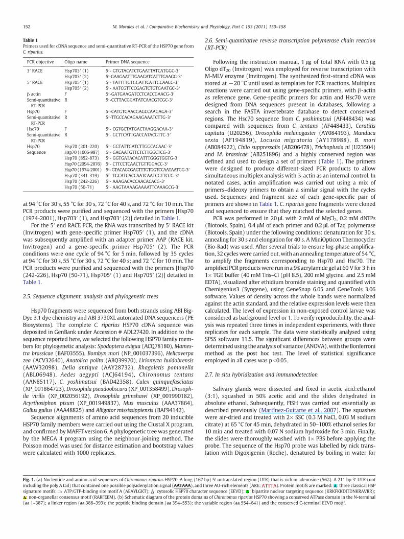
Table 1Primers used for cDNA sequence and semi-quantitative RT-PCR of the HSP70 gene fromC. riparius.
PCR objective Oligo name Primer DNA sequence
3′ RACE Hsp703′ (1) 5′- GTGTACATCTGAATTATCATGGC-3′Hsp703′ (2) 5′-GAAGAATTTGAAGATCATTTGAAGG-3′
5′ RACE Hsp705′ (1) 5′- TATTTTGTGGATTCATTGCAACC-3′Hsp705′ (2) 5′- AATCGTTCCGAGTCTGTGAATGC-3′
β actin F 5′-GATGAAGATCCTCACCGAACG-3′Semi-quantitativeRT-PCR
R 5′-CCTTACGGATATCAACGTCGC-3′
Hsp70 F 5′-CATGTGAACGAGCCAAGAGA-3′Semi-quantitativeRT-PCR
R 5′-TTGCCACAGAAGAAATCTTG-3′
Hsc70 F 5′- CGTGCTATGACTAAGGACAA-3′Semi-quantitativeRT-PCR
R 5′- GCTTCATTGACCATACGTTC-3′
Hsp70 Hsp70 (201-220) 5′- GGTATTGATCTTGGCACAAC-3′Sequence Hsp70 (1006-987) 5′- GACAATGTTCTCTTGGCTCG-3′
Hsp70 (852-873) 5′- GGTGATACACATTTGGGTGGTG-3′Hsp70 (2094-2076) 5′- CTTCCTCAACTGTTGGACC-3′Hsp70 (1974-2001) 5′-GTACAGCGAGTTTGTGGTCCAATAATGG-3′Hsp70 (341-319) 5′- TGCATCACCAATCAATCGTTCCG-3′Hsp70 (242-226) 5′- AAAGACACCAACACACG-3′Hsp70 (50-71) 5′- AAGTAAAAGAAAATTCAAAGCG-3′
152 M. Morales et al. / Comparative Biochemistry and Physiology, Part C 153 (2011) 150–158
at 94 °C for 30 s, 55 °C for 30 s, 72 °C for 40 s, and 72 °C for 10 min. ThePCR products were purified and sequenced with the primers [Hsp70(1974-2001), Hsp703′ (1), and Hsp703′ (2)] detailed in Table 1.
For the 5′ end RACE PCR, the RNA was transcribed by 5′ RACE kit(Invitrogen) with gene-specific primer Hsp705′ (1), and the cDNAwas subsequently amplified with an adapter primer AAP (RACE kit,Invitrogen) and a gene-specific primer Hsp705′ (2). The PCRconditions were one cycle of 94 °C for 5 min, followed by 35 cyclesat 94 °C for 30 s, 55 °C for 30 s, 72 °C for 40 s; and 72 °C for 10 min. ThePCR products were purified and sequenced with the primers [Hsp70(242-226), Hsp70 (50-71), Hsp705′ (1) and Hsp705′ (2)] detailed inTable 1.
2.5. Sequence alignment, analysis and phylogenetic trees
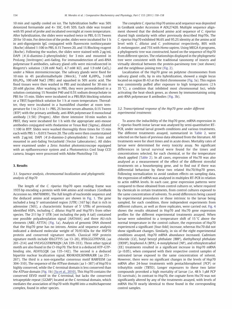
Hsp70 fragments were sequenced from both strands using ABI Big-Dye 3.1 dye chemistry and ABI 3730XL automated DNA sequencers (PEBiosystems). The complete C. riparius HSP70 cDNA sequence wasdeposited in GenBank under Accession # ADL27420. In addition to thesequence reported here, we selected the following HSP70 family mem-bers for phylogenetic analysis: Spodoptera exigua (ACQ78180), Mames-tra brassicae (BAF03555), Bombyx mori (NP_001037396), Helicoverpazea (ACV32640), Anatolica polita (ABQ39970), Liriomyza huidobrensis(AAW32098), Delia antiqua (AAY28732), Rhagoletis pomonella(ABL06948), Aedes aegypti (ACJ64194), Chironomus tentans(AAN85117), C. yoshimatsui (BAD42358), Culex quinquefasciatus(XP_001864723), Drosophila pseudoobscura (XP_001358499), Drosoph-ila virilis (XP_002056192), Drosophila grimshawi (XP_001990182),Acyrthosiphon pisum (XP_001949837), Mus musculus (AAA37864),Gallus gallus (AAA48825) and Alligator mississippiensis (BAF94142).
Sequence alignments of amino acid sequences from 20 inducibleHSP70 family members were carried out using the Clustal X program,and confirmed byMAFFT version 6. A phylogenetic tree was generatedby the MEGA 4 program using the neighbour-joining method. ThePoisson model was used for distance estimation and bootstrap valueswere calculated with 1000 replicates.
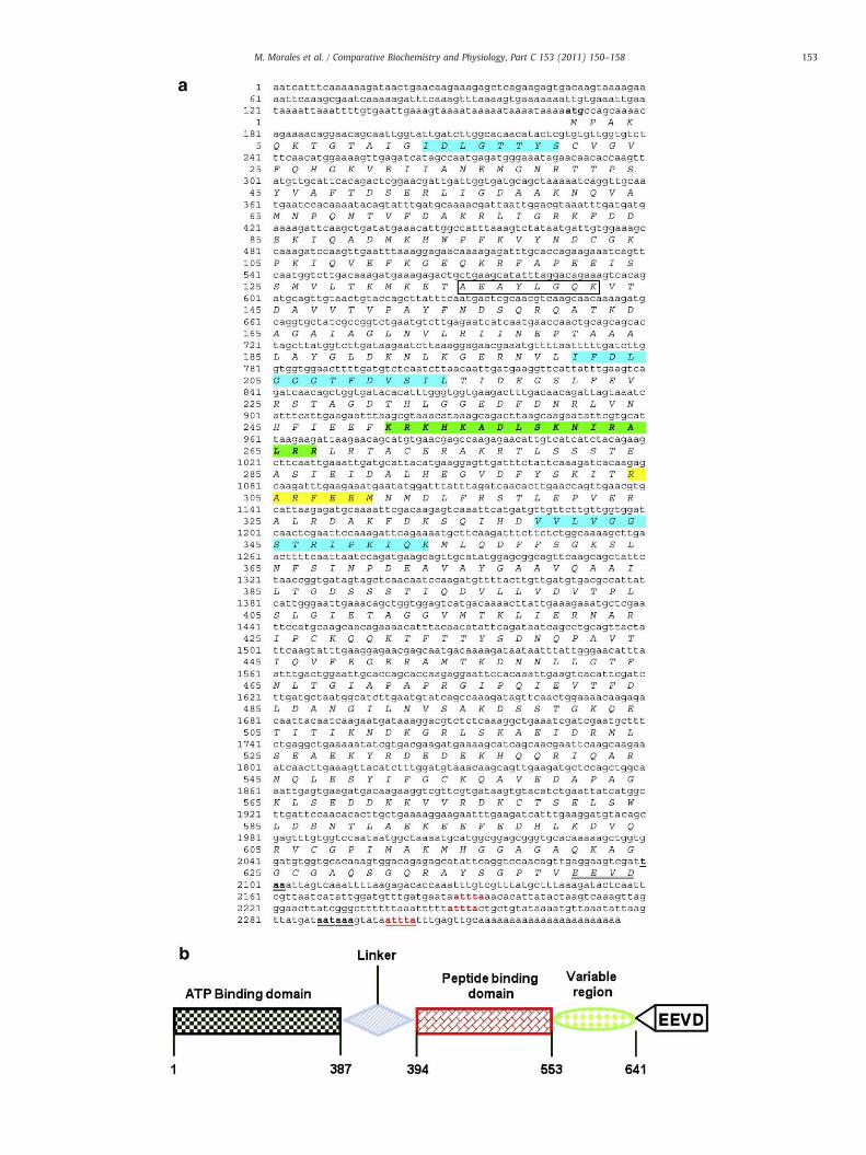
Fig. 1. (a) Nucleotide and amino acid sequences of Chironomus riparius HSP70. A long (167including the poly A tail) that contained one possible polyadenylation signal (AATAAA), and tsignature motifs; □: ATP/GTP-binding site motif A (AEAYLGKT); PPA: cytosolic HSP70 charact: non-organellar consensus motif (RARFEEM). (b) Schematic diagram of the protein domai
(aa 1–387); a linker region (aa 388–393); the peptide binding domain (aa 394–553); the v
2.6. Semi-quantitative reverse transcription polymerase chain reaction(RT-PCR)
Following the instruction manual, 1 μg of total RNA with 0.5 μgOligo dT20 (Invitrogen) was employed for reverse transcription withM-MLV enzyme (Invitrogen). The synthesized first-strand cDNA wasstored at −20 °C until used as templates for PCR reactions. Multiplexreactions were carried out using gene-specific primers, with β-actinas reference gene. Gene-specific primers for actin and Hsc70 weredesigned from DNA sequences present in databases, following asearch in the FASTA invertebrate database to detect conservedregions. The Hsc70 sequence from C. yoshimatsui (AF448434) wascompared with sequences from C. tentans (AF448433), Ceratitiscapitata (U20256), Drosophila melanogaster (AY084193), Manducasexta (AF194819), Locusta migratoria (AY178988), B. mori(AB084922), Chilo suppressalis (AB206478), Trichoplusia ni (U23504)and M. brassicae (AB251896) and a highly conserved region wasdefined and used to design a set of primers (Table 1). The primerswere designed to produce different-sized PCR products to allowsimultaneous multiplex analysis with β-actin as an internal control. Innotated cases, actin amplification was carried out using a mix ofprimers–dideoxy primers to obtain a similar signal with the cyclesused. Sequences and fragment size of each gene-specific pair ofprimers are shown in Table 1. C. riparius gene fragments were clonedand sequenced to ensure that they matched the selected genes.
PCR was performed in 20 μL with 2 mM of MgCl2, 0.2 mM dNTPs(Biotools, Spain), 0.4 μM of each primer and 0.2 μL of Taq polymerase(Biotools, Spain) under the following conditions: denaturation for 30 s,annealing for 30 s and elongation for 40 s. A MiniOpticon Thermocycler(Bio-Rad) was used. After several trials to ensure log-phase amplifica-tion, 32 cycleswere carried out,with an annealing temperature of 54 °C,to amplify the fragments corresponding to Hsp70 and Hsc70. Theamplified PCRproductswere run in a 9% acrylamide gel at 60 V for 3 h in1× TGE buffer (40 mM Tris–Cl (pH 8.5), 200 mM glycine, and 2.5 mMEDTA), visualized after ethidium bromide staining and quantified withChemigenius3 (Syngene), using GeneSnap 6.05 and GeneTools 3.06software. Values of density across the whole bands were normalizedagainst the actin standard, and the relative expression levels were thencalculated. The level of expression in non-exposed control larvae wasconsidered as background level or 1. To verify reproducibility, the anal-ysis was repeated three times in independent experiments, with threereplicates for each sample. The data were statistically analysed usingSPSS software 11.5. The significant differences between groups weredeterminedusing the analysis of variance (ANOVA),with theBonferronimethod as the post hoc test. The level of statistical significanceemployed in all cases was pb0.05.
2.7. In situ hybridization and immunodetection
Salivary glands were dissected and fixed in acetic acid:ethanol(3:1), squashed in 50% acetic acid and the slides dehydrated inabsolute ethanol. Subsequently, FISH was carried out essentially asdescribed previously (Martínez-Guitarte et al., 2007). The squasheswere air-dried and treated with 2× SSC (0.3 M NaCl, 0.03 M sodiumcitrate) at 65 °C for 45 min, dehydrated in 50–100% ethanol series for10 min and treated with 0.07 N sodium hydroxide for 3 min. Finally,the slides were thoroughly washed with 1× PBS before applying theprobe. The sequence of the Hsp70 probe was labelled by nick trans-lation with Digoxigenin (Roche), denatured by boiling in water for
bp) 5′ untranslated region (UTR) that is rich in adenosine (56%). A 211 bp 3′ UTR (nothree AU-rich elements (ARE; ). Protein motifs aremarked: : three classical HSPer sequence (EEVD); : bipartite nuclear targeting sequence (KRKFKKDITDNKRAVRR);ns of Chironomus riparius HSP70 showing a conserved ATPase domain in the N-terminalariable region (aa 554–641) and the conserved C-terminal EEVD motif.
153M. Morales et al. / Comparative Biochemistry and Physiology, Part C 153 (2011) 150–158
154 M. Morales et al. / Comparative Biochemistry and Physiology, Part C 153 (2011) 150–158
10 min and rapidly cooled on ice. The hybridization buffer was 50%deionized formamide and 4× SSC, 0.4% SDS. Each slide was treatedwith 95 pmol of probe and incubated overnight at room temperature.After hybridization, the slides were washed twice in PBS, 0.1% Tween20 for 10 min. For detection of the probe, slides were incubated for 1 hin anti-digoxigenin IgG conjugated with fluorescein isothiocyanate(Roche) diluted 1:100 in PBS, 0.1% Tween 20, and 1% Blocking reagent(Roche). Following the washes, the slides were stained with 2 μg/mLDAPI (4′,6-diamino-2-phenylindole) for 3 min and mounted inProLong (Invitrogen) anti-fading. For immunodetection of anti-RNApolymerase II antibodies, salivary gland cells were microdissected inRingertz's solution (128 mM NaCl, 4.7 mM KCl, and 1.9 mM CaCl2)under a Nikon stereomicroscope. The salivary glands were fixed for10 min in 4% paraformalhehyde (Merck), 7 mM K2HPO4, 3 mMKH2PO4, 100 mM NaCl (PBS) and squashed in 50% acetic acid. Thefixed tissues were then washed in PBS and incubated for 30 min in20 mM glycine. After washing in PBS, they were permeabilised in asolution containing 1% Nonidet P40 and 0.5% sodium deoxycholate inPBS for 15 min. Slides were incubated in a PBS/BSA blocking solutionor a TBST/Superblock solution for 1 h at room temperature. Thereaf-ter, they were incubated in a humidified chamber at room tem-perature for 1 to 2 h in 1× PBS, 2% bovine serum albumin, 0.1% Tween(BTP) with the primary antibody, anti-RNA polymerase II monoclonalantibody (1:50) (Progen). After three intensive 10 min washes inPBST, they were incubated for 1 h with the appropriate anti-mouseantibodies conjugated with rhodamine or Texas Red (Sigma), diluted1:100 in BTP. Slides were washed thoroughly three times for 15 mineachwith PBS 1×/0.01% Tween 20. The cells were then counterstainedwith 2 μg/mL DAPI (4′,6-diamino-2-phenylindole) for 5 min andmounted in anti-fading Vectashield (Vector Laboratories). All slideswere examined under a Zeiss Axiohot photomicroscope equippedwith an epifluorescence system and a Photometrics Cool Snap CCDcamera. Images were processed with Adobe PhotoShop 7.0.
3. Results
3.1. Sequence analysis, chromosomal localization and phylogeneticanalysis of Hsp70
The length of the C. riparius Hsp70 open reading frame was1935 bp encoding a protein with 644 amino acid residues (GenBankAccession no. HM769899). The full length of nucleotide sequence andthe deduced amino acid sequence are shown in Fig. 1. The geneincluded a long 5′ untranslated region (UTR) (167 bp) that is rich inadenosine (56%), a characteristic feature of 5′ UTRs of previouslyidentified HSPs, including C. dilutus Hsp70 and Hsp70′s from otherspecies. The 211 bp 3′ UTR (not including the poly A tail) containedone possible polyadenylation signal (AATAAA) and three AU-richelements (ARE; ATTTA) (Fig. 1a). Analysis of genomic DNA showedthat the Hsp70 gene has no introns. Amino acid sequence analysisindicated a deduced molecular weight of 70.93 KDa for the HSP70protein and conserved signature motifs. Classical HSP proteinsignature motifs include IDLGTTYS (aa 13–20), IFDLGGGTFDVSIL (aa201–214) and VVLVGGSTRIPKIQN (AA 339–353). Three other typicalmotifs are also found in the Cr-Hsp70. The first is a deduced ATP–GTP-binding site, AEAYLGQK (aa 135–142). The second is a deducedbipartite nuclear localization signal, RKHKADLSKNIRALRR (aa 251–267). The third is a non-organellar consensus motif RARFEEM (aa304–310). The sequence of the ATPase domain (aa 1–387) of Hsp70 ishighly conserved, while the C-terminal domain is less conserved thanthe ATPase domain (Fig. 1b) (Su et al., 2010). This Hsp70 contains theconserved EEVD motif in the C-terminal, but lacks the conservedtetrapeptide repeat (GGMP) located at the C-terminal domain, whichmediates the association of Hsp70 with Hsp90 into a multichaperonecomplex, found in other species.
The complete C. ripariusHsp70 amino acid sequencewas depositedin GenBank under Accession # ADL27420. Multiple sequence align-ment showed that the deduced amino acid sequence of C. ripariusshared high similarity with other previously described Hsp70s. TheC. riparius Hsp70 exhibited 96.6% and 95.2% identity at the amino acidlevel with C. dilutus and C. yoshimatsui respectively, 80% withD. melanogaster, and 75% with Homo sapiens. Using MEGA 4 programs,a phylogenetic tree was constructed, based on the sequence of Hsp70from different species. The relationships displayed in the phylogenetictree were consistent with the traditional taxonomy of insects andvirtually identical between the protein-parsimony tree (not shown)and the neighbour-joining tree (Fig. 2).
Localization of the Hsp70 gene on polytene chromosomes fromsalivary gland cells, by in situ hybridization, showed a single locuslocated on region III-A3 at the third chromosome (Fig. 3a). This regionwas consistently puffed after exposure to high temperatures (2 h35 °C), a condition that inhibited most chromosomal loci, whileactivating the heat-shock genes, as shown by immunostaining usinganti-RNA polymerase II antibodies (Fig. 3b).
3.2. Transcriptional response of the Hsp70 gene under differentexperimental treatments
To assess the inducibility of the Hsp70 gene, mRNA expression inC. riparius fourth instar larvae was analysed by semi-quantitative RT-PCR, under normal larval growth conditions and various treatments.The different treatments assayed, summarized in Table 2, wereselected on the basis of previous data obtained from the literature. Alltreatments were for short times and in acute doses. Mortality rates oflarvae were determined for every toxicity assay. No significantdifferences in larval survival were found for the times andconcentrations selected, for each chemical, or for the temperatureshock applied (Table 2). In all cases, expression of Hsc70 was alsoanalysed as a measurement of the effect of the different stressfultreatments on a housekeeping gene, and to find out if there wasdifferential behaviour by these two related Hsc70/Hsp70 genes.Following normalization to avoid random effects on sampling data,the expression of mRNA was analysed in multiplex RT-PCR in relationto actin mRNA levels. In each case, gene expression patterns werecompared to those obtained from control cultures or, where requiredby chemicals in certain treatments, from control cultures exposed tothe same concentration of solvents. To avoid differences being causedby experimental procedures or those intrinsic to the larvae beingsampled, for each condition, three independent experiments fromdifferent cultures, as well as three replicates, were carried out. Fig. 4shows the results obtained in Hsp70 and Hsc70 gene expressionprofiles for the different experimental treatments assayed. Whenlarvae were submitted to a temperature shift of 15 °C above thenormal temperature in the control culture, the expression of Hsp70experienced a significant (four-fold) increase, whereas Hsc70 did notshow significant changes. Similarly, in six of the eight experimentalconditions assayed, Hsp70 mRNA abundance increased. Cadmiumchloride (Cd), butyl benzyl phthalate (BBP), diethylhexyl phthalate(DEHP), bisphenol A (BPA), 4-nonylphenol (NP), and ethinylestradiol(EE) treatments resulted in a significant increase in Hsp70 mRNA(pb0.05), when compared with their respective control samples ofuntreated larvae exposed to the same concentration of solvent.However, there were no significant changes in the levels of Hsp70mRNA after 24-hour treatments with pentachlorophenol (PCP) ortributyltin oxide (TBTO). Longer exposures to these two lattercompounds provoked a high mortality of larvae (i.e. 48 h 1 μM PCP5% survivals). In contrast to Hsp70, the cognate form Hsc70 was notsignificantly altered by any of the treatments assayed, with levels ofmRNA Hsc70 nearly identical to those found in the correspondingcontrol samples.
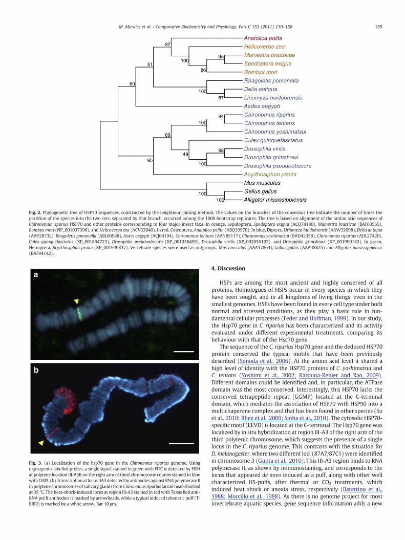
Fig. 2. Phylogenetic tree of HSP70 sequences, constructed by the neighbour-joining method. The values on the branches of the consensus tree indicate the number of times thepartition of the species into the two sets, separated by that branch, occurred among the 1000 bootstrap replicates. The tree is based on alignment of the amino acid sequences ofChironomus riparius HSP70 and other proteins corresponding to four major insect taxa. In orange, Lepidoptera, Spodoptera exigua (ACQ78180), Mamestra brassicae (BAF03555),Bombyx mori (NP_001037396), and Helicoverpa zea (ACV32640). In red, Coleoptera, Anatolica polita (ABQ39970). In blue, Diptera, Liriomyza huidobrensis (AAW32098), Delia antiqua(AAY28732), Rhagoletis pomonella (ABL06948), Aedes aegypti (ACJ64194), Chironomus tentans (AAN85117), Chironomus yoshimatsui (BAD42358), Chironomus riparius (ADL27420),Culex quinquefasciatus (XP_001864723), Drosophila pseudoobscura (XP_001358499), Drosophila virilis (XP_002056192), and Drosophila grimshawi (XP_001990182). In green,Hemiptera, Acyrthosiphon pisum (XP_001949837). Vertebrate species were used as outgroups: Mus musculus (AAA37864), Gallus gallus (AAA48825) and Alligator mississippiensis(BAF94142).
Fig. 3. (a) Localization of the hsp70 gene in the Chironomus riparius genome. Usingdigoxigenin-labelled probes, a single signal stained in green with FITC is detected by FISHat polytene location III-A3B on the right arm of third chromosome counterstained in bluewithDAPI. (b) Transcription at locus IIA3detectedbyantibodies against RNApolymerase IIin polytene chromosomes of salivary glands from Chironomus riparius larvae heat-shockedat 35 °C. The heat-shock-induced locus at region III-A3 stained in red with Texas Red anti-RNA pol II antibodies is marked by arrowheads, while a typical induced telomeric puff (T-BRIII) is marked by a white arrow. Bar 10 μm.
155M. Morales et al. / Comparative Biochemistry and Physiology, Part C 153 (2011) 150–158
4. Discussion
HSPs are among the most ancient and highly conserved of allproteins. Homologues of HSPs occur in every species in which theyhave been sought, and in all kingdoms of living things, even in thesmallest genomes. HSPs have been found in every cell type under bothnormal and stressed conditions, as they play a basic role in fun-damental cellular processes (Feder and Hoffman, 1999). In our study,the Hsp70 gene in C. riparius has been characterized and its activityevaluated under different experimental treatments, comparing itsbehaviour with that of the Hsc70 gene.
The sequence of the C. ripariusHsp70 gene and the deduced HSP70protein conserved the typical motifs that have been previouslydescribed (Sonoda et al., 2006). At the amino acid level it shared ahigh level of identity with the HSP70 proteins of C. yoshimatsui andC. tentans (Yoshimi et al., 2002; Karouna-Renier and Rao, 2009).Different domains could be identified and, in particular, the ATPasedomain was the most conserved. Interestingly, this HSP70 lacks theconserved tetrapeptide repeat (GGMP) located at the C-terminaldomain, which mediates the association of HSP70 with HSP90 into amultichaperone complex and that has been found in other species (Suet al., 2010; Rhee et al., 2009; Sinha et al., 2010). The cytosolic HSP70-specificmotif (EEVD) is located at the C-terminal. The Hsp70 genewaslocalized by in situ hybridization at region III-A3 of the right arm of thethird polytenic chromosome, which suggests the presence of a singlelocus in the C. riparius genome. This contrasts with the situation forD. melanogaster, where two different loci (87A7/87C1) were identifiedin chromosome 3 (Gupta et al., 2010). This III-A3 region binds to RNApolymerase II, as shown by immunostaining, and corresponds to thelocus that appeared de novo induced as a puff, along with other wellcharacterized HS-puffs, after thermal or CO2 treatments, whichinduced heat shock or anoxia stress, respectively (Barettino et al.,1988; Morcillo et al., 1988). As there is no genome project for mostinvertebrate aquatic species, gene sequence information adds a new
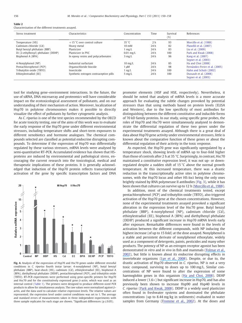
Table 2Characterization of the different treatments assayed.
Stress treatment Characteristics Concentration Time Survival(%)
References
Temperature (HS) +15 °C over control culture 35 °C 2 h 93 Morcillo et al. (1988)Cadmium chloride (Cd) Heavy metal 10 mM 24 h 82 Planelló et al. (2007)Butyl benzyl phthalate (BBP) Plasticizer 1 mg/L 24 h 83 Liu et al. (2009)Di (2-ethylhexyl) phthalate (DEHP) Plasticizer in PVC 0.01 mg/L 24 h 100 Park and Kwak (2008)Bisphenol A (BPA) In epoxy resins and polycarbonates 3 mg/L 24 h 96 Kang et al. (2007)
Segner et al. (2003)4-Nonylphenol (NP) Industrial surfactant 10 mg/L 24 h 85 Ha and Choi (2008)Pentachlorophenol (PCP) Organochloride biocide 1 μM 24 h 98 Fernández-Freire et al. (2005)Tributyltin oxide (TBTO) Insecticide 1 ng/L 24 h 100 Hahn and Schulz (2002)Ethinylestradiol (EE) Synthetic estrogen contraceptive pills 5 mg/L 24 h 100 Dussault et al. (2008)
Segner et al. (2003)
156 M. Morales et al. / Comparative Biochemistry and Physiology, Part C 153 (2011) 150–158
tool for studying gene–environment interactions. In the future, theuse of siRNA, DNA microarray and proteomics will have considerableimpact on the ecotoxicological assessment of pollutants, and on ourunderstanding of their mechanism of action. Moreover, localization ofHsp70 in polytene chromosomes makes it possible to directlyvisualize the effect of pollutants by further cytogenetic analysis.
As C. riparius is one of the test species recommended by the OECDfor acute toxicity testing, one of the aims of this work was to evaluatethe early response of the Hsp70 gene under different environmentalstressors, including temperature shifts and short-term exposures todifferent xenobiotics and hormone analogues. The chemical com-pounds selected are classified as potential endocrine disrupting com-pounds. To determine if the expression of Hsp70 was differentiallyregulated by these various stressors, mRNA levels were analysed bysemi-quantitative RT-PCR. Accumulated evidence has shown that HS-proteins are induced by environmental and pathological stress, en-couraging the current research into the toxicological, medical andtherapeutic implications of these proteins. It is generally acknowl-edged that induction of the Hsp70 protein reflects transcriptionalactivation of the gene by specific transcription factors and DNA
Fig. 4. Analysis of the expression of Hsp70 and Hsc70 genes under different stressfulconditions in C. riparius fourth instar larvae: 4-nonylphenol (NP), butyl benzylphthalate (BBP), heat-shock (HS), cadmium (Cd), ethinylestradiol (EE), bisphenol A(BPA), diethylhexyl phthalate (DEHP), pentachlorophenol (PCP), and tributyltin oxide(TBTO). RT-PCR experiments were performed using gene-specific primers for Hsp70and Hsc70 and for the constitutively expressed gene β-actin, which was used as aninternal control (Table 1). The primers were designed to produce different-sized PCRproducts to allow for simultaneous analysis. The raw values were normalized against β-actin, and the data used to calculate relative expression levels. The relative values arerepresented; the expression level under control conditions was set to 1. The averageand standard errors of measurements taken in three independent experiments withthree sample replicates for each stage are shown. *Significant differences (p≤0.05).
promoter elements (HSF and HSE, respectively). Nevertheless, itshould be noted that analysis of mRNA levels is a more accurateapproach for evaluating the subtle changes provoked by potentialstressors than that using methods based on protein levels (ELISAand Western), due to the low specificity of most antibodies fordistinguishing between the different constitutive and inducible formsof 70 kD family proteins. In our study, using specific gene probes, theroles of Hsp70 and Hsc70 were simultaneously analysed to demon-strate the differential regulation of these two genes under theexperimental treatments assayed. Although there is a great deal ofdata about Hsp70 gene activity under environmental stressors, little isknown about the comparative function of these genes or about thedifferential regulation of their activity in the toxic response.
As expected, the Hsp70 gene was significantly upregulated by atemperature shock, showing levels of mRNA up to four-fold higherthan those of controls after 2 h at 35 °C. Surprisingly, in contrast, Hsc70maintained a constitutive expression level, it was not up- or down-regulated despite a sudden shift of 15 °C above the normal growthtemperature. At this increased temperature, there was a drasticreduction in the transcriptionally active sites in polytene chromo-somes, with the Hsp70 locus and other HS-loci being the only onesbrightly stained by RNA polymerase II antibodies (Fig. 3), while it hasbeen shown that cultures can survive up to 12 h (Morcillo et al., 1988).
In addition, most of the chemical treatments tested, exceptpentachlorophenol (PCP) and tributyltin oxide (TBTO), also triggeredactivation of the Hsp70 gene at the chosen concentrations. However,none of the experimental treatments assayed provoked a significantalteration in the expression level of the Hsc70 gene. Butyl benzylphthalate (BBP), 4-nonylphenol (NP), cadmium chloride (Cd),ethinylestradiol (EE), bisphenol A (BPA) and diethylhexyl phthalate(DEHP) produced a significant increase in Hsp70 mRNA levels earlyafter exposure. Remarkable differences were found in the levels ofactivation between the different compounds, with NP inducing thehighest increase (of up to 15 fold) at the dose assayed. Nonylphenol isa stable and persistent derivate of nonylphenol ethoxylate, widelyused as a component of detergents, paints, pesticides and many otherproducts. The potency of NP as an estrogen receptor agonist has beendemonstrated in vitro and in vivo in fish and mammals (Folmar et al.,2002), but little is known about its endocrine disrupting effects ininvertebrate organisms (Lye et al., 2008). Despite, or due to, thedrastic activation of Hsp70 observed in C. riparius, NP is not a verytoxic compound, surviving to doses up to 100 mg/L. Similar con-centrations of NP were found to alter the expression of somehaemoglobin genes in this organism (Ha and Choi, 2008). DEHPinduced a lower (1.6×) but significant increase in Hsp70, and has alsopreviously been shown to increase Hsp90 and Hsp40 levels inC. riparius (Park and Kwak, 2008). DEHP is a widely used plasticizeroften found in freshwater systems and dominated the phthalateconcentrations (up to 8.44 mg/kg in sediments) evaluated in watersamples from Germany (Fromme et al., 2002). At the doses and

157M. Morales et al. / Comparative Biochemistry and Physiology, Part C 153 (2011) 150–158
temperature tested, the rest of the compounds induced an increase inHsp70 gene activity equivalent to that of a heat shock, the classicalinducer of these genes. Our study reinforces the interest of the Hsp70gene as a broad range marker of cellular stress (induced by metals,EDCs, herbicides and antibiotics), confirming previous data from ourand other laboratories (Lee et al., 2006; Karouna-Renier and Rao,2009; Park and Kwak, 2008; Planelló et al., 2008, 2010) in differentspecies of chironomids. Heavy metals are considered to be among themost consistent inducers of Hsp70 in the different organisms studiedto date (Gupta et al., 2010). Nevertheless, it is worth pointing out thatthere are some differences in the literature about the effect of otherchemical compounds on Hsp70 in different experimental systems. Forexample, and contrary to our data, NP was found to downregulate theHsp70 gene in the copepod Tigriopus whereas, as in our study, thisgenewas induced by BPA (Rhee et al., 2009). For a particular chemical,a similar mode of action at the cellular level is most likely, at least inrelated organisms; the different induction patterns might reflectdifferences in the previous basal levels of Hsps, due to culture andenvironmental conditions rather than a different mode of action.Induced thermotolerance and/or cross-tolerance to other stressorsshould also be considered, as this has long been demonstrated forheat-shock proteins in other species (Carretero et al., 1991).
Although the sensitivity of the cellular stress response makes theHsp70 gene attractive in ecotoxicology studies, the screening of aparticular Hsp may not provide a sufficiently sensitive bioindicator, asfor example in the cases of PCP and TBTO. Pentachlorophenol (PCP)belongs to the chlorophenol family; it is highly toxic and persists inthe environment. It has been widely used as a general biocide and,more particularly, as a wood preservative. Although there is evidenceof the effect of PCP on oxidative metabolism, no studies have yetexamined its direct effect on gene expression. PCP did not alter Hsp70gene levels in C. riparius after 24 h of exposure, although after afurther 24 h the larvae died (8% survival after 48 h). In a similar way, ithas been shown that HSP70 protein levels (measured by ELISA) didnot change in Raphidocelis subcapitata (Bierkens et al., 1998).Moreover, the TBTO treatments assayed did not activate the Hsp70gene, even though acute endocrine disrupting effects have beenpreviously reported for this compound in this species, differentiallyaffecting ecdysteroid synthesis and development in males andfemales (Hahn and Schulz, 2002). Although it is tempting to speculateabout the possible relationship between a lack of HSP70 and the highsensitivity and mortality induced by these two biocides, more indepth research is required to assess a causal relationship. In recentyears, it has been suggested that activation of heat-shock proteins,and in particular HSP70, in response to environmental insults mayplay an important role in protecting cells against a broad spectrum ofpotentially lethal pollutants (Gupta et al., 2010). Despite extensiveresearch, new data are still required to be able to generate a completepicture of Hsp70 regulation and its relationship to cellular toxicity.
In contrast to thedifferential effects observed inHsp70 activity, noneof the experimental treatments assayed provoked a significantalteration in the expression levels of the Hsc70 gene. These resultssuggest that the treatments assayed were not sufficiently severe to beable to inhibit a normally active gene, which seems to have a robustresistance to the effects of toxicants, and appears to be differentiallyregulated thanHsp70. Constitutive 70 kDproteins have “housekeeping”functions within a cell, related to protein assembly, folding, transloca-tion and denaturation (Feder and Hoffman, 1999). It has been shownthat some metals, such as copper, upregulate both Hsp70/Hsc70 genesin C. tentans (Karouna-Renier and Zehr, 2003; Karouna-Renier and Rao,2009).
Our results show that the Hsp70 genewas induced by the syntheticsteroid ethinylestradiol (EE) in Chironomus, which is consistent withsome reports of heat-shock protein expression induced by ecdysone inDrosophila and estrogen in mammals (Ryan and Hightower, 1998). Ithas been suggested that there may be a functional relationship
between steroid hormones (such as ecdysone in insects and estrogenin mammals) and heat-shock proteins. Indeed, it has been shown thatthree proteins, HSP90, HSC70 and HSP70, are components of thehormone nuclear receptor complex (Nolen and Morimoto, 2002).
A major advantage for using subcellular biomarkers, such as stressprotein genes, is an increased sensitivity and earlier response, whencompared to these characteristics for conventional parameters athigher levels of biological organization. However, the predictivepowermay be limited to the toxicant employed and themodel systemunder defined environmental conditions. As marked differences wereseen in the ability of relevant toxicants to induce Hsp70, a questionmark has been raised about the universal response of the Hsp70 geneto cytotoxicity. Different stressors can induce distinct stress responsepathways by targeting specific genes. More extensive research isrequired to increase the utility of heat-shock proteins in studiespredicting the level of toxicity based on stress gene expression inmodel organisms.
In summary, we found that Hsp70 gene expression in the aquaticlarvae of C. riparius is upregulated during exposure to a broad sample ofenvironmental pollutants classified as endocrine disruptors, includingmetals such as cadmium (Cd), organic man-made compounds, such aspolyphenols and phthalates (butyl benzyl phthalate, BBP; diethylhexylphthalate, DEHP; bisphenol A, BPA; andnonylphenol, NP), and syntheticsteroids (ethinylestradiol, EE),where neither the survival of the larvae isaffected nor the Hsc70 expression is altered by the exposure conditions.These results suggest that this gene could be useful as a rapid andsensitivemarker of exposure in ecotoxicological testing. However, othercompounds assayed, such as the biocide pentachlorophenol (PCP) andtributyltin oxide (TBTO), did not increase Hsp70 gene expression, noralter Hsc70mRNAbasal levels, but theywere able to kill the larvae somehours later. In conclusion, the Hsp70 gene in C. riparius showed adifferent susceptibility to the toxic effects of different environmentalpollutants. Its potential use for predicting the toxicity of chemicalsmerits further research to validate its sensitivity and specificity. Finally,gene expression studies can provide important insights into howgenomes react to stressful events and environmental hazards,which arelikely to be evenmore frequent as aquatic systems become increasinglypolluted.
Acknowledgements
The authors wish to thank Prof. José Luis Díez (Centro deInvestigaciones Biológicas, CSIC) for helpful discussions and Dr T.Carretero (University of Zaragoza) and Ted Cater for critical reading ofthe manuscript. This work was supported by grant CTM-2009-07189of the Ciencias y Tecnologías Medioambientales, Ministerio de Educacióny Ciencia, Spain.
References
Barettino, D., Morcillo, G., Díez, J.L., 1988. Induction of the heat-shock response bycarbon dioxide in Chironomus thummi. Cell Differ. 23, 27–36.
Bierkens, J., Maes, J., Vander Plaetse, F., 1998. Dose-dependent induction of heat shockprotein 70 synthesis in Raphidocelis subcapitata following exposure to differentclasses of environmental pollutants. Environ. Pollut. 101, 91–97.
Carretero, M.T., Carmona, M.J., Díez, J.L., 1991. Thermotolerance and heat shock proteinsin Chironomus. J. Insect Physiol. 37, 239–246.
Dang, W., Hu, Y.H., Zhang, M., Sun, L., 2010. Identification and molecular analysis of astress-inducible Hsp70 from Sciaenops ocellatus. Fish Shellfish Immunol. 29,600–607.
De Pomerai, D., 1996. Heat shock proteins as biomarkers of pollution. Hum. Exp.Toxicol. 15, 279–285.
Dussault, E.B., Balakrishnan, V.K., Solomon, K.R., Sibley, P.K., 2008. Chronic toxicity ofthe synthetic hormone 12alpha-ethinylestradiol to Chironomus tentans andHyalellaazteca. Environ. Toxicol. Chem. 27, 2521–2529.
EPA (US). 1996. Chironomid sediment toxicity test, ecological effects tests guidelines.EPA 712-C-96-313; 2nd ed. EPA 600/R-99/064, Washington DC, USA.
Feder, M.E., Hoffman, G.E., 1999. Heat-shock proteins, molecular chaperones and thestress response. Annu. Rev. Physiol. 61, 243–282.
Fernández-Freire, P., Labrador, V., Pérez Martín, J.M., Hazen, M.J., 2005. Cytotoxic effectsin mammalian Vero cell exposed to pentachlorophenol. Toxicology 210, 37–44.

158 M. Morales et al. / Comparative Biochemistry and Physiology, Part C 153 (2011) 150–158
Folmar, L.C., Hemmer, M.J., Denslow, N.D., Kroll, K., Chen, J., Cheek, A., Richman, H.,Meredith, H., Grau, E.G., 2002. A comparison of the estrogenic potencies ofestradiol, ethynylestradiol, diethylstilbestrol, nonylphenol and methoxychlor invivo and in vitro. Aquat. Toxicol. 60, 101–110.
Fromme, H., Kuchler, T., Otto, T., Pilz, K., Muller, J., Wenzel, A., 2002. Occurrence ofphthalates and bisphenol A and F in the environment. Water Res. 36, 1429–1438.
Gupta, S.C., Shrama, A., Mishra, M., Mishra, R.K., Chowdhuri, D.K., 2010. Heat shockproteins in toxicology: how close and how far? Life Sci. 86, 377–384.
Ha, M.H., Choi, J., 2008. Effects of environmental contaminants on hemoglobin of larvaeof aquatic midge, Chironomus riparius (Diptera: Chironomidae): a potentialbiomarker for ecotoxicity monitoring. Chemosphere 71, 1928–1936.
Hahn, T., Schulz, R., 2002. Ecdysteroid synthesis and imaginal disc development in themidge Chironomus riparius as biomarkers for endocrine effects of tributyltin.Environ. Toxicol. Chem. 21, 1052–1057.
Kang, J.H., Aasi, D., Katayama, Y., 2007. Bisphenol A in the aquatic environment and itsendocrine-disruptive effects on aquatic organisms. Crit. Rev. Toxicol. 37, 607–625.
Karouna-Renier, N.K., Rao, K.R., 2009. An inducible HSP70 gene from the midgeChironomus dilutus: characterization and transcription profile under environmentalstress. Insect Mol. Biol. 18, 87–96.
Karouna-Renier, N.K., Zehr, J.P., 2003. Short-term exposures to chronically toxic copperconcentrations induce HSP70 proteins in the midge larvae (Chironomus tentans).Sci. Total Environ. 312, 267–272.
Lee, S.M., Lee, S.B., Park, C.H., Choi, J., 2006. Expression of heat shock protein andhemoglobin genes in Chironomus tentans larvae exposed to various environmentalpollutants: a potential biomarker of freshwater monitoring. Chemosphere 65,1074–1081.
Liu, Y., Guan, Y., Yang, Z., Cai, Z., Mizuno, T., Tsuno, H., Zhu, W., Zhang, X., 2009. Toxicityof seven phthalate esters to embryonic development of the abalone Haliotisdiversicolor supertexta. Ecotoxicology 18, 293–303.
Lye, C.M., Bentley, M.G., Galloway, T., 2008. Effects of 4-nonylphenol on the endocrinesystem of the shore crab, Carcinus maenas. Environ. Toxicol. 23, 309–318.
Martinez, E.A., Moore, B.C., Schaumloffel, J., Dasgupta, N., 2003. Morphologicalabnormalities in Chironomus tentans exposed to cadmium and copper-spikedsediments. Ecotoxicol. Environ. Saf. 55, 204–212.
Martínez-Guitarte, J.L., Planelló, R., Morcillo, G., 2007. Characterization and expressionduring development and under environmental stress of the genes encodingribosomal proteins L11 and L13 in Chironomus riparius. Comp. Biochem. Physiol. B147, 590–596.
Mayer, M.P., Bukau, B., 2005. Hsp70 chaperones: cellular function and molecularmechanism. Cell. Mol. Life Sci. 62, 670–684.
Michailova, P., Petrova, N., Ilkova, J., Bovero, S., Brunetti, S., White, K., Sella, G., 2006.Genotoxic effect of copper on salivary gland polytene chromosomes of Chironomusriparius Meigen 1804 (Diptera, Chironomidae). Environ. Pollut. 144, 647–654.
Ming, J., Xie, J., Xu, P., Liu, W., Ge, X., Liu, B., He, Y., Cheng, Y., Zhou, Q., Pan, L., 2010.Molecular cloning and expression of two HSP70 genes in the Wuchang bream(Megalobrama amblycephala). Fish Shellfish Immunol. 28, 407–418.
Morcillo, G., Barettino, D., Carmona, M.J., Carretero, T., Díez, J.L., 1988. Telomeric DNAsequences differentially activated by heat shock in two Chironomus subspecies.Chromosoma 96, 139–144.
Morimoto, R.I., 1998. Regulation of the heat shock transcriptional response: cross talkbetween a family of heat shock factors, molecular chaperones, and negativeregulators. Genes Dev. 12, 3788–3796.
Nolen, E.A.A., Morimoto, R.I., 2002. Chaperoning signalling pathways: molecularchaperones as stress-sensing heat shock proteins. J. Cell Sci. 115, 2809–2816.
OECD, 2001. Organization for Economic Cooperation and Development. Guidelinefor testing of chemicals, sediment-water chironomid toxicity test using spikedsediment. 218.
Park, K., Kwak, I.S., 2008. Characterization of heat shock protein 40 and 90 inChironomus riparius larvae: effects of di(2-ethylhexyl) phthalate exposure on geneexpressions and mouthpart deformities. Chemosphere 74, 89–95.
Planelló, R., Martínez-Guitarte, J.L., Morcillo, G., 2007. Ribosomal genes as early targetsof cadmium-induced toxicity in Chironomus riparius larvae. Sci. Total Environ. 373,113–121.
Planelló, R., Martínez-Guitarte, J.L., Morcillo, G., 2008. The endocrine disruptorbisphenol A increases the expression of HSP70 and ecdysone-receptor genes inthe aquatic larvae of Chironomus riparius. Chemosphere 71, 1870–1876.
Planelló, R., Martínez-Guitarte, J.L., Morcillo, G., 2010. Effect of acute exposure tocadmium on the expression of heat-shock and hormone-nuclear receptor genes inthe aquatic midge Chironomus riparius. Sci. Total Environ. 408, 1598–1603.
Powers, M.V., Workman, P., 2007. Inhibitors of the heat shock response: biology andpharmacology. FEBS Lett. 581, 3758–3769.
Rhee, J.S., Raisuddin, S., Lee, K.W., Seo, J.S., Ki, J.S., Kim, I.C., Park, H.G., Lee, J.S., 2009. Heatshock protein (Hsp) gene responses of the intertidal copepod Tigriopus japonica toenvironmental toxicants. Comp. Biochem. Physiol. C 149, 104–112.
Ryan, J.A., Hightower, L.E., 1998. Heat shock proteins: molecular biomarkers of effects.In: Puga, A., Wallale, K.B. (Eds.), Molecular Biology of the Toxic Response. HamiltonPrinting Co., Castlton, New York, pp. 449–466.
Segner, H., Carroll, K., Fenske, M., Janssen, C.R., Maack, G., Pascoe, D., Schäfers, C.,Vanderbergh, G.F., Watts, M., Wenzel, A., 2003. Identification of endocrine-disrupting effects in aquatic vertebrates and invertebrates: report from theEuropean IDEA project. Ecotoxicol. Environ. Saf. 54, 302–314.
Simoncelli, F., Morosi, L., Di Rosa, I., Pascolini, R., Fagotti, A., 2010. Molecularcharacterization and expression of a heat-shock cognate 70 (Hsc70) and a heat-shock protein 70 (Hsp70) cDNAs in Rana (Pelophylax) lessonae embryos. Comp.Biochem. Physiol. A 156, 552–560.
Sinha, A.K., Vanparys, C., De Boeck, G., Kestemont, P.,Wang,N., Phuong, N.T., Scippo,M.L.,De Coen, W., Robbents, J., 2010. Expression characteristics of potential biomarkergenes in Tra catfish, Pangasianodon hypophthalmus, exposed to trichlorfon. Comp.Biochem. Physiol. D 5, 207–216.
Snell, T.W., Brogdon, S.E., Morgan, M.B., 2003. Gene expression profiling inecotoxicology. Ecotoxicology 12, 475–483.
Sonoda, S., Fukumoto, K., Izumi, Y., Yoshida, H., Tsumuki, H., 2006. Cloning of heat shockprotein genes (hsp90 and hsc70) and their expression during larval diapause andcold tolerance acquisition in the rice stem borer, Chilo suppressalis Walker. Arch.Insect Biochem. Physiol. 62, 36–47.
Su, X., Du, L., Li, Y., Li, Y., Zhou, J., Li, T., 2010. Cloning and expression of the HSP70 geneof sipincula Phascolosoma esculenta. Fish Shellfish Immunol. 28, 461–466.
Waagner, D., Heckmann, L.H., Malmendal, A., Nielsen, N.C., Holmstrup, M., Bayley, M.,2010. Hsp70 expression and metabolite composition in response to short-termthermal changes in Folsomia candida (Collembola). Comp. Biochem. Physiol. A 157,177–183.
Yoshimi, T., Minowa, K., Karouna-Renier, N.K., Watanabe, C., Sugaya, Y., Miura, T., 2002.Activation of stress-induced gene by insecticides in the midge Chironomusyoshimatsui. J. Biochem. Mol. Toxicol. 16, 10–17.
Young, J.C., 2010. Mechanisms of the Hsp70 chaperone system. Biochem. Cell Biol. 88,291–300.
Zhanng, Q., Denlinger, D.L., 2010. Molecular characterization of heat shock protein 90,70 and 70 cognate cDNAs and their expression patterns during thermal stress andpupal diapauses in the corn earworm. J. Insect Physiol. 56, 138–150.