009 through the inhibition of Th1 and Th17 cells differentiation
Upload
independentCategory
view
0download
0
Induction of hsp70-Mediated Th17 Autoimmunity Can Be Exploited
as Immunotherapy for Metastatic Prostate Cancer
Timothy Kottke,1Luis Sanchez-Perez,
1,2Rosa Maria Diaz,
1Jill Thompson,
1Heung Chong,
4
Kevin Harrington,5Stuart K. Calderwood,
6Jose Pulido,
3Nick Georgopoulos,
7
Peter Selby,7Alan Melcher,
7and Richard Vile
1,2,7
1Molecular Medicine Program, and Departments of 2Immunology and 3Ophthalmology and Optical Oncology, Mayo Clinic, Rochester,Minnesota; 4St George’s Hospital Medical School, and 5Institute of Cancer Research, London, United Kingdom; 6Harvard MedicalSchool, Beth Israel Deaconess Medical Center, Boston, Massachusetts; and 7Cancer Research UK Clinical Centre, Leeds TeachingHospitals NHS Trust and Leeds Institute of Molecular Medicine, University of Leeds, Leeds, United Kingdom
Abstract
A close connectivity between autoimmune and tumor rejec-tion responses is known to exist in the case of melanomaimmunotherapy. However, relatively little is known about self-antigens on other types of normal cells, their relation to thedevelopment of autoimmune disease, and their possible co-existence as potential tumor rejection antigens on associatedtumors. In the current study, we induced inflammatory kill-ing of normal prostate tissue in situ using a fusogenic mem-brane glycoprotein along with the immune adjuvant hsp70.We show here that, in the prostate, hsp70 induces interleukin(IL)-6, which triggers a CD4- and CD8-dependent progressiveautoimmune reactivity, associated with IL-17 expression. Thisautoimmune response was also able to induce the rejectionof established prostate tumors, but not other histologic typesof tumors, growing elsewhere in the animal. These data showthat the intimate connectivity between autoimmune and tumorrejection responses extends beyond the classic melanomaparadigm and may be clinically valuable for the treatmentof established metastatic disease of the prostate. [Cancer Res2007;67(24):11970–9]
Introduction
A close link between development of tumor immunity, withconcomitant induction of autoimmune reactivities, has been well-documented in both preclinical and clinical model systems. This isparticularly true for melanoma, where many melanoma antigensare known to be unaltered self-proteins of melanocytes (1, 2).Hence, successful immune therapies against melanoma also runthe risk of developing immune reactivity against normal melano-cytes (1, 3). There is frequently—but not always (4)—a correlationbetween the induction of tumor immunity and concomitantdevelopment of autoimmune pathology, most principally vitiligo,correlated with CD8, CD4, and humoral responses to melanomaantigens (1, 2, 5, 6). Strategies that enhance the potency of anti-tumor T-cell responses often also increase the severity of auto-immune variables (5–7). The positive prognostic importance of the
development of autoimmune disease in patients undergoing immu-notherapy for melanoma has recently been convincingly shown ina large-scale clinical trial (8).
The established paradigm for tumor vaccination uses tumor cell–derived immunogens to raise T-cell responses against tumor anti-gens that may then be accompanied by autoimmune antimelanocyteresponses (2, 9). Often, such vaccines require a potent adjuvantto promote the breaking of tolerance to tumor-derived antigens. Inthis respect, we (10–12) and others (13–18) have shown that hsp70can act as powerful inducer of tumor immunity and convertstolerogenic antigen presentation into immunostimulatory presen-tation to break tolerance and induce autoimmune disease (19). Heatshock proteins can enhance immunogenicity as chaperones ofimmunogenic peptides (15–17), cytokines (17, 18), immunogens (20),and their ability to mature dendritic cells (19). We have also shownthat hsp70 induces proinflammatory cytokines from monocytes (12)after ligation to receptors including Toll-like receptors (TLR) 2 and 4(18, 21), and that it also acts directly upon macrophages to suppresssecretion of interleukin (IL)-10 and phagocytosis (12), and that hsp70is a key mediator of immunogenic cell death (10–12), therebymediating effective tumor vaccination (10–12).
However, obtaining tumor cells, lysates, or derivatives; identify-ing relevant tumor antigens; and developing vectors that canspecifically target systemic tumors in vivo can be time consuming,expensive, and problematic. In this respect, we recently described acompletely novel approach to tumor vaccine design by targetingreadily accessible normal tissues in situ as a source of immunogen(9). We hypothesized that by inducing ‘‘stressful death’’ of normalcells (melanocytes), it would be possible to generate autoimmuneresponses, which may then be effective against tumor cells(melanoma) that share antigens with the normal tissue (1, 3). Weshowed that killing normal melanocytes, in the presence of thepotent immune adjuvant hsp70 (10–12, 19, 22, 23), generates T-cellreactivity against established melanomas. Intradermal plasmidDNA injections of a transcriptionally targeted cytotoxic gene [tyro-sinase promoter driving the expression of the herpes simplex virusthymidine kinase (HSVtk) gene], along with a plasmid expressinghsp70 (10), induced direct in vivo inflammatory killing of normalmelanocytes, broke tolerance to self-antigens, and led to rejectionof systemic tumors (21, 24, 25). In this system, hsp70 acts throughTLR-4 signaling and local induction of tumor necrosis factor-a topromote migration of antigen presenting cells to the lymph node(21). Progressive autoimmune disease was inhibited (24, 25)because the antimelanocyte CD8+ T-cell response was rapidlysuppressed by regulatory T cells (24–26). In addition, in suboptimaltherapeutic protocols, the T-cell response selected aggressive,amelanotic, antigen loss B16 tumor variants (24, 25). Moreover,
Note: T. Kottke and L. Sanchez-Perez contributed equally to this work. A. Melcherand R. Vile contributed equally as senior authors of this work.
Current address for L. Sanchez-Perez: National Cancer Institute, CRC-Room 3-5816,10 Center Drive MSC 1201, Bethesda, MD 20892.
Requests for reprints: Richard G. Vile, Molecular Medicine Program, Mayo Clinic,Guggenheim 1836, 200 1st Street Southwest, Rochester, MN 55902. Phone: 507-284-9941; Fax: 507-266-2122; E-mail: [email protected].
I2007 American Association for Cancer Research.doi:10.1158/0008-5472.CAN-07-2259
Cancer Res 2007; 67: (24). December 15, 2007 11970 www.aacrjournals.org
Research Article
Research. on January 20, 2015. © 2007 American Association for Cancercancerres.aacrjournals.org Downloaded from

addition of a plasmid expressing CD40L increased antitumorefficacy, generated potent immunologic memory but also inducedaggressive autoimmunity (21).
From these, and other data, it is clear that self-reactive T cellsexist in the periphery that have escaped thymic deletion and can,if correctly activated, kill both melanoma cells and normal mela-
nocytes (1, 2, 6, 8). However, there is a relative paucity of dataconcerning the link between autoimmune responses to othertissues and their relevance to generating immune responsesagainst tumors of the same histologic type. In particular, loss ofmelanocytes is not life threatening and can be toleratedconsiderably better than metastatic melanoma (8). In the current
Figure 1. Ad-VSV-G+Adhsp70 treatment ofthe prostate is associated with ongoingautoimmune destruction. A, C57Bl/6mice under anesthetics were injectedintraprostatically with 109 plaque-forming unit(pfu) of Ad-GFP or Ad-VSV-G (27–29, 31,46–50), or Ad-hsp70, or with 5 � 108 pfuAd-VSV-G+5 � 108 pfu Ad-hsp70. Aftersurgery, mice were recovered and wereeuthanized 1 wk after the intraprostaticinjection of viruses. Prostates were analysedhistologically using H&E sections. Slideswere scored by two independent pathologistsblinded to the experimental groups forinfiltration and necrosis. Ad-hsp70 inducedmoderate levels of inflammatory infiltrate(top right ) but extensive immune infiltration andnecrosis were recorded for prostates injectedwith Ad-VSV-G+Adhsp70 (bottom right ).Syncytia were only seen in prostates injectedwith Ad-VSV-G (bottom left ). Essentiallynormal prostate architecture was recordedfrom prostates injected with Ad-GFP (top left ).B, 45 d after intraprostatic injections asdescribed in A , animals were euthanizedand prostates were recovered and weighed.Mean weights of prostates from animals ofdifferent treatment groups are shown.
Autoimmunity and Antitumor Immunity in Prostate
www.aacrjournals.org 11971 Cancer Res 2007; 67: (24). December 15, 2007
Research. on January 20, 2015. © 2007 American Association for Cancercancerres.aacrjournals.org Downloaded from
report, we investigated whether the principles that we observed inthe melanocyte/melanoma system also hold true for the prostate.In response to the intentional induction of inflammatory killing ofthe prostate, we observed progressive autoimmune destruction ofthe prostate in response to the intentional induction of inflam-matory killing, associated with induction of IL-6 and a Th-17response but with no detectable Treg induction. Correspondingly,inflammatory killing of normal prostate was highly effective atcuring established metastatic prostatic tumors but not tumors ofa different histologic type. Our results are significant in severalrespects. They show that autoimmune disease of the prostate canbe induced by specific cytokine responses to one, or a few, keypathogenic-like signals. They show that the intimate connectivitybetween autoimmune and antitumor rejection responses extendsbeyond the classic melanoma paradigm; and they suggest thatthe principle of inflammatory killing of normal cells to treatneoplastic disease is applicable to tumors other than justmelanoma.
Materials and Methods
Cell lines, plasmids, and viruses. Transgenic adenocarcinoma of the
mouse prostate (TRAMP)-C2 (TC2) cells are derived from a prostate tumor
that arose in a TRAMP mouse and were characterized by Dr. Esteban
Celis. These cell lines express a variety of prostate-specific genes, including
PSMA, Hoxb-13 , and NKX3.1 . TC2 cells grow in an androgen-independent
manner and have a reduced level of expression of MHC class I which can
be up-regulated by IFN-g, making them susceptible to specific lysis by
CTL. We routinely grow TC2 tumors in C57Bl/6 male mice. The murine
melanoma B16.F1 tumor cell line has been previously described (27). Cell
lines were grown in DMEM (Life Technologies) supplemented with 10%
(v/v) FCS (Life Technologies) and L-glutamine (Life Technologies). All cell
lines were monitored routinely and found to be free of Mycoplasma
infection.
The replication-defective adenoviral vectors used in this study were all
E1 deleted serotype 5 vectors that contains the cytomegalovirus (CMV)immediate-early gene promoter-enhancer driving the inserted transgene.
Ad-VSV-G expresses the cDNA of the fusogenic membrane G glycoprotein of
vesicular stomatitis virus (VSV-G; ref. 27–29); Ad-hsp70 contains the cDNA
of the inducible murine heat shock protein 70 gene (30); and Ad-GFPcontains the cDNA of the green fluorescent protein gene (31).
In the CMV-ova plasmid (CMV-ova), the ovalbumin gene (27) is driven by
the CMV promoter in pCR3.1 (Invitrogen).
Histopathology of tumor sections. Prostates were harvested and fixedin 10% Formalin in PBS, then paraffin embedded and sectioned. H&E-
stained sections were prepared for analysis of tissue destruction and gross
infiltrate. Two independent pathologists examined H&E sections, blinded to
the experimental design, and scored the degree of necrosis.Reverse transcriptase PCR. Organ samples were snap frozen in liquid
nitrogen. RNA was prepared with the Qiagen RNA extraction kit. One
microgram of total cellular RNA was reverse transcribed in a 20 AL volumeusing oligo-(dT) as a primer. A cDNA equivalent of 1 ng RNA was amplified
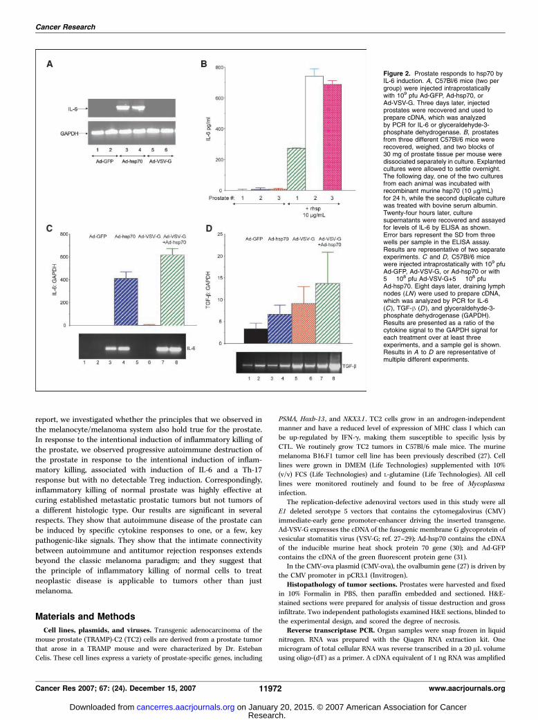
Figure 2. Prostate responds to hsp70 byIL-6 induction. A, C57Bl/6 mice (two pergroup) were injected intraprostaticallywith 109 pfu Ad-GFP, Ad-hsp70, orAd-VSV-G. Three days later, injectedprostates were recovered and used toprepare cDNA, which was analyzedby PCR for IL-6 or glyceraldehyde-3-phosphate dehydrogenase. B, prostatesfrom three different C57Bl/6 mice wererecovered, weighed, and two blocks of30 mg of prostate tissue per mouse weredissociated separately in culture. Explantedcultures were allowed to settle overnight.The following day, one of the two culturesfrom each animal was incubated withrecombinant murine hsp70 (10 Ag/mL)for 24 h, while the second duplicate culturewas treated with bovine serum albumin.Twenty-four hours later, culturesupernatants were recovered and assayedfor levels of IL-6 by ELISA as shown.Error bars represent the SD from threewells per sample in the ELISA assay.Results are representative of two separateexperiments. C and D, C57Bl/6 micewere injected intraprostatically with 109 pfuAd-GFP, Ad-VSV-G, or Ad-hsp70 or with5 � 108 pfu Ad-VSV-G+5 � 108 pfuAd-hsp70. Eight days later, draining lymphnodes (LN) were used to prepare cDNA,which was analyzed by PCR for IL-6(C ), TGF-h (D ), and glyceraldehyde-3-phosphate dehydrogenase (GAPDH).Results are presented as a ratio of thecytokine signal to the GAPDH signal foreach treatment over at least threeexperiments, and a sample gel is shown.Results in A to D are representative ofmultiple different experiments.
Cancer Research
Cancer Res 2007; 67: (24). December 15, 2007 11972 www.aacrjournals.org
Research. on January 20, 2015. © 2007 American Association for Cancercancerres.aacrjournals.org Downloaded from

by PCR for a variety of murine cytokines or vector-derived transgenes asdescribed previously (27, 32) (details of the primers upon request).
Treg-mediated inhibition of IFN; secretion from activated T cells.OT-I mice are transgenic mice whose T cells express the Va2 chain of the
transgenic OT-I T-cell receptor that specifically recognizes the SIINFEKLpeptide from the chicken ovalbumin protein (ova) in the context of H-2Kb
as expressed by B16ova tumor cells (33). For preparation of naive OT-I T
cells, spleen and lymph nodes from OT-I–transgenic mice were combined
and crushed through a 100-Am filter to prepare a single-cell suspension.RBC were removed by a 2-min incubation in ACK buffer (sterile dH2O
containing 0.15 mol/L NH4Cl, 1.0 mmol/L KHCO3, and 0.1 mmol/L EDTA
adjusted to pH 7.2–7.4). OT-I T cells were activated by incubation of
splenocyte populations with the cognate antigen recognized by the OT-I Tcells. Single-cell suspensions from spleen and lymph nodes were adjusted to
1.0 � 106 cells/mL in Iscove’s modified Dulbecco’s medium plus 5% FCS,
105 mol/L 2-ME, 100 units/mL penicillin, and 100 Ag/mL streptomycin and
stimulated with 1 Ag/mL SIINFEKL peptide and 50 IU/mL human IL-2(Mayo Clinic Pharmacy). This routinely induces large amounts of IFN-g to
be expressed from the activated OT-I T cells.
To assay for the presence of T-cell suppressive (Treg) activity within
splenocyte populations from intraprostatically injected mice, 250,000 freshly
harvested splenocytes from treatment groups were plated along with 105
naive OT-1 CD8+ T cells in the presence of either no added peptide, an irre-
levant nonactivating peptide (TRP-2 180188 SVYDFFVWL; ref. 34), or with the
Figure 3. IL-17 expression is induced by inflammatory killing of prostate without induction of Treg. A, C57Bl/6 mice were injected intraprostatically with 109 pfu Ad-GFP,Ad-VSV-G, or Ad-hsp70 or with 5 � 108 pfu Ad-VSV-G+5 � 108 pfu Ad-hsp70. Eight days later, the injected prostates were used to prepare cDNA, which was analyzedby PCR for IL-17 or GAPDH. Results are presented as a ratio of the cytokine signal to the GAPDH signal for each treatment over at least three experiments, anda sample gel is shown. B, the cDNA obtained from the experiment described in Fig. 2B (lymph node draining the prostates injected with adenoviral vectors)was also screened for expression of IL-17 as shown. The GAPDH control in this experiment is the same as in Fig. 2B . In addition, C57Bl/6 mice were treatedintraprostatically with 109 pfu Ad-GFP, Ad-hsp70, Ad-VSV-G, or 5 � 108 pfu Ad-VSV-G+5 � 108 pfu Ad-hsp70. Eight days later, 106 cells from the draining lymphnodes were cultured for 24 h. Supernatants were assayed for IL-17 using ELISA (eBioscience).*, positive on 30 additional cycles. C, C57Bl/6 mice were injectedintraprostatically with no virus (lane 1) or with 109 pfu of Ad-GFP (lane 2); intradermally with the Tyr-HSVtk/CMV-hsp70 plasmid combination that we have previouslyshown induces Treg cells (refs. 24, 25; lane 3) or with 5 � 108 pfu Ad-VSV-G+5 � 108 pfu Ad-hsp70 (lanes 4–6 ). Fourteen days postviral or plasmid injection (lanes 1–3,7, 8), or 4 or 41 days postviral injections (lanes 4 and 6), splenocytes were recovered from treated mice and 250,000 were plated with 105 naı̈ve OT1 CD8+ Tcells withH2Kb-restricted ova peptide SIINFEKL in triplicate, and 48 h later, supernatants were assayed by ELISA for IFN-g. Lane 7, OT-1 with no added splenocytes; lane 8;OT-1 with the nonactivating TRP-2 peptide instead of SIINFEKL. D, C57Bl/6 mice under anesthetics were injected intraprostatically with 0.1 mg of a plasmid expressingthe cDNA for chick ovalbumin along with 109 pfu of Ad-VSV-G or Ad-hsp70, or along with 5 � 108 pfu Ad-VSV-G+5 � 108 pfu Ad-hsp70. All groups received ani.p. injection of either a control immunoglobulin (Ig ) or the anti-CD25 Treg-depleting PC61 antibody (0.5 mg) 2 d before the intraprostatic plasmid/viral injection.After surgery, the mice recovered. One week later, 500,000 splenocytes per treatment group were harvested and cocultured with the H-2Kb restricted ova peptideSIINFEKL in triplicate, and 48 h later, supernatants were assayed by ELISA for IFN-g. Results shown are representative of two different experiments.
Autoimmunity and Antitumor Immunity in Prostate
www.aacrjournals.org 11973 Cancer Res 2007; 67: (24). December 15, 2007
Research. on January 20, 2015. © 2007 American Association for Cancercancerres.aacrjournals.org Downloaded from

synthetic H-2Kb–restricted ova peptide SIINFEKL (33) in tissue culture
wells. Splenocyte/OT-I cocultures were stimulated in triplicate and super-
natants were assayed for IFN-g production by ELISA. The degree of sup-
pressive activity in the test splenocyte cultures is reflected by their ability to
inhibit the IFN-g response of the naı̈ve OT-I T cells when presented with
their cognate, activating SIINFEKL antigen. The dependence of any such
T-cell suppressive activity on expression of transforming growth factor-h(TGF-h; ref. 35) was assayed using the recombinant human TGF-h sRII/Fcchimera (R&D Systems), a 159 amino acid extracellular domain of human
TGF-h receptor type II fused to the Fc region of human IgG1.
Antigen priming assays—splenocyte preparation and antigenpresentation. Splenocytes enriched in lymphocytes were prepared from
spleens from treated/vaccinated animals by standard techniques (36).Freshly purified splenocyte populations were washed in PBS and either
incubated with target tumor cells (TC2 or B16) typically at ratios of 100:1,
10:1, or 1:1 or, where appropriate, were pulsed with 1 Ag/mL of the target
peptide for which antigen specificity of response was being tested [SIINFEKLfor induced responses to ova (33) or TRP-2180188 SVYDFFVWL (34) as the
negative irrelevant antigen control]. Forty-eight to seventy-two hours later,
cell-free supernatants were tested for IFN-g by ELISA (PharMingen). Thesynthetic H-2Kb–restricted peptides TRP-2180188 SVYDFFVWL (34) and Ova
SIINFEKL (33) were synthesized at the Mayo Foundation Core facility.
ELISA analysis for IFN; secretion. For ELISA, cell-free supernatants
were collected from sample wells and tested by specific ELISA for IFN-g orIL-6 (BD OptEIA IFN-g; BD Biosciences) or IL-17 (R&D Systems) according
to the manufacturers’ instructions.
In vivo studies. All procedures were approved by the Mayo Foundation
Institutional Animal Care and Use Committee. C57Bl/6 mice or B6.129S2-IL6tm1Kopf/J [IL-6 knockout (IL-6KO); Jackson; No.002650] were purchased
from The Jackson Laboratory at ages 6 to 8 weeks. To establish s.c. tumors,
2 � 105 B16 cells or 2 � 106 TC2 cells, in 100 AL of PBS were injected intothe flank of mice. Intraprostatic injections (50 AL) were performed on mice
under anesthetic, typically at day 6 after tumor establishment. For survival
studies, tumor diameter in two dimensions was measured thrice weekly
using calipers, and mice were killed when tumor size was f1.0 � 1.0 cm intwo perpendicular directions.
Immune cell depletions were performed by i.p. injections (0.1 mg per
mouse) of anti-CD8 (Lyt 2.43) and anti-CD4 (GK1.5), both from the
Monoclonal Antibody Core Facility, Mayo Clinic; and IgG control(ChromPure Rat IgG; Jackson ImmunoResearch) at day 4 after tumor
implantation and then weekly thereafter. For Treg depletion, 0.5 mg of PC-
61 antibody (Monoclonal Antibody Core Facility, Mayo Clinic) per mouse
was given i.p. 4 days after tumor implantation and 2 days before the firstviral injection. Fluorescence-activated cell sorting analysis of spleens and
lymph nodes confirmed subset specific depletions.
Statistics. Survival data from the animal studies was analyzed using thelog-rank test (37), and the two-sample unequal variance Student’s t test
analysis was applied for in vitro assays. Statistical significance was deter-
mined at the level of P value of <0.05.
Results
Inflammatory killing of normal prostate inducesongoing auto-immune destruction. Previously, we used plasmids expressing theHSVtk suicide gene and hsp70 to target killing of normal melanocytesin the skin (21, 24, 25). Because HSVtk requires active division of targetcells for cytotoxicity, in the current studies, we used an adenoviralvector expressing VSV-G, the fusogenicmembrane glycoprotein (FMG)from VSV (27), to induce killing of normal prostate cells. We havepreviously shown that killing induced by fusion of cells using viral FMGcan be potently immunogenic through the fusion of cells intomultinucleated syncytia (27, 28). In addition, we used a secondadenoviral vector to express the murine hsp70 gene (30).
Direct intraprostatic injection of an adenovirus-expressing GFP(Ad-GFP) did not induce any detectable lasting damage to theprostates of C57Bl/6 mice either in terms of the architecture of
the organ or immune infiltration (Fig. 1A). Injection of Ad-hsp70alone induced an inflammatory response associated with a denseinflammatory infiltrate and some loss of normal architecture(Fig. 1A). Significant immune infiltration was also observed withinjection of Ad-VSV-G alone and, in addition, syncytial-likestructures were observed in injected prostates, consistent withthe fusogenic activity of the VSV-G protein (Fig. 1A). Intraprostaticinjection of Ad-VSV-G and Ad-hsp70 caused severe infiltration,necrosis, and tissue destruction (Fig. 1A) consistent with ourexperience of intradermal injection of plasmids expressing HSVtkand hsp70 (24, 25). Unlike those experiments, however, the denseinfiltration with immune cells was persistently present in prostatetissue and did not significantly resolve up to 3 weeks postinjection(data not shown). This persistent inflammation was associated withan ongoing autoimmune destruction of the prostate as reflectedby a progressive decrease of the wet weight of prostates recoveredfrom treated animals (Fig. 1B ; P < 0.01 for Ad-GFP and Ad-VSV-G+Ad-hsp70).Hsp70 induces IL-6 from prostate tissue. A screen of injected
prostates by reverse transcriptase PCR (RT-PCR) for differentcytokines indicated that IL-6 was consistently induced in prostatesinjected with Ad-hsp70 (whether or not Ad-VSV-G or Ad-GFP werealso injected; Fig. 2A). These results were confirmed at the pro-tein level by treating explanted and dissociated prostate withrecombinant hsp70 (Fig. 2B ; P < 0.001 for all three prostates testedwhen compared plus or minus hsp70). We also assayed the lymphnodes draining the injected prostates to investigate the profile ofcytokine expression induced by local inflammatory killing, which willdirectly influence the outcome of T-cell priming. Lymph nodedraining the injected prostates again showed IL-6 expression in miceinjected with the Ad-hsp70 vector (but not in mice injected withother adenovirus vectors, indicating that IL-6 is not a response to theadenovirus per se; Fig. 2B). Importantly, TGF-h was expressed in themajority of the lymph node, largely irrespective of the adenovirusvectors that were injected into the associated organs (Fig. 2B).Lymph nodes draining the prostates undergoing inflamma-
tory killing contain IL-17. The ongoing autoimmune inflamma-tion and destruction of the prostate, combined with the detectionof both IL-6 and TGF-h in the lymph node draining the injectedprostates, suggested that Ad-VSV-G+Ad-hsp70 treatment of pros-tate may generate progressive autoimmunity through induction ofa Th-17 response, differentiation of which is characterized by acombination of TGF-h and IL-6 (38–40). Consistent with this hypo-thesis, mRNA for IL-17 was detected in both prostates injected withAd-VSV-G+Ad-hsp70 (Fig. 3A) and also in the draining lymph node(Fig. 3B). This result was confirmed at the protein level in lymphnode (Fig. 3B ; P < 0.001 for treatment with Ad-VSV-G+Ad-hsp70compared with all the other three treatments).
It has been reported that in the presence of TGF-h, the presenceor absence of IL-6 acts a critical mediator between the differentia-tion of CD4 cells into either Th-17 (IL-6 present) or Treg (IL-6absent; refs. 38–40). Because TGF-h was present in lymph nodedraining the injected prostates, and IL-6 was induced in responseto hsp70 (Figs. 2 and 3), we tested for the generation of Tregresponses. Splenocytes recovered from normal mice (uninjected)cannot significantly suppress IFN-g secretion from activated T cellsin the presence of their cognate antigen (Fig. 3C, lanes 1 and 7 ;P > 0.05). We have previously shown, however, that splenocytesfrom mice undergoing inflammatory killing of normal melanocytescontain suppressor activity associated with the generation of Tregand TGF-h [refs. 24, 25; Fig. 3C, lanes 3 and 7 ; P < 0.002 (Tyr-HSV/
Cancer Research
Cancer Res 2007; 67: (24). December 15, 2007 11974 www.aacrjournals.org
Research. on January 20, 2015. © 2007 American Association for Cancercancerres.aacrjournals.org Downloaded from

hsp70/i.d.)]. In contrast, splenocytes from mice injected intra-prostatically with Ad-VSV-G+Ad-hsp70 were unable to exert anysuppression of activated T cells in this assay (as represented by thepositive control of OT-1 cells alone; lane 7) even when spleens wereharvested at different times after prostatic injection (Fig. 3C andlanes 4–7 ; P > 0.05), suggesting that inflammatory killing of normalprostates does not induce significant Treg responses.
The repertoires of known tissue/tumor-associated antigens inprostate cancer are much less well-characterized than for the mela-
noma model. Therefore, we used the ova as a model antigen tocharacterize how inflammatory killing of normal cells affects thegeneration of antigen-specific responses. Thus, when a plasmidexpressing the ova protein was coinjected into the prostates ofC57Bl/6 mice along with different adenoviral treatments, bothAd-VSV-G (P < 0.02 compared with Ad-hsp70/CMV-ova) and, morepotently, Ad-VSV-G+Ad-hsp70 (P < 0.01) primed easily detectableantiova responses in splenocytes from those mice (Fig. 3D). Thisantiova reactivity was neither enhanced nor diminished in mice in
Figure 4. IL-6 mediates the balance between Th17 and Treg immune responses after hsp70-mediated inflammatory killing of the normal prostate. A, IL-6KOB6.129S2-IL6tm1Kopf /J mice under anesthetics were injected intraprostatically with 109 pfu of Ad-GFP or with 5 � 108 pfu Ad-VSV-G+5 � 108 pfu Ad-hsp70.After surgery, the mice recovered and were euthanized 1 wk after the intraprostatic injection of viruses. Prostates were analysed histologically using H&E sections.Slides were scored by two independent pathologists blinded to the experimental groups for infiltration and necrosis. No significant changes were recorded in either thelevels of immune infiltration or tissue damage between the two injected groups. Results representative of three different mice per group. B, C57Bl/6 (lanes 5–8 )or B6.129S2-IL6tm1Kopf /J (lanes 1–4 ) mice were injected intraprostatically with 109 pfu of Ad-GFP (lanes 3, 4, and 6) or with 5 � 108 pfu Ad-VSV-G+5 � 108 p.f.uAd-hsp70 (lanes 1, 2, and 5 ). On day 8, 250,000 splenocytes were plated with 105 naı̈ve OT-1 CD8+ T cells with H-2Kb-restricted ova peptide SIINFEKL in triplicate,and 48 h later, supernatants were assayed by ELISA for IFN-g. Lane 5, OT-1 with splenocytes from a C57Bl/6 mouse injected intraprostatically with Ad-VSV-G+Ad-hsp70; lane 6, OT-1 with splenocytes from a C57Bl/6 mouse injected with Ad-GFP in the prostate; lane 7, OT-1 with no added splenocytes; lane 8, splenocytesfrom a C57Bl/6 mouse with no added OT-1. C, C57Bl/6 (lanes 5 and 6 ) or B6.129S2-IL6tm1Kopf /J (IL-6KO; lanes 1–4 ) mice were injected intraprostatically with 109 pfuAd-GFP (lanes 3, 4, and 6 ) or with 5 � 108 pfu Ad-VSV-G+5 � 108 pfu Ad-hsp70 (lanes 1, 2, and 5). Eight days later, prostates were used to prepare cDNA thatwas analyzed by PCR with primers specific for IL-6, IL-17, or TGF-h. PCR for GAPDH showed equal loading (data not shown).
Autoimmunity and Antitumor Immunity in Prostate
www.aacrjournals.org 11975 Cancer Res 2007; 67: (24). December 15, 2007
Research. on January 20, 2015. © 2007 American Association for Cancercancerres.aacrjournals.org Downloaded from
which Treg had previously been depleted by antibody treatmentbefore viral injections (no significant difference between lanes 1and 4).
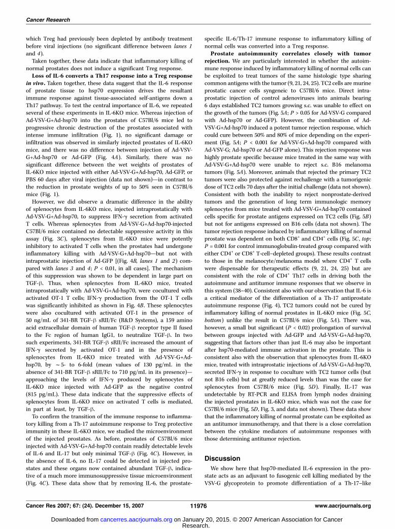
Taken together, these data indicate that inflammatory killing ofnormal prostates does not induce a significant Treg response.Loss of IL-6 converts a Th17 response into a Treg response
in vivo . Taken together, these data suggest that the IL-6 responseof prostate tissue to hsp70 expression drives the resultantimmune response against tissue-associated self-antigens down aTh17 pathway. To test the central importance of IL-6, we repeatedseveral of these experiments in IL-6KO mice. Whereas injection ofAd-VSV-G+Ad-hsp70 into the prostates of C57Bl/6 mice led toprogressive chronic destruction of the prostates associated withintense immune infiltration (Fig. 1), no significant damage orinfiltration was observed in similarly injected prostates of IL-6KOmice, and there was no difference between injection of Ad-VSV-G+Ad-hsp70 or Ad-GFP (Fig. 4A). Similarly, there was nosignificant difference between the wet weights of prostates ofIL-6KO mice injected with either Ad-VSV-G+Ad-hsp70, Ad-GFP, orPBS 60 days after viral injection (data not shown)—in contrast tothe reduction in prostate weights of up to 50% seen in C57Bl/6mice (Fig. 1).
However, we did observe a dramatic difference in the abilityof splenocytes from IL-6KO mice, injected intraprostatically withAd-VSV-G+Ad-hsp70, to suppress IFN-g secretion from activatedT cells. Whereas splenocytes from Ad-VSV-G+Ad-hsp70-injectedC57Bl/6 mice contained no detectable suppressive activity in thisassay (Fig. 3C), splenocytes from IL-6KO mice were potentlyinhibitory to activated T cells when the prostates had undergoneinflammatory killing with Ad-VSV-G+Ad-hsp70—but not withintraprostatic injection of Ad-GFP [(Fig. 4B, lanes 1 and 2) com-pared with lanes 3 and 4 ; P < 0.01, in all cases]. The mechanismof this suppression was shown to be dependent in large part onTGF-h. Thus, when splenocytes from IL-6KO mice, treatedintraprostatically with Ad-VSV-G+Ad-hsp70, were cocultured withactivated OT-1 T cells; IFN-g production from the OT-1 T cellswas significantly inhibited as shown in Fig. 4B . These splenocyteswere also cocultured with activated OT-1 in the presence of50 ng/mL of 341-BR TGF-h sRII/Fc (R&D Systems), a 159 aminoacid extracellular domain of human TGF-h receptor type II fusedto the Fc region of human IgG1, to neutralize TGF-h. In twosuch experiments, 341-BR TGF-h sRII/Fc increased the amount ofIFN-g secreted by activated OT-1 and in the presence ofsplenocytes from IL-6KO mice treated with Ad-VSV-G+Ad-hsp70, by f5- to 6-fold (mean values of 130 pg/mL in theabsence of 341-BR TGF-h sRII/Fc to 710 pg/mL in its presence)—approaching the levels of IFN-g produced by splenocytes ofIL-6KO mice injected with Ad-GFP as the negative control(815 pg/mL). These data indicate that the suppressive effects ofsplenocytes from IL-6KO mice on activated T cells is mediated,in part at least, by TGF-h.
To confirm the transition of the immune response to inflamma-tory killing from a Th-17 autoimmune response to Treg protectiveimmunity in these IL-6KO mice, we studied the microenvironmentof the injected prostates. As before, prostates of C57Bl/6 miceinjected with Ad-VSV-G+Ad-hsp70 contain readily detectable levelsof IL-6 and IL-17 but only minimal TGF-h (Fig. 4C). However, inthe absence of IL-6, no IL-17 could be detected in injected pro-states and these organs now contained abundant TGF-h, indica-tive of a much more immunosuppressive tissue microenvironment(Fig. 4C). These data show that by removing IL-6, the prostate-
specific IL-6/Th-17 immune response to inflammatory killing ofnormal cells was converted into a Treg response.Prostate autoimmunity correlates closely with tumor
rejection. We are particularly interested in whether the autoim-mune response induced by inflammatory killing of normal cells canbe exploited to treat tumors of the same histologic type sharingcommon antigens with the tumor (9, 21, 24, 25). TC2 cells are murineprostatic cancer cells syngeneic to C57Bl/6 mice. Direct intra-prostatic injection of control adenoviruses into animals bearing6 days established TC2 tumors growing s.c. was unable to effect onthe growth of the tumors (Fig. 5A ; P > 0.05 for Ad-VSV-G comparedwith Ad-hsp70 or Ad-GFP). However, the combination of Ad-VSV-G+Ad-hsp70 induced a potent tumor rejection response, whichcould cure between 50% and 80% of mice depending on the experi-ment (Fig. 5A ; P < 0.001 for Ad-VSV-G+Ad-hsp70 compared withAd-VSV-G; Ad-hsp70 or Ad-GFP alone). This rejection response washighly prostate specific because mice treated in the same way withAd-VSV-G+Ad-hsp70 were unable to reject s.c. B16 melanomatumors (Fig. 5A). Moreover, animals that rejected the primary TC2tumors were also protected against rechallenge with a tumorigenicdose of TC2 cells 70 days after the initial challenge (data not shown).Consistent with both the inability to reject nonprostate-derivedtumors and the generation of long term immunologic memorysplenocytes from mice treated with Ad-VSV-G+Ad-hsp70 containedcells specific for prostate antigens expressed on TC2 cells (Fig. 5B)but not for antigens expressed on B16 cells (data not shown). Thetumor rejection response induced by inflammatory killing of normalprostate was dependent on both CD8+ and CD4+ cells (Fig. 5C , top ;P = 0.001 for control immunoglobulin-treated group compared witheither CD4+ or CD8+ T-cell–depleted groups). These results contrastto those in the melanocyte/melanoma model where CD4+ T cellswere dispensable for therapeutic effects (9, 21, 24, 25) but areconsistent with the role of CD4+ Th17 cells in driving both theautoimmune and antitumor immune responses that we observe inthis system (38–40). Consistent also with our observation that IL-6 isa critical mediator of the differentiation of a Th-17 antiprostateautoimmune response (Fig. 4), TC2 tumors could not be cured byinflammatory killing of normal prostates in IL-6KO mice (Fig. 5C,bottom) unlike the result in C57Bl/6 mice (Fig. 5A). There was,however, a small but significant (P < 0.02) prolongation of survivalbetween groups injected with Ad-GFP and Ad-VSV-G+Ad-hsp70,suggesting that factors other than just IL-6 may also be importantafter hsp70-mediated immune activation in the prostate. This isconsistent also with the observation that splenocytes from IL-6KOmice, treated with intraprostatic injections of Ad-VSV-G+Ad-hsp70,secreted IFN-g in response to coculture with TC2 tumor cells (butnot B16 cells) but at greatly reduced levels than was the case forsplenocytes from C57Bl/6 mice (Fig. 5D). Finally, IL-17 wasundetectable by RT-PCR and ELISA from lymph nodes drainingthe injected prostates in IL-6KO mice, which was not the case forC57Bl/6 mice (Fig. 5D , Fig. 3, and data not shown). These data showthat the inflammatory killing of normal prostate can be exploited asan antitumor immunotherapy, and that there is a close correlationbetween the cytokine mediators of autoimmune responses withthose determining antitumor rejection.
Discussion
We show here that hsp70-mediated IL-6 expression in the pro-state acts as an adjuvant to fusogenic cell killing mediated by theVSV-G glycoprotein to promote differentiation of a Th-17–like
Cancer Research
Cancer Res 2007; 67: (24). December 15, 2007 11976 www.aacrjournals.org
Research. on January 20, 2015. © 2007 American Association for Cancercancerres.aacrjournals.org Downloaded from

immune response that is associated with progressive autoimmuneattack of the normal prostate. Both hsp70 and cell killing arerequired to produce optimal autoimmune reactivity, although we didobserve moderate autoimmune inflammatory responses withAd-VSV-G alone. In contrast, normal cell killing in the presence ofhsp70 expression did not generate detectable Treg responses,consistent with the presence of both IL-6 and TGF-h in the lymphnodes draining the injected prostates. In addition, prior depletion ofTreg from mice injected intraprostatically with a known immuno-
genic ova-expressing plasmid had no potentiating effect on theconcomitant T-cell response primed against the ova antigen byinflammatory killing of normal prostate tissue. The absence ofdetectable Treg responses after inflammatory killing of normalprostate may explain, at least in part, the inability to protect thenormal organ from ongoing autoimmune attack directed againstprostate specific self-antigens. These findings in the prostatecontrast with our earlier findings with the melanocyte/melanomamodel; in those experiments, inflammatory killing of normal
Figure 5. Inflammatory killing of normal prostate induces potent antitumor rejection responses. A, 2 � 105 B16 or 2 � 106 prostate TC2 cells were seeded s.c. inC57BL/6 mice. On day 6, mice under anesthetics were injected intraprostatically with 109 pfu of Ad-GFP or Ad-VSV-G (27–29, 31, 46–50), or Ad-hsp70 or with5 � 108 pfu Ad-VSV-G+5 � 108 pfu Ad-hsp70. After surgery, the mice recovered and survival (tumor, 1.0 cm) after seeding of tumors is shown for all TC2-bearingadenovirus-treated mice. Mice bearing B16 tumors s.c. treated with Ad-VSV-G+hsp70 intraprostatically are also shown. Results shown are representative of multipleexperiments. B, mice under anesthetics were injected intraprostatically with 109 pfu of Ad-GFP or Ad-VSV-G or Ad-hsp70 or with 5 � 108 pfu Ad-VSV-G+5 � 108 pfuAd-hsp70. After surgery, the mice recovered. One week later, 500,000 splenocytes per treatment group were harvested and cocultured with 50,000 TC2 or B16(data not shown) target tumor cells prepulsed with IFN-g to increase levels of MHC class I. Forty-eight hours later, supernatants were harvested and assayed byELISA for IFN-g. Cocultures of splenocytes with B16 targets produced no IFN-g over background (data not shown). Results shown are representative of twodifferent experiments. C, 2 � 106 prostate TC2 (top ), cells were seeded s.c. in C57BL/6 mice. On day 4, mice received injections of contol IgG, or CD4+ T cell- orCD8+ T-cell–depleting antibodies (see Materials and Methods). On day 6, mice under anesthetics were injected intraprostatically with 5 � 108 pfu Ad-VSV-G+5 �108 pfu Ad-hsp70. After surgery, the mice recovered and survival (tumor, 1.0 cm) after seeding of tumors is shown. Results are representative of two differentexperiments. Bottom, 2 � 106 prostate TC2 cells were seeded s.c. in B6.129S2-IL6tm1Kopf /J (IL-6KO) mice. On day 6, mice under anesthetics were injectedintraprostatically with 109 pfu of Ad-GFP or with 5 � 108 pfu Ad-VSV-G+5 � 108 pfu Ad-hsp70. After surgery, the mice recovered and survival (tumor, 1.0 cm)after seeding of tumors is shown. D, when the B6.129S2-IL6tm1Kopf /J (IL-6KO) mice of experiment C above were euthanized due to tumor size, 500,000 splenocytesper treatment group were harvested and cocultured with 50,000 TC2, or B16 target tumor cells were prepulsed with IFN-g to increase levels of MHC class I. Splenocytesfrom a C57Bl/6 mouse injected intraprostatically with Ad-VSV-G+Ad-hsp70 were used as a positive control as shown. Forty-eight hours later, supernatants wereharvested and assayed by ELISA for IFN-g. In addition, lymph nodes draining the injected prostates were recovered and assayed by ELISA for IL-17 (lymph node IL-17).The only positive sample (>3 pg/mL) came from splenocytes from the C57Bl/6-treated mouse incubated with TC2 targets (35 pg/mL).
Autoimmunity and Antitumor Immunity in Prostate
www.aacrjournals.org 11977 Cancer Res 2007; 67: (24). December 15, 2007
Research. on January 20, 2015. © 2007 American Association for Cancercancerres.aacrjournals.org Downloaded from
melanocytes (albeit through transfer of the HSVtk and hsp70 genes)induced a potent Treg response, which restrained the concomitantCD8+ T-cell–dependent response directed against self-antigens ofmelanocytes (and B16 melanomas); moreover, suppression of thisTreg response was required to induce autoimmune manifestations(24, 25). Only when the potency of the antiself–T-cell response wasaugmented by additional costimulation were overt signs of auto-immunity observed (21). It seems likely that the differences inimmunologic outcome between these two models (progressiveautoimmune destruction of the normal prostate versus suppressionof autoimmunity against melanocytes) may result from differences inthe constellation of immune cells residing within different organsand the receptors that they express for hsp70. In this respect, we arecurrently studying the expression of known hsp70 receptors (41–43),as well as additional costimulatory molecules, on cells which mightbe expected to respond to hsp70 (such as antigen presenting cellsand macrophages; refs. 12, 18, 41) from the normal prostate.
These data also suggest that strategies aimed at blockading theaction of key cytokines should be clinically very useful in treatmentof progressive autoimmune disorders—such as IL-6 and IL-17 [orassociated cytokines (IL-23)] in (hsp70) induced prostatitis (as weobserve with IL-6 KO mice). Additionally, these results support arole for the adoptive transfer of Treg populations to suppressproautoimmune T-cell responses (44). Indeed, such a strategy hasrecently been shown to be effective in treatment of ongoingautoimmune disease in the pancreas in nonobese diabetic mice(45). We are currently investigating the possibility of inducing Tregspecific for prostate antigens and their use in nullifying theprogressive autoimmune prostatitis we have reported here.
Finally, the major impetus for these studies was our interest in theconnectivity between the generation of autoimmune responses andantitumor immunity (24, 25) in tissue types other than the classically‘‘immunogenic’’ melanoma model (8, 9). Significantly, we have shownhere that the IL-6/IL-17–mediated autoimmune response raisedagainst normal prostate is also extremely effective at rejectingestablished prostate tumors growing elsewhere in the animal. Thisresponse is CD4+ and CD8+ T-cell–dependent and is highly antigenspecific because it did not effect on a tumor of a different histologictype (Fig. 5A and B). Therefore, normal prostate tissue expressesantigens that can also serve as targets for tumor rejection responses,and these responses can be primed through inflammatory killing ofnormal prostate cells. The nature of these antigens is currentlyunclear; however, our ability to isolate splenocyte populations thatare activated against prostate, but not melanoma, tumor cells offersan excellent opportunity for us to identify normal prostate antigenswhich are shared with prostate tumors and which may serve as
vaccine targets. These data also suggest that the approach of normalcell killing could be clinically valuable to treat associated cancers.Because surgical or immunologic prostatectomy is non–life threat-ening, intentional induction of killing of normal prostates couldprovide a valuable immunologic adjuvant approach to the treatmentof metastatic prostate cancer, especially given the potency of theantitumor effects described in the current study. Direct injection ofthe appropriate vectors into the normal prostate, using establishedtechniques for brachytherapy, could be used either with or withoutsubsequent surgical prostatectomy to induce priming of protectiveantitumor immunity against both the local tumor as well as againstpreexisting metastatic disease. Our data here derive from the TC2-transplantable tumor cell line injected s.c. In the human clinical situa-tion, tumors are likely to have been established for longer periods andto have metastasized to different locations than those reflected in ourmodel. Experiments are currently under way to test the efficacy ofinflammatory killing of the normal prostates of TRAMP transgenicmice to treat spontaneously arising metastatic prostate cancers,thereby mimicking more closely treatment of human disease.
The expected toxicity associated with induction of ongoingprostatitis could be resolved by prostatectomy if necessary. Toaddress the issue of systemic autoimmune toxicity induced by theinflammatory killing of normal prostates, a pathology review ofmajor organs and tissues is currently under way. Although noabnormalities have been observed at the level of epithelial damage,or immune infiltrations, of normal organs and tissues in mice 3weeks after intraprostatic injection of the Ad-VSV-G+Ad-hsp70vectors, longer term toxicology studies are currently under way todetermine whether any autoimmune manifestations becomeapparent at 6 months posttreatment.
In summary, we show here that in the prostate, hsp70 inducesIL-6, which triggers a CD4- and CD8-dependent progressiveautoimmune reactivity, associated with IL-17 expression and nosignificant Treg response. These data are significant in that theyconfirm that the intimate connectivity between autoimmune andantitumor rejection responses extends beyond the classic melano-ma paradigm and may be clinically valuable for the treatment ofestablished metastatic prostate cancer.
Acknowledgments
Received 7/3/2007; revised 9/4/2007; accepted 10/16/2007.Grant support: Mayo Foundation; NIH grants RO1CA085931, 1RO1CA94180, and
1RO1CA107082; and Cancer Research UK.The costs of publication of this article were defrayed in part by the payment of page
charges. This article must therefore be hereby marked advertisement in accordancewith 18 U.S.C. Section 1734 solely to indicate this fact.
We thank Toni L. Higgins for expert secretarial assistance.
References
1. Pardoll DM. Spinning molecular immunology intosuccessful immunotherapy. Nat Rev Immunol 2002;2:227–38.
2. Houghton AN. Cancer antigens: immune recognitionof self and altered self. J Exp Med 1994;180:1–4.
3. Parmiani G. Tumor immunity as autoimmunity: tumorantigens include normal self proteins which stimulateanergic peripheral Tcells. Immunol. Today 1993;14:536–8.
4. Bronte V, Apolloni E, Ronca R, et al. Geneticvaccination with ‘‘self ’’ tyrosinase-related protein 2causes melanoma eradication but not vitiligo. CancerRes 2000;60:253–8.
5. Hodi FS, Mihm MC, Soiffer RJ, et al. Biologic activity ofcytotoxic T lymphocyte-associated antigen 4 antibody
blockade in previously vaccinated metastatic melanomaand ovarian carcinoma patients. Proc Natl Acad SciU S A 2003;100:4712–7.
6. Dudley ME, Wunderlich JR, Robbins PF, et al. Cancerregression and autoimmunity in patients after clonalrepopulation with antitumor lymphocytes. Science 2002;298:850–4.
7. Overwijk W, Theoret M, Finkelstein S, et al. Tumorregression and autoimmunity after reversal of afunctionally tolerant state of self-reactive CD8+ T cells.J Exp Med 2003;198:569–80.
8. Gogas H, Ioannovich J, Dafni U, et al. Prognosticsignificance of autoimmunity during treatment ofmelanoma with interferon. N Engl J Med 2006;354:758–60.
9. Ferrone S. Immunotherapy dispenses with tumorantigens. Nat Biotechnol 2004;22:1096–8.
10. Melcher AA, Todryk S, Hardwick N, Ford M,Jacobson M, Vile RG. Tumor immunogenicity isdetermined by the mechanism of cell death via induc-tion of heat shock protein expression. Nat Med 1998;4:581–7.
11. Todryk S, Melcher AA, Hardwick N, et al. Heat shockprotein 70 induced during tumor cell killing induces Th1cytokines and targets immature dendritic cell precur-sors to enhance antigen uptake. J Immunol 1999;163:1398–408.
12. Gough MJ, Melcher AA, Ahmed A, et al. Macrophagesorchestrate the immune response to tumor cell death.Cancer Res 2001;61:7240–7.
13. Singh-Jasuja H, Toes RE, Spee P, et al. Cross-presentation of glycoprotein 96-associated antigens onmajor histocompatibility complex class I molecules
Cancer Research
Cancer Res 2007; 67: (24). December 15, 2007 11978 www.aacrjournals.org
Research. on January 20, 2015. © 2007 American Association for Cancercancerres.aacrjournals.org Downloaded from

requires receptor-mediated endocytosis. J Exp Med2000;191:1965–74.
14. Castellino F, Boucher PE, Eichelberg K, et al.Receptor-mediated uptake of antigen/heat shock pro-tein complexes results in major histocompatibilitycomplex class I antigen presentation via two distinctprocessing pathways. J Exp Med 2000;191:1957–64.
15. Srivastava PK, Menoret A, Basu S, Binder RJ,McQuade KL. Heat shock proteins come of age:primitive functions acquire new roles in an adaptiveworld. Immunity 1998;8:657–65.
16. Suzue K, Zhou X, Eisen HN, Young RA. Heat shockfusion proteins as vehicles for antigen delivery intothe major histocompatibility complex class I presen-tation pathway. Proc Natl Acad Sci U S A 1997;94:13146–51.
17. MacAry PA, Javid B, Floto RA, et al. HSP70 peptidebinding mutants separate antigen delivery from den-dritic cell stimulation. Immunity 2004;20:95–106.
18. Asea A, Rehli M, Kabingu E, et al. Novel signaltransduction pathway utilized by extracellular HSP70:role of Toll-like receptor (TLR) 2 and TLR4. J Biol Chem2002;277:15028–34.
19. Millar DG, Garza KM, Odermatt B, et al. Hsp70promotes antigen-presenting cell function and convertsT-cell tolerance to autoimmunity in vivo . Nat Med 2003;9:1469–76.
20. Lukacs KV, Nakakes A, Atkins CJ, Lowrie DB,Colston MJ. In vivo gene therapy of malignant tumourswith heat shock protein-65 gene. Gene Ther 1997;4:346–50.
21. Sanchez-Perez L, Kottke T, Daniels G, et al. Killing ofnormal melanocytes, combined with hsp70 and CD40Lexpression, cures large established melanomas. J Immu-nol 2006;177:4168–77.
22. Melcher AA, Gough MJ, Todryk S, Vile RG. Apoptosisor necrosis for tumour immunotherapy - what’s in aname? J Mol Med 1999;77:824–33.
23. Srivastava PK. Hypothesis: controlled necrosis as atool for immunotherapy of human cancer. Can Immunol2003;3:4.
24. Daniels G, Sanchez-Perez L, Kottke T, et al. Asimple method to cure established tumors by inflam-matory killing of normal cells. Nat Biotechnol 2004;22:1125–32.
25. Sanchez-Perez L, Kottke T, Diaz RM, et al. Potentselection of antigen loss variants of B16 melanomafollowing inflammatory killing of melanocytes in vivo .Cancer Res 2005;65:2009–17.
26. von Herrath MG, Harrison LC. Antigen-inducedregulatory T cells in autoimmunity. Nat Rev Immunol2003;3:223–32.
27. Linardakis E, Bateman A, Phan V, et al. Enhancingthe efficacy of a weak allogeneic melanoma vaccine byviral fusogenic membrane glycoprotein-mediated tumorcell-tumor cell fusion. Can Res 2002;62:5495–504.
28. Bateman A, Harrington K, Kottke T, et al. Viralfusogenic membrane glycoproteins kill solid tumor cellsby non-apoptotic mechanisms which promote crosspresentation of tumor antigens by dendritic cells. CancerRes 2002;62:5466–6578.
29. Higuchi H, Bronk S, Bateman A, Harrington KJ, VileRG, Gores GJ. Viral fusogenic membrane glycoproteinexpression causes syncytia formation with bioenergeticcell death: implications for gene therapy. Cancer Res2000;60:6396–402.
30. Melcher A, Murphy S, Vile RG. Heat shock proteinexpression in target cells infected by poorly detectablelevels of replication competent virus contributes to theimmunogenicity of adenoviral vectors. Hum Gene Ther1999;10:1431–42.
31. Ahmed A, Thompson J, Emiliusen L, et al. Aconditionally replicating adenovirus targeted to tumorcells through activated RAS/MAPK-selective mRNAstabilisation. Nat Biotechnol 2003;21:771–7.
32. Vile RG, Castleden SC, Marshall J, Camplejohn R,Upton C, Chong H. Generation of an anti-tumourimmune response in a non-immunogenic tumour:HSVtk-killing in vivo stimulates a mononuclear cellinfiltrate and a Th1-like profile of intratumouralcytokine expression. Int J Cancer 1997;71:267–74.
33. Hogquist KA, Jameson SC, Health WR, HowardJL, Bevan MJ, Carbone FR. T cell receptor anta-gonistic peptides induce positive selection. Cell 1994;76:17.
34. Dyall R, Bowne WB, Weber LW, et al. Heterocliticimmunization induces tumor immunity. J Exp Med1998;188:1553–61.
35. Thomas DA, Massague J. TGF-h directly targetscytotoxic T cell functions during tumor evasion ofimmune surveillance. Cancer Cell 2005;8:369–80.
36. Coligan JE, Kruisbeek AM, Margulies DH, ShevachEM, Strober W. Current Protocols in Immunology. Wileyand Sons, Inc.; 1998.
37. Altman DG. Analysis of survival times. In: PracticalStatistics for Medical Research 1991;365–95.
38. Veldhoen M, Hocking R, Atkins C, Locksley R,Stockinger B. TGFh in the context of an inflammatory
cytokine milieu supports de novo differentiaton ofIL-17-producing T cells. Immunity 2006;24:179–89.
39. Mangan P, Harrington L, O’Quinn D, et al. Trans-forming growth factor-h induces development of theTH17 lineage. Nature 2006;441:231–4.
40. Bettelli E, Carrier Y, Gao W, et al. Reciprocaldevelopmental pathways for the generation of patho-genic effector TH17 and regulatory T cells. Nature 2006;441:235–8.
41. Theriault JR, Adachi H, Calderwood SK. Role ofscavenger receptors in the binding and internalization ofheat shock protein 70. J Immunol 2006;177:8604–11.
42. Becker T, Hartl FU, Wieland F. CD40, an extra-cellular receptor for binding and uptake of Hsp70-peptide complexes. J Cell Biol 2002;158:1227–85.
43. Gross C, Schmidt-Wolf IG, Nagaraj S, et al. Heatshock protein 70-reactivity is associated with increasedcell surface density of CD94-56 on primary natural killercells. Cell Stress Chaperones 2003;8:348–60.
44. Jiang S, Lechler RI. CD4+CD25+ regulatory T-celltherapy for allergy, autoimmune disease and trans-plant rejection. Inflamm Allergy Drug Targets 2006;5:239–42.
45. Tarbell KV, Petit L, Zuo X, et al. Dendritic cell-expanded, islet-specific CD4+ CD25+ CD62L+ regulatoryT cells restore normoglycemia in diabetic NOD mice.J Exp Med 2007;204:191–201.
46. Bateman A, Bullough F, Murphy S, et al. Fusogenicmembrane glycoproteins as a novel class of genes forthe local and immune-mediated control of tumorgrowth. Cancer Res 2000;60:1492–7.
47. Diaz RM, Bateman A, Emiliusen L, et al. A lentiviralvector expressing a fusogenic glycoprotein for cancergene therapy. Gene Therapy 2000;7:1656–63.
48. Fu X, Tao L, Jin A, Vile R, Brenner MK, Zhang X.Expression of a fusogenic membrane glycoprotein byan oncolytic herpes simplex virus provides potent syn-ergistic anti-tumor effect. Molecular Therapy 2003;7:748–54.
49. Emiliusen L, Gough M, Bateman A, et al. A transcrip-tional feedback loop for tissue-specific expression of highlycytotoxic genes which incorporates an immunostimula-tory component. Gene Ther 2001;8:987–98.
50. Bateman A, Phan V, Melcher A, Linardakis E,Harrington H, Vile R. Fusogenic membrane proteinsfor cancer gene therapy: a better class of killer. In:Curiel D, Douglas JT, editors. Contemporary cancerresearch: cancer gene therapy. Totowa (NJ): HumanaPress Inc; 2005.
Autoimmunity and Antitumor Immunity in Prostate
www.aacrjournals.org 11979 Cancer Res 2007; 67: (24). December 15, 2007
Research. on January 20, 2015. © 2007 American Association for Cancercancerres.aacrjournals.org Downloaded from
2007;67:11970-11979. Cancer Res Timothy Kottke, Luis Sanchez-Perez, Rosa Maria Diaz, et al. Exploited as Immunotherapy for Metastatic Prostate CancerInduction of hsp70-Mediated Th17 Autoimmunity Can Be
Updated version
http://cancerres.aacrjournals.org/content/67/24/11970
Access the most recent version of this article at:
Cited Articles
http://cancerres.aacrjournals.org/content/67/24/11970.full.html#ref-list-1
This article cites by 47 articles, 17 of which you can access for free at:
Citing articles
http://cancerres.aacrjournals.org/content/67/24/11970.full.html#related-urls
This article has been cited by 12 HighWire-hosted articles. Access the articles at:
E-mail alerts related to this article or journal.Sign up to receive free email-alerts
Subscriptions
Reprints and
To order reprints of this article or to subscribe to the journal, contact the AACR Publications
Permissions
To request permission to re-use all or part of this article, contact the AACR Publications
Research. on January 20, 2015. © 2007 American Association for Cancercancerres.aacrjournals.org Downloaded from
Copyright © 2022 FDOKUMEN