Human endothelial cells generate Th17 and regulatory T cells under inflammatory conditions
6
Human endothelial cells generate Th17 and regulatory T cells under inflammatory conditions Cécile Taflin a,b , Benoit Favier b,c , Jeremy Baudhuin b,c , Alain Savenay d , Patrice Hemon b,e , Armand Bensussan b,e , Dominique Charron a,b,d , Denis Glotz a,b,f , and Nuala Mooney a,b,1 a Institut National de la Santé et de la Recherche Médicale, Unité Mixte de Recherche Santé 940, Hôpital Saint Louis, 75010 Paris, France; c Commissariat à l’Energie Atomique et Énergies Alternatives, Institut d’Imagerie Biomédicale, Service de Recherches en Hémato-Immunologie, F-75475 Paris, France; b Université Paris Diderot, 75013 Paris, France; d Laboratoire d’Immunologie et d’Histocompatibilité, Assistance Publique–Hôpitaux de Paris, Hôpital Saint Louis, 75010 Paris, France; e Institut National de la Santé et de la Recherche Médicale, Unité Mixte de Recherche Santé 976, Hôpital Saint Louis, 75010 Paris, France; and f Service de Néphrologie et Transplantation Rénale, Hôpital Saint Louis, 75010 Paris, France Edited by Harvey Cantor, Dana–Farber Cancer Institute, Boston, MA, and approved January 4, 2011 (received for review August 11, 2010) Organ transplantation represents a unique therapeutic option for irreparable organ dysfunction and rejection of transplants results from a breakdown in operational tolerance. Although endothelial cells (ECs) are the first target in graft rejection following kidney transplantation, their capacity to alloactivate and generate partic- ular T lymphocyte subsets that could intervene in this process remains unknown. By using an experimental model of microvascu- lar endothelium, we demonstrate that, under inflammatory con- ditions, human ECs induced proliferation of memory CD4 + CD45RA - T cells and selectively amplified proinflammatory Th17 and suppres- sive CD45RA - HLA-DR + FoxP3 bright regulatory CD4 + T lymphocytes (Tregs). Although HLA-DR expression on resting microvascular ECs was sufficient to induce proliferation of memory CD4 + T cells, Treg amplification was dependent on the interaction with CD54, highly expressed only under inflammatory conditions. Moreover, expan- sion of Th17 cells was dependent on IL-6 and STAT-3, and inhibition of either specifically impaired Th17, without altering Treg expan- sion. Collectively these data reveal that the HLA-DR + ECs regulate the local inflammatory allogeneic response, promoting either an IL- 6/STAT-3–dependent Th17 response or a contact-CD54–dependent regulatory response according to the cytokine environment. Finally, these data open therapeutic perspectives in human organ trans- plantation based on targeting the IL-6/STAT-3 pathway and/or pro- moting CD54 dependent Treg proliferation. immune regulation | MHC class II T he allogeneic response to organ transplantation was initially considered to be predominantly mediated by the Th1 pop- ulation whereas the Th2 population was believed to oppose Th1 cell activation, thus favoring graft survival (1). Recent studies have identified additional CD4 + T cell subsets including IL-17– producing CD4 + T cells (Th17) and CD4 + FoxP3 + regulatory T cells (Tregs) (2). A unique role for Th1 cells was excluded by using genetically deficient mice and T lymphocyte transfer experiments in MHC class II mismatched organ transplantation models (3, 4). Th2 cytokines have been implicated in acute graft rejection (5), and involvement of Th17 was shown in cardiac and lung allograft rejection (6, 7). In the absence of a Th1 alloresponse, Th17 me- diates a violent proinflammatory response, resulting in accelerated allograft rejection (6). The Th1/Th2 paradigm is thus obsolete in models of organ transplantation. Recent studies have revealed reciprocal control of differentiation of proinflammatory Th17 and tolerogenic induced Treg subsets in mouse models (8–10) and in human studies (11–13). Whether this equilibrium between Treg and Th17 subsets occurs in organ transplantation and, if so, can be skewed in one or other direction, is unknown. In the context of organ transplantation, human endothelial cells (ECs) express MHC class I and class II molecules, and may therefore act both as stimulators of and targets for alloimmune responses (14). ECs stimulate memory CD4 + T cell production of IFN-γ, thereby promoting a CD4 + Th1 profile (15). EC allosti- mulation of CD4 + T cells requires expression of HLA-DR and the costimulatory molecule LFA-3 independently of CD80 and CD86 (16, 17). ECs therefore have the potential to activate memory CD4 + T cells but their ability to orient the alloimmune response toward induced Treg-mediated tolerance or a proin- flammatory effector response remains unknown. In MHC class II fully mismatched mouse models, vascular ECs activate and in- duce programmed death ligand 1 (PD-L1)–dependent Tregs, which inhibit in vitro and in vivo proliferation of alloreactive CD4 + T cells (18). Human ECs express LFA-3 and PD-L1 (19) and we therefore investigated their ability to allostimulate CD4 + T cells and to generate regulatory or effector T cell populations. To address this question, we have studied the capacity of human microvascular ECs to stimulate HLA-DR–typed CD4 + T cells from allogeneic donors. The results reveal that human endothelium simultane- ously generates proinflammatory Th17 and functional HLA-DR + Tregs under inflammatory conditions. These data lead to the suggestion that allograft endothelium participates in controlling CD4 + T lineage plasticity and can thus induce a regulatory or proinflammatory response, depending on the local cytokine mi- croenvironment. Results HLA-DR Expression on Resting ECs Is Necessary and Sufficient to Induce CD4 + T Cell Alloproliferation. The microvascular EC line HMEC-1 (20) constitutively expressed the costimulatory mole- cule LFA-3 whereas expression of HLA-DR was induced by ac- tivation with IFN-γ [activated ECs (aECs); Fig. 1A]. In contrast with previous studies, ECs did not express the adhesion molecule CD54 in steady-state conditions (20), although it was strongly expressed after activation with IFN-γ (Fig. 1A). Constitutive ex- pression of the costimulatory molecules LFA-3 and PD-L1 and the adhesion molecule VE-cadherin was unchanged by IFN-γ treatment (i.e., aECs; Fig. 1A and Fig. S1A). To understand the role of HLA-DR in the absence of inflammatory stimuli, ECs were transduced with a lentiviral HLA-DR construct leading to constitutive surface expression of HLA-DR (ECsαβ) comparable to that of aECs. LFA-3 and VE-cadherin expression remained similar to that observed in resting ECs (Fig. 1A). CD54 expression was induced in ECsαβ activated with IFN-γ (aECsαβ) to a similar level as that observed in aECs (Fig. 1A). Author contributions: C.T., D.G., and N.M. designed research; C.T. and A.S. performed research; B.F., J.B., P.H., and A.B. contributed new reagents/analytic tools; C.T., D.C., D.G., and N.M. analyzed data; and C.T., B.F., D.G., and N.M. wrote the paper. The authors declare no conflict of interest. This article is a PNAS Direct Submission. 1 To whom correspondence should be addressed. E-mail: nuala.mooney@univ-paris- diderot.fr. This article contains supporting information online at www.pnas.org/lookup/suppl/doi:10. 1073/pnas.1011811108/-/DCSupplemental. www.pnas.org/cgi/doi/10.1073/pnas.1011811108 PNAS | February 15, 2011 | vol. 108 | no. 7 | 2891–2896 IMMUNOLOGY
-
Upload
independent -
Category
Documents
-
view
0 -
download
0
Transcript of Human endothelial cells generate Th17 and regulatory T cells under inflammatory conditions

Human endothelial cells generate Th17 andregulatory T cells under inflammatory conditionsCécile Taflina,b, Benoit Favierb,c, Jeremy Baudhuinb,c, Alain Savenayd, Patrice Hemonb,e, Armand Bensussanb,e,Dominique Charrona,b,d, Denis Glotza,b,f, and Nuala Mooneya,b,1
aInstitut National de la Santé et de la Recherche Médicale, Unité Mixte de Recherche Santé 940, Hôpital Saint Louis, 75010 Paris, France; cCommissariat àl’Energie Atomique et Énergies Alternatives, Institut d’Imagerie Biomédicale, Service de Recherches en Hémato-Immunologie, F-75475 Paris, France;bUniversité Paris Diderot, 75013 Paris, France; dLaboratoire d’Immunologie et d’Histocompatibilité, Assistance Publique–Hôpitaux de Paris, Hôpital Saint Louis,75010 Paris, France; eInstitut National de la Santé et de la Recherche Médicale, Unité Mixte de Recherche Santé 976, Hôpital Saint Louis, 75010 Paris, France;and fService de Néphrologie et Transplantation Rénale, Hôpital Saint Louis, 75010 Paris, France
Edited by Harvey Cantor, Dana–Farber Cancer Institute, Boston, MA, and approved January 4, 2011 (received for review August 11, 2010)
Organ transplantation represents a unique therapeutic option forirreparable organ dysfunction and rejection of transplants resultsfrom a breakdown in operational tolerance. Although endothelialcells (ECs) are the first target in graft rejection following kidneytransplantation, their capacity to alloactivate and generate partic-ular T lymphocyte subsets that could intervene in this processremains unknown. By using an experimental model of microvascu-lar endothelium, we demonstrate that, under inflammatory con-ditions, human ECs induced proliferation of memory CD4+CD45RA−
T cells and selectively amplified proinflammatory Th17 and suppres-sive CD45RA−HLA-DR+FoxP3bright regulatory CD4+ T lymphocytes(Tregs). Although HLA-DR expression on resting microvascular ECswas sufficient to induce proliferation of memory CD4+ T cells, Tregamplification was dependent on the interaction with CD54, highlyexpressed only under inflammatory conditions. Moreover, expan-sion of Th17 cells was dependent on IL-6 and STAT-3, and inhibitionof either specifically impaired Th17, without altering Treg expan-sion. Collectively these data reveal that the HLA-DR+ ECs regulatethe local inflammatory allogeneic response, promoting either an IL-6/STAT-3–dependent Th17 response or a contact-CD54–dependentregulatory response according to the cytokine environment. Finally,these data open therapeutic perspectives in human organ trans-plantation based on targeting the IL-6/STAT-3 pathway and/or pro-moting CD54 dependent Treg proliferation.
immune regulation | MHC class II
The allogeneic response to organ transplantation was initiallyconsidered to be predominantly mediated by the Th1 pop-
ulation whereas the Th2 population was believed to oppose Th1cell activation, thus favoring graft survival (1). Recent studieshave identified additional CD4+ T cell subsets including IL-17–producing CD4+ T cells (Th17) and CD4+FoxP3+ regulatory Tcells (Tregs) (2). A unique role for Th1 cells was excluded by usinggenetically deficient mice and T lymphocyte transfer experimentsin MHC class II mismatched organ transplantation models (3, 4).Th2 cytokines have been implicated in acute graft rejection (5),and involvement of Th17 was shown in cardiac and lung allograftrejection (6, 7). In the absence of a Th1 alloresponse, Th17 me-diates a violent proinflammatory response, resulting in acceleratedallograft rejection (6). The Th1/Th2 paradigm is thus obsolete inmodels of organ transplantation. Recent studies have revealedreciprocal control of differentiation of proinflammatory Th17 andtolerogenic induced Treg subsets in mouse models (8–10) and inhuman studies (11–13). Whether this equilibrium between Tregand Th17 subsets occurs in organ transplantation and, if so, can beskewed in one or other direction, is unknown.In the context of organ transplantation, human endothelial
cells (ECs) express MHC class I and class II molecules, and maytherefore act both as stimulators of and targets for alloimmuneresponses (14). ECs stimulate memory CD4+ T cell production ofIFN-γ, thereby promoting a CD4+ Th1 profile (15). EC allosti-mulation of CD4+ T cells requires expression of HLA-DR and
the costimulatory molecule LFA-3 independently of CD80 andCD86 (16, 17). ECs therefore have the potential to activatememory CD4+ T cells but their ability to orient the alloimmuneresponse toward induced Treg-mediated tolerance or a proin-flammatory effector response remains unknown. In MHC class IIfully mismatched mouse models, vascular ECs activate and in-duce programmed death ligand 1 (PD-L1)–dependent Tregs,which inhibit in vitro and in vivo proliferation of alloreactiveCD4+ T cells (18).Human ECs express LFA-3 and PD-L1 (19) and we therefore
investigated their ability to allostimulate CD4+ T cells and togenerate regulatory or effector T cell populations. To address thisquestion, we have studied the capacity of human microvascularECs to stimulate HLA-DR–typed CD4+ T cells from allogeneicdonors. The results reveal that human endothelium simultane-ously generates proinflammatory Th17 and functional HLA-DR+
Tregs under inflammatory conditions. These data lead to thesuggestion that allograft endothelium participates in controllingCD4+ T lineage plasticity and can thus induce a regulatory orproinflammatory response, depending on the local cytokine mi-croenvironment.
ResultsHLA-DR Expression on Resting ECs Is Necessary and Sufficient toInduce CD4+ T Cell Alloproliferation. The microvascular EC lineHMEC-1 (20) constitutively expressed the costimulatory mole-cule LFA-3 whereas expression of HLA-DR was induced by ac-tivation with IFN-γ [activated ECs (aECs); Fig. 1A]. In contrastwith previous studies, ECs did not express the adhesion moleculeCD54 in steady-state conditions (20), although it was stronglyexpressed after activation with IFN-γ (Fig. 1A). Constitutive ex-pression of the costimulatory molecules LFA-3 and PD-L1 andthe adhesion molecule VE-cadherin was unchanged by IFN-γtreatment (i.e., aECs; Fig. 1A and Fig. S1A). To understand therole of HLA-DR in the absence of inflammatory stimuli, ECswere transduced with a lentiviral HLA-DR construct leading toconstitutive surface expression of HLA-DR (ECsαβ) comparableto that of aECs. LFA-3 and VE-cadherin expression remainedsimilar to that observed in resting ECs (Fig. 1A). CD54 expressionwas induced in ECsαβ activated with IFN-γ (aECsαβ) to a similarlevel as that observed in aECs (Fig. 1A).
Author contributions: C.T., D.G., and N.M. designed research; C.T. and A.S. performedresearch; B.F., J.B., P.H., and A.B. contributed new reagents/analytic tools; C.T., D.C., D.G.,and N.M. analyzed data; and C.T., B.F., D.G., and N.M. wrote the paper.
The authors declare no conflict of interest.
This article is a PNAS Direct Submission.1To whom correspondence should be addressed. E-mail: [email protected].
This article contains supporting information online at www.pnas.org/lookup/suppl/doi:10.1073/pnas.1011811108/-/DCSupplemental.
www.pnas.org/cgi/doi/10.1073/pnas.1011811108 PNAS | February 15, 2011 | vol. 108 | no. 7 | 2891–2896
IMMUNOLO
GY
We next tested the ability of ECs and ECsαβ, activated withIFN-γ or not, to induce alloproliferation of CD4+ T cells. Fig. 1Bshows that resting ECs failed to induce proliferation whereasresting ECsαβ constitutively expressing HLA-DR induced CD4+
T cell proliferation on day 7 (15.35 ± 4.56%), as did aECs (11.72 ±4.16%) and aECsαβ (16.15 ± 3.32%). Proliferation is allor-ecognition-dependent as an HLA-DR–blocking antibody inhibitsT cell proliferation by more than 70% (5.47 ± 3.31% at day 7 vs.21.88 ± 8.81% with isotype IgG2a; P = 0.02; Fig. 1C).HLA-DR expression by resting ECs is therefore necessary and
sufficient to induce CD4+ T cell alloproliferation.
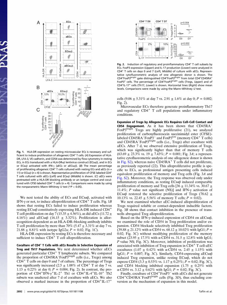
Coculture of CD4+ T Cells with aECs Results in Selective Expansion ofTreg and Th17 Populations. We next determined whether aECsgenerated particular CD4+ T lymphocyte subsets. We comparedthe proportion of CD45RA−FoxP3bright cells (i.e., Tregs) amongCD4+ T cells on days 0 and 7 of culture. The percentage of Tregswas significantly increased (2.5 ± 1.08% of CD4+ T at day 7 vs.1.13 ± 0.22% at day 0; P = 0.004; Fig. 2). In contrast, the pro-portion of CD4+IFN-γ+IL-2+ Th1 or CD4+IL-4+IL-10+ Th2subsets was unaltered after 7 d of coculture with aECs. We alsoobserved a marked increase in the proportion of CD4+IL-17+
cells (9.06 ± 5.31% at day 7 vs. 2.91 ± 1.6% at day 0; P = 0.002;Fig. 2).Microvascular ECs therefore generate proinflammatory Th17
and regulatory CD4+ T cell populations under inflammatoryconditions.
Expansion of Tregs by Allogeneic ECs Requires Cell–Cell Contact andCD54 Engagement. As it has been shown that CD45RA−
FoxP3bright Tregs are highly proliferative (21), we analyzedproliferation of carboxyfluorescein succinimidyl ester (CFSE)-labeled CD45RA−FoxP3− and FoxP3low (memory CD4+ T cells)and CD45RA−FoxP3bright cells (i.e., Tregs) after coculture withaECs. After 7 d, we observed extensive proliferation of Tregs,which was significantly higher than that of memory T cells(63.69 ± 23.3% vs. 19 ± 7.43%; P = 0.001; Fig. 3A; a represen-tative cytofluorometric analysis of one allogeneic donor is shownin Fig. S2), whereas naive CD45RA+ T cells did not proliferate,as previously reported (22). This alloproliferative profile is spe-cific to ECs, as professional antigen presenting cells inducedequivalent proliferation of memory and Treg cells (Fig. 3A andFig. S2). Moreover, the Treg response was observed only underinflammatory conditions, as resting ECsαβ induced comparableproliferation of memory and Treg cells [36 ± 11.34% vs. 30.67 ±11.4%; P value not significant (NS)] and IFN-γ activation ofECsαβ restored the selective proliferation of Tregs (78.62 ±10.13% vs. 22.45 ± 5.56% of memory T cells; P = 0.02).We next examined whether aEC-induced alloproliferation of
Tregs required soluble or contact-dependent inducible factors.Fig. 3B shows that contact inhibition in the presence of trans-wells abrogated Treg alloproliferation.Based on the IFN-γ–induced expression of CD54 on aECsαβ,
we examined the role of CD54 in Treg proliferation and/or ex-pansion. CD54 blockade selectively inhibited Treg proliferation(39.88 ± 21.12% with α-CD54 vs. 68.12 ± 10.02% with IgG1; P=0.02; Fig. 3C) without modifying proliferation of the memorysubset (23.95 ± 17.5% with α-CD54 vs. 31.5 ± 12.8% with IgG1;P value NS; Fig. 3C). Moreover, inhibition of proliferation wasassociated with inhibition of Treg expansion in CD4+ T cell-aECcocultures (1.07 ± 0.42% with α-CD54 vs. 2.45 ± 1.15% withIgG1; P = 0.007; Fig. 3C). Similarly, CD54-expressing aECsαβinduced Treg expansion, unlike resting ECsαβ, which do notexpress CD54 (3.3 ± 0.53% vs. 1.17 ± 0.25%; P = 0.02; Fig. 3C),and CD54 blocking inhibited expansion (1.75 ± 0.25% withα-CD54 vs. 3.12 ± 0.62% with IgG1; P = 0.02; Fig. 3C).Finally, coculture of CD4+FoxP3− with aECs did not generate
CD4+CD45RA−FoxP3bright cells (Fig. 3D), thus excluding con-version as the mechanism of expansion in this model.
A
aECs
ECs
ECsααβ
aECsαβ
HLA
-DR
VE
-Cad
herin
CD
54
B
lifer
atin
g +
T ce
lls12%
LFA-3
15.2%15
20
25***
ECsαβaECs
% p
roC
D4
CD
4
CFSE0
5
10
C
atin
g el
ls 253035 *
% p
rolif
erC
D4+
T c
05
101520
Fig. 1. HLA-DR expression on resting microvascular ECs is necessary and suf-ficient to induce proliferation of allogeneic CD4+ T cells. (A) Expression of HLA-DR, LFA-3, VE-cadherin, and CD54was determined by flow cytometry in restingECs, in ECs transduced with a HLA-DRαβ lentivirus construct (ECsαβ), and in ECsor ECsαβ activated with IFN-γ (aECs or aECsαβ). (B) The mean percentageof proliferating allogeneic CD4+ T cells cultured with resting ECs and aECs (n =11) or ECsαβ (n = 4) is shown. Representative proliferation of CFSE-labeled CD4+
T cells cultured with aECs (Left) and ECsαβ (Middle) is shown. (C) aECs werepretreated with a HLA-DR blocking antibody or an isotype control and cocul-tured with CFSE-labeled CD4+ T cells (n = 4). Comparisons were made by usingthe nonparametric Mann–Whitney U test (*P < 0.05).
p=0.004CD4+ T D0 CD4+ T D7
1.1% 4%
CD
45R
A
F P301234
% T
regs
/CD
4+ T
D0 D7
10%2%
CD
4
IL-17
ox
05
10152025
% T
h17/
CD
4+ T
D0
p=0.002
D7
Fig. 2. Induction of regulatory and proinflammatory CD4+ T cell subsets byECs. FoxP3 expression (Upper) and IL-17 production (Lower) were analyzed inCD4+ T cells on days 0 and 7 (Left, Middle) of culture with aECs. Represen-tative cytofluorometric analysis of one allogeneic donor is shown. TheCD4+FoxP3bright gate distinguished CD4+FoxP3bright from total CD4+CD45RA+
FoxP3+ cells. The percentage of CD4+FoxP3bright cells (Tregs, Upper) and ofCD4+IL-17+ cells (Th17, Lower) is shown. Horizontal lines (Right) show meanlevels. Comparisons were made by using the Mann–Whitney U test.
2892 | www.pnas.org/cgi/doi/10.1073/pnas.1011811108 Taflin et al.

Collectively, these data indicate that Treg expansion by aECsis a result of their alloproliferation and requires CD4+ T cellinteraction with CD54-expressing aECs.
IL-6 and STAT-3 Phosphorylation Are Selectively Required for Th17Expansion.We next investigated the origin of the expansion of theTh17 population. IL-6 secretion by ECs was high and was in-creased threefold by EC activation (Fig. 4A). We also identifiedsignificant phosphorylation of STAT-3 (P-STAT-3) in prolife-rating CD4+ T cells after 7 d of culture with aECs (20.27 ±10.65% vs. 0.3 ± 0.11% in nonproliferating CD4+ T cells; P =0.002; Fig. 4B).The role of IL-6 and P-STAT-3 in Th17 amplification was
determined by coculturing aECs with allogeneic CD4+ T cells inthe presence of an IL-6R blocking antibody or a P-STAT-3 in-hibitor (cucurbitacin I) directed against Janus kinases (23).First, IL-6R blockade inhibited STAT-3 phosphorylation in
CD4+ T cells cocultured with aECs (0.68 ± 0.43% with α-IL-6Rat day 7 vs. 12.17 ± 4.17% with IgG1; P = 0.002; Fig. 4B).
Second, memory T cell proliferation after coculture with aECswas selectively inhibited by IL-6R mAb (5.58 ± 7.17% vs. 37.3 ±10.34% with IgG1; P = 0.002; Fig. 4C) and by cucurbitacin I(10.7 ± 9% vs. 31.6 ± 9.84% with DMSO; P = 0.002), whereasTreg proliferation was unchanged by IL-6R mAb (46.5 ± 13.11%vs. 62.67 ± 10.69% with IgG1; P value NS) or cucurbitacin I(51.71 ± 19.67% vs. 60.22 ± 10.67% with DMSO; P value NS).Finally, blocking IL-6R inhibited CD4+IL-17+ cell expansion
(1.76 ± 0.32% with α-IL-6R vs. 4 ± 0.86% with IgG1; P = 0.008;Fig. 4D) without altering the proportion of Tregs (2.56 ± 0.55%of CD4+ T cells vs. 3.08 ± 0.45% with IgG1; P value NS), whichremained significantly higher than on day 0 (P = 0.004; Fig. 4D).Similarly, cucurbitacin I selectively inhibited CD4+IL-17+ ex-
A B
406080
100 * *
fera
ting
cells 100
406080
*
*
fera
ting
cells
020
% p
roli
Naive CD4+ T cellsMemory CD4+ T cellsTregs
Tm Tregs0
20
ControlaECsTW
% p
roli
C
406080
100 *
rolif
erat
ing
cells
2345
p=0.02 p=0.02
2345
egs/
CD
4+ T p=0.007
egs/
CD
4+ T
Tm Tregs0
20
% p
aECsaECs + IgG1aECs + α-CD54
D
+α-CD54
01
ECsαβ
D0
aECsαβ
- +IgG1 +α-CD54
-01
% T
r
aECs
- +IgG1D0 % T
r
Sorted CD4+ CD25
- CD127
+ T cells
Day 7
0.1%
% T
regs
/ CD
4+ TDay 0
CD
45R
A
FoxP3012345
D0 D7 D0 D7
p=0.02 NS
0.6%
Unsorted cells
Sorted cells
Fig. 3. EC expression of CD54 is necessary for the proliferation and theexpansion of Tregs. (A) Allogeneic CFSE-labeled CD4+ T cells were culturedwith aECs, ECsαβ, aECsαβ, and dendritic cells. The mean ± SEM percentageof proliferation within the regulatory (CD45RA−FoxP3bright, Tregs), mem-ory (CD45RA−FoxP3− or low, Tm), and naive (CD45RA+FoxP3−) CD4+ T lym-phocytes was determined on day 7 (n = 6). CD4+ T cell autoproliferation(control) is shown. (B) aECs were cultured in the lower chamber of 96-welltranswell (TW) plates and CFSE-labeled responder CD4+ T cells were addedto the upper chamber. The mean ± SEM proliferation of Tm and Tregs isdepicted (n = 4). (C ) aECs were pretreated with α-CD54 or IgG1 isotype andcocultured with CFSE-labeled CD4+ T cells. The mean percentage ± SEM ofTm and Treg proliferation is shown (n = 8). The percentage of Tregs (Right)within CD4+ T cells cultured with aECs and aECsαβ pretreated or not withα-CD54 or IgG1 and with ECsαβ was analyzed on day 7. The control showsthe percentage of Tregs on day 0. Horizontal lines represent mean levelsfor ECs (n = 8) and for ECsαβ (n = 4). (D) FoxP3+ expression inCD4+CD25−CD127+ T cells sorted from unprimed CD4+ T cells is shown onday 0 and after 7 d of culture with aECs. Representative analysis of oneallogeneic donor (Left, Middle) is shown. Controls were unsorted CD4+ Tcells from the same donors on days 0 and 7. Horizontal lines (Right) rep-resent the mean levels (n = 3). Comparisons were made by using theMann–Whitney U test (*P < 0.05).
A
0 50 100 150 200IL-6IL-1βTGF-βIL-27IL-23 ECs
aECs
CD4+ T Day 7
28% 0.7%
TAT-
3
STA
T-3
1
102030
*
250pg/ml
STA
T-3
10
15
20*
B
C
P-S
CFSE aECsC
% P
-
NP P
0
100
% P
-
aECs
C
100
0
5
0
20
40
60
80
*
% p
rolif
erat
ing
cells
20
40
60
80
*
% p
rolif
erat
ing
cells
D
TregsTmaECsaECs + IgG1aECs + α-IL-6R
4+T
TregsTm
aECs + DMS0aECs + CI
0
4+T
aECs
45 NS
p=0.004
810 p=0.008
NS
NS T p=0.01
% T
h17/
CD
% T
regs
/CD
0123
Day 0 Day 7- - + IgG1 +α-IL-6R
02468
Day 0 Day 7- - + IgG1 +α-IL-6R
% T
h17/
CD
4+T
10152025
05
Day 0 Day 7- - DMSO CI
p=0.04
012345
% T
regs
/CD
4+Day 0 Day 7
- - DMSO CI
NS
Fig. 4. IL-6 and STAT-3 activation is required for Th17 expansion withoutaltering regulatory T cell expansion. (A) Cytokine detection in supernatantsof resting ECs and aECs after 3 d of culture (n = 2). (B) STAT-3 phosphory-lation (P-STAT-3) was analyzed by flow cytometry in CFSE-labeled CD4+
T cells at day 7 of culture with aECs or alone (control, C). A representativescatterplot of one allogeneic donor is shown (Left). The mean ± SEM ofP-STAT-3 in proliferating (P) or nonproliferating (NP) cells (Middle) and intotal CD4+ T cells in presence or absence of an IL-6R Ab or the isotype IgG1(Right) is shown (n = 6). (C) The mean ± SEM proliferation of Tm and Tregscultured with aECs in presence or absence of IL-6R Ab, IgG1 (Left; n = 6donors), cucurbitacin I (CI), or DMSO (Right; n =8 donors) was analyzed onday 7. (D) The percentage of CD4+IL17+ (Left) and CD4+CD45RA−FoxP3bright
(Right) cells on day 7 with IL-6R Ab (Upper) or with CI or DMSO (Lower) isshown. Horizontal lines indicate the mean of each group. The groups werecompared by using the Mann–Whitney U test.
Taflin et al. PNAS | February 15, 2011 | vol. 108 | no. 7 | 2893
IMMUNOLO
GY

pansion (4.23 ± 1.87% vs. 8.53 ± 3.9%; P = 0.04; Fig. 4D) butnot Treg expansion (1.71 ± 0.56% vs. 2.4 ± 0.9%; P value NS).Expansion of Th17 cells by aEC is thus IL-6/P-STAT-3–
dependent and implicates memory CD4+ T cell proliferation.
Allogeneic CD4+CD45RA−FoxP3bright T Cell Population Expanded by ECInteraction Have Phenotypic and Functional Characteristics of Tregs.HLA-DR expression on CD4+CD25bright T cells identifies ma-ture, functionally distinct Tregs involved in contact-dependent invitro suppression (24). We therefore examined HLA-DR ex-pression on Tregs expanded by EC allostimulation. As shown inFig. 5A, Treg proliferation is associated with increased HLA-DRexpression (80.2 ± 9.45% of proliferating Tregs vs. 46.28 ±8.62% of nonproliferating Tregs; P = 0.002), leading to an en-larged proportion of HLA-DR–expressing Tregs (64.8 ± 3.5%vs. 45 ± 8.45%; P = 0.008; Fig. 5A). These data provide indirectevidence that the CD4+CD45RA−FoxP3bright population arefunctional regulatory cells.Because phenotypic markers are an imperfect gauge of Treg
function, we evaluated expanded Treg function. CD4+CD25bright
CD127low cells were sorted from CD4+ T cells harvested after 7 dof culture with aECs and added to a second allogeneic coculturecomposed of aECs and autologous responder CD4+ T cells. Theseexperiments revealed a dose-dependent inhibition of CD4+ T cellproliferation by CD4+CD25bright T cells with more than 80% in-hibition at a suppressor-to-responder ratio of 1:1 (mean, 80.6%;range, 88.7–76.2%; P < 0.05; Fig. 5B) and as much as 60% in-hibition at a ratio of 1:9 (mean, 67.3%; range, 47.3–68.4%; P <
0.05). aEC-expanded Tregs therefore significantly suppress allo-proliferation of autologous CD4+ T cells.In addition, ECαβ- or aECαβ-activated Tregs equally suppress
autologous CD4+ T cell proliferation induced by ECsαβ oraECsαβ, indicating that CD54 is not required for the suppressivefunction of Tregs induced by ECs (Fig. S3).
Primary Microvascular ECs Induced Selective Expansion of Tregs andTh17 Under Inflammatory Conditions. To further understand theability of human microvascular ECs in expanding Th17 andTregs under inflammatory conditions, we studied primary humandermal microvascular ECs (HMVECs). Similarly to HMEC-1,resting primary ECs constitutively expressed the costimulatorymolecule LFA-3 and expression of HLA-DR and CD54 wasstrongly up-regulated by IFN-γ (Fig. 6A).Activated HMVECs (aHMVECs) induced alloproliferation of
CD4+ T cells and selective proliferation of Tregs after 7 d ofcoculture (81.75 ± 7.54% of Tregs vs. 17.55 ± 6.31% of memoryT cells; P = 0.02; Fig. 6B).Alloactivation was associated with the expansion of CD4+IL-
17+ (6.62 ± 3.54% at day 7 vs. 1.32 ± 0.38% at day 0; P = 0.008;Fig. 6C) and CD4+CD45RA−FoxP3bright cells (5.02 ± 1.9% atday 7 vs. 1.37 ± 0.38% at day 0; P = 0.02; Fig. 6C).These results suggest that the expansion of proinflammatory
and regulatory T cell subsets is a general property of humanmicrovascular ECs.
95%
Tregs Day 7
45%A
DR 40
6080
100
gsH
LA-D
R+ *
HLA
-DR
+
HLA
-
CFSE0
201
%Tr
e
6070
p=0.008
NP P
%Tr
egs
H
B
304050
Day 0 Day 7
30 ***
% p
rolif
erat
ing
onde
r CD
4+ T
cel
ls
10
15
20
25
resp
0
5
+ Tregs
Fig. 5. Allospecific FoxP3brightCD4+ T cells generated by interaction withaECs have Treg characteristics and function. (A) HLA-DR expression was an-alyzed on CFSE-labeled Tregs after a 7-d culture with aECs. Representativeanalysis of one allogeneic donor (Left) is shown. The mean ± SEM of HLA-DR+ cells in proliferating (P) versus nonproliferating (NP) FoxP3brightCD4+
population (Top, Right; n = 5) is shown. The percentage of HLA-DR+ in thetotal FoxP3brightCD4+ population on days 0 and 7 of culture with aECs isshown (Bottom; n = 5). Comparisons were made by using the nonparametricMann–Whitney U test. (B) CD127lowCD25bright T cells sorted from CD4+ T cellsprimed with allogeneic aECs for 7 d were added to a second allogeneic co-culture of fresh autologous CFSE-labeled CD4+ T cells with aECs. Controlsincluded allogeneic cocultures without addition of suppressor cells (control).Responder CD4+ T cell proliferation was measured on day 7 (mean ± SEM,n = 3, paired t test).
A
i n
15%
HMVEC
HMVECs
HLA
-DR
LFA-3
VE
-Cad
heri
CD
54
* *
70%aHMVECs
% p
rolif
erat
ing
CD
4+T
cells
% p
rolif
erat
ing
cellsB
20
40
60
80
100 *
510152025
*
*
%
Control aECs aHMVECs
Naive CD4+ T cellsMemory CD4+ T cellsTregs
0
p= 0.02104+T CD4+ T D0 CD4+ T D7C
0
02468
D0 D7% T
regs
/CD
CD
45R
A
FoxP3
7.7%1.1%
4 8.2%1.2% CD
4+T p= 0.008
1015
IL-17
CD
% T
h17/
C
05
D0 D7
Fig. 6. Activated HMVECs induced the selective expansion of Th17 andTregs. (A) Expression of HLA-DR, LFA-3, VE-cadherin, and CD54 was de-termined in resting and aHMVECs. (B) Allogeneic CFSE-labeled CD4+ T cellswere cultured with aECs or HMVECs activated or not for 3 d with IFN-γ. Themean (± SEM) percentage of CD4+ T cell proliferation (Left; n = 4) and withinthe naive, memory, and Treg cells (Right; n = 4) was determined on day 7.Autoproliferation is shown (control). (C) Expression of CD45RA−FoxP3bright
(Upper) and production of IL-17 (Lower) were analyzed in CD4+ T cells ondays 0 and 7 (Left, Middle) of culture with aHMVECs. Representative analysisof one allogeneic donor is shown. Comparisons were made by using theMann–Whitney U test.
2894 | www.pnas.org/cgi/doi/10.1073/pnas.1011811108 Taflin et al.

DiscussionThis study provides evidence for human EC expansion of proin-flammatory and regulatory T cell subsets. In our experimentalsetting, Tregs and Th17 were increased in proliferating CD4+ Tlymphocytes cocultured with allogeneic ECs. The model is rele-vant to human organ transplantation for two reasons. First, manyprevious studies examined the role of ECs from large vessels(namely human umbilical vein ECs) in T cell activation (16, 19);however, it is the microvasculature and not the macrovasculaturethat is implicated in the immune response that leads to allograftrejection. We therefore concentrated on microvascular ECs. Sec-ond, MHC class II and costimulatory molecule-expressing murineECs cannot induce alloproliferation of CD4+ T lymphocytes (25).Results from studies of murine microvascular endothelium aretherefore difficult to extrapolate to human organ transplantation.Human EC activation of memory CD4+ T cells, resulting in
IL-2 secretion and proliferation (15, 22), is dependent on HLA-DR and LFA-3 expression (16, 22). This study reports that HLA-DR expression in resting human ECs is necessary and sufficientfor CD4+ T cell alloproliferation whereas selective proliferationand expansion of Tregs occurs only in response to IFN-γ aECs.HLA-DR expression in ECs is thus insufficient to induce selec-tive proliferation and expansion of Tregs.Experiments with transwells revealed that Treg proliferation
is contact-dependent, and a unique mechanism of Treg expansiondependent on CD54 expression by ECs was uncovered by thefollowing observations: lack of increase of Tregs byECs expressingHLA-DR but not CD54 (ECsαβ), Treg amplification by HLA-DRandCD54-expressing ECs (i.e., aECs and aECsαβ), and inhibitionof Treg amplification by CD54 blocking. Mechanisms of Treg ex-pansion include proliferation of existing Tregs and “education” ofCD4+FoxP3− T cells (26). We suggest that Treg expansion resultsfrom proliferation in this model, as (i) blocking Treg proliferationwith α-CD54 inhibited expansion and (ii) coculture ofCD4+FoxP3− with aECs did not generate CD4+CD45RA−
FoxP3bright cells. CD45RA−FoxP3bright Tregs are highly pro-liferative, but their capacity for self-renewal is limited by their rapidapoptosis and they seem to be mainly derived from CD45RA+-
FoxP3+ naive Tregs (21). It is probable that Treg expansion in ourmodel results from activation, proliferation, and conversion ofnaive Tregs into memory CD4+CD45RA−FoxP3bright Tregs.CD54 interacts with LFA-1 on CD4+ T cells and enhances
TCR engagement with MHC II-peptide ligand (27). Given theknown impairment of IL-2 production by CD4+ T cells in LFA-1–deficient mice (28), defective Treg proliferation in the absenceof CD54 may be caused by inadequate TCR activation and IL-2production. CD54 has also been implicated in conversion ofTh17 to a Treg phenotype by mesenchymal stem cells (29). Thus,CD54 should be considered as an important protein for Tregdevelopment under certain conditions of inflammation.Although PD-L1 has been involved in EC-mediated increased
Treg suppression (30), PD-L1 expression was unaltered by IFN-γactivation in our model and blocking PD-L1 expression did notalter the expansion of Tregs (Fig. S1B).HLA-DR expression on CD4+CD25brightFoxP3bright cells de-
fines a mature population of functional Tregs (24). In our model,Treg expression of HLA-DR correlated with proliferation inresponse to aECs, suggesting that allostimulation promotes ex-pansion of allospecific Tregs, and we have shown that theirfunctional activity targeted the CD4+ T cell population that hadinitially alloproliferated in response to the same ECs.The present study identifies a signaling profile for T cells
alloactivated by IL-6–secreting ECs. IL-6 induced P-STAT-3 inproliferating cells, and inhibition of either IL-6R or P-STAT-3disrupted memory T cell proliferation. Results of P-STAT-3 in-hibition or IL-6R blockade indicate that aEC induction of Th17cells is IL-6/P-STAT-3–dependent and implicates memory CD4+
T cell proliferation. Two IL-6–mediated mechanisms have beenproposed: IL-6 conversion of Tregs into Th17 cells (10, 13) andIL-6 activation of STAT-3, which is required for memory T cellproliferation and survival (31). Our results are consistent withthe latter and lead to the suggestion that EC production of IL-6in inflammatory conditions activates STAT-3 in memory T cells,thereby promoting proliferation of preformed Th17 cells. Wehave identified IL-6 as a key cytokine in human Th17 amplifi-cation within the context of memory T cell proliferation.One limitation of our study was the variability in the frequencies
of Tregs and Th17 induced by aECs between different donors. Thisvariability is most likely a result of different degrees of CD4+ Tallostimulation induced in the different donors by the same HLA-DR molecule. Indeed, Nadazdin et al. (32) reported that the fre-quency of allospecific memory T cells varied widely depending onthe nature of the responder/stimulator MHC combination tested,even when fully MHC-mismatched combinations were studied.This suggests that certain MHC gene disparities are poorly im-munogenic in selected responder/stimulator combinations.Our data concord with the current concept of CD4+ T regu-
latory and proinflammatory lymphocyte plasticity (10). Plasticityhas been demonstrated in the tumor microenvironment (33), andour data extend this finding to the environment of the allogeneicendothelium under inflammatory conditions such as those ob-served following organ transplantation.Extrapolation of these data leads to the proposal that ECs of the
engrafted organ orient graft-infiltrating T lymphocyte populationstoward a proinflammatory or regulatory function and thus playa cardinal role in graft survival. We propose that, whereas cyto-kine secretion would enhance the local proinflammatory Th17lymphocytes, a counterbalance would be provided by contact-dependent expansion of regulatory T lymphocytes.Finally, these data point to unique therapeutic targets in
transplantation such as increasing expression of CD54 in ECs orinhibiting the IL-6/P-STAT-3 pathway to favor a regulatoryrather than an inflammatory graft-infiltrating population underinflammatory conditions.
MethodsCell Culture and Reagents. HMEC-1 was provided by A. Kesikli (Universityof Regensburg, Germany) and used between passages eight and 15 (33).Primary ECs (i.e., HDMECs; Lonza) were used between passages three andsix (34, 35).
Lentiviral Constructs and HMEC-1 Transduction. The HLA-DR α and β cDNAswere amplified by PCR using the oligonucleotides 5′-AAAGGATCCATGGC-CATAAGTGGAGTC-3′ and 5′-AAACATATGTTACAGAGGCCCCCTGCG-3′ forHLA-DRα and 5′-AAAGGATCCATG GTGTGTCTGAAGCTC-3′ and 5′AAACA-TATGTCAGCTCACGAGTCCTGT-3′ for HLA-DRβ. PCR products were digestedwith BamHI and Nde I and cloned into the pWPXL lentiviral vector (Addg-ene) digested with BamHI and Nde I. HLADRβ1*11.02 lentiviral particleswere produced by transfecting HEK293T cells by the calcium phosphatemethod with pWPXL-HLA-DR (α or β), packaging plasmid psPAX2, andenvelope plasmid pMD2.G (Addgene), and harvesting supernatantsafter 48 h.
Blood Samples. Peripheral blood mononuclear cells (PBMCs) were collectedfromhealthy donors fully mismatched at the HLA-DR locus, with regard to theHMEC-1 line. Informed consent was obtained in accordance with the Dec-laration of Helsinki.
Phenotypic Analysis and Cell Sorting by Flow Cytometry. LFA-3-FITC, HLA-DR-PE, PD-L1, VE-cadherin or CD45RA-PC-7, CD54 or CD4-PB, and P-STAT-3-APC(S727, clone 49) (36) (BD Biosciences) antibodies were used. IntranuclearFoxP3 was detected according to the manufacturer’s instructions (e-Bioscience). Intracellular cytokines IFN-γ, TNF-α or IL-4-FITC, IL-10-PE, IL2-APC(BD Biosciences), and IL-17–Alexa Fluor 647 (e-Bioscience) were detected in50 ng/mL phorbol myristate acetate and 1 μM ionomycin-treated PBMCs orCD4+ T cells (5 h) in the presence of Golgi-Stop (BD Biosciences).
Taflin et al. PNAS | February 15, 2011 | vol. 108 | no. 7 | 2895
IMMUNOLO
GY

SelectedCD4+Tcells (MiltenyiBiotec)werestainedwithCD4-FITC,CD127-PE,andCD25-APC (BD Biosciences) before sorting on a FACSAria device (BD Biosciences).
Allostimulation Assays. CFSE-labeled PBMCs or CD4+ T cells (2.5 μM; MolecularProbes/Invitrogen) were stimulated with irradiated (20 Gy) ECs (1:1) for 7 d inRPMI-10%humanABserum(Biowest). ECswereactivatedwith IFN-γ (100UI/mL;R&D Systems) as indicated. Use of transwell inserts (4.26-mm diameter, 1-μmpore size;VWR)orpretreatmentofHMEC-1withCD54 (10μg/mL;R&DSystems),PD-L1 (5 μg/mL; e-Bioscience), HLA-DR (10 μg/mL; clone L243) or mouse IgGantibodies before coculture are indicated. Cucurbitacin I (50 nM; Tocris Bio-science) or anti-IL-6R (10 μg/mL; R&D Systems) was added on day 0 of coculture.
Cytokine Detection by ELISA. Cytokines were measured in culture super-natants by using IL-1β, IL-6, TGF- β (BD Biosciences), IL-23 (Abcam), and IL-27(R&D Systems) kits.
Alloantigen Specific Suppression Assays. CD4+CD25brightCD127low Tregs werecytofluorographically sorted from CD4+ T cells, primed by allogeneic aECs,and then added to a second coculture containing fresh autologous CD4+ Tcells and new allogeneic aECs at the indicated ratios.
Statistical Analysis. Data are expressed as mean ± SEM. In analysis of variablesdeviating from a normal distribution, Mann–Whitney U tests were used. Thepaired Student t test was used for pairwise comparisons. The program usedwas Statistica 6 (Statsoft).
ACKNOWLEDGMENTS. Financial support was received from Agence de laBiomédecine, Fondation pour la Recherche sur le Cerveau, and Fondationpour la Recherche Médicale. C.T. is supported by Institut National de la Santéet de la Recherche Médicale. J.B. is supported by a fellowship from theCommissariat à l’Energie Atomique.
1. Waaga AM, et al. (2001) Regulatory functions of self-restricted MHC class IIallopeptide-specific Th2 clones in vivo. J Clin Invest 107:909–916.
2. Zhou L, Chong MM, Littman DR (2009) Plasticity of CD4+ T cell lineage differentiation.Immunity 30:646–655.
3. Konieczny BT, et al. (1998) IFN-gamma is critical for long-term allograft survivalinduced by blocking the CD28 and CD40 ligand T cell costimulation pathways.J Immunol 160:2059–2064.
4. Zhou P, et al. (2000) Role of STAT4 and STAT6 signaling in allograft rejection andCTLA4-Ig-mediated tolerance. J Immunol 165:5580–5587.
5. Nickerson P, et al. (1996) Prolonged islet allograft acceptance in the absence ofinterleukin 4 expression. Transpl Immunol 4:81–85.
6. Yuan X, et al. (2008) A novel role of CD4 Th17 cells in mediating cardiac allograftrejection and vasculopathy. J Exp Med 205:3133–3144.
7. Burlingham WJ, et al. (2007) IL-17-dependent cellular immunity to collagen type Vpredisposes to obliterative bronchiolitis in human lung transplants. J Clin Invest 117:3498–3506.
8. Lochner M, et al. (2008) In vivo equilibrium of proinflammatory IL-17+ and regulatoryIL-10+ Foxp3+ RORgamma t+ T cells. J Exp Med 205:1381–1393.
9. Zhou L, et al. (2008) TGF-beta-induced Foxp3 inhibits T(H)17 cell differentiation byantagonizing RORgammat function. Nature 453:236–240.
10. Yang XO, et al. (2008) Molecular antagonism and plasticity of regulatory andinflammatory T cell programs. Immunity 29:44–56.
11. Beriou G, et al. (2009) IL-17-producing human peripheral regulatory T cells retainsuppressive function. Blood 113:4240–4249.
12. Ayyoub M, et al. (2009) Human memory FOXP3+ Tregs secrete IL-17 ex vivo andconstitutively express the T(H)17 lineage-specific transcription factor RORgamma t.Proc Natl Acad Sci USA 106:8635–8640.
13. Voo KS, et al. (2009) Identification of IL-17-producing FOXP3+ regulatory T cells inhumans. Proc Natl Acad Sci USA 106:4793–4798.
14. Al-Lamki RS, Bradley JR, Pober JS (2008) Endothelial cells in allograft rejection.Transplantation 86:1340–1348.
15. Shiao SL, et al. (2007) Human effector memory CD4+ T cells directly recognizeallogeneic endothelial cells in vitro and in vivo. J Immunol 179:4397–4404.
16. Marelli-Berg FM, et al. (1996) Major histocompatibility complex class II-expressingendothelial cells induce allospecific nonresponsiveness in naive T cells. J Exp Med 183:1603–1612.
17. Pober JS, et al. (1984) Interactions of T lymphocytes with human vascular endothelialcells: Role of endothelial cells surface antigens. Immunobiology 168:483–494.
18. Krupnick AS, et al. (2005) Murine vascular endothelium activates and induces thegeneration of allogeneic CD4+25+Foxp3+ regulatory T cells. J Immunol 175:6265–6270.
19. LaGier AJ, Pober JS (2006) Immune accessory functions of human endothelial cells aremodulated by overexpression of B7-H1 (PDL1). Hum Immunol 67:568–578.
20. Ades EW, et al. (1992) HMEC-1: Establishment of an immortalized human micro-vascular endothelial cell line. J Invest Dermatol 99:683–690.
21. Miyara M, et al. (2009) Functional delineation and differentiation dynamics of humanCD4+ T cells expressing the FoxP3 transcription factor. Immunity 30:899–911.
22. Hughes CC, Savage CO, Pober JS (1990) Endothelial cells augment T cell interleukin 2production by a contact-dependent mechanism involving CD2/LFA-3 interaction. J ExpMed 171:1453–1467.
23. Shi X, Franko B, Frantz C, Amin HM, Lai R (2006) JSI-124 (cucurbitacin I) inhibits Januskinase-3/signal transducer and activator of transcription-3 signalling, downregulatesnucleophosmin-anaplastic lymphoma kinase (ALK), and induces apoptosis in ALK-positive anaplastic large cell lymphoma cells. Br J Haematol 135:26–32.
24. Baecher-Allan C, Wolf E, Hafler DA (2006) MHC class II expression identifiesfunctionally distinct human regulatory T cells. J Immunol 176:4622–4631.
25. Kreisel D, et al. (2004) Vascular endothelium does not activate CD4+ directallorecognition in graft rejection. J Immunol 173:3027–3034.
26. Coombes JL, et al. (2007) A functionally specialized population of mucosal CD103+DCs induces Foxp3+ regulatory T cells via a TGF-beta and retinoic acid-dependentmechanism. J Exp Med 204:1757–1764.
27. Bachmann MF, et al. (1997) Distinct roles for LFA-1 and CD28 during activation ofnaive T cells: Adhesion versus costimulation. Immunity 7:549–557.
28. Kandula S, Abraham C (2004) LFA-1 on CD4+ T cells is required for optimal antigen-dependent activation in vivo. J Immunol 173:4443–4451.
29. Ghannam S, Pène J, Torcy-Moquet G, Jorgensen C, Yssel H (2010) Mesenchymal stemcells inhibit human Th17 cell differentiation and function and induce a T regulatorycell phenotype. J Immunol 185:302–312.
30. Bedke T, Pretsch L, Karakhanova S, Enk AH, Mahnke K (2010) Endothelial cellsaugment the suppressive function of CD4+ CD25+ Foxp3+ regulatory T cells:Involvement of programmed death-1 and IL-10. J Immunol 184:5562–5570.
31. Durant L, et al. (2010) Diverse targets of the transcription factor STAT3 contribute to Tcell pathogenicity and homeostasis. Immunity 32:605–615.
32. Nadazdin O, et al. (2010) Phenotype, distribution and alloreactive properties ofmemory T cells from cynomolgus monkeys. Am J Transplant 10:1375–1384.
33. Leveque L, et al. (2009) Interleukin 2-mediated conversion of ovarian cancer-associated CD4+ regulatory T cells into proinflammatory interleukin 17-producinghelper T cells. J Immunother 32:101–108.
34. Eissner G, et al. (2002) Fludarabine induces apoptosis, activation, and allogenicity inhuman endothelial and epithelial cells: Protective effect of defibrotide. Blood 100:334–340.
35. Shiao SL, McNiff JM, Pober JS (2005) Memory T cells and their costimulators in humanallograft injury. J Immunol 175:4886–4896.
36. Lu SX, et al. (2008) STAT-3 and ERK 1/2 phosphorylation are critical for T-cellalloactivation and graft-versus-host disease. Blood 112:5254–5258.
2896 | www.pnas.org/cgi/doi/10.1073/pnas.1011811108 Taflin et al.