Highly conserved gene and its use to generate species ...
281
Printed by Jouve, 75001 PARIS (FR) (19) EP 2 322 666 A2 TEPZZ ¥ 666A T (11) EP 2 322 666 A2 (12) EUROPEAN PATENT APPLICATION (43) Date of publication: 18.05.2011 Bulletin 2011/20 (21) Application number: 10181528.0 (22) Date of filing: 28.09.2000 (51) Int Cl.: C12Q 1/68 (2006.01) C07K 14/00 (2006.01) C12N 15/63 (2006.01) C12N 5/10 (2006.01) (84) Designated Contracting States: AT BE CH CY DE DK ES FI FR GB GR IE IT LI LU MC NL PT SE (30) Priority: 28.09.1999 CA 2283458 19.05.2000 CA 2307010 (62) Document number(s) of the earlier application(s) in accordance with Art. 76 EPC: 00965686.9 / 1 246 935 (71) Applicant: Geneohm Sciences Canada, Inc. Quebec City, Québec G1V 2K8 (CA) (72) Inventors: • Bergeron, Michel G. Québec, Québec G2K 1T8 (CA) • Boissinot, Maurice St-Augustin-de-Desmaures Québec G3A 2N2 (CA) • Huletsky, Ann Sillery, Québec G1S 4J3 (CA) • Ménard, Christian St-Lambert-de-Lévis Québec G0S 2W0 (CA) • Ouellette, Marc Sillery, Québec G1S 3S1 (CA) • Picard, François J. Cap-Rouge Québec G1Y 1A1 (CA) • Roy, Paul H. Loretteville Québec G2A 2X8 (CA) (74) Representative: Pohlman, Sandra M. df-mp Fünf Höfe Theatinerstrasse 16 80333 München (DE) Remarks: •The complete document including Reference Tables and the Sequence Listing can be downloaded from the EPO website •This application was filed on 29-09-2010 as a divisional application to the application mentioned under INID code 62. •Claims filed after the date of filing of the application / after the date of receipt of the divisional application (Rule 68(4) EPC). (54) Highly conserved gene and its use to generate species-specific, genus-specific, family- specific, group-specific and universal nucleic acid probes for microorganisms. (57) The highly conserved gene, encoding transla- tion elongation factor Tu, is used to generate a sequence repertory or bank and species-specific, genus-specific, family-specific, group-specific and universal nucleic acid probes and amplification primers to rapidly detect and identify algal, archaeal, bacterial, fungal and parasitical microorganisms from specimens for diagnosis. The de- tection of associated antimicrobial agents resistance and toxin genes are also under the scope of the present in- vention.
-
Upload
khangminh22 -
Category
Documents
-
view
3 -
download
0
Transcript of Highly conserved gene and its use to generate species ...
Printed by Jouve, 75001 PARIS (FR)
(19)E
P2
322
666
A2
TEPZZ ¥ 666A T(11) EP 2 322 666 A2
(12) EUROPEAN PATENT APPLICATION
(43) Date of publication:18.05.2011 Bulletin 2011/20
(21) Application number: 10181528.0
(22) Date of filing: 28.09.2000
(51) Int Cl.:C12Q 1/68 (2006.01) C07K 14/00 (2006.01)
C12N 15/63 (2006.01) C12N 5/10 (2006.01)
(84) Designated Contracting States:AT BE CH CY DE DK ES FI FR GB GR IE IT LI LUMC NL PT SE
(30) Priority: 28.09.1999 CA 228345819.05.2000 CA 2307010
(62) Document number(s) of the earlier application(s) inaccordance with Art. 76 EPC:00965686.9 / 1 246 935
(71) Applicant: Geneohm Sciences Canada, Inc.Quebec City, Québec G1V 2K8 (CA)
(72) Inventors:• Bergeron, Michel G.
Québec, Québec G2K 1T8 (CA)• Boissinot, Maurice
St-Augustin-de-DesmauresQuébec G3A 2N2 (CA)
• Huletsky, AnnSillery, Québec G1S 4J3 (CA)
• Ménard, ChristianSt-Lambert-de-LévisQuébec G0S 2W0 (CA)
• Ouellette, MarcSillery, Québec G1S 3S1 (CA)
• Picard, François J.Cap-RougeQuébec G1Y 1A1 (CA)
• Roy, Paul H.LorettevilleQuébec G2A 2X8 (CA)
(74) Representative: Pohlman, Sandra M.df-mpFünf HöfeTheatinerstrasse 1680333 München (DE)
Remarks:•Thecompletedocument includingReferenceTablesand the Sequence Listing can be downloaded fromthe EPO website•This application was filed on 29-09-2010 as adivisional application to the application mentionedunder INID code 62.•Claims filed after the date of filing of the application/ after the date of receipt of the divisional application(Rule 68(4) EPC).
(54) Highly conserved gene and its use to generate species-specific, genus-specific, family-specific, group-specific and universal nucleic acid probes for microorganisms.
(57) The highly conserved gene, encoding transla-tion elongation factor Tu, is used to generate a sequencerepertory or bank and species-specific, genus-specific,family-specific, group-specific and universal nucleic acidprobes and amplification primers to rapidly detect andidentify algal, archaeal, bacterial, fungal and parasitical
microorganisms from specimens for diagnosis. The de-tection of associated antimicrobial agents resistance andtoxin genes are also under the scope of the present in-vention.

EP 2 322 666 A2
2
5
10
15
20
25
30
35
40
45
50
55
Description
BACKGROUND OF THE INVENTION
Classical methods for the identifications of microorganisms
[0001] Microorganisms are classically identified by their ability to utilize different substrates as a source of carbon andnitrogen through the use of biochemical tests such as the API20E™ system (bioMérieux). For susceptibility testing,clinical microbiology laboratories use methods including disk diffusion, agar dilution and broth microdilution. Althoughidentifications based on biochemical tests and antibacterial susceptibility tests are cost-effective, generally two days arerequired to obtain preliminary results due to the necessity of two successive overnight incubations to identify the bacteriafrom clinical specimens as well as to determine their susceptibility to antimicrobial agents. There are some commerciallyavailable automated systems (i.e. the MicroScan™ system from Dade Behring and the Vitek™ system from bioMérieux)which use sophisticated and expensive apparatus for faster microbial identification and susceptibility testing (Stagerand Davis, 1992, Clin. Microbiol. Rev. 5:302-327). These systems require shorter incubation periods, thereby allowingmost bacterial identifications and susceptibility testing to be performed in less than 6 hours. Nevertheless, these fastersystems always require the primary isolation of the bacteria or fungi as a pure culture, a process which takes at least18 hours for a pure culture or 2 days for a mixed culture. So, the shortest time from sample reception to identificationof the pathogen is around 24 hours. Moreover, fungi other than yeasts are often difficult or very slow to grow from clinicalspecimens. Identification must rely on labor-intensive techniques such as direct microscopic examination of the speci-mens and by direct and/or indirect immunological assays. Cultivation of most parasites is impractical in the clinicallaboratory. Hence, microscopic examination of the specimen, a few immunological tests and clinical symptoms are oftenthe only methods used for an identification that frequently remains presumptive.[0002] The fastest bacterial identification system, the autoSCAN-Walk-Away™ system (Dade Behring) identifies bothgram-negative and gram-positive bacterial species from standardized inoculum in as little as 2 hours and gives suscep-tibility patterns to most antibiotics in 5 to 6 hours. However, this system has a particularly high percentage (i.e. 3.3 to40.5%) of non-conclusive identifications with bacterial species other than Enterobacteriaceae (Croizé J., 1995, Lett.Infectiol. 10:109-113; York et al., 1992, J. Clin. Microbiol. 30:2903-2910). For Enterobacteriaceae, the percentage ofnon-conclusive identifications was 2.7 to 11.4%. The list of microorganisms identified by commercial systems based onclassical identification methods is given in Table 15.[0003] A wide variety of bacteria and fungi are routinely isolated and identified from clinical specimens in microbiologylaboratories. Tables 1 and 2 give the incidence for the most commonly isolated bacterial and fungal pathogens fromvarious types of clinical specimens. These pathogens are the main organisms associated with nosocomial and commu-nity-acquired human infections and are therefore considered the most clinically important.
Clinical specimens tested in clinical microbiology laboratories
[0004] Most clinical specimens received in clinical microbiology laboratories are urine and blood samples. At themicrobiology laboratory of the Centre Hospitalier de l’Université Laval (CHUL), urine and blood account for approximately55% and 30% of the specimens received, respectively (Table 3). The remaining 15% of clinical specimens comprisevarious biological fluids including sputum, pus, cerebrospinal fluid, synovial fluid, and others (Table 3). Infections of theurinary tract, the respiratory tract and the bloodstream are usually of bacterial etiology and require antimicrobial therapy.In fact, all clinical samples received in the clinical microbiology laboratory are tested routinely for the identification ofbacteria and antibiotic susceptibility.
Conventional pathogen identification from clinical specimens
Urine specimens
[0005] The search for pathogens in urine specimens is so preponderant in the routine microbiology laboratory that amyriad of tests have been developed. However, the gold standard remains the classical semi-quantitative plate culturemethod in which 1 mL of urine is streaked on agar plates and incubated for 18-24 hours. Colonies are then counted todetermine the total number of colony forming units (CFU) per liter of urine. A bacterial urinary tract infection (UTI) isnormally associated with a bacterial count of 107 CFU/L or more in urine. However, infections with less than 107 CFU/Lin urine are possible, particularly in patients with a high incidence of diseases or those catheterized (Stark and Maki,1984, N. Engl. J. Med. 311:560-564). Importantly, approximately 80% of urine specimens tested in clinical microbiologylaboratories are considered negative (i.e. bacterial count of less than 107 CFU/L; Table 3). Urine specimens foundpositive by culture are further characterized using standard biochemical tests to identify the bacterial pathogen and are
EP 2 322 666 A2
3
5
10
15
20
25
30
35
40
45
50
55
also tested for susceptibility to antibiotics. The biochemical and susceptibility testing normally require 18-24 hours ofincubation.[0006] Accurate and rapid urine screening methods for bacterial pathogens would allow a faster identification ofnegative specimens and a more efficient treatment and care management of patients. Several rapid identification methods(Uriscreen™, UTIscreen™, Flash Track™ DNA probes and others) have been compared to slower standard biochemicalmethods, which are based on culture of the bacterial pathogens. Although much faster, these rapid tests showed lowsensitivities and poor specificities as well as a high number of false negative and false positive results (Koening et al.,1992, J. Clin. Microbiol. 30:342-345; Pezzlo et al., 1992, J. Clin. Microbiol. 30:640-684).
Blood specimens
[0007] The blood specimens received in the microbiology laboratory are always submitted for culture. Blood culturesystems may be manual, semi-automated or completely automated. The BACTEC™ system (from Becton Dickinson)and the BacTAlert™ system (from Organon Teknika Corporation) are the two most widely used automated blood culturesystems. These systems incubate blood culture bottles under optimal conditions for growth of most bacteria. Bacterialgrowth is monitored continuously to detect early positives by using highly sensitive bacterial growth detectors. Oncegrowth is detected, a Gram stain is performed directly from the blood culture and then used to inoculate nutrient agarplates. Subsequently, bacterial identification and susceptibility testing are carried out from isolated bacterial colonieswith automated systems as described previously. Blood culture bottles are normally reported as negative if no growthis detected after an incubation of 6 to 7 days. Normally, the vast majority of blood cultures are reported negative. Forexample, the percentage of negative blood cultures at the microbiology laboratory of the CHUL for the period February1994-January 1995 was 93.1% (Table 3).
Other clinical samples
[0008] Upon receipt by the clinical microbiology laboratory, all body fluids other than blood and urine that are fromnormally sterile sites (i.e. cerebrospinal, synovial, pleural, pericardial and others) are processed for direct microscopicexamination and subsequent culture. Again, most clinical samples are negative for culture (Table 3). In all these normallysterile sites, tests for the universal detection of algae, archaea, bacteria, fungi and parasites would be very useful.[0009] Regarding clinical specimens which are not from sterile sites such as sputum or stool specimens, the laboratorydiagnosis by culture is more problematic because of the contamination by the normal flora. The bacterial or fungalpathogens potentially associated with the infection are grown and separated from the colonizing microbes using selectivemethods and then identified as described previously. Of course, the DNA-based universal detection of bacteria wouldnot be useful for the diagnosis of bacterial infections at these non-sterile sites. On the other hand, DNA-based assaysfor species or genus or family or group detection and identification as well as for the detection of antimicrobial agentsresistance genes from these specimens would be very useful and would offer several advantages over classical iden-tification and susceptibility testing methods.
DNA-based assays with any specimen
[0010] There is an obvious need for rapid and accurate diagnostic tests for the detection and identification of algae,archaea, bacteria, fungi and parasites directly from clinical specimens. DNA-based technologies are rapid and accurateand offer a great potential to improve the diagnosis of infectious diseases (Persing et al., 1993, Diagnostic MolecularMicrobiology: Principles and Applications, American Society for Microbiology, Washington, D.C.; Bergeron and Ouellette,1995, Infection 23:69-72; Bergeron and Ouellette, 1998, J Clin Microbiol. 36:2169-72). The DNA probes and amplificationprimers which are objects of the present invention are applicable for the detection and identification of algae, archaea,bacteria, fungi, and parasites directly from any clinical specimen such as blood, urine, sputum, cerebrospinal fluid, pus,genital and gastro-intestinal tracts, skin or any other type of specimens (Table 3). These assays are also applicable todetection from microbial cultures (e.g. blood cultures, bacterial or fungal colonies on nutrient agar, or liquid cell cuturesin nutrient broth). The DNA-based tests proposed in this invention are superior in terms of both rapidity and accuracyto standard biochemical methods currently used for routine diagnosis from any clinical specimens in microbiology lab-oratories. Since these tests can be performed in one hour or less, they provide the clinician with new diagnostic toolswhich should contribute to a better management of patients with infectious diseases. Specimens from sources otherthan humans (e.g. other primates, birds, plants, mammals, farm animals, livestock, food products, environment such aswater or soil, and others) may also be tested with these assays.

EP 2 322 666 A2
4
5
10
15
20
25
30
35
40
45
50
55
A high percentage of culture-negative specimens
[0011] Among all the clinical specimens received for routine diagnosis, approximately 80% of urine specimens andeven more (around 95%) for other types of normally sterile clinical specimens are negative for the presence of bacterialpathogens (Table 3). It would also be desirable, in addition to identify bacteria at the species or genus or family or grouplevel in a given specimen, to screen out the high proportion of negative clinical specimens with a DNA-based testdetecting the presence of any bacterium (i.e. universal bacterial detection). As disclosed in the present invention, sucha screening test may be based on DNA amplification by PCR of a highly conserved genetic target found in all bacteria.Specimens negative for bacteria would not be amplified by this assay. On the other hand, those that are positive for anybacterium would give a positive amplification signal. Similarly, highly conserved genes of fungi and parasites could servenot only to identify particular species or genus or family or group but also to detect the presence of any fungi or parasitein the specimen.
Towards the development of rapid DNA-based diagnostic tests
[0012] A rapid diagnostic test should have a significant impact on the management of infections. DNA probe and DNAamplification technologies offer several advantages over conventional methods for the identification of pathogens andantimicrobial agents resistance genes from clinical samples (Persing et al., 1993, Diagnostic Molecular Microbiology:Principles and Applications, American Society for Microbiology, Washington, D.C.; Ehrlich and Greenberg, 1994, PCR-based Diagnostics in Infectious Disease, Blackwell Scientific Publications, Boston, MA). There is no need for culture ofthe pathogens, hence the organisms can be detected directly from clinical samples, thereby reducing the time associatedwith the isolation and identification of pathogens. Furthermore, DNA-based assays are more accurate for microbialidentification than currently used phenotypic identification systems which are based on biochemical tests and/or micro-scopic examination. Commercially available DNA-based technologies are currently used in clinical microbiology labo-ratories, mainly for the detection and identification of fastidious bacterial pathogens such as Mycobacterium tuberculosis,Chlamydia trachomatis, Neisseria gonorrhoeae as well as for the detection of a variety of viruses (Tang Y. and PersingD. H., Molecular detection and identification of microorganisms, In: P. Murray et al., 1999, Manual of Clinical Microbiology,ASM press, 7th edition, Washington D.C.). There are also other commercially available DNA-based assays which areused for culture confirmation assays.[0013] Others have developed DNA-based tests for the detection and identification of bacterial pathogens which areobjects of the present invention, for example: Staphylococcus sp. (US patent serial no. 5,437,978), Neisseria sp. (USpatent serial no. 5,162,199 and European patent serial no. 0,337,896,131) and Listeria monocytogenes (US. patentserial nos. 5,389,513 and 5,089,386). However, the diagnostic tests described in these patents are based either onrRNA genes or on genetic targets different from those described in the present invention. To our knowledge there areonly four patents published by others mentioning the use of any of the four highly conserved gene targets described inthe present invention for diagnostic purposes (PCT international publication number WO92/03455 and WO00/14274,European patent publication number 0 133 671 B1, and European patent publication number 0 133 288 A2). WO92/03455is focused on the inhibition of Candida species for therapeutic purposes. It describes antisense oligonucleotide probeshybridizing to Candida messenger RNA. Two of the numerous mRNA proposed as targets are coding for translationelongation factor 1 (tefl) and the beta subunit of ATPase. DNA amplification or hybrization are not under the scope oftheir invention and although diagnostic use is briefly mentioned in the body of the application, no specific claim is maderegarding diagnostics. WO00/14274 describes the use of bacterial recA gene for identification and speciation of bacteriaof the Burkholderia cepacia complex. Specific claims are made on a method for obtaining nucleotide sequence informationfor the recA gene from the target bacteria and a following comparison with a standard library of nucleotide sequenceinformation (claim 1), and on the use of PCR for amplification of the recA gene in a sample of interest (claims 4 to 7,and 13). However, the use of a discriminatory restriction enzyme in a RFLP procedure is essential to fulfill the speciationand WO00/14274 did not mention that multiple recA probes could be used simultaneously. Patent EP 0 133 288 A2describes and claims the use of bacterial tuf (and fus) sequence for diagnostics based on hybridization of a tuf (or fus)probe with bacterial DNA. DNA amplification is not under the scope of EP 0 133 288 A2. Nowhere it is mentioned thatmultiple tuf (or fus) probes could be used simultaneously. No mention is made regarding speciation using tuf (or fus)DNA nucleic acids and/or sequences. The sensitivities of the tuf hybrizations reported are 1x10⁶ bacteria or 1-100 ngof DNA. This is much less sensitive than what is achieved by our assays using nucleic acid amplification technologies.[0014] Although there are phenotypic identification methods which have been used for more than 125 years in clinicalmicrobiology laboratories, these methods do not provide information fast enough to be useful in the initial managementof patients. There is a need to increase the speed of the diagnosis of commonly encountered bacterial, fungal andparasitical infections. Besides being much faster, DNA-based diagnostic tests are more accurate than standard bio-chemical tests presently used for diagnosis because the microbial genotype (e.g. DNA level) is more stable than thephenotype (e.g. physiologic level).

EP 2 322 666 A2
5
5
10
15
20
25
30
35
40
45
50
55
[0015] Bacteria, fungi and parasites encompass numerous well-known microbial pathogens. Other microorganismscould also be pathogens or associated with human diseases. For example, achlorophylious algae of the Protothecagenus can infect humans. Archae, especially methanogens, are present in the gut flora of humans (Reeve, J.H., 1999,J. Bacteriol. 181:3613-3617). However, methanogens have been associated to pathologic manifestations in the colon,vagina, and mouth (Belay et al., 1988, Appl. Enviro. Microbiol. 54:600-603; Belay et al., 1990, J. Clin. Microbiol. 28:1666-1668; Weaver et al., 1986, Gut 27:698-704).[0016] In addition to the identification of the infectious agent, it is often desirable to identify harmful toxins and/or tomonitor the sensitivity of the microorganism to antimicrobial agents. As revealed in this invention, genetic identificationof the microorganism could be performed simultaneously with toxin and antimicrobial agents resistance genes.[0017] Knowledge of the genomic sequences of algal, archaeal, bacterial, fungal and parasitical species continuouslyincreases as testified by the number of sequences available from public databases such as GenBank. From the se-quences readily available from those public databases, there is no indication therefrom as to their potential for diagnosticpurposes. For determining good candidates for diagnostic purposes, one could select sequences for DNA-based assaysfor (i) the species-specific detection and identification of commonly encountered bacterial, fungal and parasitical path-ogens, (ii) the genus-specific detection and identification of commonly encountered bacterial, fungal or parasitical path-ogens, (iii) the family-specific detection and identification of commonly encountered bacterial, fungal or parasitical path-ogens, (iv) the group-specific detection and identification of commonly encountered bacterial, fungal or parasitical path-ogens, (v) the universal detection of algal, archaeal, bacterial, fungal or parasitical pathogens, and/or (vi) the specificdetection and identification of antimicrobial agents resistance genes, and/or (vii) the specific detection and identificationof bacterial toxin genes. All of the above types of DNA-based assays may be performed directly from any type of clinicalspecimens or from a microbial culture.[0018] In our assigned U.S. patent 6,001,564 and our WO98/20157 patent publication, we described DNA sequencessuitable for (i) the species-specific detection and identification of clinically important bacterial pathogens, (ii) the universaldetection of bacteria, and (iii) the detection of antimicrobial agents resistance genes.[0019] The WO98/20157 patent publication describes proprietary tuf DNA sequences as well as tuf sequences selectedfrom public databases (in both cases, fragments of at least 100 base pairs), as well as oligonucleotide probes andamplification primers derived from these sequences. All the nucleic acid sequences described in that patent publicationcan enter in the composition of diagnostic kits or products and methods capable of a) detecting the presence of bacteriaand fungi b) detecting specifically at the species, genus, family or group levels, the presence of bacteria and fungi andantimicrobial agents resistance genes associated with these pathogens. However, these methods and kits need to beimproved, since the ideal kit and method should be capable of diagnosing close to 100% of microbial pathogens andassociated antimicrobial agents resistance genes and toxins genes. For example, infections caused by Enterococcusfaecium have become a clinical problem because of its resistance to many antibiotics. Both the detection of thesebacteria and the evaluation of their resistance profiles are desirable. Besides that, novel DNA sequences (probes andprimers) capable of recognizing the same and other microbial pathogens or the same and additional antimicrobial agentsresistance genes are also desirable to aim at detecting more target genes and complement our earlier patent applications.[0020] The present invention improves the assigned application by disclosing new proprietary tuf nucleic acids and/orsequences as well as describing new ways to obtain tuf nucleic acids and/or sequences. In addition we disclose newproprietary atpD and recA nucleic acids and/or sequences. In addition, new uses of tuf, atpD and recA DNA nucleicacids and/or sequences selected from public databases (Table 11) are disclosed.
Highly conserved genes for identification and diagnostics
[0021] Highly conserved genes are useful for identification of microorganisms. For bacteria, the most studied genesfor identification of microorganisms are the universally conserved ribosomal RNA genes (rRNA). Among those, theprincipal targets used for identification purposes are the small subunit (SSU) ribosomal 16S rRNA genes (in prokaryotes)and 18S rRNA genes (in eukaryotes) (Relman and Persing, Genotyping Methods for Microbial Identification, In: D.H.Persing, 1996, PCR Protocols for Emerging Infectious Diseases, ASM Press, Washington D.C.). The rRNA genes arealso the most commonly used targets for universal detection of bacteria (Chen et al., 1988, FEMS Microbiol. Lett. 57:19-24; McCabe et al., 1999, Mol. Genet. Metabol. 66:205-211) and fungi (Van Burik et al., 1998, J. Clin. Microbiol. 36:1169-1175).[0022] However, it may be difficult to discriminate between closely related species when using primers derived fromthe 16S rRNA. In some instances, 16S rRNA sequence identity may not be sufficient to guarantee species identity (Foxet al., 1992, Int. J. Syst. Bacteriol. 42:166-170) and it has been shown that inter-operon sequence variation as well asstrain to strain variation could undermine the application of 16S rRNA for identification purposes (Clayton et al., 1995,Int. J. Syst. Bacteriol. 45:595-599). The heat shock proteins (HSP) are another family of very conserved proteins. Theseubiquitous proteins in bacteria and eukaryotes are expressed in answer to external stress agents. One of the mostdescribed of these HSP is HSP 60. This protein is very conserved at the amino acid level, hence it has been useful for

EP 2 322 666 A2
6
5
10
15
20
25
30
35
40
45
50
55
phylogenetic studies. Similar to 165 rRNA, it would be difficult to discriminate between species using the HSP 60nucleotide sequences as a diagnostic tool. However, Goh et al. identified a highly conserved region flanking a variableregion in HSP 60, which led to the design of universal primers amplifying this variable region (Goh et al., US patentserial no. 5,708,160). The sequence variations in the resulting amplicons were found useful for the design of species-specific assays.
SUMMARY OF THE INVENTION
[0023] It is an object of the present invention to provide a specific, ubiquitous and sensitive method using probesand/or amplification primers for determining the presence and/or amount of nucleic acids:
- from any algal, archaeal, bacterial, fungal or parasitical species in any sample suspected of containing said nucleicacids, and optionally,
- from specific microbial species or genera selected from the group consisting of the species or genera listed in Table4, and optionally,
- from an antimicrobial agents resistance gene selected from the group consisting of the genes listed in Table 5, andoptionally,
- from a toxin gene selected from the group consisting of the genes listed in Table 6,
wherein each of said nucleic acids or a variant or part thereof comprises a selected target region hybridizable with saidprobes or primers;said method comprising the steps of contacting said sample with said probes or primers and detecting the presenceand/or amount of hybridized probes or amplified products as an indication of the presence and/or amount of said anymicrobial species, specific microbial species or genus or family or group and antimicrobial agents resistance gene and/ortoxin gene.[0024] In a specific embodiment, a similar method directed to each specific microbial species or genus or family orgroup detection and identification, antimicrobial agents resistance genes detection, toxin genes detection, and universalbacterial detection, separately, is provided.[0025] In a more specific embodiment, the method makes use of DNA fragments from conserved genes (proprietarysequences and sequences obtained from public databases), selected for their capacity to sensitively, specifically andubiquitously detect the targeted algal, archaeal, bacterial, fungal or parasitical nucleic acids.[0026] In a particularly preferred embodiment, oligonucleotides of at least 12 nucleotides in length have been derivedfrom the longer DNA fragments, and are used in the present method as probes or amplification primers. To be a gooddiagnostic candidate, an oligonucleotide of at least 12 nucleotides should be capable of hybridizing with nucleic acidsfrom given microorganism(s), and with substantially all strains and representatives of said microorganism(s); said oli-gonucleotide being species-, or genus-, or family-, or group-specific or universal.[0027] In another particularly preferred embodiment, oligonucleotides primers and probes of at least 12 nucleotidesin length are designed for their specificity and ubiquity based upon analysis of our databases of tuf, atpD and recAsequences. These databases are generated using both proprietary and public sequence information. Altogether, thesedatabases form a sequence repertory useful for the design of primers and probes for the detection and identificationofalgal, archaeal, bacterial, fungal and parasitical microorganisms. The repertory can also be subdivided into subrep-ertories for sequence analysis leading to the design of various primers and probes.[0028] The tuf, atpD and recA sequences databases as a product to assist the design of oligonucleotides primers andprobes for the detection and identification ofalgal, archaeal, bacterial, fungal and parasitical microorganisms are alsocovered.[0029] The proprietary oligonucleotides (probes and primers) are also another object of this invention.[0030] Diagnostic kits comprising probes or amplification primers such as those for the detection of a microbial speciesor genus or family or phylum or group selected from the following list consisting of Abiotrophia adiacens, Acinetobacterbaumanii, Actinomycetae, Bacteroides, Cytophaga and Flexibacter phylum, Bacteroides fragilis, Bordetella pertussis,Bordetella sp., Campylobacter jejuni and C. coli, Candida albicans, Candida dubliniensis, Candida glabrata, Candidaguilliermondii, Candida krusei, Candida lusitaniae, Candida parapsilosis, Candida tropicalis, Candida zeylanoides,Candida sp., Chlamydia pneumoniae, Chlamydia trachomatis, Clostridium sp., Corynebacterium sp., Crypococcusneoformans, Cryptococcus sp., Cryptosporidium parvum, Entamoeba sp., Enterobacteriaceae group, Enterococcuscasseliflavus-flavescens-gallinarum group, Enterococcus faecalis, Enterococcus faecium, Enterococcus gallinarum,Enterococcus sp., Escherichia coli and Shigella sp. group, Gemella sp., Giardia sp., Haemophilus influenza, Klebsiellapneumoniae, Legionella pneumophila, Legionella sp., Leishmania sp., Mycobacteriaceae family, Mycoplasma pneu-moniae, Neisseria gonorrhoeae, platelets contaminants group (see Table 14), Pseudomonas aeruginosa, Pseudomon-ads group, Staphylococcus aureus, Staphylococcus epidermidis, Staphylococcus haemolyticus, Staphylococcus ho-

EP 2 322 666 A2
7
5
10
15
20
25
30
35
40
45
50
55
minis, Staphylococcus saprophyticus, Staphylococcus sp., Streptococcus agalactiae, Streptococcus pneumoniae,Streptococcus pyogenes, Streptococcus sp., Trypanosoma brucei, Trypanosoma cruzi, Trypanosoma sp., Trypano-somatidae family, are also objects of the present invention.[0031] Diagnostic kits further comprising probes or amplification primers for the detection of an antimicrobial agentsresistance gene selected from the group listed in Table 5 are also objects of this invention.[0032] Diagnostic kits further comprising probes or amplification primers for the detection of a toxin gene selectedfrom the group listed in Table 6 are also objects of this invention.[0033] Diagnostic kits further comprising probes or amplification primers for the detection of any other algal, archaeal,bacterial, fungal or parasitical species than those specifically listed herein, comprising or not comprising those for thedetection of the specific microbial species or genus or family or group listed above, and further comprising or notcomprising probes and primers for the antimicrobial agents resistance genes listed in Table 5, and further comprisingor not comprising probes and primers for the toxin genes listed in Table 6 are also objects of this invention.[0034] In a preferred embodiment, such a kit allows for the separate or the simultaneous detection and identificationof the above-listed microbial species or genus or family or group; or universal detection of algae, archaea, bacteria,fungi or parasites; or antimicrobial agents resistance genes; or toxin genes; or for the detection of any microorganism(algae, archaea, bacteria, fungi or parasites).[0035] In the above methods and kits, probes and primers are not limited to nucleic acids and may include, but arenot restricted to analogs of nucleotides such as: inosine, 3-nitropyrrole nucleosides (Nichols et al., 1994, Nature 369:492-493), Linked Nucleic Acids (LNA) (Koskin et al., 1998, Tetrahedron 54:3607-3630), and Peptide Nucleic Acids(PNA) (Egholm et al., 1993, Nature 365:566-568).[0036] In the above methods and kits, amplification reactions may include but are not restricted to: a) polymerasechain reaction (PCR), b) ligase chain reaction (LCR), c) nucleic acid sequence-based amplification (NASBA), d) self-sustained sequence replication (3SR), e) strand displacement amplification (SDA), f) branched DNA signal amplification(bDNA), g) transcription-mediated amplification (TMA), h) cycling probe technology (CPT), i) nested PCR, j) multiplexPCR, k) solid phase amplification (SPA), 1) nuclease dependent signal amplification (NDSA), m) rolling circle amplificationtechnology (RCA), n) Anchored strand displacement amplification, o) Solid-phase (immobilized) rolling circle amplifica-tion.[0037] In the above methods and kits, detection of the nucleic acids of target genes may include real-time or post-amplification technologies. These detection technologies can include, but are not limited to, fluorescence resonanceenergy transfer (FRET)-based methods such as adjacent hybridization to FRET probes (including probe-probe andprobe-primer methods), TaqMan, Molecular Beacons, scorpions, nanoparticle probes and Sunrise (Amplifluor). Otherdetection methods include target genes nucleic acids detection via immunological methods, solid phase hybridizationmethods on filters, chips or any other solid support, whether the hybridization is monitored by fluorescence, chemilumi-nescence, potentiometry, mass spectrometry, plasmon resonance, polarimetry, colorimetry, or scanometry. Sequencing,including sequencing by dideoxy termination or sequencing by hybridization, e.g. sequencing using a DNA chip, isanother possible method to detect and identify the nucleic acids of target genes.[0038] In a preferred embodiment, a PCR protocol is used for nucleic acid amplification, in diagnostic method as wellas in method of construction of a repertory of nucleic acids and deduced sequences.[0039] In a particularly preferred embodiment, a PCR protocol is provided, comprising, an initial denaturation step of1-3 minutes at 95 ˚C, followed by an amplification cycle including a denaturation step of one second at 95 ˚C and anannealing step of 30 seconds at 45-65˚C, without any time allowed specifically for the elongation step. This PCR protocolhas been standardized to be suitable for PCR reactions with most selected primer pairs, which greatly facilitates thetesting because each clinical sample can be tested with universal, species-specific, genus-specific, antimicrobial agentsresistance gene and toxin gene PCR primers under uniform cycling conditions. Furthermore, various combinations ofprimer pairs may be used in multiplex PCR assays.[0040] It is also an object of the present invention that tuf, atpD and recA sequences could serve as drug targets andthese sequences and means to obtain them revealed in the present invention can assist the screening, design andmodeling of these drugs.[0041] It is also an object of the present invention that tuf, atpD and recA sequences could serve for vaccine purposesand these sequences and means to obtain them revealed in the present invention can assist the screening, design andmodeling of these vaccines.[0042] We aim at developing a universal DNA-based test or kit to screen out rapidly samples which are free of algal,archaeal, bacterial, fungal or parasitical cells. This test could be used alone or combined with more specific identificationtests to detect and identify the above algal and/or archaeal and/or bacterial and/or fungal and/or parasitical speciesand/or genera and/or family and/or group and to determine rapidly the bacterial resistance to antibiotics and/or presenceof bacterial toxins. Although the sequences from the selected antimicrobial agents resistance genes are available frompublic databases and have been used to develop DNA-based tests for their detection, our approach is unique becauseit represents a major improvement over current diagnostic methods based on bacterial cultures. Using an amplification

EP 2 322 666 A2
8
5
10
15
20
25
30
35
40
45
50
55
method for the simultaneous or independent or sequential microbial detection-identification and antimicrobial resistancegenes detection, there is no need for culturing the clinical sample prior to testing. Moreover, a modified PCR protocolhas been developed to detect all target DNA sequences in approximately one hour under uniform amplification conditions.This procedure should save lives by optimizing treatment, should diminish antimicrobial agents resistance because lessantibiotics will be prescribed, should reduce the use of broad spectrum antibiotics which are expensive, decrease overallhealth care costs by preventing or shortening hospitalizations, and side effects of drugs, and decrease the time andcosts associated with clinical laboratory testing.[0043] In another embodiment, sequence repertories and ways to obtain them for other gene targets are also an objectof this invention, such is the case for the hexA nucleic acids and/or sequences of Streptococci.[0044] In yet another embodiment, for the detection of mutations associated with antibiotic resistance genes, we builtrepertories to distinguish between point mutations reflecting only gene diversity and point mutations involved in resistance.Such repertories and ways to obtain them for pbp1a, pbp2b and pbp2x genes of sensitive and penicillin-resistantStreptoccoccus pneumoniae and also for gyrA and parC gene fragments from various bacterial species are also anobject of the present invention.[0045] The diagnostic kits, primers and probes mentioned above can be used to identify algae, archaea, bacteria,fungi, parasites, antimicrobial agents resistance genes and toxin genes on any type of sample, whether said diagnostickits, primers and probes are used for in vitro or in situ applications. The said samples may include but are not limitedto: any clinical sample, any environment sample, any microbial culture, any microbial colony, any tissue, and any cell line.[0046] It is also an object of the present invention that said diagnostic kits, primers and probes can be used alone orin conjunction with any other assay suitable to identify microorganisms, including but not limited to: any immunoassay,any enzymatic assay, any biochemical assay, any lysotypic assay, any serological assay, any differential culture medium,any enrichment culture medium, any selective culture medium, any specific assay medium, any identification culturemedium, any enumeration cuture medium, any cellular stain, any culture on specific cell lines, and any infectivity assayon animals.[0047] In the methods and kits described herein below, the oligonucleotide probes and amplification primers havebeen derived from larger sequences (i.e. DNA fragments of at least 100 base pairs). All DNA fragments have beenobtained either from proprietary fragments or from public databases. DNA fragments selected from public databasesare newly used in a method of detection according to the present invention, since they have been selected for theirdiagnostic potential.[0048] In another embodiment, the amino acid sequences translated from the repertory of tuf, atpD and recA nucleicacids and/or sequences are also an object of the present invention.[0049] It is clear to the individual skilled in the art that other oligonucleotide sequences appropriate for (i) the universaldetection of algae, archaea, bacteria, fungi or parasites, (ii) the detection and identification of the above microbial speciesor genus or family or group, and (iii) the detection of antimicrobial agents resistance genes, and (iv) the detection oftoxin genes, other than those listed in Annexes I to III, XXI to XXII, XXXII to XXXVII, XXXIX to XLI, and XLIII to LIV mayalso be derived from the proprietary fragments or selected public database sequences. For example, the oligonucleotideprimers or probes may be shorter or longer than the ones chosen; they may also be selected anywhere else in theproprietary DNA fragments or in the sequences selected from public databases; they may be also variants of the sameoligonucleotide. If then target DNA or a variant thereof hybridizes to a given oligonucleotide, or if the target DNA or avariant thereof can be amplified by a given oligonucleotide PCR primer pair, the converse is also true; a given targetDNA may hybridize to a variant oligonucleotide probe or be amplified by a variant oligonucleotide PCR primer. Alterna-tively, the oligonucleotides may be designed from any DNA fragment sequences for use in amplification methods otherthan PCR. Consequently, the core of this invention is the identification of universal, species-specific, genus-specific,family-specific, group-specific, resistance gene-specific, toxin gene-specific genomic or non-genomic DNA fragmentswhich are used as a source of specific and ubiquitous oligonucleotide probes and/or amplification primers. Although theselection and evaluation of oligonucleotides suitable for diagnostic purposes requires much effort, it is quite possiblefor the individual skilled in the art to derive, from the selected DNA fragments, oligonucleotides other than the ones listedin Annexes I to III, XXI to XXII, XXXII to XXXVII, XXXIX to XLI, and XLIII to LIV which are suitable for diagnostic purposes.When a proprietary fragment or a public databases sequence is selected for its specificity and ubiquity, it increases theprobability that subsets thereof will also be specific and ubiquitous.[0050] Since a high percentage of clinical specimens are negative for bacteria (Table 3), DNA fragments having ahigh potential for the selection of universal oligonucleotide probes or primers were selected from proprietary and publicdatabase sequences. The amplification primers were selected from genes highly conserved in algae, archaea, bacteria,fungi and parasites, and are used to detect the presence of any algal, archaeal, bacterial, fungal or parasitical pathogenin clinical specimens in order to determine rapidly whether it is positive or negative for algae, archaea, bacteria, fungior parasites. The selected genes, designated tuf, fus, atpD and recA, encode respectively 2 proteins (elongation factorsTu and G) involved in the translational process during protein synthesis, a protein (beta subunit) responsible for thecatalytic activity of proton pump ATPase and a protein responsible for the homologous recombination of genetic material.

EP 2 322 666 A2
9
5
10
15
20
25
30
35
40
45
50
55
The alignments of tuf, atpD and recA sequences used to derive the universal primers include both proprietary and publicdatabase sequences. The universal primer strategy allows the rapid screening of the numerous negative clinical spec-imens (around 80% of the specimens received, see Table 3) submitted for microbiological testing.[0051] Table 4 provides a list of the archaeal, bacterial, fungal and parasitical species for which tuf and/or atpD and/orrecA nucleic acids and/or sequences are revealed in the present invention. Tables 5 and 6 provide a list of antimicrobialagents resistance genes and toxin genes selected for diagnostic purposes. Table 7 provides the origin of tuf, atpD andrecA nucleic acids and/or sequences listed in the sequence listing. Tables 8-10 and 12-14 provide lists of species usedto test the specificity, ubiquity and sensitivity of some assays described in the examples. Table 11 provides a list ofmicrobial species for which tuf and/or atpD and/or recA sequences are available in public databases. Table 15 lists themicroorganisms identified by commercial systems. Tables 16-18 are part of Example 42, whereas Tables 19-20 are partof Example 43. Tables 21-22 illustrate Example 44, whereas Tables 23-25 illustrate Example 45.[0052] In accordance with the present invention is provided a method for generating a repertory of nucleic acids oftuf, fus, atpD and/or recA genes from which are derived probes or primers, or both, useful for the detection of one, morethan one related microorganisms, or substantially all microorganisms of a group selected from algae, archaea, bacteria,fungi and parasites, which comprises the step of:
- amplifying the nucleic acids of a plurality of determined algal, archaeal, bacterial, fungal and parasitical species withany combination of the primer pairs defined in SEQ ID NOs.: 558-561, 562-574, 636-655, 664, 681-683, 696-697,699-700, 708, 812-815, 911-917, 919-922, 935-938, 1203-1207, 1212-1213, 1221-1229, 1605-1606, 1974-1984,1999- 2003, 2282-2285.
[0053] The terms "related microorganisms" are intended to cover microorganisms that share a common evolutiveprofile up to the speciation e.g. those that belong to a species, a genus, a family or a phyllum. The same terms are alsointended to cover a group of different species that are grouped for a specific reason, for example, because they all havea common host tissue or cell. In one specific example, a group of microorganims potentially found in platelet preparationsare grouped together and are considered "related" organisms for the purpose of their simultaneous detection in thatparticular type of sample.[0054] The repertories per se of nucleic acids and of sequences derived therefrom are also provided, as well as "genebanks" comprising these repertories.[0055] For generating sequences of probes or primers, the above method is reproduced or one may start from thesequence repertory or gene bank itself, and the following steps are added:
- aligning a subset of nucleic acid sequences of said repertory,- locating nucleic acid stretches that are present in the nucleic acids of strains or representatives of said one, more
than one related microorganisms, or substantially all microorganisms of said group, and not present in the nucleicacid sequences of other microorganisms, and deriving consensus nucleic acid sequences useful as probes orprimers from said stretches.
[0056] Once the sequences of probes or primers are designed, they are converted into real molecules by nucleic acidsynthesis.[0057] From the above methods and resulting repertories, probes and primers for the universal detection of any oneof alga, archaeon, bacterium, fungus and parasite are obtainable.[0058] More specifically, the following probes or primers having the sequence defined in SEQ ID NOs.: 543, 556-574,636-655, 658-661, 664, 681-683, 694, 696, 697, 699, 700, 708, 812-815, 911-917, 919-922, 935-938, 1203-1207,1212-1213, 1221-1229, 1605-1606, 1974-1984, 1999-2000, 2282-2285 or any variant of at least 12 nucleotides capableof hybridizing with the targeted microorganism(s) and these sequences and a diagnostic method using the same areprovided.[0059] Further, probes or primers having specific and ubiquitous properties for the detection and identification of anyone of an algal, archaeal, bacterial, fungal and parasitital species, genus, family and group are also designed and derivedfrom the same methods and repertories.[0060] More specifically, are provided definite probes or primers having specific and ubiquitous properties for thedetection and identification of microorganisms.[0061] Indeed, a general method is provided for detecting the presence in a test sample of any microorganism thatis an alga, archaeum, bacterium, fungus or parasite, which comprises:
a) putting in contact any test sample tuf or atpD or recA sequences and nucleic acid primers and/or probes, saidprimers and/or probes having been selected to be sufficiently complementary to hybridize to one or more tuf or atpDor recA sequences that are specific to said microorganism:

EP 2 322 666 A2
10
5
10
15
20
25
30
35
40
45
50
55
b) allowing the primers and/or probes and any test sample tuf or atpD or recA sequences to hybridize under specifiedconditions such as said primers and/or probes hybridize to the tuf or atpD or recA sequences of said microorganismand does not delectably hybridize to tuf or atpD or recA sequences from other microorganisms; and,c) testing for hybridization of said primers and/or probes to any test sample tuf or atpD or recA sequences.
[0062] In the latter, step c) is based on a nucleic acid target amplification method, or on a signal amplification method.[0063] The terms "sufficiently complementary" cover perfect and imperfect complementarity.[0064] In addition to the universal or the specific detection and/or identification of microorganisms, the simultaneousdetection of antimicrobial agent resistance gene or of a toxin gene is provided in compositions of matter as well as indiagnostic methods. Such detection is brought by using probes or primers having at least 12 nucleotides in lengthcapable of hybridizing with an antimicrobial agent resistance gene and/or toxin gene, a definite set thereof being par-ticularly provided.[0065] Of course, any propriatory nucleic acid and nucleotide sequence derived therefrom, and any variant of at least12 nucleotides capable of a selective hybridization with the following nucleic acids are within the scope of this inventionas well as derived recombinant vectors and hosts:
SEQ ID NOs.: 1-73, 75-241, 399-457, 498-529, 612-618, 621-624, 675, 677, 717-736, 779-792, 840-855, 865,868-888, 897-910, 932, 967-989 992, 1266-1297, 1518-1526, 1561-1575, 1578-1580, 1662-1664, 1666-1667,1669-1670, 1673-1683, 1685-1689, 1786-1843, 1874-1881, 1956-1960, 2183-2185, 2187-2188, 2193-2201,2214-2249, 2255-2272, which are all tuf sequences;SEO ID NOs.: 242-270, 272-398, 458-497, 530-538, 663, 667, 673-676, 678-680, 737-778, 827-832, 834-839,856-862, 866-867, 889-896, 929-931, 941-966, 1245-1254, 1256-1265, 1527, 1576-1577, 1600-1604,1638-1647,1649-1660, 1671, 1684, 1844-1848, 1849-1865, 2189-2192, which are all atpD sequences;SEQ ID NOs.: 990-991, 1003, 1288-1289, 1714, 1756-1763, 1866-1873 and 2202-2212, which are all recA se-quences; andSEQ ID NOs.: 1004-1075, 1255, 1607-1608, 1648, 1764-1785, 2013-2014, 2056-2064, 2273-2280, which are an-timicrobial agent resistance or toxin gene sequences found to be suitable for the detection and identification ofmicrobial species.
[0066] To complement the following repertories, another one comprising hexA nucleic acids and derived sequenceshave been construed through amplification of nucleic acids of any streptococcal species with any combination of primersSEO ID NOs.: 1179, 1181, 1182 and 1184 to 1191. From this particular repertory, primers and/or probes for detectingStreptococcus pneumoniae have been designed and obtained. Particularly, a nucleic acid sequence of at least 12nucleotides capable of hybridizing with Streptococcus pneumoniae and with any one of SEQ ID NOs.: 1184 to 1187 orwith SEQ ID NOs.: 1179, 1180, 1181 or 1182 are provided.[0067] The remarkable sequence diversity of nucleic acids that encode proteins also provides diversity of peptidesequences which constitute another repertory that is also within the scope of this invention. From the protein and nucleicacid sequence repertories is derived a use therefrom for the design of a therapeutic agent effective against a targetmicroorganism, for example, an antibiotic, a vaccine or a genic therapeutic agent.[0068] Due to the constant evolution in the diagnostic methods, here is finally provided a method for the identificationof a microorganism in a test sample, comprising the steps of:
a) obtaining a nucleic acid sequence from a tuf, fus, atpD, and/or recA genes of said microorganisms, andb) comparing said nucleic acid sequence with the nucleic acid sequences of a bank as defined in claim 5, saidrepertory comprising a nucleic acid sequence obtained from the nucleic acids of said microorganism, whereby saidmicroorganism is identify when there is a match between the sequences.
[0069] In this method, any way by which the specified given sequence is obtained is contemplated, and this sequenceis simply compared to the sequences of a bank or a repertory. If the comparison results in a match, e.g. if bank comprisesthe nucleic acid sequence of interest, the identification of the microorganism is provided.

EP 2 322 666 A2
11
5
10
15
20
25
30
35
40
45
50
55
DETAILED DESCRIPTION OF THE INVENTION
HIGHLY CONSERVED GENES AND THEIR USE TO GENERATE SPECIES-SPECIFIC, GENUS-SPECIFIC, FAM-ILY-SPECIFIC, GROUP-SPECIFIC AND UNIVERSAL NUCLEIC ACID PROBES AND AMPLIFICATION PRIMERSTO RAPIDLY DETECT AND IDENTIFY ALGAL, ARCHAEAL, BACTERIAL, FUNGAL AND PARASITICAL MICRO-ORGANISMS FROM CLINICAL SPECIMENS FOR DIAGNOSIS
[0070] The present inventors reasoned that comparing the published Haemophilus influenzae and Mycoplasma gen-italium genomes and searching for conserved genes could provide targets to develop useful diagnostic primers andprobes. This sequence comparison is highly informative as these two bacteria are distantly related and most genespresent in the minimal genome of M. genitalium are likely to be present in every bacterium. Therefore genes conservedbetween these two bacteria are likely to be conserved in all other bacteria.[0071] Following the genomic comparison, it was found that several protein-coding genes were conserved in evolution.Highly conserved proteins included the translation elongation factors G (EF-G) and Tu (EF-Tu) and the β subunit ofF0F1 type ATP-synthase, and to a lesser extent, the RecA recombinase. These four proteins coding genes were selectedamongst the 20 most conserved genes on the basis that they all possess at least two highly conserved regions suitablefor the design of universal amplification and sequencing primers. Moreover, within the fragment amplified by theseprimers, highly conserved and more variable regions are also present hence suggesting it might be possible to rapidlyobtain sequence information from various microbial species to design universal as well as species-, genus-, family-, orgroup-specific primers and probes of potential use for the detection and identification and/or quantification of microor-ganisms.[0072] Translation elongation factors are members of a family of GTP-binding proteins which intervene in the inter-actions of tRNA molecules with the ribosome machinery during essential steps of protein synthesis. The role of elongationfactor Tu is to facilitate the binding of aminoacylated tRNA molecules to the A site of the ribosome. The eukaryotic,archaeal (archaebacterial) and algal homolog of EF-Tu is called elongation factor 1 alpha (EF-1α). All protein synthesisfactors originated from a common ancestor via gene duplications and fusions (Cousineau et al., 1997, J. Mol. Evol. 45:661-670). In particular, elongation factor G (EF-G), although having a functional role in promoting the translocation ofaminoacyl-tRNA molecules from the A site to the P site of the ribosome, shares sequence homologies with EF-Tu andis thought to have arisen from the duplication and fusion of an ancestor of the EF-Tu gene.[0073] In addition, EF-Tu is known to be the target for antibiotics belonging to the elfamycin’s group as well as to otherstructural classes (Anborgh and Parmeggiani, 1991, EMBO J. 10:779-784; Luiten et al., 1992, European patent appli-cation serial No. EP 0 466 251 A1). EF-G for its part, is the target of the antibiotic fusidic acid. In addition to its crucialactivities in translation, EF-Tu has chaperone-like functions in protein folding, protection against heat denaturation ofproteins and interactions with unfolded proteins (Caldas et al., 1998, J. Biol. Chem 273:11478-11482). Interestingly, aform of the EF-Tu protein has been identified as a dominant component of the periplasm of Neisseria gonorrhoeae(Porcella et al., 1996, Microbiology 142:2481-2489), hence suggesting that at least in some bacterial species, EF-Tumight be an antigen with vaccine potential.[0074] F0F1 type ATP-synthase belongs to a superfamily of proton-translocating ATPases divided in three majorfamilies: P, V and F (Nelson and Taiz, 1989, TIBS 14:113-116). P-ATPases (or E1-E2 type) operate via a phosphorylatedintermediate and are not evolutionarily related to the other two families. V-ATPases (or V0V1 type) are present on thevacuolar and other endomembranes of eukaryotes, on the plasma membrane of archaea (archaebacteria) and algae,and also on the plasma membrane of some eubacteria especially species belonging to the order Spirochaetales as wellas to the Chlamydiaceae and Deinococcaceae families. F-ATPases (or F0F1 type) are found on the plasma membraneof most eubacteria, on the inner membrane of mitochondria and on the thylakoid membrane of chloroplasts. They functionmainly in ATP synthesis. They are large multimeric enzymes sharing numerous structural and functional features withthe V-ATPases. F and V-type ATPases have diverged from a common ancestor in an event preceding the appearanceof eukaryotes. The β subunit of the F-ATPases is the catalytic subunit and it possesses low but significant sequencehomologies with the catalytic A subunit of V-ATPases.[0075] The translation elongation factors EF-Tu, EF-G and EF-1α, and the catalytic subunit of F or V-types ATP-synthase, are highly conserved proteins sometimes used for phylogenetic analysis and their genes are also known tobe highly conserved (Iwabe et al., 1989, Proc. Natl. Acad. Sci. USA 86:9355-9359, Gogarten et al., 1989, Proc. Natl.Acad. Sci. USA 86:6661-6665, Ludwig et al., 1993, Antonie van Leeuwenhoek 64:285-305). A recent BLAST (Altschulet al., 1997, J. Mol. Biol. 215:403-410) search performed by the present inventors on the GenBank, European MolecularBiology Laboratory (EMBL), DNA Database of Japan (DDBJ) and specific genome project databases indicated thatthroughout bacteria, the EF-Tu and the β subunit of F0F1 type ATP-synthase genes may be more conserved than othergenes that are well conserved between H. influenzae and M. genitalium.[0076] The RecA recombinase is a multifunctional protein encoded by the recA gene. It plays a central role in homol-ogous recombination, it is critical for the repair of DNA damage and it is involved in the regulation of the SOS system
EP 2 322 666 A2
12
5
10
15
20
25
30
35
40
45
50
55
by promoting the proteolytic digestion of the LexA repressor. It is highly conserved in bacteria and could serve as auseful genetic marker to reconstruct bacterial phylogeny (Miller and Kokjohn, 1990, Annu. Rev. Microbiol. 44:365-394).Although RecA possesses some highly conserved sequence segments that we used to design universal primers aimedat sequencing the recA fragments, it is clearly not as well conserved EF-G, EF-Tu and β subunit of F0F1 type ATP-synthase. Hence, RecA may not be optimal for universal detection of bacteria with high sensitivity but it was chosenbecause preliminary data indicated that EF-G, EF-Tu and β subunit of F0F1 type ATP-synthase may sometimes be tooclosely related to find specific primer pairs that could discriminate between certain very closely related species andgenera. While RecA, EF-G, EF-Tu and β subunit of F0F1 type ATP-synthase genes, possesses highly conserved regionssuitable for the design of universal sequencing primers, the less conserved region between primers should be divergentenough to allow species-specific and genus-specific primers in those cases.[0077] Thus, as targets to design primers and probes for the genetic detection of microorganisms, the present inventorshave focused on the genes encoding these four proteins: tuf, the gene for elongation factor Tu (EF-Tu); fus, the genefor the elongation factor G (EF-G); atpD, the gene for β subunit of F0F1 type ATP-synthase; and recA, the gene encodingthe RecA recombinase. In several bacterial genomes tuf is often found in two highly similar duplicated copies namedtufA and tufB (Filer and Furano, 1981, J. Bacteriol. 148:1006-1011, Sela et al., 1989, J. Bacteriol. 171:581-584). In someparticular cases, more divergent copies of the tuf genes can exist in some bacterial species such as some actinomycetes(Luiten et al. European patent application publication No. EP 0 446 251 A1; Vijgenboom et al., 1994, Microbiology 140:983-998) and, as revealed as part of this invention, in several enterococcal species. In several bacterial species, tuf isorganized in an operon with its homolog gene for the elongation factor G (EF-G) encoded by the fusA gene (Figure 3).This operon is often named the str operon. The tuf, fus, atpD and recA genes were chosen as they are well conservedin evolution and have highly conserved stretches as well as more variable segments. Moreover, these four genes haveeukaryotic orthologs which are described in the present invention as targets to identify fungi and parasites. The eukaryotichomolog of elongation factor Tu is called elongation factor 1-alpha (EF-1α) (gene name: tef, tef1, ef1, ef-1 or EF-1). Infungi, the gene for EF-1α occurs sometimes in two or more highly similar duplicated copies (often named tef1, tef2,tef3...). In addition, eukaryotes have a copy of elongation factor Tu which is originating from their organelle genomeancestry (gene name: tuf1, tufM or tufA). For the purpose of the current invention, the genes for these four functionallyand evolutionarily linked elongation factors (bacterial EF-Tu and EF-G, eukaryotic EF-1α, and organellar EF-Tu) willhereafter be designated as «tuf nucleic acids and/or sequences». The eukaryotic (mitochondrial) F0F1 type ATP-synthasebeta subunit gene is named atp2 in yeast. For the purpose of the current invention, the genes of catalytic subunit ofeither F or V-type ATP-synthase will hereafter be designated as «atpD nucleic acids and/or sequences». The eukaryotichomologs of RecA are distributed in two families, typified by the Rad51 and Dmc1 proteins. Archaeal homologs of RecAare called RadA. For the purpose of the current invention, the genes corresponding to the latter proteins will hereafterbe designated as «recA nucleic acids and/or sequences».[0078] In the description of this invention, the terms «nucleic acids» and «sequences» might be used interchangeably.However, «nucleic acids» are chemical entities while «sequences» are the pieces of information derived from (inherentto) these «nucleic acids». Both nucleic acids and sequences are equivalently valuable sources of information for thematter pertaining to this invention.[0079] Analysis of multiple sequence alignments of tuf and atpD sequences permitted the design of oligonucleotideprimers (and probes) capable of amplifying (or hybridizing to) segments of tuf (and/or fus) and atpD genes from a widevariety of bacterial species (see Examples 1 to 4, 24 and 26, and Table 7). Sequencing and amplification primer pairsfor tuf nucleic acids and/or sequences are listed in Annex I and hybridization probes are listed in Annexes III and XLVII.Sequencing and amplification primer pairs for atpD nucleic acids and/or sequences are listed in Annex II. Analysis ofthe main subdivisions of tuf and atpD sequences (see Figures 1 and 2) permitted to design sequencing primers amplifyingspecifically each of these subdivisions. It should be noted that these sequencing primers could also be used as universalprimers. However, since some of these sequencing primers include several variable sequence (degenerated) positions,their sensitivity could be lower than that of universal primers developed for diagnostic purposes. Further subdivisionscould be done on the basis of the various phyla where these genes are encountered.[0080] Similarly, analysis of multiple sequence alignments of recA sequences present in the public databases permittedthe design of oligonucleotide primers capable of amplifying segments of recA genes from a wide variety of bacterialspecies. Sequencing and amplification primer pairs for recA sequences are listed in Annex XXI. The main subdivisionsof recA nucleic acids and/or sequences comprise recA, radA, rad51 and dmc1. Further subdivisions could be done onthe basis of the various phyla where these genes are encountered.[0081] The present inventor’s strategy is to get as much sequence data information from the four conserved genes(tuf, fus, atpD and recA). This ensemble of sequence data forming a repertory (with subrepertories corresponding toeach target gene and their main sequence subdivisions) and then using the sequence information of the sequencerepertory (or subrepertories) to design primer pairs that could permit either universal detection of algae or archaea orbacteria or fungi or parasites, detection of a family or group of microorganism (e.g. Enterobacteriaceae), detection of agenus (e.g. Streptococcus) or finally a specific species (e.g. Staphylococcus aureus). It should be noted that for the

EP 2 322 666 A2
13
5
10
15
20
25
30
35
40
45
50
55
purpose of the present invention a group of microorganisms is defined depending on the needs of the particular diagnostictest. It does not need to respect a particular taxonomical grouping or phylum. See Example 12 where primers weredesigned to amplify a group a bacteria consisting of the 17 major bacterial species encountered as contaminants ofplatelet concentrates. Also remark that in that Example, the primers are not only able to sensitively and rapidly detectat least the 17 important bacterial species, but could also detect other species as well, as shown in Table 14. In thesecircumstances the primers shown in Example 12 are considered universal for platelet-contaminating bacteria. To developan assay specific for the latter, one or more primers or probes specific to each species could be designed. Anotherexample of primers and/or probes for group detection is given by the Pseudomonad group primers. These primers weredesigned based upon alignment of tuf sequences from real Pseudomonas species as well as from former Pseudomonasspecies such as Stenotrophomonas maltophilia. The resulting primers are able to amplify all Pseudomonas speciestested as well as several species belonging to different genera, hence as being specific for a group including Pseu-domonas and other species, we defined that group as Pseudomonads, as several members were former Pseudomonas.[0082] For certain applications, it may be possible to develop a universal, group, family or genus-specific reaction andto proceed to species identification using sequence information within the amplicon to design species-specific internalprobes or primers, or alternatively, to proceed directly by sequencing the amplicon. The various strategies will be dis-cussed further below.[0083] The ensembles formed by public and proprietary tuf, atpD and recA nucleic acids and/or sequences are usedin a novel fashion so they constitute three databases containing useful information for the identification of microorganisms.[0084] Sequence repertories of other gene targets were also built to solve some specific identification problemsespecially for microbial species genetically very similar to each other such as E. coli and Shigella (see Example 23).Based on tuf, atpD and recA sequences, Streptococcus pneumoniae is very difficult to differentiate from the closelyrelated species S. oralis and S. mitis. Therefore, we elected to built a sequence repertory from hexA sequences (Example19), a gene much more variable than our highly conserved tuf , atpD and recA nucleic acids and/or sequences.[0085] For the detection of mutations associated with antibiotic resistance genes, we also built repertories to distinguishbetween point mutations reflecting only gene diversity and point mutations involved in resistance. This was done forpbp1α, pbp2b and pbp2x genes of penicillin-resistant and sensitive Streptoccoccus prceumoniae (Example 18) andalso for gyrA and parC gene fragments of various bacterial species for which quinolone resistance is important to monitor.
Oligonucleotide primers and probes design and synthesis
[0086] The tuf, fus, atpD and recA DNA fragments sequenced by us and/or selected from public databases (GenBankand EMBL) were used to design oligonucleotides primers and probes for diagnostic purposes. Multiple sequence alignmentswere made using subsets of the tuf or atpD or recA sequences repertory. Subsets were chosen to encompass as muchas possible of the targetted microorganism(s) DNA sequence data and also include sequence data from phylogeneticallyrelated microorganisms from which the targetted microorganism(s) should be distinguished. Regions suitable for primersand probes should be conserved for the targetted microorganism(s) and divergent for the microorganisms from which thetargetted microorganism(s) should be distinguished. The large amount of tuf or atpD or recA sequences data in our repertorypermits to reduce trialanderrors inobtainingspecificandubiquitousprimersandprobes.Wealso reliedon thecorrespondingpeptide sequences of tuf, fus, atpD and recA nucleic acids and/or sequences to facilitate the identification of regionssuitable for primers and probes design. As part of the design rules, all oligonucleotides (probes for hybridization and primersfor DNA amplification by PCR) were evaluated for their suitability for hybridization or PCR amplification by computer analysisusing standard programs (i.e. the Genetics Computer Group (GCG) programs and the primer analysis software Oligo™5.0). The potential suitability of the PCR primer pairs was also evaluated prior to the synthesis by verifying the absence ofunwanted features such as long stretches of one nucleotide and a high proportion of G or C residues at the 3’ end (Persinget al., 1993, Diagnostic Molecular Microbiology: Principles and Applications, American Society for Microbiology, Washing-ton,D.C.).OligonucleotideprobesandamplificationprimersweresynthesizedusinganautomatedDNAsynthesizer (Perkin-Elmer Corp., Applied Biosystems Division).[0087] The oligonucleotide sequence of primers or probes may be derived from either strand of the duplex DNA. Theprimers or probes may consist of the bases A, G, C, or T or analogs and they may be degenerated at one or morechosen nucleotide position(s). The primers or probes may be of any suitable length and may be selected anywherewithin the DNA sequences from proprietary fragments or from selected database sequences which are suitable for (i)the universal detection of algae or archaea or bacteria or fungi or parasites, (ii) the species-specific detection andidentification of any microorganism, including but not limited to: Abiotrophia adiacens, Bacteroides fragilis, Bordetellapertussis, Candida albicans, Candida dubliniensis, Candida glabrata, Candida guilliermondii, Candida krusei, Candidalusitaniae, Candida parapsilosis, Candida tropicalis, Candida zeylanoides, Campylobacter jejuni and C. coli, Chlamydiapneumoniae, Chlamydia trachomatis, Cryptococcus neoformans, Cryptosporidium parvum, Enterococcus faecalis,Enterococcus faecium, Enterococcus gallinarum, Escherichia coli, Haemophilus influenzae, Legionella pneumophila,Mycoplasma pneumoniae, Neisseria gonorrhoeae, Pseudomonas aeruginosa, Staphylococcus aureus, Staphylococ-

EP 2 322 666 A2
14
5
10
15
20
25
30
35
40
45
50
55
cus epidermidis, Staphylococcus haemolyticus, Staphylococcus hominis, Staphylococcus saprophyticus, Streptococ-cus agalactiae, Streptococcus pneumoniae, Trypanosoma brucei, Trypanosoma cruzi, (iii) the genus-specific detectionof Bordetella species, Candida species, Clostridium species, Corynebacterium species, Cryptococcus species, Enta-moeba species, Enterococcus species, Gemella species, Giardia species, Legionella species, Leishmania species,Staphylococcus species, Streptococcus species, Trypanosoma species, (iv) the family-specific detection of Enterobac-teriaceae family members, Mycobacteriaceae family members, Trypanosomatidae family members, (v) the detectionof Enterococcus casseliflavus-flavescens-gallinarum group, Enterococcus, Gemella and Abiotrophia adiacens group,Pseudomonads extended group, Platelet-contaminating bacteria group, (vi) the detection of clinically important antimi-crobial agents resistance genes listed in Table 5, (vii) the detection of clinically important toxin genes listed in Table 6.[0088] Variants for a given target microbial gene are naturally occurring and are attributable to sequence variationwithin that gene during evolution (Watson et al., 1987, Molecular Biology of the Gene, 4th ed., The Benjamin/CummingsPublishing Company, Menlo Park, CA; Lewin, 1989, Genes IV, John Wiley & Sons, New York, NY). For example, differentstrains of the same microbial species may have a single or more nucleotide variation(s) at the oligonucleotide hybridizationsite. The person skilled in the art is well aware of the existence of variant algal, archaeal, bacterial, fungal or parasiticalDNA nucleic acids and/or sequences for a specific gene and that the frequency of sequence variations depends on theselective pressure during evolution on a given gene product. The detection of a variant sequence for a region betweentwo PCR primers may be demonstrated by sequencing the amplification product. In order to show the presence ofsequence variants at the primer hybridization site, one has to amplify a larger DNA target with PCR primers outside thathybridization site. Sequencing of this larger fragment will allow the detection of sequence variation at this site. A similarstrategy may be applied to show variants at the hybridization site of a probe. Insofar as the divergence of the targetnucleic acids and/or sequences or a part thereof does not affect the specificity and ubiquity of the amplification primersor probes, variant microbial DNA is under the scope of this invention. Variants of the selected primers or probes mayalso be used to amplify or hybridize to a variant DNA.
Sequencing of tuf nucleic acids and/or sequences from a variety of archaeal, bacterial, fungal and parasiticalspecies
[0089] The nucleotide sequence of a portion of tuf nucleic acids and/or sequences was determined for a variety ofarchaeal, bacterial, fungal and parasitical species. The amplification primers (SEQ ID NOs. 664 and 697), which amplifya tuf gene portion of approximately 890 bp, were used along with newly designed sequencing primer pairs (See AnnexI for the sequencing primers for tuf nucleic acids and/or sequences). Most primer pairs can amplify different copies oftuf genes (tufA and tufB). This is not surprising since it is known that for several bacterial species these two genes arenearly identical. For example, the entire tufA and tufB genes fromE. coli differ at only 13 nucleotide positions (Neidhardtetal., 1996, Escherichia coli and Salmonella: Cellular and Molecular Biology, 2nd ed., American Society for MicrobiologyPress, Washington, D.C.). Similarly, some fungi are known to have two nearly identical copies of tuf nucleic acids and/orsequences (EF-1α). These amplification primers are degenerated at several nucleotide positions and contain inosinesin order to allow the amplification of a wide range of tuf nucleic acids and/or sequences. The strategy used to selectthese amplification primers is similar to that illustrated in Annex I for the selection of universal primers. The tuf sequencingprimers even sometimes amplified highly divergent copies oftuf genes (tufC) as illustrated in the case of some entero-coccal species (SEQ ID NOs.: 73, 75, 76, 614 to 618, 621 and 987 to 989). To prove this, we have determined theenterococcal tuf nucleic acids and/or sequences from PCR amplicons cloned into a plasmid vector. Using the sequencedata from the cloned amplicons, we designed new sequencing primers specific to the divergent (tufC) copy of enterococci(SEQ ID NOs.: 658-659 and 661) and then sequenced directly the tufC amplicons. The amplification primers (SEQ IDNOs.: 543, 556, 557, 643-645, 660, 664, 694, 696 and 697) could be used to amplify the tuf nucleic acids and/orsequences from any bacterial species. The amplification primers (SEQ ID NOs.: 558, 559, 560, 653, 654, 655, 813,815, 1974-1984, 1999-2003) could be used to amplify thetuf (EF-1α) genes from any fungal and/or parasitical species.The amplification primers SEQ ID NOs. 1221-1228 could be used to amplify bacterial tuf nucleic acids and/or sequencesof the EF-G subdivision (fusA) (Figure 3). The amplification primers SEQ ID NOs. 1224, and 1227-1229 could be usedto amplify bacterialtuf nucleic acids and/or sequences comprising the end of EF-G (fusA) and the beginning of EF-Tu(tuf), including the intergenic region, as shown in Figure 3. Most tuf fragments to be sequenced were amplified usingthe following amplification protocol: Oneml of cell suspension (or of purified genomic DNA 0.1-100 ng/ml) was transferreddirectly to 19 ml of a PCR reaction mixture. Each PCR reaction contained 50 mM KCI, 10 mM Tris-HCI (pH 9.0), 0.1%Triton X-100, 2.5 mM MgCl2, 1 mM of each of the 2 primers, 200 mM of each of the four dNTPs, 0.5 unit of Taq DNApolymerase (Promega Corp., Madison, WI). PCR reactions were subjected to cycling using a PTC-200 thermal cycler(MJ Research Inc., Watertown, Mass.) as follows: 3 min at 94-96 ˚C followed by 30-45 cycles of 1 min at 95 ˚C for thedenaturation step, 1 min at 50-55 ˚C for the annealing step and 1 min at 72 ˚C for the extension step. Subsequently,twenty microliters of the PCR-amplified mixture were resolved by electrophoresis in a 1.5% agarose gel. The ampliconswere then visualized by staining with methylene blue (Flores et al., 1992, Biotechniques, 13:203-205). The size of the
EP 2 322 666 A2
15
5
10
15
20
25
30
35
40
45
50
55
amplification products was estimated by comparison with a 100-bp molecular weight ladder. The band correspondingto the specific amplification product was excised from the agarose gel and purified using the QIAquick™ gel extractionkit (QIAGEN Inc., Chatsworth, CA). The gel-purified DNA fragment was then used directly in the sequencing protocol.Both strands of the tuf genes amplification product were sequenced by the dideoxynucleotide chain termination se-quencingmethod by usingan Applied Biosystems automated DNA sequencer (model 377) with their Big Dye™ TerminatorCycle Sequencing Ready Reaction Kit (Applied Biosystems, Foster City, CA). The sequencing reactions were performedby using the same amplification primers and 10 ng/100 bp of the gel-purified amplicon per reaction. For the sequencingof long amplicons such as those of eukaryotic tuf (EF-1α) nucleic acids and/or sequences, we designed internal se-quencing primers (SEQ ID NOs.: 654, 655 and 813) to be able to obtain sequence data on both strands for most of thefragment length. In order to ensure that the determined sequence did not contain errors attributable to the sequencingof PCR artefacts, we have sequenced two preparations of the gel-purified tuf amplification product originating from twoindependent PCR amplifications. For most target microbial species, the sequences determined for both amplicon prep-arations were identical. In case of discrepancies, amplicons from a third independent PCR amplification were sequenced.Furthermore, the sequences of both strands were 100% complementary thereby confirming the high accuracy of thedetermined sequence. The tuf nucleic acids and/or sequences determined using the above strategy are described inthe Sequence Listing. Table 7 gives the originating microbial species and the source for each tuf sequence in theSequence Listing.[0090] The alignment of the tuf sequences determined by us or selected from databases revealed clearly that thelength of the sequenced portion of the tuf genes is variable. There may be insertions or deletions of several amino acids.In addition, in several fungi introns were observed. Intron nucleic acids and/or sequences are part of tuf nucleic acidsand/or sequences and could be useful in the design of species-specific primers and probes. This explains why the sizeof the sequenced tuf amplification products was variable from one fungal species to another. Consequently, the nucleotidepositions indicated on top of each of Annexes IV to XX, XXIII to XXXI, XXXVIII and XLII do not correspond for sequenceshaving insertions or deletions.[0091] It should also be noted that the various tuf nucleic acids and/or sequences determined by us occasionallycontain base ambiguities. These degenerated nucleotides correspond to sequence variations between tufA and tufBgenes (or copies of the EF-G subdivision of tuf nucleic acids and/or sequences, or copies of EF-1α subdivision of tufnucleic acids and/or sequences for fungi and parasites) because the amplification primers amplify both tuf genes. Thesenucleotide variations were not attributable to nucleotide misincorporations by the Taq DNA polymerase because thesequence of both strands was identical and also because the sequences determined with both preparations of the gel-purified tuf amplicons obtained from two independent PCR amplifications were identical.
The selection of amplification primers from tuf nucleic acids and/or sequences
[0092] The tuf sequences determined by us or selected from public databases were used to select PCR primers foruniversal detection of bacteria, as well as for genus-specific, species-specific family-specific or group-specific detectionand identification. The strategy used to select these PCR primers was based on the analysis of multiple sequencealignments of various tuf sequences. For more details about the selection of PCR primers from tuf sequences pleaserefer to Examples 5, 7-14, 17, 22, 24, 28, 30-31, 33, 36, and 38-40, and to Annexes VI-IX, XI-XIX and XXV.
Sequencing of atpD and recA nucleic acids and/or sequences from a variety of archaeal, bacterial, fungal andparasitical species
[0093] The method used to obtain atpD and recA nucleic acids and/or sequences is similar to that described abovefor tuf nucleic acids and/or sequences.
The selection of amplification primers from atpD or recA nucleic acids and/or sequences
[0094] The comparison of the nucleotide sequence for the atpD or recA genes from various archaeal, bacterial, fungaland parasitical species allowed the selection of PCR primers (refer to Examples 6, 13, 29, 34 and 37, and to AnnexesIV, V, X, and XX).
DNA amplification,
[0095] For DNA amplification by the widely used PCR (polymerase chain reaction) method, primer pairs were derivedfrom proprietary DNA fragments or from database sequences. Prior to synthesis, the potential primer pairs were analyzedby using the Oligo™ 5.0 software to verify that they were good candidates for PCR amplification.[0096] During DNA amplification by PCR, two oligonucleotide primers binding respectively to each strand of the heat-

EP 2 322 666 A2
16
5
10
15
20
25
30
35
40
45
50
55
denatured target DNA from the microbial genome are used to amplify exponentially in vitro the target DNA by successivethermal cycles allowing denaturation of the DNA, annealing of the primers and synthesis of new targets at each cycle(Persing et al, 1993, Diagnostic Molecular Microbiology: Principles and Applications, American Society for Microbiology,Washington, D.C.).[0097] Briefly, the PCR protocols were as follows: Treated clinical specimens or standardized bacterial or fungal orparasitical suspensions (see below) or purified genomic DNA from bacteria, fungi or parasites were amplified in a 20 mlPCR reaction mixture. Each PCR reaction contained 50 mM KCI, 10 mM Tris-HCl (pH 9.0), 2.5 mM MgCl2, 0.4 mM ofeach primer, 200 mM of each of the four dNTPs and 0.5 unit of Taq DNA polymerase (Promega) combined with theTaqStart™antibody (Clontech Laboratories Inc., Palo Alto, CA). The TaqStart™ antibody, which is a neutralizing mon-oclonal antibody to Taq DNA polymerase, was added to all PCR reactions to enhance the specificity and the sensitivityof the amplifications (Kellogg et al., 1994, Biotechniques 16:1134-1137). The treatment of the clinical specimens varieswith the type of specimen tested, since the composition and the sensitivity level required are different for each specimentype. It consists in a rapid protocol to lyse the microbial cells and eliminate or neutralize PCR inhibitors. For amplificationfrom bacterial or fungal or parasitical cultures or from purified genomic DNA, the samples were added directly to thePCR amplification mixture without any pre-treatment step. An internal control was derived from sequences not found inthe target microorganisms or in the human genome. The internal control was integrated into all amplification reactionsto verify the efficiency of the PCR assays and to ensure that significant PCR inhibition was absent. Alternatively, aninternal control derived from rRNA was also useful to monitor the efficiency of microbial lysis protocols.[0098] PCR reactions were then subjected to thermal cycling (3 min at 94-96˚C followed by 30 cycles of 1 second at95˚C for the denaturation step and 30 seconds at 50-65˚C for the annealing-extension step) using a PTC-200 thermalcycler (MJ Research Inc.). The number of cycles performed for the PCR assays varies according to the sensitivity levelrequired. For example, the sensitivity level required for microbial detection directly from clinical specimens is higher forblood specimens than for urine specimens because the concentration of microorganisms associated with a septicemiacan be much lower than that associated with a urinary tract infection. Consequently, more sensitive PCR assays havingmore thermal cycles are probably required for direct detection from blood specimens. Similarly, PCR assays performeddirectly from bacterial or fungal or parasitical cultures may be less sensitive than PCR assays performed directly fromclinical specimens because the number of target organisms is normally much lower in clinical specimens than in microbialcultures.[0099] The person skilled in the art of DNA amplification knows the existence of other rapid amplification proceduressuch as ligase chain reaction (LCR), transcription-mediated amplification (TMA), self-sustained sequence replication(3SR), nucleic acid sequence-based amplification (NASBA), strand displacement amplification (SDA), branched DNA(bDNA), cycling probe technology (CPT), solid phase amplification (SPA), rolling circle amplification technology (RCA),solid phase RCA, anchored SDA and nuclease dependent signal amplification (NDSA) (Lee et al., 1997, Nucleic AcidAmplification Technologies: Application to Disease Diagnosis, Eaton Publishing, Boston, MA; Persing et al., 1993,Diagnostic Molecular Microbiology: Principles and Applications, American Society for Microbiology, Washington, D.C.;Westin et al., 2000, Nat. Biotechnol. 18:199-204). The scope of this invention is not limited to the use of amplificationby PCR, but rather includes the use of any rapid nucleic acid amplification method or any other procedure which maybe used to increase the sensitivity and/or the rapidity of nucleic acid-based diagnostic tests. The scope of the presentinvention also covers the use of any nucleic acids amplification and detection technology including real-time or post-amplification detection technologies, any amplification technology combined with detection, any hybridization nucleicacid chips or arrays technologies, any amplification chips or combination of amplification and hybridization chips tech-nologies. Detection and identification by any sequencing method is also under the scope of the present invention.[0100] Any oligonucleotide suitable for the amplification of nucleic acids by approaches other than PCR or for DNAhybridization which are derived from the species-specific, genus-specific and universal DNA fragments as well as fromselected antimicrobial agents resistance or toxin gene sequences included in this document are also under the scopeof this invention.
Detection of amplification products
[0101] Classically, detection of amplification is performed by standard ethidium bromide-stained agarose gel electro-phoresis. It is clear that other methods for the detection of specific amplification products, which may be faster and morepractical for routine diagnosis, may be used. Such methods may be based on the detection of fluorescence after orduring amplification. One simple method for monitoring amplified DNA is to measure its rate of formation by measuringthe increase in fluorescence of intercalating agents such as ethidium bromide or SYBR® Green I (Molecular Probes). Ifmore specific detection is required, fluorescence-based technologies can monitor the appearance of a specific productduring the reaction. The use of dual-labeled fluorogenic probes such as in the TaqMan™ system (Applied Biosystems)which utilizes the 5’-3’ exonuclease activity of the Taq polymerase is a good example (Livak K.J. et al. 1995, PCRMethods Appl. 4:357-362). TaqMan™ can be performed during amplification and this "real-time" detection can be done

EP 2 322 666 A2
17
5
10
15
20
25
30
35
40
45
50
55
in a single closed tube hence eliminating post-PCR sample handling and consequently preventing the risk of ampliconcarryover. Several other fluorescence-based detection methods can be performed in real-time. Fluorescence resonanceenergy transfer (FRET) is the principle behind the use of adjacent hybridization probes (Wittwer, C.T. et al. 1997.BioTechniques 22:130-138), molecular beacons (Tyagi S. and Framer F.R. 1996. Nature Biotechnology 14:303-308)and scorpions (Whitcomb et al. 1999. Nature Biotechnology 17:804-807). Adjacent hybridization probes are designedto be internal to the amplification primers. The 3’ end of one probe is labelled with a donor fluorophore while the 5’ endof an adjacent probe is labelled with an acceptor fluorophore. When the two probes are specifically hybridized in closedproximity (spaced by 1 to 5 nucleotides) the donor fluorophore which has been excited by an external light source emitslight that is absorbed by a second acceptor that emit more fluorescence and yields a FRET signal. Molecular beaconspossess a stem-and-loop structure where the loop is the probe and at the bottom of the stem a fluorescent moiety is atone end while a quenching moiety is at the other end. The beacons undergo a fluorogenic conformational change whenthey hybridize to their targets hence separating the fluorochrome from its quencher. The FRET principle is also used inan air thermal cycler with a built-in fluorometer (Wittwer, C.T. et al. 1997. BioTechniques 22:130-138). The amplificationand detection are extremely rapid as reactions are performed in capillaries: it takes only 18 min to complete 45 cycles.Those techniques are suitable especially in the case where few pathogens are searched for. Boehringer-Roche Inc.sells the LightCycler™, and Cepheid makes the SmartCycler. These two apparatus are capable of rapid cycle PCRcombined with fluorescent SYBR® Green I or FRET detection. We recently demonstrated in our laboratory, real-timedetection of 10 CFU in less than 40 minutes using adjacent hybridization probes on the LightCycler™. Methods basedon the detection of fluorescence are particularly promising for utilization in routine diagnosis as they are very rapid,quantitative and can be automated.[0102] Microbial pathogens detection and identification may also be performed by solid support or liquid hybridizationusing species-specific internal DNA probes hybridizing to an amplification product. Such probes may be generated fromany sequence from our repertory and designed to specifically hybridize to DNA amplification products which are objectsof the present invention. Alternatively, the internal probes for species or genus or family or group detection and identi-fication may be derived from the amplicons produced by a universal, family-, group-, genus- or species-specific ampli-fication assay(s). The oligonucleotide probes may be labeled with biotin or with digoxigenin or with any other reportermolecule (for more details see below the section on hybrid capture). Hybrization on a solid support is amendable tominiaturization.[0103] At present the oligonucleotide nucleic acid microarray technology is appealing. Currently, available low tomedium density arrays (Heller et al., An integrated microelectronics hybridization system for genomic research anddiagnostic applications. In: Harrison, D.J., and van den Berg, A., 1998, Micro total analysis systems 98, Kluwer AcademicPublisher, Dordrecht.) could specifically capture fluorescent-labelled amplicons. Detection methods for hybridization arenot limited to fluorescence; potentiometry, colorimetry and plasmon resonance are some examples of alternative de-tection methods. In addition to detection by hybridization, nucleic acid microarrays could be used to perform rapidsequencing by hybridization. Mass spectrometry could also be applicable for rapid identification of the amplicon or evenfor sequencing of the amplification products (Chiu and Cantor, 1999, Clinical Chemistry 45:1578; Berkenkamp et al.,1998, Science 281:260).[0104] For the future of our assay format, we also consider the major challenge of molecular diagnostics tools, i.e.’:integration of the major steps including sample preparation, genetic amplification, detection, data analysis and presen-tation (Anderson et al., Advances in integrated genetic analysis. In: Harrison, D.J., and van den Berg, A., 1998, Micrototal analysis systems 98, Kluwer Academic Publisher, Dordrecht.).[0105] To ensure PCR efficiency, glycerol, dimethyl sulfoxide (DMSO) or other related solvents can be used to increasethe sensitivity of the PCR and to overcome problems associated with the amplification of a target DNA having a highGC content or forming strong secondary structures (Dieffenbach and Dveksler, 1995, PCR Primer: A Laboratory Manual,Cold Spring Harbor Laboratory Press, Plainview, New York). The concentration ranges for glycerol and DMSO are5-15% (v/v) and 3-10% (v/v), respectively. For the PCR reaction mixture, the concentration ranges for the amplificationprimers and MgCl2 are 0.1-1.5 mM and 1.0-10.0 mM, respectively. Modifications of the standard PCR protocol usingexternal and nested primers (i.e. nested PCR) or using more than one primer pair (i.e. multiplex PCR) may also be used(Persing et al., 1993, Diagnostic Molecular Microbiology: Principles and Applications, American Society for Microbiology,Washington, D.C.). For more details about the PCR protocols and amplicon detection methods, see Examples.
Hybrid capture and chemiluminescence detection of amplification products
[0106] Hybridization and detection of amplicons by chemiluminescence were adapted from Nikiforov et al. (1994, PCRMethods and Applications 3:285-291 and 1995, Anal. Biochem. 227:201-209) and from the DIG™ system protocol ofBoehringer Mannheim. Briefly, 50 ml of a 25 picomoles solution of capture probe diluted in EDC {1-ethyl-3-(3-dimethyl-aminopropyl) carbodiimide hydrochloride} are immobilized in each well of 96-wells plates (Microlite™ 2, Dynex) byincubation overnight at room temperature. The next day, the plates are incubated with a solution of 1% BSA diluted into
EP 2 322 666 A2
18
5
10
15
20
25
30
35
40
45
50
55
TNTw (10 mM Tris-HCl, pH 7.5; 150 mM NaCl; 0.05% Tween™ 20) for 1 hour at 37 ˚C. The plates are then washed ona Wellwash Ascent™ (Labsystems) with TNTw followed by Washing Buffer (100 mM maleic acid pH7.5; 150 mM NaCl;0.3% Tween™ 20).[0107] The amplicons were labelled with DIG-11-dUTP during PCR using the PCR DIG Labelling Mix from BoehringerMannheim according to the manufacturer’s instructions. Hybridization of the amplicons to the capture probes is performedin triplicate at stringent temperature (generally, probes are designed to allow hybrization at 55 ˚C, the stringent temper-ature) for 30 minutes in 1.5 M NaCl; 10 mM EDTA. It is followed by two washes in 2 X SSC; 0.1% SDS, then by fourwashes in 0.1X SSC; 0.1% SDS at the stringent temperature (55 ˚C). Detection with 1,2 dioxetane chemiluminescentalkaline phosphatase substrates like CSPD® (Tropix Inc.) is performed according to the manufacturer’s instructions butwith shorter incubations times and a different antibody concentration. The plates are agitated at each step, the blockingincubation is performed for only 5 minutes, the anti-DIG-AP1 is used at a 1:1000 dilution, the incubation with antibodylasts 15 minutes, the plates are washed twice for only 5 minutes. Finally, after a 2 minutes incubation into the detectionbuffer, the plates are incubated 5 minutes with CSPD® at room temperature followed by a 10 minutes incubation at 37˚C without agitation. Luminous signal detection is performed on a Dynex Microtiter Plate Luminometer using RLU (RelativeLight Units).
Specificity, ubiquity and sensitivity tests for oligonucleotide primers and probes
[0108] The specificity of oligonucleotide primers and probes was tested by amplification of DNA or by hybridizationwith bacterial or fungal or parasitical species selected from a panel comprising closely related species and speciessharing the same anatomo-pathological site (see Annexes and Examples). All of the bacterial, fungal and parasiticalspecies tested were likely to be pathogens associated with infections or potential contaminants which can be isolatedfrom clinical specimens. Each target DNA could be released from microbial cells using standard chemical and/or physicaltreatments to lyse the cells (Sambrook et al., 1989, Molecular Cloning: A Laboratory Manual, 2nd ed., Cold Spring HarborLaboratory Press, Cold Spring Harbor, NY) or alternatively, genomic DNA purified with the GNOME™ DNA kit (Bio101,Vista, CA) was used. Subsequently, the DNA was subjected to amplification with the primer pairs. Specific primers orprobes amplified only the target microbial species, genus, family or group.[0109] Oligonucleotides primers found to amplify specifically the target species, genus, family or group were subse-quently tested for their ubiquity by amplification (i.e. ubiquitous primers amplified efficiently most or all isolates of thetarget species or genus or family or group). Finally, the sensitivity of the primers or probes was determined by using 10-fold or 2-fold dilutions of purified genomic DNA from the targeted microorganism. For most assays, sensitivity levels inthe range of 1-100 copies were obtained. The specificity, ubiquity and sensitivity of the PCR assays using the selectedamplification primer pairs were tested either directly from cultures of microbial species or from purified microbial genomicDNA.[0110] Probes were tested in hybrid capture assays as described above. An oligonucleotide probe was consideredspecific only when it hybridized solely to DNA from the species or genus or family or group from which it was selected.Oligonucleotide probes found to be specific were subsequently tested for their ubiquity (i.e. ubiquitous probes detectedefficiently most or all isolates of the target species or genus or family or group) by hybridization to microbial DNAs fromdifferent clinical isolates of the species or genus or family or group of interest including ATCC reference strains. Similarly,oligonucleotide primers and probes could be derived from antimicrobial agents resistance or toxin genes which areobjects of the present invention.
Reference strains
[0111] The reference strains used to build proprietary tuf, atpD and recA sequence data subrepertories, as well as totest the amplification and hybridization assays were obtained from (i) the American Type Culture Collection (ATCC), (ii)the Laboratoire de santé publique du Québec (LSPQ), (iii) the Centers for Disease Control and Prevention (CDC), (iv)the National Culture Type Collection (NCTC) and (v) several other reference laboratories throughout the world. Theidentity of our reference strains was confirmed by phenotypic testing and reconfirmed by analysis of tuf, atpD and recAsequences (see Example 13).
Antimicrobial agents resistance genes
[0112] Antimicrobial resistance complicates treatment and often leads to therapeutic failures. Furthermore, overuseof antibiotics inevitably leads to the emergence of microbial resistance. Our goal is to provide clinicians, in approximatelyone hour, the needed information to prescribe optimal treatments. Besides the rapid identification of negative clinicalspecimens with DNA-based tests for universal algal, archaeal, bacterial, fungal or parasitical detection and the identifi-cation of the presence of a specific pathogen in the positive specimens with species- and/or genus- and/or family- and/or

EP 2 322 666 A2
19
5
10
15
20
25
30
35
40
45
50
55
group-specific DNA-based tests, clinicians also need timely information about the ability of the microbial pathogen toresist antibiotic treatments. We feel that the mostefficient strategy to evaluate rapidly microbial resistance to antimicrobialsis to detect directly from the clinical specimens the most common and clinically important antimicrobial agents resistancegenes (i.e. DNA-based tests for the specific detection of antimicrobial agents resistance genes). Since the sequencefrom the most important and common antimicrobial agents resistance genes are available from public databases, ourstrategy is to use the sequence from a portion or from the entire resistance gene to design specific oligonucleotideprimers or probes which will be used as a basis for the development of sensitive and rapid DNA-based tests. The listof each of the antimicrobial agents resistance genes selected on the basis of their clinical relevance (i.e. high incidenceand importance) is given in Table 5; descriptions of the designed amplification primers and internal probes are given inAnnexes XXXIV-XXXVII, XXXIX, XLV, and L-LI. Our approach is unique because the antimicrobial agents resistancegenes detection and the microbial detection and identification can be performed simultaneously, or independently, orsequentially in multiplex or parallel or sequential assays under uniform PCR amplification conditions. These amplificationscan also be done separately.
Toxin genes
[0113] Toxin identification is often very important to prescribe optimal treatments. Besides the rapid identification ofnegative clinical specimens with DNA-based tests for universal bacterial detection and the identification of the presenceof a specific pathogen in the positive specimens with species- and/or genus- and/or family- and/or group-specific DNA-based tests, clinicians sometimes need timely information about the ability of certain bacterial pathogens to producetoxins. Since the sequence from the most important and common bacterial toxin genes are available from public data-bases, our strategy is to use the sequence from a portion or from the entire toxin gene to design specific oligonucleotideprimers or probes which will be used as a basis for the development of sensitive and rapid DNA-based tests. The listof each of the bacterial toxin genes selected on the basis of their clinical relevance (i.e. high incidence and importance)is given in Table 6; descriptions of the designed amplification primers and internal probes are given in Annexes XXII,XXXI, and XXXIII. Our approach is unique because the toxin genes detection and the bacterial detection and identificationcan be performed simultaneously, or independently, or sequentially, in multiplex or parallel or sequential assays underuniform PCR amplification conditions. These amplifications can also be done separately.
Universal bacterial detection
[0114] In the routine microbiology laboratory, a high percentage of clinical specimens sent for bacterial identificationare negative by culture. Testing clinical samples with universal amplification primers or universal probes to detect thepresence of bacteria prior to specific identification and screening out the numerous negative specimens is thus usefulas it reduces costs and may rapidly orient the clinical management of the patients. Several amplification primers andprobes were therefore synthesized from highly conserved portions of bacterial sequences from the tuf, atpD and recAnucleic acids and/or sequences. The universal primers selection was based on a multiple sequence alignment constructedwith sequences from our repertory.[0115] All computer analysis of amino acid and nucleotide sequences were performed by using the GCG programs.Subsequently, optimal PCR primers for the universal amplification of bacteria were selected with the help of the Oligo™program. The selected primers are degenerated at several nucleotide positions and contain several inosines in orderto allow the amplification of all clinically relevant bacterial species. Inosine is a nucleotide analog able to specificallybind to any of the four nucleotides A, C, G or T. Degenerated oligonucleotides consist of an oligonucleotide mix havingtwo or more of the four nucleotides A, C, G or T at the site of mismatches. The inclusion of inosine and/or of baseambiguities in the amplification primers allow mismatch tolerance thereby permitting the amplification of a wider arrayof target nucleotide sequences (Dieffenbach and Dveksler, 1995 PCR Primer: A Laboratory Manual, Cold Spring HarborLaboratory Press, Plainview, NY).[0116] The amplification conditions with the universal primers are very similar to those used for the species- andgenus-specific amplification assays except that the annealing temperature is slightly lower. The original universal PCRassay described in our assigned WO98/20157 (SEQ ID NOs. 23-24 of the latter application) was specific and nearlyubiquitous for the detection of bacteria. The specificity for bacteria was verified by amplifying genomic DNA isolatedfrom the 12 fungal species as well as genomic DNA from Leishmania donovani, Saccharomyces cerevisiae and humanlymphocytes. None of the above eukaryotic DNA preparations could be amplified by the universal assay, thereby sug-gesting that this test is specific for bacteria. The ubiquity of the universal assay was verified by amplifying genomic DNAsfrom 116 reference strains which represent 95 of the most clinically relevant bacterial species. These species have beenselected from the bacterial species listed in Table 4. We found that at least 104 of these strains could be amplified.However, the assay could be improved since bacterial species which could not be amplified with the original tuf nucleicacids and/or sequences-based assay included species belonging to the following genera: Corynebacterium (11 species)

EP 2 322 666 A2
20
5
10
15
20
25
30
35
40
45
50
55
and Stenotrophomonas (1 species). Sequencing of the tuf genes from these bacterial species and others has beenperformed in the scope of the present invention in order to improve the universal assay. This sequencing data has beenused to select new universal primers which may be more ubiquitous and more sensitive. Also, we improved our primerand probes design strategy by taking into consideration the phylogeny observed in analysing our repertory of tuf, atpDand recA sequences. Data from each of the 3 main subrepertories (tuf, atpD and recA) was subjected to a basicphylogenic analysis using the Pileup command from version 10 of the GCG package (Genetics Computer Group, inc.).This analysis indicated the main branches or phyla reflecting the relationships between sequences. Instead of trying todesign primers or probes able to hybridize to all phyla, we designed primers or probes able to hybridize to the mainphyla while trying to use the largest phylum possible. This strategy should allow less degenerated primers hence improvingsensitivity and by combining primers in a mutiplex assay, improve ubiquity. Universal primers SEQ ID NOs. 643-645based on tuf sequences have been designed to amplify most pathogenic bacteria except Actinomyceteae, Clostridiaceaeand the Cytophaga, Flexibacter and Bacteroides phylum (pathogenic bacteria of this phylum include mostly Bacteroides,Porphyromonas and Prevotella species). Primers to fill these gaps have been designed for Actinomyceteae (SEQ IDNOs. 646-648), Clostridiaceae (SEQ ID NOs. 796-797, 808-811), and the Cytophaga, Flexibacter and Bacteroidesphylum (SEQ ID NOs. 649-651), also derived from tuf nucleic acids and/or sequences. These primers sets could beused alone or in conjuction to render the universal assay more ubiquitous.[0117] Universal primers derived from atpD sequences include SEQ ID NOs. 562-565. Combination of these primersdoes not amplify human DNA but should amplify almost all pathogenic bacterial species except proteobacteria belongingto the epsilon subdivision (Campylobacter and Helicobacter), the bacteria from the Cytophaga, Flexibacter and Bacter-oides group and some actinomycetes and corynebacteria. By analysing atpd sequences from the latter species, primersand probes to specifically fill these gaps could be designed and used in conjuction with primers SEQ ID NOs. 562-565,also derived from atpD nucleic acids and/or sequences.[0118] In addition, universality of the assay could be expanded by mixing atpD sequences-derived primers with tufsequences-derived primers. Ultimately, even recA sequences-derived primers could be added to fill some gaps in theuniversal assay.[0119] It is important to note that the 95 bacterial species selected to test the ubiquity of the universal assay includeall of the most clinically relevant bacterial species associated with a variety of human infections acquired in the communityor in hospitals (nosocomial infections). The most clinically important bacterial and fungal pathogens are listed in Tables1 and 2.
Amino acid sequences derived from tuf, atpD and recA nucleic acids and/or sequences
[0120] The amino acid sequences translated from the repertory of tuf, atpD and recA nucleic acids and/or sequencesare also an object of the present invention. The amino acid sequence data will be particularly useful for homologymodeling of three-dimensional (3D) structure of the elongation factor Tu, elongation factor G, elongation factor 1α,ATPase subunit beta and RecA recombinase. For all these proteins, at least one structure model has been publishedusing X-ray diffraction data from crystals. Based on those structural informations it is possible to use computer sofwareto build 3D model structures for any other protein having peptide sequence homologies with the known structure (Greer,1991, Methods in Enzymology, 202:239-252; Taylor, 1994, Trends Biotechnol., 12(5):154-158; Sali, 1995, Curr. Opin.Biotechnol. 6:437-451; Sanchez and Sali, 1997, Curr. Opin. Struct. Biol. 7:206-214; Fischer and Eisenberg, 1999, Curr.Opin. Struct. Biol. 9:208-211; Guex et al., 1999, Trends Biochem. Sci. 24: 364-367). Model structures of target proteinsare used for the design or to predict the behavior of ligands and inhibitors such as antibiotics. Since EF-Tu and EF-Gare already known as antibiotic targets (see above) and since the beta subunit of ATPase and RecA recombinase areessential to the survival of the microbial cells in natural conditions of infection, all four proteins could be consideredantibiotic targets. Sequence data, especially the new data generated by us could be very useful to assist the creationof new antibiotic molecules with desired spectrum of activity. In addition, model structures could be used to improveprotein function for commercial purposes such as improving antibiotic production by microbial strains or increasingbiomass.[0121] The following detailed embodiments and appended drawings are provided as illustrative examples of his in-vention, with no intention to limit the scope thereof.
DESCRIPTION OF THE DRAWINGS
[0122] Figures 1 and 2 illustrate the principal subdivisions of the tuf and atpD sequences repertories, respectively.For the design of primers and probes, depending on the needs, one may want to use the complete data set illustratedon the top of the pyramid or use only a subset illustrated by the different branching points. Smaller subdivisions, repre-senting groups, families, genus and species, could even be made to extend to the bottom of the pyramid. Because thetuf and atpD sequences are highly conserved and evolved with each species, the design of primers and probes does

EP 2 322 666 A2
21
5
10
15
20
25
30
35
40
45
50
55
not need to include all the sequences within the database or its subdivisions. As illustrated in Annexes IV to XX, XXIIIto XXXI, XXXVIII and XLII, depending on the use, sequences from a limited number of species can be carefully selectedto represent: i) only the main phylogenetic branches from which the intended probes and primers need to be differentiating,and ii) only the species for which they need to be matching. However, for ubiquity purposes, and especially for primersand probes identifying large groups of species (genus, family, group or universal, or sequencing primers), the more datais included into the sequence analysis, the better the probes and primers will be suitable for each particular intendeduse. Similarly, for specificity purposes, a larger data set (or repertory) ensures optimal primers and probes design byreducing the chance of employing nonspecific oligonucleotides.[0123] Figure 3 illustrates the approach used to design specific amplification primers from fusA as well as from theregion between the end of fusA and the beginning of tuf in the streptomycin (str) operon (referred to as the fusA-tufintergenic spacer in Table 7).[0124] Figures 4 to 6 are illustrations to Example 42, whereas Figures 7 to 10 illustrate Example 43. Figures 11 and12 illustrate Example 44.
FIGURE LEGENDS
[0125] Figure 3. Schematic organization of universal amplification primers (SEQ ID NOs. 1221-1229) in the str operon.Amplicon sizes are given in bases pairs. Drawing not to scale, as the fusA-tuf intergenic spacer size varies dependingon the bacterial species. Indicated amplicon lengths are for E. coli.[0126] Figure 4. Abridged multiple amino acid sequence alignment of the partial tuf gene products from selectedspecies illustrated using the program Alscript. Residues highly conserved in bacteria are boxed in grey and gaps arerepresented with dots.[0127] Residues in reverse print are unique to the enterococcal tufB as well as to streptococcal and lactococcal tufgene products. Numbering is based on E. coli EF-Tu and secondary structure elements of E. coli EF-Tu are representedby cylinders (α-helices) and arrows (β-strands).[0128] Figure 5. Distance matrix tree of bacterial EF-Tu based on amino acid sequence homology. The tree wasconstructed by the neighbor-joining method. The tree was rooted using archeal and eukaryotic EF-1α genes as theoutgroup. The scale bar represents 5% changes in amino acid sequence, as determined by taking the sum of all of thehorizontal lines connecting two species.[0129] Figure 6. Southern hybridization of BglII/XbaI digested genomic DNAs of some enterococci (except for E.casseliflavus and E. gallinarum whose genomic DNA was digested with BamHIlPvuII) using the tufA gene fragment ofE. faecium as probes. The sizes of hybridizing fragments are shown in kilobases. Strains tested are listed in Table 16.[0130] Figure 7. Pantoea and Tatumella species specific signature indel in atpD genes. The nucleotide positions givenare for E. coli atpD sequence (GenBank accession no. V00267). Numbering starts from the first base of the initiation codon.[0131] Figure 8: Trees based on sequence data from tuf (left side) and atpD (right side). The phylogenetic analysiswas performed using the Neighbor-Joining method calculated using the Kimura two-parameter method. The value oneach branch indicates the occurence (%) of the branching order in 750 bootstrapped trees.[0132] Figure 9: Phylogenetic tree of members of the family Enterobacteriaceae based on tuf (a), atpD (b), and 16SrDNA (c) genes. Trees were generated by neighbor-joining method calculated using the Kimura two-parameter method.The value on each branch is the percentage of bootstrap replications supporting the branch. 750 bootstrap replicationswere calculated.[0133] Figure 10: Plot of tuf distances versus 16S rDNA distances (a), atpD distances versus 16S rDNA distances(b), and atpD distances versus tuf distances (c). Symbols: O , distances between pairs of strains belonging to the samespecies; M, distances between E. coli strains and Shigella strains; h, distances between pairs belonging to the samegenus; O, distances between pairs belonging to different genera; ∆, distances between pairs belonging to differentfamilies.
EXAMPLES AND ANNEXES
[0134] For sake of clarity, here is a list of Examples and Annexes:
Example 1: Sequencing of bacterial atpD (F-type and V-type) gene fragments.Example 2: Sequencing of eukaryotic atpD (F-type and V-type) gene fragments.Example 3: Sequencing of eukaryotic tuf (EF-1) gene fragments.
Example 4: Sequencing of eukaryotic tuf (organelle origin, M) gene fragments.
Example 5: Specific detection and identification of Streptococcus agalactiae using tuf sequences.

EP 2 322 666 A2
22
5
10
15
20
25
30
35
40
45
50
55
Example 6: Specific detection and identification of Streptococcus agalactiae using atpD sequences.
Example 7: Development of a PCR assay for detection and identification of staphylococci at genus and species levels.
Example 8: Differentiating between the two closely related yeast species Candida albicans and Candida dublinien-sis.
Example 9: Specific detection and identification of Entamoeba histolytica.
Example 10: Sensitive detection and identification of Chlamydia trachomatis.
Example 11: Genus-specific detection and identification of enterococci.
Example 12: Detection and identification of the major bacterial platelets contaminants using tuf sequences with amultiplex PCR test.
Example 13: The resolving power of the tuf and atpD sequences databases is comparable to the biochemical methodsfor bacterial identification.
Example 14: Detection of group B streptococci from clinical specimens.
Example 15: Simultaneous detection and identification of Streptococcus pyogenes and its pyrogenic exotoxin A.
Example 16: Real-time detection and identification of Shiga toxin-producing bacteria.
Example 17: Development of a PCR assay for the detection and identification of staphylococci at genus and specieslevels and its associated mecA gene.
Example 18: Sequencing of pbp1a, pbp2b and pbp2x genes of Streptoccoccus pneumoniae.
Example 19: Sequencing of hexA genes of Streptococcus species.
Example 20: Development of a multiplex PCR assay for the detection of Streptococcus pneumoniae and its penicillinresistance genes.
Example 21: Sequencing of the vancomycin resistance vanA, vanC1, vanC2 and vanC3 genes.
Example 22: Development of a PCR assay for the detection and identification of enterococci at genus and specieslevels and its associated resistance genes vanA and vanB.
Example 23: Development of a multiplex PCR assay for detection and identification of vancomycin-resistant Entero-coccus faecalis, Enterococcus faecium, Enterococcus gallinarum, Enterococcus casseliflavus, andEnterococcus flavescens.
Example 24: Universal amplification involving the EF-G (fusA) subdivision of tuf sequences.
Example 25: DNA fragment isolation from Staphylococcus saprophyticus by arbitrarily primed PCR.
Example 26: Sequencing of prokaryotic tuf gene fragments.
Example 27: Sequencing of procaryotic recA gene fragments.
Example 28: Specific detection and identification of Escherichia coli/shigella sp. using tuf sequences.
Example 29: Specific detection and identification of Klebsiella pneumoniae using atpD sequences.
Example 30: Specific detection and identification of Acinetobacter baumanii using tuf sequences.

EP 2 322 666 A2
23
5
10
15
20
25
30
35
40
45
50
55
Example 31: Specific detection and identification of Neisseria gonorrhoeae using tuf sequences.
Example 32: Sequencing of bacterial gyrA and parC gene fragments.
Example 33: Development of a PCR assay for the specific detection and identifications of Staphylococcus aureusand its quinolone resistance genes gyrA and parC.
Example 34: Development of a PCR assay for the detection and identification of Klebsiella pneumoniae and itsquinolone resistance genes gyrA and parC.
Example 35: Development of a PCR assay for the detection and identification of Streptococcus pneumoniae and itsquinolone resistance genes gyrA and parC.
Example 36: Detection of extended-spectrum TEM-type β-lactamases in Escherichia coli.
Example 37: Detection of extended-spectrum SHV-type β-lactamases in Klebsiella pneumoniae.
Example 38: Development of a PCR assay for the detection and identification of Neisseria gonorrhoeae and itsassociated tetracycline resistance gene tetM.
Example 39: Development of a PCR assay for the detection and identification of Shigella sp. and their associatedtrimethoprim resistance gene dhfr1a.
Example 40: Development of a PCR assay for the detection and identification of Acinetobacter baumanii and itsassociated aminoglycoside resistance gene aph(3’)- VIa.
Example 41: Specific detection and identification of Bacteroides fragilis using atpD (V-type) sequences.
Example 42: Evidence for horizontal gene transfer in the evolution of the elongation factor Tu in Enterococci.
Example 43: Elongation factor Tu (tuf) and the F-ATPase beta-subunit (atpD) as phylogenetic tools for species ofthe family Enterobacteriaceae.
Example 44: Testing new pairs of PCR primers selected from two speciesspecific genomic DNA fragments whichare objects of US patent 6,001,564.
Example 45: Testing modified versions of PCR primers derived from the sequence of several primers which areobjects of US patent 6,001,564.
[0135] The various Annexes show the strategies used for the selection of a variety of DNA amplification primers,nucleic acid hybridization probes and molecular beacon internal probes:
(i) Annex I shows the amplification primers used for nucleic acid amplification from tuf sequences.(ii) Annex II shows the amplification primers used for nucleic acid amplification from atpD sequences.(iii) Annex III shows the internal hybridization probes for detection of tuf sequences.(iv) Annex IV illustrates the strategy used for the selection of the amplification primers specific for atpD sequencesof the F-type.(v) Annex V illustrates the strategy used for the selection of the amplification primers specific for atpD sequencesof the V-type.(vi) Annex VI illustrates the strategy used for the selection of the amplification primers specific for the tuf sequencesof organelle lineage (M, the letter M is used to indicate that in most cases, the organelle is the mitochondria).(vii) Annex VII illustrates the strategy used for the selection of the amplification primers specific for the tuf sequencesof eukaryotes (EF-1).(viii) Annex VIII illustrates the strategy for the selection of Streptococcus agalactiae-specific amplification primersfrom tuf sequences.(ix) Annex IX illustrates the strategy for the selection of Streptococcus agalactiae-specific hybridization probes fromtuf sequences.(x) Annex X illustrates the strategy for the selection of Streptococcus agalactiae-specific amplification primers from
EP 2 322 666 A2
24
5
10
15
20
25
30
35
40
45
50
55
atpD sequences.(xi) Annex XI illustrates the strategy for the selection from tuf sequences of Candida albicansldubliniensis-specificamplification primers, Candida albicans-specific hybridization probe and Candida dubliniensis-specific hybridizationprobe.(xii) Annex XII illustrates the strategy for the selection of Staphylococcus-specific amplification primers from tufsequences.(xiii) Annex XIII illustrates the strategy for the selection of the Staphylococcus-specific hybridization probe from tufsequences.(xiv) Annex XIV illustrates the strategy for the selection of Staphylococcus saprophyticus-specific and Staphyloco-ccus haemolyticus-specific hybridization probes from tuf sequences.(xv) Annex XV illustrates the strategy for the selection of Staphylococcus aureus-specific and Staphylococcusepidermidis-specific hybridization probes from tuf sequences.(xvi) Annex XVI illustrates the strategy for the selection of the Staphylococcus hominis-specific hybridization probefrom tuf sequences.(xvii) Annex XVII illustrates the strategy for the selection of the Enterococcus-specific amplification primers from tufsequences.(xviii) Annex XVIII illustrates the strategy for the selection of the Enterococcus faecalis-specific hybridization probe,of the Enterococcus faecium-specific hybridization probe and of the Enterococcus casseliflavus-flavescens-galli-narum group-specific hybridization probe from tuf sequences.(xix) Annex XIX illustrates the strategy for the selection of primers from tuf sequences for the identification of plateletscontaminants.(xx) Annex XX illustrates the strategy for the selection of the universal amplification primers from atpD sequences.(xxi) Annex XXI shows the amplification primers used for nucleic acid amplification from recA sequences.(xxii) Annex XXII shows the specific and ubiquitous primers for nucleic acid amplification from speA sequences.(xxiii) Annex XXIII illustrates the first strategy for the selection of Streptococcus pyogenes-specific amplificationprimers from speA sequences.(xxiv) Annex XXIV illustrates the second strategy for the selection of Streptococcus pyogenes-specific amplificationprimers from speA sequences.(xxv) Annex XXV illustrates the strategy for the selection of Streptococcus pyogenes-specific amplification primersfrom tuf sequences.(xxvi) Annex XXVI illustrates the strategy for the selection of stx1-specific amplification primers and hybridizationprobe.(xxvii) Annex XXVII illustrates the strategy for the selection of stx2-specific amplification primers and hybridizationprobe.(xxviii) Annex XXVIII illustrates the strategy for the selection of vanA-specific amplification primers from van se-quences.(xxix) Annex XXIX illustrates the strategy for the selection of vanB-specific amplification primers from van sequences.(xxx) Annex XXX illustrates the strategy for the selection of vanC-specific amplification primers from vanC sequences.(xxxi) Annex XXXI illustrates the strategy for the selection of Streptococcus pneumoniae-specific amplificationprimers and hybridization probes from pbp1a sequences.(xxxii) Annex XXXII shows the specific and ubiquitous primers for nucleic acid amplification from toxin gene se-quences.(xxxiii) Annex XXXIII shows the molecular beacon internal hybridization probes for specific detection of toxin se-quences.(xxxiv) Annex XXXI, shows the specific and ubiquitous primers for nucleic acid amplification from van sequences.(xxxv) Annex XXXV shows the internal hybridization probes for specific detection of van sequences.(xxxvi) Annex XXXVI shows the specific and ubiquitous primers for nucleic acid amplification from pbp sequences.(xxxvii) Annex XXXVII shows the internal hybridization probes for specific detection of pbp sequences.(xxxviii)Annex XXXVIII illustrates the strategy for the selection of vanAB-specific amplification primers and vanA-and vanB- specific hybridization probes from van sequences.(xxxix) Annex XXXIX shows the internal hybridization probe for specific detection of mecA.(xl) Annex XL shows the specific and ubiquitous primers for nucleic acid amplification from hexA sequences.(xli) Annex XLI shows the internal hybridization probe for specific detection of hexA.(xlii) Annex XLII illustrates the strategy for the selection of Streptococcus pneumoniae species-specific amplificationprimers and hybridization probe from hexA sequences.(xliii) Annex XLIII shows the specific and ubiquitous primers for nucleic acid amplification from pcp sequences.(xliv) Annex XLIV shows specific and ubiquitous primers for nucleic acid amplification of S. saprophyticus sequencesof unknown coding potential.

EP 2 322 666 A2
25
5
10
15
20
25
30
35
40
45
50
55
(xlv) Annex XLV shows the molecular beacon internal hybridization probes for specific detection of antimicrobialagents resistance gene sequences.(xlvi) Annex XLVI shows the molecular beacon internal hybridization probe for specific detection of S. aureus genesequences of unknown coding potential.(xlvii) Annex XLVII shows the molecular beacon hybridization internal probe for specific detection of tuf sequences.(xlviii) Annex XLVIII shows the molecular beacon internal hybridization probes for specific detection of ddl and mtlsequences.(xlix) Annex XLIX shows the internal hybridization probe for specific detection of S. aureus sequences of unknowncoding potential.(1) Annex L shows the amplification primers used for nucleic acid amplification from antimicrobial agents resistancegenes sequences.(li) Annex LI shows the internal hybridization probes for specific detection of antimicrobial agents resistance genessequences.(Iii) Annex LII shows the molecular beacon internal hybridization probes for specific detection of atpD sequences.(liii) Annex LIII shows the internal hybridization probes for specific detection of atpD sequences.(liv) Annex LIVI shows the internal hybridization probes for specific detection of ddl and mtl sequences.
[0136] As shown in these Annexes, the selected amplification primers may contain inosines and/or base ambiguities.Inosine is a nucleotide analog able to specifically bind to any of the four nucleotides A, C, G or T. Alternatively, degeneratedoligonucleotides which consist of an oligonucleotide mix having two or more of the four nucleotides A, C, G or T at thesite of mismatches were used. The inclusion of inosine and/or of degeneracies in the amplification primers allowsmismatch tolerance thereby permitting the amplification of a wider array of target nucleotide sequences (Dieffenbachand Dveksler, 1995 PCR Primer: A Laboratory Manual, Cold Spring Harbor Laboratory Press, Plainview, New York).
EXAMPLES
EXAMPLE 1:
[0137] Sequencing of bacterial atpD (F-type and V-type) gene fragments. As shown in Annex IV, the comparison ofpublicly available atpD (F-type) sequences from a variety of bacterial species revealed conserved regions allowing thedesign of PCR primers able to amplify atpD sequences (F-type) from a wide range of bacterial species. Using primerspairs SEQ ID NOs. 566 and 567, 566 and 814, 568 and 567, 570 and 567, 572 and 567, 569 and 567, 571 and 567,700 and 567, it was possible to amplify and sequence atpD sequences SEQ ID NOs. 242-270, 272-398, 673-674,737-767, 866-867, 942-955, 1245-1254, 1256-1265, 1527, 1576, 1577, 1600-1604, 1640-1646, 1649, 1652, 1655, 1657,1659-1660, 1671, 1844-1845, and 1849-1865.[0138] Similarly, Annex V shows the strategy to design the PCR primers able to amplify atpD sequences of the V-type from a wide range of archaeal and bacterial species. Using primers SEQ ID NOs. 681-683, it was possible to amplifyand sequence atpD sequences SEQ ID NOs. 827-832, 929-931, 958 and 966. As the gene was difficult to amplify forseveral species, additional amplification primers were designed inside the original amplicon (SEQ ID NOs. 1203-1207)in order to obtain sequence information for these species. Other primers (SEQ ID NO. 1212, 1213, 2282-2285) werealso designed to amplify regions of the atpD gene (V-type) in archaebacteria.
EXAMPLE 2:
[0139] Sequencing of eukaryotic atpD (F-type and V-type) gene fragments. The comparison of publicly availableatpD (F-type) sequences from a variety of fungal and parasitical species revealed conserved regions allowing the designof PCR primers able to amplify atpD sequences from a wide range of fungal and parasitical species. Using primers pairsSEQ ID NOs. 568 and 573, 574 and 573, 574 and 708, and 566 and 567, it was possible to amplify and sequence atpDsequences SEQ ID NOs. 458-497, 530-538, 663, 667, 676, 678-680, 768-778, 856-862, 889-896, 941, 1638-1639,1647, 1650-1651, 1653-1654, 1656, 1658, 1684, 1846-1848, and 2189-2192.[0140] In the same manner, the primers described in Annex V (SEQ ID NOs. 681-683) could amplify the atpD (V-type) gene from various fungal and parasitical species. This strategy allowed to obtain SEQ ID NOs. 834-839, 956-957,and 959-965.
EXAMPLE 3:
[0141] Sequencing of eukaryotic tuf (EF-1) gene fragments. As shown in Annex VII, the comparison of publiclyavailable tuf (EF-1) sequences from a variety of fungal and parasitical species revealed conserved regions allowing the

EP 2 322 666 A2
26
5
10
15
20
25
30
35
40
45
50
55
design of PCR primers able to amplify tuf sequences from a wide range of fungal and parasitical species. Using primerspairs SEQ ID NOs. 558 and 559, 813 and 559, 558 and 815, 560 and 559, 653 and 559, 558 and 655, and 654 and559, 1999 and 2000, 2001 and 2003, 2002 and 2003, it was possible to amplify and sequence tuf sequences SEQ IDNOs. 399-457, 509-529, 622-624, 677, 779-790, 840-842, 865, 897-903, 1266-1287, 1561-1571 and 1685.
EXAMPLE 4:
[0142] Sequencing of eukaryotic tuf (organelle origin, M) gene fragments. As shown in Annex VI, the comparison ofpublicly available tuf (organelle origin, M) sequences from a variety of fungal and parasitical organelles revealed con-served regions allowing the design of PCR primers able to amplify tuf sequences of several organelles belonging to awide range fungal and parasitical species. Using primers pairs SEQ ID NOs. 664 and 652, 664 and 561, 911 and 914,912 and 914, 913 and 915, 916 and 561, 664 and 917, it was possible to amplify and sequence tuf sequences SEQ IDNOs. 498-508, 791-792, 843-855, 904-910, 1664, 1666-1667, 1669-1670, 1673-1683, 1686-1689, 1874-1876, 1879,1956-1960, and 2193-2199.
EXAMPLE 5:
[0143] Specific detection and identification of Streptococcus agalactiae using tuf sequences. As shown in Annex VIII,the comparison of tuf sequences from a variety of bacterial species allowed the selection of PCR primers specific forS. agalactiae. The strategy used to design the PCR primers was based on the analysis of a multiple sequence alignmentof various tuf sequences. The multiple sequence alignment includes the tuf sequences of four bacterial strains from thetarget species as well as tuf sequences from other species and bacterial genera, especially representatives of closelyrelated species. A careful analysis of this alignment allowed the selection of oligonucleotide sequences which areconserved within the target species but which discriminate sequences from other species and genera, especially fromthe closely related species, thereby permitting the species-specific, ubiquitous and sensitive detection and identificationof the target bacterial species.[0144] The chosen primer pair, oligos SEQ ID NO. 549 and SEQ ID NO. 550, gives an amplification product of 252bp. Standard PCR was carried out using 0.4 mM of each primer, 2.5 mM MgCl2, BSA 0.05 mM, 1X Taq Buffer (Promega),dNTP 0.2 mM (Pharmacia), 0,5 U Taq DNA polymerase (Promega) coupled with TaqStart™ antibody (Clontech Labo-ratories Inc., Palo Alto), 1 ml of genomic DNA sample in a final volume of 20 ml using a PTC-200 thermocycler (MJResearch Inc.). The optimal cycling conditions for maximum sensitivity and specificity were 3 minutes at 95 ˚C for initialdenaturation, then forty cycles of two steps consisting of 1 second at 95 ˚C and 30 seconds at 62 ˚C., followed by terminalextension at 72 ˚C for 2 minutes. Detection of the PCR products was made by electrophoresis in agarose gels (2 %)containing 0.25 mg/ml of ethidium bromide.[0145] Specificity of the assay was tested by adding into the PCR reactions, 0.1 ng of genomic DNA from each of thebacterial species listed in Table 8. Efficient amplification was observed only for the 5 S. agalactiae strains listed. Of theother bacterial species, including 32 species representative of the vaginal flora and 27 other streptococcal species, onlyS. acidominimus yielded amplification. The signal with 0.1 ng of S. acidominimus genomic DNA was weak and thedetection limit for this species was 10 pg (corresponding to more than 4000 genome copies) while the detection limit forS. agalactiae was 2.5 fg (corresponding to one genome copy) of genomic DNA.[0146] To increase the specificity of the assay, internal probes were designed for FRET (Fluorescence ResonanceEnergy Transfer) detection using the LightCycler™ (Idaho Technology). As illustrated in Annex IX, a multiple sequencealignment of streptococcal tuf sequence fragments corresponding to the 252 bp region amplified by primers SEQ IDNO. 549 and SEQ ID NO. 550, was used for the design of internal probes TSagHF436 (SEQ ID NO. 582) and TSagHF465(SEQ ID NO. 583). The region of the amplicon selected for internal probes contained sequences unique and specific toS. agalactiae. SEQ ID NO. 583, the more specific probe, is labelled with fluorescein in 3’, while SEQ ID NO. 582, theless discriminant probe, is labelled with CY5 in 5’ and blocked in 3’ with a phosphate group. However, since the FRETsignal is only emitted if both probes are adjacently hybridized on the same target amplicon, detection is highly specific.[0147] Real-time detection of PCR products using the LightCycler™ was carried out using 0.4 mM of each primer(SEQ ID NO. 549-550), 0.2 mM of each probe (SEQ ID NO. 582-583), 2.5 mM MgCl2, BSA 450 mg/ml, 1X PC2 Buffer(AB Peptides, St-Louis, MO), dNTP 0.2 mM (Pharmacia), 0.5 U KlenTaq1™ DNA polymerase (AB Peptides) coupledwith TaqStart™ antibody (Clontech Laboratories Inc., Palo Alto), 0.7 ml of genomic DNA sample in a final volume of 7ml using a LightCycler thermocycler (Idaho Technology). The optimal cycling conditions for maximum sensitivity andspecificity were 3 minutes at 94 ˚C for initial denaturation, then forty cycles of three steps consisting of 0 second (thissetting meaning the LightCycler will reach the target temperature and stay at it for its minimal amount of time) at 94 ˚C,10 seconds at 64 ˚C, 20 seconds at 72 ˚C. Amplification was monitored during each annealing steps using the fluorescenceratio. The streptococcal species having close sequence homologies with the tuf sequence of S. agalactiae (S: acido-minimus, S. anginosus, S. bovis, S. dysgalactiae, S. equi, S. ferus, S. gordonii, S. intermedius, S. parasanguis, S.

EP 2 322 666 A2
27
5
10
15
20
25
30
35
40
45
50
55
parauberis, S. salivarius, S. sanguis, S. suis) as well as S. agalactiae were tested in the LightCycler with 0.07 ng ofgenomic DNA per reaction. Only S. agalactiae yielded an amplification signal, hence demonstrating that the assay isspecies-specific. With the LightCycler™assay using the internal FRET probes, the detection limit for S. agalactiae was1-2 genome copies of genomic DNA.
EXAMPLE 6:
[0148] Specific detection and identification of Streptococcus agalactiae using atpD sequences. As shown in AnnexX, the comparison of atpD sequences from a variety of bacterial species allowed the selection of PCR primers specificfor S. agalactiae. The primer design strategy is similar to the strategy described in the preceding Example except thatatpD sequences were used in the alignment.[0149] Four primers were selected, ASag42 (SEQ ID NO. 627), ASag52 (SEQ ID NO. 628), ASag206 (SEQ ID NO.625) and ASag371 (SEQ ID NO. 626). The following combinations of these four primers give four amplicons; SEQ IDNO. 627 + SEQ ID NO. 625 = 190 bp, SEQ ID NO. 628 + SEQ ID NO. 625 = 180 bp, SEQ ID NO. 627 + SEQ ID NO.626 = 355 bp, and SEQ ID NO. 628 + SEQ ID NO. 626 = 345 bp.[0150] Standard PCR was carried out on PTC-200 thermocyclers (MJ Research Inc) using 0.4 mM of each primerspair, 2.5 mM MgCl2, BSA 0.05 mM, 1X taq Buffer (Promega), dNTP 0.2 mM (Pharmacia), 0.5 U Taq DNA polymerase(Promega) coupled with TaqStart™ antibody (Clontech Laboratories Inc., Palo Alto), 1 ml of genomic DNA sample in afinal volume of 20 mL. The optimal cycling conditions for maximum sensitivity and specificity were adjusted for eachprimer pair. Three minutes at 95 ˚C for initial denaturation, then forty cycles of two steps consisting of 1 second at 95˚C and 30 seconds at the optimal annealing temperature specified below were followed by terminal extension at 72 ˚Cfor 2 minutes. Detection of the PCR products was made by electrophoresis in agarose gels (2 %) containing 0.25 mg/mlof ethidium bromide. Since atpD sequences are relatively more specific than tuf sequences, only the most closely relatedspecies namely, the steptococcal species listed in Table 9, were tested.[0151] All four primer pairs only amplified the six S. agalactiae strains. With an annealing temperature of 63 ˚C, theprimer pair SEQ ID NO. 627 + SEQ ID NO. 625 had a sensitivity of 1-5 fg (equivalent to 1-2 genome copies). At 55 ˚C,the primer pair SEQ ID NO. 628 + SEQ ID NO. 625 had a sensitivity of 2.5 fg (equivalent to 1 genome copy). At 60 ˚C,the primer pair SEQ ID NO. 627 + SEQ ID NO. 626 had a sensitivity of 10 fg (equivalent to 4 genome copies). At 58 ˚C,the primer pair SEQ ID NO. 628 + SEQ ID NO. 626 had a sensitivity of 2.5-5 fg (equivalent to 1-2 genome copies). Thisproves that all four primer pairs can detect S. agalactiae with high specificity and sensitivity. Together with Example 5,this example demonstrates that both tuf and atpD sequences are suitable and flexible targets for the identification ofmicroorganisms at the species level. The fact that 4 different primer pairs based on atpD sequences led to efficient andspecific amplification of S. agalactiae demonstrates that the challenge is to find target genes suitable for diagnosticpurposes, rather than finding primer pairs from these target sequences.
EXAMPLE 7:
Development of a PCR assay for detection and identification of staphylococci at genus and species levels.
Materials and Methods
[0152] Bacterial strains. The specificity of the PCR assay was verified by using a panel of ATCC (America TypeCulture Collection) and DSMZ (Deutsche Sammlung von Mikroorganismen und Zellkulturen GmbH; German Collectionof Microorganisms and Cell Cultures) reference strains consisting of 33 gram-negative and 47 gram-positive bacterialspecies (Table 12). In addition, 295 clinical isolates representing 11 different species of staphylococci from the micro-biology laboratory of the Centre Hospitalier Universitaire de Québec, Pavillon Centre Hospitalier de l’Université Laval(CHUL) (Ste-Foy, Québec, Canada) were also tested to further validate the Staphylococcus-specific PCR assay. Thesestrains were all identified by using (i) conventional methods or (ii) the automated MicroScan Autoscan-4 system equippedwith the Positive BP Combo Panel Type 6 (Dade Diagnostics, Mississauga, Ontario, Canada). Bacterial strains fromfrozen stocks kept at -80 ˚C in brain heart infusion (BHI) broth containing 10% glycerol were cultured on sheep bloodagar or in BHI broth (Quelab Laboratories Inc, Montréal, Québec, Canada).[0153] PCR primers and internal probes. Based on multiple sequence alignments, regions of the tuf gene uniqueto staphylococci were identified. Staphylococcus-specific PCR primers TStaG422 (SEQ ID NO. 553) and TStaG765(SEQ ID NO. 575) were derived from these regions (Annex XII). These PCR primers are displaced by two nucleotidepositions compared to original Staphylococcus-specific PCR primers described in our patent publication WO98/20157(SEQ ID NOs. 17 and 20 in the said patent publication). These modifications were done to ensure specificity and ubiquityof the primer pair, in the light of new tuf sequence data revealed in the present patent application for several additionalstaphylococcal species and strains.

EP 2 322 666 A2
28
5
10
15
20
25
30
35
40
45
50
55
[0154] Similarly, sequence alignment analysis were performed to design genus and species-specific internal probes(see Annexes XIII to XVI). Two internal probes specific for Staphylococcus (SEQ ID NOs. 605-606), five specific for S.aureus (SEQ ID NOs. 584-588), five specific for S. epidermidis (SEQ ID NO. 589-593), two specific for S. haemolyticus(SEQ ID NOs. 594-595), three specific for S. hominis (SEQ ID NOs. 596-598), four specific for S. saprophyticus (SEQID NOs. 599-601 and 695), and two specific for coagulase-negative Staphylococcus species including S. epidermidis,S. hominis, S. saprophyticus, S. auricularis, S. capitis, S. haemolyticus, S. lugdunensis, S. simulans, S. cohnii and S.warneri (SEQ ID NOs. 1175-1176) were designed. The range of mismatches between the Staphylococcus-specific 371-bp amplicon and each of the 20-mer species-specific internal probes was from 1 to 5, in the middle of the probe whenpossible. No mismatches were present in the two Staphylococcus-specific probes for the 11 species analyzed: S. aureus,S. auricularis, S. capitis, S. cohnii, S. epidermidis, S. haemolyticus, S. hominis, S. lugdunensis, S. saprophyticus, S.simulans and S. warneri. In order to verify the intra-specific sequence conservation of the nucleotide sequence, se-quences were obtained for the 371-bp amplicon from five unrelated ATCC and clinical strains for each of the speciesS. aureus, S. epidermidis, S. haemolyticus, S. hominis and S. saprophyticus. The Oligo™ (version 5.0) primer analysissoftware (National Biosciences, Plymouth, Minn.) was used to confirm the absence of self-complementary regions withinand between the primers or probes. When required, the primers contained inosines or degenerated nucleotides at oneor more variable positions. Oligonucleotide primers and probes were synthesized on a model 394 DNA synthesizer(Applied Biosystems, Mississauga, Ontario, Canada). Detection of the hybridization was performed with the DIG-labeleddUTP incorporated during amplification with the Staphylococcus-specific PCR assay, and the hybridization signal wasdetected with a luminometer (Dynex Technologies) as described above in the section on luminescent detection ofamplification products. Annexes XIII to XVI illustrate the strategy for the selection of several internal probes.[0155] PCR amplification. For all bacterial species, amplification was performed from purified genomic DNA or froma bacterial suspension whose turbidity was adjusted to that of a 0.5 McFarland standard, which corresponds to approx-imately 1.5 x 10⁸ bacteria per ml. One nanogram of genomic DNA or 1 ml of the standardized bacterial suspension wastransferred directly to a 19 ml PCR mixture. Each PCR reaction contained 50 mM KCl, 10 mM Tris-HCl (pH 9.0), 0.1%Triton X-100, 2.5 mM MgCl2, 0.2 mM (each) of the two Staphylococcus genus-specific primers (SEQ ID NOs. 553 and575), 200 mM (each) of the four deoxynucleoside triphosphates (Pharmacia Biotech), 3.3 mg/ml bovine serum albumin(BSA) (Sigma-Aldrich Canada Ltd, Oakville, Ontario, Canada), and 0.5 U Taq polymerase (Promega) coupled withTaqStart™ Antibody (Clontech). The PCR amplification was performed as follows: 3 min. at 94 ˚C for initial denaturation,then forty cycles of two steps consisting of 1 second at 95 ˚C and 30 seconds at 55 ˚C, plus a terminal extension at 72˚C for 2 minutes. Detection of the PCR products was made by electrophoresis in agarose gels (2 %) containing 0.25mg/ml of ethidium bromide. Visualization of the PCR products was made under UV at 254 nm.[0156] For determination of the sensitivities of the PCR assays, two-fold dilutions of purified genomic DNA were usedto determine the minimal number of genome copies which can be detected.
Results
[0157] Amplifications with the Staphylococcus genus-specific PCR assay. The specificity of the assay wasassessed by performing 30-cycle and 40-cycle PCR amplifications with the panel of gram-positive (47 species from 8genera) and gram-negative (33 species from 22 genera) bacterial species listed in Table 12. The PCR assay was ableto detect efficiently 27 of 27 staphylococcal species tested in both 30-cycle and 40-cycle regimens. For 30-cycle PCR,all bacterial species tested other than staphylococci were negative. For 40-cycle PCR, Enterococcus faecalis andMacrococcus caseolyticus were slightly positive for the Staphylococcus-specific PCR assay. The other species testedremained negative. Ubiquity tests performed on a collection of 295 clinical isolates provided by the microbiology laboratoryof the Centre Hospitalier Universitaire de Quebec, Pavillon Centre Hospitalier de l’Université Laval (CHUL), includingStaphylococcus aureus (n=34), S. auricularis (n=2), S. capitis (n=19), S. cohnii (n=5), S. epidermidis (n=18), S. haemo-lyticus (n=21), S. hominis (n=73), S. lugdunensis (n=17), S. saprophyticus (n=6), S simulans (n=3), S. warneri (n=32)and Staphylococcus sp. (n=65), showed a uniform amplification signal with the 30-cycle PCR assays and a perfectrelation between the genotype and classical identification schemes.[0158] The sensitivity of the Staphylococcus-specific assay with 30-cycle and 40-cycle PCR protocols was determinedby using purified genomic DNA from the 11 staphylococcal species previously mentioned. For PCR with 30 cycles, adetection limit of 50 copies of genomic DNA was consistently obtained. In order to enhance the sensitivity of the assay,the number of cycles was increased. For 40-cycle PCR assays, the detection limit was lowered to a range of 5-10genome copies, depending on the staphylococcal species tested.[0159] Hybridization between the Staphylococcus-specific 371-bp amplicon and species-specific or genus-specific internal probes. Inter-species polymorphism was sufficient to generate species-specific internal probes foreach of the principal species involved in human diseases (S. aureus, S. epidermidis, S. haemolyticus, S. hominis andS. saprophyticus). In order to verify the intra-species sequence conservation of the nucleotide sequence, sequencecomparisons were performed on the 371-bp amplicon from five unrelated ATCC and clinical strains for each of the 5

EP 2 322 666 A2
29
5
10
15
20
25
30
35
40
45
50
55
principal staphylococcal species: S. aureus, S. epidermidis, S. haemolyticus, S. hominis and S. saprophyticus. Resultsshowed a high level of conservation of nucleotide sequence between different unrelated strains from the same species.This sequence information allowed the development of staphylococcal species identification assays using species-specific internal probes hybridizing to the 371-bp amplicon. These assays are specific and ubiquitous for those fivestaphylococcal species. In addition to the species-specific internal probes, the genus-specific internals probes were ableto recognize all or most Staphylococcus species tested.
EXAMPLE 8:
[0160] Differentiating between the two closely related yeast species Candida albicans and Candida dubliniensis. Itis often useful for the clinician to be able to differentiate between two very closely related species of microorganisms.Candida albicans is the most important cause of invasive human mycose. In recent years, a very closely related species,Candida dubliniensis, was isolated in immunosuppressed patients. These two species are difficult to distinguish byclassic biochemical methods. This example demonstrates the use of tuf sequences to differentiate Candida albicansand Candida dubliniensis. PCR primers SEQ ID NOs. 11-12, from previous patent publication WO98/20157, wereselected for their ability to specifically amplify a tuf (elongation factor 1 alpha type) fragment from both species (seeAnnex XI for primer positions). Within this tuf fragment, a region differentiating C. albicans and C. dubliniensis by twonucleotides was selected and used to design two internal probes (see Annex XI for probe design, SEQ ID NOs. 577and 578) specific for each species. Amplification of genomic DNA from C. albicans and C. dubliniensis was carried outusing DIG-11-dUTP as described above in the section on chemiluminescent detection of amplification products. Internalprobes SEQ ID NOs. 577 and 578 were immobilized on the bottom of individual microtiter plates and hybridization wascarried out as described above in the above section on chemiluminescent detection of amplification products. Luminom-eter data showed that the amplicon from C. albicans hybridized only to probe SEQ ID NO. 577 while the amplicon fromC. dubliniensis hybridized only to probe SEQ ID NO. 578, thereby demonstrating that each probe was species-specific.
EXAMPLE 9:
[0161] Specific identification of Entamoeba histolytica. Upon analysis of tuf (elongation factor 1 alpha) sequence data,it was possible to find four regions where Entamoeba histolytica sequences remained conserved while other parasiticaland eukaryotic species have diverged. Primers TEntG38 (SEQ ID NO. 703), TEntG442 (SEQ ID NO. 704), TEntG534(SEQ ID NO. 705), and TEntG768 (SEQ ID NO. 706) were designed so that SEQ ID NO. 703 could be paired with thethree other primers. On PTC-200 thermocyclers (MJ Research), the cycling conditions for initial sensitivity and specificitytesting were 3 min. at 94 ˚C for initial denaturation, then forty cycles of two steps consisting of 1 second at 95 ˚C and30 seconds at 55 ˚C, followed by terminal extension at 72 ˚C for 2 minutes. Detection of the PCR products was madeby electrophoresis in agarose gels (2 %) containing 0.25 mg/ml of ethidium bromide. The three primer pairs could detectthe equivalent of less than 200 E. histolytica genome copies. Specificity was tested using 0.5 ng of purified genomicDNA from a panel of microorganisms including Babesia bovis, Babesia microtti, Candida albicans, Crithidia fasciculata,Leishmania major, Leishmania hertigi and Neospora caninum. Only E. histolytica DNA could be amplified, therebysuggesting that the assay was species-specific.
EXAMPLE 10:
[0162] Sensitive identification of Chlamydia trachomatis. Upon analysis of tuf sequence data, it was possible to findtwo regions where Chlamydia trachomatis sequences remained conserved while other species have diverged. PrimersCtr82 (SEQ ID NO. 554) and Ctr249 (SEQ ID NO. 555) were designed. With the PTC-200 thermocyclers (MJ Research),the optimal cycling conditions for maximum sensitivity and specificity were determined to be 3 min. at 94 ˚C for initialdenaturation, then forty cycles of two steps consisting of 1 second at 95 ˚C and 30 seconds at 60 ˚C, followed by terminalextension at 72 ˚C for 2 minutes. Detection of the PCR products was made by electrophoresis in agarose gels (2 %)containing 0.25 mg/ml of ethidium bromide. The assay could detect the equivalent of 8 C. trachomatis genome copies.Specificity was tested with 0.1 ng of purified genomic DNA from a panel of microorganisms including 22 species commonlyencountered in the vaginal flora (Bacillus subtilis, Bacteroides fragilis, Candida albicans, Clostridium difficile, Coryne-bacterium cervicis, Corynebacterium urealyticum, Enterococcus faecalis, Enterococcus faecium, Escherichia coli, Fu-sobacterium nucleatum, Gardnerella vaginalis, Haemophilus influenzae, Klebsiella oxytoca, Lactobacillus acidophilus,Peptococcus niger, Peptostreptococcus prevotii, Porphyromonas asaccharolytica, Prevotella melaninogenica, Propi-onibacterium acnes, Staphylococcus aureus, Streptococcus acidominimus, and Streptococcus agalactiae). Only C.trachomatis DNA could be amplified, thereby suggesting that the assay was species-specific.

EP 2 322 666 A2
30
5
10
15
20
25
30
35
40
45
50
55
EXAMPLE 11:
[0163] Genus-specific detection and identification of enterococci. Upon analysis of tuf sequence data and comparisonwith the repertory of tuf sequences, it was possible to find two regions where Enterococcus sequences remainedconserved while other genera have diverged (Annex XVII). Primer pair Encg313dF and Encg599c (SEQ ID NOs. 1137and 1136) was tested for its specificity by using purified genomic DNA from a panel of bacteria listed in Table 10. Usingthe PTC-200 thermocycler (MJ Research), the optimal cycling conditions for maximum sensitivity and specificity weredetermined to be 3 min. at 94 ˚C for initial denaturation, then forty cycles of two steps consisting of 1 second at 95 ˚Cand 30 seconds at 55 ˚C, followed by terminal extension at 72 ˚C for 2 minutes. Detection of the PCR products wasmade by electrophoresis in agarose gels (2 %) containing 0.25 mg/ml of ethidium bromide. Visualization of the PCRproducts was made under UV at 254 nm. The 18 enterococcal species listed in Table 10 were all amplified efficiently.The only other species amplified were Abiotrophia adiacens, Gemella haemolysans and Gemella morbillorum, threegram-positive species. Sensitivity tested with several strains of E. casseliflavus, E. faecium, E. faecalis, E. flavescensand E. gallinarum and with one strain of each other Enterococcus species listed in Table 10 ranged from 1 to 10 copiesof genomic DNA. The sequence variation within the 308-bp amplicon was sufficient so that internal probes could beused to speciate the amplicon and differenciate enterococci from Abiotrophia adiacens, Gemella haemolysans andGemella morbillorum, thereby allowing to achieve excellent specificity. Species-specific internal probes were generatedfor each of the clinically important species, E. faecalis (SEQ ID NO. 1174), E. faecium (SEQ ID NO. 602), and the groupincluding E. casseliflavus, E. flavescens and E. gallinarum (SEQ ID NO. 1122) (Annex XVIII). The species-specificinternal probes were able to differentiate their respective Enterococcus species from all other Enterococcus species.These assays are sensitive, specific and ubiquitous for those five Enterococcus species.
EXAMPLE 12:
[0164] Identification of the major bacterial platelets contaminants using tuf sequences with a multiplex PCR test.Blood platelets preparations need to be monitored for bacterial contaminations. The tuf sequences of 17 importantbacterial contaminants of platelets were aligned. As shown in Annex XIX, analysis of these sequences allowed thedesign of PCR primers. Since in the case of contamination of platelet concentrates, detecting all species (not just themore frequently encountered ones) is desirable, perfect specificity of primers was not an issue in the design. However,sensitivity is important. That is why, to avoid having to put too much degeneracy, only the most frequent contaminantswere included in primer design, knowing that the selected primers would anyway be able to amplify more species thanthe 17 used in the design because they target highly conserved regions of tuf sequences. Oligonucleotide sequenceswhich are conserved in these 17 major bacterial contaminants of platelet concentrates were chosen (oligos Tplaq 769and Tplaq 991, respectively SEQ ID NOs. 636 and 637) thereby permitting the detection of these bacterial species.However, sensitivity was slightly deficient with staphylococci. To ensure maximal sensitivity in the detection of all themore frequent bacterial contaminants, a multiplex assay also including oligonucleotide primers targetting the Staphylo-coccus genera (oligos Stag 422, SEQ ID NO. 553; and Stag 765, SEQ ID NO. 575) was developed. The bacterial speciesdetected with the assay are listed in Table 14.[0165] The primer pairs, oligos SEQ ID NO. 636 and SEQ ID NO. 637 that give an amplification product of 245 pb,and oligos SEQ ID NO. 553 and SEQ ID NO. 575 that give an amplification product of 368 pb, were used simultaneouslyin the multiplex PCR assay. Detection of these PCR products was made on the LightCycler thermocycler (Idaho Tech-nology) using SYBR® Green I (Molecular Probe Inc.). SYBR® Green I is a fluorescent dye that binds specifically todouble-stranded DNA.[0166] Fluorogenic detection of PCR products with the LightCycler was carried out using 1.0 mM of both Tplaq primers(SEQ ID NOs. 636-637) and 0.4 mM of both TStaG primers (SEQ ID NOs. 553 and 575), 2.5 mM MgCl2, BSA 7.5 mM ,dNTP 0.2 mM (Pharmacia), 10 mM Tris-HCl (pH 8.3), 50 mM KCl, 0.5 U Taq DNA polymerase (Boerhinger Mannheim)coupled with TaqStart™ antibody (Clontech), and 0.07 ng of genomic DNA sample in a final volume of 7 ml. The optimalcycling conditions for maximum sensitivity and specificity were 1 minute at 94 ˚C for initial denaturation, then forty-fivecycles of three steps consisting of 0 second at 95 ˚C, 5 seconds at 60 ˚C and 9 seconds at 72 ˚C. Amplification wasmonitored during each elongation cycle by measuring the level of SYBR® Green I. However, real analysis takes placeafter PCR. Melting curves are done for each sample and transformation of the melting peak allows determination of Tm.Thus primer-dimer and specific PCR product are discriminated. With this assay, all prominent bacterial contaminantsof platelet concentrates listed in Annex XIX and Table 14 were detected. Sensitivity tests were performed on the 9 mostfrequent bacterial contaminants of platelets. The detection limit was less than 20 genome copies for E. cloacae, B.cereus, S. choleraesuis and S. marcescens; less than 15 genome copies for P. aeruginosa; and 2 to 3 copies weredetected for S. aureus, S. epidermidis, E. coli and K. pneumoniae. Further refinements of assay conditions shouldincrease sensitivity levels.

EP 2 322 666 A2
31
5
10
15
20
25
30
35
40
45
50
55
EXAMPLE 13:
[0167] The resolving power of the tuf and atpD sequences databases is comparable to the biochemical methods forbacterial identification. The present gold standard for bacterial identification is mainly based on key morphological traitsand batteries of biochemical tests. Here we demonstrate that the use of tuf and atpD sequences combined with simplephylogenetic analysis of databases formed by these sequences is comparable to the gold standard. In the process ofacquiring data for the tuf sequences, we sequenced the tuf gene of a strain that was given to us labelled as Staphylococcushominis ATCC 35982. That tuf sequence (SEQ ID NO. 192) was incorporated into the tuf sequences database andsubjected to a basic phylogenic analysis using the Pileup command from version 10 of the GCG package (GeneticsComputer Group). This analysis indicated that SEQ ID NO. 192 is not associated with other S. hominis strains but ratherwith the S. warneri strains. The ATCC 35982 strain was sent to the reference laboratory of the Laboratoire de santépublique du Québec (LSPQ). They used the classic identification scheme for staphylococci (Kloos and Schleifer, 1975.,J. Clin. Microbiol. 1:82-88). Their results shown that although the colonial morphology could correspond to S. hominis,the more precise biochemical assays did not. These assays included discriminant mannitol, mannose and ribose acid-ification tests as well as rapid and dense growth in deep thioglycolate agar. The LSPQ report identified strain ATCC35982 as S. warneri which confirms our database analysis. The same thing happened for S. warneri (SEQ ID NO. 187)which had initially been identified as S. haemolyticus by a routine clinical laboratory using a low resolving power automatedsystem (MicroScan, AutoScan-4™). Again, the tuf and LSPQ analysis agreed on its identification as S. warneri. Innumerous other instances, in the course of acquiring tuf and atpD sequence data from various species and genera,analysis of our tuf and/or atpD sequence databases permitted the exact identification of mislabelled or erroneouslyidentified strains. These results clearly demonstrate the usefulness and the high resolving power of our sequence-basedidentification assays using the tuf and atpD sequences databases.
EXAMPLE 14:
Detection of group B streptococci from clinical specimens.
Introduction
[0168] Streptococcus agalactiae, the group B streptococcus (GBS), is responsible for a severe illness affecting neonateinfants. The bacterium is passed from the healthy carrier mother to the baby during delivery. To prevent this infection,it is recommended to treat expectant mothers susceptible of carrying GBS in their vaginal/anal flora. Carrier status isoften a transient condition and rigorous monitoring requires cultures and classic bacterial identification weeks beforedelivery. To improve the detection and identification of GBS we developped a rapid, specific and sensitive PCR test fastenough to be performed right at delivery.
Materials and Methods
[0169] GBS clinical specimens. A total of 66 duplicate vaginal/anal swabs were collected from 41 consenting pregnantwomen admitted for delivery at the Centre Hospitalier Universitaire de Québec, Pavillon Saint-Frangois d’Assise followingthe CDC recommendations. The samples were obtained either before or after rupture of membranes. The swab sampleswere tested at the Centre de Recherche en Infectiologie de l’Université Laval within 24 hours of collection. Upon receipt,one swab was cut and then the tip of the swab was added to GNS selective broth for identification of group B streptococci(GBS) by the standard culture methods recommended by the CDC. The other swab was processed following the in-struction of the IDI DNA extraction kit (Infectio Diagnotics (IDI) Inc.) prior to PCR amplification.[0170] Oligonucleotides. PCR primers, Tsag340 (SEQ ID NO. 549) and Tsag552 (SEQ ID NO. 550) complementaryto the regions of the tuf gene unique for GBS were designed based upon a multiple sequence alignment using ourrepertory of tuf sequences. Oligo primer analysis software (version 5.0) (National Biosciences) was used to analyseprimers annealing temperature, secondary structure potential as well as mispriming and dimerization potential. Theprimers were synthesized using a model 391 DNA synthesizer (Applied Biosystems).[0171] A pair of fluorescently labeled adjacent hybridization probes Sag465-F (SEQ ID NO. 583) and Sag436-C (SEQID NO. 582) were synthesized and purified by Operon Technologies. They were designed to meet the recommendationsof the manufacturer (Idaho Technology) and based upon multiple sequence alignment analysis using our repertory oftuf sequences to be specific and ubiquitous for GBS. These adjacent probes, which are separated by one nucleotide,allow fluorescence resonance energy transfer (FRET), generating an increased fluorescence signal when both hybridizedsimultaneously to their target sequences. The probe SEQ ID NO. 583 was labeled with FITC in 3 prime while SEQ IDNO. 582 was labeled with Cy5 in 5 prime. The Cy5-labeled probes contained a 3’-blocking phosphate group to preventextension of the probes during the PCR reactions.

EP 2 322 666 A2
32
5
10
15
20
25
30
35
40
45
50
55
[0172] PCR amplification. Conventional amplifications were performed either from 2 ml of a purified genomic DNApreparation or cell lysates of vaginal/anal specimens. The 20 ml PCR mixture contained 0.4 mM of each GBS-specificprimer (SEQ ID NOs. 549-550), 200 mM of each deoxyribonucleotide (Pharmacia Biotech), 10 mM Tris-HCl (pH 9.0),50 mM KCl, 0.1% Triton X-100, 2.5 mM MgCl2, 3.3 mg/ml bovine serum albumin (BSA) (Sigma), and 0.5 U of Taqpolymerase (Promega) combined with the TaqStart™ antibody (Clontech). The TaqStart™antibody, which is a neutral-izing monoclonal antibody of Taq DNA polymerase, was added to all PCR reactions to enhance the efficiency of theamplification. The PCR mixtures were subjected to thermal cycling (3 min at 95 ˚C and then 40 cycles of 1 s at 95 ˚C,and 30 s at 62 ˚C with a 2-min final extension at 72 ˚C) with a PTC-200 DNA Engine thermocycler (MJ research). ThePCR-amplified reaction mixture was resolved by agarose gel electrophoresis.[0173] The LightCycler™ PCR amplifications were performed with 1 ml of a purified genomic DNA preparation or celllysates of vaginal/anal specimens. The 10ml amplification mixture consisted of 0.4 mM each GBS-specific primer (SEQID NOs. 549-550), 200 mM each dNTP, 0.2 mM each fluorescently labeled probe (SEQ ID NOs. 582-583), 300 mg/mlBSA (Sigma), and 1 ml of 10x PC2 buffer (containing 50 mM Tris-HCl (pH 9.1), 16 mM ammonium sulfate, 3.5 mM Mg2+,and 150 mg/ml BSA) and 0.5 U KlenTaq1™ (AB Peptides) coupled with TaqStart™ antibody (Clontech). KlenTaq1™ isa highly active and more heat-stable DNA polymerase without 5’-exonuclease activity. This prevents hydrolysis ofhybridized probes by the 5’ to 3’ exonuclease activity. A volume of 7 ml of the PCR mixture was transferred into acomposite capillary tube (Idaho Technology). The tubes were then centrifuged to move the reaction mixture to the tipsof the capillaries and then cleaned with optical-grade methanol. Subsequently the capillaries were loaded into thecarousel of a LC32 LightCycler™ (Idaho Technology), an instrument that combines rapid-cycle PCR with fluorescenceanalysis for continuous monitoring during amplification. The PCR reaction mixtures were subjected to a denaturationstep at 94 ˚C for 3 min followed by 45 cycles of 0 s at 94 ˚C, 20 s at 64 ˚C and 10 s at 72 ˚C with a temperature transitionrate of 20 ˚C/s. Fluorescence signals were obtained at each cycle by sequentially positioning each capillary on thecarousel at the focus of optical elements affiliated to the built-in fluorimeter for 100 milliseconds. Complete amplificationand analysis required about 35 min.[0174] Specificity and sensitivity tests. The specificity of the conventional and LightCycler™ PCR assays wasverified by using purified genomic DNA (0.1 ng/reaction) from a battery of ATCC reference strains representing 35clinically relevant gram-positive species (Abiotrophia defectiva ATCC 49176, Bifidobacterium breve ATCC 15700,Clostridium difficile ATCC 9689, Corynebacterium urealyticum ATCC 43042, Enterococcus casseliflavus ATCC 25788,Enterococcus durans ATCC 19432, Enterococcus faecalis ATCC 29212, Enterococcus faecium ATCC 19434, Entero-coccus gallinarum ATCC 49573, Enterococcus raffinosus ATCC 49427, Lactobacillus reuteri ATCC 23273, Lactococcuslactis ATCC 19435, Listeria monocytogenes ATCC 15313, Peptococcus niger ATCC 27731, Peptostreptococcus anaer-obius ATCC 27337, Peptostreptococcus prevotii ATCC 9321, Staphylococcus aureus ATCC 25923, Staphylococcusepidermidis ATCC 14990, Staphylococcus haemolyticus ATCC 29970, Staphylococcus saprophyticus ATCC 15305,Streptococcus agalactiae ATCC 27591, Streptococcus anginosus ATCC 33397, Streptococcus bovis ATCC 33317,Streptococcus constellatus ATCC 27823, Streptococcus dysgalactiae ATCC 43078, Streptococcus gordonii ATCC10558, Streptococcus mitis ATCC 33399, Streptococcus mutans ATCC 25175, Streptococcus oralis ATCC 35037,Streptococcus parauberis ATCC 6631, Streptococcus pneumoniae ATCC 6303, Streptococcus pyogenes ATCC 19615,Streptococcus salivarius ATCC 7073, Streptococcus sanguinis ATCC 10556, Streptococcus uberis ATCC 19436).These microbial species included 15 species of streptococci and many members of the normal vaginal and anal floras.In addition, 40 GBS isolates of human origin, whose identification was confirmed by a latex agglutination test (Streptex,Murex), were also used to evaluate the ubiquity of the assay.[0175] For determination of the sensitivities (i.e., the minimal number of genome copies that could be detected) forconventional and LightCycler™ PCR assays, serial 10-fold or 2-fold dilutions of purified genomic DNA from 5 GBS ATCCstrains were used.
Results
[0176] Evaluation of the GBS-specific conventional and LightCycler™ PCR assays. The specificity of the twoassays demonstrated that only DNAs from GBS strains could be amplified. Both PCR assays did not amplify DNAs fromany other bacterial species tested including 14 streptococcal species other than GBS as well as phylogenetically relatedspecies belonging to the genera Enterococcus, Peptostreptococcus and Lactococcus. Important members of the vaginalor anal flora, including coagulase-negative staphylococci, Lactobacillus sp., and Bacteriodes sp. were also negativewith the GBS-specific PCR assay. The LightCycler™ PCR assays detected only GBS DNA by producing an increasedfluorescence signal which was interpreted as a positive PCR result. Both PCR methods were able to amplify all of 40GBS clinical isolates, showing a perfect correlation with the phenotypic identification methods.[0177] The sensitivity of the assay was determined by using purified genomic DNA from the 5 ATCC strains of GBS.The detection limit for all of these 5 strains was one genome copy of GBS. The detection limit of the assay with theLightCycler™ was 3.5 fg of genomic DNA (corresponding to 1-2 genome copies of GBS). These results confirmed the

EP 2 322 666 A2
33
5
10
15
20
25
30
35
40
45
50
55
high sensitivity of our GBS-specific PCR assay.[0178] Direct Detection of GBS from vaginal/anal specimens. Among 66 vaginal/anal specimens tested, 11 werepositive for GBS by both culture and PCR. There was one sample positive by culture only. The sensitivity of both PCRmethods with vaginal/anal specimens for identifying colonization status in pregnant women at delivery was 91.7% whencompared to culture results. The specificity and positive predictive values were both 100% and the negative predictivevalue was 97.8%. The time for obtaining results was approximately 45 min for LightCycler™ PCR, approximately 100min for conventional PCR and 48 hours for culture.
Conclusion
[0179] We have developed two PCR assays (conventional and LightCycler™) for the detection of GBS, which arespecific (i.e., no amplification of DNA from a variety of bacterial species other than GBS) and sensitive (i.e., able todetect around 1 genome copy for several reference ATCC strains of GBS). Both PCR assays are able to detect GBSdirectly from vaginal/anal specimens in a very short turnaround time. Using the real-time PCR assay on LightCycle™,we can detect GBS carriage in pregnant women at delivery within 45 minutes.
EXAMPLE 15:
[0180] Simultaneous detection and identification of Streptococcus pyogenes and its pyrogenic exotoxin A. The rapiddetection of Streptococcus pyogenes and of its pyrogenic exotoxin A is of clinical importance. We developed a multiplexassay which permits the detection of strains of S. pyogenes carrying the pyrogenic toxin A gene, which is associatedwith scarlet fever and other pathologies. In order to specifically detect S. pyogenes, nucleotide sequences of the pyrro-lidone carboxylyl peptidase (pcp) gene were aligned to design PCR primers Spy291 (SEQ ID NO. 1211) and Spy473(SEQ ID NO. 1210). Next, we designed primers for the specific detection of the pyrogenic exotoxin A. Nucleotidesequences of the speA gene, carried on the bacteriophage T12, were aligned as shown in Annex XXIII to design PCRprimers Spytx814 (SEQ ID NO. 994) and Spytx 927 (SEQ ID NO. 995).[0181] The primer pairs: oligos SEQ ID NOs. 1210-1211, yielding an amplification product of 207 bp, and oligos SEQID NOs. 994-995, yielding an amplification product of 135 bp, were used in a multiplex PCR assay.[0182] PCR amplification was carried out using 0.4 mM of both pairs of primers, 2.5 mM MgCl2, BSA 0.05 mM , dNTP0.2 mM (Pharmacia), 10mM Tris-HCl (pH 9.0), 0.1% Triton X-100, 2.5 mM MgCl2, 0.5 U Taq DNA polymerase (Promega)coupled with TaqStart™ antibody (Clontech Laboratories Inc.), and 1 ml of genomic DNA sample in a final volume of 20ml. PCR amplification was performed using a PTC-200 thermal cycler (MJ Research). The optimal cycling conditionsfor maximum specificity and sensitivity were 3 minutes at 94 ˚C for initial denaturation, then forty cycles of two stepsconsisting of 1 second at 95 ˚C and 30 seconds at 63 ˚C, followed by a final step of 2 minutes at 72 ˚C. Detection of thePCR products was made by electrophoresis in agarose gels (2 %) containing 0.25mg/ml of ethidium bromide. Visualizationof the PCR products was made under UV at 254 nm.[0183] The detection limit was less than 5 genome copies for both S. pyogenes and its pyrogenic exotoxin A. Theassay was specific for pyrogenic exotoxin A-producing S. pyogenes: strains of the 27 other species of Streptococcustested, as well as 20 strains of various gram-positive and gram-negative bacterial species were all negative.[0184] A similar approach was used to design an alternative set of speA-specific primers (SEQ ID NOs. 996 to 998,see Annex XXIV). In addition, another set of primers based on the tuf gene (SEQ ID NOs. 999 to 1001, see Annex XXV)could be used to specifically detect Streptococcus pyogenes.
EXAMPLE 16:
[0185] Real-time detection and identification of Shiga toxin-producing bacteria. Shiga toxin-producing Escherichiacoli and Shigella dysenteriae cause bloody diarrhea. Currently, identification relies mainly on the phenotypic identificationof S. dysenteriae and E. coli serotype O157:H7. However, other serotypes of E. coli are increasingly found to beproducers of type 1 and/or type 2 Shiga toxins. Two pairs of PCR primers targeting highly conserved regions presentin each of the Shiga toxin genes, stx1 and stx2 were designed to amplify all variants of those genes (see Annexes XXVIand XXVII). The first primer pair, oligonucleotides 1SLT224 (SEQ ID NO. 1081) and 1SLT385 (SEQ ID NO. 1080), yieldsan amplification product of 186 bp from the stx1 gene. For this amplicon, the 1SLTB1-Fam (SEQ ID NO. 1084) molecularbeacon was designed for the specific detection of stx1 using the fluorescent label 6-carboxy-fluorescein. The 1SltS1-FAM (SEQ ID NO. 2012) molecular scorpion was also designed as an alternate way for the specific detection of stx1.A second pair of PCR primers, oligonucleotides 2SLT537 (SEQ ID NO. 1078) and 2SLT678b (SEQ ID NO. 1079), yieldsan amplification product of 160 bp from the stx2 gene. Molecular beacon 2SLTB1-Tet (SEQ ID NO. 1085) was designedfor the specific detection of stx2 using the fluorescent label 5-tetrachloro-fluorescein. Both primer pairs were combinedin a multiplex PCR assay.

EP 2 322 666 A2
34
5
10
15
20
25
30
35
40
45
50
55
[0186] PCR amplification was carried out using 0.8 mM of primer pair SEQ ID NOs. 1080-1081, 0.5 mM of primer pairSEQ ID NOs. 1078-1079, 0.3 mM of each molecular beacon, 8 mM MgCl2, 490 mg/mL BSA, 0.2 mM dNTPs (Pharmacia),50 mM Tris-HCl, 16 mM NH4SO4, 1X TaqMaster (Eppendorf), 2.5 U KlenTaq1 DNA polymerase (AB Peptides) coupledwith TaqStart™ antibody (Clontech Laboratories Inc.), and 1 ml of genomic DNA sample in a final volume of 25 ml. PCRamplification was performed using a SmartCycler thermal cycler (Cepheid). The optimal cycling conditions for maximumsensitivity and specificity were 60 seconds at 95 ˚C for initial denaturation, then 45 cycles of three steps consisting of10 seconds at 95 ˚C, 15 seconds at 56 ˚C and 5 seconds at 72 ˚C. Detection of the PCR products was made in real-time by measuring the fluorescent signal emitted by the molecular beacon when it hybridizes to its target at the end ofthe annealing step at 56 ˚C.[0187] The detection limit was the equivalent of less than 5 genome copies. The assay was specific for the detectionof both toxins, as demonstrated by the perfect correlation between PCR results and the phenotypic characterizationperformed using antibodies specific for each Shiga toxin type. The assay was successfully performed on several Shigatoxin-producing strains isolated from various geographic areas of the world, including 10 O157:H7 E. coli, 5 non-O157:H7 E. coli and 4 S. dysenteriae.
EXAMPLE 17:
[0188] Development of a PCR assay for the detection and identification of staphylococci at genus and species levelsand its associated mecA gene. The Staphylococcus-specific PCR primers described in Example 7 (SEQ ID NOs. 553and 575) were used in multiplex with the mecA-specific PCR primers and the S. aureus-specific primers described inour assigned US patent no. 5,994,066 (SEQ ID NOs. 261 and 262 for mecA and SEQ ID NOs. 152 and 153 for S.aureusin the said patent). Sequence alignment analysis of 10 publicly available mecA gene sequences allowed to design aninternal probe specific to mecA (SEQ ID NO. 1177). An internal probe was also designed for the S. aureus-specificamplicon (SEQ ID NO 1234). PCR amplification and agarose gel electrophoresis of the amplified products were performedas described in Example 7, with the exception that 0.4 mM (each) of the two Staphylococcus-specific primers (SEQ IDNOs. 553 and 575) and 0.4 mM (each) of the mecA-specific primers and 0.4 mM (each) of the S. aureus-specific primerswere used in the PCR mixture. The specificity of the multiplex assay with 40-cycle PCR protocols was verified by usingpurified genomic DNA from five methicillin-resistant and fifteen methicillin-sensitive staphylococcal strains. The sensitivityof the multiplex assay with 40-cycle PCR protocols was determined by using purified genomic DNA from twenty-threemethicillin-resistant and twenty-eight methicillin-sensitive staphylococcal strains. The detection limit was 2 to 10 genomecopies of genomic DNA, depending on the staphylococcal species tested. Furthermore, the mecA-specific internal probe,the S. aureus-specific internal probe and the coagulase-negative staphylococci-specific internal probe (described inExample 7) were able to recognize twenty-three methicillin-resistant staphylococcal strains and twenty-eight methicillin-sensitive staphylococcal strains with high sensitivity and specificity.[0189] The format of the assay is not limited to the one described above. A person skilled in the art could adapt theassay for different formats such as PCR with real-time detection using molecular beacon probes. Molecular beaconprobes designed to be used in this assay include, but are not limited to, SEQ ID NO. 1232 for detection of the S.aureus-specific amplicon, SEQ ID NO. 1233 for detection of coagulase-negative staphylococci and SEQ ID NO. 1231for detection of mecA.[0190] Alternatively, a multiplex PCR assay containing the Staphylococcus-specific PCR primers described in Example7 (SEQ ID NOs. 553 and 575) and the mecA-specific PCR primers described in our assigned US patent no. 5,994,066(SEQ ID NOs. 261 and 262 in the said patent) were developed. PCR amplification and agarose gel electrophoresis ofthe amplified products were performed as described in Example 7, with the exception that 0.4 mM (each) of the Staphy-lococcus-specific primers (SEQ ID NOs. 553 and 575) and 0.4 mM (each) of the mecA-specific primers described in ourassigned US patent no. 5,994,066 (SEQ ID NOs. 261 and 262 in the said patent) were used in the PCR mixture. Thesensitivity of the multiplex assay with 40-cycle PCR protocols was determined by using purified genomic DNA from twomethicillin-resistant and five methicillin-sensitive staphylococcal strains. The detection limit was 2 to 5 copies of genomicDNA, depending on the staphylococcal species tested. The specificity of the multiplex PCR assay coupled with capture-probe hybridization was tested with two strains of methicillin-resistant S. aureus, two strains of methicillin-sensitive S.aureus and seven strains of methicillin-sensitive coagulase-negative staphylococci. The mecA-specific internal probe(SEQ ID NO. 1177) and the S. aureus-specific internal probe (SEQ ID NO. 587) described in Example 7 were able torecognize all the strains with high specificity showing a perfect correlation with susceptibility to methicillin. The sensitivityof the PCR assay coupled with capture-probe hybridization was tested with one strain of methicillin-resistant S. aureus.The detection limit was around 10 copies of genomic DNA.
EXAMPLE 18:
[0191] Sequencing of pbp1a, pbp2b and pbp2x genes of Streptoccoccus pneumoniae. Penicillin resistance in Strep-

EP 2 322 666 A2
35
5
10
15
20
25
30
35
40
45
50
55
tococcus pneumoniae involves the sequential alteration of up to five penicillin-binding proteins (PBPs) 1A, 1B, 2A, 2Xand 2B in such a way that their affinity is greatly reduce toward the antibiotic molecule. The altered PBP genes havearisen as the result of interspecies recombination events from related streptococcal species. Among the PBPs usuallyfound in S. pneumoniae, PBPs 1A, 2B, and 2X play the most important role in the development of penicillin resistance.Alterations in PBP 2B and 2X mediate low-level resistance to penicillin while additional alterations in PBP 1A plays asignificant role in full penicillin resistance.[0192] In order to generate a database for pbp sequences that can be used for design of primers and/or probes forthe specific and ubiquitous detection of β-lactam resistance in S. pneumoniae, pbp1a, pbp2b and pbp2x DNA fragmentssequenced by us or selected from public databases (GenBank and EMBL) from a variety of S. pneumoniae strains wereused to design oligonucleotide primers. This database is essential for the design of specific and ubiquitous primersand/or probes for detection of β-lactam resistance in S. pneumoniae since the altered PBP 1A, PBP 2B and PBP 2X ofβ-lactam resistant S. pneumoniae are encoded by mosaic genes with numerous sequence variations among resistantisolates. The PCR primers were located in conserved regions of pbp genes and were able to amplify pbp1a, pbp2b,and pbp2x sequences of several strains of S. pneumoniae having various levels of resistance to penicillin and third-generation cephalosporins. Using primer pairs SEQ ID NOs. 1125 and 1126, SEQ ID NOs. 1142 and 1143, SEQ IDNOs. 1146 and 1147, it was possible to amplify and determine pbp1a sequences SEQ ID NOs. 1004-1018, 1648,2056-2060 and 2062-2064, pbp2b sequences SEQ ID NOs. 1019-1033, and pbp2x sequences SEQ ID NOs. 1034-1048.Six other PCR primers (SEQ ID NOs. 1127-1128, 1144-1145, 1148-1149) were also designed and used to completethe sequencing of pbp1a, pbp2b and pbp2x amplification products. The described primers (SEQ ID NOs. 1125 and1126, SEQ ID NOs. 1142 and 1143, SEQ ID NOs. 1146 and 1147, SEQ ID NOs. 1127-1128, 1144-1145, 1148-1149)represent a powerful tool for generating new pbp sequences for design of primers and/or probes for detection of β-lactam resistance in S. pneumoniae.
EXAMPLE 19:
[0193] Sequencing of hexA genes of Streptococcus species. The hexA sequence of S. pneumoniae described in ourassigned US patent no. 5,994,066 (SEQ ID NO. 31 in the said patent, SEQ ID NO. 1183 in the present application)allowed the design of a PCR primer (SEQ ID NO. 1182) which was used with primer Spn1401 described in our assignedUS patent no. 5,994,066 (SEQ ID NO. 156 in the said patent, SEQ ID NO. 1179 in the present application) to generatea database for hexA sequences that can be used to design primers and/or probes for the specific identification anddetection of S. pneumoniae (Annex XLII). Using primers SEQ ID NO. 1179 and SEQ ID NO. 1182 (Annex XLII), it waspossible to amplify and determine the hexA sequence from S. pneumoniae (4 strains) (SEQ ID NOs. 1184-1187), S.mitis (three strains) (SEQ ID NOs. 1189-1191) and S. oralis (SEQ ID NO. 1188).
EXAMPLE 20:
Development of multiplex PCR assays coupled with capture probe hybridization for the detection and identification ofStreptococcus pneumoniae and its penicillin resistance genes.
[0194] Two different assays were developed to identify S. pneumoniae and its susceptibility to penicillin.
ASSAY I:
[0195] Bacterial strains. The specificity of the multiplex PCR assay was verified by using a panel of ATCC (AmericanType Culture Collection) reference strains consisting of 33 gram-negative and 67 gram-positive bacterial species (Table13).[0196] In addition, a total of 98 strains of S. pneumoniae, 16 strains of S. mitis and 3 strains of S. oralis from theAmerican Type Culture Collection, the microbiology laboratory of the Centre Hospitalier Universitaire de Quebec, PavillonCentre Hospitalier de l’Université Laval (CHUL), (Ste-Foy, Quebec, Canada), the Laboratoire de santé publique duQuébec, (Sainte-Anne-de-Bellevue, Québec, Canada), the Sunnybrook and Women’s College Health Sciences Centre(Toronto, Canada), the Infectious Diseases Section, Department of Veterans Affairs Medical Center, (Houston, USA)were also tested to further validate the Streptococcus pneumoniae-specific PCR assay. The penicillin MICs (minimalinhibitory concentrations) were measured by the broth dilution method according to the recommended protocol of NCCLS.[0197] PCR primers and internal probes. The analysis of hexA sequences from a variety of streptococcal speciesfrom the publicly avalaible hexA sequence and from the database described in Example 19 (SEQ ID NOs. 1184-1191)allowed the selection of a PCR primer specific to S. pneumoniae, SEQ ID NO. 1181. This primer was used with the S.pneumoniae-specific primer SEQ ID NO. 1179 to generate an amplification product of 241 bp (Annex XLII). The PCRprimer SEQ ID NO. 1181 is located 127 nucleotides downstream on the hexA sequence compared to the original S.

EP 2 322 666 A2
36
5
10
15
20
25
30
35
40
45
50
55
pneumoniae-specific PCR primer Spn1515 described in our assigned US patent no. 5,994,066 (SEQ ID NO. 157 in thesaid patent). These modifications were done to ensure the design of the S. pneumoniae-specific internal probe accordingto the new hexA sequences of several streptococcal species from the database described in Example 19 (SEQ ID NOs.1184-1191).[0198] The analysis of pbp1a sequences from S. pneumoniae strains with various levels of penicillin resistance frompublic databases and from the database described in Example 18 allowed the identification of amino acid substitutionsIle-459 to Met and Ser-462 to Ala that occur in isolates with high-level penicillin resistance (MICs ≥ 1mg/ml), and aminoacid substitutions Ser-575 to Thr, Gln-576 to Gly and Phe-577 to Tyr that are common to all penicillin-resistant isolateswith MICs ≥ 0.25 mg/ml. As shown in Annex XXXI, PCR primer pair SEQ ID NOs. 1130 and 1131 were designed todetect high-level penicillin resistance (MICs ≥ 1mg/ml), whereas PCR primer pair SEQ ID NOs. 1129 and 1131 weredesigned to detect intermediate- and high-level penicillin resistance (MICs ≥ 0.25mg/ml).[0199] The analysis of hexA sequences from the publicly avalaible hexA sequence and from the database describedin Example 19 allowed the design of an internal probe specific to S. pneumoniae (SEQ ID NO. 1180) (Annex XLII). Therange of mismatches between the S. pneumoniae-specific 241-bp amplicon was from 2 to 5, in the middle of the 19-bpprobe. The analysis of pbp1a sequences from public databases and from the database described in Example 18 allowedthe design of five internal probes containing all possible mutations to detect the high-level penicillin resistance 383-bpamplicon (SEQ ID NOs. 1197, 1217-1220). Alternatively, two other internal probes (SEQ ID NOs. 2024-2025) can alsobe used to detect the high-level penicillin resistance 383-bp amplicon. Five internal probes containing all possiblemutations to detect the 157-bp amplicon which includes intermediate- and high-level penicillin resistance were alsodesigned (SEQ ID NOs. 1094, 1192-1193, 1214 and 1216). Design and synthesis of primers and probes, and detectionof the probe hybridization were performed as described in Example 7. Annex XXXI illustrates one of the internal probefor detection of the high-level penicillin resistance 383-bp amplicon (SEQ ID NO. 1197) and one of the internal probefor detection of the intermediate- and high-level penicillin resistance 157-bp amplicon (SEQ ID NO. 1193).[0200] PCR amplification. For all bacterial species, amplification was performed from purified genomic DNA usinga PTC-200 thermocycler (MJ Research). 1 ml of genomic DNA at 0.1 ng/ml, or 1 ml of a bacterial lysate, was transferredto a 19 ml PCR mixture. Each PCR reaction contained 50 mM KC1, 10 mM Tris-HCl (H 9.0), 0.1% Triton X-100, 2.5 mMMgCl2, 0.1 mM (each) of the S. pneumoniae-specific primers SEQ ID NO. 1179 and SEQ ID NO. 1181, 0.2 mM of primerSEQ ID NO. 1129, 0.7 mM of primer SEQ ID NO. 1131, and 0.6 mM of primer SEQ ID NO. 1130, 0.05 mM bovine serumalbumin (BSA), and 0.5 U Taq polymerase (Promega) coupled with TaqStart™ antibody. In order to generate Digoxigenin(DIG)-labeled amplicons for capture probe hybridization, 0.1X PCR DIG labeling four deoxynucleoside triphosphatesmix (Boehringer Mannheim GmbH) was used for amplification.[0201] For determination of the sensitivitiy of the PCR assays, 10-fold dilutions of purified genomic DNA were usedto determine the minimal number of genome copies which can be detected.[0202] Capture probe hybridization. The DIG-labeled amplicons were hybridized to the capture probes bound to96-well plates. The plates were incubated with anti-DIG-alkaline phosphatase and the chemiluminescence was measuredby using a luminometer (MLX, Dynex Technologies Inc.) after incubation with CSPD and recorded as Relative Light Unit(RLU). The RLU ratio of tested sample with and without captures probes was then calculated. A ratio ≥ 2.0 was definedas a positive hybridization signal. All reactions were performed in duplicate.
Results
[0203] Amplifications with the multiplex PCR assay. The specificity of the assay was assessed by performing 40-cyclePCR amplifications with the panel of gram-positive (67 species from 12 genera) and gram-negative (33 species from17 genera) bacterial species listed in Table 13. All bacterial species tested other than S. pneumoniae were negativeexcept S. mitis and S. oralis. Ubiquity tests were performed using a collection of 98 S. pneumoniae strains includinghigh-level penicillin resistance (n=53), intermediate resistance (n=12) and sensitive (n=33) strains. There was a perfectcorrelation between PCR and standard susceptibility testing for 33 penicillin-sensitive isolates. Among 12 S. pneumoniaeisolates with intermediate penicillin resistance based on susceptibility testing, 11 had intermediate resistance based onPCR, but one S. pneumoniae isolate with penicillin MIC of 0.25 mg/ml showed a high-level penicillin resistance basedon genotyping. Among 53 isolates with high-level penicillin resistance based on susceptibility testing, 51 had high-levelpenicillin resistance based on PCR but two isolates with penicillin MIC > 1 mg/ml showed an intermediate penicillinresistance based on genotyping. In general, there was a good correlation between the genotype and classical culturemethod for bacterial identification and susceptibility testing.[0204] The sensitivity of the S. pneumoniae-specific assay with 40-cycle PCR protocols was determined by usingpurified genomic DNA from 9 isolates of S. pneumoniae. The detection limit was around 10 copies of genomic DNA forall of them.[0205] Post-PCR hybridization with internal probes. The specificity of the multiplex PCR assay coupled withcapture-probe hybridization was tested with 98 strains of S. pneumoniae, 16 strains of S. mitis and 3 strains of S. oralis.

EP 2 322 666 A2
37
5
10
15
20
25
30
35
40
45
50
55
The internal probe specific to S. pneumoniae (SEQ ID NO. 1180) detected all 98 S. pneunoniae strains but did nothybridize to the S. mitis and S. oralis amplicons. The five internal probes specific to the high-level resistance amplicon(SEQ ID NOs. 1197, 1217-1220) detected all amplification patterns corresponding to high-level resistance. The two S.pneumoniae strains with penicillin MIC > 1 mg/ml that showed an intermediate penicillin resistance based on PCRamplification were also intermediate resistance based on probe hybridization. Similarly, among 12 strains with interme-diate-penicillin resistance based on susceptibility testing, 11 showed intermediate-penicillin resistance based on hybrid-ization with the five internal probes specific to the intermediate and high-level resistance amplicon (SEQ ID NOs. 1094,1192-1193, 1214 and 1216). The strain described above having a penicillin MIC of 0.25 mg/ml which was high-levelpenicillin resistance based on PCR amplification was also high-level resistance based on probe hybridization. In summary,the combination of the multiplex PCR and hybridization assays results in a highly specific test for the detection of penicillin-resistant Streptococcus pneumoniae.
ASSAY II:
[0206] Bacterial strains. The specificity of the multiplex PCR assay was verified by using the same strains as thoseused for the development of Assay I. The penicillin MICs (minimal inhibitory concentrations) were measured by the brothdilution method according to the recommended protocol of NCCLS.[0207] PCR primers and internal probes. The analysis of pbp1a sequences from S. pneumoniae strains with variouslevels of penicillin resistance from public databases and from the database described in Example 18 allowed the designof two primers located in the constant region of pbp1a. PCR primer pair (SEQ ID NOs. 2015 and 2016) was designedto amplify a 888-bp variable region of pbp1a from all S. pneumoniae strains. A series of internal probes were designedfor identification of the pbp1a mutations associated with penicillin resistance in S. pneumoniae. For detection of high-level penicillin resistance (MICs ≥ 1mg/ml), three internal probes were designed (SEQ ID NOs. 2017-2019). Alternaltively,ten other internal probes were designed that can also be used for detection of high-level resistance within the 888-bppbp1a amplicon: (1) three internal probes for identification of the amino acid substitutions Thr-371 to Ser or Ala withinthe motif S370TMK (SEQ ID NOs. 2031-2033); (2) two internal probes for detection of the amino acid substitutions Ile-459 to Met and Ser-462 to Ala near the motif S428RN (SEQ ID NOs. 1135 and 2026); (3) two internal probes foridentification of the amino acid substitutions Asn-443 to Asp (SEQ ID NOs. 1134 and 2027); and (4) three internal probesfor detection of all sequence variations within another region (SEQ ID NOs. 2028-2030). For detection of high-level andintermediate penicillin resistance (MICs ≥ 0.25 mg/ml), four internal probes were designed (SEQ ID NOs. 2020-2023).Alternatively, six other internal probes were designed for detection of the four consecutive amino acid substitutionsT574SQF to A574TGY near the motif K557TG (SEQ ID NOs. 2034-2039) that can also be used for detection of inter-mediate- and high-level resistance within the 888-bp pbp1a amplicon.[0208] PCR amplification. For all bacterial species, amplification was performed from purified genomic DNA usinga PTC-200 thermocycler (MJ Research). 1 ml of genomic DNA at 0.1 ng/ml, or 1 ml of a bacterial lysate, was transferredto a 19 ml PCR mixture. Each PCR reaction contained 50 mM KCl, 10 mM Tris-HCl (pH 9.0), 0.1% Triton X-100, 2.5mM MgCl2, 0.08 mM (each) of the S. pneumoniae-specific primers SEQ ID NO. 1179 and SEQ ID NO. 1181, 0.4 mM ofthe pbpla-specific primer SEQ ID NO. 2015, 1.2 mM of pbpla-specific primer SEQ ID NO. 2016, 0.05 mM bovine serumalbumin (BSA), and 0.5 U Taq polymerase (Promega) coupled with TaqStart™ antibody. In order to generate Digoxigenin(DIG)-labeled amplicons for capture probe hybridization, 0.1X PCR DIG labeling four deoxynucleoside triphosphatesmix (Boehringer Mannheim GmbH) was used for amplification.[0209] For determination of the sensitivities of the PCR assays, 10-fold dilutions of purified genomic DNA were usedto determine the minimal number of genome copies which can be detected.[0210] Capture probe hybridization. The DIG-labeled amplicons were hybridized to the capture probes bound to96-well plates as described for Assay I.
Results
[0211] Amplifications with the multiplex PCR assay. The specificity of the assay was assessed by performing 40-cycle PCR amplifications with the panel of gram-positive (67 species from 12 genera) and gram-negative (33 speciesfrom 17 genera) bacterial species listed in Table 13. All bacterial species tested other than S. pneumoniae were negativeexcept S. mitis and S. oralis. Ubiquity tests were performed using a collection of 98 S. pneumoniae strains includinghigh-level penicillin resistance (n=53), intermediate resistance (n=12) and sensitive (n=33) strains. All the above S.pneumoniae strains produced the 888-bp amplicon corresponding to pbp1a and the 241-bp fragment corresponding tohexA.[0212] The sensitivity of the S. pneumoniae-specific assay with 40-cycle PCR protocols was determined by usingpurified genomic DNA from 9 isolates of S. pneumoniae. The detection limit was around 10 copies of genomic DNA forall of them.

EP 2 322 666 A2
38
5
10
15
20
25
30
35
40
45
50
55
[0213] Post-PCR hybridization with internal probes. The specificity of the multiplex PCR assay coupled withcapture-probe hybridization was tested with 98 strains of S. pneumoniae, 16 strains of S. mitis and 3 strains of S. oralis.The internal probe specific to S. pneumoniae (SEQ ID NO. 1180) detected all 98 S. pneunoniae strains but did nothybridize to the S. mitis and S. oralis amplicons. The three internal probes (SEQ ID NOs 2017-2019) specific to high-level resistance detected all the 43 strains with high-level penicillin resistance based on susceptibility testing. Among12 isolates with intermediate-penicillin resistance based on susceptibility testing, 11 showed intermediate-penicillinresistance based on hybridization with 4 internal probes (SEQ ID NOs. 2020-2023) and one strain having penicillin MICof 0.25 mg/ml was misclassified as high-level penicillin resistance. In summary, the combination of the multiplex PCRand hybridization assays results in a highly specific test for the detection of penicillin-resistant Streptococcus pneumoniae.
EXAMPLE 21:
[0214] Sequencing of the vancomycin resistance vanA, vanC1, vanC2 and vanC3 genes.[0215] The publicly available sequences of the vanH-vanA-vanX-vanY locus of transposon Tn1546 from E. faecalis,vanC1 sequence from one strain of E. gallinarum, vanC2 and vanC3 sequences from a variety of E. casseliflavus andE. flavescens strains, respectively, allowed the design of PCR primers able to amplify the vanA, vanC1, vanC2 andvanC3 sequences of several Enterococcus species. Using primer pairs van6877 and van9106 (SEQ ID NOs. 1150 and1155), vanC1-122 and vanC1-1315 (SEQ ID NOs. 1110 and 1109), and vanC2C3-1 and vanC2C3-1064 (SEQ ID NOs.1108 and 1107), it was possible to amplify and determine vanA sequences SEQ ID NOs. 1049-1057, vanC1 sequencesSEQ ID NOs. 1058-1059, vanC2 sequences SEQ ID NOs. 1060-1063 and vanC3 sequences SEQ ID NOs. 1064-1066,respectively. Four other PCR primers (SEQ ID NOs. 1151-1154) were also designed and used to complete the sequencingof vanA amplification products.
EXAMPLE 22:
[0216] Development of a PCR assay for the detection and identification of enterococci at genus and species levelsand its associated resistance genes vanA and vanB. The comparison of vanA and vanB sequences revealed conservedregions allowing the design of PCR primers specific to both vanA and vanB sequences (Annex XXXVIII). The PCRprimer pair vanAB459 and vanAB830R (SEQ ID NOs. 1112 and 1111) was used in multiplex with the Enterococcus-specific primers Encg313dF and Encg599c (SEQ ID NOs. 1137 and 1136) described in Example 11. Sequence alignmentanalysis of vanA and vanB sequences revealed regions suitable for the design of internal probes specific to vanA (SEQID NO. 1170) and vanB (SEQ ID NO. 1171). PCR amplification and agarose gel electropheresis of the amplified productswere performed as described in Example 11. The optimal cycling conditions for maximum sensitivity and specificity werefound to be 3 min. at 94 ˚C, followed by forty cycles of two steps consisting of 1 second at 95 ˚C and 30 seconds at62˚C, plus a terminal extension at 72 ˚C for 2 minutes. The specificity of the multiplex assay with 40-cycle PCR wasverified by using 0.1 nanogram of purified genomic DNA from a panel of bacteria listed in Table 10. The sensitivity ofthe multiplex assay with 40-cycle PCR was verified with three strains of E. casseliflavus, eight strains of E. gallinarum,two strains of E. flavescens, two vancomycin-resistant strains of E. faecalis and one vancomycin-sensitive strain of E.faecalis, three vancomycin-resistant strains of E. faecium, one vancomycin-sensitive strain of E. faecium and one strainof each of the other enterococcal species listed in Table 10. The detection limit was 1 to 10 copies of genomic DNA,depending on the enterococcal species tested. The vanA- and vanB-specific internal probes (SEQ ID NOs. 1170 and1171), as well as the E. faecalis-and E. faecium-specific internal probes (SEQ ID NOs. 1174 and 602) and the internalprobe specific to the group including E. casseliflavus, E. gallinarum and E. flavescens (SEQ ID NO. 1122) describedin Example 11, were able to recognize vancomycin-resistant enterococcal species with high sensitivity, specificity andubiquity showing a perfect correlation between the genotypic and phenotypic analysis.[0217] The format of the assay is not limited to the one described above. A person skilled in the art could adapt theassay for different formats such as PCR with real-time detection using molecular beacon probes. Molecular beaconprobes designed to be used in this assay include, but are not limited to, SEQ ID NO. 1236 for the detection of E. faecalis,SEQ ID NO. 1235 for the detection of E. faecium, SEQ ID NO. 1240 for the detection of vanA, and SEQ ID NO. 1241for the detection of vanB.
EXAMPLE 23:
[0218] Development of a multiplex PCR assay for detection and identification of vancomycin-resistant Enterococcusfaecalis, Enterococcus faecium and the group including Enterococcus gallinarum, Enterococcus casseliflavus, andEnterococcus flavescens. The analysis of vanA and vanB sequences revealed conserved regions allowing design of aPCR primer pair (SEQ ID NOs. 1089 and 1090) specific to vanA sequences (Annex XXVIII) and a PCR primer pair (SEQID NOs. 1095 and 1096) specific to vanB sequences (Annex XXIX). The vanA-specific PCR primer pair (SEQ ID NOs.

EP 2 322 666 A2
39
5
10
15
20
25
30
35
40
45
50
55
1089 and 1090) was used in multiplex with the vanB-specific PCR primer pair described in our assigned US patent5,994,066 (SEQ ID NOs. 1095 and 1096 in the present patent and SEQ ID NOs. 231 and 232 in the said patent). Thecomparison of vanC1, vanC2 and vanC3 sequences revealed conserved regions allowing design of PCR primers (SEQID NOs. 1101 and 1102) able to generate a 158-bp amplicon specific to the group including E. gallinarum, E. casseliflavusand E. flavescens (Annex XXX). The vanC-specific PCR primer pair (SEQ ID NOs. 1101 and 1102) was used in multiplexwith the E. faecalis-specific PCR primer pair described in our assigned US patent 5,994,066 (SEQ ID NOs. 40 and 41in the said patent) and with the E. faecium-specific PCR primer pair described in our patent publication WO98/20157(SEQ ID NOs. 1 and 2 in the said publication). For both multiplexes, the optimal cycling conditions for maximum sensitivityand specificity were found to be 3 min. at 94 ˚C, followed by forty cycles of two steps consisting of 1 second at 95 ˚Cand 30 seconds at 58 ˚C, plus a terminal extension at 72 ˚C for 2 minutes. Detection of the PCR products was made byelectrophoresis in agarose gels (2 %) containing 0.25 mg/ml of ethidium bromide. The vanA-specific PCR primer pair(SEQ ID NOs. 1089 and 1090), the vanB-specific primer pair (SEQ ID NOs. 1095 and 1096) and the vanC-specificprimer pair (SEQ ID NOs. 1101 and 1102) were tested for their specificity by using 0.1 nanogram of purified genomicDNA from a panel of 5 vancomycin-sensitive Enterococcus species, 3 vancomycin-resistant Enterococcus species, 13other gram-positive bacteria and one gram-negative bacterium. Specificity tests were performed with the E. faecium-specific PCR primer pair described in our patent publication WO98/20157 (SEQ ID NOs. 1 and 2 in the said publication)and with the E. faecalis-specific PCR primer pair described in our assigned US patent 5,994,066 (SEQ ID NOs. 40 and41 in the said patent) on a panel of 37 gram-positive bacterial species. All Enterococcus strains were amplified with highspecificity showing a perfect correlation between the genotypic and phenotypic analysis. The sensitivity of the assayswas determined for several strains of E. gallinarum, E. casseliflavus, E. flavescens and vancomycin-resistant E. faecalisand E. faecium. Using each of the E. faecalis- and E. faecium-specific PCR primer pairs as well as vanA-, vanB- andvanC-specific PCR primers used alone or in multiplex as described above, the sensitivity ranged from 1 to 10 copies ofgenomic DNA.[0219] The format of the assay is not limited to the one described above. A person skilled in the art could adapt theassay for different formats such as PCR with real-time detection using molecular beacon probes. Molecular beaconprobes designed to be used in this assay include, but are not limited to, SEQ ID NO. 1238 for the detection of E. faecalis,SEQ ID NO. 1237 for the detection of E. faecium, SEQ ID NO. 1239 for the detection of vanA, and SEQ ID NO. 1241for the detection of vanB.[0220] Alternatively, another PCR assay was developed for the detection of vancomycin-resistant E. faecium andvancomycin-resistant E. faecalis. This assay included two multiplex: (1) the first multiplex contained the vanA-specificprimer pair (SEQ ID NOs. 1090-1091) and the vanB-specific PCR primer pair described in our assigned US patent5,994,066 (SEQ ID NOs. 1095 and 1096 in the present patent and SEQ ID NOs. 231 and 232 in the said patent), and(2) the second multiplex contained the E. faecalis-specific PCR primer pair described in our assigned US patent 5,994,066(SEQ ID NOs. 40 and 41 in the said patent) and the E. faecium-specific PCR primer pair described in our patentpublication WO98/20157 (SEQ ID NOs. 1 and 2 in the said publication). For both multiplexes, the optimal cyclingconditions for maximum sensitivity and specificity were found to be 3 min. at 94 ˚C, followed by forty cycles of two stepsconsisting of 1 second at 95 ˚C and 30 seconds at 58 ˚C, plus a terminal extension at 72 ˚C for 2 minutes. Detection ofthe PCR products was made by electrophoresis in agarose gels (2 %) containing 0.25 mg/ml of ethidium bromide. Thetwo multiplexes were tested for their specificity by using 0.1 nanogram of purified genomic DNA from a panel of twovancomycin-sensitive E. faecalis strains, two vancomycin-resistant E. faecalis strains, two vancomycin-sensitive E.faecium strains, two vancomycin-resistant E. faecium strains, 16 other enterococcal species and 31 other gram-positivebacterial species. All the E. faecium and E. faecalis strains were amplified with high specificty showing a perfect correlationbetween the genotypic analysis and the susceptibility to glycopeptide antibiotics (vancomycin and teicoplanin). Thesensitivity of the assay was determined for two vancomycin-resistant E. faecalis strains and two vancomycin-resistantE. faecium strains. The detection limit was 5 copies of genomic DNA for all the strains.[0221] This multiplex PCR assay was coupled with capture-probe hybridization. Four internal probes were designed:one specific to the vanA amplicon (SEQ ID NO. 2292), one specific to the vanB amplicon (SEQ ID NO. 2294), onespecific to the E. faecalis amplicon (SEQ ID NO. 2291) and one specific to the E. faecium amplicon (SEQ ID NO. 2287).Each of the internal probes detected their specific amplicons with high specificity and sensitivity.
EXAMPLE 24:
[0222] Universal amplification involving the EF-G (fusA) subdivision of tuf sequences. As shown in Figure 3, primersSEQ ID NOs. 1228 and 1229 were designed to amplify the region between the end of fusA and the beginning of tufgenes in the str operon. Genomic DNAs from a panel of 35 strains were tested for PCR amplification with those primers.In the initial experiment, the following strains showed a positive result: Abiotrophia adiacens ATCC 49175, Abiotrophiadefectiva ATCC 49176, Bacillus subtilis ATCC 27370, Closridium difficile ATCC 9689, Enterococcus avium ATCC14025, Enterococcus casseliflavus ATCC 25788, Enterococcus cecorum ATCC 43198, Enterococcus faecalis ATCC

EP 2 322 666 A2
40
5
10
15
20
25
30
35
40
45
50
55
29212, Enterococcus faecium ATCC 19434, Enterococcus flavescens ATCC 49996, Enterococcus gallinarum ATCC49573, Enterococcus solitarius ATCC 49428, Escherichia coli ATCC 11775, Haemophilus influenzae ATCC 9006,Lactobacillus acidophilus ATCC 4356, Peptococcus niger ATCC 27731, Proteus mirabilis ATCC 25933, Staphylococcusaureus ATCC 43300, Staphylococcus auricularis ATCC 33753, Staphylococcus capitis ATCC 27840, Staphylococcusepidemidis ATCC 14990, Staphylococcus haemolyticus ATCC 29970, Staphylococcus hominis ATCC 27844, Staphy-lococcus lugdunensis ATCC 43809, Staphylococcus saprophyticus ATCC 15305, Staphylococcus simulans ATCC27848, and Staphylococcus warneri ATCC 27836. This primer pair could amplify additional bacterial species; however,there was no amplification for some species, suggesting that the PCR cycling conditions could be optimized or theprimers modified. For example, SEQ ID NO. 1227 was designed to amplify a broader range of species.[0223] In addition to other possible primer combinations to amplify the region covering fusA and tuf, Figure 3 illustratesthe positions of amplification primers SEQ ID NOs. 1221-1227 which could be used for universal amplification of fusAsegments. All of the above mentioned primers (SEQ ID NOs. 1221-1229) could be useful for the universal and/or thespecific detection of bacteria.[0224] Moreover, different combinations of primers SEQ ID NOs. 1221-1229, sometimes in combination with tufsequencing primer SEQ ID NO. 697, were used to sequence portions of the str operon, including the intergenic region.In this manner, the following sequences were generated: SEQ ID NOs. 1518-1526, 1578-1580, 1786-1821, 1822-1834,1838-1843, 2184, 2187, 2188, 2214-2249, and 2255-2269.
EXAMPLE 25:
[0225] DNA fragment isolation from Staphylococcus saprophyticus by arbitrarily primed PCR. DNA sequences ofunknown coding potential for the species-specific detection and identification of Staphylococcus saprophyticus wereobtained by the method of arbitrarily primed PCR (AP-PCR).[0226] AP-PCR is a method which can be used to generate specific DNA probes for microorganisms (Fani et al.,1993, Molecular Ecology 2:243-250). A description of the AP-PCR protocol used to isolate a species-specific genomicDNA fragment from Staphylococcus saprophyticus follows. Twenty different oligonucleotide primers of 10 nucleotidesin length (all included in the AP-PCR kit OPAD (Operon Technologies, Inc., Alameda, CA)) were tested systematicallywith DNAs from 5 bacterial strains of Staphylococcus saprophyticus as well as with bacterial strains of 27 other staphy-lococcal (non-S. saprophyticus) species. For all bacterial species, amplification was performed directly from one mL (0.1ng/mL) of purified genomic DNA. The 25 mL PCR reaction mixture contained 50 mM KCl, 10 mM Tris-HCl (pH 9.0), 0.1%Triton X-100, 2.5 mM MgCl2, 1.2 mM of only one of the 20 different AP-PCR primers OPAD, 200 mM of each of the fourdNTPs, 0.5 U of Taq DNA polymerase (Promega Corp., Madison, Wis.) coupled with TaqStart™ antibody (ClontechLaboratories Inc., Palo Alto, CA). PCR reactions were subjected to cycling using a MJ Research PTC-200 thermal cycleras follows: 3 min at 96 ˚C followed by 42 cycles of 1 min at 94 ˚C for the denaturation step, 1 min at 31 ˚C for theannealing step and 2 min at 72 ˚C for the extension step. A final extension step of 7 min at 72 ˚C followed the 42 cyclesto ensure complete extension of PCR products. Subsequently, twenty microliters of the PCR-amplified mixture wereresolved by electrophoresis on a 1.5 % agarose gel containing 0.25 mg/ml of ethidium bromide. The size of the ampli-fication products was estimated by comparison with a 50-bp molecular weight ladder.[0227] Amplification patterns specific for Staphylococcus saprophyticus were observed with the AP-PCR primerOPAD-16 (sequence: 5’-AACGGGCGTC-3’). Amplification with this primer consistently showed a band correspondingto a DNA fragment of approximately 380 bp for all Staphylococcus saprophyticus strains tested but not for any of theother staphylococcal species tested.[0228] The band corresponding to the 380 bp amplicon, specific and ubiquitous for S. saprophyticus based on AP-PCR, was excised from the agarose gel and purified using the QIAquick™ gel extraction kit (QIAGEN Inc.). The gel-purified DNA fragment was cloned into the T/A cloning site of the pCR 2.1™ plasmid vector (Invitrogen Inc.) using T4DNA ligase (New England BioLabs). Recombinant plasmids were transformed into E. coli DH5α competent cells usingstandard procedures. All reactions were performed according to the manufacturer’s instructions. Plasmid DNA isolationwas done by the method of Birnboim and Doly (Nucleic Acid Res., 1979, 7:1513-1523) for small-scale preparations. Allplasmid DNA preparations were digested with the EcoRI restriction endonuclease to ensure the presence of the ap-proximately 380 bp AP-PCR insert into the plasmid. Subsequently, a large-scale and highly purified plasmid DNApreparation was performed from two selected clones shown to carry the AP-PCR insert by using the QIAGEN plasmidpurification kit (midi format). These large-scale plasmid preparations were used for automated DNA sequencing.[0229] The 380 bp nucleotide sequence was determined for three strains of S. saprophyticus (SEQ ID NOs. 74, 1093,and 1198). Both strands of the AP-PCR insert from the two selected clones were sequenced by the dideoxynucleotidechain termination sequencing method with SP6 and T7 sequencing primers by using the Applied Biosystems automatedDNA sequencer (model 373A) with their PRISM™ SequenaseRTM Terminator Double-stranded DNA Sequencing Kit(Applied Biosystems, Foster City, CA).[0230] Optimal species-specific amplification primers (SEQ ID NOs. 1208 and 1209) have been selected from the

EP 2 322 666 A2
41
5
10
15
20
25
30
35
40
45
50
55
sequenced AP-PCR Staphylococcus saprophyticus DNA fragments with the help of the primer analysis software Oligo™5.0 (National BioSciences Inc.). The selected primers were tested in PCR assays to verify their specificity and ubiquity.Data obtained with DNA preparations from reference ATCC strains of 49 gram-positive and 31 gram-negative bacterialspecies, including 28 different staphylococcal species, indicate that the selected primer pairs are specific for Staphylo-coccus saprophyticus since no amplification signal has been observed with DNAs from the other staphylococcal orbacterial species tested. This assay was able to amplify efficiently DNA from all 60 strains of S. saprophyticus fromvarious origins tested. The sensitivity level achieved for three S. saprophyticus reference ATCC strains was around 6genome copies.
EXAMPLE 26:
[0231] Sequencing of prokaryotic tuf gene fragments. The comparison of publicly available tuf sequences from avariety of bacterial species revealed conserved regions, allowing the design of PCR primers able to amplify tuf sequencesfrom a wide range of bacterial species. Using primer pair SEQ ID NOs. 664 and 697, it was possible to amplify anddetermine tuf sequences SEQ ID NOs.: 1-73, 75-241, 607-618, 621, 662, 675, 717-736, 868-888, 932, 967-989, 992,1002, 1572-1575, 1662-1663, 1715-1733, 1835-1837, 1877-1878, 1880-1881, 2183, 2185, 2200, 2201, and 2270-2272.
EXAMPLE 27:
[0232] Sequencing of procaryotic recA gene fragments. The comparison of publicly available recA sequences froma variety of bacterial species revealed conserved regions, allowing the design of PCR primers able to amplify recAsequences from a wide range of bacterial species. Using primer pairs SEQ ID NOs.921-922 and 1605-1606, it waspossible to amplify and determine recA sequences SEQ ID NOs.: 990-991, 1003, 1288-1289, 1714, 1756-1763,1866-1873 and 2202-2212.
EXAMPLE 28:
[0233] Specific detection and identification of Escherichia coli/Shigella sp. using tuf sequences. The analysis of tufsequences from a variety of bacterial species allowed the selection of PCR primers (SEQ ID NOs. 1661 and 1665) andof an internal probe (SEQ ID NO. 2168) specific to Escherichia coli/Shigella sp. The strategy used to design the PCRprimers was based on the analysis of a multiple sequence alignment of various tuf sequences. The multiple sequencealignment included the tuf sequences of Escherichia coli/Shigella sp. as well as tuf sequences from other species andbacterial genera, especially representatives of closely related species. A careful analysis of this alignment allowed theselection of oligonucleotide sequences which are conserved within the target species but which discriminate sequencesfrom other species, especially from the closely related species, thereby permitting the species-specific and ubiquitousdetection and identification of the target bacterial species.[0234] The chosen primer pair, oligos SEQ ID NOs. 1661 and 1665, gives an amplification product of 219 bp. StandardPCR was carried out using 0.4 mM of each primer, 2.5 mM MgCl2, BSA 0.05 mM, 50 mM KCl, 10 mM Tris-HCl (pH 9.0),0.1 % Triton X-100, dNTPs 0.2 mM (Pharmacia), 0,5 U Taq DNA polymerase (Promega) coupled with TaqStart™antibody (Clontech Laboratories Inc.), 1ml of genomic DNA sample in a final volume of 20ml using a PTC-200 thermocycler(MJ Research). The optimal cycling conditions for maximum sensitivity and specificity were 3 minutes at 95 ˚C for initialdenaturation, then forty cycles of two steps consisting of 1 second at 95 ˚C and 30 seconds at 60 ˚C, followed by terminalextension at 72 ˚C for 2 minutes. Detection of the PCR products was made by electrophoresis in agarose gels (2 %)containing 0.25 mg/ml of ethidium bromide. Visualization of the PCR products was made under UV at 254 nm.[0235] Specificity of the assay was tested by adding to the PCR reactions 0.1 ng of genomic DNA from each of thefollowing bacterial species: Escherichia coli (7 strains), Shigella sonnei, Shigella flexneri, Shigella dysenteriae, Salmo-nella typhimyurium, Salmonella typhi, Salmonella enteritidis, Tatumella ptyseos, Klebsiella pneumoniae (2 strains),Enterobacter aerogenes, Citrobacter, farmeri, Campylobacter jejuni, Serratia marcescens. Amplification was observedonly for the Escherichia coli and Shigella sp. strains listed and Escherichia fergusonii. The sensitivity of the assay with40-cycle PCR was verified with one strain of E. coli and three strains of Shigella sp. The detection limit for E. coli andShigella sp. was 1 to 10 copies of genomic DNA, depending on the strains tested.
EXAMPLE 29:
[0236] Specific detection and identification of Klebsiella pneumoniae using atpD sequences. The analysis of atpDsequences from a variety of bacterial species allowed the selection of PCR primers specific to K. pneumoniae. Theprimer design strategy is similar to the strategy described in Example 28 except that atpD sequences were used in thealignment.
EP 2 322 666 A2
42
5
10
15
20
25
30
35
40
45
50
55
[0237] Two K. pneumoniae-specific primers were selected, (SEQ ID NOs. 1331 and 1332) which give an amplificationproduct of 115 bp. Standard PCR was carried out on PTC-200 thermocyclers (MJ Research) using 0.4 mM of eachprimer as described in Example 28. The optimal cycling conditions for maximum sensitivity and specificity were as follow:three minutes at 95 ˚C for initial denaturation, then forty cycles of two steps consisting of 1 second at 95 ˚C and 30seconds at 55˚C, followed by terminal extension at 72 ˚C for 2 minutes.[0238] Specificity of the assay was tested by adding to the PCR reactions 0.1 ng of genomic DNA from each of thefollowing bacterial species: Klebsiella pneumoniae (2 strains), Klebsiella ornitholytica, Klebsiella oxytoca (2 strains),Klebsiella planticola, Klebsiella terrigena, Citrobacter freundii, Escherichia coli, Salmonella cholerasuis typhi, Serratiamarcescens, Enterobacter aerogenes, Proteus vulgaris, Kluyvera ascorbata, Kluyvera georgiana, Kluyvera cryo-crescens and Yersinia enterolitica. Amplification was detected for the two K. pneumoniae strains, K. planticola, K.terrigena and the three Kluyvera species tested. Analysis of the multiple alignment sequence of the atpD gene allowedthe design of an internal probe SEQ ID NO. 2167 which can discrimate Klebsiella pneumoniae from other Klebsiellasp. and Kluyvera sp. The sensitivity of the assay with 40-cycle PCR was verified with one strain of K. pneumoniae. Thedetection limit for K. pneumoniae was around 10 copies of genomic DNA.
EXAMPLE 30:
[0239] Specific detection and identification of Acinetobacter baumannii using atpD sequences. The analysis of atpDsequences from a variety of bacterial species allowed the selection of PCR primers specific to Acinetobacter baumannii.The primer design strategy is similar to the strategy described in Example 28.[0240] Two A. baumannii-specific primers were selected, SEQ ID NOs. 1690 and 1691, which give an amplificationproduct of 233 bp. Standard PCR was carried out on PTC-200 thermocyclers (MJ Research) using 0.4 mM of eachprimer as described in Example 28. The optimal cycling conditions for maximum sensitivity and specificity were as follow:three minutes at 95 ˚C for initial denaturation, then forty cycles of two steps consisting of 1 second at 95 ˚C and 30seconds at 60˚C, followed by terminal extension at 72 ˚C for 2 minutes.[0241] Specificity of the assay was tested by adding to the PCR reactions 0.1 ng of genomic DNA from each of thefollowing bacterial species: Acinetobacter baumannii (3 strains), Acinetobacter anitratus, Acinetobacter lwoffi, Serratiamarcescens, Enterobacter cloacae, Enterococcus faecalis, Pseudomonas aeruginosa, Psychrobacter phenylpyruvicus,Neisseria gonorrheoae, Haemophilus haemoliticus, Yersinia enterolitica, Proteus vulgaris, Eikenella corrodens, Es-cherichia coli. Amplification was detected only for A. baumannii, A anitratus and A. lwoffi. The sensitivity of the assaywith 40-cycle PCR was verified with two strains of A. baumannii. The detection limit for the two A. baumannii strainstested was 5 copies of genomic DNA. Analysis of the multiple alignment sequence of the atpD gene allowed the designof a A. baumannii-specific internal probe (SEQ ID NO. 2169).
EXAMPLE 31:
[0242] Specific detection and identification of Neisseria gonorrhoeae using tuf sequences.[0243] The analysis of tuf sequences from a variety of bacterial species allowed the selection of PCR primers specificto Neisseria gonorrhoeae. The primer design strategy is similar to the strategy described in Example 28.[0244] Two N. gonorrhoeae-specific primers were selected, SEQ ID NOs. 551 and 552, which give an amplificationproduct of 139 bp. PCR amplification was carried out on PTC-200 thermocyclers (MJ Research) using 0.4 mM of eachprimer as described in Example 28. The optimal cycling conditions for maximum sensitivity and specificity were as follow:three minutes at 95 ˚C for initial denaturation, then forty cycles of two steps consisting of 1 second at 95 ˚C and 30seconds at 65˚C, followed by terminal extension at 72 ˚C for 2 minutes.[0245] Specificity of the assay was tested by adding into the PCR reactions, 0.1 ng of genomic DNA from each of thefollowing bacterial species: Neisseria gonorrhoeae (19 strains), Neisseria meningitidis (2 strains), Neisseria lactamica,Neisseria flavescens, Neisseria animalis, Neisseria canis, Neisseria cuniculi, Neisseria elongata, Neisseria mucosa,Neisseria polysaccharea, Neisseria sicca, Neisseria subflava, Neisseria weaveri. Amplification was detected only forN. gonorrhoeae, N. sicca and N. polysaccharea. The sensitivity of the assay with 40-cycle PCR was verified with twostrains of N. gonorrhoeae. The detection limit for the N. gonorrhoeae strains tested was 5 copies of genomic DNA.Analysis of the multiple alignment sequence of the tuf gene allowed the design of an internal probe, SEQ ID NO. 2166,which can discriminate N. gonorrhoeae from N. sicca and N. polysaccharea.
EXAMPLE 32:
[0246] Sequencing of bacterial gyrA and parC gene fragments. Sequencing of bacterial gyrA and parC fragments.One of the major mechanism of resistance to quinolone in various bacterial species is mediated by target changes (DNAgyrase and/or topoisomerase IV). These enzymes control DNA topology and are vital for chromosome function and

EP 2 322 666 A2
43
5
10
15
20
25
30
35
40
45
50
55
replication. Each of these enzymes is a tetramer composed of two subunits: GyrA and GyrB forming A2B2 complex inDNA gyrase; and ParC and ParE forming and C2E2 complex in DNA topoisomerase IV. It has been shown that they arehotspots, called the quinolone-resitance-determining region (QRDR) for mutations within gyrA that encodes for the GyrAsubunit of DNA gyrase and within parC that encodes the parC subunit of topoisomerase IV.[0247] In order to generate a database for gyrA and parC sequences that can be used for design of primers and/orprobes for the specific detection of quinolone resistance in various bacterial species, gyrA and parC DNA fragmentsselected from public database (GenBanK and EMBL) from a variety of bacterial species were used to design oligonu-cleotide primers.[0248] Using primer pair SEQ ID NOs. 1297 and 1298, it was possible to amplify and determine gyrA sequences fromKlebsiella oxytoca (SEQ ID NO. 1764), Klebsiella pneumoniae subsp. ozaneae (SEQ ID NO. 1765), Klebsiella planticola(SEQ ID NO. 1766), Klebsiella pneumoniae (SEQ ID NO. 1767), Klebsiella pneumoniae subsp. pneumoniae (two strains)(SEQ ID NOs. 1768-1769), Klebsiella pneumoniae subsp. rhinoscleromatis (SEQ ID NO. 1770), Klebsiella terrigena(SEQ ID NO. 1771), Kluyvera ascorbata (SEQ ID NO. 2013), Kluyvera georgiana (SEQ ID NO. 2014) and Escherichiacoli (4 strains) (SEQ ID NOs. 2277-2280). Using primer pair SEQ ID NOs. 1291 and 1292, it was possible to amplifyand determine gyrA sequences from Legionella pneumophila subsp. pneumophila (SEQ ID NO. 1772), Proteus mirabilis(SEQ ID NO. 1773), Providencia rettgeri (SEQ ID NO. 1774), Proteus vulgaris (SEQ ID NO. 1775) and Yersinia entero-litica (SEQ ID NO. 1776). Using primer pair SEQ ID NOs. 1340 and 1341, it was possible to amplify and determine gyrAsequence from Staphylococcus aureus (SEQ ID NO. 1255).[0249] Using primers SEQ ID NOs. 1318 and 1319, it was possible to amplify and determine parC sequences fromK. oxytoca (two strains) (SEQ ID NOs. 1777-1778), Klebsiella pneumoniae subsp. ozaenae (SEQ ID NO. 1779), Kleb-siella planticola (SEQ ID NO. 1780), Klebsiella pneumoniae (SEQ ID NO. 1781), Klebsiella pneumoniae subsp. pneu-moniae (two strains) (SEQ ID NOs. 1782-1783), Klebsiella pneumoniae subsp. rhinoscleromatis (SEQ ID NO. 1784)and Klebsiella terrigena (SEQ ID NO. 1785).
EXAMPLE 33:
[0250] Development of a PCR assay for the specific detection and identification of Staphylococcus aureus and itsquinolone resistance genes gyrA and parC. The analysis of gyrA and parC sequences from a variety of bacterial speciesrevealed conserved regions allowing the design of PCR primers specific to the quinolone-resistance-determining region(QRDR) of gyrA and parC from Staphylococcus aureus. PCR primer pair SEQ ID NOs. 1,340 and 1341 was designedto amplify the gyrA sequence of S. aureus, whereas PCR primer pair SEQ ID NOs. 1342 and 1343 was designed toamplify S. aureus parC. The comparison of gyrA and parC sequences from S. aureus strains with various levels ofquinolone resistance allowed the identification of amino acid substitutions Ser-84 to Leu, Glu-88 to Gly or Lys in theGyrA subunit of DNA gyrase encoded by gyrA and amino acid changes Ser-80 to Phe or Tyr and Ala-116 to Glu in theParC subunit of topoisomerase IV encoded by parC. These amino acid substitutions in GyrA and ParC subunits occurin isolates with intermediate- or high-level quinolone resistance. Internal probes for the specific detection of wild-typeS. aureus gyrA (SEQ ID NO. 1940) and wild-type S. aureus parC (SEQ ID NO. 1941) as well as internal probes for thespecific detection of each of the gyrA (SEQ ID NOs. 1333-1335) and parC mutations identified in quinolone-resistantS. aureus (SEQ ID NOs. 1336-1339) were designed.[0251] The gyrA- and parC-specific primer pairs (SEQ ID NOs. 1340-1341 and SEQ ID NOs. 1342-1343) were usedin multiplex. PCR amplification was carried out on PTC-200 thermocyclers (MJ Research) using 0.3, 0.3, 0.6 and 0.6mM of each primers, respectively, as described in Example 28. The optimal cycling conditions for maximum sensitivityand specificity were 3 minutes at 95 ˚C for initial denaturation, then forty cycles of two steps consisting of 1 second at95 ˚C and 30 seconds at 62 ˚C, followed by terminal extension at 72 ˚C for 2 minutes. Detection of the PCR productswas made by electrophoresis in agarose gels (2 %) containing 0.25 mg/ml of ethidium bromide. The specificity of themultiplex assay with 40-cycle PCR was verified by using 0.1 ng of purified genomic DNA from a panel of gram-positivebacteria. The list included the following: Abiotrophia adiacens, Abiotrophia defectiva, Bacillus cereus, Bacillus mycoides,Enterococcus faecalis (2 strains), Enterococcus flavescens, Gemella morbillorum, Lactococcus lactis, Listeria innocua,Listeria monocytogenes, Staphylococcus aureus (5 strains), Staphylococcus auricalis, Staphylococcus capitis subsp.urealyticus, Staphylococcus carnosus, Staphylococcus chromogenes, Staphylococcus epidermidis (3 strains), Staphy-lococcus gallinarum, Staphylococcus haemolyticus (2 strains), Staphylococcus hominis, Staphylococcus hominis subsphominis, Staphylococcuslentus, Staphylococcus lugdunensis, Staphylococcus saccharolyticus, Staphylococcus sapro-phyticus (3 strains), Staphylococcus simulans, Staphylococcus warneri, Staphylococcus xylosus, Streptococcus aga-lactiae, Streptococcus pneumoniae. Strong amplification of both gyrA and parC genes was only detected for the S.aureus strains tested. The sensitivity of the multiplex assay with 40-cycle PCR was verified with one quinolone-sensitiveand four quinolone-resistant strains of S. aureus. The detection limit was 2 to 10 copies of genomic DNA, dependingon the strains tested.[0252] Detection of the hybridization with the internal probes was performed as described in Example 7. The internal
EP 2 322 666 A2
44
5
10
15
20
25
30
35
40
45
50
55
probes specific to wild-type gyrA and parC of S. aureus and to the gyrA and parC variants of S. aureus were able torecognize two quinolone-resistant and one quinolone-sensitive S. aureus strains showing a perfect correlation with thesusceptibility to quinolones.[0253] The complete assay for the specific detection of S. aureus and its susceptibility to quinolone contains theStaphylococcus-specific primers (SEQ ID NOs. 553 and 575) described in Example 7 and the multiplex containing theS. aureus gyrA- and parC-specific primer pairs (SEQ ID NOs. 1340-1341 and SEQ ID NOs. 1342-1343). Amplificationis coupled with post-PCR hybridization with the internal probe specific to S. aureus (SEQ ID NO. 587) described inExample 7 and the internal probes specific to wild-type S. aureus gyrA and parC (SEQ ID NOs. 1940-1941) and to theS. aureus gyrA and parC variants (SEQ ID NOs. 1333-1338).[0254] An assay was also developed for the detection of quinolone-resistant S. aureus using the SmartCycler (Cep-heid). Real-time detection is based on the use of S. aureus parC-specific primers (SEQ ID NOs. 1342 and 1343) andthe Staphylococcus-specific primers (SEQ ID NOs. 553 and 575) described in Example 7. Internal probes were designedfor molecular beacon detection of the wild-type S. aureus parC (SEQ ID NO.1939), for detection of the Ser-80 to Tyr orPhe amino acid substitutions in the ParC subunit encoded by S. aureus parC (SEQ ID NOs. 1938 and 1955) and fordetection of S. aureus (SEQ ID NO. 2282).
EXAMPLE 34:
[0255] Development of a PCR assay for the detection and identification of Klebsiella pneumoniae and its quinoloneresistance genes gyrA and parC. The analysis of gyrA and parC sequences from a variety of bacteria species from thepublic databases and from the database described in Example 32 revealed conserved regions allowing the design ofPCR primers specific to the quinolone-resistance-determining region (QRDR) of gyrA and parC from K. pneumoniae.PCR primer pair SEQ ID NOs. 1936 and 1937, or pair SEQ ID NOs. 1937 and 1942, were designed to amplify the gyrAsequence of K. pneumoniae, whereas PCR primer pair SEQ ID NOs. 1934 and 1935 was designed to amplify K.pneumoniae parC sequence. An alternative pair, SEQ ID NOs. 1935 and 1936, can also amplify K. pneumoniae parC.The comparison of gyrA and parC sequences from K. pneumoniae strains with various levels of quinolone resistanceallowed the identification of amino acid substitutions Ser-83 to Tyr or Phe and Asp-87 to Gly or Ala and Asp-87 to Asnin the GyrA subunit of DNA gyrase encoded by gyrA and amino acid changes Ser-80 to Ile or Arg and Glu-84 to Gly orLys in the ParC subunit of topoisomerase IV encoded by parC. These amino acid substitutions in the GyrA and ParCsubunits occur in isolates with intermediate- or high-level quinolone resistance. Internal probes for the specific detectionof wild-type K. pneumoniae gyrA (SEQ ID NO. 1943) and wild-type K. pneumoniae parC (SEQ ID NO. 1944) as wellas internal probes for the specific detection of each of the gyrA (SEQ ID NOs. 1945-1949) and parC mutations identifiedin quinolone-resistant K. pneumoniae (SEQ ID NOs. 1950-1953) were designed.[0256] Two multiplex using the K. pneumoniae gyrA- and parC-specific primer pairs were used: the first multiplexcontained K. pneumoniae gyrA-specific primers (SEQ ID NOs. 1937 and 1942) and K. pneumoniae parC-specific primers(SEQ ID NOs. 1934 and 1935) and the second multiplex contained K. pneumoniae gyrA/parC-specific primer (SEQ IDNOs. 1936), K. pneumoniae gyrA-specific primer (SEQ ID NO. 1937) and K. pneumoniae parC-specific primer (SEQID NO. 1935). Standard PCR was carried out on PTC-200 thermocyclers (MJ Research) using for the first multiplex 0.6,0.6, 0.4, 0.4 mM of each primer, respectively, and for the second multiplex 0.8, 0.4, 0.4 mM of each primer, respectively.PCR amplification and agarose gel electrophoresis of the amplified products were performed as described in Example28. The optimal cycling conditions for maximum sensitivity and specificity were 3 minutes at 95 ˚C for initial denaturation,then forty cycles of two steps consisting of 1 second at 95 ˚C and 30 seconds at 62 ˚C, followed by terminal extensionat 72 ˚C for 2 minutes. The specificity of the two multiplex assays with 40-cycle PCR was verified by using 0.1 ng ofpurified genomic DNA from a panel of gram-negative bacteria. The list included: Acinetobacter baumannii, Citrobacterfreundii, Eikenella corrodens, Enterobacter aerogenes, Enterobacter cancerogenes, Enterobacter cloacae, Escherichiacoli (10 strains), Haemophilus influenzae, Klebsiella pneumoniae, Klebsiella ornitholytica, Klebsiella oxytoca (2 strains),Klebsiella planticola, Klebsiella terrigena, Kluyvera ascorbata, Kluyvera cryocrescens, Kluyvera georgiana, Neisseriagonorrhoeae, Proteus mirabilis, Proteus vulgaris, Pseudomonas aeruginosa, Salmonella choleraesuis subsp. typhimu-rium, Salmonella enteritidis, Serratia, liquefaciens, Serratia marcescens and Yersinia enterocolytica. For both multiplex,strong amplification of both gyrA and parC was observed only for the K. pneumoniae strain tested. The sensitivity ofthe two multiplex assays with 40-cycle PCR was verified with one quinolone-sensitive strain of K. pneumoniae. Thedetection limit was around 10 copies of genomic DNA.[0257] The complete assay for the specific detection of K. pneumoniae and its susceptibility to quinolone containsthe Klebsiella-specific primers (SEQ ID NOs. 1331 and 1332) described in Example 29 and either the multiplex containingthe K. pneumoniae gyrA- and parC-specific primers (SEQ ID NOs. 1935, 1936, 1937) or the multiplex containing theK. pneumoniae gyrA- and parC-specific primers (SEQ ID NOs. 1934, 1937, 1939, 1942). Amplification is coupled withpost-PCR hybridization with the internal probe specific to K. pneumoniae (SEQ ID NO. 2167) described in Example 29and the internal probes specific to wild-type K. pneumoniae gyrA and parC (SEQ ID NOs. 1943, 1944) and to the K.

EP 2 322 666 A2
45
5
10
15
20
25
30
35
40
45
50
55
pneumoniae gyrA and parC variants (SEQ ID NOs. 1945-1949 and 1950-1953).[0258] An assay was also developed for the detection of quinolone-resistant K. pneumoniae using the SmartCycler(Cepheid). Real-time detection is based on the use of resistant K. pneumoniae gyrA-specific primers (SEQ ID NOs.1936 and 1937) and the K. pneumoniae-specific primers (SEQ ID NOs. 1331 and 1332) described in Example 29.Internal probes were designed for molecular beacon detection of the wild-type K. pneumoniae gyrA (SEQ ID NO. 2251),for detection of the Ser-83 to Tyr or Phe and/or Asp-87 to Gly or Asn in the GyrA subunit of DNA gyrase encoded bygyrA (SEQ ID NOs. 2250) and for detection of K. pneumoniae (SEQ ID NO. 2281).
EXAMPLE 35:
[0259] Development of a PCR assay for detection and identification of S. pneumoniae and its quinolone resistancegenes gyrA and parC. The analysis of gyrA and parC sequences from a variety of bacterial species revealed conservedregions allowing the design of PCR primers able to amplify the quinolone-resistance-determining region (QRDR) of gyrAand parC from all S. pneumoniae strains. PCR primer pair SEQ ID NOs. 2040 and 2041 was designed to amplify theQRDR of S. pneumoniae gyrA, whereas PCR primer pair SEQ ID NOs. 2044 and 2045 was designed to amplify theQRDR of S. pneumoniae parC. The comparison of gyrA and parC sequences from S. pneumoniae strains with variouslevels of quinolone resistance allowed the identification of amino acid substitutions Ser-81 to Phe or Tyr in the GyrAsubunit of DNA gyrase encoded by gyrA and amino acid changes Ser-79 to Phe in the ParC subunit of topoisomeraseIV encoded by parC. These amino acid substitutions in the GyrA and ParC subunits occur in isolates with intermediate-or high-level quinolone resistance. Internal probes for the specific detection of each of the gyrA (SEQ ID NOs. 2042 and2043) and parC (SEQ ID NO. 2046) mutations identified in quinolone-resistant S. pneumoniae were designed.[0260] For all bacterial species, amplification was performed from purified genomic DNA. 1 ml of genomic DNA at 0.1ng/mL was transferred directly to a 19 mlPCR mixture. Each PCR reaction contained 50 mM KC1, 10 mM Tris-HCl (pH9.0), 0.1 % Triton X-100, 2.5 mM MgCl2, 0.4 mM (each) of the above primers SEQ ID NOs. 2040, 2041, 2044 and 2045,0.05 mM bovine serum albumin (BSA) and 0.5 U Taq polymerase coupled with TaqStart™ antibody. The optimal cyclingconditions for maximum sensitivity and specificity were 3 minutes at 95 ˚C for initial denaturation, then forty cycles oftwo steps consisting of 1 second at 95 ˚C and 30 seconds at 58 ˚C, followed by terminal extension at 72 ˚C for 2 minutes.In order to generate Digoxigenin (DIG)-labeled amplicons for capture probe hybridization, 0.1X PCR DIG labeling fourdeoxynucleoside triphosphates mix (Boehringer Mannheim GmbH) was used for amplification.[0261] The DIG-labeled amplicons were hybridized to the capture probes bound to 96-well plates. The plates wereincubated with anti-DIG-alkaline phosphatase and the chemiluminescence was measured by using a luminometer (MLX,Dynex Technologies Inc.) after incubation with CSPD and recorded as Relative Light Unit (RLU). The RLU ratio of testedsample with and without captures probes was then calculated. A ratio ≥ 2.0 was defined as a positive hybridizationsignal. All reactions were performed in duplicate.[0262] The specificity of the multiplex assay with 40-cycle PCR was verified by using 0.1 ng of purified genomic DNAfrom a panel of bacteria listed in Table 13. Strong amplification of both gyrA and parC was detected only for the S.pneumoniae strains tested. Weak amplification of both gyrA and parC genes was detected for Staphylococcus simulans.The detection limit tested with purified genomic DNA from 5 strains of S. pneumoniae was 1 to 10 genome copies. Inaddition, 5 quinolone-resistant and 2 quinolone-sensitive clinical isolates of S. pneumoniae were tested to further validatethe developed multiplex PCR coupled with capture probe hybridization assays. There was a perfect correlation betweendetection of S. pneumoniae gyrA and parC mutations and the susceptibility to quinolone.[0263] The complete assay for the specific detection of S. pneumoniae and its susceptibility to quinolone containsthe S. pneumoniae-specific primers (SEQ ID NOs. 1179 and 1181) described in Exemple 20 and the multiplex containingthe S. pneumoniae gyrA-specific and parC-specific primer pairs (SEQ ID NOS. 2040 and 2041 and SEQ ID NOs. 2044and 2045). Amplification is coupled with post-PCR hybridization with the internal probe specific to S. pneumoniae (SEQID NO. 1180) described in Example and the internal probes specific to each of the S. pneumoniae gyrA and parC variants(SEQ ID NOs. 2042, 2043 and 2046).
EXAMPLE 36:
[0264] Detection of extended-spectrum TEM-type β-lactamases in Escherichia coli. The analysis of TEM sequenceswhich confer resistance to third-generation cephalosporins and to β-lactamase inhibitors allowed the identification ofamino acid substitutions Met-69 to Ile or Leu or Val, Ser-130 to Gly, Arg-164 to Ser or His, Gly-238 to Ser , Glu-240 toLys and Arg-244 to Ser or Cys or Thr or His or Leu. PCR primers SEQ ID NOs. 1907 and 1908 were designed to amplifyTEM sequences. Internal probes for the specific detection of wild-type TEM (SEQ ID NO. 2141) and for each of theamino acid substitutions (SEQ ID NOs. 1909-1926) identified in TEM variants were designed to detect resistance tothird-generation cephalosporins and to β-lactamase inhibitors. Design and synthesis of primers and probes, and detectionof the hybridization were performed as described in Example 7.

EP 2 322 666 A2
46
5
10
15
20
25
30
35
40
45
50
55
[0265] For all bacterial species, amplification was performed from purified genomic DNA. One ml of genomic DNA at0.1ng/ml was transferred directly to a 19 ml PCR mixture. Each PCR reaction contained 50 mM KC1, 10 mM Tris-HCl(pH 9.0); 0.1% Triton X-100, 2.5 mM MgCl2,0.4 mM of the TEM-specific primers SEQ ID NOs. 1907 and 1908, 200 mM(each) of the four deoxynucleoside triphosphates, 0.05 mM bovine serum albumin (BSA) and 0.5 U Taq polymerase(Promega) coupled with TaqStart™ antibody. PCR amplification and agarose gel analysis of the amplified products wereperformed as described in Example 28. The optimal cycling conditions for maximum sensitivity and specificity were 3minutes at 95 ˚C for initial denaturation, then forty cycles of three steps consisting of 5 seconds at 95 ˚C, 30 secondsat 55 ˚C and 30 seconds at 72 ˚C, followed by terminal extension at 72 ˚C for 2 minutes.[0266] The specificity of the TEM-specific primers with 40-cycle PCR was verified by using 0.1 ng of purified genomicfrom the following bacteria: three third-generation cephalosporin-resistant Escherichia coli strains (one with TEM-10,one with TEM-28 and the other with TEM-49), two third-generation cephalosporin-sensitive Escherichia coli strain (onewith TEM-1 and the other without TEM), one third-generation cephalosporin-resistant Klebsiella pneumoniae strain (withTEM-47), and one β-lactamase-inhibitor-resistant Proteus mirabilis strain (with TEM-39). Amplification with the TEM-specific primers was detected only for strains containing TEM.[0267] The sensitivity of the assay with 40-cycle PCR was verified with three E. coli strains containing TEM-1 or TEM-10 or TEM-49, one K. pneumoniae strain containing TEM-47 and one P. mirabilis strain containing TEM-39. The detectionlimit was 5 to 100 copies of genomic DNA, depending on the TEM-containing strains tested.[0268] The TEM-specific primers SEQ ID NOs. 1907 and 1908 were used in multiplex with the Escherichia coli/Shigellasp.-specific primers SEQ ID NOs. 1661 and 1665 described in Example 28 to allow the complete identification ofEscherichia coli/Shigella sp. and the susceptibility to β-lactams. PCR amplification with 0.4 mM of each of the primersand agarose gel analysis of the amplified products was performed as described above.[0269] The specificity of the multiplex with 40-cycle PCR was verified by using 0.1 ng of purified genomic DNA fromthe following bacteria: three third-generation cephalosporin-resistant Escherichia coli strains (one with TEM-10, onewith TEM-28 and the other with TEM-49), two third-generation cephalosporin-sensitive Escherichia coli strain (one withTEM-1 and the other without TEM), one third-generation cephalosporin-resistant Klebsiella pneumoniae strain (withTEM-47), and one β-lactamase-inhibitor-resistant Proteus mirabilis strain (with TEM-39). The multiplex was highly spe-cific to Escherichia coli strains containing TEM.[0270] The complete assay for detection of TEM-type β-lactamases in E. coli includes PCR amplification using themultiplex containing the TEM-specific primers (SEQ ID NOs. 1907 and 1908) and the Escherichia coli/shigella sp.-specificprimers (SEQ ID NOs. 1661 and 1665) coupled with post PCR-hybridization with the internal probes specific to wild-type TEM (SEQ ID NO. 2141) and to the TEM variants (SEQ ID NOs. 1909-1926).
EXAMPLE 37:
[0271] Detection of extended-spectrum SHV-type β-lactamases in Klebsiella pneumoniae.[0272] The comparison of SHV sequences, which confer resistance to third-generation cephalosporins and to β-lactamase inhibitors, allowed the identification of amino acid substitutions Ser-130 to Gly, Asp-179 to Ala or Asn, Gly-238 to Ser , and Glu-240 to Lys. PCR primer pair SEQ ID NOs. 1884 and 1885 was designed to amplify SHV sequences.Internal probes for the specific identification of wild-type SHV (SEQ ID NO. 1896) and for each of the amino acidsubstitutions (SEQ ID NOs. 1886-1895 and 1897-1898) identified in SHV variants were designed to detect resistanceto third-generation cephalosporins and to β-lactamase inhibitors. Design and synthesis of primers and probes, anddetection of the hybridization were performed as described in Example 7.[0273] For all bacterial species, amplification was performed from purified genomic DNA. One ml of of genomic DNAat 0.1ng/ml was transferred directly to a 19 ml PCR mixture. Each PCR reaction contained 50 mM KCl, 10 mM Tris-HCl(pH 9.0), 0.1% Triton X-100, 2.5 mM MgCl2, 0.4 mM of the SHV-specific primers SEQ ID NO. 1884 and 1885, 200 mM(each) of the four deoxynucleoside triphosphates, 0.05 mM bovine serum albumin (BSA) and 0.5 U Taq polymerase(Promega) coupled with TaqStart™ antibody. PCR amplification and agarose gel analysis of the amplified products wereperformed as described in Example 28. The optimal cycling conditions for maximum sensitivity and specificity were 3minutes at 95 ˚C for initial denaturation, then forty cycles of three steps consisting of 5 seconds at 95 ˚C, 30 secondsat 55 ˚C and 30 seconds at 72 ˚C, followed by terminal extension at 72 ˚C for 2 minutes.[0274] The specificity of the SHV-specific primers with 40-cycle PCR was verified by using 0.1 ng of purified genomicfrom the following bacteria: two third-generation cephalosporin-resistant Klebsiella pneumoniae strains (one with SHV-2a and the other with SHV-12), one third-generation cephalosporin-sensitive Klebsiella pneumoniae strain (with SHV-1), two third-generation cephalosporin-resistant Escherichia coli strains (one with SHV-8 and the other with SHV-7),and two third-generation cephalosporin-sensitive Escherichia coli strains (one with SHV-1 and the other without anySHV). Amplification with the SHV-specific primers was detected only for strains containing SHV.[0275] The sensitivity of the assay with 40-cycle PCR was verified with four strains containing SHV. The detectionlimit was 10 to 100 copies of genomic DNA, depending on the SHV-containing strains tested.

EP 2 322 666 A2
47
5
10
15
20
25
30
35
40
45
50
55
[0276] The amplification was coupled with post-PCR hybridization with the internal probes specific for identificationof wild-type SHV (SEQ ID NO. 1896) and for each of the amino acid substitutions (SEQ ID NOs. 1886-1895 and1897-1898) identified in SHV variants. The specificity of the probes was verified with six strains containing various SHVenzymes, one Klebsiella pneumoniae strain containing SHV-1, one Klebsiella pneumoniae strain containing SHV-2a,one Klebsiella pneumoniae strain containing SHV-12, one Escherichia coli strain containing SHV-1, one Escherichiacoli strain containing SHV-7 and one Escherichia coli strain containing SHV-8. The probes correctly detected each ofthe SHV genes and their specific mutations. There was a perfect correlation between the SHV genotype of the strainsand the susceptibility to β-lactam antibiotics.[0277] The SHV-specific primers SEQ ID NOs. 1884 and 1885 were used in multiplex with the K. pneumoniae-specificprimers SEQ ID NOs. 1331 and 1332 described in Example 29 to allow the complete identification of K. pneumoniaeand the susceptibility to β-lactams. PCR amplification with 0.4 mM of each of the primers and agarose gel analysis ofthe amplified products were performed as described above.[0278] The specificity of the multiplex with 40-cycle PCR was verified by using 0.1 ng of purified genomic DNA fromthe following bacteria: three K. pneumoniae strains containing SHV-1, one Klebsiella pneumoniae strain containingSHV-2a, one Klebsiella pneumoniae strain containing SHV-12, one K. rhinoscleromatis strain containing SHV-1, oneEscherichia coli strain without SHV. The multiplex was highly specific to Klebsiella pneumoniae strain containing SHV.
EXAMPLE 38:
[0279] Development of a PCR assay for the detection and identification of Neisseria gonorrhoeae and its associatedtetracycline resistance gene tetM. The analysis of publicly available tetM sequences revealed conserved regions allowingthe design of PCR primers specific to tetM sequences. The PCR primer pair SEQ ID NOs. 1588 and 1589 was used inmultiplex with the Neisseria gonorrhoeae-specific primers SEQ ID NOs. 551 and 552 described in Example 31. Sequencealignment analysis of tetM sequences revealed regions suitable for the design of an internal probe specific to tetM (SEQID NO. 2254). PCR amplification was carried out on PTC-200 thermocyclers (MJ Research) using 0.4 mM of each primerpair as described in Example 28. The optimal cycling conditions for maximum sensitivity and specificity were as follow:three minutes at 95 ˚C for initial denaturation, then forty cycles of two steps consisting of 1 second at 95 ˚C and 30seconds at 60˚C, followed by terminal extension at 72 ˚C for 2 minutes.[0280] The specificity of the multiplex PCR assay with 40-cycle PCR was verified by using 0.1 ng of purified genomicDNA from the following bacteria: two tetracycline-resistant Escherichia coli strains (one containing the tetracycline-resistant gene tetB and the other containing the tetracycline-resistant gene tetC), one tetracycline-resistant Pseu-domonas aeruginosa strain (containing the tetracycline-resistant gene tetA), nine tetracycline-resistant Neisseria gon-orrhoeae strains, two tetracycline-sensitive Neisseria meningitidis strains, one tetracycline-sensitive Neisseria polysac-charea strain, one tetracycline-sensitive Neisseria sicca strain and one tetracycline-sensitive Neisseria subflava strain.Amplification with both the tetM-specific and Neisseria gonorrhoeae-specific primers was detected only for N. gonor-rhoeae strains containing tetM. There was a weak amplification signal using Neisseria gonorrhoeae-specific primersfor the following species: Neisseria sicca, Neisseria polysaccharea and Neisseria meningitidis. There was a perfectcorrelation between the tetM genotype and the tetracycline susceptibility pattern of the Neisseria gonorrhoeae strainstested. The internal probe specific to N. gonorrhoeae SEQ ID NO. 2166 described in Example 31 can discriminateNeisseria gonorrhoeae from the other Neisseria sp.[0281] The sensitivity of the assay with 40-cycle PCR was verified with two tetracycline resistant strains of N. gonor-rhoeae. The detection limit was 5 copies of genomic DNA for both strains.
EXAMPLE 39:
[0282] Development of a PCR assay for the detection and identification of Shigella sp. and their associated trimeth-oprim resistance gene dhfrIa. The analysis of publicly available dhfrIa and other dhfr sequences revealed regionsallowing the design of PCR primers specific to dhfrIa sequences. The PCR primer pair (SEQ ID NOs. 1459 and 1460)was used in multiplex with the Escherichia coli/shigella sp.- specific primers SEQ ID NOs. 1661 and 1665 described inExample 28. Sequence alignment analysis of dhfrIa sequences revealed regions suitable for the design of an internalprobe specific to dhfrIa (SEQ ID NO. 2253). PCR amplification and agarose gel analysis of the amplified products wereperformed as described in Example 28 with an annealing temperature of 60 ˚C. The specificity of the multiplex assaywith 40-cycle PCR was verified by using 0.1 ng of purified genomic DNA from a panel of bacteria. The list included thefollowing trimethoprim-sensitive strains, Salmonella typhimyurium, Salmonella typhi, Salmonella enteritidis, Tatumellaptyseos, Klebsiella pneumoniae, Enterobacter aerogenes, Citrobacter farmeri, Campylobacter jejuni, Serratia marc-escens, Shigella dysenteriae, Shigella flexneri, Shigella sonnei, six trimethoprim-resistant Escherichia coli strains (con-taining dhfrIa or dhfrV or dhfrVII or dhfrXII or dhfrXIII or dhfrXV), four trimethoprim-resistant strains containing dhfrIa(Shigella sonnei, Shigella flexneri, Shigella dysenteriae and Escherichia coli). There was a perfect correlation between

EP 2 322 666 A2
48
5
10
15
20
25
30
35
40
45
50
55
the dhfrIa genotype and the trimethoprim susceptibility pattern of the Escherichia coli and Shigella sp. strains tested.The dhfrIa primers were specific to the dhfrIa gene and did not amplify any of the other trimethoprim-resistant dhfr genestested. The sensitivity of the multiplex assay with 40-cycle PCR was verified with three strains of trimethoprim-resistantstrains of Shigella sp. The detection limit was 5 to 10 genome copies of DNA, depending on the Shigella sp. strains tested.
EXAMPLE 40:
[0283] Development of a PCR assay for the detection and identification of Acinetobacter baumannii and its associatedaminoglycoside resistance gene aph(3’)-VIa. The comparison of publicly available aph(3’)-VIa sequence revealed re-gions allowing the design of PCR primers specific to aph(3’)-VIa. The PCR primer pair (SEQ ID NOs. 1404 and 1405)was used in multiplex with the Acinetobacter baumannii-specific primers SEQ ID NOs. 1692 and 1693 described inExample 30. Analysis of the aph(3’)-VIa sequence revealed region suitable for the design of an internal probe specificto aph(3’)-VIa (SEQ ID NO. 2252). PCR amplification and agarose gel analysis of the amplified products were performedas described in Example 28. The specificity of the multiplex assay with 40-cycle PCR was verified by using 0.1 ng ofpurified genomic DNA from a panel of bacteria including: two aminoglycoside-resistant A. baumanni strains (containingaph(3’)-VIa), one aminoglycoside-sensitive A. baumani strain, one of each of the following aminoglycoside-resistantbacteria, one Serratia marcescens strain containing the aminoglycoside-resistant gene aacC1, one Serratia marcescensstrain containing the aminoglycoside-resistant gene aacC4, one Enterobacter cloacae strain containing the aminogly-coside-resistant gene aacC2, one Enterococcus faecalis containing the aminoglycoside-resistant gene aacA-aphD, onePseudomonas aeruginosa strain containing the aminoglycoside-resistant gene aac6IIa and one of each of the followingaminoglycoside-sensitive bacterial species, Acinetobacter anitratus, Acinetobacter lwoffi, Psychobbacter phenylpyru-vian, Neisseria gonorrhoeae, Haemophilus haemolyticus, Haemophilus influenzae, Yersinia enterolitica, Proteus vul-garis, Eikenella corrodens, Escherichia coli. There was a perfect correlation between the aph(3’)-VIa genotype and theaminoglycoside susuceptibility pattern of the A. baumannii strains tested. The aph(3’)-VIa-specific primers were specificto the aph(3’)-VIa gene and did not amplify any of the other aminoglycoside-resistant genes tested. The sensitivity ofthe multiplex assay with 40-cycle PCR was verified with two strains of aminoglycoside-resistant strains of A. baumannii.The detection limit was 5 genome copies of DNA for both A. baumannii strains tested.
EXAMPLE 41:
[0284] Specific identification of Bacteroides fragilis using atpD (V-type) sequences.The comparison of atpD (V-type)sequences from a variety of bacterial species allowed the selection of PCR primers for Bacteroides fragilis. The strategyused to design the PCR primers was based on the analysis of a multiple sequence alignement of various atpD sequencesfrom B. fragilis, as well as atpD sequences from the related species B. dispar, bacterial genera and archaea, especiallyrepresentatives with phylogenetically related atpD sequences. A careful analysis of this alignment allowed the selectionof oligonucleotide sequences which are conserved within the target species but which discriminate sequences fromother species, especially from closely related species B. dispar, thereby permitting the species-specific and ubiquitousdetection and identification of the target bacterial species.[0285] The chosen primer pair, SEQ ID NOs. 2134-2135, produces an amplification product of 231 bp. Standard PCRwas carried out on PTC-200 thermocyclers (MJ Research Inc.) using 0.4mM of each primers pair as described in Example28. The optimal cycling conditions for maximum sensitivity and specificity were as follows: three minutes at 95˚C forinitial denaturation, then forty cycles of two steps consisting of 1 second at 95˚C and 30 seconds at 60˚C, followed byterminal extension at 72˚C for 2 minutes.[0286] The format of this assay is not limited to the one described above. A person skilled in the art could adapt theassay for different formats such as PCR with real-time detection using molecular beacon probes. Molecular beaconprobes designed to be used in this assay include, but are not limited to, SEQ ID NO. 2136 for the detection of the B.fragilis amplicon.
EXAMPLE 42:
[0287] Evidence for horizontal gene transfer in the evolution of the elongation factor Tu in Enterococci.
ABSTRACT
[0288] The elongation factor Tu, encoded by tuf genes, is a GTP binding protein that plays a central role in proteinsynthesis. One to three tuf genes per genome are present depending on the bacterial species. Most low G+C gram-positive bacteria carry only one tuf gene. We have designed degenerate PCR primers derived from consensus sequencesof the tuf gene to amplify partial tuf sequences from 17 enterococcal species and other phylogenetically related species.
EP 2 322 666 A2
49
5
10
15
20
25
30
35
40
45
50
55
The amplified DNA fragments were sequenced either by direct sequencing or by sequencing cloned inserts containingputative amplicons. Two different tuf genes (tufA and tufB) were found in 11 enterococcal species, including Enterococcusavium, E. casseliflavus, E. dispar, E. durans, E. faecium, E. gallinarum, E. hirae, E. malodoratus, E. mundtii, E. pseu-doavium, and E. raffinosus. For the other six enterococcal species (E. cecorum, E. columbae, E. faecalis, E. sulfureus,E. saccharolyticus, and E. solitarius), only the tufA gene was present. Based on 16S rRNA gene sequence analysis,the 11 species having two tuf genes all share a common ancestor, while the six species having only one copy divergedfrom the enterococcal lineage before that common ancestor. The presence of one or two copies of the tuf gene inenterococci was confirmed by Southern hybridization. Phylogenetic analysis of tuf sequences demonstrated that theenterococcal tufA gene branches with the Bacillus, Listeria and Staphylococcus genera, while the enterococcal tufBgene clusters with the genera Streptococcus and Lactococcus. Primary structure analysis showed that four amino acidresidues within the sequenced regions are conserved and unique to the enterococcal tufB genes and the tuf genes ofstreptococci and L. lactis. The data suggest that an ancestral streptococcus or a streptococcus-related species mayhave horizontally transferred a tuf gene to the common ancestor of the 11 enterococcal species which now carry twotuf genes.
INTRODUCTION
[0289] The elongation factor Tu (EF-Tu) is a GTP binding protein playing a central role in protein synthesis. It mediatesthe recognition and transport of aminoacyl-tRNAs and their positioning to the A-site of the ribosome. The highly conservedfunction and ubiquitous distribution render the elongation factor a valuable phylogenetic marker among eubacteria andeven throughout the archaebacterial and eukaryotic kingdoms. The tuf genes encoding elongation factor Tu are presentin various copy numbers per bacterial genome. Most gram-negative bacteria contain two tuf genes. As found in Es-cherichia coli, the two genes, while being almost identical in sequence, are located in different parts of the bacterialchromosome. However, recently completed microbial genomes revealed that only one tuf gene is found in Helicobacterpylori as well as in some obligate parasitic bacteria, such as Borrelia burgdorferi, Rickettsia prowazekii, and Treponemapallidum, and in some cyanobacteria. In most gram-positive bacteria studied so far, only one tuf gene was found.However, Southern hybridization showed that there are two tuf genes in some clostridia as well as in Streptomycescoelicolor and S. lividans. Up to three tuf-like genes have been identified in S. ramocissimus.[0290] Although massive prokaryotic gene transfer is suggested to be one of the factors responsible for the evolutionof bacterial genomes, the genes encoding components of the translation machinery are thought to be highly conservedand difficult to be transferred horizontally due to the complexity of their interactions. However, a few recent studiesdemonstrated evidence that horizontal gene transfer has also occurred in the evolution of some genes coding for thetranslation apparatus, namely, 16S rRNA and some aminoacyl-tRNA synthetases. No further data suggest that such amechanism is involved in the evolution of the elongation factors. Previous studies concluded that the two copies of tufgenes in the genomes of some bacteria resulted from an ancient event of gene duplication. Moreover, a study of the tufgene in R. prowazekii suggested that intrachromosomal recombination has taken place in the evolution of the genomeof this organism.[0291] To date, little is known about the tuf genes of enterococcal species. In this study, we analyzed partial sequencesof tuf genes in 17 enterococcal species, namely, E. avium, E. casseliflavus, E. cecorum, E. columbae, E. dispar, E.durans, E. faecalis, E. faecium, E. gallinarum, E. hirae, E. malodoratus, E. mundtii, E. pseudoavium, E. raffinosus, E.saccharolyticus, E. solitarius, and E. sulfureus. We report here the presence of two divergent copies of tuf genes in 11of these enterococcal species. The 6 other species carried a single tuf gene. The evolutionary implications are discussed.
MATERIALS AND METHODS
[0292] Bacterial strains. Seventeen enterococcal strains and other gram-positive bacterial strains obtained from theAmerican Type Culture Collection (ATCC, Manassas, Va.) were used in this study (Table 16). All strains were grownon sheep blood agar or in brain-heart infusion broth prior to DNA isolation.[0293] DNA isolation. Bacterial DNAs were prepared using the G NOME DNA extraction kit (Bio101, Vista, Calif.)as previously described.[0294] Sequencing of putative tuf genes. In order to obtain the tuf gene sequences of enterococci and other gram-positive bacteria, two sequencing approaches were used: 1) sequencing of cloned PCR products and 2) direct sequencingof PCR products. A pair of degenerate primers (SEQ ID NOs. 664 and 697) were used to amplify an 886-bp portion ofthe tuf genes from enterococcal species and other gram-positive bacteria as previously described. For E. avium, E.casseliflavus, E. dispar, E. durans, E. faecium, E. gallinarum, E. hirae, E. mundtii, E. pseudoavium, and E. raffinosus,the amplicons were cloned using the Original TA cloning kit (Invitrogen, Carlsbad, Calif.) as previously described. Fiveclones for each species were selected for sequencing. For E. cecorum, E. faecalis, E. saccharolyticus, and E. solitariusas well as the other gram-positive bacteria, the sequences of the 886-bp amplicons were obtained by direct sequencing.

EP 2 322 666 A2
50
5
10
15
20
25
30
35
40
45
50
55
Based on the results obtained from the earlier rounds of sequencing, two pairs of primers were designed for obtainingthe partial tuf sequences from the other enterococcal species by direct sequencing. One pair of primers (SEQ ID NOs.543 and 660) were used to amplify the enterococcal tuf gene fragments from E. columbae, E. malodoratus, and E.sulfureus. Another pair of primers (SEQ ID NOs. 664 and 661) were used to amplify the second tuf gene fragments fromE. avium, E. malodoratus, and E. pseudoavium.[0295] Prior to direct sequencing, PCR products were electrophoresed on 1% agarose gel at 120V for 2 hours. Thegel was then stained with 0.02% methylene blue for 30 minutes and washed twice with autoclaved distilled water for 15minutes. The gel slices containing PCR products of the expected sizes were cut out and purified with the QIAquick gelextraction kit (QIAgen Inc., Mississauga, Ontario, Canada) according to the manufacturer’s instructions. PCR mixturesfor sequencing were prepared as described previously. DNA sequencing was carried out with the Big Dye™ TerminatorReady Reaction cycle sequencing kit using a 377 DNA sequencer (PE Applied Biosystems, Foster City, Calif.). Bothstrands of the amplified DNA were sequenced. The sequence data were verified using the Sequencer™ 3.0 software(Gene Codes Corp., Ann Arbor, Mich.).[0296] Sequence analysis and phylogenetic study. Nucleotide sequences of the tuf genes and their respectiveflanking regions for E. faecalis, Staphylococcus aureus, and Streptococcus pneumoniae, were retrieved from the TIGRmicrobial genome database and S. pyogenes from the University of Oklahoma database. DNA sequences and deducedprotein sequences obtained in this study were compared with those in all publicly available databases using the BLASTand FASTA programs. Unless specified, sequence analysis was conducted with the programs from GCG package(Version 10; Genetics Computer Group, Madison, Wisc.). Sequence alignment of the tuf genes from 74 species repre-senting all three kingdoms of life (Tables 16 and 17) were carried out by use of Pileup and corrected upon visual analysis.The N- and C-termini extremities of the sequences were trimmed to yield a common block of 201 amino acids sequencesand equivocal residues were removed. Phylogenetic analysis was performed with the aid of PAUP 4.0b4 written by Dr.David L. Swofford (Sinauer Associates, Inc., Publishers, Sunderland, Mass.). The distance matrix and maximum parsi-mony were used to generate phylogenetic trees and bootstrap resampling procedures were performed using 500 and100 replications in each analysis, respectively.[0297] Protein structure analysis. The crystal structures of (i)Thermus aquaticus EF-Tu in complex with Phe-tRNA-Phe and a GTP analog and (ii) E. coli EF-Tu in complex with GDP served as templates for constructing the equivalentmodels for enterococcal EF-Tu. Homology modeling of protein structure was performed using the SWISS-MODEL serverand inspected using the SWISS-PDB viewer version 3.1.[0298] Southern hybridization. In a previous study, we amplified and cloned an 803-bp PCR product of the tuf genefragment from E. faecium. Two divergent sequences of the inserts, which we assumed to be tuf and tufB genes, wereobtained. The recombinant plasmid carrying either tufA or tufB sequence was used to generate two probes labeled withDigoxigenin (DIG)-11-dUTP by PCR incorporation following the instructions of the manufacturer (Boehringer Mannheim,Laval, Québec, Canada). Enterococcal genomic DNA samples (1-2 mg) were digested to completion with restrictionendonucleases Bg/II and XbaI as recommended by the supplier (Amersham Pharmacia Biotech, Mississauga, Ontario,Canada). These restriction enzymes were chosen because no restriction sites were observed within the amplified tufgene fragments of most enterococci. Southern blotting and filter hybridization were performed using positively chargednylon membranes (Boehringer Mannheim) and QuikHyb hybridization solution (Stratagene Cloning Systems, La Jolla,Calif.) according to the manufacturers’ instructions with modifications. Twenty ml of each digestion were electrophoresedfor 2 h at 120V on a 0.8% agarose gel. The DNA fragments were denatured with 0.5 M NaOH and transferred by Southernblotting onto a positively charged nylon membrane (Boehringer Mannheim). The filters were pre-hybridized for 15 minand then hybridized for 2 h in the QuikHyb solution at 68˚C with either DIG-labeled probe. Posthybridization washingswere performed twice with 0.5x SSC, 1% SDS at room temperature for 15 min and twice in the same solution at 60˚Cfor 15 min. Detection of bound probes was achieved using disodium 3- (4-methoxyspiro (1,2-dioxetane-3,2’- (5’-chloro)tricyclo(3,3.1.13.7) decan)-4-yl) phenyl phosphate (CSPD) (Boehringer Mannheim) as specified by the manufacturer.[0299] GenBank submission. The GenBank accession numbers for partial tuf gene sequences generated in thisstudy are given in Table 16.
RESULTS
[0300] Sequencing and nucleotide sequence analysis. In this study, all gram-positive bacteria other than entero-cocci yielded a single tuf sequence of 886 bp using primers SEQ ID NOs. 664 and 697 (Table 16). Each of four enterococcalspecies including E. cecorum, E. faecalis, E. saccharolyticus, and E. solitarius also yielded one 886-bp tuf sequence.On the other hand, for E. avium, E. casseliflavus, E. dispar, E. durans, E. faecium, E. gallinarum, E. hirae, E. mundtii,E. pseudoavium, and E. raffinosus, direct sequencing of the 886-bp fragments revealed overlapping peaks accordingto their sequence chromatograms, suggesting the presence of additional copies of the tuf gene. Therefore, the tuf genefragments of these 10 species were cloned first and then sequenced. Sequencing data revealed that two different typesof tuf sequences (tufA and tufB) are found in eight of these species including E. casseliflavus, E. dispar, E. durans, E.

EP 2 322 666 A2
51
5
10
15
20
25
30
35
40
45
50
55
faecium, E. gallinarum, E. hirae, E. mundtii, and E. raffinosus. Five clones from E. avium and E. pseudoavium yieldedonly a single tuf sequence. These new sequence data allowed the design of new primers specific for the enterococcaltufA or tufB sequences. Primers SEQ ID NOs. 543 and 660 were designed to amplify only enterococcal tufA sequencesand a 694-bp fragment was amplified from all 17 enterococcal species. The 694-bp sequences of tufA genes from E.columbae, E. malodoratus, and E. sulfureus were obtained by direct sequencing using these primers. Primers SEQ IDNOs. 664 and 661 were designed for the amplification of 730-bp portion of tufB genes and yielded the expected fragmentsfrom 11 enterococcal species, including E. malodoratus and the 10 enterococcal species in which heterogeneous tufsequences were initially found. The sequences of the tujB fragments for E. avium, E. malodoratus and E. pseudoaviumwere determined by direct sequencing using the primers SEQ ID NOs. 664 and 661. Overall, tufA gene fragments wereobtained from all 17 enterococcal species but tujB gene fragments were obtained with only 11 enterococcal species(Table 16).[0301] The identities between tufa and tujB for each enterococcal species were 68-79% at the nucleotide level and81 to 89% at the amino acid level. The tufa gene is highly conserved among all enterococcal species with identitiesvarying from 87% to 99% for DNA and 93% to 99% for amino acid sequences, while the identities among tujB genes ofenterococci varies from 77% to 92% for DNA and 91% to 99% for amino acid sequences, indicating their different originsand evolution (Table 18). Since E. solitarius has been transferred to the genus Tetragenococcus, which is also a lowG+C gram-positive bacterium, our sequence comparison did not include this species as an enterococcus. G+C contentof enterococcal tufA sequences ranged from 40.8% to 43.1%, while that of enterococcal tufB sequences varied from37.8% to 46.3%. Based on amino acid sequence comparison, the enterococcal tufA gene products share higher identitieswith those of Abiotrophia adiacens, Bacillus subtilis, Listeria monocytogenes, S. aureus, and S. epidermidis. On theother hand, the enterococcal tujB gene products share higher percentages of amino acid identity with the tuf genes ofS. pneumoniae, S. pyogenes and Lactococcus lactis (Table 18).[0302] In order to elucidate whether the two enterococcal tuf sequences encode genuine EF-Tu, the deduced aminoacid sequences of both genes were aligned with other EF-Tu sequences available in SWISSPROT (Release 38). Se-quence alignment demonstrated that both gene products are highly conserved and carry all conserved residues presentin this portion of prokaryotic EF-Tu (Figure 4). Therefore, it appears that both gene products could fulfill the function ofEF-Tu. The partial tuf gene sequences encode the portion of EF-Tu from residues 117 to 317, numbered as in E. coli.This portion makes up of the last four α-helices and two β-strands of domain I, the entire domain II and the N-terminalpart of domain III on the basis of the determined structures of E. coli EF-Tu.[0303] Based on the deduced amino acid sequences, the enterococcal tufB genes have unique conserved residuesLys129, Leu140, Ser230, and Asp234 (E. coli numbering) that are also conserved in streptococci and L. lactis, but notin the other bacteria (Figure 4). All these residues are located in loops except for Ser230. In other bacteria the residueSer230 is substituted for highly conserved Thr, which is the 5th residue of the third β-strand of domain II. This region ispartially responsible for the interaction between the EF-Tu and aminoacyl-tRNA by the formation of a deep pocket forany of the 20 naturally occurring amino acids. According to our three-dimensional model (data not illustrated), thesubstitution Thr230→Ser in domain II of EF-Tu may have little impact on the capability of the pocket to accommodateany amino acid. However, the high conservation of Thr230 comparing to the unique Ser substitution found only instreptococci and 11 enterococci could suggest a subtle functional role for this residue.[0304] The tuf gene sequences obtained for E. faecalis, S. aureus, S. pneumoniae and S. pyogenes were comparedwith their respective incomplete genome sequence. Contigs with more than 99% identity were identified. Analysis of theE. faecalis genome data revealed that the single E. faecalis tuf gene is located within an str operon where tuf is precededby fus that encodes the elongation factor G. This str operon is present in S. aureus and B. subtilis but not in the twostreptococcal genomes examined. The 700-bp or so sequence upstream the S, pneumoniae tuf gene has no homologywith any known gene sequences. In S. pyogenes, the gene upstream of tuf is similar to a cell division gene, ftsW,suggesting that the tuf genes in streptococci are not arranged in a str operon.[0305] Phylogenetic analysis. Phylogenetic analysis of the tuf amino acid sequences with representatives of eu-bacteria, archeabacteria, and eukaryotes using neighbor-joining and maximum parsimony methods showed three majorclusters representing the three kingdoms of life. Both methods gave similar topologies consistent with the rRNA genedata (data not shown). Within the bacterial clade, the tree is polyphyletic but tufA genes from all enterococcal speciesalways clustered with those from other low G+C gram-positive bacteria (except for streptococci and lactococci), whilethe tufB genes of the 11 enterococcal species form a distinct cluster with streptococci and L. lactis (Figure 5). Duplicatedgenes from the same organism do not cluster together, thereby not suggesting evolution by recent gene duplication.[0306] Southern hybridization. Southern hybridization of Bg/IIlXbaI digested genomic DNA from 12 enterococcalspecies tested with the tufA probe (DIG-labeled tufA fragment from E. faecium) yielded two bands of different sizes in9 species, which also carried two divergent tuf sequences according to their sequencing data. For E. faecalis and E.solitarius, a single band was observed indicating that one tuf gene is present (Figure 6). A single band was also foundwhen digested genomic DNA from S. aureus, S. pneumoniae, and S. pyogenes were hybridized with the tufA probe(data not shown). For E. faecium, the presence of three bands can be explained by the existence of a XbaI restriction

EP 2 322 666 A2
52
5
10
15
20
25
30
35
40
45
50
55
site in the middle of the tufA sequence, which was confirmed by sequencing data. Hybridization with the tufB probe(DIG-labeled tufB fragment of E. faecium) showed a banding profile similar to the one obtained with the tufA probe (datanot shown).
DISCUSSION
[0307] In this study, we have shown that two divergent copies of genes encoding the elongation factor Tu are presentin some enterococcal species. Sequence data revealed that both genes are highly conserved at the amino acid level.One copy (tufA) is present in all enterococcal species, while the other (tufB) is present only in 11 of the 17 enterococcalspecies studied. Based on 16S rRNA sequence analysis, these 11 species are members of three different enterococcalsubgroups (E. avium, E. faecium, and E. gallinarum species groups) and a distinct species (E. dispar). Moreover, 16SrDNA phylogeny suggests that these 11 species possessing 2 tuf genes all share a common ancestor before they furtherevolved to become the modem species. Since the six other species having only one copy diverged from the enterococcallineage before that common ancestor, it appears that the presence of one tuf gene in these six species is not attributableto gene loss.[0308] Two clusters of low G+C gram-positive bacteria were observed in the phylogenetic tree of the tuf genes: onecontains a majority of low G+C gram-positive bacteria and the other contains lactococci and streptococci. This is similarto the finding on the basis of phylogenetic analysis of the 16S rRNA gene and the hrcA gene coding for a unique heat-shock regulatory protein. The enterococcal tufA genes branched with most of the low G+C gram-positive bacteria,suggesting that they originated from a common ancestor. On the other hand, the enterococcal tufB genes branchedwith the genera Streptococcus and Lactococcus that form a distinct lineage separated from other low G+C gram-positivebacteria (Figure 5). The finding that these EF-Tu proteins share some conserved amino acid residues unique to thisbranch also supports the idea that they may share a common ancestor. Although these conserved residues might resultfrom convergent evolution upon a specialized function, such convergence at the sequence level, even for a few residues,seems to be rare, making it an unlikely event. Moreover, no currently known selective pressure, if any, would accountfor keeping one versus two tuf genes in bacteria. The G+C contents of enterococcal tufA and tufB sequences are similar,indicating that they both originated from low G+C gram-positive bacteria, in accordance with the phylogenetic analysis.[0309] The tuf genes are present in various copy numbers in different bacteria. Furthermore, the two tuf genes arenormally associated with characteristic flanking genes. The two tuf gene copies commonly encountered within gram-negative bacteria are part of the bacterial str operon and tRNA-tufB operon, respectively. The arrangement of tufA inthestr operon wasalso found ina varietyof bacteria, includingThermotoga maritima, themostancient bacteriasequencedso far, Aquifex aeolicus, cyanobacteria, Bacillus sp., Micrococcus luteus, Mycobacterium tuberculosis, and Streptomy-ces sp. Furthermore, the tRNA-tufB operon has also been identified in Aquifex aeolicus, Thermus thermophilus, andChlamydia trachomatis. The two widespread tuf gene arrangements argue in favor of their ancient origins. It is noteworthythat most obligate intracellular parasites, such as Mycoplasma sp., R. prowazekii, B. burgdorferi, and T, pallidum,contain only one tuf gene. Their flanking sequences are distinct from the two conserved patterns as a result of selectionfor effective propagation by an extensive reduction in genome size by intragenomic recombination and rearrangement.[0310] Most gram-positive bacteria with low G+C content sequenced to date contain only a single copy of the tuf geneas a part of the str operon. This is the case for B. subtilis, S. aureus and E. faecalis. PCR amplification using a primertargeting a conserved region of the fus gene and the tufA-specific primer SEQ ID NO. 660, but not the tufB-specificprimer SEQ ID NO. 661, yielded the expected amplicons for all 17 enterococcal species tested, indicating the presenceof the fus-tuf organization in all enterococci (data not shown). However, in the genomes of S. pneumoniae and S.pyogenes, the sequences flanking the tuf genes varies although the tuf gene itself remains highly conserved. Theenterococcal tufB genes are clustered with streptococci, but at present we do not have enough data to identify the genesflanking the enterococcal tufB genes. Furthermore, the functional role of the enterococcal tufB genes remains unknown.One can only postulate that the two divergent gene copies are expressed under different conditions.[0311] The amino acid sequence identities between the enterococcal tufA and tufB genes are lower than either i)those between the enterococcal tufA and the tuf genes from other low G+C gram-positive bacteria (streptococci andlactococci excluded) or ii) those between the enterococcal tufB and streptococcal and lactococcal tuf genes. Thesefindings suggest that the enterococcal tufA genes share a common ancestor with other low G+C gram-positive bacteriavia the simple scheme of vertical evolution, while the enterococcal tujB genes are more closely related to those ofstreptococci and lactococci. The facts that some enterococci possess an additional tuf gene and that the single strep-tococcal tuf gene is not clustered with other low G+C gram-positive bacteria cannot be explained by the mechanism ofgene duplication or intrachromosomal recombination. According to sequence and phylogenetic analysis, we proposethat the presence of the additional copy of the tuf genes in 11 enterococcal species is due to horizontal gene transfer.The common ancestor of the 11 enterococcal species now carrying tujB genes acquired a tuf gene from an ancestralstreptococcus or a streptococcus-related species during enterococcal evolution through gene transfer before the diver-sification of modem enterococci. Further study of the flanking regions of the gene may provide more clues for the origin

EP 2 322 666 A2
53
5
10
15
20
25
30
35
40
45
50
55
and function of this gene in enterococci.[0312] Recent studies of genes and genomes have demonstrated that considerable horizontal transfer occurred inthe evolution of aminoacyl-tRNA synthetases in all three kingdoms of life. The heterogeneity of 16S rRNA is alsoattributable to horizontal gene transfer in some bacteria, such as Streptomyces, Thennomonospora chromogena andMycobacterium celatum. In this study, we provide the first example in support of a likely horizontal transfer of the tufgene encoding the elongation factor Tu. This may be an exception since stringent functional constraints do not allowfor frequent horizontal transfer of the tuf gene as with other genes. However, enterococcal tuf genes should not be theonly such exception as we have noticed that the phylogeny of Streptomyces tuf genes is equally or more complex thanthat of enterococci. For example, the three tuf like genes in a high G+C gram-positive bacterium, S. ramocissimus,branched with the tuf genes of phylogenetically divergent groups of bacteria (Figure 5). Another example may be thetuf genes in clostridia, which represent a phylogenetically very broad range of organisms and form a plethora of linesand groups of various complexities and depths. Four species belonging to three different clusters within the genusClostridium have been shown by Southern hybridization to carry two copies of the tuf gene. Further sequence data andphylogenetic analysis may help interpreting the evolution of the elongation factor Tu in these gram-positive bacteria.Since the tuf genes and 16S rRNA genes are often used for phylogenetic study, the existence of duplicate genesoriginating from horizontal gene transfer may alter the phylogeny of microorganisms when the laterally acquired copyof the gene is used for such analysis. Hence, caution should be taken in interpreting phylogenetic data. In addition, thetwo tuf genes in enterococci have evolved separately and are distantly related to each other phylogenetically. Theenterococcal tufB genes are less conserved and unique to the 11 enterococcal species only. We previously demonstratedthat the enterococcal tufA genes could serve as a target to develop a DNA-based assay for identification of enterococci.The enterococcal tufB genes would also be useful in identification of these 11 enterococcal species.
EXAMPLE 43:
[0313] Elongation Factor Tu (tuf and the F-ATPase beta-subunit (atpD) as Phylogenetic tools for species of the familyEnterobacteriaceae.
SUMMARY
[0314] The phylogeny of enterobacterial species commonly found in clinical samples was analyzed by comparingpartial sequences of their elongation factor Tu (tuf) genes and their F-ATPase beta-subunit (atpD) genes. A 884-bpfragment for tuf and a 884- or 871-bp fragment for atpD were sequenced for 88 strains of 72 species from 25 entero-bacterial genera. The atpD sequence analysis revealed a specific indel to Pantoea and Tatumella species showing forthe first time a tight phylogenetic affiliation between these two genera. Comprehensive tuf and atpD phylogenetic treeswere constructed and are in agreement with each other. Monophyletic genera are Yersinia, Pantoea, Edwardsiella,Cedecea, Salmonella, Serratia, Proteus, and Providencia. Analogous trees were obtained based on available 16S rDNAsequences from databases. tuf and atpD phylogenies are in agreement with the 16S rDNA analysis despite the smallerresolution power for the latter. In fact, distance comparisons revealed that tuf and atpD genes provide a better resolutionfor pairs of species belonging to the family Enterobacteriaceae. However, 16S rDNA distances are better resolved forpairs of species belonging to different families. In conclusion, tuf and atpD conserved genes are sufficiently divergentto discriminate different species inside the family Enterobacteriaceae and offer potential for the development of diagnostictests based on DNA to identify enterobacterial species.
INTRODUCTION
[0315] Members of the family Enterobacteriaceae are facultatively anaerobic gram-negative rods, catalase-positiveand oxydase-positive (Brenner, 1984). They are found in soil, water, plants, and in animals from insects to man. Manyenterobacteria are opportunistic pathogens. In fact, members of this family are responsible for about 50 % of nosocomialinfections in the United States (Brenner, 1984). Therefore, this family is of considerable clinical importance.[0316] Major classification studies on the family Enterobacteriaceae are based on phenotypic traits (Brenner et al.,1999; Brenner et al., 1980; Dickey & Zumoff, tuf and atpD are the genes encoding the elongation factor Tu (EF-Tu) andthe F-ATPase beta-subunit, respectively. EF-Tu is involved in peptide chain formation (Ludwig et al., 1990). The twocopies of the tuf gene (tufA and tufB) found in enterobacteria (Sela et al., 1989) share high identity level (99 %) inSalmonella typhimurium and in E. coli. The recombination phenomenon could explain sequence homogenization be-tween the two copies (Abdulkarim & Hughes, 1996; Grunberg-Manago, 1996). F-ATPase is present on the plasmamembranes of eubacteria (Nelson & Taiz, 1989). It functions mainly in ATP synthesis (Nelson & Taiz, 1989) and thebeta-subunit contains the catalytic site of the enzyme. EF-Tu and F-ATPase are highly conserved throughout evolutionand shows functional constancy (Amann et al., 1988; Ludwig, et al., 1990). Recently, phylogenies based on protein

EP 2 322 666 A2
54
5
10
15
20
25
30
35
40
45
50
55
sequences from EF-Tu and F-ATPase beta-subunit showed good agreement with each other and with the rDNA data(Ludwig et al., 1993).[0317] We elected to sequence 884-bp fragments of tuf and atpD from 88 clinically relevant enterobacterial strainsrepresenting 72 species from 25 genera. These sequences were used to create phylogenetic trees that were comparedwith 16S rDNA trees. These trees revealed good agreement with each others and demonstrated the high resolution oftuf and atpD phylogenies at the species level.
MATERIALS AND METHODS
[0318] Bacterial strains and genomic material. All bacterial strains used in this study were obtained from theAmerican Type Culture Collection (ATCC) or the Deutsche Sammlung von Mikroorganismen und Zellkulturen GmbH(DSMZ). These enterobacteria can all be recovered from clinical specimens, but not all are pathogens. Wheneverpossible, we choose type strains. Identification of all strains was confirmed by classical biochemical tests using theautomated system MicroScan WalkAway-96 system equipped with a Negative BP Combo Panel Type 15 (Dade BehringCanada). Genomic DNA was purified using the G NOME 1988; Farmer III et al., 1980; Farmer III et al., 1985b; FarmerIII et al., 1985a) such as biochemical reactions and physiological characteristics. However, phenotypically distinct strainsmay be closely related by genotypic criteria and may belong to the same genospecies (Bercovier et al., 1980; Hartl &Dykhuizen, 1984). Also, phenotypically close strains (biogroups) may belong to different genospecies, like Klebsiellapneumoniae and Enterobacter aerogenes (Brenner, 1984) for example. Consequently, identification and classificationof certain species may be ambiguous with techniques based on phenotypic tests (Janda et al., 1999; Kitch et al., 1994;Sharma et al., 1990).[0319] More advances in the classification of members of the family Enterobacteriaceae have come from DNA-DNAhybridization studies (Brenner et al., 1993; Brenner et al., 1986; Brenner, et al., 1980; Farmer III, et al., 1980; FarmerIII, et al., 1985b; Izard et al., 1981; Steigerwalt et al., 1976). Furthermore, the phylogenetic significance of bacterialclassification based on 16S rDNA sequences has been recognized by many workers (Stackebrandt & Goebel, 1994;Wayne et al., 1987). However, members of the family Enterobacteriaceae have not been subjected to extensive phyl-ogenetic analysis of 16S rDNA (Sproer et al., 1999). In fact, this molecule was not thought to solve taxonomic problemsconcerning closely related species because of its very high degree of conservation (Brenner, 1992; Sproer, et al., 1999).Another drawback of the 16S rDNA gene is that it is found in several copies within the genome (seven in Escherichiacoli and Salmonella typhimurium) (Hill & Harnish, 1981). Due to sequence divergence between the gene copies, directsequencing of PCR products is often not suitable to achieve a representative sequence (Cilia et al., 1996; Hill & Harnish,1981). Other genes such as gap and ompA (Lawrence et al., 1991), rpoB (Mollet et al., 1997), and infB (Hedegaard etal., 1999) were used to resolve the phylogeny of enterobacteria. However, none of these studies covered an extensivenumber of species.[0320] DNA kit (Bio 101). Genomic DNA from Yersinia pestis was kindly provided by Dr. Robert R. Brubaker. Strainsused in this study and their descriptions are shown in Table 19.[0321] PCR primers. The eubacterial tuf and atpD gene sequences available from public databases were analyzedusing the GCG package (version 8.0) (Genetics Computer Group). Based on multiple sequence alignments, two highlyconserved regions were chosen for each genes, and PCR primers were derived from these regions with the help ofOligo primer analysis software (version 5.0) (National Biosciences). A second 5’ primer was design to amplify the geneatpD for few enterobacteria difficult to amplifiy with the first primer set. When required, the primers contained inosinesor degeneracies to account for variable positions. Oligonucleotide primers were synthesized with a model 394 DNA/RNAsynthesizer (PE Applied Biosystems). PCR primers used in this study are listed in Table 20.[0322] DNA sequencing. An 884-bp portion of the tuf gene and an 884-bp portion (or alternatively an 871-bp portionfor a few enterobacterial strains) of the atpD gene were sequenced for all enterobacteria listed in the first strain columnof Table 19. Amplification was performed with 4 ng of genomic DNA. The 40-m1 PCR mixtures used to generate PCRproducts for sequencing contained 1.0 mM each primer, 200 mM each deoxyribonucleoside triphosphate (PharmaciaBiotech), 10 mM Tris-HCI (pH 9.0 at 25 ˚C), 50 mM KCI, 0.1 % (w/v) Triton X-100, 2.5 mM MgCl2, 0.05 mM BSA, 0.3U of Taq DNA polymerase (Promega) coupled with TaqStart™ antibody (Clontech Laboratories). The TaqStart™ neu-tralizing monoclonal antibody for Taq DNA polymerase was added to all PCR mixtures to enhance efficiency of ampli-fication (Kellogg et al., 1994). The PCR mixtures were subjected to thermal cycling (3 min at 95 ˚C and then 35 cyclesof 1 min at 95 ˚C, 1 min at 55 ˚C for tuf or 50 ˚C for atpD, and 1 min at 72 ˚C, with a 7-min final extension at 72 ˚C) usinga PTC-200 DNA Engine thermocycler (MJ Research). PCR products having the predicted sizes were recovered froman agarose gel stained for 15 min with 0.02 % of methylene blue followed by washing in sterile distilled water for 15 mintwice (Flores et al., 1992). Subsequently, PCR products having the predicted sizes were recovered from gels using theQIAquick gel extraction kit (QIAGEN).[0323] Both strands of the purified amplicons were sequenced using the ABI Prism BigDye Terminator Cycle Se-quencing Ready Reaction Kit (PE Applied Biosystems) on an automated DNA sequencer (Model 377). Amplicons from

EP 2 322 666 A2
55
5
10
15
20
25
30
35
40
45
50
55
two independant PCR amplifications were sequenced for each strain to ensure the absence of sequencing errors at-tributable to nucleotide miscorporations by the Taq DNA polymerase. Sequence assembly was performed with the aidof Sequencher 3.0 software (Gene Codes).[0324] Phylogenetic analysis. Multiple sequence alignments were performed using PileUp from the GCG package(Version 10.0) (Genetics Computer Group) and checked by eye with the editor SeqLab to edit sequences if necessaryand to note which regions were to be excluded for phylogenetic analysis. Vibrio cholerae and Shewanella putrefacienswere used as outgroups. Bootstrap subsets (750 sets) and phylogenetic trees were generated with the Neighbor Joiningalgorithm from Dr. David Swofford’s PAUP (Phylogenetic Analysis Using Parsimony) Software version 4.0b4 (SinauerAssociates) and with tree-bisection branch-swapping. The distance model used was Kimura (1980) two-parameter.Relative rate test was performed with the aid of Phyltest program version 2.0 (c).
RESULTS AND DISCUSSION
DNA amplification, sequencing and sequence alignments
[0325] A PCR product of the expected size of 884 bp was obtained for tuf and of 884 or 871 bp for atpD from allbacterial strains tested. After subtracting for biased primer regions and ambiguous single strand data, sequences of atleast 721 bp for tuf and 713 bp for atpD were submitted to phylogenetic analyses. These sequences were aligned withtuf and atpD sequences available in databases to verify that the nucleotide sequences indeed encoded a part of testedgenes. Gaps were excluded to perform phylogenetic analysis.
Signature sequences
[0326] From the sequence alignments obtained from both tested genes, only one insertion was observed. This fiveamino acids insertion is located between the positions 325 and 326 of atpD gene of E. coli strain K-12 (Saraste et al.,1981) and can be considered a signature sequence of Tatumella ptyseos and Pantoea species (Fig. 7). The presenceof a conserved indel of defined length and sequence and flanked by conserved regions could suggest a commonancestor, particularly when members of a given taxa share this indel (Gupta, 1998). To our knowledge, high relatednessbetween the genera Tatumella and Pantoea is demonstrated for the first time.[0327] Enterobacter agglomerans ATCC 27989 sequence does not possess the five amino acid indel (Fig. 7). Thisindel could represent a useful marker to help resolve the Enterobacter agglomerans and Pantoea classification. Indeed,the transfer of Enterobacter agglomerans to Pantoea agglomerans was proposed in 1989 by Gavini et al. (Gavini etal., 1989). However, some strains are provisionally classified as Pantoea sp. until their interrelatedness is elucidated(Gavini, et al., 1989). Since the transfer was proposed, the change of nomenclature has not yet been made for allEnterobacter agglomerans in the ATCC database. The absence of the five amino acids indel suggests that some strainsof Enterobacter agglomerans most likely do not belong to the genus Pantoea.
Phylogenetic trees based on partial tuf sequences, atpD sequences, and published 16S rDNA data of membersof the Enterobacteriaceae.
[0328] Representative trees constructed from tuf and atpD sequences with the neighbor-joining method are shownin Fig. 8. The phylogenetic trees generated from partial tuf sequences and atpD sequences are very similar. Nevertheless,atpD tree shows more monophyletic groups corresponding to species that belong to the same genus. These groups aremore consistent with the actual taxonomy. For both genes, some genera are not monophyletic. These results supportprevious phylogenies based on the genes gap and ompA (Lawrence, et al., 1991), rpoB (Mollet, et al., 1997), and infB(Hedegaard, et al., 1999) which all showed that the genera Escherichia and Klebsiella are polyphyletic. There were fewdifferences in branching between tuf and atpD genes.[0329] Even though Pantoea agglomerans and Pantoea dispersa indels were excluded for phylogenetic analysis,these two species grouped together and were distant from Enterobacter agglomerans ATCC 27989, adding anotherevidence that the latter species is heterogenous and that not all members of this species belong to the genus Pantoea.In fact, the E. agglomerans strain ATCC 27989 exhibits branch lengths similar to others Enterobacter species with bothgenes. Therefore, we suggest that this strain belong to the genus Enterobacter until further reclassification of that genus.[0330] tuf and atpD trees exhibit very short genetic distances between taxa belonging to the same genetic speciesincluding species segregated for clinical considerations. This first concern E. coli and Shigella species that were confirmedto be the same genetic species by hybridization studies (Brenner et al., 1972; Brenner et al., 1972; Brenner et al., 1982)and phylogenies based on 16S rDNA (Wang et al., 1997) and rpoB genes (Mollet, et al., 1997). Hybridization studies(Bercovier, et al., 1980) and phylogeny based on 16S rDNA genes (Ibrahim et al., 1994) demonstrated also that Yersiniapestis and Y. pseudotuberculosis are the same genetic species. Among Yersinia pestis and Y. pseudotuberculosis, the

EP 2 322 666 A2
56
5
10
15
20
25
30
35
40
45
50
55
three Klebsiella pneumoniae subspecies, E. coli-Shigella species, and Salmonella choleraesuis subspecies, Salmonellais a less tightly knit species than the other genetic species. The same is true for E. coli and Shigella species.[0331] Escherichia fergusonii is very close to E. coli-Shigella genetic species. This observation is corroborated by16S rDNA phylogeny (McLaughlin et al., 2000) but not by DNA hybridization values. In fact, E. fergusonii is only 49%to 63% related to E. coli-Shigella (Farmer III, et al., 1985b). It was previously observed that very recently divergedspecies may not be recognizable based on 16S rDNA sequences although DNA hybridization established them asdifferent species (Fox et al., 1992). Therefore, E. fergusonii could be a new "quasi-species".[0332] atpD phylogeny revealed Salmonella subspecies divisions consistent with the actual taxonomy. This resultwas already observed by Christensen et al. (Christensen & Olsen, 1998). Nevertheless, tuf partial sequences discriminateless than atpD between Salmonella subspecies.[0333] Overall, tuf and atpD phylogenies exhibit enough divergence between species to ensure efficient discrimination.Therefore, it could be easy to distinguish phenotypically close enterobacteria belonging to different genetic species suchas Klebsiella pneumoniae and Enterobacter aerogenes.[0334] Phylogenetic relationships between Salmonella, E. coli and C. freundii are not well defined. 16S rDNA and23S rDNA sequence data reveals a closer relationship between Salmonella and E. coli than between Salmonella andC. freundii (Christensen et al., 1998), while DNA homology studies (Selander et al., 1996) and infB phylogeny (Hedegaard,et al., 1999) showed that Salmonella is more closely related to C. freundii than to E. coli. In that regard, tuf and atpDphylogenies are coherent with 16S rDNA and 23S rDNA sequence analysis. Phylogenetic analyses were also performedusing amino acids sequences. tuf tree based on amino acids is characterized by a better resolution between taxaoutgroup and taxa ingroup (enterobacteria) than tree based on nucleic acids whereas atpD trees based on amino acidsand nucleic acids give almost the same resolution between taxa outgroup and ingroup (data not shown).[0335] Relative rate test (or two cluster test (Takezaki et al., 1995)) evaluates if evolution is constant between twotaxa. Before to apply the test, the topology of a tree is determined by tree-building method without the assumption ofrate constancy. Therefore, two taxa (or two groups of taxa) are compared with a third taxon that is an outgroup of thefirst two taxa (Takezaki, et al., 1995). Few pairs of taxa that exhibited a great difference between their branch lengthsat particular nodes were chosen to perform the test. This test reveals that tuf and atpD are not constant in their evolutionwithin the family Enterobacteriaceae. For tuf, for example, the hypothesis of rate constancy is rejected (Z value higherthan 1.96) between Yersinia species. The same is true for Proteus species. For atpD, for example, evolution is notconstant between Proteus species, between Proteus species and Providencia species, and between Yersinia speciesand Escherichia coli. For 16S rDNA, for example, evolution is not constant between two E. coli, between E. coli andEnterobacter aerogenes, and between E. coli and Proteus vulgaris. These results suggest that tuf, atpD and 16S rDNAcould not serve as a molecular clock for the entire family Enterobacteriaceae.[0336] Since the number and the nature of taxa can influence topology of trees, phylogenetic trees from tuf and atpDwere reconstructed using sequences corresponding to strains for which 16S rDNA genes were published in GenEMBL.These trees were similar to those generated using 16S rDNA (Fig. 9). Nevertheless, 16S rDNA tree gave poorer resolutionpower than tuf and atpD gene trees. Indeed, these latter exhibited less multifurcation (polytomy) than the 16S rDNA tree.
Comparison of distances based on tuf, atpD, and 16S rDNA data.
[0337] tuf, atpD, and 16S rDNA distances (i.e. the number of differences per nucleotide site) were compared witheach other for each pair of strains. We found that the tuf and atpD distances were respectively 2·268 6 0·965 and 2·9276 0.896 times larger than 16S rDNA distances (Fig. 10a and b). atpD distances were 1.445 6 0.570 times larger thantuf distances (Fig. 10c). Figure 10 also shows that the tuf, atpD, and 16S rDNA distances between members of differentspecies of the same genus (0·053 6 0·034, 0·060 6 0·020, and 0·024 6 0·010, respectively) were in mean smaller thanthe distances between members of different genera belonging to the same family (0·103 6 0·053, 0·129 6 0·051, and0·044 6 0·013, respectively). However, the overlap exhibits with standard deviations add to a focus of evidences thatsome enterobacterial genera are not well defined (Brenner, 1984). In fact, many distances for pairs of species especiallybelonging to the genera Escheriehia, Shigella, Enterobacter, Citrobacter, Klebsiella, and Kluyvera overlap distancesfor pairs of species belonging to the same genus (Fig. 10). For example, distances for pairs composed by species ofCitrobacter and species of Klebsiella overlap distances for pairs composed by two Citrobacter or by two Klebsiella.[0338] Observing the distance distributions, 16S rDNA distances reveal a clear separation between the families En-terobacteriaceae and Vibrionaceae despite the fact that the family Vibrionaceae is genetically very close to the Entero-bacteriaceae (Fig. 10a and b). Nevertheless, tuf and atpD show higher discriminating power below the family level (Fig.10a and b).[0339] There were some discrepancies in the relative distances for the same pairs of taxa between the two genesstudied. First, distances between Yersinia species are at least two times lower for atpD than for tuf (Fig. 10c). Also,distances at the family level (between Enterobacteriaceae and Vibrionaceae) show that Enterobacteriaceae is a tightlierknit family with atpD gene (Proteus genus excepted) than with tuf gene. Both genes well delineate taxa belonging to

EP 2 322 666 A2
57
5
10
15
20
25
30
35
40
45
50
55
the same species. There is one exception with atpD: Klebsiella planticola and K. ornithinolithica belong to the samegenus but fit with taxa belonging to the same species (Fig. 10a and c). These two species are also very close genotypicallywith tuf gene. This suggest that Klebsiella planticola and K. ornithinolithica could be two newborn species. tuf and atpDgenes exhibit little distances between Escherichia fergusonii and E. coli-Shigella species. Unfortunately, comparisonwith 16S rDNA could not be achieved because the E. fergusonii 16S rDNA sequence is not yet accessible in GenEMBLdatabase. Therefore, the majority of phenotypically close enterobacteria could be easily discriminated genotypicallyusing tuf and atpD gene sequences.[0340] In conclusion, tuf and atpD genes exhibit phylogenies consistent with 16S rDNA genes phylogeny. For example,they reveal that the family Enterobacteriaceae is monophyletic. Moreover, tuf and atpD distances provide a higherdiscriminating power than 16S rDNA distances. In fact, tuf and atpD genes discriminate well between different geno-species and are conserved between strains of the same genetic species in such a way that primers and molecularprobes for diagnostic purposes could be designed. Preliminary studies support these observations and diagnostic testsbased on tuf and atpD sequence data to identify enterobacteria are currently under development.
EXAMPLE 44:
[0341] Testing new pairs of PCR primers selected from two species-specific genomic DNA fragments which areobjects of our assigned US patent 6,001,564[0342] Objective: The goal of these experiments is to demonstrate that it is relatively easy for a person skilled in theart to find other PCR primer pairs from the species-specific fragments used as targets for detection and identification ofa variety of microorganisms. In fact, we wish to prove that the PCR primers previously tested by our group and whichare objects of the present patent application are not the only possible good choices for diagnostic purposes. For thisexample, we used diagnostic targets described in our assigned US patent 6,001,564.[0343] Experimental strategy: We have selected randomly two species-specific genomic DNA fragments for thisexperiment. The first one is the 705-bp fragment specific to Staphylococcus epidermidis (SEQ ID NO: 36 from US patent6,001,564) while the second one is the 466-bp fragment specific to Moraxella catarrhalis (SEQ ID NO: 29 from USpatent 6,001,564). Subsequently, we have selected from these two fragments a number of PCR primer pairs other thanthose previously tested. We have chosen 5 new primer pairs from each of these two sequences which are well dispersedalong the DNA fragment (Figures 11 and 12). We have tested these primers for their specificity and compared themwith the original primers previously tested. For the specificity tests, we have tested all bacterial species closely relatedto the target species based on phylogenetic analysis with three conserved genes (rRNA genes, tuf and atpD ). Therational for selecting a restricted number of bacterial species to evaluate the specificity of the new primer pairs is basedon the fact that the lack of specificity of a DNA-based assay is attributable to the detection of closely related specieswhich are more similar at the nucleotide level. Based on the phylogenetic analysis, we have selected (i) species fromthe closely related genus Staphylococcus, Enterococcus, Streptococcus and Listeria to test the specificity of the S.epidermidis-specific PCR assays and (ii) species from the closely related genus Moraxella, Kingella and Neisseria totest the specificity of the M. catarrhalis-specific PCR assays.
Materials and methods
[0344] Bacterial strains. All bacterial strains used for these experiments were obtained from the American TypeCulture Collection (ATCC, Rockville, MD).[0345] Genomic DNA isolation. Genomic DNA was purified from the ATCC reference strains by using the G-nomeDNA kit (Bio 101 Inc., Vista, CA).[0346] Oligonucleotide design and synthesis. PCR primers were designed with the help of the Oligo™ primeranalysis software Version 4.0 (National Biosciences Inc., Plymouth, Minn.) and synthesized using a model 391 DNAsynthesizer (Applied Biosystems, Foster City, CA).[0347] PCR assays. All PCR assays were performed by using genomic DNA purified from reference strains obtainedfrom the ATCC. One ml of purified DNA preparation (containing 0.01 to 1 ng of DNA per ml) was added directly into thePCR reaction mixture. The 20 mL PCR reactions contained final concentrations of 50 mM KCI, 10 mM Tris-HCI (pH 9.0),0.1% Triton X-100, 2.5 mM MgCl2, 0.4 mM of each primer, 200 mM of each of the four dNTPs and 0.5 unit of Taq DNApolymerase (Promega, Madison, WI) combined with the TaqStart™ antibody (Clontech Laboratories Inc., Palo Alto,CA). An internal control was integrated into all amplification reactions to verify the efficiency of the amplification reactionas well as to ensure that significant PCR inhibition was absent. Primers amplifying a region of 252 bp from a controlplasmid added to each amplification reaction were used to provide the internal control. PCR reactions were then subjectedto thermal cycling (3 min at 95˚C followed by 30 cycles of 1 second at 95˚C for the denaturation step and 30 secondsat 50 to 65˚C for the annealing-extension step) using a PTC-200 thermal cycler (MJ Research Inc., Watertown, MA).PCR amplification products were then analyzed by standard agarose gel (2%) electrophoresis. Amplification products

EP 2 322 666 A2
58
5
10
15
20
25
30
35
40
45
50
55
were visualized in agarose gels containing 0.25 mg/mL of ethidium bromide under UV at 254 nm
Results
[0348] Tables 21 and 22 show the results of specificity tests with the 5 new primer pairs selected from SEQ ID NO:29 (specific to M. catarrhalis from US patent 6,001,564) and SEQ ID NO: 36 (specific to S. epidermidis from US patent6,001,564), respectively. In order to evaluate the performance of these new primers pairs, we compared them in parallelwith the original primer pairs previously tested.[0349] For M. catarrhalis, all of the 5 selected PCR primer pairs were specific for the target species because none ofthe closely related species could be amplified (Table 21). In fact, the comparison with the original primer pair SEQ IDNO: 118 + SEQ ID NO: 119 (from US patent 6,001,564) revaled that all new pairs showed identical results in terms ofspecificity and sensitivity thereby suggesting their suitability for diagnostic purposes.[0350] For S. epidermidis, 4 of the 5 selected PCR primer pairs were specific for the target species (Table 22). Itshould be noted that for 3 of these four primer pairs the annealing temperature had to be increased from 55 ˚C to 60 or65 ˚C to attain specificity for S. epidermidis. Again the comparison with the original primer pair SEQ ID NO: 145 + SEQID NO: 146 (from US patent 6,001,564) revealed that these four primer pairs were as good as the original pair. Increasingthe annealing temperature for the PCR amplification is well known by persons skilled in the art to be a very effectiveway to improve the specificity of a PCR assay (Persing et al., 1993, Diagnostic Molecular Microbiology: Principles andApplications, American Society for Microbiology, Washington, D.C.; Ehrlich and Greenberg, 1994, PCR-based Diag-nostics in Infectious Disease, Blackwell Scientific Publications, Boston, MA). In fact, those skilled in the art are wellaware of the fact that the annealing temperature is critical for the optimization of PCR assays. Only the primer pairVBsep3 + VBsep4 amplified bacterial species other than S. epidermidis including the staphylococcal species S. capitis,S. cohnii, S. aureus, S. haemolyticus and S. hominis (Table 22). For this non-specific primer pair, increasing the annealingtemperature from 55 to 65 ˚C was not sufficient to attain the desired specificity. One possible explanation for the factthat it appears sligthly easier to select species-specific primers for M. catarrhalis than for S. epidermidis is that M.catarrhalis is more isolated in phylogenetic trees than S. epidermidis. The large number of coagulase negative staphy-lococcal species such as S. epidermidis is largely responsible for this phylogenetic clustering.
Conclusion
[0351] These experiment clearly show that it is relatively easy for a person skilled in the art to select, from the species-specific DNA fragments selected as target for identification, PCR primer pairs suitable for diagnostic purposes otherthan those previously tested. The amplification conditions can be optimize by modifying critical variables such as theannealing temperature to attain the desired specificity and sensitivity. Consequently, we consider that it is legitimate toclaim any possible primer sequences selected from the species-specific fragment and that it would be unfair to grantonly the claims dealing with the primer pairs previously tested. By extrapolation, these results strongly suggest that it isalso relatively easy for a person skilled in the art to select, from the species-specific DNA fragments, DNA probes suitablefor diagnostic purposes other than those previously tested.
EXAMPLE 45:
[0352] Testing modified versions of PCR primers derived from the sequence of several primers which are objects ofUS patent 6,001.564.[0353] Objective: The purpose of this project is to verify the efficiency of amplification by modified PCR primers derivedfrom primers previously tested. The types of primer modifications to be tested include (i) variation of the sequence atone or more nucleotide positions and (ii) increasing or reducing the length of the primers. For this example, we useddiagnostic targets described in US patent 6,001,564.
Experimental strategy:
a) Testing primers with nucleotide changes
[0354] We have designed 13 new primers which are derived from the S. epidermidis-specific SEQ ID NO: 146 fromUS patent 6,001,564 (Table 23). These primers have been modified at one or more nucleotide positions. As shown inTable 23, the nucleotide changes were introduced all along the primer sequence. Furthermore, instead of modifying theprimer at any nucleotide position, the nucleotide changes were introduced at the third position of each codon to betterreflect potential genetic variations in vivo. It should be noted that no nucleotide changes were introduced at the 3’ endof the oligonucleotide primers because those skilled in the art are well aware of the fact that mimatches at the 3’ end

EP 2 322 666 A2
59
5
10
15
20
25
30
35
40
45
50
55
should be avoided (Persing et al., 1993, Diagnostic Molecular Microbiology: Principles and Applications, AmericanSociety for Microbiology, Washington, D.C.). All of these modified primers were tested in PCR assays in combinationwith SEQ ID NO: 145 from US patent 6,001,564 and the efficiency of the amplification was compared with the originalprimer pair SEQ ID NO: 145 + SEQ ID NO: 146 previously tested in US patent 6,001,564.
b) Testing shorter or longer versions of primers
[0355] We have designed shorter and longer versions of the original S. epidennidis-specific PCR primer pair SEQ IDNO: 145 + 146 from US patent 6,001,564 (Table 24) as well as shorter versions of the original P. aeruginosa-specificprimer pair SEQ ID NO: 83 + 84 from US patent 6,001,564 (Table 25). As shown in Tables 24 and 25, both primers ofeach pair were shortened or lengthen to the same length. Again, those skilled in the art know that the melting temperatureof both primers from a pair should be similar to avoid preferential binding at one primer binding site which is detrimentalin PCR (Persing et al., 1993, Diagnostic Molecular Microbiology: Principles and Applications, American Society forMicrobiology, Washington, D.C.; Ehrlich and Greenberg, 1994, PCR-based Diagnostics in Infectious Disease, BlackwellScientific Publications, Boston, MA). All of these shorter or longer primer versions were tested in PCR assays and theefficiency of the amplification was compared with the original primer pair SEQ ID NOs 145 and 146.
Materials and methods
[0356] See the Materials and methods section of Example 44.
Results
a) Testing primers with nucleotide changes
[0357] The results of the PCR assays with the 13 modified versions of SEQ ID NO: 146 from US patent 6,001,564are shown in Table 23. The 8 modified primers having a single nucleotide variation showed an efficiency of amplificationidentical to the original primer pair based on testing with 3 different dilutions of genomic DNA. The four primers havingtwo nucleotide variations and primer VBmutl2 having 3 nucleotide changes also showed PCR results identical to thoseobtained with the original pair. Finally, primer VBmutl3 with four nucleotide changes showed a reduction in sensitivityby approximately one log as compared with the original primer pair. However, reducing the annealing temperature from55 to 50 ˚C gave an efficiency of amplification very similar to that observed with the original primer pair (Table 23). Infact, reducing the annealing temperature of PCR cycles represents an effective way to reduce the stringency of hybrid-ization for the primers and consequently allows the binding of probes with mismatches (Persing et al., 1993, DiagnosticMolecular Microbiology: Principles and Applications, American Society for Microbiology, Washington, D.C.). Subse-quently, we have confirmed the specificity of the PCR assays with each of these 13 modified versions of SEQ ID NO:146 from US patent 6,001,564 by performing amplifications from all bacterial species closely related to S. epidermidiswhich are listed in Table 22.
b) Testing shorter or longer versions of primers
[0358] For these experiments, two primer pairs were selected: i) SEQ ID NO: 145 + 146 from US patent 6,001,564(specific to S. epidermidis) which are AT rich and ii) SEQ ID NO: 83 + 84 (specific to P. aeruginosa) which are GC rich.For the AT rich sequence, primers of 15 to 30 nucleotide in length were designed (Table 24) while for the GC richsequences, primers of 13 to 19 nucleotide in length were designed (Table 25).[0359] Table 24 shows that, for an annealing temperature of 55 ˚C, the 30- 25-, 20- and 17-nucleotide versions ofSEQ ID NO: 145 and 146 from US patent 6,001,564 all showed identical results as compared with the original primerpair except that the 17-nucleotide version amplified slightly less efficiently the S. epidermidis DNA.[0360] Reducing the annealing temperature from 55 to 45 ˚C for the 17-nucleotide version allowed to increase theamplification efficiency to a level very similar to that with the original primer pair (SEQ ID NO: 145 + 146 from US patent6,001,564). Regarding the 15-nucleotide version, there was amplification of S. epidermidis DNA only when the annealingtemperature was reduced to 45 ˚C. Under those PCR conditions the assay remained S. epidermidis-specific but theamplification signal with S. epidermidis DNA was sligthly lower as compared with the original primer pair. Subsequently,we have further confirmed the specificity of the shorter or longer versions by amplifying DNA from all bacterial speciesclosely related to S. epidermidis which are listed in Table 22.[0361] Table 25 shows that, for an annealing temperature of 55 ˚C, all shorter versions of SEQ ID NO: 83 and 84 fromUS patent 6,001,564 showed identical PCR results as compared with the original primer pair. As expected, these resultsshow that it is simpler to reduce the length of GC rich as compared with AT rich. This is attributable to the fact that GC

EP 2 322 666 A2
60
5
10
15
20
25
30
35
40
45
50
55
binding is more stable than AT binding.
Conclusion
a) Testing primers with nucleotide changes
[0362] The above experiments clearly show that PCR primers may be modified at one or more nucleotide positionswithout affecting the specificity and the sensitivity of the PCR assay. These results strongly suggest that a given oligo-nucleotide can detect variant genomic sequences from the target species. In fact, the nucleotide changes in the selectedprimers were purposely introduced at the third position of each codon to mimic nucleotide variation in genomic DNA.Thus we conclude that it is justified to claim "a variant thereof" for i) the SEQ IDs of the fragments and oligonucleotideswhich are object of the present patent application and ii) genomic variants of the target species.
b) Testing shorter or longer versions of primers
[0363] The above experiments clearly show that PCR primers may be shorter or longer without affecting the specificityand the sensitivity of the PCR assay. We have showed that oligonucleotides ranging in sizes from 13 to 30 nucleotidesmay be as specific and sensitive as the original primer pair from which they were derived. Consequently, these resultssuggest that it is not exaggerated to claim sequences having at least 12 nucleodde in length.[0364] This invention has been described herein above, and it is readily apparent that modifications can bemade thereto without departing from the spirit of this invention. These modifications are under the scope ofthis invention, as defined in the appended claims.
Table 1. Distribution (%) of nosocomial pathogens for various human infections in USA (1990-1992) .
Pathogen UTI2 SSI3 BSI4 Pneumonia CSF5
Escherichia coli 27 9 5 4 2Staphylococcus aureus 2 21 17 21 2Staphylococcus epidermidis 2 6 20 0 1Enterococcus faecalis 16 12 9 2 0Enterococcus faecium 1 1 0 0 0Pseudomonas aeruginosa 12 9 3 18 0Klebsiella pneumoniae 7 3 4 9 0Proteus mirabilis 5 3 1 2 0Streptococcus pneumoniae 0 0 3 1 18Group B Streptococci 1 1 2 1 6Other streptococci 3 5 2 1 3Haemophilus influenzae 0 0 0 6 45Neisseria meningitidis 0 0 0 0 14Listeria monocytogenes 0 0 0 0 3Other enterococci 1 1 0 0 0Other staphylococci 2 8 13 2 0Candida albicans 9 3 5 5 0Other Candida 2 1 3 1 0Enterobacter sp. 5 7 4 12 2Acinetobacter sp. 1 1 2 4 2Citrobacter sp. 2 1 1 1 0Serratia marcescens 1 1 1 3 1Other Klebsiella 1 1 1 2 1
EP 2 322 666 A2
61
5
10
15
20
25
30
35
40
45
50
55
(continued)
Pathogen UTI2 SSI3 BSI4 Pneumonia CSF5
Others 0 6 4 5 0
1 Data recorded by the National Nosocomial Infections Surveillance (NNIS) from 80 hospitals (Emori and Gaynes,1993, Clin. Microbiol. Rev., 6:428-442).2 Urinary tract infection.3 Surgical site infection.4 Bloodstream infection.5 Cerebrospinal fluid.
Table2.Distribution(%)ofbloodstreaminfectionpathogens inQuebec(1995),Canada(1992),UK(1969-1988)and USA (1990-1992).
Organism Quebec 1 Canada2 UK3 USA4
Community-acquired
Hospital-acquired
Hospital-acquired
E. coli 15.6 53.8 24.8 20.3 5.0S. epidermidisandother CoNS 25.8 - 0.5 7.2 31.0S. aureus 9.6 - 9.7 19.4 16.0S. pneumoniae 6.3 - 22.5 2.2 -E. faecalis 3.0 - 1.0 4.2 -E. faecium 2.6 - 0.2 0.5 -Enterococcussp.
- - - 9.0
H. influenzae 1.5 - 3.4 0.4 -P. aeruginosa 1.5 8.2 1.0 8.2 3.0K. pneumoniae 3.0 11.2 3.0 9.2 4.0P. mirabilis - 3.9 2.8 5.3 1.0S. pyogenes - - 1.9 0.9 -Enterobactersp.
4.1 5.5 0.5 2.3 4.0
Candida sp. 8.5 - - 1.0 8.0Others 18.5 17.4 28.7 18.9 19.0
1 Data obtained for 270 isolates collected at the Centre Hospitalier de l’Université Laval (CHUL) during a 5 monthperiod (May to October 1995).2 Data from 10 hospitals throughout Canada representing 941 gram-negative isolates. (Chamberland et al., 1992,Clin. Infect. Dis., 15:615-628).3 Data from a 20-year study (1969-1988) for nearly 4000 isolates. (Eykyn et al., 1990, J. Antimicrob. Chemother.,Suppl. C, 25:41-58).4 Data recorded by the National Nosocomial Infections Surveillance (NNIS) from 80 hospitals (Emori and Gaynes,1993, Clin. Microbiol. Rev., 6:428-442).5 Coagulase-negative staphylococci.
Table 3. Distribution of positive and negative clinical specimens tested at the microbiology laboratory ofthe CHUL (February 1994-January 1995).
Clinical specimensand/or sites
No. of samples tested (%) % of positive specimens % of negative specimens
Urine 17,981 (54.5) 19.4 80.6

EP 2 322 666 A2
62
5
10
15
20
25
30
35
40
45
50
55
(continued)
Clinical specimensand/or sites
No. of samples tested (%) % of positive specimens % of negative specimens
Blood culture/marrow 10,010 (30.4) 6.9 93.1Sputum 1,266 (3.8) 68.4 31.6Superficial pus 1,136 (3.5) 72.3 27.7Cerebrospinal fluid 553 (1.7) 1.0 99.0Synovial fluid 523 (1.6) 2.7 97.3Respiratory tract 502 (1.5) 56.6 43.4Deep pus 473 (1.4) 56.8 43.2Ears 289 (0.9) 47.1 52.9Pleural and pericardialfluid
132 (0.4) 1.0 99.0
Peritoneal fluid 101(0.3) 28.6 71.4Total: 32,966 (100.0) 20.0 80.0
Table 4. Example of microbial species for which tuf and/or atpD and/or recA nucleic acids and/or sequencesare used in the present invention.
Bacterial speciesAbiotrophia adiacens Brevibacterium flavumAbiotrophia defectiva Brevundimonas diminutaAchromobacterxylosoxidans subsp. denitrificans Buchnera aphidicolaAcetobacterium woodi Budvicia aquaticaAcetobacter aceti Burkholderia cepaciaAcetobacter altoacetigenes Burkholderia malleiAcetobacter polyoxogenes Burkholderia pseudomalleiAcholeplasma laidlawii Buttiauxella agrestisAcidothermus cellulolyticus Butyrivibrio fibrisolvensAcidiphilum facilis Campylobacter coliAcinetobacter baumannii Campylobacter curvusAcinetobacter calcoaceticus Campylobacter fetus subsp. fetusAcinetobacter lwoffii Campylobacter fetus subsp. venerealisActinomyces meyeri Campylobacter gracilisAerococcus viridans Campylobacter jejuniAeromonas hydrophila Campylobacter jejuni subsp. doyleiAeromonas salmonicida Campylobaeter jejuni subsp. jejuniAgrobacterium radiobacter Campylobacter lariAgrobacterium tumefaciens Campylobacter rectusAlcaligenes faecalis subsp. faecalis Campylobacter sputorum subsp. sputorumAllochromatium vinosum Campylobacter upsaliensisAnabaena variabilis Cedecea davisaeAnacystis nidulans Cedecea lapageiAnaerorhabdus furcosus Cedecea neteriAquifex aeolicus Chlamydia pneumoniaeAquifex pyrophilus Chlamydia psittaciArcanobacterium haemolyticum Chlamydia trachomatisArchaeoglobus fulgidus Chlorobium vibrioformeAzotobacter vinelandii Chloroflexus aurantiacusBacillus anthracis Chryseobacterium meningosepticumBacillus cereus Citrobacter amalonaticusBacillus firmus Citrobacter braakii

EP 2 322 666 A2
63
5
10
15
20
25
30
35
40
45
50
55
(continued)
Bacterial speciesBacillus halodurans Citrobacter farmeriBacillus megaterium Citrobacter freundiiBacillus mycoides Citrobacter koseriBacillus pseudomycoides Citrobacter sedlakiiBacillus stearothermophilus Citrobacter werkmaniiBacillus subtilis Citrobacter youngaeBacillus thuringiensis Clostridium acetobutylicumBacillus weihenstephanensis Clostridium beijerinckiiBacteroides distasonis Clostridium bifermentansBacteroides fragilis Clostridium botulinumBacteroides forsythus Clostridium difficileBacteroides ovatus Clostridium innocuumBacteroides vulgatus Clostridium histolyticumBartonella henselae Clostridium novyiBifidobacterium adolescentis Clostridium septicumBifidobacterium breve Clostridium perfringensBifidobacterium dentium Clostridium ramosumBifidobacterium longum Clostridium sordelliiBlastochloris viridis Clostridium tertiumBorrelia burgdorferi Clostridium tetaniBordetella pertussis Comamonas acidovoransBordetella bronchiseptica Corynebacterium accolensBrucella abortus Corynebacterium bovisBrevibacterium linens Corynebacterium cervicisCorynebacterium diphtheriae Eubacterium lentumCorynebacterium flavescens Eubacterium nodatumCorynebacterium genitalium Ewingella americanaCorynebacterium glutamicum Francisella tularensisCorynebacterium jeikeium Frankia alniCorynebacterium kutscheri Fervidobacterium islandicumCorynebacterium minutissimum Fibrobacter succinogenesCorynebacterium mycetoides Flavobacterium ferrigeneumCorynebacterium pseudodiphtheriticum Flexistipes sinusarabiciCorynebacterium pseudogenitalium Fusobacterium gonidiaformansCorynebacterium pseudotuberculosis Fusobacterium necrophorum subsp. necrophorumCorynebacterium renale Fusobacterium nucleatum subsp. polymorphumCorynebacterium striatum Gardnerella vaginalisCorynebacterium ulcerans Gemella haemolysansCorynebacteriurn urealyticum Gemella morbillorumCorynebacterium xerosis Globicatella sanguisCoxiella burnetii Gloeobacter violaceusCytophaga lytica Gloeothece sp.Deinococcus radiodurans Gluconobacter oxydansDeinonema sp. Haemophilus actinomycetemcomitansEdwardsiella hoshinae Haemophilus aphrophilusEdwardsiella tarda Haemophilus ducreyiEhrlichia canis Haemophilus haemolyticusEhrlichia risticii Haemophilus influenzaeEikenella corrodens Haemophilus parahaemolyticusEnterobacter aerogenes Haemophilus parainfluenzae

EP 2 322 666 A2
64
5
10
15
20
25
30
35
40
45
50
55
(continued)
Bacterial speciesEnterobacter agglomerans Haemophilus paraphrophilusEnterobacter amnigenus Haemophilus segnisEnterobacter asburiae Hafnia alveiEnterobacter cancerogenus Halobacterium marismortuiEnterobacter cloacae Halobacterium salinarumEnterobacter gergoviae Haloferax volcaniiEnterobacter hormaechei Helicobacter pyloriEnterobacter sakazakii Herpetoshiphon aurantiacusEnterococcus avium Kingella kingaeEnterococcus casseliflavus Klebsiella ornithinolyticaEnterococcus cecorum Klebsiella oxytocaErtterococcus columbae Klebsiella planticolaEnterococcus dispar Klebsiella pneumoniae subsp. ozaenaeEnterococcus durans Klebsiella pneumoniae subsp. pneumoniaeEnterococcus faecalis Klebsiella pneumoniae subsp.Enterococcus faecium rhinoscleromatisEnterococcus flavescens Klebsiella terrigenaEnterococcus gallinarum Kluyvera ascorbataEnterococcus hirae Kluyvera cryocrescensEnterococcus malodoratus Kluyvera georgianaEnterococcus mundtii Kocuria kristinaeEnterococcus pseudoavium Lactobacillus acidophilusEnterococcus raffinosus Lactobacillus garvieaeEnterococcus saccharolyticus Lactobacillus paracaseiEnterococcus solitarius Lactobacillus casei subsp. caseiEnterococcus sulfureus Lactococcus garvieaeErwinia amylovora Lactococcus lactisErwinia carotovora Lactococcus lactis subsp. lactisEscherichia coli Leclercia adecarboxylataEscherichia fergusonii Legionella micdadeiEscherichia hermanniiEscherichia vulnerisLegionella pneumophila subsp. pneumophila Neisseria gonorrhoeaeLeminorella grimontii Neisseria lactamicaLeminorella richardii Neisseria meningitidisLeptospira biflexa Neisseria mucosaLeptospira interrogans Neisseria perflavaLeuconostoc mesenteroides subsp. Neisseria pharyngis var. flavadextranicum Neisseria polysacchareaListeria innocua Neisseria siccaListeria ivanovii Neisseria subflavaListeria monocytogenes Neisseria weaveriListeria seeligeri Obesumbacterium proteusMacrococcus caseolyticus Ochrobactrum anthropiMagnetospirillum magnetotacticum Pantoea agglomeransMegamonas hypermegale Pantoea dispersaMethanobacterium thermoautotrophicum Paracoccus denitrificansMethanococcus jannaschii Pasteurella multocidaMethanococcus vannielii Pectinatus frisingensisMethanosarcina barkeri Peptococcus niger

EP 2 322 666 A2
65
5
10
15
20
25
30
35
40
45
50
55
(continued)
Bacterial speciesMethanosarcina jannaschii Peptostreptococcus anaerobiusMethylobacillus flagellatum Peptostreptococcus asaccharolyticusMethylomonas clara Peptostreptococcus prevotiiMicrococcus luteus Phormidium ectocarpiMicrococcus lylae Pirellula marinaMitsuokella multacidus Planobispora roseaMobiluncus curtisii subsp. holmesii Plesiomonas shigelloidesMoellerella thermoacetica Plectonema boryanumMoellerella wisconsensis Porphyromonas asaccharolyticaMoorella thermoacetica Porphyromonas gingivalisMoraxella catarrhalis Pragia fontiumMoraxella osloensis Prevotella buccalisMorganella morganii subsp. morganii Prevotella melaninogenicaMycobacterium avium Prevotella oratisMycobacterium bovis Prevotella ruminocolaMycobacterium gordonae Prochlorothrix hollandicaMycobacterium kansasii Propionibacterium acnesMycobacterium leprae Propionigenium modestumMycobacterium terrae Proteus mirabilisMycobacterium tuberculosis Proteus penneriMycoplasma capricolum Proteus vulgarisMycoplasma gallisepticum Providencia alcalifaciensMycoplasma genitalium Providencia rettgeriMycoplasma hominis Providencia rustigianiiMycoplasma pirum Providencia stuartiiMycoplasma mycoides Pseudomonas aeruginosaMycoplasma pneumoniae Pseudomonas fluorescensMycoplasma pulmonis Pseudomonas putidaMycoplasma salivarium Pseudomonas stutzeriMyxococcus xanthus Psychrobacter phenylpyruvicumNeisseria animalis Pyrococcus abyssiNeisseria canis Rahnella aquatilisNeisseria cinerea Rickettsia prowazekiiNeisseria cuniculi Rhizobium leguminosarumNeisseria elongata subsp. elongata Rhizobium phaseoliNeisseria elongata subsp. intermedia Rhodobacter capsulatusNeisseria flava Rhodobacter sphaeroidesNeisseria flavescensRhodopseudomonas palustris Streptococcus gordoniiRhodospirillum rubrum Streptococcus macacaeRuminococcus albus Streptococcus mitisRuminococcus bromii Streptococcus mutansSalmonella bongori Streptococcus oralisSalmonella choleraesuis subsp. arizonae Streptococcus parasanguinisSalmonella choleraesuis subsp Streptococcus pneumoniaecholeraesuis Streptococcus pyogenesSalmonella choleraesuis subsp. Streptococcus rattidiarizonae Streptococcus salivariusSalmonella choleraesuis subsp. Streptococcus salivarius subsp. thermophilushoutenae Streptococcus sanguinis

EP 2 322 666 A2
66
5
10
15
20
25
30
35
40
45
50
55
(continued)
Bacterial speciesSalmonella choleraesuis subsp. indica Streptococcus sobrinusSalmonella choleraesuis subsp. salamae Streptococcus suisSerpulina hyodysenteriae Streptococcus uberisSerratia ficaria Streptococcus vestibularisSerratia fonticola Streptomyces anbofaciensSerratia grimesii Streptomyces aureofaciensSerratia liquefaciens Streptomyces cinnamoneusSerratia marcescens Streptomyces coelicolorSerratia odorifera Streptomyces collinusSerratia plymuthica Streptomyces lividansSerratia rubidaea Streptomyces netropsisShewanella putrefaciens Streptomyces ramocissimusShigella boydii Streptomyces rimosusShigella dysenteriae Streptomyces venezuelaeShigella flexneri Succinivibrio dextrinosolvensShigella sonnei Synechococcus sp.Sinorhizobium meliloti Synechocystis sp.Spirochaeta aurantia Tatumella ptyseosStaphylococcus aureus Taxeobacter occealusStaphylococcus aureus subsp. aureus Tetragenococcus halophilusStaphylococcus auricularis Thermoplasma acidophilumStaphylococcus capitis subsp. capitis Thermotoga maritimaStaphylococcus cohnii subsp. cohnii Thermus aquaticusStaphylococcus epidermidis Thermus thermophilusStaphylococcus haemolyticus Thiobacillus ferrooxidansStaphylococcus hominis Thiomonas cuprinaStaphylococcus hominis subsp. hominis Trabulsiella guamensisStaphylococcus lugdunensis Treponema pallidumStaphylococcus saprophyticus Ureaplasma urealyticumStaphylococcus sciuri subsp. sciuri Veillonella parvulaStaphylococcus simulans Vibrio alginolyticusStaphylococcus warneri Vibrio anguillarumStigmatella aurantiaca Vibrio choleraeStenotrophomonas maltophilia Vibrio mimicusStreptococcus acidominimus Wolinella succinogenesStreptococcus agalactiae Xanthomonas citriStreptococcus anginosus Xanthomonas oryzaeStreptococcus bovis Xenorhabdus bovieniStreptococcus cricetus Xenorhabdus nematophilusStreptococcus cristatus Yersinia bercovieriStreptococcus downei Yersinia enterocoliticaStreptococcus dysgalactiae Yersinia frederiksensiiStreptococcus equi subsp, equi Yersinia intermediaStreptococcus ferus Yersinia pestisYersinia pseudotuberculosisYersinia rohdeiYokenella regensburgeiZoogloea ramigera

EP 2 322 666 A2
67
5
10
15
20
25
30
35
40
45
50
55
(continued)
Fungal speciesAbsidia corymbifera Fusarium moniliformeAbsidia glauca Fusarium oxysporumAlternaria alternata Fusarium solaniArxula adeninivorans Geotrichum sp.Aspergillus flavus Histoplasma capsulatumAspergillus fumigatus Hortaea werneckiiAspergillus nidulans Issatchenkia orientalis KudrjanzevAspergillus niger Kluyveromyces lactisAspergillus oryzae Malassezia furfurAspergillus terreus Malassezia pachydermatisAspergillus versicolor Malbranchea filamentosaAureobasidium pullulans Metschnikowia pulcherrimaBasidiobolus ranarum Microsporum audouiniiBipolaris hawaiiensis Microsporum canisBilophila wadsworthia Mucor circinelloidesBlastoschizomyces capitatus Neurospora crassaBlastomyces dermatitidis Paecilomyces lilacinusCandida albicans Paracoccidioides brasiliensisCandida catenulata Penicillium marneffeiCandida dubliniensis Phialaphora verrucosaCandida famata Pichia anomalaCandida glabrata Piedraia hortaiCandida guilliermondii Podospora anserinaCandida haemulonii Podospora curvicollaCandida inconspicua Puccinia graminisCandida kefyr Pseudallescheria boydiiCandida krusei Reclinomonas americanaCandida lambica Rhizomucor racemosusCandida lusitaniae Rhizopus oryzaeCandida norvegica Rhodotorula minutaCandida norvegensis Rhodotorula mucilaginosaCandida parapsilosis Saccharomyces cerevisiaeCandida rugosa Saksenaea vasiformisCandida sphaerica Schizosaccharomyces pombeCandida tropicalis Scopulariopsis koningiiCandida utilis Sordaria macrosporaCandida viswanathii Sporobolomyces salmonicolorCandida zeylanoides Sporothrix schenckiiCladophialophora carrionii Stephanoascus ciferriiCoccidioides immitis Syncephalastrum racemosumCoprinus cinereus Trichoderma reeseiCryptococcus albidus Trichophyton mentagrophytesCryptococcus humicolus Trichophyton rubrumCryptococcus laurentii Trichophyton tonsuransCryptococcus neoformans Trichosporon cutaneumCunninghamella bertholletiae Ustilago maydisCurvularia lunata Wangiella dermatitidisEmericella nidulans Yarrowia lipolyticaEmmonsia parva

EP 2 322 666 A2
68
5
10
15
20
25
30
35
40
45
50
55
(continued)
Fungal speciesEremothecium gossypiiExophiala dermatitidisExophiala jeanselmeiExophiala moniliaeExserohilum rostratumEremothecium gossypiiFonsecaea pedrosoi
Parasitical speciesBabesia bigeminaBabesia bovisBabesia microtiBlastocystis hominisCrithidia fasciculataCryptosporidium parvumEntamoeba histolyticaGiardia lambliaKentrophoros sp.Leishmania aethiopicaLeishmania amazonensisLeishmania braziliensisLeishmania donovaniLeishmania infantumLeishmania enriettiiLeishmania gerbilliLeishmania guyanensisLeishmania hertigiLeishmania majorLeishmania mexicanaLeishmania panamensisLeishmania tarentolaeLeishmania tropicaNeospora caninumOnchocerca volvulusPlasmodium bergheiPlasmodium falciparumPlasmodium knowlesiPorphyra purpureaToxoplasma gondiiTreponema pallidumTrichomonas tenaxTrichomonas vaginalisTrypanosoma bruceiTrypanosoma brucei subsp. bruceiTrypanosoma congolenseTrypanosoma cruzi

EP 2 322 666 A2
69
5
10
15
20
25
30
35
40
45
50
55
Table 5. Antimicrobial agents resistance genes selected for diagnostic purposes.
Gene Antimicrobial agent Bacteria 1 ACCESSION NO. SEQ ID NO.
aac(3)-Ib 2 Aminoglycosides EnterobacteriaceaePseudomonads
L06157
aac(3)-IIb2 Aminoglycosides Enterobacteriaceae,Pseudomonads
M97172
aac(3)-IVa 2 Aminoglycosides Enterobacteriaceae X01385aac(3)-VIa2 Aminoglycosides Enterobacteriaceae,
PseudomonadsM88012
aac(2’)-1a 2 Aminoglycosides Enterobacteriaceae,Pseudomonads
X04555
aac(6’)-aph(2")2 Aminoglycosides Enterococcus sp.,Staphylococcus
83-863
aac(6’)-Ia,2 Aminoglycosides sp.Enterobacteriaceae,
Pseudomonads
M18967
aac(6’)-Ic2 Aminoglycosides Enterobacteriaceae,Pseudomonads
M94066
aac(6’)-IIa2 Aminoglycosides Pseudomonads 1124
aadB [ant(2")-Ia Aminoglycosides Enterobacteriaceae 53-543
2]. aacC1 [aac(3)-Ia2]
Aminoglycosides Pseudomonads 55-563
aacC2 [aac(3)-IIa2] Aminoglycosides Pseudomonads 57-583
aacC3 Aminoglycosides Pseudomonads 59-603
[aac(3)-III2] aacA4[aac(6’)-Ib2]
Aminoglycosides Pseudomonads 65-663
ant(3")-Ia Aminoglycosides Enterobacteriaceae,Enterococcus sp.,Staphylococcus
X02340 M10241
ant(4’)-Ia 2 Aminoglycosides sp. Staphylococcussp.
V01282
aph(3’)-Ia2 Aminoglycosides Enterobacteriaceae,Pseudomonads
J01839
aph(3’)-IIa2 Aminoglycosides Enterobacteriaceae,Pseudomonads
V00618
aph(3’)-IIIa2 Aminoglycosides Enterococcus sp.,Staphylococcus sp.
V01547
aph(3’)-VIa2 Aminoglycosides Enterobacteriaceae,Pseudomonads
X07753
rpsL2 Streptomycin M. tuberculosis, M.avium complex
X80120 U14749X70995 L08011
blaOXA5,6 ß-lactams Enterobacteriaceae, Y10693 1104
Pseudomonads AJ238349AJ009819X06046X03037X07260U 13880X75562
AF034958J03427Z22590U59183

EP 2 322 666 A2
70
5
10
15
20
25
30
35
40
45
50
55
(continued)
Gene Antimicrobial agent Bacteria 1 ACCESSION NO. SEQ ID NO.
L38523U63835
AF043100AF060206U85514
AF043381AF024602AF064820
blaROB5 ß-lactams Haemophilus sp.
Pasteurella sp.45-483
blaSHV5,6 ß-lactams Enterobacteriacea, AF124984 41-443
Pseudomonasaeruginosa
AF148850
M59181X98099M33655
AF148851X53433L47119
AF074954X53817
AF096930X55640Y11069U20270U92041S82452X98101X98105
AF164577AJ011428AF116855AB023477AF293345AF227204AF208796AF132290
blaTEM5,6 ß-lactams Enterobacteriaceae, AF012911 37-403
Neisseria sp., U48775Haemophilus sp. AF093512
AF052748X64523Y13612X57972
AF157413U31280U36911U48775V00613

EP 2 322 666 A2
71
5
10
15
20
25
30
35
40
45
50
55
(continued)
Gene Antimicrobial agent Bacteria 1 ACCESSION NO. SEQ ID NO.
X97254AJ012256X04515
AF126482U09188M88143Y14574
AF188200AJ251946Y17581Y17582Y17583M88143U37195Y17584X64523U95363Y10279Y10280Y10281
AF027199AF104441AF104442AF062386
X57972AF047171AF188199AF157553AF190694AF190695AF190693AF190692
blaCARB5 ß-lactams Pseudomonas sp., J05162
Enterobacteriaceae S46063M69058U14749D86225D13210Z18955
AF071555AF153200AF030945
blaCTX-M-15 ß-lactams Enterobacteriaceae X92506
blaCTX-M-25 ß-lactams Enterobacteriaceae X92507
blaCMY-27 ß-lactams Enterobacteriaceae X91840
AJ007826AJ011293AJ011291

EP 2 322 666 A2
72
5
10
15
20
25
30
35
40
45
50
55
(continued)
Gene Antimicrobial agent Bacteria 1 ACCESSION NO. SEQ ID NO.
Y17716Y16783Y16781Y15130U77414S83226Y15412X78117
blaIMP5 ß-lactams Enterobacteriaceae, AJ223604
Pseudomonasaeruginosa
S71932
D50438D29636X98393
AB010417D78375
blaPER-15 ß-lactams Enterobacteriaceae,Pseudomodanaceae
Z21957
blaPER-27 ß-lactams Enterobacteriaceae X93314
bloZ12 ß-lactams Enterococcus sp.,Staphylococcus sp.
1114
mecA12 ß-lactams Staphylococcus sp. 97-983
pbp1a13 ß-lactams Streptococcuspneumoniae
1004-1018,
M90527 1648,2056-2064,X67872 2273-2276
AB006868AB006874
X67873AB006878AB006875AB006877AB006879AF046237AF046235AF026431AF046232AF046233AF046236
X67871Z49095
AF046234AB006873
X67866X67868
AB006870AB006869AB006872
X67870AB006871

EP 2 322 666 A2
73
5
10
15
20
25
30
35
40
45
50
55
(continued)
Gene Antimicrobial agent Bacteria 1 ACCESSION NO. SEQ ID NO.
X67867X67869
AB006876AF046230AF046238
Z49094pbp26 13 ß-lactams Streptococcus
pneumoniae1019-1033
X16022M25516M25518M25515U20071U20084U20082U20067U20079Z22185
pbp2b 13 ß-lactams Streptococcuspneumoniae
U20072
U20083U20081M25522U20075U20070U20077U20068Z22184U20069U20078M25521M25525M25519Z21981M25523M25526U20076U20074M25520M25517M25524Z22230U20073U20080
pbp2x 13 ß-lactams Streptococcuspneumoniae
1034-1048
X16367X65135
AB011204AB011209

EP 2 322 666 A2
74
5
10
15
20
25
30
35
40
45
50
55
(continued)
Gene Antimicrobial agent Bacteria 1 ACCESSION NO. SEQ ID NO.
AB011199AB011200AB011201AB011202AB011198AB011208AB011205AB015852AB011210AB015849AB015850AB015851AB015847AB015846AB011207AB015848
Z49096int -lactams,
trimethoprimEnterobacteriaceae, 99-1023
sul aminoglycosides,antiseptic,
Pseudomonads 103-1063
chloramphenicolermA14 ermA14 Macrolides,
lincosamides,Staphylococcus sp. 1134
streptogramin BermB14 Macrolides, Enterobacteriaceae,
Staphylococcus sp.1144
lincosamides, Enterococcus sp.streptogramin B Streptococcus sp.
ermC14 Macrolides, Enterobacteriaceae, 1154
lincosamides, Staphylococcus sp.streptogramin B
ereA12 Macrolides Enterobacteriaceae, M11277Staphylococcus sp. E01199
AF099140ereB 12 Macrolides Enterobacteriaceae A15097
Staphylococcus sp. X03988msrA12 Macrolides Staphylococcus sp. 77-803
mefA, mefE8 Macrolides Streptococcus sp. U70055U83667
mphA8 Macrolides Enterobacteriaceae, D16251Staphylococcus sp. U34344
U36578linAllinA’9 Lincosamides Staphylococcus sp. J03947
M14039A15070E01245
linB10 Lincosamides Enterococcusfaecium
AF110130
AJ238249

EP 2 322 666 A2
75
5
10
15
20
25
30
35
40
45
50
55
(continued)
Gene Antimicrobial agent Bacteria 1 ACCESSION NO. SEQ ID NO.
vga15 Streptrogramin Staphylococcus sp. M90056 89-903
U82085vgb15 Streptrogramin Staphylococcus sp. M36022
M20219AF015628
vat 15 Streptrogramin Staphylococcus sp. L07778 87-88 3
vatB 15 Streptrogramin Staphylococcus sp. U19459L38809
satA 15 Streptrogramin Enterococcusfaecium
L12033 81-823
mupA 12 Mupirocin Staphylococcusaureus
X75439
X59478X59477
gyrA 16 Quinolones Gram-positive and X95718 1255, 1607-1608,gram-negativebacteria
X06744 1764-1776,
X57174 2013-2014,X16817 2277-2280X71437
AF065152AF060881D32252
parC/grlA 16 Quinolones Gram-positive and AB005036 1777-1785gram-negativebacteria
AF056287
X95717AF129764AB017811AF065152
parE/grlB 16 Quinolones Gram-positivebacteria
X95717
AF065153AF058920
norA 16 Quinolones Staphylococcus sp. D90119M80252M97169
mexR (nalB) 16 Quinolones Pseudomonasaeruginosa
U23763
nfxB 16 Quinolones Pseudomonasaeruginosa
X65646
cat 12 Chloramphenicol Gram-positive and M55620gram-negativebacteria
X15100
A24651M28717A00568A00569X74948Y00723

EP 2 322 666 A2
76
5
10
15
20
25
30
35
40
45
50
55
(continued)
Gene Antimicrobial agent Bacteria 1 ACCESSION NO. SEQ ID NO.
A24362A00569M93113M62822M58516V01277X02166M77169X53796J01841X07848
ppflo-like Chloramphenicol AF071555embB 17 Ethambutol Mycobacterium
tuberculosisU68480
pncA 17 Pyrazinamide Mycobacteriumtuberculosis
U59967
rpoB 17 Rifampin Mycobacteriumtuberculosis
AF055891
AF055892S71246L27989
AF055893inhA 17 Isoniazid Mycobacterium
tuberculosisAF106077
U02492
vanA 12 Vancomycin Enterococcus sp. 67-703 1049-1057vanB 12 Vancomycin Enterococcus 1164
vanC1 12 Vancomycin sp. Enterococcusgallinarum
1174
1058-1059vanC2 12 Vancomycin Enterococcus
casselifavus1060-1063
U94521U94522U94523U94524U94525L29638
vanC3 12 Vancomycin Enterococcusflavescens
1064-1066
L29639U72706
vanD 18 Vancomycin Enterococcusfaecium
AF130997
vanE 12 Vancomycin Enterococcusfaecium
AF136925
tetB 19 Tetracycline Gram-negativebacteria
J01830
AF162223AP000342
S83213
EP 2 322 666 A2
77
5
10
15
20
25
30
35
40
45
50
55
(continued)
Gene Antimicrobial agent Bacteria 1 ACCESSION NO. SEQ ID NO.
U81141V00611
tetM 19 Tetracycline Gram-negative and X52632Gram-positivebacteria
AF116348
U50983X92947
M211136U08812X04388
sul II20 Sulfonamides Gram-negativebacteria
M36657
AF017389AF017391
dhfrla 20 Trimethoprim Gram-negativebacteria
AJ238350
X17477K00052U09476X00926
dhfrIb 20 Trimethoprim Gram-negativebacteria
Z50805
Z50804dhfrV 20 Trimethoprim Gram-negative
bacteriaX12868
dhfrVI 20 Trimethoprim Gram-negativebacteria
Z86002
dhfrVII 20 Trimethoprim Gram-negativebacteria
U31119
AF139109X58425
dhfrVIII 20 Trimethoprim Gram-negativebacteria
U10186
U09273dhfrIX 20 Trimethoprim Gram-negative
bacteriaX57730
dhfiXII 20 Trimethoprim Gram-negativebacteria
Z21672
AF175203AF180731M84522
dhfrXIII 20 Trimethoprim Gram-negativebacteria
Z50802
dhfrXV 20 Trimethoprim Gram-negativebacteria
Z83331
dhfrXVII20 Trimethoprim Gram-negativebacteria
AF170088
AF180469AF169041
dfrA 20 Trimethoprim Staphylococcus sp. AF045472
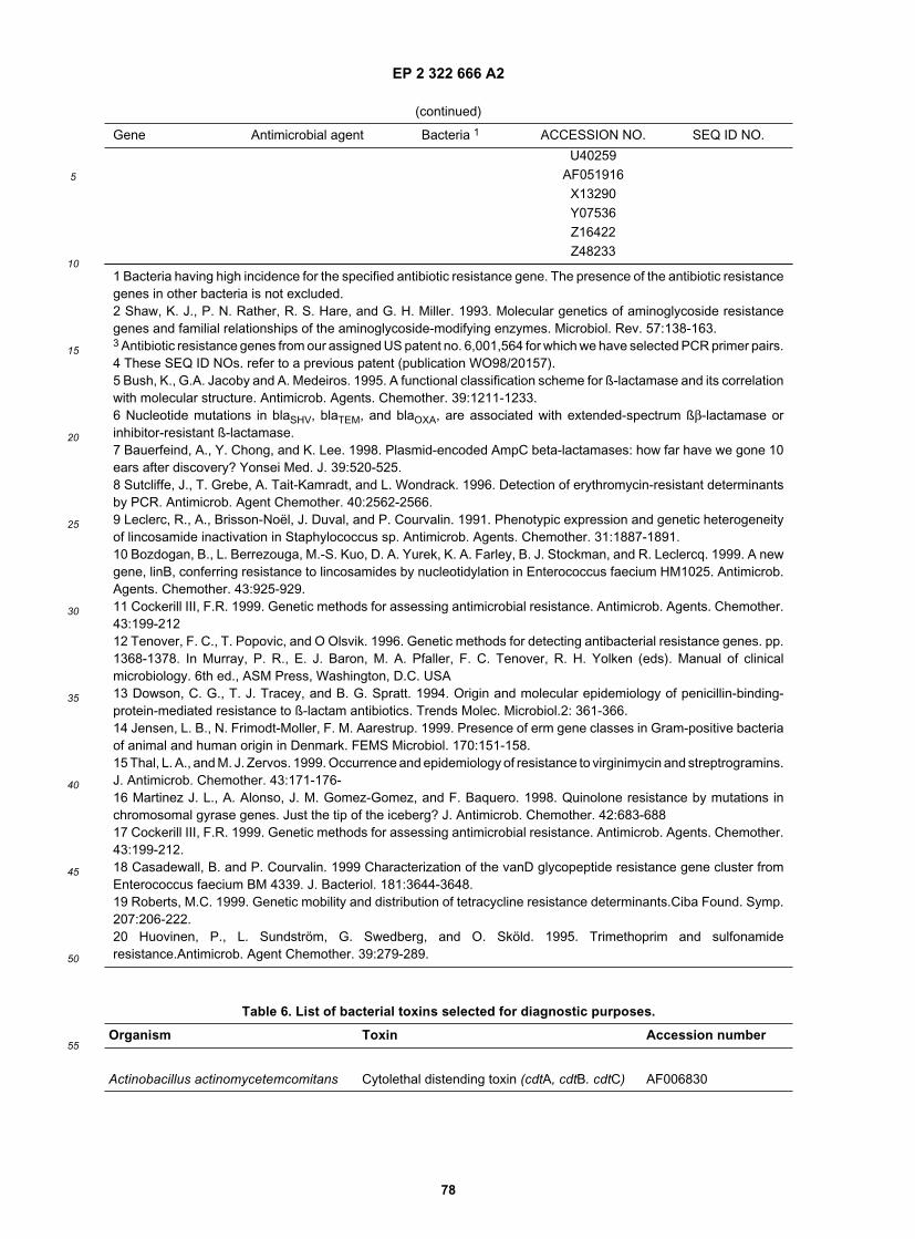
EP 2 322 666 A2
78
5
10
15
20
25
30
35
40
45
50
55
(continued)
Gene Antimicrobial agent Bacteria 1 ACCESSION NO. SEQ ID NO.
U40259AF051916
X13290Y07536Z16422Z48233
1 Bacteria having high incidence for the specified antibiotic resistance gene. The presence of the antibiotic resistancegenes in other bacteria is not excluded.2 Shaw, K. J., P. N. Rather, R. S. Hare, and G. H. Miller. 1993. Molecular genetics of aminoglycoside resistancegenes and familial relationships of the aminoglycoside-modifying enzymes. Microbiol. Rev. 57:138-163.3 Antibiotic resistance genes from our assigned US patent no. 6,001,564 for which we have selected PCR primer pairs.4 These SEQ ID NOs. refer to a previous patent (publication WO98/20157).5 Bush, K., G.A. Jacoby and A. Medeiros. 1995. A functional classification scheme for ß-lactamase and its correlationwith molecular structure. Antimicrob. Agents. Chemother. 39:1211-1233.6 Nucleotide mutations in blaSHV, blaTEM, and blaOXA, are associated with extended-spectrum ßβ-lactamase orinhibitor-resistant ß-lactamase.7 Bauerfeind, A., Y. Chong, and K. Lee. 1998. Plasmid-encoded AmpC beta-lactamases: how far have we gone 10ears after discovery? Yonsei Med. J. 39:520-525.8 Sutcliffe, J., T. Grebe, A. Tait-Kamradt, and L. Wondrack. 1996. Detection of erythromycin-resistant determinantsby PCR. Antimicrob. Agent Chemother. 40:2562-2566.9 Leclerc, R., A., Brisson-Noël, J. Duval, and P. Courvalin. 1991. Phenotypic expression and genetic heterogeneityof lincosamide inactivation in Staphylococcus sp. Antimicrob. Agents. Chemother. 31:1887-1891.10 Bozdogan, B., L. Berrezouga, M.-S. Kuo, D. A. Yurek, K. A. Farley, B. J. Stockman, and R. Leclercq. 1999. A newgene, linB, conferring resistance to lincosamides by nucleotidylation in Enterococcus faecium HM1025. Antimicrob.Agents. Chemother. 43:925-929.11 Cockerill III, F.R. 1999. Genetic methods for assessing antimicrobial resistance. Antimicrob. Agents. Chemother.43:199-21212 Tenover, F. C., T. Popovic, and O Olsvik. 1996. Genetic methods for detecting antibacterial resistance genes. pp.1368-1378. In Murray, P. R., E. J. Baron, M. A. Pfaller, F. C. Tenover, R. H. Yolken (eds). Manual of clinicalmicrobiology. 6th ed., ASM Press, Washington, D.C. USA13 Dowson, C. G., T. J. Tracey, and B. G. Spratt. 1994. Origin and molecular epidemiology of penicillin-binding-protein-mediated resistance to ß-lactam antibiotics. Trends Molec. Microbiol.2: 361-366.14 Jensen, L. B., N. Frimodt-Moller, F. M. Aarestrup. 1999. Presence of erm gene classes in Gram-positive bacteriaof animal and human origin in Denmark. FEMS Microbiol. 170:151-158.15 Thal, L. A., and M. J. Zervos. 1999. Occurrence and epidemiology of resistance to virginimycin and streptrogramins.J. Antimicrob. Chemother. 43:171-176-16 Martinez J. L., A. Alonso, J. M. Gomez-Gomez, and F. Baquero. 1998. Quinolone resistance by mutations inchromosomal gyrase genes. Just the tip of the iceberg? J. Antimicrob. Chemother. 42:683-68817 Cockerill III, F.R. 1999. Genetic methods for assessing antimicrobial resistance. Antimicrob. Agents. Chemother.43:199-212.18 Casadewall, B. and P. Courvalin. 1999 Characterization of the vanD glycopeptide resistance gene cluster fromEnterococcus faecium BM 4339. J. Bacteriol. 181:3644-3648.19 Roberts, M.C. 1999. Genetic mobility and distribution of tetracycline resistance determinants.Ciba Found. Symp.207:206-222.20 Huovinen, P., L. Sundström, G. Swedberg, and O. Sköld. 1995. Trimethoprim and sulfonamideresistance.Antimicrob. Agent Chemother. 39:279-289.
Table 6. List of bacterial toxins selected for diagnostic purposes.
Organism Toxin Accession number
Actinobacillus actinomycetemcomitans Cytolethal distending toxin (cdtA, cdtB. cdtC) AF006830

EP 2 322 666 A2
79
5
10
15
20
25
30
35
40
45
50
55
(continued)
Organism Toxin Accession number
Leukotoxin (ltxA) M27399Actinomyces pyogenes Hemolysin (pyolysin) U84782Aeromonas hydrophila Aerolysin (aerA) M16495
Haemolysin (hlyA) U81555Cytotonic enterotoxin (alt) L77573
Bacillus anthracis Anthrax toxin (cya) M23179Bacillus cereus Enterotoxin (bceT) D17312
AF192766, AF192767Enterotoxic hemolysin BL AJ237785Non-haemolytic enterotoxins A,B and C (nhe) Y19005
Bacillus mycoides Hemolytic enterotoxin HBL AJ243150 to AJ243153Bacillus pseudomycoides Hemolytic enterotoxin HBL AJ243154 to AJ243156Bacteroides fragilis Enterotoxin (bftP) U67735
Matrix metalloprotease/enterotoxin (fragilysin) S75941, AF038459Metalloprotease toxin-2 U90931
AF081785Metalloprotease toxin-3 AF056297
Bordetella bronchiseptica Adenylate cyclase hemolysin (cyaA) Z37112, U22953Dermonecrotic toxin (dnt) U59687
AB020025Bordetella pertussis Pertussis toxin (S1 subunit, tox) AJ006151
AJ006153AJ006155AJ006157AJ006159AJ007363M14378, M16494AJ007364M13223X16347
Adenyl cyclase (cya) 18323Dermonecrotic toxin (dnt) U10527
Campylobacterjejuni Cytolethal distending toxin (cdtA, cdtB. cdtC) U5112Citrobacter freundii Shiga-like toxin (slt-IIcA) X67514, S53206Clostridium botulinum Botulism toxin (BoNT) (A,B,E and F serotypes X52066, X52088
are neurotoxic for humans; the other serotypes X73423have not been considered) M30196
X70814X70819X71343Z11934X70817M81186X70818X70815X62089X62683S76749X81714X70816

EP 2 322 666 A2
80
5
10
15
20
25
30
35
40
45
50
55
(continued)
Organism Toxin Accession number
Clostridium botulinum X70820X70281L35496M92906
Clostridium difficile A toxin (enterotoxin) (tcdA) (cdtA) AB012304AF053400Y12616X51797X17194M30307
B toxin (cytotoxin) (toxB) (cdtB) Z23277X53138
Clostridium perfringens Alpha (phospholipase C) (cpa) L43545L43546L43547L43548X13608X17300D10248
Beta (dermonecrotic protein) (cpb) L13198X83275L77965
Enterotoxin (cpe) AJ000766M98037X81849X71844Y16009
Enterotoxin pseudogene (not expressed) AF037328AF037329AF037330
Epsilon toxin (etxD) M80837M95206X60694
Iota (Ia and Ib) X73562Lambda (metalloprotease) D45904Theta (perfringolysin O) M36704
Clostridium sordellii Cytotoxin L X82638Clostridium tetani Tetanos toxin X06214
X04436Corynebacterium diphtheriae Diphtheriae toxin X00703Corynebacterium pseudotuberculosis Phospholipase C A21336Eikenella corrodens lysine decarboxylase (cadA) U89166Enterobacter cloacae Shiga-like toxin II Z50754, U33502Enterococcus faecalis Cytolysin B (cylB) M38052Escherichia coli (EHEC) Hemolysin toxin (hlyA and ehxA) AF043471
X94129X79839X86087

EP 2 322 666 A2
81
5
10
15
20
25
30
35
40
45
50
55
(continued)
Organism Toxin Accession number
AB011549AF074613
Escherichia coli (EHEC) Shiga-like (Vero cytotoxin) (stx) X81418, M36727M14107, E03962M10133, E03959M12863, X07865X81417, Y10775X81416, Z50754X81415, X67515Z36900, AF043627L11078, M19473L04539, M17358L11079, M19437X65949, M24352M21534, X07903M29153, Z36899Z37725Z36901X61283AB017524U72191X61283
Escherichia coli (ETEC) Enterotoxin (heat-labile) (eltB) M17874M17873J01605AB011677
Enterotoxin (heat-stable) (astA) (estA1) L11241M58746M29255V00612J01831
Escherichia coli (other) Cytolethal-distending toxin U03293(cdt) (3 genes) U04208
U89305Cytotoxic necrotizing factor 1 (cnf1) U42629Microcin 24 (mtfS) U47048Autotransporter enterotoxin (Pet) (cytotoxin) AF056581
Haemophilus ducreyi Cytolethal distending toxin (cdtA, cdtB, cdtC) U53215Helicobacter pylori Vacuolating toxin (vacA) U07145
U80067U80068AF077938AF077939AF077940AF077941
Legionella pneumophila Structural toxin protein (rtxA) AF057703Listeria monocytogenes Listeriolysin O (lisA, hlyA) X15127
M24199

EP 2 322 666 A2
82
5
10
15
20
25
30
35
40
45
50
55
(continued)
Organism Toxin Accession number
X60035U25452U25443U25446U25449
Pasteurella multocida Mitogenic toxin (dermonecrotic toxin) X57775, Z28388X51512X52478
Proteus mirabilis Hemolysin (hpmA) M30186Pseudomoncts aeruginosa Cytotoxin (Enterotoxin A) X14956Salmonella typhimurium Calmodulin-sensitive adenylate cyalase toxin
(cya)AF060869
Cytolysin (salmolysin) (slyA) U03842Enterotoxin (stn) L16014
Serratia marcescens Hemolysin (shlA) M22618Shigella dysenteriae type 1 Shiga toxin (stxA and stxB) X07903, M32511
M19437M24352, M21947
Shigella flexneri ShET2 enterotoxin (senA) Z54211Z47381
Enterotoxin 1 (set1A and set1B) U35656Hemolysin E (hlyE, clyA, sheA) AF200955
Shigella sonnei Shiga toxin (stxA and stxB) AJ132761Sphingomonas paucimobilis Beta-hemolysin (hlyA) L01270Staphylococcus aureus Gamma-hemolysin (hlg2) D42143
L01055Enterotoxin U93688Enterotoxin A (sea) L22565, L22566
M18970Enterotoxin B M11118Enterotoxin C1 (entC1) X05815Enterotoxin C2 (entC2) P34071Enterotoxin C3 (entC3) X51661Enterotoxin D (sed) M94872Enterotoxin E M21319Enterotoxin G (seg) AF064773Enterotoxin H (seh) U11702Enterotoxin I (sei) AF064774Enterotoxin J AF053140Exfoliative toxin A (ETA, Epidermolytic toxin A) M17347
M17357L25372, M20371
Exfoliative toxin B (ETB) M17348, M13775Leukocidin R (F and S component, lukF andlukS;
X64389, S53213
Hemolysin B and C) X72700L01055
Toxic shock syndrome toxin 1 (TSST-1, X01645alpha toxin, alpha hemolysin) M90536

EP 2 322 666 A2
83
5
10
15
20
25
30
35
40
45
50
55
(continued)
Organism Toxin Accession number
J02615U93688
Staphylococcus epidermidis Delta toxin (hld) AF068634Staphylococcus intermedius Enterotoxin 1 U91526
Leukocidin R (F and S component, lukF andlukS ;
X79188
synergohymenotropic toxin)Streptococcus pneumoniae Pneumolysin X52474
Streptococcus pyogenes Streptococcus pyrogenic exotoxin A (speA) X61553 to X61573X03929U40453, M19350
Pyrogenic exotoxin B (speB) U63134M86905, M35110
Vibrio cholerae Cholerae toxin (ctxA and ctxB subunits) X00171X76390X58786X58785, S55782D30052D30053K02679AF175708
Accessory cholera enterotoxin (ace) Z22569, AF175708Heat-stable enterotoxin (sto) X74108, M85198
M97591, L03220Zonula occludens toxin (zot) M83563, AF175708
Vibrio parahaemolyticus Thermostable direct hemolysin (tdh) S67841Vibrio vulnificus Cytolysin (vvhA) M34670Yersinia enterocolitica Heat-stable enterotoxin (yst) U09235, X65999
Heat-stable enterotoxin type B (ystB) D88145Heat-stable enterotoxin type C (ystC) D63578
Yersinia kristensenii Enterotoxin X69218Yersinia pestis Toxin X92727
Table 7. Origin of the nucleic acids and/or sequences in the sequence listing.
SEQ ID NO. Archaeal, bacterial, fungal or parasitical species SourceGene*
1 Acinetobacter baumannii This patent tuf2 Actinomyces meyeri This patent tuf3 Aerococcus viridans This patent tuf4 Achromobacterxylosoxidans subsp. denitrificans This patent tuf5 Anaerorhabdus furcosus This patent tuf6 Bacillus anthracis This patent tuf7 Bacillus cereus This patent tuf8 Bacteroides distasonis This patent tuf9 Enterococcus casseliflavus This patent tuf
10 Staphylococcus saprophyticus This patent tuf11 Bacteroides ovatus This patent tuf12 Bartonella henselae This patent tuf

EP 2 322 666 A2
84
5
10
15
20
25
30
35
40
45
50
55
(continued)
SEQ ID NO. Archaeal, bacterial, fungal or parasitical species SourceGene*
13 Bifidobacterium adolescentis This patent tuf14 Bifidobacterium dentium This patent tuf15 Brucella abortus This patent tuf16 Burkholderia cepacia This patent tuf17 Cedecea davisae This patent tuf18 Cedecea neteri This patent tuf19 Cedecea lapagei This patent tuf20 Chlamydia pneumoniae This patent tuf21 Chlamydia psittaci This patent tuf22 Chlamydia trachomatis This patent tuf23 Chryseobacterium meningosepticum This patent tuf24 Citrobacter amalonaticus This patent tuf25 Citrobacter braakii This patent tuf26 Citrobacter koseri This patent tuf27 Citrobacter farmeri This patent tuf28 Citrobacter freundii This patent tuf29 Citrobacter sedlakii This patent tuf30 Citrobacter werkmanii This patent tuf31 Citrobacter youngae This patent tuf32 Clostridium perfringens This patent tuf33 Comamonas acidovorans This patent tuf34 Corynebacterium bovis This patent tuf35 Corynebacterium cervicis This patent tuf36 Corynebacterium flavescens This patent tuf37 Corynebacterium kutscheri This patent tuf38 Corynebacterium minutissimum This patent tuf39 Corynebacterium mycetoides This patent tuf40 Corynebacterium pseudogenitalium This patent tuf41 Corynebacterium renale This patent tuf42 Corynebacterium ulcerans This patent tuf43 Corynebacterium urealyticum This patent tuf44 Corynebacterium xerosis This patent tuf45 Coxiella burnetii This patent tuf46 Edwardsiella hoshinae This patent tuf47 Edwardsiella tarda This patent tuf48 Eikenella corrodens This patent tuf49 Enterobacter aerogenes This patent tuf50 Enterobacter agglomerans This patent tuf51 Enterobacter amnigenus This patent tuf52 Enterobacter asburiae This patent tuf53 Enterobacter cancerogenus This patent tuf54 Enterobacter cloacae This patent tuf55 Enterobacter gergoviae This patent tuf56 Enterobacter hormaechei This patent tuf57 Enterobacter sakazakii This patent tuf58 Enterococcus casseliflavus This patent tuf59 Enterococcus cecorum This patent tuf60 Enterococcus dispar This patent tuf61 Enterococcus durans This patent tuf
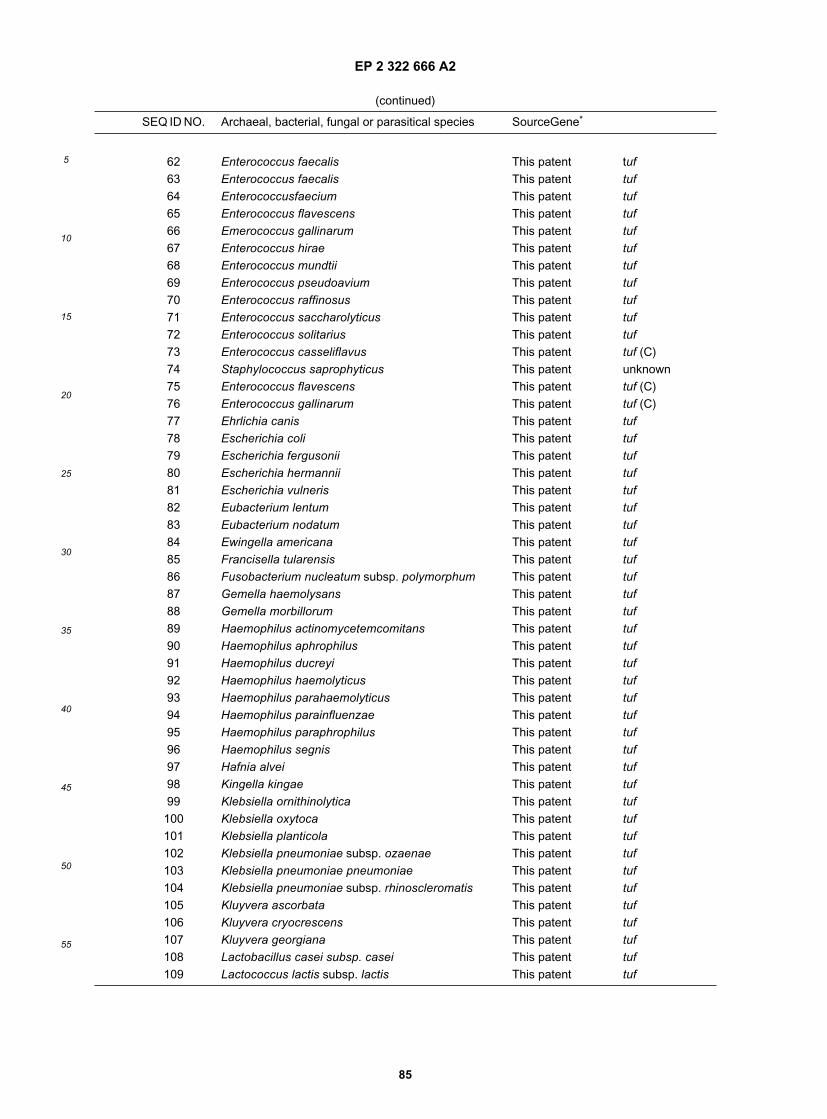
EP 2 322 666 A2
85
5
10
15
20
25
30
35
40
45
50
55
(continued)
SEQ ID NO. Archaeal, bacterial, fungal or parasitical species SourceGene*
62 Enterococcus faecalis This patent tuf63 Enterococcus faecalis This patent tuf64 Enterococcusfaecium This patent tuf65 Enterococcus flavescens This patent tuf66 Emerococcus gallinarum This patent tuf67 Enterococcus hirae This patent tuf68 Enterococcus mundtii This patent tuf69 Enterococcus pseudoavium This patent tuf70 Enterococcus raffinosus This patent tuf71 Enterococcus saccharolyticus This patent tuf72 Enterococcus solitarius This patent tuf73 Enterococcus casseliflavus This patent tuf (C)74 Staphylococcus saprophyticus This patent unknown75 Enterococcus flavescens This patent tuf (C)76 Enterococcus gallinarum This patent tuf (C)77 Ehrlichia canis This patent tuf78 Escherichia coli This patent tuf79 Escherichia fergusonii This patent tuf80 Escherichia hermannii This patent tuf81 Escherichia vulneris This patent tuf82 Eubacterium lentum This patent tuf83 Eubacterium nodatum This patent tuf84 Ewingella americana This patent tuf85 Francisella tularensis This patent tuf86 Fusobacterium nucleatum subsp. polymorphum This patent tuf87 Gemella haemolysans This patent tuf88 Gemella morbillorum This patent tuf89 Haemophilus actinomycetemcomitans This patent tuf90 Haemophilus aphrophilus This patent tuf91 Haemophilus ducreyi This patent tuf92 Haemophilus haemolyticus This patent tuf93 Haemophilus parahaemolyticus This patent tuf94 Haemophilus parainfluenzae This patent tuf95 Haemophilus paraphrophilus This patent tuf96 Haemophilus segnis This patent tuf97 Hafnia alvei This patent tuf98 Kingella kingae This patent tuf99 Klebsiella ornithinolytica This patent tuf100 Klebsiella oxytoca This patent tuf101 Klebsiella planticola This patent tuf102 Klebsiella pneumoniae subsp. ozaenae This patent tuf103 Klebsiella pneumoniae pneumoniae This patent tuf104 Klebsiella pneumoniae subsp. rhinoscleromatis This patent tuf105 Kluyvera ascorbata This patent tuf106 Kluyvera cryocrescens This patent tuf107 Kluyvera georgiana This patent tuf108 Lactobacillus casei subsp. casei This patent tuf109 Lactococcus lactis subsp. lactis This patent tuf

EP 2 322 666 A2
86
5
10
15
20
25
30
35
40
45
50
55
(continued)
SEQ ID NO. Archaeal, bacterial, fungal or parasitical species SourceGene*
110 Leclercia adecarboxylata This patent tuf111 Legionella micdadei This patent tuf112 Legionella pneumophila subsp. pneumophila This patent tuf113 Leminorella grimontii This patent tuf114 Leminorella richardii This patent tuf115 Leptospira interrogans This patent tuf116 Megamonas hypermegale This patent tuf117 Mitsuokella multacidus This patent tuf118 Mobiluncus curtisii subsp. holmesii This patent tuf119 Moellerella wisconsensis This patent tuf120 Moraxella catarrhalis This patent tuf121 Morganella morganii subsp. morganii This patent tuf122 Mycobacterium tuberculosis This patent tuf
123 Neisseria cinerea This patent tuf124 Neisseria elongata subsp. elongata This patent tuf125 Neisseria flavescens This patent tuf126 Neisseria gonorrhoeae This patent tuf127 Neisseria lactamica This patent tuf128 Neisseria meningitidis This patent tuf129 Neisseria mucosa This patent tuf130 Neisseria sicca This patent tuf131 Neisseria subflava This patent tuf132 Neisseria weaveri This patent tuf133 Ochrobactrum anthropi This patent tuf134 Pantoea agglomerans This patent tuf135 Pantoea dispersa This patent tuf136 Pasteurella multocida This patent tuf137 Peptostreptococcus anaerobius This patent tuf138 Peptostreptococcus asaccharolyticus This patent tuf139 Peptostreptococcus prevotii This patent tuf140 Porphyromonas asaccharolytica This patent tuf141 Porphyromonas gingivalis This patent tuf142 Pragia fontium This patent tuf143 Prevotella melaninogenica This patent tuf144 Prevotella oralis This patent tuf145 Propionibacterium acnes This patent tuf146 Proteus mirabilis This patent tuf147 Proteus penneri This patent tuf148 Proteus vulgaris This patent tuf149 Providencia alcalifaciens This patent tuf150 Providencia rettgeri This patent tuf151 Providencia rustigianii This patent tuf152 Providencia stuartii This patent tuf153 Pseudomonas aeruginosa This patent tuf154 Pseudomonas fluorescens This patent tuf155 Pseudomonas stutzeri This patent tuf156 Psychrobacter phenylpyruvicum This patent tuf157 Rahnella aquatilis This patent tuf

EP 2 322 666 A2
87
5
10
15
20
25
30
35
40
45
50
55
(continued)
SEQ ID NO. Archaeal, bacterial, fungal or parasitical species SourceGene*
158 Salmonella choleraesuis subsp.arizonae This patent tuf159 Salmonella choleraesuis subsp. choleraesuis
serotype CholeraesuisThis patent tuf
160 Salmonella choleraesuis subsp. diarizonae This patent tuf161 Salmonella choleraesuis subsp. choleraesuis
serotype HeidelbergThis patent tuf
162 Salmonella choleraesuis subsp. houtenae This patent tuf163 Salmonella choleraesuis subsp. indica This patent tuf164 Salmonella choleraesuis subsp. salamae This patent tuf165 Salmonella choleraesuis subsp. choleraesuis
serotype TyphiThis patent tuf
166 Serratia fonticola This patent tuf167 Serratia liquefaciens This patent tuf168 Serratia marcescens This patent tuf169 Serratia odorifera This patent tuf170 Serratia plymuthica This patent tuf171 Serratia rubidaea This patent tuf172 Shigella boydii This patent tuf173 Shigella dysenteriae This patent tuf174 Shigella flexneri This patent tuf175 Shigella sonnei This patent tuf176 Staphylococcus aureus This patent tuf177 Staphylococcus aureus This patent tuf178 Staphylococcus aureus This patent tuf179 Staphylococcus aureus This patem tuf180 Staphylococcus aureus subsp. aureus This patent tuf181 Staphylococcus auricularis This patent tuf182 Staphylococcus capitis subsp. capitis This patent tuf
183 Macrococcus caseolyticus This patent tuf184 Staphylococcus cohnii subsp. cohnii This patent tuf185 Staphylococcus epidermidis This patent tuf186 Staphylococcus haemolyticus This patent tuf187 Staphylococcus warneri This patent tuf188 Staphylococcus haemolyticus This patent tuf189 Staphylococcus haemolyticus This patent tuf190 Staphylococcus haemolyticus This patent tuf191 Staphylococcus hominis subsp. hominis This patent tuf192 Staphylococcus warneri This patent tuf193 Staphylococcus hominis This patent tuf194 Staphylococcus hominis This patent tuf195 Staphylococcus hominis This patent tuf196 Staphylococcus hominis This patent tuf197 Staphylococcus lugdunensis This patent tuf198 Staphylococcus saprophyticus This patent tuf199 Staphylococcus saprophyticus This patent tuf200 Staphylococcus saprophyticus This patent tuf201 Staphylococcus sciuri subsp. sciuri This patent tuf202 Staphylococcus warneri This patent tuf203 Staphylococcus warneri This patent tuf

EP 2 322 666 A2
88
5
10
15
20
25
30
35
40
45
50
55
(continued)
SEQ ID NO. Archaeal, bacterial, fungal or parasitical species SourceGene*
204 Bifidobacterium longum This patent tuf205 Stenotrophomonas maltophilia This patent tuf206 Streptococcus acidominimus This patent tuf207 Streptococcus agalactiae This patent tuf208 Streptococcus agalactiae This patent tuf209 Streptococcus agalactiae This patent tuf210 Streptococcus agalactiae This patent tuf211 Streptococcus anginosus This patent tuf212 Streptococcus bovis This patent tuf213 Streptococcus anginosus This patent tuf214 Streptococcus cricetus This patent tuf215 Streptococcus cristatus This patent tuf216 Streptococcus downei This patent tuf217 Streptococcus dysgalactiae This patent tuf218 Streptococcus equi subsp. equi This patent tuf219 Streptococcus ferus This patent tuf220 Streptococcus gordonii This patent tuf221 Streptococcus anginosus This patent tuf222 Streptococcus macacae This patent tuf223 Streptococcus gordonii This patent tuf224 Streptococcus mutans This patent tuf225 Streptococcus parasanguinis This patent tuf226 Streptococcus ratti This patent tuf227 Streptococcus sanguinis This patent tuf228 Streptococcus sobrinus This patent tuf229 Streptococcus suis This patent tuf230 Streptococcus uberis This patent tuf231 Streptococcus vestibularis This patent tuf232 Tatumella ptyseos This patent tuf233 Trabulsiella guamensis This patent tuf234 Veillonella parvula This patent tuf235 Yersinia enterocolitica This patent tuf236 Yersinia frederiksenii This patent tuf237 Yersinia intermedia This patent tuf238 Yersinia pestis This patent tuf239 Yersinia pseudotuberculosis This patent tuf240 Yersinia rohdei This patent tuf241 Yokenella regensburgei This patent tuf242 Achromobacter xylosoxidans subsp. denitrificans This patent atpD243 Acinetobacter baumannii This patent atpD244 Acinetobacter lwoffii This patent atpD
245 Staphylococcus saprophyticus This patent atpD246 Alcaligenes faecalis subsp. faecalis This patent atpD247 Bacillus anthracis This patent atpD248 Bacillus cereus This patent atpD249 Bacteroides distasonis This patent atpD250 Bacteroides ovatus This patent atpD251 Leclercia adecarboxylata This patent atpD

EP 2 322 666 A2
89
5
10
15
20
25
30
35
40
45
50
55
(continued)
SEQ ID NO. Archaeal, bacterial, fungal or parasitical species SourceGene*
252 Stenotrophomonas maltophilia This patent atpD253 Bartonella henselae This patent atpD254 Bifidobacterium adolescentis This patent atpD255 Brucella abortus This patent atpd256 Cedecea davisae This patent atpD257 Cedecea lapagei This patent atpD258 Cedecea neteri This patent atpD259 Chryseobacterium meningosepticum This patent atpD260 Citrobacter amalonaticus This patent atpD261 Citrobacter braakii This patent atpD262 Citrobacter koseri This patent atpD263 Citrobacter farmeri This patent atpD264 Citrobacter freundii This patent atpD265 Citrobacter koseri This patent atpD266 Citrobacter sedlakii This patent atpD267 Citrobacter werkmanii This patent atpD268 Citrobacter youngae This patent atpD269 Clostridiwn innocuum This patent atpD270 Clostridium perfringens This patent atpD272 Corynebacteriwn diphtheriae This patent atpD273 Corynebacteriwn pseudodiphtheriticum This patent atpD274 Corynebacterium ulceratts This patent atpD275 Corynebacterium urealyticum This patent atpD276 Coxiella burnetii This patent atpD277 Edwardsiella hoshinae This patent atpD278 Edwardsiella tarda This patent atpD279 Eikenella corrodens This patent atpD280 Enterobacter agglomerans This patent atpD281 Enterobacter amnigenus This patent atpD282 Enterobacter asburiae This patent atpD283 Enterobacter cancerogenus This patent atpD284 Enterobacter cloacae This patent atpD285 Enterobacter gergoviae This patent atpD286 Enterobacter hormaechei This patent atpD287 Enterobacter sakazakii This patent atpD288 Enterococcus avium This patent atpD289 Enterococcus casseliflavus This patent atpD290 Enterococcus durans This patent atpD291 Enterococcus faecalis This patent atpD292 Enterococcus faecium This patent atpD293 Enterococcus gallinarum This patent atpD294 Enterococcus saccharolyticus This patent atpD295 Escherichia fergusonii This patent atpD296 Escherichia hermannii This patent atpD297 Escherichia vulneris This patent atpD298 Eubacterium lentum This patent atpD299 Ewingella americana This patent atpD300 Francisella tularensis This patent atpD301 Fusobacterium gonidiaformans This patent atpD302 Fusobacterium necrophorum subsp. necrophorum This patent atpD

EP 2 322 666 A2
90
5
10
15
20
25
30
35
40
45
50
55
(continued)
SEQ ID NO. Archaeal, bacterial, fungal or parasitical species SourceGene*
303 Fusobacterium nucleatum subsp. polymorphum This patent atpD304 Gardnerella vaginalis This patent atpD305 Gemella haemolysans This patent atpD306 Gemella morbillorum This patent atpD
307 Haemophilus ducreyi This patent atpD308 Haemophilus haemolyticus This patent atpD309 Haemophilus parahaemolyticus This patent atpD310 Haemophilus parainfluenzae This patent atpD311 Hafnia alvei This patent atpD312 Kingella kingae This patent atpD313 Klebsiella pneumoniae subsp. ozaenae This patent atpD314 Klebsiella ornithinolytica This patent atpD315 Klebsiella oxytoca This patent atpD316 Klebsiella planticola This patent atpD317 Klebsiella pneumoniae subsp. pneumoniae This patent atpD318 Kluyvera ascorbata This patent atpD319 Kluyvera cryocrescens This patent atpD320 Kluyvera georgiana This patent atpD321 Lactobacillus acidophilus This patent atpD322 Legionella pneumophila subsp. pneumophila This patent atpd323 Leminorella grimontii This patent atpD324 Listeria monocytogenes This patent atpD325 Micrococcus lylae This patent atpD326 Moellerella wisconsensis This patent atpD327 Moraxella catarrhalis This patent atpD328 Moraxella osloensis This patent atpD329 Morganella morganii subsp. morganii This patent atpD330 Pantoea agglomerans This patent atpD331 Pantoea dispersa This patent atpD332 Pasteurella multocida This patent atpD333 Pragia fontium This patent atpD334 Proteus mirabilis This patent atpd335 Proteus vulgaris This patent atpD336 Providencia alcalifaciens This patent atpD337 Providencia rettgeri This patent atpD338 Providencia rustigianii This patent atpD339 Providencia stuartii This patent atpD340 Psychrobacter phenylpyruvicum This patent atpD341 Rahnella aquatilis This patent atpD342 Salmonella choleraesuis subsp. arizonae This patent atpD343 Salmonella choleraesuis subsp. choleraesuis
serotype CholeraesuisThis patent atpD
344 Salmonella choleraesuis subsp. diarizonae This patent atpD345 Salmonella choleraesuis subsp. houtenae This patent atpD346 Salmonella choleraesuis subsp. indica This patent atpD347 Salmonella choleraesuis subsp. choleraesuis
serotype Paratyphi AThis patent atpD
348 Salmonella choleraesuis subsp. choleraesuisserotype Paratyphi B
This patent atpD

EP 2 322 666 A2
91
5
10
15
20
25
30
35
40
45
50
55
(continued)
SEQ ID NO. Archaeal, bacterial, fungal or parasitical species SourceGene*
349 Salmonella choleraesuis subsp. salamae This patent atpD350 Salmonella choleraesuis subsp. choleraesuis
serotype TyphiThis patent atpD
351 Salmonella choleraesuis subsp. choleraesuisserotype Typhimurium
This patent atpD
352 Salmonella choleraesuis subsp. choleraesuisserotype Virchow
This patent atpD
353 Serratia ficaria This patent atpD354 Serratia fonticola This patent atpD355 Serratia grimesii This patent atpD356 Serratia liquefaciens This patent atpD357 Serratia marcescens This patent atpD358 Serratia odorifera This patent atpD359 Serratia plymuthica This patent atpD360 Serratia rubidaea This patent atpD361 Pseudomonas putida This patent atpD362 Shigella boydii This patent atpD363 Shigella dysenteriae This patent atpD
364 Shigella flexneri This patent atpD365 Shigella sonnei This patent atpD366 Staphylococcus aureus This patent atpD367 Staphylococcus auricularis This patent atpD368 Staphylococcus capitis subsp. capitis This patent atpD369 Staphylococcus cohnii subsp. cohnii This patent atpD370 Staphylococcus epidermidis This patent atpD371 Staphylococcus haemolyticus This patent atpD372 Staphylococcus hominis subsp. hominis This patent atpD373 Staphylococcus hominis This patent atpD374 Staphylococcus lugdunensis This patent atpD375 Staphylococcus saprophyticus This patent atpD376 Staphylococcus simulans This patent atpD377 Staphylococcus warneri This patent atpD378 Streptococcus acidominimus This patent atpD379 Streptococcus agalactiae This patent atpD380 Streptococcus agalactiae This patent atpD381 Streptococcus agalactiae This patent atpD382 Streptococcus agalactiae This patent atpD383 Streptococcus agalactiae This patent atpD384 Streptococcus dysgalactiae This patent atpD385 Streptococcus equi subsp. equi This patent atpD386 Streptococcus anginosus This patent atpD387 Streptococcus salivarius This patent atpD388 Streptococcus suis This patent atpD389 Streptococcus uberis This patent atpD390 Tatumella ptyseos This patent atpD391 Trabulsiella guamensis This patent atpD392 Yersinia bercovieri This patent atpD393 Yersinia enterocolitica This patent atpD394 Yersinia frederiksenii This patent atpD
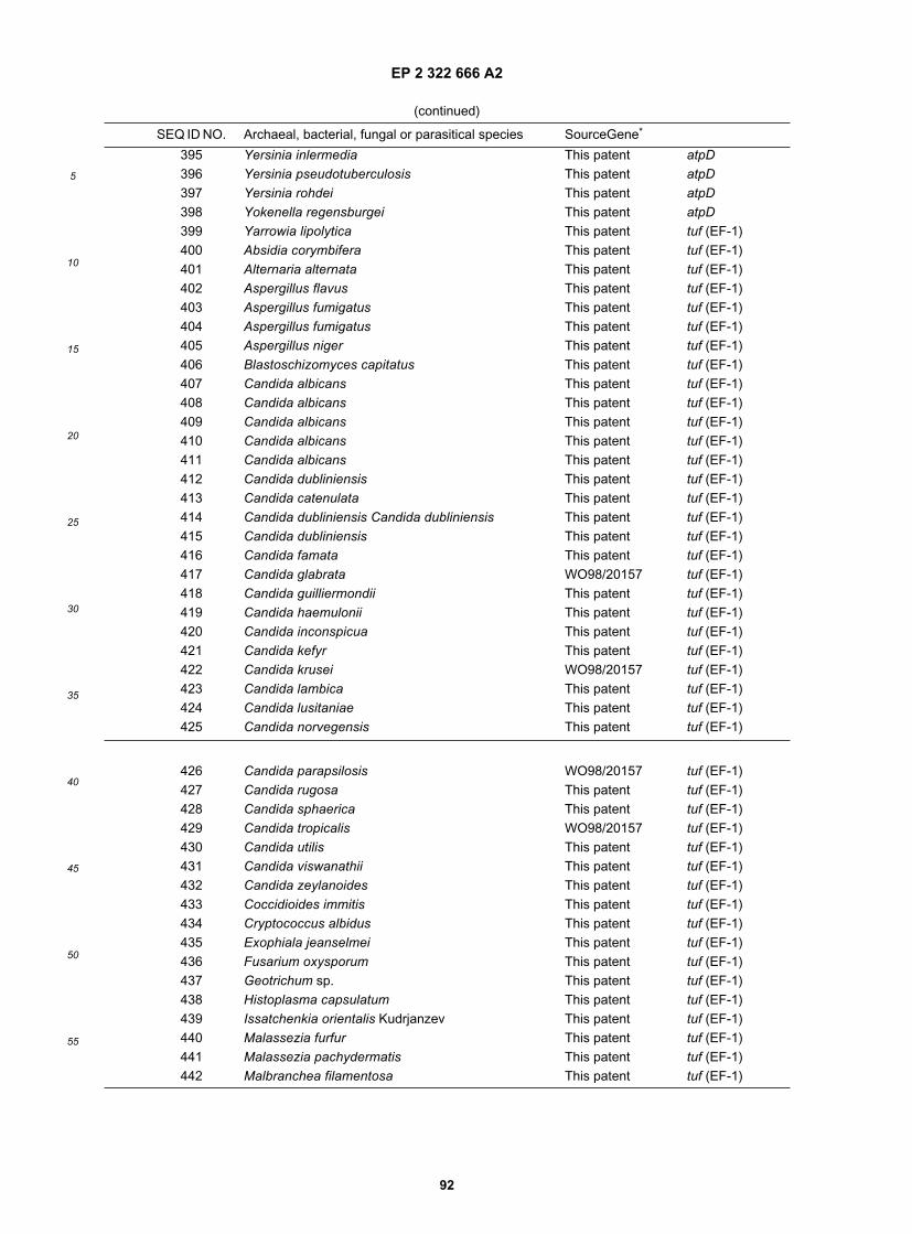
EP 2 322 666 A2
92
5
10
15
20
25
30
35
40
45
50
55
(continued)
SEQ ID NO. Archaeal, bacterial, fungal or parasitical species SourceGene*
395 Yersinia inlermedia This patent atpD396 Yersinia pseudotuberculosis This patent atpD397 Yersinia rohdei This patent atpD398 Yokenella regensburgei This patent atpD399 Yarrowia lipolytica This patent tuf (EF-1)400 Absidia corymbifera This patent tuf (EF-1)401 Alternaria alternata This patent tuf (EF-1)402 Aspergillus flavus This patent tuf (EF-1)403 Aspergillus fumigatus This patent tuf (EF-1)404 Aspergillus fumigatus This patent tuf (EF-1)405 Aspergillus niger This patent tuf (EF-1)406 Blastoschizomyces capitatus This patent tuf (EF-1)407 Candida albicans This patent tuf (EF-1)408 Candida albicans This patent tuf (EF-1)409 Candida albicans This patent tuf (EF-1)410 Candida albicans This patent tuf (EF-1)411 Candida albicans This patent tuf (EF-1)412 Candida dubliniensis This patent tuf (EF-1)413 Candida catenulata This patent tuf (EF-1)414 Candida dubliniensis Candida dubliniensis This patent tuf (EF-1)415 Candida dubliniensis This patent tuf (EF-1)416 Candida famata This patent tuf (EF-1)417 Candida glabrata WO98/20157 tuf (EF-1)418 Candida guilliermondii This patent tuf (EF-1)419 Candida haemulonii This patent tuf (EF-1)420 Candida inconspicua This patent tuf (EF-1)421 Candida kefyr This patent tuf (EF-1)422 Candida krusei WO98/20157 tuf (EF-1)423 Candida lambica This patent tuf (EF-1)424 Candida lusitaniae This patent tuf (EF-1)425 Candida norvegensis This patent tuf (EF-1)
426 Candida parapsilosis WO98/20157 tuf (EF-1)427 Candida rugosa This patent tuf (EF-1)428 Candida sphaerica This patent tuf (EF-1)429 Candida tropicalis WO98/20157 tuf (EF-1)430 Candida utilis This patent tuf (EF-1)431 Candida viswanathii This patent tuf (EF-1)432 Candida zeylanoides This patent tuf (EF-1)433 Coccidioides immitis This patent tuf (EF-1)434 Cryptococcus albidus This patent tuf (EF-1)435 Exophiala jeanselmei This patent tuf (EF-1)436 Fusarium oxysporum This patent tuf (EF-1)437 Geotrichum sp. This patent tuf (EF-1)438 Histoplasma capsulatum This patent tuf (EF-1)439 Issatchenkia orientalis Kudrjanzev This patent tuf (EF-1)440 Malassezia furfur This patent tuf (EF-1)441 Malassezia pachydermatis This patent tuf (EF-1)442 Malbranchea filamentosa This patent tuf (EF-1)

EP 2 322 666 A2
93
5
10
15
20
25
30
35
40
45
50
55
(continued)
SEQ ID NO. Archaeal, bacterial, fungal or parasitical species SourceGene*
443 Metschnikowia pulcherrima This patent tuf (EF-1)444 Paecilomyces lilacinus This patent tuf (EF-1)445 Paracoccidioides brasiliensis This patent tuf (EF-1)446 Penicillium marneffei This patent tuf (EF-1)447 Pichia anomala This patent tuf (EF-1)448 Pichia anomala This patent tuf (EF-1)449 Pseudallescheria boydii This patent tuf (EF-1)450 Rhizopus oryzae This patent tuf (EF-1)451 Rhodotorula minuta This patent tuf (EF-1)452 Sporobolomyces salmonicolor This patent tuf (EF-1)453 Sporothrix schenckii This patent tuf (EF-1)454 Stephanoascus ciferrii This patent tuf (EF-1)455 Trichophyton mentagrophytes This patent tuf (EF-1)456 Trichosporon cutaneum This patent tuf (EF-1)457 Wangiella dermatitidis This patent tuf (EF-1)458 Aspergillus fumigatus This patent atpD459 Blastoschizomyces capitatus This patent atpD460 Candida albicans This patent atpD461 Candida dubliniensis This patent atpD462 Candida famata This patent atpD463 Candida glabrata This patent atpD464 Candida guilliermondii This patent atpD465 Candida haemulonii This patent atpD466 Candida inconspicua This patent atpD467 Candida kefyr This patent atpD468 Candida krusei This patent atpD469 Candida lambica This patent atpD470 Candida lusitaniae This patent atpD471 Candida norvegensis This patent atpD472 Candida parapsilosis This patent atpD473 Candida rugosa This patent atpD474 Candida sphaerica This patent atpD475 Candida tropicalis This patent atpD476 Candida utilis This patent atpD477 Candida viswanathii This patent atpD478 Candida zeylanoides This patent atpD479 Coccidioides immitis This patent atpD480 Cryptococcus albidus This patent atpD481 Fusarium oxysporum This patent atpD482 Geotrichum sp. This patent atpD483 Histoplasma capsulatum This patent atpD484 Malassezia furfur This patent atpD485 Malassezia pachydermatis This patent atpD486 Metschnikowia pulcherrima This patent atpD487 Penicillium marneffei This patent atpD
488 Pichia anomala This patent atpD489 Pichia anomala This patent atpD490 Rhodotorula minuta This patent atpD

EP 2 322 666 A2
94
5
10
15
20
25
30
35
40
45
50
55
(continued)
SEQ ID NO. Archaeal, bacterial, fungal or parasitical species SourceGene*
491 Rhodotorula mucilaginosa This patent atpD492 Sporobolomyces salmonicolor This patent atpD493 Sporothrix schenckii This patent atpD494 Stephanoascus ciferrii This patent atpD495 Trichophyton mentagrophytes This patent atpD496 Wangiella dermatitidis This patent atpD497 Yarrowia lipolytica This patent atpD498 Aspergillus fumigatus This patent tuf (M)499 Blastoschizomyces capitatus This patent tuf (M)500 Candida rugosa This patent tuf (M)501 Coccidioides immitis This patent tuf (M)502 Fusarium oxysporum This patent tuf (M)503 Histoplasma capsulatum This patent tuf (M)504 Paracoccidioides brasiliensis This patent tuf (M)505 Penicillium marneffei This patent tuf (M)506 Pichia anomala This patent tuf (M)507 Trichophyton mentagrophytes This patent tuf (M)508 Yarrowia lipolytica This patent tuf (M)509 Babesia bigemina This patent tuf (EF-1)510 Babesia bovis This patent tuf (EF-1)511 Crithidia fasciculata This patent tuf (EF-1)512 Entamoeba histolytica This patent tuf (EF-1)513 Giardia lamblia This patent tuf (EF-1)514 Leishmania tropica This patent tuf (EF-1)515 Leishmania aethiopica This patent tuf (EF-1)516 Leishmania tropica This patent tuf (EF-1)517 Leishmania donovani This patent tuf (EF-1)518 Leishmania infantum This patent tuf (EF-1)519 Leishmania enriettii This patent tuf (EF-1)520 Leishmania gerbilli This patent tuf (EF-1)521 Leishmania hertigi This patent tuf (EF-1)522 Leishmania major This patent tuf (EF-1)523 Leishmania amazonensis This patent tuf (EF-1)524 Leishmania mexicana This patent tuf (EF-1)525 Leishmania tarentolae This patent tuf (EF-1)526 Leishmania tropica This patent tuf (EF-1)527 Neospora caninum This patent tuf (EF-1)528 Trichomonas vaginalis This patent tuf (EF-1)529 Trypanosoma brucei subsp. brucei This patent tuf (EF-1)530 Crithidia fasciculata This patent atpD531 Leishmania tropica This patent atpD532 Leishmania aethiopica This patent atpD533 Leishmania donovani This patent atpD534 Leishmania infantum This patent atpD535 Leishmania gerbilli This patent atpD536 Leishmania hertigi This patent atpD537 Leishmania major This patent atpD538 Leishmania amazonensis This patent atpD607 Enterococcus faecalis WO98/20157 tuf608 Enterococcus faecium WO98/20157 tuf

EP 2 322 666 A2
95
5
10
15
20
25
30
35
40
45
50
55
(continued)
SEQ ID NO. Archaeal, bacterial, fungal or parasitical species SourceGene*
609 Enterococcus gallinarum WO98/20157 tuf610 Haemophilus influenzae WO98/20157 tuf611 Staphylococcus epidermidis WO98/20157 tuf612 Salmonella choleraesuis subsp. choleraesuis
serotype Paratyphi AThis patent tuf
613 Serratia ficaria This patent tuf614 Enterococcus malodoratus This patent tuf (C)615 Enterococcus durans This patent tuf (C)616 Enterococcus pseudoavium This patent tuf (C)
617 Enterococcus dispar This patent tuf (C)618 Enterococcus avium This patent tuf (C)619 Saccharomyces cerevisiae Database tuf (M)621 Enterococcus faecium This patent tuf (C)622 Saccharomyces cerevisiae This patent tuf (EF-1)623 Cryptococcus neoformans This patent tuf (EF-1)624 Candida albicans WO98/20157 tuf (EF-1)662 Corynebacterium diphtheriae WO98/20157 tuf663 Candida catenulata This patent atpD665 Saccharomyces cerevisiae Database tuf (EF-1)666 Saccharomyces cerevisiae Database atpD667 Trypanosoma cruzi This patent atpD668 Corynebacterium glutamicum Database tuf669 Escherichia coli Database atpD670 Helicobacter pylori Database atpD671 Clostridium acetobutylicum Database atpD672 Cytophaga lytica Database atpD673 Ehrlichia risticii This patent atpD674 Vibrio cholerae This patent atpD675 Vibrio cholerae This patent tuf676 Leishmania enriettii This patent atpD677 Babesia microti This patent tuf (EF-1)678 Cryptococcus neoformans This patent atpD679 Cryptococcus neoformans This patent atpD680 Cunninghamella benholletiae This patent atpD684 Candida tropicalis Database atpD (V)685 Enterococcus hirae Database atpD (V)686 Chlamydia pneumoniae Database atpD (V)687 Halobacterium salinarum Database atpD (V)688 Homo sapiens Database atpD (V)689 Plasmodium falciparum Database atpD (V)690 Saccharomyces cerevisiae Database atpD (V)691 Schizosaccharomyces pombe Database atpD (V)692 Trypanosoma congolense Database atpD (V)693 Thermus thermophilus Database atpD (V)698 Escherichia coli WO98/20157 tuf709 Borrelia burgdorferi Database atpD (V)710 Treponema pallidum Database atpD (V)711 Chlamydia trachomatis Genome project atpD (V)

EP 2 322 666 A2
96
5
10
15
20
25
30
35
40
45
50
55
(continued)
SEQ ID NO. Archaeal, bacterial, fungal or parasitical species SourceGene*
712 Enterococcus faecalis Genome project atpD (V)713 Methanosarcina barkeri Database atpD (V)714 Methanococcus jannaschii Database atpD (V)715 Porphyromonas gingivalis Genome project atpD (V)716 Streptococcus pneumoniae Genome project atpD (V)717 Burkholderia mallei This patent tuf718 Burkholderia pseudomallei This patent tuf719 Clostridium beijerinckii This patent tuf720 Clostridium innocuum This patent tuf721 Clostridium novyi This patent tuf722 Clostridium septicum This patent tuf723 Clostridium tertium This patent tuf724 Clostridium tetani This patent tuf725 Enterococcus malodoratus This patent tuf726 Enterococcus sulfureus This patent tuf727 Lactococcus garvieae This patent tuf728 Mycoplasma pirum This patent tuf729 Mycoplasma salivarium This patent tuf730 Neisseria polysaccharea This patent tuf731 Salmonella choleraesuis subsp. choleraesuis
serotype EnteritidisThis patent tuf
732 Salmonella choleraesuis subsp. choleraesuisserotype Gallinarum
This patent tuf
733 Salmonella choleraesuis subsp. choleraesuisserotype Paratyphi B
This patent tuf
734 Salmonella choleraesuis subsp. choleraesuisserotype Virchow
This patent tuf
735 Serratia grimesii This patent tuf736 Clostridium difficile This patent tuf737 Burkholderia pseudomallei This patent atpD738 Clostridium bifermentans This patent atpD739 Clostridium beijerinckii This patent atpD740 Clostridium difficile This patent atpD741 Clostridium ramosum This patent atpD742 Clostridium septicum This patent atpD743 Clostridium tertium This patent atpD744 Comamonas acidovorans This patent atpD745 Klebsiella pneumoniae subsp. rhinoscleromatis This patent atpD746 Neisseria canis This patent atpD747 Neisseria cinerea This patent atpD748 Neisseria cuniculi This patent atpD749 Neisseria elongata subsp. elongata This patent atpD750 Neisseria flavescens This patent atpD751 Neisseria gonorrhoeae This patent atpD752 Neisseria gonorrhoeae This patent atpD753 Neisseria lactamica This patent atpD754 Neisseria meningitidis This patent atpD755 Neisseria mucosa This patent atpD756 Neisseria subflava This patent atpD757 Neisseria weaveri This patent atpD

EP 2 322 666 A2
97
5
10
15
20
25
30
35
40
45
50
55
(continued)
SEQ ID NO. Archaeal, bacterial, fungal or parasitical species SourceGene*
758 Neisseria animalis This patent atpD759 Proteus penneri This patent atpD760 Salmonella choleraesuis subsp. choleraesuis
serotype EnteritidisThis patent atpD
761 Yersinia pestis This patent atpD762 Burkholderia mallei This patent atpD763 Clostridium sordellii This patent atpD764 Clostridium novyi This patent atpD765 Clostridium botulinum This patent atpD766 Clostridium histolyticum This patent atpD767 Peptostreptococcus prevotii This patent atpD768 Absidia corymbifera This patent atpD769 Alternaria alternata This patent atpD770 Aspergillus flavus This patent atpD771 Mucor circinelloides This patent atpD772 Piedraia hortai This patent atpD773 Pseudallescheria boydii This patent atpD774 Rhizopus oryzae This patent atpD775 Scopulariopsis koningii This patent atpD776 Trichophyton mentagrophytes This patent atpD777 Trichophyton tonsurans This patent atpD778 Trichosporon cutaneum This patent atpD779 Cladophialophora carrionii This patent tuf (EF-1)780 Cunninghamella bertholletiae This patent tuf (EF-1)781 Curvularia lunata This patent tuf (EF-1)782 Fonsecaea pedrosoi This patent tuf (EF-1)783 Microsporum audouinii This patent tuf (EF-1)784 Mucor circinelloides This patent tuf (EF-1)785 Phialophora verrucosa This patent tuf (EF-1)786 Saksenaea vasiformis This patent tuf (EF-1)787 Syncephalastrum racemosum This patent tuf (EF-1)788 Trichophyton tonsurans This patent tuf (EF-1)789 Trichophyton mentagrophytes This patent tuf (EF-1)
790 Bipolaris hawaiiensis This patent tuf (EF-1)791 Aspergillus fumigatus This patent tuf (M)792 Trichophyton mentagrophytes This patent tuf (M)827 Clostridium novyi This patent atpD (V)828 Clostridium difficile This patent atpD (V)829 Clostridium septicum This patent atpD (V)830 Clostridium botulinum This patent atpD (V)831 Clostridium perfringens This patent atpD (V)832 Clostridium tetani This patent atpD (V)833 Streptococcus pyogenes Database atpD (V)834 Babesia bovis This patent atpD (V)835 Cryptosporidium parvum This patent atpD (V)836 Leishmania infantum This patent atpD (V)837 Leishmania major This patent atpD (V)838 Leishmania tarentolae This patent atpD (V)

EP 2 322 666 A2
98
5
10
15
20
25
30
35
40
45
50
55
(continued)
SEQ ID NO. Archaeal, bacterial, fungal or parasitical species SourceGene*
839 Trypanosoma brucei This patent atpD (V)840 Trypanosoma cruzi This patent tuf (EF-1)841 Trypanosoma cruzi This patent tuf (EF-1)842 Trypanosoma cruzi This patent tuf (EF-1)843 Babesia bovis This patent tuf (M)844 Leishmania aethiopica This patent tuf (M)845 Leishmania amazonensis This patent tuf (M)846 Leishmania donovani This patent tuf (M)847 Leishmania infantum This patent tuf (M)848 Leishmania enriettii This patent tuf (M)849 Leishmania gerbilli This patent tuf (M)850 Leishmania major This patent tuf (M)851 Leishmania mexicana This patent tuf (M)852 Leishmania tarentolae This patent tuf (M)853 Trypanosoma cruzi This patent tuf (M)854 Trypanosoma cruzi This patent tuf (M)855 Trypanosoma cruzi This patent tuf (M)856 Babesia bigemina This patent atpD857 Babesia bovis This patent atpD858 Babesia microti This patent atpD859 Leishmania guyanensis This patent atpD860 Leishmania mexicana This patent atpD861 Leishmania tropica This patent atpD862 Leishmania tropica This patent atpD863 Bordetella pertussis Database tuf864 Trypanosoma brucei brucei Database tuf (EF-1)865 Cryptosporidium parvum This patent tuf (EF-1)866 Staphylococcus saprophyticus This patent atpD867 Zoogloea ramigera This patent atpD868 Staphylococcus saprophyticus This patent tuf869 Enterococcus casseliflavus This patent tuf870 Enterococcus casseliflavus This patent tuf871 Enterococcus flavescens This patent tuf872 Enterococcus gallinarum This patent tuf873 Enterococcus gallinarum This patent tuf874 Staphylococcus haemolyticus This patent tuf875 Staphylococcus epidermidis This patent tuf876 Staphylococcus epidermidis This patent tuf877 Staphylococcus epidermidis This patent tuf878 Staphylococcus epidermidis This patent tuf879 Enlerococcus gallinarum This patent tuf880 Pseudomonas aeruginosa This patent tuf881 Enterococcus casseliflavus This patent tuf882 Enterococcus casseliflavus This patent tuf883 Enterococcus faecalis This patent tuf884 Enterococcus faecalis This patent tuf885 Enterocaccus faecium This patent tuf
886 Enterococcus faecium This patent tuf

EP 2 322 666 A2
99
5
10
15
20
25
30
35
40
45
50
55
(continued)
SEQ ID NO. Archaeal, bacterial, fungal or parasitical species SourceGene*
887 Zoogloea ramigera This patent tuf888 Enterococcus faecalis This patent tuf889 Aspergillus fumigatus This patent atpD890 Penicillium marneffei This patent atpD891 Paecilomyces lilacinus This patent atpD892 Penicillium marneffei This patent atpD893 Sporothrix schenckii This patent atpD894 Malbranchea filamentosa This patent atpD895 Paecilomyces lilacinus This patent atpD896 Aspergillus niger This patent atpD897 Aspergillus fumigatus This patent tuf (EF-1)898 Penicillium marneffei This patent tuf (EF-1)899 Piedraia hortai This patent tuf (EF-1)900 Paecilomyces lilacinus This patent tuf (EF-1)901 Paracoccidioides brasiliensis This patent tuf (EF-1)902 Sporothrix schenckii This patent tuf (EF-1)903 Penicillium marneffei This patent tuf (EF-1)904 Curvularia lunata This patent tuf (M)905 Aspergillus niger This patent tuf (M)906 Bipolaris hawaiiensis This patent tuf (M)907 Aspergillus flavus This patent tuf (M)908 Alternaria alternata This patent tuf (M)909 Penicillium marneffei This patent tuf (M)910 Penicillium marneffei This patent tuf (M)
918 Escherichia coli Database recA929 Bacteroides fragilis This patent atpD (V)930 Bacteroides distasonis This patent atpD (V)931 Porphyromonas asaccharolytica This patent atpD (V)932 Listeria monocytogenes This patent tuf939 Saccharomyces cerevisiae Database recA (Rad51)940 Saccharomyces cerevisiae Database recA (Dmc1)941 Cryptococcus humicolus This patent atpD942 Escherichia coli This patent atpD943 Escherichia coli This patent atpD944 Escherichia coli This patent atpD945 Escherichia coli This patent atpD946 Neisseria polysaccharea This patent atpD947 Neisseria sicca This patent atpD948 Streptococcus mitis This patent atpD949 Streptococcus mitis This patent atpD950 Streptococcus mitis This patent atpD951 Streptococcus oralis This patent atpD952 Streptococcus pneumoniae This patent atpD953 Streptococcus pneumoniae This patent atpD954 Streptococcus pneumoniae This patent atpD955 Streptococcus pneumoniae This patent atpD956 Babesia microti This patent atpD (V)957 Entamoeba histolytica This patent atpD (V)958 Fusobacterium nucleatum subsp. polymorphum This patent atpD (V)959 Leishmania aethiopica This patent atpD (V)

EP 2 322 666 A2
100
5
10
15
20
25
30
35
40
45
50
55
(continued)
SEQ ID NO. Archaeal, bacterial, fungal or parasitical species SourceGene*
960 Leishmania tropica This patent atpD (V)961 Leishmania guyanensis This patent atpD (V)962 Leishmania donovani This patent atpD (V)963 Leishmania hertigi This patent atpD (V)964 Leishmania mexicana This patent atpD (V)965 Leishmania tropica This patent atpD (V)966 Peptostreptococcus anaerobius This patent atpD (V)967 Bordetella pertussis This patent tuf968 Bordetella pertussis This patent tuf969 Enterococcus columbae This patent tuf
970 Enterococcus flavescens This patent tuf971 Streptococcus pneumoniae This patent tuf972 Escherichia coli This patent tuf973 Escherichia coli This patent tuf974 Escherichia coli This patent tuf975 Escherichia coli This patent tuf976 Mycobacterium avium This patent tuf977 Streptococcus pneumoniae This patent tuf978 Mycobacterium gordonae This patent tuf979 Streptococcus pneumoniae This patent tuf980 Mycobacterium tuberculosis This patent tuf981 Staphylococcus warneri This patent tuf982 Streptococcus mitis This patent tuf983 Streptococcus mitis This patent tuf984 Streptococcus mitis This patent tuf985 Streptococcus oralis This patent tuf986 Streptococcus pneumoniae This patent tuf987 Enterococcus hirae This patent tuf (C)988 Enterococcus mundtii This patent tuf (C)989 Enterococcus raffinosus This patent tuf (C)990 Bacillus anthracis This patent recA991 Prevotella melaninogenica This patent recA992 Enterococcus casseliflavus This patent tuf993 Streptococcus pyogenes Database speA1002 Streptococcus pyogenes WO98/20157 tuf1003 Bacillus cereus This patent recA1004 Streptococcus pneumoniae This patent pbp1a1005 Streptococcus pneumoniae This patent pbp1a1006 Streptococcus pneumoniae This patent pbp1a1007 Streptococcus pneumoniae This patent pbp1a1008 Streptococcus pneumoniae This patent pbp1a1009 Streptococcus pneumoniae This patent pbp1a1010 Streptococcus pneumoniae This patent pbp1a1011 Streptococcus pneumoniae This patent pbp1a1012 Streptococcus pneumoniae This patent pbp1a1013 Streptococcus pneumoniae This patent pbp1a1014 Streptococcus pneumoniae This patent pbp1a1015 Streptococcus pneumoniae This patent pbp1a

EP 2 322 666 A2
101
5
10
15
20
25
30
35
40
45
50
55
(continued)
SEQ ID NO. Archaeal, bacterial, fungal or parasitical species SourceGene*
1016 Streptococcus pneumoniae This patent pbp1a1017 Streptococcus pneumoniae This patent pbp1a1018 Streptococcus pneumoniae This patent pbp1a1019 Streptococcus pneumoniae This patent pbp2b1020 Streptococcus pneumoniae This patent pbp2b1021 Streptococcus pneumoniae This patent pbp2b1022 Streptococcus pneumoniae This patent pbp2b1023 Streptococcus pneumoniae This patent pbp2b1024 Streptococcus pneumoniae This patent pbp2b1025 Streptococcus pneumoniae This patent pbp2b1026 Streptococcus pneumoniae This patent pbp2b1027 Streptococcus pneumoniae This patent pbp2b1028 Streptococcus pneumoniae This patent pbp2b1029 Streptococcus pneumoniae This patent pbp2b1030 Streptococcus pneumoniae This patent pbp2b1031 Streptococcus pneumoniae This patent pbp2b1032 Streptococcus pneumoniae This patent pbp2b1033 Streptococcus pneumoniae This patent pbp2b1034 Streptococcus pneumoniae This patent pbp2x1035 Streptococcus pneumoniae This patent pbp2x1036 Streptococcus pneumoniae This patent pbp2x1037 Streptococcus pneumoniae This patent pbp2x
1038 Streptococcus pneumoniae This patent pbp2x1039 Streptococcus pneumoniae This patent pbp2x1040 Streptococcus pneumoniae This patent pbp2x1041 Streptococcus pneumoniae This patent pbp2x1042 Streptococcus pneumoniae This patent pbp2x1043 Streptococcus pneumoniae This patent pbp2x1044 Streptococcus pneumoniae This patent pbp2x1045 Streptococcus pneumoniae This patent pbp2x1046 Streptococcus pneumoniae This patent pbp2x1047 Streptococcus pneumoniae This patent pbp2x1048 Streptococcus pneumoniae This patent pbp2x1049 Enterococcus faecium This patent vanA1050 Enterococcus gallinarum This patent vanA1051 Enterococcus faecium This patent vanA1052 Enterococcus faecium This patent vanA1053 Enterococcus faecium This patent vanA1054 Enterococcus faecalis This patent vanA1055 Enterococcus gallinarum This patent vanA1056 Enterococcus faecium This patent vanA1057 Enterococcus flavescens This patent vanA1058 Enterococcus gallinarum This patent vanC11059 Enterococcus gallinarum This patent vanC11060 Enterococcus casseliflavus This patent vanC21061 Enterococcus casseliflavus This patent vanC21062 Enterococcus casseliflavus This patent vanC21063 Enterococcus casseliflavus This patent vanC2

EP 2 322 666 A2
102
5
10
15
20
25
30
35
40
45
50
55
(continued)
SEQ ID NO. Archaeal, bacterial, fungal or parasitical species SourceGene*
1064 Enterococcus flavescens This patent vanC31065 Enterococcus flavescens This patent vanC31066 Enterococcus flavescens This patent vanC31067 Enterococcus faecium This patent vanXY1068 Enterococcus faecium This patent vanXY1069 Enterococcus faecium This patent vanXY1070 Enterococcus faecalis This patent vanXY1071 Enterococcus gallinarum This patent vanXY1072 Enterocaccus faecium This patent vanXY1073 Enterococcus flavescens This patent vanXY1074 Enterococcus faecium This patent vanXY1075 Enterococcus gallinarum This patent vanXY1076 Escherichia coli Database stx1
1077 Escherichia coli Database stx2
1093 Staphylococcus saprophyticus This patent unknown1117 Enterococcus faecium Database vanB1138 Enterococcus gallinarum Database vanC11139 Enterococcus faecium Database vanA1140 Enterococcus casseliflavus Database vanC21141 Enterococcus faecium Database vanHAXY1169 Streptococcus pneumoniae Database pbp1a1172 Streptococcus pneumoniae Database pbp2b1173 Streptococcus pneumoniae Database pbp2x1178 Staphylococcus aureus Database mecA1183 Streptococcus pneumoniae Database hexA1184 Streptococcus pneumoniae This patent hexA1185 Streptococcus pneumoniae This patent hexA1186 Streptococcus pneumoniae This patent hexA1187 Streptococcus pneumoniae This patent hexA
1188 Streptococcus oralis This patent hexA1189 Streptococcus mitis This patent hexA1190 Streptococcus mitis This patent hexA1191 Streptococcus mitis This patent hexA1198 Staphylococcus saprophyticus This patent unknown1215 Streptococcus pyogenes Database pcp1230 Escherichia coli Database tuf (EF-G)1242 Enterococcus faecium Database ddl1243 Enterococcus faecalis Database mtlF, mtlD1244 Staphylococcus aureus subsp. aureus This patent unknown1245 Bacillus anthracis This patent atpD1246 Bacillus mycoides This patent atpD1247 Bacillus thuringiensis This patent atpD1248 Bacillus thuringiensis This patent atpD1249 Bacillus thuringiensis This patent atpD1250 Bacillus weihenstephanensis This patent atpD1251 Bacillus thuringiensis This patent atpD1252 Bacillus thuringiensis This patent atpD1253 Bacillus cereus This patent atpD1254 Bacillus cereus This patent atpD

EP 2 322 666 A2
103
5
10
15
20
25
30
35
40
45
50
55
(continued)
SEQ ID NO. Archaeal, bacterial, fungal or parasitical species SourceGene*
1255 Staphylococcus aureus This patent gyrA1256 Bacillus weihenstephanensis This patent atpD1257 Bacillus anthracis This patent atpD1258 Bacillus thuringiensis This patent atpD1259 Bacillus cereus This patent atpD1260 Bacillus cereus This patent atpD1261 Bacillus thuringiensis This patent atpD1262 Bacillus thuringiensis This patent atpD1263 Bacillus thuringiensis This patent atpD1264 Bacillus thuringiensis This patent atpD1265 Bacillus anthracis This patent atpD1266 Paracoccidioides brasiliensis This patent tuf (EF-1)1267 Blastomyces dermatitidis This patent tuf (EF-1)1268 Histoplasma capsulatum This patent tuf (EF-1)1269 Trichophyton rubrum This patent tuf (EF-1)1270 Microsporum canis This patent tuf (EF-1)1271 Aspergillus versicolor This patent tuf (EF-1)1272 Exophiala moniliae This patent tuf(EF-1)1273 Hortaea wemeckii This patent tuf (EF-1)1274 Fusarium solani This patent tuf (EF-1)1275 Aureobasidium pullulans This patent tuf (EF-1)1276 Blastomyces dermatitidis This patent tuf (EF-1)1277 Exophiala dermatitidis This patent tuf (EF-1)1278 Fusanum moniliforme This patent tuf (EF-1)1279 Aspergillus terreus This patent tuf (EF-1)1280 Aspergillus fumigatus This patent tuf (EF-1)1281 Cryptococcus laurentii This patent tuf (EF-1)1282 Emmonsia parva This patent tuf (EF-1)1283 Fusarium solani This patent tuf (EF-1)1284 Sporothrix schenckii This patent tuf (EF-1)1285 Aspergillus nidulans This patent tuf (EF-1)1286 Cladophialophora carrionii This patent tuf (EF-1)1287 Exserohilum rostratum This patent tuf (EF-1)1288 Bacillus thuringiensis This patent recA1289 Bacillus thuringiensis This patent recA1299 Staphylococcus aureus Database gyrA1300 Escherichia coli Database gyrA1307 Staphylococcus aureus Database gyrB1320 Escherichia coli Database parC (grlA)1321 Staphylococcus aureus Database parC (grlA)1328 Staphylococcus aureus Database parE (grlB)
1348 unidentified bacterium Database aac2la1351 Pseudomonas aeruginosa Database aac3lb1356 Serratia marcescens Database aac3llb1361 Escherichia coli Database aac3lVa1366 Enterobacter cloacae Database aac3Vla1371 Citrobacter koseri Database aac6la1376 Serratia marcescens Database aac6lc

EP 2 322 666 A2
104
5
10
15
20
25
30
35
40
45
50
55
(continued)
SEQ ID NO. Archaeal, bacterial, fungal or parasitical species SourceGene*
1381 Escherichia coli Database ant3la1386 Staphylococcus aureus Database ant4la1391 Escherichia coli Database aph3la1396 Escherichia coli Database aph3lla1401 Enterococcus faecalis Database aph3llla1406 Acinetobacter baumannii Database aph3Vla1411 Pseudomonas aeruginosa Database blaCARB1416 Klebsiella pneumoniae Database blaCMY-21423 Escherichia coli Database blaCTX-M-11428 Salmonella choleraesuis subsp. choleraesuis
serotype TyphimuriumDatabase blaCTX-M-2
1433 Pseudomonas aeruginosa Database blalMP1438 Escherichia coli Database blaOXA21439 Pseudomonas aeruginosa Database blaOXA101442 Pseudomonas aeruginosa Database blaPER11445 Salmonella choleraesuis subsp. choleraesuis
serotype TyphimuriumDatabase blaPER2
1452 Staphylococcus epidermidis Database dfrA1461 Escherichia coli Database dhfrla1470 Escherichia coli Database dhfrlb1475 Escherichia coli Database dhfrv1480 Proteus mirabilis Database dhfrVI1489 Escherichia coli Database dhfrVII1494 Escherichia coli Database dhfrVIII1499 Escherichia coli Database dhfrIX1504 Escherichia coli Database dhfrXII1507 Escherichia coli Database dhfrXIII1512 Escherichia coli Database dhfrXV1517 Escherichia coli Database dhfrXVII1518 Acinetobacter Iwoffii This fusA1519 Acinatobacter Iwoffii patent This patent fusA-tuf spacer1520 Acinatobacter Iwoffii This patent tuf1521 Haemophilus influenzae This patent fusA1522 Haemophilus influenzae This patent fusA-tuf spacer1523 Haemophilus influenzae This patent tuf1524 Proteus mirabilis This patent fusA1525 Proteus mirabilis This patent fusa-tuf spacer1526 Proteus mirabilis This patent tuf1527 Campylobacter curvus This patent atpD1530 Escherichia coli Database ereA1535 Escherichia coli Database ereB1540 Staphylococcus haemolyticus Database linA1545 Enterococcus faecium Database linB1548 Streptococcus pyogenes Database mefA1551 Streptococcus pneumoniae Database metE1560 Escherichia coli Database mphA1561 Candida albicans This patent tuf (EF-1)1562 Candida dubliniensis This patent tuf (EF-1)1563 Candida famata This patent tuf (EF-1)1564 Candida glabrata This patent tuf (EF-1)

EP 2 322 666 A2
105
5
10
15
20
25
30
35
40
45
50
55
(continued)
SEQ ID NO. Archaeal, bacterial, fungal or parasitical species SourceGene*
1565 Candida guilliermondii This patent tuf (EF-1)1566 Candida haemulonii This patent tuf (EF-1)1567 Candida kefyr This patent tuf (EF-1)1568 Candida lusitaniae This patent tuf (EF-1)
1569 Candida sphaerica This patent tuf (EF-1)1570 Candida tropicalis This patent tuf (EF-1)1571 Candida viswanathii This patent tuf (EF-1)1572 Alcaligenes faecalis subsp. faecalis This patent tuf1573 Prevotella buccalis This patent tuf1574 Succinivibrio dextrinosolvens This patent tuf1575 Tetragenococcus halophilus This patent tuf1576 Campylobacter jejuni subsp. jejuni This patent atpD1577 Campylobacter rectus This patent atpD1578 Enterococcus casseliflavus This patent fusA1579 Enterococcus gallinarum This patent fusA1580 Streptococcus mitis This patent fusA1585 Enterococcus faecium Database satG1590 Cloning vector pFW16 Database tetM1594 Enterococcus faecium Database vanD1599 Enterococcus faecalis Database vanE1600 Campylobacter jejuni subsp. doylei This patent atpD1601 Enterococcus sulfureus This patent atpD1602 Enterococcus solitarius This patent atpD1603 Campylobacter sputorum subsp. sputorum This patent atpD1604 Enterococcus pseudoavium This patent atpD1607 Klebsiella omithinolytica This patent gyrA1608 Klebsiella oxytoca This patent gyrA1613 Staphylococcus aureus Database vatB1618 Staphylococcus cohnii Database vatC1623 Staphylococcus aureus Database vga1628 Staphylococcus aureus Database vgaB1633 Staphylococcus aureus Database vgb1638 Aspergillus fumigatus This patent atpD1639 Aspergillus fumigatus This patent atpD1640 Bacillus mycoides This patent atpD1641 Bacillus mycoides This patent atpD1642 Bacillus mycoides This patent atpD1643 Bacillus pseudomycoides This patent atpD1644 Bacillus pseudomycoides This patent atpD1645 Budvicia aquatica This patent atpD1646 Buttiauxella agrestis This patent atpD1647 Candida norvegica This patent atpD1648 Streptococcus pneumoniae This patent pbp1a1649 Campylobacter lari This patent atpD1650 Coccidioides immitis This patent atpD1651 Emmonsia parve This patent atpD1652 Erwinia amylovora This patent atpD1653 Fonsecaea pedrosoi This patent atpD

EP 2 322 666 A2
106
5
10
15
20
25
30
35
40
45
50
55
(continued)
SEQ ID NO. Archaeal, bacterial, fungal or parasitical species SourceGene*
1654 Fusarium moniliforme This patent atpD1655 Klebsiella oxytoca This patent atpD1656 Microsporum audouinii This patent atpD1657 Obesumbacterium proteus This patent atpD1658 Paracoccidioides brasiliensis This patent atpD1659 Plesiomonas shigelloides This patent atpD1660 Shewanella putrefaciens This patent atpD1662 Campylobacter curvus This patent tuf1663 Campylobacter rectus This patent tuf1664 Fonsecaea pedrosoi This patent tuf1666 Microsporum audouinii This patent tuf1667 Piedraia hortai This patent tuf1668 Escherichia coli Database tuf1669 Saksenaea vasiformis This patent tuf1670 Trichophyton tonsurans This patent tuf1671 Enterobacter aerogenes This patent atpD1672 Bordetella pertussis Database atpD1673 Arcanobacterium haemolyticum This patent tuf
1674 Butyrivibrio fibrisolvens This patent tuf1675 Campylobacter jejuni subsp. doylei This patent tuf1676 Campylobacter lari This patent tuf1677 Campylobactersputorum subsp. sputorum This patent tuf1678 Campylobacter upsaliensis This patent tuf1679 Globicatella sanguis This patent tuf1680 Lactobacillus acidophilus This patent tuf1681 Leuconostoc mosenteroides subsp. dextranicum This patent tuf1682 Prevotella buccalis This patent tuf1683 Ruminococcus bromii This patent tuf1684 Paracoccidioides brasiliensis This patent atpD1685 Candida norvegica This patent tuf (EF-1)1686 Aspergillus nidulans This patent tuf1687 Aspergillus terreus This patent tuf1688 Candida norvegica This patent tuf1689 Candida parapsilosis This patent tuf1702 Streptococcus gordonii WO98/20157 recA1703 Streptococcus mutans WO98/20157 recA1704 Streptococcus pneumoniae WO98/20157 recA1705 Streptococcus pyogenes WO98/20157 recA1706 Streptococcus salivarius subsp. thermophilus WO98/20157 recA1707 Escherichia coli WO98/20157 oxa1708 Enterococcus faecalis WO98/20157 blaZ1709 Pseudomonas aeruginosa WO98/20157 aac6’-lla1710 Staphylococcus aureus WO98/20157 ermA1711 Escherichia coli WO98/20157 ermB1712 Staphylococcus aureus WO98/20157 ermC1713 Enterococcus faecalis WO98/20157 vanB1714 Campylobacter jejuni subsp. jejuni This patent recA1715 Abiotrophia adiacens WO98/20157 tuf

EP 2 322 666 A2
107
5
10
15
20
25
30
35
40
45
50
55
(continued)
SEQ ID NO. Archaeal, bacterial, fungal or parasitical species SourceGene*
1716 Abiotrophia defectiva WO98/20157 tuf1717 Corynebacterium accolens WO98/20157 tuf1718 Corynebacterium genitalium WO98/20157 tuf1719 Corynebacterium jeikeium WO98/20157 tuf1720 Corynebacterium pseudodiphtheriticum WO98/20157 tuf1721 Corynebacterium striatum WO98/20157 tuf1722 Enterococcus avium WO98/20157 tuf1723 Gardnerella vaginalis WO98/20157 tuf1724 Listeria innocua WO98/20157 tuf1725 Listeria ivanovii WO98/20157 tuf1726 Listeria monocytogenes WO98/20157 tuf1727 Listeria seeligeri WO98/20157 tuf1728 Staphylococcus aureus WO98/20157 tuf1729 Staphylococcus saprophyticus WO98/20157 tuf1730 Staphylococcus simulans WO98/20157 tuf1731 Streptococcus agalactiae WO98/20157 tuf1732 Streptococcus pneumoniae WO98/20157 tuf1733 Streptococcus salivarius WO98/20157 tuf1734 Agrobacterium radiobacter WO98/20157 tuf1735 Bacillus subtilis WO98/20157 tuf1736 Bacteroides fragilis WO98/20157 tuf1737 Borrelia burgdorferi WO98/20157 tuf1738 Brevibacterium linens WO98/20157 tuf1739 Chlamydia trachomatis WO98/20157 tuf1740 Fibrobactersuccinogenes WO98/20157 tuf1741 Flavobacterium ferrugineum WO98/20157 tuf1742 Helicobacter pylori WO98/20157 tuf1743 Micrococcus luteus WO98/20157 tuf1744 Mycobacterium tuberculosis WO98/20157 tuf1745 Mycoplasma genitalium WO98/20157 tuf1746 Neisseria gonorrhoeae WO98/20157 tuf
1747 Rickettsia prowazekii WO98/20157 tuf1748 Salmonella choleraesuis subsp. choleraesuis
serotype TyphimuriumWO98/20157 tuf
1749 Shewanella putrefaciens WO98/20157 tuf1750 Stigmatella aurantiaca WO98/20157 tuf1751 Thiomonas cuprina WO98/20157 tuf1752 Treponema pallidum WO98/20157 tuf1753 Ureaplasma urealyticum WO98/20157 tuf1754 Wolinella succinogenes WO98/20157 tuf1755 Burkholderia cepacia WO98/20157 tuf1756 Bacillus anthracis This patent recA1757 Bacillus anthracis This patent recA1758 Bacillus cereus This patent recA1759 Bacillus cereus This patent recA1760 Bacillus mycoides This patent recA1761 Bacillus pseudomycoides This patent recA1762 Bacillus thuringiensis This patent recA

EP 2 322 666 A2
108
5
10
15
20
25
30
35
40
45
50
55
(continued)
SEQ ID NO. Archaeal, bacterial, fungal or parasitical species SourceGene*
1763 Bacillus thuringiensis This patent recA1764 Klebsiella oxytoca This patent gyrA1765 Klebsiella pneumoniae subsp. ozaenae This patent gyrA1766 Klebsiella planticola This patent gyrA1767 Klebsiella pneumoniae This patent gyrA1768 Klebsiella pneumoniae subsp. pneumoniae This patent gyrA1769 Klebsiella pneumoniae subsp. pneumoniae This patent gyrA1770 Klebsiella pneumoniae subsp. rhinoscleromatis This patent gyrA1771 Klebsiella terrigena This patent gyrA1772 Legionella pneumophila subsp. pneumophila This patent gyrA1773 Proteus mirabilis This patent gyrA1774 Providencia rettgeri This patent gyrA1775 Proteus vulgaris This patent gyrA1776 Yersinia enterocolitica This patent gyrA1777 Klebsiella oxytoca This patent parC (grlA)1778 Klebsiella oxytoca This patent parC (grlA)1779 Klebsiella pneumoniae subsp. ozaenae This patent parC (grlA)1780 Klebsiella planticola This patent parC (grlA)1781 Klebsiella pneumoniae This patent parC (grlA)1782 Klebsiella pneumoniae subsp. pneumoniae This patent parC (grlA)1783 Klebsiella pneumoniae subsp. pneumoniae This patent parC (grlA)1784 Klebsiella pneumoniae subsp. rhinoscleromatis This patent parC (grlA)1785 Klebsiella terrigena This patent parC (grlA)1786 Bacillus cereus This patent fusA1787 Bacillus cereus This patent fusA1788 Bacillus anthracis This patent fusA1789 Bacillus cereus This patent fusA1790 Bacillus anthracis This patent fusA1791 Bacillus pseudomycoides This patent fusA1792 Bacillus cereus This patent fusA1793 Bacillus anthracis This patent fusA1794 Bacillus cereus This patent fusA1795 Bacillus weihenstephanensis This patent fusA1796 Bacillus mycoides This patent fusA1797 Bacillus thuringiensis This patent fusA1798 Bacillus welhenstephanensis This patent fusA-tuf spacer1799 Bacillus thuringiensis This patent fusA-tuf spacer1800 Bacillus anthracis This patent fusA-tuf spacer1801 Bacillus pseudomycoides This patent fusA-tuf spacer1802 Bacillus anthracis This patent fusA-tuf spacer1803 Bacillus cereus This patent fusA-tuf spacer1804 Bacillus cereus This patent fusA-tuf spacer1805 Bacillus mycoides This patent fusA-tuf spacer1806 Bacillus cereus This patent fusA-tuf spacer
1807 Bacillus cereus This patent fusA-tut spacer1808 Bacillus cereus This patent fusA-tuf spacer1809 Bacillus anthracis This patent fusA-tuf spacer1810 Bacillus mycoides This patent tuf

EP 2 322 666 A2
109
5
10
15
20
25
30
35
40
45
50
55
(continued)
SEQ ID NO. Archaeal, bacterial, fungal or parasitical species SourceGene*
1811 Bacillus thuringiensis This patent tuf1812 Bacillus cereus This patent tuf1813 Bacillus weihenstephanensis This patent tuf1814 Bacillus anthracis This patent tuf1815 Bacillus cereus This patent tuf1816 Bacillus cereus This patent tuf1817 Bacillus anthracis This patent tuf1818 Bacillus cereus This patent tuf1819 Bacillus anthracis This patent tuf1820 Bacillus pseudomycoides This patent tuf1821 Bacillus cereus This patent tuf1822 Streptococcus oralis This patent fusA1823 Budvicia aquatica This patent fusA1824 Buttiauxella agrestis This patent fusA1825 Klebsiella oxytoca This patent fusA1826 Plesiomonas shigelloides This patent fusA1827 Shewanella putrefaciens This patent fusA1828 Obesumbacterium proteus This patent fusA1829 Klebsiella oxytoca This patent fusA-tuf spacer1830 Budvicia aquatica This patent fusA-tuf spacer1831 Plesiomonas shigelloides This patent fusA-tuf spacer1832 Obesumbacterium proteus This patent fusA-tuf spacer1833 Shewanella putrefaciens This patent fusA-tuf spacer1834 Buttiauxella agrestis This patent fusA-tuf spacer1835 Campylobacter coli This patent tuf1836 Campylobacter fetus subsp. fetus This patent tuf1837 Campylobacter fetus subsp. venerealis This patent tuf1838 Buttiauxella agrestis This patent tuf1839 Klebsiella oxytoca This patent tuf1840 Plasiomonas shigelloides This patent tuf1841 Shewanella putrefaciens This patent tuf1842 Obesumbacterium proteus This patent tuf1843 Budvicia aquatica This patent tuf1844 Abiotrophia adiacens This patent atpD1845 Arcanobacterium haemolyticum This patent atpD1846 Basidiobolus ranarum This patent atpD1847 Blastomyces dermatitidis This patent atpD1848 Blastomyces dermatitidis This patent atpD1849 Campylobacter coli This patent atpD1850 Campylobacter fetus subsp. fetus This patent atpD1851 Campylobacter fetus subsp. venerealis This patent atpD1852 Campylobacter gracilis This patent atpD1853 Campylobacter jejuni subsp. jejuni This patent atpD1854 Enterococcus cecorum This patent atpD1855 Enterococcus columbae This patent atpD1856 Enterococcus dispar This patent atpD1857 Enterococcus malodoratus This patent atpD1858 Enterococcus mundtii This patent atpD1859 Enterococcus raffinosus This patent atpD1860 Globicatella sanguis This patent atpD

EP 2 322 666 A2
110
5
10
15
20
25
30
35
40
45
50
55
(continued)
SEQ ID NO. Archaeal, bacterial, fungal or parasitical species SourceGene*
1861 Lactococcus garvieae This patent atpD1862 Lactococcus lactis This patent atpD1863 Listeria ivanovii This patent atpD1864 Succinivibrio dextrinosolvens This patent atpD1865 Tetragenococcus halophilus This patent atpD1866 Campylobacter fetus subsp. fetus This patent recA1867 Campylobacter fetus subsp. venerealis This patent recA1868 Campylobacter jejuni subsp. jejuni This patent recA
1869 Enterococcus avium This patent recA1870 Enterococcus faecium This patent recA1871 Listeria monocytogenes This patent recA1872 Streptococcus mitis This patent recA1873 Streptococcus oralis This patent recA1874 Aspergillus fumigatus This patent tuf (M)1875 Aspergillus versicolor This patent tuf (M)1876 Basidiobolus ranarum This patent tuf (M)1877 Campylobacter gracilis This patent tuf1878 Campylobacter jejuni subsp. jejuni This patent tuf1879 Coccidioides immitis This patent tuf (M)1880 Erwinia amylovora This patent tuf1881 Salmonella choleraesuis subsp. choleraesuis
serotype TyphimuriumThis patent tuf
1899 Klebsiella pneumoniae Database blaSHV1900 Klebsiella pneumoniae Database blaSHV1901 Escherichia coli Database blaSHV1902 Klebsiella pneumoniae Database blaSHV1903 Klebsiella pneumoniae Database blaSHV1904 Escherichia coli Database blaSHV1905 Pseudomonas aeruginosa Database blaSHV1927 Neisseria meningitidis Database blaTEM1928 Escherichia coli Database blaTEM1929 Klebsiella oxytoca Database blaTEM1930 Escherichia coli Database blaTEM1931 Escherichia coli Database blaTEM1932 Escherichia coli Database blaTEM1933 Escherichia coli Database blaTEM1954 Klebsiella pneumoniae subsp. pneumoniae Database gyrA1956 Candida inconspicua This patent tuf (M)1957 Candida utilis This patent tuf (M)1958 Candida zeylanoides This patent tuf (M)1959 Candida catenulata This patent tuf (M)1960 Candida krusei This patent tuf (M)1965 Plasmid pGS05 Database sulll1970 Transposon Tn10 Database tetB1985 Cryptococcus neoformans Database tuf (EF-1)1986 Cryptococcus neoformans Database tuf (EF-1)1987 Saccharomyces cerevisiae Database tuf (EF-1)1988 Saccharomyces cerevisiae Database tuf (EF-1)

EP 2 322 666 A2
111
5
10
15
20
25
30
35
40
45
50
55
(continued)
SEQ ID NO. Archaeal, bacterial, fungal or parasitical species SourceGene*
1989 Eremothecium gossypii Database tuf (EF-1)1990 Eremothecium gossypii Database tuf (EF-1)1991 Aspergillus oryzae Database tuf (EF-1)1992 Aureobasidium pullulans Database tuf (EF-1)1993 Histoplasma capsulatum Database tuf (EF-1)1994 Neurospora crassa Database tuf (EF-1)1995 Podospora anserina Database tuf (EF-1)1996 Podospora curvicolla Database tuf (EF-1)1997 Sordaria macrospora Database tuf (EF-1)1998 Trichoderma reesei Database tuf (EF-1)2004 Candida albicans Database tuf (M)2005 Schizosaccharomyces pombe Database tuf (M)2010 Klebsiella pneumoniae Database blaTEM2011 Klebsiella pneumoniae Database blaTEM2013 Kluyvera ascorbata This patent gyrA2014 Kluyvera georgiana This patent gyrA2047 Streptococcus pneumoniae Database pbp1A2048 Streptococcus pneumoniae Database pbp1A2049 Streptococcus pneumoniae Database pbp1A
2050 Streptococcus pneumoniae Database pbp1A2051 Streptococcus pneumoniae Database pbp1A2052 Streptococcus pneumoniae Database pbp1A2053 Streptococcus pneumoniae Database pbp1A2054 Streptococcus pneumoniae Database gyrA2055 Streptococcus pneumoniae Database parC2056 Streptococcus pneumoniae This patent pbp1A2057 Streptococcus pneumoniae This patent pbp1A2058 Streptococcus pneumoniae This patent pbp1A2059 Streptococcus pneumoniae This patent pbp1A2060 Streptococcus pneumoniae This patent pbp1A2061 Streptococcus pneumoniae This patent pbp1A2062 Streptococcus pneumoniae This patent pbp1A2063 Streptococcus pneumoniae This patent pbp1A2064 Streptococcus pneumoniae This patent pbp1A2072 Mycobacterium tuberculosis Database rpoB2097 Mycoplasma pneumoniae Database tuf2101 Mycobacterium tuberculosis Database inhA2105 Mycobacterium tuberculosis Database embB2129 Clostridium difficile Database cdtA2130 Clostridium difficile Database cdtB2137 Pseudomonas putida Genome project tuf2138 Pseudomonas aeruginosa Genome project tuf2139 Campylobacterjejuni Database atpD2140 Streptococcus pneumoniae Database pbp1a2144 Staphylococcus aureus Database mupA2147 Escherichia coli Database catl2150 Escherichia coli Database catll2153 Shigella flexneri Database catlll
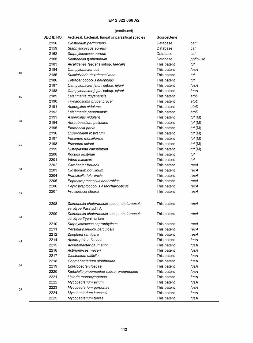
EP 2 322 666 A2
112
5
10
15
20
25
30
35
40
45
50
55
(continued)
SEQ ID NO. Archaeal, bacterial, fungal or parasitical species SourceGene*
2156 Clostridium perfringens Database catP2159 Staphylococcus aureus Database cat2162 Staphylococcus aureus Database cat2165 Salmonella typhimurium Database ppflo-like2183 Alcaligenes faecalis subsp. faecalis This patent tuf2184 Campylobacter coli This patent fusA2185 Succinivibrio dextrinosolvens This patent tuf2186 Tetragenococcus halophilus This patent tuf2187 Campylobacter jejuni subsp. jejuni This patent fusA2188 Campylobacter jejuni subsp. jejuni This patent fusA2189 Leishmania guyanensis This patent atpD2190 Trypanosoma brucei brucei This patent atpD2191 Aspergillus nidulans This patent atpD2192 Leishmania panamensis This patent atpD2193 Aspergillus nidulans This patent tuf (M)2194 Aureobasidium pullulans This patent tuf (M)2195 Emmonsia parva This patent tuf (M)2196 Exserohilum rostratum This patent tuf (M)2197 Fusarium moniliforme This patent tuf (M)2198 Fusarium solani This patent tuf (M)2199 Histoplasma capsulatum This patent tuf (M)2200 Kocuria kristinae This patent tuf2201 Vibrio mimicus This patent tuf2202 Citrobacter freundii This patent recA2203 Clostridium botulinum This patent recA2204 Francisella tularensis This patent recA2205 Peptostreptococcus anaerobius This patent recA2206 Peptostreptococcus asaccharolyticus This patent recA2207 Providencia stuartii This patent recA
2208 Salmonella choleraesuis subsp. choleraesuisserotype Paratyphi A
This patent recA
2209 Salmonella choleraesuis subsp. choleraesuisserotype Typhimurium
This patent recA
2210 Staphylococcus saprophyticus This patent recA2211 Yersinia pseudotuberculosis This patent recA2212 Zoogloea ramigera This patent recA2214 Abiotrophia adiacens This patent fusA2215 Acinetobacter baumannii This patent fusA2216 Actinomyces meyeri This patent fusA2217 Clostridium difficile This patent fusA2218 Corynebacterium diphtheriae This patent fusA2219 Enterobactercloacae This patent fusA2220 Klebsiella pneumoniae subsp. pneumoniae This patent fusA2221 Listeria monocytogenes This patent fusA2222 Mycobacterium avium This patent fusA2223 Mycobacterium gordonae This patent fusA2224 Mycobacterium kansasii This patent fusA2225 Mycobacterium terrae This patent fusA
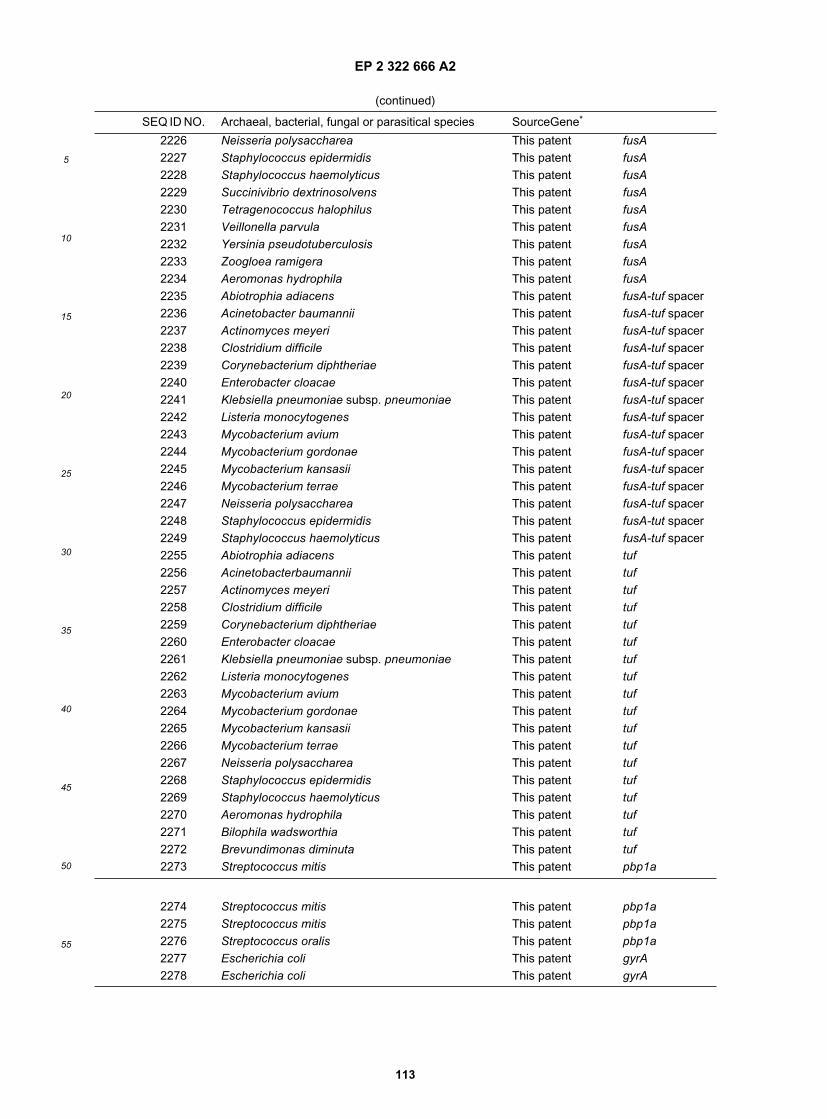
EP 2 322 666 A2
113
5
10
15
20
25
30
35
40
45
50
55
(continued)
SEQ ID NO. Archaeal, bacterial, fungal or parasitical species SourceGene*
2226 Neisseria polysaccharea This patent fusA2227 Staphylococcus epidermidis This patent fusA2228 Staphylococcus haemolyticus This patent fusA2229 Succinivibrio dextrinosolvens This patent fusA2230 Tetragenococcus halophilus This patent fusA2231 Veillonella parvula This patent fusA2232 Yersinia pseudotuberculosis This patent fusA2233 Zoogloea ramigera This patent fusA2234 Aeromonas hydrophila This patent fusA2235 Abiotrophia adiacens This patent fusA-tuf spacer2236 Acinetobacter baumannii This patent fusA-tuf spacer2237 Actinomyces meyeri This patent fusA-tuf spacer2238 Clostridium difficile This patent fusA-tuf spacer2239 Corynebacterium diphtheriae This patent fusA-tuf spacer2240 Enterobacter cloacae This patent fusA-tuf spacer2241 Klebsiella pneumoniae subsp. pneumoniae This patent fusA-tuf spacer2242 Listeria monocytogenes This patent fusA-tuf spacer2243 Mycobacterium avium This patent fusA-tuf spacer2244 Mycobacterium gordonae This patent fusA-tuf spacer2245 Mycobacterium kansasii This patent fusA-tuf spacer2246 Mycobacterium terrae This patent fusA-tuf spacer2247 Neisseria polysaccharea This patent fusA-tuf spacer2248 Staphylococcus epidermidis This patent fusA-tut spacer2249 Staphylococcus haemolyticus This patent fusA-tuf spacer2255 Abiotrophia adiacens This patent tuf2256 Acinetobacterbaumannii This patent tuf2257 Actinomyces meyeri This patent tuf2258 Clostridium difficile This patent tuf2259 Corynebacterium diphtheriae This patent tuf2260 Enterobacter cloacae This patent tuf2261 Klebsiella pneumoniae subsp. pneumoniae This patent tuf2262 Listeria monocytogenes This patent tuf2263 Mycobacterium avium This patent tuf2264 Mycobacterium gordonae This patent tuf2265 Mycobacterium kansasii This patent tuf2266 Mycobacterium terrae This patent tuf2267 Neisseria polysaccharea This patent tuf2268 Staphylococcus epidermidis This patent tuf2269 Staphylococcus haemolyticus This patent tuf2270 Aeromonas hydrophila This patent tuf2271 Bilophila wadsworthia This patent tuf2272 Brevundimonas diminuta This patent tuf2273 Streptococcus mitis This patent pbp1a
2274 Streptococcus mitis This patent pbp1a2275 Streptococcus mitis This patent pbp1a2276 Streptococcus oralis This patent pbp1a2277 Escherichia coli This patent gyrA2278 Escherichia coli This patent gyrA

EP 2 322 666 A2
114
5
10
15
20
25
30
35
40
45
50
55
(continued)
SEQ ID NO. Archaeal, bacterial, fungal or parasitical species SourceGene*
2279 Escherichia coli This patent gyrA2280 Escherichia coli This patent gyrA2288 Enterococcus faecium Database ddl2293 Enterococcus faecium Database vanA2296 Enterococcus faecalis Database vanB
* tuf indicates tuf sequences, tuf (C) indicates tuf sequences divergent from main (usually A and B) copies of theelongation factor-Tu, tuf(EF-1) indicates tuf sequences of the eukaryotic type (elongation factor 1α), tuf (M) indicatestuf sequences from organellar (mostly mitochondrial) origin. fusA indicates fusA sequences; fusA-tuf spacer indicatesthe intergenic region between fusA and tuf. atpD indicates atpD sequences ot the F-type, atpD (V) indicates atpDsequences of the V-type. recA indicates recA sequences, recA(Rad51) indicates rad51 sequences or homologs andrecA(Dmc1) indicates dmc1 sequences or homologs.
Table 8. Bacterial species used to test the specificity of the Streptococcus agalactlae-specific amplificationprimers derived from tuf sequences.
Strain Reference number Strain Reference number
Streptococcusacidominimus
ATCC 51726 Bacteroides caccae ATCC 43185
Streptococcus agalactiae ATCC 12403 Bacteroides vulgatus ATCC 8482Streptococcus agalactiae ATCC 12973 Bacteroides fragilis ATCC 25285Streptococcus agalactiae ATCC 13813 Candida albicans ATCC 11006Streptococcus agalactiae ATCC 27591 Clostridium innoculum ATCC 14501Streptococcus agalactiae CDCs 1073 Clostridium ramosum ATCC 25582Streptococcus anginosus ATCC 27335 Lactobacillus casei
subsp. caseiATCC 393
Streptococcus anginosus ATCC 33397 Clostridium septicum ATCC 12464Streptococcus bovis ATCC 33317 Corynebacterium cervicis NCTC 10604Streptococcus anginosus ATCC 27823 Corynebacterium
genitaliumATCC 33031
Streptococcus cricetus ATCC 19642 Corynebacteriumurealyticum
ATCC 43042
Streptococcus cristatus ATCC 51100 Enterococcus faecalis ATCC 29212Streptococcus downei ATCC 33748 Enterococcus faecium ATCC 19434Streptococcusdysgalactiae
ATCC 43078 Eubacterium lentum ATCC 43055
Streptococcus equi subsp.equi
ATCC 9528 Eubacterium nodutum ATCC 33099
Streptococcus ferus ATCC 33477 Gardnerella vaginalis ATCC 14018Streptococcus gordonii ATCC 10558 Lactobacillus acidophilus ATCC 4356Streptococcus macacae ATCC 35911 Lactobacillus crispatus ATCC 33820Streptococcus mitis ATCC 49456 Lactobacillus gasseri ATCC 33323Streptococcus mutans ATCC 25175 Lactobacillus johnsonii ATCC 33200Streptococcus oralis ATCC 35037 Lactococcus lactis subsp.
lactisATCC 19435
Streptococcusparasanguinis
ATCC 15912 Lactococcus lactis subsp.lactis
ATCC 11454
Streptococcus parauberis DSM 6631 Listeria innocua ATCC 33090Streptococcuspneumoniae
ATCC 27336 Micrococcus luteus ATCC 9341

EP 2 322 666 A2
115
5
10
15
20
25
30
35
40
45
50
55
(continued)
Strain Reference number Strain Reference number
Streptococcus pyogenes ATCC 19615 Escherichia coli ATCC 25922Streptococcus ratti ATCC 19645 Micrococcus lylae ATCC 27566Streptococcus salivarius ATCC 7073 Porphyromonas
asaccharolyticaATCC 25260
Streptococcus sanguinis ATCC 10556 Prevotella corporis ATCC 33547Streptococcus sobrinus ATCC 27352 Prevotella melanogenica ATCC 25845Streptococcus suis ATCC 43765 Staphylococcus aureus ATCC 13301Streptococcus uberis ATCC 19436 Staphylococcus
epidermidisATCC 14990
Streptococcusvestubularis
ATCC 49124 Staphylococcussaprophyticus
ATCC 15305
Table 9. Bacterial species used to test the specificity of the Streptococcus agalactiae-specific amplificationprimers derived from atpD sequences.
Strain Reference number Strain Reference number
Streptococcusacidominimus
ATCC 51726 Streptococcus gordonii ATCC 10558
Streptococcus agalactiae ATCC 12400 Streptococcus macacae ATCC 35911Streptococcus agalactiae ATCC 12403 Streptococcus mitis ATCC 49456Streptococcus agalactiae ATCC 12973 Streptococcus mutans ATCC 25175Streptococcus agalactiae ATCC 13813 Streptococcus oralis ATCC 35037Streptococcus agalactiae ATCC 27591 Streptococcus
parasanguinisATCC 15912
Streptococcus agalactiae CDCs-1073 Streptococcus parauberis DSM 6631Streptococcus anginosus ATCC 27335 Streptococcus
pneumoniaeATCC 27336
Streptococcus anginosus ATCC 27823 Streptococcus pyogenes ATCC 19615Streptococcus bovis ATCC 33317 Streptococcus ratti ATCC 19645Streptococcus cricetus ATCC 19642 Streptococcus salivarius ATCC 7073Streptococcus cristatus ATCC 51100 Streptococcus sanguinis ATCC 10556Streptococcus downei ATCC 33748 Streptococcus sobrinus ATCC 27352Streptococcusdysgalactiae
ATCC 43078 Streptococcus suis ATCC 43765
Streptococcus equisubsp. equi
ATCC 9528 Streptococcus uberis ATCC 19436
Streptococcus ferus ATCC 33477 Streptococcusvestibularis
ATCC 49124
Table 10. Bacterial species used to test the specificity of the Enterococcus-specific amplification primersderived from tuf sequences.
Strain Reference number Strain Reference number
Gram-positive species (n=74)Abiotrophia adiacens ATCC 49176 Listeria innocua ATCC 33090Abiotrophia defectiva ATCC 49175 Listeria ivanovii ATCC 19119Bacillus cereus ATCC 14579 Listeria monocytogenes ATCC 15313Bacillus subtilis ATCC 27370 Listeria seeligeri ATCC 35967

EP 2 322 666 A2
116
5
10
15
20
25
30
35
40
45
50
55
(continued)
Strain Reference number Strain Reference number
Bifidobacteriumadolescentis
ATCC 27534 Micrococcus luteus ATCC 9341
Bifidobacterium breve ATCC 15700 Pediococcus acidilacti ATCC 33314Bifidobacterium dentium ATCC 27534 Pediococcus pentosaceus ATCC 33316Bifidobacterium longum ATCC 15707 Peptococcus niger ATCC 27731Clostridium perfringens ATCC 3124 Peptostreptococcus
anaerobiusATCC 27337
Clostridium septicum ATCC 12464 Peptostreptococcusindolicus
ATCC 29247
Corynebacteriumaquaticus
ATCC 14665 Peptostreptococcus micros ATCC 33270
Corynebacterium ATCC 10700 Propionibacterium acnes ATCC 6919pseudodiphtheriticum Staphylococcus aureus ATCC 43300Enterococcus avium ATCC 14025 Staphylococcus capitis ATCC 27840Enterococcus casseliflavus ATCC 25788 Staphylococcus epidermidis ATCC 14990Enterococcus cecorum ATCC 43199 Staphylococcus
haemolyticusATCC 29970
Enterococcus columbae ATCC 51263 Staphylococcus hominis ATCC 27844Enterococcus dispar ATCC 51266 Staphylococcus lugdunensis ATCC 43809Enterococcus durans ATCC 19432 Staphylococcus
saprophyticusATCC 15305
Enterococcus faecalis ATCC 29212 Staphylococcus simulans ATCC 27848Enterococcus faecium ATCC 19434 Staphylococcus warneri ATCC 27836Enterococcus flavescens ATCC 49996 Streptococcus agalactiae ATCC 13813Enterococcus gallinarum ATCC 49573 Streptococcus anginosus ATCC 33397Enterococcus hirae ATCC 8044 Streptococcus bovis ATCC 33317Enterococcus malodoratus ATCC 43197 Streptococcus constellatus ATCC 27823Enterococcus mundtii ATCC 43186 Streptococcus cristatus ATCC 51100Enterococcuspseudoavium
ATCC 49372 Streptococcus intermedius ATCC 27335
Enterococcus raffinosus ATCC 49427 Streptococcus mitis ATCC 49456Enterococcussaccharolyticus
ATCC 43076 Streptococcus mitis ATCC 3639
Enterococcus solitarius ATCC 49428 Streptococcus mutans ATCC 27175Enterococcus sulfureus ATCC 49903 Streptococcus
parasanguinisATCC 15912
Eubacterium lentum ATCC 49903 Streptococcus pneumoniae ATCC 27736Gemella haemolysans ATCC 10379 Streptococcus pneumoniae ATCC 6303Gemella morbillorum ATCC 27842 Streptococcus pyogenes ATCC 19615Lactobacillus acidophilus ATCC 4356 Streptococcus salivarius ATCC 7073Leuconostocmesenteroides
ATCC 19225 Streptococcus sanguinis ATCC 10556
Listeria grayi ATCC 19120 Streptococcus suis ATCC 43765Listeria grayi ATCC 19123
Gram-negative species (n=39)

EP 2 322 666 A2
117
5
10
15
20
25
30
35
40
45
50
55
(continued)
Strain Reference number Strain Reference number
Acidominococcusfermentans
ATCC 2508 Hafnia alvei ATCC 13337
Acinetobacter baumannii ATCC 19606 Klebsiella oxytoca ATCC 13182Alcaligenes faecalis ATCC 8750 Meganomonas hypermegas ATCC 25560Anaerobiospirillum ATCC 29305 Mitsukoella multiacidus ATCC 27723succiniproducens Moraxella catarrhalis ATCC 43628Anaerorhabdus furcosus ATCC 25662 Morganella morganii ATCC 25830Bacteroides distasonis ATCC 8503 Neisseria meningitidis ATCC 13077Bacteroidesthetaiotaomicron
ATCC 29741 Pasteurella aerogenes ATCC 27883
Bacteroides vulgatus ATCC 8482 Proteus vulgaris ATCC 13315Bordetella pertussis LSPQ 3702 Providencia alcalifaciens ATCC 9886Bulkholderia cepacia LSPQ 2217 Providencia rettgeri ATCC 9250Butyvibrio fibrinosolvens ATCC 19171 Pseudomonas aeruginosa ATCC 27853Cardiobacterium hominis ATCC 15826 Salmonella typhimurium ATCC 14028Citrobacter freundii ATCC 8090 Serratia marcescens ATCC 13880Desulfovibrio vulgaris ATCC 29579 Shigella flexneri ATCC 12022Edwardsiellae tarda ATCC 15947 Shigella sonnei ATCC 29930Enterobacter cloacae ATCC 13047 Succinivibrio
dextrinosolvensATCC 19716
Escherichia coli ATCC 25922 Tissierella praeacuta ATCC 25539Fusobacterium russii ATCC 25533 Veillonella parvula ATCC 10790Haemophilus influenzae ATCC 9007 Yersinia enterocolitica ATCC 9610
Table 11. Microbial species for which tuf and/or atpD and/or recA sequences are available in public databases.
Species Strain Accession number Coding gene*
tuf sequencesBacteriaActinobacillusactinomycetemcomitans
HK1651 Genome project2 tuf
Actinobacillusactinomycetemcomitans
HK1651 Genome project2 tuf (EF-G)
Agrobacterium tumefaciens X99673 tufAgrobacterium tumefaciens X99673 tuf (EF-G)Agrobacterium tumefaciens X99674 tufAnacystis nidulans PCC 6301 X17442 tufAquifex aeolicus VF5 AE000669 tufAquifex aeolicus VF5 AE000669 tuf (EF-G)Aquifex pyrophilus Genome project2 tuf (EF-G)Aquifex pyrophilus Y15787 tufBacillus anthracis Ames Genome project2 tufBacillus anthracis Ames Genome project2 tuf (EF-G)Bacillus halodurans C-1 25 AB017508 tufBacillus halodurans C-125 AB017508 tuf (EF-G)Bacillus stearothermophilus CCM 2184 AJ000260 tufBacillus subtilis 168 D64127 tufBacillus subtilis 168 D64127 tuf (EF-G)Bacillus subtilis DSM 10 Z99104 tuf

EP 2 322 666 A2
118
5
10
15
20
25
30
35
40
45
50
55
(continued)
Species Strain Accession number Coding gene*
Bacillus subtilis DSM 10 Z99104 tuf (EF-G)Bacteroides forsythus ATCC 43037 AB035466 tufBacteroides fragilis DSM 1151 -1 tufBordetella bronchiseptica RB50 Genome project2 tufBordetella pertussis Tohama 1 Genome project2 tufBordetella pertussis Tohama 1 Genome project2 tuf (EF-G)Borrelia burdorgferi B31 U78193 tufBorrelia burgdorferi AE001155 tuf (EF-G)Brevibacterium linens DSM 20425 X76863 tufBuchnera aphidicola Ap Y12307 tufBurkholderia pseudomallei K96243 Genome project2 tuf (EF-G)Campylobacter jejuni NCTC 11168 Y17167 tufCampylobacter jejuni NCTC 11168 CJ11168X2 tuf (EF-G)Chlamydia pneumoniae CWL029 AE001592 tufChlamydia pneumoniae CWL029 AE001639 tuf (EF-G)Chlamydia trachomatis M74221 tufChlamydia trachomatis D/UW-3/CX AE001317 tuf (EF-G)Chlamydia trachomatis D/UW-3/CX AE001305 tufChlamydia trachomatis F/IC-Cal-13 L22216 tufChlorobium vibrioforme DSM 263 X77033 tufChloroflexus aurantiacus DSM 636 X76865 tufClostridium acetobutylicum ATCC 824 Genome project2 tufClostridium difficile 630 Genome project2 tufClostridium difficile 630 Genome project2 tuf (EF-G)Corynebacterium diphtheriae NCTC 13129 Genome project2 ufCorynebacterium diphtheriae NCTC 13129 Genome project2 tuf (EF-G)Corynebacterium glutamicum ASO 19 X77034 tufCorynebacterium glutamicum MJ-233 E09634 tufCoxiella burnetii Nine Mile phase I AF136604 tufCytophaga lytica DSM 2039 X77035 tufDeinococcus radiodurans R1 AE001891 tuf(EF-G)Deinococcus radiodurans R1 AE180092 tuf
Deinococcus radiodurans R1 AE002041 tufDeinonema sp. tufEikenella corrodens ATCC 23834 Z12610 tufEikenella corrodens ATCC 23834 Z12610 tuf (EF-G)Enterococcus faecalis Genome project2 tuf (EF-G)Escherichia coli J01690 tufEscherichia coli J01717 tufEscherichia coli X00415 tuf (EF-G)Escherichia coli X57091 tufEscherichia coli K-12 MG1655 U00006 tufEscherichia coli K-12 MG1655 U00096 tufEscherichia coli K-12 MG1655 AE000410 tuf(EF-G)Fervidobacterium islandicum DSM 5733 Y15788 tufFibrobacter succinogenes S85 X76866 tufFlavobacterium ferrigeneum DSM 13524 X76867 tufFlexistipes sinusarabici X59461 tuf

EP 2 322 666 A2
119
5
10
15
20
25
30
35
40
45
50
55
(continued)
Species Strain Accession number Coding gene*
Gloeobacter violaceus PCC 7421 U09433 tufGloeothece sp. PCC 6501 U09434 tufHaemophilusactinomycetemcomitans
HK1651 Genome project2 tuf
Haemophilus ducreyi 35000 AF087414 tuf (EF-G)Haemophilus influenzae Rd U32739 tufHaemophilus influenzae Rd U32746 tufHaemophilus influenzae Rd U32739 tuf (EF-G)Helicobacter pylori 26695 AE000511 tufHelicobacter pylori J99 AE001539 tuf (EF-G)Helicobacter pylori J99 AE001541 tufHerpetosiphon aurantiacus Hpga1 X76868 tufKlebsiella pneumoniae M6H 78578 Genome project2 tufKlebsiella pneumoniae M6H 78578 Genome project2 tuf (EF-G)Lactobacillus paracasei E13922 tufLegionella pneumophila Philadelphia-1 Genome project2 tufLeptospira interrogans AF115283 tufLeptospira interrogans AF115283 tuf (EF-G)Micrococcus luteus IFO 3333 M17788 tuf (EF-G)Micrococcus luteus I FO 3333 M 17788 tufMoraxella sp. TAC 1125 AJ249258 tufMycobacterium avium 104 Genome project2 tufMycobacterium avium 104 Genome project2 tuf (EF-G)Mycobacterium bovis AF2122/97 Genome project2 tufMycobacterium bovis AF2122/97 Genome project2 tuf (EF-G)Mycobacterium leprae L13276 tufMycobacterium leprae Z14314 tufMycobacterium leprae Z14314 tuf (EF-G)Mycobacterium leprae Thai 53 D13869 tufMycobacterium tuberculosis Erdmann S40925 tufMycobacterium tuberculosis H37Rv AL021943 tuf (EF-G)Mycobacterium tuberculosis H37Rv Z84395 tufMycobacterium tuberculosis y42 AD000005 tufMycobacterium tuberculosis CSU#93 Genome project2 tufMycobacterium tuberculosis CSU#93 Genome project2 tuf (EF-G)Mycoplasma capricolum PG-31 X16462 tufMycoplasma genitalium G37 U39732 tufMycoplasma genitalium G37 U39689 tuf (EF-G)Mycoplasma hominis X57136 tufMycoplasma hominis PG21 M57675 tuf
Mycoplasma pneumoniae M129 AE000019 tufMycoplasma pneumoniae M129 AE000058 tuf(EF-G)Neisseria gonorrhoeae MS11 L36380 tufNeisseria gonorrhoeae MS11 L36380 tuf(EF-G)Neisseria meningitidis Z2491 Genome project2 tuf (EF-G)Neisseria meningitidis Z2491 Genome project2 tufPasteurella multocida Pm70 Genome project2 tufPeptococcus niger DSM 20745 X76869 tuf

EP 2 322 666 A2
120
5
10
15
20
25
30
35
40
45
50
55
(continued)
Species Strain Accession number Coding gene*
Phormidium ectocarpi PCC 7375 U09443 tufPlanobispora rosea ATCC 53773 U67308 tufPlanobispora rosea ATCC 53733 X98830 tufPlanobispora rosea ATCC 53733 X98830 tuf (EF-G)Plectonema boryanum PCC 73110 U09444 tufPorphyromonas gingivalis W83 Genome project2 tufPorphyromonas gingivalis W83 Genome projec2 tuf (EF-G)Porphyromonas gingivalis FDC 381 AB035461 tufPorphyromonas gingivalis W83 AB035462 tufPorphyromonas gingivalis SUNY 1021 AB035463 tufPorphyromonas gingivalis A7A1-28 AB035464 tufPorphyromonas gingivalis ATCC 33277 AB035465 tufPorphyromonas gingivalis ATCC 33277 AB035471 tuf (EF-G)Prochlorothrix hollandica U09445 tufPseudomonas aeruginosa PAO-1 Genome project2 tufPseudomonas putida Genome project2 tufRickettsia prowazekii Madrid E AJ235272 tufRickettsia prowazekii Madrid E AJ235270 tuf (EF-G)Rickettsia prowazekii Madrid E Z54171 tuf (EF-G)Salmonella choleraesuis subsp.choleraesuis serotype Typhimurium X64591 tuf (EF-G)Salmonella choleraesuis subsp.choleraesuis serotype Typhimurium LT2 trpE91 X55116 tufSalmonella choleraesuis subsp.choleraesuis serotype Typhimurium LT2 trpE91 X55117 tufSerpulina hyodysenteriae B204 U51635 tufSerratia marcescens AF058451 tufShewanella putrefaciens DSM 50426 tufShewanella putrefaciens MR-1 Genome project2 tufSpirochaeta aurantia DSM 1902 X76874 tufStaphylococcus aureus AJ237696 tuf (EF-G)Staphylococcus aureus EMRSA-16 Genome project2 tufStaphylococcus aureus NCTC 8325 Genome project2 tufStaphylococcus aureus COL Genome project2 tufStaphylococcus aureus EMRSA-16 Genome project2 tuf (EF-G)Stigmatella aurantiaca DW4 X82820 tufStigmatella aurantiaca Sg a1 X76870 tufStreptococcus mutans GS-5 Kuramitsu U75481 tufStreptococcus mutans UAB159 Genome project2 tufStreptococcus oralis NTCC 11427 P331701 tufStreptococcus pyogenes Genome project2 tuf (EF-G)Streptococcus pyogenes M1-GAS Genome project2 tufStreptomyces aureofaciens ATCC 10762 AF007125 tufStreptomyces cinnamoneus Tue89 X98831 tufStreptomyces coelicolor A3(2) AL031013 tuf(EF-G)Streptomyces coelicolor A3(2) X77039 tuf (EF-G)Streptomyces coelicolor M 145 X77039 tuf
Streptomyces collinus BSM 40733 S79408 tuf

EP 2 322 666 A2
121
5
10
15
20
25
30
35
40
45
50
55
(continued)
Species Strain Accession number Coding gene*
Streptomyces netropsis Tu1063 AF153618 tufStreptomyces ramocissimus X67057 tufStreptomyces ramocissimus X67058 tufStreptomyces ramocissimus X67057 tuf (EF-G)Synechococcus sp. PCC 6301 X17442 tuf (EF-G)Synechococcus sp. PCC 6301 X17442 tufSynechocystis sp. PCC 6803 D90913 tuf (EF-G)Synechocystis sp. PCC 6803 D90913 tufSynechocystis sp. PCC 6803 X65159 tuf (EF-G)Taxeobacter occealus Myx 2105 X77036 tufThermotoga maritima Genome project2 tuf (EF-G)Thermotoga maritima M27479 tufThermus aquaticus EP 00276 X66322 tufThermus thermophilus HB8 X16278 tuf(EF-G)Thermus thermophilus HB8 X05977 tufThermus thermophilus HB8 X06657 tufThiomonas cuprina DSM 5495 U78300 tufThiomonas cuprina DSM 5495 U78300 tuf(EF-G)Thiomonas cuprina Hoe5 X76871 tufTreponema denticola Genome project2 tutTreponema denticola Genome project2 tuf (EF-G)Treponema pallidum AE001202 tufTreponema pallidum AE001222 tuf (EF-G)Treponema pallidum AE001248 tuf (EF-G)Ureaplasma urealyticum ATCC 33697 Z34275 tufUreaplasma urealyticum serovar 3 biovar 1 AE002151 tufUreaplasma urealyticum serovar 3 biovar 1 AE002151 tuf (EF-G)Vibrio cholerae N16961 Genome project2 tufWolinella succinogenes DSM 1740 X76872 tufYersinia pestis CO-92 Genome project2 tufYersinia pestis CO-92 Genome project2 tuf (EF-G)ArchaebacteriaArchaeoglobus fulgidus Genome project2 tuf(EF-G)Halobacterium marismortui X16677 tufMethanobacteriumthermoautrophicum
delta H AE000877 tuf
Methanococcus jannaschii ATCC 43067 U67486 tufMethanococcus vannielii X05698 tufPyrococcus abyssi Orsay AJ248285 tufThermoplasma acidophilum DSM 1728 X53866 tufFungiAbsidia glauca CBS 101.48 X54730 tuf (EF-1)Arxula adeninivorans Ls3 Z47379 tuf(EF-1)Aspergillus oryzae KBN616 AB007770 tuf (EF-1)Aureobasidium pullulans R106 U19723 tuf (EF-1)Candida albicans SC5314 Genome project2 tuf (M)Candida albicans SC5314 M29934 tuf (EF-1)Candida albicans SC5314 M29935 tuf (EF-1)Cryptococcus neoformans B3501 U81803 tuf (EF-1)

EP 2 322 666 A2
122
5
10
15
20
25
30
35
40
45
50
55
(continued)
Species Strain Accession number Coding gene*
Cryptococcus neoformans M1-106 U81804 tuf (EF-1)Eremothecium gossypii ATCC 10895 X73978 tuf (EF-1)Eremothecium gossypii A29820 tuf (EF-1)Fusarium oxysporum NRRL 26037 AF008498 tuf (EF-1)Histoplasma capsulatum 186AS U14100 tuf (EF-1)Podospora anserina X74799 tuf (EF-1)Podospora curvicolla VLV X96614 tuf (EF-1)Prototheca wickerhamii 263-11 AJ245645 tuf (EF-1)Puccinia graminis race 32 X73529 tuf (EF-1)Reclinomonas americana ATCC 50394 AF007261 tuf (M)Rhizomucor racemosus ATCC 1216B X17475 tuf (EF-1)Rhizomucor racemosus ATCC 1216B J02605 tuf (EF-1)Rhizomucor racemosus ATCC 1216B X17476 tuf (EF-1)Rhodotorula mucilaginosa AF016239 tuf (EF-1)Saccharomyces cerevisiae K00428 tuf (M)Saccharomyces cerevisiae M59369 tuf (EF-G)Saccharomyces cerevisiae X00779 tuf (EF-1)Saccharomyces cerevisiae X01638 tuf (EF-1)Saccharomyces cerevisiae M10992 tuf (EF-1)Saccharomyces cerevisiae Alpha S288 X78993 tuf (EF-1)Saccharomyces cerevisiae M 15666 tuf (EF-1)Saccharomyces cerevisiae Z35987 tuf (EF-1)Saccharomyces cerevisiae S288C (AB972) U51033 tuf (EF-1)Schizophyllum commune 1-40 X94913 tuf (EF-1)Schizosaccharomyces pombe 972h- AL021816 tuf (EF-1)Schizosaccharomyces pombe 972h- AL021813 tuf (EF-1)Schizosaccharomyces pombe 972h- D82571 tuf (EF-1)Schizosaccharomyces pombe U42189 tuf (EF-1)Schizosaccharomyces pombe PR745 D89112 tuf (EF-1)Sordaria macrospora 000 X96615 tuf (EF-1)Trichoderma reesei QM9414 Z23012 tuf (EF-1)Yarrowia lipolytica AF054510 tuf (EF-1)
Parasites
Blastocystis hominis HE87-1 D64080 tuf(EF-1)Cryptosporidium parvum U69697 tuf (EF-1)Eimeria tenella LS18 A1755521 tuf (EF-1)Entamoeba histolytica HM1:IMSS X83565 tuf(EF-1)Entamoeba histolytica NIH 200 M92073 tuf (EF-1)Giardia lamblia D14342 tuf (EF-1)Kentrophoros sp. AF056101 tuf (EF-1)Leishmania amazonensis IFLA/BR/67/PH8 M92653 tuf(EF-1)Leishmania braziliensis U72244 tuf (EF-1)Onchocerca volvulus M64333 tuf(EF-1)Porphyra purpurea Avonport U08844 tuf(EF-1)Plasmodium berghei ANKA AJ224150 tuf (EF-1)Plasmodium falciparum K1 X60488 tuf(EF-1)

EP 2 322 666 A2
123
5
10
15
20
25
30
35
40
45
50
55
(continued)
Species Strain Accession number Coding gene*
Plasmodium knowlesi line H AJ224153 tuf (EF-1)Toxoplasma gondii RH Y11431 tuf (EF-1)Trichomonas tenax ATCC 30207 D78479 tuf (EF-1)Trypanosoma brucei LVH/75/ USAMRU-K/18 U10562 tuf (EF-1)Trypanosoma cruzi Y L76077 tuf (EF-1)
Human and plants
Arabidopsis thaliana Columbia X89227 tuf(EF-1)Glycine max Ceresia X89058 tuf(EF-1)Glycine max Ceresia Y15107 tuf(EF-1)Glycine max Ceresia Y15108 tuf(EF-1)Glycine max Maple Arrow X66062 tuf (EF-1)Homo sapiens X03558 tuf (EF-1)Pyramimonas disomata AB008010 tuf
atpD sequences
Bacteria
Acetobacterium woodi DSM 1030 U10505 atpDActinobacillusactinomycetemcomitans
HK1651 Genome project2 atpD
Bacillus anthracis Ames Genome project2 atpDBacillus firmus OF4 M60117 atpDBacillus megaterium QM B1551 M20255 atpDBacillus stearothermophilus D38058 atpDBacillus stearothermophilus IFO1035 D38060 atpDBacillus subtilis 168 Z28592 atpDBacteroides fragilis DSM 2151 M22247 atpDBordetella bronchiseptica RB50 Genome project2 atpDBordetella pertussis Tohama 1 Genome project2 atpDBorrelia burgdorferi B31 AE001122 atpD (V)Burkholderia cepacia DSM50181 X76877 atpDBurkholderia pseudomallei K96243 Genome project2 atpDCampylobacterjejuni NCTC 11168 CJ11168X1 atpDChlamydia pneumoniae Genome project2 atpD (V)Chlamydia trachomatis MoPn Genome project2 atpD (V)Chlorobium vibrioforme DSM 263 X76873 atpDCitrobacter treundii JE0503 AF037156 atpDClostridium acetobutylicum ATCC 824 Genome project2 atpDClostridium acetobutylicum DSM 792 AF101055 atpDClostridium difficile 630 Genome project2 atpDCorynebacterium diphtheriae NCTC13129 Genome project2 atpDCorynebacterium glutamicum ASO 19 X76875 atpDCorynebacterium glutamicum MJ-233 E09634 atpDCytophaga lytica DSM 2039 M22535 atpDEnterobacter aerogenes DSM 30053 -3 atpDEnterococcus faecalis V583 Genome project2 atpD (V)Enterococcus hirae M90060 atpD

EP 2 322 666 A2
124
5
10
15
20
25
30
35
40
45
50
55
(continued)
Species Strain Accession number Coding gene*
Enterococcus hirae ATCC 9790 D17462 atpD (V)Escherichia coli J01594 atpDEscherichia coli M25464 atpDEscherichia coli V00267 atpDEscherichia coli V00311 atpDEscherichia coli K12 MG1655 L10328 atpDFlavobacterium ferrugineum DSM 13524 -3 atpDHaemophilusactinomycetemcomitans
Genome project2 atpD
Haemophilus influenzae Rd U32730 atpDHelicobacter pylori NCTC 11638 AF004014 atpD
Helicobacter pylori 26695 Genome project2 atpDHelicobacter pylori J99 Genome project2 atpDKlabsiella pneumoniae M6H 78578 Genome project2 atpDLactobacillus casei DSM 20021 X64542 atpDLegionella pneumophila Philadelphia-1 Genome project2 atpDMoorella thennoacetica ATCC 39073 U64318 atpDMycobacterium avium 104 Genome projecf2 atpDMycobacterium bovis AF2122/97 Genome project2 atpDMycobacterium leprae U15186 atpDMycobacterium leprae Genome project2 atpDMycobacterium tuberculosis H37Rv Z73419 atpDMycobacterium tuberculosis CSU#93 project2 Genome project2 atpDMycoplasma gallisepticum X64256 atpDMycoplasma genitalium G37 U39725 atpDMycoplasma pneumoniae M129 U43738 atpDNeisseria gonorrhoeae FA 1090 Genome project2 atpDNeisseria meningitidis Z2491 Genome project2 atpDPasteurella multocida Pm70 Genome project2 atpDPectinatus frisingensis DSM 20465 X64543 atpDPeptococcus niger DSM 20475 X76878 atpDPirellula marina IFAM 1313 X57204 atpDPorphyromonas gingivalis W83 Genome project2 atpD (V)Propionigenium modestum DSM 2376 X58461 atpDPseudomonas aeruginosa PAO1 Genome project2 atpDPseudomonas putida Genome project2 atpDRhodobacter capsulatus B100 X99599 atpDRhodospirillum rubrum X02499 atpDRickettsia prowazekii F-12 AF036246 atpDRickettsia prowazekii Madrid Genome project2 atpDRuminococcus albus 7ATCC AB006151 atpDSalmonella bongori JE04162 AF037155 atpDSalmonella bongori BR1859 AF037154 atpDSalmonella choleraesuis S83769 AF037146 atpDsubsp. arizonaeSalmonella choleraesuis u24 AF037147 atpDsubsp. arizonaeSalmonella choleraesuis subsp. K228 AF037140 atpD

EP 2 322 666 A2
125
5
10
15
20
25
30
35
40
45
50
55
(continued)
Species Strain Accession number Coding gene*
choleraesuis serotype DublinSalmonella choleraesuis subsp. K771 AF037139 atpDcholeraesuis serotype DublinSalmonella choleraesuis subsp. Div36-86 AF037142 atpDcholeraesuis serotype InfantisSalmonella choleraesuis subsp. Div95-86 AF037143 atpDcholeraesuis serotype TennesseeSalmonella choleraesuis subsp. LT2 AF037141 atpDcholeraesuis serotype TyphimuriumSalmonella choleraesuis DS210/89 AF037149 atpDsubsp. diarizonaeSalmonella choleraesuis JE0307 AF037148 atpDsubsp. diarizonaeSalmonella choleraesuis S109671 AF037150 atpDsubsp. diarizonaeSalmonella choleraesuis S84366 AF037151 atpDsubsp. houtenaeSalmonella choleraesuis S84098 AF037152 atpD
subsp. houtenaeSalmonella choleraesuis BR2047 AF037153 atpDsubsp. indicaSalmonella choleraesuis NSC72 AF037144 atpDsubsp. salamaeSalmonella choleraesuis S114655 AF037145 atpDsubsp. salamaeShewanella putrefaciens MR-1 Genome project atpDStaphylococcus aureus COL Genome project2 atpDStigmatella aurantiaca Sga1 X76879 atpDStreptococcus bovis JB-1 AB009314 atpDStreptococcus mutans GS-5 U31170 atpDStreptococcus mutans UAB159 Genome project2 atpDStreptococcus pneumoniae Type 4 Genome project2 atpD (V)Streptococcus pneumoniae Type 4 Genome project2 atpDStreptococcus pyogenes M1-GAS Genome project2 atpD (V)Streptococcus pyogenes M1-GAS Genome project2 atpDStreptococcus sanguinis 10904 AF001955 atpDStreptomyces lividans 1326 Z22606 atpDThermus thermophilus HB8 D63799 atpD (V)Thiobacillus ferrooxidans ATCC 33020 M81087 atpDTreponema pallidum Nichols AE001228 atpD (V)Vibrio alginolyticus X16050 atpDVibrio cholerae N16961 Genome project2 atpDWolinella succinogenes DSM 1470 X76880 atpDYersinia enterocolitica NCTC 10460 AF037157 atpDYersinia pestis CO-92 project2 Genome project2 atpDArchaebacteria
Archaeoglobus fulgidus DSM 4304 AE001023 atpD (V)

EP 2 322 666 A2
126
5
10
15
20
25
30
35
40
45
50
55
(continued)
Species Strain Accession number Coding gene*
Halobacterium salinarum S56356 atpD (V)Haloferax volcanii WR 340 X79516 atpDMethanococcus jannaschii DSM 2661 U67477 atpD (V)Methanosarcina barkeri DSM 800 J04836 atpD (V)
Fungi
Candida albicans SC5314 Genome project2 atpDCandida tropicalis M64984 atpD (V)Kluyveromyces lactis 2359/152 U37764 atpDNeurospora crassa X53720 atpDSaccharomyces cerevisiae M12082 atpDSaccharomyces cerevisiae X2180-1 A J05409 atpD (V)Schizosaccharomyces pombe 972 h- S47814 atpD (V)Schizosaccharomyces pombe 972 h- M57956 atpD
Parasites
Giardia lamblia WB U18938 atpDPlasmodium falciparum 3D7 L08200 atpD (V)Trypanosoma congolense IL3000 Z25814 atpD (V)
Human and plants
Homo sapiens L09234 atpD(V)Homo sapiens M27132 atpD
recA sequences
Bacteria
Acetobacter aceti no. 1023 S60630 recAAcetobacter attoacetigenes MH-24 E05290 recAAcetobacter polyoxogenes NBI 1028 D13183 recAAcholeplasma laidlawii 8195 M81465 recAAcidiphilium facilis ATCC 35904 D16538 recAAcidothermus cellulolyticus ATCC 43068 AJ006705 recAAcinetobacter calcoaceticus BD413/ADP1 L26100 recAActinobacillusactinomycetemcomitans
HK1651 Genome project2 recA
Aeromonas salmonicida A449 U83688 recAAgrobacterium tumefaciens C58 L07902 recAAllochromatium vinosum AJ000677 recAAquifex aeolicus VF5 AE000775 recAAquifex pyrophilus Kol5a L23135 recAAzotobacter vinelandii S96898 recABacillus stearothermophilus 10 Genome project2 reeABacillus subtilis PB1831 U87792 recABacillus subtilis 168 Z99112 recABacteroides fragilis M63029 recA

EP 2 322 666 A2
127
5
10
15
20
25
30
35
40
45
50
55
(continued)
Species Strain Accession number Coding gene*
Bifidobacterium breve NCFB 2258 AF094756 recABlastochloris viridis DSM 133 AF022175 recABordetella pertussis 165 X53457 recABordetella pertussis Tohama I Genome project2 recABorrelia burgdorferi Sh-2-82 U23457 recABorrelia burgdorferi B31 AE001124 recABrevibacterium flavum MJ-233 E10390 recABrucella abortus 2308 L00679 recABurkholderia cepacia ATCC 17616 U70431 recABurkholderia cepacia D90120 recABurkholderia pseudomallei K96243 Genome project2 recACampylobacter fetus subsp. fetus 23D AF020677 recACampylobacter jejuni 81-176 U03121 recACampylobacterjejuni NCTC 11168 AL139079 recAChlamydia trachomatis L2 U16739 recAChlamydia trachomatis D/UW-3/CX AE001335 recAChlamydophifa pneumoniae CWL029 AE001658 recAChloroflexus aurantiacus J-10-fl AF037259 recAClostridium aeetobutylicum M94057 recAClostridium perfringens 13 U61497 recACorynebacterium diphtheriae NCTC13129 Genome project2 recACorynebacterium glutamicum AS019 U14965 recACorynebacteriumpseudotuberculosis
C231 U30387 recA
Deinococcus radiodurans KD8301 AB005471 recADeinococcus radiodurans R1 U01876 recA
Enterobacter agglomerans 339 L03291 recAEnterococcus faecalis OGIX M81466 recAErwinia carotovora X55554 recAEscherichia coli J01672 recAEscherichia coli X55552 recAEscherichia coli K-12 AE000354 recAFrankia alni Arl3 AJ006707 recAGluconobacter oxydans U21001 recAHaemophilus influenzae Rd U32687 recAHaemophilus influenzae Rd U32741 recAHaemophilus influenzae Rd L07529 recAHelicobacter pylori 69A Z35478 recAHelicobacter pylori 26695 AE000536 recAHelicobacter pylori J99 AE001453 recAKlebsiella pneumoniae M6H 78578 Genome project2 recALactococcus lactis ML3 M88106 recALegionella pneumophila X55453 recALeptospira biflexa serovar patoc U32625 recALeptospira interrogans serovar pomona U29169 recAMagnetospirillum magnetotacticum MS-1 X17371 recAMethylobacillus flagellatus MFK1 M35325 recAMethylomonas clara ATCC 31226 X59514 recA

EP 2 322 666 A2
128
5
10
15
20
25
30
35
40
45
50
55
(continued)
Species Strain Accession number Coding gene*
Mycobacterium avium 104 Genome project2 recAMycobacterium bovis AF122/97 Genome project2 recAMycobacterium leprae X73822 recAMycobacterium tuberculosis H37Rv X58485 recAMycobacterium tuberculosis CSU#93 Genome project2 recAMycoplasma genitalium G37 U39717 recAMycoplasma mycoides GM9 L22073 recAMycoplasma pneumoniae ATCC 29342 MPAE000033 recAMycoplasma pulmonis KD735 L22074 recAMyxococcus xanthus L40368 recAMyxococcus xanthus L40367 recANeisseria animalis NCTC 10212 U57910 recANeisseria cinerea LCDC 81-176 AJ223869 recANeisseria cinerea LNP 1646 U57906 recANeisseria cinerea NCTC 10294 AJ223871 recANeisseria cinerea Vedros M601 AJ223870 recANeisseria elongata CCUG 2131 AJ223882 recANeisseria elongata CCUG 4165A AJ223880 recANeisseria elongata NCTC 10660 AJ223881 recANeisseria elongata NCTC 11050 AJ223878 recANeisseria elongata NHITCC 2376 AJ223877 recANeisseria elongata CCUG 4557 AJ223879 recAsubsp. intermediaNeisseria flava Bangor 9 AJ223873 recANeisseria flavescens LNP 444 U57907 recANeisseria gonorrhoeae CH95 U57902 recANeisseria gonorrhoeae FA19 X64842 recANeisseria gonorrhoeae MS11 X17374 recANeisseria gonorrhoeae Genome project2 recANeisseria lactamica CCUC 7757 AJ223866 recANeisseria lactamica CCUG 7852 Y11819 recANeisseria lactamica LCDC 77-143 Y11818 recANeisseria lactamica LCDC 80-111 AJ223864 recA
Neisseria lactamica LCDC 845 AJ223865 recANeisseria lactamica NCTC 10617 U57905 recANeisseria lactamica NCTC 10618 AJ223863 recANeisseria meningitidis 44/46 X64849 recANeisseria meningitidis Bangor 13 AJ223868 recANeisseria meningitidis HF116 X64848 recANeisseria meningitidis HF130 X64844 recANeisseria meningitidis HF46 X64847 recANeisseria meningitidis M470 X64850 recANeisseria meningitidis N9411 X64846 recANeisseria meningitidis NCTC 8249 AJ223867 recANeisseria meningitidis P63 X64845 recANeisseria meningitidis S3446 U57903 recANeisseria meningitidis FAM18 Genome project2 recANeisseria mucosa LNP 405 U57908 recA

EP 2 322 666 A2
129
5
10
15
20
25
30
35
40
45
50
55
(continued)
Species Strain Accession number Coding gene*
Neisseria mucosa Vedros M1801 AJ223875 recANeisseria perflava CCUG 17915 AJ223876 recANeisseria perflava LCDC 85402 AJ223862 recANeisseria pharyngis var. flava NCTC 4590 U57909 recANeisseria polysaccharea CCUG 18031 Y11815 recANeisseria polysaccharea CCUG 24845 Y11816 recANeisseria polysaccharea CCUG 24846 Y11814 recANeisseria polysaccharea INS MA 3008 Y11817 recANeisseria polysaccharea NCTC 11858 U57904 recANeisseria sicca NRL 30016 AJ223872 recANeisseria subflava NRL 30017 AJ223874 recAParacoccus denitrificans DSM 413 U59631 recAPasteurella multocida X99324 recAPorphyromonas gingivalis W83 U70054 recAPrevotella ruminicola JCM 8958 U61227 recAProteus mirabilis pG1300 X14870 recAProteus vulgaris X55555 recAPseudomonas aeruginosa X05691 recAPseudomonas aeruginosa PAM 7 X52261 recAPseudomonas aeruginosa PA012 D13090 recAPseudomonas fluorescens OE 28.3 M96558 recAPseudomonas putida L12684 recAPseudomonas putida PpS145 U70864 recARhizobium leguminosarum VF39 X59956 recAbiovar viciaeRhizobium phaseoli CNPAF512 X62479 recARhodobacter capsulatus J50 X82183 recARhodobacter sphaeroides 2.4.1 X72705 recARhodopseudomonas palustris N 7 D84467 recARickettsia prowazekii Madrid E AJ235273 recARickettsia prowazekii Madrid E U01959 recASerratia marcescens M22935 recAShigella flexneri X55553 recAShigella sonnei KNIH104S AF101227 recASinorhizobium meliloti 2011 X59957 recAStaphylococcus aureus L25893 recAStreptococcus gordonii Challis V288 L20574 recAStreptococcus mutans UA96 M81468 recAStreptococcus mutans GS-5 M61897 recAStreptococcus pneumoniae Z17307 recA
Streptococcus pneumoniae R800 Z34303 recAStreptococcus pyogenes NZ131 U21934 recAStreptococcus pyogenes D471 M81469 recAStreptococcus salivarius M94062 recAsubsp. thermophilusStreptomyces ambofaciens DSM 40697 Z30324 recAStreptomyces coelicolor A3(2) AL020958 recAStreptomyces lividans TK24 X76076 recA

EP 2 322 666 A2
130
5
10
15
20
25
30
35
40
45
50
55
(continued)
Species Strain Accession number Coding gene*
Streptomyces rimosus R6 X94233 recAStreptomyces venezuelae ATCC10712 U04837 recASynechococcus sp. PR6 M29495 recASynechocystis sp. PCC6803 D90917 recAThermotoga maritima L23425 recAThermotoga maritima AE001823 recAThermus aquaticus L20095 recAThermus thermophilus HB8 D17392 recAThiobacillus ferrooxidans M26933 recATreponema denticola Genome project2 recATreponema pallidum Nichols AE001243 recAVibrio anguillarum M80525 recAVibrio cholerae 017 X71969 recAVibrio cholerae 2740-80 U10162 recAVibrio cholerae 569B L42384 recAVibrio cholerae M549 AF117881 recAVibrio cholerae M553 AF117882 recAVibrio cholerae M645 AF117883 recAVibrio cholerae M793 AF117878 recAVibrio cholerae M794 AF117880 recAVibrio cholerae M967 AF117879 recAXanthomonas citri XW47 AF006590 recAXanthomonas oryzae AF013600 recAXenorhabdus bovienii T228/1 U87924 recAXenorhabdus nematophilus AN6 AF127333 recAYersinia pestis 231 X75336 recAYersinia pestis CO-92 Genome projecf2 recA
Fungi, parasites, human andplants
Anabaena variabilis ATCC 29413 M29680 recAArabidopsis thaliana U43652 recA (Rad51)Candida albicans U39808 recA (Dmc1)Coprinus cinereus Okayama-7 U21905 recA (Rad51)Emericella nidulans Z80341 recA (Rad51)Gauss gallus L09655 recA (Rad51)Homo sapiens D13804 recA (Rad51)Homo sapiens D63882 recA (Dmc1)Leishmania major Friedlin AF062379 recA (Rad51)Leishmania major Friedlin AF062380 recA (Dmc1)Mus musculus D58419 recA (Dmc1)Neurospora crassa 74-OR23-1A D29638 recA (Rad51)Saccharomyces cerevisiae D10023 recA (Rad51)Schizosaccharomyces pombe Z22691 recA (Rad51)Schizosaccharomyces pombe 972h- AL021817 recA (Dmc1)Tetrahymena thermophila PB9R AF064516 recA (Rad51)
Trypanosoma brucei stock 427 Y13144 recA (Rad51)

EP 2 322 666 A2
131
5
10
15
20
25
30
35
40
45
50
55
(continued)
Species Strain Accession number Coding gene*
Ustilago maydis U62484 recA (Rad51)Xenopus laevis D38488 recA (Rad51)Xenopus laevis D38489 recA (Rad51)
* tuf indicates tuf sequences, including tuf genes, tusA genes and fusA-tuf intergenic spacers. tuf (C) indicates tufsequences divergent from main (usually A and B) copies of the elongation factor-Tu tuf (EF-1) indicates tuf sequencesof the eukaryotic type (elongation factor 1α) tuf (M) indicates tufsequences from organellar (mostly mitochondrial)origin atpD indicates atpD sequences ot the F-type atpD (V) indicates atpD sequences of the V-Type recA indicatesrecA sequences recA (Rad51) indicates rad51 sequences or homologs recA (Dmc1) indicates dmc1 sequences orhomologs1 Nucleotides sequences published in Arch. Microbiol. 1990 153:241-2472 These sequences are from the TIGR database (http://www.tigr.org/tdb/tdb.html)3 Nucleotides sequences published in FEMS Microbiology Letters 1988 50:101-106
Table 12. Bacterial species used to test the specificity of the Staphylococcus-specific amplification primersderived from tuf sequences.
Strain Reference number Strain Reference number
Staphylococcal species (n=27) Other Gram-positive bacteria (n=20)Staphylococcus arlettae ATCC 43957 Bacillus subtilis ATCC 27370Staphylococcus aureus ATCC 35844 Enterococcus avium ATCC 14025subsp. anaerobiusStaphylococcus aureus ATCC 43300 Enterococcus durans ATCC 19432subsp. aureusStaphylococcusauricularis
ATCC 33753 Enterococcus faecalis ATCC 19433
Staphylococcus capitis ATCC 27840 Enterococcus faecium ATCC 19434subsp. capitisStaphylococcus caprae ATCC 35538 Enterococcus flavescens ATCC 49996Staphylococcus camosus ATCC 51365 Enterococcus gallinarum ATCC 49573Staphylococcuschromogenes
ATCC 43764 Lactobacillus acidophilus ATCC 4356
Staphylococcus cohnii DSM 20260 Lactococcus lactis ATCC 11454subsp. urealyticumStaphylococcus delphini ATCC 49171 Listeria innocua ATCC 33090Staphylococcusepidermidis
ATCC 14990 Listeria ivanovii ATCC 19119
Staphylococcus equorum ATCC 43958 Listeria monocytogenes ATCC 15313Staphylococcus felis ATCC 49168 Macrococcus
caseolyticusATCC 13548
Staphylococcusgallinarum
ATCC 35539 Streptococcus agalactiae ATCC 13813
Staphylococcushaemolyticus
ATCC 29970 Streptococcus anginosus ATCC 33397
Staphylococcus hominis ATCC 27844 Streptococcus bovis ATCC 33317Staphylococcus hyicus ATCC 11249 Streptococcus mutans ATCC 25175Staphylococcusintermedius
ATCC 29663 Streptococcuspneumoniae
ATCC 6303
Staphylococcus kloosis ATCC 43959 Streptococcus pyogenes ATCC 19615Staphylococcus lentus ATCC 29070 Streptococcus salivarius ATCC 7073

EP 2 322 666 A2
132
5
10
15
20
25
30
35
40
45
50
55
(continued)
Strain Reference number Strain Reference number
Staphylococcuslugdunensis
ATCC 43809
Staphylococcussaprophyticus
ATCC 15305
Staphylococcus schleiferi ATCC 49545subsp. coagulansStaphylococcus sciuri ATCC 29060subsp. sciuriStaphylococcus simulans ATCC 27848Staphylococcus warneri ATCC 27836Staphylococcus xylosus ATCC 29971Gram-negative bacteria (n=33)Acinetobacter baumannii ATCC 19606 Morganella morganii ATCC 25830Bacteroides distasonis ATCC 8503 Neisseria gonorrhoeae ATCC 35201Bacteroides fragilis ATCC 25285 Neisseria meningitidis ATCC 13077Bulkholderia cepacia ATCC 25416 Proteus mirabilis ATCC 25933Bordetella pertussis ATCC 9797 Proteus vulgaris ATCC 13315Citrobacter freundii ATCC 8090 Providencia rettgeri ATCC 9250Enterobacter aerogenes ATCC 13048 Providencia stuartii ATCC 29914Enterobacter cloacae ATCC 13047 Pseudomonas
aeruginosaATCC 27853
Escherichia coli ATCC 25922 Pseudomonasfluorencens
ATCC 13525
Haemophilus influenzae ATCC 8907 Salmonella choleraesuis ATCC 7001Haemophilusparahaemolyticus
ATCC 10014 Salmonella typhimurium ATCC 14028
Haemophilusparainfluenzae
ATCC 7901 Serratia marcescens ATCC 8100
Hafnia alvei ATCC 13337 Shigella flexneri ATCC 12022Kingella indologenes ATCC 25869 Shigella sonnei ATCC 29930Klebsiella oxytoca ATCC 13182 Stenotrophomonas
maltophiliaATCC 13843
Klebsiella pneumoniae ATCC 13883 Yersinia enterocolitica ATCC 9610Moraxella catarrhalis ATCC 25240
Table 13. Bacterial species used to test the specificity of the penicillin-resistant Streptococcus pneumoniaeassay.
Strain Reference number Strain Reference number
Gram-positive species (n=67)Abiotrophia adiacens ATCC 49175 Staphylococcus hominis ATCC 27844Abiotrophia defectiva ATCC 49176 Staphylococcus
lugdunensisATCC 43809
Actinomyces pyogenes ATCC 19411 Staphylococcussaprophyticus
ATCC 15305
Bacillus anthracis ATCC 4229 Staphylococcus simulans ATCC 27848Bacillus cereus ATCC 14579 Staphylococcus. warneri ATCC 27836Bifidobacterium breve ATCC 15700 Streptococcus
acidominimusATCC 51726
Clostridium difficile ATCC 9689 Streptococcus agalactiae ATCC 12403

EP 2 322 666 A2
133
5
10
15
20
25
30
35
40
45
50
55
(continued)
Strain Reference number Strain Reference number
Enterococcus avium ATCC 14025 Streptococcus anginosus ATCC 33397Enterococcus casseliflavus ATCC 25788 Streptococcus bovis ATCC 33317Enterococcus dispar ATCC 51266 Streptococcus constellatus ATCC 27823Enterococcus durans ATCC 19432 Streptococcus cricetus ATCC 19624Enterococcus faecalis ATCC 29212 Streptococcus cristatus ATCC 51100Enterococcus faecium ATCC 19434 Streptococcus downei ATCC 33748Enterococcus flavescens ATCC 49996 Streptococcus dysgalactiae ATCC 43078Enterococcus gallinarum ATCC 49573 Streptococcus equi ATCC 9528Enterococcus hirae ATCC 8043 Streptococcus ferus ATCC 33477Enterococcus mundtii ATCC 43186 Streptococcus gordonii ATCC 10558Enterococcus raffinosus ATCC 49427 Streptococcus intermedius ATCC 27335Lactobacillus lactis ATCC 19435 Streptococcus mitis ATCC 903Lactobacillusmonocytogenes
ATCC 15313 Streptococcus mitis LSPQ 2583
Mobiluncus curtisii ATCC 35242 Streptococcus mitis ATCC 49456Peptococcus niger ATCC 27731 Streptococcus mutans ATCC 27175Peptostreptococcus acones ATCC 6919 Streptococcus oralis ATCC 10557Peptostreptococcusanaerobius
ATCC 27337 Streptococcus oralis ATCC 9811
Peptostreptococcus ATCC 2639 Streptococcus oralis ATCC 35037asaccharolyticus Streptococcus
parasanguinisATCC 15912
Peptostreptococcuslactolyticus
ATCC 51172 Streptococcus parauberis ATCC 6631
Peptostreptococcusmagnus
ATCC 15794 Streptococcus rattus ATCC 15912
Peptostreptococcus prevotii ATCC 9321 Streptococcus salivarius ATCC 7073Peptostreptococcustetradius
ATCC 35098 Streptococcus sanguinis ATCC10556
Staphylococcus aureus ATCC 25923 Streptococcus suis ATCC 43765Staphylococcus capitis ATCC 27840 Streptococcus uberis ATCC 19436Staphylococcus epidermidis ATCC 14990 Streptococcus vestibularis ATCC 49124Staphylococcushaemolyticus
ATCC 29970
Gram-negative species (n=33)Actinetobacter baumannii ATCC 19606 Moraxella morganii ATCC 13077Bordetella pertussis ATCC 9797 Neisseria gonorrhoeae ATCC 35201Citrobacter diversus ATCC 27028 Neisseria meningitidis ATCC 13077Citrobacter freundii ATCC 8090 Proteus mirabilis ATCC 25933Enterobacter aerogenes ATCC 13048 Proteus vulgaris ATCC 13315Enterobacter agglomerans ATCC 27155 Providencia alcalifaciens ATCC 9886Enterobacfer cloacae ATCC 13047 Providencia rettgeri ATCC 9250Escherichia coli ATCC 25922 Providencia rustigianii ATCC 33673Haemophilus ducreyi ATCC 33940 Providencia stuartii ATCC 33672Haemophilus haemolyticus ATCC 33390 Pseudomonas aeruginosa ATCC 35554Haemophilus influenzae ATCC 9007 Pseudomonas fluorescens ATCC 13525Haemophilusparainfluenzae
ATCC 7901 Pseudomonas stutzeri ATCC 17588
Hafnia alvei ATCC 13337 Salmonella typhimurium ATCC 14028Klebsiella oxytoca ATCC 13182 Serratia marcescens ATCC 13880

EP 2 322 666 A2
134
5
10
15
20
25
30
35
40
45
50
55
(continued)
Strain Reference number Strain Reference number
Klebsiella pneumoniae ATCC 13883 Shigella flexneri ATCC 12022Moraxella atlantae ATCC 29525 Yersina enterocolitica ATCC 9610Moraxella catarrhalis ATCC 43628
Table 14. Bacterial species (n=104) detected by the platelet contaminants assay. Bold characters indicate themajor bacterial contaminants found in platelet concentrates.
Abiotrophia adiacens Klebsiella oxytoca Staphylococcus simulansAbiotrophia defectiva Klebsiella pneumoniae Staphylococcus warneriAcinetobacter baumannii Legionella pneumophila Stenotrophomonas maltophiliaAcinetobacter Iwoffi Megamonas hypermegale Streptococcus acidominimusAerococcus viridans Moraxella atlantae Streptococcus agalactiaeBacillus anthracis Moraxella catarrhalis Streptococcus anginosusBacillus cereus Morganella morganii Streptococcus bovisBacillus subtilis Neisseria gonorrheae Streptococcus constellatusBrucella abortus Neisseria meningitidis Streptococcus cricetusBurkholderia cepacia Pasteurella aerogenes Streptococcus cristatusCitrobacter diversus Pasteurella multocida Streptococcus dysgalactiaeCitrobacter freundii Peptostreptococcus magnus Streptococcus equiEnterobacter aerogenes Proteus mirabilis Streptococcus ferusEnterobacter agglomerans Providencia alcalifaciens Streptococcus gordoniiEnterobacter cloacae Providencia rettgeri Streptococcus intermediusEnterococcus avium Providencia rustigianii Streptococcus macacaeEnterococcus casseliflavus Providencia stuartii Streptococcus mitisEnterococcus dispar Pseudomonas aeruginosa Streptococcus mutansEnterococcus durans Pseudomonas fluorescens Streptococcus oralisEnterococcus faecalis Pseudomonas stutzeri Streptococcus parasanguinisEnterococcus faecium Salmonella bongori Streptococcus parauberisEnterococcus flavescens Salmonella choleraesuls Streptococcus pneumoniaeEnterococcus gallinarum Salmonella enteritidis Streptococcus pyogenesEnterococcus mundtii Salmonella gallinarum Streptococcus rattiEnterococcus raffinosus Salmonella typhimurium Streptococcus salivariusEnterococcus solitarius Serratia liquefaciens Streptococcus sanguinisEscherichia coli Serratia marcescens Streptococcus sobrinusGemella morbillorum Shigella flexneri Streptococcus uberisHaemophilus ducreyi Shigella sonnei Streptococcus vestibularisHaemophilus haemolyticus Staphylococcus aureus Vibrio choleraeHaemophilus influenzae Staphylococcus capitis Yersinia enterocoliticaHaemophilus Staphylococcus epidermidis Yersinia pestisparahaemolyticus Staphylococcus haemolyticus Yersinia pseudotuberculosisHaemophilus parainfluenzae Staphylococcus hominisHafnia alvei Staphylococcus lugdunensisKingella kingae Staphylococcus saprophyticus
Table 15. Microorganisme identifled by commercial systems1.
Abiotrophia adiacens (Streplococcus Alcaligenes xylosoxidans subsp. Brevibacterium speciesadjacens) xylosoxidans Brevundimonas (Pseudomonas)

EP 2 322 666 A2
135
5
10
15
20
25
30
35
40
45
50
55
(continued)Abiotrophia defectiva (Streptococcus Alloiococcus otitis diminutadefectivus) Anaerobiospirillum
succiniciproducensBrevundimonas (Pseudomonas)
Achromobacter species Anaerovibrio lipolytica vesiculatisAcidaminococcus fermen/ans Arachnia propionica Brevundimonas speciesAcinetobacter a/ealigenes Arcanobacterium (Actinomyces) Brochothrix thermosphactaAcinetobacter anitratus bernardiae Brucella abortusAcinatobacter baumannii Arcanobacterium (Actinomyces) Brucella canisAcinetobacter calcoaceticus pyogenes Brucella melitensisAcinetobacter calcoaceticus biovar Arcanobacterium hasmolyticum Brucella ovisanitratus Arcobacter cyarophilus Brucella speciesAcinetobacter calcoaceticus biovar (Campylobacter cryaerophlla) Brucella suisIwoffi Arthrobacter globiformis Budvicia aquaticaAcinetobactergenomospecies Arthrobacterspecies Burkholderia (Pseudomonas) cepaciaAeinetobacter haemolyticus Arxiozyma telluns (Torulopsis Burkholderia (Pseudomonas) gladioliAcinetobacter johnsonii pintolopesil) Burkholderia (Pseudomonas) malleiAcinetobacter junii Atopobium minutum (lactobacillus Burkholderia (Pseudomonas)Acinetobacter lwoffii minutus) pseudomalleiAcinetobacter radioresistens Aureobacterium species Burkholderia speciesAcinetobacter species Bacillus amyloliquefaciens Buttiauxella agrestisActinobacillusactinomycetemeomitans
Bacillus anthracis Campy/obacter coli
Actinobacillus capsulatus Bacillus badius Campylobacter concisusActinobacillus equuli Bacillus cereus Campylobacter fetusActinobacillus hominis Bacillus circulans Campylobacter fetus subsp. fetusActinobacillus lignieresil Bacillus coagulans Campylobacter fetus subsp.Actinobacillus pleuropneumoniae Bacillus firmus venerealisActinobacillus species Bacillus lentus Compylobacter hyointestinalisActinobacillus suis Bacillus lichenifoimis Campylobacter jejuni subsp. doyleiActinobacillus ureae Bacillus megaterium Campylobacterjeitinisubsp, je/un/Actinomyces bovis Bacillus mycoides CampylobacterlariActinomyces israelii Bacillus pantothenticus Compylobacter fari subsp. UPTCActinomyces meyeri Bacillus pumilus Campylobacter mucosalisActinomyces naeslundii Bacillus species Campylobacter speciesActinomyces neuii subsp. anitratus Bacillus sphaericus Campylobacter sputorumActinomyces neuii subsp. neui Bacillus stearothermophilus Campylobacter sputorum subsp.Actinomyces odontolyticus Bacillus subtiles bubulusActinomyces pyogenes Bacillus thuringiensis Campy/obaeter sputorum subsp.Actinomyces radingae Bacteroides caccae fecalisActinomyces species Bacteroides capillosus Campylobacter sputorum subsp.Actinomyces turicensis Bacteroides distasonis sputorumActinomyces viscosus Bacteroides eggerthii Campylobacter upsaliensisAerococcus species Bacteroides fragilis Candida (Clavispora) lusitaniaeAerococcus viridans Becteroides merdae Candida (Pichia) guilliermondiiAeromonas caviae Bacteroides ovatus Candida (Torulopsis) glabrataAeromonas hydrophila Bacteroides species Candida albicansAeromonas hydrophila group Bacteroides splanchnicus Candida boidiniiAeromonas jandaei Bacteroides stercoris Candida catenu/ata
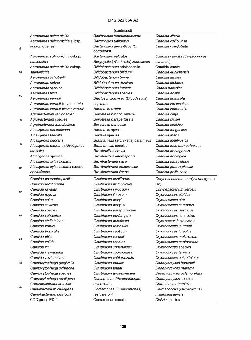
EP 2 322 666 A2
136
5
10
15
20
25
30
35
40
45
50
55
(continued)Aeromonas salmonicida Bacteroides thetaiotaomicron Candida ciferrliAeromonas salmomcida subsp. Bacteroides uniformis Candida colliculosaachromogenes Bacteroides ureolytlcus (B.
corrodens)Candida conglobata
Aaromonas salmonicida subsp. Bacteroides vulgatus Candida curvata (Cryptococcusmasoucida Bergeyella (Weeksella) zoohelcum curvatus)Aeromonas salmonicida subsp. Bifidobacterium adolescen/is Candida dattilasalmonicida Bifidobacterium bifidum Candida dubliniensisAeromonas schuberlii Bifidobactenum breve Candida famataAeromonas sobria Bifidobacterium dentium Candida globoseAeromonas species Bifidobacterium infantis Candid hellenicaAeromonas trota Bitidobacterium species Candida holmiiAeromonas veronii Blastoschizomyces (Dipodascus) Candida humicolaAeromonas varonli biovar sobria capitatus Candida inconspicuaAeromonas veronii biovar veronii Bordetella avium Candida intermediaAgrobactenum radiobacter Bordetella bronchiseptica Candida kefyrAgrobacterium species Bordetella parapertussis Candida kruselAgrobacterium tumefaciens Bordetelia pertussis Candida lambicaAlcaligenes denitrificans Bordetelia species Candida magnoliaeAlcaligenes faecalis Borrelia species Candida marisAlcaligenes odorans Branhamella (Moraxella) catafthalis Candida melibiosicaAlcaligenes odorans (Alcaligenes Branhamella species Candida membranaefacienstaecalis) Brevibacillus brevis Candida norvegensisAtcaligenes species Brevibacillus laterosponts Candida norvegicaAlcaligenes xylosoxidans Brovibacterium casei Candida parapsilosisAlcaligenes xylosoxidans subsp. Brevibacterium epidermidis Candida paratropicallisdenitrificans Brevibacterium linens Candida pelliculosa
Candida pseudotropicalis Clostndium hastiforme Corynebacterium urealyticum (groupCandida pulcherrima Clostridium histolyticum D2)Candida ravautii Clostridium innocuum Corynebacterium xerosisCandida rugosa Clostridium limosum Cryptococcus albidusCandida sake Clostridium novyi Cryptococcus aterCandida silvicola Clostridium novyi A Cryptococcus cereanusCandida species Clostridium paraputtificum Cryptococcus gastricusCandida sphaerica Clostridium perfringens Cryptococcus humicolusCandida stellatoidea Clostridium putrificum Cryptxoccus lactativorusCandida tenuis Clostridium ramosum Cryptococcus laurentiiCandida tropicalis Clostridium septicum Cryptococcus luteolusCandida utilis Clostndium sordelli Cryptococcus melibiosumCandida valida Clostridium species Cryptococcus neoformansCandida vini Clostridium sphenoides Crypfococcus speciesCandida viswanathii Clostridium sporogenes Cryptococcus terreusCandida zeylanoides Clostridium subterminale Cryptococcus uniguttulatusCapnocytophaga gingivalis Clostridium tertium Debaryomyces hanseniiCapnocytophaga ochracea Clostridium tetani Debaryomyces maramaCapnocytophaga species Clostridium tyrobutyricum Debaryomyces polymorphusCapnocytophaga sputigene Comamonas (Pseudomonas) Debaryomyces speciesCardiobacterium hominis acidovorans Dermabacter hominisCamobacterium divergens Comamonas (Pseudomonas) Dermacoccus (Micrococcus)Camobacterium piscicola testosteroni nishinomiyaensisCDC group ED-2 Comamonas species Dietzia species

EP 2 322 666 A2
137
5
10
15
20
25
30
35
40
45
50
55
(continued)CDC group EF4 (Pasteurella sp.) Corynebacterium accolens Edwardsiella hoshinaeCDC group EF-4A Corynebacterium afermentans Edwardsiella ictaluriCDC group EF-4B Corynebacterium amycolatum Edwardsiella speciesCDC group EQ-Z Corynebacterium aquaticum Edwardsiella tardaCDC group HB-5 Corynebacterium argentoratense Eikenella corrodensCDC group II K-2 Corynebacterium auris Empedobacter brevis
(FlavobacteriumCDC group IV C-2 (Bordetella-like) Corynebacterium bovis breve)CDC group M5 Corynebacterium coyleae Enterobacter aerogenesCDC group M6 Corynebacterium cystitidis Enterobacter agglomeransCedecea davisae Corynebacterium diphtheriae Enterobacter amnigenusCedecea lapagei Corynebacterium diphtheriae biotype Enterobacter amnigenus asburiaeCedecea neteri belfanti (CDC enteric group 17)Cedecea species Corynebacterium diphtheriae biotype Enterobacter amnigenus biogroup 1Collulomonas (Oerskovia) tumata gravis Enterobacter amnigenus biogroup 2Cellulomonas species Corynebacterium diphtheriae biotype Enterobacter asburiaeChiamydia species Intermedius Enterobacter cancerogenusChromobacterium violaceum Coynebacterium diphtheriae biotype Enterobacter cloacaeChryseobacterium (Flavobacterium) mitis Enterobacter gargoviasindologenes Corynebacterium flavescens Enterobacter hormaecheiChryseobacterium (Flavobacterium) Corynebacterium glucuronolyticum Enterobacter intermediusmeningosepticum Corynebacterium glucuronolyticum- Enterobacter sakazakiiChryseobacterium gleum seminale Enterobacter speciesChryseobacterium species Corynebacterium group A Enterobacter tayloraeChryseomonas Indologenes Corynebacterium group A-4 Enterobacter taylorae (CDC entericCiteromyces matritensis Corynebacterium group A-5 group 19)Citrobacter amalonaticus Corynebacterium group ANF Enterococcus (Streptococcus)Citrobacter braakii Corynebacterium group B cecorumCitrobacter diversus Corynebacterium group B-3 Enterococcus (Streptococcus)
faecalisCitrobacter farmeri Corynebacterium group F (Group D)Cltrobacter freundii Corynebacterium group F-1 Enterococcus (Streptococcus)Citrobacter freundii complex Corynebacterium group F-2 faecium(Group D)Citrobacter koseri Corynebacterium group G Enterococcus (Streptococcus)Citrobacter sedlakii Corynebacterium group G-1 saccharolyticusCftrobacter species Corynebacterium group G-2 Enterococcus avium (Group D)Citrobacter werkmanii Corynebacterium group I Enterococcus casseliflavusCitrobacteryoungae Corynebacterium group I-2 (Steptococcus faecium sutrsp.Clostridium acetobutylicum Catynobacterium jeikeium (group JK) cassetiflavus)Clostridmm barati Corynebacterium kutscheri (C. Enterococcus durans (StreptococcusClostridium beijerinckii murium) faecium subsp. durans) (Group D)Clostridium bifermentans Corynebacterium macginieyl Enterococcus gallinarumClostddmm botulinum Corynobactenum minutissimum Enterococcus hiraeClostridium botulinum (NP) B&F Corynebacterium pilosum Enterococcus malodoratusClostridium botulinum (NP) E Corynebacterium propinquum Enterococcus mundtil

EP 2 322 666 A2
138
5
10
15
20
25
30
35
40
45
50
55
(continued)Clastrfdium botullnum (P) A&H Corynebacterium Enterococcus rafflnosusClostridium botulinum (P) F pseudodiphtheriticum Enterococcus speciesClostridium botulinum G1 Corynebacterium
pseudotuberculosisErwinia amylovora
Clostridium botulinum G2 Corynebacterium pyogenes Erwinia carotovoraClostridium butyricum Corynebacterium renale Erwinia carotovora subsp. atrosepticaClostridium cadaveris Corynebacterium renale group Erwinia carotovora subsp.Clostridium chauvoel Corynebacterium seminale betavasculorumClostridium clostridiiforme Corynebacterium species Erwinia carotovora subsp. carotovoraClostridium difficile Corynebacterium striatum (C. Erwinia chrysanthemiClostridium fallax flavidum) Erwinia cypripediiClostridium glycolicum Corynebacterium ulcerans Erwinia mallativora
Erwinia nigrifluens VII Lactobacillus paracasei subsp.Erwinia quercina Haemophilus parainfluenzae biotype paracaseiErwinia rhapontici VIII Lactobacillus pentosusErwinia rubrifaciens Haemophilus paraphrohaemolyticus Lactobacillus plantarumErwinia salicis Haemophilus paraphrophilus Lactobacillus salivariusErwinia species Haemophilus segnis Lactobacillus salivarius var. salicinusErysipelothrix rhusiopathiae Haemophilus somnus Lactobacillus speciesErysipelothrix species Haemophilus species Lactococcus diacitilactisEscheichia blattae Hafnia alvel Lactococcus garvieaeEschenchia coli Hansenlaspora guilliermondil Lactococcus lactis subsp. cremorisEscherichia coli A-D Hansenlaspora uvarum Lactococcus lactis subsp. diacitilactisEscherichia coli O157:H7 Hansenlaspora valbyensis Lactococcus lactis subsp. hordniaeEscherichia fergusonii Hansenula anomala Lactococcus lactis subsp. lactisEscherichia hermannii Hansenula holstii Lactococcus plantarumEscherichia species Hansenula polymorpha Lactococcus raffinolactisEscherichia vulneris Helicobacter (Campylobacter)
cinaediLeclercia adecarboxylata
Eubacterium aerofaciens Helicobacter (Campylobacter) Legionella speciesEubacterium alactolyticum fennelliae Leminorella speciesEubacterium lentum Helicobacter (Campylobacter) pylori Leptospira speciesEubacterium limosum Issatchenkia orientalis Leptotrichia buccalisEubacterium species Kingella denitrificans Leuconostoc (Weissella)Ewingella americana Kingella Indologenes peramesenteroidesFilobasidiella neoformans Kingella kingae Leuconostoc camosumFilobasidium floriforme Kingella species Leuconostoc citreumFilobasidium uniguttulatum Klebsiella omithinolytica Leuconostoc gelidumFlavimonas oryzihabitans Klebsiella oxytoca Leuconostoc lactisFlavobacterium gleum Klebsiella planticola Leuconostoc mesenteroidesFlavobacterium indologenes Klebsiella pneumoniae subsp. Leuconostoc mesenteroides subsp.Flavobacterium odoratum ozaenae cremorisFlavobacterium species Klebsiella pneumoniae subsp. Leuconostoc mesenteroides subsp.Francisella novicida pneumoniae dextranicumFrancisella philomiragia Klebsiella pneumoniae subsp. Leuconostoc mosenteroides subsp.Francisella species rhinoscleromatis mesenteroides

EP 2 322 666 A2
139
5
10
15
20
25
30
35
40
45
50
55
(continued)Francisella tularensis Klebsiella species Leuconostoc speciesFusobacterium mortiferum Klebsiella terrigena Listeria grayiFusobacterium necrogenes Kloeckera apiculata Listeria innocuaFusobacterium necrophorum Kloeckera apis Listeria ivanoviiFusobacterium nucleatum Kloeckera japonica Listeria monocytogenesFusobacterium species Kloeckera species Listeria murrayiFusobacterium varium Kluyvera ascorbata Listeria seeligeriGaffkya species Kluyvera cryocrescens Listeria speciesGardnerella vaginalis Kluyvera species Listeria welshimeriGemella haemolysans Kluyveromyces lactis Megasphaera elsdeniiGemella morblllorum Kluyveromyces marxianus Methylobacterium mesophilicumGemella species Kluyveromyces thermotolerans Metschnikowia pulchenimaGeotrichum candidum Kocuria (Micrococcus) kristinae Microbacterium speciesGeotrichum fermentans Kocuria (Micrococcus) rosea Micrococcus luteusGeotrichum penicillarum Kocuria(Micrococcus) varians Micrococcus lylaeGeotrichum penicillatum Koserella trabulsii Micrococcus speciesGeotrichum species Kytococcus (Micrococcus)
sedentariusMobiluncus curtisii
Gordona species Lactobacillus (Weissella) viridescens Mobiluncus mulierisHaemophilus aegyptius Lactobacillus A Mobiluncus speciesHaemophilus aphrophilus Lactobacillus acidophilus Moellerella wisconsensisHaemophilus ducrey Lactobacillus B Moraxella (Branhamella) catarrhalisHaemophilus haemoglobinophilus Lactobacillus brevis Moraxe/ia atiantaeHaemophilus haemolyticus Lactobacillus buchneri Moraxella bovisHaemophilus influenzae Lactobacillus casei Moraxella lacunataHaemophilus influenzae biotype I Lactobacillus casel subsp. casei Moraxella nonliquefaciensHaemophilus influenzae biotype II Lactobacillus casei subsp. lactosus Moraxella osloensisHaemophilus influenzae biotype III Lactobacillus casei subsp.
rhamnosusMoraxotia phenylpyruvica
Haemophilus influenzae biotype IV Lactobacillus catenaformis Moraxella speciesHaemophilus influenzae biotype V Lactobacillus cellobiosus Morganella morganiiHaemophilus influenzae biotype VI Lactobacillus collinoides Morganella morganii subsp. morganilHaemophilus influenzae biotype VII Lactobacillus coprophilus Morganella morganii subsp. siboniiHaemophilus influenzae biotype VIII Lactobacillus cispatus Mycobacterium africanumHaemophilus paragallinarum Lactobacillus curvatus Mycobacterium asiaticumHaemophilus parahaemolyticus Lactobacillus delbrueckii subsp. Mycobacterium aviumHaemophilus parainfluenzae bulgaricus Mycobacterium bovisHaemophilus parainfluenzae biotype I Lactobacillus delbrueckii subsp. Mycobacterium chelonaeHaemophilus parainfluenzae biotype II delbrueckii Mycobacterium fortuitumHaemophilus parainfluenzae biotype Lactobacillus deibrueckii subsp. lactis Mycobacterium gordonaeIII Lactobacillus fermentum Mycobacterium kansasiiHaemophilus parainfluenzae biotype Lactobacillus fructivorans Mycobacterium malmoenseIV Lactobacillus helveticus Mycobacterium marinumHaemophilus parainfluenzae biotypeV
Lactobacillus helveticus subsp.jugurti
Mycobacterium phlel
Haemophilus parainfluenzae biotype Lactobacillus jensenii Mycobacterium scrofulaceumVI Lactobacillus lindneri Mycobacterium smegmatis

EP 2 322 666 A2
140
5
10
15
20
25
30
35
40
45
50
55
(continued)Haemophilus parainfluenzae biotype Lactobacillus minutus Mycobacterium species
Mycobacterium tuberculosis Pichia fermentans Saccharomyces exiguusMycobacterium ulcerans Pichia membranaefaciens Saccharomyces kluyveriiMycobacterium xenopi Pichia norvegensis Saccharomyces speciesMycoplasma fermentans Pichia ohmeri Sakaguchia dacryoidesMycoplasma hominis Pichia spartinae (Rhodosporidium dacryoidum)Mycoplasma orale Pichia species Salmonella arizonaeMycoplasma pneumoniae Plasiomonas shigelloides Salmonella choleraesuisMycoplasma species Porphyromonas asaccharolytica Salmonella enteritidisMyroides species Porphyromonas endodontalis Salmonella gallinarumNeisseria cinerea Porphyromonas gingivalis Salmonella paratyphi ANeisseria elongata subsp. elongata Porphyromonas levii Salmonella paratyphi BNeisseria flava Prevotella (Bacteroides) buccae Salmonella pullorumNeisseria flavescens Prevotella (Bacteroides) buccalis Salmonella speciesNeisseria gonorrhoeae Prevotella (Bacteroides) corporis Salmonella typhiNeisseria lactamica Prevotella (Bacteroides) denticola Salmonella TyphimuriumNeisseria meningitidis Prevotella (Bacteroides) loescheii Salmonella typhisuisNeisseria mucosa Prevotella (Bacteroides) oralis Salmonella/ArizonaNeisseria perflava Prevotella (Bacteroides) disiens Serratia ficariaNeisseria polysaccharea Prevotella (Bacteroides) oris Serratia fonticolaNeisseria saprophytes Prevotella bivia (Bacteroides bivius) Serratia grimesilNeisseria sicca Prevotella intermedia (Bacteroides Serratia liquefaciensNeisseria subflava intermedius) Serratia marcescensNeisseria weaveri Prevotella malaninogenica Serratia odoriferaNeisseria weaven (CDC group M5) (Bacteroides melaninogenicus) Serratia odorifera type 1Nocardia species Prevotella ruminicola Serratia odorifera type 2Ochrobactrum anthropi Propionibacterium acnes Serratia plymuthicaOerskovia species Propionibacterium avidum Serratia proteamaculansOerskovia xanthineolytica Propionibacterium granulosum Serratia proteamaculans subsp.Oligella (Moraxella) urethralis Propionibacterium propionicum proteamaculansOligella species Propionibacterium species Serratia proteamaculans subsp.Oligella ureolytica Proteus mirabilis quinovoraPaenibacillus alvei Proteus penneri Serratia rubidaeaPaenibacillus macerans Proteus species Serratia speciesPaenibacillus polymyxa Proteus vulgaris Shewanella (Pseudomonas,Pantoea agglomerans Prototheca species Alteromonas) putrefaciensPantoea ananas (Erwinia uredovora) Prototheca wickerhamii Shigella boydiiPantoea dispersa Prototheca zopfii Shigella dysenteriaePantoea species Providencia alcalifaciens Shigella flexneriPantoea stewartii Providencia heimbachae Shigella sonneiPasteurella (Haemophilus) avium Providencia rettgeri Shigella speciesPasteurella aerogenes Providencia rustigianii Sphingobacterium multivorumPasteurella gallinarum Providencia species Sphingobacterium speciesPasteurella haemolytica Providencia stuartii Sphingobacterium spititivorumPasteurella haemolyticus Providencia stuartii urea + Sphingobacterium thalpophilumPasteurella multocida Pseudomonas (Chryseomonas) Sphingomonas (Pseudomonas)Pasteurella multocida SF luteola paucimobilisPasteurella multocida subsp. Pseudomonas acidovorans Spondiobolus salmonicolormultocida Pseudomonas aeruginosa Sporobolomyces roseusPasteurella multocida subsp. septica Pseudomonas alcaligenes Sporobolomyces salmonicolorPasteurella pneumotropica Pseudomonas cepacia Sporobolomyces species

EP 2 322 666 A2
141
5
10
15
20
25
30
35
40
45
50
55
(continued)Pasteurella species Pseudomonas chlororaphis (P. Staphylococcus (Peptococcus)Pasteurella ureae aureofaciens) saccharolyticusPediococcus acidilactici Pseudomonas fluorescens Staphylococcus ariettaePediococcus damnosus Pseudomonas fluorescens group Staphylococcus aureusPediococcus pentosaceus Pseudomonas mendocina Staphylococcus aureus (Coagulase-Pediococcus species Pseudomonas pseudoalcaligenes negative)Peptococcus niger Pseudomonas putida Staphylococcus auricularisPeptococcus species Pseudomonas species Staphylococcus capitisPeptostreptococcus anaerobius Pseudomonas stutzeri Staphylococcus capitis subsp capitisPeptostreptococcus asaccharolyticus Pseudomonas testosteronl Staphylococcus capitis subsp.Peptostreptococcus indolicus Pseudomonas vesicularis ureolyticusPeptostreptococcus magnus Pseudoramibacter (Eubacterium) Staphylococcus capraePeptostreptococcus micros alactolyticus Staphylococcus camosusPeptostreptococcus parvulus Psychrobacter (Moraxella) Staphylococcus caseolyticusPeptostreptococcus prevotii phenylpyruvicus Staphylococcus chromogenesPeptostreptococcus productus Rahnella aquatilis Staphylococcus cohniiPeptostreptococcus species Ralstonia (Pseudomonas, Staphylococcus cohnii subsp. cohniiPeptostreptococcus tetradius Burkholderia) pickettii Staphylococcus cohnil subsp.Phaecoccomyces exophiallae Rhodococcus (Corynebacterium)
equiurealyticum
Photobacterium damselae Rhodococcus species Staphylococcus epidermidisPichia (Hansenula) anomala Rhodosporidium toruloides Staphylococcus equorumPichia (Hansenula) jadinii Rhodotorula glutinis Staphylococcus gallinarumPichia (Hansenula) petersonii Rhodotorula minuta Staphylococcus haemolyticusPichia angusta (Hansenula Rhodotorula mucilaginosa (R. rubra) Staphylococcus hominispolymorpha) Rhodotorula species Staphylococcus hominis subsp.Pichia carsonii (P. vini) Rickettsia species hominisPichia etchellsii Rothia dentocariosa Staphylococcus hominis subsp.Pichia farinosa Saccharomyces cerevisiae novobiosepticus
Staphylococcus hyicus Streptococcus Gamma (non).hemolytic
Tetragenococcus (Pediococcus)halophilus
Staphylococcus intermedius Streptococcus gordonii Torulaspora delbrueckiiStaphylococcus kloosii Streptococcus Group B (Saccharomyces rosei)Staphylococcus lentus Streptococcus Group C Torulopsis candidaStaphylococcus lugdunensis Streptococcus Group D Torulopsis haemuloniiStaphylococcus saprophyticus Streptococcus Group E Torulopsis inconspicuaStaphylococcus schleiferi Streptococcus Group F Treponema speciesStaphylococcus sciuri Streptococcus Group G Trichosporon asahiiStaphylococcus simulans Streptococcus Group L Trichosporon asteroidesStaphylococcus species Streptococcus Group P Trichosporon beigelilStaphylococcus wameri Streptococcus Group U Trichosporon cutaneumStaphylococcus xylosus Streptococcus intermedius Trichosporon inkinStenotrophomonas (Xanthomonas) Streptococcus intermedius Trichosporon mucoidesmaltophilia (Streptococcus milleri II) Trichosporon ovoidesStephanoascus ciferrii Streptococcus intermedius (viridans Trichosporon pullulansStomatococcus mucilaginosus Streptococcus) Trichosporon speciesStreptococcus acidominimus Streptococcus milleri group Turicella otitidisStreptococcus agalactiae Streptococcus mitis Ureaplasma speciesStreptococcus agalactiae (Group B) Streptococcus mitis (viridans Ureaplasma urealyticum

EP 2 322 666 A2
142
5
10
15
20
25
30
35
40
45
50
55
(continued)Streptococcus agalactia hemolytic Streptococcus) Veillonella parvula (V. alcalescens)Streptococcus agalactiae non- Streptococcus mitis group Veillonella specieshemolytic Streptococcus mutans Vibrio alginolyticusStreptococcus alactolyticus Streptococcus mutans (viridans Vibrio choleraeStreptococcus anginosus Streptococcus) Vibrio damselaStreptococcus anginosus (Group D, Streptococcus oralis Vibrio fluvialisnonenterococci) Streptococcus parasanguis Vibrio fumissiiStreptococcus beta-hemolytic group A Streptococcus pneumoniae Vibrio harveyiStreptococcus beta-hemolytic non- Streptococcus porcinus Vibrio hollisaegroup A or B Streptococcus pyogenes Vibrio metschnikoviiStreptococcus beta-hemolytic non- Streptococcus pyogenes (Group A) Vibrio mimicusgroup A Streptococcus salivarius Vibrio parahaemolyticusStreptococcus beta-hemolytic Streptococcus salivarius (viridans Vibrio speciesStreptococcus bovis (Group D, Streptococcus) Vibrio species SFnonenterococci) Streptococcus salivarius subsp. Vibrio vulnificusStreptococcus bovis I salivarius Weeksella (Bergeylla) virosaStreptococcus bovis II Streptococcus salivarius subsp. Weeksella speciesStreptococcus canis thermophilus Weeksella virosaStreptococcus constellatus Streptococcus sanguis Williopsis (Hansenula) satumusStreptococcus constellatus Streptococcus sanguis I (viridans Xanthomonas campestris(Streptococcus milleri I) Streptococcus) Xanthomonas speciesStreptococcus constellatus (viridans Streptococcus sanguis II Yarrowia (Candida) lipolylicaStreptococcus) Streptococcus sanguis II (viridans Yersinia aldovaeStreptococcus downei Streptococcus) Yersinia enterocoliticaStreptococcus dysgalactiae subsp. Streptococcus sobrinus Yersinia enterocolitica groupdysgalactiae Streptococcus species Yersinia frederikseniiStreptococcus dysgalactiae subsp. Streptococcus suis I Yersinia intermediaequisimilis Streptococcus suis II Yersinia intermediusStreptococcus equi (Group C/Group G Streptococcus uberis Yersinia kristenseniiStreptococcus) Streptococcus uberis (viridans Yersinia pestisStreptococcus equi subsp. equi Streptococcus) Yersinia pseudotuberculosisStreptococcus equi subsp. Streptococcus vestibularis Yersinia pseudotuberculosis SFzooepidemicus Streptococcus zooepidemicus Yersinia ruckedStreptococcus equinus Streptococcus zooepidemicus
(GroupYersinia species
Streptococcus equinus (Group D, C) Yokenella regensburgeinonenterococci) Streptomyces somaliensis Yokenella regensburgei (KoserellaStreptococcus equisimilis Streptomyces species trabulsli)Streptococcus equisimulis (Group Suttonella (Kingella) indologenes Zygoascus hellenicusC/Group G Streptococcus) Tatumella ptyseos Zygosaccharomyces species
1 The list includes microorganisms that may be identified by API identification test systems and VITEK® automatedidentification system from bioMérieux Inc., or by the MicroScan® - WalkAway® automated systems from Dade Behring.Identification relies on classical identification methods using batteries of biochemical and other phenotypical tests.
Table 16. tuf gene sequences obtained in our laboratory (Example 42).
Species Strain no. Gene GenBank Accession no.*
Abiotrophia adiacens ATCC49175 tuf AF124224Enterococcus avium ATCC14025 tufA tufB AF124220 AF274715

EP 2 322 666 A2
143
5
10
15
20
25
30
35
40
45
50
55
(continued)
Species Strain no. Gene GenBank Accession no.*
Enterococcus casseliflavus ATCC25788 tufA tufB AF274716 AF274717Enterococcus cecorum ATCC43198 tuf AF274718Enterococcus columbae ATCC51263 tuf AF274719Enterococcus dispar ATCC51266 tufA tufB AF274720 AF274721Enterococcus durans ATCC19432 tufA tufB AF274722 AF274723Enterococcus faecalis ATCC29212 tuf AF124221Enterococcus faecium ATCC 19434 tufA tufB AF124222 AF274724Enterococcus gallinarum ATCC49573 tufA tufB AF124223 AF274725Enterococcus hirae ATCC8043 tufA tufB AF274726 AF274727Enterococcus malodoratus ATCC43197 tufA tufB AF274728 AF274729Enterococcus mundtii ATCC43186 tufA tufB AF274730 AF274731Enterococcus pseudoavium ATCC49372 tufA tufB AF274732 AF274733Enterococcus raffinosus ATCC49427 tufA tufB AF274734 AF274735Enterococcus saccharolyticus ATCC43076 tuf AF274736Enterococcus solitarius ATCC49428 tuf AF274737Enterococcus sulfureus ATCC49903 tuf AF274738Lactococcus lactis ATCC11154 tuf AF274745Listeria monocytogenes ATCC15313 tuf AF274746Listeria seeligeri ATCC35967 tuf AF274747Staphylococcus aureus ATCC25923 tuf AF274739Staphylococcus epidermidis ATCC14990 tuf AF274740Streptococcus mutans ATCC25175 tuf AF274741Streptococcus pneumoniae ATCC6303 tuf AF274742Streptococcus pyogenes ATCC19615 tuf AF274743Streptococcus suis ATCC43765 tuf AF274744
*Corresponding sequence ID NO. for the above ATCC strains are given in table 7.
Table 17. tuf gene sequences selected from databases for Example 42.
Species Gene Accession no.*
Agrobacterium tumefaciens tufA X99673tufB X99674
Anacystis nidulans tuf X17442Aquifex aeolicus tufA AE000657
tufB AE000657Bacillus stearothermophilus tuf AJ000260Bacillus subtilis tuf AL009126Bacteroides fragilis tuf P33165Borrelia burgdorferi tuf AE000783Brevibacterium linens tuf X76863Bulkholderia cepacia tuf P33167Campylobacter jejuni tufB Y17167Chlamydia pneumoniae tuf AE001363Chlamydia trachomatis tuf M74221Corynebacterium glutamicum tuf X77034Cytophaga lytica tuf X77035Deinococcus radiodurans tuf AE000513Escherichia coli tufA J01690
tufB J01717
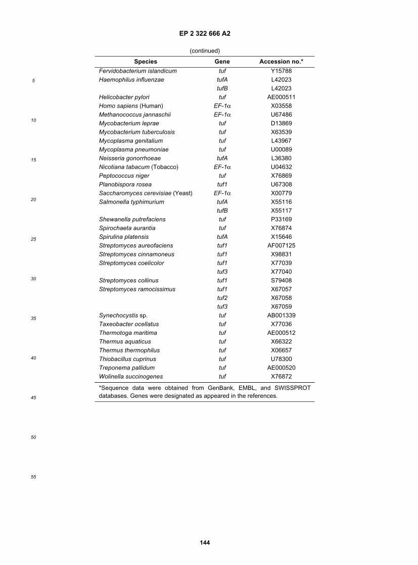
EP 2 322 666 A2
144
5
10
15
20
25
30
35
40
45
50
55
(continued)
Species Gene Accession no.*
Fervidobacterium islandicum tuf Y15788Haemophilus influenzae tufA L42023
tufB L42023Helicobacter pylori tuf AE000511Homo sapiens (Human) EF-1α X03558Methanococcus jannaschii EF-1α U67486Mycobacterium leprae tuf D13869Mycobacterium tuberculosis tuf X63539Mycoplasma genitalium tuf L43967Mycoplasma pneumoniae tuf U00089Neisseria gonorrhoeae tufA L36380Nicotiana tabacum (Tobacco) EF-1α U04632Peptococcus niger tuf X76869Planobispora rosea tuf1 U67308Saccharomyces cerevisiae (Yeast) EF-1α X00779Salmonella typhimurium tufA X55116
tufB X55117Shewanella putrefaciens tuf P33169Spirochaeta aurantia tuf X76874Spirulina platensis tufA X15646Streptomyces aureofaciens tuf1 AF007125Streptomyces cinnamoneus tuf1 X98831Streptomyces coelicolor tuf1 X77039
tuf3 X77040Streptomyces collinus tuf1 S79408Streptomyces ramocissimus tuf1 X67057
tuf2 X67058tuf3 X67059
Synechocystis sp. tuf AB001339Taxeobacter ocellatus tuf X77036Thermotoga maritima tuf AE000512Thermus aquaticus tuf X66322Thermus thermophilus tuf X06657Thiobacillus cuprinus tuf U78300Treponema pallidum tuf AE000520Wolinella succinogenes tuf X76872
*Sequence data were obtained from GenBank, EMBL, and SWISSPROTdatabases. Genes were designated as appeared in the references.

EP 2 322 666 A2
145
5
10
15
20
25
30
35
40
45
50
55
Table 19. Strains analyzed in Example 43.
Taxon Strain* Straint 16S rDNA sequence accession number
Cedecea ATCC 33431T
Cedecea lapagei ATCC 33432T
Cedecea neteri ATCC 33855T
Citrobacter amalonaticus ATCC 25405T CDC 9020-77T AF025370Citrobacter braakii ATCC 43162 CDC 080-58T AF025368Citrobacter farmeri ATCC 51112T CDC 2991-81T AF025371Citrobacter freundii ATCC 8090T DSM 30039T AJ233408Citrobacter koseri ATCC 27156T
Citrobacter sedlakii ATCC 51115T CDC 4696-86T AF025364Citrobacter werkmanii ATCC 51114T CDC 0876-58T AF025373Citrobacter youngae ATCC 29935T
Edwardsiella hoshinae ATCC 33379T
Edwardsiella tarda ATCC 15947T CDC 4411-68 AF015259Enterobacter aerogenes ATCC 13048T JCM 1235T AB004750Enterobacter agglomerans ATCC 27989Enterobacter amnigenus ATCC 33072T JCM 1237T AB004749Enterobacter asburiae ATCC 35953T JCM 6051T AB004744Enterobacter cancerogenus ATCC 35317T
Enterobacter cloacae ATCC 13047T
Enterobactergergoviae ATCC 33028T JCM 1234T AB004748Enterobacter hormaechei ATCC 49162T

EP 2 322 666 A2
146
5
10
15
20
25
30
35
40
45
50
55
(continued)
Taxon Strain* Straint 16S rDNA sequence accession number
Enterobacter sakazakii ATCC 29544T JCM 1233T AB004746Escherichia coli ATCC 11775T ATCC 11775T X80725Escherichia coli ATCC 25922 ATCC 25922 X80724Escherichia coli (ETEC) ATCC 35401Escherichia coli (0157:H7) ATCC 43895 ATCC 43895 Z83205Escherichia fergusonii ATCC 35469T
Escherichia hermanii ATCC 33650T
Escherichia vulneris ATCC 33821T ATCC 33821T X80734Ewingella americana ATCC 33852T NCPPB 3905 X88848Hafnia alvei ATCC 13337T ATCC 13337T M59155Klebsiella omithinolytica ATCC 31898 CIP 103.364 U78182Klebsiella oxytoca ATCC 33496 ATCC 13182T U78183Klebsiella planticola ATCC 33531T JCM 7251T AB004755Klebsiella pneumoniaesubsp. pneumoniae ATCC 13883T DSM 30104T AJ233420subsp. ozaenae ATCC 11296T ATCC 11296T Y17654subsp. rhinoscleromatis ATCC 13884T
Kluyvera ascorbata ATCC 33433T ATCC 14236 Y07650Kluyvera cryocrescens ATCC 33435T
Kluyvera georgiana ATCC 51603T
Leclercia adecarboxylata ATCC 23216T
Leminorella grimontii ATCC 33999T DSM 5078T AJ233421Moellerella wisconsensis ATCC 35017T
Morganella morganii ATCC 25830T
Pantoea agglomerans ATCC 27155T DSM 3493T AJ233423Pantoea dispersa ATCC 14589T
Plesiomonas shigelloides ATCC 14029T
Pragia fontium ATCC 49100T DSM 5563T AJ233424Proteus mirabilis ATCC 25933Proteus penneri ATCC 33519T
Proteus vulgaris ATCC 13315T DSM 30118T AJ233425Providencia alcalifaciens ATCC 9886T
Providencia rettgeri ATCC 9250Providencia rustigianii ATCC 33673T
Providencia stuartii ATCC 33672Rahnella aquatilis ATCC 33071T DSM 4594T AJ233426Salmonella choleraesuissubsp. arizonae ATCC 13314T
subsp. choleraesuisserotype Choleraesuis ATCC 7001serotype Enteritidis‡ ATCC 13076T SE22 SE22serotype Gallinarum ATCC 9184serotype Heidelberg ATCC 8326serotype Paratyphi A ATCC 9150serotype Paratyphi B ATCC 8759serotype Typhi‡ ATCC 10749 St111 U88545serotype Typhimurium‡ ATCC 14028serotype Virchow ATCC 51955subsp. diarizonae ATCC 43973T

EP 2 322 666 A2
147
5
10
15
20
25
30
35
40
45
50
55
(continued)
Taxon Strain* Straint 16S rDNA sequence accession number
subsp. houtenae ATCC 43974T
subsp. indica ATCC 43976T
subsp. salamae ATCC 43972T
Serratia fonticola DSM 4576T DSM 4576T AJ233429Serratia grimesii ATCC 14460T DSM 30063T AJ233430Serratia liquefaciens ATCC 27592T
Serratia marcescens ATCC 13880T DSM 30121T AJ233431Serratia odorifera ATCC 33077T DSM 4582T AJ233432Serratia plymuthica DSM 4540T DSM 4540T AJ233433Serratia rubidaea DSM 4480T DSM 4480T AJ233436Shigella boydii ATCC 9207 ATCC 9207 X96965Shigella dysenteriae ATCC 11835
ATCC 13313T X96966ATCC 25931 X96964
Shigella flexneri ATCC 12022 ATCC 12022 X96963Shigella sonnei ATCC 29930T
Tatumella ptyseos ATCC 33301T DSM 5000T AJ233437Trabulsiella guamensis ATCC 49490T
Yersinia enterocolitica ATCC 9610T ATCC 9610T M59292Yersinia frederiksenii ATCC 33641T
Yersinia intermedia ATCC 29909T
Yersinia pestis RRB KIMD27 ATCC 19428T X75274Yersinia pseudotuberculosis ATCC 29833T
Yersinia rohdei ATCC 43380T ER-2935T X75276Shewanella putrefaciens ATCC 8071T
Vibrio cholerae ATCC 25870 ATCC 14035T X74695
T Type strain*Strains used in this study for sequencing of partial tuf and atpD genes. SEQ ID NOs. for tuf and atpD sequencescorresponding to the above reference strains are given in table 7.†strains used in other studies for sequencing of 16S rDNA gene. When both strain numbers are on the samerow, both strains are considered to be the same although strain numbers may be different.‡Phylogenetic serotypes considered species by the Bacteriological Code (1990 Revision).
Table 20. PCR primer pairs used In this study
Primer SEQ IDNO.
Sequence Nucleotidepositions*
Amplicon length(bp)
tuf664 271-299 884
697 5’-CCIACIGTICKICCRCCYTCRCG-3’ 1132-1156atpD568 5’-RTIATIGGIGCIGTIRTIGAYGT-3’ 25-47 884567 5’-TCRTCIGCIGGIACRTAIAYIGCYTG-3’ 883-908700 5’-TIRTIGAYGTCGARTTCCCTCARG-3’ 38-61 871

EP 2 322 666 A2
148
5
10
15
20
25
30
35
40
45
50
55
(continued)
Primer SEQ IDNO.
Sequence Nucleotidepositions*
Amplicon length(bp)
567 5’-TCRTCIGCIGGIACRTAIAYIGCYTG-3’ 883-908
*The nucleotide positions given are for E. coli tuf and atpD sequences (GenBank accession no. AE000410 o andV00267, respectively). Numbering starts from the first base of the initiation codon.

EP 2 322 666 A2
149
5
10
15
20
25
30
35
40
45
50
55

EP 2 322 666 A2
150
5
10
15
20
25
30
35
40
45
50
55

EP 2 322 666 A2
151
5
10
15
20
25
30
35
40
45
50
55

EP 2 322 666 A2
152
5
10
15
20
25
30
35
40
45
50
55

EP 2 322 666 A2
153
5
10
15
20
25
30
35
40
45
50
55
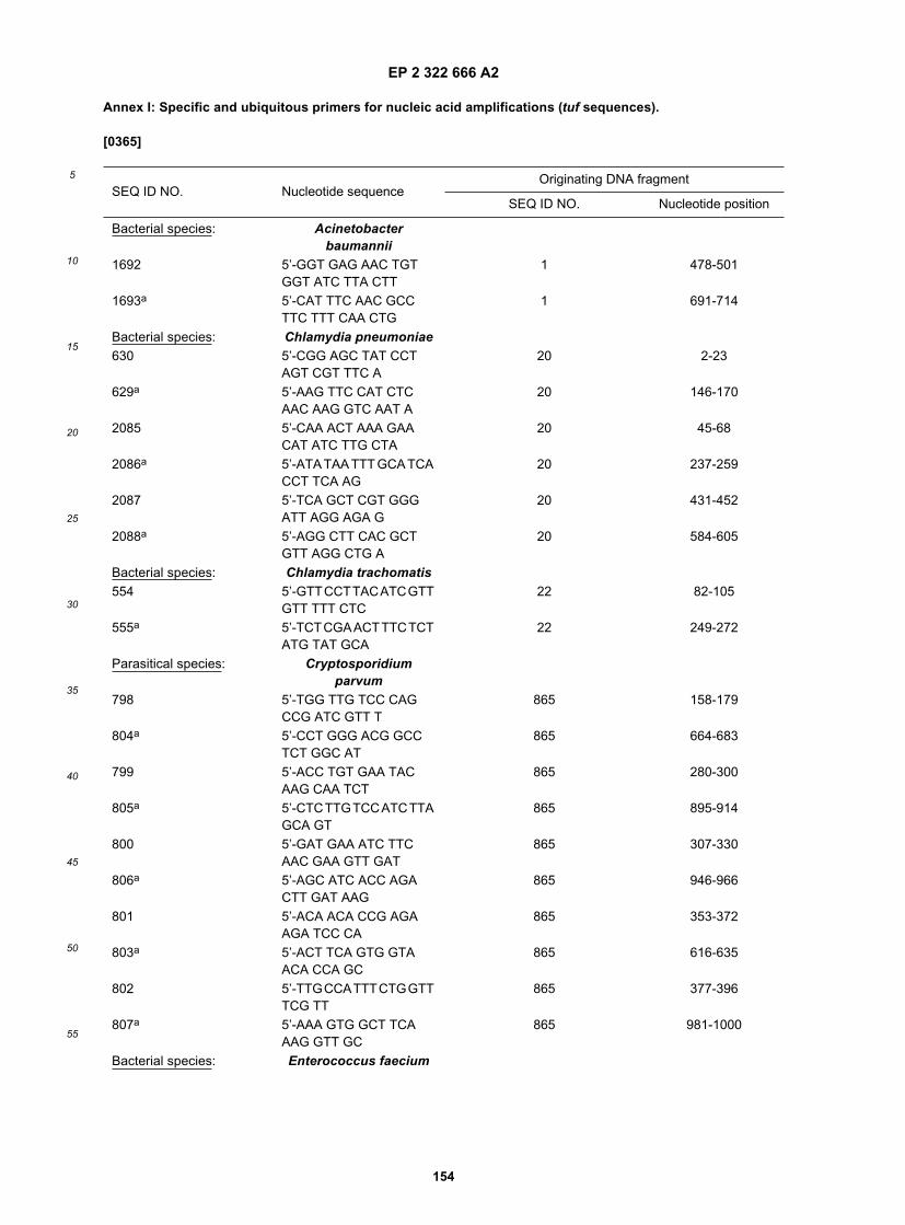
EP 2 322 666 A2
154
5
10
15
20
25
30
35
40
45
50
55
Annex I: Specific and ubiquitous primers for nucleic acid amplifications (tuf sequences).
[0365]
SEQ ID NO. Nucleotide sequenceOriginating DNA fragment
SEQ ID NO. Nucleotide position
Bacterial species: Acinetobacterbaumannii
1692 5’-GGT GAG AAC TGTGGT ATC TTA CTT
1 478-501
1693a 5’-CAT TTC AAC GCCTTC TTT CAA CTG
1 691-714
Bacterial species: Chlamydia pneumoniae630 5’-CGG AGC TAT CCT
AGT CGT TTC A20 2-23
629a 5’-AAG TTC CAT CTCAAC AAG GTC AAT A
20 146-170
2085 5’-CAA ACT AAA GAACAT ATC TTG CTA
20 45-68
2086a 5’-ATA TAA TTT GCA TCACCT TCA AG
20 237-259
2087 5’-TCA GCT CGT GGGATT AGG AGA G
20 431-452
2088a 5’-AGG CTT CAC GCTGTT AGG CTG A
20 584-605
Bacterial species: Chlamydia trachomatis554 5’-GTTCCTTACATCGTT
GTT TTT CTC22 82-105
555a 5’-TCTCGAACTTTCTCTATG TAT GCA
22 249-272
Parasitical species: Cryptosporidiumparvum
798 5’-TGG TTG TCC CAGCCG ATC GTT T
865 158-179
804a 5’-CCT GGG ACG GCCTCT GGC AT
865 664-683
799 5’-ACC TGT GAA TACAAG CAA TCT
865 280-300
805a 5’-CTCTTGTCCATCTTAGCA GT
865 895-914
800 5’-GAT GAA ATC TTCAAC GAA GTT GAT
865 307-330
806a 5’-AGC ATC ACC AGACTT GAT AAG
865 946-966
801 5’-ACA ACA CCG AGAAGA TCC CA
865 353-372
803a 5’-ACT TCA GTG GTAACA CCA GC
865 616-635
802 5’-TTGCCATTTCTGGTTTCG TT
865 377-396
807a 5’-AAA GTG GCT TCAAAG GTT GC
865 981-1000
Bacterial species: Enterococcus faecium

EP 2 322 666 A2
155
5
10
15
20
25
30
35
40
45
50
55
(continued)
SEQ ID NO. Nucleotide sequenceOriginating DNA fragment
SEQ ID NO. Nucleotide position
1696 5’-ATG TTC CTG TAGTTG CTG GA
64 189-208
1697a 5’-TTTCTTCAGCAATACCAA CAA C
64 422-443
Bacterial species: Klebsiella pneumoniae1329 5’-TGT AGA GCG CGG
TAT CAT CAA AGT A103 352-377
1330a 5’-AGA TTC GAA CTTGGT GTG CGG G
103 559-571
aThese sequences are from the complementary DNA strand of the sequence of the originating fragment given in theSequence Listing.Bacterial species: Mycoplasma
pneumoniae2093 5’-TGT TGG CAA TCG
AAG ACA CC2097a 635-654
2094b 5’-TTCAATTTC TTGACCTAC TTT CAA
2097a 709-732
Bacterial species: Neisseria gonorrhoeae551 5’-GAA GAA AAA ATC
TTC GAA CTG GCT A126 256-280
552b 5’-TAC ACG GCC GGTGAC TAC G
126 378-396
2173 5’-AAG AAA AAA TCTTCG AAC TGG CTA
126 257-280
2174b 5’-TCT ACA CGG CCGGTG
126 384-398
2175 5’-CCG CCA TAC CCCGTT T
126 654-669
2176b 5’-CGG CAT TAC CATTTC CAC ACC TTT
126 736-759
Bacterial species: Pseudomonasaeruginosa
1694 5’-AAG GCA AGG ATGACA ACG GC
153 231-250
1695b 5’-ACGATTTCCACTTCTTCC TGG
153 418-438
Bacterial species: Streptococcusagalactiae
549 5’-GAA CGT GAT ACTGAC AAA CCT TTA
207-210c 308-331d
550b 5’-GAA GAA GAA CACCAA CGT TG
207-210c 520-539d
Bacterial species: Streptococcuspyogenes
999 5’-TTG ACC TTG TTGATG ACG AAG AG
1002 143-165
1000b 5’-TTA GTG TGT GGGTTG ATT GAA CT
1002 622-644
1001 5’-AAG AGT TGC TTGAAT TAG TTG AG
1002 161-183

EP 2 322 666 A2
156
5
10
15
20
25
30
35
40
45
50
55
(continued)
SEQ ID NO. Nucleotide sequenceOriginating DNA fragment
SEQ ID NO. Nucleotide position
1000b 5’-TTA GTG TGT GGGTTG ATT GAA CT
1002 622-644
Parasitical species: Trypanosoma brucei820 5’-GAA GGA GGT GTC
TGC TTA CAC864 513-533
821b 5’-GGC GCA AAC GTCACC ACA TCA
864 789-809
820 5’-GAA GGA GGT GTCTGC TTA CAC
864 513-533
822b 5’-CGG CGG ATG TCCTTA ACA GAA
864 909-929
a Sequence from databases.b These sequences are from the complementary DNA strand of the sequence of the originating fragment given inthe Sequence Listing.c These sequences were aligned to derive the corresponding primer.d The nucleotide positions refer to the S. agalactiae tuf sequence fragment (SEQ ID NO. 209).
Originating DNA fragment
SEQ ID NO. Nucleotide sequence SEQ ID NO. Nucleotide position
Parasitical species: Trypanosoma cruzi794 5’-GAC GAC AAG TCG
GTG AAC TT840-842a 281-300c
795b 5’-ACT TGC ACG CGATGT GGC AG
840-842a 874-893c
Bacterial genus: Clostridium sp.796 5’-GGT CCA ATG CCW
CAA ACW AGA32,719- 724,736a 32-52d
797b 5’-CAT TAA GAA TGGYTT ATC TGT SKC TCT
32,719- 724,736a 320-346d
808 5’-GCI TTA IWR GCA TTAGAA RAY CCA
32,719- 724,736a 224-247d
809b 5’-TCT TCC TGT WGCAAC TGT TCC TCT
32,719- 724,736a 337-360d
810 5’-AGA GMW ACA GATAAR SCA TTC TTA
32,719- 724,736a 320-343d
811b 5’-TRA ART AGA ATTGTG GTC TRT ATC C
32,719- 724,736a 686-710d
Bacterial genus: Corynebacterium sp.545 5’-TAC ATC CTB GTY GCI
CTI AAC AAG TG34-44,662a 89-114e
546b 5’-CCR CGI CCG GTRATG GTG AAG AT
34-44,662a 350-372e
Bacterial genus: Enterococcus sp.656 5’-AAT TAA TGG CTG
CAG TTG AYG A58-72a 273-294f
657b 5’-TTG TCC ACG TTCGAT RTC TTC A
58-72a 556-577f

EP 2 322 666 A2
157
5
10
15
20
25
30
35
40
45
50
55
(continued)
Originating DNA fragment
SEQ ID NO. Nucleotide sequence SEQ ID NO. Nucleotide position
656 5’-AAT TAA TGG CTGCAG TTG AYG A
58-72a 273-294f
271b 5’-TTG TCC ACG TTGGAT RTC TTC A
58-72a 556-577f
1137 5’-AAT TAA TGG CTGCWG TTG AYG AA
58-72a 273-295f
1136b 5’-ACT TGT CCA CGTTSG ATR TCT
58-72a 559-579f
a These sequences were aligned to derive the corresponding primer.b These sequences are from the complementary DNA strand of the sequence of the originating fragment given inthe Sequence Listing.c The nucleotide positions refer to the T. cruzi tuf sequence fragment (SEQ ID NO. 842).d The nucleotide positions refer to the C. perfringens tuf sequence fragment (SEQ ID NO. 32).e The nucleotide positions refer to the C. diphtheriae tuf sequence fragment (SEQ ID NO. 662).f The nucleotide positions refer to the E. durans tuf sequence fragment (SEQ ID NO. 61).Bacterial genus: Legionella sp.2081 5’-GRA TYR TYA AAG
TTG GTG AGG AAG111-112a 411-434b
2082c 5’-CMA CTT CAT CYCGCT TCG TAC C
111-112a 548-569b
Bacterial genus: Staphylococcus sp.553 5’-GGC CGT GTT GAA
CGT GGT CAA ATC A176-203a 313-337d
575c 5’-TIA CCA TTT CAG TACCTT CTG GTA A
176-203a 653-677d
553 5’-GGC CGT GTT GAACGT GGT CAA ATC A
176-203a 313-337d
707c 5’-TWA CCA TTT CAGTAC CTT CTG GTA A
176-203a 653-677d
Bacterial genus: Streptococcus sp.547 5’-GTA CAG TTG CTT
CAG GAC GTA TC206-231a 372-394e
548c 5’-ACG TTC GAT TTCATC ACG TTG
206-231a 548-568e
Fungal genus: Candida sp.576 5’-AAC TTC RTC AAG
AAG GTY GGT TAC AA407-426, 428-432a 332-357f
632c 5’-CCC TTT GGT GGRTCS TKC TTG GA
407-426, 428-432a 791-813f
631 5’-CAGACCAACYGAIAARCC ATT RAG AT
407-426, 428-432a 523-548f
632c 5’-CCC TTT GGT GGRTCS TKC TTG GA
407-426, 428-432a 791-813f
633 5’-CAGACCAACYGAIAARCC ITT RAG AT
407-426, 428-432a 523-548f
632c 5’-CCC TTT GGT GGRTCS TKC TTG GA
407-426, 428-432a 791-813f
a These sequences were aligned to derive the corresponding primer.b The nucleotide positions refer to the L. pneumophila tuf sequence fragment (SEQ ID NO. 112).
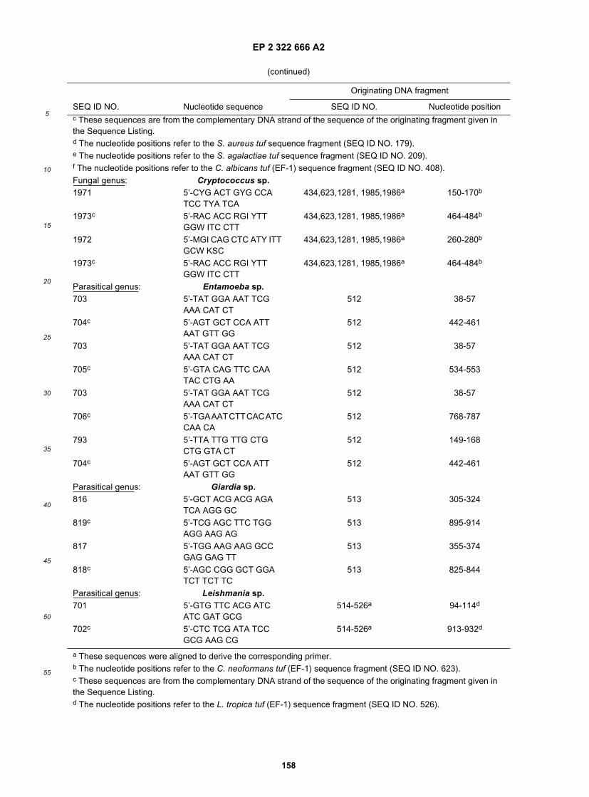
EP 2 322 666 A2
158
5
10
15
20
25
30
35
40
45
50
55
(continued)
Originating DNA fragment
SEQ ID NO. Nucleotide sequence SEQ ID NO. Nucleotide positionc These sequences are from the complementary DNA strand of the sequence of the originating fragment given inthe Sequence Listing.d The nucleotide positions refer to the S. aureus tuf sequence fragment (SEQ ID NO. 179).e The nucleotide positions refer to the S. agalactiae tuf sequence fragment (SEQ ID NO. 209).f The nucleotide positions refer to the C. albicans tuf (EF-1) sequence fragment (SEQ ID NO. 408).Fungal genus: Cryptococcus sp.1971 5’-CYG ACT GYG CCA
TCC TYA TCA434,623,1281, 1985,1986a 150-170b
1973c 5’-RAC ACC RGI YTTGGW ITC CTT
434,623,1281, 1985,1986a 464-484b
1972 5’-MGI CAG CTC ATY ITTGCW KSC
434,623,1281, 1985,1986a 260-280b
1973c 5’-RAC ACC RGI YTTGGW ITC CTT
434,623,1281, 1985,1986a 464-484b
Parasitical genus: Entamoeba sp.703 5’-TAT GGA AAT TCG
AAA CAT CT512 38-57
704c 5’-AGT GCT CCA ATTAAT GTT GG
512 442-461
703 5’-TAT GGA AAT TCGAAA CAT CT
512 38-57
705c 5’-GTA CAG TTC CAATAC CTG AA
512 534-553
703 5’-TAT GGA AAT TCGAAA CAT CT
512 38-57
706c 5’-TGAAATCTTCACATCCAA CA
512 768-787
793 5’-TTA TTG TTG CTGCTG GTA CT
512 149-168
704c 5’-AGT GCT CCA ATTAAT GTT GG
512 442-461
Parasitical genus: Giardia sp.816 5’-GCT ACG ACG AGA
TCA AGG GC513 305-324
819c 5’-TCG AGC TTC TGGAGG AAG AG
513 895-914
817 5’-TGG AAG AAG GCCGAG GAG TT
513 355-374
818c 5’-AGC CGG GCT GGATCT TCT TC
513 825-844
Parasitical genus: Leishmania sp.701 5’-GTG TTC ACG ATC
ATC GAT GCG514-526a 94-114d
702c 5’-CTC TCG ATA TCCGCG AAG CG
514-526a 913-932d
a These sequences were aligned to derive the corresponding primer.b The nucleotide positions refer to the C. neoformans tuf (EF-1) sequence fragment (SEQ ID NO. 623).c These sequences are from the complementary DNA strand of the sequence of the originating fragment given inthe Sequence Listing.d The nucleotide positions refer to the L. tropica tuf (EF-1) sequence fragment (SEQ ID NO. 526).

EP 2 322 666 A2
159
5
10
15
20
25
30
35
40
45
50
55
(continued)
Originating DNA fragment
SEQ ID NO. Nucleotide sequence SEQ ID NO. Nucleotide position
Parasitical genus: Trypanosoma sp.823 5’-GAG CGG TAT GAY
GAG ATT GT529,840- 842,864a 493-512b
824c 5’-GGC TTC TGC GGCACC ATG CG
529,840- 842,864a 1171-1190b
Bacterial family: Enterobacteriaceae933 5’-CAT CAT CGT ITT CMT
GAA CAA RTG78,103,146, 168,238,698a 390-413d
934c 5’-TCA CGY TTR RTACCA CGC AGI AGA
78,103,146, 168,238,698a 831-854d
Bacterial family: Mycobacteriaceae539 5’-CCI TAC ATC CTB GTY
GCI CTI AAC AAG122 85-111
540c 5’-GGD GCI TCY TCRTCG WAI TCC TG
122 181-203
Bacterial group: Escherichia coli and Shigella1661 5’-TGG GAA GCG AAA
ATC CTG1668e 283-300
1665c 5’-CAG TAC AGG TAGACT TCT G
1668e 484-502
Bacterial group: Pseudomonads group541 5’-GTK GAA ATG TTC
CGC AAG CTG CT153-155a 476-498f
542c 5’-CGG AAR TAG AACTGS GGA CGG TAG
153-155a 679-702f
541 5’-GTK GAA ATG TTCCGC AAG CTG CT
153-155a 476-498f
544c 5’-AYG TTG TCG CCMGGC ATT MCC AT
153-155a 749-771f
a These sequences were aligned to derive the corresponding primer.b The nucleotide positions refer to the T. brucei tuf (EF-1) sequence fragment (SEQ ID NO. 864).c These sequences are from the complementary DNA strand of the sequence of the originating fragment given inthe Sequence Listing.d The nucleotide positions refer to the E. coli tuf sequence fragment (SEQ ID O. 698).e Sequence from databases.f The nucleotide positions refer to the P. aeruginosa tuf sequence fragment (SEQ ID NO. 153).Parasitical group: Trypanosamatidae
family923 5’-GAC GCI GCC ATC
CTG ATG ATC511,514-526, 529,840-842,
864a166-188b
924c 5’-ACC TCA GTC GTCACG TTG GCG
511,514-526, 529,840-842,864a
648-668b
925 5’-AAG CAG ATG GTTGTG TGC TG
511,514-526, 529,840-842,864a
274-293b
926c 5’-CAG CTG CTC GTGGTG CAT CTC GAT
511,514-526, 529,840-842,864a
676-699b
927 5’-ACG CGG AGA AGGTGC GCT T
511,514-526, 529,840-842,864a
389-407b
928c 5’-GGT CGT TCT TCGAGT CAC CGC A
511,514-526, 529,840-842,864a
778-799b

EP 2 322 666 A2
160
5
10
15
20
25
30
35
40
45
50
55
(continued)
Originating DNA fragment
SEQ ID NO. Nucleotide sequence SEQ ID NO. Nucleotide position
Universal primers (bacteria)636 5’-ACTGGYGTTGAIATG
TTC CGY AA7,54,78, 100,103,159,
209,224,227b470-492d
637c 5’-ACG TCA GTI GTACGG AAR TAG AA
7,54,78, 100,103,159,209,224,227b
692-714d
638 5’-CCAATGCCACAAACICGT GAR CAC AT
7,54,78, 100,103,159,209,224,227b
35-60e
639c 5’-TTTACGGAACATTTCWAC ACC WGT IAC A
7,54,78, 100,103,159,209,224,227b
469-496e
a These sequences were aligned to derive the corresponding primer.b The nucleotide positions refer to the L. tropica tuf (EF-1) sequence fragment (SEQ ID NO. 526).c These sequences are from the complementary DNA strand of the sequence of the originating fragment given inthe Sequence Listing.d The nucleotide positions refer to the E. coli tuf sequence fragment (SEQ ID NO. 78).e The nucleotide positions refer to the B. cereus tuf sequence fragment (SEQ ID NO. 7).
Universal primers (bacteria) (continued)643 5’-ACT GGI GTI GAR ATG
TTC CGY AA1,3,4,7,12, 13,16,49,54,
72,78,85,88, 91,94,98,103,108,112,115, 116,120,121,126,128,134, 136,146,154,159,179,186, 205,209,212,
224,238a
470-492b
644c 5’-ACG TCI GTI GTI CKGAAR TAG AA
same as SEQ ID NO. 643 692-714b
643 5’-ACT GGI GTI GAR ATGTTC CGY AA
1,3,4,7,12, 13,16,49,54,72,78,85,88, 91,94,98,103,108,112,115, 116,120,121,126,128,134, 136,146,154,159,179,186, 205,209,212,
224,238a
470-492b
645c 5’-ACG TCI GTI GTI CKGAAR TAR AA
same as SEQ ID NO. 643 692-714b
646 5’-ATCGACAAGCCI TTCYTI ATG SC
2,13,82 122,145a 317-339d
647c 5’-ACG TCC GTS GTRCGG AAG TAG AAC TG
2,13,82 122,145a 686-711d
646 5’-ATCGACAAGCCI TTCYTI ATG SC
2,13,82 122,145a 317-339d
648c 5’-ACG TCS GTS GTRCGG AAG TAG AAC TG
2,13,82 122,145a 686-711d
a These sequences were aligned to derive thecorresponding
primer.
b The nucleotide positions refer to the E. coli tuf sequence fragment (SEQ ID NO. 78).c These sequences are from the complementary DNA strand of the sequence of the originating fragment given inthe Sequence Listing.d The nucleotide positions refer to the A. meyeri tuf sequence fragment (SEQ ID NO. 2)
Universal primers (bacteria) (continued)649 5’-GTC CTA TGC CTC
ARA CWC GIG AGC AC8,86,141,143a 33-58b

EP 2 322 666 A2
161
5
10
15
20
25
30
35
40
45
50
55
(continued)
Originating DNA fragment
SEQ ID NO. Nucleotide sequence SEQ ID NO. Nucleotide position
650c 5’-TTA CGG AAC ATYTCA ACA CCI GT
8,86,141,143a 473-495b
636 5’-ACTGGYGTTGAIATGTTC CGY AA
8,86,141,143a 473-495b
651c 5’-TGACGACCACCITCYTCY TTY TTC A
8,86,141,143a 639-663b
Universal primers (fungi)1974 5’-ACA AGG GIT GGR
MSA AGG AGA C404,405,433, 445,898,1268,
1276,1986a443-464d
1975c 5’-TGR CCR GGG TGGTTR AGG ACG
404,405,433, 445,898,1268,1276,1986a
846-866d
1976 5’-GAT GGA YTC YGTYAA ITG GGA
407-412,414-426,428-431,439,443,447,
448,622,624,665,1685,1987-1990a
286-306e
1978c 5’-CAT CIT GYA ATG GYAATC TYA AT
same as SEQ ID NO. 1976 553-575e
1977 5’-GAT GGA YTC YGTYAA RTG GGA
same as SEQ ID NO. 1976 286-306e
1979c 5’-CAT CYT GYA ATGGYA ASC TYA AT
same as SEQ ID NO. 1976 553-575e
1981 5’-TGG ACA CCI SCA AGIGGK CYG
401-405, 433,435,436,438,444,445,449,453,455,457,779,781-783,785,786,788-790,897-903,
1267-1272,1274-1280,1282-1287,1991-1998a
281-301d
1980c 5’-TCR ATG GCI TCI AIRAGR GTY T
same as SEQ ID NO. 1981 488-509d
a These sequences were aligned to derive the corresponding primer.b The nucleotide positions refer to the B. distasonis tuf sequence fragment (SEQ ID NO. 8).c These sequences are from the complementary DNA strand of the sequence of the originating fragment given inthe Sequence Listing.d The nucleotide positions refer to the A. fumigatus tuf (EF-1) sequence fragment (SEQ ID NO. 404).e The nucleotide positions refer to the C. albicans tuf (EF-1) sequence fragment (SEQ ID NO. 407).
Universal primers (fungi) (continued)1982 5’-TGG ACA CYI SCA AGI
GGK CYGsame as SEQ ID NO. 1981 281-301a
1980b 5’-TCR ATG GCI TCI AIRAGR GTY T
same as SEQ ID NO. 1981 488-509a
1983 5’-CYGAYTGCGCYATICTCA TCA
same as SEQ ID NO. 1981 143-163a
1980b 5’-TCR ATG GCI TCI AIRAGR GTY T
same as SEQ ID NO. 1981 488-509a
1984 5’-CYG AYT GYG CYATYC TSA TCA
same as SEQ ID NO. 1981 143-163a
1980b 5’-TCR ATG GCI TCI AIRAGR GTY T
same as SEQ ID NO. 1981 488-509a
Sequencing primers

EP 2 322 666 A2
162
5
10
15
20
25
30
35
40
45
50
55
(continued)
Originating DNA fragment
SEQ ID NO. Nucleotide sequence SEQ ID NO. Nucleotide position
556 5’-CGG CGC NAT CYTSGT TGT TGC
668C 306-326
557b 5’-CCM AGG CAT RACCAT CTC GGT G
668C 1047-1068
694 5’-CGG CGC IAT CYTSGT TGT TGC
668C 306-326
557b 5’-CCM AGG CAT RACCAT CTC GGT G
668C 1047-1068
664 5’-AAY ATG ATI ACI GGIGCI GCI CAR ATG GA
619C 604-632
652b 5’-CCW AYA GTI YKI CCICCY TCY CTI ATA
619C 1482-1508
664 5’-AAY ATG ATI ACI GGIGCI GCI CAR ATG GA
619C 604-632
561b 5’-ACIGTICGGCCRCCCTCA CGG AT
619C 1483-1505
543 5’-ATC TTA GTA GTT TCTGCT GCT GA
607 8-30
660b 5’-GTA GAA TTG AGGACG GTA GTT AG
607 678-700
658 5’-GAT YTA GTC GATGAT GAA GAA TT
621 116-138
659b 5’-GCT TTT TGI GTT TCWGGT TTR AT
621 443-465
658 5’-GAT YTA GTC GATGAT GAA GAA TT
621 116-138
661b 5’-GTA GAA YTG TGGWCG ATA RTT RT
621 678-700
558 5’-TCI TTY AAR TAY GCITGG GT
665C 157-176
559b 5’-CCG ACR GCR AYIGTY TGI CKC AT
665C 1279-1301
813 5’-AAT CYG TYG AAATGC AYC ACG A
665C 687-708
559b 5’-CCG ACR GCR AYIGTY TGI CKC AT
665C 1279-1301
a The nucleotide positions refer to the A. fumigatus tuf (EF-1) sequence fragment (SEQ ID NO. 404).b These sequences are from the complementary DNA strand of the sequence of the originating fragment given inthe Sequence Listing.c Sequences from databases.
Sequencing primers(continued)
558 5’-TCI TTY AAR TAY GCITGG GT
665a 157-176
815b 5’-TGG TGC ATY TCKACR GAC TT
665a 686-705
560 5’-GAYTTCATYAARAAYATG ATY AC
665a 289-311
559b 5’-CCG ACR GCR AYIGTY TGI CKC AT
665a 1279-1301

EP 2 322 666 A2
163
5
10
15
20
25
30
35
40
45
50
55
(continued)
Originating DNA fragment
SEQ ID NO. Nucleotide sequence SEQ ID NO. Nucleotide position
653 5’-GAY TTC ATI AAR AAYATG AT
665a 289-308
559b 5’-CCG ACR GCR AYIGTY TGI CKC AT
665a 1279-1301
558 5’-TCI TTY AAR TAY GCITGG GT
665a 157-176
655b 5’-CCRATACCICMRATYTTG TA
665a 754-773
654 5’-TAC AAR ATY KGI GGTATY GG
665a 754-773
559b 5’-CCG ACR GCR AYIGTY TGI CKC AT
665a 1279-1301
696 5’-ATI GGI CAY RTI GAYCAY GGI AAR AC
698a 52-77
697b 5’-CCI ACI GTI CKI CCRCCY TCR CG
698a 1132-1154
911 5’-GAC GGM KKC ATGCCG CAR AC
853 22-41
914b 5’-GAA RAG CTG CGGRCG RTA GTG
853 700-720
912 5’-GAC GGC GKC ATGCCG CAR AC
846 20-39
914b 5’-GAA RAG CTG CGGRCG RTA GTG
846 692-712
913 5’-GAC GGY SYC ATGCCK CAG AC
843 251-270
915b 5’-AAA CGC CTG AGGRCG GTA GTT
843 905-925
916 5’-GCC GAG CTG GCCGGC TTC AG
846 422-441
561b 5’-ACIGTICGGCCRCCCTCA CGG AT
619a 1483-1505
664 5’-AAY ATG ATI ACI GGIGCI GCI CAR ATG GA
619a 604-632
917b 5’-TCG TGC TAC CCGTYG CCG CCA T
846 593-614
a Sequences from databases.b These sequences are from the complementary DNA strand of the sequence of the originating fragment given inthe Sequence Listing.
Sequencing primers(continued)
1221 5’-GAY ACI CCI GGI CAYGTI GAY TT
1230a 292-314
1226b 5’-GTI RMR TAI CCR AACATY TC
1230a 2014-2033
1222 5’-ATY GAY ACI CCI GGICAY GTI GAY TT
1230a 289-314
1223b 5’-AYI TCI ARR TGI ARYTCR CCC ATI CC
1230a 1408-1433

EP 2 322 666 A2
164
5
10
15
20
25
30
35
40
45
50
55
Annex II: Specific and ubiquitous primers for nucleic acid amplification (atpD sequences).
[0366]
(continued)
Originating DNA fragment
SEQ ID NO. Nucleotide sequence SEQ ID NO. Nucleotide position
1224 5’-CCI GYI HTI YTI GARCCI ATI ATG
1230a 1858-1881
1225b 5’-TAI CCR AAC ATY TCISMI ARI GGI AC
1230a 2002-2027
1227 5’-GTI CCI YTI KCI GARATG TTY GGI TA
1230a 2002-2027
1229b 5’-TCC ATY TGI GCI GCICCI GTI ATC AT
698a 4-29
1228 5’-GTI CCI YTI KCI GARATG TTY GGI TAY GC
1230a 2002-2030
1229b 5’-TCC ATY TGI GCI GCICCI GTI ATC AT
698a 4-29
1999 5’-CAT GTC AAY ATTGGT ACT ATT GGT CAT
GT 498-500, 502,505,506,508,619,2004,2005c
25-53d
2000b 5’-CCA CCY TCI CTCAMG TTG AAR CGT T
same as SEQ ID NO. 1999 1133-1157d
2001 5’-ACY ACI TTR ACI GCYGCY ATY AC
same as SEQ ID NO. 1999 67-89d
2003b 5’-CAT YTC RAI RTT GTCACC TGG
same as SEQ ID NO. 1999 1072-1092d
2002 5’-CCIGARGARAGAGCIMGW GGT
same as SEQ ID NO. 1999 151-171d
2003b 5’-CAT YTC RAI RTT GTCACC TGG
same as SEQ ID NO. 1999 1072-1092d
a Sequences from databases.b These sequences are from the complementary DNA strand of the sequence of the originating fragment given inthe Sequence Listing.c These sequences were aligned to derive the corresponding primer.d The nucleotide positions refer to the C. albicans tuf sequence fragment (SEQ ID NO. 2004).
Originating DNA fragment
SEQ ID NO. Nucleotide sequence SEQ ID NO. Nucleotide positionBacterial species: Acinetobacter baumannii1690 5’-CAG GTC CTG TTG
CGA CTG AAG AA243 186-208
1691b 5’-CAC AGA TAA ACCTGA GTG TGC TTT C
243 394-418
Bacterial species: Bacteroides fragilis2134 5’-CGC GTG AAG CTT
CTG TG929 184-200
2135b 5’-TCTCGCCGTTATTCAGTT TC
929 395-414
Bacterial species: Bordetellapertussis
2180 5’-TTC GCC GGC GTGGGC
1672C 544-558

EP 2 322 666 A2
165
5
10
15
20
25
30
35
40
45
50
55
(continued)
Originating DNA fragment
SEQ ID NO. Nucleotide sequence SEQ ID NO. Nucleotide position218,b 5’-AGC GCC ACG CGC
AGG1672C 666-680
Bacterial species: Enterococcus faecium1698 5’-GGA ATC AAC AGA
TGG TTT ACA AA292 131-153
2699b 5’-GCA TCT TCT GGGAAA GGT GT
292 258-277
1700 5’-AAG ATG CGG AAAGAA GCG AA
292 271-290
1701b 5’-ATT ATG GAT CAG TTCTTG GAT CA
292 439-461
Bacterial species: Klebsiella pneumoniae1331 5’-GCC CTT GAG GTA
CAG AAT GGT AAT GAAGTT
317 88-118
1332b 5’-GAC CGC GGC GCAGAC CAT CA
317 183-203
a These sequences were aligned to derive the corresponding primer.b These sequences are from the complementary DNA strand of the sequence of the originating fragment given inthe Sequence Listing.C Sequence from databases.Bacterial species: Streptococcus
agalactiae627 5’-ATT GTC TAT AAA AAT
GGC GAT AAG TC379-383a 42-67b
625c 5’-CGT TGA AGA CACGAC CCA AAG TAT CC
379-383a 206-231b
628 5’-AAA ATG GCG ATAAGT CAC AAA AAG TA
379-383a 52-77b
625C 5’-CGT TGA AGA CACGAC CCA AAG TAT CC
379-383a 206-231b
627 5’-ATT GTC TAT AAA AATGGC GAT AAG TC
379-383a 42-67b
626c 5’-TAC CAC CTT TTA AGTAAG GTG CTA AT
379-383a 371-396b
628 5’-AAA ATG GCG ATAAGT CAC AAA AAG TA
379-383a 52-77b
626c 5’-TAC CAC CTT TTA AGTAAG GTG CTA AT
379-383a 371-396b
Bacterial ground: Campylobacter jejuni and C. coli2131 5’-AAG CMA TTG TTG
TAA ATT TTG AAA G1576,1600,
1849,1863,2139d,a7-3,e
2132c 5’-TCA TAT CCA TAG CAATAG TTC TA
1576,1600,1849,1863,2139d,a
92-114e
Bacterial genus: Bordetella sp.825 5’-ATG AGC ARC GSA
ACC ATC GTT CAG TG1672d 1-26
826C 5’-TCG ATC GTG CCGACC ATG TAG AAC GC
1672d 1342-1367
Fungal genus: Candida sp.

EP 2 322 666 A2
166
5
10
15
20
25
30
35
40
45
50
55
(continued)
Originating DNA fragment
SEQ ID NO. Nucleotide sequence SEQ ID NO. Nucleotide position634 5’-AAC ACY GTC AGR RCI
ATT GCY ATG GA460-472, 474-478a 101-126f
635c 5’-AAACCRGTIARRGCRACT CTI GCT CT
460-472, 474-478a 617-642f
a These sequences were aligned to derive the corresponding primer.b The nucleotide positions refer to the S. agalactiae atpD sequence fragment (SEQ ID NO. 380).c These sequences are from the complementary DNA strand of the sequence of the originating fragment given inthe Sequence Listing.d Sequence from databases.e The nucleotide positions refer to the C. jejuni atpD sequence fragment (SEQ ID NO. 1576).f The nucleotide positions refer to the C. albicans atpD sequence fragment (SEQ ID NO. 460).
SEQ ID NO. Nucleotide sequence SEQ ID NO. Nucleotide position
Universal primers562 5’-CAR ATG RAY GAR CCI
CCI GGI GYI MGI ATG243,244,262, 264,280,284,291,297,309, 311,315,317,324,329,332, 334-336,339,
342,343,351,356,357,364-366,370,375,
379,393a
528-557b
563c 5’-GGY TGR TAI CCI ACIGCI GAI GGC AT
243,244,262, 264,280,284,291,297,309, 311,315,317,324,329,332, 334-336,339,
342,343,351,356,357,364-366,370,375,
379,393a
687-712b
564 5’-TAY GGI CAR ATG AAYGAR CCI CCI GGI AA
243,244,262, 264,280,284,291,297,309, 311,315,317,324,329,332, 334-336,339,
342,343,351,356,357,364-366,370,375,
379,393a
522-550b
565c 5’-GGY TGR TAI CCI ACIGCI GAI GGD AT
. 243,244,262,264,280,284, 291,297,309,311,315,317, 324,329,332,334-336,339, 342,343,351,356,357,364-366,370,375,
379,393a
687-712b
a These sequences were aligned to derive the corresponding primer.b The nucleotide positions refer to the K. pneumoniae atpD sequence fragment (SEQ ID NO. 317).c These sequences are from the complementary DNA strand of the sequence of the originating fragment given inthe Sequence Listing.
SEQ ID NO. Nucleotide sequence SEQ ID NO. Nucleotide position
Universal primers(continued)
640 5’-TCC ATG GTI TWY GGICAR ATG AA
248,284,315, 317,343,357,366,370,379,393a
513-535b

EP 2 322 666 A2
167
5
10
15
20
25
30
35
40
45
50
55
(continued)
SEQ ID NO. Nucleotide sequence SEQ ID NO. Nucleotide position
641c 5’-TGA TAA CCW ACI GCIGAI GGC ATA CG
248,284,315, 317,343,357,366,370,379,393a
684-709b
642 5’-GGC GTI GGI GAR CGIACI CGT GA
248,284,315, 317,343,357,366,370,379,393a
438-460b
641c 5’-TGA TAA CCW ACI GCIGAI GGC ATA CG
248,284,315, 317,343,357,366,370,379,393a
684-709b
Sequencing primers566 5’-TTY GGI GGI GCI GGI
GTI GGI AAR AC669d 445-470
567c 5’-TCR TCI GCI GGI ACRTAI AYI GCY TG
669d 883-908
566 5’-TTY GGI GGI GCI GGIGTI GGI AAR AC
669d 445-470
814 5’-GCI GGC ACG TAC ACIGCC TG
666d 901-920
568 5’-RTIATIGGIGCIGTIRTIGAY GT
669d 25-47
567c 5’-TCR TCI GCI GGI ACRTAI AYI GCY TG
669d 883-908
570 5’-RTIRYIGGICCIGTIRTIGAY GT
672d 31-53
567C 5’-TCR TCI GCI GGI ACRTAI AYI GCY TG
669d 883-908
572 5’-RTI RTI GGISCI GTIRTIGA
669d 25-44
567C 5’-TCR TCI GCI GGI ACRTAI AYI GCY TG
669d 883-908
569 5’-RTI RTI GGISCI GTIRTIGAT AT
671d 31-53
567c 5’-TCR TCI GCI GGI ACRTAI AYI GCY TG
669d 883-908
571 5’-RTIRTIGGICCIGTIRTIGAT GT
670d 31-53
567C 5’-TCR TCI GCI GGI ACRTAI AYI GCY TG
669d 883-908
a These sequences were aligned to derive the corresponding primer.b The nucleotide positions refer to the K. pneumoniae atpD sequence fragment (SEQ ID NO. 317).c These sequences are from the complementary DNA strand of the sequence of the originating fragment given inthe Sequence Listing.d Sequences from databases.
Sequencing primers(continued)
700 5’-TIR TIG AYG TCG ARTTCC CTC ARG
669a 38-61
567b 5’-TCR TCI GCI GGI ACRTAI AYI GCY TG
669a 883-908
568 5’-RTIATIGGIGCIGTIRTIGAY GT
669a 25-47
573b 5’-CCI CCI ACC ATR TARAAI GC
666a 1465-1484

EP 2 322 666 A2
168
5
10
15
20
25
30
35
40
45
50
55
(continued)
SEQ ID NO. Nucleotide sequence SEQ ID NO. Nucleotide position
574 5’-ATI GCI ATG GAY GGIACI GAR GG
666a 283-305
573b 5’-CCI CCI ACC ATR TARAAI GC
666a 1465-1484
574 5’-ATI GCI ATG GAY GGIACI GAR GG
666a 283-305
708b 5’-TCR TCC ATI CCI ARIATI GCI ATI AT
666a 1258-1283
681 5’-GGI SSI TTY GGI ISIGGI AAR AC
685 694-716
682b 5’-GTI ACI GGY TCY TCRAAR TTI CCI CC
686 1177-1202
681 5’-GGI SSI TTY GGI ISIGGI AAR AC
685 694-716
683b 5’-GTI ACI GGI TCI SWIAWR TCI CCI CC
685 1180-1205
681 5’-GGI SSI TTY GGI ISIGGI AAR AC
685 694-716
699 5’-GTI ACI GGY TCY TYRARR TTI CCI CC
686 1177-1202
681 5’-GGI SSI TTY GGI ISIGGI AAR AC.
685 694-716
812b 5’-GTI ACI GGI TCY TYRARR TTI CCI CC
685 1180-1205
1213 5’-AAR GGI GGI ACI GCIGCI ATH CCI GG
714a 697-722
1212b 5’-CCI CCI RGI GGI GAIACI GCW CC
714a 1189-1211
1203 5’-GGI GAR MGI GGI AAYGAR ATG
709a 724-744
1207b 5’-CCI TCI TCW CCI GGCATY TC
709a 985-1004
1204 5’-GCI AAY AAC ITC IWMYAT GCC
709a 822-842
1206b 5’-CKI SRI GTI GAR TCIGCC A
709a 926-944
1205 5’-AAY ACI TCI AWY ATGCCI GT
709a 826-845
1207b 5’-CCI TCI TCW CCI GGCATY TC
709a 985-1004
2282 5’-AGRRGCIMARATGTATGA
714a 84-101
2284b 5’-TCTGWGTRACIGGYTCKG AGA
714a 1217-1237
2283 5’-ATI TAT GAY GGK ITTCAG AGG C
714a 271-292
2285b 5’-CMC CIC CWG GTGGWG AWA C
714a 1195-1213
a Sequences from databases.b These sequences are from the complementary DNA strand of the sequence of the originating fragment given inthe Sequence Listing.

EP 2 322 666 A2
169
5
10
15
20
25
30
35
40
45
50
55
Annex III: Internal hybridization probes for specific detection of tuf sequences.
[0367]
Originating DNA fragment
SEQ ID NO. Nucleotide sequence SEQ ID NO. Nucleotide position
Bacterial species: Abiotrophia adiacens2170 5’-ACG TGA CGT TGA
CAA ACC A1715 313-331
Bacterial species: Chlamydia pneumoniae2089 5’-ATG CTG AAC TTA TTG
ACC TT20 136-155
2090 5’-CGT TAC TGG AGTCGA AAT G
20 467-485
Bacterial species: Enterococcus faecalis580 5’-GCT AAA CCA GCT
ACA ATC ACT CCA C62-63,607a 584-608b
603 5’-GGT ATT AAA GACGAA ACA TC
62-63,607a 440-459b
1174 5’-GAA CGT GGT GAAGTT CGC
62-63,607a 398-415b
Bacterial species: Enterococcus faecium602 5’-AAGTTGAAGTTGTTG
GTA TT64,608a 426-445C
Bacterial species: Enterococcusgallinarum
604 5’-GGT GAT GAA GTAGAA ATC GT
66,609a 419-438d
Bacterial species: Escherichia coli579 5’-GAA GGC CGT GCT
GGT GAG AA78 503-522
2168 5’-CAT CAA AGT TGGTGA AGA AGT TG
78 409-431
Bacterial species: Neisseria gonorrhoeae2166 5’-GAC AAA CCA TTC
CTG CTG126 322-339e
Fungal species: Candida albicans577 5’-CATGATTGAACCATC
CAC CA407-411a 406-425f
Fungal species: Candida dubliniensis578 5’-CATGATTGAAGCTTC
CAC CA412,414-415a 418-437g
a These sequences were aligned to derive the corresponding primer.b The nucleotide positions refer to the E. faecalis tuf sequence fragment (SEQ ID NO. 607).c The nucleotide positions refer to the E. faecium tuf sequence fragment (SEQ ID NO. 608).d The nucleotide positions refer to the E. gallinarum tuf sequence fragment (SEQ ID NO. 609).e The nucleotide positions refer to the N. gonorrhoeae tuf sequence fragment (SEQ ID NO. 126).f The nucleotide positions refer to the C. albicans tuf (EF-1) sequence fragment (SEQ ID NO. 408).g The nucleotide positions refer to the C. dubliniensis tuf (EF-1) sequence fragment (SEQ ID NO. 414).Bacterial species: Haemophilus influenzae581 5’-ACATCGGTGCATTAT
TAC GTG G610a 551-572

EP 2 322 666 A2
170
5
10
15
20
25
30
35
40
45
50
55
(continued)
Originating DNA fragment
SEQ ID NO. Nucleotide sequence SEQ ID NO. Nucleotide position
Bacterial species: mycoplamnapneumoniae
2095 5’-CGG TCG GGT TGAACG TGG
2097a 687-704
Bacterial species: Staphylococcus aureus584 5’-ACA TGACAC ATCTAA
AAC AA176-180b 369-388C
585 5’-ACC ACA TAC TGA ATTCAA AG
176-180b 525-544C
586 5’-CAGAAGTATACGTATTAT CA
176-180b 545-564C
587 5’-CGT ATT ATC AAA AGACGA AG
176-180b 555-574C
588 5’-TCT TCT CAA ACT ATCGTC CA
176-180b 593-612C
Bacterial species: Staphylococcus epidermidis589 5’-GCACGAAACTTCTAA
AAC AA185,611b 445-464d
590 5’-TAT ACG TAT TAT CTAAAG AT
185,611b 627-646d
591 5’-TCC TGG TTC TAT TACACC AC
185,611b 586-605d
592 5’-CAAAGCTGAAGTATACGT AT
185,611b 616-635d
593 5’-TTC ACT AAC TAT CGCCCA CA
185,611b 671-690d
Bacterial species: Staphylococcus haemolyticus594 5’-ATTGGTATCCATGAC
ACT TC186,188-190b 437-456e
595 5’-TTAAAGCAGACGTATACG TT
186,188-190b 615-634e
Bacterial species: Staphylococcus hominis596 5’-GAA ATT ATT GGT ATC
AAA GA191,193-196b 431-450f
597 5’-ATTGGTATCAAA GAAACT TC
191,193-196b 437-456f
598 5’-AAT TACACC TCA CACAAA AT
191,193-196b 595-614f
a Sequences from databases.b These sequences were aligned to derive the corresponding probe.c The nucleotide positions refer to the S. aureus tuf sequence fragment (SEQ ID NO. 179).d The nucleotide positions refer to the S. epidermidis tuf sequence fragment (SEQ ID NO. 611).e The nucleotide positions refer to the S. haemolyticus tuf sequence fragment (SEQ ID NO. 186).f The nucleotide positions refer to the S. hominis tuf sequence fragment (SEQ ID NO. 191).Bacterial species: Staphylococcus saprophyticus599 5’-CGG TGA AGA AAT
CGA AAT CA198-200a 406-425b
600 5’-ATG CAA GAA GAATCA AGC AA
198-200a 431-450b

EP 2 322 666 A2
171
5
10
15
20
25
30
35
40
45
50
55
(continued)
Originating DNA fragment
SEQ ID NO. Nucleotide sequence SEQ ID NO. Nucleotide position
601 5’-GTTTCACGTGATGATGTA CA
198-200a 536-555b
695 5’-GTT TCA CGT GATGAC GTA CA
198-200a 563-582b
Bacterial species: Streptococcus agalactiae582C 5’-TTT CAA CTT CGT CGT TGA
CAC GAA CAG T207-210a 404-431d
583c 5’-CAA CTG CTT TTT GGA TATCTT CTT TAA TAC CAA
CG 207-2,oa 433-467d
1199 5’-GTA TTA AAG AAG ATA TCCAAA AAG C
207-210a 438-462d
Bacterial species: Streptococcus pneumoniae1201 5’-TCAAAGAAGAAACTA
AAA AAG CTG T971,977, 979,986a 513-537e
Bacterial species: Streptococcus pyogenes1200 5’-TCAAAGAAGAAACTA
AAA AAG CTG T1002 473-497
Bacterial group: Enterococcus casseliflavus-flavescens-gallinarum group620 5’-ATTGGTGCATTGCTA
CGT58,65,66a 527-544f
1122 5’-TGG TGC ATT GCTACG TGG
58,65,66a 529-546f
Bacterial group: Enterococcus sp., Gemella sp., A. adiacens2172 5’-GTGTTGAAATGTTCC
GTA AA58-62,67-71,
87-88,607-609, 727,8711715,1722a
477-496g
a These sequences were aligned to derive the corresponding primer.b The nucleotide positions refer to the S. saprophyticus tuf sequence fragment (SEQ ID NO. 198).c These sequences are from the complementary DNA strand of the sequence of the originating fragment given inthe Sequence Listing.d The nucleotide positions refer to the S. agalactiae tuf sequence fragment (SEQ ID NO. 209).e The nucleotide positions refer to the S. pneumoniae tuf sequence fragment (SEQ ID NO. 986).f The nucleotide positions refer to the E. flavescens tuf sequence fragment (SEQ ID NO. 65).g The nucleotide positions refer to the E. faecium tuf sequence fragment (SEQ ID NO. 608).
Originating DNA fragmentSEQ ID NO. Nucleotide sequence SEQ ID NO. Nucleotide position
Bacterial genus: Gemella2171 5’-TCGTTGGATTAACTG
AAG AA87,88a 430-449b
Bacterial genus: Staphylococcus sp.605 5’-GAAATGTTCCGTAAA
TTA TT176-203a 403-422c
606 5’-ATTAGACTACGCTGAAGC TG
176-203a 420-439c
1175 5’-GTT ACT GGT GTAGAA ATG TTC
176-203a 391-411c

EP 2 322 666 A2
172
5
10
15
20
25
30
35
40
45
50
55
(continued)
Originating DNA fragmentSEQ ID NO. Nucleotide sequence SEQ ID NO. Nucleotide position
1176 5’-TACTGGTGTAGAAATGTT C
176-203a 393-411c
Bacterial genus: Streptococcus sp.1202 5’-GTGTTGAAATGTTCC
GTA AAC A206-231,971,
977,979,982-986a466-487d
Fungal species: Candida albicans1156 5’-GTTGAAATGCATCAC
GAA CAA TT407-412,624a 680-702e
Fungal group: Candida albicans and C. tropicalis1160 5’-CGT TTC TGT TAA AGA
AAT TAG AAG407-412, 429,624a 748-77,e
Fungal species: Candida dubliniensis1166 5’-ACG TTA AGA ATG TTT
CTG TCA A414-415a 750-771f
1168 5’-GAACAATTGGTTGAAGGT GT
414-415a 707-726f
Fungal species: Candida glabrata1158 5’-AAG AGG TAA TGT
CTG TGG T417 781-799
1159 5’-TGA AGG TTT GCCAGG TGA
417 718-735
Fungal species: Candida krusei1161 5’-TCC AGG TGA TAA
CGT TGG422 720-737
a These sequences were aligned to derive the corresponding primer.b The nucleotide positions refer to the G. haemolysans tuf sequence fragment (SEQ ID NO. 87).c The nucleotide positions refer to the S. aureus tuf sequence fragment (SEQ ID NO. 179).d The nucleotide positions refer to the S. pneumoniae tuf sequence fragment (SEQ ID NO. 986).e The nucleotide positions refer to the C. albicans tuf(EF-1) sequence fragment (SEQ ID NO. 408).f The nucleotide positions refer to the C. dubliniensis tuf(EF-1) sequence fragment (SEQ ID NO. 414).
Originating DNA fragment
SEQ ID NO. Nucleotide sequence SEQ ID NO. Nucleotide position
Fungal group: Candida lusitaniae and C. guillermondii1162 5’-CAA GTC CGT GGA
AAT GCA418,424a 682-699b
Fungal species: Candida parapsilosis1157 5’-AAG AACGTT TCAGTT
AAG GAA AT426 749-771
Fungal species: Candida zeylanoides1165 5’-GGT TTC AAC GTG
AAG AAC432 713-730
Fungal genus: Candida sp.1163 5’-GTTGGT TTC AAC GTT
AAG AAC407-412,414-415,417,418,
422,429a728-748C
1164 5’-GGT TTC AAC GTCAAG AAC
413,416,420, 421,424,425,426,428,431a
740-757b
1167 5’-GTT GGT TTC AAC GT 406-426, 428- 432, 624a 728-741C

EP 2 322 666 A2
173
5
10
15
20
25
30
35
40
45
50
55
(continued)
Originating DNA fragment
SEQ ID NO. Nucleotide sequence SEQ ID NO. Nucleotide position
a These sequences were aligned to derive the corresponding primer.b The nucleotide positions refer to the C. lusitaniae tuf (EF-1) sequence fragment (SEQ ID NO. 424).C The nucleotide positions refer to the C. albicans tuf(EF-1) sequence fragment (SEQ ID NO. 408).

EP 2 322 666 A2
174
5
10
15
20
25
30
35
40
45
50
55

EP 2 322 666 A2
175
5
10
15
20
25
30
35
40
45
50
55

EP 2 322 666 A2
176
5
10
15
20
25
30
35
40
45
50
55

EP 2 322 666 A2
177
5
10
15
20
25
30
35
40
45
50
55

EP 2 322 666 A2
178
5
10
15
20
25
30
35
40
45
50
55

EP 2 322 666 A2
179
5
10
15
20
25
30
35
40
45
50
55

EP 2 322 666 A2
180
5
10
15
20
25
30
35
40
45
50
55
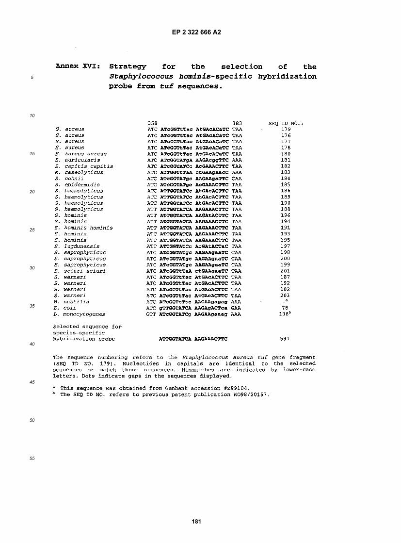
EP 2 322 666 A2
181
5
10
15
20
25
30
35
40
45
50
55

EP 2 322 666 A2
182
5
10
15
20
25
30
35
40
45
50
55
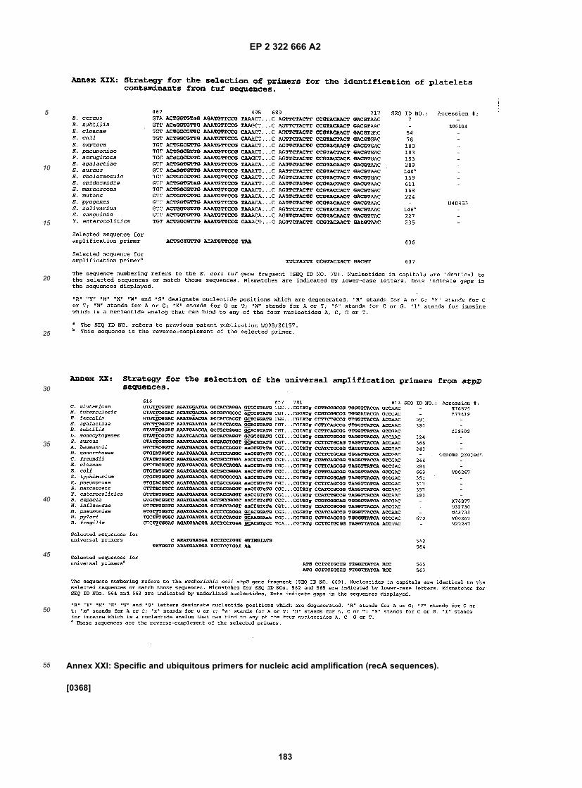
EP 2 322 666 A2
183
5
10
15
20
25
30
35
40
45
50
55 Annex XXI: Specific and ubiquitous primers for nucleic acid amplification (recA sequences).
[0368]

EP 2 322 666 A2
184
5
10
15
20
25
30
35
40
45
50
55
Annex XXII: Specific and ubiquitous primers for nucleic acid amplification (speA sequences).
[0369]
Originating DNA fragment
SEQ ID NO. Nucleotide sequence SEQ ID NO. Nucleotide position
Universal primers (recA)919 5’-GGI CCI GAR TCI TMI GGI AAR AC 918a 437-459920b 5’-TCI CCV ATI TCI CCI TCI AIY TC 918a 701-723921 5’-TIY RTI GAY GCI GAR CAI GC 918a 515-534922b 5’-TAR AAY TTI ARI GCI YKI CCI CC Sequencing primers
(recA)918a 872-894
1605 5’-ATY ATY GAA RTI TAY GCI CC 1704a 220-2391606 5’-CCR AAC ATI AYI CCI ACT TTT TC Universal primers
(rad51)1709a 628-650
935 5’-GGI AAR WSI CAR YTI TGY CAY AC 939a 568-590936b 5’-TCI SIY TCI GGI ARR CAI GG Universal primers (dmc1) 939a 1126-1145937 5’-ATI ACI GAR GYI TTY GGI GAR TT 940a 1038-1060938b 5’-CYI GTI GYI SWI GCR TGI GC 940a 1554-1573
a Sequences from databases.b These sequences are from the complementary DNA strand of the sequence of the originating fragment given inthe Sequence Listing.
Originating DNA fragment
SEQ ID NO. Nucleotide sequence SEQ ID NO. Nucleotide position
Bacterial species: Streptococcus pyogenes994 5’-TGG ACT AAC AAT CTC GCA AGA GG 993a 60-82995b 5’-ACA TTC TCG TGA GTA ACA GGG T 993a 173-194996 5’-ACA AAT CAT GAA GGG AAT CAT TTA G 993a 400-424997b 5’-CTA ATT CTT GAG CAG TTA CCA TT 993a 504-526998 5’-GGA GGG GTA ACA AAT CAT GAA GG 993a 391-413997b 5’-CTA ATT CTT GAG CAG TTA CCA TT 993a 504-526
a Sequence from databases.b These sequences are from the complementary DNA strand of the sequence of the originating fragmentgiven in the Sequence Listing.

EP 2 322 666 A2
185
5
10
15
20
25
30
35
40
45
50
55

EP 2 322 666 A2
186
5
10
15
20
25
30
35
40
45
50
55

EP 2 322 666 A2
187
5
10
15
20
25
30
35
40
45
50
55

EP 2 322 666 A2
188
5
10
15
20
25
30
35
40
45
50
55

EP 2 322 666 A2
189
5
10
15
20
25
30
35
40
45
50
55

EP 2 322 666 A2
190
5
10
15
20
25
30
35
40
45
50
55
Annex XXXII: Specific and ubiquitous primers for nucleic acid amplification (toxin sequences).
[0370]
Annex XXXIII: Molecular beacon internal hybridization probes for specific detection of toxin sequences.
[0371]
Originating DNA fragment
SEQ ID NO. Nucleotide sequence SEQ ID NO. Nucleotide position
Toxin gene: cdtA2123 5’-TCT ACC ACT GAA GCA TTA C 2129a 442-4602124b 5’-TAG GTA CTG TAG GTT TAT TG 2129a 580-599Toxin gene: cdtB2126 5’-ATA TCA GAG ACT GAT GAG 2130a 2665-26822127b 5’-TAG CAT ATT CAG AGA ATA TTG T 2130a 2746-2767Toxin gene: stx1
1081 5’-ATG TCA GAG GGA TAG ATC CA 1076a 233-2521080b 5’-TAT AGC TAC TGT CAC CAG ACA ATG T 1076a 394-418Toxin gene: stx2
1078 5’-AGT TCT GCG TTT TGT CAC TGT C 1077a 546-5671079b 5’-CGG AAG CAC ATT GCT GAT T 1077a 687-705Toxin genes: stx1 and stx2
1082 5’-TTG ARC RAA ATA ATT TAT ATG TG 1076a 278-3001083b 5’-TGA TGA TGR CAA TTC ACT AT 1076a 781-800
a Sequences from databases.b These sequences are from the complementary DNA strand of the sequence of the originatingfragment given in the Sequence Listing.
Originating DNA fragment
SEQ ID NO. Nucleotide sequencea SEQ ID NO. Nucleotide position
Toxin gene: cdtA2125b 2129C 462-488
Toxin qene: cdtB2128 2130C 2714-2740
Toxin gene: stx1
1084 1076C 337-363
2012d 1076C 248-264
Toxin gene: stx2

EP 2 322 666 A2
191
5
10
15
20
25
30
35
40
45
50
55
Annex XXXIV : Specific and ubiquitous primers for nucleic acid amplification (van sequences).
[0372]
(continued)
Originating DNA fragment
SEQ ID NO. Nucleotide sequencea SEQ ID NO. Nucleotide position
1085 1077C 617-638
a Underlined nucleotides indicate the molecular beacon’s stem.b These sequences are from the complementary DNA strand of the sequence of the originating fragment givenin the Sequence Listing.c Sequences from databases.d Scorpion primer.
Originating DNA fragment
SEQ ID NO. Nucleotide sequence SEQ ID NO. Nucleotide position
Resistance gene: vanA1086 5’-CTA CTC CCG CCT
TTT GGG TT1049-1057a 513-532b
1087C 5’-CTC ACA GCC CGAAAC AGC CT
1049-1057a 699-718b
1086 5’-CTA CTC CCG CCTTTT GGG TT
1049-1057a 513-532b
1088C 5’-TGC CGT TTC CTGTAT CCG TC
1049-1057a 885-904b
1086 5’-CTA CTC CCG CCTTTT GGG TT
1049-1057a 513-532b
1089C 5’-ATC CAC ACG GGCTAG ACC TC
1049-1057a 933-952b
1090 5’-AAT AGC GCG GACGAA TTG GAC
1049-1057a 629-649b
1091C 5’-AAC GCG GCA CTGTTT CCC AA
1049-1057a 734-753b
1090 5’-AAT AGC GCG GACGAA TTG GAC
1049-1057a 629-649b
1089C 5’-ATC CAC ACG GGCTAG ACC TC
1049-1057a 933-952b
1092 5’-TCG GCA AGA CAATAT GAC AGC
1049-1057a 662-682b
1088C 5’-TGC CGT TTC CTGTAT CCG TC
1049-1057a 885-904b
Resistance gene: vanB1095 5’-CGA TAG AAG CAG
CAG GAC AA1117d 473-492
1096C 5’CTG ATG GAT GCGGAA GAT AC
1117d 611-630
Resistance genes: vanA, vanB1112 5’-GGC TGY GAT ATT
CAA AGC TC1049-1057,1117a 437-456b
1113C 5’-ACC GAC CTC ACAGCC CGA AA
1049-1057,1117a 705-724b

EP 2 322 666 A2
192
5
10
15
20
25
30
35
40
45
50
55
(continued)
Originating DNA fragment
SEQ ID NO. Nucleotide sequence SEQ ID NO. Nucleotide position
1112 5’-GGC TGY GAT ATTCAA AGC TC
1049-1057,1117a 437-456b
1114C 5’-TCW GAG CCT TTTTCC GGC TCG
1049-1057,1117a 817-837b
1115 5’-TTT CGG GCT GTGAGG TCG GBT GHG CG
1049-1057,1117a 705-730b
1114C 5’-TCW GAG CCT TTTTCC GGC TCG
1049-1057,1117a 817-837b
1116 5’-TTT CGG GCT GTGAGG TCG GBT GHG CGG
1049-1057,1117a 705-731b
1114C 5’-TCW GAG CCT TTTTCC GGC TCG
1049-1057,1117a 817-837b
1112 5’-GGC TGY GAT ATTCAA AGC TC
1049-1057,1117a 437-456b
1118C 5’-TTT TCW GAG CCTTTT TCC GGC TCG
1049-1057,1117a 817-840b
a These sequences were aligned to derive the corresponding primer.b The nucleotide positions refer to the vanA sequence fragment (SEQ ID No. 1051).c These sequences are from the complementary DNA strand of the sequence of the originating fragment given inthe Sequence Listing.d Sequences from databases.Resistance genes: vanA, vanB (continued)1115 5’-TTT CGG GCT GTG
AGG TCG GBT GHG CG1049-1057,1117a 705-730b
1118C 5’-TTT TCW GAG CCTTTT TCC GGC TCG
1049-1057,1117a 817-840b
1116 5’-TTT CGG GCT GTGAGG TCG GBT GHG CGG
1049-1057,1117a 705-731b
1118C 5’-TTT TCW GAG CCTTTT TCC GGC TCG
1049-1057,1117a 817-840b
1119 5’-TTT CGG GCT GTGAGG TCG GBT GHG C
1049-1057,1117a 705-729b
1118C 5’-TTT TCW GAG CCTTTT TCC GGC TCG
1049-1057,1117a 817-840b
1120 5’-TTT CGG GCT GTGAGG TCG GBT GHG
1049-1057,1117a 705-728b
1118C 5’-TTT TCW GAG CCTTTT TCC GGC TCG
1049-1057,1117a 817-840b
1121 5’-TGT TTG WAT TGTCYG GYA TCC C
1049-1057,1117a 408-429b
1111C 5’-CTT TTT CCG GCTCGW YTT CCT GAT G
1049-1057,1117a 806-830b
1112 5’-GGC TGY GAT ATTCAA AGC TC
1049-1057,1117a 437-456b
1111C 5’-CTT TTT CCG GCTCGW YTT CCT GAT G
1049-1057,1117a 806-830b
1123 5’-TTT CGG GCT GTGAGG TCG GBT G
1049-1057,1117a 705-726b
1111c 5’-CTT TTT CCG GCTCGW YTT CCT GAT G
1049-1057,1117a 806-830b

EP 2 322 666 A2
193
5
10
15
20
25
30
35
40
45
50
55
(continued)
Originating DNA fragment
SEQ ID NO. Nucleotide sequence SEQ ID NO. Nucleotide position
1112 5’-GGC TGY GAT ATTCAA AGC TC
1049-1057,1117a 437-456b
1124C 5’-GAT TTG RTC CACYTC GCC RAC A
1049-1057,1117a 757-778b
Resistance gene: vanc11103 5’-ATC CCG CTA TGA
AAA CGA TC1058-1059a 519-538d
1104C 5’-GGA TCA ACA CAGTAG AAC CG
1058-1059a 678-697d
Resistance genes: vanc1, vanC2, vanC31097 5’-TCY TCA AAA GGG
ATC ACW AAA GTM AC1058-1066a 607-632d
1098C 5’-TCT TCA AAA TCGAAA AAG CCG TC
10581066a 787-809d
1099 5’-TCA ACA GGG ATCACW AAA GTM AC
1058-1066a 610-632d
1100C 5’-GTA AAK CCC GGCATR GTR TTG ATT TC
1058-1066a 976-1001d
1101 5’-GAC GGY TTT TTYGAT TTT GAA GA
1058-1066a 787-809d
1102C 5’-AAA AAR TCG ATKCGA GCM AGA CC
1058-1066a 922-944d
Resistance genes: vanC2, vanC31105 5’-CTC CTA CGA TTC
TCT TGA YAA ATC A1060-1D66,1140a 487-511e
1106C 5’-CAA CCG ATC TCAACA CCG GCA AT
1060-1066,1140a 690-712e
a These sequences were aligned to derive the corresponding primer.b The nucleotide positions refer to the vanA sequence fragment (SEQ ID NO. 1051).c These sequences are from the complementary DNA strand of the sequence of the originating fragment given inthe Sequence Listing.d The nucleotide positions refer to the vanC1 sequence fragment (SEQ ID NO. 1058).e The nucleotide positions refer to the vanC2 sequence fragment (SEQ ID NO. 1140).Resistance gene: vanD1591 5’-ATG AGG TAA TAG
AAC GGA TT1594 797-837
1592b 5’-CAG TAT TTC AGTAAG CGT AAA
1594 979-999
Resistance gene: vanE1595 5’-AAA TAA TGC TCC
ATC AAT TTG CTG A1599a 74-98
1596b 5’-ATA GTC GAA AAAGCC ATC CAC AAG
1599a 394-417
1597 5’-GAT GAA TTT GCGAAA ATA CAT GGA
1599a 163-186
1598b 5’-CAG CCA ATT TCTACC CCT TTC AC
1599a 319-341
Sequencing primers (vanAB)1112 5’-GGC TGY GAT ATT
CAA AGC TC1139a 737-756

EP 2 322 666 A2
194
5
10
15
20
25
30
35
40
45
50
55
Annex XXXV: Internal hybridization probes for specific detection of van sequences.
[0373]
(continued)
Originating DNA fragment
SEQ ID NO. Nucleotide sequence SEQ ID NO. Nucleotide position
1111b 5’-CTT TTT CCG GCTCGW YTT CCT GAT G
1139a 1106-1130
Sequencing primers (vanA, vanX, vanY)1150 5’-TGA TAA TCA CAC
CGC ATA CG1141a 860-879
1151b 5’-TGC TGT CAT ATTGTC TTG CC
1141a 1549-1568
1152 5’-ATA AAG ATG ATAGGC CGG TG
1141a 1422-1441
1153b 5’-CTC GTA TGT CCCTAC AAT GC
1141a 2114-2133
1154 5’-GTT TGA AGC ATATAG CCT CG
1141a 2520-2539
1155b 5’-CAG TGC TTC ATTAAC GTA GTC
1141a 3089-3109
a Sequences from databases.These sequences are from the complementary DNA strand of the sequence of the originating fragment given in theSequence Listing.
SEQ ID NO. NucleotidesequenceSEQID
Nucleotide NO. position
Sequencing primers (vanC1)1110 5’-ACG AGA AAG ACA
ACA GGA AGA CC1138a 122-144
1109b 5’-ACA TCG TGA TCGCTA AAA GGA GC
1138a 1315-1337
Sequencing primers (vanC2, vanC3)1108 5’-GTA AGA ATC GGA
AAA GCG GAA GG1140a 1-23
1107b 5’-CTC ATT TGA CTTCCT CCT TTG CT
1140a 1064-1086
a Sequences from databases.bThese sequences are from the complementary DNA strand of the sequence of the originating fragment given in theSequence Listing.
Originating DNA fragment
SEQ ID NO. Nucleotide sequence SEQ ID NO. Nucleotide position
Resistance gene: vanA1170 5’-ACG AAT TGG ACT ACG CAA TT 1049-1057a 639-658b
2292 5’-GAA TCG GCA AGA CAA TAT G 2293c 583-601Resistance gene: vanB1171 5’-ACG AGG ATG ATT TGA TTG TC 1117c 560-5792294 5’-AAA CGA GGA TGA TTT GAT TG 2296a 660-6792295 5’-TTG AGC AAG CGA TTT CGG 2296a 614-631Resistance gene: vanD

EP 2 322 666 A2
195
5
10
15
20
25
30
35
40
45
50
55
Annex XXXVI: Specific and ubiquitous primers for nucleic acid amplification (pbp sequences).
[0374]
(continued)
Originating DNA fragment
SEQ ID NO. Nucleotide sequence SEQ ID NO. Nucleotide position
2297 5’-TTC AGG AGG GGG ATC GC 1594c 458-474
a These sequences were aligned to derive the corresponding primer.bThe nucleotide positions refer to the vanA sequence fragment (SEQ ID NO. 1051).c Sequences from databases.
Originating DNA fragment
SEQ ID NO. Nucleotide sequence SEQ ID NO. Nucleotide position
Resistance gene: pbp1a1129 5’-ATG ATG ACH GAM ATG ATG AAA AC 1004-1018a 681-703b
1131c 5’-CAT CTG GAG CTA CRT ARC CAG T 1004-1018a 816-837b
1130 5’-GAC TAT CCA AGC ATG CAT TAT G 1004-1018a 456-477b
1131 5’-CAT CTG GAG CTA CRT ARC CAG T 1004-1018a 816-837b
2015 5’-CCA AGA AGC TCA AAA ACA TCT G 2047d 909-9302016c 5’-TAD CCT GTC CAW ACA GCC AT 2047d 1777-1796
Sequencing primers (pbp1a)1125 5’-ACT CAC AAC TGG GAT GGA TG 1169d 873-8921126c 5’-TTA TGG TTG TGC TGG TTG AGG 1169d 2140-21601125 5’-ACT CAC AAC TGG GAT GGA TG 1169d 873-8921128c 5’-GAC GAC YTT ATK GAT ATA CA 1169d 1499-15181127 5’-KCA AAY GCC ATT TCA AGT AA 1169d 1384-14031126c 5’-TTA TGG TTG TGC TGG TTG AGG 1169d 2140-2160
Sequencing primers (pbp2b)1142 5’-GAT CCT CTA AAT GAT TCT CAG GTG
G1172d 1-25
1143c 5’-CAA TTA GCT TAG CAA TAG GTG TTGG
1172d 1481-1505
1142 5’-GAT CCT CTA AAT GAT TCT CAG GTGG
1172d 1-25
1145c 5’-AAC ATA TTK GGT TGA TAG GT 1172d 793-8121144 5’-TGT YTT CCA AGG TTC AGC TC 1172d 657-6761143c 5’-CAA TTA GCT TAG CAA TAG GTG TTG
G1172d 1481-1505
Sequencing primers (pbp2x)1146 5’-GGG ATT ACC TAT GCC AAT ATG AT 1173d 219-2411147c 5’-AGC TGT GTT AGC VCG AAC ATC TTG 1173d 1938-19611146 5’-GGG ATT ACC TAT GCC AAT ATG AT 1173d 219-2411149c 5’-TCC YAC WAT TTC TTT TTG WG 1173d 1231-12501148 5’-GAC TTT GTT TGG CGT GAT AT 1173d 711-7301147c 5’-AGC TGT GTT AGC VCG AAC ATC TTG 1173d 1938-1961
a These sequences were aligned to derive the corresponding primer.b The nucleotide positions refer to the pbp1a sequence fragment (SEQ ID NO. 1004).c These sequences are from the complementary DNA strand of the sequence of the originating fragmentgiven in the Sequence Listing.d Sequences from databases.

EP 2 322 666 A2
196
5
10
15
20
25
30
35
40
45
50
55
Annex XXXVII: Internal hybridization probes for specific detection of pbp sequences.
[0375]
Originating DNA fragment
SEQ ID NO. Nucleotide sequence SEQ ID NO. Nucleotide position
Resistance gene: pbp1a1132 5’-AGT GAA AAR ATG GCT GCT GC 1004-1018a 531-550b
1133 5’-CAT CAA GAA CAC TGG CTA YGT AG 1004-1018a 806-828b
1134 5’-CTA GAT AGA GCT AAA ACC TTC CT 1004-1018a 417-439b
1135 5’-CAT TAT GCA AAC GCC ATT TCA AG 1004-1018a 471-493b
1192 5’-GGT AAA ACA GGA ACC TCT AAC T 1004-1018a 759-780b
1193 5’-GGT AAG ACA GGT ACT TCT AAC T 1004-1018a 759-780b
1194 5’-CAT TTC AAG TAA TAC AAC AGA ATC 1004-1018a 485-508b
1195 5’-CAT TTC AAG TAA CAC AAC TGA ATC 1004-1018a 485-508b
1196 5’-GCC ATT TCA AGT AAT ACA ACA GAA 1004-1018a 483-506b
1197 5’-CAA ACG CCA TTT CAA GTA ATA CAA C 1004-1018a 478-502b
1094 5’-GGT AAA ACA GGT ACT TCT AAC TA 1004-1018a 759-781b
1214 5’-GGT AAA ACA GGT ACC TCT AAC TA 1004-1018a 759-781b
1216 5’-GGT AAG ACT GGT ACA TCA AAC TA 1004-1018a 759-781b
1217 5’-CAA ATG CCA TTT CAA GTA ACA CAA C 1004-1018a 478-502b
1218 5’-CAA ACG CCA TTT CAA GTA ACA CAA C 1004-1018a 478-502b
1219 5’-CAA ATG CTA TTT CAA GTA ATA CAA C 1004-1018a 478-502b
1220 5’-CAA ACG CCA TTT CAA GTA ATA CGA C 1004-1018a 478-502b
2017 5’-ACT TTG AAT AAG GTC GGT CTA G 2047c 1306-13272018 5’-ACA CTA AAC AAG GTT GGT TTA G 2063 354-3752019 5’-ACA CTA AAC AAG GTC GGT CTA G 2064 346-3672020 5’-GTA GCT CCA GAT GAA ATG TTT G 2140c 1732-17532021 5’-GTA GCT CCA GAC GAA ATG TTT G 2057 831-8522022 5’-GTA GCT CCA GAT GAA ACG TTT G 2053c 805-8262023 5’-GTA ACT CCA GAT GAA ATG TTT G 2056 819-8402024 5’-AGT GAA AAG ATG GCT GCT GC 2048c 1438-14572025 5’-AGT GAG AAA ATG GCT GCT GC 2047c 1438-14572026 5’-TCC AAG CAT GCA TTA TGC AAA CG 2047c 1368-13902027 5’-TCG GTC TAG ATA GAG CTA AAA CG 2047c 1319-13412028 5’-TAT GCT CTT CAA CAA TCA CG 2047c 1267-12862029 5’-AGC CGT TGA GAC TTT GAA TAA G 2047c 1296-13172030 5’-CTT AAT GGT CTT GGT ATC G 2047c 1345-13662031 5’-CGT GAC TGG GGT TCT GCT ATG A 2049c 1096-11172032 5’-CGT GAC TGG GGA TCA TCA ATG A 2047c 1096-11172033 5’-CGT GAC TGG GGT TCT GCC ATG A 2057 195-2162034 5’-ATC AAG AAC ACT GGC TAT GTA G 2050c 787-808
a These sequences were aligned to derive the corresponding primer.b The nucleotide positions refer to the pbp1a sequence fragment (SEQ ID NO. 1004).c Sequence from databases.Resistance gene: pbp1a (continued)2035 5’-ATC AAG AAC ACT GGC TAC GTA G 2051c 787-8082036 5’-ATC AAG AAC ACT GGT TAC GTA G 2047 1714-17352037 5’-ATC AAA AAT ACT GGT TAT GTA G 2057 813-8342038 5’-ATC AAG AAT ACT GGC TAC GTA G 2052c 757-7782039 5’-ATC AAA AAC ACT GGC TAT GTA G 2053c 787-808

EP 2 322 666 A2
197
5
10
15
20
25
30
35
40
45
50
55
Annex XXXIX: Internal hybridization probe for specific detection of mecA.
[0376]

EP 2 322 666 A2
198
5
10
15
20
25
30
35
40
45
50
55
Annex XL: Specific and ubiquitous primers for nucleic acid amplification (hexA sequences).
[0377]
Annex XLI: Internal hybridization probe for specific detection of hexA sequences.
[0378]
Originating DNA fragment
SEQ ID NO. Nucleotide sequence SEQ ID NO. Nucleotide position
Resistance gene: mecA1177 5’-GCT CAA CAA GTT CCA GAT TA 1178a 1313-1332
Sequence from databases.
Originating DNA fragment
SEQ ID NO. Nucleotide sequence SEQ ID NO. Nucleotide position
Bacterial species: Streptococcus pneumoniae1179 5’-ATT TGG TGA CGG GTG ACT TT 1183a 431-4501181b 5’-AGC AGC TTA CTA GAT GCC GT 1183-1191c 652-671d
Sequencing primer1179 5’-ATT TGG TGA CGG GTG ACT TT 1183a 431-4501182b 5’-AAC TGC AAG AGA TCC TTT GG 1183a 1045-1064
a Sequences from databases.b These sequences are from the complementary DNA strand of the sequence of the originatingfragment given in the Sequence Listing.c These sequences were aligned to derive the corresponding primer.d The nucleotide positions refer to the hexA sequence fragment (SEQ ID NO. 1183).
Originating DNA fragment
SEQ ID NO. Nucleotide sequence SEQ ID NO. Nucleotide position
Bacterial species: Streptococcus pneumoniae1180a 5’-TCC ACC GTT GCC
AAT CGC A1183-1191b 629-647c
a This sequences is from the complementary DNA strand of the sequence of the originating fragment given in theSequence Listing.b These sequences were aligned to derive the corresponding primer.c The nucleotide positions refer to the hexA sequence fragment (SEQ ID NO. 1183).
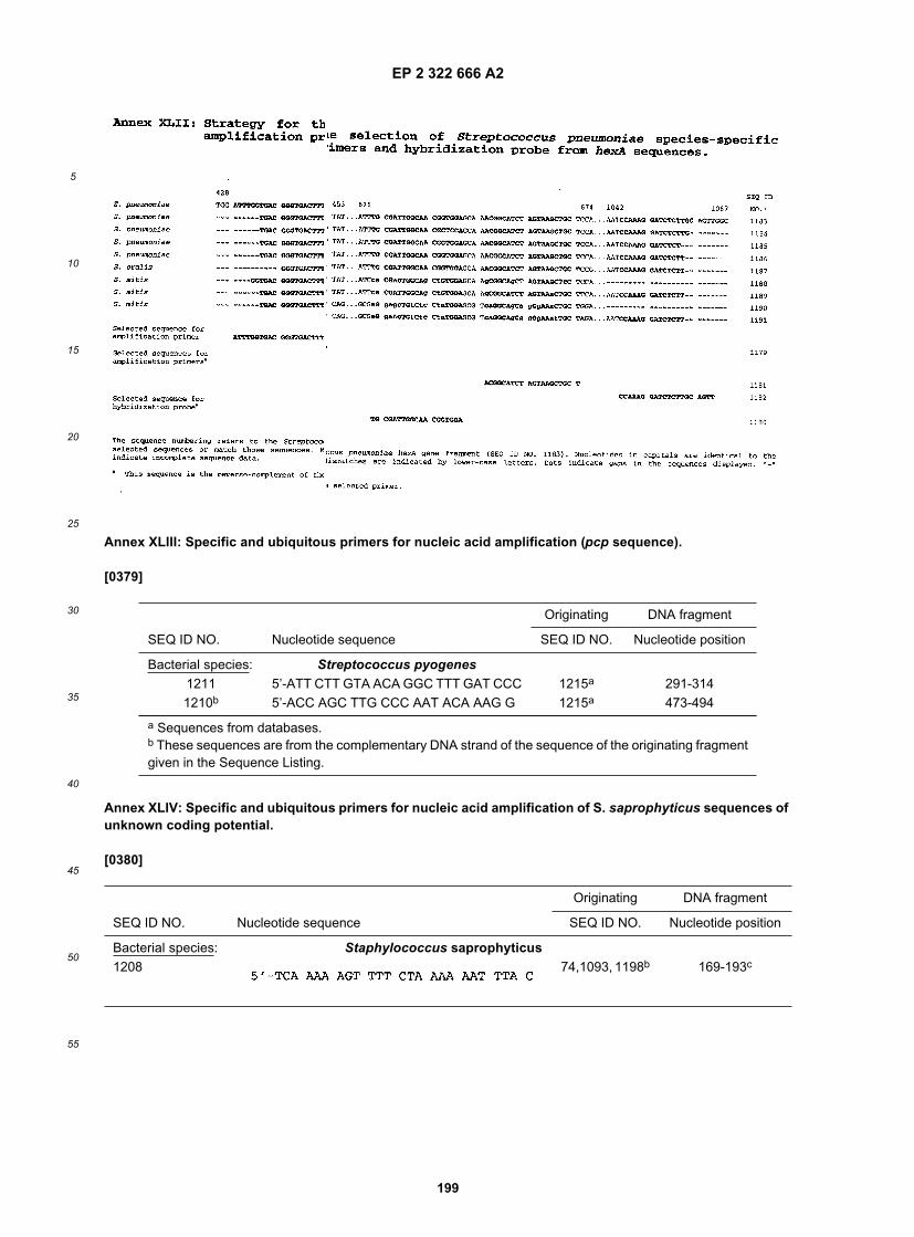
EP 2 322 666 A2
199
5
10
15
20
25
30
35
40
45
50
55
Annex XLIII: Specific and ubiquitous primers for nucleic acid amplification (pcp sequence).
[0379]
Annex XLIV: Specific and ubiquitous primers for nucleic acid amplification of S. saprophyticus sequences ofunknown coding potential.
[0380]
Originating DNA fragment
SEQ ID NO. Nucleotide sequence SEQ ID NO. Nucleotide position
Bacterial species: Streptococcus pyogenes1211 5’-ATT CTT GTA ACA GGC TTT GAT CCC 1215a 291-3141210b 5’-ACC AGC TTG CCC AAT ACA AAG G 1215a 473-494
a Sequences from databases.b These sequences are from the complementary DNA strand of the sequence of the originating fragmentgiven in the Sequence Listing.
Originating DNA fragment
SEQ ID NO. Nucleotide sequence SEQ ID NO. Nucleotide position
Bacterial species: Staphylococcus saprophyticus1208 74,1093, 1198b 169-193c

EP 2 322 666 A2
200
5
10
15
20
25
30
35
40
45
50
55
Annex XLV: Molecular beacon internal hybridization probes for specific detection of antimicrobial agentsresistance gene sequences.
[0381]
(continued)
Originating DNA fragment
SEQ ID NO. Nucleotide sequence SEQ ID NO. Nucleotide position
1209a 74,1093, 1198b 355-379c
a This sequence is from the complementary DNA strand of the sequence of the originating fragment given in theSequence Listing.These sequences were aligned to derive the corresponding primer.c The nucleotide positions refer to the S. saprophyticus unknown gene sequence fragment (SEQ ID NO. 1198).
Originating DNA fragment
SEQ ID NO Nucleotide sequencea SEQ ID NO. Nucleotideposition
Resistancegene:
gyrA
2250 1954b 218-243
2251 1954b 259-286
Resistancegene:
mecA
1231 1178b 1291-1315
Resistancegene:
parC
1938b 1321c 232-260
1939 1321C 317-346
1955b 1321C 235-260
Resistancegene:
vanA
1239 1051 860-880
1240 1051 663-688
Resistancegene:
vanB
1241 1117 555-577
EP 2 322 666 A2
201
5
10
15
20
25
30
35
40
45
50
55
Annex XLVI: Molecular beacon internal hybridization probe for specific detection of S. aureus gene sequencesof unknown coding potential.
[0382]
Annex XLVII: Molecular beacon internal hybridization probes for specific detection of tuf sequences.
[0383]
(continued)
Originating DNA fragment
SEQ ID NO Nucleotide sequencea SEQ ID NO. Nucleotideposition
Resistancegene:
vanD
1593 1594 835-845
a Underlined nucleotides indicate the molecular beacon’s stem.b This sequence is from the complementary DNA strand of the sequence of the originating fragment given in theSequence Listing.c Sequence from databases.
Originating DNA fragment
SEQ ID NO. Nucleotide sequencea SEQ ID NO. Nucleotideposition
Bacterialspecies:
S. aureus
1232 1244 53-80
a Underlined nucleotides indicate the molecular beacon’s stem.
Originating DNA fragment
SEQ ID NO. Nucleotide sequencea SEQ ID NO. Nucleotideposition
Bacterialspecies:
Chlamydia pneumoniae
2091 20 157-183
2092 20 491-516
Bacterialspecies:
Chlamydia trachomatis
2213 1739b 412-441
Bacterialspecies:
Enterococcus faecalis

EP 2 322 666 A2
202
5
10
15
20
25
30
35
40
45
50
55
(continued)
Originating DNA fragment
SEQ ID NO. Nucleotide sequencea SEQ ID NO. Nucleotideposition
1236 883 370-391
Bacterialspecies:
Enterococcus faecium
1235 64 412-437
Bacterialspecies:
Legionella pneumopbila
2084c 112 461-486
Bacterialspecies:
Mycoplasma pneumoniae
2096c 2097b 658-679
Bacterialspecies:
Neisseria gonorrhoea
2177 126 323-357
2178 126 323-348
2179 126 692-718
a Underlined nucleotides indicate the molecular beacon’s stem.b Sequence from databasesc This sequence is from the complementary DNA strand of the sequence of the originating fragment given in theSequence Listing.
SEQ ID NO. Nucleotide sequencea SEQ ID NO. Nucleotideposition
Bacterialspecies:
Pseudomonas aeruginosa
2122 153,880,2138b,c 280-302d
Bacterialspecies:
Staphylococcus aureus
2186 1728 615-646
Bacterialgroup:
Staphylococcus sp. other than S. aureus
1233 878 372-394
Fungalspecies:
Candida albicans

EP 2 322 666 A2
203
5
10
15
20
25
30
35
40
45
50
55
(continued)
SEQ ID NO. Nucleotide sequencea SEQ ID NO. Nucleotideposition
2073 408 404-429
Fungalspecies:
Candida dubliniensis
2074 414 416-441
Fungalspecies:
Candida glabrata
2110b 417 307-335
2111 417 419-447
Fungalspecies:
Candida krusei
2112b 422 318-347
2113 422 419-447
2114 422 505-533
a Underlined nucleotides indicate the molecular beacon’s stem.b Sequence from databases.c These sequences were aligned to derive the corresponding primer.d The nucleotide positions refer to the P. aeruginosa tuf sequence fragment (SEQ ID NO. 153).
SEQ ID NO. Nucleotide sequencea SEQ ID NO. Nucleotideposition
Fungalspecies:
Candida lusitaniae
2115b 424 304-330
2116 424 476-502
2117 424 512-535
Fungalspecies:
Candida parapsilosis
2118b 426 301-330
Fungalspecies:
Candida tropicalis
2119 429 357-384

EP 2 322 666 A2
204
5
10
15
20
25
30
35
40
45
50
55
Annex XLVIII: Molecular beacon internal hybridization probes for specific detection of dd1 and mt1 genesequences.
[0384]
(continued)
SEQ ID NO. Nucleotide sequencea SEQ ID NO. Nucleotideposition
2120 429 459-487
Fungalspecies:
Cryptococcus neoformans
2106 623,1985,1986c 226-244d
2107 623,1985,1986c 390-408d
Bacterialgenus:
Legionella sp.
2083 111-112a 488-519e
a Underlined nucleotides indicate the molecular beacon’s stem.b This sequence is from the complementary DNA strand of the sequence of the originating fragment given in theSequence Listing.c These sequences were aligned to derive the corresponding primer.d The nucleotide positions refer to the C. neoformans tuf (EF-1) sequence fragment (SEQ ID NO. 623).e The nucleotide positions refer to the L. pneumophila tuf (EF-1) sequence fragment (SEQ ID NO. 112).Fungal genus: Candida sp.2108 414,417,
422,424,426,429,624b
52-80c
2109 Same as SEQ IDNO. 2108
100-125c
Bacterialgroup:
Pseudomonads
2121 153-155,205,880,2137d,
2138d,b
598-616e
a Underlined nucleotides indicate the molecular beacon’s stem.These sequences were aligned to derive the corresponding primer.c The nucleotide positions refer to the C. albicans tuf (EF-1) sequence fragment (SEQ ID NO. 624).d Sequence from databases.The nucleotide positions refer to the P. aeruginosa tuf sequence fragment (SEQ ID NO. 153).
Originating DNA fragment
SEQ ID NO. Nucleotide sequencea SEQ ID NO. Nucleotide position
Bacterial species: E. faeciusn (dd1)

EP 2 322 666 A2
205
5
10
15
20
25
30
35
40
45
50
55
Annex XLIX: Internal hybridization probe for specific detection of S. aureus sequences of unknown codingpotential.
[0385]
Annex L: Specific and ubiquitous primers for nucleic acid amplification (antimicrobial agents resistance genessequences).
[0386]
(continued)
Originating DNA fragment
SEQ ID NO. Nucleotide sequencea SEQ ID NO. Nucleotide position
1237 1242b 334-359
Bacterial species: E. faecalis (mt1)1238 1243b 631-656
a Underlined nucleotides indicate the molecular beacon’s stem.Sequence from databases.
Originating DNA fragment
SEQ ID NO. Nucleotide sequence SEQ ID NO. Nucleotide position
Bacterial species: Staphylococcus aureus1234 5’-ACT AAA TAA ACG CTC ATT CG 1244 35-54
Originating DNA fragment
SEQ ID NO. Nucleotide sequence SEQ ID NO. Nucleotide position
Resistance gene: aac(2’)-Ia1344 5’-AGC AGC AAC GAT
GTT ACG CAG CAG1348a 163-186
1345b 5’-CCC GCC GAG CATTTC AAC TAT TG
1348a 392-414
1346 5’-GAT GTT ACG CAGCAG GGC AGT C
1348a 172-193
1347b 5’-ACC AAG CAG GTTCGC AGT CAA GTA
1348a 467-490
Resistance gene: aac(3’)-Ib1349 5’-CAG CCG ACC AAT
GAG TAT CTT GCC1351a 178-201
1350b 5’-TAA TCA GGG CAGTTG CGA CTC CTA
1351a 356-379
Resistance gene: aac(3’)-IIb1352 5’-CCA CGC TGA CAG
AGC CGC ACC G1356a 383-404
1353b 5’-GGC CAG CTC CCATCG GAC CCT G
1356a 585-606
1354 5’-CAC GCT GAC AGAGCC GCA CCG
1356a 384-404

EP 2 322 666 A2
206
5
10
15
20
25
30
35
40
45
50
55
(continued)
Originating DNA fragment
SEQ ID NO. Nucleotide sequence SEQ ID NO. Nucleotide position
1355b 5’-ATG CCG TTG CTGTCG AAA TCC TCG
1356a 606-629
Resistance gene: aac(3’)-IVa1357 5’-GCC CAT CCA TTT
GCC TTT GC1361a 295-314
1358b 5’-GCG TAC CAA CTTGCC ATC CTG AAG
1361a 517-540
1359 5’-TGC CCC TGC CACCTC ACT C
1361a 356-374
1360b 5’-CGT ACC AAC TTGCCA TCC TGA AGA
1361a 516-539
Resistance gene: aac(3’)-VIa1362 5’-CGC CGC CAT CGC
CCA AAG CTG G1366a 285-306
1363b 5’-CGG CAT AAT GGAGCG CGG TGA CTG
1366a 551-574
1364 5’-TTT CTC GCC CACGCA GGA AAA ATC
1366a 502-525
1365b 5’-CAT CCT CGA CGAATA TGC CGC G
1366a 681-702
Resistance gene: aac(6’)-Ia1367 5’-CAA ATA TAC TAA CAG
AAG CGT TCA1371a 56-79
1368b 5’-AGGATCTTGCCAATACCT TTA T
1371a 269-290
1379 5’-AAA CCTTTGTTTCGGTCT GCT AAT
1371a 153-176
1380b 5’-AAGCGATTCCAATAATAC CTT GCT
1371a 320-343
a Sequence from databases.b These sequences are from the complementary DNA strand of the sequence of the originating fragment given in theSequence Listing.Resistance gene: aac (6’) -Ic1372 5’ -GCTTTCGTTGCCTTT
GCC GAG GTC1376a 157-180
1373b 5’ -CAC CCC TGT TGCTTC GCC CAC TC
1376a 304-326
1374 5’ -AGA TAT TGG CTTCGC CGC ACC ACA
1376a 104-127
1375b 5’ -CCC TGT TGC TTCGCC CAC TCC TG
1376a 301-323
Resistance gene: ant (3’) -Ia1377 5’-GCC GTG GGT CGA
TGT TTG ATG TTA1381a 100-123
1378b 5’-GCT CGA TGA CGCCAA CTA CCT CTG
1381a 221-244
1379 5’-AGC AGC AAC GATGTT ACG CAG CAG
1381a 127-150
1380b 5’ -CGC TCG ATG ACGCCA ACT ACC TCT
1381a 222-245

EP 2 322 666 A2
207
5
10
15
20
25
30
35
40
45
50
55
(continued)
Originating DNA fragment
SEQ ID NO. Nucleotide sequence SEQ ID NO. Nucleotide position
Resistance gene: ant (4’) -Ia1382 5’-TAG ATA TGA TAG
GCG GTA AAA AGC1386a 149-172
1383b 5’ -CCC AAA TTC GAGTAA GAG GTA TT
1386a 386-408
1384 5’-GAT ATG ATA GGCGGT AAA AAG C
1386a 151-172
1385b 5’-TCCCAAATTCGAGTAAGA GGT A
1386a 388-409
Resistance gene: aph (3’) -Ia1387 5’ -TTA TGC CTC TTC
CGA CCA TCA AGC1391a 233-256
1338b 5’-TACGCTCGTCATCAAAAT CAC TCG
1391a 488-511
1389 5’ -GAA TAA CGG TTTGGT TGA TGC GAG
1391a 468-491
1390b 5’-ATG GCA AGA TCCTGG TAT CGG TCT
1391a 669-692
Resistance gene: aph (3’) -IIa1392 5’ -TGG GTG GAG AGG
CTA TTC GGC TAT1396a 43-66
1393b 5’ -CAG TCC CTT CCCGCT TCA GTG AC
1396a 250-272
1394 5’-GAC GTT GTC ACTGAA GCG GGA AGG
1396a 244-267
1395b 5’ -CTT GGT GGT CGAATG GGC AGG TAG
1396a 386-409
a Sequence from databases.b These sequences are from the complementary DNA strand of the sequence of the originating fragment given in theSequence Listing.Resistance gene: aph (3’) -IIIa1397 5’ -GTG GGA GAA AAT
GAA AAC CTA T1401a 103-124
1398b 5’-ATG GAG TGA AAGAGC CTG AT
1401a 355-374
1399 5’ -ACC TAT GAT GTGGAA CGG GAA AAG
1401a 160-183
1400b 5’ -CGA TGG AGT GAAAGA GCC TGA TG
1401a 354-376
Resistance gene: aph (3’) -VIa1402 5’ -TAT TCA ACA ATT TAT
CGG AAA CAG1406a 18-41
1403b 5’ -TCA GAG AGC CAACTC AAC ATT TT
1406a 175-197
1404 5’-AAACAGCGTTTTAGAGCC AAA TAA
1406a 36-59
1405b 5’ -TTC TCA GAG AGCCAA CTC AAC ATT
1406a 177-200
Resistance gene: blaCARB

EP 2 322 666 A2
208
5
10
15
20
25
30
35
40
45
50
55
(continued)
Originating DNA fragment
SEQ ID NO. Nucleotide sequence SEQ ID NO. Nucleotide position
1407 5’ -CCC TGT AAT AGAAAA GCA AGT AGG
1411a 351-374
1408b 5’ -TTG TCG TAT CCCTCA AAT CAC C
1411a 556-577
1409 5’ -TGG GAT TAC AATGGC AAT CAG CG
1411a 205-227
1410b 5’ -GGG GAA TAG GTCACA AGA TCT GCT T
1411a 329-353
Resistance gene: blaCMY-21412 5’ -GAG AAA ACG CTC
CAG CAG GGC1416a 793-813
1413b 5’-CATGAGGCTTTCACTGCG GGG
1416a 975-995
1414 5’ -TAT CGT TAA TCGCAC CAT CAC
1416a 90-110
1415b 5’ -ATG CAG TAA TGCGGC TTT ATC
1416a 439-459
Resistance genes: blaCTX-M-1, blaCTX-M-21417 5’ -TGGTTAACTAYAATC
CSA TTG CGG A1423a 314-338
1418b 5’ -ATG CTT TAC CCAGCG TCA GAT T
1423a 583-604
Resistance gene: blaCTX-M-11419 5’ -CGA TGA ATA AGC
TGA TTT CTC ACG1423a 410-433
1420b 5’ -TGC TTT ACC CAGCGT CAG ATT ACG
1423a 580-603
1421 5’ -AAT TAG AGC GGCAGT CGG GAG GAA
1423a 116-139
1422b 5’ -GAAATCAGCTTATTCATC GCC ACG
1423a 405-428
a Sequence from databases.b These sequences are from the complementary DNA strand of the sequence of the originating fragment given in theSequence Listing.Resistance gene: blaCTX-M-21424 5’-GTT AAC GGT GAT
GGC GAC GCT AC1428a 30-52
1425b 5’ -GAA TTA TCG GCGGTG TTA ATC AGC
1428a 153-176
1426 5’ -CAC GCT CAA TACCGC CAT TCC A
1428a 510-531
1427b 5’-TTATCGCCCACTACCCAT GAT TTC
1428a 687-710
Resistance gene: blaIMP1429 5’-TTTACGGCTAAAGAT
ACT GAA AAG T1433a 205-229
1430b 5’-GTT TAA TAA AAC AACCAC CGA ATA AT
1433a 513-538
1431 5’ -TAATTGACACTCCATTTA CGG CTA A
1433a 191-215

EP 2 322 666 A2
209
5
10
15
20
25
30
35
40
45
50
55
(continued)
Originating DNA fragment
SEQ ID NO. Nucleotide sequence SEQ ID NO. Nucleotide position
1432b 5’ -ACC GAA TAA TAT TTTCCT TTC AGG CA
1433a 497-522
Resistance gene: blaOXA21434 5’ -CAC AAT CAA GAC
CAA GAT TTG CGA T1438a 319-343
1435b 5’-GAA AGG GCA GCTCGT TAC GAT AGA G
1438a 532-556
Resistance gene: blaOXA101436 5’ -CAGCATCAACATTTA
AGA TCC CCA1439a 194-217
1437b 5’ -CTCCACTTGATTAACTGC GGA AAT TC
1439a 479-504
Resistance gene: blaPER-11440 5’ -AGA CCG TTA TCG
TAA ACA GGG CTA AG1442a 281-306
1441b 5’ -TTT TTT GCT CAA ACTTTT TCA GGA TC
1442a 579-604
Resistance gene: blaPER-21443 5’-CTTCTGCTCTGCTGA
TGC TTG GC1445a 32-54
1444b 5’-GGC GAC CAG GTATTT TGT AAT ACT GC
1445a 304-329
Resistance genes : blaPER-1, blaPER-21446 5’ -GGC CTG YGA TTT
GTT ATT TGA ACT GGT1442a 414-440
1447b 5’ -CGC TST GGT CCTGTG GTG GTT TC
1442a 652-674
1448 5’ -GAT CAG GTG CARTAT CAA AAC TGG AC
1442a 532-557
1449b 5’ -AGC WGG TAA CAAYCC TTT TAA CCG CT
1442a 671-696
a Sequence from databases.b These sequences are from the complementary DNA strand of the sequence of the originating fragment given in theSequence Listing.Resistance gene: blaSHV1883 5’ -AGC CGC TTG AGC
AAA TTA AAC TA1900a 71-93
1884b 5’ -GTA TCC CGC AGATAA ATC ACC AC
1900a 763-785
1885 5’ -AGC GAA AAA CACCTT GCC GAC
1900a 313-333
1884b 5’ -GTA TCC CGC AGATAA ATC ACC AC
1900a 763-785
Resistance gene: blaTEM1906 5’ -CCT TAT TCC CTT TTT
TGC GG1927a 27-46
1907b 5’ -CAC CTA TCT CAGCGA TCT GTC T
1927a 817-838
1908 5’ -AAC AGC GGT AAGATC CTT GAG AG
1927a 148-170

EP 2 322 666 A2
210
5
10
15
20
25
30
35
40
45
50
55
(continued)
Originating DNA fragment
SEQ ID NO. Nucleotide sequence SEQ ID NO. Nucleotide position
1907b 5’ -CAC CTA TCT CAGCGA TCT GTC T
1927a 817-838
Resistance gene: catI2145 5’ -GCA AGA TGT GGC
GTG TTA CGG T2147a 363-384
2146b 5’ -GGG GCG AAG AAGTTG TCC ATA TT
2147a 484-506
Resistance gene: catII2148 5’ -CAG ATT AAA TGC
GGA TTC AGC C2150a 67-88
2149b 5’ -ATC AGG TAA ATCATC AGC GGA TA
2150a 151-173
Resistance gene: catIII2151 5’ -ATA TTT CAG CAT TAC
CTT GGG TT2153a 419-441
2152b 5’ -TACACAACTCTTGTAGCC GAT TA
2153a 603-625
Resistance gene: catP2154 5’ -CGC CAT TCA GAG
TTT AGG AC2156a 178-197
2155b 5’ -TTC CAT ACC GTTGCG TAT CAC TT
2156a 339-361
Resistance gene: cat2157 5’ -CCA CAG AAA TTG
ATA TTA GTG TTT TAT2159a 89-115
2158b 5’ -TCG CTA TTG TAACCA GTT CTA
2159a 201-221
2160 5’ -TTT TGA ACA CTA TTTTAA CCA GC
2162a 48-70
2161b 5’ -GAT TTA ACT TAT CCCAAT AAC CT
2162a 231-253
a Sequence from databases.b These sequences are from the complementary DNA strand of the sequence of the originating fragment given in theSequence Listing.Resistance gene: dfrA1450 5’ -ACC ACT GGG AAT
ACA CTT GTA ATG GC1452a 106-131
1451b 5’ -ATC TAC CTG GTCAAT CAT TGC TTC GT
1452a 296-321
Resistance gene: dhfrIa1457 5’ -CAA AGG TGA ACA
GCT CCT GTT T1461a 75-96
1458b 5’ -TCC GTT ATT TTC TTTAGG TTG GTT AAA
1461a 249-275
1459 5’ -AAG GTG AAC AGCTCC TGT TT
1461a 77-96
1560b 5’ -GAT CAC TAC GTTCTC ATT GTC A
1461a 207-228
Resistance genes: dhfrIa, dhfrXV

EP 2 322 666 A2
211
5
10
15
20
25
30
35
40
45
50
55
(continued)
Originating DNA fragment
SEQ ID NO. Nucleotide sequence SEQ ID NO. Nucleotide position
1453 5’ -ATC GAA GAA TGGAGT TAT CGG RAA TG
1461a 27-52
1454b 5’ -CCT AAA AYT RCTGGG GAT TTC WGG A
1461a 384-408
1455 5’ -CAG GTG GTG GGGAGA TAT ACA AAA
1461a 290-313
1456b 5’ -TAT GTT AGA SRCGAA GTC TTG GKT AA
1461a 416-441
Resistance gene: dhfrIb1466 5’ -AAG CAT TGA CCT
ACA ATC AGT GT1470a 98-120
1467b 5’ -AATACAACTACATTGTCA TCA TTT GAT
1470a 204-230
1468 5’ -CGT TAC CCG CTCAGG TTG GAC ATC AA
1470a 183-208
1469b 5’ -CAT CCC CCT CTGGCT CGA TGT CG
1470a 354-376
Resistance gene: dhfrV1471 5’ -GAT AAT GAC AAC
GTA ATA GTA TTC CC1475a 208-233
1472b 5’-GCTCAATATCAATCGTCG ATA TA
1475a 342-364
1473 5’ -TTA AAG CCT TGACGT ACA ACC AGT GG
1475a 95-120
1474b 5’ -TGG GCA ATG TTTCTC TGT AAA TCT CC
1475a 300-325
Resistance genes: dhfrIb, dhfrV1462 5’ -GCA CTC CCY AAT
AGG AAA TAC GC1470a 157-179
1463b 5’ -AGT GTT GCT CAAAAA CAA CTT CG
1470a 405-427
1464 5’ -ACG TTY GAA TCTATG GGM GCA CT
1470a 139-161
1465b 5’-GTC GAT AAG TGGAGC GTA GAG GC
1470a 328-350
a Sequence from databases.b These sequences are from the complementary DNA strand of the sequence of the originating fragment given in theSequence Listing.Resistance gene: dhfrVI1476 5’-GGC GAG CAG CTC
CTA TTC AAA G1480a 79-100
1477b 5’ -TAG GTA AGC TAATGC CGA TTC AAC A
1480a 237-261
1478 5’ -GAG AAT GGA GTAATT GGC TCT GGA TT
1480a 31-56
1479b 5’-GCG AAA TAC ACAACA TCA GGG TCA T
1480a 209-233
Resistance gene: dhfrVII1485 5’-AAA ATG GCG TAA
TCG GTA ATG GC1489a 32-54

EP 2 322 666 A2
212
5
10
15
20
25
30
35
40
45
50
55
(continued)
Originating DNA fragment
SEQ ID NO. Nucleotide sequence SEQ ID NO. Nucleotide position
1486b 5’ -CAT TTG AGC TTGAAA TTC CTT TCC TC
1489a 189-214
1487 5’ -AAT CGA AAA TATGCA GTA GTG TCG AG
1489a 166-191
1488b 5’ -AGACTATTGTAGATTTGA CCG CCA
1489a 294-317
Resistance genes: dhfrVII, dhfrXVII1481 5’ -RTT ACA GAT CAT KTA
TAT GTC TCT1489a 268-291
1482b 5’ -TAA TTT ATA TTA GACAWA AAA AAC TG
1489a 421-446
1483 5’ -CAR YGT CAG AAAATG GCG TAA TC
1489a 23-45
1484b 5’ -TKC AAA GCR WTTTCT ATT GAA GGA AA
1489a 229-254
Resistance gene: dhfrVIII1490 5’-GAC CTA TGA GAG
CTT GCC CGT CAA A1494a 144-168
1491b 5’ -TCG CCT TCG TACAGT CGC TTA ACA AA
1494a 376-401
1492 5’ -CAT TTT AGC TGCCAC CGC CAA TGG TT
1494a 18-43
1493D 5’-GCG TCG CTG ACGTTG TTC ACG AAG A
1494a 245-269
Resistance gene: dhfrIX1495 5’ -TCT CTA AAC ATG ATT
GTC GCT GTC1499a 7-30
1496b 5’ -CAG TGA GGC AAAAGT TTT TCT ACC
1499a 133-156
1497 5’ -CGG ACG ACT TCATGT GGT AGT CAG T
1499a 171-195
1498b 5’ -TTT GTT TTC AGT AATGGT CGG GAC CT
1499a 446-471
a Sequence from databases.b These sequences are from the complementary DNA strand of the sequence of the originating fragment given in theSequence Listing.Resistance gene: dhfrXII1500 5’ -ATC GGG TTA TTG
GCA ATG GTC CTA1504a 50-73
1501b 5’ -GCG GTA GTT AGCTTG GCG TGA GAT T
1504a 201-225
1502 5’ -GCG GGC GGA GCTGAG ATA TAC A
1504a 304-325
1503b 5’ -AAC GGA GTG GGTGTA CGG AAT TAC AG
1504a 452-477
Resistance gene: dhfrXIII1505 5’ -ATT TTT CGC AGG
CTC ACC GAG AGC1507a 106-129
1506b 5’ -CGG ATG AGA CAACCT CGA ATT CTG CTG
1507a 413-439

EP 2 322 666 A2
213
5
10
15
20
25
30
35
40
45
50
55
(continued)
Originating DNA fragment
SEQ ID NO. Nucleotide sequence SEQ ID NO. Nucleotide position
Resistance gene: dhfrXV1508 5’ -AGAATGTATTGGTAT
TTC CAT CTA TCG1512a 215-241
1509b 5’ -CAA TGT CGA TTGTTG AAA TAT GTA AA
1512a 336-361
1510 5’ -TGG AGT GCC AAAGGG GAA CAA T
1512a 67-88
1511b 5’ -CAG ACA CAA TCACAT GAT CCG TTA TCG
1512a 266-292
Resistance gene: dhfrXYII1513 5’ -TTC AAG CTC AAA
TGA AAA CGT CC1517a 201-223
1514b 5’ -GAA ATT CTC AGGCAT TAT AGG GAA T
1517a 381-405
1515 5’ -GTG GTC AGT AAAAGG TGA GCA AC
1517a 66-88
1516b 5’ -TCT TTC AAA GCA TTTTCT ATT GAA GG
1517a 232-257
Resistance gene: embR2102 5’ -CAC CTT CAC CCT
GAC CGA CG2105a 822-841
2103b 5’ -CGA ACC AGC GGAAAT AGT TGG AC
2105a 948-970
Resistance genes: ereA, ereA21528 5’-AAC TTG AGC GAT TTT
CGG ATA CCC TG1530a 80-105
1529b 5’ -TTG CCG ATG AAATAA CCG CCG ACT
1530a 317-340
a Sequence from databases.b These sequences are from the complementary DNA strand of the sequence of the originating fragment given in theSequence Listing.Resistance gene: ereB1531 5’-TCT TTT TGT TAC GAC
ATA CGC TTT T1535a 152-176
1532b 5’-AGT GCT TCT TTA TCCGCT GTT CTA
1535a 456-479
1533 5’ -CAG CGG ATA AAGAAG CAC TAC ACA TT
1535a 461-486
1534b 5’ -CCT CCT GAA ATAAAG CCC GAC AT
1535a 727-749
Resistance gene: gyrA1340 5’-GAA CAA GGT ATG
ACA CCG GAT AAA T1299a 163-188
1341b 5’ -GAT AAC TGA AATCCT GAG CCA TAC G
1299a 274-299
1936 5’ -TAC CAC CCG CACGGC
1954a 205-219
1937b 5’-CGG AGT CGC CGTCGA TG
1954a 309-325

EP 2 322 666 A2
214
5
10
15
20
25
30
35
40
45
50
55
(continued)
Originating DNA fragment
SEQ ID NO. Nucleotide sequence SEQ ID NO. Nucleotide position
1942 5’ -GAC TGG AAC AAAGCC TAT AAA AAA TCA
1954a 148-174
1937b 5’-CGG AGT CGC CGTCGA TG
1954a 309-325
2040 5’-TGT GAC CCC AGACAA ACC C
2054a 33-51
2041b 5’-GTT GAG CGG CAGCAC TAT CT
2054a 207-226
Resistance gene: inhA2098 5’-CTG AGT CAC ACC
GAC AAA CGT C2101a 910-931
2099b 5’-CCA GGA CTG AACGGG ATA CGA A
2101a 1074-1095
Resistance genes: linA, linA’1536 5’ -AGA TGT ATT AAC
TGG AAA ACA ACA A1540a 99-123
1537b 5’ -CTT TGT AAT TAG TTTCTG AAA ACC A
1540a 352-376
1538 5’ -TTA GAA GAT ATAGGA TAC AAA ATA GAA G
1540a 187-214
1539b 5’ -GAA TGA AAA AGAAGT TGA GCT T
1540a 404-425
Resistance gene: linB1541 5’ -TGA TAA TCT TAT ACG
TGG GGA ATT T1545a 246-270
1542b 5’ -ATA ATT TTC TAA TTGCCC TGT TTC AT
1545a 359-384
1543 5’ -GGG CAA TTA GAAAAT TAT TTA TCA GA
1545a 367-392
1544b 5’ -TTT TAC TCA TGT TTAGCC AAT TAT CA
1545a 579-604
a Sequence from databases.b These sequences are from the complementary DNA strand of the sequence of the originating fragment given in theSequence Listing.Resistance gene: mefA1546 5’-CAA GAA GGA ATG
GCT GTA CTA C1548a 625-646
1547b 5’-TAA TTC CCA AAT AACCCT AAT AAT AGA
1548a 816-842
Resistance gene: mefE1549 5’ -GCTTATTATTAGGAA
GAT TAG GGG GC1551a 815-840
1550b 5’-TAG CAA GTG ACATGA TAC TTC CGA
1551a 1052-1075
Resistance genes: mefA, mefE1552 5’ -GGC AAG CAG TAT
CAT TAA TCA CTA1548a 50-73
1553b 5’ -CAA TGC TAC GGATAA ACA ATA CTA TC
1548a 318-343

EP 2 322 666 A2
215
5
10
15
20
25
30
35
40
45
50
55
(continued)
Originating DNA fragment
SEQ ID NO. Nucleotide sequence SEQ ID NO. Nucleotide position
1554 5’ -AGA AAA TTA AGCCTG AAT ATT TAG GAC
1548a 1010-1035
1555b 5’ -TAG TAA AAA CCAATG ATT TAC ACC G
1548a 1119-1143
Resistance genes: mphA, mghK1556 5’-ACT GTA CGC ACT
TGC AGC CCG ACA T1560a 33-57
1557b 5’ -GAA CGG CAG GCGATT CTT GAG CAT
1560a 214-237
1558 5’ -GTG GTG GTG CATGGC GAT CTC T
1560a 583-604
1559b 5’-GCC GCA GCG AGGTAC TCT TCG TTA
1560a 855-878
Resistance gene: mupA2142 5’-GCCTTAATTTCGGAT
AGT GC2144a 1831-1850
2143b 5’ GAG AAA GAG CCCAAT TAT CTA ATG T
2144a 2002-2026
Resistance gene: parC1342 5’ -GAT GTT ATT GGT
CAA TAT CAT CCA1321a 205-229
1343b 5’-AAG AAA CTG TCT CTTTAT TAA TAT CAC GT
1321a 396-425
1934 5’-GAA CGC CAG CGCGAA ATT CAA AAA G
1781 67-91
1935b 5’ -AGC TCG GCA TACTTC GAC AGG
1781 277-297
2044 5’ -ACC GTA AGT CGGCCA AGT CA
2055a 176-195
2045b 5’ -GTT CTT TCT CCG TATCGT C
2055a 436-454
a Sequence from databases.b These sequences are from the complementary DNA strand of the sequence of the originating fragment given in theSequence Listing.Resistance gene: ppflo-like2163 5’ -ACC TTC ATC CTA
CCG ATG TGG GTT2165a 922-945
2164b 5’ -CAA CGA CAC CAGCAC TGC CAT TG
2165a 1136-1158
Resistance gene: rpoB2065 5’-CCA GGA CGT GGA
GGC GAT CAC A2072a 1218-1239
2066b 5’ -CAC CGA CAG CGAGCC GAT CAG A
2072a 1485-1506
Resistance gene: satG1581 5’-AAT TGG GGA CTA
CAC CTA TTA TGA TG1585a 93-118
1582b 5’ -GGC AAA TCA GTCAGT TCA GGA GT
1585a 310-332

EP 2 322 666 A2
216
5
10
15
20
25
30
35
40
45
50
55
(continued)
Originating DNA fragment
SEQ ID NO. Nucleotide sequence SEQ ID NO. Nucleotide position
1583 5’-CGA TTG GCA ACAATA CAC TCC TG
1585a 294-316
1584b 5’ -TCA CCT ATT TTT ACGCCT GGT AGG AC
1585a 388-413
Resistance gene: sulII1961 5’-GCT CAA GGC AGA
TGG CAT TCC C1965a 222-243
1962b 5’ -GGA CAA GGC GGTTGC GTT TGA T
1965a 496-517
1963 5’ -CAT TCC CGT CTCGCT CGA CAG T
1965a 237-258
1964b 5’ -ATC TGC CTG CCCGTC TTG C
1965a 393-411
Resistance gene: tetB1966 5’-CAT GCC AGT CTT
GCC AAC G1970a 66-84
1967b 5’ -CAG CAA TAA GTAATC CAG CGA TG
1970a 242-264
1968 5’ -GGA GAG ATT TCACCG CAT AG
1970a 457-476
1969b 5’ -AGC CAA CCA TCATGC TAT TCC A
1970a 721-742
Resistance gene: tetM1586 5’ -ATT CCC ACA ATC TTT
TTT ATC AAT AA1590a 361-386
1587b 5’ -CAT TGT TCA GATTCG GTA AAG TTC
1590a 501-524
1588 5’ -GTT TTT GAA GTT AAATAG TGT TCT T
1590a 957-981
1589b 5’ -CTT CCA TTT GTA CTTTCC CTA
1590a 1172-1192
a Sequence from databases.b These sequences are from the complementary DNA strand of the sequence of the originating fragment given in theSequence Listing.Resistance gene: vatB1609 5’-GCC CTG ATC CAA
ATA GCA TAT A1613a 11-32
1610b 5’ -CCT GGC ATA ACAGTA ACA TTC TG
1613a 379-401
1611 5’-TGG GAA AAA GCAACT CCA TCT C
1613a 301-322
1612b 5’-ACAACTGAATTCGCAGCA ACA AT
1613a 424-446
Resistance gene: vatC1614 5’-CCA ATC CAG AAG
AAA TAT ACC C1618a 26-47
1615b 5’ -ATT AGT TTA TCC CCAATC AAT TCA
1618a 177-200
1616 5’ -ATA ATG AAT GGGGCT AAT CAT CGT AT
1618a 241-266

EP 2 322 666 A2
217
5
10
15
20
25
30
35
40
45
50
55
(continued)
Originating DNA fragment
SEQ ID NO. Nucleotide sequence SEQ ID NO. Nucleotide position
1617b 5’ -GCC AAC AAC TGAATA AGG ATC AAC
1618a 463-486
Resistance gene: vga1619 5’-AAG GCA AAA TAA
AAG GAG CAA AGC1623a 641-664
1620b 5’ -TGT ACC CGA GACATC TTC ACC AC
1623a 821-843
1621 5’-AAT TGA AGG ACGGGT ATT GTG GAA AG
1623a 843-868
1622b 5’ -CGA TTT TGA CAGATG GCG ATA ATG AA
1623a 975-1000
Resistance gene: vgaB1624 5’ -TTC TTT AAT GCT CGT
AGA TGA ACC TA1628a 354-379
1625b 5’ -TTT TCG TAT TCT TCTTGT TGC TTT C
1628a 578-602
1626 5’ -AGG AAT GAT TAAGCC CCC TTC AAA AA
1628a 663-688
1627b 5’ -TTA CAT TGC GACCAT GAA ATT GCT CT
1628a 849-874
Resistance genes: vgb, vgh1629 5’ -AAG GGG AAA GTT
TGG ATT ACA CAA CA1633a 73-98
1630b 5’ -GAA CCA CAG GGCATT ATC AGA ACC
1633a 445-468
1631 5’ -CGA CGA TGC TTTATG GTT TGT
1633a 576-596
1632b 5’ -GTT AAT TTG CCT ATCTTG TCA CAC TC
1633a 850-875
a Sequence from databases.b These sequences are from the complementary DNA strand of the sequence of the originating fragment given in theSequence Listing.Resistance gene: vgbE1634 5’-TTA ACT TGT CTA TTC
CCG ATT CAG G1882a 23-47
1635b 5’-GCT GTG GCA ATGGAT ATT CTG TA
1882a 267-289
1636 5’-TTCCTACCCCTGATGCTA AAG TGA
1882a 155-178
1637b 5’-CAA AGT GCG TTATCC GAA CCT AA
1882a 442-464
Sequencing primersResistance gene: gyrA1290 5’-GAY TAY GCI ATG ISI
GTI ATH GT1299a 70-83
1292b 5’-ARI SCY TCI ARI ATRTGI GC
1299a 1132-1152
1291 5’-GCI YTI CCI GAY GTIMGI GAY GG
1299a 100-123
EP 2 322 666 A2
218
5
10
15
20
25
30
35
40
45
50
55
(continued)
Originating DNA fragment
SEQ ID NO. Nucleotide sequence SEQ ID NO. Nucleotide position
1292b 5’ -ARI SCY TCI ARI ATRTGI GC
1299a 1132-1152
1293 5’ -ATG GCT GAA TTACCT CAA TC
1299a 1-21
1294b 5’ -ATG ATT GTT GTA TATCTT CTT CAA C
1299a 2626-2651
1295b 5’ -CAG AAA GTT TGAAGC GTT GT
1299a 1255-1275
1296 5’ -AAC GAT TCG TGAGTC AGA TA
1299a 1188-1208
1297 5’-CGG TCA ACA TTGAGG AAG AGC T
1300a 29-51
1298b 5’ -ACG AAA TCG ACCGTC TCT TTT TC
1300a 415-437
Resistance gene: gyrB1301 5’ -GTI MGT AWT MGT
CCT GST ATG TA1307 82-105
1302b 5’ -TAI ADI GGI GGI KKIGCI ATR TA
1307a 1600-1623
1303 5’ -GGI GAI GAI DYI MGIGAR GG
1307a 955-975
1304b 5’ -CIA RYT TIK YIT TIGTYT G
1307a 1024-1043
1305 5’ -ATG GTG ACT GCATTG TCA GAT G
1307a 1-23
1306b 5’ -GTCTACGGTTTTCTACAA CGT C
1307a 1858-1888
a Sequence from databases.b These sequences are from the complementary DNA strand of the sequence of the originating fragment given in theSequence Listing.
Sequencing primers(continued)
Resistance gene: parC1308 5’-ATG TAY GTI ATI ATG
GAY MGI GC1320a 67-90
1309b 5’-ATI ATY TTR TTI CCYTTI CCY TT
1320a 1993-2016
1310 5’-ATI ATI TSI ATI ACYTCR TC
1320a 1112-1132
1311b 5’-GAR ATG AAR ATI MGIGGI GAR CA
1320a 1288-1311
1312 5’-AAR TAY ATI ATI CARGAR MGI GC
1321a 67-90
1313b 5’-AMI AYI CKR TGI GGITTI TTY TT
1321a 2212-2235
1314 5’-TAI GAI TTY ACI GAISMI CAR GC
1321a 1228-1251
1315b 5’-ACI ATI GCI TCI GCYTGI KSY TC
1321a 1240-1263
EP 2 322 666 A2
219
5
10
15
20
25
30
35
40
45
50
55
Annex LI: Internal hybridization probes for specific detection of antimicrobial agents resistance genes se-quences.
[0387]
(continued)
Originating DNA fragment
SEQ ID NO. Nucleotide sequence SEQ ID NO. Nucleotide position
1316 5’-GTGAGTGAAATAATTCAA GAT T
1321a 1-23
1317b 5’-CACCAAAATCATCTGTAT CTA C
1321a 2356-2378
1318 5’-ACC TAY TCS ATG TACGTR ATC ATG GA
1320a 58-84
1319b 5’-AGR TCG TCI ACC ATCGGY AGY TT
1320a 832-855
Resistance gene: pasE1322 5’-RTI GAI AAY ISI GTI
GAY GAR G1328a 133-155
1325b 5’-RTT CAT YTC ICC IARICC YTT
1328a 1732-1752
1323 5’-ACI AWR SAI GGI GGIACI CAY G
1328a 829-850
1324b 5’ CCI CCI GCI SWR TCICCY TC
1328a
1326 5’-TGATTCAATACAGGTTTT AGA G
1328a 27-49
1327b 5’-CTA GAT TTC CTC CTCATC AAA T
1328a 1971-1993
a Sequence from databases.b These sequences are from the complementary DNA strand of the sequence of the originating fragment given in theSequence Listing.
Originating DNA fragment
SEQ ID NO. Nucleotide sequence SEQ ID NO. Nucleotide position
Resistance gene: aph3’VIa2252 5’-CCA CAT ACA GTG TCT CTC 1406a 149-166Resistance gene: blaSHV1886 5’-GAC GCC CGC GCC ACC ACT 1900a 484-5011887 5’-GAC GCC CGC GAC ACC ACT A 1899a 514-5321888 5’-GAC GCC CGC AAC ACC ACT A 1901a 514-5321889 5’-GTT CGC AAC TGC AGC TGC TG 1899a 593-6121890 5’-TTC GCA ACG GCA GCT GCT G 1899a 594-6121891 5’-CCG GAG CTG CCG AIC GGG 1902a 692-7091892 5’-CGG AGC TGC CAA RCG GGG 1903a 693-7101893 5’-GGA GCT GGC GAR CGG GGT 1899a 694-7111894 5’-GAC CGG AGC TAG CGA RCG 1904a 690-7071895 5’-CGG AGC TAG CAA RCG GGG T 1905a 693-7111896 5’-GAA ACG GAA CTG AAT GAG GCG 1899a 484-5041897 5’-CAT TAC CAT GGG CGA TAA CAG 1899a 366-3861898 5’-CCA TTA CCA TGA GCG ATA ACAG 1899a 365-386

EP 2 322 666 A2
220
5
10
15
20
25
30
35
40
45
50
55
(continued)
Originating DNA fragment
SEQ ID NO. Nucleotide sequence SEQ ID NO. Nucleotide position
Resistance gene: blaTEM1909 5’-ATG ACT TGG TTA AGT ACT CAC C 1928a 293-3141910 5’-ATG ACT TGG TTG AGT ACT CAC C 1927a 293-3141911 5’-CCA TAA CCA TGG GTG ATA ACA C 1928a 371-3921912 5’-CCA TAA CCA TGA GTG ATA ACA C 1927a 371-3921913 5’-CGC CTT GAT CAT TGG GAA CC 1928a 475-4941914 5’-CGC CTT GAT CGT TGG GAA CC 1927a 475-4941915 5’-CGC CTT GAT AGT TGG GAA CC 1929a 475-4941916 5’-CGT GGG TCT TGC GGT ATC AT 1927a 712-7311917 5’-CGT GGG TCT GGC GGT ATC AT 1930a 712-7311918 5’-GTG GGT CTC ACG GTA TCA TTG 1927a 713-7331919 5’-CGT GGG TCT CTC GGT ATC ATT 1931a 712-7321920 5’-CGT GGI TCT CGC GGT ATC AT 1927a 712-7311921 5’-CGT GGG TCT AGC GGT ATC ATT 1932a 713-7331922 5’-GTT TTC CAA TGA TTA GCA CTT TTA 1927a 188-2111923 5’-GTT TTC CAA TGA TAA GCA CTT TTA 1927a 188-2111924 5’-GTT TTC CAA TGC TGA GCA CTT TT 1932a 188-2101925 5’-CGT TTT CCA ATG ATG AGC ACT TT 1927a 187-2091926 5’-GTT TTC CAA TGG TGA GCA CTT TT 1933a 188-2102006 5’-TGG AGC CGG TGA GCG TGG 1927a 699-716
a Sequence from databases.
Originating DNA fragment
SEQ ID NO. Nucleotide sequence SEQ ID NO. Nucleotide position
Resistance gene: blaTEM (continued)2007 5’-TGG AGC CAG TGA GCG TGG 2010a 699-7162008 5’-TCT GGA GCC GAT GAG CGT G 1929a 697-7152009 5’-CTG GAG CCA GTA AGC GTG G 2011a 698-7162141 5’-CAC CAG TCA CAG AAA AGC 1927a 311-328Resistance gene: dhfrIa2253 5’-CAT TAC CCA ACC GAA AGT A 1461a 158-176Resistance gene: embB2104 5’-CTG GGC ATG GCI CGA GTC 2105a 910-927Resistance gene: gyrA1333 5’-TCA TGG TGA CTT ATC TAT TTA TG 1299a 240-2631334 5’-CAT CTA TTT ATA AAG CAA TGG TA 1299a 251-2741335 5’-CTA TTT ATG GAG CAA TGG T 1299a 254-2731940 5’-GTA TCG TTG GTG ACG TAA T 1299a 206-2241943 5’-GCT GGT GGA CGG CCA G 1954a 279-2941945 5’-CGG CGA CTA CGC GGT AT 1954a 216-2321946 5’-CGG CGA CTT CGC GGT AT 1954a 216-2321947 5’-CGG TAT ACG GCA CCA TCG T 1954a 227-2451948 5’-GCG GTA TAC AAC ACC ATC G 1954a 226-2441949 5’-CGG TAT ACG CCA CCA TCG T 1954a 227-2452042 5’-CAC GGG GAT TTC TCT ATT TA 2054a 103-1222043 5’-CAC GGG GAT TAC TCT ATT TA 2054a 103-122Resistance gene: inhA

EP 2 322 666 A2
221
5
10
15
20
25
30
35
40
45
50
55
Annex LII: Molecular beacon internal hybridization probes for specific detection of atpD sequences.
[0388]
(continued)
Originating DNA fragment
SEQ ID NO. Nucleotide sequence SEQ ID NO. Nucleotide position
2100 5’-GCG AGA CGA TAG GTT GTC 2101a 1017-1034Resistance gene: parC1336 5’-TGG AGA CTA CTC AGT GT 1321a 232-2491337 5’-TGG AGA CTT CTC AGT GT 1321a 232-2491338 5’-GTG TAC GGA GCA ATG 1321a 245-2601339 5’-CCA GCG GAA ATG CGT 1321a 342-3571941 5’-GCA ATG GTC CGT TTA AGT 1321a 253-2701944 5’-TTT CGC CGC CAT GCG TTA C 1781 247-2651950 5’-GGC GAC ATC GCC TGC 1781 137-1511951 5’-GGC GAC AGA GCC TGC TA 1781 137-153
a Sequence from databases.Resistance gene: parC (continued)1952 5’-CCT GCT ATG GAG CGA TGG T 1781 147-1651953 5’-CGC CTG CTA TAA AGC GAT GGT 1781 145-1652046 5’-ACG GGG ATT TTT CTA TCT AT 2055a 227-246Resistance gene: rpoB2067 5’-AGC TGA GCC AAT TCA TGG 2072a 1304-13212068 5’-ATT CAT GGA CCA GAA CAA C 2072a 1314-13322069 5’-CGC TGT CGG GGT TGA CCC 2072a 1334-13512070 5’-GTT GAC CCA CAA GCG CCG 2072a 1344-13612071 5’-CGA CTG TCG GCG CTG GGG 2072a 1360-1377Resistance gene: tetM2254 5’-ACC TGA ACA GAG AGA AAT G 1590a 1062-1080a Sequence from databases.
Originating DNA fragment
SEQ ID NO. Nucleotide sequencea SEQ ID NO. Nucleotideposition
Bacterialspecies:
Bacteroides fragilis
2136 929 353-382
Bacterialspecies:
Bordetella pertussis
2182 1672 576-605
Bacterialgroup:
Campylobacter jejuni and C. coli
2133 1576,1600,1849,
1863,2139b,c
44-73d

EP 2 322 666 A2
222
5
10
15
20
25
30
35
40
45
50
55
Annex LIII: Internal hybridization probes for specific detection of atpD sequences.
[0389]
(continued)
Originating DNA fragment
SEQ ID NO. Nucleotide sequencea SEQ ID NO. Nucleotideposition
Fungalspecies:
Candida glabrata
2078 463 442-463
Fungalspecies:
Candida krusei
2075 468 720-748
Fungalspecies:
Candida lusitaniae
2080 470 520-538
Fungalspecies:
Candida parapsilosis
2079 472 837-860
Fungalspecies:
Candida tropicalis
2077 475 877-897
Bacterialspecies:
Klebsiella pneumoniae
2281 5’ -CCC CCA GCT GGG CGG CGG TAT CGA TGG GGG 317 40-59
a Underlined nucleotides indicate the molecular beacon’s stem.b Sequence from databases.c These sequences were aligned to derive the corresponding primer.d The nucleotide positions refer to the C. jejuni atpD sequence fragment (SEQ ID NO. 1576).
Originating DNA fragment
SEQ ID NO. Nucleotide sequencea SEQ ID NO. Nucleotideposition
Fungal genus: Candida sp.2076 460-478, 663b 697-723c
a Underlined nucleotides indicate the molecular beacon’s stem.b These sequences were aligned to derive the corresponding primer.c The nucleotide positions refer to the C. albicans atpD sequence fragment (SEQ ID NO. 460).

EP 2 322 666 A2
223
5
10
15
20
25
30
35
40
45
50
55
Annex LIV: Internal hybridization probes for specific detection of dd1 and mt1 sequences.
[0390]
[0391] In view of the foregoing, it will be appreciated that the invention described herein inter alia relates to the followingitems:1. A method for generating a repertory of nucleic acids of tuf, fus, atpD and/or recA genes from which are derived probesor primers, or both, useful for the detection of one, more than one related microorganisms, or substantially all microor-ganisms of a group selected from algae, archaea, bacteria, fungi and parasites, which comprises the step of:
- amplifying the nucleic acids of a plurality of determinedalgal, archaeal, bacterial, fungal and parasitical species withany combination of the primer pairs defined in SEQ ID NOs.: 543, 556-574, 636-655, 664, 681-683, 694, 696-697,699-700, 708, 812-815, 911-917, 919-922, 935-938, 1203-1207, 1212-1213, 1221-1229, 1605-1606, 1974-1984,1999-2003, 2282-2285.
2. A method for generating a repertory of nucleic acid sequences, which comprises the steps of:
- reproducing the method of item 1, and- adding the step of:
O sequencing said nucleic acids.
3. A method for generating sequences of probes, or primers, or both, useful for the detection of one, more than onerelated microorganisms, or substantially all microorganisms of a group selected from algae,archaea, bacteria, fungi andparasites, which comprises the steps of:
- reproducing the method of item 2, and- adding the steps of:
O aligning a subset of nucleic acid sequences of said repertory,O locating nucleic acid stretches that are present in the nucleic acids of strains or representatives of said one,more than one related microorganisms, or substantially all microorganisms of said group, and not present inthe nucleic acid sequences of other microorganisms, andO deriving consensus nucleic acid sequences useful as probes or primers from said stretches.
Originating DNA fragment
SEQ ID NO. Nucleotide sequence SEQ ID NO. Nucleotide position
Bacterial species: Acinetobacter baumannii2169 5’-CCC GTT TGC GAA AGG TGG 243 304-321Bacterial species: Klebsiella pneumoniae2167 5’-CAG CAG CTG GGC GGC GGT 317 36-53
SEQ ID NO. Nucleotide sequenceOriginating DNA fragment
SEQ ID NO. Nucleotide position
Bacterial species: Enterococcus faecium (dd1)2286 5’-AGT TGC TGT ATT AGG AAA TG 2288a 784-8032287 5’-TCG AAG TTG CTG TAT TAG GA 2288a 780-799Bacterial species: Enterococcus faecalis mt1)2289 5’-CAC CGA AGA AGA TGA AAA AA 1243a 264-2832290 5’-TGG CAC CGA AGA AGA TGA 1243a 261-2782291 5’-ATT TTG GCA CCG AAG AAG A 1243a 257-275
a Sequence from databases.

EP 2 322 666 A2
224
5
10
15
20
25
30
35
40
45
50
55
4. A bank of nucleic acids comprising the repertory of nucleic acids obtained from the method of item 1.5. A bank of nucleic acid sequences comprising the repertory of nucleic acid sequences obtained from the method of item 2.6. A method for generating sequences of probes, or primers, or both, useful for the detection of one, more than one relatedmicroorganisms, or substantially all microorganisms of a group selected from algae,archaea, bacteria, fungi and parasites,which comprises the steps of.
- aligning a subset of nucleic acid sequences of the bank as defined in item 5,- locating nucleic acid sequence stretches that are present in the nucleic acid sequences of strains or representatives
of said one, more than one related microorganisms, or substantially all microorganisms of said group, and notpresent in the nucleic acid sequences of other microorganisms, and
- deriving consensus nucleic acid sequences useful as probes or primers from said stretches.
7. A method for generating probes, or primers or both, useful for the detection of one, more than one related microor-ganisms, or substantially all microorganisms of a group selected from algae, archaea, bacteria, fungi and parasites,which comprises the steps of:
- reproducing the method of item 3 or 6, and- adding the step of:
O synthesising said probes or primers upon the nucleic acid sequences thereof.
8. A nucleic acid used for universal detection of any one of alga, archaeon, bacterium, fungus and parasite which isobtained from the method of item 7.9. A nucleic acid used for universal detection as set forth in item 8, which has a nucleic acid sequence of at least 12nucleotides capable of hybridizing with said any one of alga, archaeon, bacterium, fungus and parasite and with anyone of SEQ ID NOs.: 543, 556-574, 636-655, 658-661, 664, 681-683, 694, 696, 697, 699, 700, 708, 812-815, 911-917,919-922, 935-938, 1203-1207, 1212-1213, 1221-1229, 1605-1606, 1974-1984, 1999-2003, 2282-2285.10. A nucleic acid used for the specific and ubiquitous detection and for identification of any one of a algal, archaeal,bacterial, fungal and parasitital species, genus, family and group, which is obtained from the method of item 7.11. A nucleic acid as set forth in item 10 having any one of the nucleotide sequences which are defined in SEQ ID NOs.:
539, 540 for the detection and/or identification of Mycobacteriaceae family541, 542, 544, for the detection and/or identification of Pseudomonads2121 group545, 546 for the detection and/or identification of Corynebacterium sp.547, 548, 1202 for the detection and/or identification of Streptococcus sp.549, 550, 582, 583, for the detection and/or identification of Streptococcus625, 626, 627, 628, agalactiae1199551,552,2166, for the detection and/or identification of Neisseria2173, 2174, 2175, gonorrhoea2176, 2177, 2178,2179553, 575, 605, 606, for the detection and/or identification of Staphylococcus sp.707,1175,1176554, 555,2213 for the detection and/or identification of Chlamydia trachomatis576, 631, 632, 633, for the detection and/or identification of Candida sp.634,635,1163,1164, 1167, 2076,2108,2109577, 1156, 1160 for the detection and/or identification of Candida albicans2073578, 1166, 1168, for the detection and/or identification of Candida dubliniensis2074579, 2168 for the detection and/or identification of Escherichia coli

EP 2 322 666 A2
225
5
10
15
20
25
30
35
40
45
50
55
(continued)
580, 603, 1174, for the detection and/or identification of Enterococcus1236, 1238, 2289, faecalis2290, 2291581 for the detection and/or identification of Haemophilus influenzae584, 585, 586, 587, for the detection and/or identification of Staphylococcus588, 1232, 1234, aureus2186589, 590, 591, 592, for the detection and/or identification of Staphylococcus593 epidermidis594, 595 for the detection and/or identification of Staphylococcus haemolyticus596, 597, 598 for the detection and/or identification of Staphylococcus hominis599,600,601,695, for the detection and/or identification of Staphylococcus1208, 1209 saprophyticus602, 1235, 1237, for the detection and/or identification of Enterococcus1696, 1697, 1698, faecium1699, 1700, 1701,2286,2287604 for the detection and/or identification of Enterococcus gallinarum620, 1122 for the detection and/or identification of Enterococcus casseliflavus, E. flavescens and E.
gallinarum629,630,2085, for the detection and/or identification of Chlamydia2086, 2087, 2088, pneumoniae2089, 2090, 2091,2092636, 637, 638, 639, for the detection and/or identification of at least the640, 641, 642 following:
EP 2 322 666 A2
226
5
10
15
20
25
30
35
40
45
50
55
(continued)
Abiotrophia adiacens, Abiotrophia defectiva, Acinetobacter baumannii, Acinetobacter Iwoffi,Aerococcus viridans, Bacillus anthracis, Bacillus cereus, Bacillus subtilis, Brucella abortus,Burkholderia cepacia, Citrobacter diversus, Citrobacter freundii, Enterobacter aerogenes,Enterobacter agglomerans, Enterobacter cloacae, Enterococcus avium, Enterococcuscasseliflavus, Enterococcus dispar, Enterococcus durans, Enterococcus faecalis,Enterococcus faecium, Enterococcus flavescens, Enterococcus gallinarum, Enterococcusmundtii, Enterococcus raffinosus, Enterococcus solitarius, Escherichia coli, Gemellamorbillorum, Haemophilus ducreyi, Haemophilus haemolyticus, Haemophilus influenzae,Haemophilus parahaemolyticus, Haemophilus parainfluenzae, Hafnia alvei, Kingella kingae,Klebsiella oxytoca, Klebsiella pneumoniae, Legionella pneumophila, Megamonashypermegale, Moraxella atlantae, Moraxella catarrhalis, Morganella morganii, Neisseriagonorrheae, Neisseria meningitidis, Pasteurella aerogenes, Pasteurella multocida,Peptostreptococcus magnus, Proteus mirabilis, Providencia alcalifaciens,Providenciarettgeri, Providencia rustigianii, Providencia stuartii, Pseudomonas aeruginosa,Pseudomonas fluorescens, Pseudomonas stutzeri, Salmonella bongori, Salmonellacholeraesuis, Salmonella enteritidis, Salmonella gallinarum, Salmonella typhimurium,Serratia liquefaciens,Serratia marcescens,Shigella flexneri, Shigella sonnei, Staphylococcusaureus, Staphylococcus capitis Staphylococcus epidermidis, Staphylococcus haemolyticus,Staphylococcus hominis, Staphylococcus lugdunensis, Staghyfococcus saprophyticus,Staphylococcus simulans, Staphylococcus warneri, Stenotrophomonas maltophilia,Streptococcus acidominimus, Streptococcus agalactiae, Streptococcus anginosus,Streptococcus bovis, Streptococcus constellatus, Streptococcus cricetus, Streptococcuscristatus, Streptococcus dysgalactiae, Streptococcus equi, Streptococcus ferus,Streptococcus gordonii, Streptococcus intermedius, Streptococcus macacae, Streptococcusmitis, Streptococcus mutans, Streptococcus oralis, Streptococcus parasanguinis,Streptococcus parauberis, Streptococcus pneumoniae, Streptococcus pyogenes,Streptococcus ratti, Streptococcus salivarius, Streptococcus sanguinis, Streptococcussobrinus, Streptococcusuberis, Streptococcus vestibularis, Vibrio cholera, Yersiniaenterocolitica, Yersinia pestis, Yersinia pseudotuberculosis.
656, 657, 271, for the detection and/or identification of Enterococcus sp.1136, 1137701, 702 for the detection and/or identification of Leishmania sp.703, 704, 705, 706, for the detection and/or identification of Entamoeba sp.793794, 795 for the detection and/or identification of Trypanosoma cruzi796, 797, 808, 809, for the detection and/or identification of Clostridium sp.810, 811798, 799, 800, 801, for the detection and/or identification of Cryptosporidium802,803,804,805, parvum806,807816, 817, 818, 819 for the detection and/or identification of Giardia sp.820, 821, 822 for the detection and/or identification of Trypanosoma brucei823, 824 for the detection and/or identification of Trypanosoma sp.825, 826 for the detection and/or identification of Bordetella sp.923, 924, 925, 926, for the detection and/or identification of Trypanosomatidae927,928 family933, 934 for the detection and/or identification of Enterobacteriaceae group994, 995, 996, 997, for the detection and/or identification of Streptococcus998, 999, 1000, pyogenes1001, 1200, 1210,12111157, 2079, 2118 for the detection and/or identification of Candida parapsilosis1158, 1159, 2078, for the detection and/or identification of Candida glabrata

EP 2 322 666 A2
227
5
10
15
20
25
30
35
40
45
50
55
12. A method for detecting the presence in a test sample of a microorganism that is an alga, archaeum, bacterium,fungus or parasite, which comprises:
a) putting in contact any test sample tuf or atpD or recA nucleic acids and nucleic acid primers and/or probes, saidprimers and/or probes having been selected to be sufficiently complementary tohybridize to one or more tuf or atpDor recA nucleic acids that are specific to said group of microorganisms;b) allowing the primers and/or probes and any test sample tuf or atpD or recA nucleic acids to hybridize underspecified conditions such as said primers and/or probes hybridize to the tuf or atpD or recA nucleic acids of saidmicroorganism and does not detectably hybridize to tuf or atpD or recA sequences from other microorganisms; and,c) testing for hybridization of said primers and/or probes to any test sample tuf or atpD or recA nucleic acids.
13. The method of item 12 wherein c) is based on a nucleic acid target amplification method.14. The method of item 12 wherein c) is based on a signal amplification method.15. The method of any one of items 12 to 14 wherein said primers and/or probes that are sufficiently complementaryare perfectly complementary.16. The method of any one of items 12 to 14 wherein said primers and/or probes that are sufficiently complementaryare not perfectly complementary.17. A method for the specific detection and/or identification of a microorganism that is an algal, archaeal, bacterial,fungal or parasitical species, genus, family or group in any sample, using a panel of probes or amplification primers orboth, each individual probe or primer being derived from a nucleic acid which has a nucleotide sequence of at least 12nucleotides in length capable of hybridizing with the nucleic acids of said microorganism and with a nucleic acid havingany one of the nucleotide sequences defined in SEQ ID NOs.:
(continued)
2110, 21111160, 2077, 2119, for the detection and/or identification of Candida tropicalis21201161, 2075, 2112, for the detection and/or identification of Candida krusei2113, 21141162 for the detection and/or identification of Candida guilliermondii1162, 2080, 2115 for the detection and/or identification of Candida lusitaniae2116,21171165 for the detection and/or identification of Candida zeylanoides1201 for the detection and/or identification of Streptococcus pneumoniae1233 for the detection and/or identification of Staphylococcus sp. other than S. aureus1329, 1330, 1331, for the detection and/or identification of Klebsiella1332, 2167, 2281 pneumoniae1661, 1665 for the detection and/or identification of Escherichia coli and Shigella sp.1690, 1691, 1692, for the detection and/or identification of Acinetobacter1693,2169 baumanii1694, 1695, 2122 for the detection and/or identification of Pseudomonas aeruginosa1971, 1972, 1973 for the detection and/or identification of Cryptococcus sp.2081, 2082, 2083 for the detection and/or identification of Legionella sp.2084 for the detection and/or identification of Legionella pneumophila2093, 2094, 2095, for the detection and/or identification of Mycoplasma2096 pneumoniae2106, 2107 for the detection and/or identification of Cryptococcus neoformans2131, 2132, 2133 for the detection and/or identification of Campylobacter jejuni and C. coli2134, 2135, 2136 for the detection and/or identification of Bacteroides fragilis2170 for the detection and/or identification of Abiotrophia adiacens2171 for the detection and/or identification of Gemella sp.2172 for the detection and/or identification of Enterococcus sp., Gemella sp., A. adiacens2180,2181,2182 for the detection and/or identification of Bordetella pertussis.

EP 2 322 666 A2
228
5
10
15
20
25
30
35
40
45
50
55
539, 540 for the detection and/or identification of Mycobacteriaceae family541, 542, 544, 2121 for the detection and/or identification of Pseudomonads group545, 546 for the detection and/or identification of Corynebacterium sp.547, 548, 1202 for the detection and/or identification of Streptococcus sp.549, 550, 582, 583, for the detection and/or identification of Streptococcus625, 626, 627, 628, agalactiae1199551, 552,2166, for the detection and/or identification of Neisseria2173, 2174, 2175, gonorrhoea2176,2177,2178,2179553, 575, 605, 606, for the detection and/or identification of Staphylococcus sp.707, 1175, 1176554,555,2213 for the detection and/or identification of Chlamydia trachomatis576, 631, 632, 633, for the detection and/or identification of Candida sp.634, 635, 1163,1164,1167,2076,2108,2109577, 1156, 1160 for the detection and/or identification of Candida albicans2073578, 1166, 1168, for the detection and/or identification of Candida2074 dubliniensis579, 2168 for the detection and/or identification of Escherichia coli580, 603, 1174, for the detection and/or identification of Enterococcus1236, 1238, 2289, faecalis2290, 2291581 for the detection and/or identification of Haemophilus influenzae584, 585, 586, 587, for the detection and/or identification of Staphylococcus588, 1232, 1234, aureus2186589, 590, 591, 592, for the detection and/or identification of Staphylococcus593 epidermidis594, 595 for the detection and/or identification of Staphylococcus haemolyticus596, 597, 598 for the detection and/or identification of Staphylococcus hominis599, 600, 601, 695, for the detection and/or identification of Staphylococcus1208, 1209 saprophyticus602, 1235, 1237, for the detection and/or identification of Enterococcus1696, 1697, 1698, faecium1699, 1700, 1701,2286, 2287604 for the detection and/or identification of Enterococcus gallinarum620, 1122 for the detection and/or identification of Enterococcus casseliflavus, E. flavescens and E.
gallinarum629, 630, 2085, for the detection and/or identification of Chlamydia2086,2087,2088, pneumoniae2089, 2090, 2091,2092636, 637, 638, 639, for the detection and/or identification of at least the following:

EP 2 322 666 A2
229
5
10
15
20
25
30
35
40
45
50
55
(continued)
640, 641, 642 Abiotrophia adiacens, Abiotrophia defectiva, Acinetobacter baumannii, Acinetobacter lwoffi,Aerococcus viridans, Bacillus anthracis, Bacillus cereus, Bacillus subtilis, Brucella abortus,Burkholderia cepacia, Citrobacter diversus, Citrobacter freundii, Enterobacter aerogenes,Enterobacter agglomerans, Enterobacter cloacae, Enterococcus avium, Enterococcuscasseliflavus, Enterococcus disbar, Enterococcus durans, Enterococcus faecalis,Enterococcus faecium, Enterococcus flavescens, Enterococcus gallinarum, Enterococcusmundtii, Enterococcus raffinosus, Enterococcus solitarius, Escherichia coli, Gemellamorbillorum, Haemophilus ducreyi, Haemophilus haemolyticus, Haemophilus influenzae,Haemophilus parahaemolyticus, Haemophilus parainfluenzae, Hafnia alvei, Kingella kingae,Klebsiella oxytoca, Klebsiella pneumoniae, Legionella pneumophila, Megamonashypermegale, Moraxella atlantae, Moraxella catarrhalis, Morganella morganii, Neisseriagonorrheae, Neisseria meningitidis, Pasteurella aerogenes, Pasteurella multocida,Peptostreptococcus magnus, Proteus mirabilis, Providencia alcalifaciens, Providenciarettgeri, Providencia rustigianii, Providencia stuartii, Pseudomonas aeruginosa,Pseudomonas fluorescens, Pseudomonas stutzeri, Salmonella bongori, Salmonellacholeraesuis, Salmonella enteritidis, Salmonella gallinarum, Salmonella typhimurium,Serratia liquefaciens, Serratia marcescens, Shigella flexneri, Shigella sonnei,Staphylococcus aureus, Staphylococcus capitis Staphylococcus epidermidis,Staphylococcus haemolyticus, Staphylococcus hominis, Staphylococcus lugdunensis,Staphylococcus saprophyticus, Staphylococcus simulans, Staphylococcus warneri,Stenotrophomonasmaltophilia, Streptococcus acidominimus, Streptococcus agalactiae, Streptococcusanginosus, Streptococcus bovis, Streptococcus constellatus, Streptococcus cricetus,Streptococcus constellatus Streptococcus cricetus Streptococcus cristatus, Streptococcusdysgalactiae, Streptococcus equi, Streptococcus ferus, Streptococcus gordonii,Streptococcus intermedius, Streptococcus macacae, Streptococcus mitis, Streptococcusmutans, Streptococcus oralis, Streptococcus parasanguinis, Streptococcus parauberis,Streptococcus pneumoniae, Streptococcus pyogenes, Streptococcus ratti, Streptococcussalivarius, Streptococcus sanguinis, Streptococcus sobrinus, Streptococcus uberis,Streptococcus vestibularis, Vibrio cholerae, Yersinia enterocolitica, Yersinia pestis, Yersiniapseudotuberculosis.
656, 657, 271, for the detection and/or identification of Enterococcus sp.1136, 1137701, 702 for the detection and/or identification of Leishmania sp.703, 704, 705, 706, for the detection and/or identification of Entamoeba sp.793794, 795 for the detection and/or identification of Trypanosoma cruzi796, 797, 808, 809, for the detection and/or identification of Clostridium sp.810,811798, 799, 800, 801, for the detection and/or identification of Cryptosporidium802, 803, 804, 805, parvum806,807816, 817, 818, 819 for the detection and/or identification of Giardia sp.820, 821, 822 for the detection and/or identification of Trypanosoma brucei823, 824 for the detection and/or identification of Trypanosoma sp.825, 826 for the detection and/or identification of Bordetella sp.923, 924, 925, 926, for the detection and/or identification of Trypanosomatidae927, 928 family933, 934 for the detection and/or identification of Enterobacteriaceae group994, 995, 996, 997, for the detection and/or identification of Streptococcus998, 999, 1000, pyogenes1001, 1200, 1210,1211
EP 2 322 666 A2
230
5
10
15
20
25
30
35
40
45
50
55
said method comprising the step of contacting the nucleic acids of the sample with said primers or probes under suitableconditions of hybridization or of amplification and detecting the presence of hybridized probes or amplified products asan indication of the presence of said specificalgal, archaeal, bacterial, fungal or parasitical species, genus, family or group.18. A method for the universal detection of any bacterium, fungus or parasite in a sample, using a panel of probes oramplification primers or both, each individual probe or primer being derived from a nucleic acid as defined in items 8 or9, the method comprising the step of contacting the nucleic acids of the sample with said primers or probes under suitableconditions ofhybridization or of amplification and detecting the presence of any alga, archaeon, bacterium, fungus orparasite.19. A method as set forth in item 17 or 18, which further comprises probes or primers, or both, for the detection of atleast one antimicrobial agent resistance gene.20. A method as set forth in item 17, 18 or 19, which further comprises probes or primers, or both, for the detection ofat least one toxin gene.21. A method as set forth in item 19 or 20, wherein the probes or primers for the detection of said antimicrobial agentresistance gene or toxin gene have at least 12 nucleotides in length capable ofhybridizing with an antimicrobial agentresistance gene and/or toxin gene selected from SEQ ID NOs.:
(continued)
1157, 2079, 2118 for the detection and/or identification of Candida parapsilosis1158, 1159, 2078, for the detection and/or identification of Candida glabrata2110, 21111160, 2077, 2119, for the detection and/or identification of Candida tropicalis21201161, 2075, 2112, for the detection and/or identification of Candida krusei2113,21141162 for the detection and/or identification of Caudida guilliermondii1162, 2080, 2115 for the detection and/or identification of Candida lusitaniae2116,21171165 for the detection and/or identification of Candida zeylanoides1201 for the detection and/or identification of Streptococcus pheumoniae1233 for the detection and/or identification of Staphylococcus sp. other than S. aureus1329, 1330, 133 l, for the detection and/or identification of Klebsiella1332, 2167, 2281 pneumoniae1661, 1665 for the detection and/or identification of Escherichia coli and Shigella sp.1690, 1691, 1692, for the detection and/or identification of Acinetobacter1693, 2169 baumanii1694, 1695, 2122 for the detection and/or identification of Pseudomonas aeruginosa1971, 1972, 1973 for the detection and/or identification of Cryptococcus sp.2081, 2082, 2083 for the detection and/or identification of Legionella sp.2084 for the detection and/or identification of Legionella pneumophila2093, 2094, 2095, for the detection and/or identification of Mycoplasma2096 pneumoniae2106, 2107 for the detection and/or identification of Cryptococcus neoformans2131, 2132, 2133 for the detection and/or identification of Campylobacter jejuni and C. coli2134, 2135, 2136 for the detection and/or identification of Bacteroides fragilis2170 for the detection and/or identification of Abiotrophia adiacens2171 for the detection and/or identification of Gemella sp.2172 for the detection and/or identification of Enterococcus sp., Gemella sp., A. adiacens2180, 2181, 2182 for the detection and/or identification of bordetella pertussis,
1078, 1079, 1085 for the detection and/or identification of the E. coli Shiga-like toxin 2 (stx2) gene1080, 1081, 1084, for the detection and/or identification of the E. coli Shiga-2012 like toxin 1 (stx1) gene1082, 1083 for the detection and/or identification ofE. coli Shiga-like toxins 1 and 2 (stx) genes

EP 2 322 666 A2
231
5
10
15
20
25
30
35
40
45
50
55
(continued)
1086, 1087, 1088, for the detection and/or identification of the vanA resistance1089, 1090, 1091, gene1092, 1170, 1239,1240, 22921095, 1096, 1171, for the detection and/or identification of the vanB resistance1241,2294,2295 gene1111, 1112, 1113, for the detection and/or identification of the vanAB1114, 1115, 1116, resistance genes1118, 1119, 1120,1121, 1123, 11241103,1104,1109, for the detection and/or identification of the vanC11110 resistance gene1105, 1106, 1107, for the detection and/or identification of the vanC2 and1108 vanC3 resistance genes1097, 1098, 1099, for the detection and/or identification of the vanC1, vanC21100, 1101, 1102 and vanC3 resistance genes1150, 1153, 1154, for the detection and/or identification of the vanAXY1155 resistance genes1094, 1125, 1126, for the detection and/or identification of the S. pneumonias1127, 1128, 1129, pbpla gene1130, 1131, 1132,1133, 1134, 1135,1192, 1193, 1194,1195, 1196, 1197,1214, 1216, 1217,1218, 1219, 1220,2015, 2016, 20I7,2018, 2019, 2020,2021, 2022, 2023,2024, 2025, 2026,2027, 2028, 2029,2030, 2031, 2032,2033, 2034, 2035,2036, 2037, 2038,20391142, 1143, 1144, for the detection and/or identification of the S. pneumoniae1145 pbp2b gene1146, 1147, 1148, for the detection and/or identification of the S. pneumoniae1149 pbp2x gene1177, 1231 for the detection and/or identification of the mecA resistance gene1290, 1291, 1292, for the detection and/or identification of the gyrA resistance1293, 1294, 1295, gene1296, 1297, 1298,1333, 1334, 1335,1340, 1341, 1936,1937, 1940, 1942,1943, 1945, 1946,1947, 1948, 1949,2040,2041,2042,2043, 2250, 22511301, 1302, 1303, for the detection and/or identification of the gyrB resistance

EP 2 322 666 A2
232
5
10
15
20
25
30
35
40
45
50
55
(continued)
1304, 1305, 1306 gene1308, 1309, 1310, for the detection and/or identification of the parC resistance1311, 1312, 1313, gene1314, 1315, 1316,1317, 1318, 1319,1336, 1337, 1338,1339, 1342, 1343,1934, 1935, 1938,1939, 1941, 1944,1950, 1951, 1952,1953, 1955, 2044,2045, 20461322, 1323, 1324, for the detection and/or identification of the parE resistance1325, 1326, 1327 gene1344, 1345, 1346, for the detection and/or identification of the aac(2’)-Ia1347 resistance gene1349, 1350 for the detection and/or identification of the aac(3’)-Ib resistance gene1352, 1353, 1354, for the detection and/or identification of the aac(3’)-IIb1355 resistance gene1357, 1358, 1359, for the detection and/or identification of the aac(3’)-IVa1360 resistance gene1362, 1363, 1364, for the detection and/or identification of the aac(3’)-VIa1365 resistance gene1367, 1368, 1369, for the detection and/or identification of the aac(6’)-Ia1370 resistance gene1372, 1373, 1374, for the detection and/or identification of the aac(6’)-Ic1375 resistance gene1377, 1378, 1379, for the detection and/or identification of the ant(3’)-Ia1380 resistance gene1382, 1383, 1384, for the detection and/or identification of the ant(4’)-Ia.1385 resistance gene1387, 1388, 1389, for the detection and/or identification of the aph(3’)-Ia1390 resistance gene1392, 1393, 1394, for the detection and/or identification of the aph(3’)-IIa1395 resistance gene1397, 1398, 1399, for the detection and/or identification of the aph(3’)-IIIa1400 resistance gene1402, 1403, 1404, for the detection and/or identification of the aph(3’)-VIa1405, 2252 resistance gene1407, 1408, 1409 for the detection and/or identification of the blaCARB1410 resistance gene1412, 1413, 1414, for the detection and/or identification of the blaCMY-21415 resistance gene1417, 1418 for the detection and/or identification of the blaCTX-M-1and blaCTX-M-2 resistance genes1419, 1420, 1421, for the detection and/or identification of the blaCTX-M-11422 resistance gene1424, 1425, 1426, for the detection and/or identification of the blaCTX-M-21427 resistance gene1429, 430, 1431, for the detection and/or identification of the blaIMP1432 resistance gene1434, 1435 for the detection and/or identification of the blaOXA2 resistance gene

EP 2 322 666 A2
233
5
10
15
20
25
30
35
40
45
50
55
(continued)
1436, 1437 for the detection and/or identification of the blaOXA10 resistance gene1440, 1441 for the detection and/or identification of the blaPER-1 resistance gene1443, 1444 for the detection and/or identification of the blaPER-2 resistance gene1446, 1447, 1448, for the detection and/or identification of the blaPER-1 and1449 blaPER -2 resistance genes1450, 1451 for the detection and/or identification of the dfrA resistance gene1453, 1454, 1455, for the detection and/or identification of the dhfrIa and1456 dhfrXV resistance genes1457, 1458, 1459, for the detection and/or identification of the dhfrIa1460, 2253 resistance gene1462, 1463, 1464, for the detection and/or identification of the dhfrIb and1465 dhfrV resistance genes1466, 1467, 1468, for the detection and/or identification of the dhfrIb1469 resistance gene1471, 1472, 1473, for the detection and/or identification of the dhfrV resistance1474 gene1476, 1477, 1478, for the detection and/or identification of the dhfrVI1479 resistance gene1481, 1482, 1483, for the detection and/or identification of the dhfrVII and1484 dhfrXVII resistance genes1485, 1486, 1487, for the detection and/or identification of the dhfrTVII1488 resistance gene1490, 1491, 1492, for the detection and/or identification of the dhfrVIII1493 resistance gene1495, 1496, 1497, for the detection and/or identification of the dhfrIX1498 resistance gene1500, 1501, 1502, for the detection and/or identification of the dhfrXII1503 resistance gene1505, 1506 for the detection and/or identification of the dhfrXIII resistance gene1508, 1509, 1510, for the detection and/or identification of the dhfrXV1511 resistance gene1513, 1514, 1515, for the detection and/or identification of the dhfrXVII1516 resistance gene1528, 1529 for the detection and/or identification of the ere A and ereA2 resistance genes1531, 1532, 1533, for the detection and/or identification of the ere B resistance1534 gene1536, 1537, 1538, for the detection and/or identification of the linA and linA’1539 resistance genes1541, 1542, 1543, for the detection and/or identification of the linB resistance1544 gene1546, 1547 for the detection and/or identification of the mefA resistance gene1549, 1550 for the detection and/or identification of the mefE resistance gene1552, 1553, 1554, for the detection and/or identification of the mefA and mefE1555 resistance genes1556, 1557, 1558, for the detection and/or identification of the mphA and1559 mphK resistance genes1581, 1582, 1583, for the detection and/or identification of the satG resistance1584 gene1586, 1587, 1588, for the detection and/or identification of the tetM resistance1589,2254 gene1591, 1592, 1593, for the detection and/or identification of the vanD resistance

EP 2 322 666 A2
234
5
10
15
20
25
30
35
40
45
50
55
22. A composition of matter comprising a specific nucleic acid as set forth in item 10 or 11, which is specific for a bacterial,
(continued)
2297 gene1595, 1596, 1597, for the detection and/or identification of the vanE resistance1598 gene1609, 1610, 1611, for the detection and/or identification of the vatB resistance1612 gene1614, 1615, 1616, for the detection and/or identification of the vatC resistance1617 gene1619, 1620, 1621, for the detection and/or identification of the vga resistance1622 gene1624, 1625, 1626, for the detection and/or identification of the vgaB resistance1627 gene1629, 1630, 1631, for the detection and/or identification of the vgb and vgh1632 resistance genes1634, 1635, 1636, for the detection and/or identification of the vgbB resistance1637 gene1883, 1884, 1885, for the detection and/or identification of the blaSHV1886, 1887, 1888, resistance gene1889, 1890, 1891,1892, 1893, 894,1895, 1896, 1897,18981906, 1907, 1908, for the detection and/or identification of the blaTEM1909, 1910, 1911, resistance gene1912, 1913, 1914,1915, 1916, 1917,1918, 1919, 1920,1921, 1922, 1923,1924, 1925, 1926,2006, 2007, 2008,2009, 21411961, 1962, 1963, for the detection and/or identification of the sulII resistance1964 gene1966, 1967, 1968, for the detection and/or identification of the tetB resistance1969 gene2065, 2066, 2067, for the detection and/or identification of the rpoB resistance2068,2069,2070, gene20712098, 2099, 2100 for the detection and/or identification of the inhA resistance gene2102, 2103, 2104 for the detection and/or identification of the embB resistance gene2123, 2124, 2125 for the detection and/or identification of the C. difficile cdtA toxin gene2126, 2127, 2128 for the detection and/or identification of the C. difficile cdtB toxin gene2142,2143 for the detection and/or identification of the mupA resistance gene2145, 2146 for the detection and/or identification of the catI resistance gene2148, 2149 for the detection and/or identification of the catII resistance gene2151,2152 for the detection and/or identification of the catIII resistance gene2154, 2155 for the detection and/or identification of the catP resistance gene2157, 2158, 2160, for the detection and/or identification of the cat resistance2161 gene2163,2164 for the detection and/or identification of the ppflo-like resistance gene.

EP 2 322 666 A2
235
5
10
15
20
25
30
35
40
45
50
55
fungal or parasitical species, genus, family, or group, or a nucleic acid as set forth in item 8 or 9 which is universal fora bacterium, fungus or parasite, or both specific and universal nucleic acids, in conjunction with a nucleic acid sequenceof at least 12 nucleotides capable of hybridizing with an antimicrobial agent resistance gene and/or toxin gene.23. A composition as set forth in item 22, wherein the nucleic acid capable of hybridizing with an antimicrobial agentresistance gene and/or toxin gene is any one of:
1078, 1079, 1085 for the detection and/or identification of the E. coli Shiga-like toxin 2 (stx2) gene1080, 1081, 1084, for the detection and/or identification of the E. coli Shiga-2012 like toxin 1 (stx1) gene1082, 1083 for the detection and/or identification of E. coli Shiga-like toxins 1 and 2 (stx) genes1086, 1087, 1088, for the detection and/or identification of the vanA resistance1089, 1090, 1091, gene1092, 1170, 1239,1240,22921095, 1096, 1171, for the detection and/or identification of the vanB resistance1241,2294,2295 gene1111, 1112, 1113, for the detection and/or identification of the vanAB1114, 1115, 1116, resistance genes1118,1119,1120,1121, 1123, 11241103, 1104, 1109, for the detection and/or identification of the vanC11110 resistance gene1105, 1106, 1107, for the detection and/or identification of the vanC2 and1108 vanC3 resistance genes1097, 1098, 1099, for the detection and/or identification of the vanC1, vanC21100, 1101, 1102 and vanC3 resistance genes1150, 1153, 1154, for the detection and/or identification of the vanAXY1155 resistance genes1094, 1125, 1126, for the detection and/or identification of the S. pneumoniae1127, 1128, 1129, pbp1a gene1130, 1131, 1132,1133, 1134, 1135,1192, 1193, 1194,1195, 1196, 1197,1214, 1216, 1217,1218, 1219, 1220,2015, 2016, 2017,2018, 2019, 2020,2021,2022,2023,2024, 2025, 2026,2027, 2028, 2029,2030, 2031, 2032,2033, 2034, 2035,2036,2037,2038,20391142, 1143, 1144, for the detection and/or identification of the S. pneumoniae1145 pbp2b gene1146, 1147, 1148, for the detection and/or identification of the S. pneumoniae1149 pbp2x gene1177, 1231 for the detection and/or identification of the mecA resistance gene1290, 1291, 1292, for the detection and/or identification of the gyrA resistance1293, 1294, 1295, gene1296, 1297, 1298,

EP 2 322 666 A2
236
5
10
15
20
25
30
35
40
45
50
55
(continued)
1333, 1334, 1335,1340, 1341, 1936,1937, 1940, 1942,1943, 1945, 1946,1947, 1948, 1949,2040, 2041, 2042,2043, 2250, 22511301, 1302, 1303, for the detection and/or identification of the gyrB resistance1304, 1305, 1306 gene1308, 1309, 1310, for the detection and/or identification of the parC resistance1311, 1312, 1313, gene1314, 1315, 1316,1317, 1318, 1319,1336, 1337, 1338,1339, 1342, 1343,1934, 1935, 1938,1939, 1941, 1944,1950, 1951, 1952,1953,1955,2044,2045, 20461322, 1323, 1324, for the detection and/or identification of the parE resistance1325, 1326, 1327 gene1344, 1345, 1346, for the detection and/or identification of the aac(2’)-Ia1347 resistance gene1349, 1350 for the detection and/or identification of the aac(3’)-Ib resistance gene1352, 1353, 1354, for the detection and/or identification of the aac(3’)-IIb1355 resistance gene1357, 1358, 1359, for the detection and/or identification of the aac(3’)-IVa1360 resistance gene1362, 1363, 1364, for the detection and/or identification of the aac(3’)-VIa1365 resistance gene1367, 1368, 1369, for the detection and/or identification of the aac(6’)-Ia1370 resistance gene1372, 1373, 1374, for the detection and/or identification of the aac(6’)-Ic1375 resistance gene1377, 1378, 1379, for the detection and/or identification of the ant(3’)-Ia1380 resistance gene1382, 1383, 1384, for the detection and/or identification of the ant(4’)-Ia1385 resistance gene1387, 1388, 1389, for the detection and/or identification of the aph(3’)-Ia1390 resistance gene1392, 1393, 1394, for the detection and/or identification of the aph(3’)-IIa1395 resistance gene1397, 1398, 1399, for the detection and/or identification of the aph(3’)-IIIa1400 resistance gene1402, 1403, 1404, for the detection and/or identification of the aph(3’)-VIa1405, 2252 resistance gene1407, 1408, 1409 for the detection and/or identification of the blaCARB1410 resistance gene1412, 1413, 1414, for the detection and/or identifications of the blaCMY-21415 resistance gene

EP 2 322 666 A2
237
5
10
15
20
25
30
35
40
45
50
55
(continued)
1417, 1418 for the detection and/or identification of the blaCTX-M-1and blaCTX-M-2 resistance genes1419, 1420, 1421, for the detection and/or identification of the blaCTX-M-11422 resistance gene1424, 1425, 1426, for the detection and/or identification of the blaCTX-M-21427 resistance gene1429, 1430, 1431, for the detection and/or identification of the blaIMP1432 resistance gene1434, 1435 for the detection and/or identification of the blaOXA2 resistance gene1436, 1437 for the detection and/or identification of the blaOXA10 resistance gene1440, 1441 for the detection and/or identification of the blaPER-1 resistance gene1443, 1444 for the detection and/or identification of the blaPER-2 resistance gene1446, 1447, 1448, for the detection and/or identification of the blaPER-1 and1449 blaPER -2 resistance genes1450, 1451 for the detection and/or identification of the dftA resistance gene1453, 1454, 1455, for the detection and/or identification of the dhfrIa and1456 dhfrXV resistance genes1457, 1458, 1459, for the detection and/or identification of the dhfrIa1460,2253 resistance gene1462, 1463, 1464, for the detection and/or identification of the dhfrIb and1465 dhfrV resistance genes1466, 1467, 1468, for the detection and/or identification of the dhfrIb1469 resistance gene1471, 1472, 1473, for the detection and/or identification of the dhfrV resistance1474 gene1476, 1477, 1478, for the detection and/or identification of the dhfrVI1479 resistance gene1481, 1482, 1483, for the detection and/or identification of the dhfrVII and1484 dhfrXVII resistance genes1485, 1486, 1487, for the detection and/or identification of the dhfrVII1488 resistance gene1490, 1491, 1492, for the detection and/or identification of the dhfrVIII1493 resistance gene1495, 1496, 1497, for the detection and/or identification of the dhfrIX1498 resistance gene1500, 1501, 1502, for the detection and/or identification of the dhfrXII1503 resistance gene1505, 1506 for the detection and/or identification of the dhfrXIII resistance gene1508, 1509, 1510, for the detection and/or identification of the dhfrXV1511 resistance gene1513, 1514, 1515, for the detection and/or identification of the dhfrXVII1516 resistance gene1528, 1529 for the detection and/or identification of the errA and ereA2 resistance genes1531, 1532, 1533, for the detection and/or identification of the ereB resistance1534 gene1536, 1537, 1538, for the detection and/or identification of the linA and linA’1539 resistance genes1541, 1542, 1543, for the detection and/or identification of the linB resistance1544 gene1546, 1547 for the detection and/or identification of the mefA resistance gene1549, 1550 for the detection and/or identification of the mefE resistance gene1552, 1553, 1554, for the detection and/or identification of the mefA and mefE

EP 2 322 666 A2
238
5
10
15
20
25
30
35
40
45
50
55
(continued)
1555 resistance genes1556, 1557, 1558, for the detection and/or identification of the mphA and1559 mphK resistance genes1581, 1582, 1583, for the detection and/or identification of the satG resistance1584 gene1586, 1587, 1588, for the detection and/or identification of the tetM resistance1589,2254 gene1591, 1592, 1593, for the detection and/or identification of the vanD resistance2297 gene1595, 1596, 1597, for the detection and/or identification of the vanE resistance1598 gene1609, 1610, 1611, for the detection and/or identification of the vatB resistance1612 gene1614, 1615, 1616, for the detection and/or identification of the vatC resistance1617 gene1619, 1620, 1621, for the detection and/or identification of the vga resistance1622 gene1624, 1625, 1626, for the detection and/or identification of thevgaB resistance1627 gene1629, 1630, 1631, for the detection and/or identification of the vgb and vgh1632 resistance genes1634, 1635, 1636, for the detection and/or identification of the vgbB resistance1637 gene1883, 1884, 1885, for the detection and/or identification of the blaSHV1886, 1887, 1888, resistance gene1889, 1890, 1891,1892, 1893, 1894,1895, 1896, 1897,18981906, 1907, 1908, for the detection and/or identification of the blaTEM1909, 1910, 1911, resistance gene1912, 1913, 1914,1915, 1916, 1917,1918, 1919, 1920,1921, 1922, 923,1924, 1925, 1926,2006, 2007, 2008,2009,21411961, 1962, 1963, for the detection and/or identification of the suIII resistance1964 gene1966, 1967, 1968, for the detection and/or identification of the tetB resistance1969 gene2065, 2066, 2067, for the detection and/or identification of the rpoB resistance2068,2069,2070, gene20712098, 2099, 2100 for the detection and/or identification of the inhA resistance gene2102, 2103, 2104 for the detection and/or identification of the embB resistance gene2123,2124,2125 for the detection and/or identification of the C. difficile cdtA
toxin gene2126, 2127, 2128 for the detection and/or identification of the C. difficile cdtB toxin gene2142,2143 for the detection and/or identification of the mupA resistance gene

EP 2 322 666 A2
239
5
10
15
20
25
30
35
40
45
50
55
24. A nucleic acid having at least 12 nucleotides in length, capable of hybridizing with the nucleotide sequence of anyone of the tuf sequences defined in SEQ ID NOs.: 1-73, 75-241, 399-457, 498-529, 612-618, 621-624, 675, 677, 717-736,779-792, 840-855, 865, 868-888, 897-910, 932, 967-989, 992, 1266-1287, 1518-1526, 1561-1575, 1578-1580,1662-1664, 1666-1667, 1669-1670, 1673-1683, 1685-1689, 1786-1843, 1874-1881, 1956-1960, 2183-2185, 2187-2188,2193-2201, 2214-2249, 2255-2272.25. A nucleic acid having at least 12 nucleotides in length, capable of hybridizing with the nucleotide sequence of anyone of the atpD sequences defined in SEQ ID NOs.: 242-270, 272-398, 458-497, 530-538, 663, 667, 673, 674, 676,678-680, 737-778, 827-832, 834-839, 856-862, 866-867, 889-896, 929-931, 941-966, 1245-1254, 1256-1265, 1527,1576-1577, 1600-1604,1638-1647, 1649-1660, 1671, 1684, 1844-1848, 1849-1865, 2189-2192.26. A nucleic acid having at least 12 nucleotides in length, capable of hybridizing with the nucleotide sequence of anyone of the recA sequences defined in SEQ ID NOs.: 990-991, 1003, 1288-1289, 1714, 1756-1763, 1866-1873 and2202-2212.27. A nucleic acid having at least 12 nucleotides in length, capable of selectively hybridizing with the nucleotide sequenceof any one of the antimicrobial agent resistance gene sequences defined in SEQ ID NOs.: 1004-1075, 1255, 1607-1608,1648, 1764-1785, 2013-2014, 2056-2064, 2273-2280.28. The nucleic acid sequences of the nucleic acids of any one of items 24 to 27.29. The use of a nucleic acid having at least 12 nucleotides in length capable of hybridizing with the nucleic acids ofany one of the antimicrobial agent resistance genes sequences defined in SEQ ID NOs.: 1004-1075, 1255, 1607-1608,1648, 1764-1785, 2013-2014, 2056-2064, 2273-2280 for the detection and identification of microbial species.30. The use of a nucleic acid having at least 12 nucleotides in length capable of hybridizing with the nucleic acids ofany one of the toxin genes defined in SEQ ID NOs.: 1078-1085, 2012 and 2123 to 2128 for the detection and identificationof microbial species.31. A repertory of hexA nucleic acids used for the detection and/or identification of Streptococcus pneumoniae, whichrepertory is created by amplifying the nucleic acids of any streptococcal species with any combination ot primers SEQID NOs.: 1179, 1181 and 1182.32. A repertory as defined in item 31, which comprises the nucleic acids having a nucleotide sequence defined in SEQID NOs.: 1184 to 1191.33. A repertory of nucleic acid sequences derived from the repertory of item 31 or 32.34. A nucleic acid used for the specific and ubiquitous detection and for identification of Streptococcus pneumoniae,which is derived from the repertory of item 31.35. A nucleic acid as set forth in item 34 which has a nucleic acid sequence of at least 12 nucleotides capable ofhybridizing with said any Streptococcus pneumoniae and with any one of SEQ ID NOs.: 1184 to 1187.36. A nucleic acid as set forth in item 34, which has a nucleic acid sequence of at least 12 nucleotides capable ofhybridizingwith the nucleic acids of Streptococcus pneumoniae and with any one of the nucleic acids having SEQ ID NOs.: 1179,1180, 1181, 1182.37. A peptide derived from the translation of the nucleic acids from the repertory obtained from the method of item 1,31 or 32, or of the nucleic acids defined in any one of items 24 to 27, 35 and 36.38. A peptide sequence derived from the peptide of item 37.39. A recombinant vector comprising a nucleic acid obtained from the method of item 1, 31 or 32, or from the nucleicacids defined in any one of items 24 to 27, 3 5 and 3 6.40. A recombinant vector as defined in item 39 which is an expression vector.41. A recombinant host cell comprising the recombinant vector defined in item 39 or 40.42. The use of the nucleic acid sequences defined in item 28 or 33, or obtained from the method of item 2 and of theprotein sequences deduced from said nucleic acid sequences, for the design of a therapeutic agent effective againstsaid microorganisms.43. The use as defined in item 42, wherein said therapeutic agent is an antimicrobial agent, a vaccine or a genictherapeutic agent.
(continued)
2145, 2146 for the detection and/or identification of the catI resistance gene2148, 2149 for the detection and/or identification of the catII resistance gene2151,2152 for the detection and/or identification of the catIII resistance gene2154, 2155 for the detection and/or identification of the catP resistance gene2157, 2158, 2160, for the detection and/or identification of the cat resistance2161 gene2163, 2164 for the detection and/or identification of the ppflo-like resistance gene.

EP 2 322 666 A2
240
5
10
15
20
25
30
35
40
45
50
55
44. A method for identification of a microorganism in a test sample, comprising the steps of:
a) obtaining a nucleic acid sequence for a tuf, atpD, and/or recA genes of said microorganisms, andb) comparing said nucleic acid sequence with the nucleic acid sequences of a bank as defined in item 5, saidrepertory comprising a nucleic acid sequence obtained from the nucleic acids of said microorganism, whereby saidmicroorganism is identified when said comparison results in a match between said sequences.
Claims
1. A method of detecting the presence or absence of one or more microorganisms in a sample, wherein said one ormore microorganisms is selected from bacterial, fungal or parasitical species, genus, family or group, said methodcomprising:
(a) putting in contact any test sample tuf nucleic acids and nucleic acid primers and/or probes, said primersand/or probes having been selected to be sufficiently complementary to specifically hybridize to one or moretuf nucleic acids that are specific for the one or more microorganisms to be detected;(b) allowing the primers and/or probes and any test sample tuf nucleic acids to hybridize under specified con-ditions such that said primers or probes hybridize to the tuf nucleic acids of said microorganism to be detectedand do not detectably hybridize to tuf sequences from other microorganisms; and(c) testing for hybridization of said primers and/or probes to any test sample tuf nucleic acids;
wherein said primers and/or probes are derived from a tuf nucleic acid or its complement having any one of thenucleotide sequences defined in Table 1:
Table 1
SEQ ID NOs: 1, 2256 or complementary sequences thereof, for the detectionand/or identification of Acinetobacter baumannii;
SEQ ID NOs: 100, 1839 or complementary sequences thereof, for the detectionand/or identification of Klebsiella oxytoca;
SEQ ID NO:49 or a complementary sequence thereof, for the detectionand/or identification of Enterobacter aerogenes;
SEQ ID NOs: 54, 2260 or complementary sequences thereof, for the detectionand/or identification of Enterobacter cloacae;
SEQ ID NOs: 1675, 1878 or complementary sequences thereof, for the detectionand/or identification of Campylobacter jejuni;
SEQ ID NO: 153 or complementary sequences thereof, for the detectionand/or identification of Pseudomonas aeruginosa;
SEQ ID NOs: 153-155 or complementary sequences thereof, for the detectionand/or identification of Pseudomonads group;
SEQ ID NOs: 111-112 or complementary sequences thereof, for the detectionand/or identification of Legionella genus;
SEQ ID NOs:412, 414, 415 or complementary sequences thereof, for the detectionand/or identification of Candida dubliniensis;
SEQ ID NO: 20 or a complementary sequence thereof, for the detectionand/or identification of Chlamedia pneumoniae;
SEQ ID NO:122 for the detection and/or identification ofMycobacteriaceae family;
SEQ ID NOs: 186, 188-190 or complementary sequences thereof, for the detectionand/or identification of Staphylococcus haemolyticus;

EP 2 322 666 A2
241
5
10
15
20
25
30
35
40
45
50
55
(continued)
SEQ ID NO: 191, 193-196 or complementary sequences thereof, for the detectionand/or identification of Staphylococcus hominis;
SEQ ID NOs: 514-526 or complementary sequences thereof, for the detectionand/or identification of Leishmania genus;
SEQ ID NO: 512 or a complementary sequence thereof, for the detectionand/or identification of Entamoeba genus;
SEQ ID NOs: 840-842 or complementary sequences thereof, for the detectionand/or identification of Trypanosoma cruzi;
SEQ ID NOs: 32, 719, 724 and 736 or complementary sequences thereof, for the detectionand/or identification of Clostrodium genus;
SEQ ID NO: 865 or a complementary sequence thereof, for the detectionand/or identification of Cryptosporidium parvum;
SEQ ID NO: 513 or a complementary sequence thereof, for the detectionand/or identification of Giardia genus;
SEQ ID NO: 864 or a complementary sequence thereof, for the detectionand/or identification of Trypanosoma brucei;
SEQ ID NOs: 529, 840-842 and 864 or complementary sequences thereof, for the detectionand/or identification of Trypanosoma genus;
SEQ ID NOs: 511, 514-526, 529, 840-842 and 864 or complementary sequences thereof, for the detectionand/or identification of Trypanosomatidae family;
SEQ ID NOs: 418 and 424 or complementary sequences thereof, for the detectionand/or identification of Candida Guilliermondii andCandida lusitaniae;
SEQ ID NO: 432 or complementary sequences thereof, for the detectionand/or identification of Candida zeylanoides;
SEQ ID NOs: 434, 623, 1281, 1985 and 1986 or complementary sequences thereof, for the detectionand/or identification of Cryptococcus genus;
SEQ ID NO: 2097 or a complementary sequence thereof, for the detectionand/or identification of Mycoplasma pneumoniae;
SEQ ID NOs: 87, 88 or complementary sequences thereof, for the detectionand/or identification of Gemella;
SEQ ID NOs: 64, 608, 621,885,886 or complementary sequences thereof, for the detectionand/or identification of Entorococcus faecium;
SEQ ID NOs: 207-210, 1731 or complementary sequences thereof, for the detectionand/or identification of Streptococcus agalactiae;
SEQ ID NO: 1002 or a complementary sequence thereof, for the detectionand/or identification of Streptococcus pyogenes;
SEQ ID NOs: 103, 2261 or a complementary sequence thereof, for the detectionand/or identification of Klebsiella pneumoniae subsp.pneumoniae
SEQ ID NO: 22 or a complementary sequence thereof, for the detection
and/or identification of Chlamedia trachomatis;
SEQ ID NO: 126 or a complementary sequence thereof, for the detectionand/or identification of Neisseria gonorrhoeae;
SEQ ID NOs: 34-44 and 662 or complementary sequences thereof, for the detectionand/or identification of Corynebacterium genus;

EP 2 322 666 A2
242
5
10
15
20
25
30
35
40
45
50
55
(continued)
SEQ ID NOs: 58-72 or complementary sequences thereof, for the detectionand/or identification of Enterococcus genus;
SEQ ID NOs:176-203, 611, 874-876, 981, 1728 or complementary sequences thereof, for the detectionand/or identification of Staphylococcus genus;
SEQ ID NOs: 206-231 or complementary sequences thereof, for the detectionand/or identification of Streptococcus genus;
SEQ ID NOs: 407-426 and 428-432 or complementary sequences thereof, for the detectionand/or identification of Candida genus;
SEQ ID NOs: 78, 103, 146, 168, 238,698 or complementary sequences thereof, for the detectionand/or identification of Enterobacteriaceae;
SEQ ID NO: 1715 or complementary sequences thereof, for the detectionand/or identification of Abiotrophia adiacens;
SEQ ID NOs: 62, 63, 607, 883, 884, 888 or complementary sequences thereof, for the detectionand/or identification of Enterococcus faecalis;
SEQ ID NOs: 66, 76, 609, 872, 873, 879 or complementary sequences thereof, for the detectionand/or identification of Enterococcus gallinarum;
SEQ ID NOs: 78, 698, 972-975 or complementary sequences thereof, for the detectionand/or identification of E. coli;
SEQ ID NOs: 407-411 or complementary sequences thereof, for the detectionand/or identification of Candida albicans;
SEQ ID NO: 610, 1523 or complementary sequences thereof, for the detectionand/or identification of Heamophilus influenza;
SEQ ID NOs: 176-180, 1728 or complementary sequences thereof, for the detectionand/or identification of Staphylococcus aureus;
SEQ ID NOs: 185, 611, 875-878 or complementary sequences thereof, for the detectionand/or identification of Staphylococcus epidermis;
SEQ ID NOs: 198-200, 10,68,1729 or complementary sequences thereof, for the detectionand/or identification of Staphylococcus saprophyticus;
SEQ ID NOs: 407, 429 or complementary sequences thereof, for the detectionand/or identification of Candida albicans and C. tropicalis;
SEQ ID NO: 417 or a complementary sequence thereof, for the detectionand/or identification of Candida glabrata;
SEQ ID NO: 422 or a complementary sequence thereof, for the detectionand/or identification of Candida krusei;
SEQ ID NO: 426 or a complementary sequence thereof, for the detectionand/or identification of Candida parapsilosis;
SEQ ID NOs: 971, 977, 979, 986, 1732 or complementary sequences thereof, for the detectionand/or identification of Streptococcus pneumoniae;
SEQ ID NOs: 58, 65, 66 or complementary sequences thereof, for the detectionand/or identification of Enterococcus casseliflavus-flavescens-gallinarum group; and
SEQ ID NOs: 58-62, 67-71, 607-609, 727, 871, 1715,1722
or complementary sequences thereof, for the detectionand/or identification of Enterococcus genus, Gemellagenusand A. adiacens.
SEQ ID NOs: 972-975 or complementary sequences thereof, for the detectionand/or identification of Enterococcus coli

EP 2 322 666 A2
243
5
10
15
20
25
30
35
40
45
50
55
(continued)
SEQ ID NOs: 883-888 or complementary sequences thereof, for the detectionand/or identification of Enterococcus faecalis
SEQ ID NOs: 885, 886, 621 or complementary sequences thereof, for the detectionand/or identification of Enterococcus faecium
SEQ ID NOs: 872-879, 76 or complementary sequences thereof, for the detectionand/or identification of Enterococcus gallinarum
SEQ ID NO: 1523 or a complementary sequence thereof, for the detectionand/or identification of Haemophilus influenzae
SEQ ID NO: 102 or a complementary sequence thereof, for the detectionand/or identification of Klebsiella pneumoniae subsp.ozaenae
SEQ ID NO: 1728 or a complementary sequence thereof, for the detectionand/or identification of Staphylococcus aureus
SEQ ID NOs: 874-878 or complementary sequences thereof, for the detectionand/or identification of Staphylococcus epidermidis
SEQ ID NOs: 10, 868, 1729 or complementary sequences thereof, for the detectionand/or identification of Staphylococcus saprophyticus
SEQ ID NO: 981 or a complementary sequence thereof, for the detectionand/or identification of Staphylococcus warneri
SEQ ID NO: 1731 or a complementary sequence thereof, for the detectionand/or identification of Streptococcus agalactiae
SEQ ID NO: 1732 or a complementary sequence thereof, for the detectionand/or identification of Streptococcus pneumoniae
SEQ ID NO: 1835 or a complementary sequence thereof, for the detectionand/or identification of Campylobacter coli
SEQ ID NO: 1662 or a complementary sequence thereof, for the detectionand/or identification of Campylobacter curvus
SEQ ID NO: 1836 or a complementary sequence thereof, for the detectionand/or identification of Campylobacter fetus subsp. fetus
SEQ ID NO: 1837 or a complementary sequence thereof, for the detectionand/or identification of Campylobacter fetus subsp.venerealis
SEQ ID NO: 1877 or a complementary sequence thereof, for the detectionand/or identification of Campylobacter gracilis
SEQ ID NO: 1676 or a complementary sequence thereof, for the detectionand/or identification of Campylobacter lari
SEQ ID NO: 1663 or a complementary sequence thereof, for the detectionand/or identification of Campylobacter rectus
SEQ ID NO: 1677 or a complementary sequence thereof, for the detectionand/or identification of Campylobactersputorum subsp.sputorum
SEQ ID NO: 1678 or a complementary sequence thereof, for the detectionand/or identification of Campylobacter upsaliensis
SEQ ID NO: 50 or a complementary sequence thereof, for the detectionand/or identification of Enterobacter agglomerans
EP 2 322 666 A2
244
5
10
15
20
25
30
35
40
45
50
55
(continued)
SEQ ID NO: 51 or a complementary sequence thereof, for the detectionand/or identification of Enterobacter amnigenus
SEQ ID NO: 52 or a complementary sequence thereof, for the detectionand/or identification of Enterobacter asburiae
SEQ ID NO: 53 or a complementary sequence thereof, for the detectionand/or identification of Enterobacter cancerogenus
SEQ ID NO: 55 or a complementary sequence thereof, for the detectionand/or identification of Enterobacter gergoviae
SEQ ID NO: 56 or a complementary sequence thereof, for the detectionand/or identification of Enterobacter hormaechei
SEQ ID NO: 57 or a complementary sequence thereof, for the detectionand/or identification of Enterobactersakazakii
SEQ ID NO: 618 or a complementary sequence thereof, for the detectionand/or identification of Enterococcus avium
SEQ ID NOs: 9, 73, 869, 870, 881-882, 992 or complementary sequences thereof, for the detectionand/or identification of Enterococcus casseliflavus
SEQ ID NOs: 969 or a complementary sequence thereof, for the detection
and/or identification of Enterococcus columbae
SEQ ID NO: 617 or a complementary sequence thereof, for the detectionand/or identification of Enterococcus dispar
SEQ ID NO: 615 or a complementary sequence thereof, for the detectionand/or identification of Enterococcus durans
SEQ ID NOs: 75, 970 or complementary sequences thereof, for the detectionand/or identification of Enterococcus flavescens
SEQ ID NO: 987 or a complementary sequence thereof, for the detectionand/or identification of Enterococcus hirae
SEQ ID NOs: 614, 725 or complementary sequences thereof, for the detectionand/or identification of Enterococcus malodoratus
SEQ ID NO: 988 or a complementary sequence thereof, for the detectionand/or identification of Enterococcus mundtii
SEQ ID NO: 616 or a complementary sequence thereof, for the detectionand/or identification of Enterococcus pseudoavium
SEQ ID NO: 989 or a complementary sequence thereof, for the detectionand/or identification of Enterococcus raffinosus
SEQ ID NO: 726 or a complementary sequence thereof, for the detectionand/or identification of Enterococcus sulfureus
SEQ ID NO: 89 or a complementary sequence thereof, for the detectionand/or identification of Haemophilusactinomycetemcomitans
SEQ ID NO: 90 or a complementary sequence thereof, for the detectionand/or identification of Haemophilus aphrophilus
SEQ ID NO: 91 or a complementary sequence thereof, for the detectionand/or identification of Haemophilus ducreyi
SEQ ID NO: 92 or a complementary sequence thereof, for the detectionand/or identification of Haemophilus haemolyticus

EP 2 322 666 A2
245
5
10
15
20
25
30
35
40
45
50
55
(continued)
SEQ ID NO: 93 or a complementary sequence thereof, for the detectionand/or identification of Haemophilus parahaemolyticus
SEQ ID NO: 94 or a complementary sequence thereof, for the detectionand/or identification of Haemophilus parainfluenzae
SEQ ID NO: 95 or a complementary sequence thereof, for the detectionand/or identification of Haemophilus paraphrophilus
SEQ ID NO: 96 or a complementary sequence thereof, for the detectionand/or identification of Haemophilus segnis
SEQ ID NO: 2261 or a complementary sequence thereof, for the detectionand/or identification of Klebsiella pneumoniae subsp.pneumoniae
SEQ ID NO: 2261 or a complementary sequence thereof, for the detectionand/or identification of Klebsiella pneumoniae subsp.pneumoniae
SEQ ID NO: 104 or a complementary sequence thereof, for the detectionand/or identification of Klebsiella pneumoniae subsp.rhinoscleromatis
SEQ ID NO: 147 or a complementary sequence thereof, for the detectionand/or identification of Proteus penneri
SEQ ID NO: 148 or a complementary sequence thereof, for the detectionand/or identification of Proteus vulgaris
SEQ ID NO: 880 or a complementary sequence thereof, for the detectionand/or identification of Pseudomonas aeruginosa
SEQ ID NO: 1730 or a complementary sequence thereof, for the detectionand/or identification of Staphylococcus simulans
SEQ ID NOs: 211, 213, 221 or complementary sequences thereof, for the detectionand/or identification of Streptococcus anginosus
SEQ ID NO: 212 or a complementary sequence thereof, for the detection
and/or identification of Streptococcus bovis
SEQ ID NO: 214 or a complementary sequence thereof, for the detectionand/or identification of Streptococcus cricetus
SEQ ID NO: 215 or a complementary sequence thereof, for the detectionand/or identification of Streptococcus cristatus
SEQ ID NO: 216 or a complementary sequence thereof, for the detectionand/or identification of Streptococcus downei
SEQ ID NO: 217 or a complementary sequence thereof, for the detectionand/or identification of Streptococcus dysgalactiae
SEQ ID NO: 218 or a complementary sequence thereof, for the detectionand/or identification of Streptococcus equi subsp. equi
SEQ ID NO: 219 or a complementary sequence thereof, for the detectionand/or identification of Streptococcus ferus
SEQ ID NOs: 220, 223 or complementary sequences thereof, for the detectionand/or identification of Streptococcus gordonii
SEQ ID NO: 222 or a complementary sequence thereof, for the detectionand/or identification of Streptococcus macacae

EP 2 322 666 A2
246
5
10
15
20
25
30
35
40
45
50
55
2. A method for the specific detection and/or identification of one or more microorganisms in a sample, wherein saidone or more microorganisms is selected from bacterial, fungal or parasitical species, genus, family or group, saidmethod comprising:
a) contacting said sample with one or more oligonucleotides derived from a tuf nucleic acid; andb) detecting the presence or absence of said one or more microorganisms; wherein said one or more oligonu-cleotides is capable to specifically identify its target tuf nucleic acid of said one or more microorganisms; and
wherein said tuf nucleic acid of said one or more microorganisms is selected from the group consisting of thenucleotide sequences defined in Table 1 of claim 1.
3. A method for the specific detection and/or identification of one or more microorganisms in a sample, wherein saidone or more microorganisms is selected from bacterial, fungal or parasitical species, genus, family or group, usinga panel of probes and/or amplification primers, each individual probe and/or primer being capable of hybridizingwith a tuf nucleic acid having any one of the nucleotide sequences defined in the group consisting of the nucleotidesequences defined in Table 1 of claim 1.
4. The method of any one of claims 1 to 3, wherein said oligonucleotide or said primer and/or probe comprises asequence selected from the group consisting of the nucleotide sequences defined in Table 2:
(continued)
SEQ ID NOs: 982-984 or complementary sequences thereof, for the detectionand/or identification of Streptococcus mitis
SEQ ID NO: 224 or a complementary sequence thereof, for the detectionand/or identification of Streptococcus mutans
SEQ ID NO: 985 or a complementary sequence thereof, for the detectionand/or identification of Streptococcus oralis
SEQ ID NO: 225 or a complementary sequence thereof, for the detectionand/or identification of Streptococcus parasanguinis
SEQ ID NO: 226 or a complementary sequence thereof, for the detectionand/or identification of Streptococcus ratti
SEQ ID NO: 227 or a complementary sequence thereof, for the detectionand/or identification of Streptococcus sanguinis
SEQ ID NO: 228 or a complementary sequence thereof, for the detectionand/or identification of Streptococcus sobrinus
SEQ ID NO: 229 or a complementary sequence thereof, for the detectionand/or identification of Streptococcus suis
SEQ ID NO: 230 or a complementary sequence thereof, for the detectionand/or identification of Streptococcus uberis
SEQ ID NO: 231 or a complementary sequence thereof, for the detectionand/or identification of Streptococcus vestibularis
Table 2:
SEQ ID NO(s):
539, 540 or complementary sequences thereof, for the detectionand/or identification of Mycobacteriaceae family;
541,542,544,2121 or complementary sequences thereof, for the detectionand/or identification of Pseudomonads group;
578, 1166, 1168, 2074, 1166, 1168 or complementary sequences thereof, for the detectionand/or identification of Candida dubliniensis;

EP 2 322 666 A2
247
5
10
15
20
25
30
35
40
45
50
55
(continued)
SEQ ID NO(s):
594, 595 or complementary sequences thereof, for the detectionand/or identification of Staphylococcus haemolyticus;
596,597,598 or complementary sequences thereof, for the detectionand/or identification of Staphylococcus hominis;
629,630,2085,2086, 2087,2088,2089, 2090,2091,2092 or complementary sequences thereof, for the detectionand/or identification of Chlamydia pneumoniae;
701,702 or complementary sequences thereof, for the detectionand/or identification of Leishmania genus;
703, 704, 705, 706, 793 or complementary sequences thereof, for the detectionand/or identification of Entamoeba genus;
794, 795 or complementary sequences thereof, for the detectionand/or identification of Trypanosoma cruzi;
796,797,808,809, 810, 811 or complementary sequences thereof, for the detectionand/or identification of Clostridium genus;
798, 799, 800, 801, 802,803,804,805, 806,807 or complementary sequences thereof, for the detectionand/or identification of Cryptosporidium parvum;
816,817,818,819 or complementary sequences thereof, for the detectionand/or identification of Giardia genus;
820,821,822 or complementary sequences thereof, for the detectionand/or identification of Trypanosoma brucei;
823,824 or complementary sequences thereof, for the detectionand/or identification of Trypanosoma genus;
923,924,925,926, 927, 928 or complementary sequences thereof, for the detectionand/or identification of Trypanosomatidae family;
1162 or complementary sequences thereof, for the detectionand/or identificationofCandidaguilliermondiiandCandidalusitaniae;
1162,2080,2115, 2116,2117 or complementary sequences thereof, for the detectionand/or identification of Candida lusitaniae;
1165 or a complementary sequence thereof, for the detectionand/or identification of Candida zeylanoides
1329,1330,1331, 1332,2167,2281 or complementary sequences thereof, for the detectionand/or identification of Klebsiella pneumoniae;
1690, 1691, 1692, 1693, 2169 or complementary sequences thereof, for the detectionand/or identification of Acinetobacter baumannii;
1694, 1695, 2122 or complementary sequences thereof, for the detectionand/or identification of Pseudomonas aeruginosa;
1971,1972,1973 or complementary sequences thereof, for the detectionand/or identification of Cryptococcus genus;
2081,2082,2083 or complementary sequences thereof, for the detectionand/or identification of Legionella genus;
2093, 2094, 2095, 2096 or complementary sequences thereof, for the detectionand/or identification of Mycoplasma pneumoniae;
2171 or complementary sequences thereof, for the detectionand/or identification of Gemella genus;

EP 2 322 666 A2
248
5
10
15
20
25
30
35
40
45
50
55
(continued)
SEQ ID NO(s):
1696, 1697, 602 *** or complementary sequences thereof, for the detectionand/or identification of Entorococcus faecium;
549, 550, 582, 583, 1199 or complementary sequences thereof, for the detectionand/or identification of Streptococcus agalactiae;
999, 1000, 1001, 1200 or complementary sequences thereof, for the detectionand/or identification of Streptococcus pyogenes;
1329, 1330 or complementary sequences thereof, for the detectionand/or identification of Klebsiella pneumoniae;
554, 555 or complementary sequences thereof, for the detectionand/or identification of Chlamedia trachomatis;
551,552,2173,2174, 2175,2176,2166 or complementary sequences thereof, for the detectionand/or identification of Neisseria gonorrhoeae;
545, 546 or complementary sequences thereof, for the detectionand/or identification of Corynebacterium genus;
656,657,271,1137, 1136. or a complementary sequence thereof, for the detectionand/or identification of Enterococcus genus;
553, 575, 707, 605, 606, 1175, 1175 or complementary sequences thereof, for the detectionand/or identification of Staphylococcus genus;
547, 548, 1202 or complementary sequences thereof, for the detectionand/or identification of Streptococcus genus;
576, 632, 631, 633, 1163,1164,1167 or complementary sequences thereof, for the detectionand/or identification of Candida genus;
933, 934 or complementary sequences thereof, for the detectionand/or identification of Enterobacteriaceae;
2170 or a complementary sequence thereof, for the detectionand/or identification of Abiotrophia adiacens;
580,603,1174 or complementary sequences thereof, for the detectionand/or identification of Enterococcus faecalis;
604 or a complementary sequence thereof, for the detectionand/or identification of Enterococcus gallinarum;
579,2168 or complementary sequences thereof, for the detectionand/or identification of E. coli;
577, 1156 or complementary sequences thereof, for the detectionand/or identification of Candida albicans;
581 or complementary sequences thereof, for the detectionand/or identification of Heamophilus influenza;
584-588 or complementary sequences thereof, for the detectionand/or identification of Staphylococcus aureus;
589-593 or complementary sequences thereof, for the detectionand/or identification of Staphylococcus epidermis;
599,600,601,695 or complementary sequences thereof, for the detectionand/or identification of Staphylococcus saprophyticus;
1160 or a complementary sequence thereof, for the detectionand/or identification of Candida albicans and C. tropicalis;

EP 2 322 666 A2
249
5
10
15
20
25
30
35
40
45
50
55
5. The method of any one of claims 1 to 4, comprising the use of a probe for the specific detection and/or identificationof one or more bacterial species or genus, said probe comprising a sequence selected from the group consistingof the nucleotide sequences defined in Table 3:
(continued)
SEQ ID NO(s):
1158, 1159 or complementary sequences thereof, for the detectionand/or identification of Candida glabrata;
1161 or a complementary sequence thereof, for the detectionand/or identification of Candida krusei;
1157 or a complementary sequence thereof, for the detectionand/or identification of Candida parapsilosis;
1201 or a complementary sequence thereof, for the detectionand/or identification of Streptococcus pneumoniae;
620, 1122 or complementary sequences thereof, for the detectionand/or identification of Enterococcus casseliflavus-flavescens-gallinarum group; and
2172 or a complementary sequence thereof, for the detectionand/or identification of Enterococcus genus, Gemellagenusand A. adiacens.
Table 3:
SEQ ID NO(s)
578 or a complementary sequence thereof, for the detection and/or identification of Candidadubliniensis;
594, 595 for the detection and/or identification of Staphylococcus haemolyticus;
596-598 or complementary sequences thereof, for the detection and/or identification of Staphylococcushominis;
2089,2090 or complementary sequences thereof, for the detection and/or identification of Chlamydiapneumoniae;
2170 or a complementary sequence thereof, for the detection and/or identification of Abiotrophiaadiacens;
580,603,1174 or complementary sequences thereof, for the detection and/or identification of Enterococcusfaecalis;
604 or a complementary sequence thereof, for the detection and/or identification of Enterococcusgallinarum;
579,2168 or complementary sequences thereof, for the detection and/or identification of E. coli;
577 or a complementary sequence thereof, for the detection and/or identification of Candida albicans;
581 or a complementary sequence thereof, for the detection and/or identification of Heamophilusinfluenza;
584-588 or complementary sequences thereof, for the detection and/or identification of Staphylococcusaureus;
589-593 or complementary sequences thereof, for the detection and/or identification of Staphylococcusepidermis;
599,600,601,695 or complementary sequences thereof, for the detection and/or identification of Staphylococcussaprophyticus;

EP 2 322 666 A2
250
5
10
15
20
25
30
35
40
45
50
55
6. The method of any one of claims 1 to 5, wherein said method comprises amplifying said tuf nucleic acids in saidsample.
7. The method of claim 6, wherein said method comprises amplifying said nucleic acids by a) polymerase chain reaction(PCR), b) ligase chain reaction (LCR), c) nucleic acid sequence-based amplification (NASBA), d) self-sustainedsequence replication (3SR), e) strand displacement amplification (SDA), f) branched DNA signal amplification(bDNA), g) transcription-mediated amplification (TMA), h) cycling probe technology (CPT), i) nested PCR, j) multiplexPCR, k) solid phase amplification (SPA), I) nuclease dependent signal amplification (NDSA), m) rolling circle am-plification technology (RCA), n) Anchored strand displacement amplification, or o) Solid-phase (immobilized) rollingcircle amplification.
8. The method of claim 7, wherein said method comprises amplifying said nucleic acids by polymerase chain reaction(PCR).
9. The method of claim 8, wherein the PCR comprises thermal cycling 3 min at 94-96˚C followed by 30 cycles of 1second at 95˚C for the denaturation step and 30 seconds at 50-65˚C for the annealing-extension step.
10. The method of any one of claims 1 to 9, wherein a plurality of primers are used in a multiplex PCR assay.
11. The method of claim 10, for determining or detecting the presence of a plurality of bacterial, fungal, parasiticalspecies, genera, families or groups.
12. The method of any one of claims 1 to 11, which further comprises probes and/or primers, for the detection of atleast one antimicrobial agent resistance gene.
13. The method of any one of claims 1 to 12, which further comprises probes and/or primers, for the detection of atleast one toxin gene.
14. The method of any one of claims 1 to 13, wherein said primers and/or probes or said oligonucleotide are capableof hybridizing to said nucleic acids under the following conditions: hybridization at 55˚C for 30 minutes in 1.5 MNaCl; 10 mM EDTA, followed by two washes in 2 X SSC; 0.1 % SDS, then by four washes in 0.1 X SSC; 0.1 %SDS at 55˚C.
15. The method of any one of claims 1 to 14, using a primer pair specific for the detection and/or identification of oneor more bacterial, fungal or parasitical species, genus, family or group.
16. A tuf oligonucleotide for the specific detection and/or identification of one or more bacterial, fungal or parasiticalspecies, genus, family or group in a sample, wherein said oligonucleotide is capable of ubiquitously and specificallyidentify as a probe or as a primer its respective target microorganism DNA selected from the group consisting ofthe nucleotide sequences defined in Table 1 of claim 1.
17. The oligonucleotide of claim 16, wherein said oligonucleotide comprises a sequence selected from the group con-sisting of the nucleotide sequences defined in Table 2 of claim 4.
18. The oligonucleotide of claim 17, wherein said oligonucleotide is selected from the group consisting of the nucleotidesequences defined in Table 2 of claim 4.
(continued)
SEQ ID NO(s)
1201 or a complementary sequence thereof, for the detection and/or identification of Streptococcuspneumoniae;
620, 1122 or complementary sequences thereof, for the detection and/or identification of Enterococcuscasseliflavus-flavescens-gallinarum group; and
2172 or a complementary sequence thereof, for the detection and/or identification of Enterococcusgenus, Gemella genus and A. adiacens.

EP 2 322 666 A2
251
5
10
15
20
25
30
35
40
45
50
55
19. The oligonucleotide of any one of claims 16 to 18, wherein said oligonucleotide is capable of specifically hybridizingwith each of said tuf bacterial, fungal or parasitical nucleotide sequences under the following conditions: hybridizationat 55˚C for 30 minutes in 1.5 M NaCl; 10 mM EDTA, followed by two washes in 2 X SSC; 0.1% SDS, then by fourwashes in 0.1 X SSC; 0.1 % SDS 55 ˚C.
20. A universal oligonucleotide for the detection of bacterial nucleic acids comprising a sequence selected from thegroup consisting of SEQ NOs: 636, 637, 638, 639, 643, 644, 645, 646, 647, 648, 649, 650 and 651.
21. A universal oligonucleotide for the detection of bacterial nucleic acids selected from the group consisting of SEQNOs: 636, 637, 638, 639, 643, 644, 645, 646, 647, 648, 649, 650 and 651 and complements thereof.
22. A universal oligonucleotide for the detection of fungal nucleic acids comprising a sequence selected from the groupconsisting of: SEQ ID NOs: 1974-1984 and complements thereof.
23. A universal oligonucleotide for the detection of fungal nucleic acids selected from the group consisting of SEQ IDNOs:1974-1984, and complements thereof.
24. Use of the oligonucleotide of any one of claims 16 to 19, for determining and/or detecting the presence and/oramount of one or more bacterial, fungal or parasitical species, genus, family or group in a sample.
25. Use of the oligonucleotide of any one of claims 20 to 23, for determining and/or detecting the presence and/oramount of one or more bacterial or fungal species, genus, family or group in a sample.
26. The method of any one of claims 1 to 15, or the oligonucleotide of any one of claims 16 to 20 and 22, wherein saidoligonucleotide, primer or probe is at least 12 nucleotides in length.
27. The method of any one of claims 1 to 15, or the oligonucleotide of any one of claims 16 to 20 and 22, wherein saidoligonucleotide, primer or probe is at least 13 nucleotides in length.
28. The method of any one of claims 1 to 15, or the oligonucleotide of any one of claims 16 to 20 and 22, wherein saidoligonucleotide, primer or probe is at least 15 nucleotides in length.
29. A primer pair specific for the detection and/or identification of one or more bacterial, fungal or parasitical species,genus, family or group comprising two oligonucleotides, wherein at least one of said two oligonucleotides is derivedfrom a tuf sequence selected from the group consisting of the nucleotide sequences defined in Table 1 of claim 1.
30. The primer pair of claim 29, comprising an oligonucleotide comprising a sequence selected from the group consistingof the nucleotide sequences defined in Table 2 of claim 4.
31. The primer pair of claim 30, comprising an oligonucleotide selected from the group consisting of the nucleotidesequences defined in Table 2 of claim 4.
32. A diagnostic kit for the detection and/or identification of one or more bacterial, fungal or parasitical species, genus,family or group comprising the oligonucleotide of any one of claims 16 to 23 and 26 to 28.
33. The kit of claim 32, comprising the primer pair of any one of claims 29 to 31.

EP 2 322 666 A2
252

EP 2 322 666 A2
253

EP 2 322 666 A2
254

EP 2 322 666 A2
255

EP 2 322 666 A2
256

EP 2 322 666 A2
257

EP 2 322 666 A2
258
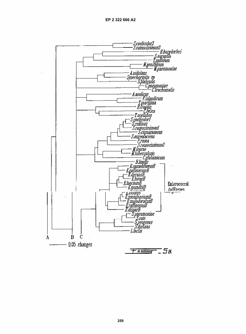
EP 2 322 666 A2
259

EP 2 322 666 A2
260

EP 2 322 666 A2
261

EP 2 322 666 A2
262

EP 2 322 666 A2
263

EP 2 322 666 A2
264

EP 2 322 666 A2
265

EP 2 322 666 A2
266

EP 2 322 666 A2
267

EP 2 322 666 A2
268

EP 2 322 666 A2
269

EP 2 322 666 A2
270

EP 2 322 666 A2
271

EP 2 322 666 A2
272

EP 2 322 666 A2
273

EP 2 322 666 A2
274

EP 2 322 666 A2
275

EP 2 322 666 A2
276

EP 2 322 666 A2
277

EP 2 322 666 A2
278

EP 2 322 666 A2
279
REFERENCES CITED IN THE DESCRIPTION
This list of references cited by the applicant is for the reader’s convenience only. It does not form part of the Europeanpatent document. Even though great care has been taken in compiling the references, errors or omissions cannot beexcluded and the EPO disclaims all liability in this regard.
Patent documents cited in the description
• US 5437978 A [0013]• US 5162199 A [0013]• EP 0337896131 A [0013]• US 5389513 A [0013]• US 5089386 A [0013]• WO 9203455 A [0013]• WO 0014274 A [0013]• EP 0133671 B1 [0013]• EP 0133288 A2 [0013]
• US 6001564 A [0018] [0134] [0341] [0342] [0343][0348] [0349] [0350] [0352] [0353] [0354] [0355][0357] [0358] [0359] [0360] [0361] [0364]
• WO 9820157 A [0018] [0019] [0116] [0153] [0160][0218] [0220] [0364]
• US 5708160 A, Goh [0022]• EP 0466251 A1 [0073]• EP 0446251 A1 [0077]• US 5994066 A [0188] [0190] [0193] [0197] [0218]
[0220]
Non-patent literature cited in the description
• STAGER ; DAVIS. Clin. Microbiol. Rev., 1992, vol.5, 302-327 [0001]
• CROIZÉ J. Lett. Infectiol., 1995, vol. 10, 109-113[0002]
• YORK et al. J. Clin. Microbiol., 1992, vol. 30,2903-2910 [0002]
• STARK ; MAKI. N. Engl. J. Med., 1984, vol. 311,560-564 [0005]
• KOENING et al. J. Clin. Microbiol., 1992, vol. 30,342-345 [0006]
• PEZZLO et al. J. Clin. Microbiol., 1992, vol. 30,640-684 [0006]
• PERSING et al. Diagnostic Molecular Microbiology:Principles and Applications. American Society forMicrobiology, 1993 [0010] [0012] [0086] [0096][0099] [0105] [0350] [0355] [0357]
• BERGERON ; OUELLETTE. Infection, 1995, vol.23, 69-72 [0010]
• BERGERON ; OUELLETTE. J Clin Microbiol., 1998,vol. 36, 2169-72 [0010]
• EHRLICH ; GREENBERG. PCR-based Diagnosticsin Infectious Disease. Blackwell Scientific Publica-tions, 1994 [0012] [0350] [0355]
• Molecular detection and identification of microorgan-isms. TANG Y. ; PERSING D. H. et al. Manual ofClinical Microbiology. ASM press, 1999 [0012]
• REEVE, J.H. J. Bacteriol., 1999, vol. 181, 3613-3617[0015]
• BELAY et al. Appl. Enviro. Microbiol., 1988, vol. 54,600-603 [0015]
• BELAY et al. J. Clin. Microbiol., 1990, vol. 28,1666-1668 [0015]
• WEAVER et al. Gut, 1986, vol. 27, 698-704 [0015]
• Genotyping Methods for Microbial Identification.RELMAN ; PERSING. PCR Protocols for EmergingInfectious Diseases. ASM Press, 1996 [0021]
• CHEN et al. FEMS Microbiol. Lett., 1988, vol. 57,19-24 [0021]
• MCCABE et al. Mol. Genet. Metabol., 1999, vol. 66,205-211 [0021]
• VAN BURIK et al. J. Clin. Microbiol., 1998, vol. 36,1169-1175 [0021]
• FOX et al. Int. J. Syst. Bacteriol., 1992, vol. 42,166-170 [0022]
• CLAYTON et al. Int. J. Syst. Bacteriol., 1995, vol.45, 595-599 [0022]
• NICHOLS et al. Nature, 1994, vol. 369, 492-493[0035]
• KOSKIN et al. Tetrahedron, 1998, vol. 54,3607-3630 [0035]
• EGHOLM et al. Nature, 1993, vol. 365, 566-568[0035]
• COUSINEAU et al. J. Mol. Evol., 1997, vol. 45,661-670 [0072]
• ANBORGH ; PARMEGGIANI. EMBO J., 1991, vol.10, 779-784 [0073]
• CALDAS et al. J. Biol. Chem, 1998, vol. 273,11478-11482 [0073]
• PORCELLA et al. Microbiology, 1996, vol. 142,2481-2489 [0073]
• NELSON ; TAIZ. TIBS, 1989, vol. 14, 113-116[0074]
• IWABE et al. Proc. Natl. Acad. Sci. USA, 1989, vol.86, 9355-9359 [0075]
• GOGARTEN et al. Proc. Natl. Acad. Sci. USA, 1989,vol. 86, 6661-6665 [0075]
• LUDWIG et al. Antonie van Leeuwenhoek, 1993, vol.64, 285-305 [0075]

EP 2 322 666 A2
280
• ALTSCHUL et al. J. Mol. Biol., 1997, vol. 215,403-410 [0075]
• MILLER ; KOKJOHN. Annu. Rev. Microbiol., 1990,vol. 44, 365-394 [0076]
• FILER ; FURANO. J. Bacteriol., 1981, vol. 148,1006-1011 [0077]
• SELA et al. J. Bacteriol., 1989, vol. 171, 581-584[0077]
• VIJGENBOOM et al. Microbiology, 1994, vol. 140,983-998 [0077]
• WATSON et al. Molecular Biology of the Gene. TheBenjamin/Cummings Publishing Company, 1987[0088]
• LEWIN. Genes IV. John Wiley & Sons, 1989 [0088]• NEIDHARDT et al. Escherichia coli and Salmonella:
Cellular and Molecular Biology. American Society forMicrobiology Press, 1996 [0089]
• LEE et al. Nucleic Acid Amplification Technologies:Application to Disease Diagnosis. Eaton Publishing,1997 [0099]
• WESTIN et al. Nat. Biotechnol., 2000, vol. 18,199-204 [0099]
• LIVAK K.J. et al. PCR Methods Appl., 1995, vol. 4,357-362 [0101]
• WITTWER, C.T. et al. BioTechniques, 1997, vol. 22,130-138 [0101]
• TYAGI S. ; FRAMER F.R. Nature Biotechnology,1996, vol. 14, 303-308 [0101]
• WHITCOMB et al. Nature Biotechnology, 1999, vol.17, 804-807 [0101]
• An integrated microelectronics hybridization systemfor genomic research and diagnostic applications.HELLER et al. Micro total analysis systems 98. Klu-wer Academic Publisher, 1998 [0103]
• CHIU ; CANTOR. Clinical Chemistry, 1999, vol. 45,1578 [0103]
• BERKENKAMP et al. Science, 1998, vol. 281, 260[0103]
• Advances in integrated genetic analysis. ANDER-SON et al. Micro total analysis systems 98. KluwerAcademic Publisher, 1998 [0104]
• DIEFFENBACH ; DVEKSLER. PCR Primer: A Lab-oratory Manual. Cold Spring Harbor LaboratoryPress, 1995 [0105] [0115] [0136]
• NIKIFOROV et al. PCR Methods and Applications,1994, vol. 3, 285-291 [0106]
• Anal. Biochem., 1995, vol. 227, 201-209 [0106]• SAMBROOK et al. Molecular Cloning: A Laboratory
Manual. Cold Spring Harbor Laboratory Press, 1989[0108]
• GREER. Methods in Enzymology, 1991, vol. 202,239-252 [0120]
• TAYLOR. Trends Biotechnol., 1994, vol. 12 (5),154-158 [0120]
• SALI. Curr. Opin. Biotechnol., 1995, vol. 6, 437-451[0120]
• SANCHEZ ; SALI. Curr. Opin. Struct. Biol., 1997,vol. 7, 206-214 [0120]
• FISCHER ; EISENBERG. Curr. Opin. Struct. Biol.,1999, vol. 9, 208-211 [0120]
• GUEX et al. Trends Biochem. Sci., 1999, vol. 24,364-367 [0120]
• KLOOS ; SCHLEIFER. J. Clin. Microbiol., 1975, vol.1, 82-88 [0167]
• FANI et al. Molecular Ecology, 1993, vol. 2, 243-250[0226]
• BIRNBOIM ; DOLY. Nucleic Acid Res., 1979, vol. 7,1513-1523 [0228]
• SHAW, K. J. ; P. N. RATHER ; R. S. HARE ; G. H.MILLER. Molecular genetics of aminoglycoside re-sistance genes and familial relationships of theaminoglycoside-modifying enzymes. Microbiol.Rev., 1993, vol. 57, 138-163 [0364]
• BUSH, K. ; G.A. JACOBY ; A. MEDEIROS. A func-tional classification scheme for ß-lactamase and itscorrelation with molecular structure. Antimicrob.Agents. Chemother., 1995, vol. 39, 1211-1233[0364]
• BAUERFEIND, A. ; Y. CHONG ; K. LEE. Plas-mid-encoded AmpC beta-lactamases: how far havewe gone 10 ears after discovery?. Yonsei Med. J.,vol. 39, 520-525 [0364]
• SUTCLIFFE, J. ; T. GREBE ; A. TAIT-KAMRADT ;L. WONDRACK. Detection of erythromycin-resistantdeterminants by PCR. Antimicrob. Agent Chemoth-er., 1996, vol. 40, 2562-2566 [0364]
• LECLERC, R. ; A., BRISSON-NOËL ; J. DUVAL ;P. COURVALIN. Phenotypic expression and geneticheterogeneity of lincosamide inactivation in Staphy-lococcus sp. Antimicrob. Agents. Chemother., 1991,vol. 31, 1887-1891 [0364]
• BOZDOGAN, B. ; L. BERREZOUGA ; M.-S. KUO ;D. A. YUREK ; K. A. FARLEY ; B. J. STOCKMAN ;R. LECLERCQ. A new gene, linB, conferring resist-ance to lincosamides by nucleotidylation in Entero-coccus faecium HM1025. Antimicrob. Agents.Chemother., 1999, vol. 43, 925-929 [0364]
• COCKERILL III, F.R. Genetic methods for assessingantimicrobial resistance. Antimicrob. Agents. Chem-other., 1999, vol. 43, 199-212 [0364]
• Genetic methods for detecting antibacterial resist-ance genes. TENOVER, F. C. ; T. POPOVIC ; OOLSVIK. Manual of clinical microbiology. ASMPress, 1996, 1368-1378 [0364]
• DOWSON, C. G. ; T. J. TRACEY ; B. G. SPRATT.Origin and molecular epidemiology of penicillin-bind-ing-protein-mediated resistance to ß-lactam antibiot-ics. Trends Molec. Microbiol., 1994, vol. 2, 361-366[0364]
• JENSEN, L. B. ; N. FRIMODT-MOLLER ; F. M.AARESTRUP. Presence of erm gene classes inGram-positive bacteria of animal and human originin Denmark. FEMS Microbiol., 1999, vol. 170,151-158 [0364]

EP 2 322 666 A2
281
• THAL, L. A. ; M. J. ZERVOS. Occurrence and epi-demiology of resistance to virginimycin and streptro-gramins. J. Antimicrob. Chemother., 1999, vol. 43,171-176 [0364]
• MARTINEZ J. L. ; A. ALONSO ; J. M.GOMEZ-GOMEZ ; F. BAQUERO. Quinolone resist-ance by mutations in chromosomal gyrase genes.Just the tip of the iceberg?. J. Antimicrob. Chemoth-er., 1998, vol. 42, 683-688 [0364]
• CASADEWALL, B. ; P. COURVALIN. Characteri-zation of the vanD glycopeptide resistance gene clus-ter from Enterococcus faecium BM 4339. J. Bacteri-ol., 1999, vol. 181, 3644-3648 [0364]
• ROBERTS, M.C. Genetic mobility and distribution oftetracycline resistance determinants. Ciba Found.Symp., 1999, vol. 207, 206-222 [0364]
• HUOVINEN, P. ; L. SUNDSTRÖM ; G.SWEDBERG ; O. SKÖLD. Trimethoprim and sulfon-amide resistance.Antimicrob. Agent Chemother.,1995, vol. 39, 279-289 [0364]