Immunotherapeutic Targeting of Membrane Hsp70-Expressing Tumors Using Recombinant Human Granzyme B
11
Immunotherapeutic Targeting of Membrane Hsp70- Expressing Tumors Using Recombinant Human Granzyme B Mathias Gehrmann 1. , Stefan Stangl 1. , Andreas Kirschner 1 , Gemma A. Foulds 1,2 , Wolfgang Sievert 1 , Brigitte T. Doß 1 , Axel Walch 3 , Alan G. Pockley 2 , Gabriele Multhoff 1 * 1 Department of Radiation Oncology, Klinikum rechts der Isar, Technische Universita ¨t Mu ¨ nchen, and Clinical Cooperation Group (CCG) ‘‘Innate Immunity in Tumor Biology’’, Helmholtz Zentrum Mu ¨ nchen, Deutsches Forschungszentrum fu ¨ r Gesundheit und Umwelt, Munich, Germany, 2 Department of Oncology, The Medical School, University of Sheffield, Sheffield, United Kingdom, 3 Institute of Pathology, Helmholtz Zentrum Mu ¨ nchen, Deutsches Forschungszentrum fu ¨ r Gesundheit und Umwelt, Munich, Germany Abstract Background: We have previously reported that human recombinant granzyme B (grB) mediates apoptosis in membrane heat shock protein 70 (Hsp70)-positive tumor cells in a perforin-independent manner. Methodology/Principal Findings: Optical imaging of uptake kinetics revealed co-localization of grB with recycling endosomes (Rab9/11) as early as 5 min after internalization, with late endosomes (Rab7) after 30 min, and the lysosomal compartment (LAMP1/2) after 60 to 120 min. Active caspase-3-mediated apoptosis was induced in mouse CT26 monolayer cells and 3D tumor spheroids, but not in normal mouse endothelial cells. Granzyme B selectively reduced the proportion of membrane Hsp70-positive cells in CT26 tumor spheroids. Consecutive i.v. injections of recombinant human grB into mice bearing membrane Hsp70-positive CT26 tumors resulted in significant tumor suppression, and a detailed inspection of normal mouse organs revealed that the administration of anti-tumoral concentrations of grB elicited no clinicopathological changes. Conclusions/Significance: These findings support the future clinical evaluation of human grB as a potential adjuvant therapeutic agent, especially for treating immunosuppressed patients that bear membrane Hsp70-positive tumors. Citation: Gehrmann M, Stangl S, Kirschner A, Foulds GA, Sievert W, et al. (2012) Immunotherapeutic Targeting of Membrane Hsp70-Expressing Tumors Using Recombinant Human Granzyme B. PLoS ONE 7(7): e41341. doi:10.1371/journal.pone.0041341 Editor: Michael Platten, University Hospital of Heidelberg, Germany Received March 12, 2012; Accepted June 20, 2012; Published July 19, 2012 Copyright: ß 2012 Gehrmann et al. This is an open-access article distributed under the terms of the Creative Commons Attribution License, which permits unrestricted use, distribution, and reproduction in any medium, provided the original author and source are credited. Funding: This study was supported by the Deutsche Forschungsgemeinschaft (DFG) SFB824, (DFG) INST95/980-1 FUGG, DFG-Cluster of Excellence: Munich Advanced Photonics (MAP), Bundesministerium fu ¨r Bildung und Forschung (BMBF) MOBITUM (01EZ0826), m4 Cluster of Excellence (01EX1021C), Kompetenzverbund Strahlenforschung (03NUK007E), the European Union (EU CARDIORISK, FP7-211403)and multimmune GmbH. The funders had no role in study design, data collection and analysis, decision to publish, or preparation of the manuscript. Competing Interests: Gabriele Multhoff and Alan Graham Pockley serve as Academic Editors of PLoS ONE. This study was partly funded by multimmune GmbH. The membrane Hsp70 phenotype on viable tumor and metastatic cells of patients was determined by flow cytometry using FITC-conjugated cmHsp70.1 mAb (multimmune GmbH, Germany). There are no other patents, products in development or marketed products to declare. This does not alter the authors’ adherence to all the PLoS ONE policies on sharing data and materials, as detailed online in the guide for authors. * E-mail: [email protected] . These authors contributed equally to this work. Introduction Heat shock protein 70 (Hsp70) is frequently overexpressed in tumors and cytosolic Hsp70 mediates the protection of tumor cells against environmental stress [1–3]. Hsp70 has also been found to be localized in the plasma membrane of a large proportion of different tumor entities, but not in the plasma membrane of normal cells/tissues [4–11]. Although the precise role of mem- brane-associated Hsp70 is not fully understood, overall survival of patients with lower rectal carcinomas and non-small cell lung cancer (NSCLC) exhibiting a membrane Hsp70-positive pheno- type has been found to be significantly lower than that of their membrane Hsp70-negative counterparts [12]. Furthermore, most standard therapies, including radiochemotherapy, increase the membrane densities of Hsp70 on cancer, but not normal cells [7,8,13]. These findings highlight the clinical significance of determining the membrane Hsp70 status, and the urgent need for innovative treatment modalities that can specifically target highly aggressive, membrane Hsp70-positive tumors. We have previously demonstrated that membrane Hsp70 serves as a tumor-specific recognition structure for pre-activated natural killer (NK) cells, but not for resting NK cells [14]. Full-length Hsp70, as well as the extracellularly-accessible Hsp70-derived peptide TKDNNLLGRFELSG (TKD), in combination with low dose IL-2 increase the expression density of activating receptors such as NKG2D, NKG2C/CD94 and NCRs and stimulate the cytolytic activity of NK cells to attack membrane Hsp70-positive tumor cells in vitro [5,14]. Tolerability and safety of patient- derived, ex vivo TKD/IL-2-activated NK cells as an immunother- apeutic option has been demonstrated in a Phase I clinical trial PLoS ONE | www.plosone.org 1 July 2012 | Volume 7 | Issue 7 | e41341
-
Upload
independent -
Category
Documents
-
view
0 -
download
0
Transcript of Immunotherapeutic Targeting of Membrane Hsp70-Expressing Tumors Using Recombinant Human Granzyme B

Immunotherapeutic Targeting of Membrane Hsp70-Expressing Tumors Using Recombinant HumanGranzyme BMathias Gehrmann1., Stefan Stangl1., Andreas Kirschner1, Gemma A. Foulds1,2, Wolfgang Sievert1,
Brigitte T. Doß1, Axel Walch3, Alan G. Pockley2, Gabriele Multhoff1*
1Department of Radiation Oncology, Klinikum rechts der Isar, Technische Universitat Munchen, and Clinical Cooperation Group (CCG) ‘‘Innate Immunity in Tumor
Biology’’, Helmholtz Zentrum Munchen, Deutsches Forschungszentrum fur Gesundheit und Umwelt, Munich, Germany, 2Department of Oncology, The Medical School,
University of Sheffield, Sheffield, United Kingdom, 3 Institute of Pathology, Helmholtz Zentrum Munchen, Deutsches Forschungszentrum fur Gesundheit und Umwelt,
Munich, Germany
Abstract
Background: We have previously reported that human recombinant granzyme B (grB) mediates apoptosis in membraneheat shock protein 70 (Hsp70)-positive tumor cells in a perforin-independent manner.
Methodology/Principal Findings: Optical imaging of uptake kinetics revealed co-localization of grB with recyclingendosomes (Rab9/11) as early as 5 min after internalization, with late endosomes (Rab7) after 30 min, and the lysosomalcompartment (LAMP1/2) after 60 to 120 min. Active caspase-3-mediated apoptosis was induced in mouse CT26 monolayercells and 3D tumor spheroids, but not in normal mouse endothelial cells. Granzyme B selectively reduced the proportion ofmembrane Hsp70-positive cells in CT26 tumor spheroids. Consecutive i.v. injections of recombinant human grB into micebearing membrane Hsp70-positive CT26 tumors resulted in significant tumor suppression, and a detailed inspection ofnormal mouse organs revealed that the administration of anti-tumoral concentrations of grB elicited no clinicopathologicalchanges.
Conclusions/Significance: These findings support the future clinical evaluation of human grB as a potential adjuvanttherapeutic agent, especially for treating immunosuppressed patients that bear membrane Hsp70-positive tumors.
Citation: Gehrmann M, Stangl S, Kirschner A, Foulds GA, Sievert W, et al. (2012) Immunotherapeutic Targeting of Membrane Hsp70-Expressing Tumors UsingRecombinant Human Granzyme B. PLoS ONE 7(7): e41341. doi:10.1371/journal.pone.0041341
Editor: Michael Platten, University Hospital of Heidelberg, Germany
Received March 12, 2012; Accepted June 20, 2012; Published July 19, 2012
Copyright: � 2012 Gehrmann et al. This is an open-access article distributed under the terms of the Creative Commons Attribution License, which permitsunrestricted use, distribution, and reproduction in any medium, provided the original author and source are credited.
Funding: This study was supported by the Deutsche Forschungsgemeinschaft (DFG) SFB824, (DFG) INST95/980-1 FUGG, DFG-Cluster of Excellence: MunichAdvanced Photonics (MAP), Bundesministerium fur Bildung und Forschung (BMBF) MOBITUM (01EZ0826), m4 Cluster of Excellence (01EX1021C),Kompetenzverbund Strahlenforschung (03NUK007E), the European Union (EU CARDIORISK, FP7-211403)and multimmune GmbH. The funders had no role instudy design, data collection and analysis, decision to publish, or preparation of the manuscript.
Competing Interests: Gabriele Multhoff and Alan Graham Pockley serve as Academic Editors of PLoS ONE. This study was partly funded by multimmune GmbH.The membrane Hsp70 phenotype on viable tumor and metastatic cells of patients was determined by flow cytometry using FITC-conjugated cmHsp70.1 mAb(multimmune GmbH, Germany). There are no other patents, products in development or marketed products to declare. This does not alter the authors’ adherenceto all the PLoS ONE policies on sharing data and materials, as detailed online in the guide for authors.
* E-mail: [email protected]
. These authors contributed equally to this work.
Introduction
Heat shock protein 70 (Hsp70) is frequently overexpressed in
tumors and cytosolic Hsp70 mediates the protection of tumor cells
against environmental stress [1–3]. Hsp70 has also been found to
be localized in the plasma membrane of a large proportion of
different tumor entities, but not in the plasma membrane of
normal cells/tissues [4–11]. Although the precise role of mem-
brane-associated Hsp70 is not fully understood, overall survival of
patients with lower rectal carcinomas and non-small cell lung
cancer (NSCLC) exhibiting a membrane Hsp70-positive pheno-
type has been found to be significantly lower than that of their
membrane Hsp70-negative counterparts [12]. Furthermore, most
standard therapies, including radiochemotherapy, increase the
membrane densities of Hsp70 on cancer, but not normal cells
[7,8,13]. These findings highlight the clinical significance of
determining the membrane Hsp70 status, and the urgent need for
innovative treatment modalities that can specifically target highly
aggressive, membrane Hsp70-positive tumors.
We have previously demonstrated that membrane Hsp70 serves
as a tumor-specific recognition structure for pre-activated natural
killer (NK) cells, but not for resting NK cells [14]. Full-length
Hsp70, as well as the extracellularly-accessible Hsp70-derived
peptide TKDNNLLGRFELSG (TKD), in combination with low
dose IL-2 increase the expression density of activating receptors
such as NKG2D, NKG2C/CD94 and NCRs and stimulate the
cytolytic activity of NK cells to attack membrane Hsp70-positive
tumor cells in vitro [5,14]. Tolerability and safety of patient-
derived, ex vivo TKD/IL-2-activated NK cells as an immunother-
apeutic option has been demonstrated in a Phase I clinical trial
PLoS ONE | www.plosone.org 1 July 2012 | Volume 7 | Issue 7 | e41341

[15,16] and a proof-of-concept Phase II study in NSCLC patients
following radiochemotherapy is ongoing.
The mechanism by which activated NK cells kill membrane
Hsp70-positive tumor cells is associated with an enhanced
production and release of the pro-apoptotic serine protease
Granzyme B (grB) [17]. Sepharose column chromatography has
revealed that the epitope of Hsp70 which is exposed to the
extracellular milieu on tumor cells enables binding of recombinant
human grB [17]. Furthermore, we have demonstrated that grB-
induced apoptosis in Hsp70-positive tumor cells occurs in the
absence of perforin [17], and that the interaction of grB with the
membrane form of Hsp70 is dependent on an eukaryotic
glycosylation pattern of grB [18]. It has also been shown that
membrane Hsp70 shows a fast turn-over rate [19] and this might
enable the uptake of grB.
Presuming that grB is only internalized into membrane Hsp70-
positive tumor cells, but not in healthy tissues that lack membrane
Hsp70, human grB might provide a novel strategy to induce tumor
cell apoptosis in a highly selective manner with a low risk of
generating adverse effects. This study therefore investigates the
potential of the therapeutic potential of grB using 3D tumor
spheroids and a syngeneic CT26 tumor mouse model. The
internalization pathway into tumor cells has been visualized using
fluorophor-conjugated grB and confocal microscopy.
Our findings demonstrate that grB selectively induces caspase-3
dependent apoptosis in membrane Hsp70-positive cells in CT26
mouse tumor cell monolayers and spheroids. Furthermore, the
administration of grB significantly reduces the size of solid tumors
in mice. The lack of any adverse effects in mice receiving 4
repeated injections of grB supports the proposition that grB might
be effective for the treatment of tumor patients that lack active
immune protection during and/or directly after therapeutic
interventions such as radiochemotherapy.
Results
In contrast to normal cells, tumors frequently express Hsp70 on
their plasma membrane [4], and we show here that the membrane
density of Hsp70 is considerably higher on metastases compared to
primary and relapse tumors (Fig. 1). As grB has previously been
shown to selectively initiate perforin-independent apoptosis in
membrane Hsp70-positive human tumor cells [17], herein we
studied the capacity of HEK293 cell-derived, recombinant human
grB [18] to kill CT26 mouse colon adenocarcinoma cells.
Approximately 60% of the cells in monolayer and 3D tumor
spheroids express Hsp70 on their plasma membrane.
Active and inactive, HEK293 cell-derived, recombinant human
grB was generated as described previously [18] and its enzymatic
activity at different stages in the process was monitored (Fig. 2).The endotoxin content of grB preparations before and after
enterokinase digestion was determined using the E-TOXATE Kit
(Sigma-Aldrich, Catalog #ET0100) and was found to be below
the detection limit of the assay (0.05–0.1 endotoxin units (EU)/
mL) in each fraction (data not shown).
Induction of Apoptosis in CT26 Mouse Tumor CellMonolayers by Human Granzyme BThe enzymatic activity of grB in conditioned medium contain-
ing tumor cells did not differ significantly between day 0 to day 3
(data not shown). GrB significantly reduced the clonogenic survival
of CT26 tumor cells at concentrations of 0.6, 0.8, 1, 2, and 4 mg/ml (Fig. 3A). Apoptosis (on the basis of active caspase-3
expression) was determined 12, 24 and 48 h after incubation of
CT26 monolayer cells with grB (4 mg/ml). The percentage of
caspase-3 positive cells increased from 360% (PBS control) to
2269% (12 h, black bars), to 39617% (24 h, light grey bars) and
to 5468% (48 h, dark grey bars) after different times of grB
treatment (Fig. 3B). Similar effects were induced by the
topoisomerase inhibitor camptothecin (cam) which was used as
a positive control (Fig. 3B). Light microscopy performed before
and 48 h after treatment with grB (4 mg/ml) and cam (4 mg/ml)
confirmed apoptosis in adherent CT26 tumor cells on the basis of
reduced adherence, cell rounding and an increase in granularity.
In contrast, inactive grB exhibited no effects (Fig. 3C).Given that the therapeutic application of grB in an in vivo setting
will require intravenous injection, it is important to ascertain the
potential effects of grB on endothelial cells (ECs). The effects of
grB on the viability of CD31-positive ECs that were freshly
isolated from normal BALB/c mice were therefore assessed. As
shown in Fig. 3D, the percentage of caspase-3 positive mouse ECs
remained unchanged after treatment with grB (4 mg/ml; 360% to
766%; p= 0.2) and normal mouse ECs retained their adherence
and regular morphology (Fig. 3E).
Internalization of Granzyme BThe uptake of grB into membrane Hsp70-positive tumor cells
was visualized using confocal microscopy. As illustrated in Fig. 4A,grB co-locates with recycling endosomes (Rab9/Rab11) as early as
5 min after endocytosis, and with late endosomes (Rab7) after
30 min. GrB subsequently resides in lysosomal compartments 60
to 120 min after internalization, as visualized by co-staining with
fluorophor-labeled LAMP1 and LAMP2 antibodies (Fig. 4A). Thekinetics and tumor-specific internalization of grB are schematically
represented in Fig. 4B and Fig. 4C respectively.
Induction of Apoptosis in CT26 Mouse Tumor Spheroidsby Human Granzyme BThe approximate amount of grB that might be needed to
effectively target tumors in vivo was assessed by measuring the
capacity of grB to induce apoptosis in 3D tumor spheroids on day
2 after incubation with 4, 10, 20, 40 and 80 mg/ml grB.
Concentrations of grB as low as 4 mg/ml significantly increased
the proportion of caspase-3 positive cells (362% to 860%), and
this effect gradually increased at higher grB concentrations
(5763% caspase-3 positive cells at 80 mg/ml) (Fig. 5A). No
significant increase in apoptosis was observed for PBS-treated
spheroids (ctrl).
Morphological changes in CT26 tumor spheroids after co-
incubation with grB or cam were determined, and their diameters
were measured. Untreated spheroids are round with a clearly
defined, smooth border. Tumor spheroids exposed to grB lost their
integrity and regular shape but significant shrinkage was only
observed from day 7 onwards. In contrast, spheroids treated with
cam (4 mg/ml) remained regular during shrinkage (data not
shown). A slight initial swelling of tumor spheroids was apparent
between 1 and 3 days after treatment with grB (Fig. 5B). This ismost likely due to loss of integrity which is induced by apoptotic
cell death, as is illustrated by the caspase-3 apoptosis assay
(Fig. 5A).The overall size of spheroids that had been treated with grB and
cam was significantly decreased on day 14 (583630 mm to
446636 and 556625 mm to 429637 mm respectively (Fig. 5B),whereas the size of untreated, control spheroids had increased
(555627 mm to 610648 mm, Fig. 5B). The initial sizes of the
spheroids were similar across the treatment groups (565630 mm).
These findings demonstrate that grB induces apoptosis in
membrane Hsp70-positive cells in monolayers and 3D tumor
spheroids.
Therapeutic Potential of Human Granzyme B
PLoS ONE | www.plosone.org 2 July 2012 | Volume 7 | Issue 7 | e41341

Figure 1. Comparative flow cytometric histograms of membrane Hsp70 expression on viable (7-AAD negative) cells from primarytumors and distant metastases of three patients, and on a relapse tumor and a distant metastases of another patient using FITC-labelled IgG1 isotype-matched control antibody (open histogram) and cmHsp70.1 mAb (grey histogram). The mean fluorescenceintensity of Hsp70 is much higher on metastases compared to primary and relapse tumors, as indicated by a shift of the grey peak to the right.doi:10.1371/journal.pone.0041341.g001
Therapeutic Potential of Human Granzyme B
PLoS ONE | www.plosone.org 3 July 2012 | Volume 7 | Issue 7 | e41341

Caspase-3 positivity was only observed in tumor spheroids that
had been treated with active grB (Fig. 5C). The specificity of grB-
mediated killing was demonstrated by the finding that the
percentage of membrane Hsp70-positive cells in CT26 tumor
spheroids that had been treated with active grB reduced from 60%
to 40%. In contrast, the treatment of CT26 tumor spheroids with
inactive grB had no effect on the percentage of membrane Hsp70-
positive cells contained therein (Fig. 5D).
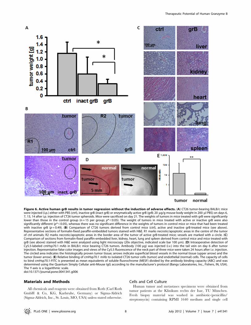
GrB Induces Tumor Suppression and is Well Tolerated inTumor-bearing MiceIn order to determine the anti-tumoral activity of human grB
in vivo, mice were injected (i.p.) with CT26 tumor spheroids of
identical size. Tumor-bearing mice were injected (i.v.) with PBS
(200 ml; ctrl) or with 20 mg/g body weight inactive or active grB
diluted in 200 ml PBS on days 6, 7, 13, and 14. Mice were
sacrificed on day 21, at which time tumors and organs (liver,
kidney, heart, lung, spleen) were collected. A significant reduction
in tumor weight was observed only in mice treated with active grB
(grB; 0.3360.12 g; p* = 0.04 vs. 1.1760.31 g in control animals)
(Fig. 6A). Treatment of mice with inactive grB preparations
resulted in a non-significant reduction of the tumor weight (inact
grB; 0.6060.45 g; p=0.49) compared to control tumors (Fig. 6A).Overall survival of mice that had been treated with active human
grB was significantly greater than that of control mice (p* = 0.023;
data not shown).
Four tumors were analysed by H&E staining. Viable tumor
tissue is characterized by numerous areas of mitosis, compact
tissue, and rare single cell apoptosis. In contrast, necrotic/
apoptotic areas are characterized by an increased eosin staining,
the presence of apoptotic bodies, bulked extracellular matrices,
dense nuclei and nuclear fragmentations. A representative view of
one of the sections is shown in Fig. 6B. In all control animals
carrying larger tumors than grB-treated animals (n = 4), necrosis
was only found in the central part (marked with R1). In contrast,
necrosis was predominantly found on the outer borders (marked
with R2), and in proximity to blood vessels (circled) in tumor-
bearing mice that had been treated with active grB. This result was
also confirmed in 4 out of 4 animals. None of the tumors treated
with active grB exhibited central necrosis. Tumors in mice treated
with inactive grB showed no reduction in size, and no damage to
tumor tissue in the vicinity of vessels was apparent (Fig. 6B,vessels are circled).
Although no obvious adverse side-effects occurred following the
administration of active human grB, the organs of control mice
and of those treated with active grB exhibited no pathophysiolog-
ical changes (Fig. 6C). Individual organs exhibited normal tissue
architecture, with no evidence of infarcts or internal bleeding.
Active human grB at concentrations that can reduce tumor size
does therefore not induce detectable histological or morphological
changes in normal organs.
The Hsp70 status of tumor and normal tissues in CT26 tumor-
bearing mice was determined by measuring the in vivo binding
capacity of fluorescently-labelled cmHsp70.1 antibody. For this,
intraoperative in vivo imaging of tumor-bearing mice was per-
formed 24 h after tail vein injection of 100 ml of a 1 mg/ml
dilution of Cy5.5-labelled cmHsp70.1 mAb in PBS. The fluores-
cence of the dissected tumor and of the surrounding normal tissue
was measured and compared to the relevant normal tissue by false
color imaging. A representative fluorescent image revealed an
accumulation of cmHsp70.1 mAb in the tumor but not in the
adjacent normal tissue and in the blood vessels (Fig. 6D). The
delineation of tumor and normal tissue was confirmed using H&E
staining of the imaged area (data not shown).
The relative binding of cmHsp70.1 mAb to isolated tumor and
endothelial cells was determined by flow cytometry using
Quantum Simply Cellular beads. This revealed a significantly
higher binding of cmHsp70.1 mAb to tumor cells compared to
endothelial cells (Fig. 6E).
Discussion
Granzyme B (grB) derived from E. coli [20] has been shown to
induce apoptosis in tumor target cells via perforin pores that
trigger the release of endocytosed grB [21]. Lytic agents such as
cationic lipid formulation BioporterH (Gene Therapy Systems Inc.,
San Diego, CA, USA) can be used to facilitate the release of grB
[22,23]. The activity of grB purified from cytotoxic T and NK cells
has been found to be weak due to a covalent binding of grB to the
human protease inhibitor PI-9 [24–26]. Furthermore, Trapani
and co-workers [28] have shown that more than 50% of total grB
resides in the nucleus and not in the cytosol of human effector
cells.
We have described a perforin-independent pathway by which
human grB can specifically induce apoptosis in membrane Hsp70-
positive tumor cells [17]. Mammalian glycosylation and post-
translational modifications are a prerequisite for this Hsp70-
mediated uptake of human grB into tumor cells [18], since not
even Pichia pastoris derived grB [27] was effective. The present
study used a mammalian HEK293 expression system to produce
high yields of enzymatically and biologically active, glycosylated
human grB following enterokinase (EK) cleavage of the in-
activation site [18].
In the presence of perforin, grB-mediated apoptosis in mono-
layer cells is typically induced after an incubation period of 4 h to
48 h at concentrations ranging between 10 ng/ml and 10 mg/ml
[20,24]. Herein, we show that human grB induces apoptosis in
membrane Hsp70-positive mouse tumor cells, but not in normal
mouse tissues in a comparable time and concentration range.
Dose-response effects, as determined by measuring clonogenic
cell survival assays, revealed a significant reduction in clonogeni-
city already at a very low grB concentration (0.6 mg/ml). Less
Figure 2. Enzymatic activity during the purification of grB. A–FgrB before enterokinase digestion; H, J’ flow through during purificationprocess; G, I, J, K grB after enterokinase digestion: A. cell culturesupernatant of the HEK293 producing cells; B. after addition ofimidazole; C. flow through of Ni column; D. His-tagged grB; E. pooledHis-tagged grB; F. after exchange to enterokinase buffer; G. afterenterokinase digestion; H. flow through of heparin column; I. pooledgrB containing fractions after heparin column; J. concentrated grB; J’.flow through from concentration procedure; K. sterile filtered (0.2 ml)active grB (final product).doi:10.1371/journal.pone.0041341.g002
Therapeutic Potential of Human Granzyme B
PLoS ONE | www.plosone.org 4 July 2012 | Volume 7 | Issue 7 | e41341

than 20% of the tumor colonies were found to be alive at
a concentration of 4 mg/ml of human grB. In order to estimate
the concentration of human grB that might be necessary to treat
solid mouse tumors, multicellular 3D tumor spheroid assays were
performed. Tumor spheroids mimic the cellular and organotypic
histomorphological features of tumors without vascularization
[29–32]. In comparison to monolayer cells, a 20-fold higher
concentration of human grB was required to induce the
shrinkage of tumor spheroids. In contrast, significant active
caspase-3 positivity could be achieved in tumor spheroids at grB
concentrations as low as 4 mg/ml. These findings indicate that
spheroid diameter is not an indicative surrogate for grB-mediated
Figure 3. Recombinant human grB induces apoptosis in monolayer CT26 tumor, but not normal cells. (A) Clonogenic cell survival ofCT26 tumor cells after treatment with active human grB (0.04, 0.2, 0.4, 0.6, 0.8, 1, 2, 4 mg/ml) on day 6. The data represent the mean of threeindependent experiments 6 S.D. Significant differences in the survival of grB-treated CT26 vs. untreated controls were seen from 0.6 mg/ml upwards(p*,0.05). (B) CT26 cells were treated with PBS (ctrl), granzyme B (4 mg/ml; grB) or camptothecin (4 mg/ml; cam) for 12 h (black bars), 24 h (grey bars),and 48 h (white bars). Permeabilized cells were stained with a FITC-conjugated active caspase-3 specific antibody and the percentage of activecaspase-3 positive cells determined by flow cytometry. The data represent the mean of 3–10 independent experiments 6 S.D. (p***,0.001;p** = 0.01). (C) Light microscopical views of CT26 tumor cells after treatment with PBS (ctrl), grB (4 mg/ml), cam (4 mg/ml) or inactive grB (inact grB) for48 h (20x objective, scale bar 100 mm). Representative images are shown from at least 3 experiments using CT26 tumor cells (D) CD31-positivemouse endothelial cells from healthy BALB/c mice (normal) and CT26 tumor cells (tumor) were treated either with PBS (ctrl; black bars) or grB (4 mg/ml; grey bars) and the percentage of cells that positively stained for active caspase-3 was determined by flow cytometry. The data represent the meanof six independent experiments 6 S.D. Asterisks represent significantly different values (p*** = 0.001). Inactive grB showed similar results like PBS(data not shown) (E) Light microscopic phase contrast analyses of adherent growing CD31-positive mouse endothelial cells (normal) treated for 24 hwith PBS (ctrl, upper graph) or grB (4 mg/ml). Both untreated and treated CD31-positive mouse endothelial cells show regular cell morphology.Similar results were obtained in three independent experiments (20x objective, scale bar 100 mm).doi:10.1371/journal.pone.0041341.g003
Therapeutic Potential of Human Granzyme B
PLoS ONE | www.plosone.org 5 July 2012 | Volume 7 | Issue 7 | e41341

tumor cell killing. Cell death and loss in spheroid integrity
following treatment with grB was also associated with irregularly
shaped spheroid surfaces. In contrast, treatment with cam
resulted in a gradual decrease in spheroid size. GrB pre-
dominantly induces apoptosis through mitochondrial leakage and
activation of the caspase cascade [33]. In contrast, cam does not
Figure 4. Confocal analysis of immunofluorescence staining of grB (ATTO488, green) and Rab4, Rab5, Rab7, Rab9, Rab11, LAMP1,and LAMP2 (Cy3, red). (A) CT26 tumor cells were grown on MatTek Glass Bottom Culture Dishes then incubated with 4 mg/ml ATTO488-labeledgrB for 0, 5, 15, 30, 60, 120 and 240 min. After fixation, cells were stained with anti-endosomal (Rab4, Rab5, Rab7, Rab9, Rab11) or lysosomalantibodies (LAMP1, LAMP2) followed by appropriate Cy3-labeled secondary antibodies. Cells were mounted in DAPI-Vectashield and imaged usinga Zeiss LSM 510 Inverted microscope (63x objective). Co-localization, as determined by an overlap of the green and red fluorescence, is visible asyellow. One representative image from 2 independent experiments is shown (scale bar, 10 mm). (B) Semi-quantitative analysis of the co-stainingintensity and kinetics of endocytosis of grB and Rab9/11 and LAMP1/LAMP2. (C) Schematic representation of the mode of uptake and intracellulartrafficking of grB in tumor cells.doi:10.1371/journal.pone.0041341.g004
Therapeutic Potential of Human Granzyme B
PLoS ONE | www.plosone.org 6 July 2012 | Volume 7 | Issue 7 | e41341

significantly interfere with the mitochondrial apoptosis pathway
[34], but causes apoptosis via inhibition of the DNA topoisome-
rase I. Despite these mechanistic differences, grB and cam
showed similar results with respect to the induction of active
caspase-3-mediated apoptosis in membrane Hsp70-positive
tumor cells [18].
Although mouse and human grB show distinct structural and
functional characteristics, and despite slight differences in their
substrate specificity, we have shown that human grB effectively
initiates cell death in membrane Hsp70-positive mouse tumor cells.
In vitro experiments indicated that procaspase-3 is identically cleaved
by human and mouse-derived grB [35]. However, differences exist
with respect to the cleavage ofBid by humanandmouse grB. Bid, the
cleavage product which results in the release of cytochrome c from
mitochondria, is not processed as efficiently by mouse grB, as it is by
human grB [34,36–38]. Regarding these differences, it is assumed
that doses which are effective in mice might have to be altered and
adapted to the human system.
Our mouse data reveal that 4 consecutive treatment cycles using
20 mg grB per g body weight significantly reduce tumor weight,
and the safety and tolerability of the treatment was proven by
immunohistochemistry. No pathophysiological changes were
found in the liver, kidney, lung, heart and spleen of the mice
after this treatment regimen. The concentrations that were used in
our study are relatively low compared to those used by Rosenblum
and Liu who injected 5637.5 mg per g body weight E. coli
immunotoxin coupled to grB without inducing any severe side
effects [22,23]. Upcoming dose-escalation experiments will de-
termine the maximal effective and tolerated dose.
Serum levels of active grB in healthy human individuals range
between 15 and 40 pg/ml and can exceed up to 250 pg/ml during
severe infections [39,40]. Our study therefore used grB concen-
trations that are similar to those that have previously been used to
induce perforin-dependent apoptosis in vitro, but higher than
naturally-occurring levels. However, no adverse effects were
associated with the administration of active grB.
It was also interesting to observe that necrotic regions in the
tumor tissue were selectively found in proximity to tumor blood
vessels and in peripheral tumor areas after an i.v. injection of
active grB into mice, whereas necrosis was only seen in central
areas of the tumors in control animals with larger tumor volumes.
These data suggest that grB targets tumors via blood vessels.
On the basis of our findings, we propose that patients during and/
or after standard radiochemotherapy might especially profit from
receiving biologically active human grB. Our results warrant future
clinical testingofhumangrBasapotentialadjuvant therapeuticagent
for the treatment of transiently immunosuppressed tumor patients
with a membrane Hsp70-positive tumor phenotype.
Figure 5. Recombinant human grB induces apoptosis in CT26tumor spheroids. (A) CT26 tumor spheroids were treated either withPBS (ctrl) or grB (4, 10, 20, 40, 80 mg/ml) for 48 h. Single cell suspensionswere generated from tumor spheroids by trypsinization and the cellspermeabilized before being stained for active caspase-3. The percent-age of active caspase-3 positive cells was determined using a BDBiosciences FACSCaliburTM. The data represent mean values of 3–4independent experiments 6 S.D. All grB-treated samples significantlydiffered from ctrl values (p***,0.001). (B) CT26 tumor spheroids wereincubated for up to 14 days either with PBS (ctrl; filled circles), grB(80 mg/ml; open circles) or cam (4 mg/ml; filled triangles). Diameters ofspheroids were measured on days 0, 1, 3, 6, 7, 10, and 14 aftertreatment. The data represent mean values of 6 independent
experiments 6 S.D. After a slight increase in diameter size, grB initiateda significant shrinkage of spheroids from day 7 onwards (day 7,p** = 0.004; day 10, p** = 0.0063; day 14, p*** = 0.0003). A significantreduction in tumor size was observed after treatment with cam fromday 6 onwards (day 6, p** = 0.0061; day 7, p** = 0.0022; day 10,p** = 0.0019; day 14, p** = 0.0012). (C) Effect of treatment with inactiveand active grB for 24 h and 48 h on the percentage of active caspase-3positive cells in CT26 cells isolated from tumor spheroids. (D) Effect oftreatment with inactive and active grB for 24 h and 48 h on thepercentage of membrane Hsp70-positive CT26 cells in tumor spheroids.Spheroids were incubated with activated or inactivated grB for theindicated time periods, after which single cell suspensions wereprepared following trypsinization. Cells were then incubated withcmHsp70.1-FITC mAb and membrane Hsp70 expression was de-termined by flow cytometry.doi:10.1371/journal.pone.0041341.g005
Therapeutic Potential of Human Granzyme B
PLoS ONE | www.plosone.org 7 July 2012 | Volume 7 | Issue 7 | e41341
Materials and Methods
All chemicals and reagents were obtained from Roth (Carl Roth
GmbH & Co, KG, Karlsruhe, Germany) or Sigma-Aldrich
(Sigma-Aldrich, Inc., St. Louis, MO, USA) unless stated otherwise.
Cells and Cell CultureHuman tumor and metastases specimens were obtained from
tumor patients at the Klinikum rechts der Isar, TU Munchen.
Fresh biopsy material was washed in antibiotic-(penicillin/
streptomycin) containing RPMI 1640 medium and single cell
Figure 6. Active human grB results in tumor regression without the induction of adverse effects. (A) CT26 tumor-bearing BALB/c micewere injected (i.p.) either with PBS (ctrl), inactive grB (inact grB) or enzymatically active grB (grB; 20 mg/g mouse body weight in 200 ml PBS) on days 6,7, 13, 14 after i.p. injection of CT26 tumor spheroids. Mice were sacrificed on day 21. The weights of tumors in mice treated with grB were significantlylower than those in the control group (n = 15 per group; p*,0.05). The weight of tumors in mice treated with active or inactive grB were alsosignificantly different (p*,0.05), whereas there was no significant difference in the weights of tumors in control mice or mice that had been treatedwith inactive grB (p= 0.49). (B) Comparison of CT26 tumors derived from control mice (ctrl), active and inactive grB-treated mice (see above).Representative sections of formalin-fixed paraffin-embedded tumors stained with H&E. R1 marks necrotic/apoptotic areas in the centre of the tumorof ctrl animals; R2 marks necrotic/apoptotic areas in the border area of the tumor of active grB-treated mice; vessels are marked with a circle. (C)Comparison of sections from formalin-fixed paraffin-embedded liver, kidney, heart, lung and spleen derived from control mice and mice treated withgrB (see above) stained with H&E were analyzed using light microscopy (20x objective, indicated scale bar 100 mm). (D) Intraoperative detection ofCy5.5-labeled cmHsp70.1 mAb in BALB/c mice bearing CT26 tumors. Antibody (100 mg) was injected (i.v.) into the tail vein on day 6 after tumorinjection. Representative false color images and views of the Cy5.5 fluorescence of the neck part of three mice were taken 24 hours after i.v. injection.The circled area indicates the histologically proven tumor tissue; arrows indicate superficial blood vessels in the normal tissue (upper arrow) and thetumor (lower arrow). (E) Relative binding of cmHsp70.1 mAb to isolated CT26 tumor cells (tumor) and endothelial (normal) cells. The capacity of cellsto bind cmHsp70.1-FITC is presented as mean equivalents of soluble fluorochrome (MESF) divided by the antibody binding capacity (ABC) and wasdetermined using the Quantum Simply Cellular anti-Mouse IgG according to the manufacturer’s protocol (Bangs Laboratories, Inc., Fishers, IN, USA).The Y-axis is a logarithmic scale.doi:10.1371/journal.pone.0041341.g006
Therapeutic Potential of Human Granzyme B
PLoS ONE | www.plosone.org 8 July 2012 | Volume 7 | Issue 7 | e41341

suspensions were prepared by mincing the tissue and forcing it
through a sterile mesh. The study was approved by the
Institutional Review Board of the Medical Faculty of the Klinikum
rechts der Isar, TU Munchen, Germany and all patients included
in the study provided signed informed consent. All clinical
investigations have been deducted according to the principles
expressed in the Declaration of Helsinki.
The mouse colon adenocarcinoma cell line CT26 (CT26.WT;
ATCC CRL-2638) [41], which is derived from a BALB/c mouse
tumor was cultured in RPMI 1640 medium containing 10% (v/v)
heat-inactivated fetal calf serum (FCS), 2 mM L-glutamine, 1 mM
sodium-pyruvate, antibiotics (100 IU/ml penicillin and 100 mg/ml streptomycin) at 37uC in 5% (v/v) CO2. This cell line is 60%
positive for membrane-Hsp70 expression, as determined by flow
cytometry [11]. Single-cell suspensions were derived by short-term
(less than 1 min) treatment with 0.25% (w/v) Trypsin-0.53 mM
EDTA.
CD31-positive endothelial cells (normal cells) were isolated from
BALB/c mice using magnetic dynabeads (Invitrogen, Karlsruhe,
Germany) coated with CD31 antibody (BD Biosciences; Heidel-
berg, Germany) according to the manufacturer’s instructions
(Invitrogen). The cells were cultured in endothelial cell growth
medium (PromoCell, Heidelberg, Germany) for up to 4 cell
passages.
All cell lines were regularly screened for mycoplasma contam-
ination using an enzyme immunoassay which detects Mycoplasma
arginini, Mycoplasma hyorhinis, Achdeoplasma laidlawii, and Mycoplasma
orale (Roche Diagnostics, Basel, Switzerland). Only mycoplasma-
free cell lines were used in this study.
Flow CytometryThe membrane Hsp70 phenotype on viable tumor and
metastatic cells of patients was determined by flow cytometry
using FITC-conjugated cmHsp70.1 monoclonal antibody (mAb;
multimmune GmbH, Germany) an IgG1 isotype-matched control
antibody, as described elsewhere [11].
Confocal MicroscopyCT26 cells were seeded into MatTek Glass Bottom Culture
Dishes (16105 cells in 2 ml RPMI 1640 medium). After 48 h,
culture medium was exchanged for RPMI 1640 medium contain-
ing grB (4 mg/ml) and incubated for various durations (0, 5, 15,
30 min, 1, 2, 4 h). Cells were then washed in PBS, fixed in 0.25%
w/v paraformaldehyde, permeabilized in PBS containing 0.1% w/
v saponin and 0.1% w/v BSA and stained with primary antibodies
to Rab4, Rab5, Rab7, Rab9, Rab11, LAMP1 or LAMP2. Primary
antibody binding was detected using appropriate Cy3-labeled
secondary antibodies.
The glass bottom of the dishes was detached by immersing the
underside in 750 ml Coverslip Removal Fluid (MatTek), after
which cells were mounted onto microscopy slides with DAPI-
Vectashield and sealed with nail varnish. Z-stacks were produced
using a Zeiss LSM 510 Inverted microscope (63x objective,
216 mm pinhole). Post-imaging analysis was conducted in ImageJ
(version 1.45, NIH, U.S.A.) by producing composite images from
5 stack slices (5 mm) and then analyzing the co-localization of
granzyme B to the endosomal/lysosomal marker. For this, images
were split into red and green channels using the ‘RG2B
Colocalization’ plug-in and the overlap is seen as light areas in
a converted binary image. For quantification, the ‘Histogram’
macro was used to measure size and intensity of light areas. Values
were normalized to units per cell. (n = 3–6 for each staining).
Tumor SpheroidsCT26 tumor cells (56103 in 200 ml RPMI 1640 medium) were
pipetted into high affinity 96-well plates (Costar EIA/RIA) coated
with 1% (v/v) sterile agarose (Sigma). Each initial seeding of
56103 cells into 96 well plates generated spheroids. On day 4, the
spheroids were washed in PBS twice and transferred into agarose-
free 96-well U-bottom low evaporation lid tissue culture plates (BD
Biosciences). Single cell suspensions were derived from 10 tumor
spheroids by incubating them for 15 min in trypsin/EDTA at
37uC. The Hsp70 status of tumor spheroids was comparable to
that of monolayer CT26 tumor cells (60% Hsp70 membrane-
positivity).
Granzyme B (grB) and Camptothecin (cam) TreatmentMouse cells and tumor spheroids were incubated with inactive
and active human grB purified from mammalian HEK293 cells
[18] and the topoisomerase I inhibitor cam at 37uC at the
indicated concentrations.
Colony Forming Assays (CFA)CT26 tumor cells (1.56102) were plated in 24-well plates in
0.5 ml RPMI 1640 medium. The medium was exchanged after
24 h and CT26 cells were incubated with grB (0.04, 0.1, 0.2, 0.4,
0.6, 0.8, and 1 mg/ml in conditioned RPMI 1640 medium) for 7
days. After methanol fixation and crystal violet staining, colonies
consisting of more than 50 cells were counted. The plating
efficiency was determined as the number of counted colonies
divided through the number of seeded cells and multiplied by 100.
The survival fraction was calculated as number of treated cells
divided by the number of untreated colonies.
Caspase-3 Apoptosis Assay and Fluorescence MicroscopyFollowing treatment of CT26 tumor cells, tumor spheroids and
normal cells (56105) with grB and cam, single cell suspensions
were staining using an FITC-conjugated active caspase-3 antibody
apoptosis kit according to the manufacturer’s instructions (BD
Biosciences). Fluorescence analyses were performed using a FACS-
CaliburTM flow cytometer using equipped with CellQuest Pro
(v6.0) acquisition and analysis software (BD Biosciences).
For immunofluorescence analysis, untreated or treated cells
(0.256106 cells in 1.5 ml RPMI 1640 medium) were seeded into
2-well chamber slides (Thermo Fisher Scientific). After 48 h cells
were washed in PBS and stained using a PE-conjugated active
caspase-3 antibody. After another washing step, cells were covered
in mounting medium containing DAPI (Vectashield; Vector
Laboratories Inc., Burlingame, USA), visualized by light and
fluorescence microscopy (Axioscop 2 plus/Axio CamMRc5, Zeiss)
and analyzed using AxioVision 4.7.1 (Zeiss) software.
Measurement of Spheroid Diameters and Induction ofApoptosis in grB-treated Tumor SpheroidsTumor spheroids were incubated with grB (0 to 80 mg/ml) or
4 mg/ml cam. Conditioned medium was exchanged every third
day. The diameter of the tumor spheroids was regularly measured
over 14 days by light microscopical analysis using an Axiovert
3500, 10x objective microscope and a FinePix S1 Pro; Carl Zeiss
camera system (MicroImaging GmbH, Oberkochen, Germany).
Treatment of Tumor-bearing Mice with Human grBBALB/c mice were obtained from an animal breeding colony
(Harlan Winkelmann) and maintained in pathogen-free, individ-
ually ventilated cages (Tecniplast). Animals were fed with
sterilized, laboratory rodent diet (MEIKA) and used for experi-
Therapeutic Potential of Human Granzyme B
PLoS ONE | www.plosone.org 9 July 2012 | Volume 7 | Issue 7 | e41341

ments between 6 and 12 weeks of age. All animal experiments
were approved by the ‘‘Regierung von Oberbayern’’ and were
performed according to the institutional guidelines. For the tumor
regression analysis, CT26 tumor spheroids were injected i.p. into
mice on day 1. Tumor take was 60% in this model and only mice
with ultrasound proven tumors were used for the experiments. All
mice received equivalent-sized spheroids (500–600 mm) which had
been generated from an initial in vitro seeding of 56103 tumor cells.
Tumors grew at similar rates in vivo and this was confirmed by
microscopic inspection after 7 days.
Active and inactive human grB (20 mg/g body weight) and PBS
(ctrl) were injected i.v. on days 6, 7, 13, and 14 after tumor
inoculation. Mice were sacrificed by cervical dislocation on day 21,
at which time tumors and organs were collected. Overall survival
was determined in CT26 tumor-bearing mice (tumor spheroids,
i.p.; n = 15) that had been injected i.v. with active and inactive
human grB (20 mg/g body weight).
Intraoperative in vivo Imaging of Hsp70 Expression Usingthe cmHsp70.1 Monoclonal AntibodyCT26 tumor cells (16106, in 100 ml PBS) were subcutaneously
injected into the neck area of 8–12 week old female BALB/c mice
and 6 days thereafter, 100 mg Cy5.5-conjugated cmHsp70.1 mAb
was injected (i.v.) into the tail vain. Twenty four hours later, mice
were sacrificed and the fluorescence of the dissected tumor area
was measured using an EM-CCD camera (iXon DV887, Andor,
Belfast, Northern Ireland) equipped with a 710/10 nm band pass
filter. For homogenous fluorophore excitation, laser light from
a 450 mW 670 nm laser source (B&W tec. Inc., Newark, DE) was
guided through a multimode fiber (200 lm core/0.22 NA) to
a collimator and a diffuser (F260SMA-b; ED1-S20, Thorlabs,
Newton, NJ). Sequential color images were taken from the
identical area. Images were analyzed using imageJ processing and
analysis software.
Determination of Antibody Binding Capacity of IsolatedCT26 Tumor Cells and Normal Endothelial Cells (ECs)The capacity of CT26 cells that had been isolated from tumors
and isolated endothelial cells to bind cmHsp70.1 mAb was
determined using Quantum Simply Cellular anti-Mouse IgG
beads according to the manufacturer’s protocol (Bangs Laborato-
ries, Inc., Fishers, IN, U.S.A.). Briefly, beads with defined antibody
binding capacity were incubated with cmHsp70.1-FITC mAb and
analysed on a BD Biosciences FACSCaliburTM flow cytometer to
obtain a standard curve. The fluorescence of cells was then
measured using the same settings. The capacity of cells to bind
cmHsp70.1-FITC is presented as mean equivalents of soluble
fluorochrome (MESF) divided by the antibody binding capacity
(ABC).
Histochemistry and ImmunohistochemistryFormalin-fixed, paraffin-embedded tissues were serially cut
(2.5 mm) and stained with eosin (eosin y-solution 0.5% aqueos,
Merck) and hematoxylin (Mayer’s haematoxylin). Light micros-
copy images (10x objective) were recorded by Axioskop 2 plus
Axio Cam MRc5 (Zeiss).
Statistical AnalysisThe significance of the data was determined by the Student’s t-
test or the paired Student’s t-test using SigmaPlot (Erkrath,
Germany). The significance levels were p* # 0.05 (5%); p ** #
0.01 (1%); p *** # 0.001 (0.1%). Significant differences in overall
survival were determined using the Generalized Wilcoxon test.
Author Contributions
Conceived and designed the experiments: GM. Performed the experi-
ments: SS AK GAF WS BTD. Analyzed the data: AW AGP GM.
Contributed reagents/materials/analysis tools: AW. Wrote the paper: GM.
References
1. Nylandsted J, Gyrd-Hansen M, Danielewicz A, Fehrenbacher N, Lademann U,
et al. (2004) Heat shock protein 70 promotes cell survival by inhibiting lysosomal
membrane permeabilization. J Exp Med 200: 425–435.
2. Schmitt E, Gehrmann M, Brunet M, Multhoff G, Garrido C (2007) Intracellular
and extracellular functions of heat shock proteins: repercussions in cancer
therapy. J Leukoc Biol 81: 15–27.
3. Rerole AL, Jego G, Garrido C (2011) Hsp70: anti-apoptotic and tumorigenic
protein. Methods Mol Biol 787: 205–230.
4. Multhoff G, Botzler C, Wiesnet M, Muller E, Meier T, et al. (1995) A stress-
inducible 72-kDa heat-shock protein (HSP72) is expressed on the surface of
human tumor cells, but not on normal cells. Int J Cancer 61: 272–279.
5. Botzler C, Issels R, Multhoff G (1996) Heat-shock protein 72 cell-surface
expression on human lung carcinoma cells in associated with an increased
sensitivity to lysis mediated by adherent natural killer cells. Cancer Immunol
Immunother 43: 226–230.
6. Botzler C, Li G, Issels RD, Multhoff G (1998) Definition of extracellular
localized epitopes of Hsp70 involved in an NK immune response. Cell Stress
Chaperones 3: 6–11.
7. Gehrmann M, Brunner M, Pfister K, Reichle A, Kremmer E, et al. (2004)
Differential up-regulation of cytosolic and membrane-bound heat shock protein
70 in tumor cells by anti-inflammatory drugs. Clin Cancer Res 10: 3354–3364.
8. Gehrmann M, Marienhagen J, Eichholtz-Wirth H, Fritz E, Ellwart J, et al.
(2005) Dual function of membrane-bound heat shock protein 70 (Hsp70), Bag-4,
and Hsp40: protection against radiation-induced effects and target structure for
natural killer cells. Cell Death Differ 12: 38–51.
9. Gehrmann M, Liebisch G, Schmitz G, Anderson R, Steinem C, et al. (2008)
Tumor-specific Hsp70 plasma membrane localization is enabled by the
glycosphingolipid Gb3. PLos One 3: e1925.
10. Schilling D, Gehrmann M, Steinem C, DeMaio A, Pockley AG, et al. (2009)
Binding of heat shock protein 70 to extracellular phosphatidylserine promotes
killing of normoxic and hypoxic tumor cells. FASEB J 23: 2467–2477.
11. Stangl S, Gehrmann M, Riegger J, Kuhs I, Riederer I, et al. (2011) Targeting
membrane heat-shock protein 70 (Hsp70) on tumors by cmHsp70.1 antibody.
Proc Natl Acad Sci USA 108: 733–738.
12. Pfister K, Radons J, Busch R, Tidball JG, Pfeifer M, et al. (2007) Patient survival
by Hsp70 membrane phenotype: association with different routes of metastasis.
Cancer 110: 926–935.
13. Gehrmann M, Pfister K, Hutzler P, Gastpar R, Margulis B, et al. (2002) Effects
of antineoplastic agents on cytoplasmic and membrane-bound heat shock
protein 70 (Hsp70) levels. Biol Chem 383: 1715–1725.
14. Multhoff G, Botzler C, Jennen L, Schmidt J, Ellwart J, et al. (1997) Heat shock
protein 72 on tumor cells: a recognition structure for natural killer cells.
J Immunol 158: 4341–4350.
15. Krause SW, Gastpar R, Andreesen R, Gross C, Ullrich H, et al. (2004)
Treatment of colon and lung cancer patients with ex vivo heat shock protein 70-
peptide-activated, autologous natural killer cells: a clinical phase I trial. Clin
Cancer Res 10: 3699–3707.
16. Milani V, Stangl S, Issels R, Gehrmann M, Wagner B, et al. (2009) Anti-tumor
activity of patient-derived NK cells after cell-based immunotherapy - a case
report. J Transl Med 7: 50.
17. Gross C, Koelch W, DeMaio A, Arispe N, Multhoff G (2003) Cell surface-bound
heat shock protein 70 (Hsp70) mediates perforin-independent apoptosis by
specific binding and uptake of granzyme B. J Biol Chem 278: 41173–41181.
18. Gehrmann M, Doss BT, Wagner M, Zettlitz KA, Kontermann RE, et al. (2011)
A novel expression and purification system for the production of enzymatic and
biologically active human granzyme B. J. Immunol. Methods 371: 8–17.
19. Stangl S, Gehrmann M, Dressel R, Alves F, Dullin C, et al. (2011) In vivo
imaging of CT26 mouse tumors by using cmHsp70.1 monoclonal antibody.
J Cell Mol Med 15: 874–887.
20. Kurschus FC, Bruno R, Fellows E, Falk CS, Jenne DE (2005) Membrane
receptors are not required to deliver granzyme B during killer cell attack. Blood
105: 2049–2058.
21. Thiery J, Keefe D, Boulant S, Boucort E, Walch M, et al. (2011) Perforin pores
in the endosomal membrane trigger the release of endocytosed granzyme B into
the cytosol of target cells. Nat Immunol 12: 770–777.
22. Liu Y, Cheung LH, Hittelman WN, Rosenblum MG (2003) Targeted delivery of
human pro-apoptotic enzymes to tumor cells: In vitro studies describing a novel
class of recombinant highly cytotoxic agents. Mol Cancer Ther 2: 1341–1350.
Therapeutic Potential of Human Granzyme B
PLoS ONE | www.plosone.org 10 July 2012 | Volume 7 | Issue 7 | e41341

23. Liu Y, Zhang W, Niu T, Cheung LH, Munshi A, et al. (2006) Targeted
apoptosis activation with GrB/scFvMEL modulates melanoma growth,metastatic spread, chemosensitivity, and radiosensitivity. Neoplasia 8: 125–135.
24. Sun J, Bird CH, Sutton V, McDonald L, Coughlin PB, et al. (1996) A cytosolic
granzyme B inhibitor related to the viral apoptotic regulator cytokine responsemodifier A is present in cytotoxic lymphocytes. J Biol Chem 271: 27802–27809.
25. Xia Z, Kam CM, Huang C, Powers JC, Mandle RJ, et al. (1998) Expression andpurification of enzymatically active recombinant granzyme B in a baculovirus
system. Biochem Biophys Res Commun 243: 384–389.
26. Mahrus S, Kisiel W, Craik CS (2004) Granzyme M is a regulatory protease thatinactivates proteinase inhibitor 9, an endogenous inhibitor of granzyme B. J Biol
Chem 279: 54275–54282.27. Giesubel U, Dalken B, Mahmud H, Wels WS (2006) Cell binding,
internalization and cytotoxic activity of human granzyme B expressed in theyeast Pichia pastoris. Biochem J 394: 563–573.
28. Trapani JA, Browne KA, Smyth MJ, Jans DA (1996) Localization of granzyme
B in the nucleus. A putative role in the mechanism of cytotoxic lymphocyte-mediated apoptosis. J Biol Chem 271: 4127–4133.
29. Dertinger H, Hulser DF (1984) Intercellular communication in spheroids.Recent Results Cancer Res 95: 67–83.
30. Friedrich J, Ebner R, Kunz-Schughart LA (2007) Experimental anti-tumor
therapy in 3-D: spheroids - old hat or new challenge? Int J Radiat Biol 83: 849–871.
31. Lin RZ, Chang HY (2008) Recent advances in three-dimensional multicellularspheroid culture for biomedical research. Biotechnol J 3: 1172–1184.
32. Friedrich J, Seidel C, Ebner R, Kunz-Schughart LA (2009) Spheroid-based drugscreen: considerations and practical approach. Nat Protoc 4: 309–324.
33. Ciocca DR, Calderwood SK (2005) Heat shock proteins in cancer: diagnostic,
prognostic, predictive, and treatment implications. Cell Stress Chaperones 10:
86–103.
34. MacDonald G, Shi L, Vande VC, Lieberman J, Greenberg AH (1999)
Mitochondria-dependent and -independent regulation of Granzyme B-induced
apoptosis. J Exp Med 189: 131–144.
35. Kaiserman D, Bird CH, Sun J, Matthews A, Ung K, et al. (2006) The major
human and mouse granzymes are structurally and functionally divergent. J Cell
Biol 175: 619–630.
36. Boivin WA, Cooper DM, Hiebert PR, Granville DJ (2009) Intracellular versus
extracellular granzyme B in immunity and disease: challenging the dogma. Lab
Invest 89: 1195–1220.
37. Chavez-Galan L, Renas-Del Angel MC, Zenteno E, Chavez R, Lascurain R
(2009) Cell death mechanisms induced by cytotoxic lymphocytes. Cell Mol.
Immunol. 6: 15–25.
38. Cullen SP, Brunet M, Martin SJ (2010) Granzymes in cancer and immunity.
Cell Death Differ 17: 616–623.
39. Balkow S, Kersten A, Tran TT, Stehle T, Grosse P, et al. (2001) Concerted
action of the FasL/Fas and perforin/granzyme A and B pathways is mandatory
for the development of early viral hepatitis but not for recovery from viral
infection. J Virol 75: 8781–8791.
40. Buzza MS, Bird PI (2006) Extracellular granzymes: current perspectives. Biol
Chem 387: 827–837.
41. Wang M, Bronte V, Chen PW, Gritz L, Panicali D, et al. (1995) Active
immunotherapy of cancer with a nonreplicating recombinant fowlpox virus
encoding a model tumor-associated antigen. J Immunol 154: 4685–4692.
Therapeutic Potential of Human Granzyme B
PLoS ONE | www.plosone.org 11 July 2012 | Volume 7 | Issue 7 | e41341