Increased expression of HSP70 by colon cancer cells is not always associated with access to the...
12
INCREASED EXPRESSION OF HSP70 BY COLON CANCER CELLS IS NOT ALWAYS ASSOCIATED WITH ACCESS TO THE DENDRITIC CELL CROSS-PRESENTATION PATHWAY LINA MATERA 1 *, SARAH FORNO 1 , ALESSANDRA GALETTO 1,I , FRANCESCO MORO 2 , STEFANO GARETTO 1 and ANTONIO MUSSA 3 1 Deptartment of Internal Medicine, University of Turin, Italy, 2 Unit of Liver Transplantation, Molinette Hospital, Turin, Italy, 3 S.C.D.U. of Surgical Oncology, University of Turin, Italy Abstract: Dendritic cells (DCs) are highly specialized antigen-presenting cells endowed with the unique ability to not only present exogenous antigens upon exposure to MHC II, but also to cross-present these upon exposure to MHC I. This property was exploited to generate the tumor-specific CD8 cytotoxic lymphocyte (CTL) response in DCs-based cancer vaccine protocols. In this context, the source of tumor antigens remains a critical challenge. A crude tumor in the context of danger signals is believed to represent an efficient source of tumor antigens (TAs) for DCs loading. In our previous work, increased DCs cross-presentation of antigens from necrotic gastric carcinoma cells paralleled up-regulation of the heat shock protein hsp70. We studied the expression of hsp70 on primary colon carcinoma cells and its relevance in the cross-priming of anti-tumor CTL by tumor-loaded DCs. Hsp70 was expressed on all three of the tumors studied, but was never detected in the peritumoral normal mucosa (NM). The uptake of the tumor induced a trend towards down-modulation of the monocyte-specific marker CD14, but had no effect on the chemokine receptors CCR4 and CCR7. The IFN-γ enzyme-linked immunospot assay (ELIspot) CELLULAR & MOLECULAR BIOLOGY LETTERS Volume 12 (2007) pp 268 - 279 http://www.cmbl.org.pl DOI: 10.2478/s11658-007-0001-6 Received: 01 August 2006 Revised form accepted: 23 October 2006 Published online: 19 January 2007 © 2007 by the University of Wrocław, Poland * Author for correspondence: e-mail: [email protected], tel: +390116961813, fax: +390116334146 I Present address: University of East Piedmont “Amedeo Avogadro”, Novara, Italy Abbreviations used: Ag – antigen; AJCC – American Joint Committee on Cancer; APC – antigen-presenting cell; CCR – chemokines receptor; CD4 – cluster of differentiation 4; CD8 – cluster of 8; CTL – cytotoxic T lymphocyte; DCs – dendritic cell; DMSO – dimethylsulfoxide; ELIspot – enzyme linked immunospot; HSP differentiation – heat shock protein; IFN – interferon; Il – interleukin; MHC – major histocompatibility complex; NM – normal mucosa; PBMC – peripheral blood mononuclear cell; RIPA buffer – radioimmunoprecipitation buffer; TAs – tumor associated antigen; TNF – tumor necrosis factor
-
Upload
independent -
Category
Documents
-
view
0 -
download
0
Transcript of Increased expression of HSP70 by colon cancer cells is not always associated with access to the...
INCREASED EXPRESSION OF HSP70 BY COLON CANCER CELLS IS NOT ALWAYS ASSOCIATED WITH ACCESS TO THE DENDRITIC
CELL CROSS-PRESENTATION PATHWAY
LINA MATERA1*, SARAH FORNO1, ALESSANDRA GALETTO1,I, FRANCESCO MORO2, STEFANO GARETTO1 and ANTONIO MUSSA3
1Deptartment of Internal Medicine, University of Turin, Italy, 2Unit of Liver Transplantation, Molinette Hospital, Turin, Italy, 3S.C.D.U. of Surgical
Oncology, University of Turin, Italy Abstract: Dendritic cells (DCs) are highly specialized antigen-presenting cells endowed with the unique ability to not only present exogenous antigens upon exposure to MHC II, but also to cross-present these upon exposure to MHC I. This property was exploited to generate the tumor-specific CD8 cytotoxic lymphocyte (CTL) response in DCs-based cancer vaccine protocols. In this context, the source of tumor antigens remains a critical challenge. A crude tumor in the context of danger signals is believed to represent an efficient source of tumor antigens (TAs) for DCs loading. In our previous work, increased DCs cross-presentation of antigens from necrotic gastric carcinoma cells paralleled up-regulation of the heat shock protein hsp70. We studied the expression of hsp70 on primary colon carcinoma cells and its relevance in the cross-priming of anti-tumor CTL by tumor-loaded DCs. Hsp70 was expressed on all three of the tumors studied, but was never detected in the peritumoral normal mucosa (NM). The uptake of the tumor induced a trend towards down-modulation of the monocyte-specific marker CD14, but had no effect on the chemokine receptors CCR4 and CCR7. The IFN-γ enzyme-linked immunospot assay (ELIspot)
CELLULAR & MOLECULAR BIOLOGY LETTERS Volume 12 (2007) pp 268 - 279
http://www.cmbl.org.pl
DOI: 10.2478/s11658-007-0001-6 Received: 01 August 2006 Revised form accepted: 23 October 2006 Published online: 19 January 2007 © 2007 by the University of Wrocław, Poland
* Author for correspondence: e-mail: [email protected], tel: +390116961813, fax: +390116334146
I Present address: University of East Piedmont “Amedeo Avogadro”, Novara, Italy
Abbreviations used: Ag – antigen; AJCC – American Joint Committee on Cancer; APC –antigen-presenting cell; CCR – chemokines receptor; CD4 – cluster of differentiation 4; CD8 – cluster of 8; CTL – cytotoxic T lymphocyte; DCs – dendritic cell; DMSO – dimethylsulfoxide; ELIspot – enzyme linked immunospot; HSP differentiation – heat shock protein; IFN – interferon; Il – interleukin; MHC – major histocompatibility complex; NM – normal mucosa; PBMC – peripheral blood mononuclear cell; RIPA buffer – radioimmunoprecipitation buffer; TAs – tumor associated antigen; TNF – tumor necrosis factor
showed cross-priming of CTL by tumor-loaded but not NM-loaded DCs in four of the six cases studied. The CTL response generated in DC+tumor cultures was directed towards the tumor, but not towards NM, and it was characterized by refractoriness to polyclonal (Ca ionophores, PKC activators) stimuli. Of the three CTL-generating tumors, only one expressed hsp70. This data indicates a tumor-specific expression of hsp70, but does not support its relevance in the DC cross-presentation of TAs. Key words: Dendritic cells, Cytotoxic T lymphocyte, Colon cancer, Necrosis, Heat shock proteins INTRODUCTION Colorectal carcinoma is the fourth most commonly diagnosed type of cancer. It accounts for 10 to 15% of cancer deaths in Western countries [1]. In the case of metastatic disease (AJCC stage IV), the prognosis is poor and most patients will ultimately succumb. Toxicity and the lack of tumor specificity are the most important limits of conventional approaches (chemotherapy, radiation therapy) [1]. Investigators are therefore seeking novel therapeutic options, such as immunotherapy, which could embody an ideal non-toxic treatment capable of evoking tumor-specific immune responses [2-5]. The recent discovery of tumor-associated antigens (TAs) [6] has both lent weight to the concept of immunosurveillance and provided a valuable tool to target and destroy cells that resist surgical and chemical intervention. The response to these TAs has been demostrated in patients receiving dendritic cell-based (DC-based) TA vaccines [7]. One feature of DCs is their ability to present extracellular antigens in the class II and class I MHC pathways, thereby priming both CD4 (direct priming) and CD8 (cross-priming) T cells [8-9]. Finding an efficient TA-loading strategy remains one of the critical challenges in DC-based vaccination protocols [7]. Using crude tumor material as opposed to TA peptides [10] can provide the complete array of TAs, thus targeting multiple specificities and reducing the risk of tumor escape variants. Native tumor antigens can be delivered to DCs in the form of tumor cell lysates (i.e. necrotic death ) or apoptotic tumor cells, and conflicting data has been provided as to which of these forms of death is the most immunogenic [10-16]. Apoptotic cell death occurring during normal development is expected to induce inert clearance by scavenger cells, whereas non-physiological death should be perceived as threatening. Danger signals have been associated with stress proteins (e.g. the family of heat shock protein hsp). Since hsp are produced during necrotic death, this has been assumed to be highly immunogenic by definition [17, 19-21]. Hsps constitute a superfamily of distinct proteins that are operationally named according to their molecular mass, e.g. 70 KDa hsp (hsp70) [19]. When released into the extracellular milieu, they can act simultaneously as a source of antigen due to their ability to chaperone peptides, and as a maturation signal for DCs, thereby inducing them to cross-present antigens to CD8+ T-cells [21, 22].
CELLULAR & MOLECULAR BIOLOGY LETTERS
269
In our previous studies, DCs loaded with apoptotic autologous gastrointestinal tumors had either stimulatory or suppressive effects, depending on the patient [23]. Further work demonstrated superior CTL priming when DCs were loaded with a gastric cancer cell line in late apoptosis (secondary necrosis) compared with DCs loaded with early apoptotic tumor cells [24, 25]. The increased immunogenic capability of necrotic tumor death by secondary necrosis was related to high hsp70 expression [25]. In order to expand on our earlier findings, we addressed the access of a gastrointestinal tumor in an autologous setting in primary necrosis (i.e. cell lysate) to the DC cross-presentation pathway and the relevance of tumor hsp70 expression to CTL priming. MATERIALS AND METHODS Tissues Tumor and peritumor tissues, which were excised as an adjuvant surgical procedure and thereafter judged to be of transformed/nontrasformed nature by the local Anatomopathologist Unit, were derived from ten colon cancer patients who had not received chemotherapy, radiation therapy, immunotherapy or immunosuppresive medications within the preceding 4 weeks. The patients gave signed informed consent. Surgical specimens were minced and incubated for 2 h at 37ºC in PBS with 0.1% collagenase type IV, 0.002% Dnase type I, and 0.01% hyaluronidase type V (Sigma, Italy). The recovered cells were separated on a Ficoll-Hypaque gradient (Pharmacia, Uppsala, Sweden) and stored frozen in 10% DMSO or processed as described below. Cell lysates Necrotic cell lysates were obtained by repetitive freezing (dry ice/ethanol) and thawing (37ºC) water bath cycles. The supernatants from tumor tissues (K) and peritumor normal mucosa (NM), prepared by spinning necrotic cells at 1200 rpm for 10 min, were brought up at the same protein concentration (1 mg/ml) in serum-free RPMI and frozen at 70ºC until use. APC generation and generation of CTL progenitors and cytotoxic T lymphocytes DCs were generated from monocyte-enriched PBMC (plastic-adherent). Monocytes were cultured with GM-CSF (Myelogen) (1000 U/ml) and rIL-4 (CellGenix, Freibourg, Germany) (1000 U/ml) in CellGro® medium (CellGenix). Cytokines were added on days 0 and 3, and immature DCs (iDCs) were collected on day 6. CTL progenitors were derived from autologous monocyte-depleted PBMC, either frozen or freshly isolated. To generate cytotoxic T lymphocytes mDCs were cultured with autologous non-adherent PBMC at a ratio of 1:10 in RPMI containing 5% heat-inactivated AB human serum and rIL-7 (10 ng/ml). rIL-2 (20 u/ml) was added to the culture after 24 h, and the sensitization procedure was repeated weekly, for up to three total stimulations.
Vol. 12. No. 2. 2007 CELL. MOL. BIOL. LETT.
270
Ag loading of DCs iDCs were cultured in the same cytokine medium with the addition of cell lysate (100 ug/ml). After 24 h, the DCs were exposed for another 24 h to the maturation cytokine TNF-α (10 ng/ml) and used as antigen-presenting mature DCs (mDCs). Flow cytometric analysis After loading with cell lysates from K and NM, iDCs were incubated on ice for 30 min in PBS with 0.1% BSA and 0.01% NaN3, in the presence of appropriate dilutions of FITC- or PE-labeled mouse mAbs to CD14 (Caltag Laboratories), CCR7 and CCR4 (both from R&D Systems). Cells were analysed by FACSCalibur (Becton Dickinson) using the CellQuest™ software (Becton Dickinson). A total of 10,000 events were analysed for each sample. Analysis of hsp70 expression For immunoblot analysis, 2 x 10⁶ cells/sample, treated as indicated in the Results section, were washed twice with PBS and lysed with RIPA buffer. The recovered proteins were separated by SDS-PAGE (12% resolving gel) and blotted onto nitrocellulose. The filters were incubated with a mouse mAb against hsp70 (W27; Santa Cruz Biotechnology, Santa Cruz, CA), and the primary mAb was recognized by horseradish peroxidase-coniugated sheep anti-mouse polyclonal antibody (Amersham Pharmacia Biotech, Rainham, UK). The proteins were detected by ECL (Amersham). Analysis of IFN-γ release by enzyme-linked immunospot (ELIspot) Nitrocellulose membrane 96-well microtiter plates (MAHA S45, Millipore Multiscreen, Vimodrome, Mi, Italy) were coated with anti-IFN-γ antibody (Cod. 551221, Pharmingen, Italy) and incubated overnight at 4ºC. The remaining binding sites were blocked by incubation with 10% FCS. After extensive washings, 105 T cells were seeded in each well with medium alone, medium containing the indicated targets at a 2:1 ratio, or medium containing phorbol myristate acetate (50 ng/ml) and ionomycin (1 ug/ml). After 24 h, the cells were lysed and the plates were incubated with biotinylated anti-IFN-γ antibody (cod. 554550, Pharmingen) followed by Streptavidin-Alkaline Phosphatase Conjugate (cod. 554066, Pharmingen). The plates were washed with PBS-Tween, and the substrate solution (3-amino-9-ethylcarbazole) (Sigma, Italy) was added for 15-30 min. The reaction was stopped by rinsing with tap water. After the plates had dried overnight, red spots (indicative of reactive lymphocytes) were enumerated by computer-assisted video image analysis (CVIA) with the AID Elispot-Reader (Bioline Amplimedical, MI, Italy).
CELLULAR & MOLECULAR BIOLOGY LETTERS
271
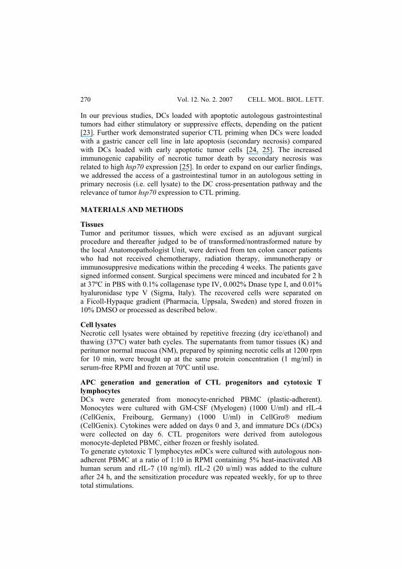
RESULTS Lysates from tumors induce only partial maturation of iDCs Day 6 iDCs display a high phagocytic activity which is lost once they complete their maturation under the influence of pro-inflammatory cytokines and become highly efficient APC. Necrotic cells produce a wide array of inflammatory cytokines. We therefore tested tumor and normal cell lysates for their ability to induce maturation of iDCs. We monitored the expression of CD14, a monocyte marker which is lost during the maturation of DCs [8], and the chemokine receptors CCR7 and CCR4, which are respectively up- and down-regulated during DC maturation [26]. Five tumors were subjected to flow cytometric analysis. The results from five experiments, each performed with a different patient (Tab. 1) show a donor-to-donor variability with a trend towards decreased expression of CD14 in autologous iDCs loaded with K (85.10 ± 43.03) compared with iDCs loaded with NM (108 ± 55.2), although this difference did not reach statistical significance. No changes were observed in the expression of CCR7 (46.4 ± 21.13 for DC+K vs 55.8 ± 14.4 for DC+NM) and CCR4 (17.3 ± 5.8 for DC+K vs 20.1 ± 5.4 for DC+NM). Tab. 1. The effect of lysates from tumor and normal mucosa cells on the maturation of iDCs. Five hundred thousand iDCs were loaded with 100 μg/ml cell lysate from tumor (K) or normal mucosa (NM), and after 48 h, analyzed by flow cytometry. The numbers refer to the mean fluorescence intensity (MFI) of cells labeled with mAbs directed against the indicated molecules.
CD14 CCR7 CCR4 DC+K DC+NM DC+K DC+NM DC+K DC+NM
EXP 1 54.49 101.72 34.89 56.59 13.82 16.76 EXP 2 149.47 150.58 83.71 67.88 21.26 25.56 EXP 3 76.88 169.33 42.21 54.56 25.41 24.79 EXP 4 103.43 89.12 36.92 67.38 14.37 20.43 EXP 5 41.25 29.15 34.00 32.48 11.82 12.91 MEAN 85.10 107.98 46.35 55.78 17.34 20.09 SD 43.03 55.20 21.13 14.37 5.75 5.36 T-Test p = 0.32 p = 0.32 p = 0.06 Wilcoxon p > 0.06 p > 0.06 p > 0.06
DCs loaded with tumor lysate induce cross-priming of tumor-specific CTL The efficiency of TA cross-presentation by DCs was studied in terms of the tumor-specific CTL response. In four out of six experiments, each performed with tissue from a different patient, exposure to the autologous tumor cells produced higher IFN-γ release in culture stimulated by DC+K compared to cultures stimulated by DC+NM (Fig. 1). Cultures displaying high tumor-specific
Vol. 12. No. 2. 2007 CELL. MOL. BIOL. LETT.
272
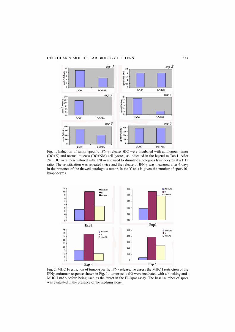
Fig. 1. Induction of tumor-specific IFN-γ release. iDC were incubated with autologous tumor (DC+K) and normal mucosa (DC+NM) cell lysates, as indicated in the legend to Tab.1. After 24 h DC were then matured with TNF-α and used to stimulate autologous lymphocytes at a 1:15 ratio. The sensitization was repeated twice and the release of IFN-γ was measured after 4 days in the presence of the thawed autologous tumor. In the Y axis is given the number of spots/105 lymphocytes.
Fig. 2. MHC I-restriction of tumor-specific IFNγ release. To assess the MHC I restriction of the IFNγ antitumor response shown in Fig. 1., tumor cells (K) were incubated with a blocking anti-MHC I mAb before being used as the target in the ELIspot assay. The basal number of spots was evaluated in the presence of the medium alone.
CELLULAR & MOLECULAR BIOLOGY LETTERS
273
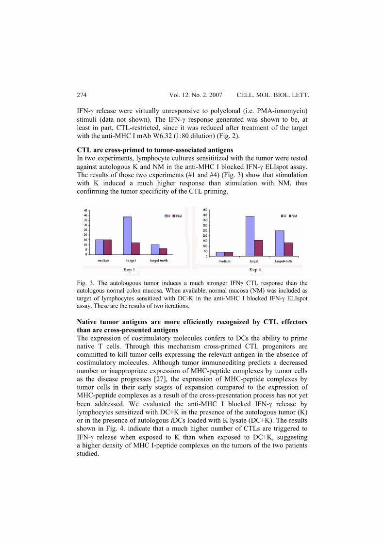
IFN-γ release were virtually unresponsive to polyclonal (i.e. PMA-ionomycin) stimuli (data not shown). The IFN-γ response generated was shown to be, at least in part, CTL-restricted, since it was reduced after treatment of the target with the anti-MHC I mAb W6.32 (1:80 dilution) (Fig. 2). CTL are cross-primed to tumor-associated antigens In two experiments, lymphocyte cultures sensititized with the tumor were tested against autologous K and NM in the anti-MHC I blocked IFN-γ ELIspot assay. The results of those two experiments (#1 and #4) (Fig. 3) show that stimulation with K induced a much higher response than stimulation with NM, thus confirming the tumor specificity of the CTL priming.
Fig. 3. The autolougous tumor induces a much stronger IFNγ CTL response than the autologous normal colon mucosa. When available, normal mucosa (NM) was included as target of lymphocytes sensitized with DC-K in the anti-MHC I blocked IFN-γ ELIspot assay. These are the results of two iterations. Native tumor antigens are more efficiently recognized by CTL effectors than are cross-presented antigens The expression of costimulatory molecules confers to DCs the ability to prime native T cells. Through this mechanism cross-primed CTL progenitors are committed to kill tumor cells expressing the relevant antigen in the absence of costimulatory molecules. Although tumor immunoediting predicts a decreased number or inappropriate expression of MHC-peptide complexes by tumor cells as the disease progresses [27], the expression of MHC-peptide complexes by tumor cells in their early stages of expansion compared to the expression of MHC-peptide complexes as a result of the cross-presentation process has not yet been addressed. We evaluated the anti-MHC I blocked IFN-γ release by lymphocytes sensitized with DC+K in the presence of the autologous tumor (K) or in the presence of autologous iDCs loaded with K lysate (DC+K). The results shown in Fig. 4. indicate that a much higher number of CTLs are triggered to IFN-γ release when exposed to K than when exposed to DC+K, suggesting a higher density of MHC I-peptide complexes on the tumors of the two patients studied.
Vol. 12. No. 2. 2007 CELL. MOL. BIOL. LETT.
274
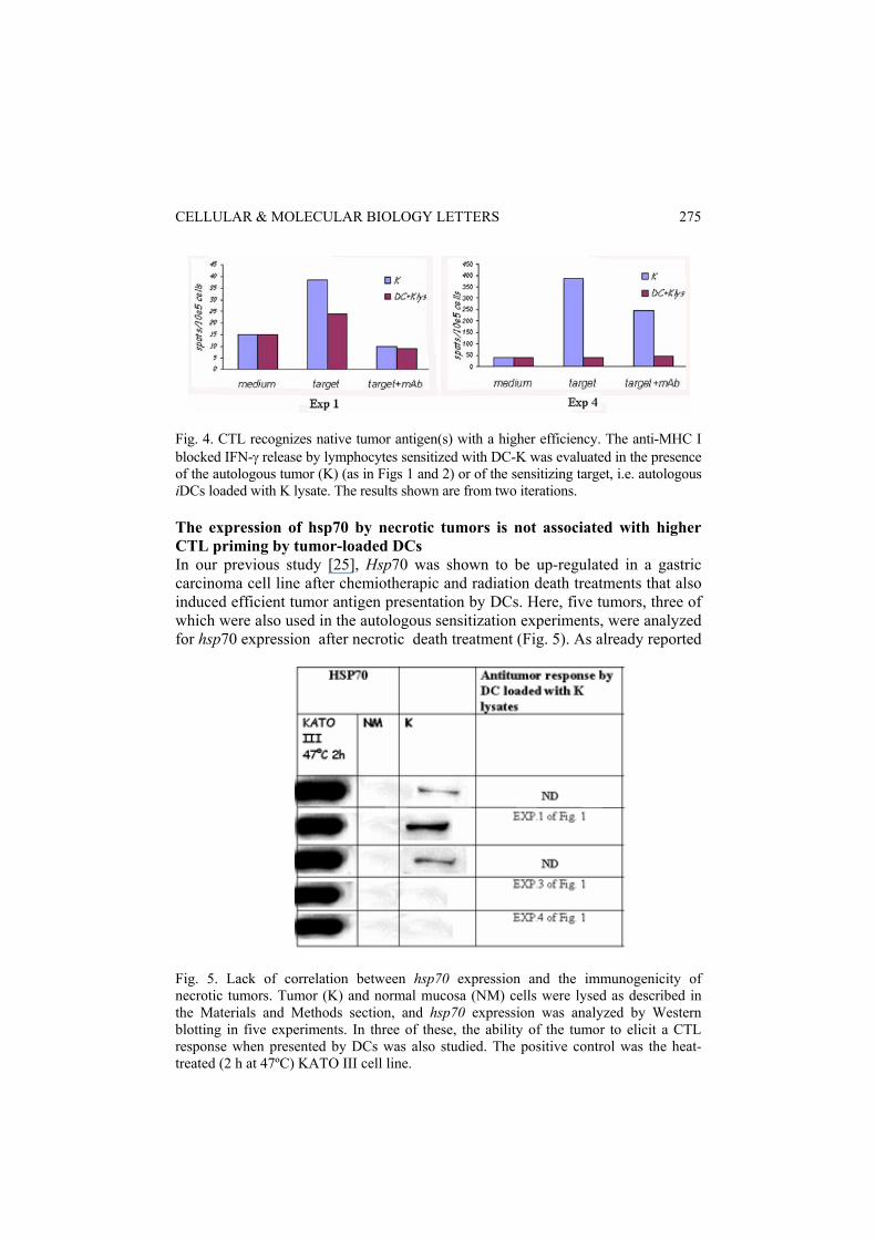
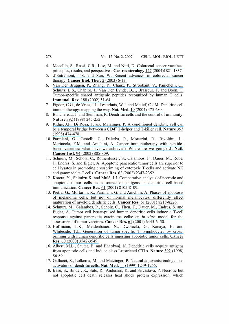
Fig. 4. CTL recognizes native tumor antigen(s) with a higher efficiency. The anti-MHC I blocked IFN-γ release by lymphocytes sensitized with DC-K was evaluated in the presence of the autologous tumor (K) (as in Figs 1 and 2) or of the sensitizing target, i.e. autologous iDCs loaded with K lysate. The results shown are from two iterations. The expression of hsp70 by necrotic tumors is not associated with higher CTL priming by tumor-loaded DCs In our previous study [25], Hsp70 was shown to be up-regulated in a gastric carcinoma cell line after chemiotherapic and radiation death treatments that also induced efficient tumor antigen presentation by DCs. Here, five tumors, three of which were also used in the autologous sensitization experiments, were analyzed for hsp70 expression after necrotic death treatment (Fig. 5). As already reported
Fig. 5. Lack of correlation between hsp70 expression and the immunogenicity of necrotic tumors. Tumor (K) and normal mucosa (NM) cells were lysed as described in the Materials and Methods section, and hsp70 expression was analyzed by Western blotting in five experiments. In three of these, the ability of the tumor to elicit a CTL response when presented by DCs was also studied. The positive control was the heat-treated (2 h at 47ºC) KATO III cell line.
CELLULAR & MOLECULAR BIOLOGY LETTERS
275

[25], treating the human gastric carcinoma cell line KATO III at 47ºC induced high levels of hsp70. The protein was expressed by three of the five tumors analyzed, but its expression was never observed on normal mucosa. Unexpectedly, tumors negative for hsp70 induced the highest number of CTL (Exps 3 and 4). DISCUSSION Danger signals such as hsps are believed to provide tumor cells with an increased ability to convey their antigens to the DC cross-presentation pathway. Indeed, in a previous study, increased cross-presentation of gastric and colon carcinoma antigens was associated with up-regulation of hsp70 [25]. Here, we addressed the expression of hsp70 by primary colon carcinoma cells and their ability to efficiently provide autologus DCs with TAs to cross-present DCs. The results show that: a) loading DCs with lysates from tumor specimens can lead to the generation of a specific T lymphocyte response against the autologous tumor; b) this response is only weakly elicited by lysates from the peritumoral normal mucosa and is not directed against these cells; c) in vitro acquisition of specific antitumor immunocompetence against the autologous tumor is accompanied by loss of responsiveness to aspecific (Ca ionophores, PKC activators) stimuli; and d) hsp70 is exclusively expressed on tumor tissues but its expression does not seem to be necessary for the generation of the antitumor reponse. Unlike in our earlier study, where tumor death was by post-apoptotic necrosis, the form of cell death employed here was primary necrosis induced by thawing and freezing. This procedure was chosen because it is currently used in clinical protocols to load DCs with TA, and hsp70 was tested in necrotic cells obtained by conventional lysis in the presence of protease inhibitors. DCs were exposed to cell lysates in their immature state, and characterized by preferential phagocytic activity. The uptake of tumor, but not of normal, cell lysate material by immature DCs produced a mixed maturational response. The monocyte-specific marker CD14, which is lost during DC maturation, showed a trend towards down-modulation. By contrast, the CCR7 molecule, which defines the acquisition of migratory capacity, was down-regulated on DC+K as compared to DC+NM, even if the difference was not statistically significant. The CCR4 molecules, which are down-modulated during DC maturation, were similarly expressed by DC+K and DC+NM. This data may indicate the release by the tumor of cytokines which exert distinct effects, stimulatory or inhibitory, on DC maturation. CTL priming was documented in four out of six experiments by the IFN-γ ELIspot assay. This assay is the gold standard in the immunological monitoring of DC-based cancer vaccines. Pre-treatment of the tumor with anti-MHC I blocking antibodies allows accurate discrimination between the MHC I-restricted and MHC I-unrestricted IFN-γ response, with the former being
Vol. 12. No. 2. 2007 CELL. MOL. BIOL. LETT.
276

attributable to bona fide CD8+ lymphocytes. MHC-restricted IFN-γ release was specific for cultures sensitized with DCs loaded with K, but not with NM. Moreover, lymphocytes from these cultures recognized the autologous tumor but not the corresponding normal mucosa in the IFN-γ ELIspot assay, thus confirming the tumor-specificity of the CTL response. As for the variability between donors observed in our study, it must be stressed that in an autologous setting, where lymphocytes were derived from a patient, the cell-demanding sensitization experiments can only be performed once. On the other hand, in our experience and that of other researchers, lymphocytes from different donors can respond very differently to the same allogeneic tumor cell line. Moreover, in our hands, the same donor routinely gives reproducible results when tested on several occasions against the same tumor cell line. On these bases, we feel that individual variability rather than experimental variability is a major contributor to the difference observed in our study. The same variability may explain the different efficacy of a given cancer vaccine when administered to different patients. The fact that DCs loaded with tumor lysate (Fig. 4) were much less efficient than the tumor itself when used as targets in the IFN-γ assay is of interest, since it shows that the master role of DCs is in the CD8 lymphocyte cross-priming process, as opposed to the sensitizing tumor, which is the physiological target for the post-vaccination, in vivo generated CTLs. The lack of correlation between hsp expression and immunogenicity found in our study may have several causes. As mentioned, in our previous study, a correlation between hsp70 and the CTL response was observed with tumor cells which were in secondary necrosis. The expression of hsp70 can be weaker after primary necrotic death, applied here to tumor cells. Other molecules associated with necrotic death which have recently been described to be released during necrosis and to be highly immunogenic [28, 29] could account for the CTL priming observed in exps 3 and 4, where no hsp70 production occurred. Further studies are needed to elucidate this open issue. Acknowledgements. Supported in part by MIUR, Regione Piemonte (Oncology project) and the CERMS/COES project funded by the “Compagnia di San Paolo/FIRMS”. REFERENCE 1. Jemal, A., Murray, T., Samuels, A., Ghafoor, A., Ward, E. and Thun, M.J.
Cancer statistics. CA Cancer J. Clin. 49 (1999) 202-219. 2. Midgley, R. and Kerr, D. Immunotherapy for colorectal cancer: a challenge
to clinical trial design. Lancet Oncol. 1 (2000) 159-168. 3. Coutinho, A.K. and Rocha Lima, C.M. Metastatic colorectal cancer: systemic
treatment in the new millennium. Cancer Control 10 (2003) 224-238.
CELLULAR & MOLECULAR BIOLOGY LETTERS
277
4. Mocellin, S., Rossi, C.R., Lise, M. and Nitti, D. Colorectal cancer vaccines: principles, results, and perspectives. Gastroenterology 127 (2004)1821-1837.
5. d’Entremont, T.S. and Sun, W. Recent advances in colorectal cancer therapy. Cancer Biol. Ther. 2 (2003) 6-13.
6. Van Der Bruggen, P., Zhang, Y., Chaux, P., Stroobant, V., Panichelli, C., Schultz, E.S., Chapiro, J., Van Den Eynde, B.J., Brasseur, F. and Boon, T. Tumor-specific shared antigenic peptides recognized by human T cells. Immunol. Rev. 188 (2002) 51-64.
7. Figdor, C.G., de Vries, I.J., Lesterhuis, W.J. and Melief, C.J.M. Dendritic cell immunotherapy: mapping the way. Nat. Med. 10 (2004) 475-480.
8. Banchereau, J. and Steinman, R. Dendritic cells and the control of immunity. Nature 392 (1998) 245-252.
9. Ridge, J.P., Di Rosa, F. and Matzinger, P. A conditioned dendritic cell can be a temporal bridge between a CD4+ T-helper and T-killer cell. Nature 393 (1998) 474-478.
10. Parmiani, G., Castelli, C., Dalerba, P., Mortarini, R., Rivoltini, L., Marincola, F.M. and Anichini, A. Cancer immunotherapy with peptide-based vaccines: what have we achieved? Where are we going? J. Natl. Cancer Inst. 94 (2002) 805-809.
11. Schnurr, M., Scholz, C., Rothenfusser, S., Galambos, P., Dauer, M., Robe, J., Endres, S. and Eigler, A. Apoptotic pancreatic tumor cells are superior to cell lysates in promoting crosspriming of cytotoxic T cells and activate NK and gammadelta T cells. Cancer Res. 62 (2002) 2347-2352.
12. Kotera, Y., Shimizu K. and Mulé, J.J. Comparative analysis of necrotic and apoptotic tumor cells as a source of antigens in dendritic cell-based immunization. Cancer Res. 61 (2001) 8105-8109.
13. Pietra, G., Mortarini, R., Parmiani, G. and Anichini, A. Phases of apoptosis of melanoma cells, but not of normal melanocytes, differently affect maturation of myeloid dendritic cells. Cancer Res. 61 (2001) 8218-8226.
14. Schnurr, M., Galambos, P., Scholz, C., Then, F., Dauer, M., Endres, S. and Eigler, A. Tumor cell lysate-pulsed human dendritic cells induce a T-cell response against pancreatic carcinoma cells: an in vitro model for the assessment of tumor vaccines. Cancer Res. 61 (2001) 6445-6450.
15. Hoffmann, T.K., Meidenbauer. N., Dworacki, G., Kanaya, H. and Whiteside, T.L. Generation of tumor-specific T lymphocytes by cross-priming with human dendritic cells ingesting apoptotic tumor cells. Cancer Res. 60 (2000) 3542-3549.
16. Albert, M.L., Sauter, B. and Bhardwaj, N. Dendritic cells acquire antigens from apoptotic cells and induce class I-restricted CTLs. Nature 392 (1998) 86-89.
17. Gallucci, S., Lolkema, M. and Matzinger, P. Natural adjuvants: endogenous activators of dendritic cells. Nat. Med. 11 (1999) 1249-1255.
18. Basu, S., Binder, R., Suto, R., Anderson, K. and Srivastava, P. Necrotic but not apoptotic cell death releases heat shock protein expression, which
Vol. 12. No. 2. 2007 CELL. MOL. BIOL. LETT.
278
deliver a partial maturation signal to dendritic cells and activate the NK-kB pathway. Int. Immunol. 12 (2000) 1539-1546.
19. Melcher, A., Todryk, S., Hardwick, N., Ford, M., Jacobson, M. and Vile, R.G. Tumor immunogenicity is determined by the mechanism of cell death via induction of heat shock protein expression. Nat. Med. 4 (1998) 581-587.
20. Hongzhen S., Tinghua C., Connolly, J.E., Monnet, L., Bennett, L., Chapel, S., Bagnis, C., Mannoni, P., Davoust, J., Palucka, A.K. and Banchereau, J. Hyperthermia Enhances CTL Cross-Priming. J. Immunol. 176 (2006) 2134-2141.
21. Milani, V., Noessner, E., Ghose, S., Kuppner, M., Ahrens, B., Scharner, A., Gastpar, R. and Issels, R.D. Heat shock protein 70: role in antigen presentation and immune stimulation. Int. J. Hyperthermia 18 (2002) 563-575.
22. Kuppner, M., Gastpar, R., Gelwer, S., Noessner, E., Ochmann, O., Scharner, A. and Issels, R.D. The role of heath shock protein (hsp70) in dendritic cell maturation: hsp70 induces the maturation of immature dendritic cells but reduces DC differentiation from monocytes precursors. J. Eur. Immunol. 31 (2001) 1602-1609.
23. Galetto, A., Buttiglieri, S., Forno, S., Moro, F., Mussa, A. and Matera, L. Drug- and cell-mediated antitumor cytotoxicities modulate cross-presentation of tumor antigens by myeloid dendritic cells. Anti-Cancer Drugs 14 (2003) 883-843.
24. Galetto, A., Contarini, M., Sapino, A., Cassoni, P., Consalvo, E., Forno, S., Pezzi, C., Barnaba, V., Mussa, A. and Matera, L. Ex-vivo host response to gastrointestinal cancer cells presented by autologous dendritic cells. J. Surgical. Res. 100 (2001) 32-38.
25. Buttiglieri, S., Galetto, A., Forno, S., De Andrea, M. and Matera, L. Influence of drug-induced apoptotic death on processing and presentation of tumor antigens by dendritic cells. Int. J. Cancer 106 (2003) 516-520.
26. Luft, T., Jefford, M., Luetjens, P., Toy, T., Hochrein, H., Masterman, K.A., Maliszewski, C., Shortman, K., Cebon, J. and Maraskovsky, E. Functionally distinct dendritic cell (DC) populations induced by physiologic stimuli: prostaglandin E(2) regulates the migratory capacity of specific DC subsets. Blood 100 (2002) 1362-1372.
27. Chang, C.C. and Ferrone, S. Immune selective pressure and HLA class I antigen defects in malignant lesions. Cancer. Immunol. Immunother. (2006) Epub ahead of print.
28. Vakkila, J. and Lotze, M.T. Inflammation and necrosis promote tumour growth. Nat. Rev. Immunol. 8 (2004) 641-648.
29. Rovere-Querini, P., Capobianco, A., Scaffidi, P., Valentinis, B., Catalanotti, F., Giazzon, M., Dumitriu, I.E., Muller, S., Iannacone, M., Traversari, C., Bianchi, M.E. and Manfredi, A.A. HMGB1 is an endogenous immune adjuvant released by necrotic cells. EMBO Rep. 8 (2004) 825-830.
CELLULAR & MOLECULAR BIOLOGY LETTERS
279