Toxic potential of synthesized graphene zinc oxide nanocomposite in the third instar larvae of...
10
Research Article Toxic Potential of Synthesized Graphene Zinc Oxide Nanocomposite in the Third Instar Larvae of Transgenic Drosophila melanogaster (hsp70-lacZ)Bg 9 Yasir Hasan Siddique, 1 Wasi Khan, 2 Saba Khanam, 1 Smita Jyoti, 1 Falaq Naz, 1 Rahul, 1 Braj Raj Singh, 2 and Alim H. Naqvi 2 1 Drosophila Transgenic Laboratory, Section of Genetics, Department of Zoology, Faculty of Life Sciences, Aligarh Muslim University, Aligarh, Uttar Pradesh 202002, India 2 Centre of Excellence in Materials Sciences (Nanomaterials), Department of Applied Physics, Z.H. College of Engineering & Technology, Aligarh Muslim University, Aligarh, Uttar Pradesh 202002, India Correspondence should be addressed to Yasir Hasan Siddique; yasir hasansiddique@rediffmail.com Received 12 February 2014; Revised 15 May 2014; Accepted 18 May 2014; Published 15 June 2014 Academic Editor: Mohammad Owais Copyright © 2014 Yasir Hasan Siddique et al. is is an open access article distributed under the Creative Commons Attribution License, which permits unrestricted use, distribution, and reproduction in any medium, provided the original work is properly cited. In the present study the graphene zinc oxide nanocomposite (GZNC) was synthesized, characterized, and evaluated for its toxic potential on third instar larvae of transgenic Drosophila melanogaster (hsp70-lacZ)Bg 9 . e synthesized GZNC was characterized by X-ray diffraction (XRD), Fourier transform infrared spectroscopy (FTIR), thermogravimetric analysis (TGA), scanning electron microscopy (SEM), and transmission electron microscopy (TEM). e GZNC in 0.1% dimethyl sulphoxide (DMSO) was sonicated for 10 minutes and the final concentrations 0.033, 0.099, 0.199, and 3.996 g/L of diet were established. e third instar larvae were allowed to feed on it separately for 24 and 48 hr. e hsp70 expression was measured by o-nitrophenyl--D-galactopyranoside assay, tissue damage was measured by trypan blue exclusion test, and -galactosidase activity was monitored by in situ histochemical -galactosidase staining. Oxidative stress was monitored by performing lipid peroxidation assay and total protein estimation. Ethidium bromide/acridine orange staining was performed on midgut cells for apoptotic index and the comet assay was performed for the DNA damage. e results of the present study showed that the exposure of 0.199 and 3.996 g/L of GZNC was toxic for both 24 hr and 48 hr of exposure. e doses of 0.033 g/L and 0.099 of GZNC showed no toxic effects on its exposure to the third instar larvae for 24 hr as well as 48 hr of duration. 1. Introduction Nanoparticles (NPs) have novel properties, large specific surface, and high reaction activity [1, 2]. Due to the rapid development of nanotechnology, nanomaterials with various shapes and diameters have been prepared for the use in some industrial products and commodities [3]. Besides having applications in drug delivery, cell imaging, and cancer ther- apy, metal oxide nanoparticles have been manufactured for both industrial and household applications [3]. It has been reported that zinc oxide nanoparticles have negative impacts on the survival and growth of organisms [4]. e physical parameters of nanoparticles can affect their nonspecific uptake in cells, with potential to induce cellular responses [5]. Graphene is an allotrope of carbon and is being used in nanocomposites due to its intrinsic properties [6]. It is typically free of impurities as compared to carbon nanotubes, having an advantage of being used in the construction of reli- able sensors as well as storage devices [6, 7]. Due to the wide range of potential applications and novel properties, graphitic nanomaterials such as carbon nanotubes, fullerenes, and more recently graphene have gained a great deal of interest in the scientific community [8]. e biological applications and potential adverse effects of graphene still remain to be clear and need a detailed study that would provide information in this regard. However, till date the reports on the toxic eval- uation of its nanocomposites are warranted. In the present study the graphene zinc oxide nanocomposite (GZNC) was Hindawi Publishing Corporation BioMed Research International Volume 2014, Article ID 382124, 10 pages http://dx.doi.org/10.1155/2014/382124
-
Upload
independent -
Category
Documents
-
view
4 -
download
0
Transcript of Toxic potential of synthesized graphene zinc oxide nanocomposite in the third instar larvae of...

Research ArticleToxic Potential of Synthesized Graphene Zinc OxideNanocomposite in the Third Instar Larvae of TransgenicDrosophila melanogaster (hsp70-lacZ)Bg9
Yasir Hasan Siddique,1 Wasi Khan,2 Saba Khanam,1 Smita Jyoti,1 Falaq Naz,1
Rahul,1 Braj Raj Singh,2 and Alim H. Naqvi2
1 Drosophila Transgenic Laboratory, Section of Genetics, Department of Zoology, Faculty of Life Sciences, Aligarh Muslim University,Aligarh, Uttar Pradesh 202002, India
2 Centre of Excellence inMaterials Sciences (Nanomaterials), Department of Applied Physics, Z.H. College of Engineering&Technology,Aligarh Muslim University, Aligarh, Uttar Pradesh 202002, India
Correspondence should be addressed to Yasir Hasan Siddique; yasir [email protected]
Received 12 February 2014; Revised 15 May 2014; Accepted 18 May 2014; Published 15 June 2014
Academic Editor: Mohammad Owais
Copyright © 2014 Yasir Hasan Siddique et al. This is an open access article distributed under the Creative Commons AttributionLicense, which permits unrestricted use, distribution, and reproduction in any medium, provided the original work is properlycited.
In the present study the graphene zinc oxide nanocomposite (GZNC) was synthesized, characterized, and evaluated for its toxicpotential on third instar larvae of transgenic Drosophila melanogaster (hsp70-lacZ)Bg9. The synthesized GZNC was characterizedby X-ray diffraction (XRD), Fourier transform infrared spectroscopy (FTIR), thermogravimetric analysis (TGA), scanning electronmicroscopy (SEM), and transmission electron microscopy (TEM).The GZNC in 0.1% dimethyl sulphoxide (DMSO) was sonicatedfor 10 minutes and the final concentrations 0.033, 0.099, 0.199, and 3.996𝜇g/𝜇L of diet were established.The third instar larvae wereallowed to feed on it separately for 24 and 48 hr.The hsp70 expressionwasmeasured by o-nitrophenyl-𝛽-D-galactopyranoside assay,tissue damage was measured by trypan blue exclusion test, and 𝛽-galactosidase activity was monitored by in situ histochemical𝛽-galactosidase staining. Oxidative stress was monitored by performing lipid peroxidation assay and total protein estimation.Ethidium bromide/acridine orange staining was performed onmidgut cells for apoptotic index and the comet assay was performedfor the DNA damage. The results of the present study showed that the exposure of 0.199 and 3.996𝜇g/𝜇L of GZNC was toxic forboth 24 hr and 48 hr of exposure. The doses of 0.033𝜇g/𝜇L and 0.099 of GZNC showed no toxic effects on its exposure to the thirdinstar larvae for 24 hr as well as 48 hr of duration.
1. Introduction
Nanoparticles (NPs) have novel properties, large specificsurface, and high reaction activity [1, 2]. Due to the rapiddevelopment of nanotechnology, nanomaterials with variousshapes and diameters have been prepared for the use in someindustrial products and commodities [3]. Besides havingapplications in drug delivery, cell imaging, and cancer ther-apy, metal oxide nanoparticles have been manufactured forboth industrial and household applications [3]. It has beenreported that zinc oxide nanoparticles have negative impactson the survival and growth of organisms [4]. The physicalparameters of nanoparticles can affect their nonspecificuptake in cells, with potential to induce cellular responses
[5]. Graphene is an allotrope of carbon and is being used innanocomposites due to its intrinsic properties [6]. It istypically free of impurities as compared to carbon nanotubes,having an advantage of being used in the construction of reli-able sensors as well as storage devices [6, 7]. Due to the widerange of potential applications and novel properties, graphiticnanomaterials such as carbon nanotubes, fullerenes, andmore recently graphene have gained a great deal of interest inthe scientific community [8]. The biological applications andpotential adverse effects of graphene still remain to be clearand need a detailed study that would provide informationin this regard. However, till date the reports on the toxic eval-uation of its nanocomposites are warranted. In the presentstudy the graphene zinc oxide nanocomposite (GZNC) was
Hindawi Publishing CorporationBioMed Research InternationalVolume 2014, Article ID 382124, 10 pageshttp://dx.doi.org/10.1155/2014/382124

2 BioMed Research International
synthesized, characterized, and evaluated for its toxicpotential on third instar larvae of transgenic Drosophilamelanogaster (hsp70-lacZ)Bg9 as a model.
2. Materials and Methods
2.1. Synthesis of Graphene Zinc Oxide Nanocomposites.Graphene oxide (GO) was synthesized from 2 g of naturalgraphite powder by using amodifiedHummer andOffeman’smethod [9].The synthesized GOwas centrifuged andwashedsuccessively with 4% HCl until it reached neutral pH value.The obtained GO was dried in vacuum oven at 70∘C forovernight and used for the synthesis of GZNC. For the syn-thesis of GZNC, 1 g of GO was dissolved in 100mL of MilliQwater using ultrasonicator. The zinc nitrate hexahydrate(25mM)was added into the dissolvedGO solution under vig-orous stirring and pH was increased to 11 using NaOH solu-tion.The reaction mixture was stirred for 1 hour at 150∘C andobtainedGZNCwas centrifuged.The synthesizedGZNCwaswashed several times with water and ethanol thoroughly.Thesynthesized GZNC was dried and stored for further study.
2.2. Characterizations of GZNC. TheX-ray diffraction (XRD)pattern of powder sample of GZNC was recorded on Mini-Flex II benchtop XRD system (Rigaku Corporation, Tokyo,Japan) operating at 30 kV and a current of 15mA withCu K𝛼 radiation (𝜆 = 1.54 A). The diffracted X-rays wererecorded from 20∘ to 80∘ at 2𝜃 angles. For the FTIR spec-troscopic measurement of GZNC powder was mixed withspectroscopic grade potassium bromide (KBr) in the ratio of1 : 100 and spectra were recorded in the range of 400–4000wavenumber (cm−1) on Perkin Elmer FTIR Spectrum BX(PerkinElmer Life and Analytical Sciences, CT, USA) inthe diffuse reflectance mode at a resolution of 4 cm−1 inKBr pellets. The synthesis of GZNC in ethanol solution wasmonitored bymeasuring the absorbance (A) usingUV-visiblespectrophotometer (Perkin Elmer Life and Analytical Sci-ences, CT, USA) in the wavelength range of 200–800 nm.The thermal stability of the GZNC was investigated by ther-mogravimetric analysis (TGA) at a heating rate of 10∘C/minunder nitrogen atmosphere. The microstructure and mor-phology analyses of samplewere done using a JEOL transmis-sion electron microscope (TEM) (JEM-2010) and scanningelectron microscope (SEM) (JSM-6510LV) equipped with anenergy dispersive spectrometer (EDS).
2.3. Fly Strain. A transgenicDrosophila melanogaster (hsp70-lacZ)Bg9 line that expresses bacterial 𝛽-galactosidase as aresponse to stress was used in the present study [10].The fliesand larvae were cultured on standard Drosophila food con-taining agar, corn meal, sugar, and yeast at 24 ± 1∘C [11, 12].
2.4. Experimental Design. GZNC in 0.1% DMSO was son-icated for 10min and the final concentrations 0.033, 0.099,0.199, and 3.996 𝜇g/𝜇L of diet were established. The larvaewere allowed to feed on diet separately for 24 and 48 hr.Untreated and negative control (0.1% DMSO) were also runsimultaneously.
2.5. Soluble o-Nitrophenyl-𝛽-D-Galactopyranoside Assay.The expression of hsp70 provides a measurement ofcytotoxicity [13, 14].Themethod described by Nazir et al. [11]was used in this study. After washing in phosphate buffer,larvae were placed in a microcentrifuge tube (20 larvae/tube;five replicates/group), permeabilized for 10min by acetone,and incubated overnight at 37∘C in 600𝜇L of ONPG stainingbuffer. Following incubation, the reaction was stopped byadding 300 𝜇L of Na
2CO3. The extent of the reaction was
quantified by measuring absorbance at 420 nm.
2.6. Trypan Blue Exclusion Test. The extent of tissue damagein larvae caused by the exposure to different concentrationsof GZNC was assayed by a dye exclusion test [11, 15]. Briefly,the internal tissues of larvae were explanted in a drop of Pole’ssalt solution (PSS), washed in phosphate buffer saline (PBS),stained in trypan blue (0.2mg/mL in PBS) for 30min, washedthoroughly in PBS, and scored immediately for dark bluestaining. About 50 larvae per treatment (10 larvae per dose; 5replicates per group) were scored for the trypan blue stainingon an average composite index per larvae: no color = 0; anyblue = 1; darkly stained = 2; large patches of darkly stainedcells = 3; or complete staining of most cells in the tissue = 4[15].
2.7. In SituHistochemical 𝛽-Galactosidase Activity. The larvae(10 larvae/treatment; 5 replicates/group)were dissected out inPSS and X-gal staining was performed using the method asdescribed by Chowdhuri et al. [13]. The tissue explants werefixed in 2.5% glutaraldehyde, washed in 50mM sodiumphosphate buffer (pH 8.0), and stained overnight in X-galstaining solution at 37∘C in the dark.
2.8. Preparation of Larval Homogenate for Lipid Peroxida-tion Assay and Total Protein Content. The larvae (10 lar-vae/experiment; 5 replicates/group) were homogenized in1mL of cold homogenizing buffer (0.1M phosphate buffercontaining 0.15MKCl; pH 7.4). The supernatant after cen-trifugation at 9000 g was used for estimating lipid peroxida-tion and total protein content.
2.9. Lipid Peroxidation Assay. Lipid peroxidation assay wasperformed using 1,1,3,3-tetramethoxypropane as a standardaccording to the method described by Siddique et al. [16, 17].
2.10. Protein Estimation. Estimation of protein level in all thetreated groups aswell as control groupswas done according tothe method of Bradford [18], using bovine serum albumin(BSA) as a standard.
2.11. Assay to Detect Apoptosis. The apoptotic cells were ana-lyzed by stainingwith an ethidiumbromide (EB) and acridineorange (AO) staining according to the procedure described inour earlier published work [17]. About 100 cells were scoredper treatment (5 replicates/group) for estimating the apop-totic index and expressed in percentages [19].
2.12. Analysis of DNA Damage by Comet Assay. The cometassay was performed according to Mukhopadhyay et al. [20].

BioMed Research International 3
The midguts from 20 larvae were explanted in PSS. PSS inmicrocentrifuge tube was replaced by 300 𝜇L of collagenase(0.5mg/mL in PBS, pH 7.4) and kept for 15min at 25∘C. Thecell suspension was prepared by washing three times in PBSand finally the cells were suspended in 80 𝜇L of PBS. The cellviability was checked by performing trypan blue assay beforebeginning the experiment and the assay was performedaccording to the procedure described in our earlier publishedwork [17, 21]. Each experiment was performed in triplicateand the slides were prepared in duplicate. Twenty-five cellsper slide were randomly captured at a constant depth of thegel, and mean tail length was calculated to measure DNAdamage by using Comet Score 1.5 Software (Comet Score v1.5Software, TriTek Corporation, Sumerduck).
2.13. Statistical Analysis. Student’s 𝑡-test and regression anal-ysis were performed by using commercial software Statisticafrom Stat-Soft Inc.
3. Results
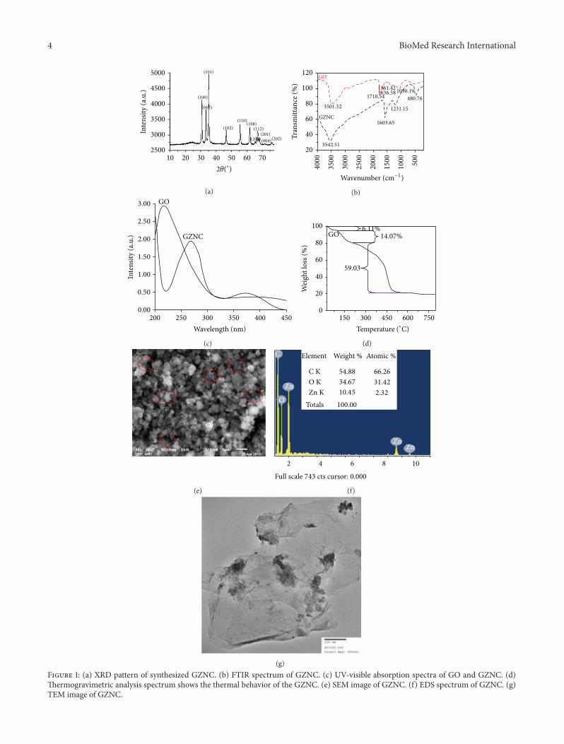
3.1. X-Ray Diffraction (XRD). X-ray diffraction (XRD) mea-surementwas employed to investigate the phase and structureof the synthesized GZNC sample. The XRD pattern ofthe GZNC is shown in Figure 1(a); there were eleven maindiffraction peaks located at 2𝜃 = 31.33∘, 34.0∘, 35.34∘, 46.43∘,55.68∘, 61.91∘, 65.46∘, 67.18∘, 68.14∘, 71.67∘, and 76.36∘ whichcorrespond to the crystal planes (100), (002), (101), (102),(110), (103), (200), (112), (201), (004), and (202) of thehexagonal wurtzite structure of ZnO reported in JCDDScard (number 36–1451, 𝑎 = 3.249 A, and 𝑐 = 5.206 A),respectively.TheXRD data of GZNC indicated the absence ofany other impurities, which reflected its high quality. Theaverage crystallite size (𝐷) of ZnO nanoparticles was calcu-lated using the Debye-Scherrer formula:
𝐷 =𝑘𝜆
𝛽 cos 𝜃, (1)
where 𝑘 = 0.9 is the shape factor, 𝜆 is the X-ray wavelength ofCu K𝛼 radiation (1.54 A), 𝜃 is the Bragg diffraction angle, and𝛽 is the full width at half maximum height (FWHM) of the(111) plane diffraction peak.The calculated average crystallitesize was found to be ∼9 nm. Further the main characteristicdiffraction peak was observed in the synthesized GZNC at35∘, which corresponds to (101) reflection plane of graphenewith basal spacing of 𝑑
022= 3.62 A. The XRD data clearly
indicates the successful synthesis of GZNC in this study.Figure 1(b) shows the FTIR spectrum of synthesized GZNC.The absence of the characteristic peaks of carboxyl group at1710 cm−1, hydroxide group at 1361 cm−1, and C–O (alkoxy)groups at 1090 cm−1 indicates that most oxygen-containingfunctional groups in the GO were removed. The spectrum ofthe GZNC shows an absorption band at 1603 cm−1, C=Cstretching, indicating the restoration of the graphene networkon reduction. The presence of the absorption band at480 cm−1 in GZNC is identified as ZnO nanoparticles(NPs).
3.2. Optical Characteristics. The UV-visible spectra of GOand GZNC are shown in Figure 1(c). The GO sample showedthe absorption peak at ∼220 nm and a shoulder at ∼280 nm.The peak at 220 nm is assigned to the pi to anti-pi (𝜋 → 𝜋∗)transition of the aromatic C–C bonds and the shoulder at278 nm is assigned to the 𝑛 to anti-pi (𝑛 → 𝜋∗) transitions ofthe C=O bonds. The UV spectrum of GZNC showed twodistinct peaks at ∼270 and ∼370 nmwhich corresponds to theexcitation of the 𝜋-plasmon of the graphitic structure andZnO NPs characteristics, respectively. The graphene absorp-tion peak is entirely different fromGOpeak and red shifted to270 nmwhich indicates the fully reduced graphene fromGO.The absorption data confirmed again the successful synthesisof GZNC in this study.
3.3. Thermal Characteristics. The TGA curve of GZNC isshown in Figure 1(d). The weight loss of 6.11% occurring atabout 63∘C is associated with adsorbed water. Pyrolysis of thelabile oxygen-containing functional groups at about 200∘Caccounts for 14.07% of weight loss. The thermal decompo-sition observed in the temperature range 200–500∘C with59.03% of weight loss is attributed to the pyrolysis of thecarbon skeleton. These results illustrate that GZNC has aremarkable thermal stability (Figure 1(d)).
Figure 1(e) shows the SEM image of GZNC. It can beseen that most of the graphene nanosheets are curled andentangled together. The results reveal that the presence ofgraphene sheet is uniformly distributed in the sample. More-over ZnO NPs were also clearly shown in the low dimensionand well dispersed all over the sample. The EDS spectrumdemonstrates that ZnOcontents are uniformly doped into thegraphene matrix. An oxygen peak at about 0.52 keV, Zn sig-nals at about 1 keV and 8.6 keV, and presence of the graphene(i.e., carbon) at 0.25 keV were observed in the EDS spectrumas displayed in Figure 1(f). These results were consistent withthe analysis of the XRD data.
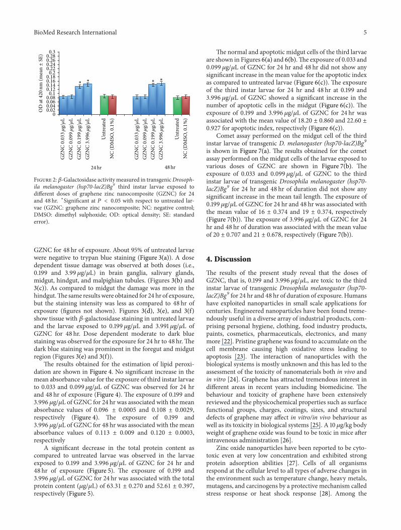
Transmission electron microscopy (TEM) analysis wasperformed on GZNC sample to determine its features innanometer domain as shown in Figure 1(g). It can be clearlyseen that the graphene nanosheets were well decorated byZnO nanoparticles, which densely and evenly deposited onboth sides of these sheets to form a sandwich-like compositestructure. Moreover, almost no ZnO nanoparticles werefound outside of the graphene nanosheets.This indicates thatthe combination between ZnO nanoparticles and graphenenanosheets was almost perfect.The observed sheets were fewlayers and entangled with each other. Meanwhile, the Znnanoparticles were identified in the range of ∼9 nm andattached on both sides of graphene sheets with a nonuniformdistribution on the sheet. The exposure of third instar larvaeof transgenic Drosophila melanogaster (hsp70-lacZ)Bg9 to0.033 and 0.099 𝜇g/𝜇L of GZNC for 24 and 48 hr did notshowany significant increase in the activity of𝛽-galactosidase(Figure 2). However the larvae exposed to 0.199 and3.996 𝜇g/𝜇L of GZNC showed a dose and time dependentsignificant increase in the expression of 𝛽-galactosidase(Figure 2).
Figures 3(a), 3(b), and 3(c) show the trypan blue stainingfor the third instar larvae exposed to 0.199 and 3.996 𝜇g/𝜇L of
4 BioMed Research International
(101)
(100)
(002)
(102)
(110)(108)
(200)
(112)
(201)
(004)(202)
5000
4500
4000
3500
3000
2500In
tens
ity (a
.u.)
10 20 30 40 50 60 70
2𝜃(∘)
(a)
GO
3501.32
GZNC
3542.51
1361.421636.58
1710.541090.19
480.76
1231.15
1603.65
120
100
80
60
40
20
Tran
smitt
ance
(%)
4000
3500
3000
2500
2000
1500
1000
500
Wavenumber (cm−1)
(b)3.00
2.50
2.00
1.50
1.00
0.50
0.00
Inte
nsity
(a.u
.) GZNC
200 250 300 350 400 450
Wavelength (nm)
GO
(c)
150 300 450 600 7500
20
40
60
80
100
Wei
ght l
oss (
%)
6.11% 14.07%
59.03
GO
Temperature (∘C)
(d)
(e)
C
O
Zn
2 4 6 8 10
Full scale 743 cts cursor: 0.000
Element Weight % Atomic %
C KO KZn K
Totals
54.88
34.67
10.45
100.00
66.26
31.42
2.32
ZnZn
(f)
(g)Figure 1: (a) XRD pattern of synthesized GZNC. (b) FTIR spectrum of GZNC. (c) UV-visible absorption spectra of GO and GZNC. (d)Thermogravimetric analysis spectrum shows the thermal behavior of the GZNC. (e) SEM image of GZNC. (f) EDS spectrum of GZNC. (g)TEM image of GZNC.
BioMed Research International 5
0.30.280.260.240.220.20.180.160.140.120.10.080.060.040.02
0
OD
at420
nm (m
ean±
SE)
GZN
C0.033𝜇
g/𝜇
LG
ZNC0.099𝜇
g/𝜇
LG
ZNC0.199𝜇
g/𝜇
LG
ZNC3.996𝜇
g/𝜇
L
Unt
reat
edN
C (D
MSO
,0.1
%)
24hr 48hr
GZN
C0.033𝜇
g/𝜇
LG
ZNC0.099𝜇
g/𝜇
LG
ZNC0.199𝜇
g/𝜇
LG
ZNC3.996𝜇
g/𝜇
L
Unt
reat
edN
C (D
MSO
,0.1
%)
∗ ∗ ∗ ∗
Figure 2: 𝛽-Galactosidase activity measured in transgenicDrosoph-ila melanogaster (hsp70-lacZ)Bg9 third instar larvae exposed todifferent doses of graphene zinc nanocomposite (GZNC) for 24and 48 hr. ∗Significant at 𝑃 < 0.05 with respect to untreated lar-vae (GZNC: graphene zinc nanocomposite; NC: negative control;DMSO: dimethyl sulphoxide; OD: optical density; SE: standarderror).
GZNC for 48 hr of exposure. About 95% of untreated larvaewere negative to trypan blue staining (Figure 3(a)). A dosedependent tissue damage was observed at both doses (i.e.,0.199 and 3.99 𝜇g/𝜇L) in brain ganglia, salivary glands,midgut, hindgut, and malpighian tubules. (Figures 3(b) and3(c)). As compared to midgut the damage was more in thehindgut.The same resultswere obtained for 24 hr of exposure,but the staining intensity was less as compared to 48 hr ofexposure (figures not shown). Figures 3(d), 3(e), and 3(f)show tissue with 𝛽-galactosidase staining in untreated larvaeand the larvae exposed to 0.199 𝜇g/𝜇L and 3.991 𝜇g/𝜇L ofGZNC for 48 hr. Dose dependent moderate to dark bluestaining was observed for the exposure for 24 hr to 48 hr.Thedark blue staining was prominent in the foregut and midgutregion (Figures 3(e) and 3(f)).
The results obtained for the estimation of lipid peroxi-dation are shown in Figure 4. No significant increase in themean absorbance value for the exposure of third instar larvaeto 0.033 and 0.099𝜇g/𝜇L of GZNC was observed for 24 hrand 48 hr of exposure (Figure 4). The exposure of 0.199 and3.996 𝜇g/𝜇L of GZNC for 24 hr was associated with the meanabsorbance values of 0.096 ± 0.0005 and 0.108 ± 0.0029,respectively (Figure 4). The exposure of 0.199 and3.996 𝜇g/𝜇L of GZNC for 48 hr was associated with the meanabsorbance values of 0.113 ± 0.009 and 0.120 ± 0.0003,respectively
A significant decrease in the total protein content ascompared to untreated larvae was observed in the larvaeexposed to 0.199 and 3.996 𝜇g/𝜇L of GZNC for 24 hr and48 hr of exposure (Figure 5). The exposure of 0.199 and3.996 𝜇g/𝜇L of GZNC for 24 hr was associated with the totalprotein content (𝜇g/𝜇L) of 63.31 ± 0.270 and 52.61 ± 0.397,respectively (Figure 5).
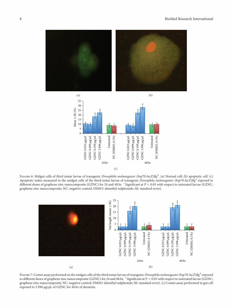
The normal and apoptotic midgut cells of the third larvaeare shown in Figures 6(a) and 6(b).The exposure of 0.033 and0.099 𝜇g/𝜇L of GZNC for 24 hr and 48 hr did not show anysignificant increase in the mean value for the apoptotic indexas compared to untreated larvae (Figure 6(c)). The exposureof the third instar larvae for 24 hr and 48 hr at 0.199 and3.996 𝜇g/𝜇L of GZNC showed a significant increase in thenumber of apoptotic cells in the midgut (Figure 6(c)). Theexposure of 0.199 and 3.996 𝜇g/𝜇L of GZNC for 24 hr wasassociated with the mean value of 18.20 ± 0.860 and 22.60 ±0.927 for apoptotic index, respectively (Figure 6(c)).
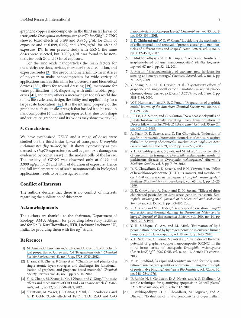
Comet assay performed on the midgut cell of the thirdinstar larvae of transgenic D. melanogaster (hsp70-lacZ)Bg9is shown in Figure 7(a). The results obtained for the cometassay performed on the midgut cells of the larvae exposed tovarious doses of GZNC are shown in Figure 7(b). Theexposure of 0.033 and 0.099 𝜇g/𝜇L of GZNC to the thirdinstar larvae of transgenic Drosophila melanogaster (hsp70-lacZ)Bg9 for 24 hr and 48 hr of duration did not show anysignificant increase in the mean tail length. The exposure of0.199 𝜇g/𝜇L of GZNC for 24 hr and 48 hr was associated withthe mean value of 16 ± 0.374 and 19 ± 0.374, respectively(Figure 7(b)). The exposure of 3.996 𝜇g/𝜇L of GZNC for 24hr and 48 hr of duration was associated with the mean valueof 20 ± 0.707 and 21 ± 0.678, respectively (Figure 7(b)).
4. Discussion
The results of the present study reveal that the doses ofGZNC, that is, 0.199 and 3.996 𝜇g/𝜇L, are toxic to the thirdinstar larvae of transgenic Drosophila melanogaster (hsp70-lacZ)Bg9 for 24 hr and 48 hr of duration of exposure. Humanshave exploited nanoparticles in small scale applications forcenturies. Engineered nanoparticles have been found treme-ndously useful in a diverse array of industrial products, com-prising personal hygiene, clothing, food industry products,paints, cosmetics, pharmaceuticals, electronics, and manymore [22]. Pristine graphene was found to accumulate on thecell membrane causing high oxidative stress leading toapoptosis [23]. The interaction of nanoparticles with thebiological systems is mostly unknown and this has led to theassessment of the toxicity of nanomaterials both in vivo andin vitro [24]. Graphene has attracted tremendous interest indifferent areas in recent years including biomedicine. Thebehaviour and toxicity of graphene have been extensivelyreviewed and the physicochemical properties such as surfacefunctional groups, charges, coatings, sizes, and structuraldefects of graphene may affect in vitro/in vivo behaviour aswell as its toxicity in biological systems [25]. A 10 𝜇g/kg bodyweight of graphene oxide was found to be toxic in mice afterintravenous administration [26].
Zinc oxide nanoparticles have been reported to be cyto-toxic even at very low concentration and exhibited strongprotein adsorption abilities [27]. Cells of all organismsrespond at the cellular level to all types of adverse changes inthe environment such as temperature change, heavy metals,mutagens, and carcinogens by a protective mechanism calledstress response or heat shock response [28]. Among the

6 BioMed Research International
FG
PV
SG
BGMG
MT
(a)
BG
MG
MT
(b)
MT
BGSG
FG
MG
HG
(c)
FG
PV
SG
BG
MG
MT
HGGC
(d)
FG
PV
BG
MG
MT
HG
(e)
FG
PV
SG
BG
MG
MT
HG
(f)
Figure 3: Trypan blue (a–c) and 𝛽-galactosidase (d–f) staining in the tissues of third instar larvae of transgenic Drosophila melanogaster(hsp70-lacZ)Bg9 for untreated larvae (a, d) and the larvae exposed to different doses of graphene zinc nanocomposite (GZNC) for 48 hr ofduration (0.199 𝜇g/𝜇L (b, e) and 3.996 𝜇g/𝜇L (c, f)) (BG: brain ganglia, SG: salivary gland, PV: proventriculus, FG: foregut, MG: midgut, HG:hindgut, MT: malpighian tubule, and GC: gastric caeca).
various families of stress gene, hsp70 and its products areconsidered to be highly conserved and are extensively studied[29].
A hybrid gene that consists of the Drosophila heat shockgene, hsp70, fused to the E. coli beta-galactosidase genehas been introduced into the Drosophila germ line by theP-element microinjection method [10]. The 𝛽-galactosidaseexpression was found to be significantly higher at 0.199 and3.996 𝜇g/𝜇L of GZNC for 24 hr and 48 hr of duration ofexposure. Trypan blue staining showed the tissue damage athigher doses, that is, 0.199 and 3.996 𝜇g/𝜇L of GZNC forboth 24 and 48 hr of duration of exposure.The concentrationabove 15 ppm of zinc oxide nanoparticles has been reported
toxic to all human and rodent cells [30]. In earlier studies theexposure of zinc oxide nanoparticles to mice resulted in thedamage of heart, lung, liver, and kidney [31]. A time andconcentration dependent increase in the LPO product indi-cates the oxidative stress evoked by the 0.199 and 3.996 𝜇g/𝜇Lof GZNC. LPO represents a reliable marker of free-radicalgeneration and indicates the membrane damage [32]. Oxida-tive stress has been reported to damage membrane of lipid,protein, and DNA [33]. A significant time and concentrationdependent decrease in the total protein content in the presentstudy at 0.199 and 3.996 𝜇g/𝜇L of GZNC clearly demonstratesthe damage of the protein, which can be correlated to theincrease in LPO product. The present method used for the

BioMed Research International 7
Table 1: Regression analysis for hsp70 induction, lipid peroxidation, protein level, apoptosis, and comet tail length in the third instar larvaeof transgenic Drosophila melanogaster (hsp70-lacZ)Bg9.
S. number Groups 24 hr 48 hrRegression equation 𝑟 𝑃 𝐹 Regression equation 𝑟 𝑃 𝐹
1 𝛽gal versus L 𝑌L = −0.0547 + 1.1112𝑋gal 0.99965 <0.0007 2840.376 𝑌L = −0.0739 + 1.2863𝑋gal 0.99998 <0.000 47791.922 𝛽gal versus Ap 𝑌Ap = −6.705 + 193.01𝑋gal 0.98517 <0.0659 65.95374 𝑌AP = −14.84 + 270.58Xgal 0.98381 <0.0227 60.271023 𝛽gal versus CTL 𝑌CTL = 14.202 − 11.96𝑋gal −0.5084 <0.2514 0.697138 𝑌CTL = −21.28 + 278.95𝑋gal 0.99991 <0.0000 10878.494 𝛽gal versus P 𝑌P = 330.13 − 1927𝑋gal −0.9983 <0.0002 596.5014 𝑌P = 307.64 − 1753𝑋gal −0.9976 <0.0003 415.95805 L versus Ap 𝑌AP = 2.7587 + 174.09𝑋L 0.98779 <0.3076 80.38286 𝑌AP = 0.71960 + 210.10𝑋L 0.98265 <0.9604 56.143976 L versus CTL 𝑌CTL = −7.405 + 249.55𝑋L 0.99883 <0.0035 851.3046 𝑌CTL = −5.250 + 216.83𝑋L 0.99980 <0.0015 4985.1037 L versus P 𝑌P = 235.19 − 1732𝑋L −0.9975 <0.0007 397.5076 𝑌P = 206.95 − 1365𝑋L −0.9976 <0.0007 415.79338 Ap versus P 𝑌P = 257.31 − 9.602𝑋AP −0.9743 <0.0057 37.48653 𝑌P = 207.65 − 6.252𝑋AP −0.9786 <0.0064 45.205319 Ap versus CTL 𝑌CTL = −10.98 + 1.4084𝑋AP 0.99351 <0.0086 152.6177 𝑌CTL = −5.454 + 1.000𝑋AP 0.98611 <0.0736 70.4783610 P versus CTL 𝑌CTL = 26.344 − 0.1429𝑋P −0.9934 <0.0016 149.0998 𝑌CTL = 27.605 − 0.1584𝑋P −0.9975 <0.0007 393.9279𝛽gal: 𝛽-galactosidase, L: lipid peroxidation, Ap: apoptosis, P: protein, and CTL: comet tail length.
GZN
C0.033𝜇
g/𝜇
LG
ZNC0.099𝜇
g/𝜇
LG
ZNC0.199𝜇
g/𝜇
LG
ZNC3.996𝜇
g/𝜇
L
Unt
reat
edN
C (D
MSO
,0.1
%)
24hr 48hr
GZN
C0.033𝜇
g/𝜇
LG
ZNC0.099𝜇
g/𝜇
LG
ZNC0.199𝜇
g/𝜇
LG
ZNC3.996𝜇
g/𝜇
L
Unt
reat
edN
C (D
MSO
,0.1
%)
0.14
0.12
0.1
0.08
0.06
0.04
0.02
0OD
at586
nm (m
ean±
SE)
∗∗ ∗
∗
Figure 4: Lipid peroxidation in the third instar larvae of trans-genic Drosophila melanogaster (hsp70-lacZ)Bg9 exposed to differentdoses of graphene zinc nanocomposite (GZNC) for 24 and 48 hr.∗Significant at 𝑃 < 0.05 with respect to untreated larvae (GZNC:graphene zinc nanocomposite; NC: negative control; DMSO:dimethyl sulphoxide; OD: optical density; SE: standard error).
estimation of lipid peroxidation is based on the reaction ofmalondialdehyde (MDA) with 1-methyl-2-phenylindole at45∘C. Two molecules of 1-methyl-2-phenylindole react withone molecule of MDA to form a stable chromophore havingmaximal absorbance at 586 nm [34]. The major reactivealdehyde resulting from the peroxidation of biological mem-branes is malondialdehyde (MDA) [35].
A dose dependent decrease in the total protein content isclearly correlated with the increase in lipid peroxidation (𝑟 =−0.9975 (24 hr),𝑃 < 0.0007; 𝑟 = −0.9976,𝑃 < 0.0007 (48 hr))and apoptotic index (𝑟 = −0.9743 (24 hr), 𝑃 < 0.0057;𝑟 = −0.98611,𝑃 < 0.0736 (48 hr)) (Table 1). Zinc oxide nano-particles have been reported to induce cytotoxicity byincreasing oxidative stress (increased levels of hydrogen per-oxide and hydroxyl radicals and decreased levels ofmolecularoxygen and glutathione) in the human colon cancer cell
250
200
150
100
50
0
Prot
ein
conc
entr
atio
n (𝜇
g/𝜇
L)(m
ean±
SE)
∗G
ZNC0.033𝜇
g/𝜇
LG
ZNC0.099𝜇
g/𝜇
LG
ZNC0.199𝜇
g/𝜇
LG
ZNC3.996𝜇
g/𝜇
L
Unt
reat
edN
C (D
MSO
,0.1
%)
24hr 48hrG
ZNC0.033𝜇
g/𝜇
LG
ZNC0.099𝜇
g/𝜇
LG
ZNC0.199𝜇
g/𝜇
LG
ZNC3.996𝜇
g/𝜇
L
Unt
reat
edN
C (D
MSO
,0.1
%)
∗ ∗∗
Figure 5: Protein content in the third instar larvae of trans-genic Drosophila melanogaster (hsp70-lacZ)Bg9 exposed to differentdoses of graphene zinc nanocomposite (GZNC) for 24 and 48 hr.∗Significant at 𝑃 < 0.05 with respect to untreated larvae (GZNC:graphene zinc nanocomposite; NC: negative control; DMSO:dimethyl sulphoxide; SE: standard error).
lines [36]. The midgut cells were selected for the analysis ofapoptosis and DNA damage by comet assay as these cellshave been reported to be rich in cytochrome P-450 speciesand high microsomal oxidase activity [37]. A time andconcentration dependent significant increase in the apoptoticcells and comet tail length at 0.199 and 3.996𝜇g/𝜇L of GZNCclearly demonstrates the toxic effects of GZNC for 24 and48 hr of exposure to the third instar larvae of transgenicDrosophila melanogaster (hsp70-lacZ)Bg9. A positive corre-lation was observed in DNA damage and apoptotic index(𝑟 = 0.9935 (24 hr), 𝑃 < 0.0086; 𝑟 = 0.9861 (48 hr), 𝑃 <0.0736). A negative correlation observed between the 𝛽-galactosidase expression and protein content (𝑟 = −0.998(24 hr), 𝑃 < 0.0002; 𝑟 = −0.9976 (48 hr), 𝑃 < 0.003) clearlydemonstrates the proteotoxicity in the larvae exposed tohigher doses of GZNC (Table 1). In our earlier study with
8 BioMed Research International
(a) (b)
35
30
25
20
15
10
5
0
∗
GZN
C0.033𝜇
g/𝜇
LG
ZNC0.099𝜇
g/𝜇
LG
ZNC0.199𝜇
g/𝜇
LG
ZNC3.996𝜇
g/𝜇
L
Unt
reat
edN
C (D
MSO
,0.1
%)
24hr 48hr
GZN
C0.033𝜇
g/𝜇
LG
ZNC0.099𝜇
g/𝜇
LG
ZNC0.199𝜇
g/𝜇
LG
ZNC3.996𝜇
g/𝜇
L
Unt
reat
edN
C (D
MSO
,0.1
%)
∗ ∗
∗
Mea
n±
SE(%
)
(c)
Figure 6: Midgut cells of third instar larvae of transgenic Drosophila melanogaster (hsp70-lacZ)Bg9. (a) Normal cell; (b) apoptotic cell. (c)Apoptotic index measured in the midgut cells of the third instar larvae of transgenic Drosophila melanogaster (hsp70-lacZ)Bg9 exposed todifferent doses of graphene zinc nanocomposite (GZNC) for 24 and 48 hr. ∗Significant at 𝑃 < 0.05 with respect to untreated larvae (GZNC:graphene zinc nanocomposite; NC: negative control; DMSO: dimethyl sulphoxide; SE: standard error).
(a)
25
20
15
10
5
0Tail
leng
th (m
ean±
SE)
∗
GZN
C0.033𝜇
g/𝜇
LG
ZNC0.099𝜇
g/𝜇
LG
ZNC0.199𝜇
g/𝜇
LG
ZNC3.996𝜇
g/𝜇
L
Unt
reat
edN
C (D
MSO
,0.1
%)
24hr 48hr
GZN
C0.033𝜇
g/𝜇
LG
ZNC0.099𝜇
g/𝜇
LG
ZNC0.199𝜇
g/𝜇
LG
ZNC3.996𝜇
g/𝜇
L
Unt
reat
edN
C (D
MSO
,0.1
%)
∗ ∗∗
(b)
Figure 7: Comet assay performed on themidgut cells of the third instar larvae of transgenicDrosophilamelanogaster (hsp70-lacZ)Bg9 exposedto different doses of graphene zinc nanocomposite (GZNC) for 24 and 48 hr. ∗Significant at𝑃 < 0.05with respect to untreated larvae (GZNC:graphene zinc nanocomposite; NC: negative control; DMSO: dimethyl sulphoxide; SE: standard error). (c) Comet assay performed in gut cellexposed to 3.996 𝜇g/𝜇L of GZNC for 48 hr of duration.
BioMed Research International 9
graphene copper nanocomposite in the third instar larvae oftransgenic Drosophila melanogaster (hsp70-lacZ)Bg9, GCNCshowed toxic effects at 0.199 and 3.996𝜇g/𝜇L for 24 hr ofexposure and at 0.099, 0.199, and 3.996 𝜇g/𝜇L for 48 hr ofexposure [17]. In our present study with GZNC the samedoses were selected, but 0.099𝜇g/𝜇L was found to be non-toxic for both 24 and 48 hr of exposure.
For the zinc oxide nanoparticles the main factors forthe toxicity are sizes, surface, characteristics, dissolution, andexposure routes [3].Theuse of nanomaterial into thematricesof polymer to make nanocomposites for wide variety ofapplications such as thin films for biosensors and biomedicaldevices [38], fibres for wound dressing [39], membrane forwater purification [40], dispersing with antimicrobial prop-erties [41], andmany others is increasing in today’s world dueto low life cycle cost, design, flexibility, and applicability for alarge scale fabrication [42]. It is the intrinsic property of thegraphene such as tensile strength that has led it to be used innanocomposites [6]. It has been reported that, due to its shapeand structure, graphene and its oxides may show toxicity [2].
5. Conclusions
We have synthesized GZNC and a range of doses werestudied on the third instar larvae of transgenic Drosophilamelanogaster (hsp70-lacZ)Bg9. It shows cytotoxicity as evi-denced by (hsp70 expression) as well as genotoxic damage (asevidenced by comet assay) in the midgut cells of the larvae.The toxicity of GZNC was observed only at 0.199 and3.999 𝜇g/𝜇L for 24 and 48 hr of duration of exposure. Hencethe full implementation of such nanomaterials in biologicalapplications needs to be investigated more.
Conflict of Interests
The authors declare that there is no conflict of interestsregarding the publication of this paper.
Acknowledgments
The authors are thankful to the chairman, Department ofZoology, AMU, Aligarh, for providing laboratory facilitiesand for Dr. D. Kar Chowdhary, IITR, Lucknow, Lucknow, UP,India, for providing them with the Bg9 strain.
References
[1] M. Amelia, C. Lincheneau, S. Silvi, and A. Credi, “Electrochem-ical properties of Cd Se and Cd Te quantum dots,” ChemicalSociety Reviews, vol. 41, no. 17, pp. 5728–5743, 2012.
[2] L. Yan, Y. B. Zheng, F. Zhao et al., “Chemistry and physics of asingle atomic layer: strategies and challenges for functional-ization of graphene and graphene-based materials,” ChemicalSociety Reviews, vol. 41, no. 1, pp. 97–114, 2012.
[3] Y.-N.Chang,M. Zhang, L. Xia, J. Zhang, andG.Xing, “The toxiceffects and mechanisms of CuO and ZnO nanoparticles,”Mate-rials, vol. 5, no. 12, pp. 2850–2871, 2012.
[4] S. Nations, M. Wages, J. E. Canas, J. Maul, C. Theodorakis, andG. P. Cobb, “Acute effects of Fe
2
O3
, TiO2
, ZnO and CuO
nanomaterials on Xenopus laevis,” Chemosphere, vol. 83, no. 8,pp. 1053–1061, 2011.
[5] B.D. Chithrani andW.C.W.Chan, “Elucidating themechanismof cellular uptake and removal of protein-coated gold nanopar-ticles of different sizes and shapes,” Nano Letters, vol. 7, no. 6,pp. 1542–1550, 2007.
[6] P. Mukhopadhyay and R. K. Gupta, “Trends and frontiers ingraphene-based polymer nanocomposites,” Plastics Engineer-ing, vol. 67, no. 1, pp. 32–42, 2011.
[7] P. Martin, “Electrochemistry of gaphene: new horizons forsensing and energy storage,” Chemical Record, vol. 9, no. 4, pp.211–223, 2009.
[8] Y. Zhang, S. F. Ali, E. Dervishi et al., “Cytotoxicity effects ofgraphene and single-wall carbon nanotubes in neural phaeo-chromocytoma-derived pc12 cells,” ACS Nano, vol. 4, no. 6, pp.3181–3186, 2010.
[9] W. S. Hummers Jr. and R. E. Offeman, “Preparation of graphiticoxide,” Journal of the American Chemical Society, vol. 80, no. 6,p. 1339, 1958.
[10] J. T. Lis, J. A. Simon, andC.A. Sutton, “Newheat shock puffs and𝛽-galactosidase activity resulting from transformation ofDrosophilawith an hsp70-lacZ hybrid gene,”Cell, vol. 35, no. 2 I,pp. 403–410, 1983.
[11] A. Nazir, D. K. Saxena, and D. Kar Chowdhuri, “Induction ofhsp70 in transgenic Drosophila: biomarker of exposure againstphthalimide group of chemicals,” Biochimica et Biophysica Acta:General Subjects, vol. 1621, no. 2, pp. 218–225, 2003.
[12] Y. H. G. Siddique, Ara, S. Jyoti, and M. Afzal, “Protective effectof curcumin in transgenic Drosophila melanogaster model ofparkinson’s disease in Drosophila melanogaster,” AlternativeMedicine Studies, vol. 3, pp. 7–79, 2012.
[13] D. K. Chowdhuri, D. K. Saxena, and P. N. Viswanathan, “Effectof hexachlorocyclohexane (HCH), its isomers, and metaboliteson hsp70 expression in transgenic Drosophila melanogaster,”Pesticide Biochemistry and Physiology, vol. 63, no. 1, pp. 15–25,1999.
[14] D. K. Chowdhuri, A. Nazir, and D. K. Saxena, “Effect of threechlorinated pesticides on hsr𝜔 stress gene in transgenic Dro-sophila melanogaster,” Journal of Biochemical and MolecularToxicology, vol. 15, no. 4, pp. 173–186, 2001.
[15] R. A. Krebs andM. E. Feder, “Tissue-specific variation in hsp70expression and thermal damage in Drosophila Melanogasterlarvae,” Journal of Experimental Biology, vol. 200, no. 14, pp.2007–2015, 1997.
[16] Y. H. Siddique, G. Ara, and M. Afzal, “Estimation of lipidperoxidation induced by hydrogen peroxide in cultured humanlymphocytes,” Dose-Response, vol. 10, no. 1, pp. 1–10, 2012.
[17] Y. H. Siddique, A. Fatima, S. Jyoti et al., “Evaluation of the toxicpotential of graphene copper nanocomposite (GCNC) in thethird instar larvae of transgenic Drosophila melanogaster(hsp70-lacZ)Bg9,” PloS ONE, vol. 8, no. 12, Article ID e80944,2013.
[18] M. M. Bradford, “A rapid and sensitive method for the quanti-tation of microgram quantities of protein utilizing the principleof protein dye binding,”Analytical Biochemistry, vol. 72, no. 1-2,pp. 248–254, 1976.
[19] D. Ribble, N. B. Goldstein, D. A. Norris, and Y. G. Shellman, “Asimple technique for quantifying apoptosis in 96-well plates,”BMC Biotechnology, vol. 5, article 12, 2005.
[20] I. Mukhopadhyay, D. K. Chowdhuri, M. Bajpayee, and A.Dhawan, “Evaluation of in vivo genotoxicity of cypermethrin
10 BioMed Research International
in Drosophila melanogaster using the alkaline Comet assay,”Mutagenesis, vol. 19, no. 2, pp. 85–90, 2004.
[21] Y. H. Siddique, G. Ara, T. Beg, J. Gupta, and M. Afzal, “Assess-ment of cell viability, lipid peroxidation and quantification ofDNA fragmentation after the treatment of anticancerous drugmitomycin C and curcumin in cultured human blood lympho-cytes,” Experimental and Toxicologic Pathology, vol. 62, no. 5, pp.503–508, 2010.
[22] N. Armstrong, M. Ramamoorthy, D. Lyon, K. Jones, and A.Duttaroy, “Mechanisms of silver nanoparticles action on insectpigmentation reveals intervention of copper homeostasis,”PLoSONE, vol. 8, no. 1, Article ID e53186, 2013.
[23] A. Sasidharan, L. S. Panchakarla, P. Chandran et al., “Differen-tial nano-bio interactions and toxicity effects of pristine versusfunctionalized graphene,” Nanoscale, vol. 3, no. 6, pp. 2461–2464, 2011.
[24] A.D.Ostrowski, T.Martin, J. Conti, I. Hurt, and B.H.Harthorn,“Nanotoxicology: characterizing the scientific literature, 2000–2007,” Journal of Nanoparticle Research, vol. 11, no. 2, pp. 251–257,2009.
[25] K. Yang, Y. Li, X. Tan, R. Peng, andZ. Liu, “Behavior and toxicityof graphene and its functionalized derivatives in biologicalsystems,” Small, vol. 9, no. 9-10, pp. 1492–1503, 2013.
[26] X. Zhang, J. Yin, C. Peng et al., “Distribution and biocom-patibility studies of graphene oxide in mice after intravenousadministration,” Carbon, vol. 49, no. 3, pp. 986–995, 2011.
[27] M. Horie, K. Nishio, K. Fujita et al., “Protein adsorption ofultrafine metal oxide and its influence on cytotoxicity towardcultured cells,” Chemical Research in Toxicology, vol. 22, no. 3,pp. 543–553, 2009.
[28] L. Norver, The Heat Shock Response, CRC Press, Boca Raton,Fla, USA, 1991.
[29] N. Yokoyama, M. Hirata, K. Ohtsuka et al., “Co-expression ofhuman chaperoneHsp70 andHsdj orHsp40 co-factor increasessolubility of overexpressed target proteins in insect cells,”Biochimica et Biophysica Acta: Gene Structure and Expression,vol. 1493, no. 1-2, pp. 119–124, 2000.
[30] T. J. Brunner, P. Wick, P. Manser et al., “In vitro cytotoxicity ofoxide nanoparticles: comparison to asbestos, silica, and theeffect of particle solubility,” Environmental Science and Technol-ogy, vol. 40, no. 14, pp. 4374–4381, 2006.
[31] X. Wang, L. Yang, Z. Chen, and D. M. Shin, “Application ofnanotechnology in cancer therapy and imaging,” CA: CancerJournal for Clinicians, vol. 58, no. 2, pp. 97–110, 2008.
[32] M. Ali, S. Parvez, S. Pandey et al., “Fly ash leachate induces oxi-dative stress in freshwater fish Channa punctata (Bloch),”Environment International, vol. 30, no. 7, pp. 933–938, 2004.
[33] S. W. Ryter, P. K. Hong, A. Hoetzel et al., “Mechanisms of celldeath in oxidative stress,”Antioxidants and Redox Signaling, vol.9, no. 1, pp. 49–89, 2007.
[34] I. Erdelmeier, D. Gerard-Monnier, K. Regnard, N. Moze-Henry, J.-C. Yadan, and J. Chaudiere, “Reactions of 1-methyl-2-phenylindole with malondialdehyde and 4- hydroxyalkenals.Analytical applications to a colorimetric assay of lipid peroxida-tion,” Chemical Research in Toxicology, vol. 11, no. 10, pp. 1176–1183, 1998.
[35] C. E. Vaca, J. Wilhelm, and M. Harms-Ringdahl, “Interactionof lipid peroxidation products with DNA. A review,” MutationResearch, vol. 195, no. 2, pp. 137–149, 1988.
[36] B. De Berardis, G. Civitelli, M. Condello et al., “Exposure toZnO nanoparticles induces oxidative stress and cytotoxicity in
human colon carcinoma cells,” Toxicology and Applied Pharma-cology, vol. 246, no. 3, pp. 116–127, 2010.
[37] C. F. Wilkinson and L. B. Brattsten, “Microsomal drug metabo-lizing enzymes in insects,” Drug Metabolism Review, vol. 1, no.1, pp. 153–228, 1972.
[38] T. V. Duncan, “Applications of nanotechnology in food pack-aging and food safety: barrier materials, antimicrobials andsensors,” Journal of Colloid and Interface Science, vol. 363, no.1, pp. 1–24, 2011.
[39] P. Dallas, V. K. Sharma, and R. Zboril, “Silver polymericnanocomposites as advanced antimicrobial agents: classifica-tion, synthetic paths, applications, and perspectives,” Advancesin Colloid and Interface Science, vol. 166, no. 1-2, pp. 119–135,2011.
[40] V. K. K. Upadhyayula and V. Gadhamshetty, “Appreciating therole of carbon nanotube composites in preventing biofoulingand promoting biofilms on material surfaces in environmentalengineering: a review,” Biotechnology Advances, vol. 28, no. 6,pp. 802–816, 2010.
[41] V. K. K. Upadhyayula, S. Deng, M. C. Mitchell, and G. B. Smith,“Application of carbon nanotube technology for removal ofcontaminants in drinking water: a review,” Science of the TotalEnvironment, vol. 408, no. 1, pp. 1–13, 2009.
[42] C.M. Santos, J.Mangadlao, F. Ahmed, A. Leon, R. C. Advincula,and D. F. Rodrigues, “Graphene nanocomposite for biomedicalapplications: fabrication, antimicrobial and cytotoxic investiga-tions,” Nanotechnology, vol. 23, no. 39, Article ID 395101, 2012.