
Preparation of Thai silk fibroin/gelatin electrospun fiber mats for controlled release applications
Upload
independentCategory
view
4download
0
2C
RS
a
ARRAA
K2EEHNG
1
smbaLhatNfirsccAd
(
0d
Steroids 76 (2011) 125–134
Contents lists available at ScienceDirect
Steroids
journa l homepage: www.e lsev ier .com/ locate /s tero ids
0-Hydroxyecdysone regulation of H-fibroin gene in the stored grain pestorcyra cephalonica, during the last instar larval development
.K. Chaitanya, P. Sridevi, B. Senthilkumaran, Aparna Dutta Gupta ∗
chool of Life Sciences, University of Hyderabad, Sir. C.R. Rao Road, Hyderabad 500046, India
r t i c l e i n f o
rticle history:eceived 20 February 2010eceived in revised form 27 July 2010ccepted 30 September 2010vailable online 27 October 2010
ey words:0-Hydroxyecdysone (20E)cdysone response element (EcRE)cdysone receptor (EcR)
a b s t r a c t
20-Hydroxyecdysone (20E) controls molting, metamorphosis and reproduction of insects. It binds to aheterodimeric complex of ecdysone receptor (EcR) and ultraspiracle (USP), and regulates the transcriptionof genes containing ecdysone response elements (EcREs). However, the 20E regulation of silk fibroin genesis largely unexplored. In most lepidopteran larvae, the silk fibroin primarily consists of a large protein,heavy chain fibroin (H-fibroin) that is associated with two small proteins, L-chain fibroin and P25. In thepresent study, we demonstrate that 20E regulates the expression of H-fibroin gene in Corcyra cephalonica,in a dose-dependent manner during the last instar larval development. Semi-quantitative and real-timePCR studies reveal that physiological doses of 20E do not alter the normal expression, whereas higherdoses cause a significant decline in the expression. Luciferase activity assays and gel shift experiments
-fibroinon-steroidal ecdysteroid agonistsene expression
further confirm the presence of a functional EcRE in the upstream region of H-fibroin which regulatesthe ecdysteroid dependent transcriptional activity of fibroin gene through EcR.
In vitro treatment with 20E mimicking insecticides, RH-5849 and RH-5992 decreases the expressionof H-fibroin in isolated salivary glands. Insects fed with similar concentrations of these insecticides,metamorphose abnormally. Differences are also observed in the ultrastructure of the silk fibers of controland insecticide fed insects providing additional insight into the disruptive effects of these non-steroidal
ecdysteroid agonists.. Introduction
Most lepidopteran larvae produce silk from a pair of modifiedalivary glands, each of which consists of silk-secreting posterior,iddle and anterior regions [1]. The fibrous silk core is produced
y the posterior silk gland (PSG), that is generally constituted ofcomplex of three proteins namely, H-chain fibroin (H-fibroin),
-chain fibroin (L-fibroin) and glycoprotein P25 (also called fibro-examerin) [2,3]. Both H-fibroin and L-fibroin are disulphide linkednd this linkage is indispensable for the secretion of these pro-eins [4,5]. Non-covalent interaction of P25 with the H-fibroin-terminus facilitates transport and secretion of the H-fibroin/L-broin heterodimers. Simultaneously, L-fibroin plays a protectiveole in P25 glycosylation [6]. The middle silk gland (MSG) secreteseveral sericins (ranging from 65 to 400 kDa) that provide a sticky
oating of the fiber and also add several low molecular weightomponents with presumably protective functions to the silk [7,8].lthough synthesis and secretion of silk fibroin has been studied inetail in insects like Bombyx mori [9] and Galleria mellonella [10],∗ Corresponding author. Tel.: +91 40 3010052; fax: +91 40 3010120.E-mail addresses: [email protected], [email protected]
A. Dutta Gupta).
039-128X/$ – see front matter © 2010 Elsevier Inc. All rights reserved.oi:10.1016/j.steroids.2010.09.009
© 2010 Elsevier Inc. All rights reserved.
the mechanism by which it is hormonally regulated is not welldocumented.
In the present study, we made an attempt to understand the 20-hydroxyecdysone (20E) regulation of H-fibroin gene in the storedgrain pest, Corcyra cephalonica, during the last instar larval devel-opment. H-fibroin makes up the bulk of the lepidopteran silk [10]hence, we chose to work on it. The principal biologically activesteroid hormone in insects is 20E [11]. The action of 20E is mediatedby a heterodimer of two nuclear receptors, ecdysone receptor (EcR)and ultraspiracle (USP). Ecdysteroids may specifically promote EcRassociation with USP, the insect homologue of retinoid X receptor(RXR), and control transcription of genes containing EcRE [12]. Bothin vitro and in vivo analyses have led to the identification of variousEcREs, which are invariantly composed of direct or inverted repeatsof the consensus sequence (G/T)NTCANTNN(A/C)(A/C) [13–16].
Although the mechanism of 20E action on various genes,mediated through EcR/USP heterodimer, is well known [13,17],however, the presence of EcR in insect salivary glands was recentlyreported [18]. It was identified and characterized in Drosophila
melanogaster by photo affinity labeling studies which show that 20Egets covalently linked to the receptor protein. The EcR gene of D.melanogaster encodes three protein isoforms namely EcRA, EcRB1and EcRB2, which differ only in their transactivation domain andexhibit differential transcriptional capabilities [19]. The findings in
1 Steroid
BEEiFtiifsiiesi
oimumcamwf
dfETtn2
2
2
trshvafi(fw
2
TRsbGHCF[ct
26 R.K. Chaitanya et al. /
. mori suggest that the anterior silk gland (ASG) has high levels ofcR. Isoform EcRA was present during the intermolt period, whilecRB1 appeared at the time of the molts. Ecdysone was shown tonduce the expression of the EcR gene in cultured silk glands [20].urther, it was demonstrated that B. mori ASG undergoes degenera-ion through programmed cell death in response to a metamorphicncrease in hemolymphatic ecdysteroid at the end of last larvalnstar [21]. These reports indicate that the development as well asunction of salivary gland in insects is 20E regulated. Most hormonaltudies on H-fibroin are limited to B. mori, being stimulated by thenterests of commercial sericulture [22]. However, these studies,n particular, do not analyze the regulation of 20E on H-fibroinxpressed in the salivary glands. Furthermore, till date no reporthows that the transcriptional regulation of H-fibroin gene by 20Es mediated through EcR.
We chose C. cephalonica, which is a serious lepidopteran pestf stored cereals such as wheat, rice, sorghum, maize, and millet,n tropical and sub-tropical regions of the world [23], to study the
echanism of 20E regulation of H-fibroin gene. Insect growth reg-lators that mimic ecdysone and interfere with the molting andetamorphosis are in use as potential insecticides currently. These
ompounds are selectively toxic against lepidopteran pests andre environmentally benign [24]. Hence, an understanding of theechanism of 20E regulation of H-fibroin gene of C. cephalonicaould predictably provide an insight to efficiently target the larval
orms of this pest using 20E mimicking analogs.In the present study, we cloned 5′ region of H-fibroin gene to
emonstrate the effect of 20E on its expression. The presence of aunctional EcRE in the upstream region suggests the involvement ofcR complex in 20E regulation of this gene at transcriptional level.he decreased H-fibroin expression, abnormal metamorphosis andhe differences in the ultrastructure of silk fibers observed withonsteroidal agonist treatment further confirm the role played by0E in its regulation.
. Materials and methods
.1. Experimental insects
C. cephalonica is commonly known as rice moth and belongs tohe order Lepidoptera and family Galleridae. The larval forms wereeared in culture troughs that contained coarsely crushed sorghumeeds. The cultures were maintained at 26 ± 1 ◦C, 60 ± 5% relativeumidity (RH) and 14:10 h light:dark (LD) photoperiod. The lar-al development proceeds through five instars and is completed inbout 45–50 days. The final (5th) larval instar was further classi-ed based on their body weight and head capsule size into early-lastELI), mid-last (MLI) and late-last instar (LLI) followed by the non-eeding prepupal (PP) stage [25], that was conveniently divided intoandering or early-prepupal (EPP) and late-prepupal (LPP) stages.
.2. Cloning of 5′ fragment of H-fibroin
Total RNA was isolated from the larval salivary glands usingrizol reagent (Sigma Chem. Co., USA). Five micrograms of totalNA was used for cDNA preparation using SuperscriptTM III firsttrand synthesis system (Invitrogen, USA). Primers were designedased on the existing H-fibroin sequences of various insects, usingeneRunner software (version 3.05). Multiple alignment with-fibroins of other known insect species was performed using
lustalW program. A partial clone of H-fibroin was amplified using,1 [5′-ATG AGA GTC ACA ACC TTC-3′] as the forward primer and R15′-TAT AAC TAC GTC TTC TTC GTA-3′] as the reverse primer. Theloned fragment was sequenced and submitted to GenBank underhe accession no. GQ901977.s 76 (2011) 125–134
2.3. Chemicals and experimental treatments
20E and RH-5992 (3,5-dimethylbenzoic acid 1-(1,1dimethyl-ethyl)-2-(4-ethylbenzoyl) hydrazide) were commercially procuredfrom Sigma Chemical Company (USA). RH-5849 (1,2-dibenzoyl-1-tert-butylhydrazine) was purchased from Rohm and Haas Company(USA). RH-5849 and RH-5992 are bisacylhydrazine ecdysone ago-nists that mimic natural insect moulting hormone (principally20-hydroxyecdysone) by binding competitively to ecdysteroidreceptors in insect cells. The actions of these ecdysone agonists arespecific to lepidopteran larvae. The chemical structures of 20E, RH-5849 and RH-5992 are given in AppendixBSupplementary Fig. 1. Allthe stock solutions were prepared in ethanol at a concentration of1 mg/ml and diluted as per the requirement. The heterologous anti-Chironomus ecdysone receptor polyclonal antibody rose against aconserved synthetic peptide sequence of 15 amino acid residueswas supplied by Pierce Protein Research Products (USA). This anti-body was reactive against ecdysone receptor of various insectspecies.
For in vitro studies, salivary glands dissected under sterile con-dition in ice cold insect Ringer (130 mM NaCl, 5 mM KCl, 0.1 mMCaCl2 and 1 mM PMSF) were rinsed in 100 �l of TC-100 insectculture medium (Sigma Chem. Co., USA) supplemented with strep-tomycin sulfate (50 �g/ml). This was followed by transfer to fresh500 �l medium for 1 h for preconditioning prior to the requiredexperimental set up. Based on the experiment, the tissue was incu-bated in the culture medium along with different concentrationsof 20E/RH-5849/RH-5992 for varying time periods in the incubator(95% relative humidity, 5% CO2, and 25 ◦C). Solvent treated (0.05%ethanol) salivary glands were used as control. After incubation, thetissue was rinsed in ice cold Ringer and processed for RNA isolation.For in vivo studies with RH-5849 and RH-5992, concentrations sim-ilar to that used in in vitro experiments were given along with thefeed. Control larvae received 0.05% ethanol treated feed.
2.4. Semi-quantitative and real-time PCR
Total RNA was isolated from control as well as treated tissue.The purity and quantity of RNA was checked by using Nanodrop(ND-1000) spectrophotometer. PCR reactions (20 �l) were carriedout using Power SYBR Green Master mix (Applied Biosystems, USA).Reaction was set up using primer sets for H-fibroin (sense 5′-CCTTCG TGA TCT TGT GCT GTG CC-3′ and antisense 5′-CTG ACG ATCTTG TCC TCA CCG GA-3′) and �-actin gene (sense 5′-GGT AGT AGACAA TGG CTC CGG-3′ and antisense 5′-CCC AGT TAG TGA CGA TTCCGT G-3′). Semi-quantitative PCR products were visualized on 1%agarose gel. For real-time PCR, reactions were measured using Fast7500 Real time PCR system (Applied Biosystems, USA). Dissociationcurve analysis was performed after the last cycle to confirm ampli-fication of a single product. The real-time results are presented aschange in expression relative to control using target gene Ct valuesnormalized to that of �-actin gene Ct values based on the 2 −(��C(T))
method [26].
2.5. Construction of genome walking library and isolation of5′-upstream region
Genomic DNA was isolated from the larval salivary glands.Genome walking library was constructed using Universal GenomeWalker kit (Clontech, USA) following manufacturer’s protocol.Twenty-five micrograms of genomic DNA was digested overnight
either with EcoRV or PvuII or DraI or StuI. Digested DNA was puri-fied using phenol–chloroform extraction method. Genomic DNAdigested with each restriction enzyme was ligated to adaptors sep-arately. H-fibroin upstream region was isolated using gene specificprimer 1 (5′-GCA AGG CAC AGC ACA AGA TCA CGA AGG-3′) and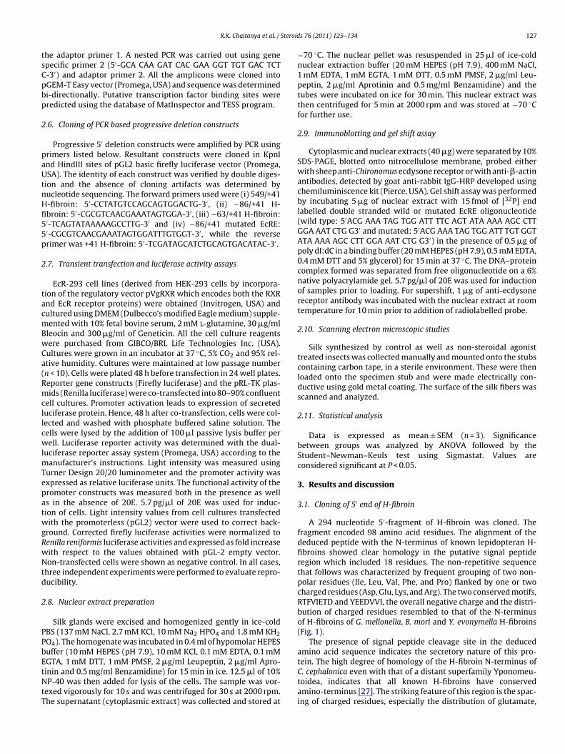
Steroid
tsCpbp
2
paUtnHfi55p
2
tacmBwCa(RmcllcwlmTepatwgRwNtd
2
PPbEtNtT
R.K. Chaitanya et al. /
he adaptor primer 1. A nested PCR was carried out using genepecific primer 2 (5′-GCA CAA GAT CAC GAA GGT TGT GAC TCT-3′) and adaptor primer 2. All the amplicons were cloned intoGEM-T Easy vector (Promega, USA) and sequence was determinedi-directionally. Putative transcription factor binding sites wereredicted using the database of MatInspector and TESS program.
.6. Cloning of PCR based progressive deletion constructs
Progressive 5′ deletion constructs were amplified by PCR usingrimers listed below. Resultant constructs were cloned in KpnInd HindIII sites of pGL2 basic firefly luciferase vector (Promega,SA). The identity of each construct was verified by double diges-
ion and the absence of cloning artifacts was determined byucleotide sequencing. The forward primers used were (i) 549/+41-fibroin: 5′-CCTATGTCCAGCAGTGGACTG-3′, (ii) −86/+41 H-broin: 5′-CGCGTCAACGAAATAGTGGA-3′, (iii) −63/+41 H-fibroin:′-TCAGTATAAAAAGCCTTG-3′ and (iv) −86/+41 mutated EcRE:′-CGCGTCAACGAAATAGTGGATTTGTGGT-3′, while the reverserimer was +41 H-fibroin: 5′-TCGATAGCATCTGCAGTGACATAC-3′.
.7. Transient transfection and luciferase activity assays
EcR-293 cell lines (derived from HEK-293 cells by incorpora-ion of the regulatory vector pVgRXR which encodes both the RXRnd EcR receptor proteins) were obtained (Invitrogen, USA) andultured using DMEM (Dulbecco’s modified Eagle medium) supple-ented with 10% fetal bovine serum, 2 mM l-glutamine, 30 �g/ml
leocin and 300 �g/ml of Geneticin. All the cell culture reagentsere purchased from GIBCO/BRL Life Technologies Inc. (USA).ultures were grown in an incubator at 37 ◦C, 5% CO2 and 95% rel-tive humidity. Cultures were maintained at low passage numbern < 10). Cells were plated 48 h before transfection in 24 well plates.eporter gene constructs (Firefly luciferase) and the pRL-TK plas-ids (Renilla luciferase) were co-transfected into 80–90% confluent
ell cultures. Promoter activation leads to expression of secreteduciferase protein. Hence, 48 h after co-transfection, cells were col-ected and washed with phosphate buffered saline solution. Theells were lysed by the addition of 100 �l passive lysis buffer perell. Luciferase reporter activity was determined with the dual-
uciferase reporter assay system (Promega, USA) according to theanufacturer’s instructions. Light intensity was measured using
urner Design 20/20 luminometer and the promoter activity wasxpressed as relative luciferase units. The functional activity of theromoter constructs was measured both in the presence as wells in the absence of 20E. 5.7 pg/�l of 20E was used for induc-ion of cells. Light intensity values from cell cultures transfectedith the promoterless (pGL2) vector were used to correct back-
round. Corrected firefly luciferase activities were normalized toenilla reniformis luciferase activities and expressed as fold increaseith respect to the values obtained with pGL-2 empty vector.on-transfected cells were shown as negative control. In all cases,
hree independent experiments were performed to evaluate repro-ucibility.
.8. Nuclear extract preparation
Silk glands were excised and homogenized gently in ice-coldBS (137 mM NaCl, 2.7 mM KCl, 10 mM Na2 HPO4 and 1.8 mM KH2O4). The homogenate was incubated in 0.4 ml of hypomolar HEPESuffer (10 mM HEPES (pH 7.9), 10 mM KCl, 0.1 mM EDTA, 0.1 mM
GTA, 1 mM DTT, 1 mM PMSF, 2 �g/ml Leupeptin, 2 �g/ml Apro-inin and 0.5 mg/ml Benzamidine) for 15 min in ice. 12.5 �l of 10%P-40 was then added for lysis of the cells. The sample was vor-exed vigorously for 10 s and was centrifuged for 30 s at 2000 rpm.he supernatant (cytoplasmic extract) was collected and stored at
s 76 (2011) 125–134 127
−70 ◦C. The nuclear pellet was resuspended in 25 �l of ice-coldnuclear extraction buffer (20 mM HEPES (pH 7.9), 400 mM NaCl,1 mM EDTA, 1 mM EGTA, 1 mM DTT, 0.5 mM PMSF, 2 �g/ml Leu-peptin, 2 �g/ml Aprotinin and 0.5 mg/ml Benzamidine) and thetubes were incubated on ice for 30 min. This nuclear extract wasthen centrifuged for 5 min at 2000 rpm and was stored at −70 ◦Cfor further use.
2.9. Immunoblotting and gel shift assay
Cytoplasmic and nuclear extracts (40 �g) were separated by 10%SDS-PAGE, blotted onto nitrocellulose membrane, probed eitherwith sheep anti-Chironomus ecdysone receptor or with anti-�-actinantibodies, detected by goat anti-rabbit IgG-HRP developed usingchemiluminiscence kit (Pierce, USA). Gel shift assay was performedby incubating 5 �g of nuclear extract with 15 fmol of [32P] endlabelled double stranded wild or mutated EcRE oligonucleotide(wild type: 5′ACG AAA TAG TGG ATT TTC AGT ATA AAA AGC CTTGGA AAT CTG G3′ and mutated: 5′ACG AAA TAG TGG ATT TGT GGTATA AAA AGC CTT GGA AAT CTG G3′) in the presence of 0.5 �g ofpoly dI:dC in a binding buffer (20 mM HEPES (pH 7.9), 0.5 mM EDTA,0.4 mM DTT and 5% glycerol) for 15 min at 37 ◦C. The DNA–proteincomplex formed was separated from free oligonucleotide on a 6%native polyacrylamide gel. 5.7 pg/�l of 20E was used for inductionof samples prior to loading. For supershift, 1 �g of anti-ecdysonereceptor antibody was incubated with the nuclear extract at roomtemperature for 10 min prior to addition of radiolabelled probe.
2.10. Scanning electron microscopic studies
Silk synthesized by control as well as non-steroidal agonisttreated insects was collected manually and mounted onto the stubscontaining carbon tape, in a sterile environment. These were thenloaded onto the specimen stub and were made electrically con-ductive using gold metal coating. The surface of the silk fibers wasscanned and analyzed.
2.11. Statistical analysis
Data is expressed as mean ± SEM (n = 3). Significancebetween groups was analyzed by ANOVA followed by theStudent–Newman–Keuls test using Sigmastat. Values areconsidered significant at P < 0.05.
3. Results and discussion
3.1. Cloning of 5′ end of H-fibroin
A 294 nucleotide 5′-fragment of H-fibroin was cloned. Thefragment encoded 98 amino acid residues. The alignment of thededuced peptide with the N-terminus of known lepidopteran H-fibroins showed clear homology in the putative signal peptideregion which included 18 residues. The non-repetitive sequencethat follows was characterized by frequent grouping of two non-polar residues (Ile, Leu, Val, Phe, and Pro) flanked by one or twocharged residues (Asp, Glu, Lys, and Arg). The two conserved motifs,RTFVIETD and YEEDVVI, the overall negative charge and the distri-bution of charged residues resembled to that of the N-terminusof H-fibroins of G. mellonella, B. mori and Y. evonymella H-fibroins(Fig. 1).
The presence of signal peptide cleavage site in the deducedamino acid sequence indicates the secretory nature of this pro-
tein. The high degree of homology of the H-fibroin N-terminus ofC. cephalonica even with that of a distant superfamily Yponomeu-toidea, indicates that all known H-fibroins have conservedamino-terminus [27]. The striking feature of this region is the spac-ing of charged residues, especially the distribution of glutamate,
128 R.K. Chaitanya et al. / Steroids 76 (2011) 125–134
F specieo i (GenC l pept
eof
3
wpTw0crpoq
F(ic
ig. 1. Comparison of 5′ region of H-fibroin gene of C. cephalonica with other insectf Corcyra cephalonica, Galleria mellonella, Yponomeuta evonymella and Bombyx moronserved motifs are represented in rectangles. Arrow (↓) shows the putative signa
ach of them close to a valine or a similar residue. Conservationf such a pattern in all the compared insect species indicates theirunctional importance [2].
.2. Effect of 20E on H-fibroin gene expression
Earlier studies in our laboratory showed that the salivary glandsere viable in the culture medium till 24 h and hence the timeoints from 0.5 h to 24 h were chosen for various treatments [28].he hemolymph levels of 20E during ELI, LLI larval and EPP stagesere estimated by enzyme immunoassay and were found to be
.008 pg/�l, 0.4 pg/�l and 5.7 pg/�l respectively [28]. These con-
entrations of 20E were considered as physiological levels of theespective stage. We examined the effect of physiological dose of aarticular stage as well as the subsequent stage on the expressionf H-fibroin gene using semi-quantitative (Figs. 2A and 3A) anduantitative PCR (Figs. 2B and 3B). The H-fibroin transcript levelsig. 2. Effect of 20E on H-fibroin expression of LLI larval silk glands. (A) Semi-quantitative0.05% ethanol) and 20E (0.4 pg/�l and 5.7 pg/�l) treated glands. �-Actin was used as intern control (0.05% ethanol) and 20E (0.4 pg/�l and 5.7 pg/�l) treated glands. H-fibroin expommon letter do not differ significantly (P < 0.05), while the values with different letters
s. Sequence alignment of the deduced amino acid sequence of 5′-N-terminal regionBank accession nos. GQ901977, AF095239, AB195979 and AF226688 respectively).ide cleavage site.
of ethanol (0.05%) treated glands that were cultured through thesame time points were used as external control. The physiologi-cal concentration of 20E during the LLI stage (0.4 pg/�l) did notalter the normal expression levels of H-fibroin, when comparedwith control. However, when the glands from LLI were treatedwith the hemolymph levels of 20E found during the EPP stage(5.7 pg/�l), there was a significant decrease in H-fibroin expres-sion (Fig. 2B). Similarly, the physiological concentration of 20Eduring the ELI stage (0.008 pg/�l) did not change the expressionpattern of H-fibroin in ELI glands as compared to control. However,when the ELI glands were treated for 12 h with the physiologicallevels of 20E found during LLI stage (0.4 pg/�l), the expression of
H-fibroin declined significantly (Fig. 3B). The semi-quantitative PCRdata (Figs. 2A and 3A) matches with that obtained in real-time PCR(Figs. 2B and 3B).In B. mori, the function and development of silk glands wereshown to be under hormonal control [1]. Maximum silk fibroin
PCR showing the expression at various time points (0.5, 3, 6, 9 and 12 h) in controlnal control. (B) Real-time PCR (n = 3) showing the expression at various time pointsression is reported as fold change relative to 0 h control. The values labelled withdiffer significantly and are compared group-wise (P < 0.05).
R.K. Chaitanya et al. / Steroids 76 (2011) 125–134 129
F itative( ed asp H-fibw rent l
slrrT[E
ttweodwehHsmBnlawfidatE
ig. 3. Effect of 20E on H-fibroin expression of ELI larval silk glands. (A) Semi-quant0.05% ethanol) and 20E (0.008 pg/�l and 0.4 pg/�l) treated glands. �-Actin was usoints in control (0.05% ethanol) and 20E (0.008 pg/�l and 0.4 pg/�l) treated glands.ith common letter do not differ significantly (P < 0.05), while the values with diffe
ynthesis and greater cellular changes were observed in the lastarval instar [29]. In G. mellonella too, the silk glands were shown toeadily respond to hormones during this stage [30]. Based on theseeports, the last instar larvae were chosen to study the effect of 20E.he hemolymph titre of 20E varies among insects and at each stage31,32]. Therefore, we estimated the concentration of 20E duringLI, LLI and EPP stages in C. cephalonica.
In C. cephalonica, in vitro treatment with physiological concen-ration of 20E found during LLI larval stage (0.4 pg/�l), did not alterhe normal expression pattern of H-fibroin gene when comparedith that of solvent. However, Sehnal and Akai [1] reported a high
xpression of silk fibroin during the last instar larval developmentf B. mori [1]. Hence, it was not clear whether the physiologicalose of 20E has any effect on the H-fibroin expression. Therefore,e monitored the effect of physiological levels of 20E during the
arlier stage, i.e. ELI stage, on H-fibroin expression. Once again theemolymph levels of 20E during ELI stage (0.008 pg/�l) did not alter-fibroin gene expression, when compared with the control. This
tudy shows that the physiological dose of 20E facilitates the nor-al expression of H-fibroin gene during last instar. Earlier study on
. mori silk glands also suggested that low titers of ecdysteroid wereecessary for proper silk gland function [33]. Further, in G. mel-
onella, in vitro application of 20E (0.5 �g/ml concentration) causedn increase in the level of PSG mRNAs. However, the stimulationas restricted to day 1 of last instar larvae after which no effect was
ound on the mRNA levels [34]. In cultured PSGs of Galleria larvae,
t was observed that the effect of 20E on RNA synthesis was dose-ependent. Furthermore, the lower concentrations of 20E did notlter the RNA levels during the LLI stage [35]. Taken together andhe finding that the physiological levels of 20E estimated during theLI as well as the LLI stage of C. cephalonica were low, we suggestPCR showing the expression at various time points (0.5, 3, 6, 9 and 12 h) in controlinternal control. (B) Real-time PCR (n = 3) showing the expression at various time
roin expression is reported as fold change relative to 0 h control. The values labelledetters differ significantly and are compared group-wise (P < 0.05).
that probably low or physiological doses of 20E is required for thenormal expression of H-fibroin gene.
On the other hand, higher concentrations of 20E exerted aninhibitory effect on the RNA synthesis in G. mellonella [35,36]. Fur-ther, in B. mori, treatment with high concentrations of ecdysteroidscaused silk gland degeneration at the end of last instar larval devel-opment [1]. Our results in C. cephalonica further strengthen thispoint. Upon treatment of LLI larval silk glands with higher dosageof 20E (5.7 pg/�l seen during EPP stage), the expression of H-fibroin decreased significantly. Such a prominent effect of 20E wasmost likely mediated by ecdysone receptors present in the salivaryglands cells [37]. Further, the pupal molt in various insect species isknown to be associated with high titers of ecdysteroids [31,32]. Thedeclined expression of L-fibroin, P25 [28] and H-fibroin (unpub-lished) during the pupal stage of insect development as well as thesignificant decrease in the expression observed upon treatment ofLLI larval silk glands with dosage of 20E similar to that found dur-ing EPP stage supports the fact that higher concentrations of 20Ecause a decline in the H-fibroin expression. Treatment of ELI larvalsilk gland with 0.4 pg/�l of 20E (the physiological concentrationof 20E found during LLI larval stage) once again reconfirmed thedeclined expression of H-fibroin at higher concentration of ecdys-teroid. Present study shows that H-fibroin is under 20E controland it regulates H-fibroin expression in a dose-dependant mannerduring the last instar larval development.
3.3. Identification of a novel functional ecdysone responseelement (EcRE) in the 5′ flanking region of H-fibroin gene
In the present study, a PCR product of 590 bp was amplifiedfrom the EcoRV library and the 5′ flanking region of H-fibroin
130 R.K. Chaitanya et al. / Steroids 76 (2011) 125–134
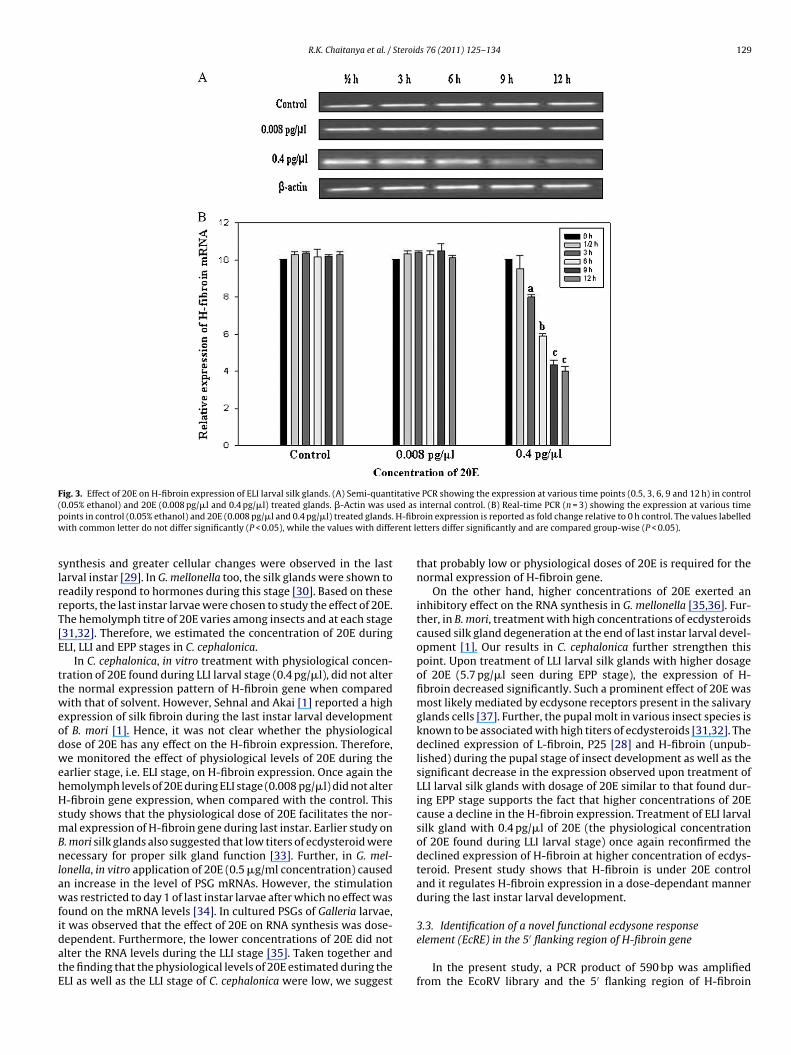
Fig. 4. Functional activity of ecdysone response element (EcRE) present in the 5′ flanking region of H-fibroin gene of C. cephalonica. (A) Sequence of 5′ upstream region of theC ding si ransfea valuew
w−mtlop4E(
gsseoficttcrpstinEeiTtbr
t
. cephalonica H-fibroin gene (GenBank accession no. GQ901977). The potential binn a rectangle. (B) Luciferase activity of different H-fibroin promoter constructs co-tctivity is presented as relative to the activities measured for Renilla luciferase. Theith different letters differ significantly and are compared group-wise (P < 0.05).
as cloned. The upstream analysis revealed a TATA sequence at59 relative to the ATG sequence. An ecdysone response ele-ent (EcRE) was identified at −65 position that overlaps with
he TATA box. A silk gland specific factor binding site (SGF-1) isocated at −254 position. A ten base A + T region that consistsf TAAT motif in the core is found to be the binding site forosterior silk gland specific (SGF2) and ubiquitous (SGF-3 and SGF-) factors. At −322 position, Drosophila homeodomain proteins,VE (even-skipped) and ZEN (zerknullt) binding site is harboredFig. 4A).
Structural analyses of the 5′ flanking region of the H-fibroinene from several strains of B. mori [38,39] and a closely relatedpecies, B. mandarina [40] showed that the immediate 5′ flankingequence, which is highly conserved, was probably important for itsxpression. The binding sites for SGF-1, -2, -3 and -4 factors, home-domain proteins which are found in 5′ flanking sequence of B. moribroin [38,39], were also seen in the H-fibroin upstream region of C.ephalonica. It was also predicted that SGF-1 might play an impor-ant role in the expression of silk protein genes, because it also bindso a similar site in sericin-1 gene. The regions for SGF-2, -3, and -4ontain ten-base pairs of A + T repeats. The TAAT motif in the coreegion was shown to be important for the binding of these threeroteins and also Drosophila homeodomain proteins, EVE (even-kipped) and ZEN (zerknullt) [41]. In this context, it is noteworthyhat an EcRE sequence overlapping the TATA box is also presentn the H-fibroin upstream region of B. mori [42]. However, it wasot analyzed for its functional activity. The consensus sequence ofcRE obtained from the comparison of various hormone responselements was shown to be (G/T)NTCANTNN(A/C)(A/C) [13–16]. Thedentified sequence of EcRE in the H-fibroin of C. cephalonica isTTCAGTATAA, which is well in agreement with the above men-
ioned consensus sequence. Based on the upstream sequence, PCRased progressive deletion constructs were generated and geneeporter assays were performed in engineered HEK-293 cells.Luciferase activity assay was performed primarily to find outhe functional activity of the C. cephalonica EcRE in the presence
ites for specific transcription factors are underlined. EcRE sequence is representedcted with pRL-TK into EcR293 cells in the presence or absence of 20E. The enzyme
s labelled with common letter do not differ significantly (P < 0.05), while the values
or absence of 20E. The H-fibroin gene expression declined signifi-cantly at LLI larval stage of C. cephalonica in the presence of 5.7 pg/�lof 20E (Fig. 2A). Therefore, this concentration of 20E was used inluciferase as well as gel shift assays. The −63/+41 construct whichcontained TATA box showed similar activity both in the presence orabsence of 20E suggesting that this region is not affected by 20E. Asignificantly lower activity of the −86/+41 construct in the presenceof 20E, that harbors both TATA box and EcRE suggests that EcREmight act as a negative regulatory element. The mutated EcRE con-struct as well as the wild type construct in the absence of 20E didnot show significant changes in promoter activity when comparedwith the −63/+41 construct which consisted TATA box (Fig. 4B).Furthermore, the whole upstream fragment 590 bp also showeddiminished activity in the presence of 20E (Fig. 4B).
These results strengthen the view that EcRE present in theupstream is functional and regulates H-fibroin expression in C.cephalonica. The presence of functional EcRE indicates that 20Eeffect on H-fibroin expression is probably mediated by EcR asdescribed in the case of other 20E regulated genes [13,17].
3.4. EcR binds to EcRE upon 20E induction
The presence of EcR in the nuclear extract was confirmedby immunoblot (Fig. 5A). 20E induced silk gland nuclear extractdemonstrated a complex formation with [�32P] dATP labelledoligomeric sequence containing EcRE motif, while no complex wasobserved in the absence of 20E induction. The shift in the bandobserved when probed with EcR specific antibody further confirmsthe binding. The mutated oligonucleotide did not show any bindingin the presence of 20E (Fig. 5B).
Upon 20E exposure, puffing responses of the polytene chromo-
somes in D. melanogaster salivary glands were observed. In thismodel, 20E binds with an ecdysone receptor protein and directlyactivates the transcription of a small set of early genes, which thenactivate many late genes [43]. The presence of EcR and its inductionby 20E was also shown in B. mori silk glands [20]. These studies
R.K. Chaitanya et al. / Steroids 76 (2011) 125–134 131
Fig. 5. EcR mediated 20E regulation of H-fibroin. (A) Nuclear and cytoplasmic extracts (40 �g each) from the larval silk glands were immunoblotted against anti-ecdysoner t (laneE e olign t and[ oligo
cvsmerhb
F1aa
eceptor antibody. Please note the presence of cross reactivity in the nuclear extraccR protein to EcRE oligomer in the presence of 20E. Lane 1: [�32P] labelled wild typuclear extract and radiolabelled wild type oligo, lane 4: 20E induced nuclear extrac�32P] labelled mutated oligo, lane 6: nuclear extract and the radiolabelled mutated
learly suggest that 20E regulates various processes in the sali-ary gland via EcR, which control gene transcription by binding topecific sequences. An EcRE in the upstream region of Hsp27 pro-
oter was previously shown in Drosophila [13]. Treatment withcdysone, in vitro, enhanced the DNA binding capability of theeceptor. The most extensively studied promoters are the four smalleat shock protein genes of Drosophila, were shown to be activatedy ecdysone as well as heat shock [17]. In the present study, in the
ig. 6. Effect of RH-5849 and RH-5992 on H-fibroin expression. (A) Relative expression of Hng/�l, 1.5 ng/�l, 2 ng/�l and 0.05% ethanol (control). (B) Relative expression of H-fibroinnd 1.5 ng/�l and 0.05% ethanol (control). The values labelled with common letter do not dnd are compared group-wise (P < 0.05).
1). �-Actin was used as loading control. (B) Gel shift assay showing the binding ofo, lane 2: nuclear extract and the radiolabelled wild type oligo, lane 3: 20E inducedradiolabelled wild type oligo probed with anti-ecdysone receptor antibody, lane 5:and lane 7: 20E induced nuclear extract and radiolabelled mutated oligo.
absence of 20E there was no complex formation and hence no signalis observed. The mutated oligonucleotide did not bind to EcR evenafter induction by 20E further confirms that EcR binds specifically
to EcRE sequence in the upstream region of H-fibroin gene, onlywhen induced by 20E. Thus, it would be reasonable to hypothesizethat 20E induces EcR pairing with RXR in the absence of USP andpredictably binds to the EcRE region [12,14] that might regulate theH-fibroin gene expression.-fibroin gene upon treatment with increased concentrations of RH-5849, 0.5 ng/�l,gene upon treatment with increased concentrations of RH-5992, 0.5 ng/�l, 1 ng/�l
iffer significantly (P < 0.05), while the values with different letters differ significantly

132 R.K. Chaitanya et al. / Steroids 76 (2011) 125–134
F expea rmal( treat
3l
RaeR
FaR
ig. 7. Effect of RH-5849 and RH-5992 treatment on insect metamorphosis. For thisnd were allowed to undergo development and adult emergence. (A) (i) control—noB) Cocoons obtained from (i) control—normal pupa, (ii) RH-5849 and (iii) RH-5992
.5. Effect of RH-5849 and RH-5992 on H-fibroin expression,arval–pupal–adult transition and the ultrastructure of silk fibers
Increasing doses of RH-5849 (0.5, 1, 1.5 and 2 ng/�l) (Fig. 6A) and
H-5992 (0.5, 1 and 1.5 ng/�l) (Fig. 6B) were added to the glandsnd the expression of H-fibroin gene was monitored. The H-fibroinxpression decreased significantly upon treatment with 2 ng/�l ofH-5849 in vitro (Fig. 6A). This decline was more prominent inig. 8. Effect of RH-5849 and RH-5992 on ultrastructure of silk fibers. (A) Scanning electrnd (iii) RH-5992 fed insects. All the images were captured at same magnification. (B)H-5992 fed insects. All the images were captured at same magnification.
riment the LLI larvae were treated with RH-5849 (2 ng/�l) and RH-5992 (1.5 ng/�l)adult, (ii) RH-5849 treated defective adult and (iii) RH-5992 treated defective adulted insects.
the cultured glands that received RH-5992 at a concentration of1.5 ng/�l (Fig. 6B).
RH-5849 and RH-5992 are bisacylhydrazine insecticides thatexhibit their activity via interaction with the ecdysteroid recep-
tor proteins. Upon treatment with these compounds, a state ofecdysteroid excess, called hyperecdysonism is induced and theinsecticides bind to the ecdysteroid receptor complex with highaffinity [24]. Changes in EcR expression were also observed in RH-on micrograph showing the fibers from (i) control insects, (ii) RH-5849 fed insectsSticky coating found in (i) control silk fibers, was absent in (ii) RH-5849 and (iii)
Steroid
5peg
laompwaleivad
fiT1(eciwtisirl
4
acdtgEs2sp
A
3SRfI
A
t
R
[
[
[
[
[
[
[
[
[
[
[
[
[
[
[
[
[
[
[
[
[
R.K. Chaitanya et al. /
992 treated larvae of Cydia pomonella [44]. Results obtained in theresent study indicate that RH-5849 and RH-5992 mimic the 20Excessive state that might cause the down regulation of H-fibroinene via EcR complex.
In the current study, when the insects were fed with simi-ar concentrations of these insecticides, they metamorphosed intobnormal adults (Fig. 7A). Precocious slippage of head capsule wasbserved (Fig. 7A; ii and iii). This was also associated with highortality of insects during pupal stage due to improper metamor-
hosis and defective cocoon formation (Fig. 7B; ii and iii). Thosehich survived metamorphosed into defective adults (Fig. 7A; ii
nd iii). Previous studies also reported that insecticide intoxicatedarvae prematurely slipped their old head capsules in an attempt tocdyse [24]. In Manduca sexta, RH-5849 halted feeding and resultedn premature lethal molt [45]. Based on these reports and our obser-ations, it is clear that the RH-5849 and RH-5992 treatment causeddisruption in the normal larval–pupal–adult transition, in lepi-
opteran insects.Differences were also observed in the ultrastructure of the silk
bers of control and insecticide fed C. cephalonica larvae (Fig. 8).he mean diameter of a single fiber of RH-5849 fed insects was.19 �m (Fig. 8A; ii) and that of RH-5992 fed insects was 876 nmFig. 8A; iii). A remarkable decrease in the diameter of the fiber wasvident, when the aforementioned values were compared with theontrol, which shows a mean fiber diameter of 2.16 �m (Fig. 8A;). The fibers from insects treated with either RH-5849 or RH-5992
ere found to be devoid of sticky coating (bleb like structures onhe fiber) (Fig. 8B; ii and iii) which were seen in the control (Fig. 8B;). It is known that the sticky coating of the silk fiber is provided byericin proteins, which are secreted in the MSG [46,47]. However,t remains to be investigated whether sericin expression is downegulated upon treatment with these hormone analogs and thuseads to decrease in the overall diameter of the silk fiber.
. Conclusion
In conclusion, our results describe 20E regulation of H-fibroint transcriptional level during the last instar development of C.ephalonica. 20E modulates H-fibroin expression in a dose depen-ent manner. The action of 20E is mediated through EcR that bindso the functional EcRE shown in the upstream region of H-fibroinene. To our knowledge, this study provides the first report of ancRE in H-fibroin promoter. The various effects observed upon non-teroidal ecdysteroids agonist treatment further signify the role of0E mediated regulation of H-fibroin in insect development. Moretudies on such silk producing insect pests in this direction mayrovide an insight for their efficient control.
cknowledgements
Financial assistance through a Research Grant (No.7/1260/06/EMR II) to Aparna Dutta Gupta from Council ofcientific and Industrial Research, India is greatly acknowledged..K. Chaitanya acknowledges CSIR for awarding senior research
ellowship. P. Sridevi acknowledges University Grants Commission,ndia, for awarding senior research fellowship.
ppendix A. Supplementary data
Supplementary data associated with this article can be found, inhe online version, at doi:10.1016/j.steroids.2010.09.009.
eferences
[1] Sehnal F, Akai H. Insect silk glands: their types, development and function, andeffects of environmental factors and morphogenetic hormones on them. Int JInsect Morphol Embryol 1990;19:79–132.
[
[
s 76 (2011) 125–134 133
[2] Fedic R, Zurovec M, Sehnal F. Correlation between fibroin amino acid sequenceand physical silk properties. J Biol Chem 2003;278:35255–64.
[3] Sehnal F, Zurovec M. Construction of silk fiber core in Lepidoptera. Biomacro-molecules 2004;5:666–74.
[4] Takei F, Kikuchi Y, Mizuno S, Shimura K. Further evidence for importance of thesubunit combination of silk fibroin in its efficient secretion from the posteriorsilk gland cells. J Cell Biol 1987;105:175–80.
[5] Takei F, Oyama F, Kimura K, Hyodo A, Mizuno S, Shimura K. Reduced level ofsecretion and absence of subunit combination for the fibroin synthesized by amutant silkworm, Nd (2). J Cell Biol 1984;99:2005–10.
[6] Tanaka K, Inoue S, Mizuno S. Hydrophobic interaction of P25, containing Asn-linked oligosaccharide chains, with the H-L complex of silk fibroin producedby Bombyx mori. Insect Biochem Mol Biol 1999;29:269–76.
[7] Nirmala X, Kodrik D, Zurovec M, Sehnal F. Insect silk contains both aKunitz-type and a unique Kazal-type proteinase inhibitor. Eur J Biochem2001;268:2064–73.
[8] Zurovec M, Yang C, Kodrik D, Sehnal F. Identification of a novel type of silkprotein and regulation of its expression. J Biol Chem 1998;273:15423–8.
[9] Inoue S, Tanaka K, Arisaka F, Kimura S, Ohotomo K, Mizuno S. Silk fibroin ofBombyx mori is secreted, assembling a high molecular mass elementary unitconsisting of H-chain, L-chain and P25, with a 6:6:1 molar ratio. J Biol Chem2000;275:40517–28.
10] Zurovec M, Sehnal F. Unique molecular architecture of silk fibroin in the wax-moth, Galleria mellonella. J Biol Chem 2002;277:22639–47.
11] Gilbert LI, Bollenbacher WE, Goodman W, Smith SL, Agui N, Granger N, et al.Insect endocrinology: regulation of endocrine glands, hormone titer and hor-mone metabolism. Ann Rev Physiol 1980;42:493–510.
12] Yao TP, Forman BM, Jiang Z, Cherbas L, Cherbas JD, McKeown M, et al. Func-tional ecdysone receptor is the product of EcR and Ultraspiracle genes. Nature1993;366:476–9.
13] Riddihough G, Pelham HR. An ecdysone response element in the Drosophilahsp27 promoter. EMBO J 1987;6:3729–34.
14] Oro AE, McKeown M, Evans RM. Relationship between the product of theDrosophila ultraspiracle locus and the vertebrate retinoid X receptor. Nature1990;347:298–301.
15] Luo Y, Amin J, Voellmy R. Ecdysterone receptor is a sequence-specific transcrip-tion factor involved in the developmental regulation of heat shock genes. MolCell Biol 1991;11:3660–75.
16] Antoniewski C, Laval M, Lepesant JA. Structural features critical to the activityof an ecdysone receptor binding site. Insect Biochem Mol Biol 1993;23:105–14.
17] Ireland RC, Berger E, Sirotkin K, Yund MA, Osterbur D, Fristrom J. Ecdysteroneinduces the transcription of four heat-shock genes in Drosophila S3 cells andimaginal discs. Dev Biol 1982;93:498–507.
18] Schaltmann K, Pongs O. Identification and characterization of the ecdysteronereceptor in Drosophila melanogaster by photoaffinity labeling. Proc Natl AcadSci USA 1982;79:6–10.
19] Talbot WS, Swyryd EA, Hogness DS. Drosophila tissues with different metamor-phic responses to ecdysone express different ecdysone receptor isoforms. Cell1993;73:1323–37.
20] Kamimura M, Tomita S, Kiuchi M, Fujiwara H. Tissue-specific and stage-specificexpression of two silkworm ecdysone receptor isoforms ecdysteroid-dependent transcription in cultured anterior silk glands. Eur J Biochem1997;248:786–93.
21] Terashima J, Yasuhara N, Iwami M, Sakurai S. Programmed cell death triggeredby insect steroid hormone, 20-hydroxyecdysone, in the anterior silk gland ofthe silkworm, Bombyx mori. Dev Genes Evol 2000;210:545–58.
22] Miao YG, Shi LG, Nair KS. Ecdysteroid as a mediator in the regulation of silkprotein synthesis and its influence on silkworm (Bom., Lepidoptera) genome. JAppl Ent 2004;128:348–53.
23] Krishna Ayyar PN. A very destructive pest of stored products in south IndiaCorcyra cephalonica (Stainton). Bull EntomolRes 1930;25:155–69.
24] Dhadialla TS, Carlson GR, Le DP. New insecticides with ecdysteroidal and juve-nile hormone activity. Ann Rev Entomol 1998;43:545–69.
25] Lakshmi M, Dutta-Gupta A. Juvenile hormone mediated DNA synthesis duringlarval development of Corcyra cephalonica. Biochem Int 1990;22:269–78.
26] Livak KJ, Schmittgen TD. Analysis of relative gene expression data usingreal-time quantitative PCR and the 2(-Delta Delta C(T)) method. Methods2001;25:402–8.
27] Yonemura N, Sehnal F. The design of silk fiber composition in moths has beenconserved for more than 150 million years. J Mol Evol 2006;63:42–53.
28] Chaitanya RK, Dutta-Gupta A. Light chain fibroin and P25 genes of Corcyracephalonica: molecular cloning, characterization, tissue specific expression,synchronous developmental and 20-hydroxyecdysone regulation during thelast instar larval development. Gen Comp Endocrinol 2010;167:113–21.
29] Suzuki Y, Suzuki E. Quantitative measurements of fibroin messenger RNA syn-thesis in the posterior silk gland of normal and mutant Bombyx mori. J Mol Biol1974;88:393–407.
30] Sehnal F, Zanda V, Nemec V. Composition, synthetic and cytolytic activities ofGalleria mellonella silk glands during the last-larval instar under the action ofjuvenile hormone. J Insect Physiol 1983;29:237–48.
31] Calvez B, Hirn M, De Reggi M. Ecdysone changes in the haemolymph of twosilkworms (Bombyx mori and Philosamia Cynthia) during larval and pupal devel-opment. FEBS Lett 1976;71:57–61.
32] Plantevin G, De Reggi M, Nardon C. Changes in ecdysteroid and juvenile hor-mone titers in the hemolymph of Galleria mellonella larvae and pupae. GenComp Endocrinol 1984;56:218–30.

1 Steroid
[
[
[
[
[
[
[
[
[
[
[
[
[larval Lepidoptera. Science 1988;241:467–9.
[46] Gamo T, Inokuchi T, Laufer H. Polypeptides of fibroin and sericin secreted
34 R.K. Chaitanya et al. /
33] Shigematsu H, Moriyama H. Effect of ecdysterone on fibroin synthesis in theposterior division of the silk gland of the silkworm, Bombyx mori. J Insect Physiol1970;16:2015–22.
34] Grzelak K, Kludkiewich B, Lasota Z. The effect of 20-hydroxyecdysone onexpression of genes coding for low molecular weight silk proteins of Galleriamellonella. Insect Biochem 1993;18:223–8.
35] Kodrik D, Sehnal F. Juvenile hormone counteracts the action of ecdysterone onsilk glands of Galleria mellonella L. (Lepidoptera: Pyralidae). Int J Insect MorpholEmbryol 1994;23:39–56.
36] Sehnal F, Michalik J. Control of activity and regression of the silk glands in thelast-larval instar of Galleria mellonella. J Insect Physiol 1984;30:119–26.
37] Elmogy M, Iwami M, Sakurai S. Presence of membrane ecdysone recep-tor in the anterior silk gland of the silkworm Bombyx mori. Eur J Biochem2004;271:3171–9.
38] Suzuki Y, Adachi S. Signal sequences associated with fibroin gene expressionare identical in fibroin-producer and -nonproducer tissues. Develop Growth
Diff 1984;26:139–47.39] Ueda H, Mizuno S, Shimura K. Sequence polymorphisms around the 5′-end ofthe silkworm fibroin H-chain gene suggesting the occurrence of crossing-overbetween heteromorphic alleles. Gene 1985;34:351–5.
40] Kusuda J, Tazima Y, Onimaru K, Ninaki O, Suzuki Y. The sequence around the5′ end of the fibroin gene from the wild silkworm, Bombyx mandarina, and
[
s 76 (2011) 125–134
comparison with that of the domesticated species, B. mori. Mol Gen Genet1986;203:359–64.
41] Hui C-c, Matsuno K, Suzuki Y. Fibroin gene promoter contains a cluster of home-odomain binding sites that interact with three silk gland factors. J Mol Biol1990;213:651–70.
42] TSujimoto Y, Suzuki Y. Structural analysis of the fibroin gene at the 5′ end andits surrounding regions. Cell 1979;16:425–36.
43] Ashburner M, Chihara C, Meltzer P, Richards G. Temporal control of puff-ing activity in polytene chromosomes. Cold Spring Harbor Symp Quant Biol1974;38:655–62.
44] Sun X, Song Q, Barrett B. Effects of ecdysone agonists on the expression ofEcR, USP and other specific proteins in the ovaries of the coding moth (Cydiapomonella L.). Insect Biochem Mol Biol 2003;33:829–40.
45] Wing KD, Slawecki RA, Carlson GR. A nonsteroidal ecdysone agonist: effects on
from the different sections of the silk gland in Bombyx mori. Insect Biochem1977;7:285–95.
47] Sinohara H. Glycopeptides isolated from sericin of the silkworm, Bombyx mori.Comp Biochem Physiol 1979;63B:87–91.
Copyright © 2022 FDOKUMEN