Silk proteins for biomedical applications: Bioengineering perspectives
Upload
independentCategory
view
5download
0
ARTICLE IN PRESS
0142-9612/$ - se
doi:10.1016/j.bi
�CorrespondE-mail addr
1Contributed
Biomaterials 28 (2007) 1643–1652
www.elsevier.com/locate/biomaterials
Biocompatibility evaluation of silk fibroin with peripheralnerve tissues and cells in vitro
Yumin Yang1, Xuemei Chen1, Fei Ding, Peiyun Zhang, Jie Liu, Xiaosong Gu�
Jiangsu Key Laboratory of Neuroregeneration, Nantong University, Nantong, 19 Qixiu Road, Jiangsu Province 226001, PR China
Received 1 September 2006; accepted 1 December 2006
Available online 26 December 2006
Abstract
Silk-based materials have been used in the field of bone or ligament tissue engineering. In order to explore the feasibility of using
purified silk fibroin to construct artificial nerve grafts, it is necessary to evaluate the biocompatibility of silk fibroin material with
peripheral nerve tissues and cells. We cultured rat dorsal root ganglia (DRG) on the substrate made up of silk fibroin fibers and observed
the cell outgrowth from DRG during culture by using light and electron microscopy coupled with immunocytochemistry. On the other
hand, we cultured Schwann cells from rat sciatic nerves in the silk fibroin extract fluid and examined the changes of Schwann cells after
different times of culture. The results of light microscopy, MTT test and cell cycle analysis showed that Schwann cells cultured in the silk
fibroin extract fluid showed no significant difference in their morphology, cell viability and proliferation as compared to that in plain L15
medium. Furthermore, no significant difference was found in expression of the factors secreted by Schwann cells, such as nerve growth
factor (NGF), brain-derived neurotrophic factor (BDNF) and S-100, between Schwann cells cultured in the silk fibroin extraction fluid
and in plain L15 medium by the aid of immunocytochemistry, RT-PCR and Western analysis. Collectively, these data indicate that silk
fibroin has good biocompatibility with DRG and is also beneficial to the survival of Schwann cells without exerting any significant
cytotoxic effects on their phenotype or functions, thus providing an experimental foundation for the development of silk fibroin as a
candidate material for nerve tissue engineering applications.
r 2006 Elsevier Ltd. All rights reserved.
Keywords: Silk fibroin; Dorsal root ganglia; Schwann cell; Biocompatibility; Cytotoxicity
1. Introduction
Peripheral nerve repair remains a common but challen-ging clinical problem. Direct end-to-end suturing issuggested for a short nerve injury. For larger nerve defectsor gaps, implantation of a nerve graft is often necessary tobridge the proximal and distal nerve stumps for facilitatingnerve regeneration and functional recovery. The typicalchoice is a nerve autograft that is harvested from anothersite in the body. However, this recognized ‘‘gold standard’’technique for peripheral nerve repair is limited by tissueavailability, donor-site morbidity, secondary deformities,as well as potential differences in tissue structure and size[1,2]. Xenografts and allografts have been tried, but they
e front matter r 2006 Elsevier Ltd. All rights reserved.
omaterials.2006.12.004
ing author. Tel./Fax: +86 513 85511585.
ess: [email protected] (X. Gu).
equally to this work.
are subject to immunosuppression and have achieved verypoor success [3–7].Much effort has been devoted to seeking promising
alternatives to conventional nerve autografts, and theemergence of tissue engineering has greatly stimulated thedevelopment of artificial nerve grafts, or nerve guidancechannels [8–10]. In recent years, many synthetic or naturalbiopolymers such as polyglycolic acid, polylactic acid,chitosan, alginate and their composites or derivatives havebeen utilized to construct nerve guidance channels,showing considerable potential of these materials to findtheir applications in nerve tissue engineering [11–15].Native silkworm silk protein from Bombyx mori consists
of a core structural fibroin protein surrounded by sericin, afamily of glue-like proteins. A highly repeated hydrophobicand crystallizable sequence has been described for theprimary structure of fibroin heavy chain: Gly-Ala-Gly-Ala-Gly-X (X stands for Ser or Tyr). Sericin is a more

ARTICLE IN PRESSY. Yang et al. / Biomaterials 28 (2007) 1643–16521644
hydrophilic protein, whose primary structure is richer inpolar residues, but some of its fractions are not completelywater-soluble due to b-sheet portions [16–19].
Silks have a long history of use as suture materials in themedical field because of their impressive mechanicalproperties and flexibility [20–23]. However, undesirableimmunological reactions associated with silks diminishedtheir clinical applications during the last years [16,24,25].The identification of contaminating sericin as the source ofimmunological problems sparks new interest in silk fibroinas a native biomaterial. Hence purified silk fibroin hasfound broad applications in pharmaceutical and biomedi-cal fields [26–31], especially in tissue engineering for thegeneration of artificial bones or ligaments [16,32–38].However, to our knowledge, there have hitherto been fewreports on the utility of silk-based or silk-coated materialsfor peripheral nerve repair. Based upon this, we culturedrat dorsal root ganglia (DRG) on the substrate made up ofsilk fibroin fibers to examine the in vitro biocompatibilityof silk-based materials with peripheral nerve tissues, andalso cultured Schwann cells from rat sciatic nerves in thesilk fibroin extract fluid to evaluate (as per InternationalStandards ISO 10993-5) the in vitro cytotoxicity ofsilk-based materials for peripheral nerve cells.
2. Materials and methods
2.1. Preparation of silk fibroin and its extract fluid
Bombyx mori silk, obtained from Xinyuan sericulture company,
Nantong, Jiangsu, China, was degummed by boiling twice, 0.5 h each, in
aqueous Na2CO3 solution (0.5%), in order to remove the sericins.
Degummed silk was thoroughly washed in hot double distillated water for
several times, and then dried at room temperature. The resulting silk
fibroin needed to be sterilized by autoclave prior to use [39,40].
After sterilization, silk fibroin (1 g) was placed in an extraction
container with an addition of 10ml of L15 medium (Sigma) and allowed
to incubate at 3770.5 1C for 7270.5 h [39]. The resulting silk fibroin
extract fluid had to be used within 24-h period. For control, the extraction
fluids of hydroxyapatite, obtained from Sigma, or organotin, from
Hongding Chemicals Company, Nantong, Jiangsu, China, were prepared
in the same manner, respectively.
2.2. Tissue and cell culture
In this study, all experimental procedures involving animals were
conducted as per Institutional Animal Care guidelines and approved
ethically by the Administration committee of experimental animals,
Jiangsu Province, China.
For tissue culture, silk fibroin fibers were sheared into 0.7–0.8 cm long
fragments which were then soaked in L15 medium for about 30min. The
rat DRG were obtained from neonatal Sprague-Dawley rats, 1–2-d old,
and stripped with connective tissue carefully at their surface. The treated
DRG together with silk fibroin fragments were placed onto a 24-well
culture plate. Afterwards, L15 medium supplemented with 15% fetal
bovine serum (FBS) was added to the culture plate (800ml per well),
followed by incubation at 37 1C in a humidified, 5% CO2 atmosphere.
Half of the medium was replaced every other day. At different culture
times, the cell growth of DRG was observed under an inverted light
microscope (Olympus IX-70, Japan).
For cell culture, rat Schwann cells were harvested as follows. The
bilateral sciatic nerves of neonatal Sprague-Dawley rats (1–2-d old) were
excised out under sterile condition. After the epineurium and perineurium
were removed from sciatic nerves carefully, the nerves were sheared into
0.01mm3 tissue fragments which were then planted onto a polylysine-
coated coverslip in a 6-well tissue culture plate. L15 medium supplemented
with 15% FBS was added to the plate (2ml per well), followed by
incubation at 37 1C, with humidity and 5% CO2. Half of the medium was
replaced every other day. After 7 d incubation, the subcultured Schwann
cells from sciatic nerves of neonatal Sprague-Dawley rats were seeded in a
96-well plate at a cell density of 1� 104/well and the plain L15 medium or
different extract fluids were added to the plate, respectively. At different
times of culture, the morphological changes of Schwann cells in
these different culture mediums were observed under an inverted light
microscope.
2.3. Immunocytochemistry
After 4 or 7 ds of culture of DRG on silk fibroin, the samples were fixed
with a fresh-prepared 4% paraformaldehyde solution in PBS for 30min at
room temperature. The fixed samples were incubated for 1 h in a solution
containing 10% goat serum, 3% bovine serum albumin and 0.1% Triton-
X 100 at room temperature to block nonspecific binding. Then, they were
allowed to incubate with mouse monoclonal anti-S-100 antibody (1:1200
dilution, Sigma) overnight at 4 1C in a humidified chamber. After being
washed three times with PBS, the samples were further reacted with
second antibody FITC-labeled goat anti-mouse IgG (1:250 dilution,
Sigma) for 2 h at 37 1C. The samples were washed three times with PBS,
mounted in fluorescent mounting medium and observed under a confocal
laser scanning microscope (TCS SP2, Leica Microsystems, Germany).
Controls included leaving out the primary antibody and using non-labeled
secondary antibodies to confirm inexistence of nonspecific binding.
After 24-, 48- or 72-h culture either in the plain L15 medium or the silk
fibroin extract fluid, Schwann cells were incubated with 5 mg/ml Hoechst
33342 (Sigma) at 37 1C for 1 h and then fixed and blocked as the above
mentioned. Afterwards, they were incubated with mouse anti-S-100
antibody (1:1200 dilution, Sigma) and rabbit polyclonal anti-rat nerve
growth factor (NGF) antibody (1:1000, Chemicon, Temecula, CA, USA)
or with mouse anti-S-100 antibody (1:1200 dilution, Sigma) and rabbit
anti-rat brain-derived neurotrophic factor (BDNF) polyantibody (1:1000,
Chemicon) at 4 1C overnight in a humidified chamber, respectively. After
being washed with PBS three times, the samples were further reacted with
FITC-labeled goat anti-mouse IgG (1:250 dilution, Sigma) and Cy3-
labeled goat anti-rabbit IgG (1:100) at 37 1C for 2 h. Finally, the samples
were viewed under a confocal laser scanning microscope. Controls were
treated similarly as the above mentioned.
2.4. Electron microscopy
After 4- or 7-d incubation of DRG on the silk fibroin fibers, the
samples were washed twice with PBS and fixed in 4% glutaraldehyde.
They were then post-fixed with 1% OsO4, dehydrated stepwise in
increasing concentrations of ethanol, and dried in a critical point drier
(Hitachi, Tokyo, Japan). Afterwards, the samples were coated with gold in
a JFC-1100 unit (Jeol Inc., Japan) and observed under a scanning electron
microscope (JEM-T300, Jeol Inc., Japan).
After 21-d incubation of DRG onto the silk fibroin fibers, the samples
were washed twice with PBS and fixed in 4% glutaraldehyde. They were
then post-fixed with 1% OsO4, dehydrated stepwise in increasing
concentrations of ethanol, and embedded in Epon 812 epoxy resin.
Semi-thin sections were stained with toluidine blue, and ultrathin sections
were then localized and viewed with a transmission electron microscope
(JEM-1230, JEOL Ltd., Japan).
2.5. MTT assay
A modified MTT [3-(4,5-dimethylthiazol-2-yl)-2,5-diphenyltetrazolium
bromide] test, in which the yellow MTT is reduced to a purple formazan

ARTICLE IN PRESSY. Yang et al. / Biomaterials 28 (2007) 1643–1652 1645
by mitochondrial dehydrogenase in cells, was used to assess the cell
viability.
After 12-, 24-, 48- or 72-h or 7-d incubation in different mediums, the
viability of Schwann cells was assessed. Briefly, Schwann cells were washed
three times with L15 medium. The culture medium in each well of the plate
was replaced with 100ml L15 medium and 25 ml MTT (5mg/ml in PBS).
After 4 h incubation at 37 1C, the reaction solution was carefully removed
from each well and 100ml dimethyl sulfoxide was added. The plates were
gently agitated until the formazan precipitate was dissolved, followed by
measurement of OD values by spectrophotometry at 490 nm with an ElX-
800 Microelisa reader (Bio-Tek Inc., USA).
The serum concentration in the silk fibroin extract fluid was decreased
in a graded manner to 10%, 5% even 2.5%, and the serum concentration
in the plain L15 culture medium was also decreased in the same manner.
2.6. Cell cycle test
After 24-, 48- or 72-h incubation either in the plain L15 medium
or the silk fibroin extract fluid, the Schwann cells were collected
by trypsinization (106 per tube) and washed twice (5min per time)
with PBS via centrifugation at 1000 rpm. The cells were suspended
in PBS, fixed with 70% ethanol (v/v) at �20 1C. Samples were washed with
PBS and stained with PI/RNase Staining Buffer (BD PharMingen) for
30min in dark at 4 1C. The number of cells at different phases of the cell
cycle was analyzed using a flow cytometer (BD FACScalibur, BD
Bioscience, USA).
2.7. RT-PCR analysis
After 12-, 24- or 48-h culture either in the plain L15 medium or the silk
fibroin extract fluid, the Schwann cells were homogenized in Trizol
Reagent (Gibco, Gaithersburg, MD, USA). Total RNA was extracted and
reverse transcriptized according to the manufacturer’s instructions. The
sequences of primers for S-100, NGF, BDNF and GAPDH (used as an
internal control) are shown in Table 1. The PCR reaction was conducted
in a total volume of 25ml containing 1 ml cDNA of samples, 2.5 ml10�PCR buffer, 2.0 ml MgSO4, 2.5ml dNTPmix (2mmol/l), 0.5ml senseprimer (10 mmol/l), 0.5ml antisense primer (10mmol/l), 0.2 ml Taq DNA
Polymerase (2U/ml) and 15.8ml H2O. The reaction mixture was heated to
94 1C for 2.5min and then amplified for 31 cycles (S-100), 31 cycles
(NGF), 33 cycles (BDNF), or 26 cycles (GAPDH), respectively, as
specified: 94 1C for 40 s (denaturation), 53 1C for 40 s (annealing) and 72 1C
for 35 s (extension). Amplification products were separated by a 1.2%
agarose gel electrophoresis and visualized by ethidium bromide staining.
2.8. Western blot analysis
After 24-, 48- or 72-h culture either in the plain L15 medium or the silk
fibroin extract fluid, the Schwann cells were washed with PBS and lysed
with lysis buffer containing protease inhibitors (Promega, Madison, WI,
USA). Protein concentration was detected by BCA method (calibrated on
bovine serum albumin) to maintain the same loads. Protein extracts were
heat denatured at 100 1C for 5min, electrophoretically separated on a 12%
SDS-PAGE, and transferred to a PVDF membrane. The membrane was
blocked with 5% non-fat dry milk in TBST buffer (50mM Tris–HCl,
100mM NaCl, and 0.1% Tween-20, pH 7.4) and incubated with a 1:600
Table 1
Oligonucleotide primers for detecting mRNA levels of GAPDH, NGF, BDNF
Sense (50–30)
GAPDH tggtgaaggtcggtgtga
S-100 tgcttgtcctctgtgcaaa
NGF gatcggcgtacaggcaga
BDNF gtcaagtgcctttggagcc
dilution of rabbit anti-rat S-100 polyclonal antibody in 5% non-fat dry
milk in TBST buffer at 4 1C overnight. The membranes were washed with
TBST buffer (5min� 3), and further incubated with a 1:200 dilution of
HRP-conjugated goat anti-rabbit IgG at room temperature for 1.5 h.
After the membrane was washed, the HRP activity was detected using
an ECL kit. The image was scanned with GS800 Densitometer Scanner
(Bio-Rad, Hercules, CA, USA), and the data of optical density were
analyzed using PDQuest 7.2.0 software (Bio-Rad). b-actin (1:4000) was
used as an internal control.
2.9. Statistical analysis
At least three repetitive tests were performed, and all data were
expressed with means7SEM. A one-way ANOVA with the Stata 6.0
software package (Stata Corp., College Station, TX, USA) was used to
conduct statistical analysis, and statistical significance was accepted at the
0.05 confidence level.
3. Results
3.1. Silk fibroin fibers as substrate for cell outgrowth of
DRG
Transmission electron microscopy showed that thetransverse section of the degummed silk threads was ahomogeneous entity in an olivary shape with a maximumdiameter of about 15 mm and had smooth and completeedges without surface attachments. But undegummed silkthreads were wrapped with a layer of materials of a variedthickness, which were assumed as sericin (Fig. 1).Light microscopy was used for visualizing the cell
growth of DRG on silk fibroin fibers. After 24-h cultureof DRG on the silk fibroin bundles (3–8 fibers per bundle),cells started to crawl out of DRG. Among them, some cellswere spherical in shape and adhesive to the silk fibroinmaterial; some cells were spindle in shape and suspended inthe culture medium. After 3–4-d culture, the large numberof cells migrated out of DRG. Some of them enwrappedthe silk fibroin material in cell groups; some of them singlyattached to the material and gradually turned into a spindleshape encircling the material; while the neurites were notedto elongate from the neurons of DRG and extend along thematerial. After 7 and more days of culture, more number ofcells migrated out of DRG, and they spiraled tightly alongthe silk fibroin material and enwrapped it in a multilayerfashion, forming long cell chains. Immunocytochemistryfurther indicated that there existed S-100 positive cells,identified as Schwann cells, which attached to and encircledthe silk fibroin material in virtue of their relatively stronggreen fluorescence emission (Fig. 2).
and S100
Anti-sense (50–30)
ac ttcccattctcagccttgac
c ccatcattcctcagcagctc
ac ggctcggcacttggtctcaa
t cttatgaaccgccagccaat

ARTICLE IN PRESS
Fig. 1. Transmission electron micrographs of transverse sections of the
undegummed (A) and degummed (B) silk threads.Fig. 2. Immunocytochemistry obtained after 4-d (A) or 7-d (B) culture of
DRG on the silk fibroin fibers. Green color represents S-100 immunopo-
sitive cells that might be considered Schwann cells.
Y. Yang et al. / Biomaterials 28 (2007) 1643–16521646
Visual inspection under scanning electron microscopyfurther showed that after 4-d culture, cells tightly attachedto and partly coiled about the silk fibroin fibers and theyexhibited either a spherical or spindle shape; after 7-dculture, a large quantity of cells took compact arrange-ments of either side-by-side or end-to-end configuration,forming a single- or multi-layer structure to enwrap the silkfibroin fibers (Fig. 3).
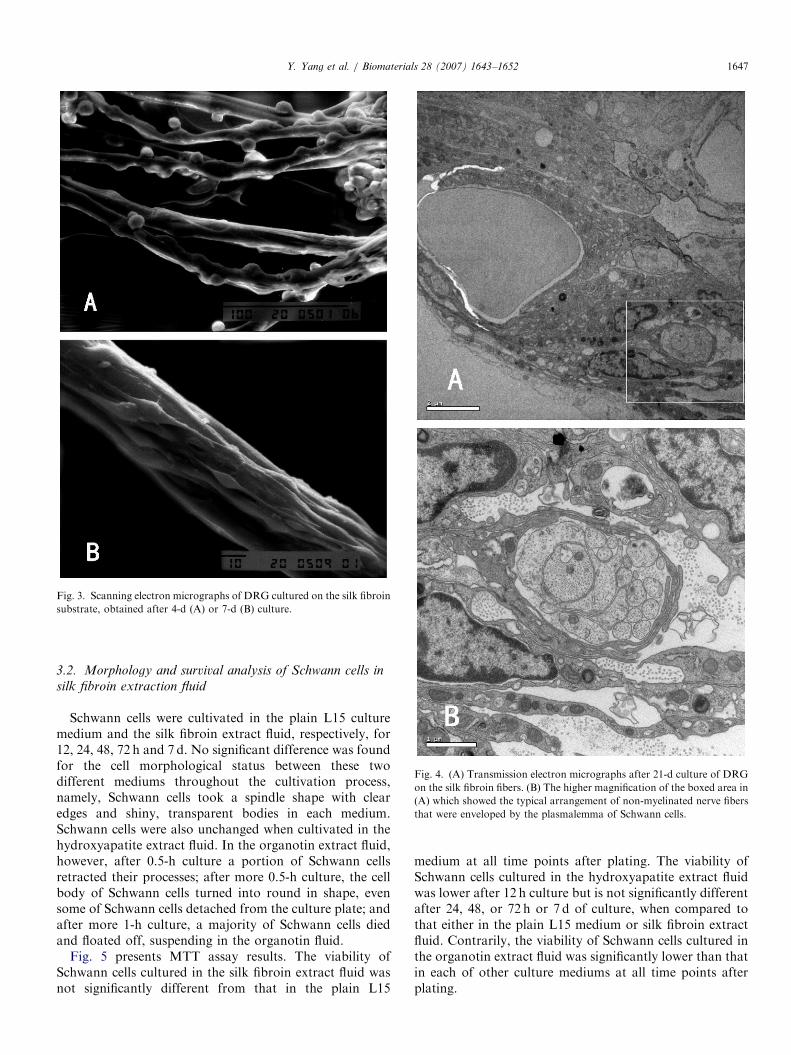
Transmission electron micrograph of the transversesections of silk fibroin fibers demonstrated thatSchwann cells enwrapped the silk fibroin fibers comp-
letely or partly, and they had a clear and completeinner structure and a normal appearance; on theother hand, axons grew in parallel along the silkfibroin fibers, while Schwann cells separated theaxons into bundles and enwrapped some of the axonscompletely or partly (Fig. 4). The typical arrangementof non-myelinated nerve fibers that were envelopedby the plasmalemma of Schwann cells was also observedin Fig. 4B.
ARTICLE IN PRESS
Fig. 3. Scanning electron micrographs of DRG cultured on the silk fibroin
substrate, obtained after 4-d (A) or 7-d (B) culture.
Fig. 4. (A) Transmission electron micrographs after 21-d culture of DRG
on the silk fibroin fibers. (B) The higher magnification of the boxed area in
(A) which showed the typical arrangement of non-myelinated nerve fibers
that were enveloped by the plasmalemma of Schwann cells.
Y. Yang et al. / Biomaterials 28 (2007) 1643–1652 1647
3.2. Morphology and survival analysis of Schwann cells in
silk fibroin extraction fluid
Schwann cells were cultivated in the plain L15 culturemedium and the silk fibroin extract fluid, respectively, for12, 24, 48, 72 h and 7 d. No significant difference was foundfor the cell morphological status between these twodifferent mediums throughout the cultivation process,namely, Schwann cells took a spindle shape with clearedges and shiny, transparent bodies in each medium.Schwann cells were also unchanged when cultivated in thehydroxyapatite extract fluid. In the organotin extract fluid,however, after 0.5-h culture a portion of Schwann cellsretracted their processes; after more 0.5-h culture, the cellbody of Schwann cells turned into round in shape, evensome of Schwann cells detached from the culture plate; andafter more 1-h culture, a majority of Schwann cells diedand floated off, suspending in the organotin fluid.
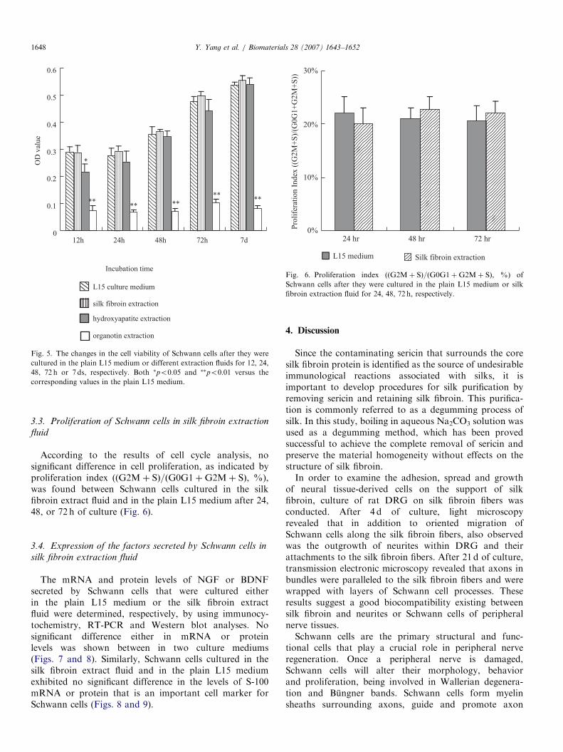
Fig. 5 presents MTT assay results. The viability ofSchwann cells cultured in the silk fibroin extract fluid wasnot significantly different from that in the plain L15
medium at all time points after plating. The viability ofSchwann cells cultured in the hydroxyapatite extract fluidwas lower after 12 h culture but is not significantly differentafter 24, 48, or 72 h or 7 d of culture, when compared tothat either in the plain L15 medium or silk fibroin extractfluid. Contrarily, the viability of Schwann cells cultured inthe organotin extract fluid was significantly lower than thatin each of other culture mediums at all time points afterplating.
ARTICLE IN PRESS
0%
10%
20%
30%
24 hr 48 hr 72 hr
Pro
life
rati
on
In
dex
((G
2M
+S
)/(G
0G
1+
G2
M+
S))
L15 medium Silk fibroin extraction
Fig. 6. Proliferation index (ðG2Mþ SÞ=ðG0G1þG2Mþ SÞ, %) of
Schwann cells after they were cultured in the plain L15 medium or silk
fibroin extraction fluid for 24, 48, 72 h, respectively.
0
0.1
0.2
0.3
0.4
0.5
0.6
12h 24h 48h 72h 7d
Incubation time
OD
val
ue
L15 culture medium
silk fibroin extraction
hydroxyapatite extraction
organotin extraction
*
**** **
****
Fig. 5. The changes in the cell viability of Schwann cells after they were
cultured in the plain L15 medium or different extraction fluids for 12, 24,
48, 72 h or 7 ds, respectively. Both �po0:05 and ��po0:01 versus the
corresponding values in the plain L15 medium.
Y. Yang et al. / Biomaterials 28 (2007) 1643–16521648
3.3. Proliferation of Schwann cells in silk fibroin extraction
fluid
According to the results of cell cycle analysis, nosignificant difference in cell proliferation, as indicated byproliferation index (ðG2Mþ SÞ=ðG0G1þG2Mþ SÞ, %),was found between Schwann cells cultured in the silkfibroin extract fluid and in the plain L15 medium after 24,48, or 72 h of culture (Fig. 6).
3.4. Expression of the factors secreted by Schwann cells in
silk fibroin extraction fluid
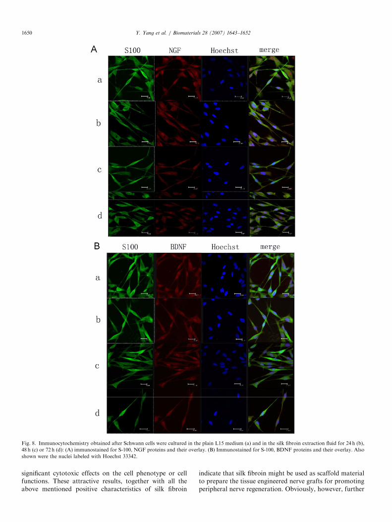
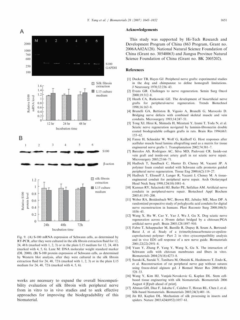
The mRNA and protein levels of NGF or BDNFsecreted by Schwann cells that were cultured eitherin the plain L15 medium or the silk fibroin extractfluid were determined, respectively, by using immunocy-tochemistry, RT-PCR and Western blot analyses. Nosignificant difference either in mRNA or proteinlevels was shown between in two culture mediums(Figs. 7 and 8). Similarly, Schwann cells cultured in thesilk fibroin extract fluid and in the plain L15 mediumexhibited no significant difference in the levels of S-100mRNA or protein that is an important cell marker forSchwann cells (Figs. 8 and 9).
4. Discussion
Since the contaminating sericin that surrounds the coresilk fibroin protein is identified as the source of undesirableimmunological reactions associated with silks, it isimportant to develop procedures for silk purification byremoving sericin and retaining silk fibroin. This purifica-tion is commonly referred to as a degumming process ofsilk. In this study, boiling in aqueous Na2CO3 solution wasused as a degumming method, which has been provedsuccessful to achieve the complete removal of sericin andpreserve the material homogeneity without effects on thestructure of silk fibroin.In order to examine the adhesion, spread and growth
of neural tissue-derived cells on the support of silkfibroin, culture of rat DRG on silk fibroin fibers wasconducted. After 4 d of culture, light microscopyrevealed that in addition to oriented migration ofSchwann cells along the silk fibroin fibers, also observedwas the outgrowth of neurites within DRG and theirattachments to the silk fibroin fibers. After 21 d of culture,transmission electronic microscopy revealed that axons inbundles were paralleled to the silk fibroin fibers and werewrapped with layers of Schwann cell processes. Theseresults suggest a good biocompatibility existing betweensilk fibroin and neurites or Schwann cells of peripheralnerve tissues.Schwann cells are the primary structural and func-
tional cells that play a crucial role in peripheral nerveregeneration. Once a peripheral nerve is damaged,Schwann cells will alter their morphology, behaviorand proliferation, being involved in Wallerian degenera-tion and Bungner bands. Schwann cells form myelinsheaths surrounding axons, guide and promote axon

ARTICLE IN PRESS
A
0
0.2
0.4
0.6
0.8
1
12 hr 24 hr 48 hr
Incubation time
Rel
ativ
e N
GF
mR
NA
lev
el
silk fibroin
extraction
L15 culture
medium
B
0
0.2
0.4
0.6
0.8
1
1.2
12 hr 24 hr 48 hr
Incubation time
Rel
ativ
e B
DN
F
mR
NA
lev
el
silk fibroinextraction
L15 culturemedium
2000
NGF
GADPH
GADPH
BDNF
1000760500
250100
2000
1000
760
600
250
100
Fig. 7. The changes in mRNA levels of NGF (A) and BDNF (B) secreted
by Schwann cells after they were cultured in the silk fibroin extraction fluid
for 12, 24, 48 h (marked with 1, 2, 3) or in the plain L15 medium for 12, 24,
48 h (marked with 4, 5, 6). Lane M: DNA molecular weight standard
marker (DL 2000).
Y. Yang et al. / Biomaterials 28 (2007) 1643–1652 1649
growth to establish a precise innervation [41–43].Therefore, it is important to gain a thorough insightinto the in vitro biocompatibility between silk fibroinand Schwann cells, especially to carry out the cytotoxi-city test as per International Standards compiled inISO 10993-5. For this reason, we focused on testingwhether or not the silk fibroin extract fluid had cytotoxiceffects on Schwann cells that were harvested from ratsciatic nerves.
In this study, MTT assay results suggest that silk fibroinextract fluid has no effects on the survival of Schwann cellscultured in it. It was suspected that in MTT tests, thecytotoxic effects might be completely or partially maskedby high concentration of serum in the culture medium. Tocheck this suspicion, the serum concentration in the silkfibroin extract fluid or the plain L15 culture medium waschanged in a graded manner. The comparison of Schwanncell viability between silk fibroin extract fluid and L15medium were shown to be independent on theserum concentration despite a decrease in Schwann cellviability for both mediums with the serum concentrationdecreasing.Since the proliferation index, referred to as the ratio of
(G2M+S) to (G2M+G0G1+S), is used to evaluate theproliferative status of cells, flow cytometric analysis on cellcycle was performed in this study to compare theproliferation index of Schwann cells cultured in the silkfibroin extract fluid with those in L15 medium at differenttimes of culture. The results showed no significantdifference, verifying that silk fibroin extract fluid had nocytotoxic effects on the in vitro proliferation of Schwanncells.Considering that Schwann cells secrete a variety of
neurotrophic factors, cell adhesion molecules, and otherassociated factors, among which NGF, BDNF areimportant for the development, maintenance and responseto injury of peripheral nervous system [44–48], weinvestigated the expression of NGF and BDNF secretedby Schwann cells that were cultured in the silk fibroinextract fluid by the aid of immunocytochemistry andRT-PCR. Moreover, since S-100 protein is an important,albeit nonspecific, marker for Schwann cells, we alsodetermined the expression of S-100 associated withSchwann cells that were cultured in the silk fibroin extractfluid by the aid of immunocytochemistry, RT-PCR andWestern blot analysis. All the experimental results showedthat the silk fibroin extract fluid did not influenceexpressions of neurotrophic factors or cell markerassociated with Schwann cells, providing further evidencefor little cytotoxicity of silk fibroin on Schwann cells.
5. Conclusion
As has been documented over decades, silk proteinexhibits high mechanical strength and flexibility, perme-ability to water and oxygen and can be made into nets,sponges or membranes, being easily handled, manipulatedand sterilized [23]. Purified silk fibroin proteins exhibit ahigh biocompatibility with some biological tissues and donot show the unwanted immunological responses that havebeen attributed to the silk sericin protein [17]. In addition,recent research has shown that a slow degradation of silktakes place in vivo [49,50].In this study, we have shown that silk fibroin
supports the cell growth of DRG and facilitatesthe survival of Schwann cells without exerting any
ARTICLE IN PRESS
Fig. 8. Immunocytochemistry obtained after Schwann cells were cultured in the plain L15 medium (a) and in the silk fibroin extraction fluid for 24 h (b),
48 h (c) or 72 h (d): (A) immunostained for S-100, NGF proteins and their overlay. (B) Immunostained for S-100, BDNF proteins and their overlay. Also
shown were the nuclei labeled with Hoechst 33342.
Y. Yang et al. / Biomaterials 28 (2007) 1643–16521650
significant cytotoxic effects on the cell phenotype or cellfunctions. These attractive results, together with all theabove mentioned positive characteristics of silk fibroin
indicate that silk fibroin might be used as scaffold materialto prepare the tissue engineered nerve grafts for promotingperipheral nerve regeneration. Obviously, however, further
ARTICLE IN PRESS
A
0
0.2
0.4
0.6
0.8
1
1.2
1.4
1.6
12 hr 24 hr 48 hr
Rel
ativ
e S
100
mR
NA
lev
el
Silk fibroinextraction
medium
L15 culture
B
0
0.2
0.4
0.6
0.8
1
1.2
24h 48h 72h
Incubation time
Incubation time
Rel
ativ
e S
100
pro
tein
lev
el
silk fibroin
extraction
L15 culture
medium
2000
S100
GAPDH
1000
760
600
250
100
1 2 3 4 5 6
S100
β-action
Fig. 9. (A) S-100 mRNA expression of Schwann cells, as determined by
RT-PCR, after they were cultured in the silk fibroin extraction fluid for 12,
24, 48 h (marked with 1, 2, 3) or in the plain L15 medium for 12, 24, 48 h
(marked with 4, 5, 6). Lane M: DNA molecular weight standard marker
(DL 2000). (B) S-100 protein expression of Schwann cells, as determined
by Western blot analysis, after they were cultured in the silk fibroin
extraction fluid for 24, 48, 72 h (marked with 1, 2, 3) or in the plain L15
medium for 24, 48, 72 h (marked with 4, 5, 6).
Y. Yang et al. / Biomaterials 28 (2007) 1643–1652 1651
works are necessary to expand the overall biocompati-bility evaluation of silk fibroin with peripheral nervefrom in vitro to in vivo studies and to seek effectiveapproaches for improving the biodegradability of thisbiomaterial.
Acknowledgements
This study was supported by Hi-Tech Research andDevelopment Program of China (863 Program, Grant no.2006AA02A128). National Natural Science Foundation ofChina (Grant no. 30540063) and Jiangsu Province NaturalScience Foundation of China (Grant no. BK 2005202).
References
[1] Ducker TB, Hayes GJ. Peripheral nerve grafts: experimental studies
in the dog and chimpanzee to define homograft limitations.
J Neurosurg 1970;32:236–43.
[2] Evans GR. Challenges to nerve regeneration. Semin Surg Oncol
2000;19:312–8.
[3] Heath CA, Rutkowski GE. The development of bioartificial nerve
grafts for peripheral-nerve regeneration. Trends Biotechnol
1998;16:163–8.
[4] Brunelli GA, Battiston B, Vigasio A, Brunelli G, Marocolo D.
Bridging nerve defects with combined skeletal muscle and vein
conduits. Microsurgery 1993;14:247–51.
[5] Tong XJ, Hirai K, Shimada H, Mizutani Y, Izumi T, Toda N, et al.
Sciatic nerve regeneration navigated by laminin-fibronectin double
coated biodegradable collagen grafts in rats. Brain Res 1994;663:
155–62.
[6] Fansa H, Schneider W, Wolf G, Keilhoff G. Host responses after
acellular muscle basal lamina allografting used as a matrix for tissue
engineered nerve grafts 1. Transplantation 2002;74:381–7.
[7] Barcelos AS, Rodrigues AC, Silva MD, Padovani CR. Inside-out
vein graft and inside-out artery graft in rat sciatic nerve repair.
Microsurgery 2003;23:66–71.
[8] Hadlock T, Sundback C, Hunter D, Cheney M, Vacanti JP. A
polymer foam conduit seeded with Schwann cells promotes guided
peripheral nerve regeneration. Tissue Eng 2000;6(2):119–27.
[9] Hadlock T, Elisseeff J, Langer R, Vacanti J, Cheney M. A tissue-
engineered conduit for peripheral nerve repair. Arch Otolaryngol
Head Neck Surg 1998;124(10):1081–6.
[10] Kannan RY, Salacinski HJ, Butler PE, Seifalian AM. Artificial nerve
conduits in peripheral-nerve repair. Biotechnol Appl Biochem
2005;41:193–200.
[11] Weber RA, Breidenbach WC, Brown RE, Jabaley ME, Mass DP. A
randomized prospective study of polyglycolic acid conduits for digital
nerve reconstruction in humans. Plast Reconstr Surg 2000;106(5):
1036–45.
[12] Wang X, Hu W, Cao Y, Yao J, Wu J, Gu X. Dog sciatic nerve
regeneration across a 30-mm defect bridged by a chitosan/PGA
artificial nerve graft. Brain 2005;128:1897–910.
[13] Fabre T, Schappacher M, Bareille R, Dupuy B, Soum A, Bertrand-
Barat J, et al. Study of a (trimethylenecarbonate-co-epsilon-
caprolactone) polymer—Part 2: in vitro cytocompatibility analysis
and in vivo ED1 cell response of a new nerve guide. Biomaterials
2001;22(22):2951–8.
[14] Yuan Y, Zhang P, Yang Y, Wang X, Gu X. The interaction of
Schwann cells with chitosan membranes and fibers in vitro.
Biomaterials 2004;25(18):4273–8.
[15] Suzuki K, Suzuki Y, Tanihara M, Ohnishi K, Hashimoto T, Endo K,
et al. Reconstruction of rat peripheral nerve gap without sutures
using freeze-dried alginate gel. J Biomed Mater Res 2000;49(4):
528–33.
[16] Wang Y, Kim HJ, Vunjak-Novakovic G, Kaplan DL. Stem cell-
based tissue engineering with silk biomaterials. Biomaterials 2006
August 4 [Epub ahead of print].
[17] Altman GH, Diaz F, Jakuba C, Calabro T, Horan RL, Chen J, et al.
Silk-based biomaterials. Biomaterials 2003;24(3):401–16.
[18] Jin HJ, Kaplan DL. Mechanism of silk processing in insects and
spiders. Nature 2003;424(6952):1057–61.
ARTICLE IN PRESSY. Yang et al. / Biomaterials 28 (2007) 1643–16521652
[19] Servoli E, Maniglio D, Motta A, Predazzer R, Migliaresi C. Surface
properties of silk fibroin films and their interaction with fibroblasts.
Macromol Biosci 2005;5(12):1175–83.
[20] Moy RL, Lee A, Zalka A. Commonly used suture materials in skin
surgery. Am Fam Physician 1991;44:2123–8.
[21] Minoura N, Aiba S, Gotoh Y, Tsukada M, Imai Y. Attachment and
growth of cultured fibroblast cells on silk protein matrices. J Biomed
Mater Res 1995;29:1215–21.
[22] Gosline JM, DeMont ME, Denny MW. The structure and properties
of spider silk. Endeavour 1986;10:37–43.
[23] Kaplan DL, Mello SM, Arcidiacono S, Fossey S, Senecal K, Muller
W. Silk. In: McGrath K, Kaplan DL, editors. Protein based
materials. Boston: Birkhauser; 1998. p. 103–31.
[24] Salthouse TN, Matlaga BF, Wykoff MH. Comparative tissue
response to six suture materials in rabbit cornea, sclera, and ocular
muscle. Am J Ophthalmol 1977;84(2):224–33.
[25] Salthouse TN. Biologic response to sutures. Otolaryngol Head Neck
Surg 1980;88(6):658–64.
[26] Uebersax L, Fedele DE, Schumacher C, Kaplan DL, Merkle HP,
Boison D, et al. The support of adenosine release from adenosine
kinase deficient ES cells by silk substrates. Biomaterials 2006;
27(26):4599–607.
[27] Bayraktar O, Malay O, Ozgarip Y, Batigun A. Silk fibroin as a novel
coating material for controlled release of theophylline. Eur J Pharm
Biopharm 2005;60(3):373–81.
[28] Yamada H, Igarashi Y, Takasu Y, Saito H, Tsubouchi K.
Identification of fibroin-derived peptides enhancing the proliferation
of cultured human skin fibroblasts. Biomaterials 2004;25(3):467–72.
[29] Dal Pra I, Freddi G, Minic J, Chiarini A, Armato U. De novo
engineering of reticular connective tissue in vivo by silk fibroin
nonwoven materials. Biomaterials 2005;26(14):1987–99.
[30] Unger RE, Peters K, Wolf M, Motta A, Migliaresi C, Kirkpatrick
CJ. Endothelialization of a non-woven silk fibroin net for use in tissue
engineering: growth and gene regulation of human endothelial cells.
Biomaterials 2004;25(21):5137–46.
[31] Gotoh Y, Niimi S, Hayakawa T, Miyashita T. Preparation of lactose-
silk fibroin conjugates and their application as a scaffold for
hepatocyte attachment. Biomaterials 2004;25(6):1131–40.
[32] Meinel L, Hofmann S, Betz O, Fajardo R, Merkle HP, Langer R,
et al. Osteogenesis by human mesenchymal stem cells cultured on silk
biomaterials: comparison of adenovirus mediated gene transfer and
protein delivery of BMP-2. Biomaterials 2006;27(28):4993–5002.
[33] Meinel L, Betz O, Fajardo R, Hofmann S, Nazarian A, Cory E, et al.
Kirker-Head C. Silk based biomaterials to heal critical sized femur
defects. Bone 2006 [Epub ahead of print].
[34] Karageorgiou V, Tomkins M, Fajardo R, Meinel L, Snyder B, Wade
K, et al. Porous silk fibroin 3-D scaffolds for delivery of bone
morphogenetic protein-2 in vitro and in vivo. J Biomed Mater Res A
2006;78(2):324–34.
[35] Li C, Vepari C, Jin HJ, Kim HJ, Kaplan DL. Electrospun silk-BMP-
2 scaffolds for bone tissue engineering. Biomaterials 2006;27(16):
3115–24.
[36] Meinel L, Fajardo R, Hofmann S, Langer R, Chen J, Snyder B, et al.
Silk implants for the healing of critical size bone defects. Bone 2005;
37(5):688–98.
[37] Chen J, Altman GH, Karageorgiou V, Horan R, Collette A, Volloch
V, et al. Human bone marrow stromal cell and ligament fibroblast
responses on RGD-modified silk fibers. J Biomed Mater Res A 2003;
67(2):559–70.
[38] Altman GH, Horan RL, Lu HH, Moreau J, Martin I, Richmond JC,
et al. Silk matrix for tissue engineered anterior cruciate ligaments.
Biomaterials 2002;23(20):4131–41.
[39] Fini M, Motta A, Torricelli P, Giavaresi G, Nicoli Aldini N,
Tschon M, et al. The healing of confined critical size cancellous
defects in the presence of silk fibroin hydrogel. Biomaterials 2005;
26(17):3527–36.
[40] Meinel L, Hofmann S, Karageorgiou V, Carl KH, McCool J,
Gronowicz G. The inflammatory responses to silk films in vitro and
in vivo. Biomaterials 2005;26:147–55.
[41] Krekoski CA, Neubauer D, Zuo J, Muir D. Axonal regeneration into
acellular nerve grafts is enhanced by degradation of chondroitin
sulfate proteoglycan. J Neurosci 2001;21(16):6206–13.
[42] Mosahebi A, Wiberg M, Terenghi G. Addition of fibronectin to
alginate matrix improves peripheral nerve regeneration in tissue-
engineered conduits. Tissue Eng 2003;9(2):209–18.
[43] Lundborg G. Alternatives to autologous nerve grafts. Handchir
Mikrochir Plast Chir 2004;36(1):1–7.
[44] Klein R. Role of neurotrophins in mouse neuronal development.
FASEB J 1994;8(10):738–44.
[45] Yin Q, Kemp GJ, Frostick SP. Neurotrophins, neurones and
peripheral nerve regeneration. J Hand Surg 1998;23(4):433–7.
[46] Frostick SP, Yin Q, Kemp GJ. Schwann cells, neurotrophic factors,
and peripheral nerve regeneration. Microsurgery 1998;18(7):397–405.
[47] Anand U, Otto WR, Casula MA, Day NC, Davis JB, Bountra C,
et al. The effect of neurotrophic factors on morphology, TRPV1
expression and capsaicin responses of cultured human DRG sensory
neurons. Neurosci Lett 2006;399(1–2):51–6.
[48] Moore K, MacSween M, Shoichet M. Immobilized concentration
gradients of neurotrophic factors guide neurite outgrowth of primary
neurons in macroporous scaffolds. Tissue Eng 2006;12(2):267–78.
[49] Li M, Ogiso M, Minoura N. Enzymatic degradation behavior of
porous silk fibroin sheets. Biomaterials 2003;24:357–65.
[50] Horan RL, Antle K, Collette AL, Wang Y, Huang J, Moreau JE. In
vitro degradation of silk fibroin. Biomaterials 2005;26:3385–93.
Copyright © 2022 FDOKUMEN