The new mutation, E46K, of ?-synuclein causes parkinson and Lewy body dementia
Upload
conicet-arCategory
view
7download
0
Chaperone proteostasis in Parkinson’s disease:stabilization of the Hsp70/a-synuclein complex by Hip
Cintia Roodveldt1,6,*, Carlos W Bertoncini1,6,August Andersson1, Annemieke T van derGoot2, Shang-Te Hsu1, RafaelFernandez-Montesinos3, Jannie de Jong2,Tjakko J van Ham2, Ellen A Nollen2,David Pozo3, John Christodoulou1,4,5,*and Christopher M Dobson1,*1Department of Chemistry, University of Cambridge, Cambridge, UK,2Department of Genetics, University Medical Centre Groningen andUniversity of Groningen, Groningen, The Netherlands, 3CABIMER-Andalusian Center for Molecular Biology and Regenerative Medicine(CSIC-University of Seville-UPO-Junta de Andalucia), Seville, Spain,4Research Department of Structural and Molecular Biology, Instituteof Structural and Molecular Biology, University College London (UCL),London, UK and 5School of Crystallography, Birkbeck College,University of London, London, UK
The ATP-dependent protein chaperone heat-shock protein
70 (Hsp70) displays broad anti-aggregation functions and
has a critical function in preventing protein misfolding
pathologies. According to in vitro and in vivo models of
Parkinson’s disease (PD), loss of Hsp70 activity is associated
with neurodegeneration and the formation of amyloid de-
posits of a-synuclein (aSyn), which constitute the intraneur-
onal inclusions in PD patients known as Lewy bodies. Here,
we show that Hsp70 depletion can be a direct result of the
presence of aggregation-prone polypeptides. We show a
nucleotide-dependent interaction between Hsp70 and
aSyn, which leads to the aggregation of Hsp70, in the
presence of ADP along with aSyn. Such a co-aggregation
phenomenon can be prevented in vitro by the co-chaperone
Hip (ST13), and the hypothesis that it might do so also
in vivo is supported by studies of a Caenorhabditis elegans
model of aSyn aggregation. Our findings indicate that a
decreased expression of Hip could facilitate depletion of
Hsp70 by amyloidogenic polypeptides, impairing chaperone
proteostasis and stimulating neurodegeneration.
The EMBO Journal (2009) 28, 3758–3770. doi:10.1038/
emboj.2009.298; Published online 29 October 2009
Subject Categories: proteins; molecular biology of disease
Keywords: amyloid; Hip; Hsp70; Parkinson’s disease; a-synuclein
Introduction
Protein conformational diseases include a range of degenerative
disorders in which specific peptides or proteins misfold and
aberrantly self-assemble, often in the form of amyloid fibrils,
which can be deposited in a variety of tissues, the process of
which may lead to toxicity and cell death. These disorders,
among others, include Alzheimer’s (AD), Parkinson’s (PD)
and Huntington’s diseases (HD) (Chiti and Dobson, 2006;
Luheshi et al, 2008). One of the most studied amyloid-forming
proteins involved in neurodegeneration is a-synuclein (aSyn),
the aggregation of which is linked to the pathogenesis of
PD. Indeed, aSyn is the major component of Lewy bodies,
the protein-rich aggregates found post-mortem in the brains
of patients suffering from PD or a number of related
diseases.
The pathological conversion of misfolded proteins into
cytotoxic species is modulated by interactions with several
proteins, among them are molecular chaperones (Dobson,
2003; Stefani and Dobson, 2003; Young et al, 2004; Balch
et al, 2008), which are now recognized as key players in the
avoidance of misfolding and hence in protein homeostasis
(Dobson, 2003; Young et al, 2004; Balch et al, 2008). A very
important class of chaperones is the family of stress-inducible
70 kDa heat-shock proteins (Hsp70s), which have a critical
function in a range of cellular processes including the folding
of newly synthesized proteins (Frydman et al, 1994) and the
rescue of misfolded proteins (Gragerov et al, 1992; Mogk
et al, 1999), hence avoiding the potentially harmful effects of
the aggregation of the latter species (Hartl, 1996). Hsp70
proteins are composed of an N-terminal ATPase domain
and a C-terminal substrate-binding domain (SBD), connected
by a short linker region (Mayer and Bukau, 2005). Within the
SBD, the substrate-binding pocket recognizes unstructured
segments in polypeptides (Bukau and Horwich, 1998; Mayer
et al, 2001) and the current view is that Hsp70 prevents
misfolding by binding to certain patterns of hydrophobic and
positively charged amino acids in the polypeptide substrate
(Rudiger et al, 1997a, b; Maeda et al, 2007). The ATPase cycle
of Hsp70 involves alternation between an ATP-bound or
‘open’ state, which has lower affinity for substrates, and an
ADP-bound or ‘closed’ state with higher affinity (Mayer and
Bukau, 2005). This alternation appears to be achieved by a
structural ‘coupling’ between the ATPase domain and the
SBD, driven by an allosteric mechanism that is not yet fully
understood (Saibil, 2008). The ATPase cycle is typically
modulated by several co-chaperones, most notably Hsp40,
resulting in an increase in ATPase activity (Minami et al,
1996; Bukau and Horwich, 1998). Other co-chaperones in-
clude Bag-1, which functions as a nucleotide-exchange factor
(Takayama and Reed, 2001), Hop, which interacts with Hsp70
to enhance certain functions (Scheufler et al, 2000), and Hip
(or ST13), which binds to the ATPase domain of the chaper-
one in its ADP-bound state and appears to increase the half-
life of substrate complexes (Hohfeld et al, 1995).Received: 18 August 2009; accepted: 15 September 2009; publishedonline: 29 October 2009
*Corresponding authors. C Roodveldt or CM Dobson, Department ofChemistry, University of Cambridge, Lensfield Road, Cambridge CB21EW, UK. Tel.: þ 44 1223 336366, Fax: þ 44 1223 336362;E-mail: [email protected] or Tel.: þ 44 1223 763070;Fax: þ 44 1223 336362; E-mail: [email protected] orJ Christodoulou, Research Department of Structural and MolecularBiology, Institute of Structural and Molecular Biology, UniversityCollege London, and School of Crystallography, Birkbeck College,Gower Street, London WC1E 6BT, UK. Tel.: þ 44 207 6792375;Fax: þ 44 207 6797193; E-mail: [email protected] authors contributed equally to this work
The EMBO Journal (2009) 28, 3758–3770 | & 2009 European Molecular Biology Organization | All Rights Reserved 0261-4189/09
www.embojournal.org
The EMBO Journal VOL 28 | NO 23 | 2009 &2009 European Molecular Biology Organization
EMBO
THE
EMBOJOURNAL
THE
EMBOJOURNAL
3758
There seems to be a strong link between problems in the
regulation of chaperone function and the panoply of confor-
mational diseases and amyloidoses (Dobson, 2003; Macario
and De Macario, 2007). For example, patients with PD show
highly perturbed expression of Hsp70 in the substantia nigra,
which is the site of neurodegeneration in this condition
(Grunblatt et al, 2001; Hauser et al, 2005). Indeed, Hsp70
has been found in deposits in the brain of AD patients, in
polyglutamine aggregates of sufferers of HD, and in Lewy
bodies from PD patients (Muchowski and Wacker, 2005).
Furthermore, in PD fly models (Auluck et al, 2002) and in
human neuroglioma cells (Klucken et al, 2004; Zhou et al,
2004), over-expression of Hsp70 has been found to reduce
significantly the toxicity linked to aSyn. Also, it is particularly
interesting that the co-chaperone Hip is consistently present
at lower levels in the blood of PD patients relative to healthy
controls, even at the early stages of the disease (Scherzer
et al, 2007).
Despite the compelling in vivo evidence of the implications
of Hsp70 in disease, in vitro studies of the molecular
mechanisms of the Hsp70-mediated inhibition of amyloid
formation are relatively sparse. For example, the nature
of the misfolded species recognized by Hsp70 and of the
complexes formed during amyloid aggregation of polypep-
tides, are not yet known. Equally important is the need to
understand the ways in which nucleotides control the
interactions with amyloidogenic protein substrates and the
potentially important functions by co-chaperones in assisting
Hsp70 in its amyloid-inhibiting functions. In this work,
we consider the impact of different nucleotide conditions
and of co-chaperones on the anti-aggregation activity of
Hsp70. Using aSyn as the substrate, we have probed
substrate–chaperone interactions for Hsp70 as a function
of the nucleotide state of the chaperone and the different
species of aSyn attained along the aggregation pathways.
By using fluorescence and NMR spectroscopy, we have
characterized a structurally compact ADP-Hsp70/aSyn
complex that may promote the co-aggregation of Hsp70
and may therefore lead to chaperone depletion. We further
identify an important function for the co-chaperone Hip
in sustaining the Hsp70-mediated anti-amyloid activity,
both in vitro and in vivo, the latter studies by means of a
Caenorhabditis elegans model of aSyn aggregation, and
speculate that this specific co-chaperone could have
an important function in the onset and progression
of PD.
Results
The dependence of the solubility and anti-amyloid
activity of Hsp70 on binding of nucleotides
To study the effects of ATP on the anti-aggregation activity of
Hsp70, a series of in vitro aggregation experiments were set
up in which aSyn amyloid formation was monitored by
thioflavin T (ThT) fluorescence (Figure 1A). The presence
of nucleotide-free chaperone at even a 1:10 molar ratio
(Hsp70:aSyn) dramatically inhibited amyloid formation, an
observation in agreement with our earlier work (Dedmon
et al, 2005). Interestingly, however, addition of 2 mM ATP
(using an ATP-regeneration system, see Materials and meth-
ods) to the solution containing Hsp70 was found to inhibit
aSyn aggregation initially, but at longer times an increase in
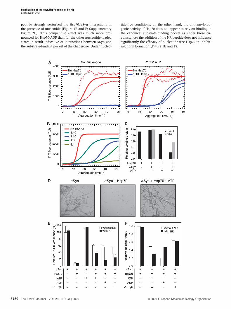
ThT fluorescence is evident, indicating the onset of aggrega-
tion. Analysis of the kinetics of aSyn aggregation in the
presence of various molar ratios of Hsp70 (Figure 1B;
Supplementary Figure 1) shows that, in the presence of
ATP, both the lag phase and half-time for aggregation increase
as the concentration of Hsp70 is increased. The rate of
aggregation was also found to be affected by the presence
of Hsp70, being reduced by ca. 60–70%, although in a
concentration-independent manner (Supplementary Figure
1). Transmission electron microscopy (TEM) was used to
assess the morphology of the species formed during
the aggregation reaction (Figure 1D), and revealed that
the presence of nucleotide-free Hsp70 strongly abrogates
fibril formation and produces exclusively small amorphous
aggregates that do not enhance the fluorescence of ThT
(Dedmon et al, 2005). Conversely, addition of Hsp70 together
with ATP was found to result in the formation of fibrillar
species, which are indistinguishable from amyloid fibril
controls.
Although an Hsp70–nucleotide mixture clearly reduces
aggregation, we sought to investigate the origins of the
apparent loss with time of chaperone activity of nucleotide-
loaded Hsp70. We initially compared the quantity of protein
in solution at the beginning and end points of a typical
aggregation reaction. Quantitative SDS–PAGE analysis
(Figure 1C; Supplementary Figure 2B) of soluble and inso-
luble fractions generated by low-speed centrifugation showed
that a very substantial proportion (B80%) of Hsp70 is
present in the insoluble fraction but only when ATP and
aSyn are both present. This surprising result suggests that
the observed surge of ThT fluorescence in the presence of
Hsp70 and ATP after ca. 30 h is likely to be a consequence of
aSyn-mediated depletion of Hsp70 in a nucleotide-dependent
manner.
To define the contribution of the different nucleotide-
bound states along the ATPase cycle to this phenomenon,
we conducted aggregation experiments in the absence or
presence of ATP, ADP or ATPgS, a non-hydrolyzable analogue
of ATP. ATP binds to Hsp70 and its hydrolysis stimulates the
cycling between conformations with low affinity (ATP-
bound) and high affinity (ADP-bound) for substrates.
Addition of ADP is therefore expected to stabilize the high
affinity state, and ATPgS to maintain Hsp70 in its low affinity
conformation (Mayer and Bukau, 2005). The rise in ThT
fluorescence indicates, however, that addition of all three
types of nucleotide-loaded Hsp70, but especially ATP and
ADP, ultimately results in the formation of aSyn amyloid
fibrils (Figure 1E). In addition to this conclusion, analysis by
SDS–PAGE of the soluble and insoluble fractions correspond-
ing to the end of the aggregation reaction (Figure 1F;
Supplementary Figure 2C) shows that, while Hsp70 remains
in solution in the absence of nucleotides, the addition of
nucleotides, and ADP in particular, promotes the partitioning
of Hsp70 into the aggregated fraction.
We next probed the specificity of the Hsp70/aSyn interac-
tion by assaying the same samples in the presence of the
peptide NR (NH2-NRLLLTG-COOH), that is expected to com-
pete with aSyn for the substrate-binding pocket of Hsp70
(Gragerov et al, 1994; Rudiger et al, 1997a). The reduction in
the level of ThT fluorescence signal at the endpoint of the
assay, and an increase in the solubility of the chaperone
during the aggregation process, showed clearly that the NR
Stabilization of the asyn/Hsp70 complex by HipC Roodveldt et al
&2009 European Molecular Biology Organization The EMBO Journal VOL 28 | NO 23 | 2009 3759
peptide strongly perturbed the Hsp70/aSyn interactions in
the presence of nucleotide (Figure 1E and F; Supplementary
Figure 2C). This competitive effect was much more pro-
nounced for Hsp70-ADP than for the other nucleotide-loaded
states, a result indicative of interactions between aSyn and
the substrate-binding pocket of the chaperone. Under nucleo-
tide-free conditions, on the other hand, the anti-amyloido-
genic activity of Hsp70 does not appear to rely on binding to
the canonical substrate-binding pocket as under these cir-
cumstances the addition of the NR peptide does not influence
significantly the efficacy of nucleotide-free Hsp70 in inhibit-
ing fibril formation (Figure 1E and F).
Stabilization of the asyn/Hsp70 complex by HipC Roodveldt et al
The EMBO Journal VOL 28 | NO 23 | 2009 &2009 European Molecular Biology Organization3760

The effect of Hsp70 on the morphology and cytotoxicity
of aSyn aggregates
The currently accepted view is that the process of aggregation
of aSyn involves the population of oligomeric intermediates
that cause cellular damage (Lashuel et al, 2002; Danzer
et al, 2007), which is likely to be a generic feature of such
amyloid-related species (Demuro et al, 2005; Chiti and
Dobson, 2006; Lashuel and Lansbury, 2006; Danzer et al,
2007). The nucleotide-dependent anti-aggregation properties
of Hsp70 are likely to be reflected in the assembly of different
intermediates along the route towards fibril formation. To
characterize the properties of species formed during the
aggregation of aSyn, we have studied the morphology and
monitored the cytotoxicity of the protein adducts formed at
early and late stages of the aggregation process under differ-
ent conditions. Of particular interest is the observation that
early aSyn aggregates formed in the presence of Hsp70 and
ATP are protofibrillar in nature, whereas only spherical
oligomeric species can be observed when the nucleotide-
free chaperone is present (Figure 2A and B).
We also assayed the toxicity of the soluble oligomeric pre-
fibrillar adducts formed under a variety of conditions by
adding these species, separated by centrifugation from larger
aggregates, to human neuroblastoma SH-SY5Y cells in culture
Figure 2 Effect of Hsp70 on the morphology and cytotoxicity of aSyn aggregating species throughout the reaction. (A) Fibril formation by aSynwas monitored by ThT fluorescence. Samples correspond to untreated or Hsp70-treated aSyn at a 1:10 ratio, in the absence or presence of ATP.Samples labels in (B). A negative control with added ovalbumin was used. AU, arbitrary units. (B) TEM analysis of samples corresponding to24 and 64 h of the aggregation reaction. A negative control with ovalbumin was also included. Scale bar, 500 nm. (C) The toxicity levels of thesoluble species from samples corresponding to the different conditions and time points of the reaction were determined by LDH releasemeasurement (left axes) using human SH-SY5Y cells. Samples correspond to untreated aSyn (white bars) or aSyn treated with 1:10 Hsp70:aSyn(black bars), either in the absence (left panel) or presence (right panel) of ATP. The bars represent the intrinsic toxicity, that is the net toxicity(with background toxicity levels subtracted) normalized to the aSyn concentration. In all cases, values represent the average results ofduplicate experiments. Connected circles represent the Hsp70 soluble levels at each time point (right axis).
Figure 1 Effect of nucleotides and a competing peptide on the modulation of aSyn aggregation by Hsp70. (A) Aggregation experimentsof aSyn in the absence or presence of a 1:10 molar ratio of Hsp70 without (left) or with the addition (right) of 2 mM ATP (and anATP-regeneration system). Fibril formation was monitored by ThT fluorescence. Solid lines correspond to fitted data according to a nucleation-polymerization model and crosses represent the maximum and minimum values measured at each time point. (B) Concentration dependenceof the Hsp70-ATP anti-amyloidogenic effect. Samples correspond to aSyn alone or with a 1:40, 1:10, 1:8 or 1:4 molar ratio of Hsp70:aSyn, inthe presence of 2 mM ATP (and an ATP-regenerating system). Solid lines correspond to fitted data according to a nucleation-polymerizationmodel. (C) Densitometric analysis of Hsp70 and aSyn protein bands resolved in an SDS–PAGE corresponding to samples of the soluble fractionat the end of an aggregation reaction, relative to the initial concentration. Where indicated, samples contained a 1:10 molar ratio Hsp70:aSyn(see gel picture in Supplementary Figure 2B). (D) Representative TEM images of aSyn aggregates corresponding to the final time point of anaggregation experiment. Samples correspond to untreated aSyn (left), or aSyn treated with a 1:10 Hsp70 ratio either in the absence of addednucleotide (centre) or in the presence of ATP (right). Scale bar: 500 nm. (E) Bar diagram representing the relative ThT levels reached at the endof the aggregation reaction. Hsp70 at a 1:10 ratio, and ATP, ADP or ATPgS were included in the reaction mixture where indicated. The NRpeptide was included where indicated at a 1:15 molar ratio (Hsp70:NR). Error bars denote one s.d. from the mean, calculated from threeindependent experiments. (F) Densitometric analysis of Hsp70 and aSyn protein bands from an SDS–PAGE gel analysis of samplescorresponding to the soluble fraction at the end of the aggregation reaction shown in (E), relative to the initial protein concentration (seegel pictures in Supplementary Figure 2C).
Stabilization of the asyn/Hsp70 complex by HipC Roodveldt et al
&2009 European Molecular Biology Organization The EMBO Journal VOL 28 | NO 23 | 2009 3761
and measuring the release of the enzyme lactate dehydro-
genase (LDH) (Figure 2C), which is commonly used as an
indicator of cell death. Treatment with Hsp70 alone was
observed to reduce cell death only for samples containing
soluble intermediates formed very early (8 h) and very late
(64 h) in the aggregation process (ca. 40% and ca. 50%
decrease in LDH release, respectively), probably caused by
a change in the nature of the soluble aggregates present
during the course of aggregation. The addition of ATP to
the Hsp70-treated sample, however, caused a strong and
sustained reduction of toxicity during the lag phase of aggre-
gation (p24 h) (virtually up to 100% suppression), but much
later in the aggregation reaction (X48 h) toxicity was again
observed to develop. By examining quantitatively the content
of soluble Hsp70 at each time point, we can conclude that the
capacity of Hsp70 to suppress the toxicity of the oligomeric
aggregates of aSyn under the different conditions examined
in this study is strongly correlated with its ability to remain in
solution and depends on whether nucleotides are present.
Structural features of the interaction between
Hsp70 and aSyn
We have shown that nucleotides have profound effects on the
anti-amyloidogenic activity of Hsp70 and, in addition, can
promote its co-aggregation with aSyn. Next, we looked in
detail at the interactions between these two proteins as a
function of the nucleotide state of Hsp70. The existence of
interactions between Hsp70 and aSyn has been suggested
earlier from experiments with cell extracts (Zhou et al, 2004)
and with live cells (Klucken et al, 2006), as well as from
studies in vitro using purified proteins (Dedmon et al, 2005;
Huang et al, 2006). No interaction of Hsp70 with monomeric
aSyn has been reported earlier; however, we here show that
such interactions exist by means of band-shift assays and
fluorescence spectroscopy (Supplementary data and
Supplementary Figure 3A and B). Indeed, we also found
evidence of a variety of different complexes being formed
along the aggregation pathway of aSyn (Supplementary data
and Supplementary Figure 3C and D). The binding affinity of
monomeric aSyn for Hsp70 was estimated to lie within the
low micromolar range (Supplementary Figure 3B) and to be
somewhat higher in the presence of ADP than in the absence
of nucleotides (B1.5-fold), in line with reports on other
proteins and model peptides (Palleros et al, 1993; Gao et al,
1995; Greene et al, 1995).
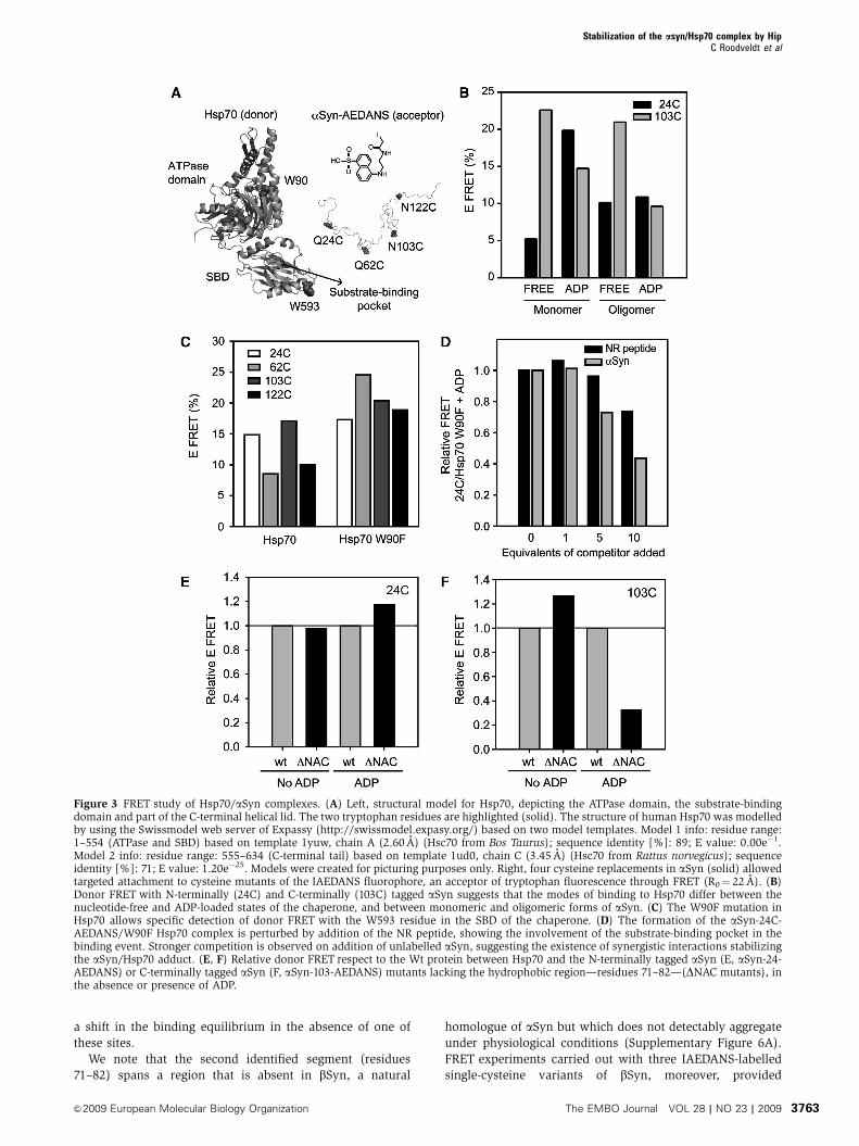
To probe the nature of the various complexes formed
between different forms of aSyn and Hsp70, we devised a
FRET-based spectroscopic strategy. The two naturally occur-
ring tryptophan residues in Hsp70, Trp90 in the ATPase
domain and Trp593 in the SBD, were used as donors, and
IAEDANS (5-((2-[(iodoacetyl)amino]ethyl)amino)naphtha-
lene-1-sulfonic acid), a widely used dye with a Forster radius
(R0) of 22 A (Matsumoto and Hammes, 1975; Jeganathan
et al, 2006), as an acceptor. In each case, IAEDANS was
attached to aSyn through one of four single-cysteine replace-
ments created in the protein (Gln24Cys, Gln62Cys,
Asn103Cys and Asn122Cys) (Figure 3A) and FRET experi-
ments were carried out with purified fractions of both mono-
meric and oligomeric forms of aSyn (Supplementary Figure
3F). For monomeric aSyn-24-AEDANS, large changes (4-fold
increase) in FRET efficiency were observed upon addition of
nucleotides (Figure 3B), whereas the highest FRET signal was
observed when the high or low affinity states in Hsp70 were
stabilized with ADP or ATPgS, respectively (Figure 3B;
Supplementary Figure 3F). For oligomeric aSyn-24-
AEDANS, by contrast, the FRET efficiency was found to be
largely nucleotide independent. For aSyn-103-AEDANS, we
observed that the FRETefficiency was greatest for the nucleo-
tide-free state of Hsp70, with no significant difference be-
tween the monomeric and oligomeric forms of aSyn
(Figure 3B).
A Trp90Phe mutant bearing a unique Trp residue located in
the SBD (see Supplementary data) was also generated to
enable FRET measurements with isolated donor/acceptor
pairs. Given that the ADP-state of Hsp70 seems to be a key
determinant of the substrate-mediated co-aggregation of the
chaperone (Figure 1E and F; Supplementary Figure 2C), we
explored its interaction with aSyn in further detail. A large
increase in FRET efficiency towards aSyn-AEDANS was ob-
served for the ADP-loaded Trp90Phe mutant compared with
that seen with the wild-type chaperone (Figure 3C), indicat-
ing that these FRET measurements essentially report on the
binding of aSyn to the SBD of Hsp70. Indeed, the NR peptide
was found to compete with aSyn-24-AEDANS for binding to
Trp90Phe-Hsp70, as the FRET efficiency decreased up to
B25% at a 10-fold molar excess (Figure 3D). Notably,
unlabelled aSyn reduced the transfer efficiency by almost
50% under the same conditions, suggesting that monomeric
aSyn interacts with Hsp70 with a higher apparent affinity
than does the NR peptide. Importantly, independent confir-
mation of the complex formation in the presence of nucleo-
tide was obtained by heteronuclear NMR spectroscopy
(Supplementary data and Supplementary Figure 5). Indeed,
by using 13C-detected 13CO-15N (CON) correlation experi-
ments on 13C-15N-labelled aSyn (Hsu, 2009), we found
that the addition of Hsp70 and ADP perturbed resonances
specifically at the N-terminus and NAC region of aSyn
(Supplementary Figure 5A and B).
Mapping the recognition site for Hsp70 on aSyn
We have used an algorithm (Rudiger et al, 1997b) to predict
the regions of aSyn where binding to Hsp70 is likely to be
strongest. Two segments show relatively high scores: residues
32–43 and residues 71–82. The predicted free energies of
binding (DDG) are �9.7 and �4.3 kJ/mol, respectively
(Supplementary Figure 6A).
FRET studies with labelled variants of an aSyn derivative
lacking the second putative binding segment, termed DNAC
(residues 71–82) (Rivers et al, 2008), show that, as predicted,
this region is involved in the binding process. We found that
the interaction of Hsp70 with the DNAC aSyn-24-AEDANS
mutant is not very different from that of Wt aSyn (Figure 3E).
However, the DNAC aSyn-103-AEDANS species in the pre-
sence of ADP (Figure 3F) shows a dramatic reduction in the
FRET efficiency (ca. 70% lower) when compared with full-
length aSyn, indicating the importance of the NAC region.
Moreover, nucleotide-free Hsp70 shows a slightly higher (ca.
30% greater) FRET with DNAC aSyn-103-AEDANS than with
the full-length protein. Taken together, these results suggest
that both the predicted binding sites are important for the
interaction with Hsp70 in the presence of nucleotide.
Furthermore, the increase in the relative signals observed
under certain nucleotide conditions when removing the NAC
region, suggests a competition between the binding sites, and
Stabilization of the asyn/Hsp70 complex by HipC Roodveldt et al
The EMBO Journal VOL 28 | NO 23 | 2009 &2009 European Molecular Biology Organization3762
a shift in the binding equilibrium in the absence of one of
these sites.
We note that the second identified segment (residues
71–82) spans a region that is absent in bSyn, a natural
homologue of aSyn but which does not detectably aggregate
under physiological conditions (Supplementary Figure 6A).
FRET experiments carried out with three IAEDANS-labelled
single-cysteine variants of bSyn, moreover, provided
Figure 3 FRET study of Hsp70/aSyn complexes. (A) Left, structural model for Hsp70, depicting the ATPase domain, the substrate-bindingdomain and part of the C-terminal helical lid. The two tryptophan residues are highlighted (solid). The structure of human Hsp70 was modelledby using the Swissmodel web server of Expassy (http://swissmodel.expasy.org/) based on two model templates. Model 1 info: residue range:1–554 (ATPase and SBD) based on template 1yuw, chain A (2.60 A) (Hsc70 from Bos Taurus); sequence identity [%]: 89; E value: 0.00e�1.Model 2 info: residue range: 555–634 (C-terminal tail) based on template 1ud0, chain C (3.45 A) (Hsc70 from Rattus norvegicus); sequenceidentity [%]: 71; E value: 1.20e�25. Models were created for picturing purposes only. Right, four cysteine replacements in aSyn (solid) allowedtargeted attachment to cysteine mutants of the IAEDANS fluorophore, an acceptor of tryptophan fluorescence through FRET (R0¼ 22 A). (B)Donor FRET with N-terminally (24C) and C-terminally (103C) tagged aSyn suggests that the modes of binding to Hsp70 differ between thenucleotide-free and ADP-loaded states of the chaperone, and between monomeric and oligomeric forms of aSyn. (C) The W90F mutation inHsp70 allows specific detection of donor FRET with the W593 residue in the SBD of the chaperone. (D) The formation of the aSyn-24C-AEDANS/W90F Hsp70 complex is perturbed by addition of the NR peptide, showing the involvement of the substrate-binding pocket in thebinding event. Stronger competition is observed on addition of unlabelled aSyn, suggesting the existence of synergistic interactions stabilizingthe aSyn/Hsp70 adduct. (E, F) Relative donor FRET respect to the Wt protein between Hsp70 and the N-terminally tagged aSyn (E, aSyn-24-AEDANS) or C-terminally tagged aSyn (F, aSyn-103-AEDANS) mutants lacking the hydrophobic region—residues 71–82—(DNAC mutants), inthe absence or presence of ADP.
Stabilization of the asyn/Hsp70 complex by HipC Roodveldt et al
&2009 European Molecular Biology Organization The EMBO Journal VOL 28 | NO 23 | 2009 3763
evidence for the formation of a complex between bSyn
and Hsp70 and showed the nucleotide dependence of the
interaction involved (Supplementary Figure 6B). When com-
pared with the equivalent experiment with aSyn, a similar
FRET profile was observed for both complexes (see
Figure 3B), except that the C-terminally labelled derivative
(102 in aSyn and 103 in bSyn) showed significantly lower
FRET efficiency for bSyn than for aSyn. This difference is
more pronounced in the absence of nucleotides and likely
reflects the sequence variations at the C-termini of both
proteins as well as it strongly supports the involvement of
the C-terminus of aSyn in the interaction with nucleotide-free
Hsp70. Taken together, these results indicate that ADP-Hsp70
interacts with both predicted regions of aSyn, whereas
nucleotide-free Hsp70 could interact, in addition, with the
C-terminal tail of the protein.
Modulation of the anti-amyloid activity of Hsp70
by co-chaperones
The co-chaperone Hsp40 is generally thought to act together
with Hsp70 and regulate complex formation with client
polypeptides (Minami et al, 1996; Bukau and Horwich,
1998). To explore its effect on the anti-amyloidogenic cap-
abilities of Hsp70, we performed experiments in which Hsp40
was included in solutions of aggregating aSyn and Hsp70.
The addition of Hsp40, however, had no detectable effect on
the conversion of aSyn into fibrils, and did not modify the
chaperone activity of Hsp70, either in the free or in the
nucleotide-bound states (Supplementary Figure 6C).
Recently, the Hsp70-interacting protein Hip (ST13) has
been found to be significantly under-expressed in serum
taken from patients suffering from PD (Scherzer et al,
2007). Hip modulates Hsp70 chaperone activity both in
vitro (Hohfeld et al, 1995) and in vivo (Nollen et al, 2001),
by interacting with the ATPase domain of the chaperone in its
ADP-bound state (Hohfeld et al, 1995). As nothing is yet
known concerning the possible effects of Hip on the anti-
amyloidogenic activity of Hsp70, we have investigated this
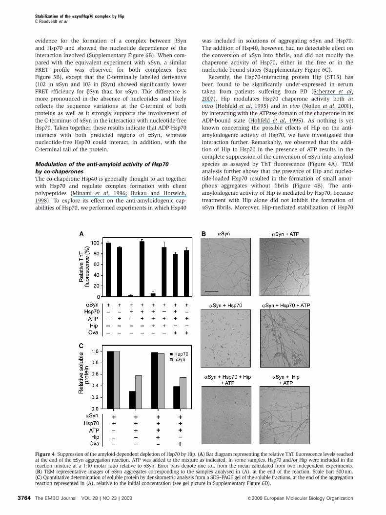
interaction further. Remarkably, we observed that the addi-
tion of Hip to Hsp70 in the presence of ATP results in the
complete suppression of the conversion of aSyn into amyloid
species as assayed by ThT fluorescence (Figure 4A). TEM
analysis further shows that the presence of Hip and nucleo-
tide-loaded Hsp70 resulted in the formation of small amor-
phous aggregates without fibrils (Figure 4B). The anti-
amyloidogenic activity of Hip is mediated by Hsp70, because
treatment with Hip alone did not inhibit the formation of
aSyn fibrils. Moreover, Hip-mediated stabilization of Hsp70
Figure 4 Suppression of the amyloid-dependent depletion of Hsp70 by Hip. (A) Bar diagram representing the relative ThT fluorescence levels reachedat the end of the aSyn aggregation reaction. ATP was added to the mixture as indicated. In some samples, Hsp70 and/or Hip were included in thereaction mixture at a 1:10 molar ratio relative to aSyn. Error bars denote one s.d. from the mean calculated from two independent experiments.(B) TEM representative images of aSyn aggregates corresponding to the samples analysed in (A), at the end of the reaction. Scale bar: 500nm.(C) Quantitative determination of soluble protein by densitometric analysis from a SDS–PAGE gel of the soluble fractions, at the end of the aggregationreaction represented in (A), relative to the initial concentration (see gel picture in Supplementary Figure 6D).
Stabilization of the asyn/Hsp70 complex by HipC Roodveldt et al
The EMBO Journal VOL 28 | NO 23 | 2009 &2009 European Molecular Biology Organization3764
completely abrogated the chaperone co-aggregation that
occurs in the presence of ATP, and appears to act by
maintaining both aSyn and Hsp70 in solution (Figure 4C;
Supplementary Figure 6D). This data suggests that Hip exerts
a stabilizing effect on Hsp70 that supports chaperone-
mediated inhibition of amyloid formation.
Inactivation of Hip increases a-Syn inclusion formation
in C. elegans in an Hsp70-dependent manner
To establish the relevance of the anti-amyloidogenic function
of the Hsp70/Hip complex in vivo, we have exploited a
C. elegans model of aSyn inclusion formation (van Ham
et al, 2008) and used knock-down techniques to suppress
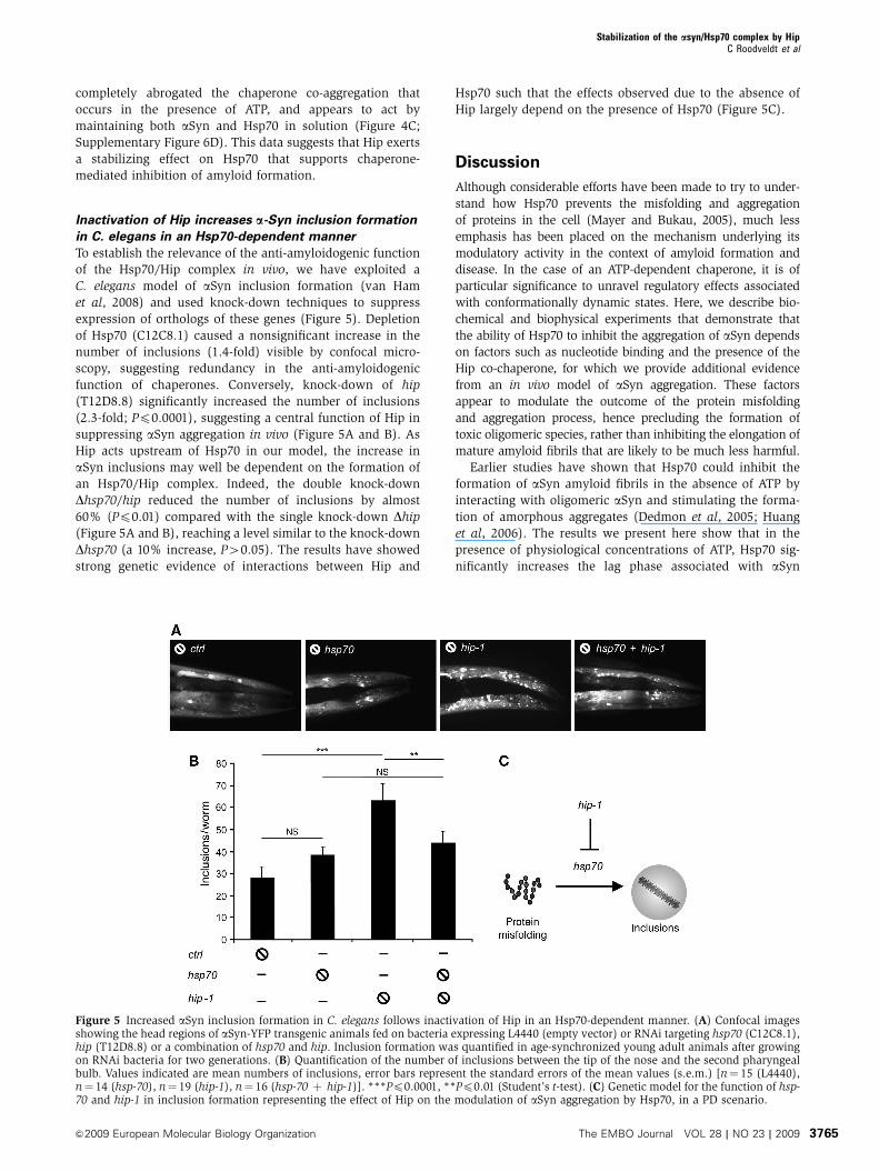
expression of orthologs of these genes (Figure 5). Depletion
of Hsp70 (C12C8.1) caused a nonsignificant increase in the
number of inclusions (1.4-fold) visible by confocal micro-
scopy, suggesting redundancy in the anti-amyloidogenic
function of chaperones. Conversely, knock-down of hip
(T12D8.8) significantly increased the number of inclusions
(2.3-fold; Pp0.0001), suggesting a central function of Hip in
suppressing aSyn aggregation in vivo (Figure 5A and B). As
Hip acts upstream of Hsp70 in our model, the increase in
aSyn inclusions may well be dependent on the formation of
an Hsp70/Hip complex. Indeed, the double knock-down
Dhsp70/hip reduced the number of inclusions by almost
60% (Pp0.01) compared with the single knock-down Dhip
(Figure 5A and B), reaching a level similar to the knock-down
Dhsp70 (a 10% increase, P40.05). The results have showed
strong genetic evidence of interactions between Hip and
Hsp70 such that the effects observed due to the absence of
Hip largely depend on the presence of Hsp70 (Figure 5C).
Discussion
Although considerable efforts have been made to try to under-
stand how Hsp70 prevents the misfolding and aggregation
of proteins in the cell (Mayer and Bukau, 2005), much less
emphasis has been placed on the mechanism underlying its
modulatory activity in the context of amyloid formation and
disease. In the case of an ATP-dependent chaperone, it is of
particular significance to unravel regulatory effects associated
with conformationally dynamic states. Here, we describe bio-
chemical and biophysical experiments that demonstrate that
the ability of Hsp70 to inhibit the aggregation of aSyn depends
on factors such as nucleotide binding and the presence of the
Hip co-chaperone, for which we provide additional evidence
from an in vivo model of aSyn aggregation. These factors
appear to modulate the outcome of the protein misfolding
and aggregation process, hence precluding the formation of
toxic oligomeric species, rather than inhibiting the elongation of
mature amyloid fibrils that are likely to be much less harmful.
Earlier studies have shown that Hsp70 could inhibit the
formation of aSyn amyloid fibrils in the absence of ATP by
interacting with oligomeric aSyn and stimulating the forma-
tion of amorphous aggregates (Dedmon et al, 2005; Huang
et al, 2006). The results we present here show that in the
presence of physiological concentrations of ATP, Hsp70 sig-
nificantly increases the lag phase associated with aSyn
Figure 5 Increased aSyn inclusion formation in C. elegans follows inactivation of Hip in an Hsp70-dependent manner. (A) Confocal imagesshowing the head regions of aSyn-YFP transgenic animals fed on bacteria expressing L4440 (empty vector) or RNAi targeting hsp70 (C12C8.1),hip (T12D8.8) or a combination of hsp70 and hip. Inclusion formation was quantified in age-synchronized young adult animals after growingon RNAi bacteria for two generations. (B) Quantification of the number of inclusions between the tip of the nose and the second pharyngealbulb. Values indicated are mean numbers of inclusions, error bars represent the standard errors of the mean values (s.e.m.) [n¼ 15 (L4440),n¼ 14 (hsp-70), n¼ 19 (hip-1), n¼ 16 (hsp-70 þ hip-1)]. ***Pp0.0001, **Pp0.01 (Student’s t-test). (C) Genetic model for the function of hsp-70 and hip-1 in inclusion formation representing the effect of Hip on the modulation of aSyn aggregation by Hsp70, in a PD scenario.
Stabilization of the asyn/Hsp70 complex by HipC Roodveldt et al
&2009 European Molecular Biology Organization The EMBO Journal VOL 28 | NO 23 | 2009 3765
aggregation, such that amyloid fibrils still appear but typi-
cally much more slowly than when otherwise be the case
(Figure 1). This finding is consistent with in vivo observa-
tions where over-expression of Hsp70 was found to reduce
aSyn toxicity, but did not prevent the accumulation of
amyloid aggregates in tissue (Auluck et al, 2002). We find
the inhibitory effect of Hsp70 in the presence of ATP to be
dependent on the Hsp70/aSyn ratio, and have observed that a
combination of both aSyn and ATP, or its hydrolytic product
ADP, causes Hsp70 itself to aggregate, regardless of the
presence of Hsp40. These observations are in line with earlier
findings related to HD, in which treatment of amyloidogenic
huntingtin with Hsc70-Hsp40 and ATP disfavoured the popu-
lation of oligomeric species and resulted in the accumulation
of amyloid fibrils (Muchowski et al, 2000; Wacker et al,
2004). Moreover, the finding that Hsp70 has a tendency to
aggregate in the presence of aSyn and ATP (or ADP) provides
the basis for the well-established co-localization of Hsp70 and
aSyn in Lewy bodies (Lee and Lee, 2002; Muchowski and
Wacker, 2005). The depletion of functional chaperones and
co-chaperones would heavily impair the ability of proteins to
resist aggregation and to maintain protein homeostasis, phe-
nomena that are thought to lie at the foundations of amyloid
diseases (Dobson, 2003; Balch et al, 2008).
An interesting mechanistic observation from the current
studies is that the addition of the competing peptide substrate
NR does not detectably disrupt the efficacy of Hsp70 to act as
a chaperone towards aSyn in the nucleotide-free state, and
still allows the inhibition of amyloid formation by Hsp70. In
the nucleotide-loaded state, however, the NR peptide reduces
the extent of the co-aggregation of Hsp70 with aSyn
(Figure 1), probably by competing with the protein for the
substrate-binding pocket (Figure 3). These results suggest
strongly the existence of distinct modes of binding for aSyn to
nucleotide-loaded or nucleotide-free Hsp70, i.e. canonical
and non-canonical binding modes, which are likely to deter-
mine the result of the aggregation reaction (Figure 6). FRET
and NMR spectroscopy have enabled us to discover that
Hsp70 does indeed recognize and bind to aSyn monomers
as well as oligomers through at least three different types of
interactions (Figure 3; Supplementary Figures 3–5). In the
ADP-loaded state, aSyn monomers are located closer to the
substrate-binding pocket of Hsp70, an interaction mediated
by two regions, present in the N-terminal and NAC region of
aSyn. We propose that this compact nucleotide-Hsp70/mono-
meric aSyn complex is critical in delaying the onset of fibril
formation, but could also be responsible for the co-aggregation
of Hsp70 and aSyn. A recent study mapped the region recog-
nized on aSyn by Hsp70 as the broad segment between residues
21 and 110 (Luk et al, 2008). We have refined this region and
located two stretches of amino acids with the highest prob-
ability of binding (residues 32–43 and 71–83) and then show
that Hsp70 binds to the latter site, the stretch of hydrophobic
residues that readily forms amyloid fibrils in vitro, and is
generally assumed to be involved in initiating the conversion
of aSyn into amyloid fibrils (Biere et al, 2000; Giasson et al,
2001). Moreover, binding to the N-terminal region of aSyn is
supported by a comparative study of bSyn, in which we pro-
vide evidence of a complex formed with Hsp70, dispelling the
general assumption that such an interaction is unlikely to occur.
Although likely to be less physiologically relevant, we have
found that nucleotide-free Hsp70 also interacts with mono-
meric aSyn through which appears to be a non-canonical
mode of interaction. This leads to the stabilization of soluble
amorphous aggregates and hence inhibits fibril formation.
These results are in line with the proposition (Gao et al, 1995)
that when the nucleotide is absent from the nucleotide-
binding site of Hsc70—the constitutively expressed analogue
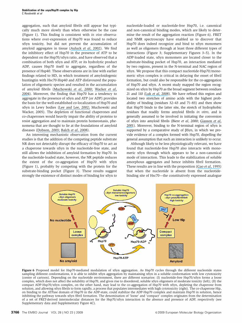
Figure 6 Proposed model for Hsp70-mediated modulation of aSyn aggregation. As Hsp70 cycles through the different nucleotide statessampling different conformations, it is able to inhibit aSyn aggregation by maintaining aSyn in a soluble conformation with low cytotoxicity(centre of cartoon). Depending on the nucleotide environment, there are different scenarios: (I) nucleotide-free Hsp70/aSyn forms a loosecomplex, which does not affect the solubility of Hsp70, and gives rise to disordered, soluble aSyn oligomers of moderate toxicity (left); (II) thecompact ADP-Hsp70/aSyn complex, on the other hand, may lead to the co-aggregation of Hsp70 with aSyn, depleting the chaperone fromsolution, and allowing aSyn fibrils to form rapidly, a process that populates intermediates with high cytotoxicity (right). The co-chaperone Hip,on binding to the ATPase domain of Hsp70 in the ADP-state, could stabilize the ADP-Hsp70 complex and maintain Hsp70 in solution, henceinhibiting the pathway towards aSyn fibril formation. The denomination of ‘loose’ and ‘compact’ complex originates from the determinationof a set of FRET-derived intermolecular distances for the Hsp70/aSyn interaction in the absence and presence of ADP, respectively (seeSupplementary data and Supplementary Figure 4C).
Stabilization of the asyn/Hsp70 complex by HipC Roodveldt et al
The EMBO Journal VOL 28 | NO 23 | 2009 &2009 European Molecular Biology Organization3766
of Hsp70—the substrate-binding region interacts more flex-
ibly with a protein substrate. Possibly such transient interac-
tions, recently suggested for the Hsp70/aSyn complex
(Luk et al, 2008), do not compromise the solubility of
Hsp70. With ADP in the nucleotide-binding site, however,
the SBD appears to be much less dynamic, as the residence
time of the substrate in the binding pocket is increased.
Finally, the FRET data suggest that aSyn oligomers are
preferentially bound by nucleotide-free Hsp70, consistent
with the view that N-terminal and central domains of aSyn
are likely to be buried in the aggregated species.
One possible reason why nucleotide-free Hsp70 impairs
the ability of aSyn to form amyloid fibrils could be related
to an off-pathway nature of the intermediate species
stabilized by the formation of the Hsp70/aSyn complex. Indeed,
we observed non-fibrillar oligomers to be predominantly popu-
lated at early incubation time points in the presence of nucleo-
tide-free chaperone, whereas short protofibrils of aSyn were
found to co-aggregate with Hsp70 in the presence of ATP.
Studies of the effects of such soluble pre-fibrillar aggregates on
a human neuronal cell line have shown that protofibrils initially
formed by treatment with Hsp70 and ATP are less toxic than the
oligomers stabilized by nucleotide-free Hsp70 (Figure 2). This
protective effect of Hsp70 in a medium with ATP, however,
disappears during the course of the elongation phase of fibril
formation in parallel with the depletion of soluble Hsp70. A shift
in population towards highly toxic soluble aSyn species at later
points in the aggregation reaction suggests that Hsp70 is co-
aggregating with less toxic aSyn species.
Biologically, Hsp70 does not function independently as many
co-chaperones and auxiliary factors are involved in regulating its
cellular functions in the cell. In this regard, it is extremely
interesting that the Hsp70-interacting protein Hip (ST13) has
recently been found to be consistently under-expressed in PD
patients even in the early stages of the disease (Scherzer et al,
2007), a conclusion that suggests a coupling between both
proteins in disease progression or initiation. We have found
strong evidence in this study for a dramatic effect of Hip on
the availability of functionally competent Hsp70 in the presence
of aggregating aSyn. Hip is in fact capable of suppressing the co-
aggregation of Hsp70 with aSyn, and hence the extent of amyloid
fibril formation observed in the presence of nucleotides is
virtually completely suppressed in the presence of Hip
(Figure 4). Moreover, our results with an in vivo aSyn aggrega-
tion model of C. elegans strongly support the hypothesis derived
from the in vitro experiments, indicating that Hip is indeed
required for suppression of aSyn inclusion formation in an
Hsp70-dependent manner (Figure 5). In line with this important
finding, a recent study of a polyQ model of HD found that Hip
assists Hsp70 in the anti-aggregation activity of Hsp70 (Howarth
et al, 2009). The observation in our C. elegans model that the
absence of Hip alone could give rise to more inclusions than
when both Hip and Hsp70 are absent is consistent with a
scenario in which there is a redundancy of chaperone pathways,
likely mediated by the constitutive presence of Hsc70 and other
chaperones such as Hsp90 (Uryu et al, 2006). Interestingly, Hip
has been shown to interact with Hsp70 by binding to its ATPase
domain specifically in the ADP-bound state, both in vitro and in
vivo, without affecting its ATPase activity (Hohfeld et al, 1995;
Nollen et al, 2001). A possible explanation for its stabilizing effect
on the ADP-Hsp70/aSyn complex in solution is that the binding
of Hip could shield hydrophobic regions in the ATPase domain of
Hsp70 that become exposed in the ADP-bound complex with
aSyn (Figure 6). Alternatively, we speculate that binding of Hip
to the ATPase domain could induce a structural change in the
SBD, favouring a conformation of Hsp70 similar to that popu-
lated in the nucleotide-free state, which we have shown does not
promote the co-aggregation of Hsp70 and aSyn. This situation
could be reminiscent of that proposed for the Hsp70 escorting
protein (Hep) when bound to the nucleotide-free state of mito-
chondrial Hsp70 (mtHsp70), which inhibits self-aggregation of
mtHsp70, both in vitro and in vivo (Sichting et al, 2005).
In summary, three central conclusions from this study
appear to be of broad importance in the quest to unravel
the highly complex function of chaperone availability and
proteostasis in amyloid diseases. The first is the observation
that the ATP cycle modulates the ability of Hsp70 to inhibit
fibril formation by amyloid-forming proteins, shedding light
on the mechanism by which ATP-dependent chaperones act
in the context of misfolding diseases. The second important
finding is that ADP-bound Hsp70 has a very high propensity
to co-aggregate with aSyn, suggesting that chaperone deple-
tion favoured under certain conditions could be an important
feature in the onset and progression of amyloid disorders.
Finally, we have identified a functional role of Hip, an
auxiliary factor for Hsp70, in preventing the co-aggregation
of Hsp70 with aSyn, thereby reducing the toxicity of amyloi-
dogenesis. Maintaining the cellular level of Hip may be one
solution for intervening the onset and development of PD.
Materials and methods
Further details of the Materials and methods are provided inSupplementary data.
MaterialsATP, ADP and ThT were purchased from Sigma. Adenosine-50-O-(3-thio triphosphate) (ATPgS, 490% purity, HPLC-purified) waspurchased from Roche. The NR peptide (H2N-NRLLLTG-COOH) wassynthesized and purified (495% purity) by Genemed Synthesis Inc.(USA). IAEDANS and 5-(dimethylamino)naphthalene-1-sulfonyl-methyl sulfoxide (DANSYL-MTS) were obtained from Invitrogenand Toronto Research, respectively.
Protein expression and purificationHuman cytoplasmic Hsp70 (Hsp70 1A, gi:194248072) was ex-pressed, purified and characterized as described in Supplementarydata. Recombinant human Hsp40 and purified rat Hip wereobtained from Stressgen (#SPP-400 and SPP-767, respectively).The wild-type human aSyn (gi:80475099) or bSyn (gi:12804099)genes were inserted in pT7-7 and pRK172 vectors, respectively, andsubsequently expressed and purified as described earlier (Riverset al, 2008). Protein purity exceeded 95% as determined by SDS–PAGE, and the aSyn concentration was determined from theabsorbance measurements at 275 nm using an extinction coefficientof 5400 M�1cm�1.
aSyn aggregation experimentsSolutions for aggregation experiments contained 70 mM aSyn aloneor together with 7mM Hsp70 (except where a ratio other than 1:10 isindicated) in 50 mM Tris (pH 7.4), 50 mM KCl, 2 mM MgCl2,0.35 mM SDS and 0.02% NaN3. Where indicated, samplescontained and ATP-regeneration system: 0.2 units/ml pyruvatekinase and 5 mM phosphoenol pyruvate, in the absence or presenceof 2 mM ATP (or ADP or ATPgS). Alternatively, reactions werecarried out in the presence of 5 mM nucleotide in the absence of anATP-regeneration system. Formation of fibrils by a-Syn wasmonitored by measuring ThT fluorescence and fitting to the kineticsfollowed a nucleation polimerization model as described elsewhere(Rivers et al, 2008). For kinetic aggregation studies using SDS,either two or three replicas were used for each set of conditions. Inthese samples, 20 mM ThT was included before initiating the
Stabilization of the asyn/Hsp70 complex by HipC Roodveldt et al
&2009 European Molecular Biology Organization The EMBO Journal VOL 28 | NO 23 | 2009 3767

aggregation reaction. Aggregation was induced by heating thesamples in a 96-well plate to 371C while shaking, and readings weretaken every B7 min in a FluoStar OPTIMA spectrophotometer withexcitation and emission windows at 440±10 and 480±10 nm,respectively, and an averaging time of 20 s. For kinetic studies in theabsence of SDS, aggregation was induced by magnetic stirring at371C, and ThT was measured in a Cary Eclipse spectrofluorimeterwith an excitation wavelength of 440 nm (slit-width 5 nm) and anemission scan from 450 to 600 nm (slit-width 5 nm). It might berelevant to note that this Hsp70-ATP-dependent, aSyn fibrilformation was not related to the presence or absence of SDS inthe reaction mixture (Supplementary Figure 2A).
SDS–PAGE analysisSamples were analysed by SDS–PAGE using 4–12% Bis-TrisNuPAGE gels (Invitrogen) in MES buffer under reducing conditions.Gels were stained using Coomassie brilliant blue dye. Densitometryanalysis was performed on scanned gels (Supplementary Figures 2and 6) using the Image J (NIH) software.
Cytotoxicity assaysSamples for cytotoxicity assays were taken from aggregation assaysperformed in the absence of ThT. Aliquots were removed at 0, 4, 8,24, 42 and 64 h time periods and subjected to electron microscopy,ex situ measurement of ThT fluorescence intensity, and theremaining stored at �801C for cytotoxicity studies. Where required,samples were centrifuged at 16 000� g for 10 min to separate theaggregates into insoluble and soluble fractions. The retention ofaggregate morphology after freeze/thawing was confirmed byelectron microscopy (not shown). LDH was measured on SH-SY5Y cells as a way of parametrizing the differential toxicity ofaggregated samples on the viability, as described in Supplementarydata.
Protein labelling and purification of oligomersLabelling of aSyn and bSyn cysteine mutants with IAEDANS orDANSYL-MTS was performed as follows: 1–5 mg of lyophilized Cys-containing proteins were dissolved in PBS and 5 mM DTT wasadded to ensure complete reduction of the sole cysteine residue.DTTwas removed by gel filtration in PD10 columns (GE Healthcare)and a five-fold molar excess of the dye (dissolved in DMSO) wasthen added immediately. Conjugation of the dye was optimized byovernight incubation at 41C in the dark. Excess dye was removed byperforming an added gel filtration step using PD10 columns(Sepharose G-25). The eluted proteins were subjected to sizeexclusion in an analytical Superdex 75 column (GE Healthcare) toseparate oligomeric and monomeric species. Proteins were con-centrated by centrifugal devices (10 kDa cut-off for monomer and100 kDa for oligomers) and stored at �801C. Labelling efficiencywas typically found to be between 60 and 95%.
Fluorescence spectroscopyFor FRET experiments, the tryptophan residues of Hsp70 acted asdonors and AEDANS as acceptors (Jeganathan et al, 2006), usingAEDANS-labelled aSyn or bSyn cysteine-mutants. In all theexperiments, FRETwas determined as donor desensitization, exceptfor the case of the aggregation experiments, where amyloid-dependent insolubility of Hsp70 precluded the accurate determina-tion of donor quenching. In this case, the acceptor sensitizationmethod was used. For determinations of the relative affinity of theaSyn/Hsp70 complex, aSyn cysteine mutants labelled with theDANSYL fluorophore were used.
Transmission electron microscopySamples were prepared from 10ml aliquots of aggregation reactionsusing negative staining by 2% (w/v) uranyl acetate on Formvar/carbon-coated copper grids (Agar Scientific). Images were obtained
at � 25 000 magnification using a Phillips CEM100 transmissionelectron microscope.
C. elegans strain and RNAiThe transgenic aSyn strain used for RNAi experiments, OW40(OW40 zgIs15[P(unc-54)::a-synuclein::YFP(xScaI)N2(xPvuII)] wascreated by microinjection and integrated by g-irradiation. Synchro-nized larvae were fed on bacterial strains expressing double-stranded RNA as described earlier (van Ham et al, 2008). An RNAiclone targeting hsp-70 (C12C8.1) was obtained from the Ahringerbacterial RNAi library. We constructed an RNAi clone targetinghip-1 (T12D8.8) by cloning an B1000 bp DNA fragment (generated byPCR amplification with primers: T12D8.8_F: 50-CTAAGCTAGCGAAAATGGACCACGTCGC ATTG-30 and T12D8.8_R: 50-GATACTCGAGACTTGTCTTGTCGCGAAGCA-30 into L4440. Worms were grown on thedifferent RNAi clones and allowed to have progeny. L4 progeny(second generation) was transferred to new RNAi agar plates andinclusions were quantified in age-synchronized adults. All genetargets of the positive-RNAi used for feeding were verified bysequencing of the insert of the RNAi plasmids.
Solamere confocal laser scanning microscopyTransgenic animals were mounted on glass microscope slides on2% agarose pads containing 40 mM NaN3 as an anaesthetic.Animals were imaged using a Solamere Nipkow confocal live cellimaging system (Solamere, USA) based on a Leica DM IRE2 invertedconfocal microscope with a 40� oil immersion objective (HCX PLAPO CS). Images shown are 2D maximal projections of z-stacks andwere captured using In Vivo3 Software (Mediacybernetics, USA).For quantification of the number of inclusions, all distinct focibetween the nose and second pharyngeal bulb were counted.Measurements on inclusions were performed using ImageJ soft-ware. Statistical significance was determined using Student’s t-tests.
Supplementary dataSupplementary data are available at The EMBO Journal Online(http://www.embojournal.org).
Acknowledgements
We are grateful to Drs Robert Rivers, Xavier Salvatella and SophieJackson for reagents and discussions. We acknowledge with grati-tude the use of the Biomolecular NMR Facility, Department ofChemistry, University of Cambridge, at Cambridge, UK, and theadvice of the staff. We thank K Sjollema from the UMCGMicroscopy and Imaging Center (UMIC) for advice on microscopy.CR and AA held FEBS Long-Term Fellowships. CWB is an EMBOLong-Term Postdoctoral Fellow, ATvdG was the recipient of aTopmaster fellowship of the graduate school GUIDE for DrugExploration of the University of Groningen. STDH is a recipient ofa Human Frontier Science Program Long-termFellowship (LT0798/2005) and is supported in part by the National Science Council ofthe Republic of China, Taiwan(NSC97-2917-1-564-102). JC is reci-pient of a Human Frontier Young Investigators Award (RGY67/2007) and also thanks the BBSRC (9015651/1). CMD and JCacknowledge funding from The Wellcome Trust and TheLeverhulme Trust. DP is grateful to The Spanish Ministry ofHealth (PI05/2056; PI06/1641), The Spanish Ministry of Scienceand Innovation (SAF2007-29418E) and the PAIDI Program from theRegional Government (BIO323) for funding. EAAN acknowledgesZonMW Research Institute of Diseases in the Elderly and DeNederlandse Hersenstichting for funding.
Conflict of interest
The authors declare that they have no conflict of interest.
References
Auluck PK, Chan HY, Trojanowski JQ, Lee VM, Bonini NM(2002) Chaperone suppression of alpha-synuclein toxicity in aDrosophila model for Parkinson’s disease. Science 295: 865–868
Balch WE, Morimoto RI, Dillin A, Kelly JW (2008) Adaptingproteostasis for disease intervention. Science 319: 916–919
Biere AL, Wood SJ, Wypych J, Steavenson S, Jiang Y, Anafi D,Jacobsen FW, Jarosinski MA, Wu GM, Louis JC, Martin F, NarhiLO, Citron M (2000) Parkinson’s disease-associated alpha-synu-clein is more fibrillogenic than beta- and gamma-synuclein andcannot cross-seed its homologs. J Biol Chem 275: 34574–34579
Stabilization of the asyn/Hsp70 complex by HipC Roodveldt et al
The EMBO Journal VOL 28 | NO 23 | 2009 &2009 European Molecular Biology Organization3768
Bukau B, Horwich AL (1998) The Hsp70 and Hsp60 chaperonemachines. Cell 92: 351–366
Chiti F, Dobson CM (2006) Protein misfolding, functional amyloid,and human disease. Annu Rev Biochem 75: 333–366
Danzer KM, Haasen D, Karow AR, Moussaud S, Habeck M, Giese A,Kretzschmar H, Hengerer B, Kostka M (2007) Different species ofalpha-synuclein oligomers induce calcium influx and seeding.J Neurosci 27: 9220–9232
Dedmon MM, Christodoulou J, Wilson MR, Dobson CM (2005) Heatshock protein 70 inhibits alpha-synuclein fibril formation viapreferential binding to prefibrillar species. J Biol Chem 280:14733–14740
Demuro A, Mina E, Kayed R, Milton SC, Parker I, Glabe CG (2005)Calcium dysregulation and membrane disruption as a ubiquitousneurotoxic mechanism of soluble amyloid oligomers. J Biol Chem280: 17294–17300
Dobson CM (2003) Protein folding and misfolding. Nature 426:884–890
Frydman J, Nimmesgern E, Ohtsuka K, Hartl FU (1994) Folding ofnascent polypeptide chains in a high molecular mass assemblywith molecular chaperones. Nature 370: 111–117
Gao B, Eisenberg E, Greene L (1995) Interaction of nucleotide-freeHsc70 with clathrin and peptide and effect of ATP analogues.Biochemistry 34: 11882–11888
Giasson BI, Murray IV, Trojanowski JQ, Lee VM (2001) A hydro-phobic stretch of 12 amino acid residues in the middle of alpha-synuclein is essential for filament assembly. J Biol Chem 276:2380–2386
Gragerov A, Nudler E, Komissarova N, Gaitanaris GA, GottesmanME, Nikiforov V (1992) Cooperation of GroEL/GroES andDnaK/DnaJ heat shock proteins in preventing protein mis-folding in Escherichia coli. Proc Natl Acad Sci USA 89:10341–10344
Gragerov A, Zeng L, Zhao X, Burkholder W, Gottesman ME (1994)Specificity of DnaK-peptide binding. J Mol Biol 235: 848–854
Greene LE, Zinner R, Naficy S, Eisenberg E (1995) Effect of nucleo-tide on the binding of peptides to 70-kDa heat shock protein.J Biol Chem 270: 2967–2973
Grunblatt E, Mandel S, Maor G, Youdim MB (2001) Gene expressionanalysis in N-methyl-4-phenyl-1,2,3,6-tetrahydropyridine micemodel of Parkinson’s disease using cDNA microarray: effect ofR-apomorphine. J Neurochem 78: 1–12
Hartl FU (1996) Molecular chaperones in cellular protein folding.Nature 381: 571–579
Hauser MA, Li YJ, Xu H, Noureddine MA, Shao YS, Gullans SR,Scherzer CR, Jensen RV, McLaurin AC, Gibson JR, Scott BL,Jewett RM, Stenger JE, Schmechel DE, Hulette CM, Vance JM(2005) Expression profiling of substantia nigra in Parkinsondisease, progressive supranuclear palsy, and frontotemporaldementia with parkinsonism. Arch Neurol 62: 917–921
Hohfeld J, Minami Y, Hartl FU (1995) Hip, a novel cochaperoneinvolved in the eukaryotic Hsc70/Hsp40 reaction cycle. Cell 83:589–598
Howarth JL, Glover CP, Uney JB (2009) HSP70 interacting proteinprevents the accumulation of inclusions in polyglutamine dis-ease. J Neurochem 108: 945–951
Hsu ST, Bertoncini CW, Dobson CM (2009) Use of protonless NMRspectroscopy to alleviate the loss of information resulting fromexchange-broadening. J Am Chem Soc 131: 7222–7223
Huang C, Cheng H, Hao S, Zhou H, Zhang X, Gao J, Sun QH, Hu H,Wang CC (2006) Heat shock protein 70 inhibits alpha-synucleinfibril formation via interactions with diverse intermediates. J MolBiol 364: 323–336
Jeganathan S, von Bergen M, Brutlach H, Steinhoff HJ, MandelkowE (2006) Global hairpin folding of tau in solution. Biochemistry45: 2283–2293
Klucken J, Outeiro TF, Nguyen P, McLean PJ, Hyman BT (2006)Detection of novel intracellular alpha-synuclein oligomeric speciesby fluorescence lifetime imaging. FASEB J 20: 2050–2057
Klucken J, Shin Y, Masliah E, Hyman BT, McLean PJ (2004) Hsp70reduces alpha-synuclein aggregation and toxicity. J Biol Chem279: 25497–25502
Lashuel HA, Lansbury Jr PT (2006) Are amyloid diseases caused byprotein aggregates that mimic bacterial pore-forming toxins? QRev Biophys 39: 167–201
Lashuel HA, Petre BM, Wall J, Simon M, Nowak RJ, Walz T,Lansbury Jr PT (2002) Alpha-synuclein, especially the
Parkinson’s disease-associated mutants, forms pore-like annularand tubular protofibrils. J Mol Biol 322: 1089–1102
Lee HJ, Lee SJ (2002) Characterization of cytoplasmic alpha-synu-clein aggregates. Fibril formation is tightly linked to the inclusion-forming process in cells. J Biol Chem 277: 48976–48983
Luheshi LM, Crowther DC, Dobson CM (2008) Protein misfoldingand disease: from the test tube to the organism. Curr Opin ChemBiol 12: 25–31
Luk KC, Mills IP, Trojanowski JQ, Lee VM (2008) Interactionsbetween Hsp70 and the hydrophobic core of alpha-synucleininhibit fibril assembly. Biochemistry 47: 12614–12625
Macario AJ, De Macario EC (2007) Chaperonopathies by defect,excess, or mistake. Ann N Y Acad Sci 1113: 178–191
Maeda H, Sahara H, Mori Y, Torigo T, Kamiguchi K, Tamura Y,Tamura Y, Hirata K, Sato N (2007) Biological heterogeneityof the peptide-binding motif of the 70-kDa heat shock proteinby surface plasmon resonance analysis. J Biol Chem 282:26956–26962
Matsumoto S, Hammes GG (1975) Fluorescence energy transferbetween ligand binding sites on aspartate transcarbamylase.Biochemistry 14: 214–224
Mayer MP, Brehmer D, Gassler CS, Bukau B (2001) Hsp70 chaper-one machines. Adv Protein Chem 59: 1–44
Mayer MP, Bukau B (2005) Hsp70 chaperones: cellular functionsand molecular mechanism. Cell Mol Life Sci 62: 670–684
Minami Y, Hohfeld J, Ohtsuka K, Hartl FU (1996) Regulation of theheat-shock protein 70 reaction cycle by the mammalian DnaJhomolog, Hsp40. J Biol Chem 271: 19617–19624
Mogk A, Tomoyasu T, Goloubinoff P, Rudiger S, Roder D, Langen H,Bukau B (1999) Identification of thermolabile Escherichia coliproteins: prevention and reversion of aggregation by DnaK andClpB. EMBO J 18: 6934–6949
Muchowski PJ, Schaffar G, Sittler A, Wanker EE, Hayer-Hartl MK,Hartl FU (2000) Hsp70 and hsp40 chaperones can inhibit self-assembly of polyglutamine proteins into amyloid-like fibrils. ProcNatl Acad Sci USA 97: 7841–7846
Muchowski PJ, Wacker JL (2005) Modulation of neurodegenerationby molecular chaperones. Nat Rev Neurosci 6: 11–22
Nollen EA, Kabakov AE, Brunsting JF, Kanon B, Hohfeld J,Kampinga HH (2001) Modulation of in vivo HSP70 chaperoneactivity by Hip and Bag-1. J Biol Chem 276: 4677–4682
Palleros DR, Reid KL, Shi L, Welch WJ, Fink AL (1993) ATP-inducedprotein-Hsp70 complex dissociation requires K+ but not ATPhydrolysis. Nature 365: 664–666
Rivers RC, Kumita JR, Tartaglia GG, Dedmon MM, Pawar A,Vendruscolo M, Dobson CM, Christodoulou J (2008) Moleculardeterminants of the aggregation behavior of alpha- andbeta-synuclein. Protein Sci 17: 887–898
Rudiger S, Buchberger A, Bukau B (1997a) Interaction of Hsp70chaperones with substrates. Nat Struct Biol 4: 342–349
Rudiger S, Germeroth L, Schneider-Mergener J, Bukau B (1997b)Substrate specificity of the DnaK chaperone determined byscreening cellulose-bound peptide libraries. EMBO J 16: 1501–1507
Saibil HR (2008) Chaperone machines in action. Curr Opin StructBiol 18: 35–42
Scherzer CR, Eklund AC, Morse LJ, Liao Z, Locascio JJ, Fefer D,Schwarzschild MA, Schlossmacher MG, Hauser MA, Vance JM,Sudarsky LR, Standaert DG, Growdon JH, Jensen RV, Gullans SR(2007) Molecular markers of early Parkinson’s disease based ongene expression in blood. Proc Natl Acad Sci USA 104: 955–960
Scheufler C, Brinker A, Bourenkov G, Pegoraro S, Moroder L,Bartunik H, Hartl FU, Moarefi I (2000) Structure of TPR do-main-peptide complexes: critical elements in the assembly of theHsp70-Hsp90 multichaperone machine. Cell 101: 199–210
Sichting M, Mokranjac D, Azem A, Neupert W, Hell K (2005)Maintenance of structure and function of mitochondrialHsp70 chaperones requires the chaperone Hep1. EMBO J 24:1046–1056
Stefani M, Dobson CM (2003) Protein aggregation and aggregatetoxicity: new insights into protein folding, misfolding diseasesand biological evolution. J Mol Med 81: 678–699
Takayama S, Reed JC (2001) Molecular chaperone targeting andregulation by BAG family proteins. Nat Cell Biol 3: E237–E241
Uryu K, Richter-Landsberg C, Welch W, Sun E, Goldbaum O, NorrisEH, Pham CT, Yazawa I, Hilburger K, Micsenyi M, Giasson BI,Bonini NM, Lee VM, Trojanowski JQ (2006) Convergence of
Stabilization of the asyn/Hsp70 complex by HipC Roodveldt et al
&2009 European Molecular Biology Organization The EMBO Journal VOL 28 | NO 23 | 2009 3769
heat shock protein 90 with ubiquitin in filamentous alpha-synu-clein inclusions of alpha-synucleinopathies. Am J Pathol 168:947–961
van Ham TJ, Thijssen KL, Breitling R, Hofstra RM, Plasterk RH,Nollen EA (2008) C. elegans model identifies genetic modifiers ofalpha-synuclein inclusion formation during aging. PLoS Genet 4:e1000027
Wacker JL, Zareie MH, Fong H, Sarikaya M, Muchowski PJ (2004)Hsp70 and Hsp40 attenuate formation of spherical and annular
polyglutamine oligomers by partitioning monomer. Nat StructMol Biol 11: 1215–1222
Young JC, Agashe VR, Siegers K, Hartl FU (2004) Pathways ofchaperone-mediated protein folding in the cytosol. Nat Rev MolCell Biol 5: 781–791
Zhou Y, Gu G, Goodlett DR, Zhang T, Pan C, Montine TJ, MontineKS, Aebersold RH, Zhang J (2004) Analysis of alpha-synuclein-associated proteins by quantitative proteomics. J Biol Chem 279:39155–39164
Stabilization of the asyn/Hsp70 complex by HipC Roodveldt et al
The EMBO Journal VOL 28 | NO 23 | 2009 &2009 European Molecular Biology Organization3770
Copyright © 2022 FDOKUMEN