Exploring the accessible conformations of N-terminal acetylated α-synuclein
11
Review Exploring the accessible conformations of N-terminal acetylated a-synuclein Gina M. Moriarty a , Maria K. Janowska a , Lijuan Kang a , Jean Baum a,b,⇑ a Department of Chemistry and Chemical Biology, Rutgers University, Piscataway, NJ 08854, United States b BioMaPS Institute for Quantitative Biology, Rutgers University, Piscataway, NJ 08854, United States article info Article history: Received 4 February 2013 Revised 28 February 2013 Accepted 28 February 2013 Available online 13 March 2013 Edited by Wilhelm Just Keywords: a-Synuclein Acetylation Aggregation Ensemble Fibril Fibril-resistance Intrinsically disordered protein Oligomer Parkinson’s disease abstract Alpha synuclein (asyn) fibrils are found in the Lewy Bodies of patients with Parkinson’s disease (PD). The aggregation of the asyn monomer to soluble oligomers and insoluble fibril aggregates is believed to be one of the causes of PD. Recently, the view of the native state of asyn as a monomeric ensemble was challenged by a report suggesting that asyn exists in its native state as a helical tet- ramer. This review reports on our current understanding of asyn within the context of these recent developments and describes the work performed by a number of groups to address the monomer/ tetramer debate. A number of in depth studies have subsequently shown that both non-acetylated and acetylated asyn purified under mild conditions are primarily monomer. A description of the accessible states of acetylated asyn monomer and the ability of asyn to self-associate is explored. Ó 2013 Federation of European Biochemical Societies. Published by Elsevier B.V. All rights reserved. 1. Introduction Parkinson’s disease (PD) research has sought to answer ques- tions of alpha synuclein (asyn) function and the mechanism of aggregation surrounding disease pathology. Both remain to be fully articulated today, but several observations have been established and a range of neurodegenerative diseases termed the ‘‘synuclein- opathies’’ have been identified [1,2]. PD in particular is the synuc- leinopathy characterized by the loss of dopaminergic neurons and is largely considered to be an age-related disease, accompanied in part by age-related deposition of asyn [3]. asyn, a major protein component of Lewy Bodies [4,5] in patients with Parkinson’s, is a small primarily neuronal protein that is known to make a struc- tural transition to amyloid fibrils [6–8]. asyn is expressed abun- dantly in the nervous system and localizes near presynaptic nerve terminals [9–13]. It is also expressed at high levels in eryth- rocytes and platelets [14]. asyn’s function is unknown, but there is strong evidence that it exhibits lipid binding in vesicles and synap- tic membranes [15] and may somehow exert its pathology through this behavior [16]. There is evidence that asyn functions in assem- bly of the SNARE complex involved in vesicle transport [17], that it may more generally be involved in synaptic vesicle trafficking and regulation and/or may play a key role in neuronal cell survival [18– 22]. The deposition of asyn has largely been thought to originate from an intrinsically disordered monomer ensemble that under fi- bril promoting conditions forms amyloid [7,23,24], but recently this view of asyn’s native state was challenged [25]. Selkoe and colleagues pushed the biophysical community’s long-held view of asyn as an intrinsically disordered monomer by suggesting that the protein exists in its native state as a fibril-resistant helical tet- ramer. They purified the sample from human erythrocytes, opting to exclude a potentially ‘‘harsh’’ and commonly used boiling step 0014-5793/$36.00 Ó 2013 Federation of European Biochemical Societies. Published by Elsevier B.V. All rights reserved. http://dx.doi.org/10.1016/j.febslet.2013.02.049 Abbreviations: asyn, a-synuclein; Ac-asyn, acetylated a-synuclein; BOG, beta-octyl glucopyranoside; GST, glutathione S-transferase; CD, circular dichroism; CN-PAGE, clear native PAGE; ESI-MS, electrospray ionization-mass spectrometry; ESI–IMS-MS, electro- spray ionization–ion mobility spectrometry-mass spectrometry; ELISA, enzyme-linked immunosorbent assay; IDP, intrinsically disordered protein; Nat, N-acetyltransferase; NatB, N-acetyltransferase B; NAC, non-amyloid component region; NMR, nuclear magnetic resonance; PD, Parkinson’s disease; PTM, post-translational modifications; RBC, red blood cells; SE-AUC, sedimentation equilibrium-analytical ultracentrifugation; SEC, size exclusion chromatography; SLS, static light scattering; ThT, thioflavin T ⇑ Corresponding author at: Department of Chemistry and Chemical Biology, Rutgers University, Piscataway, NJ 08854, United States. Fax: +1 732 445 5312. E-mail address: [email protected] (J. Baum). FEBS Letters 587 (2013) 1128–1138 journal homepage: www.FEBSLetters.org
Transcript of Exploring the accessible conformations of N-terminal acetylated α-synuclein

FEBS Letters 587 (2013) 1128–1138
journal homepage: www.FEBSLetters .org
Review
Exploring the accessible conformations of N-terminal acetylateda-synuclein
0014-5793/$36.00 � 2013 Federation of European Biochemical Societies. Published by Elsevier B.V. All rights reserved.http://dx.doi.org/10.1016/j.febslet.2013.02.049
Abbreviations: asyn, a-synuclein; Ac-asyn, acetylated a-synuclein; BOG, beta-octylglucopyranoside; GST, glutathione S-transferase; CD, circular dichroism; CN-PAGE, clearnative PAGE; ESI-MS, electrospray ionization-mass spectrometry; ESI–IMS-MS, electro-spray ionization–ion mobility spectrometry-mass spectrometry; ELISA, enzyme-linkedimmunosorbent assay; IDP, intrinsically disordered protein; Nat, N-acetyltransferase;NatB, N-acetyltransferase B; NAC, non-amyloid component region; NMR, nuclearmagnetic resonance; PD, Parkinson’s disease; PTM, post-translational modifications;RBC, red blood cells; SE-AUC, sedimentation equilibrium-analytical ultracentrifugation;SEC, size exclusion chromatography; SLS, static light scattering; ThT, thioflavin T⇑ Corresponding author at: Department of Chemistry and Chemical Biology,
Rutgers University, Piscataway, NJ 08854, United States. Fax: +1 732 445 5312.E-mail address: [email protected] (J. Baum).
Gina M. Moriarty a, Maria K. Janowska a, Lijuan Kang a, Jean Baum a,b,⇑a Department of Chemistry and Chemical Biology, Rutgers University, Piscataway, NJ 08854, United Statesb BioMaPS Institute for Quantitative Biology, Rutgers University, Piscataway, NJ 08854, United States
a r t i c l e i n f o
Article history:Received 4 February 2013Revised 28 February 2013Accepted 28 February 2013Available online 13 March 2013
Edited by Wilhelm Just
Keywords:a-SynucleinAcetylationAggregationEnsembleFibrilFibril-resistanceIntrinsically disordered proteinOligomerParkinson’s disease
a b s t r a c t
Alpha synuclein (asyn) fibrils are found in the Lewy Bodies of patients with Parkinson’s disease (PD).The aggregation of the asyn monomer to soluble oligomers and insoluble fibril aggregates isbelieved to be one of the causes of PD. Recently, the view of the native state of asyn as a monomericensemble was challenged by a report suggesting that asyn exists in its native state as a helical tet-ramer. This review reports on our current understanding of asyn within the context of these recentdevelopments and describes the work performed by a number of groups to address the monomer/tetramer debate. A number of in depth studies have subsequently shown that both non-acetylatedand acetylated asyn purified under mild conditions are primarily monomer. A description of theaccessible states of acetylated asyn monomer and the ability of asyn to self-associate is explored.� 2013 Federation of European Biochemical Societies. Published by Elsevier B.V. All rights reserved.
1. Introduction
Parkinson’s disease (PD) research has sought to answer ques-tions of alpha synuclein (asyn) function and the mechanism ofaggregation surrounding disease pathology. Both remain to be fullyarticulated today, but several observations have been establishedand a range of neurodegenerative diseases termed the ‘‘synuclein-opathies’’ have been identified [1,2]. PD in particular is the synuc-leinopathy characterized by the loss of dopaminergic neurons andis largely considered to be an age-related disease, accompanied inpart by age-related deposition of asyn [3]. asyn, a major protein
component of Lewy Bodies [4,5] in patients with Parkinson’s, is asmall primarily neuronal protein that is known to make a struc-tural transition to amyloid fibrils [6–8]. asyn is expressed abun-dantly in the nervous system and localizes near presynapticnerve terminals [9–13]. It is also expressed at high levels in eryth-rocytes and platelets [14]. asyn’s function is unknown, but there isstrong evidence that it exhibits lipid binding in vesicles and synap-tic membranes [15] and may somehow exert its pathology throughthis behavior [16]. There is evidence that asyn functions in assem-bly of the SNARE complex involved in vesicle transport [17], that itmay more generally be involved in synaptic vesicle trafficking andregulation and/or may play a key role in neuronal cell survival [18–22].
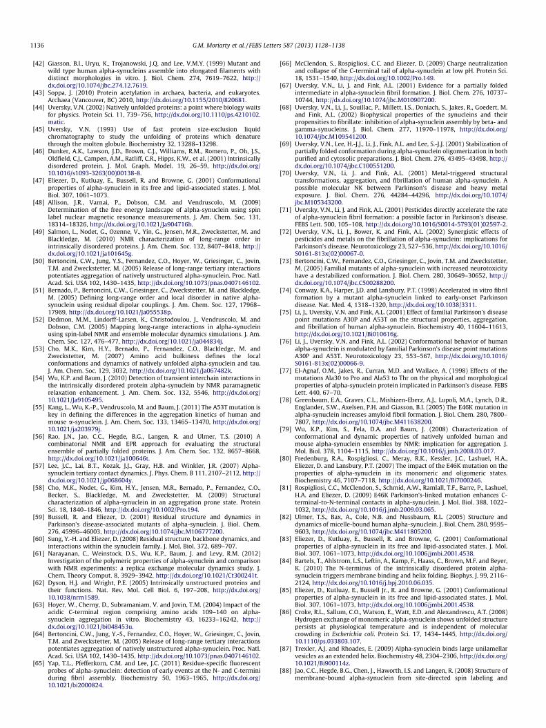
The deposition of asyn has largely been thought to originatefrom an intrinsically disordered monomer ensemble that under fi-bril promoting conditions forms amyloid [7,23,24], but recentlythis view of asyn’s native state was challenged [25]. Selkoe andcolleagues pushed the biophysical community’s long-held viewof asyn as an intrinsically disordered monomer by suggesting thatthe protein exists in its native state as a fibril-resistant helical tet-ramer. They purified the sample from human erythrocytes, optingto exclude a potentially ‘‘harsh’’ and commonly used boiling step
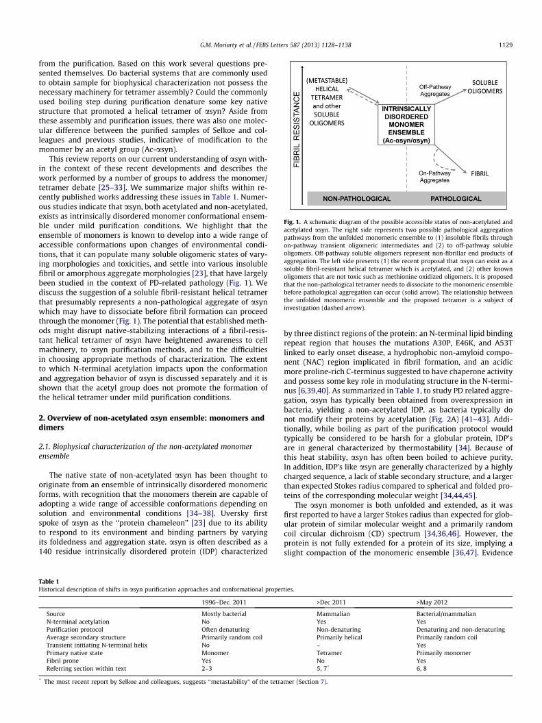
Fig. 1. A schematic diagram of the possible accessible states of non-acetylated andacetylated asyn. The right side represents two possible pathological aggregationpathways from the unfolded monomeric ensemble to (1) insoluble fibrils throughon-pathway transient oligomeric intermediates and (2) to off-pathway solubleoligomers. Off-pathway soluble oligomers represent non-fibrillar end products ofaggregation. The left side presents (1) the recent proposal that asyn can exist as asoluble fibril-resistant helical tetramer which is acetylated, and (2) other knownoligomers that are not toxic such as methionine oxidized oligomers. It is proposedthat the non-pathological tetramer needs to dissociate to the monomeric ensemblebefore pathological aggregation can occur (solid arrow). The relationship betweenthe unfolded monomeric ensemble and the proposed tetramer is a subject ofinvestigation (dashed arrow).
G.M. Moriarty et al. / FEBS Letters 587 (2013) 1128–1138 1129
from the purification. Based on this work several questions pre-sented themselves. Do bacterial systems that are commonly usedto obtain sample for biophysical characterization not possess thenecessary machinery for tetramer assembly? Could the commonlyused boiling step during purification denature some key nativestructure that promoted a helical tetramer of asyn? Aside fromthese assembly and purification issues, there was also one molec-ular difference between the purified samples of Selkoe and col-leagues and previous studies, indicative of modification to themonomer by an acetyl group (Ac-asyn).
This review reports on our current understanding of asyn with-in the context of these recent developments and describes thework performed by a number of groups to address the monomer/tetramer debate [25–33]. We summarize major shifts within re-cently published works addressing these issues in Table 1. Numer-ous studies indicate that asyn, both acetylated and non-acetylated,exists as intrinsically disordered monomer conformational ensem-ble under mild purification conditions. We highlight that theensemble of monomers is known to develop into a wide range ofaccessible conformations upon changes of environmental condi-tions, that it can populate many soluble oligomeric states of vary-ing morphologies and toxicities, and settle into various insolublefibril or amorphous aggregate morphologies [23], that have largelybeen studied in the context of PD-related pathology (Fig. 1). Wediscuss the suggestion of a soluble fibril-resistant helical tetramerthat presumably represents a non-pathological aggregate of asynwhich may have to dissociate before fibril formation can proceedthrough the monomer (Fig. 1). The potential that established meth-ods might disrupt native-stabilizing interactions of a fibril-resis-tant helical tetramer of asyn have heightened awareness to cellmachinery, to asyn purification methods, and to the difficultiesin choosing appropriate methods of characterization. The extentto which N-terminal acetylation impacts upon the conformationand aggregation behavior of asyn is discussed separately and it isshown that the acetyl group does not promote the formation ofthe helical tetramer under mild purification conditions.
2. Overview of non-acetylated asyn ensemble: monomers anddimers
2.1. Biophysical characterization of the non-acetylated monomerensemble
The native state of non-acetylated asyn has been thought tooriginate from an ensemble of intrinsically disordered monomericforms, with recognition that the monomers therein are capable ofadopting a wide range of accessible conformations depending onsolution and environmental conditions [34–38]. Uversky firstspoke of asyn as the ‘‘protein chameleon’’ [23] due to its abilityto respond to its environment and binding partners by varyingits foldedness and aggregation state. asyn is often described as a140 residue intrinsically disordered protein (IDP) characterized
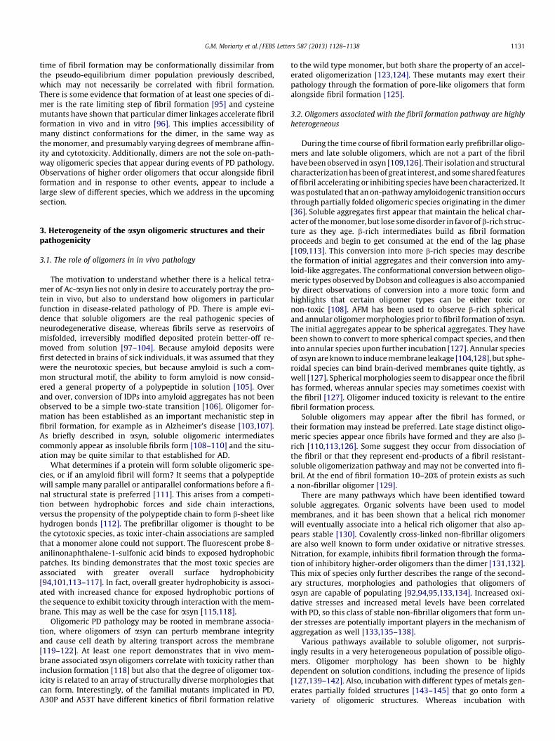
Table 1Historical description of shifts in asyn purification approaches and conformational proper
1996–Dec. 2011
Source Mostly bacterialN-terminal acetylation NoPurification protocol Often denaturingAverage secondary structure Primarily random coilTransient initiating N-terminal helix NoPrimary native state MonomerFibril prone YesReferring section within text 2–3
* The most recent report by Selkoe and colleagues, suggests ‘‘metastability’’ of the tetra
by three distinct regions of the protein: an N-terminal lipid bindingrepeat region that houses the mutations A30P, E46K, and A53Tlinked to early onset disease, a hydrophobic non-amyloid compo-nent (NAC) region implicated in fibril formation, and an acidicmore proline-rich C-terminus suggested to have chaperone activityand possess some key role in modulating structure in the N-termi-nus [6,39,40]. As summarized in Table 1, to study PD related aggre-gation, asyn has typically been obtained from overexpression inbacteria, yielding a non-acetylated IDP, as bacteria typically donot modify their proteins by acetylation (Fig. 2A) [41–43]. Addi-tionally, while boiling as part of the purification protocol wouldtypically be considered to be harsh for a globular protein, IDP’sare in general characterized by thermostability [34]. Because ofthis heat stability, asyn has often been boiled to achieve purity.In addition, IDP’s like asyn are generally characterized by a highlycharged sequence, a lack of stable secondary structure, and a largerthan expected Stokes radius compared to spherical and folded pro-teins of the corresponding molecular weight [34,44,45].
The asyn monomer is both unfolded and extended, as it wasfirst reported to have a larger Stokes radius than expected for glob-ular protein of similar molecular weight and a primarily randomcoil circular dichroism (CD) spectrum [34,36,46]. However, theprotein is not fully extended for a protein of its size, implying aslight compaction of the monomeric ensemble [36,47]. Evidence
ties.
>Dec 2011 >May 2012
Mammalian Bacterial/mammalianYes YesNon-denaturing Denaturing and non-denaturingPrimarily helical Primarily random coil– YesTetramer Primarily monomerNo Yes5, 7* 6, 8
mer (Section 7).

Fig. 2. A pictoral representation of the co-expression system designed to generateAc-asyn in bacteria. In this figure: ring-like circles represent plasmids, linesrepresent the asyn protein, and two ellipses represent the NatB protein. (A)Bacteria, lacking NatB, express non-acetylated protein. (B) Yeast house yeast–NatBwhich acts upon asyn and generates Ac-asyn. (C) To obtain Ac-asyn within abacterial expression system, plasmids encoding yeast–NatB can be co-expressedwith the plasmid encoding for asyn so that Ac-asyn is obtained[172].
1130 G.M. Moriarty et al. / FEBS Letters 587 (2013) 1128–1138
for contact between the C-terminus and both the NAC and N-ter-minal regions of the protein from nuclear magnetic resonance(NMR) [38,48–55], electron paramagnetic resonance [56] andmolecular dynamic studies [52,57] indicates a possible source ofthis compaction, as well as some transient secondary structure[55,58–60]. The compaction may be at least be partially drivenby hydrophobic patches located in the C-terminal (residues 115–119, and 125–129) associating with and shielding both the hydro-phobic N-terminal and NAC regions [50,61]. It should be empha-sized that evidence for contact between the N- and C- terminidoes not imply a static closed picture of asyn as an IDP [59,62].Rather, this is a dynamic interaction, and observation of slightcompaction is the result of observations on a highly averaged bulkensemble [48].
Under conditions promoting pathological aggregation of asyn,conformational shifts in the ensemble are observed. There is evi-dence that these interactions may keep the N-terminus from path-ological misfolding [39,63], as their release is associated withincreased fibril formation [38,50,52,64,65]. For example, whenthe solution pH is lowered, there is a structural rearrangement ofthe monomer ensemble with enhanced contacts NAC-C terminusand C–C contacts resulting from charge neutralization and com-paction of the C-terminal region [38,54,58,66]. Environmental orexperimental shifts that reduce the net charge or increase hydro-phobicity of the protein [67–69], or interaction with small mole-cules or metal ions [70–72] can change subtly, but significantly,both the long-range and short-range contacts and conformationssampled in the monomeric ensemble.
Therefore, small changes to the asyn monomer can potentiatebig effects on aggregation behavior, yet only small differences to
the monomer ensemble. For example, the familial mutations(A30P, E46K, A53T) of asyn are structurally comparable, as theyare similarly unfolded and have similar radii of gyration, but theyhave distinct kinetics of fibril formation [55,73–79]. NMR spectros-copy has revealed that mutations affect chemical shifts surround-ing the mutation site and that we can correlate these shifts toregion-specific shifts in the population of transient secondarystructure. These relatively small shifts in transient secondarystructure populations can explain bulk differences in fibril forma-tion rates [59,80,81].
The N-terminal region is also known to adopt helical structureupon binding lipids, representing a more dramatic conformationalshift of the monomer ensemble [82–84]. NMR groups have demon-strated that asyn displays chemical shifts characteristic of a mostlyunfolded peptide, but that the first 100 residues transiently popu-late helical structure. When bound to lipids or micelles, however,chemical shifts of these residues indicate a structured helical envi-ronment. [85,86]. Bax et al. characterized the structure of the mi-celle bound form of the protein, and the fact that asyn adoptshelical structure at its N-termini through its repeat region uponbinding lipids membranes and micelles has become well-known[82]. Because asyn localizes near synaptic nerve termini [9], its li-pid-induced helical structure [35,87–89] may be crucial in under-standing the protein’s function at the membrane, yet it is stillunknown how this N-terminally helical monomer conformationis related to the trigger of fibril formation.
2.2. Dimers: equilibrium species and pathological intermediates
Dimers can exist in a small population in pseudo-equilibriumwith the monomer ensemble. The Baum lab demonstrated that in-deed antiparallel transient inter-chain contacts between theC-terminal hydrophobic patches and the N-terminal region(residues: 3–15 and 35–50) could be detected by using NMR para-magnetic relaxation enhancement experiments [54]. Electrosprayionization mass spectrometry (ESI-MS) and electrospray ioniza-tion–ion mobility spectrometry-mass spectrometry (ESI–IMS-MS)obtained under similar conditions has shown that the predominantoligomeric form we observe in asyn is the dimer. This soft ionizationtechnique has revealed the ‘‘conformational heterogeneity’’ of asyn,where the monomers and dimers themselves exist in both extendedand compact conformations [30,90,91], suggesting that the ensem-ble view of asyn also extends into its higher oligomeric states.
It is unknown whether this pseudo-equilibrium anti-parallel di-mer is on pathway to the fibril formation. It is reasonable to as-sume that inter-chain N-N species, which adopts the sameparallel orientation as monomer units as in the core of the fibril,lies further along the pathway to fibril formation than anti-paralleloriented monomers [92]. This would imply reorientation of mono-mer units as an obligatory step before formation of the fibril. How-ever, at least one report demonstrates that toxic prefibrillaramyloid aggregates adopt an antiparallel orientation [93], and inthis sense we cannot draw any analogy to this pseudo-equilibriumdimer population that exists with the fibril accessible monomerensemble, and how the monomers therein may interact with it.
As the monomeric ensemble is shifted towards more fibrilprone conditions, previously described conformational changes(Section 2.1), as well as changes in population of oligomeric speciesoccurs. Incubation at high temperature is one external factorinducing this shift [94]. Under these conditions, soluble oligomersof asyn spontaneously associate and a dimer is the predominantoligomeric species of asyn to appear alongside formation of the fi-bril, along with smaller populations of higher-order oligomers. Bio-physical characterization of this intermediate which includes somedimer shows it has more hydrophobic patches exposed than thenative monomer [36]. This on-pathway dimer that appears at the
G.M. Moriarty et al. / FEBS Letters 587 (2013) 1128–1138 1131
time of fibril formation may be conformationally dissimilar fromthe pseudo-equilibrium dimer population previously described,which may not necessarily be correlated with fibril formation.There is some evidence that formation of at least one species of di-mer is the rate limiting step of fibril formation [95] and cysteinemutants have shown that particular dimer linkages accelerate fibrilformation in vivo and in vitro [96]. This implies accessibility ofmany distinct conformations for the dimer, in the same way asthe monomer, and presumably varying degrees of membrane affin-ity and cytotoxicity. Additionally, dimers are not the sole on-path-way oligomeric species that appear during events of PD pathology.Observations of higher order oligomers that occur alongside fibrilformation and in response to other events, appear to include alarge slew of different species, which we address in the upcomingsection.
3. Heterogeneity of the asyn oligomeric structures and theirpathogenicity
3.1. The role of oligomers in in vivo pathology
The motivation to understand whether there is a helical tetra-mer of Ac-asyn lies not only in desire to accurately portray the pro-tein in vivo, but also to understand how oligomers in particularfunction in disease-related pathology of PD. There is ample evi-dence that soluble oligomers are the real pathogenic species ofneurodegenerative disease, whereas fibrils serve as reservoirs ofmisfolded, irreversibly modified deposited protein better-off re-moved from solution [97–104]. Because amyloid deposits werefirst detected in brains of sick individuals, it was assumed that theywere the neurotoxic species, but because amyloid is such a com-mon structural motif, the ability to form amyloid is now consid-ered a general property of a polypeptide in solution [105]. Overand over, conversion of IDPs into amyloid aggregates has not beenobserved to be a simple two-state transition [106]. Oligomer for-mation has been established as an important mechanistic step infibril formation, for example as in Alzheimer’s disease [103,107].As briefly described in asyn, soluble oligomeric intermediatescommonly appear as insoluble fibrils form [108–110] and the situ-ation may be quite similar to that established for AD.
What determines if a protein will form soluble oligomeric spe-cies, or if an amyloid fibril will form? It seems that a polypeptidewill sample many parallel or antiparallel conformations before a fi-nal structural state is preferred [111]. This arises from a competi-tion between hydrophobic forces and side chain interactions,versus the propensity of the polypeptide chain to form b-sheet likehydrogen bonds [112]. The prefibrillar oligomer is thought to bethe cytotoxic species, as toxic inter-chain associations are sampledthat a monomer alone could not support. The fluorescent probe 8-anilinonaphthalene-1-sulfonic acid binds to exposed hydrophobicpatches. Its binding demonstrates that the most toxic species areassociated with greater overall surface hydrophobicity[94,101,113–117]. In fact, overall greater hydrophobicity is associ-ated with increased chance for exposed hydrophobic portions ofthe sequence to exhibit toxicity through interaction with the mem-brane. This may as well be the case for asyn [115,118].
Oligomeric PD pathology may be rooted in membrane associa-tion, where oligomers of asyn can perturb membrane integrityand cause cell death by altering transport across the membrane[119–122]. At least one report demonstrates that in vivo mem-brane associated asyn oligomers correlate with toxicity rather thaninclusion formation [118] but also that the degree of oligomer tox-icity is related to an array of structurally diverse morphologies thatcan form. Interestingly, of the familial mutants implicated in PD,A30P and A53T have different kinetics of fibril formation relative
to the wild type monomer, but both share the property of an accel-erated oligomerization [123,124]. These mutants may exert theirpathology through the formation of pore-like oligomers that formalongside fibril formation [125].
3.2. Oligomers associated with the fibril formation pathway are highlyheterogeneous
During the time course of fibril formation early prefibrillar oligo-mers and late soluble oligomers, which are not a part of the fibrilhave been observed in asyn [109,126]. Their isolation and structuralcharacterization has been of great interest, and some shared featuresof fibril accelerating or inhibiting species have been characterized. Itwas postulated that an on-pathway amyloidogenic transition occursthrough partially folded oligomeric species originating in the dimer[36]. Soluble aggregates first appear that maintain the helical char-acter of the monomer, but lose some disorder in favor of b-rich struc-ture as they age. b-rich intermediates build as fibril formationproceeds and begin to get consumed at the end of the lag phase[109,113]. This conversion into more b-rich species may describethe formation of initial aggregates and their conversion into amy-loid-like aggregates. The conformational conversion between oligo-meric types observed by Dobson and colleagues is also accompaniedby direct observations of conversion into a more toxic form andhighlights that certain oligomer types can be either toxic ornon-toxic [108]. AFM has been used to observe b-rich sphericaland annular oligomer morphologies prior to fibril formation of asyn.The initial aggregates appear to be spherical aggregates. They havebeen shown to convert to more spherical compact species, and theninto annular species upon further incubation [127]. Annular speciesofasyn are known to induce membrane leakage [104,128], but sphe-roidal species can bind brain-derived membranes quite tightly, aswell [127]. Spherical morphologies seem to disappear once the fibrilhas formed, whereas annular species may sometimes coexist withthe fibril [127]. Oligomer induced toxicity is relevant to the entirefibril formation process.
Soluble oligomers may appear after the fibril has formed, ortheir formation may instead be preferred. Late stage distinct oligo-meric species appear once fibrils have formed and they are also b-rich [110,113,126]. Some suggest they occur from dissociation ofthe fibril or that they represent end-products of a fibril resistant-soluble oligomerization pathway and may not be converted into fi-bril. At the end of fibril formation 10–20% of protein exists as sucha non-fibrillar oligomer [129].
There are many pathways which have been identified towardsoluble aggregates. Organic solvents have been used to modelmembranes, and it has been shown that a helical rich monomerwill eventually associate into a helical rich oligomer that also ap-pears stable [130]. Covalently cross-linked non-fibrillar oligomersare also well known to form under oxidative or nitrative stresses.Nitration, for example, inhibits fibril formation through the forma-tion of inhibitory higher-order oligomers than the dimer [131,132].This mix of species only further describes the range of the second-ary structures, morphologies and pathologies that oligomers ofasyn are capable of populating [92,94,95,133,134]. Increased oxi-dative stresses and increased metal levels have been correlatedwith PD, so this class of stable non-fibrillar oligomers that form un-der stresses are potentially important players in the mechanism ofaggregation as well [133,135–138].
Various pathways available to soluble oligomer, not surpris-ingly results in a very heterogeneous population of possible oligo-mers. Oligomer morphology has been shown to be highlydependent on solution conditions, including the presence of lipids[127,139–142]. Also, incubation with different types of metals gen-erates partially folded structures [143–145] that go onto form avariety of oligomeric structures. Whereas incubation with

1132 G.M. Moriarty et al. / FEBS Letters 587 (2013) 1128–1138
Cu2+/Fe3+/Ni2+ produce spherical oligomers of 0.8–4 nm particles,incubation with Co2+/Ca2+ produces yields pore-like annular rings70–90 nm in diameter [146].
3.3. Stabilization of non-fibrillar oligomers that appear to be non-pathogenic
In previous paragraphs our focus was on oligomers more closelylinked to pathology, but as mentioned, some oligomers can be sta-bilized in non-fibrillar forms not capable of adopting cross-b struc-ture on their own and may not necessarily be linked to cytotoxicity(Fig. 1). The asyn monomer that has been modified by methionineoxidation of the asyn monomer to the sulfoxides is one example ofthese non-toxic non-fibrillar species. This modification at methio-nine residues promotes the stabilization of an oligomer that ap-pears slightly more unfolded than monomeric asyn. Whileprobably not covalently cross-linked, these oligomers exhibit sta-bility and do not go onto form fibrils [147–149]. Furthermore,these oligomers do not exhibit toxicity toward dopaminergic neu-rons, suggesting that particular conformational features are indeednecessary to exert pathology as an oligomer (Fig. 1) [149]. Interac-tion with small molecules like the flavonoid baicalein can also pre-vent formation of the fibril by stabilizing soluble oligomeric endproducts and these oligomers also do not disrupt membranes[150]. Structurally these species are spherical, have a well devel-oped secondary structure, and are relatively globular with a pack-ing density intermediate between globular protein and pre-moltenglobule and very high thermodynamic stability. In contrast oligo-mers stabilized by modification of the monomer with 4-hydroxy-nonenal are non-fibrillar, but are also toxic [150]. Could a helicaltetramer be similarly stabilized, such that the stabilization in theoligomer conformation is more favorable than in amyloid, andcould it also share conformational features of non-pathology withaforementioned species? Descriptions of size, morphology or over-all conformation may not be sufficient to describe the cytotoxicityof an oligomer of asyn. For example, in amyloid-b two oligomers ofsimilar size but dissimilar toxicity have been identified, where themore toxic species adopts a conformation in which hydrophobicregions remain more exposed [151].
4. asyn is N-terminally acetylated in vivo
Before attention was drawn to the possible role of the acetylgroup by the recent report [25] of tetramer formation of asyn, asynwas studied from a variety of sources, some of which were mam-malian and were likely to be N-terminally acetylated. Althoughthe acetyl group had not previously warranted an explicit exami-nation, drawing comparisons between in-cell work and in vitrowork could be challenging. Therefore, the report by Selkoe and col-leagues clearly suggested that co- or post-translational modifica-tions (PTM’s), namely acetylation, may have significant influenceon asyn structure and aggregation properties so specific investiga-tion of the acetyl group naturally followed in the reports we de-scribe in this review (Table 1). PTM’s to asyn are known toregulate/modify asyn’s propensity to aggregate [134,147,152–155]. It has been known for some time that asyn in human tissuesis acetylated, but the role of N-terminal acetylation is unclear, as itis seen in both healthy and individuals sick with synucleinopathies.Two mass spectrometry (MS) studies of asyn from human tissues,both report that the base mass of the protein before any othermodifications is the acetylated form – consistent with that re-ported by Selkoe and colleagues from red blood cells (RBCs)[25,156,157]. The report indicated that acetylation of asyn wasnot limited to neuronal tissue; however, the site of acetylationwas not identified.
4.1. Bacteria lack the machinery for N-terminal acetylation
Mammals modify the proteins they produce with many morePTM’s than yeast and bacteria, as these may play a role in morecomplex signaling pathways [158,159]. N-terminal acetylation isa well-known modification in eukaryotic cells. Up to 80% of pro-teins are modified by N-terminal acetylation in mammals, whereasbacteria rarely acetylate their N-termini and if they do, by dis-tinctly different mechanisms [43]. The aforementioned MS studies[156,157] indicated that acetylation occurs at the N-terminus,where an acetyl group has removed the a-amino charge of the ini-tiating amino acid by covalent modification at that site.
N-terminal acetylation is carried out mostly co-translationallyby a group of enzymes known as N-acetyltransferases (Nat) ineukaryotes [160,161]. Mammalian cells have these complexes,and yeast an analogous enzyme complex, but bacteria do not. Nat’scatalyze the transfer of an N-acetyl group from acetyl-coenzyme Ato the N-termini of proteins with sequence specificity. DifferentNat’s (types A–F in eukaryotes) work upon different initiating ami-no acid substrates, dependent upon the identity of the first two tothree amino acids of the protein polypeptide [158,159,162]. There-fore, depending on the type of cell to synthesize asyn, the proteinmay or may not be acetylated (Fig. 2A and B). Specifically,N-acetyltransferase B (NatB) has asyn as a substrate, producingacetylated asyn (Ac-asyn). NatB targets proteins beginning withMet-Asn-, Met-Glu- or as in the case of asyn, Met-Asp-. Substratesof NatB are acetylated nearly 100% of the time, as the acidic aminoacids in the second position are thought to stimulate the transfer ofthe acetyl group [159].
4.2. Possible roles of N-terminal acetylation in vivo
Recognizing that asyn acetylation does indeed occur, one studyprior to Selkoe and colleagues’ investigated the role of NatB activityin a yeast model by disrupting NatB activity [163]. They foundNatB activity to be essential for proper membrane targeting ofasyn. Without NatB activity, non-acetylated asyn is produced,and a much more diffuse cytoplasmic localization of asyn com-pared to those with whole NatB activity (producing Ac-asyn) wasobserved. While this in vivo effect was observed in this one in-stance for Ac-asyn, the role of N-terminal acetylation is not gener-ally well understood [164]. One study suggests that N-terminalacetylation represents an early sorting step, where acetylated pro-teins are targeted toward the endoplasmic reticulum, unless theyremain non-acetylated and are kept localized to the cytosol instead[165]. N-terminal acetylation may also regulate degradation path-ways [166] or be responsible for structural effects at the N-termi-nus [167]. Levels of acetylation may be related to regulation ofother post translational modifications. NatB, specifically, has beenshown to induce elevated phosphorylation levels in yeast [168]consistent with the aforementioned yeast study of asyn, where de-creased levels of phosphorylation were observed when the proteinremained non-acetylated and localized in the cytosol [163]. Acety-lation may not be necessary at all for proteins, but some examplesdo point to a necessity. For example, tropomyosin requires N-ter-minal acetylation so that it may bind to actin [169].
5. asyn is proposed to be a helical tetramer in its native state
Selkoe and colleagues strived to isolate asyn under physiologicalconditions and have challenged the existence of the asyn mono-meric ensemble [25] by proposing a fibril-resistant helical tetramerform of the protein (Table 1). In contrast to the typical protocol inwhich asyn has been derived from bacterial systems, overexpres-sed and denatured, they purified asyn from gently-treated RBCs

G.M. Moriarty et al. / FEBS Letters 587 (2013) 1128–1138 1133
known to have a high endogenous expression level of humanAc-asyn. From both RBC lysate and endogenously expressed asynfrom neuronal and non-neuronal cells lines, Selkoe and colleaguesshowed on Clear Native PAGE (CN-PAGE), that asyn migrates nearthe tetramer position against folded, globular protein standards.The unusual migration of an IDP against globular standards wasnot unfamiliar. Native gels are unreliable objective determinantsof molecular weight, as protein migration through the acrylamidematrix depends strongly on protein charge and shape. IDP’s typi-cally display a Stokes radius of a much higher molecular weightspecies, and this has previously been attributed to enhanced inter-actions with the matrix [36], so that Escherichia coli derived boiledasyn, too, will migrate near the position of the tetramer at 58 kDaon a native gel [25].
Selkoe and colleagues’ report also stated that they obtained aCD spectrum that indicated largely helical structure that was sen-sitive to irreversible heat denaturation. Isolation from human cellline 3D5 (which are M17D cells stably expressing asyn) yieldedsimilar results to RBC derived asyn, and they showed that asyn de-rived from E. coli was random coil even after non-denaturing puri-fication, consistent with previous reports [41]. Therefore Bartelset al. implied that expression in human cell lines and a non-dena-turing purification are necessary to ‘‘preserve’’ this native tetramerstructure. If this is indeed the native form of asyn, non-denaturingmethods of purification and mammalian machinery may be neces-sary to observe it. When denatured, RBC asyn became random coil,and migrated more similarly to E. coli derived asyn on a native gel,rather than the mildly purified helical sample.
Helical structure of asyn can be induced by its interaction withmembranes. Therefore, it might logically follow that the milderpurification did not fully remove helix inducing lipids. However,treatment with Lipidex and subsequent phosphate analysis indi-cated the sample was relatively pure in that regard (0.25 mol phos-phate/asyn monomer). At the same time, a higher lipid bindingcapacity for this ‘‘native’’ asyn was demonstrated with surfaceplasmon resonance. They also employed some unbiased methodsof MW determination including sedimentation equilibrium-ana-lytical ultracentrifugation (SE-AUC) and scanning transmissionelectron microscopy both indicating a tetramer. Bartels et al. alsoobserved one other unprecedented trait of the sample – that understandard fibril assay conditions, Thioflavin T (ThT) fluorescence didnot indicate that RBC ac-asyn formed any fibrils in vitro – clearlyalso in contrast to previously observed results and the in vivo con-dition (Table 1).
Not too long after, Wang et al. [140] similarly reported a dy-namic tetramer form of the protein. The protein was obtained byrecombinant expression methods and was modified by a 10 resi-due N-terminal tag left over from a glutathione S-transferase(GST) construct, making it difficult to compare directly with thetetramer obtained from RBCs. The purification method was simi-larly ‘‘non-denaturing’’ but included the non-physiological deter-gent beta-octyl glucopyranoside (BOG) typically used to purifymembrane bound protein. Perhaps N-terminal acetylation wassomehow mimicked by the cleaved GST-tag and would prove tobe important in the context of a non-denaturing purification.Researchers now would interpret data in light of a greater possibil-ity of the tetramer, and more carefully consider their assumptionthat their expression and purification methods did not precludean accurate representation of the protein in vivo.
6. Subsequent studies indicate that asyn exists as a primarilyunfolded monomer
A rapid period of overlapping work began to determine the olig-omeric state of asyn from various cell sources, under non-denatur-
ing conditions and to investigate the role of the acetyl groupmodification. Lashuel and colleagues examined asyn from mouse,rat and human brains and addressed the issue of the source, thepurification and the characterization methods of the protein andtheir impact on the oligomerization state (Table 1) [29].
In response to report by Bartels et al., Lashuel and colleaguesdetermine that asyn exists as an unfolded monomer within neuro-nal sources. Fauvet et al. [29] examined bacterial lysates underdenaturing and non-denaturing conditions (with and without aboiling purification step respectively) against a range of non-glob-ular standards, including: (1) E. coli derived unfolded monomericasyn (2) disulfide linked A140C asyn including some dimer and(3) Ac-asyn. Regardless of purification, samples from bacterial ly-sates elute and migrate at identical positions on a size exclusionchromatography (SEC) column or CN-PAGE. This indicated thatthe various samples are either all unfolded monomers, all morecompact tetramers, or that coincidentally these structures migrateat identical positions. Coupled now with a far UV spectrum of a pri-marily random coil protein, rather than a helical spectrum ob-served by CD, however, Lashuel and colleagues bacterial asynappears to be unfolded regardless of whether it has been boiledand it resembles unfolded monomeric asyn. A random coil spec-trum is not necessarily synonymous with a monomeric protein.Static light scattering (SLS) was used as a more unbiased molecularweight determinant alongside elution from SEC [170]. While datafrom size exclusion chromatography (SEC), indicated a Stokes ra-dius close to a globular standard at 64 kDa, SLS indicates a proteinof 14 kDa. Therefore bacteria, consistent with Selkoe and col-leagues’ observations, do not assemble into a helical tetramer, evenwithout boiling.
Lashuel and coworkers [29] demonstrated a sensitivity of CN-PAGE to small differences in the protein composition and wentonto use CN-PAGE to explore the role of mammalian machineryand denaturation by boiling. Whether endogenous or overexpres-sed, whether boiled or not, whether isolated from bacteria or pres-ent in mouse, rat samples or HEK293, HeLa, SH-SY5Y, CHO, andCOS-7 mammalian cell lines – identical CN-page migration andsometimes SEC–SLS, repeatedly indicated the unfolded monomer.Across research groups, acrylamide percentages, purification pro-tocols and the source, the samples of asyn co-migrate with recom-binant asyn. To test whether factors present in cell could promotetetramerization, they examined fresh or aged samples, since agedsamples are expected to be more oligomer-rich, along with a con-trol of exogenously added recombinant protein. In vivo oligomer-specific enzyme-linked immunosorbent assay (ELISA) could notdetect any other oligomers in the samples, confirming that purifi-cation has not disturbed this observation [29,171]. In addition, thereport explored the possibility that the tetramer population couldbe dynamic and unstable, so that if the protein for some fraction ofthe time populates a tetrameric state, it would have a differentcross-linking profile than a protein that populates primarily themonomer state. They observed that no significant amount of olig-omers beyond the dimer are observed, indicating that DSS couldnot effectively capture a tetramer either. This report additionallyrepeated the RBC purification procedure [29]. Unable to replicatethe tetramer, it was still concluded that disordered monomer isisolated from RBC’s. It is not clear what Selkoe and colleagues[25] did differently, but Fauvet et al. [29] does note that samplesof sufficient quantity and purity could not be obtained using thispurification, even with another hydrophobic interaction chroma-tography (HIC) step, suggesting some complicating interactionsin either sample. Fauvet et al. also attempted the GST-constructedasyn protocol and cannot replicate the dynamic tetramer observa-tions of Wang et al.
Concurrently, Rhoades and colleagues sought to determine if thenature of the purification method [32] or N-terminal acetylation

1134 G.M. Moriarty et al. / FEBS Letters 587 (2013) 1128–1138
had enough biophysical consequence to promote the fibril-resistanttetramer. They examined samples purified with and without BOGand the N-terminal acetyl-group. Trexler et al. is the first to use abacterial co-expression system to generate Ac-asyn (Fig. 2C). In thisco-expression system developed by Mulvihill et al. [172], the yeastanalog to NatB is cloned into a bacterial plasmid, allowing overex-pression of asyn into more unsophisticated expression systems.The yeast NatB is shown to function in bacteria to produce N-termi-nally acetylated proteins, and it seems to acetylate asyn close to100% of the time in E. coli. Trexler et al. finds that N-terminal acet-ylation and non-physiological purification including BOG were nec-essary for observation of helical oligomeric asyn. Non-acetylated orBOG free asyn was disordered and presumably monomeric, but theCD spectrum of Ac-asyn purified in the presence of BOG was helical.Rhoades and colleagues also encounter the complication that disor-dered monomer and helical tetramer have similar hydrodynamicradii, but coupled with SE-AUC, which is ‘‘independent’’ of molecu-lar shape, Ac-asyn(BOG) was shown to have a sedimentation curvethat exchanged with an oligomer. That the sample was specificallytetrameric is not clear. While the report by Trexler et al., does notexclude the possibility that N-terminally acetylated asyn has ahigher affinity for membranes and/or BOG itself, the work was fur-ther provocative towards the role of acetylation in helicity andoligomerization.
7. Continued discussion on the oligomerization state of asyn
In response to the studies that indicate that cellular asyn is anunfolded monomer [27,29], Selkoe and coworkers [33], with aneven further heightened awareness to experimental conditions,most recently reported that endogenous asyn is predominantlytetramer. Using in vivo cross-linking as their primary tool, theyidentify several factors which might matter in terms of isolatingthe tetramer. During overexpression, particularly in protein de-rived from IPTG induction, more monomer is found. More mono-mer is also isolated when cross-linking is done at 4 �C asopposed to 37 �C. The tetramer is ‘‘preserved’’ in a concentrationdependent manner at the time of lysis, where a higher concentra-tion at the time of lysis favors the tetramer. This suggests that mac-romolecular crowding in cell may favor folding and stabilization ofthe native non-pathological tetramer. For this reason and for thefact that the Ac-asyn level is endogenously high in erythrocytes,Selkoe and colleagues’ considers RBC’s an ideal system to obtainAc-asyn. These results may reflect of the experiments themselves,or may reflect the preference of the monomer to associate with it-self, even in the presence of other binding partners, but that it isalso stabilized in the monomeric form.
8. N-terminal acetylation of monomeric asyn induces helixformation and affects lipid binding
Because Ac-asyn is now recognized to be the physiologicallyrelevant species in the brain, its biophysical characterization hasbeen pursued. Questions that have been raised include the mono-mer or oligomeric preference of the Ac-asyn species, its conforma-tion, interactions with membranes and ability to form fibrils. Kanget al. [30], show that recombinant 100% acetylated Ac-asyn puri-fied under mild physiological conditions exists primarily as amonomeric protein. ESI–IMS-MS experiments indicate a smallpopulation (5–10%) of dimer that is consistent with previousobservations of dimer species in solution. Lashuel and colleagues[28] use similar techniques as in their first report and again donot observe any higher-order oligomers, now in the acetylated pro-tein. This suggests that acetylation by itself is not sufficient to favora helical tetramer. Selenko and colleagues [27] show by in-cell
NMR that non-acetylated asyn is a disordered monomer in themacromolecular environment of the cell. Lashuel’s group [28]additionally compares Ac-asyn and asyn with in-cell NMR anddraws similar conclusions. Although the possibility of exchangewith higher-order oligomers cannot be ruled out, the predominantcellular form indicated by these experiments of Ac-asyn is un-folded monomer (Table 1).
The conformational properties of Ac-asyn have also thus farbeen investigated and it is suggested that there is minimal changein the hydrodynamic radius and intra-chain long-range interac-tions, if any [28,31]. However, the N-terminal acetyl group affectsthe transient secondary structure as observed by NMR [28,30,31].Residue-specific NMR chemical shift analysis shows that there isan increase in the transient helical propensity at the initiating N-terminus [28,30,31] that may arise as the acetyl group masks thealpha-amino positive charge and interacts favorably with the helixdipole moment. Additionally, the acetyl group itself is a good N-cap, favoring hydrogen bonding for an N-terminal alpha helix atthe initiating residues [173,174].
At least one report suggests the membrane binding propertiesof Ac-asyn are strongly altered by acetylation and indicates atwo-fold higher lipid affinity. It is suggested that the increase inN-terminal transient helix may be critical to initiating membranebinding. Preformed transient helix at the N-terminus may there-fore play an important role in the recognition of binding partners,may be important for membrane recognition, or may imply that li-pid mediated association of the hydrophobic surfaces of helicesmay relevant to routes of self-association of the acetylatedmonomer.
Fibril formation of Ac-asyn was investigated by measuring thefibrillation kinetics using ThT fluorescence. While groups of Lash-uel [28] and Bax [31] found no significant differences in fibril for-mation rate, Kang et al. [30] found that N-terminal acetylationslows the rates of fibril formation by approximately a factor oftwo. Clearly the acetyl group alone cannot inhibit fibril formation,but it does impart a small inhibitory effect, which may arise from aredistribution of the monomeric protein ensemble.
9. Conclusions
Recent studies that suggested that asyn exists as a soluble, tet-rameric, fibril-resistant form of asyn were provocative, and amonomer/tetramer debate followed. The discussions about theaccessible states of asyn have raised many important questions re-lated to cellular machinery, asyn purification methods and the ex-tent to which acetylation impacts on a monomer–oligomerequilibrium. A number of in depth studies have subsequently re-vealed that both non-acetylated and acetylated asyn purified un-der mild or harsh conditions is primarily a monomer.
Despite the controversy surrounding the notion of a helical non-pathological tetramer, the concept of a soluble, non-pathologicalasyn oligomer was perhaps not new. Biophysical studies haveshown that asyn can be induced to self-associate into a heteroge-neous variety of soluble oligomers, some of which may be benefi-cial, or non-pathological. For example, methionine oxidation,arising from conditions of oxidative stress, stabilizes a fibril resis-tant oligomer of asyn that is non-toxic to dopaminergic cells[149]. This may be consistent with the regulatory role methionineoxidation is suggested to have, sometimes being beneficial.
In order to understand the interplay between aggregation proneand aggregation-resistant kinetic pathways from the unfoldedmonomer, the initial interchain associations between monomerswithin the starting ensemble and associations present in already-isolated stable soluble oligomers may need to be considered fur-ther. Defining the properties that drive these different species

G.M. Moriarty et al. / FEBS Letters 587 (2013) 1128–1138 1135
may lend to our understanding of how to enhance fibril-resistant,fibril prone and/or non-toxic pathways in vivo. Because of the greatability of asyn to adopt many conformations in a variety of oligo-meric states, working from the monomer ensemble of Ac-asyn wemay (again) isolate stabilizing interactions of a helical oligomericspecies that does not tend toward fibril and we may begin to betterelucidate shared features of non-pathology and fibril-resistanceamidst the entirety of the currently known heterogeneous oligo-mer population of asyn.
Acknowledgment
This work was supported by NIH Grant GM087012.
References
[1] Spillantini, M.G. and Goedert, M. (2000) The alpha-synucleinopathies:Parkinson’s disease, dementia with Lewy bodies, and multiple systematrophy. Ann. N.Y. Acad. Sci. 920, 16–27.
[2] Stefanis, L. (2012) Alpha-synuclein in Parkinson’s disease. Cold Spring Harb.Perspect. Med. 2, a009399, http://dx.doi.org/10.1101/cshperspect.a009399.
[3] Hindle, J.V. (2010) Ageing, neurodegeneration and Parkinson’s disease. AgeAgeing 39, 156–161, http://dx.doi.org/10.1093/ageing/afp223.
[4] Spillantini, M.G., Crowther, R.A., Jakes, R., Cairns, N.J., Lantos, P.L. and Goedert,M. (1998) Filamentous alpha-synuclein inclusions link multiple systematrophy with Parkinson’s disease and dementia with Lewy bodies. Neurosci.Lett. 251, 205–208.
[5] Spillantini, M.G., Crowther, R.A., Jakes, R., Hasegawa, M. and Goedert, M.(1998) Alpha-synuclein in filamentous inclusions of Lewy bodies fromParkinson’s disease and dementia with Lewy bodies. Proc. Natl. Acad. Sci.USA 95, 6469–6473, http://dx.doi.org/10.1073/pnas.95.11.6469.
[6] Breydo, L., Wu, J.W. and Uversky, V.N. (2012) a-Synuclein misfolding andParkinson’s disease. Biochim. Biophys. Acta 1822, 261–285, http://dx.doi.org/10.1016/j.bbadis.2011.10.002.
[7] Fink, A.L. (2006) The aggregation and fibrillation of alpha-synuclein. Acc.Chem. Res. 39, 628–634, http://dx.doi.org/10.1021/Ar050073t.
[8] Lashuel, H.A., Overk, C.R., Oueslati, A. and Masliah, E. (2013) The many facesof alpha-synuclein: from structure and toxicity to therapeutic target. Nat.Rev. Neurosci. 14, 38–48, http://dx.doi.org/10.1038/Nrn3406.
[9] Maroteaux, L., Campanelli, J.T. and Scheller, R.H. (1988) Synuclein – a neuron-specific protein localized to the nucleus and presynaptic nerve-terminal. J.Neurosci. 8, 2804–2815.
[10] Shibayamaimazu, T., Okahashi, I., Omata, K., Nakajo, S., Ochiai, H., Nakai, Y.,Hama, T., Nakamura, Y. and Nakaya, K. (1993) Cell and tissue distribution anddevelopmental-change of neuron-specific 14 kDa protein(phosphoneuroprotein-14). Brain Res. 622, 17–25, http://dx.doi.org/10.1016/0006-8993(93)90796-P.
[11] Jakes, R., Spillantini, M.G. and Goedert, M. (1994) Identification of 2 distinctsynucleins from human brain. FEBS Lett. 345, 27–32, http://dx.doi.org/10.1016/0014-5793(94)00395-5.
[12] Iwai, A., Masliah, E., Yoshimoto, M., Ge, N., Flanagan, L., de Silva, H.A., Kittel, A.and Saitoh, T. (1995) The precursor protein of non-A beta component ofAlzheimer’s disease amyloid is a presynaptic protein of the central nervoussystem. Neuron 14, 467–475.
[13] Bottner, M., Zorenkov, D., Hellwig, I., Barrenschee, M., Harde, J., Fricke, T.,Deuschl, G., Egberts, J.H., Becker, T., Fritscher-Ravens, A., et al. (2012)Expression pattern and localization of alpha-synuclein in the human entericnervous system. Neurobiol. Dis. 48, 474–480, http://dx.doi.org/10.1016/j.nbd.2012.07.018.
[14] Barbour, R., Kling, K., Anderson, J.P., Banducci, K., Cole, T., Diep, L., Fox, M.,Goldstein, J.M., Soriano, F., Seubert, P., et al. (2008) Red blood cells are themajor source of alpha-synuclein in blood. Neurodegener. Dis. 5, 55–59,http://dx.doi.org/10.1159/000112832.
[15] Davidson, W.S., Jonas, A., Clayton, D.F. and George, J.M. (1998) Stabilization ofalpha-synuclein secondary structure upon binding to synthetic membranes.J. Biol. Chem. 273, 9443–9449, http://dx.doi.org/10.1074/jbc.273.16.9443.
[16] Bisaglia, M., Tessari, I., Mammi, S. and Bubacco, L. (2009) Interaction betweenalpha-synuclein and metal ions, still looking for a role in the pathogenesis ofParkinson’s disease. Neuromolecular Med. 11, 239–251, http://dx.doi.org/10.1007/s12017-009-8082-1.
[17] Chandra, S., Gallardo, G., Fernandez-Chacon, R., Schluter, O.M. and Sudhof,T.C. (2005) Alpha-synuclein cooperates with CSPalpha in preventingneurodegeneration. Cell 123, 383–396, http://dx.doi.org/10.1016/j.cell.2005.09.028.
[18] da Costa, C.A., Ancolio, K. and Checler, F. (2000) Wild-type but not Parkinson’sdisease-related Ala-53 ? Thr mutant alpha-synuclein protects neuronal cellsfrom apoptotic stimuli. J. Biol. Chem. 275, 24065–24069, http://dx.doi.org/10.1074/jbc.M002413200.
[19] Murphy, D.D., Rueter, S.M., Trojanowski, J.Q. and Lee, V.M.Y. (2000)Synucleins are developmentally expressed, and alpha-synuclein regulates
the size of the presynaptic vesicular pool in primary hippocampal neurons. J.Neurosci. 20, 3214–3220.
[20] Dev, K.K., Hofele, K., Barbieri, S., Buchman, V.L. and van der Putten, H. (2003)Part II: Alpha-synuclein and its molecular pathophysiological role inneurodegenerative disease. Neuropharmacology 45, 14–44.
[21] Maries, E., Dass, B., Collier, T.J., Kordower, J.H. and Steece-Collier, K. (2003)The role of alpha-synuclein in Parkinson’s disease: insights from animalmodels. Nat. Rev. Neurosci. 4, 727–738, http://dx.doi.org/10.1038/Nrn1199.
[22] Jin, H., Kanthasamy, A., Ghosh, A., Yang, Y., Anantharam, V. and Kanthasamy,A.G. (2011) Alpha-synuclein negatively regulates protein kinase Cdeltaexpression to suppress apoptosis in dopaminergic neurons by reducingp300 histone acetyltransferase activity. J. Neurosci. 31, 2035–2051, http://dx.doi.org/10.1523/JNEUROSCI.5634-10.2011.
[23] Uversky, V.N. (2003) A protein-chameleon: conformational plasticity ofalpha-synuclein, a disordered protein involved in neurodegenerativedisorders. J. Biomol. Struct. Dyn. 21, 211–234.
[24] Uversky, V.N. and Fink, A.L. (2004) Conformational constraints for amyloidfibrillation: the importance of being unfolded. Biochim. Biophys. Acta,Proteins Proteomics 1698, 131–153, http://dx.doi.org/10.1016/j.bbapap.2003.12.008.
[25] Bartels, T., Choi, J.G. and Selkoe, D.J. (2011) a-Synuclein occursphysiologically as a helically folded tetramer that resists aggregation.Nature 477, 107–110, http://dx.doi.org/10.1038/nature10324.
[26] Wang, W., Perovic, I., Chittuluru, J., Kaganovich, A., Nguyen, L.T.T., Liao, J.,Auclair, J.R., Johnson, D., Landeru, A. and Simorellis, A.K. (2011) A soluble a-synuclein construct forms a dynamic tetramer. Proc. Natl. Acad. Sci., http://dx.doi.org/10.1073/pnas.1113260108.
[27] Binolfi, A., Theillet, F.X. and Selenko, P. (2012) Bacterial in-cell NMR of humanalpha-synuclein: a disordered monomer by nature? Biochem. Soc. Trans. 40,950-U292, http://dx.doi.org/10.1042/Bst20120096.
[28] Fauvet, B., Fares, M.B., Samuel, F., Dikiy, I., Tandon, A., Eliezer, D. and Lashuel,H.A. (2012) Characterization of semisynthetic and naturally N-alpha-acetylated alpha-synuclein in vitro and in intact cells implications foraggregation and cellular properties of alpha-synuclein. J. Biol. Chem. 287,28243–28262, http://dx.doi.org/10.1074/jbc.M112.383711.
[29] Fauvet, B., Mbefo, M.K., Fares, M.-B., Desobry, C., Michael, S., Ardah, M.T.,Tsika, E., Coune, P., Prudent, M., Lion, N., et al. (2012) Alpha-synuclein in thecentral nervous system and from erythrocytes, mammalian cells and E. coliexists predominantly as a disordered monomer. J. Biol. Chem. 287, 15345–15364, http://dx.doi.org/10.1074/jbc.M111.318949.
[30] Kang, L.J., Moriarty, G.M., Woods, L.A., Ashcroft, A.E., Radford, S.E. and Baum, J.(2012) N-terminal acetylation of alpha-synuclein induces increased transienthelical propensity and decreased aggregation rates in the intrinsicallydisordered monomer. Protein Sci. 21, 911–917, http://dx.doi.org/10.1002/Pro.2088.
[31] Maltsev, A.S., Ying, J.F. and Bax, A. (2012) Impact of N-terminal acetylation ofalpha-synuclein on its random coil and lipid binding properties. Biochemistry51, 5004–5013, http://dx.doi.org/10.1021/Bi300642h.
[32] Trexler, A.J. and Rhoades, E. (2012) N-terminal acetylation is critical forforming a-helical oligomer of a-synuclein. Protein Sci. 21, 601–605, http://dx.doi.org/10.1002/Pro.2056.
[33] Dettmer, U., Newman, A.J., Luth, E.S., Bartels, T. and Selkoe, D. (2013) In vivocrosslinking reveals principally oligomeric forms of alpha-synuclein andbeta-synuclein in neurons and non-neural cells. J. Biol. Chem., http://dx.doi.org/10.1074/jbc.M112.403311.
[34] Weinreb, P.H., Zhen, W., Poon, A.W., Conway, K.A. and Lansbury Jr., P.T.(1996) NACP, a protein implicated in Alzheimer’s disease and learning, isnatively unfolded. Biochemistry 35, 13709–13715, http://dx.doi.org/10.1021/bi961799n.
[35] Bussell, R. and Eliezer, D. (2003) A structural and functional role for 11-merrepeats in alpha-synuclein and other exchangeable lipid binding proteins. J.Mol. Biol. 329, 763–778, http://dx.doi.org/10.1016/S0022-2836(03)00520-5.
[36] Uversky, V.N., Li, J. and Fink, A.L. (2001) Evidence for a partially foldedintermediate in alpha-synuclein fibril formation. J. Biol. Chem. 276, 10737–10744, http://dx.doi.org/10.1074/jbc.M010907200.
[37] Munishkina, L.A., Fink, A.L. and Uversky, V.N. (2009) Accelerated fibrillationof alpha-synuclein induced by the combined action of macromolecularcrowding and factors inducing partial folding. Curr. Alzheimer Res. 6, 252–260.
[38] Wu, K.P., Weinstock, D.S., Narayanan, C., Levy, R.M. and Baum, J. (2009)Structural reorganization of alpha-synuclein at low pH observed by NMR andREMD simulations. J. Mol. Biol. 391, 784–796, http://dx.doi.org/10.1016/j.jmb.2009.06.063.
[39] Hong, D.-P., Xiong, W., Chang, J.-Y. and Jiang, C. (2011) The role of the C-terminus of human a-synuclein: intra-disulfide bonds between the C-terminus and other regions stabilize non-fibrillar monomeric isomers. FEBSLett. 585, 561–566, http://dx.doi.org/10.1016/j.febslet.2011.01.009.
[40] Park, S.M., Jung, H.Y., Kim, T.D., Park, J.H., Yang, C.H. and Kim, J. (2002)Distinct roles of the N-terminal-binding domain and the C-terminal-solubilizing domain of alpha-synuclein, a molecular chaperone. J. Biol.Chem. 277, 28512–28520, http://dx.doi.org/10.1074/jbc.M111971200.
[41] Weinreb, P.H., Zhen, W.G., Poon, A.W., Conway, K.A. and Lansbury, P.T. (1996)NACP, a protein implicated in Alzheimer’s disease and learning, is nativelyunfolded. Biochemistry 35, 13709–13715, http://dx.doi.org/10.1021/bi961799n.
1136 G.M. Moriarty et al. / FEBS Letters 587 (2013) 1128–1138
[42] Giasson, B.I., Uryu, K., Trojanowski, J.Q. and Lee, V.M.Y. (1999) Mutant andwild type human alpha-synucleins assemble into elongated filaments withdistinct morphologies in vitro. J. Biol. Chem. 274, 7619–7622, http://dx.doi.org/10.1074/jbc.274.12.7619.
[43] Soppa, J. (2010) Protein acetylation in archaea, bacteria, and eukaryotes.Archaea (Vancouver, BC) 2010, http://dx.doi.org/10.1155/2010/820681.
[44] Uversky, V.N. (2002) Natively unfolded proteins: a point where biology waitsfor physics. Protein Sci. 11, 739–756, http://dx.doi.org/10.1110/ps.4210102.matic.
[45] Uversky, V.N. (1993) Use of fast protein size-exclusion liquidchromatography to study the unfolding of proteins which denaturethrough the molten globule. Biochemistry 32, 13288–13298.
[46] Dunker, A.K., Lawson, J.D., Brown, C.J., Williams, R.M., Romero, P., Oh, J.S.,Oldfield, C.J., Campen, A.M., Ratliff, C.R., Hipps, K.W., et al. (2001) Intrinsicallydisordered protein. J. Mol. Graph. Model. 19, 26–59, http://dx.doi.org/10.1016/s1093-3263(00)00138-8.
[47] Eliezer, D., Kutluay, E., Bussell, R. and Browne, G. (2001) Conformationalproperties of alpha-synuclein in its free and lipid-associated states. J. Mol.Biol. 307, 1061–1073.
[48] Allison, J.R., Varnai, P., Dobson, C.M. and Vendruscolo, M. (2009)Determination of the free energy landscape of alpha-synuclein using spinlabel nuclear magnetic resonance measurements. J. Am. Chem. Soc. 131,18314–18326, http://dx.doi.org/10.1021/Ja904716h.
[49] Salmon, L., Nodet, G., Ozenne, V., Yin, G., Jensen, M.R., Zweckstetter, M. andBlackledge, M. (2010) NMR characterization of long-range order inintrinsically disordered proteins. J. Am. Chem. Soc. 132, 8407–8418, http://dx.doi.org/10.1021/ja101645g.
[50] Bertoncini, C.W., Jung, Y.S., Fernandez, C.O., Hoyer, W., Griesinger, C., Jovin,T.M. and Zweckstetter, M. (2005) Release of long-range tertiary interactionspotentiates aggregation of natively unstructured alpha-synuclein. Proc. Natl.Acad. Sci. USA 102, 1430–1435, http://dx.doi.org/10.1073/pnas.0407146102.
[51] Bernado, P., Bertoncini, C.W., Griesinger, C., Zweckstetter, M. and Blackledge,M. (2005) Defining long-range order and local disorder in native alpha-synuclein using residual dipolar couplings. J. Am. Chem. Soc. 127, 17968–17969, http://dx.doi.org/10.1021/Ja055538p.
[52] Dedmon, M.M., Lindorff-Larsen, K., Christodoulou, J., Vendruscolo, M. andDobson, C.M. (2005) Mapping long-range interactions in alpha-synucleinusing spin-label NMR and ensemble molecular dynamics simulations. J. Am.Chem. Soc. 127, 476–477, http://dx.doi.org/10.1021/ja044834j.
[53] Cho, M.K., Kim, H.Y., Bernado, P., Fernandez, C.O., Blackledge, M. andZweckstetter, M. (2007) Amino acid bulkiness defines the localconformations and dynamics of natively unfolded alpha-synuclein and tau.J. Am. Chem. Soc. 129, 3032, http://dx.doi.org/10.1021/Ja067482k.
[54] Wu, K.P. and Baum, J. (2010) Detection of transient interchain interactions inthe intrinsically disordered protein alpha-synuclein by NMR paramagneticrelaxation enhancement. J. Am. Chem. Soc. 132, 5546, http://dx.doi.org/10.1021/Ja9105495.
[55] Kang, L., Wu, K.-P., Vendruscolo, M. and Baum, J. (2011) The A53T mutation iskey in defining the differences in the aggregation kinetics of human andmouse a-synuclein. J. Am. Chem. Soc. 133, 13465–13470, http://dx.doi.org/10.1021/ja203979j.
[56] Rao, J.N., Jao, C.C., Hegde, B.G., Langen, R. and Ulmer, T.S. (2010) Acombinatorial NMR and EPR approach for evaluating the structuralensemble of partially folded proteins. J. Am. Chem. Soc. 132, 8657–8668,http://dx.doi.org/10.1021/ja100646t.
[57] Lee, J.C., Lai, B.T., Kozak, J.J., Gray, H.B. and Winkler, J.R. (2007) Alpha-synuclein tertiary contact dynamics. J. Phys. Chem. B 111, 2107–2112, http://dx.doi.org/10.1021/jp068604y.
[58] Cho, M.K., Nodet, G., Kim, H.Y., Jensen, M.R., Bernado, P., Fernandez, C.O.,Becker, S., Blackledge, M. and Zweckstetter, M. (2009) Structuralcharacterization of alpha-synuclein in an aggregation prone state. ProteinSci. 18, 1840–1846, http://dx.doi.org/10.1002/Pro.194.
[59] Bussell, R. and Eliezer, D. (2001) Residual structure and dynamics inParkinson’s disease-associated mutants of alpha-synuclein. J. Biol. Chem.276, 45996–46003, http://dx.doi.org/10.1074/jbc.M106777200.
[60] Sung, Y.-H. and Eliezer, D. (2008) Residual structure, backbone dynamics, andinteractions within the synuclein family. J. Mol. Biol. 372, 689–707.
[61] Narayanan, C., Weinstock, D.S., Wu, K.P., Baum, J. and Levy, R.M. (2012)Investigation of the polymeric properties of alpha-synuclein and comparisonwith NMR experiments: a replica exchange molecular dynamics study. J.Chem. Theory Comput. 8, 3929–3942, http://dx.doi.org/10.1021/Ct300241t.
[62] Dyson, H.J. and Wright, P.E. (2005) Intrinsically unstructured proteins andtheir functions. Nat. Rev. Mol. Cell Biol. 6, 197–208, http://dx.doi.org/10.1038/nrm1589.
[63] Hoyer, W., Cherny, D., Subramaniam, V. and Jovin, T.M. (2004) Impact of theacidic C-terminal region comprising amino acids 109–140 on alpha-synuclein aggregation in vitro. Biochemistry 43, 16233–16242, http://dx.doi.org/10.1021/bi048453u.
[64] Bertoncini, C.W., Jung, Y.-S., Fernandez, C.O., Hoyer, W., Griesinger, C., Jovin,T.M. and Zweckstetter, M. (2005) Release of long-range tertiary interactionspotentiates aggregation of natively unstructured alpha-synuclein. Proc. Natl.Acad. Sci. USA 102, 1430–1435, http://dx.doi.org/10.1073/pnas.0407146102.
[65] Yap, T.L., Pfefferkorn, C.M. and Lee, J.C. (2011) Residue-specific fluorescentprobes of alpha-synuclein: detection of early events at the N- and C-terminiduring fibril assembly. Biochemistry 50, 1963–1965, http://dx.doi.org/10.1021/bi2000824.
[66] McClendon, S., Rospigliosi, C.C. and Eliezer, D. (2009) Charge neutralizationand collapse of the C-terminal tail of alpha-synuclein at low pH. Protein Sci.18, 1531–1540, http://dx.doi.org/10.1002/Pro.149.
[67] Uversky, V.N., Li, J. and Fink, A.L. (2001) Evidence for a partially foldedintermediate in alpha-synuclein fibril formation. J. Biol. Chem. 276, 10737–10744, http://dx.doi.org/10.1074/jbc.M010907200.
[68] Uversky, V.N., Li, J., Souillac, P., Millett, I.S., Doniach, S., Jakes, R., Goedert, M.and Fink, A.L. (2002) Biophysical properties of the synucleins and theirpropensities to fibrillate: inhibition of alpha-synuclein assembly by beta- andgamma-synucleins. J. Biol. Chem. 277, 11970–11978, http://dx.doi.org/10.1074/jbc.M109541200.
[69] Uversky, V.N., Lee, H.-J.J., Li, J., Fink, A.L. and Lee, S.-J.J. (2001) Stabilization ofpartially folded conformation during alpha-synuclein oligomerization in bothpurified and cytosolic preparations. J. Biol. Chem. 276, 43495–43498, http://dx.doi.org/10.1074/jbc.C100551200.
[70] Uversky, V.N., Li, J. and Fink, A.L. (2001) Metal-triggered structuraltransformations, aggregation, and fibrillation of human alpha-synuclein. Apossible molecular NK between Parkinson’s disease and heavy metalexposure. J. Biol. Chem. 276, 44284–44296, http://dx.doi.org/10.1074/jbc.M105343200.
[71] Uversky, V.N., Li, J. and Fink, A.L. (2001) Pesticides directly accelerate the rateof alpha-synuclein fibril formation: a possible factor in Parkinson’s disease.FEBS Lett. 500, 105–108, http://dx.doi.org/10.1016/S0014-5793(01)02597-2.
[72] Uversky, V.N., Li, J., Bower, K. and Fink, A.L. (2002) Synergistic effects ofpesticides and metals on the fibrillation of alpha-synuclein: implications forParkinson’s disease. Neurotoxicology 23, 527–536, http://dx.doi.org/10.1016/S0161-813x(02)00067-0.
[73] Bertoncini, C.W., Fernandez, C.O., Griesinger, C., Jovin, T.M. and Zweckstetter,M. (2005) Familial mutants of alpha-synuclein with increased neurotoxicityhave a destabilized conformation. J. Biol. Chem. 280, 30649–30652, http://dx.doi.org/10.1074/jbc.C500288200.
[74] Conway, K.A., Harper, J.D. and Lansbury, P.T. (1998) Accelerated in vitro fibrilformation by a mutant alpha-synuclein linked to early-onset Parkinsondisease. Nat. Med. 4, 1318–1320, http://dx.doi.org/10.1038/3311.
[75] Li, J., Uversky, V.N. and Fink, A.L. (2001) Effect of familial Parkinson’s diseasepoint mutations A30P and A53T on the structural properties, aggregation,and fibrillation of human alpha-synuclein. Biochemistry 40, 11604–11613,http://dx.doi.org/10.1021/Bi010616g.
[76] Li, J., Uversky, V.N. and Fink, A.L. (2002) Conformational behavior of humanalpha-synuclein is modulated by familial Parkinson’s disease point mutationsA30P and A53T. Neurotoxicology 23, 553–567, http://dx.doi.org/10.1016/S0161-813x(02)00066-9.
[77] El-Agnaf, O.M., Jakes, R., Curran, M.D. and Wallace, A. (1998) Effects of themutations Ala30 to Pro and Ala53 to Thr on the physical and morphologicalproperties of alpha-synuclein protein implicated in Parkinson’s disease. FEBSLett. 440, 67–70.
[78] Greenbaum, E.A., Graves, C.L., Mishizen-Eberz, A.J., Lupoli, M.A., Lynch, D.R.,Englander, S.W., Axelsen, P.H. and Giasson, B.I. (2005) The E46K mutation inalpha-synuclein increases amyloid fibril formation. J. Biol. Chem. 280, 7800–7807, http://dx.doi.org/10.1074/jbc.M411638200.
[79] Wu, K.P., Kim, S., Fela, D.A. and Baum, J. (2008) Characterization ofconformational and dynamic properties of natively unfolded human andmouse alpha-synuclein ensembles by NMR: implication for aggregation. J.Mol. Biol. 378, 1104–1115, http://dx.doi.org/10.1016/j.jmb.2008.03.017.
[80] Fredenburg, R.A., Rospigliosi, C., Meray, R.K., Kessler, J.C., Lashuel, H.A.,Eliezer, D. and Lansbury, P.T. (2007) The impact of the E46K mutation on theproperties of alpha-synuclein in its monomeric and oligomeric states.Biochemistry 46, 7107–7118, http://dx.doi.org/10.1021/Bi7000246.
[81] Rospigliosi, C.C., McClendon, S., Schmid, A.W., Ramlall, T.F., Barre, P., Lashuel,H.A. and Eliezer, D. (2009) E46K Parkinson’s-linked mutation enhances C-terminal-to-N-terminal contacts in alpha-synuclein. J. Mol. Biol. 388, 1022–1032, http://dx.doi.org/10.1016/j.jmb.2009.03.065.
[82] Ulmer, T.S., Bax, A., Cole, N.B. and Nussbaum, R.L. (2005) Structure anddynamics of micelle-bound human alpha-synuclein. J. Biol. Chem. 280, 9595–9603, http://dx.doi.org/10.1074/jbc.M411805200.
[83] Eliezer, D., Kutluay, E., Bussell, R. and Browne, G. (2001) Conformationalproperties of alpha-synuclein in its free and lipid-associated states. J. Mol.Biol. 307, 1061–1073, http://dx.doi.org/10.1006/jmbi.2001.4538.
[84] Bartels, T., Ahlstrom, L.S., Leftin, A., Kamp, F., Haass, C., Brown, M.F. and Beyer,K. (2010) The N-terminus of the intrinsically disordered protein alpha-synuclein triggers membrane binding and helix folding. Biophys. J. 99, 2116–2124, http://dx.doi.org/10.1016/j.bpj.2010.06.035.
[85] Eliezer, D., Kutluay, E., Bussell Jr., R. and Browne, G. (2001) Conformationalproperties of alpha-synuclein in its free and lipid-associated states. J. Mol.Biol. 307, 1061–1073, http://dx.doi.org/10.1006/jmbi.2001.4538.
[86] Croke, R.L., Sallum, C.O., Watson, E., Watt, E.D. and Alexandrescu, A.T. (2008)Hydrogen exchange of monomeric alpha-synuclein shows unfolded structurepersists at physiological temperature and is independent of molecularcrowding in Escherichia coli. Protein Sci. 17, 1434–1445, http://dx.doi.org/10.1110/ps.033803.107.
[87] Trexler, A.J. and Rhoades, E. (2009) Alpha-synuclein binds large unilamellarvesicles as an extended helix. Biochemistry 48, 2304–2306, http://dx.doi.org/10.1021/Bi900114z.
[88] Jao, C.C., Hegde, B.G., Chen, J., Haworth, I.S. and Langen, R. (2008) Structure ofmembrane-bound alpha-synuclein from site-directed spin labeling and

G.M. Moriarty et al. / FEBS Letters 587 (2013) 1128–1138 1137
computational refinement. Proc. Natl. Acad. Sci. USA 105, 19666–19671,http://dx.doi.org/10.1073/pnas.0807826105.
[89] Ulmer, T.S., Bax, A., Cole, N.B. and Nussbaum, R.L. (2005) Structure anddynamics of micelle-bound human alpha-synuclein. J. Biol. Chem. 280, 9595–9603, http://dx.doi.org/10.1074/jbc.M411805200.
[90] Bernstein, S.L., Liu, D.F., Wyttenbach, T., Bowers, M.T., Lee, J.C., Gray, H.B. andWinkler, J.R. (2004) Alpha-synuclein: stable compact and extendedmonomeric structures and pH dependence of dimer formation. J. Am. Soc.Mass Spectrom. 15, 1435–1443, http://dx.doi.org/10.1016/j.jasms.2004.08.003.
[91] Frimpong, A.K., Abzalimov, R.R., Uversky, V.N. and Kaltashov, I.A. (2010)Characterization of intrinsically disordered proteins with electrosprayionization mass spectrometry: conformational heterogeneity of alpha-synuclein. Proteins Struct. Funct. Bioinform. 78, 714–722, http://dx.doi.org/10.1002/prot.22604.
[92] Pivato, M., De Franceschi, G., Tosatto, L., Frare, E., Kumar, D., Aioanei, D.,Brucale, M., Tessari, I., Bisaglia, M., Samori, B., et al. (2012) Covalent a-synuclein dimers: chemico-physical and aggregation properties. PLoS ONE 7,e50027, http://dx.doi.org/10.1371/journal.pone.0050027.
[93] Celej, M.S., Sarroukh, R., Goormaghtigh, E., Fidelio, G.D., Ruysschaert, J.M. andRaussens, V. (2012) Toxic prefibrillar alpha-synuclein amyloid oligomersadopt a distinctive antiparallel beta-sheet structure. Biochem. J. 443, 719–726, http://dx.doi.org/10.1042/BJ20111924.
[94] Uversky, V.N., Lee, H.J., Li, J., Fink, A.L. and Lee, S.J. (2001) Stabilization ofpartially folded conformation during alpha-synuclein oligomerization in bothpurified and cytosolic preparations. J. Biol. Chem. 276, 43495–43498, http://dx.doi.org/10.1074/jbc.C100551200.
[95] Krishnan, S., Chi, E.Y., Wood, S.J., Kendrick, B.S., Li, C., Garzon-Rodriguez, W.,Wypych, J., Randolph, T.W., Narhi, L.O., Biere, A.L., et al. (2003) Oxidativedimer formation is the critical rate-limiting step for Parkinson’s diseasealpha-synuclein fibrillogenesis. Biochemistry 42, 829–837, http://dx.doi.org/10.1021/bi026528t.
[96] Zhou, W. and Freed, C.R. (2004) Tyrosine-to-cysteine modification of humanalpha-synuclein enhances protein aggregation and cellular toxicity. J. Biol.Chem. 279, 10128–10135, http://dx.doi.org/10.1074/jbc.M307563200.
[97] Goldberg, M.S. and Lansbury, P.T. (2000) Is there a cause-and-effectrelationship between alpha-synuclein fibrillization and Parkinson’sdisease? Nat. Cell Biol. 2, E115–E119.
[98] Conway, K.A., Lee, S.J., Rochet, J.C., Ding, T.T., Harper, J.D., Williamson, R.E. andLansbury, P.T. (2000) Accelerated oligomerization by Parkinson’s diseaselinked alpha-synuclein mutants. Ann. N.Y. Acad. Sci. 920, 42–45.
[99] Masliah, E., Rockenstein, E., Veinbergs, I., Mallory, M., Hashimoto, M., Takeda,A., Sagara, Y., Sisk, A. and Mucke, L. (2000) Dopaminergic loss and inclusionbody formation in alpha-synuclein mice: implications for neurodegenerativedisorders. Science 287, 1265–1269, http://dx.doi.org/10.1126/science.287.5456.1265.
[100] Amer, D.A.M., Irvine, G.B. and El-Agnaf, O.M.A. (2006) Inhibitors of alpha-synuclein oligomerization and toxicity: a future therapeutic strategy forParkinson’s disease and related disorders. Exp. Brain Res. 173, 223–233,http://dx.doi.org/10.1007/s00221-006-0539-y.
[101] Kayed, R., Head, E., Thompson, J.L., McIntire, T.M., Milton, S.C., Cotman, C.W.and Glabe, C.G. (2003) Common structure of soluble amyloid oligomersimplies common mechanism of pathogenesis. Science 300, 486–489, http://dx.doi.org/10.1126/science.1079469.
[102] Wolfe, K.J. and Cyr, D.M. (2011) Amyloid in neurodegenerative diseases:friend or foe? Semin. Cell Dev. Biol. 22, 476–481, http://dx.doi.org/10.1016/j.semcdb.2011.03.011.
[103] Haass, C. and Selkoe, D.J. (2007) Soluble protein oligomers inneurodegeneration: lessons from the Alzheimer’s amyloid beta-peptide.Nat. Rev. Mol. Cell Biol. 8, 101–112, http://dx.doi.org/10.1038/nrm2101.
[104] Caughey, B. and Lansbury, P.T. (2003) Protofibrils, pores, fibrils, andneurodegeneration: separating the responsible protein aggregates from theinnocent bystanders. Annu. Rev. Neurosci. 26, 267–298, http://dx.doi.org/10.1146/annurev.neuro.26.010302.081142.
[105] Dobson, C.M. (1999) Protein misfolding, evolution and disease. TrendsBiochem. Sci. 24, 329–332.
[106] Zerovnik, E. (2011) Oligomerization preceding amyloid fibril formation: aprocess in common to intrinsically disordered and globular proteins.Network 22, 154–161, http://dx.doi.org/10.3109/0954898X.2011.639842.
[107] Lee, J., Culyba, E.K., Powers, E.T. and Kelly, J.W. (2011) Amyloid-b forms fibrilsby nucleated conformational conversion of oligomers. Nat. Chem. Biol. 7,600–607, http://dx.doi.org/10.1038/nchembio.624.
[108] Cremades, N., Cohen, S.I.A., Deas, E., Abramov, A.Y., Chen, A.Y., Orte, A.,Sandal, M., Clarke, R.W., Dunne, P., Aprile, F.A., et al. (2012) Directobservation of the interconversion of normal and toxic forms of alpha-synuclein. Cell 149, 1048–1059, http://dx.doi.org/10.1016/j.cell.2012.03.037.
[109] Apetri, M.M., Maiti, N.C., Zagorski, M.G., Carey, P.R. and Anderson, V.E. (2006)Secondary structure of alpha-synuclein oligomers: characterization byraman and atomic force microscopy. J. Mol. Biol. 355, 63–71, http://dx.doi.org/10.1016/j.jmb.2005.10.071.
[110] Lee, J.-H., Lee, I.-H., Choe, Y.-J., Kang, S., Kim, H.Y., Gai, W.-P., Hahn, J.-S. andPaik, S.R. (2009) Real-time analysis of amyloid fibril formation of alpha-synuclein using a fibrillation-state-specific fluorescent probe of JC-1.Biochem. J. 418, 311–323, http://dx.doi.org/10.1042/BJ20081572.
[111] Gsponer, J., Haberthur, U. and Caflisch, A. (2003) The role of side-chaininteractions in the early steps of aggregation: molecular dynamics simulationsof an amyloid-forming peptide from the yeast prion Sup35. Proc. Natl. Acad.Sci. USA 100, 5154–5159, http://dx.doi.org/10.1073/pnas.0835307100.
[112] Cheon, M., Chang, I., Mohanty, S., Luheshi, L.M., Dobson, C.M., Vendruscolo,M. and Favrin, G. (2007) Structural reorganisation and potential toxicity ofoligomeric species formed during the assembly of amyloid fibrils. PLoSComput. Biol. 3, 1727–1738, http://dx.doi.org/10.1371/journal.pcbi.0030173.
[113] Dusa, A., Kaylor, J., Edridge, S., Bodner, N., Hong, D.P. and Fink, A.L. (2006)Characterization of oligomers during alpha-synuclein aggregation usingintrinsic tryptophan fluorescence. Biochemistry 45, 2752–2760, http://dx.doi.org/10.1021/Bi051426z.
[114] Bolognesi, B., Kumita, J.R., Barros, T.P., Esbjorner, E.K., Luheshi, L.M.,Crowther, D.C., Wilson, M.R., Dobson, C.M., Favrin, G. and Yerbury, J.J.(2010) ANS binding reveals common features of cytotoxic amyloid species.ACS Chem. Biol. 5, 735–740, http://dx.doi.org/10.1021/cb1001203.
[115] Outeiro, T.F., Putcha, P., Tetzlaff, J.E., Spoelgen, R., Koker, M., Carvalho, F.,Hyman, B.T. and McLean, P.J. (2008) Formation of toxic oligomeric alpha-synuclein species in living cells. PLoS ONE 3, e1867, http://dx.doi.org/10.1371/journal.pone.0001867.
[116] Campioni, S., Mannini, B., Zampagni, M., Pensalfini, A., Parrini, C., Evangelisti,E., Relini, A., Stefani, M., Dobson, C.M., Cecchi, C., et al. (2010) A causative linkbetween the structure of aberrant protein oligomers and their toxicity. Nat.Chem. Biol. 6, 140–147, http://dx.doi.org/10.1038/nchembio.283.
[117] Krishnan, R., Goodman, J.L., Mukhopadhyay, S., Pacheco, C.D., Lemke, Ea,Deniz, Aa and Lindquist, S. (2012) Conserved features of intermediates inamyloid assembly determine their benign or toxic states. Proc. Natl. Acad.Sci. USA 109, 11172–11177, http://dx.doi.org/10.1073/pnas.1209527109.
[118] Winner, B., Jappelli, R., Maji, S.K., Pa, Desplats., Boyer, L., Aigner, S., Hetzer, C.,Loher, T., Vilar, M., Campioni, S., et al. (2011) In vivo demonstration thatalpha-synuclein oligomers are toxic. Proc. Natl. Acad. Sci. USA 108, 4194–4199, http://dx.doi.org/10.1073/pnas.1100976108.
[119] Kim, H.Y., Cho, M.K., Kumar, A., Maier, E., Siebenhaar, C., Becker, S.,Fernandez, C.O., Lashuel, H.A., Benz, R., Lange, A., et al. (2009) Structuralproperties of pore-forming oligomers of alpha-synuclein. J. Am. Chem. Soc.131, 17482–17489, http://dx.doi.org/10.1021/Ja9077599.
[120] Tsigelny, I.F., Sharikov, Y., Wrasidlo, W., Gonzalez, T., Desplats, P.A., Crews, L.,Spencer, B. and Masliah, E. (2012) Role of alpha-synuclein penetration intothe membrane in the mechanisms of oligomer pore formation. FEBS J. 279,1000–1013, http://dx.doi.org/10.1111/j.1742-4658.2012.08489.x.
[121] Danzer, K.M., Haasen, D., Karow, A.R., Moussaud, S., Habeck, M., Giese, A.,Kretzschmar, H., Hengerer, B. and Kostka, M. (2007) Different species ofalpha-synuclein oligomers induce calcium influx and seeding. J. Neurosci. 27,9220–9232, http://dx.doi.org/10.1523/Jneurosci.2617-07.2007.
[122] Cole, N.B., Murphy, D.D., Grider, T., Rueter, S., Brasaemle, D. and Nussbaum,R.L. (2002) Lipid droplet binding and oligomerization properties of theParkinson’s disease protein alpha-synuclein. J. Biol. Chem. 277, 6344–6352,http://dx.doi.org/10.1074/jbc.M108414200.
[123] Conway, K.A., Lee, S.J., Rochet, J.C., Ding, T.T., Harper, J.D., Williamson, R.E.and Lansbury Jr., P.T. (2000) Accelerated oligomerization by Parkinson’sdisease linked alpha-synuclein mutants. Ann. N.Y. Acad. Sci. 920, 42–45.
[124] Conway, K.A., Lee, S.J., Rochet, J.C., Ding, T.T., Williamson, R.E. and LansburyJr., P.T. (2000) Acceleration of oligomerization, not fibrillization, is a sharedproperty of both alpha-synuclein mutations linked to early-onset Parkinson’sdisease: implications for pathogenesis and therapy. Proc. Natl. Acad. Sci. USA97, 571–576.
[125] Lashuel, H.A., Petre, B.M., Wall, J., Simon, M., Nowak, R.J., Walz, T. andLansbury, P.T. (2002) Alpha-synuclein, especially the Parkinson’s disease-associated mutants, forms pore-like annular and tubular protofibrils. J. Mol.Biol. 322, 1089–1102, http://dx.doi.org/10.1016/S0022-2836(02)00735-0.
[126] Kaylor, J., Bodner, N., Edridge, S., Yamin, G., Hong, D.-P. and Fink, A.L. (2005)Characterization of oligomeric intermediates in a-synuclein fibrillation:FRET studies of Y125W/Y133F/Y136F alpha-synuclein. J. Mol. Biol. 353, 357–372, http://dx.doi.org/10.1016/j.jmb.2005.08.046.
[127] Ding, T.T., Lee, S.J., Rochet, J.C. and Lansbury, P.T. (2002) Annular alpha-synuclein protofibrils are produced when spherical protofibrils are incubatedin solution or bound to brain-derived membranes. Biochemistry 41, 10209–10217, http://dx.doi.org/10.1021/Bi020139h.
[128] Pountney, D.L., Voelcker, N.H. and Gai, W.P. (2005) Annular alpha-synucleinoligomers are potentially toxic agents in alpha-synucleinopathy. Hypothesis.Neurotox. Res. 7, 59–67.
[129] Hong, D.P., Han, S., Fink, A.L. and Uversky, V.N. (2011) Characterization of thenon-fibrillar alpha-synuclein oligomers. Protein Pept. Lett. 18, 230–240.
[130] Munishkina, L.A., Phelan, C., Uversky, V.N. and Fink, A.L. (2003)Conformational behavior and aggregation of alpha-synuclein in organicsolvents: modeling the effects of membranes. Biochemistry 42, 2720–2730,http://dx.doi.org/10.1021/bi027166s.
[131] Souza, J.M., Giasson, B.I., Chen, Q.P., Lee, V.M.Y. and Ischiropoulos, H. (2000)Dityrosine cross-linking promotes formation of stable alpha-synucleinpolymers – implication of nitrative and oxidative stress in thepathogenesis of neurodegenerative synucleinopathies. J. Biol. Chem. 275,18344–18349, http://dx.doi.org/10.1074/jbc.M000206200.
[132] Uversky, V.N., Yamin, G., La, Munishkina., Karymov, Ma, Millett, I.S., Doniach,S., Lyubchenko, Y.L. and Fink, A.L. (2005) Effects of nitration on the structure

1138 G.M. Moriarty et al. / FEBS Letters 587 (2013) 1128–1138
and aggregation of alpha-synuclein. Brain Res. Mol. Brain Res. 134, 84–102,http://dx.doi.org/10.1016/j.molbrainres.2004.11.014.
[133] Norris, E.H., Giasson, B.I., Ischiropoulos, H. and Lee, V.M.Y. (2003) Effects ofoxidative and nitrative challenges on alpha-synuclein fibrillogenesis involvedistinct mechanisms of protein modifications. J. Biol. Chem. 278, 27230–27240, http://dx.doi.org/10.1074/jbc.M212436200.
[134] Hodara, R., Norris, E.H., Giasson, B.I., Mishizen-Eberz, A.J., Lynch, D.R., Lee,V.M.-Y.M.-Y. and Ischiropoulos, H. (2004) Functional consequences of alpha-synuclein tyrosine nitration: diminished binding to lipid vesicles andincreased fibril formation. J. Biol. Chem. 279, 47746–47753, http://dx.doi.org/10.1074/jbc.M408906200.
[135] Yamin, G., Glaser, C.B., Uversky, V.N. and Fink, A.L. (2003) Certain metalstrigger fibrillation of methionine-oxidized alpha-synuclein. J. Biol. Chem.278, 27630–27635, http://dx.doi.org/10.1074/jbc.M303302200.
[136] Cole, N.B. (2008) Metal catalyzed oxidation of alpha-synuclein – a role foroligomerization in pathology? Current, 599–606.
[137] Paik, S.R., H-j, Shin. and J-h, Lee. (2000) Metal-catalyzed oxidation of a-synuclein in the presence of copper(II) and hydrogen peroxide. Arch.Biochem. Biophys. 378, 269–277, http://dx.doi.org/10.1006/abbi.2000.1822.
[138] Cole, N.B., Murphy, D.D., Lebowitz, J., Di Noto, L., Levine, R.L. and Nussbaum,R.L. (2005) Metal-catalyzed oxidation of alpha-synuclein: helping to definethe relationship between oligomers, protofibrils, and filaments. J. Biol. Chem.280, 9678–9690, http://dx.doi.org/10.1074/jbc.M409946200.
[139] Jo, E.J., McLaurin, J., Yip, C.M., St George-Hyslop, P. and Fraser, P.E. (2000)Alpha-synuclein membrane interactions and lipid specificity. J. Biol. Chem.275, 34328–34334, http://dx.doi.org/10.1074/jbc.M004345200.
[140] Lee, H.J., Choi, C. and Lee, S.J. (2002) Membrane-bound alpha-synuclein has ahigh aggregation propensity and the ability to seed the aggregation of thecytosolic form. J. Biol. Chem. 277, 671–678, http://dx.doi.org/10.1074/jbc.M107045200.
[141] Wright, J.A., Wang, X. and Brown, D.R. (2009) Unique copper-inducedoligomers mediate alpha-synuclein toxicity. FASEB J. 23, 2384–2393, http://dx.doi.org/10.1096/fj.09-130039.
[142] Haque, F., Pandey, A.P., Cambrea, L.R., Rochet, J.-C. and Hovis, J.S. (2010)Adsorption of alpha-synuclein on lipid bilayers: modulating the structureand stability of protein assemblies. J. Phys. Chem. B 114, 4070–4081, http://dx.doi.org/10.1021/jp1006704.
[143] Munishkina, L.A., Fink, A.L. and Uversky, V.N. (2008) Concerted action ofmetals and macromolecular crowding on the fibrillation of alpha-synuclein.Protein Pept. Lett. 15, 1079–1085, http://dx.doi.org/10.2174/092986608786071102.
[144] Uversky, V.N., Li, J. and Fink, A.L. (2001) Metal-triggered structuraltransformations, aggregation, and fibrillation of human alpha-synuclein – apossible molecular link between Parkinson’s disease and heavy metalexposure. J. Biol. Chem. 276, 44284–44296, http://dx.doi.org/10.1074/jbc.M105343200.
[145] Santner, A. and Uversky, V.N. (2010) Metalloproteomics and metal toxicologyof alpha-synuclein. Metallomics 2, 378–392, http://dx.doi.org/10.1039/b926659c.
[146] Lowe, R., Pountney, D.L., Jensen, P.H. and Gai, W.E.I.P. (2004) Calcium(II)selectively induces-synuclein annular oligomers via interaction with the C-terminal domain. Protein Sci. 3245–3252, http://dx.doi.org/10.1110/ps.04879704.its.
[147] Uversky, V.N., Yamin, G., Souillac, P.O., Goers, J., Glaser, C.B. and Fink, A.L.(2002) Methionine oxidation inhibits fibrillation of human alpha-synucleinin vitro. FEBS Lett. 517, 239–244, http://dx.doi.org/10.1016/S0014-5793(02)02638-8.
[148] Glaser, C.B., Yamin, G., Uversky, V.N. and Fink, A.L. (2005) Methionineoxidation, alpha-synuclein and Parkinson’s disease. Biochim. Biophys. Acta,Proteins Proteomics 1703, 157–169, http://dx.doi.org/10.1016/j.bbapap.2004.10.008.
[149] Zhou, W., Long, C., Reaney, S.H., Di Monte, D.A., Fink, A.L. and Uversky, V.N.(2010) Methionine oxidation stabilizes non-toxic oligomers of alpha-synuclein through strengthening the auto-inhibitory intra-molecular long-range interactions. Biochim. Biophys. Acta, Mol. Basis Dis. 1802, 322–330,http://dx.doi.org/10.1016/j.bbadis.2009.12.004.
[150] Hong, D.P., Fink, A.L. and Uversky, V.N. (2008) Structural characteristics ofalpha-synuclein oligomers stabilized by the flavonoid baicalein. J. Mol. Biol.383, 214–223, http://dx.doi.org/10.1016/j.jmb.2008.08.039.
[151] Ladiwala, A.R., Litt, J., Kane, R.S., Aucoin, D.S., Smith, S.O., Ranjan, S., Davis, J.,Van Nostrand, W.E. and Tessier, P.M. (2012) Conformational differencesbetween two amyloid beta oligomers of similar size and dissimilar toxicity. J.Biol. Chem. 287, 24765–24773, http://dx.doi.org/10.1074/jbc.M111.329763.
[152] Lee, K.W., Chen, W., Junn, E., Im, J.Y., Grosso, H., Sonsalla, P.K., Feng, X.Y., Ray,N., Fernandez, J.R., Chao, Y., et al. (2011) Enhanced phosphatase activityattenuates alpha-synucleinopathy in a mouse model. J. Neurosci. 31, 6963–6971, http://dx.doi.org/10.1523/Jneurosci.6513-10.2011.
[153] Tofaris, G.K., Razzaq, A., Ghetti, B., Lilley, K.S. and Spillantini, M.G. (2003)Ubiquitination of alpha-synuclein in Lewy bodies is a pathological event not
associated with impairment of proteasome function. J. Biol. Chem. 278,44405–44411, http://dx.doi.org/10.1074/jbc.M308041200.
[154] Kim, Y.M., Jang, W.H., Quezado, M.M., Oh, Y., Chung, K.C., Junn, E., Mouradian,M.M., Man, Y., Hee, W. and Chul, K. (2011) Proteasome inhibition induces a-synuclein SUMOylation and aggregate formation. J. Neurol. Sci. 307, 157–161, http://dx.doi.org/10.1016/j.jns.2011.04.015.
[155] Beyer, K. (2006) Alpha-synuclein structure, posttranslational modificationand alternative splicing as aggregation enhancers. Acta Neuropathol. 112,237–251, http://dx.doi.org/10.1007/s00401-006-0104-6.
[156] Ohrfelt, A., Zetterberg, H., Andersson, K., Persson, R., Secic, D., Brinkmalm, G.,Wallin, A., Mulugeta, E., Francis, P.T., Vanmechelen, E., et al. (2011)Identification of novel a-synuclein isoforms in human brain tissue by usingan online nano LC–ESI–FTICR–MS method. Neurochem. Res., http://dx.doi.org/10.1007/s11064-011-0527-x.
[157] Anderson, J.P., Walker, D.E., Goldstein, J.M., de Laat, R., Banducci, K.,Caccavello, R.J., Barbour, R., Huang, J.P., Kling, K., Lee, M., et al. (2006)Phosphorylation of Ser-129 is the dominant pathological modification ofalpha-synuclein in familial and sporadic Lewy body disease. J. Biol. Chem.281, 29739–29752, http://dx.doi.org/10.1074/jbc.M600933200.
[158] Polevoda, B., Norbeck, J., Takakura, H., Blomberg, A. and Sherman, F. (1999)Identification and specificities of N-terminal acetyltransferases fromSaccharomyces cerevisiae. EMBO J. 18, 6155–6168, http://dx.doi.org/10.1093/emboj/18.21.6155.
[159] Polevoda, B. and Sherman, F. (2003) N-terminal acetyltransferases andsequence requirements for N-terminal acetylation of eukaryotic proteins. J.Mol. Biol. 325, 595–622, http://dx.doi.org/10.1016/S0022-2836(02)01269-X.
[160] Starheim, K.K., Gromyko, D., Velde, R., Varhaug, J.E. and Arnesen, T. (2009)Composition and biological significance of the human Nalpha-terminalacetyltransferases. BMC Proc. 3 (Suppl. 6), S3, http://dx.doi.org/10.1186/1753-6561-3-S6-S3.
[161] Van Damme, P., Arnesen, T. and Gevaert, K. (2011) Protein alpha-N-acetylation studied by N-terminomics. FEBS J. 278, 3822–3834, http://dx.doi.org/10.1111/j.1742-4658.2011.08230.x.
[162] Helbig, A.O., Gauci, S., Raijmakers, R., van Breukelen, B., Slijper, M.,Mohammed, S. and Heck, A.J.R. (2010) Profiling of N-acetylated proteintermini provides in-depth insights into the N-terminal nature of theproteome. Mol. Cell. Proteomics 9, 928–939, http://dx.doi.org/10.1074/mcp.M900463-MCP200.
[163] Zabrocki, P., Bastiaens, I., Delay, C., Bammens, T., Ghillebert, R., Pellens, K., DeVirgilio, C., Van Leuven, F. and Winderickx, J. (2008) Phosphorylation, lipid raftinteraction and traffic of alpha-synuclein in a yeast model for Parkinson.Biochim. Biophys. Acta 1783, 1767–1780, http://dx.doi.org/10.1016/j.bbamcr.2008.06.010.
[164] Arnesen, T. (2011) Towards a functional understanding of protein N-terminalacetylation. PLoS Biol. 9, e1001074, http://dx.doi.org/10.1371/journal.pbio.1001074.
[165] Forte, G.M., Pool, M.R. and Stirling, C.J. (2011) N-terminal acetylation inhibitsprotein targeting to the endoplasmic reticulum. PLoS Biol. 9, e1001073,http://dx.doi.org/10.1371/journal.pbio.1001073.
[166] Hwang, C.-S., Shemorry, A. and Varshavsky, A. (2010) N-terminal acetylationof cellular proteins creates specific degradation signals. Science (New York,NY) 327, 973–977, http://dx.doi.org/10.1126/science.1183147.
[167] Jornvall, H. (1975) Acetylation of protein N-terminal amino groups structuralobservations on alpha-amino acetylated proteins. J. Theor. Biol. 55, 1–12.
[168] Helbig, A.O., Rosati, S., Pijnappel, P.W.W.M., van Breukelen, B., Timmers,M.H.T.H., Mohammed, S., Slijper, M. and Heck, A.J.R. (2010) Perturbation ofthe yeast N-acetyltransferase NatB induces elevation of proteinphosphorylation levels. BMC Genomics 11, 685, http://dx.doi.org/10.1186/1471-2164-11-685.
[169] Greenfield, N.J., Stafford, W.F. and Hitchcock-DeGregori, S.E. (1994) The effectof N-terminal acetylation on the structure of an N-terminal tropomyosinpeptide and alpha alpha-tropomyosin. Protein Sci. 3, 402–410, http://dx.doi.org/10.1002/pro.5560030304.
[170] Gast, K. and Fiedler, C. (2012) Dynamic and static light scattering ofintrinsically disordered proteins. Methods Mol. Biol. 896, 137–161, http://dx.doi.org/10.1007/978-1-4614-3704-8_9.
[171] El-Agnaf, O.M.A., Salem, S.A., Paleologou, K.E., Curran, M.D., Gibson, M.J.,Court, J.A., Schlossmacher, M.G. and Allsop, D. (2006) Detection of oligomericforms of alpha-synuclein protein in human plasma as a potential biomarkerfor Parkinson’s disease. FASEB J. 20, 419–425, http://dx.doi.org/10.1096/fj.03-1449com.
[172] Johnson, M., Coulton, A.T., Geeves, Ma and Mulvihill, D.P. (2010) Targetedamino-terminal acetylation of recombinant proteins in E. coli. PLoS ONE 5,e15801.
[173] Aurora, R. and Rose, G.D. (1998) Helix capping. Protein Sci. 7, 21–38.[174] Chakrabartty, A., Doig, A.J. and Baldwin, R.L. (1993) Helix capping
propensities in peptides parallel those in proteins. Proc. Natl. Acad. Sci.USA 90, 11332–11336, http://dx.doi.org/10.1073/pnas.90.23.11332.