Self-assembled nanoparticles of acetylated cashew gum: characterization and evaluation as potential...
6
Carbohydrate Polymers 117 (2015) 610–615 Contents lists available at ScienceDirect Carbohydrate Polymers j ourna l ho me pa g e: www.elsevier.com/locate/carbpol Self-assembled nanoparticles of acetylated cashew gum: Characterization and evaluation as potential drug carrier Nadia A.O. Pitombeira a , José Guilherme Veras Neto a , Durcilene A. Silva b , Judith P.A. Feitosa a , Haroldo C.B. Paula c , Regina C.M. de Paula a,∗ a Depto de Química Orgânica e Inorgânica—UFC—CEP, Caixa Postal 6021–R. Humberto Monte s/n, Campus do Pici, 60455-760 Fortaleza, Brazil b Departamento de Ciências do Mar-UFPI, Parnaiba Brazil c Departamento de Química Analítica e Físico-Química–UFC, Fortaleza, Brazil a r t i c l e i n f o Article history: Received 30 April 2014 Received in revised form 24 September 2014 Accepted 25 September 2014 Available online 16 October 2014 Chemical compounds studied in this article: Indomethacin (CID: 3715) Formamide (CID: 713) Pyridine (CID: 1049) Acetic anhydride (CID: 7918) Dimethyl sulfoxide (CID: 679) Keywords: Hydrophobic cashew gum Acetylation Hydrophobic polysaccharide Nanoparticle Drug release Indomethacin a b s t r a c t Acetylated cashew gum (ACG) was synthesized and self-assembled nanoparticles were obtained through the dialysis of an organic solution (DMSO) against a non-solvent (water). The ACG was characterized by infrared spectroscopy. The degree of substitution was 2.8 as determined by NMR spectroscopy. The physicochemical properties of the self-assembled nanoparticles in aqueous media were characterized by DLS, SEM and fluorescence spectroscopy. The mean diameter of the self-assembled nanoparticles obtained was 179 nm and the critical aggregation concentration (CAC) in water was 2.1 × 10 −3 g/L. Indomethacin (IND) was used as a hydrophobic model drug and was incorporated into the hydrophobized polysaccharide. Both loaded and unloaded nanoparticles were found to be spherical with diameters in the ranges of 70–170 nm and 108–314 nm (determined by SEM), respectively. Controlled drug release was observed for up to 72 h. © 2014 Elsevier Ltd. All rights reserved. 1. Introduction Progressively increasing interest has been focused on self- assembled nanoparticles obtained from hydrophobized water- soluble polymers due to their potential biomedical and pharma- ceutical applications (Ding, Richter, Matuana, & Heiden., 2011; Gonc ¸ alves & Gama, 2008; Jeong et al., 2006; Na, Lee, & Bae, 2003; Tan et al., 2011; Zhang et al., 2009). Hydrophobized or hydrophobic polymers can self-assemble in aqueous solution form- ing nanoparticles. In this context, amphiphilic block copolymers and hydrophobized polymers have been previously synthesized and their structure-function relationships studied (Ding et al., 2011; Lee et al., 2012; Na et al., 2003; Suksiriworapong, Sripha, ∗ Corresponding author. Tel.: +55 85 33669979; fax: +55 8533669978. E-mail address: [email protected] (R.C.M. de Paula). Kreuter, & Junyaprasert, 2012; Park, Park, & Na, 2010; Pawar & Edgar, 2012). Nanoparticulate drug delivery systems seem to represent a viable and promising strategy for the biopharmaceutical industry. They can increase the bioavailability, solubility and permeability of many potent drugs which are otherwise difficult to deliver orally (Kumari, Yadav, & Yadav, 2010). The nanometer size ranges of such systems offer certain distinct advantages, such as the possibility of penetrating deep into tissues through fine capillaries (e.g., liver), and they are generally taken up efficiently by the cells (Vinagradov, Bronich, & Kabanov, 2002). Polysaccharides, being hydrophilic, can be modified with hydrophobic molecules or oligomers, resulting in amphiphilic biopolymers, which can self-assemble in water form- ing nanoparticles or nanogels, which are useful for drug delivery applications. The use of self-assembled polysaccharides as drug delivery sys- tems has recently been reviewed (Hassani, Hendra, & Bouchemal, 2012). Hydrophobized polysaccharides such as cellulose (Horning http://dx.doi.org/10.1016/j.carbpol.2014.09.087 0144-8617/© 2014 Elsevier Ltd. All rights reserved.
Transcript of Self-assembled nanoparticles of acetylated cashew gum: characterization and evaluation as potential...
SC
NJa
b
c
a
ARR2AA
CIFPAD
KHAHNDI
1
ascG2hiaa2
h0
Carbohydrate Polymers 117 (2015) 610–615
Contents lists available at ScienceDirect
Carbohydrate Polymers
j ourna l ho me pa g e: www.elsev ier .com/ locate /carbpol
elf-assembled nanoparticles of acetylated cashew gum:haracterization and evaluation as potential drug carrier
adia A.O. Pitombeiraa, José Guilherme Veras Netoa, Durcilene A. Silvab,udith P.A. Feitosaa, Haroldo C.B. Paulac, Regina C.M. de Paulaa,∗
Depto de Química Orgânica e Inorgânica—UFC—CEP, Caixa Postal 6021–R. Humberto Monte s/n, Campus do Pici, 60455-760 Fortaleza, BrazilDepartamento de Ciências do Mar-UFPI, Parnaiba BrazilDepartamento de Química Analítica e Físico-Química–UFC, Fortaleza, Brazil
r t i c l e i n f o
rticle history:eceived 30 April 2014eceived in revised form4 September 2014ccepted 25 September 2014vailable online 16 October 2014
hemical compounds studied in this article:ndomethacin (CID: 3715)ormamide (CID: 713)yridine (CID: 1049)cetic anhydride (CID: 7918)imethyl sulfoxide (CID: 679)
a b s t r a c t
Acetylated cashew gum (ACG) was synthesized and self-assembled nanoparticles were obtained throughthe dialysis of an organic solution (DMSO) against a non-solvent (water). The ACG was characterizedby infrared spectroscopy. The degree of substitution was 2.8 as determined by NMR spectroscopy. Thephysicochemical properties of the self-assembled nanoparticles in aqueous media were characterizedby DLS, SEM and fluorescence spectroscopy. The mean diameter of the self-assembled nanoparticlesobtained was 179 nm and the critical aggregation concentration (CAC) in water was 2.1 × 10⁻³ g/L.Indomethacin (IND) was used as a hydrophobic model drug and was incorporated into the hydrophobizedpolysaccharide. Both loaded and unloaded nanoparticles were found to be spherical with diameters inthe ranges of 70–170 nm and 108–314 nm (determined by SEM), respectively. Controlled drug releasewas observed for up to 72 h.
© 2014 Elsevier Ltd. All rights reserved.
eywords:ydrophobic cashew gumcetylationydrophobic polysaccharideanoparticlerug release
ndomethacin
. Introduction
Progressively increasing interest has been focused on self-ssembled nanoparticles obtained from hydrophobized water-oluble polymers due to their potential biomedical and pharma-eutical applications (Ding, Richter, Matuana, & Heiden., 2011;onc alves & Gama, 2008; Jeong et al., 2006; Na, Lee, & Bae,003; Tan et al., 2011; Zhang et al., 2009). Hydrophobized orydrophobic polymers can self-assemble in aqueous solution form-
ng nanoparticles. In this context, amphiphilic block copolymersnd hydrophobized polymers have been previously synthesized
nd their structure-function relationships studied (Ding et al.,011; Lee et al., 2012; Na et al., 2003; Suksiriworapong, Sripha,∗ Corresponding author. Tel.: +55 85 33669979; fax: +55 8533669978.E-mail address: [email protected] (R.C.M. de Paula).
ttp://dx.doi.org/10.1016/j.carbpol.2014.09.087144-8617/© 2014 Elsevier Ltd. All rights reserved.
Kreuter, & Junyaprasert, 2012; Park, Park, & Na, 2010; Pawar &Edgar, 2012).
Nanoparticulate drug delivery systems seem to represent aviable and promising strategy for the biopharmaceutical industry.They can increase the bioavailability, solubility and permeability ofmany potent drugs which are otherwise difficult to deliver orally(Kumari, Yadav, & Yadav, 2010). The nanometer size ranges of suchsystems offer certain distinct advantages, such as the possibilityof penetrating deep into tissues through fine capillaries (e.g., liver),and they are generally taken up efficiently by the cells (Vinagradov,Bronich, & Kabanov, 2002). Polysaccharides, being hydrophilic, canbe modified with hydrophobic molecules or oligomers, resulting inamphiphilic biopolymers, which can self-assemble in water form-ing nanoparticles or nanogels, which are useful for drug delivery
applications.The use of self-assembled polysaccharides as drug delivery sys-tems has recently been reviewed (Hassani, Hendra, & Bouchemal,2012). Hydrophobized polysaccharides such as cellulose (Horning
ydrate
&2DNZ(aSKaba
cPaBdnch
cwieaiatcDaBocP
wtocp
2
2
t(PPpV
2
cm(aa
N.A.O. Pitombeira et al. / Carboh
Heinze, 2008; Lucero, Claro, Casas, & Jimenez-Castellanos,013), dextran (Hassani et al., 2012; Aumelas, Serrero, Durand,ellacherie, & Leonard, 2007; Hornig, Bunjes, & Heinze, 2009;ichifor, Lopes, Carpov, & Melo, 1999), pullulan (Lee et al., 2012;hang et al., 2009), chondroitin sulfate (Park et al., 2010), chitosanHassani et al., 2012; Hwang, Kim, Kwon, & Kim, 2008), hyaluroniccid (Surace et al., 2009), amylose (Lalush, Bar, Zakaria, Eichler, &himoni, 2005), heparin (Park et al., 2004) and curdlan (Na, Park,im, & Bae, 2000), amylopectin (Lu, Zhang, Wang, & Chen, 2011)lginate (Shi, Zhang, Qi, & Cao, 2012; Pawar & Edgar, 2012) haveeen used for the formation of self-assembling nanoparticles with
view to their application as drug carriers.The hydrophobic groups are covalently linked to the polysac-
haride and can be polymer or small hydrophobic segments.oly(alkylcyanoacrylates) and poly(methyl methacrylates)re examples of polymeric hydrophobic groups (Vauthier &ouchemal, 2009). Oleoyl chloride, carboxylic acid (cholic andeoxycholic acids), acid anhydrides (acetic, propionic, n-butyric,-valeric, hexanoic, octanoic, lauric, palmitic and stearic) andholesterol are some of the small molecules used for polysaccharideydrophobization (Hassani et al., 2012).
Cashew gum (CG) is a polysaccharide extracted from a lowost and easily available source, the Anacardium occidentale tree,idely distributed in the northeast of Brazil. According to the IBGE,
n 2006, cashew trees covered an area of 710,404 hectares. Thexudation of the gum occurs easily and often spontaneously. Theverage production is 700 g gum/plant/year (Bandeira, 1991). Tak-ng into account that the average density is 100 plants/hectare,
production 50,000 t gum/year is possible, which is sufficiento supply many industrial applications. The gum is reported toontain, in wt.%, �-D-galactose (72–73%), �-D-glucose (11–14%), �--arabinose (4.6–5%), �-D-rhamnose (3.2–4%) and �-D-glucuroniccid (4.7–6.3%) (Rodrigues & de Paula, 1995; de Paula, Heatley, &udd, 1998). The GC polysaccharide is composed by a main chainf �-D-galactose 1→3 linked with side chains of galactose and glu-ose. The other monosaccharides are present as terminal units (deaula et al., 1998).
Indomethacin (IND), a non-steroidal anti-inflammatory drug,as chosen as the model drug due to its hydrophobic characteris-
ics. The aim of this study was the production and characterizationf self-assembled nanoparticles from hydrophobized cashew gumontaining IND as a model drug and the evaluation of its releaserofile as a proof-of-concept for a drug delivery device.
. Material and methods
.1. Materials
Crude CG samples were collected from native trees in For-aleza, Ceará, Brazil. They were purified as a sodium saltMw = 1.8 × 10⁵ g/mol) using a previously described method (deaula et al., 1998). Indomethacin was purchased from Evidenceharmac. All other reagents were of analytical grade (Formamide,yridine, acetic anhydride, dimethyl sulfoxide were purchase frometec and pyrene was obtained from Fluka).
.2. Cashew gum acetylation
In order to induce amphiphilic properties, acetyl moieties werehemically introduced into cashew gum polysaccharide using the
ethodology proposed by Motozato, Ihara, Tomoda, and Hirayama1986). Cashew gum (1 g) was suspended in 20 mL of formamidend dissolved with vigorous stirring at 50 ◦C. Pyridine (6 mL) andcetic anhydride (15 mL) were added and the mixture was stirred
Polymers 117 (2015) 610–615 611
for 24 h. Acetylated cashew gum (ACG) was precipitated with400 mL of water, filtered, washed with water and dried in hot air.
2.3. Preparation of self-assembled nanoparticles
The ACG (20 mg) was dissolved in 20 mL of dimethyl sulfoxide(DMSO) and the solution was dialyzed (molecular weight cut-off14 kD) against distilled water. The end of dialysis was determinedwhen the conductance of the outside water became equal to thatof the distilled water (after 3 days). The dialyzeted solution wasfiltered with a 0.45 �m syringe filter in order to remove dust andprecipitants and ACG solid was recovered by freeze drying.
2.4. Critical aggregation concentration (CAC)
The CAC of the acetylated cashew gum was determined by flu-orescence spectroscopy using pyrene as the fluorescence probe(Jung, Jeong, & Kim, 2003; Park et al., 2007). The ACG (20 mg)was dissolved in 20 mL of DMSO and the solution was dialyzedagainst water. The resultant suspension was diluted to give variousnanoparticle concentrations. A stock solution of pyrene in acetone(6 × 10⁻⁶ mol/L, 1 mL) was added to different nanoparticle suspen-sion concentrations (final volume 10 mL) to give a final pyreneconcentration of 6 × 10⁻⁷ mol/L. The fluorescence intensity wasmeasured in a fluorescence spectrometer (Hitachi, F4500) apply-ing an emission wavelength of 390 nm. The excitation and emissionbandwidths were both 5 nm.
2.5. Indomethacin loading and in vitro release studies
Indomethacin loaded nanospheres were prepared as follows:ACG (180 mg) and indomethacin (20 mg) were dissolved in DMSO,dialyzed against water (molecular weight cut-off 14 kD) filteredand then freeze dried. To measure the drug loading content, 5 mgof IND loaded nanoparticles were dissolved in DMSO (10 mL) andstirred for 1 h. The indomethacin content was determined by UVabsorption at 319 nm (Shimadzu UV 1800 spectrometer) and it wascalculated using a IND calibration curve relating absorbance andconcentration. The drug load (DL) was calculated according to thefollowing equation (Park et al., 2010):
DL (%) = mass of loaded INDmass of nanoparticles
× 100% (1)
In vitro drug release studies were carried out in triplicate asfollows: 20 mg of the IND-loaded nanoparticles were introducedinto dialysis bags (molecular weight cut-off 14 kD) which wereplaced in 20 mL of phosphate buffer solution (PBS), pH 7.4, at 37 ◦Cwith stirring (100 rpm). Aliquots (2 mL) of the samples were with-drawn periodically, replaced with an equal amount of fresh PBS,and the drug content was determined spectrophotometrically at319 nm. The measurements of the absorbance at a wavelength wereconverted into the percentage of drug released according to a pre-viously established calibration curve and confirmed linearity of theequation (R = 0.999).
2.6. Methods
2.6.1. FT-IR spectroscopyFT-IR spectra were recorded with KBr pellets on an FT-IR Shi-
madzu 8300 spectrophotometer in the range of 4000 to 400 cm−1,with a resolution of 2 cm−1 and 15 scans.
2.6.2. Nuclear magnetic resonanceNMR spectra of 3% w/v solutions of derivative in DMSO-d6 were
recorded at 353 K on a Fourier transform Bruker Avance DRX 500spectrometer with an inverse multinuclear gradient probe-head
612 N.A.O. Pitombeira et al. / Carbohydrate Polymers 117 (2015) 610–615
4000 35 00 3000 25 00 20 00 15 00 1000 50 0
4000 3500 3000 2500 200 0 150 0 100 0 500
Abs
-1
A3400
Abs
B1752
eww
2
6dc
2
odwosi
3
3g
FstOSiogtac
DH
nanoparticles obtained from acetylated cashew gum
Acetylated nanoparticles were prepared by dialysis methodsusing DMSO as the initial solvent. The formation of nanoparticles
Wave number (cm )
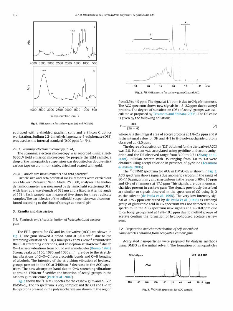
Fig. 1. FTIR spectra for cashew gum (A) and ACG (B).
quipped with z-shielded gradient coils and a Silicon Graphicsorkstation. Sodium 2,2-dimethylsilapentane-5-sulphonate (DSS)as used as the internal standard (0.00 ppm for 1H).
.6.3. Scanning electron microscopy (SEM)The scanning electron microscopy was recorded using a Jeol-
360LV field emission microscope. To prepare the SEM sample, arop of the nanoparticle suspension was deposited on double-stickarbon tape on aluminum stubs, dried and coated with gold.
.6.4. Particle size measurements and zeta potentialParticle size and zeta potential measurements were carried out
n a Malvern Zetasizer Nano, Model ZS 3600, analyzer. The hydro-ynamic diameter was measured by dynamic light scattering (DLS)ith laser at a wavelength of 633 nm and a fixed scattering angle
f 173◦. Each sample was measured five times for three replicateamples. The particle size of the colloidal suspension was also mon-tored according to the time of storage at neutral pH.
. Results and discussion
.1. Synthesis and characterization of hydrophobized cashewum
The FTIR spectra for CG and its derivative (ACG) are shown inig. 1. The gum showed a broad band at 3400 cm−1 due to thetretching vibration of O H, a small peak at 2933 cm−1 attributed tohe C H stretching vibrations, and absorption at 1649 cm−1 due to
H scissor vibrations from bound water molecules (Bueno, 1990).trong peaks at 1150, 1080 and 1030 cm−1 are due to the stretch-ng vibrations of C O C from glycosidic bonds and O H bendingf alcohols. The intensity of the stretching vibration of hydroxylroups present in the CG at 3400 cm−1 decrease in the ACG spec-rum. The new absorption band due to C O stretching vibrationst around 1750 cm−1 verifies the insertion of acetyl groups in the
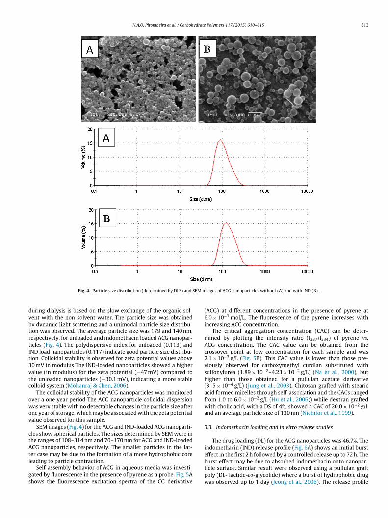
ashew gum structure (Park et al., 2007).Fig. 2 shows the 1H NMR spectra for the cashew gum and ACG inMSO-d6. The CG spectrum is very complex and the OH and H-1 to-6 protons present in the polysaccharide are shown in the region
Fig. 2. 1H NMR spectra for cashew gum (CG) and ACG.
from 3.5 to 4.9 ppm. The signal at 1.1 ppm is due to CH3 of rhamnose.The ACG spectrum shows new signals in 1.8–2.2 ppm due to acetylprotons. The degree of substitution (DS) of acetyl groups was cal-culated as proposed by Teramoto and Shibata (2006). The DS valueis given by the following equation:
DS = 10A
(3B + A)(2)
where A is the integral area of acetyl protons at 1.8–2.2 ppm and Bis the integral value for OH and H-1 to H-6 polysaccharide protonsobserved at >3.5 ppm.
The degree of substitution (DS) obtained for the derivative (ACG)was 2.8. Pullulan was acetylated using pyridine and acetic anhy-dride and the DS observed range from 3.00 to 2.71 (Zhang et al.,2009). Pullulan acetate with DS ranging from 1.0 to 3.0 wereobtained using acetyl chloride in presence of pyridine (Teramoto& Shibata, 2006).
The 13C NMR spectrum for ACG in DMSO-d6 is shown in Fig. 3.ACG spectrum shows signals due anomeric carbons in the range of90–110 ppm, primary and ring carbons in the region of 60 to 85 ppmand CH3 of rhamnose at 17.5 ppm This signals are due monosac-charides present in cashew gum. The signals previously describedare similar to signals observed in the spectrum of CG using D2Oas the solvent (de Paula et al., 1998). The very low intensity sig-nal at 175.7 ppm attributed by de Paula et al. (1998) as carbonylgroup of glucuronic acid in CG spectrum was not detected in ACGspectrum. In the ACG spectrum new signals at 169–168 ppm dueto carbonyl groups and at 19.8–19.5 ppm due to methyl groups ofacetate confirm the formation of hydrophobized acetate cashewgum.
3.2. Preparation and characterization of self-assembled
Fig. 3. 13C NMR spectrum for ACG sample.
N.A.O. Pitombeira et al. / Carbohydrate Polymers 117 (2015) 610–615 613
SEM im
dvbtrtIt3vtc
owov
ctAtl
gs
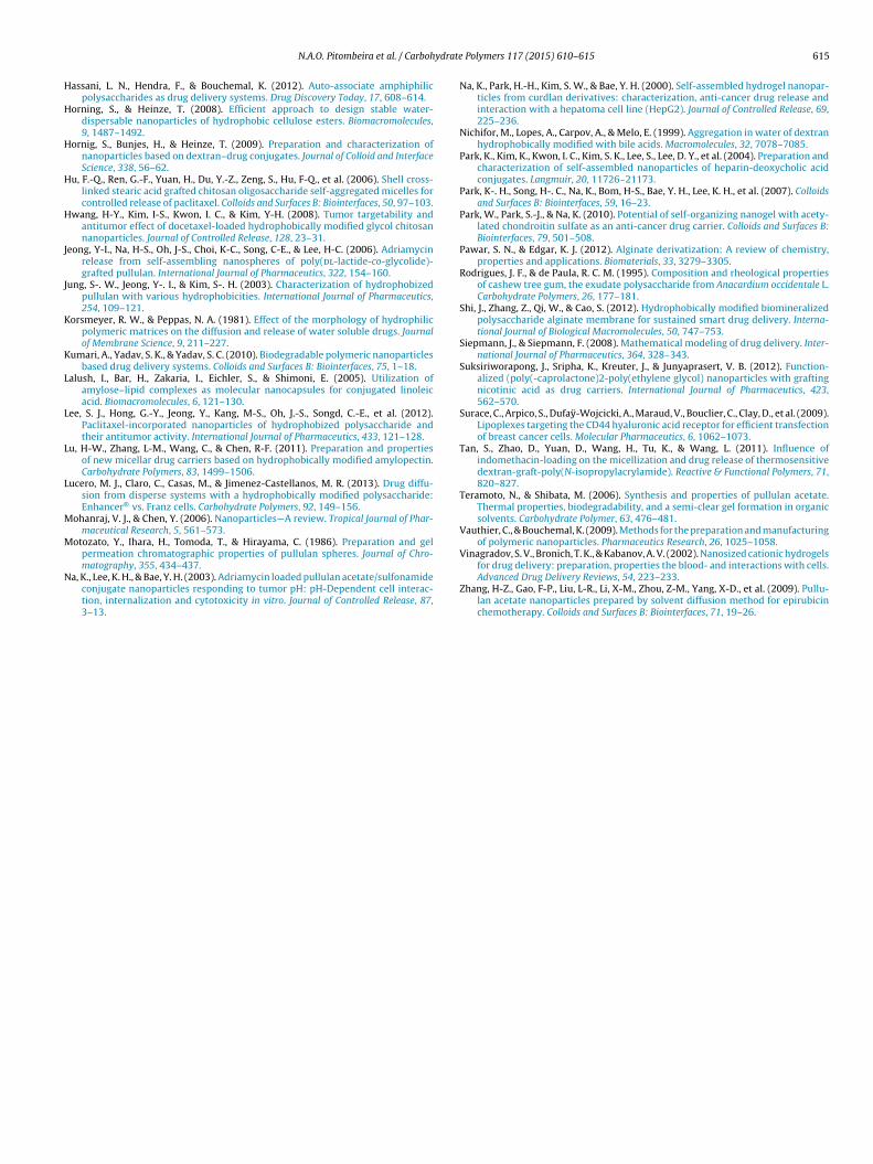
Fig. 4. Particle size distribution (determined by DLS) and
uring dialysis is based on the slow exchange of the organic sol-ent with the non-solvent water. The particle size was obtainedy dynamic light scattering and a unimodal particle size distribu-ion was observed. The average particle size was 179 and 140 nm,espectively, for unloaded and indomethacin loaded ACG nanopar-icles (Fig. 4). The polydispersive index for unloaded (0.113) andND load nanoparticles (0.117) indicate good particle size distribu-ion. Colloidal stability is observed for zeta potential values above0 mV in modulus The IND-loaded nanoparticles showed a higheralue (in modulus) for the zeta potential (−47 mV) compared tohe unloaded nanoparticles (−30.1 mV), indicating a more stableolloid system (Mohanraj & Chen, 2006).
The colloidal stability of the ACG nanoparticles was monitoredver a one year period The ACG nanoparticle colloidal dispersionas very stable with no detectable changes in the particle size after
ne year of storage, which may be associated with the zeta potentialalue observed for this sample.
SEM images (Fig. 4) for the ACG and IND-loaded ACG nanoparti-les show spherical particles. The sizes determined by SEM were inhe ranges of 108–314 nm and 70–170 nm for ACG and IND-loadedCG nanoparticles, respectively. The smaller particles in the lat-
er case may be due to the formation of a more hydrophobic core
eading to particle contraction.Self-assembly behavior of ACG in aqueous media was investi-ated by fluorescence in the presence of pyrene as a probe. Fig. 5Ahows the fluorescence excitation spectra of the CG derivative
ages of ACG nanoparticles without (A) and with IND (B).
(ACG) at different concentrations in the presence of pyrene at6.0 × 10−7 mol/L. The fluorescence of the pyrene increases withincreasing ACG concentration.
The critical aggregation concentration (CAC) can be deter-mined by plotting the intensity ratio (I337/I334) of pyrene vs.ACG concentration. The CAC value can be obtained from thecrossover point at low concentration for each sample and was2.1 × 10−3 g/L (Fig. 5B). This CAC value is lower than those pre-viously observed for carboxymethyl curdlan substituted withsulfonylurea (1.89 × 10⁻²–4.23 × 10⁻² g/L) (Na et al., 2000), buthigher than those obtained for a pullulan acetate derivative(3–5 × 10⁻⁴ g/L) (Jung et al., 2003). Chitosan grafted with stearicacid formed micelles through self-association and the CACs rangedfrom 1.0 to 6.0 × 10⁻² g/L (Hu et al., 2006;) while dextran graftedwith cholic acid, with a DS of 4%, showed a CAC of 20.0 × 10⁻² g/Land an average particle size of 130 nm (Nichifor et al., 1999).
3.3. Indomethacin loading and in vitro release studies
The drug loading (DL) for the ACG nanoparticles was 46.7%. Theindomethacin (IND) release profile (Fig. 6A) shows an initial bursteffect in the first 2 h followed by a controlled release up to 72 h. The
burst effect may be due to absorbed indomethacin onto nanopar-ticle surface. Similar result were observed using a pullulan graftpoly (DL- lactide-co-glycolide) where a burst of hydrophobic drugwas observed up to 1 day (Jeong et al., 2006). The release profile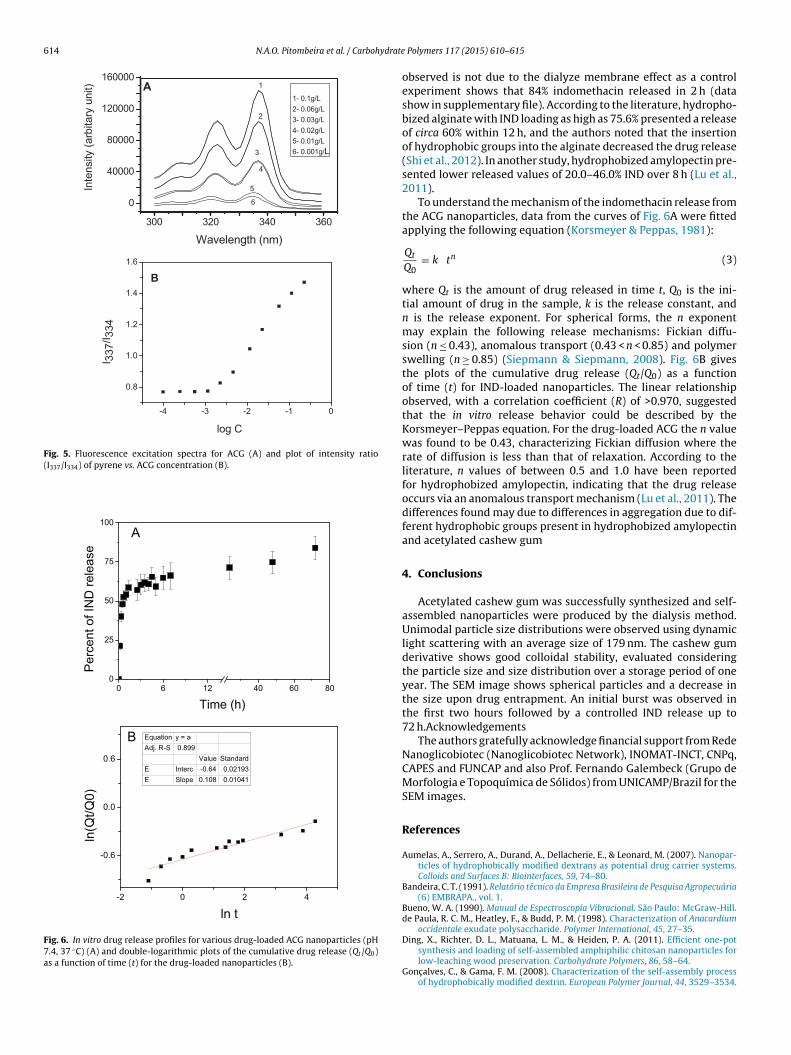
614 N.A.O. Pitombeira et al. / Carbohydrate
300 32 0 34 0 36 0
0
4000 0
8000 0
12000 0
16000 0
Waveleng th (nm )
Inte
nsity
(arb
itary
uni
t)
6
5
4
3
2
1- 0.1g/L2- 0.06g/L3- 0.03g/L4- 0.02g/L5- 0.01g/L6- 0.001g /L
1A
-4 -3 -2 -1 0
0.8
1.0
1.2
1.4
1.6
log C
I 337
/I 334
B
Fig. 5. Fluorescence excitation spectra for ACG (A) and plot of intensity ratio(I337/I334) of pyrene vs. ACG concentration (B).
0 6 12 40 60 800
25
50
75
100
Per
cent
of I
ND
rele
ase
Time (h )
A
-2 0 2 4
-0.6
0.0
0.6
ln(Q
t/Q0)
ln t
Equation y = a Adj. R-S 0.89 9
Value Standar dE Inte rc -0.6 4 0.0219 3E Slope 0.108 0.01041
B
Fig. 6. In vitro drug release profiles for various drug-loaded ACG nanoparticles (pH7.4, 37 ◦C) (A) and double-logarithmic plots of the cumulative drug release (Qt /Q0)as a function of time (t) for the drug-loaded nanoparticles (B).
Polymers 117 (2015) 610–615
observed is not due to the dialyze membrane effect as a controlexperiment shows that 84% indomethacin released in 2 h (datashow in supplementary file). According to the literature, hydropho-bized alginate with IND loading as high as 75.6% presented a releaseof circa 60% within 12 h, and the authors noted that the insertionof hydrophobic groups into the alginate decreased the drug release(Shi et al., 2012). In another study, hydrophobized amylopectin pre-sented lower released values of 20.0–46.0% IND over 8 h (Lu et al.,2011).
To understand the mechanism of the indomethacin release fromthe ACG nanoparticles, data from the curves of Fig. 6A were fittedapplying the following equation (Korsmeyer & Peppas, 1981):
Qt
Q0= k tn (3)
where Qt is the amount of drug released in time t, Q0 is the ini-tial amount of drug in the sample, k is the release constant, andn is the release exponent. For spherical forms, the n exponentmay explain the following release mechanisms: Fickian diffu-sion (n ≤ 0.43), anomalous transport (0.43 < n < 0.85) and polymerswelling (n ≥ 0.85) (Siepmann & Siepmann, 2008). Fig. 6B givesthe plots of the cumulative drug release (Qt/Q0) as a functionof time (t) for IND-loaded nanoparticles. The linear relationshipobserved, with a correlation coefficient (R) of >0.970, suggestedthat the in vitro release behavior could be described by theKorsmeyer–Peppas equation. For the drug-loaded ACG the n valuewas found to be 0.43, characterizing Fickian diffusion where therate of diffusion is less than that of relaxation. According to theliterature, n values of between 0.5 and 1.0 have been reportedfor hydrophobized amylopectin, indicating that the drug releaseoccurs via an anomalous transport mechanism (Lu et al., 2011). Thedifferences found may due to differences in aggregation due to dif-ferent hydrophobic groups present in hydrophobized amylopectinand acetylated cashew gum
4. Conclusions
Acetylated cashew gum was successfully synthesized and self-assembled nanoparticles were produced by the dialysis method.Unimodal particle size distributions were observed using dynamiclight scattering with an average size of 179 nm. The cashew gumderivative shows good colloidal stability, evaluated consideringthe particle size and size distribution over a storage period of oneyear. The SEM image shows spherical particles and a decrease inthe size upon drug entrapment. An initial burst was observed inthe first two hours followed by a controlled IND release up to72 h.Acknowledgements
The authors gratefully acknowledge financial support from RedeNanoglicobiotec (Nanoglicobiotec Network), INOMAT-INCT, CNPq,CAPES and FUNCAP and also Prof. Fernando Galembeck (Grupo deMorfologia e Topoquímica de Sólidos) from UNICAMP/Brazil for theSEM images.
References
Aumelas, A., Serrero, A., Durand, A., Dellacherie, E., & Leonard, M. (2007). Nanopar-ticles of hydrophobically modified dextrans as potential drug carrier systems.Colloids and Surfaces B: Biointerfaces, 59, 74–80.
Bandeira, C. T. (1991). Relatório técnico da Empresa Brasileira de Pesquisa Agropecuária(6) EMBRAPA., vol. 1.
Bueno, W. A. (1990). Manual de Espectroscopia Vibracional. São Paulo: McGraw-Hill.de Paula, R. C. M., Heatley, F., & Budd, P. M. (1998). Characterization of Anacardium
occidentale exudate polysaccharide. Polymer International, 45, 27–35.
Ding, X., Richter, D. L., Matuana, L. M., & Heiden, P. A. (2011). Efficient one-potsynthesis and loading of self-assembled amphiphilic chitosan nanoparticles forlow-leaching wood preservation. Carbohydrate Polymers, 86, 58–64.
Gonc alves, C., & Gama, F. M. (2008). Characterization of the self-assembly processof hydrophobically modified dextrin. European Polymer Journal, 44, 3529–3534.
ydrate
H
H
H
H
H
J
J
K
K
L
L
L
L
M
M
N
N.A.O. Pitombeira et al. / Carboh
assani, L. N., Hendra, F., & Bouchemal, K. (2012). Auto-associate amphiphilicpolysaccharides as drug delivery systems. Drug Discovery Today, 17, 608–614.
orning, S., & Heinze, T. (2008). Efficient approach to design stable water-dispersable nanoparticles of hydrophobic cellulose esters. Biomacromolecules,9, 1487–1492.
ornig, S., Bunjes, H., & Heinze, T. (2009). Preparation and characterization ofnanoparticles based on dextran–drug conjugates. Journal of Colloid and InterfaceScience, 338, 56–62.
u, F.-Q., Ren, G.-F., Yuan, H., Du, Y.-Z., Zeng, S., Hu, F-Q., et al. (2006). Shell cross-linked stearic acid grafted chitosan oligosaccharide self-aggregated micelles forcontrolled release of paclitaxel. Colloids and Surfaces B: Biointerfaces, 50, 97–103.
wang, H-Y., Kim, I-S., Kwon, I. C., & Kim, Y-H. (2008). Tumor targetability andantitumor effect of docetaxel-loaded hydrophobically modified glycol chitosannanoparticles. Journal of Controlled Release, 128, 23–31.
eong, Y-I., Na, H-S., Oh, J-S., Choi, K-C., Song, C-E., & Lee, H-C. (2006). Adriamycinrelease from self-assembling nanospheres of poly(dl-lactide-co-glycolide)-grafted pullulan. International Journal of Pharmaceutics, 322, 154–160.
ung, S-. W., Jeong, Y-. I., & Kim, S-. H. (2003). Characterization of hydrophobizedpullulan with various hydrophobicities. International Journal of Pharmaceutics,254, 109–121.
orsmeyer, R. W., & Peppas, N. A. (1981). Effect of the morphology of hydrophilicpolymeric matrices on the diffusion and release of water soluble drugs. Journalof Membrane Science, 9, 211–227.
umari, A., Yadav, S. K., & Yadav, S. C. (2010). Biodegradable polymeric nanoparticlesbased drug delivery systems. Colloids and Surfaces B: Biointerfaces, 75, 1–18.
alush, I., Bar, H., Zakaria, I., Eichler, S., & Shimoni, E. (2005). Utilization ofamylose–lipid complexes as molecular nanocapsules for conjugated linoleicacid. Biomacromolecules, 6, 121–130.
ee, S. J., Hong, G.-Y., Jeong, Y., Kang, M-S., Oh, J.-S., Songd, C.-E., et al. (2012).Paclitaxel-incorporated nanoparticles of hydrophobized polysaccharide andtheir antitumor activity. International Journal of Pharmaceutics, 433, 121–128.
u, H-W., Zhang, L-M., Wang, C., & Chen, R-F. (2011). Preparation and propertiesof new micellar drug carriers based on hydrophobically modified amylopectin.Carbohydrate Polymers, 83, 1499–1506.
ucero, M. J., Claro, C., Casas, M., & Jimenez-Castellanos, M. R. (2013). Drug diffu-sion from disperse systems with a hydrophobically modified polysaccharide:Enhancer® vs. Franz cells. Carbohydrate Polymers, 92, 149–156.
ohanraj, V. J., & Chen, Y. (2006). Nanoparticles—A review. Tropical Journal of Phar-maceutical Research, 5, 561–573.
otozato, Y., Ihara, H., Tomoda, T., & Hirayama, C. (1986). Preparation and gelpermeation chromatographic properties of pullulan spheres. Journal of Chro-
matography, 355, 434–437.a, K., Lee, K. H., & Bae, Y. H. (2003). Adriamycin loaded pullulan acetate/sulfonamideconjugate nanoparticles responding to tumor pH: pH-Dependent cell interac-tion, internalization and cytotoxicity in vitro. Journal of Controlled Release, 87,3–13.
Polymers 117 (2015) 610–615 615
Na, K., Park, H.-H., Kim, S. W., & Bae, Y. H. (2000). Self-assembled hydrogel nanopar-ticles from curdlan derivatives: characterization, anti-cancer drug release andinteraction with a hepatoma cell line (HepG2). Journal of Controlled Release, 69,225–236.
Nichifor, M., Lopes, A., Carpov, A., & Melo, E. (1999). Aggregation in water of dextranhydrophobically modified with bile acids. Macromolecules, 32, 7078–7085.
Park, K., Kim, K., Kwon, I. C., Kim, S. K., Lee, S., Lee, D. Y., et al. (2004). Preparation andcharacterization of self-assembled nanoparticles of heparin-deoxycholic acidconjugates. Langmuir, 20, 11726–21173.
Park, K-. H., Song, H-. C., Na, K., Bom, H-S., Bae, Y. H., Lee, K. H., et al. (2007). Colloidsand Surfaces B: Biointerfaces, 59, 16–23.
Park, W., Park, S.-J., & Na, K. (2010). Potential of self-organizing nanogel with acety-lated chondroitin sulfate as an anti-cancer drug carrier. Colloids and Surfaces B:Biointerfaces, 79, 501–508.
Pawar, S. N., & Edgar, K. J. (2012). Alginate derivatization: A review of chemistry,properties and applications. Biomaterials, 33, 3279–3305.
Rodrigues, J. F., & de Paula, R. C. M. (1995). Composition and rheological propertiesof cashew tree gum, the exudate polysaccharide from Anacardium occidentale L.Carbohydrate Polymers, 26, 177–181.
Shi, J., Zhang, Z., Qi, W., & Cao, S. (2012). Hydrophobically modified biomineralizedpolysaccharide alginate membrane for sustained smart drug delivery. Interna-tional Journal of Biological Macromolecules, 50, 747–753.
Siepmann, J., & Siepmann, F. (2008). Mathematical modeling of drug delivery. Inter-national Journal of Pharmaceutics, 364, 328–343.
Suksiriworapong, J., Sripha, K., Kreuter, J., & Junyaprasert, V. B. (2012). Function-alized (poly(-caprolactone)2-poly(ethylene glycol) nanoparticles with graftingnicotinic acid as drug carriers. International Journal of Pharmaceutics, 423,562–570.
Surace, C., Arpico, S., Dufay-Wojcicki, A., Maraud, V., Bouclier, C., Clay, D., et al. (2009).Lipoplexes targeting the CD44 hyaluronic acid receptor for efficient transfectionof breast cancer cells. Molecular Pharmaceutics, 6, 1062–1073.
Tan, S., Zhao, D., Yuan, D., Wang, H., Tu, K., & Wang, L. (2011). Influence ofindomethacin-loading on the micellization and drug release of thermosensitivedextran-graft-poly(N-isopropylacrylamide). Reactive & Functional Polymers, 71,820–827.
Teramoto, N., & Shibata, M. (2006). Synthesis and properties of pullulan acetate.Thermal properties, biodegradability, and a semi-clear gel formation in organicsolvents. Carbohydrate Polymer, 63, 476–481.
Vauthier, C., & Bouchemal, K. (2009). Methods for the preparation and manufacturingof polymeric nanoparticles. Pharmaceutics Research, 26, 1025–1058.
Vinagradov, S. V., Bronich, T. K., & Kabanov, A. V. (2002). Nanosized cationic hydrogels
for drug delivery: preparation, properties the blood- and interactions with cells.Advanced Drug Delivery Reviews, 54, 223–233.Zhang, H-Z., Gao, F-P., Liu, L-R., Li, X-M., Zhou, Z-M., Yang, X-D., et al. (2009). Pullu-lan acetate nanoparticles prepared by solvent diffusion method for epirubicinchemotherapy. Colloids and Surfaces B: Biointerfaces, 71, 19–26.